
Staphylococcus aureus infective endocarditis and septic pulmonary embolism after septic abortion
Upload
khangminh22Category
view
0download
0
Endocarditis: Evaluation and ManagementRekha Mankad, MD, FACC
Assistant Professor of Medicine
Mayo Clinic College of Medicine
Director, Women’s Heart Clinic
Mayo Clinic, Rochester, MN
@RMankadMD

Disclosure Information
Relevant Financial Relationship(s)None
Off Label UsageNone
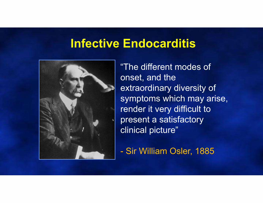
“The different modes of onset, and the extraordinary diversity of symptoms which may arise, render it very difficult to present a satisfactory clinical picture”
- Sir William Osler, 1885
Infective Endocarditis
What is the annual incidence of IE in contemporary Western cohorts?
1. 5-7/100,000 person years
2. 50-70/100,000 person years
3. 5-7/1000 person years
4. 5-7/10,000 person years4
Epidemiology
• Infective endocarditis (IE) is uncommon– Annual incidence of 5-7/100,000 person years
• Associated with significant morbidity and mortality– 3rd most life threatening infection after sepsis/ pneumonia &
intra-abdominal abscess
• Male: Female approximately 2:1
• Age of onset > 60 yo (men 6-7 years older than women)
• Uncommon in children (when occurs typically due to congenital heart disease)
• Mitral valve > aortic valve >> tricuspid valve 5

Major Criteria Minor Criteria
Positive blood culture for IE with typical organism
Predisposition: predisposing heart condition or IVDU
Persistently positive blood cultures for any organism
Fever ≥ 380C
Single positive blood culture for C.burnetti
Vascular phenomena: arterial embolism, septic pulmonary infarcts, mycotic aneurysm, ICH, Janeway lesions
Echocardiogram positive for IE Microbiologic evidence that does not meet major criteria
Positive blood culture not meeting major criteria
Immunologic phenomena 6
Diagnosis of Infective Endocarditis
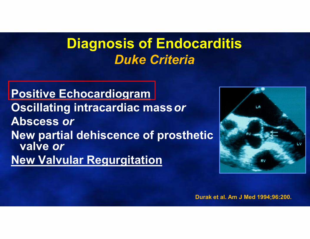
Positive EchocardiogramOscillating intracardiac massor Abscess orNew partial dehiscence of prosthetic
valve orNew Valvular Regurgitation
Diagnosis of EndocarditisDuke Criteria
Durak et al. Am J Med 1994;96:200.
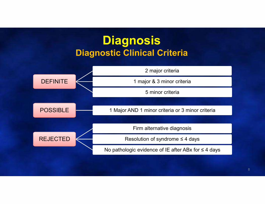
DiagnosisDiagnostic Clinical Criteria
8
DEFINITE
2 major criteria
1 major & 3 minor criteria
5 minor criteria
POSSIBLE 1 Major AND 1 minor criteria or 3 minor criteria
REJECTED
Firm alternative diagnosis
Resolution of syndrome ≤ 4 days
No pathologic evidence of IE after ABx for ≤ 4 days
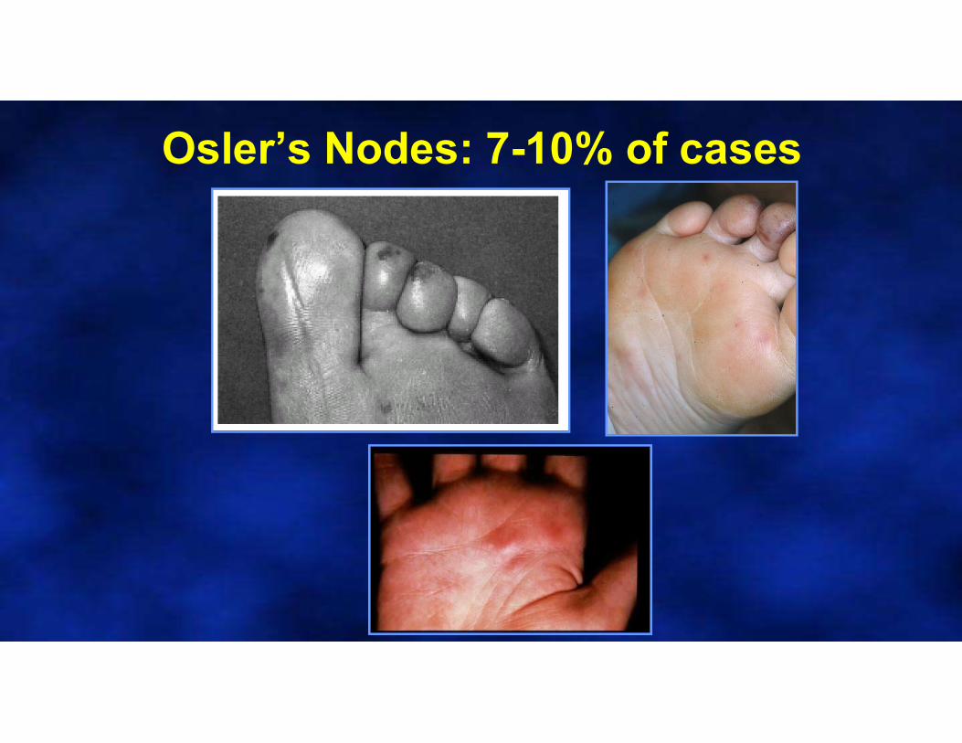
Osler’s Nodes: 7-10% of cases
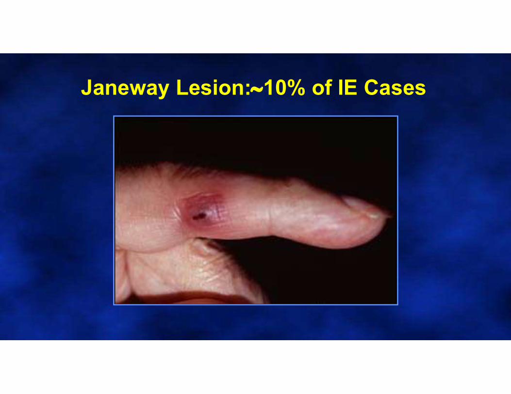
Janeway Lesion:10% of IE Cases
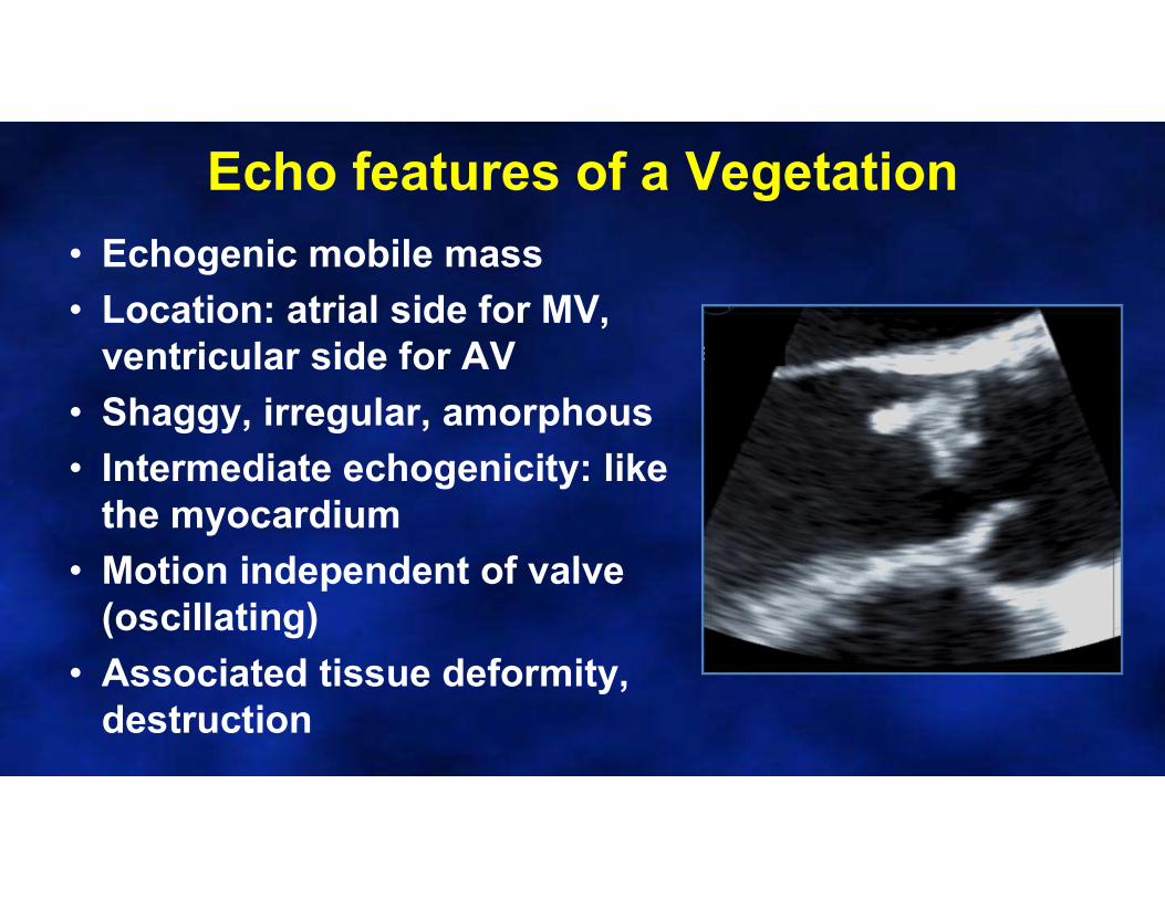
Echo features of a Vegetation
• Echogenic mobile mass
• Location: atrial side for MV, ventricular side for AV
• Shaggy, irregular, amorphous
• Intermediate echogenicity: like the myocardium
• Motion independent of valve (oscillating)
• Associated tissue deformity, destruction
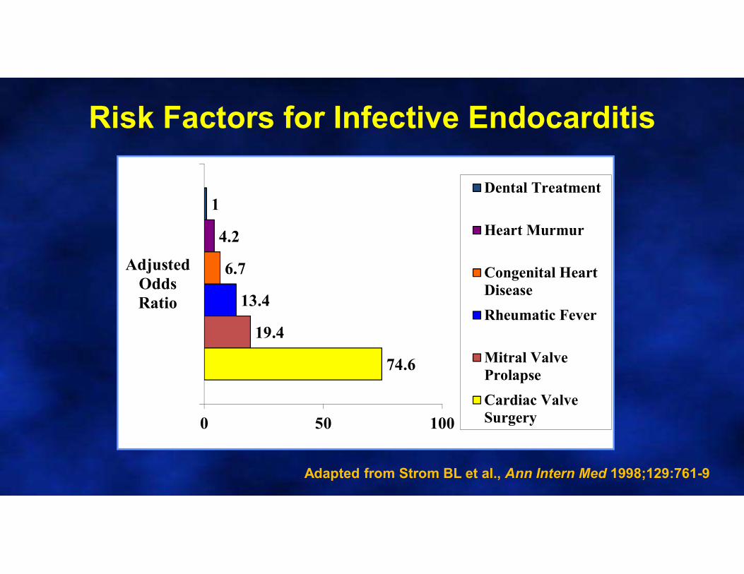
Risk Factors for Infective Endocarditis
74.6
19.4
13.4
6.7
4.2
1
0 50 100
AdjustedOddsRatio
Dental Treatment
Heart Murmur
Congenital HeartDisease
Rheumatic Fever
Mitral ValveProlapse
Cardiac ValveSurgery
Adapted from Strom BL et al., Ann Intern Med 1998;129:761-9

Endocarditis Prevention
13
Who needs prophylaxis?
Prior IE
Prosthetic valves
Congenital Heart Disease
Valvulopathyafter cardiac transplantation
Unrepaired cyanotic congenital heart disease
Completely repaired CHD with prosthetic materials placed within 6 months
CHD repair with residual defects next to prosthetic materials
Includes TAVR valves and patients with prosthetic material used in valve repair
Case27 year old pregnant woman with cough
• 17 weeks pregnant
• 1-2 weeks of productive cough
– Scant hemoptysis
• ROS: Subjective fevers, dizziness
Courtesy of Dr. Anavekar
Case27 year old pregnant woman with cough
• Vital Signs
• BP 103/67 mmHg, HR 130 bpm, RR 24, Temp 38.90C
• HEENT: JVP mildly elevated
• Resp: Good air intensity bilaterally, scattered areas of wheeze and crackles
• CV: Tachycardic, regular rhythm, II / VI holosystolicmurmur
• Ext: 1+ pitting edema
Case
• Labs: Blood cultures growing S. aureus
–3 of 3 bottles in 8 hours
–Blood work: Hgb 8.0, WBC 17.8, Plt26K, Sodium 120, Creatinine 0.6
What is the most appropriate next diagnostic step?
1. Cardiac CT
2. Cardiac MRI
3. Transthoracic echocardiogram
4. Transesophageal echocardiogram
5. PET/CT
©2016 MFMER
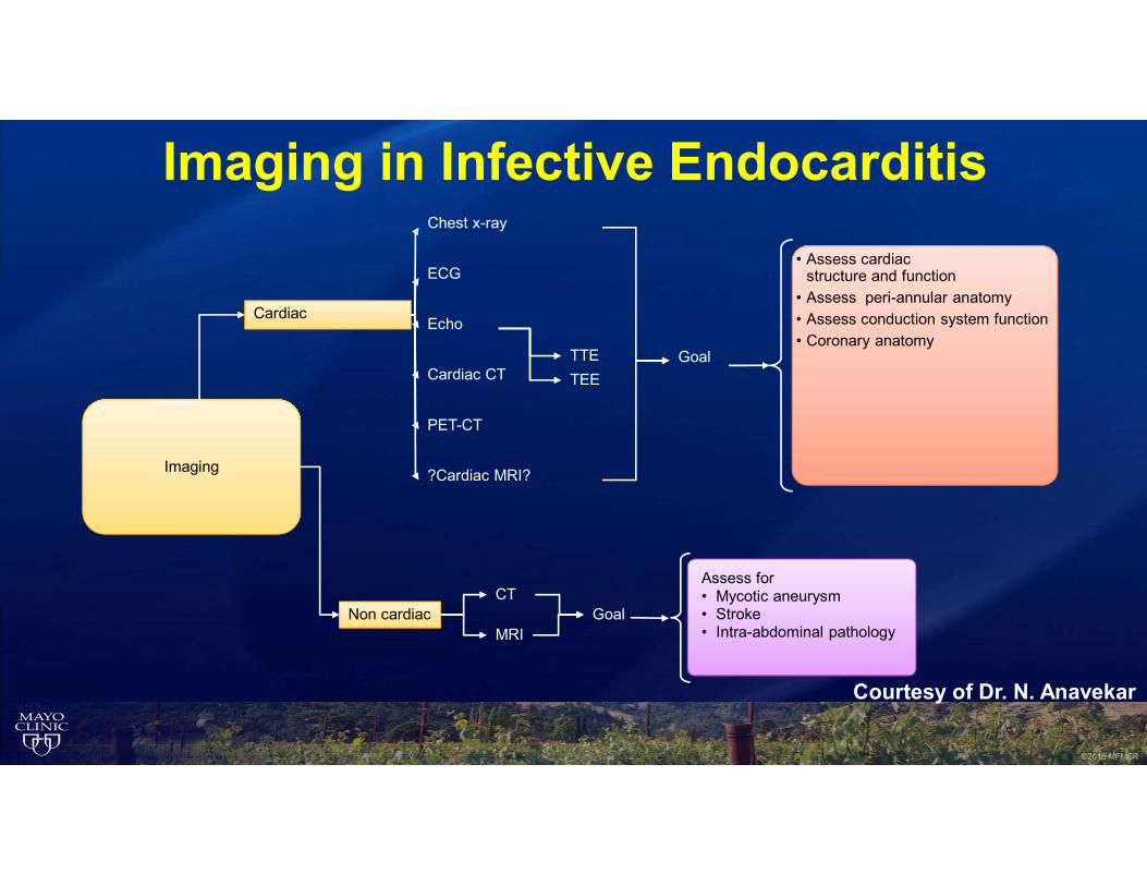
Imaging in Infective Endocarditis
Imaging
Non cardiacCT
MRI
Cardiac
Chest x-ray
ECG
Echo
Cardiac CT
PET-CT
?Cardiac MRI?
TTE
TEE
Goal
• Assess cardiacstructure and function
• Assess peri-annular anatomy• Assess conduction system function• Coronary anatomy
Assess for• Mycotic aneurysm• Stroke• Intra-abdominal pathology
Goal
Courtesy of Dr. N. Anavekar
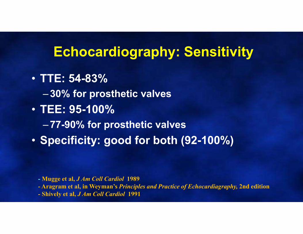
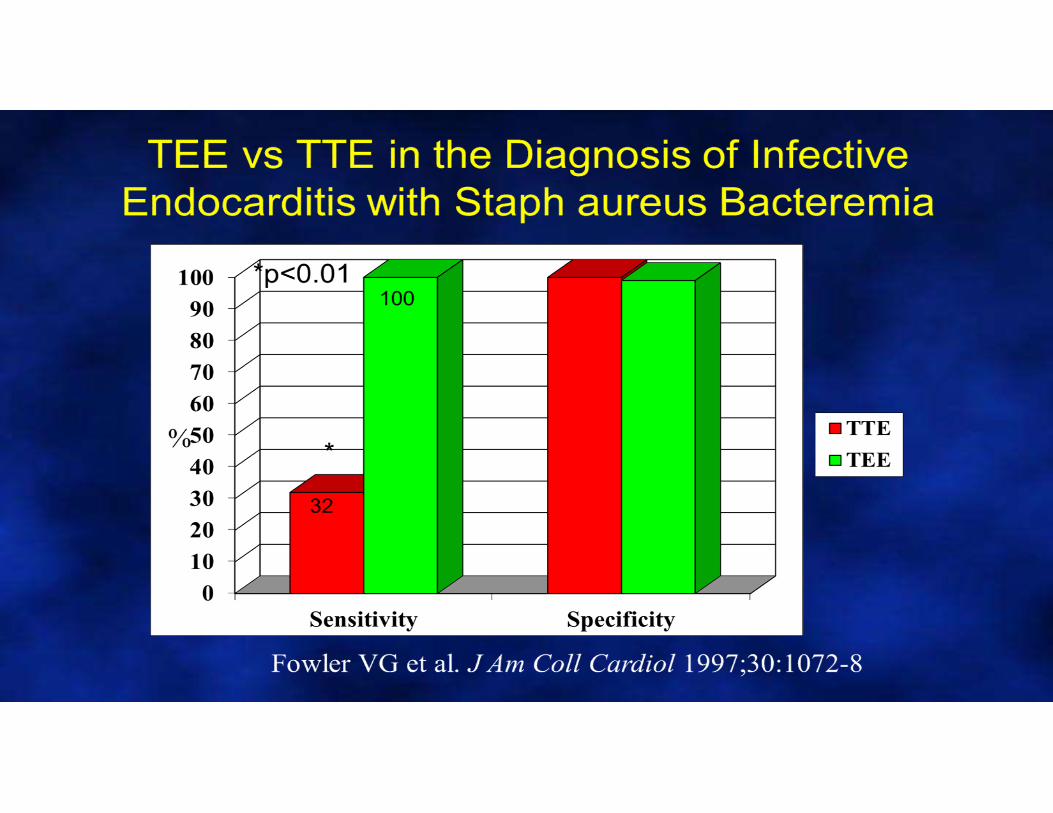
Echocardiography: Sensitivity
• TTE: 54-83% – 30% for prosthetic valves
• TEE: 95-100% – 77-90% for prosthetic valves
• Specificity: good for both (92-100%)
- Mugge et al, J Am Coll Cardiol 1989- Aragram et al, in Weyman’s Principles and Practice of Echocardiagraphy, 2nd edition- Shively et al, J Am Coll Cardiol 1991
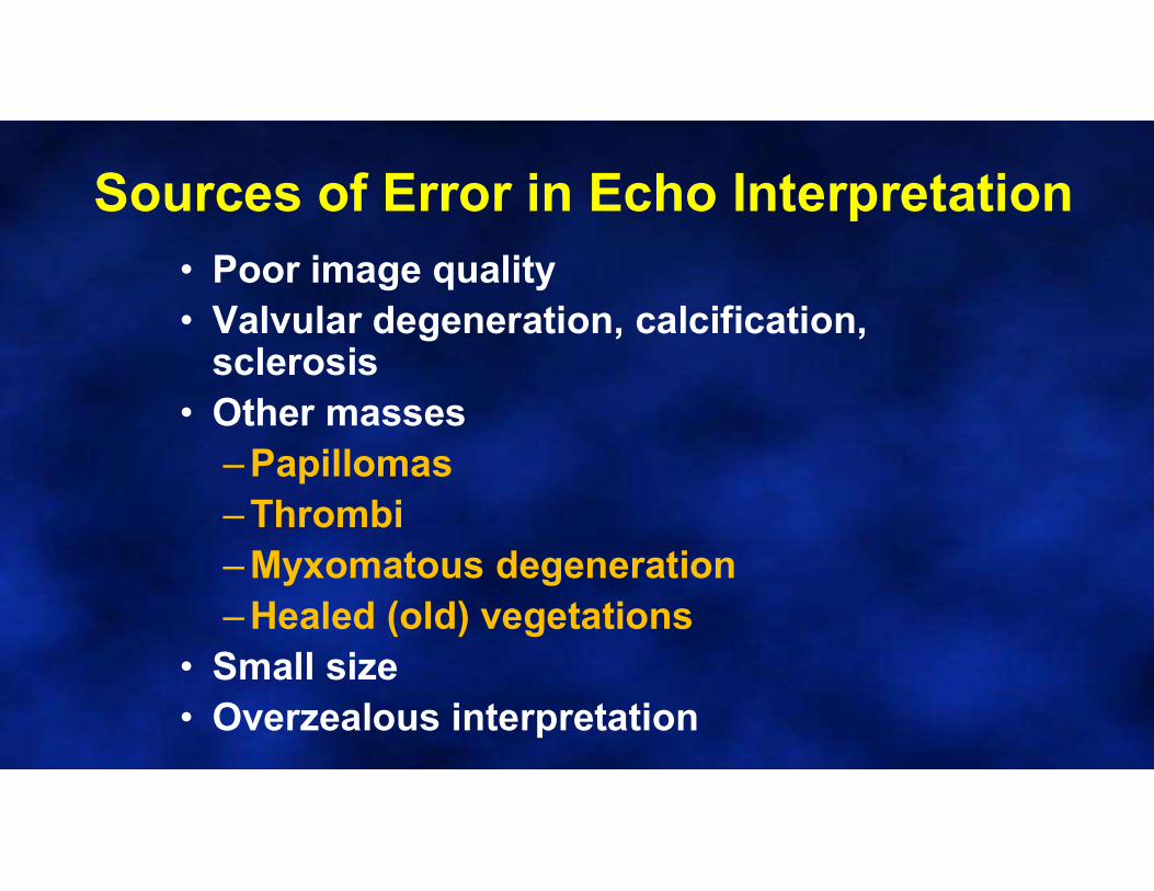
Sources of Error in Echo Interpretation• Poor image quality• Valvular degeneration, calcification,
sclerosis• Other masses
– Papillomas– Thrombi– Myxomatous degeneration– Healed (old) vegetations
• Small size• Overzealous interpretation
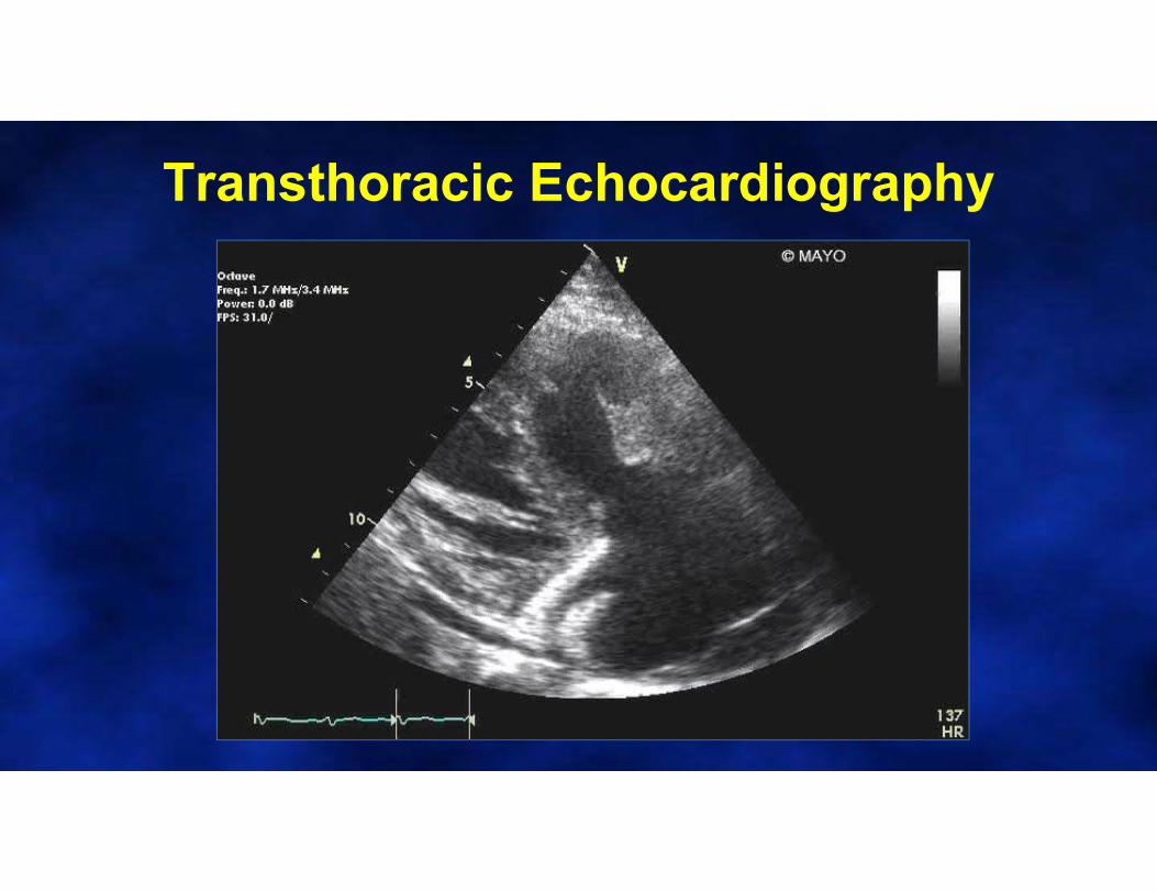
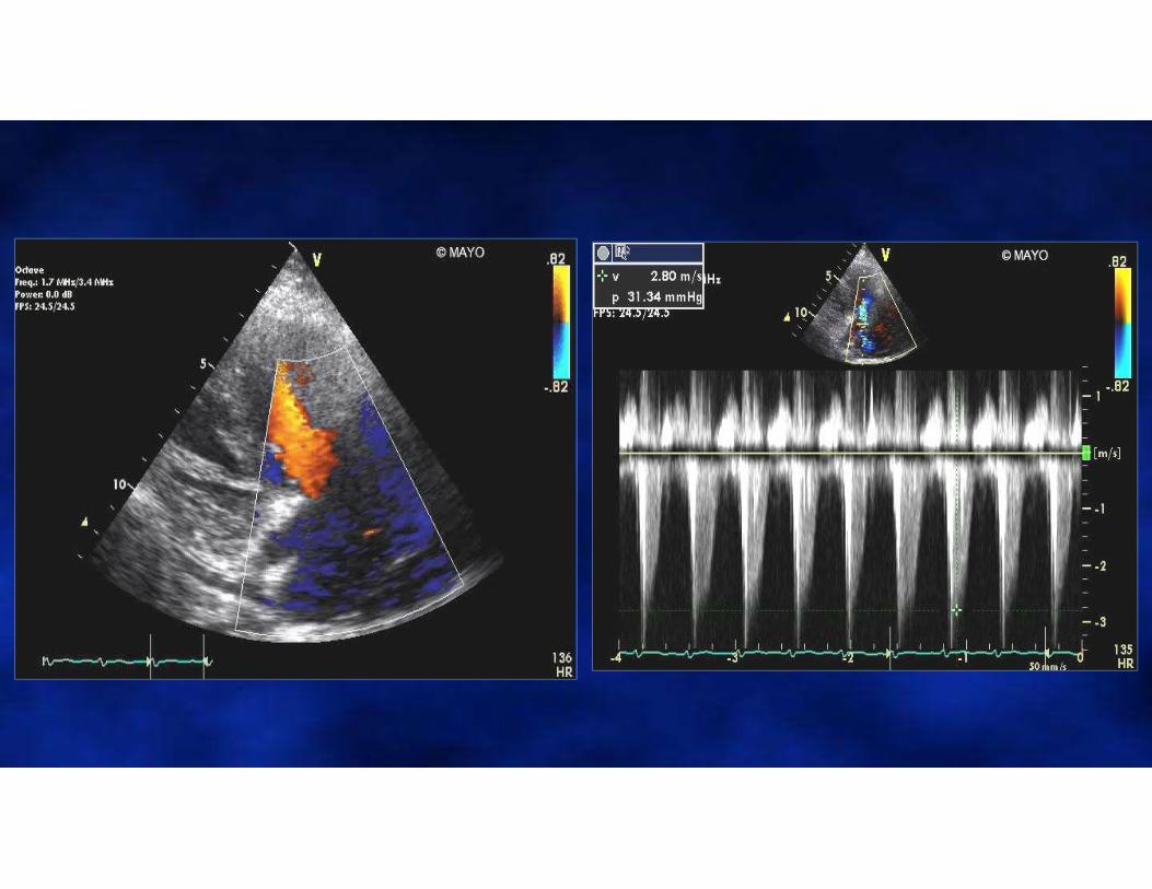
Transthoracic Echocardiography
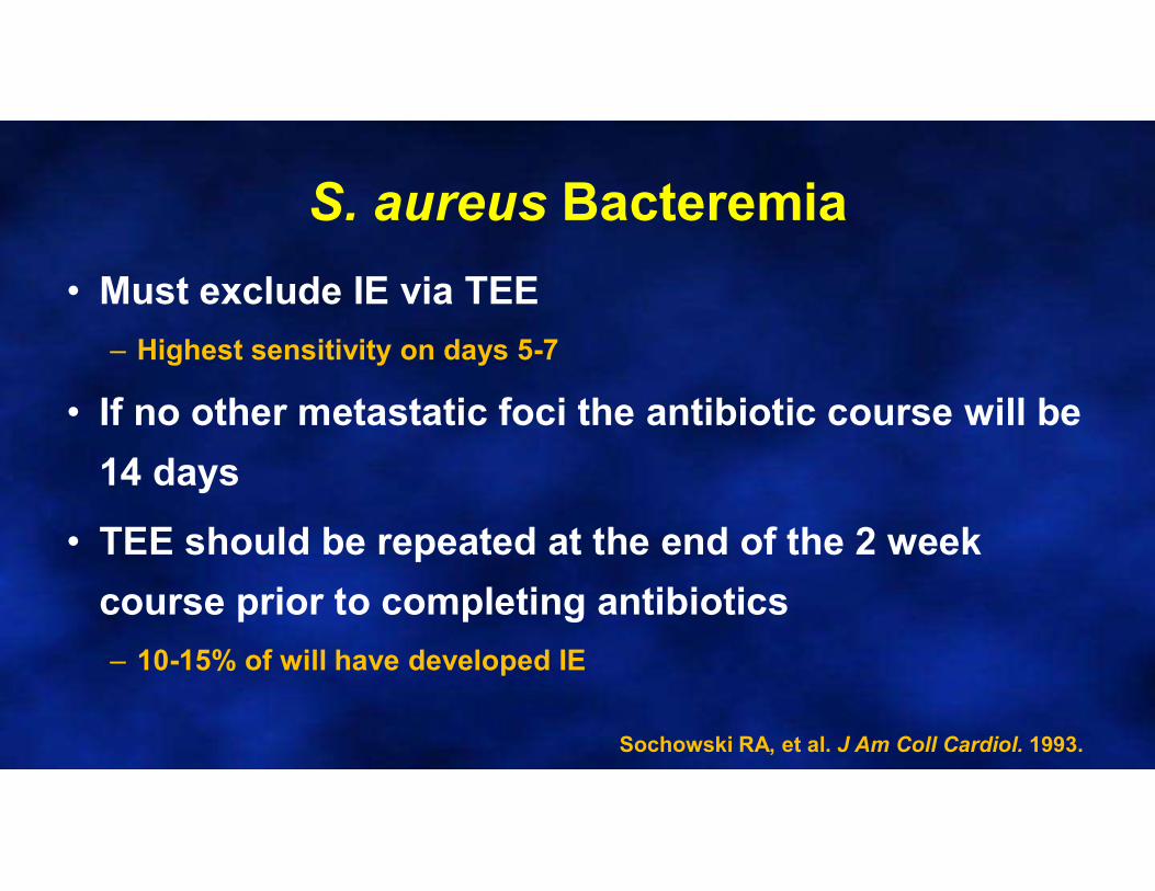
S. aureus Bacteremia
• Must exclude IE via TEE
– Highest sensitivity on days 5-7
• If no other metastatic foci the antibiotic course will be
14 days
• TEE should be repeated at the end of the 2 week
course prior to completing antibiotics
– 10-15% of will have developed IE
Sochowski RA, et al. J Am Coll Cardiol. 1993.
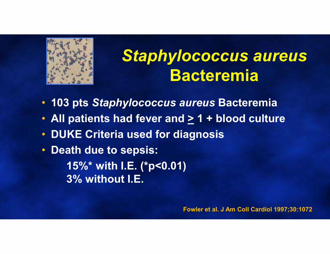
Staphylococcus aureusBacteremia
• 103 pts Staphylococcus aureus Bacteremia
• All patients had fever and > 1 + blood culture
• DUKE Criteria used for diagnosis
• Death due to sepsis:
15%* with I.E. (*p<0.01)3% without I.E.
Fowler et al. J Am Coll Cardiol 1997;30:1072

Right-sided Infective Endocarditis• Associated with IV drug abuse or Indwelling
catheters/devices
• Septic pulmonary emboli– Often multifocal and cavitating
• Right heart failure– Dyspnea on exertion
– JVD + Lower extremity edema
• Perivalvular extension of infection– Increased mortality (23%)
– Increased embolic risk (64%)
Omari B, et al. Chest. 1989.Daniel WG, et al. N Engl J Med. 1991.

Case continued
• Hospital day 14 – clinical deterioration
– Low grade fevers
– Rising leukocytosis
– TEE performed
• To assess for progression of cardiac disease
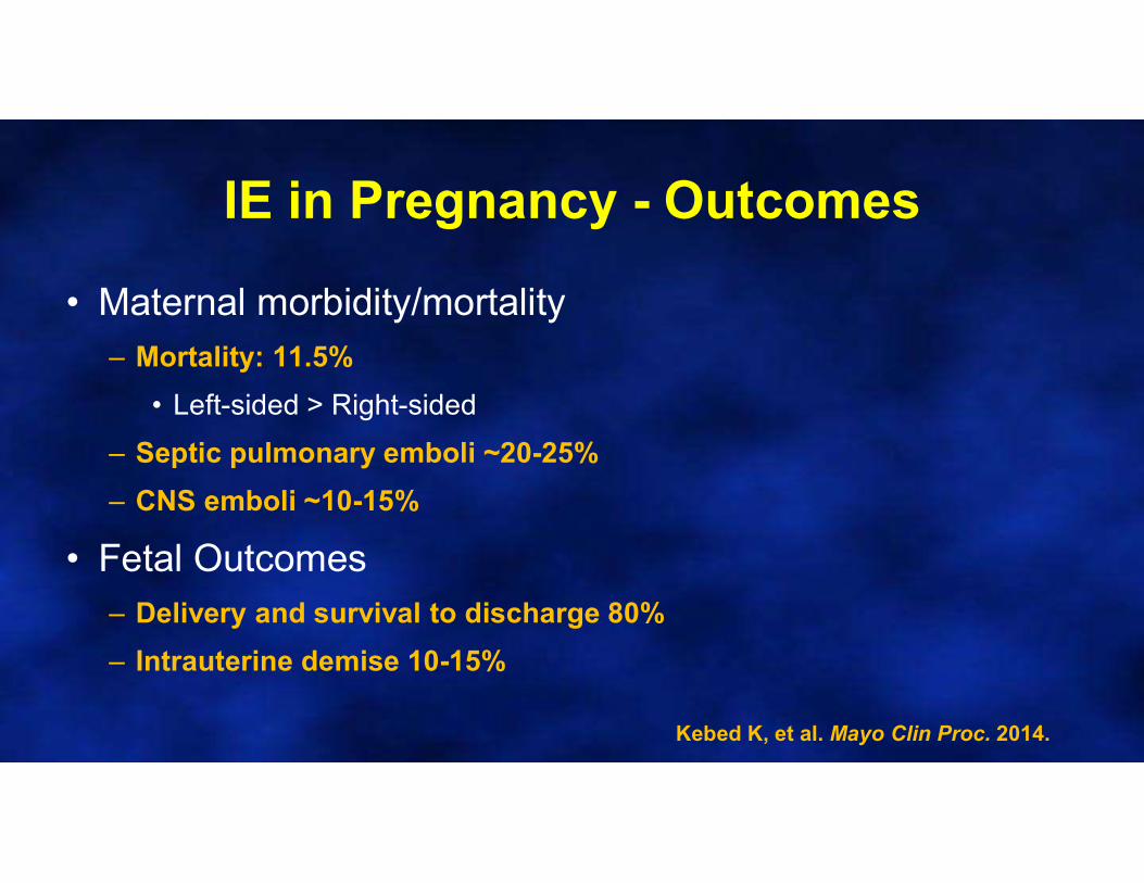
IE in Pregnancy - Outcomes
• Maternal morbidity/mortality– Mortality: 11.5%
• Left-sided > Right-sided
– Septic pulmonary emboli ~20-25%
– CNS emboli ~10-15%
• Fetal Outcomes– Delivery and survival to discharge 80%
– Intrauterine demise 10-15%
Kebed K, et al. Mayo Clin Proc. 2014.
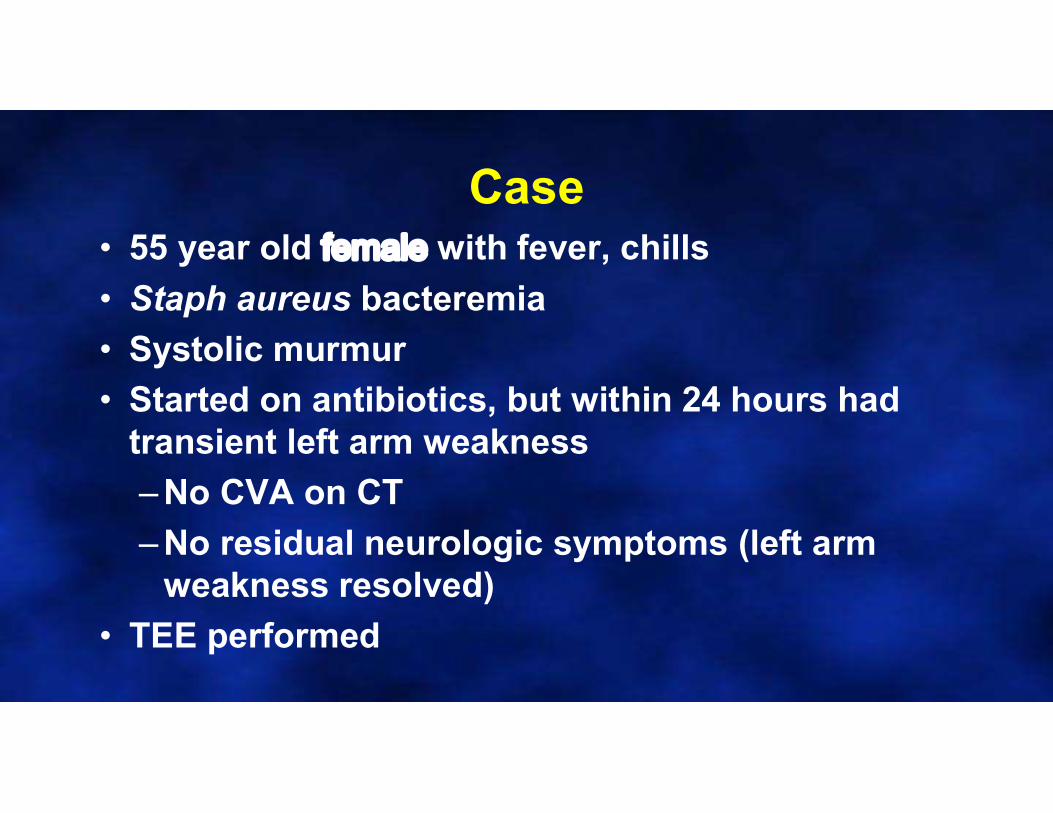
Case• 55 year old female with fever, chills
• Staph aureus bacteremia
• Systolic murmur
• Started on antibiotics, but within 24 hours had transient left arm weakness
– No CVA on CT
– No residual neurologic symptoms (left arm weakness resolved)
• TEE performed
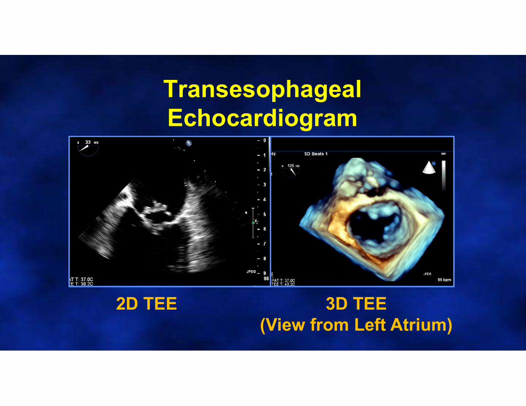
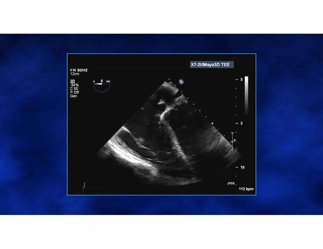
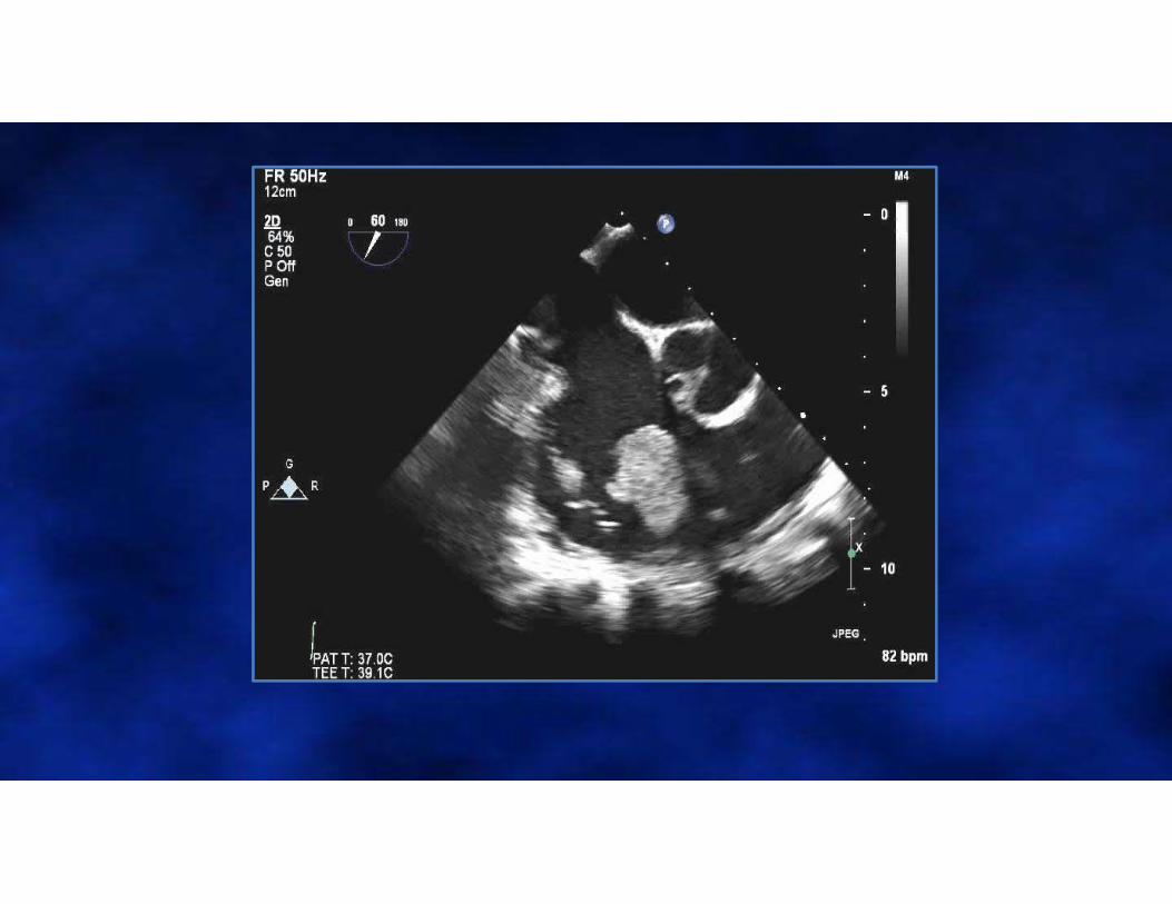
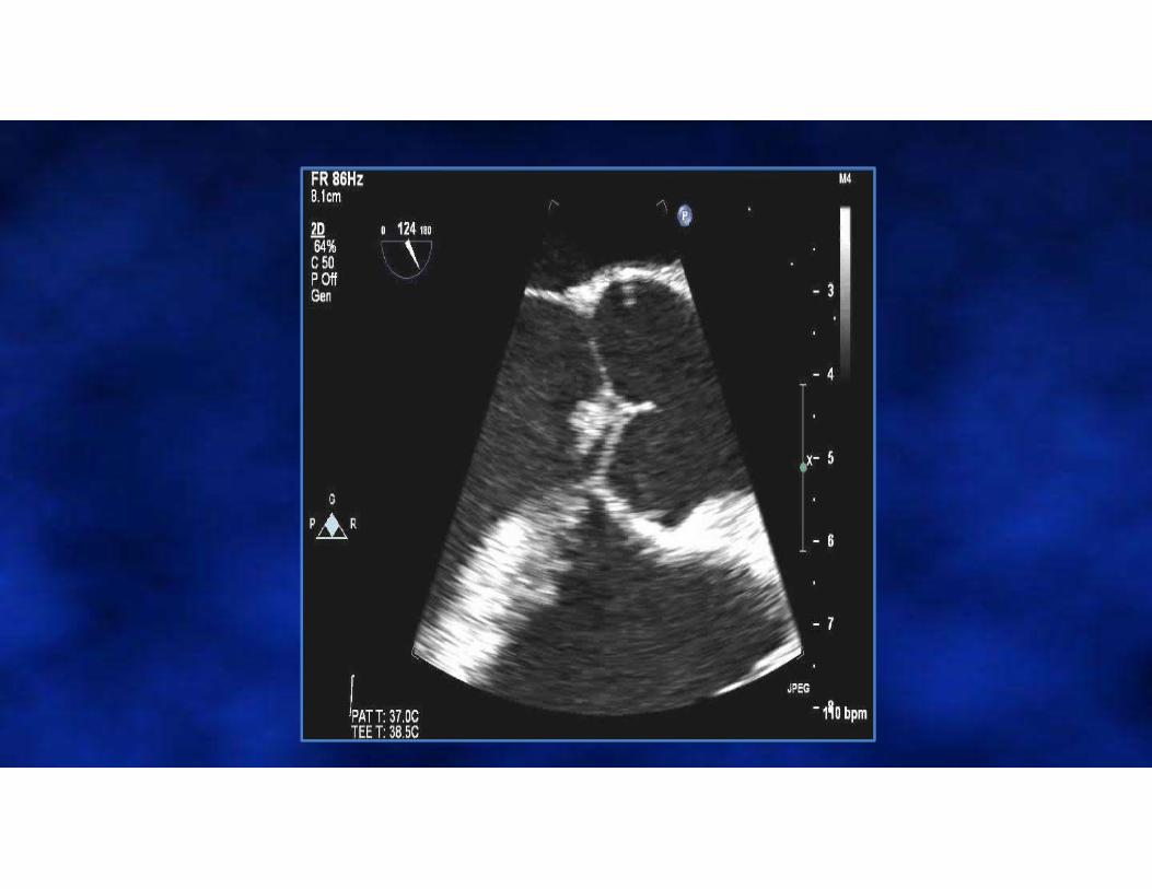
Transesophageal Echocardiogram
2D TEE 3D TEE(View from Left Atrium)
LA
LV
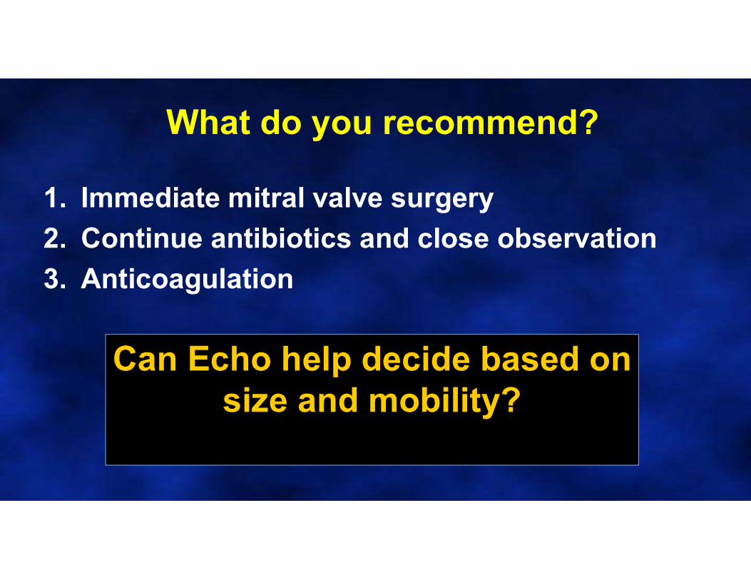
What do you recommend?
1. Immediate mitral valve surgery
2. Continue antibiotics and close observation
3. Anticoagulation
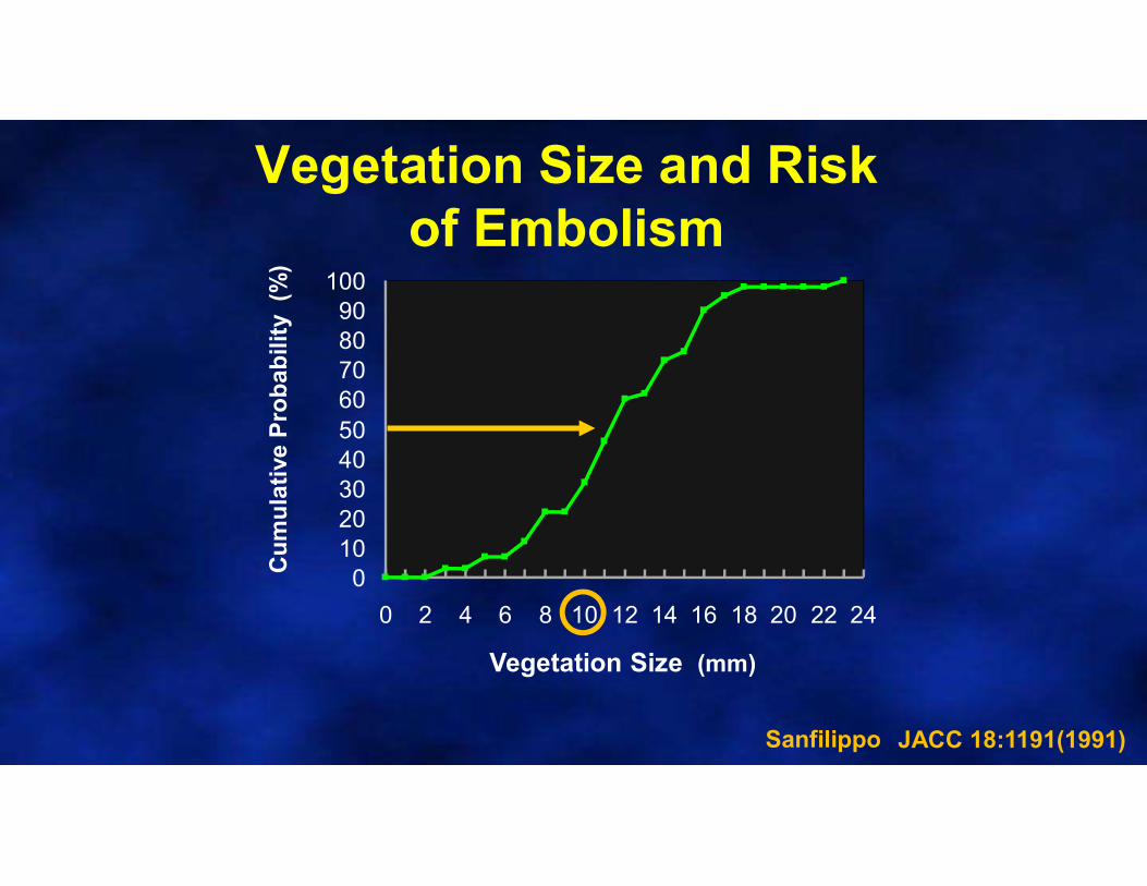
Can Echo help decide based on size and mobility?
Cu
mu
lati
ve P
rob
ab
ility
(%
)
Vegetation Size (mm)
Sanfilippo JACC 18:1191(1991)
0102030405060708090
100
0 2 4 6 8 10 12 14 16 18 20 22 24
Vegetation Size and Risk of Embolism
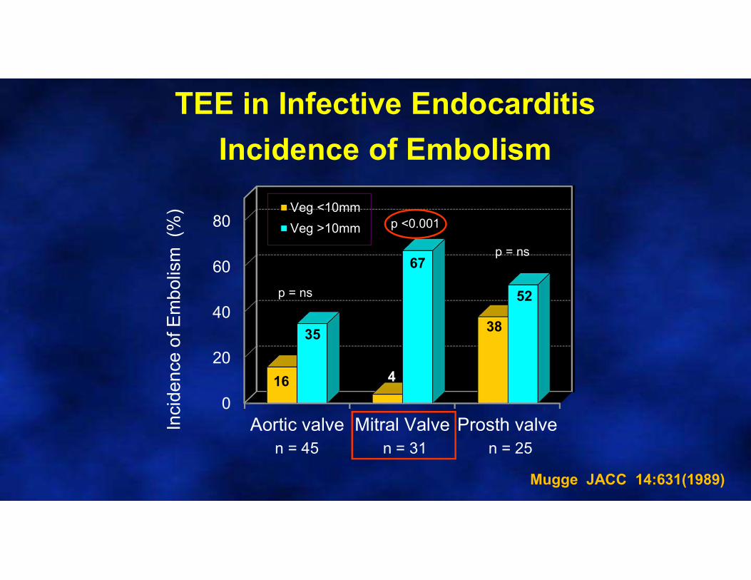
TEE in Infective Endocarditis
Incidence of Embolism
0
20
40
60
80
Aortic valve Mitral Valve Prosth valveInci
den
ce o
f Em
bol
ism
(%
) Veg <10mm
Veg >10mm
n = 45 n = 31 n = 25
16
35
67
4
38
52p = ns
p <0.001
p = ns
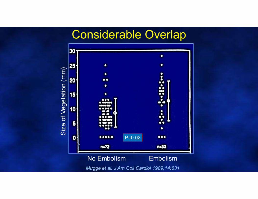
Mugge JACC 14:631(1989)
0
10
20
30
40
50
60
70
80
90
Absent <10 10-15 >15
Vegetation size (mm)
Em
bo
lic
eve
nts
(%
)
0
10
20
30
40
50
60
70
80
90
Absent Low Mod Severe
Vegetation mobility
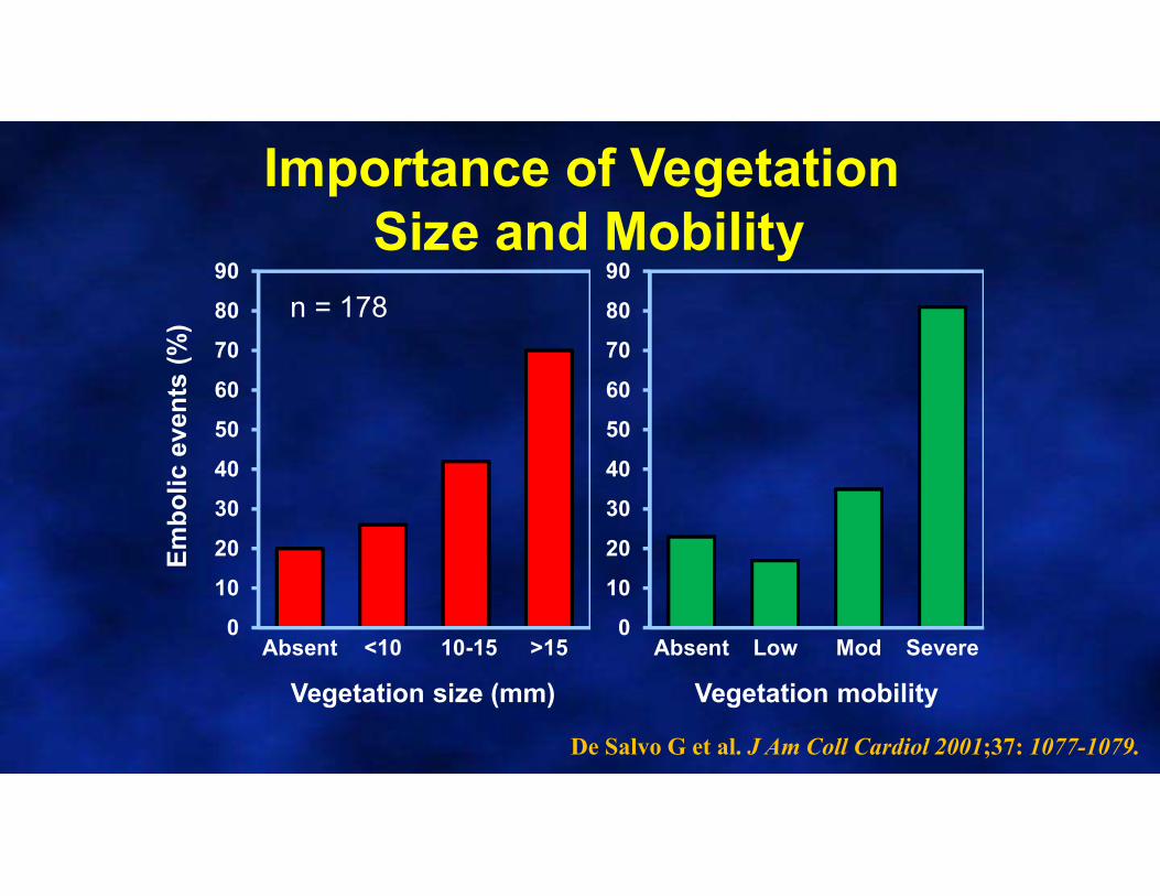
Importance of VegetationSize and Mobility
De Salvo G et al. J Am Coll Cardiol 2001;37: 1077-1079.
n = 178
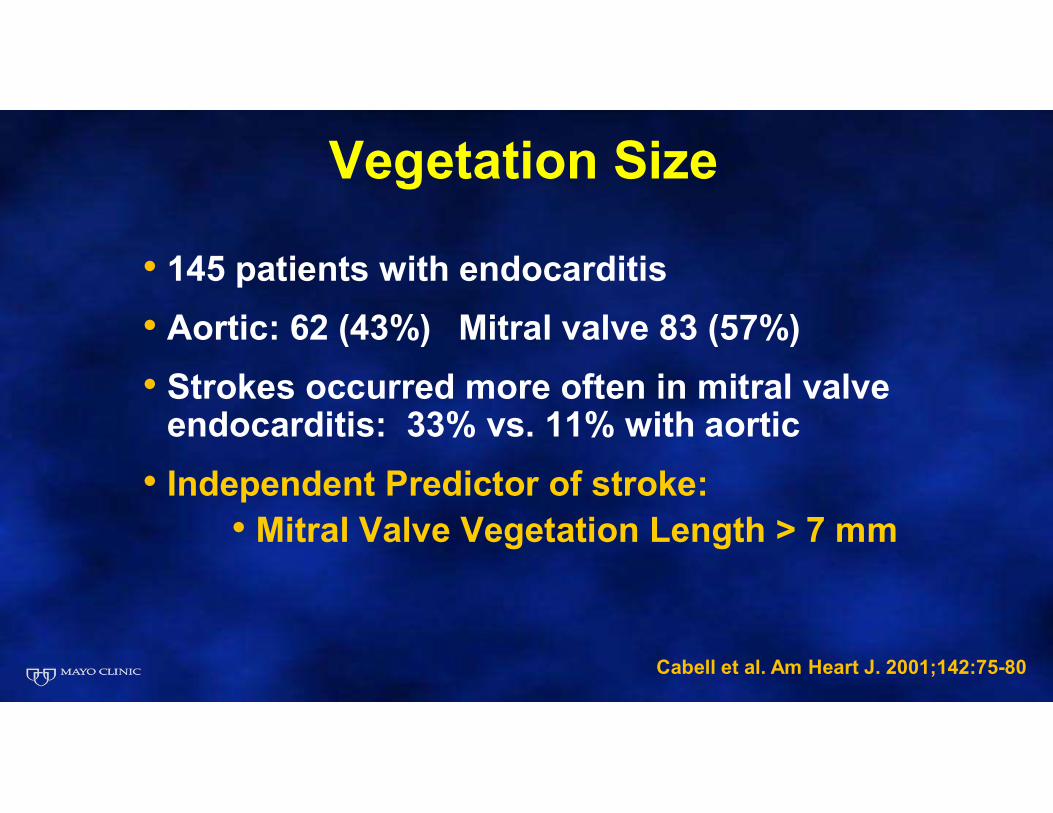
Vegetation Size
• 145 patients with endocarditis
• Aortic: 62 (43%) Mitral valve 83 (57%)
• Strokes occurred more often in mitral valve endocarditis: 33% vs. 11% with aortic
• Independent Predictor of stroke: • Mitral Valve Vegetation Length > 7 mm
Cabell et al. Am Heart J. 2001;142:75-80

Relation of embolism to
vegetation size
Relation of embolism to
vegetation size
Embolic eventsEmbolic events
Di Salvo et al: Positive 37% 9%JACC, 2001 (>10 mm)(178 pt)
Di Salvo et al: Positive 37% 9%JACC, 2001 (>10 mm)(178 pt)
Cabell et al: Positive 23% 11%AHJ, 2001 (>7 mm)(145 pt)
Cabell et al: Positive 23% 11%AHJ, 2001 (>7 mm)(145 pt)
Vilacosta et al: Positive 33% 13%JACC, 2002 (>10 mm)(211 pt)
Vilacosta et al: Positive 33% 13%JACC, 2002 (>10 mm)(211 pt)
Embolism in Infective EndocarditisVegetation Size by TEE and Impact of Therapy
Embolism in Infective EndocarditisVegetation Size by TEE and Impact of Therapy
On therapyOn therapyTotalTotal
CP1189948-74
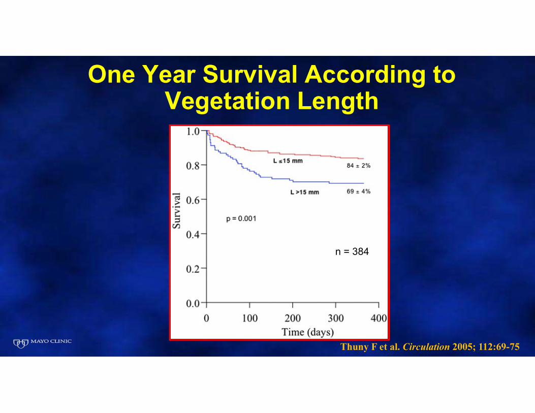
One Year Survival According to Vegetation Length
Thuny F et al. Circulation 2005; 112:69-75
n = 384
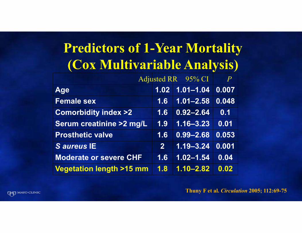
Predictors of 1-Year Mortality (Cox Multivariable Analysis)
Adjusted RR 95% CI P
Age 1.02 1.01–1.04 0.007
Female sex 1.6 1.01–2.58 0.048
Comorbidity index >2 1.6 0.92–2.64 0.1
Serum creatinine >2 mg/L 1.9 1.16–3.23 0.01
Prosthetic valve 1.6 0.99–2.68 0.053
S aureus IE 2 1.19–3.24 0.001
Moderate or severe CHF 1.6 1.02–1.54 0.04
Vegetation length >15 mm 1.8 1.10–2.82 0.02
Thuny F et al. Circulation 2005; 112:69-75
N Engl J Med 2012;366:2466-73
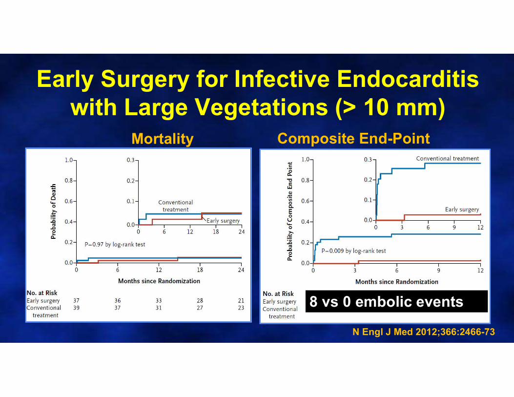
Early Surgery for Infective Endocarditis with Large Vegetations (> 10 mm)
N Engl J Med 2012;366:2466-73
Mortality Composite End-Point
8 vs 0 embolic events
Risk of Embolism
• Consider early surgical treatment for:
• Larger vegetations
• Highly mobile vegetations
• Mitral valve location
• Controversial
• Risk diminishes significantly over time with antibiotics

Indications for Intervention in Infective Endocarditis
• Class IIA: Early surgery (during initial hospitalization before completion of a full therapeutic course of antibiotics) is reasonable in patients with IE who present with recurrent emboli and persistent vegetations despite appropriate antibiotic therapy. (Level of Evidence: B)
• Class IIb: Early surgery (during initial hospitalization before completion of a full therapeutic course of antibiotics) may be considered in patients with native valve endocarditis who exhibit mobile vegetationsgreater than 10 mm in length (with or without clinical evidence of embolic phenomenon). (Level of Evidence: B)
2014 AHA/ACC Valve Guidelines, Circulation 2014

Timing of Surgery in Endocarditis After Embolic CVA
–Embolic stroke-wait 7-21 days
–Hemorrhagic stroke- wait 4 weeks
–If headache, think mycotic aneurysm (avoid valves that need anticoagulation)
Hoen B and Duval X. N Engl J Med 2013;368:1425-33
2017 Focused Valve Update: IE
• Operation without delay may be considered in patients with IE and an indication for surgery who have suffered a stroke, but have no evidence of intracranial hemorrhage or extensive neurological damage (Class IIb, LOE B-NR).
• If hemodynamically stable, delaying valve surgery for ≥4 weeks may be considered among patients with IE and major ischemic stroke or intracranial hemorrhage (Class IIb, LOE B-NR).
Complications of Endocarditis Identified by Echocardiography
• Abscess• Aneurysm of intervalvular fibrosa• Fistula• Perforation• Other Mechanical Complications Secondary to
Leaflet Destruction• Hemodynamic
– Most common cause of death is a regurgitant lesion with CHF (Lerner et al, N Engl J Med 1966)
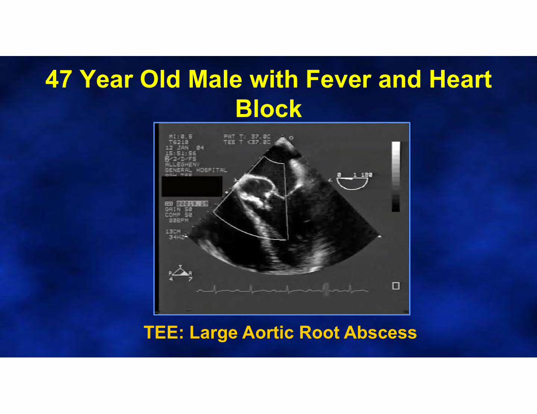
47 Year Old Male with Fever and Heart Block
TEE: Large Aortic Root Abscess

Peri-Valvular Infection: Phlegmon
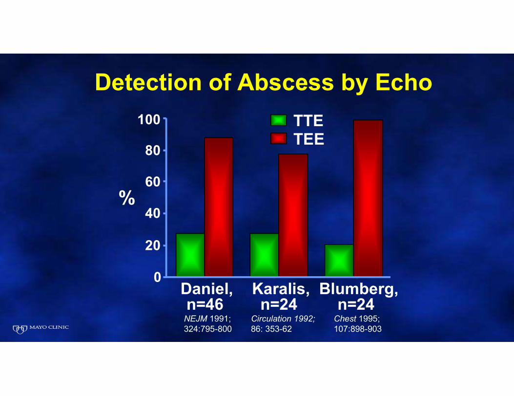
Detection of Abscess by Echo
TTETEE
0
20
40
60
80
100
Daniel,n=46
Karalis,n=24
Blumberg,n=24
%
Circulation 1992;86: 353-62
NEJM 1991; 324:795-800
Chest 1995;107:898-903
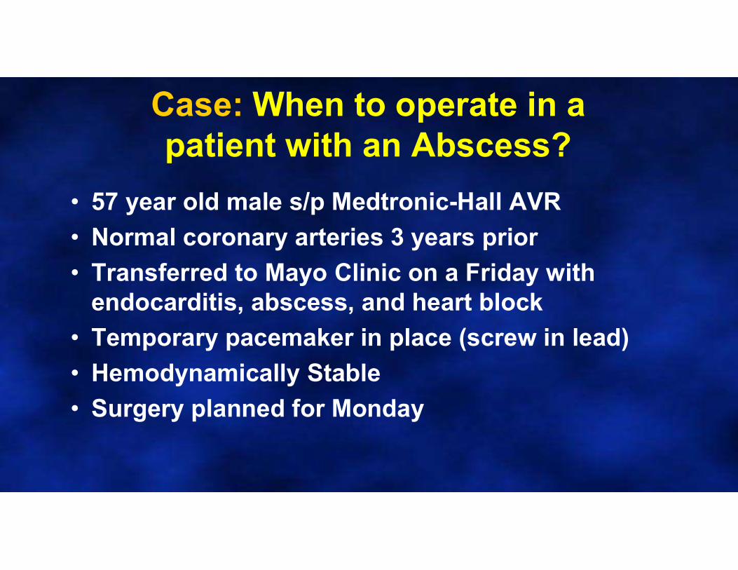
Case: When to operate in a patient with an Abscess?
• 57 year old male s/p Medtronic-Hall AVR
• Normal coronary arteries 3 years prior
• Transferred to Mayo Clinic on a Friday with endocarditis, abscess, and heart block
• Temporary pacemaker in place (screw in lead)
• Hemodynamically Stable
• Surgery planned for Monday
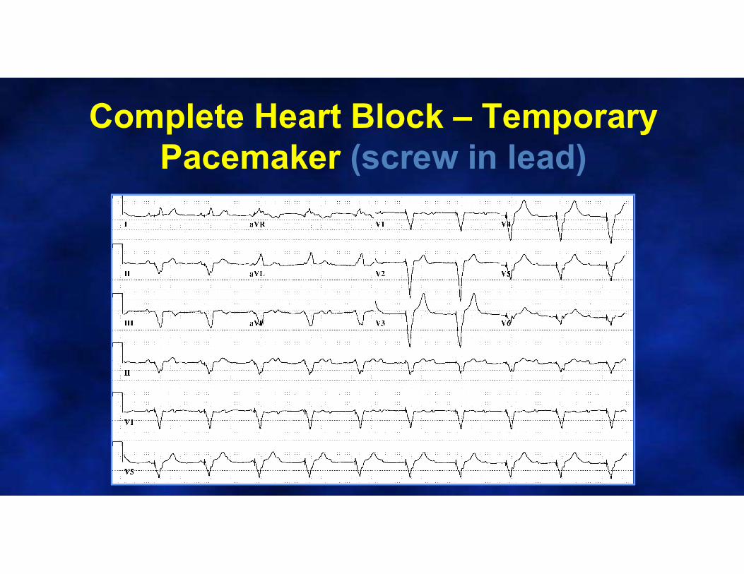
Complete Heart Block – Temporary Pacemaker (screw in lead)

TEE
LA
LV
Normally Functioning AVR
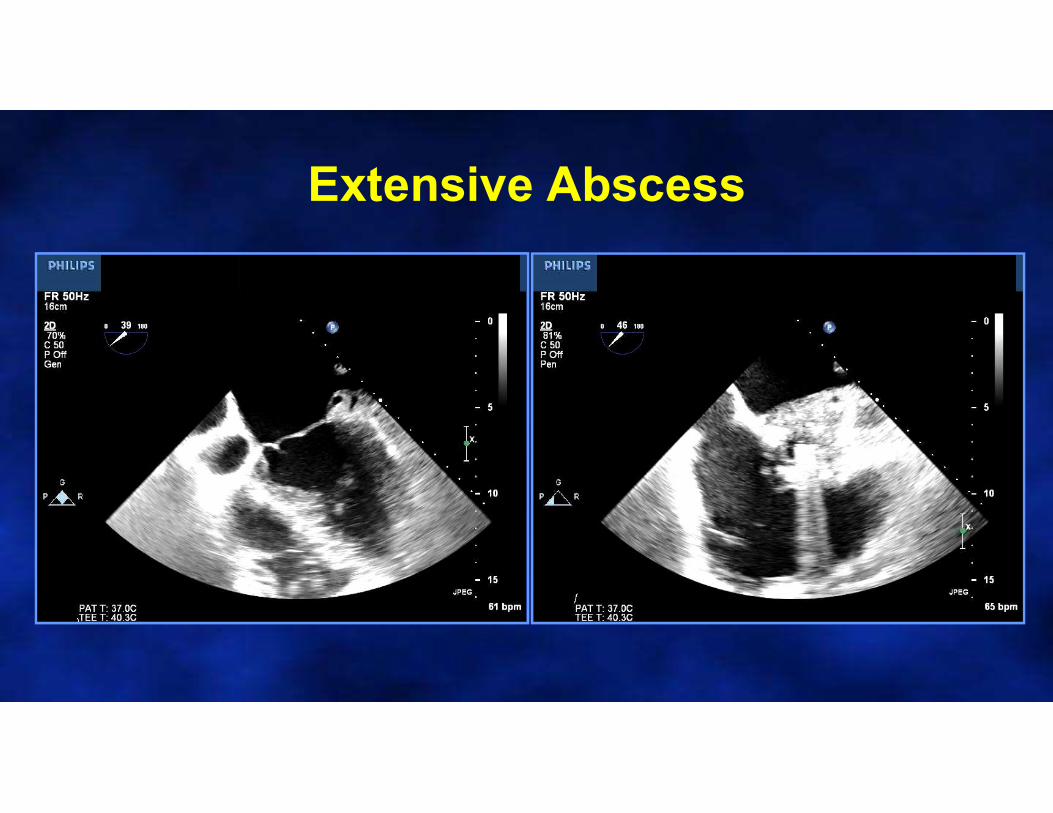
Extensive Abscess
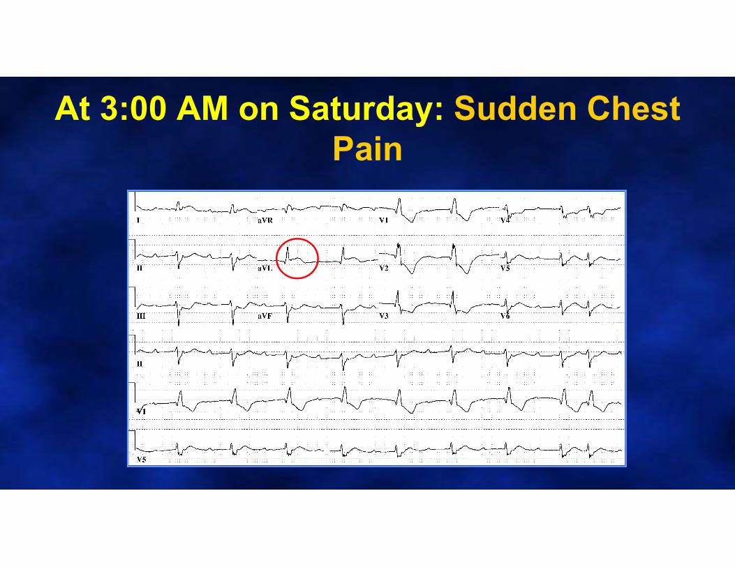
At 3:00 AM on Saturday: Sudden Chest Pain
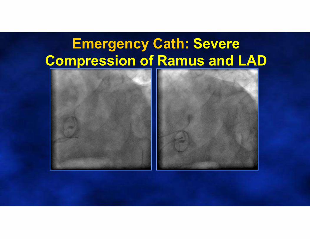
Emergency Cath: Severe Compression of Ramus and LAD

• Patient being prepped for emergency surgery
• Suddenly developed hypotension followed by ventricular fibrillation
• 45 minutes of resuscitation– Unsuccesful
• Patient died before he could make it to operating roomWhen to operate in a
patient with an abscess? Urgently !
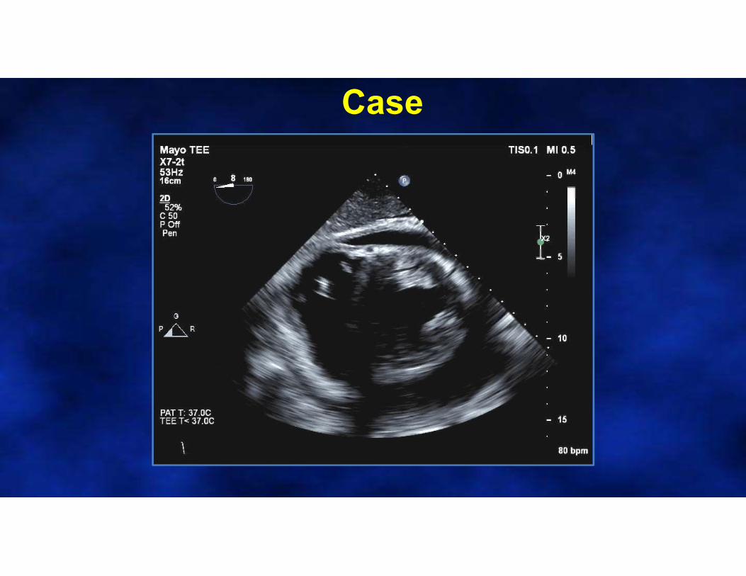
Case
• 68 yo male presented at an outside hospital with 4 weeks of chills, night sweats, and fatigue.
–PMH: s/p TAVR 1 year prior, hepatitis C and alcoholic cirrhosis
–Blood cultures drawn at the outside hospital were positive for Gemella haemolysans
Courtesy of Dr. J. Thaden
Case

Case
Case
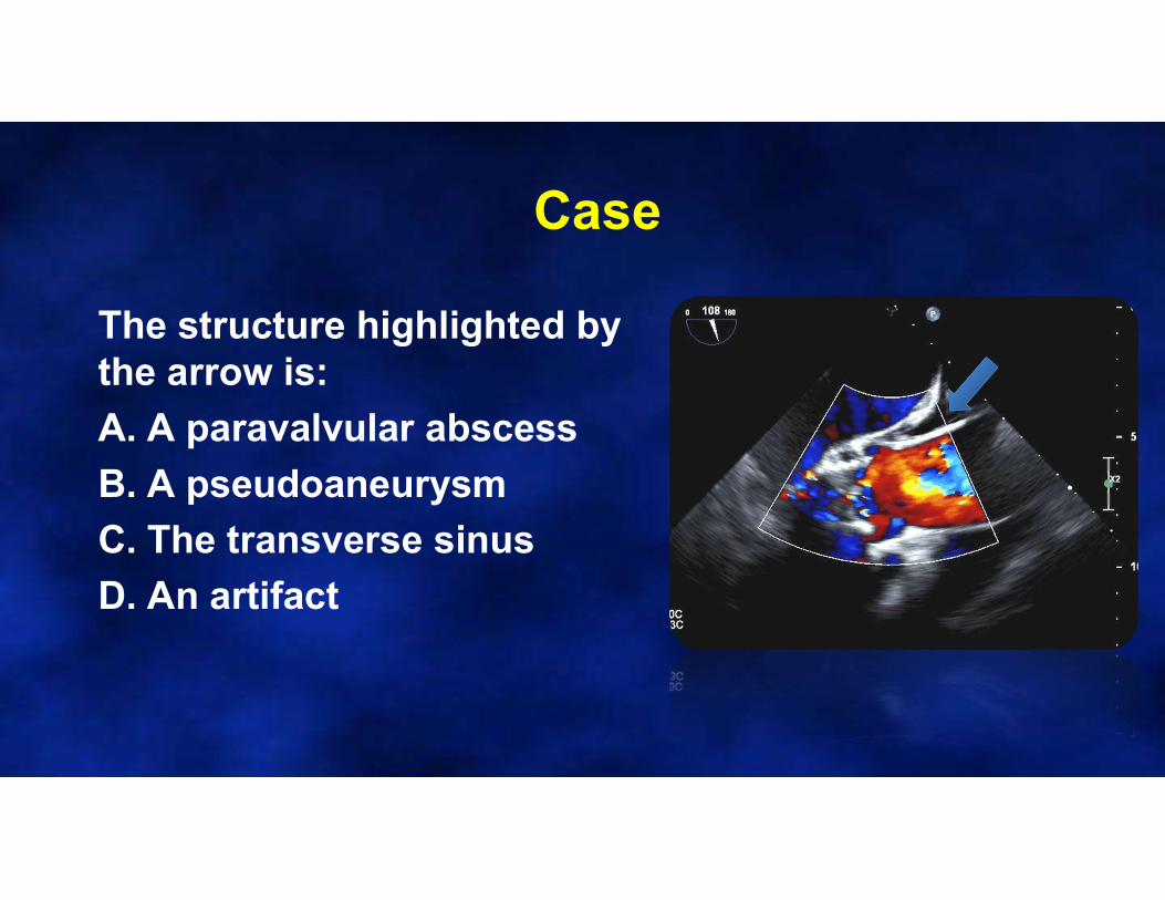
The structure highlighted by the arrow is:
A. A paravalvular abscess
B. A pseudoaneurysm
C. The transverse sinus
D. An artifact
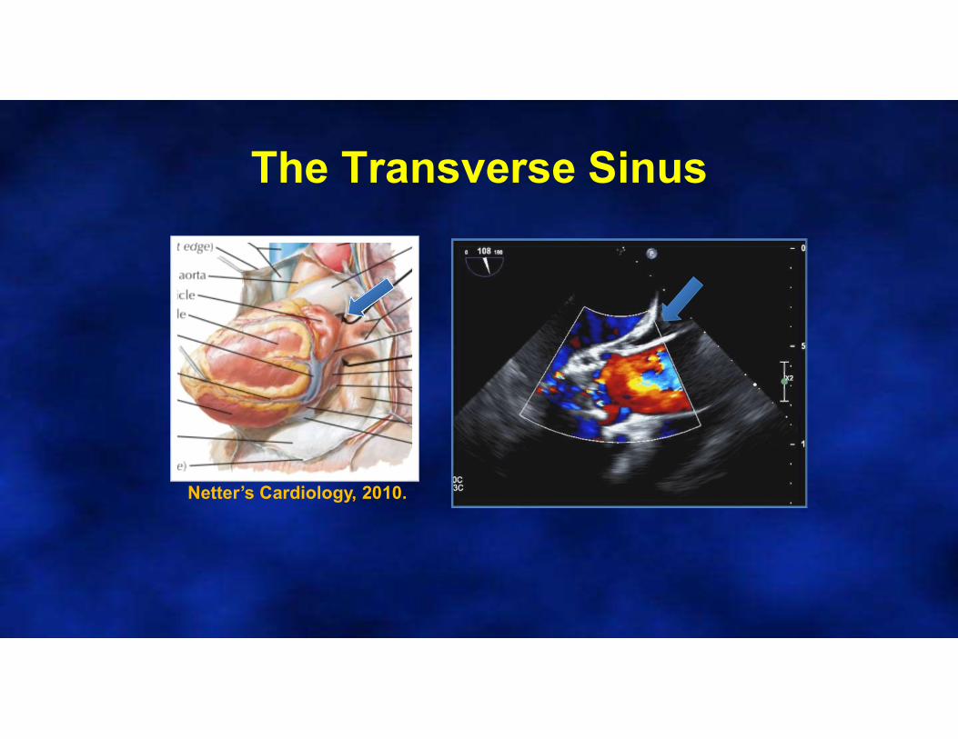
The Transverse Sinus
Netter’s Cardiology, 2010.

Case
Case
• Echo-guided pericardiocentesis (575 mL)
• 6 weeks of IV antibiotics
• Plans to undergo liver transplant workup prior to potential aortic valve replacement
• Repeat TEE at 4 weeks…..
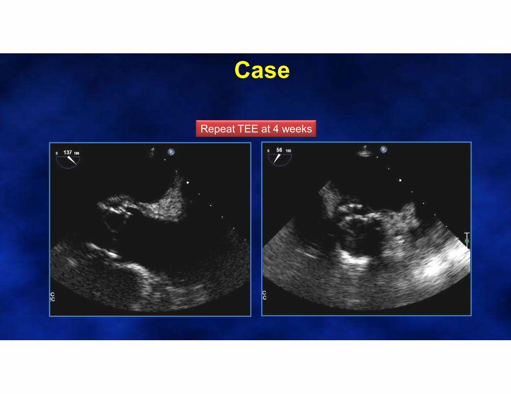
Case
Repeat TEE at 4 weeks
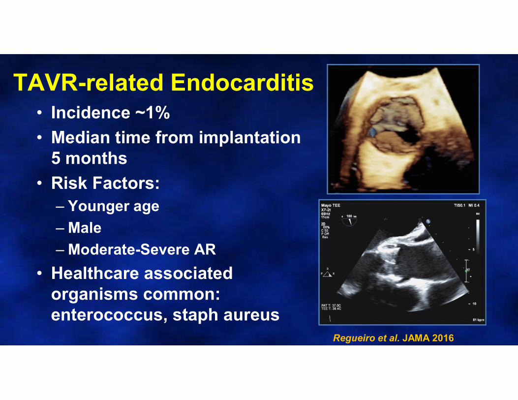
TAVR-related Endocarditis• Incidence ~1%
• Median time from implantation 5 months
• Risk Factors:– Younger age
– Male
– Moderate-Severe AR
• Healthcare associated organisms common: enterococcus, staph aureus
Regueiro et al. JAMA 2016
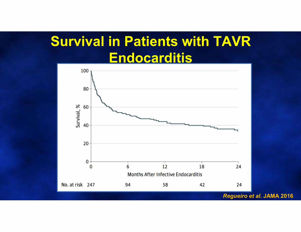
Survival in Patients with TAVR Endocarditis
Regueiro et al. JAMA 2016

SUMMARY: ECHO and ENDOCARDITIS Clinically Suspected Endocarditis
Transthoracic Echo
+ Vegetation - Vegetation
No Complications
Antibiotics +Observation
Any Clinical Instability + Clinical Suspicion
TEE
+ Vegetation - Vegetation
Probably NOT Endocarditis
Abscess/Perforation? Large Vegetation
Antibiotics + Surgery
No Complications
Antibiotics +Observation
Staph aureus bacteremia
Copyright © 2022 FDOKUMEN