Wear resistance and wear mechanisms of WC–12%Co thermal sprayed coatings in three-body abrasion
Upload
khangminh22Category
view
5download
0
�����������������
Citation: Eubank, T.A.;
Gonzales-Luna, A.J.; Hurdle, J.G.;
Garey, K.W. Genetic Mechanisms of
Vancomycin Resistance in
Clostridioides difficile: A Systematic
Review. Antibiotics 2022, 11, 258.
https://doi.org/10.3390/
antibiotics11020258
Academic Editor: Michael Samarkos
Received: 20 January 2022
Accepted: 10 February 2022
Published: 16 February 2022
Publisher’s Note: MDPI stays neutral
with regard to jurisdictional claims in
published maps and institutional affil-
iations.
Copyright: © 2022 by the authors.
Licensee MDPI, Basel, Switzerland.
This article is an open access article
distributed under the terms and
conditions of the Creative Commons
Attribution (CC BY) license (https://
creativecommons.org/licenses/by/
4.0/).
antibiotics
Review
Genetic Mechanisms of Vancomycin Resistance inClostridioides difficile: A Systematic ReviewTaryn A. Eubank 1, Anne J. Gonzales-Luna 1, Julian G. Hurdle 2 and Kevin W. Garey 1,*
1 Department of Pharmacy Practice and Translational Research, University of Houston College of Pharmacy,Houston, TX 77204, USA; [email protected] (T.A.E.); [email protected] (A.J.G.-L.)
2 Center of Infectious and Inflammatory Diseases, Institute of Biosciences and Technology,TX A&M Health Science Center, Houston, TX 77030, USA; [email protected]
* Correspondence: [email protected]
Abstract: Antimicrobial resistance to treatments for Clostridioides difficile infection (CDI) poses asignificant threat to global health. C. difficile is widely thought to be susceptible to oral vancomycin,which is increasingly the mainstay of CDI treatment. However, clinical labs do not conduct C. difficilesusceptibility testing, presenting a challenge to detecting the emergence and impact of resistance.In this systematic review, we describe gene determinants and associated clinical and laboratorymechanisms of vancomycin resistance in C. difficile, including drug-binding site alterations, effluxpumps, RNA polymerase mutations, and biofilm formation. Additional research is needed to furthercharacterize these mechanisms and understand their clinical impact.
Keywords: Clostridium difficile; antimicrobial resistance; reduced susceptibility; van genes; plasmids;efflux pumps; biofilm
1. Introduction
Clostridioides difficile infection (CDI) is the most common healthcare-associated in-fection in the United States with an estimated 223,900 cases in hospitalized patients and12,800 associated deaths in 2017 [1,2]. For more than four decades, metronidazole andvancomycin have been the leading antimicrobial therapies for CDI, with fidaxomicinalso indicated for use. However, metronidazole is no longer recommended as a first-linetreatment option. Vancomycin is a glycopeptide antimicrobial that inhibits cell wall syn-thesis by binding to the D-Ala-D-Ala termini of lipid II, thereby inhibiting peptidoglycanbiosynthesis and assembly. Vancomycin has excellent in vitro activity against C. difficileand achieves stool concentrations between 500–2000 mg/L [3]. Systemic susceptibilitybreakpoints vary from concentrations of 2 mg/L (European Committee on AntimicrobialSusceptibility Testing [EUCAST]) to 4 mg/L (Clinical and Laboratory Standards Institute[CLSI]) [4,5]. Clinical labs do not routinely perform C. difficile culture and susceptibilitytesting as a part of the diagnostic work-up, making it difficult to detect resistance. Thus,vancomycin resistance in patients with CDI has not been studied in depth. However,resistance to other antibiotics used to treat CDI, namely metronidazole, has been identified,leading to the question of whether C. difficile may also evolve clinically meaningful resis-tance to vancomycin [6–8]. Prescription rates for oral vancomycin increased following the2017 IDSA/SHEA recommendation of vancomycin as first-line therapy for CDI [9]. Thislikely also increased the selection pressure for the spread of vancomycin-resistant C. difficile.
Vancomycin clinical cure rates have decreased over time [10–14], and range from 82–88%,which is much lower than the cure rates of 93–100% reported in earlier trials [15–18]. Populationsurveillance studies have also noted increased C. difficile vancomycin minimum inhibitoryconcentrations (MICs) [19–21]. However, the underlying mechanisms of vancomycinresistance in C. difficile are still under-characterized, as is their role in treatment failures.
Antibiotics 2022, 11, 258. https://doi.org/10.3390/antibiotics11020258 https://www.mdpi.com/journal/antibiotics
Antibiotics 2022, 11, 258 2 of 11
In this systematic review, we aim to summarize the body of literature on vancomycinresistance mechanisms in C. difficile.
2. Results
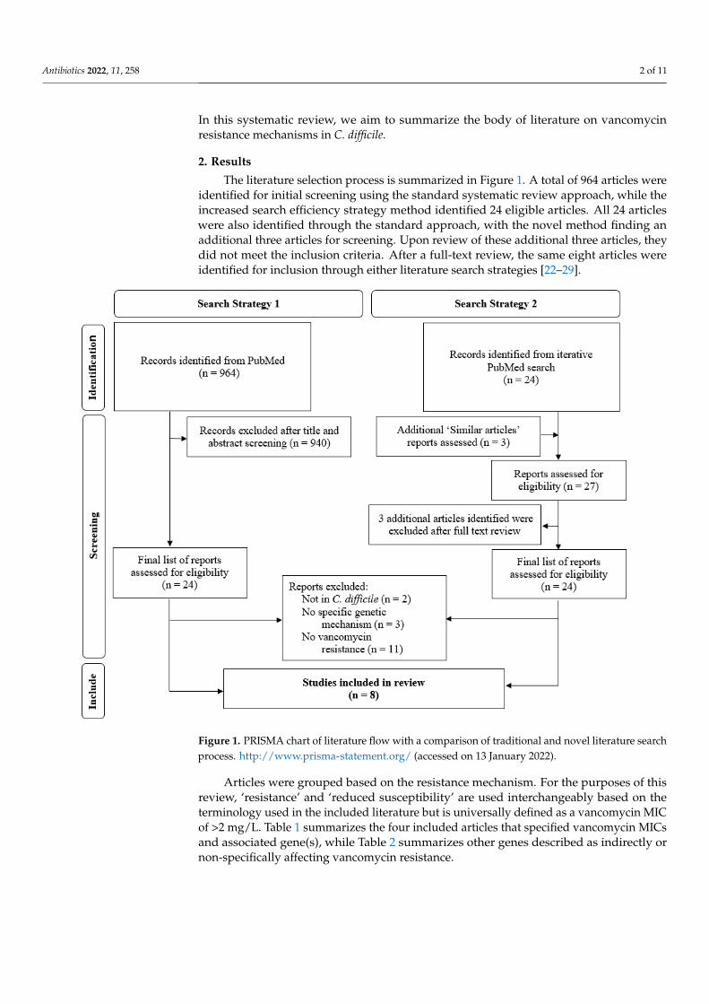
The literature selection process is summarized in Figure 1. A total of 964 articles wereidentified for initial screening using the standard systematic review approach, while theincreased search efficiency strategy method identified 24 eligible articles. All 24 articleswere also identified through the standard approach, with the novel method finding anadditional three articles for screening. Upon review of these additional three articles, theydid not meet the inclusion criteria. After a full-text review, the same eight articles wereidentified for inclusion through either literature search strategies [22–29].
Antibiotics 2022, 11, x 2 of 11
vancomycin resistance in C. difficile are still under-characterized, as is their role in treat-
ment failures. In this systematic review, we aim to summarize the body of literature on
vancomycin resistance mechanisms in C. difficile.
2. Results
The literature selection process is summarized in Figure 1. A total of 964 articles were
identified for initial screening using the standard systematic review approach, while the
increased search efficiency strategy method identified 24 eligible articles. All 24 articles
were also identified through the standard approach, with the novel method finding an
additional three articles for screening. Upon review of these additional three articles, they
did not meet the inclusion criteria. After a full-text review, the same eight articles were
identified for inclusion through either literature search strategies [22–29].
Figure 1. PRISMA chart of literature flow with a comparison of traditional and novel literature
search process. http://www.prisma-statement.org/. (Accessed on 14 February 2022)
Articles were grouped based on the resistance mechanism. For the purposes of this
review, ‘resistance’ and ‘reduced susceptibility’ are used interchangeably based on the
terminology used in the included literature but is universally defined as a vancomycin
MIC of >2 mg/L. Table 1 summarizes the four included articles that specified vancomycin
MICs and associated gene(s), while Table 2 summarizes other genes described as indi-
rectly or non-specifically affecting vancomycin resistance.
Table 1. Summary of genes associated with elevated vancomycin MICs.
Figure 1. PRISMA chart of literature flow with a comparison of traditional and novel literature searchprocess. http://www.prisma-statement.org/ (accessed on 13 January 2022).
Articles were grouped based on the resistance mechanism. For the purposes of thisreview, ‘resistance’ and ‘reduced susceptibility’ are used interchangeably based on theterminology used in the included literature but is universally defined as a vancomycin MICof >2 mg/L. Table 1 summarizes the four included articles that specified vancomycin MICsand associated gene(s), while Table 2 summarizes other genes described as indirectly ornon-specifically affecting vancomycin resistance.

Antibiotics 2022, 11, 258 3 of 11
Table 1. Summary of genes associated with elevated vancomycin MICs.
Gene (Mutation, If Known)Ref.
No.Isolates
†
StrainOrigin Ribotype ST
Type Other vanG/vanSR vanA vanB vanW vanZ murG rpoCVAN MIC
(mg/L)[22] 4 Clinical 012 54 NAPCR1 vanG 4
[23] *
1 Laboratory 027 1 WS2/R20291
vanS(Arg314Leu) 8
1 Laboratory 027 1 WS4/R20291
vanS(Gly319Asp) 16
1 Clinical 027 1 MT1470 vanS(Ser313Phe) 8
1 Clinical 027 1 MT5006 vanS(Thr349Ile) 8
9 Clinical 027 1 see note ‡ vanR(Thr115Ala) 4–8 ‡
1 Clinical 35 vanA vanW 41 Clinical 014-020 2 vanB 81 Clinical 42 vanB 41 Clinical 67 vanB vanW >16
[24]
1 Clinical 012 54 vanW vanZ 4
[25] *1 Laboratory 087 NB95013/ATCC
43255murG/cd2725(Pro108Leu) 16
1 Laboratory NB95026 rpoC/cd0067(Gly733Thr) 16
Abbv: Ref., reference; no., number; ST, multilocus sequence type; MIC, minimum inhibitory concentration;VAN, vancomycin. Notes:* These studies utilized serial passaging and selection for mutants with decreasedsusceptibility. † Number of isolates expressing resistance, defined as vancomycin MIC > 2 mg/L [4,5]. ‡ 7 isolatesfrom the Texas Medical Center had MICs = 4 mg/L and 2 isolates from Israel had MICs = 8 mg/L
2.1. Binding Site Alterations2.1.1. Van Genes
The most well-documented mechanisms of vancomycin resistance are target site alter-ations mediated by van genes that modify the terminal D-Ala-D-Ala motif of lipid II, wherevancomycin binds to exhibit its mechanism of action. These resistance genes have been welldescribed in Enterococcus spp. and are also present in C. difficile, albeit without evidence ofthe same clinical implications [22–24,30–37]. Multiple van gene clusters, including vanA,vanB, vanG, and vanW, and vanZ orthologs, have been identified in C. difficile and havebeen associated with elevated vancomycin MICs (Table 1) [22–24,35]. The expression ofthese clusters is controlled by a two-component regulatory system, VanSR, that containsVanS, a membrane sensor kinase, and VanR, a cytoplasmic response regulator [23,30]. VanStypically detects the presence of vancomycin, leading to autophosphorylation and transferof its phosphoryl group to VanR. In turn, phosphorylated VanR binds to the promoterregion to induce the transcription of vanG.
Reduced vancomycin susceptibility in C. difficile isolates harboring vanG has beendescribed in two articles [22,23]. Expression of the vanG operon leads to the production ofD-Ala-D-Ser rather than D-Ala-D-Ala, altering the vancomycin binding site and decreasingits binding affinity [23,30]. As 85% of C. difficile carry a functional vanG gene cluster, themere presence of the gene is not linked with clinical resistance [35,38]. Ramírez-Vargas et al.analyzed 38 isolates of a locally endemic Costa Rican North American pulsed-field variant(NAPCR1) and similarly found all possessed VanG-like sequences but only four isolateswere resistant (MIC 4 mg/L) [22]. However, Shen et al. recently demonstrated that levels ofvanG expression may correlate with resistance in ribotype (RT) 027 after identifying VanSRmutations leading to constitutive vanG expression and decreased vancomycin killing [23].Serial passaging of a reference strain, R20291, was performed to create laboratory mutantsdemonstrating elevated MICs (8–16 mg/L), which were found to have developed mutationsin vanS. Eleven clinical isolates with elevated MICs (4–8 mg/L) were then analyzed andfound to exhibit similar vanSR mutations leading to constitutive vanG expression (Table 1).
Antibiotics 2022, 11, 258 4 of 11
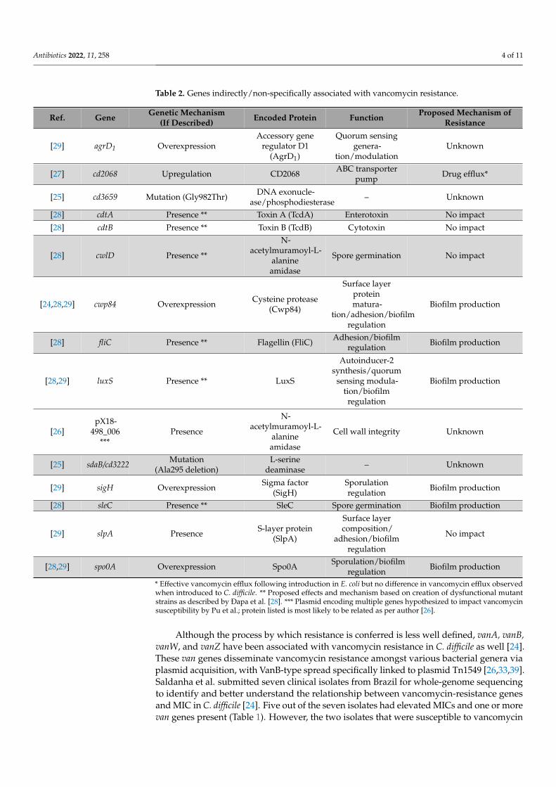
Table 2. Genes indirectly/non-specifically associated with vancomycin resistance.
Ref. Gene Genetic Mechanism(If Described) Encoded Protein Function Proposed Mechanism of
Resistance
[29] agrD1 OverexpressionAccessory gene
regulator D1(AgrD1)
Quorum sensinggenera-
tion/modulationUnknown
[27] cd2068 Upregulation CD2068 ABC transporterpump Drug efflux*
[25] cd3659 Mutation (Gly982Thr) DNA exonucle-ase/phosphodiesterase – Unknown
[28] cdtA Presence ** Toxin A (TcdA) Enterotoxin No impact
[28] cdtB Presence ** Toxin B (TcdB) Cytotoxin No impact
[28] cwlD Presence **
N-acetylmuramoyl-L-
alanineamidase
Spore germination No impact
[24,28,29] cwp84 Overexpression Cysteine protease(Cwp84)
Surface layerproteinmatura-
tion/adhesion/biofilmregulation
Biofilm production
[28] fliC Presence ** Flagellin (FliC) Adhesion/biofilmregulation Biofilm production
[28,29] luxS Presence ** LuxS
Autoinducer-2synthesis/quorum
sensing modula-tion/biofilmregulation
Biofilm production
[26]pX18-
498_006***
Presence
N-acetylmuramoyl-L-
alanineamidase
Cell wall integrity Unknown
[25] sdaB/cd3222 Mutation(Ala295 deletion)
L-serinedeaminase – Unknown
[29] sigH Overexpression Sigma factor(SigH)
Sporulationregulation Biofilm production
[28] sleC Presence ** SleC Spore germination Biofilm production
[29] slpA Presence S-layer protein(SlpA)
Surface layercomposition/
adhesion/biofilmregulation
No impact
[28,29] spo0A Overexpression Spo0A Sporulation/biofilmregulation Biofilm production
* Effective vancomycin efflux following introduction in E. coli but no difference in vancomycin efflux observedwhen introduced to C. difficile. ** Proposed effects and mechanism based on creation of dysfunctional mutantstrains as described by Ðapa et al. [28]. *** Plasmid encoding multiple genes hypothesized to impact vancomycinsusceptibility by Pu et al.; protein listed is most likely to be related as per author [26].
Although the process by which resistance is conferred is less well defined, vanA, vanB,vanW, and vanZ have been associated with vancomycin resistance in C. difficile as well [24].These van genes disseminate vancomycin resistance amongst various bacterial genera viaplasmid acquisition, with VanB-type spread specifically linked to plasmid Tn1549 [26,33,39].Saldanha et al. submitted seven clinical isolates from Brazil for whole-genome sequencingto identify and better understand the relationship between vancomycin-resistance genesand MIC in C. difficile [24]. Five out of the seven isolates had elevated MICs and one or morevan genes present (Table 1). However, the two isolates that were susceptible to vancomycin
Antibiotics 2022, 11, 258 5 of 11
also contained vanW and vanZ genes, supporting the theory that the presence of van genesalone does not correlate with vancomycin resistance. Future studies investigating therole of specific genetic mutations and/or gene expression levels are needed to provide aframework for interpreting the implications of these other van genes.
2.1.2. MurG
MurG is a glycosyltransferase enzyme responsible for converting lipid I to lipid II inpeptidoglycan biosynthesis, which results in the formation of the cell wall [40]. Alterationsin this pathway may affect vancomycin activity since vancomycin binds the D-Ala-D-Alaterminal on lipid II to prevent cell wall formation. Leeds et al. conducted experimentalevolutions and determined that a mutation in the murG/cd2725 gene was one of threemutations identified in a mutant with elevated vancomycin MICs [25]. This mutationcaused a conserved proline to leucine change (P108L) in the MurG enzyme near the secondof three G loops needed for binding the phosphate groups of UDP-GlcNAc. As the isolateexhibiting this change also had two additional mutations present, it is unknown whatspecific role this murG mutation played in decreasing vancomycin binding and/or activity.
2.2. Plasmid Acquisition of N-Acetylmuramoyl-L-Alanine
A plasmid is typically a small circular DNA strand that can replicate independentlyin microorganisms. Plasmids can be transferred between bacteria through horizontalgene transfer, thus providing genes of antimicrobial resistance. The gut microbiome isan abundant source of extrachromosomal elements to be potentially transferred amongstbacteria due to the diverse inhabitants. Pu et al. identified the plasmid pX18-498 in CDI-positive patients who were non-responders to vancomycin treatment [26]. When thisplasmid was transferred to C. difficile isolates from vancomycin responders (MIC < 1 mg/L),the isolates exhibited an 8-fold decrease in susceptibility to vancomycin. The authorsidentified the gene encoding N-acetylmuramoyl-L-alanine amidase as one of the top fivedifferentially expressed genes contained within the plasmid pX18-498. N-acetylmuramoyl-L-alanine amidase is essential for susceptibility to antibiotics that target the cell wall inother bacterial species, and implantation of pX18-498 with this amidase-encoding gene intoC. difficile caused cell rupture and decreased permeability. Furthermore, mice infected withC. difficile strains carrying pX18-498 demonstrated a more severe disease phenotype. Studiesare needed to confirm this gene expression is indeed the cause of the observed decreasedvancomycin susceptibility and to further elucidate possible resistance mechanisms C. difficilecan acquire from plasmid acquisition.
2.3. Efflux Pumps
Efflux pumps are active transporters residing in the bacterial cell membrane thatutilize an energy source to transport solutes in or out of the cell. One major family of effluxpumps is the ATP-binding cassette (ABC) transporters, which use adenosine triphosphate(ATP) to transport solutes ranging in size from simple ions to larger molecules, such asantibiotics [27,41]. These transporters are known to be a major cause of multidrug resistancein many bacteria and have been identified in several Clostridium species as well [42,43].In C. difficile, cationic antimicrobial peptides (CAMPs), including vancomycin, have beenshown to induce the expression of a proposed ABC transporter operon, which in turndecreased the effectiveness of various CAMP antibiotics [44]. However, specific ABCtransporters had not been recognized as contributing to multidrug resistance in C. difficileuntil Ngernsombat et al. identified and characterized the CD2068 transporter [27].
CD2068 was identified when genome analysis of a reference CD630 strain identified itas having a high level of homology to two other known ABC transporters in C. hathewayi andC. perfringens [27,42,43]. Following identification, Ngernsombat et al. conducted variousexperiments to determine the function of CD2068 [27]. First, the gene expression of cd2068was shown to significantly increase following exposure to various antibiotics, includingvancomycin (0.25 mg/L). When cloned and functionally characterized in Escherichia coli,
Antibiotics 2022, 11, 258 6 of 11
CD2068 increased the half maximal inhibitory concentration (IC50) of vancomycin andwas associated with 2.6 times higher relative resistance. However, when a cd2068 gene,following insertional activation and complementation, was introduced into C. difficile,no significant difference in vancomycin IC50 was observed. The reason(s) behind thelessened ability of CD2068 to cause multidrug resistance in C. difficile vs. E. coli is notknown, but the authors offer several hypotheses including poor natural expression ofCD2068 in C. difficile, compensation by other ABC transporters in their mutant strain,and/or the presence of other mechanisms mediating resistance to certain antibiotics inC. difficile. Regardless, 243 other genes in the CD630 genome are thought to encode forputative ABC transporters, offering a promising line of research in this area [45]. Clearly,more studies are needed to identify other efflux pumps and their role in contributing toC. difficile vancomycin resistance.
2.4. RNA Polymerase Mutations
Alterations in the genes encoding bacterial RNA polymerases have been well de-scribed for antibiotics that inhibit RNA polymerase as a mechanism of action, such asmutations in C. difficile rpoB or rpoC leading to rifampicin or fidaxomicin resistance, re-spectively [46–48]. However, rpoB mutations have also been associated with vancomycinand daptomycin resistance in Staphylococcus aureus, despite these drugs not targeting RNApolymerase [49,50]. Conversely, rpoB mutations have not been described as contribut-ing to vancomycin resistance in C. difficile. Instead, Leeds et al. described a mutation inrpoC/cd0067 in an experimentally evolved mutant with a vancomycin MIC of 16 mg/L [25].The mutation was a single nucleotide change involving a D244Y substitution in the β’subunit of RNA polymerase. The authors postulated that rpoC mutations lead to changes inglobal gene expression that may affect multiple pathways, therefore impacting vancomycinactivity, but further research is needed to investigate the exact mechanism of vancomycinresistance resulting from this mutation.
2.5. Biofilm Production
A biofilm is a structure of aggregated microbial communities displaying reducedmetabolic activity [51]. Biofilms can serve as a physical barrier preventing host immuneresponses and sufficient concentrations of antimicrobials from reaching the site of infec-tion, and have accordingly been associated with antimicrobial resistance and recurrentinfections [52]. Additionally, the slowed metabolic state of the microbial species can renderantibiotics, whose mechanism of resistance relies on fast dividing cells, less active [51,53].Although the ability of C. difficile and related Clostridium species to form biofilms in theintestine has been described and linked to antimicrobial resistance, few articles describingthe genetic mechanism(s) behind biofilm development and antimicrobial resistance havebeen published [28,54,55].
Two studies have implicated C. difficile biofilm formation with decreased vancomycinsusceptibility [28,29]. Both conducted in-depth analyses to determine molecular and geneticfactors contributing to their findings as discussed by functional category. The first wasconducted by Ðapa et al. who developed a biofilm assay and used it to measure thegrowth of two C. difficile strains (CD630 and R20291) [28]. Growth was measured in 1-, 3-, and 5-day-old biofilms following exposure to 20 mg/L of vancomycin (100 timesgreater than the MIC). The C. difficile survival percentage was 5- and 12-fold higher inthe 1- and 3-day old biofilms, respectively, than in planktonic C. difficile without a biofilm,demonstrating potentially decreased vancomycin efficacy. The second was performedby Tijerina-Rodriguez et al. who analyzed antimicrobial susceptibilities in biofilm- andnon-biofilm-producing clinical isolates from Mexico [29]. Vancomycin MICs were up to100 times higher in biofilm-producing cells when compared to planktonic cell counterparts.Additionally, a sub-group analysis comparing isolates from patients with recurrent CDI (R-CDI) and nonrecurrent CDI (NR-CDI) demonstrated that isolates from R-CDI patients hadsignificantly higher rates of reduced vancomycin susceptibility (28%) than those from NR-
Antibiotics 2022, 11, 258 7 of 11
CDI patients (9%; p = 0.013). Genetic investigations in both studies found the developmentof a mature biofilm to be multifactorial, as summarized in Table 2 and detailed below.
2.5.1. Surface Factors Involved in Biofilm Formation
Various factors within the surface layer of C. difficile, such as cell wall proteins (CWP)and adhesins (flagella or pili), are thought to play important roles in biofilm formationand early bacterial adhesion [28]. Specifically, Cwp84 is a surface-associated cysteineprotease that has been shown to contribute to surface layer protein maturation and aidin degrading host tissues [56,57]. Ðapa et al. seemed to confirm this finding through ademonstration that mutants with a deletion of cwp84 had a dramatic decrease in biofilmformation, with larger defects observed in early biofilm growth on day 1 compared to days3 and 5 [28]. Tijerina-Rodriguez et al. measured and linked cwp84 expression levels to worseclinical outcomes after observing isolates from patients with R-CDI had significantly highercwp84 expression than isolates from patients with NR-CDI (4.31 vs. 0.29 relative mRNAexpression; p = 0.01) [29]. cwp84 was also identified in three of the five vancomycin-resistantisolates submitted for whole-genome sequencing in the study conducted by Saldanha et al.but was additionally present in one of the two non-resistant isolates [24].
Similarly, flagella are known to contribute to mature biofilm formation in other bac-terial species, but with a lesser understood function in clostridial species [58]. Ðapa et al.mutated the fliC gene encoding flagellin, a major component of flagella, in C. difficile, anddemonstrated a significant decrease in biofilm production [28]. In contrast to the growthchanges seen with cwp84 mutants, the decrease in formation was observed in day 5 biofilms,but not on days 1 or 3, indicating flagella may be more important for the later stages ofbiofilm formation.
2.5.2. Sporulation and Germination
C. difficile sporulation is initiated under stress and is mainly regulated by the Spo0Atranscription factor. However, Spo0A is thought to switch between controlling sporulationand biofilm growth depending on the concentration of phosphorylated Spo0A, a processwell described in Bacillus species [59,60]. As spo0A is highly conserved between Bacillusand Clostridium species, Ðapa et al. studied the relationship between spo0A and biofilms inC. difficile [28]. A spo0A mutant with decreased sporulation was created and found to exhibitsignificantly less biofilm growth than strains with wild-type spo0A. Tijerina-Rodriguez et al.also looked at differences in the gene expression of spo0A and sigH, another sporulationregulator encoding the sigma factor of sporulation, in their clinical cohorts [29]. sigHand spo0A expression were significantly higher in biofilm-producing R-CDI isolates thanNR-CDI isolates (relative sigH mRNA expression 6.91 vs. 0.57 and relative spo0A mRNAexpression 47.40 vs. 0.31, respectively; p < 0.01). As previously discussed, both studiesdescribed decreasing vancomycin susceptibility associated with biofilm presence, andthus sigH and spo0A appear to play potential roles in mediating this form of vancomycinresistance [28,29].
2.5.3. Quorum Sensing
Quorum sensing is a density-dependent form of cell-to-cell signaling used by mi-crobial communities to coordinate various cellular processes, including the developmentof antimicrobial resistance [61]. LuxS is one of many regulatory molecules involved inquorum sensing in C. difficile and has been shown to both mediate communication withinbiofilms as well as contribute to biofilm formation itself [28,62,63]. Following the groups’findings that biofilms appeared to decrease vancomycin efficacy, Ðapa et al. created aluxS mutant and observed a dramatically decreased ability to form biofilm [28]. Tijerina-Rodriguez et al. measured the expression of luxS in addition to agrD1, which is part ofthe Agr quorum sensing system homologous to the Agr system in Staphylococcus aureus; inS. aureus, Agr is known to influence biofilm formation and AgrD is the precursor of theautoinducing peptide (quorum sensing signal) [29,64]. Interestingly, the group found a
Antibiotics 2022, 11, 258 8 of 11
significantly higher expression of agrD1 in biofilm-producing R-CDI isolates vs. NR-CDIisolates (relative mRNA expression 14.02 vs. 0.38; p < 0.01) but no difference in luxS ex-pression. Although this does not diminish the possibility of luxS playing a role in biofilmproduction, further exploration of agrD1 is warranted to explore its exact role in quorumsensing and antimicrobial resistance [29].
Although biofilms have been shown to be associated with increased vancomycin MICand recurrent CDI, biofilm formation is multifactorial, and current research has been unableto definitively associate any one mechanism specifically with vancomycin resistance.
3. Materials and Methods
A systematic literature search was conducted using two separate strategies. A PubMedsearch was conducted using the keywords (“Clostridium difficile” OR “Clostridioides difficile”)AND (“resistance”) AND (“vancomycin” OR ‘teicoplanin”). A filter for the English languagewas applied. Each article from the search was evaluated for inclusion regardless of publicationdate. Included studies must have been conducted in C. difficile and included a measure ofvancomycin susceptibility and description of the underlying genes associated with suscepti-bility changes. Articles categorized as reviews, commentaries, opinions, meta-analyses, andsystematic reviews were excluded. The references of the included publications were evaluatedfor relevant articles and cross-referenced with previous related reviews.
A second literature search method designed for increased search efficiency was conductedin parallel. A PubMed search was performed using the keywords: (“Clostridium difficile” OR“Clostridioides difficile”) AND (“resistance”) AND (“vancomycin” OR ‘teicoplanin”). Unlikethe previous search strategy, not all results were screened, and instead, select journal articlesdiscussing the role of specific genes on vancomycin susceptibilities in C. difficile were chosenfor screening. Then, articles designated as ‘Similar articles in PubMed’ were evaluated andused to compile a list of articles that subsequently underwent title and abstract review usingthe same inclusion criteria as above. The references of the included publications from thissearch strategy were evaluated for relevant articles and were cross-referenced with previousrelated reviews.
4. Conclusions
As few antibiotic treatment options are available for CDI, an increasing amount ofselection pressure is placed on the development of antibiotic resistance. Oral vancomycin isincreasingly used as the first-line drug due to recent treatment guideline updates, costs andavailability concerns [8,9,14,65]. However, clinical resistance to vancomycin is yet to be ap-preciated as a major threat in CDI as separating the effects of host factors, antibiotic failure,and infection recurrence in reported poor outcomes remains a challenge. Future studieswill need to better understand the role of other microbiota in the gut to elucidate resistancein vancomycin. In this review, we aimed to summarize the existing literature on genesassociated with vancomycin resistance to serve as a primer for clinicians and researchersworking in this area. As the clinical significance of these resistance mechanisms becomeclearer, methods to identify these gene targets in clinical practice will become necessary.
Author Contributions: Writing—original draft preparation, T.A.E.; writing—review and editing,A.J.G.-L.; writing—review and editing, J.G.H.; writing—review and editing, K.W.G. All authors haveread and agreed to the published version of the manuscript.
Funding: This research was funded in part by research grants from the American College of ClinicalPharmacy, the Society of Infectious Diseases Pharmacists, and the National Institute of Allergy andInfectious Diseases (5T32AI141349).
Conflicts of Interest: K.W.G. has research grants from Summit and Acurx Pharmaceuticals. All otherauthors: No reported conflict of interest.

Antibiotics 2022, 11, 258 9 of 11
References1. Magill, S.S.; O’Leary, E.; Janelle, S.J.; Thompson, D.L.; Dumyati, G.; Nadle, J.; Wilson, L.E.; Kainer, M.A.; Lynfield, R.;
Greissman, S.; et al. Changes in Prevalence of Health Care-Associated Infections in U.S. Hospitals. N. Engl. J. Med. 2018,379, 1732–1744. [CrossRef] [PubMed]
2. Antibiotic Resistance Threats in the United States. 2019. Available online: https;//www.cdc.gov/drugresistance/pdf/threats-report/2019-ar-threats-report-508.pdf (accessed on 13 January 2022).
3. Gonzales, M.; Pepin, J.; Frost, E.H.; Carrier, J.C.; Sirard, S.; Fortier, L.C.; Valiquette, L. Faecal pharmacokinetics of orallyadministered vancomycin in patients with suspected Clostridium difficile infection. BMC Infect. Dis. 2010, 10, 363. [CrossRef][PubMed]
4. European Committee on Antimicrobial Susceptibility Testing. Breakpoint Tables for Interpretation of MICs and Zone Diameters.Version 12.0. 2022. Available online: https://www.eucast.org/clinical_breakpoints/ (accessed on 5 January 2022).
5. CLSI M100-ED31: 2021 Performance Standards for Antimicrobial Susceptibility Testing, 31st Edition. Available online: http://em100.edaptivedocs.net/GetDoc.aspx?doc=CLSI%20M100%20ED31:2021&scope=user (accessed on 13 January 2022).
6. Gonzales-Luna, A.J.; Olaitan, A.O.; Shen, W.J.; Deshpande, A.; Carlson, T.J.; Dotson, K.M.; Lancaster, C.; Begum, K.; Alam, M.J.;Hurdle, J.G.; et al. Reduced Susceptibility to Metronidazole Is Associated With Initial Clinical Failure in Clostridioides difficileInfection. Open Forum Infect. Dis. 2021, 8, ofab365. [CrossRef] [PubMed]
7. Johnson, S.; Louie, T.J.; Gerding, D.N.; Cornely, O.A.; Chasan-Taber, S.; Fitts, D.; Gelone, S.P.; Broom, C.; Davidson, D.M.Vancomycin, Metronidazole, or Tolevamer for Clostridium difficile Infection: Results From Two Multinational, Randomized,Controlled Trials. Clin. Infect. Dis. 2014, 59, 345–354. [CrossRef]
8. McDonald, L.C.; Gerding, D.N.; Johnson, S.; Bakken, J.S.; Carroll, K.C.; Coffin, S.E.; Dubberke, E.R.; Garey, K.W.; Gould, C.V.;Kelly, C.; et al. Clinical Practice Guidelines for Clostridium difficile Infection in Adults and Children: 2017 Update by theInfectious Diseases Society of America (IDSA) and Society for Healthcare Epidemiology of America (SHEA). Clin. Infect. Dis.2018, 66, 987–994. [CrossRef]
9. Clancy, C.J.; Buehrle, D.; Vu, M.; Wagener, M.M.; Nguyen, M.H. Impact of Revised Infectious Diseases Society of America andSociety for Healthcare Epidemiology of America Clinical Practice Guidelines on the Treatment of Clostridium difficile Infectionsin the United States. Clin. Infect. Dis. 2021, 72, 1944–1949. [CrossRef]
10. Louie, T.J.; Miller, M.A.; Mullane, K.M.; Weiss, K.; Lentnek, A.; Golan, Y.; Gorbach, S.; Sears, P.; Shue, Y.K.; Group, O.P.T.C.S.Fidaxomicin versus vancomycin for Clostridium difficile infection. N. Engl. J. Med. 2011, 364, 422–431. [CrossRef]
11. Cornely, O.A.; Crook, D.W.; Esposito, R.; Poirier, A.; Somero, M.S.; Weiss, K.; Sears, P.; Gorbach, S. Fidaxomicin versus vancomycinfor infection with Clostridium difficile in Europe, Canada, and the USA: A double-blind, non-inferiority, randomised controlledtrial. Lancet Infect. Dis. 2012, 12, 281–289. [CrossRef]
12. Mikamo, H.; Tateda, K.; Yanagihara, K.; Kusachi, S.; Takesue, Y.; Miki, T.; Oizumi, Y.; Gamo, K.; Hashimoto, A.; Toyoshima, J.; et al.Efficacy and safety of fidaxomicin for the treatment of Clostridioides (Clostridium) difficile infection in a randomized, double-blind, comparative Phase III study in Japan. J. Infect. Chemother. 2018, 24, 744–752. [CrossRef]
13. Guery, B.; Menichetti, F.; Anttila, V.J.; Adomakoh, N.; Aguado, J.M.; Bisnauthsing, K.; Georgopali, A.; Goldenberg, S.D.; Karas, A.;Kazeem, G.; et al. Extended-pulsed fidaxomicin versus vancomycin for Clostridium difficile infection in patients 60 years andolder (EXTEND): A randomised, controlled, open-label, phase 3b/4 trial. Lancet Infect. Dis. 2018, 18, 296–307. [CrossRef]
14. Johnson, S.; Lavergne, V.; Skinner, A.M.; Gonzales-Luna, A.J.; Garey, K.W.; Kelly, C.P.; Wilcox, M.H. Clinical Practice Guideline bythe Infectious Diseases Society of America (IDSA) and Society for Healthcare Epidemiology of America (SHEA): 2021 FocusedUpdate Guidelines on Management of Clostridioides difficile Infection in Adults. Clin. Infect. Dis. 2021, 73, 755–757. [CrossRef][PubMed]
15. Wenisch, C.; Parschalk, B.; Hasenhundl, M.; Hirschl, A.M.; Graninger, W. Comparison of vancomycin, teicoplanin, metronidazole,and fusidic acid for the treatment of Clostridium difficile-associated diarrhea. Clin. Infect. Dis. 1996, 22, 813–818. [CrossRef][PubMed]
16. Zar, F.A.; Bakkanagari, S.R.; Moorthi, K.M.L.S.T.; Davis, M.B. A Comparison of Vancomycin and Metronidazole for the Treatmentof Clostridium difficile-Associated Diarrhea, Stratified by Disease Severity. Clin. Infect. Dis. 2007, 45, 302–307. [CrossRef]
17. Louie, T.J.; Peppe, J.; Watt, C.K.; Johnson, D.; Mohammed, R.; Dow, G.; Weiss, K.; Simon, S.; John, J.F., Jr.; Garber, G.; et al.Tolevamer, a novel nonantibiotic polymer, compared with vancomycin in the treatment of mild to moderately severe Clostridiumdifficile-associated diarrhea. Clin. Infect. Dis. 2006, 43, 411–420. [CrossRef] [PubMed]
18. Dudley, M.N.; McLaughlin, J.C.; Carrington, G.; Frick, J.; Nightingale, C.H.; Quintiliani, R. Oral bacitracin vs vancomycin therapyfor Clostridium difficile-induced diarrhea. A randomized double-blind trial. Arch. Intern. Med. 1986, 146, 1101–1104. [CrossRef]
19. Jon, J.V.; Mark, H.W.; Jane, F. Antimicrobial resistance progression in the United Kingdom: A temporal comparison of Clostrid-ioides difficile antimicrobial susceptibilities. Anaerobe 2021, 70, 102385. [CrossRef]
20. Adler, A.; Miller-Roll, T.; Bradenstein, R.; Block, C.; Mendelson, B.; Parizade, M.; Paitan, Y.; Schwartz, D.; Peled, N.;Carmeli, Y.; et al. A national survey of the molecular epidemiology of Clostridium difficile in Israel: The dissemination of theribotype 027 strain with reduced susceptibility to vancomycin and metronidazole. Diagn. Microbiol. Infect. Dis. 2015, 83, 21–24.[CrossRef]

Antibiotics 2022, 11, 258 10 of 11
21. Jarrad, A.M.; Blaskovich, M.A.T.; Prasetyoputri, A.; Karoli, T.; Hansford, K.A.; Cooper, M.A. Detection and Investigation of EagleEffect Resistance to Vancomycin in Clostridium difficile With an ATP-Bioluminescence Assay. Front. Microbiol. 2018, 9, 1420.[CrossRef]
22. Ramirez-Vargas, G.; Quesada-Gomez, C.; Acuna-Amador, L.; Lopez-Urena, D.; Murillo, T.; Del Mar Gamboa-Coronado, M.;Chaves-Olarte, E.; Thomson, N.; Rodriguez-Cavallini, E.; Rodriguez, C. A Clostridium difficile Lineage Endemic to Costa RicanHospitals Is Multidrug Resistant by Acquisition of Chromosomal Mutations and Novel Mobile Genetic Elements. Antimicrob.Agents Chemother. 2017, 61, e02054-16. [CrossRef]
23. Shen, W.J.; Deshpande, A.; Hevener, K.E.; Endres, B.T.; Garey, K.W.; Palmer, K.L.; Hurdle, J.G. Constitutive expression of thecryptic vanGCd operon promotes vancomycin resistance in Clostridioides difficile clinical isolates. J. Antimicrob. Chemother. 2020,75, 859–867. [CrossRef]
24. Saldanha, G.Z.; Pires, R.N.; Rauber, A.P.; de Lima-Morales, D.; Falci, D.R.; Caierao, J.; Pasqualotto, A.C.; Martins, A.F. Geneticrelatedness, Virulence factors and Antimicrobial Resistance of C. difficile strains from hospitalized patients in a multicentricstudy in Brazil. J. Glob. Antimicrob. Resist. 2020, 22, 117–121. [CrossRef] [PubMed]
25. Leeds, J.A.; Sachdeva, M.; Mullin, S.; Barnes, S.W.; Ruzin, A. In vitro selection, via serial passage, of Clostridium difficile mutantswith reduced susceptibility to fidaxomicin or vancomycin. J. Antimicrob. Chemother. 2014, 69, 41–44. [CrossRef] [PubMed]
26. Pu, M.; Cho, J.M.; Cunningham, S.A.; Behera, G.K.; Becker, S.; Amjad, T.; Greenwood-Quaintance, K.E.; Mendes-Soares, H.;Jones-Hall, Y.; Jeraldo, P.R.; et al. Plasmid Acquisition Alters Vancomycin Susceptibility in Clostridioides difficile. Gastroenterology2021, 160, 941–945.e8. [CrossRef]
27. Ngernsombat, C.; Sreesai, S.; Harnvoravongchai, P.; Chankhamhaengdecha, S.; Janvilisri, T. CD2068 potentially mediatesmultidrug efflux in Clostridium difficile. Sci. Rep. 2017, 7, 9982. [CrossRef] [PubMed]
28. Ðapa, T.; Leuzzi, R.; Ng, Y.K.; Baban, S.T.; Adamo, R.; Kuehne, S.A.; Scarselli, M.; Minton, N.P.; Serruto, D.; Unnikrishnan, M.Multiple factors modulate biofilm formation by the anaerobic pathogen Clostridium difficile. J. Bacteriol. 2013, 195, 545–555.[CrossRef] [PubMed]
29. Tijerina-Rodriguez, L.; Villarreal-Trevino, L.; Baines, S.D.; Morfin-Otero, R.; Camacho-Ortiz, A.; Flores-Trevino, S.;Maldonado-Garza, H.; Rodriguez-Noriega, E.; Garza-Gonzalez, E. High sporulation and overexpression of virulence fac-tors in biofilms and reduced susceptibility to vancomycin and linezolid in recurrent Clostridium [Clostridioides] difficile infectionisolates. PLoS ONE 2019, 14, e0220671. [CrossRef] [PubMed]
30. Depardieu, F.; Mejean, V.; Courvalin, P. Competition between VanU(G) repressor and VanR(G) activator leads to rheostatic controlof vanG vancomycin resistance operon expression. PLoS Genet. 2015, 11, e1005170. [CrossRef]
31. O’Grady, K.; Knight, D.R.; Riley, T.V. Antimicrobial resistance in Clostridioides difficile. Eur. J. Clin. Microbiol. Infect. Dis. 2021, 40,2459–2478. [CrossRef]
32. Woods, E.C.; Wetzel, D.; Mukerjee, M.; McBride, S.M. Examination of the Clostridioides (Clostridium) difficile VanZ ortholog,CD1240. Anaerobe 2018, 53, 108–115. [CrossRef]
33. Knight, D.R.; Androga, G.O.; Ballard, S.A.; Howden, B.P.; Riley, T.V. A Phenotypically Silent vanB2 Operon Carried on aTn1549-Like Element in Clostridium difficile. mSphere 2016, 1, e00177-16. [CrossRef]
34. Wu, Y.; Liu, C.; Li, W.G.; Xu, J.L.; Zhang, W.Z.; Dai, Y.F.; Lu, J.X. Independent Microevolution Mediated by Mobile GeneticElements of Individual Clostridium difficile Isolates from Clade 4 Revealed by Whole-Genome Sequencing. mSystems 2019, 4,e00252-28. [CrossRef]
35. Ammam, F.; Meziane-Cherif, D.; Mengin-Lecreulx, D.; Blanot, D.; Patin, D.; Boneca, I.G.; Courvalin, P.; Lambert, T.; Candela, T.The functional vanGCd cluster of Clostridium difficile does not confer vancomycin resistance. Mol. Microbiol. 2013, 89, 612–625.[CrossRef]
36. Roxas, B.A.P.; Roxas, J.L.; Claus-Walker, R.; Harishankar, A.; Mansoor, A.; Anwar, F.; Jillella, S.; Williams, A.; Lindsey, J.;Elliott, S.P.; et al. Phylogenomic analysis of Clostridioides difficile ribotype 106 strains reveals novel genetic islands and emergentphenotypes. Sci. Rep. 2020, 10, 22135. [CrossRef] [PubMed]
37. Peng, Z.; Liu, S.; Meng, X.; Liang, W.; Xu, Z.; Tang, B.; Wang, Y.; Duan, J.; Fu, C.; Wu, B.; et al. Genome characterization of a novelbinary toxin-positive strain of Clostridium difficile and comparison with the epidemic 027 and 078 strains. Gut Pathog. 2017, 9, 42.[CrossRef] [PubMed]
38. Ammam, F.; Marvaud, J.C.; Lambert, T. Distribution of the vanG-like gene cluster in Clostridium difficile clinical isolates. Can. J.Microbiol. 2012, 58, 547–551. [CrossRef] [PubMed]
39. Tsvetkova, K.; Marvaud, J.C.; Lambert, T. Analysis of the mobilization functions of the vancomycin resistance transposon Tn1549,a member of a new family of conjugative elements. J. Bacteriol. 2010, 192, 702–713. [CrossRef] [PubMed]
40. Ha, S.; Gross, B.; Walker, S. E. coli MurG: A paradigm for a superfamily of glycosyltransferases. Curr. Drug Targets Infect. Disord.2001, 1, 201–213. [CrossRef]
41. Liu, X. ABC Family Transporters. Adv. Exp. Med. Biol. 2019, 1141, 13–100. [CrossRef]42. Rafii, F.; Park, M. Detection and characterization of an ABC transporter in Clostridium hathewayi. Arch. Microbiol. 2008, 190,
417–426. [CrossRef]43. Rafii, F.; Park, M.; Carman, R.J. Characterization of an ATP-binding cassette from Clostridium perfringens with homology to an
ABC transporter from Clostridium hathewayi. Anaerobe 2009, 15, 116–121. [CrossRef]

Antibiotics 2022, 11, 258 11 of 11
44. McBride, S.M.; Sonenshein, A.L. The dlt operon confers resistance to cationic antimicrobial peptides in Clostridium difficile.Microbiology (Reading) 2011, 157, 1457–1465. [CrossRef] [PubMed]
45. Dannheim, H.; Riedel, T.; Neumann-Schaal, M.; Bunk, B.; Schober, I.; Sproer, C.; Chibani, C.M.; Gronow, S.; Liesegang, H.;Overmann, J.; et al. Manual curation and reannotation of the genomes of Clostridium difficile 630Deltaerm and C. difficile 630. J.Med. Microbiol. 2017, 66, 286–293. [CrossRef]
46. Curry, S.R.; Marsh, J.W.; Shutt, K.A.; Muto, C.A.; O’Leary, M.M.; Saul, M.I.; Pasculle, A.W.; Harrison, L.H. High frequency ofrifampin resistance identified in an epidemic Clostridium difficile clone from a large teaching hospital. Clin. Infect. Dis. 2009, 48,425–429. [CrossRef]
47. Babakhani, F.; Gomez, A.; Robert, N.; Sears, P. Killing kinetics of fidaxomicin and its major metabolite, OP-1118, againstClostridium difficile. J. Med. Microbiol. 2011, 60, 1213–1217. [CrossRef]
48. Gonzales-Luna, A.J.; Spinler, J.K.; Oezguen, N.; Khan, M.A.W.; Danhof, H.A.; Endres, B.T.; Alam, M.J.; Begum, K.; Lancaster, C.;Costa, G.P.; et al. Systems biology evaluation of refractory Clostridioides difficile infection including multiple failures of fecalmicrobiota transplantation. Anaerobe 2021, 70, 102387. [CrossRef] [PubMed]
49. Friedman, L.; Alder, J.D.; Silverman, J.A. Genetic changes that correlate with reduced susceptibility to daptomycin in Staphylo-coccus aureus. Antimicrob. Agents Chemother. 2006, 50, 2137–2145. [CrossRef] [PubMed]
50. Watanabe, Y.; Cui, L.; Katayama, Y.; Kozue, K.; Hiramatsu, K. Impact of rpoB mutations on reduced vancomycin susceptibility inStaphylococcus aureus. J. Clin. Microbiol. 2011, 49, 2680–2684. [CrossRef] [PubMed]
51. Meza-Torres, J.; Auria, E.; Dupuy, B.; Tremblay, Y.D.N. Wolf in Sheep’s Clothing: Clostridioides difficile Biofilm as a Reservoir forRecurrent Infections. Microorganisms 2021, 9, 1922. [CrossRef] [PubMed]
52. Stewart, P.S. Mechanisms of antibiotic resistance in bacterial biofilms. Int. J. Med. Microbiol. 2002, 292, 107–113. [CrossRef]53. Wu, X.; Cherian, P.T.; Lee, R.E.; Hurdle, J.G. The membrane as a target for controlling hypervirulent Clostridium difficile infections.
J. Antimicrob. Chemother. 2013, 68, 806–815. [CrossRef]54. Donelli, G.; Vuotto, C.; Cardines, R.; Mastrantonio, P. Biofilm-growing intestinal anaerobic bacteria. FEMS Immunol. Med.
Microbiol. 2012, 65, 318–325. [CrossRef] [PubMed]55. Semenyuk, E.G.; Laning, M.L.; Foley, J.; Johnston, P.F.; Knight, K.L.; Gerding, D.N.; Driks, A. Spore formation and toxin production
in Clostridium difficile biofilms. PLoS ONE 2014, 9, e87757. [CrossRef] [PubMed]56. Janoir, C.; Pechine, S.; Grosdidier, C.; Collignon, A. Cwp84, a surface-associated protein of Clostridium difficile, is a cysteine
protease with degrading activity on extracellular matrix proteins. J. Bacteriol. 2007, 189, 7174–7180. [CrossRef] [PubMed]57. Kirby, J.M.; Ahern, H.; Roberts, A.K.; Kumar, V.; Freeman, Z.; Acharya, K.R.; Shone, C.C. Cwp84, a surface-associated cysteine
protease, plays a role in the maturation of the surface layer of Clostridium difficile. J. Biol. Chem. 2009, 284, 34666–34673.[CrossRef]
58. Barken, K.B.; Pamp, S.J.; Yang, L.; Gjermansen, M.; Bertrand, J.J.; Klausen, M.; Givskov, M.; Whitchurch, C.B.; Engel, J.N.;Tolker-Nielsen, T. Roles of type IV pili, flagellum-mediated motility and extracellular DNA in the formation of mature multicellu-lar structures in Pseudomonas aeruginosa biofilms. Environ. Microbiol. 2008, 10, 2331–2343. [CrossRef] [PubMed]
59. Hamon, M.A.; Lazazzera, B.A. The sporulation transcription factor Spo0A is required for biofilm development in Bacillus subtilis.Mol. Microbiol. 2001, 42, 1199–1209. [CrossRef] [PubMed]
60. Vlamakis, H.; Chai, Y.; Beauregard, P.; Losick, R.; Kolter, R. Sticking together: Building a biofilm the Bacillus subtilis way. Nat.Rev. Microbiol. 2013, 11, 157–168. [CrossRef] [PubMed]
61. Zhao, X.; Yu, Z.; Ding, T. Quorum-Sensing Regulation of Antimicrobial Resistance in Bacteria. Microorganisms 2020, 8, 425.[CrossRef]
62. Slater, R.T.; Frost, L.R.; Jossi, S.E.; Millard, A.D.; Unnikrishnan, M. Clostridioides difficile LuxS mediates inter-bacterial interactionswithin biofilms. Sci. Rep. 2019, 9, 9903. [CrossRef]
63. Hardie, K.R.; Heurlier, K. Establishing bacterial communities by ‘word of mouth’: LuxS and autoinducer 2 in biofilm development.Nat. Rev. Microbiol. 2008, 6, 635–643. [CrossRef]
64. Kleerebezem, M.; Quadri, L.E.; Kuipers, O.P.; de Vos, W.M. Quorum sensing by peptide pheromones and two-componentsignal-transduction systems in Gram-positive bacteria. Mol. Microbiol. 1997, 24, 895–904. [CrossRef] [PubMed]
65. Buehrle, D.; Clancy, C.J. Medicare Prescription Plans Limit Access to Recommended Drugs for Clostridioides difficile Infection.Clin. Infect. Dis. 2021, ciab898. [CrossRef] [PubMed]
Copyright © 2022 FDOKUMEN