Disruptive environmental chemicals and cellular mechanisms that confer resistance to cell death
22
Received: February 23, 2014; Revised: January 28, 2015; Accepted: February 3, 2015 © The Author 2015. Published by Oxford University Press. All rights reserved. For Permissions, please email: [email protected]. Carcinogenesis, 2015, Vol. 36, Supplement 1, S89–S110 doi:10.1093/carcin/bgv032 Review S89 review Disruptive environmental chemicals and cellular mechanisms that confer resistance to cell death Kannan Badri Narayanan, Manaf Ali 1 , Barry J.Barclay 2 , Qiang Cheng 3 , Leandro D’Abronzo 4 , Rita Dornetshuber-Fleiss 5 , Paramita M.Ghosh 4 , Michael J.Gonzalez Guzman 6 , Tae-Jin Lee 7 , Po Sing Leung 8 , Lin Li 8 , Suidjit Luanpitpong 9 , Edward Ratovitski 10 , Yon Rojanasakul 11 , Maria Fiammetta Romano 12 , Simona Romano 12 , Ranjeet Kumar Sinha 13 , Clement Yedjou 14 , Fahd Al-Mulla 15 , Rabeah Al-Temaimi 15 , Amedeo Amedei 16 , Dustin G.Brown 17 , Elizabeth P.Ryan 17 , Annamaria Colacci 18 , Roslida A.Hamid 19 , Chiara Mondello 20 , Jayadev Raju 21 , Hosni K.Salem 22 , Jordan Woodrick 23 , Ivana Scovassi 20 , Neetu Singh 24 , Monica Vaccari 18 , Rabindra Roy 23 , Stefano Forte 25 , Lorenzo Memeo 25 , Seo Yun Kim 26 , William H.Bisson 27 , Leroy Lowe 28 , Hyun Ho Park* Department of Chemistry and Biochemistry, Yeungnam University, Gyeongsan 712-749, South Korea, 1 Sultan Zainal Abidin University, Malaysia, 2 Plant Biotechnologies Inc, St. Albert AB, Canada, 3 Computer Science Department, Southern Illinois University, Carbondale, IL 62901, USA, 4 Department of Urology, University of California Davis, Sacramento, CA 95817, USA, 5 Department of Pharmacology and Toxicology, University of Vienna, Austria, 6 University of Puerto Rico, Medical Sciences Campus, School of Public Health, Nutrition Program, San Juan Puerto Rico 00936-5067, USA, 7 Department of Anatomy, College of Medicine, Yeungnam University, Daegu, 705-717, South Korea, 8 School of Biomedical Science, The Chinese University Of Hong Kong, Hong Kong, China, 9 Siriraj Center of Excellence for Stem Cell Research, Faculty of Medicine Siriraj Hospital, Mahidol University, Bangkok 10700, Thailand, 10 Department of Otolaryngology/Head and Neck Surgery, Head and Neck Cancer Research Division, Johns Hopkins University School of Medicine, Baltimore, MD 21231, USA, 11 Department of Pharmaceutical Sciences, Mary Babb Randolph Cancer Center, West Virginia University, Morgantown, WV 26506, USA, 12 Department of Molecular Medicine and Medical Biotechnology, Federico II University of Naples, 80131 Naples, Italy, 13 Department of Molecular and Experimental Medicine, MEM 180, The Scripps Research Institute, La Jolla, CA 92037, USA, 14 Department of Biology, Jackson State University, Jackson, MS 39217, USA, 15 Department of Pathology, Kuwait University, Safat 13110, Kuwait, 16 Department of Experimental and Clinical Medicine, University of Firenze, Firenze, 50134, Italy, 17 Department of Environmental and Radiological Health Sciences, Colorado state University/ Colorado School of Public Health, Fort Collins, CO 80523-1680, USA, 18 Center for Environmental Carcinogenesis and Risk Assessment, Environmental Protection and Health Prevention Agency, Bologna, 40126, Italy, 19 Faculty of Medicine and Health Sciences, Universiti Putra Malaysia, Serdang, Selangor 43400, Malaysia, 20 Institute of Molecular Genetics, National Research Council, Pavia, 27100, Italy, 21 Toxicology Research Division, Bureau of Chemical Safety Food Directorate, Health Products and Food Branch Health Canada, Ottawa, Ontario, K1A0K9, Canada, 22 Urology Department, Kasr Al-Ainy School of Medicine, Cairo University, El Manial, Cairo, 12515, Egypt, 23 Molecular Oncology Program, Lombardi Comprehensive Cancer Center, Georgetown University Medical Center, Washington DC, 20057, USA, 24 Advenced Molecular Science Research Centre, King George’s Medical University, Lucknow, Uttar Pradesh, 226003, India, 25 Mediterranean Institute of Oncology, Viagrande, 95029, Italy, 26 Department of Internal Medicine, Korea Cancer Center Hospital, Seoul 139-706, South Korea, 27 Environmental and Molecular Toxicology, Environmental Health Science Center, Oregon State University, Corvallis, OR 97331, USA and 28 Getting to Know Cancer, Truro, Nova Scotia, Canada *To whom correspondence should be addressed. Tel: +82 53 810 3015; Fax: +82 53 810 4619; Email: [email protected] by guest on June 23, 2015 http://carcin.oxfordjournals.org/ Downloaded from
Transcript of Disruptive environmental chemicals and cellular mechanisms that confer resistance to cell death

Received: February 23, 2014; Revised: January 28, 2015; Accepted: February 3, 2015
© The Author 2015. Published by Oxford University Press. All rights reserved. For Permissions, please email: [email protected].
Carcinogenesis, 2015, Vol. 36, Supplement 1, S89–S110
doi:10.1093/carcin/bgv032Review
S89
review
Disruptive environmental chemicals and cellular mechanisms that confer resistance to cell deathKannan Badri Narayanan, Manaf Ali1, Barry J.Barclay2, Qiang Cheng3, Leandro D’Abronzo4, Rita Dornetshuber-Fleiss5, Paramita M.Ghosh4, Michael J.Gonzalez Guzman6, Tae-Jin Lee7, Po Sing Leung8, Lin Li8, Suidjit Luanpitpong9, Edward Ratovitski10, Yon Rojanasakul11, Maria Fiammetta Romano12, Simona Romano12, Ranjeet Kumar Sinha13, Clement Yedjou14, Fahd Al-Mulla15, Rabeah Al-Temaimi15, Amedeo Amedei16, Dustin G.Brown17, Elizabeth P.Ryan17, Annamaria Colacci18, Roslida A.Hamid19, Chiara Mondello20, Jayadev Raju21, Hosni K.Salem22, Jordan Woodrick23, Ivana Scovassi20, Neetu Singh24, Monica Vaccari18, Rabindra Roy23, Stefano Forte25, Lorenzo Memeo25, Seo Yun Kim26, William H.Bisson27, Leroy Lowe28, Hyun Ho Park*Department of Chemistry and Biochemistry, Yeungnam University, Gyeongsan 712-749, South Korea, 1Sultan Zainal Abidin University, Malaysia, 2Plant Biotechnologies Inc, St. Albert AB, Canada, 3Computer Science Department, Southern Illinois University, Carbondale, IL 62901, USA, 4Department of Urology, University of California Davis, Sacramento, CA 95817, USA, 5Department of Pharmacology and Toxicology, University of Vienna, Austria, 6 University of Puerto Rico, Medical Sciences Campus, School of Public Health, Nutrition Program, San Juan Puerto Rico 00936-5067, USA, 7Department of Anatomy, College of Medicine, Yeungnam University, Daegu, 705-717, South Korea, 8School of Biomedical Science, The Chinese University Of Hong Kong, Hong Kong, China, 9Siriraj Center of Excellence for Stem Cell Research, Faculty of Medicine Siriraj Hospital, Mahidol University, Bangkok 10700, Thailand, 10Department of Otolaryngology/Head and Neck Surgery, Head and Neck Cancer Research Division, Johns Hopkins University School of Medicine, Baltimore, MD 21231, USA, 11Department of Pharmaceutical Sciences, Mary Babb Randolph Cancer Center, West Virginia University, Morgantown, WV 26506, USA, 12Department of Molecular Medicine and Medical Biotechnology, Federico II University of Naples, 80131 Naples, Italy, 13Department of Molecular and Experimental Medicine, MEM 180, The Scripps Research Institute, La Jolla, CA 92037, USA, 14Department of Biology, Jackson State University, Jackson, MS 39217, USA, 15Department of Pathology, Kuwait University, Safat 13110, Kuwait, 16Department of Experimental and Clinical Medicine, University of Firenze, Firenze, 50134, Italy, 17 Department of Environmental and Radiological Health Sciences, Colorado state University/ Colorado School of Public Health, Fort Collins, CO 80523-1680, USA, 18Center for Environmental Carcinogenesis and Risk Assessment, Environmental Protection and Health Prevention Agency, Bologna, 40126, Italy, 19Faculty of Medicine and Health Sciences, Universiti Putra Malaysia, Serdang, Selangor 43400, Malaysia, 20Institute of Molecular Genetics, National Research Council, Pavia, 27100, Italy, 21Toxicology Research Division, Bureau of Chemical Safety Food Directorate, Health Products and Food Branch Health Canada, Ottawa, Ontario, K1A0K9, Canada, 22Urology Department, Kasr Al-Ainy School of Medicine, Cairo University, El Manial, Cairo, 12515, Egypt, 23Molecular Oncology Program, Lombardi Comprehensive Cancer Center, Georgetown University Medical Center, Washington DC, 20057, USA, 24Advenced Molecular Science Research Centre, King George’s Medical University, Lucknow, Uttar Pradesh, 226003, India, 25Mediterranean Institute of Oncology, Viagrande, 95029, Italy, 26Department of Internal Medicine, Korea Cancer Center Hospital, Seoul 139-706, South Korea, 27Environmental and Molecular Toxicology, Environmental Health Science Center, Oregon State University, Corvallis, OR 97331, USA and 28Getting to Know Cancer, Truro, Nova Scotia, Canada
*To whom correspondence should be addressed. Tel: +82 53 810 3015; Fax: +82 53 810 4619; Email: [email protected]
by guest on June 23, 2015http://carcin.oxfordjournals.org/
Dow
nloaded from

S90 | Carcinogenesis, 2015, Vol. 36, Supplement 1
Abstract
Cell death is a process of dying within biological cells that are ceasing to function. This process is essential in regulating organism development, tissue homeostasis, and to eliminate cells in the body that are irreparably damaged. In general, dysfunction in normal cellular death is tightly linked to cancer progression. Specifically, the up-regulation of pro-survival factors, including oncogenic factors and antiapoptotic signaling pathways, and the down-regulation of pro-apoptotic factors, including tumor suppressive factors, confers resistance to cell death in tumor cells, which supports the emergence of a fully immortalized cellular phenotype. This review considers the potential relevance of ubiquitous environmental chemical exposures that have been shown to disrupt key pathways and mechanisms associated with this sort of dysfunction. Specifically, bisphenol A, chlorothalonil, dibutyl phthalate, dichlorvos, lindane, linuron, methoxychlor and oxyfluorfen are discussed as prototypical chemical disruptors; as their effects relate to resistance to cell death, as constituents within environmental mixtures and as potential contributors to environmental carcinogenesis.
Introduction Cancer death is one of the major causes of mortality worldwide. According to the World Health Organization, there were ~32.6 million cancer patients in the world in 2012 (http://www.iarc.fr/en/media-centre/pr/2013/pdfs/pr223_E.pdf). The projected fig-ures show that this year alone >14 million new cancer cases will be diagnosed and ~8.2 million cancer estimated deaths within 5 years of diagnosis worldwide. Among these, 57% (8 million) of new cancer cases, 65% (5.3 million) of the cancer deaths and 48% (15.6 million) of the 5 year prevalent cancer cases occurred in the less/under-developed regions of the world (http://www.iarc.fr/en/media-centre/pr/2013/pdfs/pr223_E.pdf). In all cancers, an abnormal and ongoing division of damaged/dysfunctional cells initially leads to the formation of a tumor (initiation), where the immortalized cells that have avoided cell death continue to proliferate in an unregulated manner (progression) and then ultimately invade other tissues at later stages in the disease (metastasis).
The immortalized cellular phenotypes that emerge in most cancers have largely avoided cell death, which can be defined as a terminal failure of a cell to maintain essential life functions, and can be classified according to its morphological appear-ance, as apoptosis, necrosis, autophagy or mitotic catastrophe. During cell death, numerous enzymes and signaling pathways are modulated [nucleases, distinct classes of proteases (e.g. caspases, calpains, cathepsins and transglutaminases, protein binding signaling intermediates and so on)], which can exhibit immunogenic or non-immunogenic responses (1). Tumor cells are genetically programmed to undergo apoptotic and non-apoptotic death pathways (e.g. necrosis, autophagy, senescence and mitotic catastrophe). Normally, apoptotic resistance is ren-dered by the up-regulation of antiapoptotic molecules and the down-regulation, inactivation or alteration of pro-apoptotic molecules. However, dysfunction in these cell-death pathways is associated with initiation and progression of tumorigenesis. An increased resistance to apoptotic cell death (involving the inhibition of both intrinsic and extrinsic apoptotic pathways) is therefore an important hallmark for cancer cells.
Several tumor suppressor proteins, such as TP53, recognize DNA damage and activate DNA repair processes. Irreparable DNA damage can induce apoptosis and prevent neoplastic transformation (2) and can also trigger cellular senescence of transformed cells. Regulation of apoptosis is influenced by BCL-2 family members of pro-apoptotic and antiapoptotic fac-tors, death receptors and the caspase network. Alterations of proto-oncogenes, tumor suppressor genes and de-regulation in epigenetic factors such as microRNAs are potent causes of cancer growth. Proto-oncogenes encode proteins that stimulate cell proliferation, inhibit apoptosis or both. They are classified
Abbreviations
AIF apoptosis-inducing factor APAF apoptosis-activating factor-1BH BCL-2 homologyBPA bisphenol A CAR constitutive androstane receptor CDK cyclin-dependent kinase CSCs cancer stem cells DBP dibutyl phthalate DD death domain DDT dichlorodiphenyltrichloroethane DEHP diethylhexyl phthalate DISC death-inducing signaling complex 4EBP1 4E binding protein 1 EGFR epidermal growth factor receptor; ERK extracellular signal-regulated kinase FADD Fas-associated death domain protein FLIP FADD-like apoptosis regulator GJIC gap junctional intracellular communication IAP inhibitor of apoptosis proteinJNK C-Jun N-terminal kinase LH luteinizing hormone MDM2 murine double minute 2 mRNA messenger RNA mtDNA mitochondrial DNA mTOR mammalian target of rapamycin MXC methoxychlor NADPH nicotinamide adenine dinucleotide phosphateNF-κB nuclear factor-κB PI3K phosphoinositide 3-kinase; PIDD TP53-induced protein with death domain PP peroxisome proliferators PPAR-α peroxisome proliferator-activated receptor-α PTEN phosphatase and tensin homolog PXR pregnane X receptor RAIDD RIP-associated Ich-1/Ced-3-homologue protein with a death domain RB retinoblastoma RIP receptor-interacting protein ROS reactive oxygen species RTK receptor tyrosine kinase SMAC second mitochondrial activator of caspases TGF-β transforming growth factor-β TNF tumor necrosis factor TP tumor protein TRADD TNF receptor-1-associated death domain TRAIL TNF-related apoptosis apoptosis-inducing ligand receptor XIAP X-linked inhibitor of apoptosis protein
by guest on June 23, 2015http://carcin.oxfordjournals.org/
Dow
nloaded from
K.B.Narayanan et al. | S91
into six broad groups: transcription factors, chromatin remodel-ers, growth factors, growth factor receptors, signal transducers and apoptosis regulators. Normally, they are activated by genetic alterations (e.g. mutations or gene fusions, amplification during tumor progression or by juxtaposition to enhancer elements into an oncogene) (3–5). These genetic changes can alter oncogene structure or increase/decrease its expression. Similarly, tumor suppressor genes, which are involved in DNA repair, regulation of cell division (cell cycle arrest) and apoptosis, when mutated or inactivated by epigenetic mechanisms can cause cancer (4,5).
In this review, we discuss these mechanisms, their rela-tionship to resistance to apoptosis and the importance of this hallmark characteristic of cancer as a potential enabler of envi-ronmental carcinogenesis. In 2011, a non-profit organization called Getting to Know Cancer launched an initiative called ‘The Halifax Project’ with the aim of producing a series of overarch-ing reviews to assess the relevance of biologically disruptive chemicals (i.e. chemicals that are known to have the ability to act in an adverse manner on important cancer-related mecha-nisms) for carcinogenesis. To that end, our team was specifically tasked to review the hallmark of cancer ‘resistance to cell death’ and its relationships to other hallmarks of cancer. We were also tasked to identify a list of important, prototypical target sites for chemical disruption and a corresponding list of environmental chemicals that have been shown to have the potential to act on these targets. Ultimately, this review was not intended as a means to implicate specific chemicals in environmental car-cinogenesis. Rather we undertook this review to explore what is known on this topic to provide a basis for further discussion of this idea and to help us identify future research needs.
To begin, we offer a brief review of several key mecha-nisms and pathways that are related to resistance to cell death. Specifically, we highlight apoptotic pathways, necrosis and necroptosis, the role of autophagy and the relationship that these mechanisms and pathways have with cancer (Table 1). For those who are seeking more in-depth treatment of these topics, several recent reviews can provide additional information (6,7). In doing so, we also focus on a number of important mechanisms and pathways that are relevant for disruption [i.e. binding to estrogen receptor α (ERα), P53, ErbB-2/HER-2 tyrosine kinase, extracellular signal-regulated kinase (ERK)/mitogen-activated protein kinase (MAPK), MAP kinase, P16/P53, BCL-2/P53, peroxisome proliferator-activated receptor-α (PPAR-α), gap junctional intracellular com-munication (GJIC), hypersecretion of luteinizing hormone (LH) by gonadotroph cells in pituitary gland]. This list of target sites is not intended to be comprehensive. Other targets exist, includ-ing well-known mechanisms such as ALK, CD20/22/79b, MDM2, PD-L1, VEGF, HER receptors, BRAF, Rho-associated protein kinase, fibroblast growth factor-9, cathepsins, cyclooxygenases, prosta-glandins and so on. We selected these targets because each of them are actively involved in resistance to cell death and all of them have been shown to be of considerable importance.
Apoptotic pathways
The extrinsic pathway: death receptor-mediated apoptosisReceptor-mediated pathways are initiated by death ligands that bind to their specific death receptors, which include TNF-receptor 1, Fas/CD95 and TNF-related apoptosis-inducing ligand (TRAIL) receptor (8). All of these receptors contain the death domain, which is essential for the transduction of an apoptotic signal. After death ligands bind to their receptors, adapter mol-ecules including Fas-associated death domain (FADD) or TNF receptor-1-associated death domain (TRADD) recruit the procas-pase-8 for forming the death-inducing signaling complex. This
leads to the initiation of the caspase cascade through activation of CASP-8 or -10, followed by subsequent activation of executive caspases such as CASP-3 and -7, and an irreversible commit-ment to apoptosis (9).
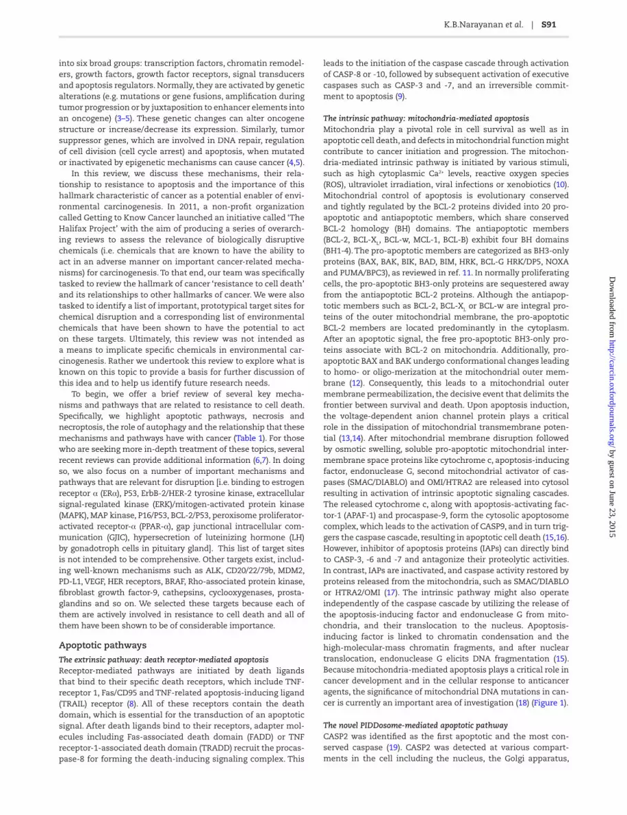
The intrinsic pathway: mitochondria-mediated apoptosis Mitochondria play a pivotal role in cell survival as well as in apoptotic cell death, and defects in mitochondrial function might contribute to cancer initiation and progression. The mitochon-dria-mediated intrinsic pathway is initiated by various stimuli, such as high cytoplasmic Ca2+ levels, reactive oxygen species (ROS), ultraviolet irradiation, viral infections or xenobiotics (10). Mitochondrial control of apoptosis is evolutionary conserved and tightly regulated by the BCL-2 proteins divided into 20 pro-apoptotic and antiapoptotic members, which share conserved BCL-2 homology (BH) domains. The antiapoptotic members (BCL-2, BCL-XL, BCL-w, MCL-1, BCL-B) exhibit four BH domains (BH1-4). The pro-apoptotic members are categorized as BH3-only proteins (BAX, BAK, BIK, BAD, BIM, HRK, BCL-G HRK/DP5, NOXA and PUMA/BPC3), as reviewed in ref. 11. In normally proliferating cells, the pro-apoptotic BH3-only proteins are sequestered away from the antiapoptotic BCL-2 proteins. Although the antiapop-totic members such as BCL-2, BCL-XL or BCL-w are integral pro-teins of the outer mitochondrial membrane, the pro-apoptotic BCL-2 members are located predominantly in the cytoplasm. After an apoptotic signal, the free pro-apoptotic BH3-only pro-teins associate with BCL-2 on mitochondria. Additionally, pro-apoptotic BAX and BAK undergo conformational changes leading to homo- or oligo-merization at the mitochondrial outer mem-brane (12). Consequently, this leads to a mitochondrial outer membrane permeabilization, the decisive event that delimits the frontier between survival and death. Upon apoptosis induction, the voltage-dependent anion channel protein plays a critical role in the dissipation of mitochondrial transmembrane poten-tial (13,14). After mitochondrial membrane disruption followed by osmotic swelling, soluble pro-apoptotic mitochondrial inter-membrane space proteins like cytochrome c, apoptosis-inducing factor, endonuclease G, second mitochondrial activator of cas-pases (SMAC/DIABLO) and OMI/HTRA2 are released into cytosol resulting in activation of intrinsic apoptotic signaling cascades. The released cytochrome c, along with apoptosis-activating fac-tor-1 (APAF-1) and procaspase-9, form the cytosolic apoptosome complex, which leads to the activation of CASP9, and in turn trig-gers the caspase cascade, resulting in apoptotic cell death (15,16). However, inhibitor of apoptosis proteins (IAPs) can directly bind to CASP-3, -6 and -7 and antagonize their proteolytic activities. In contrast, IAPs are inactivated, and caspase activity restored by proteins released from the mitochondria, such as SMAC/DIABLO or HTRA2/OMI (17). The intrinsic pathway might also operate independently of the caspase cascade by utilizing the release of the apoptosis-inducing factor and endonuclease G from mito-chondria, and their translocation to the nucleus. Apoptosis-inducing factor is linked to chromatin condensation and the high-molecular-mass chromatin fragments, and after nuclear translocation, endonuclease G elicits DNA fragmentation (15). Because mitochondria-mediated apoptosis plays a critical role in cancer development and in the cellular response to anticancer agents, the significance of mitochondrial DNA mutations in can-cer is currently an important area of investigation (18) (Figure 1).
The novel PIDDosome-mediated apoptotic pathwayCASP2 was identified as the first apoptotic and the most con-served caspase (19). CASP2 was detected at various compart-ments in the cell including the nucleus, the Golgi apparatus,
by guest on June 23, 2015http://carcin.oxfordjournals.org/
Dow
nloaded from

S92 | Carcinogenesis, 2015, Vol. 36, Supplement 1
Table 1. Characteristics of apoptosis, autophagy and necrosis pathways
Morphological and biochemical features and modulators of cell deathMethods of detection
Apoptosis Morphological features: cellular shrinking, condensation and margination of chromatin, nuclear fragmentation and DNA laddering, plasma membrane budding and formation of apoptotic bodies in cytoplasm. Not surrounded by tissue injury of inflammation. Biochemical features: caspase-dependent cell death pathway. Activation: activation of intrinsic apoptotic pathway; BCL-2, c-FLIP, survivin IAP–antisense mRNA technology; recombinant TRAIL for DR4 and/or DR5 receptor; E2F-1 gene therapy; TWEAK (tumor necrosis factor-related weak inducer of apoptosis) is a cytokine belonging to TNF-ligand family for Tweak-receptor inducing apoptosis. Inhibition: natural and synthetic inhibitors of caspases; nitrosylation of caspase 9 or 3; c-Jun–mRNA antisense technology; CEP 1347–inhibitor of JNK signaling blocks Aβ-induced cortical neuron apoptosis.
Microscopic techniques: cellular features by light microscopy, nuclear DNA analysis by fluorescent stains (annexin V), confocal laser microscopy and electron microscopy. Assessment of DNA fragmentation: enzyme-linked immunosorbent assay, terminal deoxynucleotidyl transferase-mediated dUTP nick end labeling assay, comet method, DNA diffusion, immunohistochemistry for single-stranded DNA and gel electrophoresis. Flow cytometry: cell cycle. Laser scanning cytometry: DNA content, phosphatidylserine translocation, inner mitochondrial transmembrane potential and caspase activity. Gene expression: northern blot, RNA protection assay, reverse transcription–polymerase chain reaction and immunohistochemistry. Evaluation of apoptosis-associated proteins: enzyme-linked immunosorbent assay, western blot and electrophoretic mobility shift assay.
Autophagy Morphological features: partial chromatin condensation, no DNA laddering, cell membrane blebbing and formation of more autophagosome. Biochemical features: caspase-independent cell death pathway. Activators: conventional cytotoxic drugs and irradiation; BCR-ABL tyrosine kinase inhibitor–imatinib; anti-EGFR–cetuximab; proteasome inhibitors; TRAIL and histone deacetylase inhibitors; mTOR inhibitors and its analogs; ATP-competitive inhibitors of mTORC1 and mTORC2; dual PI3K-mTOR inhibitor; antidiabetic drug–metformin; serotonin reuptake inhibitor–fluoxetine; norepinephrine reuptake inhibitor–maprotiline; antiepileptic drug–valproic. Inhibitors: antibody against EGFR–cetuximab; Class III PI3K inhibitors-3-methyadenine, wortmannin and LY294002; antimalarial drugs–hydroxychloroquine; vacuolar ATPase–bafilomycin A1; lysosomotropic drug–monensin; microtubule-disrupting agents–taxanes, nocodazole, colchicine, vinca alkaloids; antidepressant drug–clomipramine; antischistome agent–lucanthone (autophagosome degradation) .
Electron microscopy, immunohistochemical staining of microtubule-associated protein 1 light chain 3 (LC3) as a general marker for autophagic membranes, monodansylcadaverine staining of autophagic vacuoles and protein degradation assays.
Necrosis Morphological features: cell size increases, clumping and random degradation of nuclear DNA, cell membrane swelling and rupture, swelling of organelles, gain in cell volume (oncosis), organelle degeneration mitochondrial swelling and increased vacuolation. Activation: hyperactivation of poly(ADP-ribose) polymerase 1 (PARP1) enzyme with depletion of β-nicotinamide adenine dinucleotide and of ATP, hypoxic injury and oxida-tive stress (ROS/reactive nitrogen species); chetomin–inhibi-tor of tumor growth by inducing necrosis in vivo; dimethoxy-naphthoquinone–generation of ROS and induces apoptosis or necrosis; myristoleic acid methyl ester–induces apoptosis and necrosis in prostate cancer cells; sterigmatocystin–a mycotoxin inhibits DNA synthesis and causes necrosis. Inhibition: necrox-2 [5-(1,1-dioxo-thiomorpholin-4-ylmethyl)-2-phenyl-1H-indol-7-yl]-(1-methanesulfonyl-piperidin-4-yl)-amine] and necrox-5 [5-(1,1-dioxo-thiomorpholin-4-ylmethyl)-2-phenyl-1H-indol-7-yl]-(tetrahydro-pyran-4-ylmethyl)-amine]–is a cell-permeable necrosis inhibitor selectively locks oxidative stress-induced necrosis with antioxidant property; tyrphostin AG 126–reduces LPS-induced tyrosine phosphorylation of p42MAPK; cyclosporin A–inhibitor of the mitochondrial permeability transition pore (MPTP) and prevents necrosis; IM-54 (indolylmaleimide derivative)–inhibits necrotic cell death induced by H2O2 in promyelocytic leukemia HL-60 cells; PARP inhibitor VIII, PJ34 [2-(dimethylamino)-N-(6-oxo-5,6-dihydrophen-anthridin-2-yl)acetamide hydrochloride]–inhibitor of PARP-1 and PARP-2 and inhibits necrosis; IM-54 [2-(1H-indol-3-yl)-3-pentylami-no-maleimide]—a selective inhibitor of oxidative stress-induced necrosis.
Electron microscopy; nuclear negative staining; ethidium homodimer III DNA assay; detection of inflammation and damage in surrounding tissues.
by guest on June 23, 2015http://carcin.oxfordjournals.org/
Dow
nloaded from
K.B.Narayanan et al. | S93
endoplasmic reticulum and cytoplasm. Previous studies have shown that CASP2 can be activated by DNA damage induced by anticancer drugs, such as cisplatin and etoposide, or by ultravio-let and γ-irradiation, and it is a critically involved in genotoxic stress-induced apoptosis (19). CASP2 activation leads to the release of cytochrome c, indicating that CASP2 acts upstream of mitochondria-mediated intrinsic pathway (20). Moreover, the treatment of cells with the CASP2 inhibitor and/or small interfer-ing RNA to block CASP2 from inducing the release of cytochrome c is followed by the activation of CASP9 and 3. Similar to other initiator caspases, pro-caspase-2 contains a Caspase Activation and Recruitment Domain at the N-terminus. CASP2 recognizes a pentapeptide VDVAD for cleavage of target proteins, and its known target proteins are BID, PARP, Plakin, Huntingtin and DNA fragmentation factor 45. Because CASP2 is activated by a proximity-induced self-cleavage mechanism, it obtains proxim-ity by forming a PIDDosome, which is composed of three protein components, PIDD (TP53-induced protein with death domain), RAIDD (RIP-associated Ich-1/Ced-3-homolog protein with a death domain) and CASP2, whose interaction supported by their respective death domains. PIDD death domain can also interact with the death domain of receptor-interacting protein-1 kinase implicated in the nuclear factor-κB (NF-κB) activation (21). PIDD appears to act as a molecular switch, controlling the balance between life and death upon DNA damage (Figure 1).
Necrosis and necroptosis
In contrast with apoptosis, necrosis is a genetically controlled process; necrosis involves an uncontrolled and progressive loss of cytoplasmic membrane integrity, a rapid influx of Na+, Ca2+ and water, resulting in cytoplasmic swelling, nuclear pyknosis and the release of lysosomal and granular contents into the surrounding extracellular space (22). Although the molecular mechanisms underlying apoptosis are better understood, little is known about the molecular events leading to necrosis. Necrosis has recently emerged as an important and physiologically relevant signal-ing process contributing to ovulation, immune defense, death of chondrocytes controlling the longitudinal growth of bones and cellular turnover in the intestine (23). In vivo studies indicated that removal of interdigital cells in the paws of Apaf1−/− mice during embryogenesis occurs by a caspase-independent necrotic-like process (24). However, accumulating evidence by many research-ers suggests that necrosis is not just an unregulated and uncon-trollable process. Rather, it involves a programmed and actively
regulated process (aptly named necroptosis), which is regulated by the kinase activity of RIPK1 and RIPK3 that form the necro-some complex (25). This leads to the plasma membrane per-meabilization, release of cell contents and exposure of damage/danger-associated molecular patterns (DAMPs), such as HMGB1, S100 protein, IL33 and mitochondrial DNA. Under normal physi-ological conditions, autophagy and the caspase-8-FLIPL-FADD platform are apparently gatekeepers preventing necroptosis (26).
The paradoxical role of autophagy in cancer
Autophagy is the basic catabolic mechanism in response to starvation or other stressful conditions whereby unnecessary or dysfunctional misfolded or aggregated proteins and cellular components (e.g. mitochondria, endoplasmic reticulum and peroxisomes) are engulfed within double-membrane vesicles called autophagosomes and are eventually digested by lyso-somal enzymes to sustain cellular metabolism (27,28). During macroautophagy, a cytoplasmic cargo is delivered to the lyso-some through an autophagosome, which fuses with the lyso-some to form an autolysosome. Microautophagy involves the inward folding of the lysosomal membrane, which delivers a small portion of cytoplasm into the lysosomal lumen. Both macro- and micro-autophagy can be either non-selective or selective in the removal of large cellular components and pro-tein aggregates (29). Autophagy involves several key steps for the final degradation of cellular components in lysosomes: (i) initia-tion and nucleation of phagophore; (ii) expansion and matura-tion of autophagosomes; (iii) fusion of the autophagosome with the lysosome to form the autolysosome and (iv) execution of autophagy (degradation). These steps are tightly regulated by highly conserved Atg genes and non-Atg genes (30).
Disorders in autophagic signaling pathways are frequently observed in cancer patients. Autophagy has been referred to as a ‘double-edged sword’ because it acts as an activator of tumor cell death (tumor suppression) as well as it plays a part in tumor cell survival during tumor development and in cancer therapy. Impaired autophagy was shown leading to failure of removing damaged protein and organelles, and exerting genomic instabil-ity and aneuploidy, which promotes tumorigenesis (31–33). The loss of BECN1 was found in human breast and ovarian cancers (34), whereas Becn1 null mice were shown to be tumor prone (35). In contrast, the BECN1 forced expression can inhibit tumor development. Additionally, sustained p62 (SQSTM1) expres-sion, which results from autophagy defects, was found to be
Figure 1. Apoptotic and non-apoptotic signaling pathways and the involvement of anthropogenic chemicals.
by guest on June 23, 2015http://carcin.oxfordjournals.org/
Dow
nloaded from

S94 | Carcinogenesis, 2015, Vol. 36, Supplement 1
important in the promotion of tumorigenesis through de-regu-lation of NF-κB expression (33). Tumor cells experience elevated cytotoxic and metabolic stresses (e.g. hypoxia and deprivation of growth factor and oxygen), which can activate autophagy to maintain cellular biosynthesis and survival (28). Recent data indicate that suppression of autophagic proteins inhibited cell growth and conferred or potentiated the induction of cell death, indicating that autophagy contributes to cell survival in human cancer cells, as well as plays a role in adaptive response of tumor cells to anticancer therapies (36). A careful examination of the literature shows that an increased level of autophagic markers in the dying cell might not be the result of increased autophagic flux but due to a blockage of autophagy at its maturation. Therefore, the simple determination of numbers of autophago-somes is insufficient for an overall estimation of autophagic activity. It is necessary to distinguish by performing ‘autophagic flux’ assays whether autophagosome accumulation is due to autophagy induction or, alternatively, a blockade of steps in the downstream of autophagosome formation. Now, it is agreed that the true meaning of ‘autophagic cell death’ should be cell death by autophagy, not cell death with autophagy (Figure 1).
Dysfunctional apoptosis in cancers
The fundamental link between malignancy and apoptosis is exemplified by the ability of oncogenes, such as MYC and RAS, and tumor suppressors, such as TP53 and RB (Retinoblastoma), to actively engage apoptosis as well as the aberrant alterations of apoptosis regulatory proteins such as BCL-2 and c-FLIP in vari-ous solid tumors (37–39). Acquired apoptosis resistance is a hall-mark of most human cancers. With regard to apoptosis triggers, a variety of signals (irradiation, growth/survival factor depletion, hypoxia, oxidative stress, DNA damage, cell cycle checkpoints defects, telomere malfunction and oncogenic mutations, chemo-therapeutic agents and heavy metals) appear to provide the selec-tive pressure needed to alter apoptotic programs during tumor development in support of tumor evolution (40–42). The ability of tumor cells to acquire resistance to apoptosis is a compensatory mechanism, which gives tumor cells a distinct (survival) advan-tage over normal cells. Defects in apoptosis have been implicated in many events relevant to tumorigenesis: (i) cell accumulation from the imbalance of cell proliferation and cell death or a fail-ure of normal turnover process; (ii) permissive cell survival in the face of antigrowth signals, for example, hypoxia in tumor mass, cell–matrix and cell–cell adherence or contact inhibition; (iii) promoting resistance to the killing mechanisms of immune cell attack and (iv) fostering tumor metastasis by promoting cell sur-vival in the circulation under detachment conditions, also known as anoikis resistance (43). The importance of this sort of dysfunc-tion is underscored by the fact that tumor cells that possess alterations in proteins involved in apoptosis are often resistant to chemotherapy and are more difficult to treat (because anticancer drugs primarily work by inducing apoptosis). Tumor cell survival, unlike the survival of normal cells, is therefore highly dependent on aberrations of apoptosis signaling pathways (37).
Emerging evidence indicates that cancer stem cells (CSCs), the rare subpopulation of undifferentiated tumorigenic cells, are potential driving force for tumor growth and maintenance (44). To date, CSCs have been identified and isolated from various solid tumors including the lung, brain, breast, colon and skin. These CSCs are highly capable of self-renewal and are able to generate a progeny of differentiated cells that constitute a large majority of cells in the tumors (45). Most importantly, CSCs are apoptosis resistant and very likely responsible for tumor resist-ance to chemotherapy and irradiation (46). This can be attrib-uted to the undifferentiated status of CSCs and to the extrinsic
factors such as the tumor microenvironment and adhesion-based interactions, which also support their apoptotic resist-ance (47). Furthermore, the epithelial/mesenchymal transition (EMT) mechanism has been found to underlie the CSC charac-teristics that are linked to anoikis resistance (48). Accumulating evidence also suggests that this inherent resistance in CSCs shares similar extrinsic and intrinsic apoptotic pathway as nor-mal stem cells and differentiated cancer cells (49,50).
Regulation of apoptosis in cancer
Evasion of apoptotic pathways allows cells to sustain chronic proliferation, which is a hallmark of cancer. Recently, two work-ing models of apoptosis (both regulated by BCL-2 family and BH3-only proteins) were reviewed (51). The direct model proposes that the activator BH3-only proteins (BIM, BID and PUMA) can directly activate BAX and/or BAK oligomerization in addition to neutralizing BCL-2-like proteins, whereas the sensitizer BH3-only proteins (BAD and NOXA) release activator from activator/pro-survival protein complex. The indirect model suggests that BAX is primed in normal cells by BH3-only protein and bound with BCL-2. In excess of pro-apoptotic signaling, BH3-only proteins compete with BCL-2 allowing oligomerization of BAX and BAK leading to apoptosis (52). The BAX/BAK oligomerization loosens the integrity of mitochondria and culminates with mitochon-drial outer membrane permeabilization facilitating the release of cytochrome c into the cytoplasm, which interacts with APAF-1, and leads to the ATP-dependent formation of apoptosome, and the recruitment and activation of the CASP-9, -3 and -7. In the absence of APAF-1 and CASP-9, cytochrome c release itself is not sufficient to induce apoptosis (53–55). Cytochrome c diffu-sion and death receptor signaling mediates modulation of XIAP by SMAC/DIABLO and OMI/HTRA2, and activation of caspases (56) (Figure 1). Up-regulation of XIAP, survivin and down-regula-tion of APAF-1 has been observed in several tumors.
Cellular stress and DNA damage are regulated through two tumor suppressor genes TP53, which induces expression of NOXA, PUMA and RB upon various environmental and chemical stresses. Recently, a bona fide tumor suppressor gene neurofi-bromin 2 (NF2/Merlin) was shown to regulate apoptosis through the Hippo pathway (57). RB integrates outside inhibitory signals, whereas TP53 senses irreparable damage in genomic integrity, intracellular organelles and nucleotides, as well as subopti-mal level of glucose, and growth inhibitory signals (58). TP53 activities are tightly regulated by a network of protein–pro-tein interactions, microRNAs and a range of post-translational modifications, including phosphorylation, acetylation, methyla-tion and ubiquitination (59,60). TP53 activity is suppressed by a direct binding of TP53 to murine double minute 2 (MDM2), which targets TP53 for proteasomal degradation. NOXA also induces apoptosis in TP53/TP73-dependent manner in response to DNA damage, whereas PUMA, the most potent pro-apoptotic regula-tor, induces apoptosis both in a TP53-dependent and -independ-ent fashion (61–63).
Cellular metabolism is a key for the survival of cells, whereas altered metabolism in cells induces either apoptosis or resist-ance to apoptotic stimuli. Metabolic enzymes and its inter-mediates from glycolysis, pentose phosphate pathway and tricarboxylic acid cycle have shown deregulated in many cancer types to provide nicotinamide adenine dinucleotide phosphate (NADPH), citrate, acetyl CoA and various other metabolites for high demand of biosynthesis and proliferation (64). Chronic pro-liferating cells short circuit their metabolic pathways and mostly depend on aerobic glycolysis to sustain the massive biosynthe-sis of intracellular structures. Various post-translational modi-fication regulates cellular growth especially phosphorylation
by guest on June 23, 2015http://carcin.oxfordjournals.org/
Dow
nloaded from

K.B.Narayanan et al. | S95
and acetylation and increase apoptotic sensitivity. Metabolic intermediates also regulate pro- and anti-apoptotic regulators (BCL-2 family protein). Perturbations in acetyl-CoA production may extend to other oncogenic contexts beyond that of BCL-xL (65–67). Redox status of tissues/cells affects their sensitivity to cytochrome c. Reduced glutathione mostly produced by NADPH inactivates cytochrome c, whereas apoptotic agents produce ROS to activate cytochrome C and apoptosis (68). Key regulatory metabolic enzymes, which affect apoptosis (e.g. hexokinase, fructose 2,6-bisphosphate kinase, lactate dehydrogenase M and pyruvate dehydrogenase kinase), are also implicated in cancer (55). Growth factors/cytokines regulate pro-survival signaling by RAS- and PI3K-AKT pathways through cognate receptor tyrosine kinase (RTK). Most human cancers harbor mutations in AKT and PTEN, which leads to AKT activation and resistance to apopto-sis (69,70). Death receptor signaling triggers the recruitment of FADD and TRADD adapter proteins to induce dimerization of CASP-8 and subsequent activation of CASP-3 and -7. In some cell types, CASP-8 directly cleaves BH3-only protein BID to localize it to the mitochondria and activate BAX (71).
Additionally, ‘anoikis’, the detachment of cells, is another major regulator of apoptosis. The detachment of adherent cells (loss of critical interaction between the cell and the extracellu-lar cell matrix) leads to apoptosis due to the loss of integrin α-5 or β-5 signaling and the loss of focal adhesion kinase, a reduc-tion of talin–integrin interaction, and of c-Jun N-terminal kinase signaling (72).
Oncogenes, tumor suppressor genes and apoptosis
Human health is continuously challenged by exposure to a wide range of environmental chemicals that affect DNA integrity (73). When DNA repair capacity is exhausted, DNA damage accumu-lates in cells at a higher level, and this excess damage causes an increased frequency of mutation and/or epigenetic alterations of specific genes (oncogenes and tumor suppressors) resulting in the disruption of the cellular networking that controls cellu-lar homeostasis and leads to cellular transformation and cancer development (74). The inactivation of expression of tumor sup-pressor genes via genetic and epigenetic changes (DNA hyper-methylation, histone deacetylation/methylation and microRNA targeting) often leads to tumor initiation and progression, whereas amplification and overexpression of oncogenes result in the similar tumorigenic phenotype (75). Tumor suppressor ‘driver’ genes include: genes for retinoblastoma protein (RB), tumor protein TP53 (TP53), BRCA1 and 2, PTEN, VHL, APC, CD95, ST5, 7 and 14, YPEL3, whereas ‘driver’ oncogenes include: growth factors (e.g. C-SIS, WNT), RTKs (EGFR, PDGFR, VEGFR, TRK, ERBB2), cytoplasmic tyrosine (SRC, ABL and BTK) and serine/threonine (ATM, MTOR, ERK, PI3KCA, AKT1, 2 and 3, LKB1 and RAF) kinases, transcriptional factors (MYC and E2F), GTP-ases (RAS) and oth-ers (CCND1), as reviewed by Lee et al. (74). Discovery of microRNA genes added new members to both tumor suppressor (e.g. miR-34a) and oncogene (e.g. miR-17–92) families (76).
As part of the DNA damage response to genotoxic stress, apoptosis is triggered by chemical-induced DNA lesions and represents a first line of defense allowing the organism to elimi-nate damaged cells. Notably, cells respond to stress-induced DNA damage by increasing their levels of TP53 (77). The wild-type TP53 prevents cancer formation through the activation of cell cycle arrest or apoptosis via transcriptional regulation of hundreds of specific gene targets or via multiple protein–pro-tein interactions. TP53 and its evolutionary older relatives, TP63 and TP73, exhibit a similar modular structure and share signifi-cant structural and functional homologies; however, their tumor suppressive role is not as straightforward as TP53. Genes for all
TP53 family members produce proteins with the transactivation domain displaying a tumor suppressive function and proteins without transactivation domain acting as oncoproteins (78). TP53 is mutated in >50% of human cancers, whereas in other cancers, its function is compromised by de-regulation of the TP53 pathway. Both TP63 and TP73 are rarely mutated or epi-genetically altered in human cancers. Tp53−/− mice develop tumors with short latency and 100% penetrance (77). Tumor suppressive function for TP73 was confirmed using Tp73−/− mice (79). Tp53+/− and tp63+/+ mice are less cancer prone than Tp53−/− and tp63+/− mice, respectively.
The synergistic effects of the TP53 family members in tumor suppression were highlighted using mice heterozygous for mutations in both TP53 and TP63, or TP53 and TP73 displaying higher tumor burden and metastasis, compared with tp53+/− mice (80). Accumulating data show that TP53 family proteins can regulate cell survival via cell cycle arrest, senescence and apoptosis and are abnormally expressed in different cancer types (breast tumors, acute myeloid leukemia, head and neck tumors, melanoma, renal cell carcinoma, colon, ovarian and lung tumors) suggesting that their differential expression may disrupt the TP53 response and contribute to tumor initiation/progression and linked to cancer prognosis and treatment (78).
Although mutations of TP63 mutations are almost non-exist-ent in human cancers, >80% of primary head and neck squamous cell carcinomas, other squamous cell epithelial malignancies and non-small cell lung cancer retain TP63 expression, where it is often over-expressed and occasionally amplified. The TP63 expression strongly influences the tumor cell response to gen-otoxic stress (81). TP63 activates death domain receptor- and mitochondria-mediated apoptosis pathways, which are clearly reinforced by concomitant treatment with genotoxic stress. However, ΔNp63α confers resistance to apoptosis via a transcrip-tional regulation of AKT1, as well as via down-regulation of sev-eral microRNAs (miR-181a, -519a and -374a) and up-regulation of miR-630, which targets proteins involved in cell cycle arrest and apoptosis for down-regulation, hence conferring tumor cell chemoresistance (82,83). It is likely that apoptosis sensitivity to genotoxic agents may be determined not only by TP53 but also by TP73 and TP63 function, and its isoforms (84).
The disruption of normal cell death
From a disruption standpoint, the inactivation or attenua-tion of the TP53 apoptotic response, achieved by mutations or epigenetic alterations, is known to promote cell transforma-tion (77). For example, non-polycyclic aromatic hydrocarbon components present in tobacco smoke condensate are able to attenuate the TP53 apoptotic response, as suggested by studies in mouse epidermal cells (78). The transcription factor C/EBPβ, which is induced by cigarette smoke has also been involved in TP53 repression (85,86). Following a prolonged exposure to environmental chemicals, bulky DNA adducts may not be removed by DNA repair mechanisms but converted into muta-tions. Subsequent DNA replication cycles may lead to hot spot mutations in key growth regulatory genes, thereby resulting in malfunction of tumor suppressor genes and amplification/over-expression of oncogenes (74).
Similarly, mutated RAS oncogenes were found in the experi-mental tumors of rodents that had been exposed to chemical or physical compounds, as well as in many human cancers (87). For example, exposure to hydrocarbon solvents has been associated with an increased risk of exocrine pancreatic cancer, the human tumor with the highest prevalence of K-RAS mutations (88). And heterocyclic amines have been implicated in both initiation and maintenance of breast tumorigenesis mediated by upregulated
by guest on June 23, 2015http://carcin.oxfordjournals.org/
Dow
nloaded from

S96 | Carcinogenesis, 2015, Vol. 36, Supplement 1
H-RAS expression, ERK pathway activation, NOX1 expression and elevation of ROS (89). Although the sustained activation of the NF-κB transcription factor is another important element involved in chemical tumorigenesis, tobacco, alcohol, high-fat diet, envi-ronment pollutants, cancer-causing viruses (human papillomavi-rus, hepatitis B and C viruses, human immunodeficiency virus and bacteria (Helicobacter pylori), ultraviolet light, ionizing radiation, obesity and oxidative stress are all potent NF-κB stimuli (90,91). The following proteins: pro-inflammatory proteins (cyclooxyge-nase-2, inducible nitric oxide synthase, TNF, interleukin-8); prolif-erative/pro-survival factors (bone morphogenetic proteins, stem cell factor, vascular endothelial growth factor, granulocyte–mono-cyte colony stimulating factor) and antiapoptotic proteins [TRAF-1 and -2, the CASP-8 inhibitor (FLIP), IAPs, XIAP, BCL2 and its homo-logues and matrix metalloproteinases] are overall involved in tumor promotion, initiation and progression (92).
The critical research gaps for a clear understanding of chem-ical carcinogenesis include the following:
1. Understanding how genetic modifications by low-dose environmental mixtures can disrupt/overcome normal cell death.
2. Understanding the molecular processes and pathways activated/blocked by individual chemicals and mixture of chemicals with disruptive potential.
3. Understanding the low-dose effects of environmental chemicals (single and mixtures) on cell death within differ-ent tissues and organs of human.
4. Clearly distinguishing the differences between the contri-butions of both carcinogenic and non-carcinogenic chemi-cals (individually and in mixtures) in environmental car-cinogenesis by experimental methods.
Key target sites for disruption
In this review, we wanted to look at several key target sites that disrupt normal cell death and potentially have relevance for envi-ronmental carcinogenesis. It is generally agreed that many can-cers arise from a single cell that has accumulated genetic and epigenetic mutations of a few crucial genes of proto-oncogenes and tumor suppressors, and that this is caused by random errors in DNA replication or a reaction of the DNA with free radicals or other chemical species of endogenous or exogenous origin (93). However, we also know that chemicals with disruptive potential are capable of a wide range of additional cellular level effects that are relevant to cancer (94), and the general population now faces ongoing exposures to thousands of environmental chemicals that are present in consumer products, our food, our water and in the air (95). At the same time, regulators worldwide have remained largely focused on the effects of single chemicals while placing very little emphasis on the effects of exposures to mixtures of chemicals in the environment (96). Accordingly, in this review, we emphasize the pivotal and enabling role that resistance to cell death plays in carcinogenesis and we highlight some of the key mechanisms and pathways that can be chemically disrupted (i.e. in a manner that results in dysfunction of normal cell death rou-tines) and that have the potential to be supportive of the emer-gence of an immortalized cellular phenotype.
To that end, we first identify and review a number of key tar-gets of this nature that have been shown to be active sites for chemical disruption in the past as follows:
Binding to ERαGiven that many anthropogenic agents are xenoestrogens, a considerable amount of environmental health research has
focused on ER level disturbances (97). Many xenoestrogens binds to ER and either activates it or inhibits it. ERα activation stimulates cell proliferation and initiates cancer through tumor promotion, whereas the activation of ERβ stimulates terminal cell differentiation and disrupts cancer progression, which is an anticancer effect. For example, many of the organochlorine (OC) pesticides such as lindane or their metabolites fall into the category of xenoestrogens that disrupt endocrine processes by acting as agonists of ERα and/or antagonists of ERβ and by exert-ing antiandrogenic effects (by binding to androgen receptors). ERα and tumor suppressor protein p53 exert opposing effects on cellular proliferation. ERα’s repression of p53-mediated cell death has been widely investigated, especially in breast cancer (98), but emerging evidence suggests a much more complex role for ERα-controlled pathways in other tumor-related phenom-ena. ERα interacts with p53 bound to promoters of Survivin and multidrug resistance gene 1(MDR1), and inhibits p53-mediated transcriptional repression of these genes in human cancer cells in vivo. It was found that p53 is necessary for ERα to access the promoters and there is cross-talk between the pathways medi-ated by ERα and p53 (99). It has been also been shown that an increase of ERα messenger RNA (mRNA) level in ERα-positive breast cancer is associated with de-regulation of metabolism, which produce a complementary effect on cell differentiation and proliferation (100). On the other hand, evidence of ERα’s role in the EMT has also been reported. In endometrial carci-nomas and breast cancer, ERα’s activity is negatively associated with the activation of EMT via the Wnt, Sonic Hedgehog and transforming growth factor-β (TGF-β) signaling (101,102). EMT involves the loss of cell–cell adhesion and a consequent increase in mobility and invasiveness. It has been proposed that ERα acts to promote invasive growth in breast cancer cells by a direct, ERα-dependent expression of metastasis-associated genes, such as the MTA-3 protein. It is important to note that a physiological feedback mechanism regulates the efficiency of ERα activation through the state of cell–cell interactions that are mediated by E-cadherin (103). Although EMT promotes a decrease in cellular contacts, it also inhibits ERα transcription thus limiting its own ERα-dependent activation. Although no effects on genetic insta-bility and immune system evasion of systematic ERα activation have been reported, the synergy of action involved in these dif-ferent (deregulated) pathways may be very important for cancer onset and progression.
Gap junctional intracellular communicationIn addition to tumor promotion ability, some environmental chemicals directly or indirectly cause DNA mutations through free radical production (ROS/reactive nitrogen species) and may cause both tumor initiation and tumor promotion by inhibiting GJICs and connexins (Cxs) (104,105). Blockage of GJIC between the normal and the pre-neoplastic cells creates an intra-tissue microenvironment in which tumor-initiated pre-neoplastic cells are isolated from growth controlling factors of normal surround-ing cells resulting in clonal expansion (106). Gap junction chan-nels and Cxs control cell apoptosis by facilitating the influx and flux of apoptotic signals between adjacent cells and hemi-chan-nels between the intracellular and extracellular environments. Recently, it has also been demonstrated that Cx proteins in con-junction with their intracytoplasmic localization, may act as signaling effectors that are able to activate the canonical mito-chondrial apoptotic pathway (107). Tumor-promoting chemicals such as phenobarbital, dichlorodiphenyltrichloroethane (DDT) and 12-O-tetradecanoylphorbol 13-acetate block apoptosis and also block GJIC, whereas several antitumor chemicals, such
by guest on June 23, 2015http://carcin.oxfordjournals.org/
Dow
nloaded from

K.B.Narayanan et al. | S97
as retinoids and dexamethasone, increase GJIC and increase apoptosis. So, it has been hypothesized that GJIC is necessary for apoptosis and blockage of apoptosis with chemicals/car-cinogens could therefore promote the initiation of premalignant cells in tumorigenesis (108). For example, knockdown of con-nexin 43 (Cx43) had an inhibitory effect on GJIC and resulted in a reduction of cell death after treatment with cisplatin and Salmonella (109), and Kang et al. (105) reported on the inhibition of GJICs in normal human breast epithelial cells by pesticides, polychlorinated biphenyls, polybrominated biphenyls and halo-genated hydrocarbons (when given as single compounds or as mixtures), and suggested that they may contribute to carcino-genesis through this mechanism.
Peroxisome proliferator-activated receptor-αPPAR-α receptors are mainly found in the liver and belong to the steroid hormone receptor superfamily that functions as a tran-scription factor for genes involved in glucose, lipid and amino acid metabolism, and that also induces enzymes involved in the metabolism of xenobiotics. Upon ligand binding, PPARs heter-odimerize with the retinoid X receptor and bind to the specific promoter sequence and trigger the expression of target genes (110). A variety of chemicals including certain herbicides and plasticizers induce peroxisome proliferation with increased rep-licative DNA synthesis, suppression of apoptosis and increased expression of peroxisomal acyl-CoA oxidase in rodent liver and other tissues. In rodents, these peroxisome proliferating chemi-cals act as non-genotoxic carcinogens that promote the devel-opment of tumors (111). Chemicals of industrial importance such as diethylhydroxylamine and chlorinated solvents are per-oxisome proliferators (PP) that induce elevated S-phase in rat and mouse and play an important role in hepatocarcinogenicity. The molecular mechanisms of altered expression of cell cycle regulatory proteins resulting in the elevation of S-phase, and suppression of apoptotic cell death and induction of prolifera-tion are evidenced by the activation of PPAR-α and survival sign-aling by p38 MAPK in hepatocellular carcinomas (112).
Hypersecretion of LH by gonadotroph cells in pituitary gland Neuroendocrine disruptors are environmental pollutants that are agonists/antagonists or modulators of the synthesis and/or metabolism of neurohormones, neuropeptides and neurotrans-mitters. Sustained hypersecretion of LH occurs following the disruption of the hypothalamic–pituitary–testicular axis. The tumorigenic response to a chemical in an endocrine tumor is generally dose responsive. As the dose of environmental chemi-cal increases, the extent of perturbation of normal endocrine homeostasis increases resulting in a stronger trophic stimulus to the target cell (113). LH up-regulates the expression of apop-totic inhibitor, survivin in a dose-dependent manner via ERK1/2 signaling pathway and inhibits apoptosis in ovarian epithelial tumors in vitro (114).
p53 As noted previously, p53 is a tumor suppressor gene and has been described as the ‘guardian of the genome’. p53 is a tran-scriptional activator regulating the expression of Mdm2 (nega-tive regulator of p53) and genes involved in growth arrest (p21, Gadd45 and stratifin), DNA repair (p53R2) and apoptosis (Bax, Apaf-1, PUMA and NoxA). Its activity disrupts the formation of tumors by arresting growth and inducing apoptosis. This 53 kDa phosphoprotein induces apoptosis by stimulating BAX and FAS antigen expression, or by repression of BCL-2 expression. p53 mutations are found in most of the tumors and contribute to the
complex molecular network events leading to tumor formation. Notably, the progression of cancers which overcome cell death [via the inactivation of tumor suppressor genes (p53) and acti-vation of oncogenes (c-Ha-ras)] after exposures to organophos-phorus pesticides is also associated with an increase in genome instability (115). Accordingly, one the most important candi-dates, as a key regulator of malignant transformation, is P53. Somatic mutations of this gene or perturbations in its pathways are among the most frequent alterations in human cancers (116). Arguably, the most important decision maker in cellu-lar process that unfold in response to every kind of stress and harm, P53 is involved in cell cycle arrest, apoptosis, regulation of metabolism, DNA repair and every pathway connected to them. Its action is therefore opposed to evasion from growth control, genetic instability, sustained proliferative signaling and cellular motility, whereas it can be an important promoter of metabolic changes and even replicative immortality. Cross-talk between P53 pathways and most molecular mechanisms that trans-duce external signals (to promote or inhibit cell proliferation) is branched and efficient so chemical disruptors that systemati-cally impair P53 can readily produce harmful effects on almost all of the hallmarks involved in malignant transformation.
p16/p53p16INK4A (p16) and p53 are tumor suppressor genes (antionco-genes). p16 is known as cyclin-dependent kinase (CDK) inhibi-tor and specifically blocks the activity of CDK4 and CDK6. The binding of 16 kDa protein p16INK4A to CDK4 inhibits the phos-phorylation of retinoblastoma protein (pRB) and subsequently inhibits the transcription factor (E2F), the release and arrest of the G1 phase of cell cycle and the suppression of cellular pro-liferation. p16 also inhibits the growth of breast cancer cells by inhibiting the VEGF signaling pathway and angiogenesis. And recently, it has been demonstrated that the anticancerous abil-ity of p16 is additionally attributed to its ability to induce tumor cells to enter senescence. It also induces apoptosis both in vitro and in vivo (117). The functional or structural loss of p16INK4A therefore leads to the cell cycle propagation of genetically dam-aged/mutated cells and increases the subsequent risk of tumor development. p16 is encoded by INK4a gene and an alternative reading frame of INK4a transcribes to p14ARF, which mediates the link between p16 and p53 pathways. So, loss of the INK4a gene disrupts p16INK4A/CDK4/6/pRB and p14ARF/MDM2/p53 path-ways, which controls cell proliferation (118). Notably, the p16 locus was found to be inactivated in many cancers such as lung, breast, melanoma, pancreatic, brain and >80% of squamous cell carcinoma of the head and neck tumors (119). Thus, p16INK4A, p14ARF and p53 genes involved in cell cycle pathways are major targets of inactivation in carcinogenesis. Occupational exposure to chemicals and metal dusts form ROS and reactive nitrogen species in humans through oxidant-mediated responses, which causes hypermethylation of p16INK4A and p53 along with the acti-vation of MAP kinase to induce carcinogenesis.
BCL-2/p53 BCL-2 is a proto-oncogene, which regulates cell cycle progres-sion and apoptosis (antiapoptotic), whereas p53 is a tumor sup-pressor gene. BCL-2 constitutively suppresses p53-dependent apoptosis. The BCL-2/p53 axis requires pro-apoptotic protein (Bax) and the effector molecule (CASP-2) as essential apop-totic mediators following the silencing of Bcl-2 or Bcl-xL. p53 possesses pro-apoptotic properties that appear to be constitu-tively active despite its suppression by Bcl-2 (120). Both p53 and Bcl-2 are strong predictors of recurrence and survival in rectal
by guest on June 23, 2015http://carcin.oxfordjournals.org/
Dow
nloaded from

S98 | Carcinogenesis, 2015, Vol. 36, Supplement 1
cancer (121). And the chemical 7,12-dimethyl benz-(a)-anthra-cene induces tumor growth in breast cancer that is apparently due to the inactivation of p53 aided by the absence of Bcl-2 (122).
ErbB-2/HER-2 tyrosine kinase The human epidermal growth factor receptor (EGFR/HER) family consists of ErbB/HER lineage of receptor proteins (ErbB1–4) as it shows similarity to the v-ErbB oncogene of avian erythroblasto-sis virus (123). The ErbB-2/HER protein tyrosine kinase receptor contains an extracellular domain followed by a single trans-membrane segment and intracellular tyrosine kinase activity, which regulates cell growth and differentiation particularly during embryogenesis (124). Overexpression of ErbB2/HER2 is related with cancer. Binding of epidermal growth factor ligands to their cognate ErbB receptors induces homo- or hetero-dimer-ization of ErbB2 and autophosphorylation of phosphotyrosine residues in the cytoplasmic domain, which serve as docking sites for adaptor proteins to downstream signals for growth and survival. Up-regulation of PI3K/AKT signaling pathway is found in ErbB2+ breast cancers, where it exerts pro-survival effects overcoming cell death (125).
The PI3K/AKT/mammalian target of rapamycin (mTOR) pathway is important for cell growth and survival, and it is also frequently activated in cancer. PI3Ks are a family of intra-cellular signal transducer enzymes involved in many cellular functions such as cellular growth, proliferation, differentiation and survival playing an important role in tumorigenesis (126). Upon activation of the RTKs by growth factors, PI3Ks con-vert phosphatidylinositol-4,5-biphosphate into phosphati-dylinositol-3,4,5-triphosphate, which provides docking sites for pleckstrin homology domain containing proteins, includ-ing phosphoinositide-dependent kinase-1 and protein kinase B. This conversion is mainly controlled by the phosphatase and tensin homolog (PTEN), which dephosphorylates PIP3 into phosphatidylinositol-4,5-biphosphate, thereby regulat-ing the uncontrolled activation of AKT pathway. Loss of PTEN tumor suppressor is common in malignancies and correlates with increased AKT activity. AKT is activated by phosphoryla-tion of Thr308 by phosphoinositide-dependent kinase-1 (PDK1) and Ser473 by the mammalian target of rapamycin complex 2 (mTORC2). Activated AKT phosphorylates glycogen synthase kinase 3, forkhead box transcription factors, BCL-2 family members and the tuberous sclerosis complex (TSC1/2) thereby regulating a range of pathways involved in protein synthesis, proliferation, metabolism and apoptosis (127).
The mTOR pathway is the main target of the rapamycin, a natural compound produced by the Streptomyces hygroscopicus, which displays potent immunosuppressant and antiprolif-erative properties. The mTOR pathway integrates stimuli from diverse upstream pathways including the PI3K/AKT pathway and is responsible for the synthesis of a wide range of proteins involved in cell growth, proliferation, survival and tumorigen-esis. mTOR can act in complex with Raptor (mTORC1) or Rictor (mTORC2) and these complexes regulate entirely different pro-grams in the cell. When activated, mTORC2 activates and stabi-lizes AKT by its phosphorylation at Ser473, and controls actin cytoskeleton organization and cell migration, whereas mTORC1 increases mRNA translation by phosphorylation of the down-stream molecules p70S6K (S6K) and 4E binding protein 1 (4EBP1). Phosphorylation of p70S6K leads to mRNA biogenesis, transla-tion of ribosomal proteins and cellular proliferation. 4EBP1, in the hypophosphorylated state, binds the eukaryotic initiation factor 4E preventing its binding to eIF4G, and thereby to form the translational initiation complex eIF4F. Once phosphorylated,
4EBP1 is unable to bind to eukaryotic initiation factor 4E, which results in increase of translation of proteins related to cell pro-liferation and viability (128,129).
AKT activation affects components of the apoptosis regula-tory machinery, including the BCL-2 family, the caspase family or the function of death domain receptors. AKT directly phos-phorylates the BAD protein, which is a pro-apoptotic member of the BCL-2 family, whereas the dephosphorylated BAD pro-motes apoptosis (130,131). AKT might also prevent apopto-sis by phosphorylation and inactivation of glycogen synthase kinase-3, CASP-9 and indirect activation of NF-κB leading to the altered transcription of pro-survival genes (e.g. IAP1, IAP2), as reviewed in refs 132–134. The mTOR pathway has also been linked by several studies to play a role in cell death by apoptosis and autophagy (135). One of the proposed pathways by which mTOR regulates autophagy was discovered in studies from yeast essential autophagy genes (Atgs), as reviewed in ref. 136. Atg1/Atg13/Atg17 complex is required for maximal catalytic activ-ity of mTOR leading to Atg13 phosphorylation, subsequently destabilizing the complex and inactivating Atg1. In the mam-malian cells, unc-51-like kinase 1 (ULK1) and focal adhesion kinase interacting protein of 200 kD (FIP200) form the complex with mammalian ATG13. mTORC1 activation correlates with the phosphorylation of ULK1-ATG13-FIP200 complex and inhi-bition of autophagy. Activation of P70S6k by mTOR may block apoptosis by increasing antiapoptotic BCL-2/BCL-xL protein expression and inactivating the pro-apoptotic protein BAD (137). In human prostate cancer cell lines, ErbB-2 kinase activity was increased by OC insecticides such as lindane, DDT and fungicide chlorothalonil. DDT induces cellular proliferation of the andro-gen-dependent human prostate cancer cell line LNCaP by phos-phorylation of MAP kinase. However, no proliferative effect was induced in androgen-independent PC-3 cell line (138).
Mitogen-activated protein kinase MAPK are serine/threonine kinases that transduce extracel-lular signals from a diverse range of stimuli and elicit cellular responses such as proliferation, differentiation, survival, migra-tion, development, inflammatory responses and apoptosis. In mammalian cells, three MAPK families have been characterized namely classical MAPK (ERK), C-Jun N-terminal kinase/stress-activated protein kinase (JNK/SAPK) and p38 kinase. MAPK pathways involve a series of protein kinase cascades, and each cascade consists of more than three enzymes that are activated in a series: a MAPK kinase kinase (MAPKKK/MAP3K), a MAPK kinase (MAPKK/MAP2K/MEK) and a MAP kinase (MAPK) (139). MAPK has a pleiotropic role in cancer, especially p38 and JNK MAPK pathways that are involved in the cross-talk between autophagy and apoptosis induced by genotoxic stress. p38 MAPK plays a dual role in genotoxic stress-induced apoptosis. Rottlerin-induced apoptosis of HT29 colon carcinoma cells was contributed by the up-regulation of non-steroidal anti-inflam-matory drug activated gene-1 (NAG-1) via a p38 MAPK-dependent mechanism (140). However, under certain circumstances, it also involved in mediating resistance to apoptosis. The phospho-rylation of p38 significantly increased the resistance to doc-etaxel-induced apoptosis in prostate cancer cells (141). And the suppression of p38 MAPK reversed the overexpression of micro RNA-214, which is linked to the radiotherapy resistance of non-small-cell lung carcinoma cells (142). It also regulates autophagy both as a positive and negative regulator. Platinum compounds such as E-platinum induced autophagic cell death in gastric carcinoma BGC-823 cells via suppression of mTOR by decreas-ing phosphorylation of p38 MAPK (143). On the other hand,
by guest on June 23, 2015http://carcin.oxfordjournals.org/
Dow
nloaded from

K.B.Narayanan et al. | S99
suppression of the p38 signaling pathway induced autophagic and necroptotic cell death in TNFα-treated L929 cells. JNK MAPK promotes the phosphorylation of c-Jun and activating transcrip-tion factor-2 (ATF-2) resulting in the activation of AP-1 and the expression of Fas/FasL signaling pathway proteins, which sub-sequently activate effector caspase 3 and trigger apoptosis (144). JNK activation is associated with transformation in many onco-gene and growth-factor-mediated pathways, whereas p38 MAPK activation involves in cell differentiation processes such as adi-pocytes, erythroblasts, myoblasts, cardiomyocytes and neurons. Regulation of the cell cycle is critical in cellular proliferation and development of multicellular organisms, and abnormalities in MAPK signaling play a critical role in the development and progression of cancer. MAP kinases are reported to be involved in several pathological conditions such as cancer and other diseases. MEK4/MKK4 is involved in stress-activated pathways such as JNK, and p38 is consistently inactivated by mutation in many cancers including cancers of the ovary, breast, pancreas, bile duct, lung, colon and testes (145).
ERK/MAPK ERK pathway is a well-characterized MAPK signaling cascade with the Raf-MEK-ERK pathway. The stimulation of RTKs initi-ates the multistep cascade process resulting in the phospho-rylation of p44MAPK (ERK1) and p42MAPK (ERK2) and increasing its enzymatic activity. The activated ERKs translocate into the nucleus and transactivate many transcription factors and regu-late expression of genes to promote cell growth, differentiation or mitosis (139). It also regulates post-translational regulation of the assembly of cyclin D-cdk4/6 complexes, which subsequently phosphorylates the RB protein causing the activation of tran-scription factor E2F and regulates the genes involved in G1/S progression of cell cycle. In human hepatocytes, TGF-β induces apoptosis by the up-regulation of Rac-independent NADPH oxi-dase NOX4 mediating the production of ROS, which precedes the loss of mitochondrial transmembrane potential, cytochrome c release and caspase activation, for an efficient mitochondrial-dependent apoptosis (146). However, NOX4 up-regulation was inhibited by intracellular antiapoptotic signals such as PI3K and ERK/MAPK pathways. The overactivation of the MEK/ERK path-way in hepatocellular carcinoma HCC cell line confers resist-ance to TGF-β-induced apoptosis by impairing the up-regulation of the NADPH oxidase NOX4 expression (147). De-regulation of ERK activity is common in cancer leading to proliferation, migra-tion, resistance to apoptosis and loss of differentiated pheno-types. In particular, cancerous mutations are mostly affecting Ras and B-Raf along with the overexpression of EGFR and ERBB2 in the ERK-signaling pathway. ERK signaling also plays a crucial role in disrupting the antiproliferative effects of ligands such as TGF-β (145). OC pesticides or their metabolites (endosulfan, diel-drin and DDE) and p-nonylphenol, a detergent by-product from plastic manufacturing, all produced dose-dependent ERK-1/2 phosphorylation in a pituitary tumor cell line GH3/B6/F10, which expresses high levels of membrane receptors for ER-α (148).
Environmental chemicals that confer resistance to cell death
In this review, we wanted to further consider the possibility that low-dose exposures to combination of environmental chemicals might have a role to play in environmental carcinogenesis. To that end, we developed a list of environmental chemicals that had been shown to act disruptively on the key target sites men-tioned previously. Specifically, we sought to identify chemicals
that were ubiquitous in the environment and not known to be carcinogenic, or classified as carcinogenic to humans. We focused on bisphenol A (BPA), chlorothalonil, dibutyl phtha-late (DBP), diethylhexyl phthalate (DEHP), dichlorvos (DDVP), lindane, methoxychlor (MXC), linuron, and oxyfluorfen. These reported actions of these chemicals on these important target sites are described below.
Bisphenol A Ubiquitous environmental anthropogenic chemicals such as BPA (4,4ʹ-(propane-2,2-diyl)diphenol) and phthalates are commonly found in consumer products, and act as obesogens by disrupting the metabolic homeostasis pathway. This involves the activation of PPARγ, which is a critical regulator of fat formation and also regulates lipid, glucose and energy in humans. BPA in particular is an estrogenic mimic which does not cause mutations per se, but increases breast cancer incidence (149–151). BPA-exposed to HRBEC cell lines and T47D breast cancer cells showed markedly reduced pro-apoptotic negative regulators of the cell cycle (p53, p21WAF1 and BAX) with concomitant increases in proliferation initiating gene products (proliferating cell nuclear antigen, cyc-lins, CDKs and phosphorylated pRB). It also induced an increase in the ERα: ERβ ratio (152). In addition, TP53 loss of function pro-moted activation of mTOR pathway, together with PI3K, AKT and 4EBP1 and, concurrently, PTEN was suppressed which resulted in enhanced cell growth and proliferation, and ultimately breast tumorigenesis (153). Besides increasing the risk of breast cancer, BPA neutralizes the effects of tamoxifen, undermining a widely used preventive measure to control disease. It has been shown that BPA affects the P53 pathway, inducing a prominent evasion of apoptosis coupled by an increased proliferation (152), and the GPER/EGFR/ERK pathway influencing proliferation and migra-tion (154). This action seems to be delivered mainly through a substantial activation of mTOR pathways and a negative regula-tion of pro-apoptotic proteins like P53, P21 and BAX. In a num-ber of cases, this BPA-induced cellular misbehavior persists even after BPA has been removed thus providing additional evidences of the chronic potential of this chemical disruptor. BPA has also been shown to disrupt double-strand break repair machinery in vivo suggesting that consistent environmental exposure to BPA may severely and dangerously affect the stability of DNA in mammalian cells (155). And BPA exerts a pro-metastatic influ-ence in at least one mouse model of mammary carcinogenesis (156).
Chlorothalonil Chlorothalonil (2,4,5,6-tetrachloroisophthalonitrile) is a broad-spectrum, non-systemic, OC pesticide (fungicide). It is used to control pathogenic fungi that attack vegetables, fruits, trees and agricultural crops. It is predominantly used on peanuts, potatoes and tomatoes and as an antifungal additive in paints, emulsion and resin. The carcinogenicity of chlorothalonil was evalu-ated in rodents, and the studies have shown evidence of renal tubular carcinomas and adenomas, and stomach tumors (157). Chlorothalonil up-regulates the expression of ErbB-2 tyrosine kinase and MAP kinase leading to cell proliferation in a prostate cancer cell line (138). Chlorothalonil readily reacts and conju-gates with glutathione in the liver, and chlorothalonil metabo-lites consist of a mixture of di- and tri-glutathione conjugates, cysteine S-conjugates and mercapturic acids. In the proximal tubules of kidney, glutathione conjugates are completely cleaved by enzymes γ-glutamyl transpeptidase and dipeptidases to the cysteine S-conjugates, which are further cleaved by enzyme β-lyases to the corresponding thiol derivatives. The production
by guest on June 23, 2015http://carcin.oxfordjournals.org/
Dow
nloaded from

S100 | Carcinogenesis, 2015, Vol. 36, Supplement 1
of thiol derivatives is thought to be responsible for the toxicity seen in the kidneys (158). In a eukaryotic system, chlorothalonil reacted with proteins and decreased cell viability by formation of substituted chlorothalonil-reduced glutathione (GSH) deriva-tives and inhibition of specific NAD thiol-dependent glycolytic and respiratory enzymes (159). Caspases (cysteine-dependent proteases) and transglutaminase are some of the thiol-depend-ent enzymes involved in apoptosis. So, inhibition of these thiol-dependent enzymes in tumor-initiated cells (by chlorothalonil) may disrupt apoptotic cell death and aid in tumor survival. Chlorothalonil is considered to be non-genotoxic but classified as ‘likely’ to be a human carcinogen by all routes of exposure (95). It may also act as cytochrome P-450 inducer with the for-mation of ROS and peroxisome proliferation, which increases the subsequent risk of tumor development (160).
DBP and DEHPDiesters of phthalic acid are commonly referred to as phtha-lates. These man-made chemicals are widely used in con-sumer products, food processing and medical applications. They are measured in residential indoor environments (indoor air and house dust) and also in foods, milk and drinking water. High-molecular-weight phthalates are used as plasticizers in the manufacturing of polyvinyl chloride and low-molecular-weight phthalates are used in making varnishes, lacquers and personal-care products. All of the phthalates have been shown to disrupt reproductive tract development in male rodents in an antiandrogenic manner (161). Phthalate compounds such as DBP, butyl benzyl phthalate and DEHP mimic the function or activity of the endogenous estrogen 17β-estradiol (E2) and bind to ERs. Interestingly, phthalates can mimic estrogen in the inhibition of tamoxifen-induced apoptosis in human breast cancer cell lines by increasing intracellular BCL-2/BAX ratio, which promotes drug resistance to the ER antagonist tamoxifen in breast cancer (162). DEHP also up-regulates the expression of antiapoptotic activating transcription factor-3 (ATF-3) and down-regulates the pro-apoptotic P53 transcription and thereby suppresses apoptotic cell death in fetal mouse genital tubercle (163). It also inhibited apoptosis of Syrian hamster embryo cells (164). DBP induces proliferation and invasiveness of ER-negative breast cancer through AhR/HDAC6/c-Myc signaling pathway (165) and induces cell proliferation of ovarian cancer cells by inducing the expression of cyclin D and cdk-4 (166), whereas butyl benzyl phthalate promotes breast cancer progression by inducing the expression of lymphoid enhancer-binding factor 1 (165). DEHP induces hepatocarcinogenesis in rodents by acti-vating PPARα and peroxisomal genes or cell proliferation and also decreases GJIC with enhanced replicative DNA synthesis (167,168), whereas DEHP and its main metabolite mono(2-eth-ylhexyl) phthalate induce ROS species and activate nuclear p53 and p21 in a human prostate adenocarcinoma cell line (169).
DDVPDDVP (2,2-dichlorovinyl dimethyl phosphate) is an organo-phosphate insecticide used on crops and animals, and to con-trol household pests. It is effective as an external insecticide against flies, aphids, spider mites and caterpillar, and also as anthelmintic in the treatment of parasitic worm infections in dogs, livestock and humans (170). It acts as a cholinesterase inhibitor on the nervous systems of insects. The United States Environmental Protection Agency (USEPA) has classified DDVP as a probable carcinogen, and DDVP administration induced adenomas of the pancreatic acinar in male rats, mononuclear cell leukemia in male rats, mammary gland fibroadenomas in
female rats and squamous cell papilloma of the forestomach in both male and female mice (171). DDVP is also both mutagenic and clastogenic, actions that probably involve the alkylation of DNA or protein (172,173), and it generates ROS species, which induce oxidative stress in human erythrocytes in vitro (174). It also significantly induced the levels of DNA repair enzyme, ataxia telangiectasia mutated in primary rat microglial cells (175), and it has been shown to cause cancer in mouse gastric tissues by upregulating the expression of p16, Bcl-2 and c-myc genes. DDVP induces DNA methylation in multiple tissues in an animal toxicity study. Pro-apoptotic gene silencing mediated by DNA hypermethylation causes apoptosis resistance (176) and it is the link between DDVP and cancer risk observed in some epidemiology studies (177). However, its impact on resistance to apoptosis is not entirely clear. For example, it was also reported to cause an increase in the expression of caspase-1 and TNF-α in brain tissues and intracellular caspase-3 in natural killer cells (in a dose- and time-dependent manner) inducing apoptosis (178).
Lindane Lindane (γ-hexachlorocyclohexane) is a pesticide that has been used heavily in the past. Its long-term use and the dumping of its production waste have resulted in a widespread and persis-tent environmental presence. Recently, the effects of lindane, as an activator of ERα and a promoter of angiogenesis, have been investigated both in vitro and in vivo (179). It has been demon-strated that this pesticide positively influences endothelial cell proliferation and migration. Lindane strongly potentiates met-alloprotease activity and nitric oxide production through the enhancement of eNOS. Lindane also exerts a cytotoxic effect on human peripheral blood lymphocytes (180) and disrupts the autophagic pathway by activating MAPK/ERK pathway. This high constitutive induction of MAPK/ERK pathways impedes the tumor suppressive function and provides a malignant advantage to tumors. Lindane disrupts the autophagic process evidenced by enlarged acidic vesicles labeled with specific autophagic vacuole maturation markers LC3, Rab7 and LAMP1, the conver-sion of cytosolic form of LC3-I into membrane-bound LC3-II and enhanced formation of the Bcl-xL/Beclin 1 complex.
Lindane also inhibits mitochondrial apoptotic cell death by the up-regulation of Bcl-xL, Bax down-expression, prevention of cytochrome c release and inhibition of caspase-3 and -9 activities in rat hepatocytes. So, the disruption of two pro-survival mech-anisms (autophagic and apoptotic pathways) occurs in parallel with necrosis induction (181). Lindane also up-regulates antiapop-totic isoforms of protein kinase C in rat hepatocytes by increas-ing oxidative stress in a cytochrome P-450 (CYP)-dependent manner. Overall, these events clearly demonstrate that the acute and chronic effects of lindane in vivo with the induction of necrotic cell death and tumor promotion, respectively (182). In vivo studies demonstrated a decline in the activity of tricarbox-ylic acid cycle dehydrogenase enzymes with the modulation of acid phosphatase and lactic dehydrogenase in hepatocarcino-genesis induced by lindane in mice (179,180). Lindane also acti-vates ERK1/2 and c-Jun cascades in human HaCaT keratinocytes cells, but had no effect on p38 MAPK activation. The activation of ERK1/2 results in the activation of Raf and MEK1/2 as well as activation of protein kinase C. It also stimulated ROS generation, which activated ERK and JNK cascades through ROS-dependent mechanism with no effect on MEK1/2 phosphorylation.
Linuron Linuron (3-(3,4-dichlorophenyl)-1-methoxy-1-methylurea) is a wide-spectrum herbicide and applied to soils to control pre-emergent
by guest on June 23, 2015http://carcin.oxfordjournals.org/
Dow
nloaded from

K.B.Narayanan et al. | S101
and post-emergent broad-leaved and annual grasses amongst cultivated crops and vegetables. It enters humans either through contaminated food or drinking water, or by dermal contact. It is an endocrine disruptor structurally related to the non-steroidal antian-drogen (androgen receptor antagonist), flutamide, which inhibits 5α-reductase enzyme. It produces Leydig cell tumors via an antian-drogenic mechanism, where sustained hypersecretion of LH and increased serum estradiol follow the disruption of hypothalamic–pituitary–testicular axis, and appears to be responsible for the devel-opment of dose-dependent increase in Leydig cell hyperplasia and adenomas (113). Linuron showed in vitro influence on the cell growth rate and GJIC on the endothelial cell line F-BAE GM 7373 and demon-strated tumor-promoting activity. The inhibition of GJIC by linuron (between the normal and pre-neoplastic cells) creates an environ-ment in which tumor-initiated pre-neoplastic cells are isolated from several growth regulators and results in clonal expansion. Several tumor-promoting chemicals have been reported to block GJIC and thereby disrupt apoptosis (108). The loss of lymphocytes after expo-sure to the pesticide may also lead to a severely impaired immuno-logical function (183).
Methoxychlor MXC (1,1,1-trichloro-2,2-bis(4-methoxyphenyl)ethane) is a DDT-derivative OC pesticide that was developed after the ban of DDT and exhibits antiandrogenic and estrogenic activity. It disrupts prolactin secretion by inhibition of dopamine in the hypothalamus and decreases circulatory LH. MXC blocks the surge in LH and follicle-stimulating hormone secretion during the female reproductive cycle (184). MXC stimulates prolifera-tion and human breast cancer cell growth by the up-regulation of genes that involve cell cycle (cyclin D1), and the down-regu-lation of genes p21 and Bax affecting G1/S transition and apop-tosis, respectively, through ERα signaling (185). MXC reduces fertility in female rodents by causing ovarian atrophy and antral follicle atresia (apoptotic cell death) by inducing oxida-tive stress through mitochondrial production of ROS (186). MXC induced premature nuclear expression of ER gene in neonatal uterine epithelium of mice (187). MXC itself exhibits ER binding potential and the metabolism of MXC forms monohydroxy and dihydroxy metabolites exhibiting estrogenic activity. Another MXC metabolite 2,2-bis(p-hydroxyphenyl)-1,1,1-trichloroethane exhibits reproductive toxicity by binding to ERα receptor and acts as an AR antagonist (188). Chronic exposure to estrogenic chemicals such as MXC leads to persistent cell proliferation causing the formation of neoplastic lesions. MXC interact with nuclear receptors and activates either pregnane X receptor (PXR) or both PXR and constitutive androstane receptor (CAR) (189). In recent years, researches have revealed most unsuspected roles for PXR and CAR in modulating hormone, lipid and energy homeostasis as well as cancer (190). Activation of both PXR and CAR induces CYP3A, and there is a positive association between CYP3A activity, breast cancer disease genesis and lymph node metastasis (191).
Oxyfluorfen Oxyfluorfen (2-chloro-α,α,α-trifluoro-p-tolyl 3-ethoxy-4-ni-trophenyl ether) is a diphenyl-ether herbicide used to control pre-emergent and post-emergent broadleaf and grass weeds in agriculture. Mostly, handlers (mixers, loaders and applicators) are exposed during the use of liquid or granular formulations of oxyfluorfen. It is rapidly absorbed and excreted unchanged in feces and urine with little remaining in the tissues of humans. The primary toxic effects of oxyfluorfen are related to blood and liver disorders. In rodents, it inhibits protoporphyrinogen
IX oxidase enzyme resulting in the inhibition of heme bio-synthesis and also induces the symptoms consistent with the expression of human variegate porphyria. The USEPA has classified it as a possible human carcinogen (as an increased incidence of combined hepatocellular adenomas and carci-nomas was observed in mice treated with oxyfluorfen). It has also been demonstrated to have the potential to induce mouse liver tumors by non-genotoxic means, but it is not predicted to be carcinogenic in humans (192). Toxicological studies in male mice showed expression of Cyp2b10 and Cyp4a10 tran-scripts, markers of CAR and PPARα nuclear receptor activation (192). PPs cause hepatomegaly, peroxisome proliferation and increased fatty acid catabolism. Chronic administration of PPs causes liver tumors in rodents (193). Chemicals that interact with PPARα are known to induce or facilitate liver tumors (194). Though the molecular mechanisms involved in PPARα-induced hepatocarcinogenesis has not been fully uncovered, recently, it has been demonstrated that PPs may induce severe liver toxicity causing mortality in mice with hepatocyte-restricted PPARα activation in the absence of ligand (VP16PPARα). Long-term exposure to PPs results in hepatocellular carcinomas in VP16PPARα mice by modulating DNA damage response signal-ing network especially Chek1 and its checkpoint signaling cas-cade causing increase in DNA synthesis, cell proliferation and apoptosis suppression (195) (Figure 1).
Cross-hallmark relationships
Given that the carcinogenicity of low-dose exposures to chemi-cal mixtures in any given tissue will likely depend upon simul-taneous instigation of several important tumor promotion mechanisms and the disruption of several important defense mechanisms, it was felt that one way of visualizing the poten-tial synergies of combinations of chemicals could involve a thor-ough review of disruptive actions of each chemical across the full range of mechanisms that are known to be relevant in can-cer biology. Accordingly, we undertook a cross-validation activ-ity to illustrate the cross-hallmark relationships that are known for the target sites that we identified and to illustrate the extent that these chemicals are also known to disrupt other mecha-nisms that are also relevant to carcinogenesis.
These relationships are depicted in Tables 2 and 3. Target sites and chemicals that were not only relevant for resistance to cell death but also known to be relevant for other areas of can-cer biology were noted as pro-carcinogenic in the areas where evidence existed. Targets and chemicals that were found to have anticarcinogenic potential in other areas of cancer biology were also highlighted where supporting evidence could be found. In instances where reports on relevant actions in other aspects of cancer biology were mixed (i.e. reports showing both pro-car-cinogenic potential and anticarcinogenic potential), this was also noted. Finally, in instances where no literature support was found to document the relevance of a target site or chemical in a particular aspect of cancer’s biology, this was documented as well.
Perspective
Cell death is intrinsically connected to different kinds of biologi-cal damage caused by environmental pollutants. For example, chemicals that promote genetic instability usually trigger apop-tosis (a defensive mechanism intended to prevent functionally compromised cells from harming the system). So, hypotheti-cally speaking, exogenous exposures to combinations of disrup-tive chemicals that act on the mechanisms described previously
by guest on June 23, 2015http://carcin.oxfordjournals.org/
Dow
nloaded from
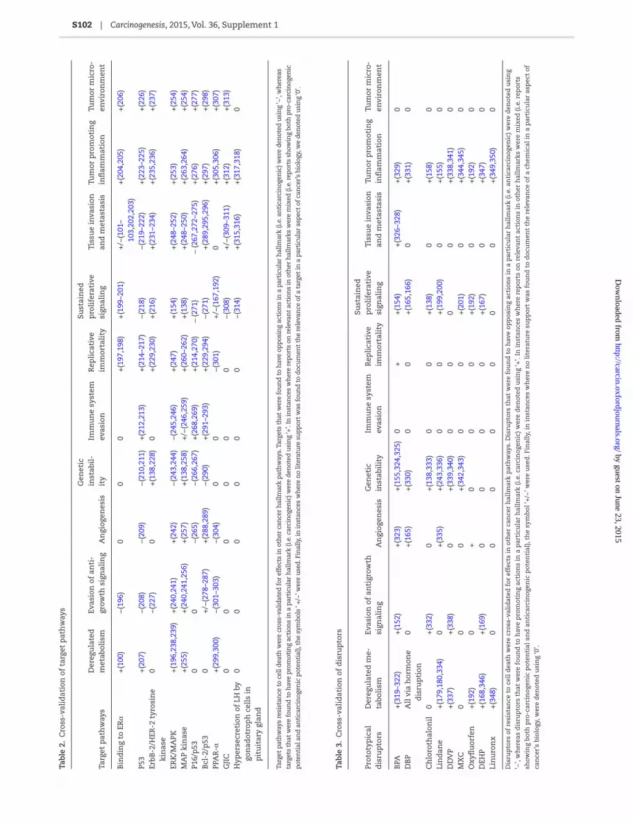
S102 | Carcinogenesis, 2015, Vol. 36, Supplement 1
Tab
le 2
. C
ross
-val
idat
ion
of
targ
et p
ath
way
s
Targ
et p
ath
way
sD
ereg
ula
ted
m
etab
olis
mEv
asio
n o
f an
ti-
grow
th s
ign
alin
gA
ngi
ogen
esis
Gen
etic
in
stab
il-
ity
Imm
un
e sy
stem
ev
asio
nR
epli
cati
ve
imm
orta
lity
Sust
ain
ed
pro
life
rati
ve
sign
alin
gT
issu
e in
vasi
on
and
met
asta
sis
Tum
or p
rom
otin
g in
flam
mat
ion
Tum
or m
icro
-en
viro
nm
ent
Bin
din
g to
ER
α+
(100
)−
(196
)0
00
+(1
97,1
98)
+(1
99–2
01)
+/−
(101
– 10
3,20
2,20
3)+
(204
,205
)+
(206
)
P53
+(2
07)
−(2
08)
−(2
09)
−(2
10,2
11)
+(2
12,2
13)
+(2
14–2
17)
−(2
18)
−(2
19–2
22)
+(2
23–2
25)
+(2
26)
ErbB
-2/H
ER-2
tyr
osin
e ki
nas
e0
−(2
27)
0+
(138
,228
)0
+(2
29,2
30)
+(2
16)
+(2
31–2
34)
+(2
35,2
36)
+(2
37)
ERK
/MA
PK+
(196
,238
,239
)+
(240
,241
)+
(242
)−
(243
,244
)−
(245
,246
)+
(247
)+
(154
)+
(248
–252
)+
(253
)+
(254
)M
AP
kin
ase
+(2
55)
+(2
40,2
41,2
56)
+(2
57)
+(1
38,2
58)
+/−
(246
,259
)+
(260
–262
)+
(138
)+
(248
–250
)+
(263
,264
)+
(254
)P1
6/p
530
0−
(265
)−
(266
,267
)+
(268
,269
)+
(214
,270
) (2
71)
(267
,272
–275
)+
(276
)+
(277
)B
cl-2
/p53
0+
/−(2
78–2
87)
+(2
88,2
89)
−(2
90)
+(2
91–2
93)
+(2
29,2
94)
−(2
71)
+(2
89,2
95,2
96)
+(2
97)
+(2
98)
PPA
R-α
+(2
99,3
00)
−(3
01–3
03)
−(3
04)
00
−(3
01)
+/−
(167
,192
)0
+(3
05,3
06)
+(3
07)
GJI
C0
00
00
0−
(308
)+
/−(3
09–3
11)
+(3
12)
+(3
13)
Hyp
erse
cret
ion
of
LH b
y go
nad
otro
ph
cel
ls in
p
itu
itar
y gl
and
00
00
00
−(3
14)
+(3
15,3
16)
+(3
17,3
18)
0
Targ
et p
ath
way
s re
sist
ance
to
cell
dea
th w
ere
cros
s-va
lidat
ed f
or e
ffec
ts in
oth
er c
ance
r h
allm
ark
pat
hw
ays.
Tar
gets
th
at w
ere
fou
nd
to
hav
e op
pos
ing
acti
ons
in a
par
ticu
lar
hal
lmar
k (i.
e. a
nti
carc
inog
enic
) wer
e d
enot
ed u
sin
g ‘−
’, w
her
eas
targ
ets
that
wer
e fo
un
d t
o h
ave
pro
mot
ing
acti
ons
in a
par
ticu
lar
hal
lmar
k (i.
e. c
arci
nog
enic
) wer
e d
enot
ed u
sin
g ‘+
’. In
inst
ance
s w
her
e re
por
ts o
n r
elev
ant
acti
ons
in o
ther
hal
lmar
ks w
ere
mix
ed (i
.e. r
epor
ts s
how
ing
both
pro
-car
cin
ogen
ic
pot
enti
al a
nd
an
tica
rcin
ogen
ic p
oten
tial
), th
e sy
mbo
ls ‘
+/−’
wer
e u
sed
. Fin
ally
, in
inst
ance
s w
her
e n
o lit
erat
ure
su
pp
ort
was
fou
nd
to
doc
um
ent
the
rele
van
ce o
f a
targ
et in
a p
arti
cula
r as
pec
t of
can
cer’s
bio
logy
, we
den
oted
usi
ng
‘0’.
Tab
le 3
. C
ross
-val
idat
ion
of
dis
rup
tors
Prot
otyp
ical
d
isru
pto
rsD
ereg
ula
ted
me-
tabo
lism
Evas
ion
of
anti
grow
th
sign
alin
gA
ngi
ogen
esis
Gen
etic
in
stab
ilit
yIm
mu
ne
syst
em
evas
ion
Rep
lica
tive
im
mor
tali
ty
Sust
ain
ed
pro
life
rati
ve
sign
alin
gT
issu
e in
vasi
on
and
met
asta
sis
Tum
or p
rom
otin
g in
flam
mat
ion
Tum
or m
icro
-en
viro
nm
ent
BPA
+(3
19–3
22)
+(1
52)
+(3
23)
+(1
55,3
24,3
25)
0+
+(1
54)
+(3
26–3
28)
+(3
29)
0D
BP
All
via
hor
mon
e d
isru
pti
on0
+(1
65)
+(3
30)
00
+(1
65,1
66)
0+
(331
)0
Ch
loro
thal
onil
0+
(332
)0
+(1
38,3
33)
00
+(1
38)
0+
(158
)0
Lin
dan
e+
(179
,180
,334
)0
+(3
35)
+(2
43,3
36)
00
+(1
99,2
00)
0+
(155
)0
DD
VP
+(3
37)
+(3
38)
0+
(339
,340
)0
00
0+
(338
,341
)0
MX
C0
00
+(3
42,3
43)
00
+(2
01)
0+
(344
,345
)0
Oxy
flu
orfe
n+
(192
)0
+0
00
+(1
92)
0+
(192
)0
DEH
P+
(168
,346
)+
(169
)0
00
0+
(167
)0
+(3
47)
0Li
nu
ron
x+
(348
)0
00
00
00
+(3
49,3
50)
0
Dis
rup
tors
of
resi
stan
ce t
o ce
ll d
eath
wer
e cr
oss-
vali
dat
ed f
or e
ffec
ts in
oth
er c
ance
r h
allm
ark
pat
hw
ays.
Dis
rup
tors
th
at w
ere
fou
nd
to
hav
e op
pos
ing
acti
ons
in a
par
ticu
lar
hal
lmar
k (i
.e. a
nti
carc
inog
enic
) wer
e d
enot
ed u
sin
g
‘−’,
wh
erea
s d
isru
pto
rs t
hat
wer
e fo
un
d t
o h
ave
pro
mot
ing
acti
ons
in a
par
ticu
lar
hal
lmar
k (i
.e. c
arci
nog
enic
) wer
e d
enot
ed u
sin
g ‘+
’. In
inst
ance
s w
her
e re
por
ts o
n r
elev
ant
acti
ons
in o
ther
hal
lmar
ks w
ere
mix
ed (i
.e. r
epor
ts
show
ing
both
pro
-car
cin
ogen
ic p
oten
tial
an
d a
nti
carc
inog
enic
pot
enti
al),
the
sym
bol ‘
+/−
’ wer
e u
sed
. Fin
ally
, in
inst
ance
s w
her
e n
o li
tera
ture
su
pp
ort
was
fou
nd
to
doc
um
ent
the
rele
van
ce o
f a
chem
ical
in a
par
ticu
lar
asp
ect
of
can
cer’
s bi
olog
y, w
ere
den
oted
usi
ng
‘0’.
by guest on June 23, 2015http://carcin.oxfordjournals.org/
Dow
nloaded from

K.B.Narayanan et al. | S103
(conferring resistance to cell death) could exacerbate the effects of unrepaired cellular damage. The potential for dysfunction in this key safeguard is therefore an important consideration because this sort of genetic instability increases the overall probability of damage and mutations that could support the emergence of a fully immortalized cellular phenotype.
Past studies have indicated that several cancer hallmark pro-cesses are impacted by a variety of chemical carcinogens and oncogenes (95,96), and there are various agencies worldwide such as USEPA and International Agency for Research on Cancer working on classifying the environmental chemicals based on the carcinogenic potential with the purpose of protecting human health. However, the effects of environmental chemical mixtures have had much less attention, and in this project, we have looked specifically at a number of prototypical chemicals with disruptive potential that is relevant to apoptosis. The con-cern that we have relates to the possibility that individual chem-icals that are disruptive of these key mechanisms and pathways may have the potential to contribute to environmental carcino-genesis without being carcinogenic per se.
For example, as we noted previously, BPA strikingly impairs TP53 activity and its downstream targets, cell cycle regulators, p21WAF1 and RB, or pro-apoptotic BAX, thereby enhancing the threshold for apoptosis (152). BPA activates mTOR pathway and enhances cell growth and proliferation (351). It also activates PPAR-α (which sup-presses apoptosis and enhances cell proliferation), and it inhibits GJIC through a modulation of the gating of gap junction channels, which contributes to tumor formation by increasing intracellular signaling and enhancing proliferation (352). And BPA influences cell proliferation and migration by GPER/EGFR/ERK pathway. But despite a significant and growing body of literature that has docu-mented all of these mechanistic contributions, researchers and regulators have had considerable difficulty proving whether or not BPA has carcinogenic effects in humans (319).
From this perspective, it seems that the longstanding focus on the carcinogenic potential of individual chemicals is really too narrow (given the wide range of environmental chemi-cal exposures that we now face). Instead, it would seem more prudent to focus on mechanistic contributions and anticipated synergies of mixtures of individual constituents that have been shown to individually exert disruptive effects on hallmark cancer processes (at dose levels that are environmentally rel-evant). In this case, BPA is a good example because it is ubiq-uitous in the environment and it has also been shown to exert its effects on TP53 at low-dose levels (353). So, even if it cannot be definitively categorized as a human carcinogen, it appears to have potential to play a contributing role in environmental carcinogenesis. Future research will therefore need to illustrate how chemicals that have the potential to produce this sort of a disruptive effect can be experimentally combined with other environmental chemicals that disrupt other hallmark processes to enable carcinogenesis.
We fully recognize that much of the evidence in the toxico-logical literature that documents the disruptive actions of these chemicals has been produced under a wide range of differing experimental circumstances, and it is not our intent to jump to conclusions about the role that aggregated exposures to mix-tures of these chemicals might play in environmental carcino-genesis. But it is our contention that the ubiquitous presence of these (and other) chemicals with disruptive potential needs to be carefully considered, even if these chemicals are not individ-ually carcinogenic. Moreover, researchers who investigate this possibility will also need to consider other mechanisms that are affected by these individual chemicals as well (see Tables 2 and
3). In some cases, dose–response research may reveal thresholds that make these actions unlikely at levels of exposure that are seen in the environment, but to the extent that low-dose effects can been found, these additional disruptive effects may also be important factors to consider.
ConclusionsThe disruption of the mechanisms that regulate cell death is fundamentally important to our understanding of environmen-tal carcinogenesis. The enablement of an immortalized cellular phenotype can only occur when many important safeguards have been bypassed. It therefore appears reasonable to consider the effects of ubiquitous environmental chemicals that have been shown to disrupt cell death as it is a very important safe-guard. Although a considerable amount of research has been done to characterize the effects that many chemicals have on the mechanisms that are relevant for normal cell death, very little attention has been given to the combined effects of this chemi-cals on this hallmark of cancer, or the role that these sorts of disruptions at the mechanistic level might serve to contribute to environmental carcinogenesis. In this review, we have identified a number of important targets that are highly relevant for cell death and we have identified a number of ubiquitous environ-mental chemicals that have been shown to act disruptively on these targets. Future research is needed that looks carefully the role of these prototypical disruptors and other disruptive chemi-cals that can act on these same mechanisms at levels of expo-sure that are commonly seen in the environment. Regulators who now focus solely on determining the carcinogenic potential of individual chemicals would be well served to additionally con-sider the synergies that might occur when chemicals that are disruptive at the mechanistic level are combined with other dis-ruptive chemicals (i.e. those that can enable other complemen-tary processes that are similarly instrumental and enabling in carcinogenesis). To anticipate the sorts of synergies that might be produced, the pleiotropic nature of these chemicals will need to be considered as well. Individual chemicals may produce a range of disruptive effects that are relevant for a multitude of mecha-nisms, yet individual constituents in any given combination of exposures may not need to be carcinogenic per se. Combinations of these chemicals may produce foundational effects that enable carcinogenesis, so progress in our understanding of this poten-tial will help us to refine our approach to cancer risk assessment.
FundingThis research was supported by Yeungnam University research grants in 2014 to Hyun Ho Park. Po Sing Leung was supported by the Health and Medical Research Fund of Food and Health Bureau, Hong Kong Special Administrative Region, Ref. No: 10110021. Yon Rojanasakul was supported by NIH (R01-ES022968) and NSF (CBET-1434503). Rita Dornetshuber-Fleiss was supported by the Austrian Science Fund (FWF, project number T 451-B18) and the Johanna Mahlke, geb.-Obermann-Stiftung. Clement Yedjou was supported by National Institutes of Health (Grant No. NIMHD-G12MD007581, Grant No. NIGMS- P20GM103476).Conflict of Interest Statement: None declared.
References 1. Nicholson, D.W. et al. (2003) Apoptosis. Life and death decisions. Sci-
ence, 299, 214–215. 2. Fridman, J.S. et al. (2003) Control of apoptosis by p53. Oncogene, 22,
9030–9040.
by guest on June 23, 2015http://carcin.oxfordjournals.org/
Dow
nloaded from

S104 | Carcinogenesis, 2015, Vol. 36, Supplement 1
3. Shortt, J. et al. (2012) Oncogenes in cell survival and cell death. Cold Spring Harb. Perspect. Biol., 4.
4. Orr, B. et al. (2013) A double-edged sword: how oncogenes and tumor suppressor genes can contribute to chromosomal instability. Front. Oncol., 3, 164.
5. Croce, C.M. (2008) Oncogenes and cancer. N. Engl. J. Med., 358, 502–511. 6. Hetz, C.A. et al. (2005) Beyond apoptosis: nonapoptotic cell death in
physiology and disease. Biochem. Cell Biol., 83, 579–588. 7. de Bruin, E.C. et al. (2008) Apoptosis and non-apoptotic deaths in can-
cer development and treatment response. Cancer Treat. Rev., 34, 737–749.
8. Elmore, S. (2007) Apoptosis: a review of programmed cell death. Toxicol. Pathol., 35, 495–516.
9. Falschlehner, C. et al. (2007) TRAIL signalling: decisions between life and death. Int. J. Biochem. Cell Biol., 39, 1462–1475.
10. Suh, D.H. et al. (2013) Mitochondrial permeability transition pore as a selective target for anti-cancer therapy. Front. Oncol., 3, 41.
11. Mayer, B. et al. (2003) Mitochondrial regulation of apoptosis. News Physiol. Sci., 18, 89–94.
12. Adams, J.M. (2003) Ways of dying: multiple pathways to apoptosis. Genes Dev., 17, 2481–2495.
13. Keinan, N. et al. (2010) Oligomerization of the mitochondrial protein voltage-dependent anion channel is coupled to the induction of apop-tosis. Mol. Cell. Biol., 30, 5698–5709.
14. Vander Heiden, M.G. et al. (2001) Bcl-xL promotes the open configu-ration of the voltage-dependent anion channel and metabolite pas-sage through the outer mitochondrial membrane. J. Biol. Chem., 276, 19414–19419.
15. Bröker, L.E. et al. (2005) Cell death independent of caspases: a review. Clin. Cancer Res., 11, 3155–3162.
16. Vermeulen, K. et al. (2005) Apoptosis: mechanisms and relevance in cancer. Ann. Hematol., 84, 627–639.
17. Yang, Y.L. et al. (2000) The IAP family: endogenous caspase inhibitors with multiple biological activities. Cell Res., 10, 169–177.
18. Carew, J.S. et al. (2002) Mitochondrial defects in cancer. Mol. Cancer, 1, 9. 19. Park, H.H. et al. (2007) Death domain assembly mechanism revealed by
crystal structure of the oligomeric PIDDosome core complex. Cell, 128, 533–546.
20. Kim, I.R. et al. (2009) DNA damage- and stress-induced apoptosis occurs independently of PIDD. Apoptosis, 14, 1039–1049.
21. Park, H.H. (2011) Structural analyses of death domains and their inter-actions. Apoptosis, 16, 209–220.
22. Van Cruchten, S. et al. (2002) Morphological and biochemical aspects of apoptosis, oncosis and necrosis. Anat. Histol. Embryol., 31, 214–223.
23. Jäättelä, M. et al. (2003) Caspase-independent cell death in T lympho-cytes. Nat. Immunol., 4, 416–423.
24. Chautan, M. et al. (1999) Interdigital cell death can occur through a necrotic and caspase-independent pathway. Curr. Biol., 9, 967–970.
25. Galluzzi, L. et al. (2008) Necroptosis: a specialized pathway of pro-grammed necrosis. Cell, 135, 1161–1163.
26. Kaczmarek, A. et al. (2013) Necroptosis: the release of damage-associ-ated molecular patterns and its physiological relevance. Immunity, 38, 209–223.
27. Mehrpour, M. et al. (2010) Autophagy in health and disease. 1. Regula-tion and significance of autophagy: an overview. Am. J. Physiol. Cell Physiol., 298, C776–C785.
28. Degenhardt, K. et al. (2006) Autophagy promotes tumor cell survival and restricts necrosis, inflammation, and tumorigenesis. Cancer Cell, 10, 51–64.
29. Glick, D. et al. (2010) Autophagy: cellular and molecular mechanisms. J. Pathol., 221, 3–12.
30. He, C. et al. (2009) Regulation mechanisms and signaling pathways of autophagy. Annu. Rev. Genet., 43, 67–93.
31. Karantza-Wadsworth, V. et al. (2007) Autophagy mitigates metabolic stress and genome damage in mammary tumorigenesis. Genes Dev., 21, 1621–1635.
32. Mathew, R. et al. (2007) Autophagy suppresses tumor progression by limiting chromosomal instability. Genes Dev., 21, 1367–1381.
33. Mathew, R. et al. (2009) Autophagy suppresses tumorigenesis through elimination of p62. Cell, 137, 1062–1075.
34. Aita, V.M. et al. (1999) Cloning and genomic organization of beclin 1, a candidate tumor suppressor gene on chromosome 17q21. Genomics, 59, 59–65.
35. Qu, X. et al. (2003) Promotion of tumorigenesis by heterozygous disrup-tion of the beclin 1 autophagy gene. J. Clin. Invest., 112, 1809–1820.
36. Hu, Y.L. et al. (2012) Tumor cell autophagy as an adaptive response mediating resistance to treatments such as antiangiogenic therapy. Cancer Res., 72, 4294–4299.
37. Fesik, S.W. (2005) Promoting apoptosis as a strategy for cancer drug discovery. Nat. Rev. Cancer, 5, 876–885.
38. Lowe, S.W. et al. (2000) Apoptosis in cancer. Carcinogenesis, 21, 485–495.
39. Plati, J. et al. (2011) Apoptotic cell signaling in cancer progression and therapy. Integr. Biol. (Camb)., 3, 279–296.
40. Dewey, W.C. et al. (1995) Radiation-induced apoptosis: relevance to radiotherapy. Int. J. Radiat. Oncol. Biol. Phys., 33, 781–796.
41. Bal, W. et al. (2002) Induction of oxidative DNA damage by carcinogenic metals. Toxicol. Lett., 127, 55–62.
42. Johnstone, R.W. et al. (2002) Apoptosis: a link between cancer genetics and chemotherapy. Cell, 108, 153–164.
43. Stenner-Liewen, F. et al. (2003) Apoptosis and cancer: basic mechanisms and therapeutic opportunities in the postgenomic era. Cancer Res., 63.
44. Dalerba, P. et al. (2007) Cancer stem cells: models and concepts. Annu. Rev. Med., 58, 267–284.
45. Levina, V. et al. (2008) Drug-selected human lung cancer stem cells: cytokine network, tumorigenic and metastatic properties. PLoS One, 3, e3077.
46. D’Andrea, F.P. (2012) Intrinsic radiation resistance of mesenchymal cancer stem cells and implications for treatment response in a murine sarcoma model. Dan. Med. J., 59, B4388.
47. Kruyt, F.A. et al. (2010) Apoptosis and cancer stem cells: implications for apoptosis targeted therapy. Biochem. Pharmacol., 80, 423–430.
48. Frisch, S.M. et al. (2013) Mechanisms that link the oncogenic epithelial-mesenchymal transition to suppression of anoikis. J. Cell Sci., 126(Pt 1), 21–29.
49. Liu, G. et al. (2006) Analysis of gene expression and chemoresistance of CD133+ cancer stem cells in glioblastoma. Mol. Cancer, 5, 67.
50. Zobalova, R. et al. (2008) CD133-positive cells are resistant to TRAIL due to up-regulation of FLIP. Biochem. Biophys. Res. Commun., 373, 567–571.
51. Ouyang, L. et al. (2012) Programmed cell death pathways in cancer: a review of apoptosis, autophagy and programmed necrosis. Cell Prolif., 45, 487–498.
52. Strasser, A. et al. (2011) Deciphering the rules of programmed cell death to improve therapy of cancer and other diseases. EMBO J., 30, 3667–3683.
53. Fiandalo, M.V. et al. (2012) Caspase control: protagonists of cancer cell apoptosis. Exp. Oncol., 34, 165–175.
54. Reubold, T.F. et al. (2012) A molecular view on signal transduction by the apoptosome. Cell Signal., 24, 1420–1425.
55. Galluzzi, L. et al. (2012) Mitochondria: master regulators of danger sig-nalling. Nat. Rev. Mol. Cell Biol., 13, 780–788.
56. de Almagro, M.C. et al. (2012) The inhibitor of apoptosis (IAP) proteins are critical regulators of signaling pathways and targets for anti-cancer therapy. Exp. Oncol., 34, 200–211.
57. Harvey, K.F. et al. (2013) The Hippo pathway and human cancer. Nat. Rev. Cancer, 13, 246–257.
58. Sherr, C.J. et al. (2002) The RB and p53 pathways in cancer. Cancer Cell, 2, 103–112.
59. Galluzzi, L. et al. (2011) Mitochondrial liaisons of p53. Antioxid. Redox Signal., 15, 1691–1714.
60. Matoba, S. et al. (2006) p53 regulates mitochondrial respiration. Sci-ence, 312, 1650–1653.
61. Kim, J.Y. et al. (2004) BH3-only protein Noxa is a mediator of hypoxic cell death induced by hypoxia-inducible factor 1alpha. J. Exp. Med., 199, 113–124.
62. Yu, J. et al. (2008) PUMA, a potent killer with or without p53. Oncogene, 27(suppl 1), S71–S83.
63. Hikisz, P. et al. (2012) PUMA, a critical mediator of cell death–one dec-ade on from its discovery. Cell. Mol. Biol. Lett., 17, 646–669.
by guest on June 23, 2015http://carcin.oxfordjournals.org/
Dow
nloaded from
K.B.Narayanan et al. | S105
64. Icard, P. et al. (2012) A global view of the biochemical pathways involved in the regulation of the metabolism of cancer cells. Biochim. Biophys. Acta, 1826, 423–433.
65. Zhao, S. et al. (2010) Regulation of cellular metabolism by protein lysine acetylation. Science, 327, 1000–1004.
66. Yi, C.H. et al. (2011) Metabolic regulation of protein N-alpha-acetyla-tion by Bcl-xL promotes cell survival. Cell, 146, 607–620.
67. Gnoni, G.V. et al. (2009) The mitochondrial citrate carrier: metabolic role and regulation of its activity and expression. IUBMB Life, 61, 987–994.
68. Vaughn, A.E. et al. (2008) Glucose metabolism inhibits apoptosis in neurons and cancer cells by redox inactivation of cytochrome c. Nat. Cell Biol., 10, 1477–1483.
69. Delbridge, A.R. et al. (2012) The role of the apoptotic machinery in tumor suppression. Cold Spring Harb. Perspect. Biol., 4.
70. Labbé, D.P. et al. (2012) Protein tyrosine phosphatases in cancer: friends and foes! Prog. Mol. Biol. Transl. Sci., 106, 253–306.
71. Sayers, T.J. (2011) Targeting the extrinsic apoptosis signaling pathway for cancer therapy. Cancer Immunol. Immunother., 60, 1173–1180.
72. Frisch, S.M. et al. (1996) A role for Jun-N-terminal kinase in anoikis; suppression by bcl-2 and crmA. J. Cell Biol., 135, 1377–1382.
73. Schottenfeld, D. et al. (2013) Current perspective on the global and United States cancer burden attributable to lifestyle and environ-mental risk factors. Annu. Rev. Public Health, 34, 97–117.
74. Lee, E.Y. et al. (2010) Oncogenes and tumor suppressor genes. Cold Spring Harb. Perspect. Biol., 2, a003236.
75. Reddy, K.L. et al. (2013) Higher order chromatin organization in can-cer. Semin. Cancer Biol., 23, 109–115.
76. Iorio, M.V. et al. (2012) microRNA involvement in human cancer. Car-cinogenesis, 33, 1126–1133.
77. Vousden, K.H. et al. (2002) Live or let die: the cell’s response to p53. Nat. Rev. Cancer, 2, 594–604.
78. Murray-Zmijewski, F. et al. (2006) p53/p63/p73 isoforms: an orchestra of isoforms to harmonise cell differentiation and response to stress. Cell Death Differ., 13, 962–972.
79. Rosenbluth, J.M. et al. (2008) The jury is in: p73 is a tumor suppressor after all. Genes Dev., 22, 2591–2595.
80. Flores, E.R. et al. (2005) Tumor predisposition in mice mutant for p63 and p73: evidence for broader tumor suppressor functions for the p53 family. Cancer Cell, 7, 363–373.
81. Maddocks, O.D. et al. (2011) Metabolic regulation by p53. J. Mol. Med. (Berl), 89, 237–245.
82. Huang, Y. et al. (2011) Phospho-ΔNp63α is a key regulator of the cis-platin-induced microRNAome in cancer cells. Cell Death Differ., 18, 1220–1230.
83. Sen, T. et al. (2011) DeltaNp63alpha confers tumor cell resistance to cisplatin through the AKT1 transcriptional regulation. Cancer Res., 71, 1167–1176.
84. Ory, B. et al. (2011) A microRNA-dependent circuit controlling p63/p73 homeostasis: p53 family cross-talk meets therapeutic opportu-nity. Oncotarget, 2, 259–264.
85. Xi, S. et al. (2010) Cigarette smoke induces C/EBP-β-mediated acti-vation of miR-31 in normal human respiratory epithelia and lung cancer cells. PLoS One, 5, e13764.
86. Ewing, S.J. et al. (2008) C/EBPbeta represses p53 to promote cell sur-vival downstream of DNA damage independent of oncogenic Ras and p19(Arf). Cell Death Differ., 15, 1734–1744.
87. Balmain, A. et al. (1984) Activation of the mouse cellular Harvey-ras gene in chemically induced benign skin papillomas. Nature, 307, 658–660.
88. Alguacil, J. et al. (2002) Occupational exposure to organic solvents and K-ras mutations in exocrine pancreatic cancer. Carcinogenesis, 23, 101–106.
89. Zarbl, H. et al. (1985) Direct mutagenesis of Ha-ras-1 oncogenes by N-nitroso-N-methylurea during initiation of mammary carcinogen-esis in rats. Nature, 315, 382–385.
90. Shishodia, S. et al. (2003) Curcumin (diferuloylmethane) down-regulates cigarette smoke-induced NF-kappaB activation through inhibition of IkappaBalpha kinase in human lung epithelial cells:
correlation with suppression of COX-2, MMP-9 and cyclin D1. Car-cinogenesis, 24, 1269–1279.
91. Srivastava, S.K. et al. (2004) Cell cycle arrest, apoptosis induction and inhibition of nuclear factor kappa B activation in anti-proliferative activity of benzyl isothiocyanate against human pancreatic cancer cells. Carcinogenesis, 25, 1701–1709.
92. Romano, S. et al. (2011) FKBP51 and the NF-κB regulatory pathway in cancer. Curr. Opin. Pharmacol., 11, 288–293.
93. Hanahan, D. et al. (2000) The hallmarks of cancer. Cell, 100, 57–70. 94. Trosko, J.E. et al. (2010) A paradigm shift is required for the risk
assessment of potential human health after exposure to low level chemical exposures: a response to the toxicity testing in the 21st cen-tury report. Int. J. Toxicol., 29, 344–357.
95. Kleinstreuer, N.C. et al. (2013) In vitro perturbations of targets in cancer hallmark processes predict rodent chemical carcinogenesis. Toxicol. Sci., 131, 40–55.
96. Kortenkamp, A. (2014) Low dose mixture effects of endocrine dis-rupters and their implications for regulatory thresholds in chemical risk assessment. Curr. Opin. Pharmacol., 19, 105–111.
97. Fucic, A. et al. (2012) Environmental exposure to xenoestrogens and oestrogen related cancers: reproductive system, breast, lung, kidney, pancreas, and brain. Environ. Health, 11(suppl 1), S8.
98. Bailey, S.T. et al. (2012) Estrogen receptor prevents p53-dependent apoptosis in breast cancer. Proc. Natl Acad. Sci. USA, 109, 18060–18065.
99. Sayeed, A. et al. (2007) Estrogen receptor alpha inhibits p53-mediated transcriptional repression: implications for the regulation of apopto-sis. Cancer Res., 67, 7746–7755.
100. Al-Dhaheri, M. et al. (2011) CARM1 is an important determinant of ERα-dependent breast cancer cell differentiation and proliferation in breast cancer cells. Cancer Res., 71, 2118–2128.
101. Wik, E. et al. (2013) Lack of estrogen receptor-α is associated with epithelial-mesenchymal transition and PI3K alterations in endome-trial carcinoma. Clin. Cancer Res., 19, 1094–1105.
102. Dhasarathy, A. et al. (2007) The transcription factor snail mediates epithelial to mesenchymal transitions by repression of estrogen receptor-alpha. Mol. Endocrinol., 21, 2907–2918.
103. Huet, G. et al. (2008) Loss of E-cadherin-mediated cell contacts reduces estrogen receptor alpha (ER alpha) transcriptional efficiency by affecting the respective contribution exerted by AF1 and AF2 transactivation functions. Biochem. Biophys. Res. Commun., 365, 304–309.
104. Rivedal, E. et al. (2005) Connexin43 synthesis, phosphorylation, and degradation in regulation of transient inhibition of gap junction intercellular communication by the phorbol ester TPA in rat liver epithelial cells. Exp. Cell Res., 302, 143–152.
105. Kang, K.S. et al. (1996) Inhibition of gap junctional intercellular communication in normal human breast epithelial cells after treat-ment with pesticides, PCBs, and PBBs, alone or in mixtures. Environ. Health Perspect., 104, 192–200.
106. Klaunig, J.E. et al. (1990) Gap-junctional intercellular communica-tion and murine hepatic carcinogenesis. Prog. Clin. Biol. Res., 331, 277–291.
107. Carette, D. et al. (2014) Connexin a check-point component of cell apoptosis in normal and physiopathological conditions. Biochimie, 101, 1–9.
108. Trosko, J.E. et al. (1994) The role of modulated gap junctional inter-cellular communication in epigenetic toxicology. Risk Anal., 14, 303–312.
109. Chang, W.W. et al. (2013) Salmonella enhance chemosensitivity in tumor through connexin 43 upregulation. Int. J. Cancer, 133, 1926–1935.
110. Abu Aboud, O. et al. (2013) Inhibition of PPARα induces cell cycle arrest and apoptosis, and synergizes with glycolysis inhibition in kidney cancer cells. PLoS One, 8, e71115.
111. Klaunig, J.E. et al. (2003) PPARalpha agonist-induced rodent tumors: modes of action and human relevance. Crit. Rev. Toxicol., 33, 655–780.
112. Roberts, R.A. (2002) Evidence for cross talk between PPARalpha and p38 MAP kinase. Toxicol. Sci., 68, 270–274.
by guest on June 23, 2015http://carcin.oxfordjournals.org/
Dow
nloaded from

S106 | Carcinogenesis, 2015, Vol. 36, Supplement 1
113. Cook, J.C. et al. (1993) Investigation of a mechanism for Leydig cell tumorigenesis by linuron in rats. Toxicol. Appl. Pharmacol., 119, 195–204.
114. Zhang, Z. et al. (2011) Luteinizing hormone upregulates survivin and inhibits apoptosis in ovarian epithelial tumors. Eur. J. Obstet. Gynecol. Reprod. Biol., 155, 69–74.
115. Calaf, G.M. et al. (2009) Organophosphorous pesticides and estrogen induce transformation of breast cells affecting p53 and c-Ha-ras genes. Int. J. Oncol., 35, 1061–1068.
116. Petitjean, A. et al. (2007) TP53 mutations in human cancers: func-tional selection and impact on cancer prognosis and outcomes. Oncogene, 26, 2157–2165.
117. Lu, Y. et al. (2012) Inhibition of breast tumor cell growth by ectopic expression of p16/INK4A via combined effects of cell cycle arrest, senescence and apoptotic induction, and angiogenesis inhibition. J. Cancer, 3, 333–344.
118. Ruas, M. et al. (1998) The p16INK4a/CDKN2A tumor suppressor and its relatives. Biochim. Biophys. Acta, 1378, F115–F177.
119. Geisler, S.A. et al. (2002) p16 and p53 Protein expression as prognostic indicators of survival and disease recurrence from head and neck cancer. Clin. Cancer Res., 8, 3445–3453.
120. Jiang, M. et al. (2003) Bcl-2 constitutively suppresses p53-dependent apoptosis in colorectal cancer cells. Genes Dev., 17, 832–837.
121. Schwandner, O. et al. (2000) p53 and Bcl-2 as significant predictors of recurrence and survival in rectal cancer. Eur. J. Cancer, 36, 348–356.
122. Miliaras, S. et al. (2011) The role of P53 and Bcl-2 proteins in 7, 12-dimethylbenz-(a)-anthracene-induced tumor growth. J. Biol. Regul. Homeost. Agents, 25, 359–364.
123. Centelles, J.J. (2012) General aspects of colorectal cancer. ISRN Oncol., 2012, 139268.
124. Roskoski, R. Jr. (2014) The ErbB/HER family of protein-tyrosine kinases and cancer. Pharmacol. Res., 79, 34–74.
125. Chen, F.L. et al. (2008) Acquired resistance to small molecule ErbB2 tyrosine kinase inhibitors. Clin. Cancer Res., 14, 6730–6734.
126. Willems, L. et al. (2012) PI3K and mTOR signaling pathways in can-cer: new data on targeted therapies. Curr. Oncol. Rep., 14, 129–138.
127. Laplante, M. et al. (2009) mTOR signaling at a glance. J. Cell Sci., 122(Pt 20), 3589–3594.
128. Graff, J.R. et al. (2008) Targeting the eukaryotic translation initiation factor 4E for cancer therapy. Cancer Res., 68, 631–634.
129. Jia, Y. et al. (2012) Cap-dependent translation initiation factor eIF4E: an emerging anticancer drug target. Med. Res. Rev., 32, 786–814.
130. Blume-Jensen, P. et al. (1998) The kit receptor promotes cell survival via activation of PI 3-kinase and subsequent Akt-mediated phospho-rylation of Bad on Ser136. Curr. Biol., 8, 779–782.
131. Downward, J. (2004) PI 3-kinase, Akt and cell survival. Semin. Cell Dev. Biol., 15, 177–182.
132. Nicholson, K.M. et al. (2002) The protein kinase B/Akt signalling pathway in human malignancy. Cell. Signal., 14, 381–395.
133. Romashkova, J.A. et al. (1999) NF-kappaB is a target of AKT in anti-apoptotic PDGF signalling. Nature, 401, 86–90.
134. Cardone, M.H. et al. (1998) Regulation of cell death protease cas-pase-9 by phosphorylation. Science, 282, 1318–1321.
135. Maiese, K. et al. (2012) Targeting disease through novel pathways of apoptosis and autophagy. Expert Opin. Ther. Targets, 16, 1203–1214.
136. Tsukada, M. et al. (1993) Isolation and characterization of autophagy-defective mutants of Saccharomyces cerevisiae. FEBS Lett., 333, 169–174.
137. Pastor, M.D. et al. (2009) mTOR/S6 kinase pathway contributes to astrocyte survival during ischemia. J. Biol. Chem., 284, 22067–22078.
138. Tessier, D.M. et al. (2001) Increased ErbB-2 tyrosine kinase activity, MAPK phosphorylation, and cell proliferation in the prostate cancer cell line LNCaP following treatment by select pesticides. Toxicol. Sci., 60, 38–43.
139. Zhang, W. et al. (2002) MAPK signal pathways in the regulation of cell proliferation in mammalian cells. Cell Res., 12, 9–18.
140. Lim, J.H. et al. (2012) Rottlerin induces apoptosis of HT29 colon carcinoma cells through NAG-1 upregulation via an ERK and p38 MAPK-dependent and PKC δ-independent mechanism. Chem. Biol. Interact., 197, 1–7.
141. Gan, L. et al. (2011) Resistance to docetaxel-induced apoptosis in prostate cancer cells by p38/p53/p21 signaling. Prostate, 71, 1158–1166.
142. Salim, H. et al. (2012) miRNA-214 modulates radiotherapy response of non-small cell lung cancer cells through regulation of p38MAPK, apoptosis and senescence. Br. J. Cancer, 107, 1361–1373.
143. Hu, C. et al. (2012) E Platinum, a newly synthesized platinum com-pound, induces autophagy via inhibiting phosphorylation of mTOR in gastric carcinoma BGC-823 cells. Toxicol. Lett., 210, 78–86.
144. Sui, X. et al. (2014) p38 and JNK MAPK pathways control the bal-ance of apoptosis and autophagy in response to chemotherapeutic agents. Cancer Lett., 344, 174–179.
145. Dhillon, A.S. et al. (2007) MAP kinase signalling pathways in cancer. Oncogene, 26, 3279–3290.
146. Carmona-Cuenca, I. et al. (2008) Upregulation of the NADPH oxidase NOX4 by TGF-beta in hepatocytes is required for its pro-apoptotic activity. J. Hepatol., 49, 965–976.
147. Caja, L. et al. (2009) Overactivation of the MEK/ERK pathway in liver tumor cells confers resistance to TGF-{beta}-induced cell death through impairing up-regulation of the NADPH oxidase NOX4. Can-cer Res., 69, 7595–7602.
148. Bulayeva, N.N. et al. (2004) Xenoestrogen-induced ERK-1 and ERK-2 activation via multiple membrane-initiated signaling pathways. Environ. Health Perspect., 112, 1481–1487.
149. Andersen, M. et al. (1978) Mutagenic action of aromatic epoxy resins. Nature, 276, 391–392.
150. Ashby, J. et al. (1988) Chemical structure, Salmonella mutagenicity and extent of carcinogenicity as indicators of genotoxic carcino-genesis among 222 chemicals tested in rodents by the U.S. NCI/NTP. Mutat. Res., 204, 17–115.
151. Masuda, S. et al. (2005) Changes in the mutagenic and estrogenic activities of bisphenol A upon treatment with nitrite. Mutat. Res., 585, 137–146.
152. Dairkee, S.H. et al. (2013) Bisphenol-A-induced inactivation of the p53 axis underlying deregulation of proliferation kinetics, and cell death in non-malignant human breast epithelial cells. Carcinogen-esis, 34, 703–712.
153. Goodson, W.H. III et al. (2011) Activation of the mTOR pathway by low levels of xenoestrogens in breast epithelial cells from high-risk women. Carcinogenesis, 32, 1724–1733.
154. Pupo, M. et al. (2012) Bisphenol A induces gene expression changes and proliferative effects through GPER in breast cancer cells and can-cer-associated fibroblasts. Environ. Health Perspect., 120, 1177–1182.
155. Allard, P. et al. (2010) Bisphenol A impairs the double-strand break repair machinery in the germline and causes chromosome abnor-malities. Proc. Natl Acad. Sci. USA, 107, 20405–20410.
156. Jenkins, S. et al. (2011) Chronic oral exposure to bisphenol A results in a nonmonotonic dose response in mammary carcinogenesis and metastasis in MMTV-erbB2 mice. Environ. Health Perspect., 119, 1604–1609.
157. Mozzachio, A.M. et al. (2008) Chlorothalonil exposure and cancer incidence among pesticide applicator participants in the agricul-tural health study. Environ. Res., 108, 400–403.
158. Wilkinson, C.F. et al. (1996) A mechanistic interpretation of the onco-genicity of chlorothalonil in rodents and an assessment of human relevance. Regul. Toxicol. Pharmacol., 24(1 Pt 1), 69–84.
159. Leung-Toung, R. et al. (2006) Thiol proteases: inhibitors and potential therapeutic targets. Curr. Med. Chem., 13, 547–581.
160. Rakitsky, V.N. et al. (2000) Nongenotoxic (epigenetic) carcinogens: pesticides as an example. A critical review. Teratog. Carcinog. Muta-gen., 20, 229–240.
161. Swan, S.H. (2008) Environmental phthalate exposure in relation to reproductive outcomes and other health endpoints in humans. Envi-ron. Res., 108, 177–184.
162. Kim, I.Y. et al. (2004) Phthalates inhibit tamoxifen-induced apoptosis in MCF-7 human breast cancer cells. J. Toxicol. Environ. Health. A, 67, 2025–2035.
163. Liu, X. et al. (2009) Di-(2-ethylhexyl) phthalate upregulates ATF3 expression and suppresses apoptosis in mouse genital tubercle. J. Occup. Health, 51, 57–63.
by guest on June 23, 2015http://carcin.oxfordjournals.org/
Dow
nloaded from

K.B.Narayanan et al. | S107
164. Maire, M.A. et al. (2005) Di-(2-ethylhexyl)phthalate (DEHP) increases Bcl-2/Bax ratio and modifies c-myc expression in Syrian hamster embryo (SHE) cells. Toxicol. Lett., 158, 237–245.
165. Hsieh, T.H. et al. (2012) Phthalates induce proliferation and invasive-ness of estrogen receptor-negative breast cancer through the AhR/HDAC6/c-Myc signaling pathway. FASEB J., 26, 778–787.
166. Park, M.A. et al. (2012) Cell growth of BG-1 ovarian cancer cells is promoted by di-n-butyl phthalate and hexabromocyclododecane via upregulation of the cyclin D and cyclin-dependent kinase-4 genes. Mol. Med. Rep., 5, 761–766.
167. Rusyn, I. et al. (2006) Modes of action and species-specific effects of di-(2-ethylhexyl)phthalate in the liver. Crit. Rev. Toxicol., 36, 459–479.
168. Isenberg, J.S. et al. (2001) Reversibility and persistence of di-2-ethyl-hexyl phthalate (DEHP)- and phenobarbital-induced hepatocellular changes in rodents. Toxicol. Sci., 64, 192–199.
169. Erkekoğlu, P. et al. (2011) Induction of ROS, p53, p21 in DEHP- and MEHP-exposed LNCaP cells-protection by selenium compounds. Food Chem. Toxicol., 49, 1565–1571.
170. Reuber, M.D. (1981) Carcinogenicity of dichlorvos. Clin. Toxicol., 18, 47–84.
171. Koutros, S. et al. (2008) Dichlorvos exposure and human cancer risk: results from the Agricultural Health Study. Cancer Causes Control, 19, 59–65.
172. Page, A.C. et al. (1972) Metabolic fate of dichlorvos in swine. Arch. Toxikol., 30, 19–27.
173. Chan, P.C. et al. (1991) Carcinogenesis studies of dichlorvos in Fischer rats and B6C3F1 mice. Jpn. J. Cancer Res., 82, 157–164.
174. Eroğlu, S. et al. (2013) Protective role of vitamins C and E in dichlo-rvos-induced oxidative stress in human erythrocytes in vitro. Biol. Res., 46, 33–38.
175. Sunkaria, A. et al. (2013) Dichlorvos-induced cell cycle arrest and DNA damage repair activation in primary rat microglial cells. J. Neu-rosci. Res., 91, 444–452.
176. Hervouet, E. et al. (2013) DNA methylation and apoptosis resistance in cancer cells. Cells, 2, 545–573.
177. Alavanja, M.C. et al. (2005) Pesticides and human cancers. Cancer Invest., 23, 700–711.
178. Li, Q. et al. (2007) Organophosphorus pesticides induce apoptosis in human NK cells. Toxicology, 239, 89–95.
179. Bhatt, D.K. et al. (2009) Modulation of tricarboxylic acid cycle dehy-drogenases during hepatocarcinogenesis induced by hexachlorocy-clohexane in mice. Exp. Toxicol. Pathol., 61, 325–332.
180. Bhatt, D.K. et al. (2012) Modulation of acid phosphatase and lactic dehydrogenase in hexachlorocyclohexane-induced hepatocarcino-genesis in mice. J. Biochem. Mol. Toxicol., 26, 439–444.
181. Zucchini-Pascal, N. et al. (2009) Lindane and cell death: at the cross-roads between apoptosis, necrosis and autophagy. Toxicology, 256, 32–41.
182. Zucchini-Pascal, N. et al. (2011) Molecular investigation of the effects of lindane in rat hepatocytes: microarray and mechanistic studies. Food Chem. Toxicol., 49, 3128–3135.
183. Roloff, B.D. et al. (1992) Cytogenetic studies of herbicide interactions in vitro and in vivo using atrazine and linuron. Arch. Environ. Contam. Toxicol., 22, 267–271.
184. Gao, X. et al. (2000) Effects of polychlorinated dibenzofurans, biphe-nyls, and their mixture with dibenzo-p-dioxins on ovulation in the gonadotropin-primed immature rat: support for the toxic equiva-lency concept. Toxicol. Appl. Pharmacol., 163, 115–124.
185. Kim, J.Y. et al. (2014) Methoxychlor and triclosan stimulates ovarian cancer growth by regulating cell cycle- and apoptosis-related genes via an estrogen receptor-dependent pathway. Environ. Toxicol. Phar-macol., 37, 1264–1274.
186. Gupta, R.K. et al. (2006) Methoxychlor causes mitochondrial dysfunc-tion and oxidative damage in the mouse ovary. Toxicol. Appl. Phar-macol., 216, 436–445.
187. Eroschenko, V.P. et al. (1996) Estradiol or methoxychlor stimulates estrogen receptor (ER) expression in uteri. Reprod. Toxicol., 10, 265–271.
188. Gray, L.E. et al. (2001) Effects of environmental antiandrogens on reproductive development in experimental animals. Hum. Reprod. Update, 7, 248–264.
189. Kretschmer, X.C. et al. (2005) CAR and PXR: xenosensors of endocrine disrupters? Chem. Biol. Interact., 155, 111–128.
190. di Masi, A. et al. (2009) Nuclear receptors CAR and PXR: molecu-lar, functional, and biomedical aspects. Mol. Aspects Med., 30, 297–343.
191. Huang, P. et al. (2003) Relationship between CYP3A activity and breast cancer susceptibility in Chinese Han women. Eur. J. Clin. Phar-macol., 59, 471–476.
192. Stagg, N.J. et al. (2012) Assessment of possible carcinogenicity of oxy-fluorfen to humans using mode of action analysis of rodent liver effects. Toxicol. Sci., 128, 334–345.
193. Reddy, J.K. et al. (1980) Hypolipidaemic hepatic peroxisome prolifera-tors form a novel class of chemical carcinogens. Nature, 283, 397–398.
194. Corton, J. (2010) Cancer risk assessment: chemical carcinogenesis, hazard evaluation, and risk quantification. In Hsu, C.H. and Sted-eford, T. (eds). John Wiley & Sons, Hoboken, NJ, pp. 439–467.
195. Qu, A. et al. (2010) PPARalpha-dependent activation of cell cycle con-trol and DNA repair genes in hepatic nonparenchymal cells. Toxicol. Sci., 118, 404–410.
196. Guenther, M.K. et al. (2013) Synthetic lethal interaction between PI3K/Akt/mTOR and Ras/MEK/ERK pathway inhibition in rhabdo-myosarcoma. Cancer Lett., 337, 200–209.
197. Daniel, M. et al. (2012) Regulation of the human catalytic subunit of telomerase (hTERT). Gene, 498, 135–146.
198. Kyo, S. et al. (2008) Understanding and exploiting hTERT promoter regulation for diagnosis and treatment of human cancers. Cancer Sci., 99, 1528–1538.
199. Clere, N. et al. (2012) Estrogen receptor alpha as a key target of organochlorines to promote angiogenesis. Angiogenesis, 15, 745–760.
200. Silva, E. et al. (2010) Cross-talk between non-genomic and genomic signalling pathways–distinct effect profiles of environmental estro-gens. Toxicol. Appl. Pharmacol., 245, 160–170.
201. Lee, H.R. et al. (2012) Treatment with bisphenol A and methoxychlor results in the growth of human breast cancer cells and alteration of the expression of cell cycle-related genes, cyclin D1 and p21, via an estrogen receptor-dependent signaling pathway. Int. J. Mol. Med., 29, 883–890.
202. Park, S.H. et al. (2008) Estrogen regulates Snail and Slug in the down-regulation of E-cadherin and induces metastatic potential of ovarian cancer cells through estrogen receptor alpha. Mol. Endocrinol., 22, 2085–2098.
203. Giretti, M.S. et al. (2008) Extra-nuclear signalling of estrogen receptor to breast cancer cytoskeletal remodelling, migration and invasion. PLoS One, 3, e2238.
204. Eritja, N. et al. (2012) ERα-mediated repression of pro-inflammatory cytokine expression by glucocorticoids reveals a crucial role for TNFα and IL1α in lumen formation and maintenance. J. Cell Sci., 125(Pt 8), 1929–1944.
205. Soreq, H. (1985) The biosynthesis of biologically active proteins in mRNA-microinjected Xenopus oocytes. CRC Crit. Rev. Biochem., 18, 199–238.
206. Grubisha, M.J. et al. (2013) Local endocrine, paracrine and redox sign-aling networks impact estrogen and androgen crosstalk in the pros-tate cancer microenvironment. Steroids, 78, 538–541.
207. Kim, H.R. et al. (2013) p53 regulates glucose metabolism by miR-34a. Biochem. Biophys. Res. Commun., 437, 225–231.
208. Vousden, K.H. et al. (2009) Blinded by the light: the growing complex-ity of p53. Cell, 137, 413–431.
209. Teodoro, J.G. et al. (2006) p53-mediated inhibition of angiogenesis through up-regulation of a collagen prolyl hydroxylase. Science, 313, 968–971.
210. Hanel, W. et al. (2012) Links between mutant p53 and genomic insta-bility. J. Cell. Biochem., 113, 433–439.
211. Harris, C.C. et al. (1993) Clinical implications of the p53 tumor-sup-pressor gene. N. Engl. J. Med., 329, 1318–1327.
212. Niebler, M. et al. (2013) Post-translational control of IL-1β via the human papillomavirus type 16 E6 oncoprotein: a novel mechanism of innate immune escape mediated by the E3-ubiquitin ligase E6-AP and p53. PLoS Pathog., 9, e1003536.
by guest on June 23, 2015http://carcin.oxfordjournals.org/
Dow
nloaded from

S108 | Carcinogenesis, 2015, Vol. 36, Supplement 1
213. Gildener-Leapman, N. et al. (2013) Promising systemic immunother-apies in head and neck squamous cell carcinoma. Oral Oncol., 49, 1089–1096.
214. Sperka, T. et al. (2012) DNA damage checkpoints in stem cells, ageing and cancer. Nat. Rev. Mol. Cell Biol., 13, 579–590.
215. Qian, Y. et al. (2013) Senescence regulation by the p53 protein family. Methods Mol. Biol., 965, 37–61.
216. Lu, W. et al. (2013) Telomeres-structure, function, and regulation. Exp. Cell Res., 319, 133–141.
217. Artandi, S.E. et al. (2010) Telomeres and telomerase in cancer. Car-cinogenesis, 31, 9–18.
218. Lee, J.S. et al. (2014) A novel tumor-promoting role for nuclear factor IA in glioblastomas is mediated through negative regulation of p53, p21, and PAI1. Neuro. Oncol., 16, 191–203.
219. Muller, P.A. et al. (2011) p53 and its mutants in tumor cell migration and invasion. J. Cell Biol., 192, 209–218.
220. Goh, A.M. et al. (2011) The role of mutant p53 in human cancer. J. Pathol., 223, 116–126.
221. Roger, L. et al. (2006) Control of cell migration: a tumour suppressor function for p53? Biol. Cell, 98, 141–152.
222. Goh, H.S. et al. (1999) p53 point mutation and survival in colorectal cancer patients: effect of disease dissemination and tumour loca-tion. Int. J. Oncol., 15, 491–498.
223. Luo, M. et al. (2013) Two-stage model of chemically induced hepato-cellular carcinoma in mouse. Oncol. Res., 20, 517–528.
224. Cooks, T. et al. (2013) Mutant p53 prolongs NF-κB activation and pro-motes chronic inflammation and inflammation-associated colorec-tal cancer. Cancer Cell, 23, 634–646.
225. Hussain, S.P. et al. (2007) Inflammation and cancer: an ancient link with novel potentials. Int. J. Cancer, 121, 2373–2380.
226. Lorin, S. et al. (2013) Autophagy regulation and its role in cancer. Semin. Cancer Biol., 23, 361–379.
227. Tsai, C.M. et al. (1996) Correlations between intrinsic chemoresist-ance and HER-2/neu gene expression, p53 gene mutations, and cell proliferation characteristics in non-small cell lung cancer cell lines. Cancer Res., 56, 206–209.
228. Arriola, E. et al. (2008) Genomic analysis of the HER2/TOP2A ampli-con in breast cancer and breast cancer cell lines. Lab. Invest., 88, 491–503.
229. Biroccio, A. et al. (2004) Telomerase as a new target for the treatment of hormone-refractory prostate cancer. Endocr. Relat. Cancer, 11, 407–421.
230. Vageli, D. et al. (2009) Transcriptional activation of hTERT in breast car-cinomas by the Her2-ER81-related pathway. Oncol. Res., 17, 413–423.
231. Fuchs, I.B. et al. (2002) The prognostic significance of epithelial-mesenchymal transition in breast cancer. Anticancer Res., 22(6A), 3415–3419.
232. Ortega-Cava, C.F. et al. (2011) Continuous requirement of ErbB2 kinase activity for loss of cell polarity and lumen formation in a novel ErbB2/Neu-driven murine cell line model of metastatic breast cancer. J. Carcinog., 10, 29.
233. Xi, C. et al. (2012) Heat shock factor Hsf1 cooperates with ErbB2 (Her2/Neu) protein to promote mammary tumorigenesis and metas-tasis. J. Biol. Chem., 287, 35646–35657.
234. Moreira Sousa, C. et al. (2013) The Huntington disease protein accel-erates breast tumour development and metastasis through ErbB2/HER2 signalling. EMBO Mol. Med., 5, 309–325.
235. Yuan, H. et al. (2013) PPARδ induces estrogen receptor-positive mam-mary neoplasia through an inflammatory and metabolic phenotype linked to mTOR activation. Cancer Res., 73, 4349–4361.
236. Mohammed, Z.M. et al. (2012) The relationship between components of tumour inflammatory cell infiltrate and clinicopathological fac-tors and survival in patients with primary operable invasive ductal breast cancer. Br. J. Cancer, 107, 864–873.
237. Olson, O.C. et al. (2013) Microenvironment-mediated resistance to anticancer therapies. Cell Res., 23, 179–181.
238. Min, J.W. et al. (2013) INPP4B-mediated tumor resistance is associ-ated with modulation of glucose metabolism via hexokinase 2 regu-lation in laryngeal cancer cells. Biochem. Biophys. Res. Commun., 440, 137–142.
239. Kim, J.G. et al. (2013) Association between phosphorylated AMP-activated protein kinase and MAPK3/1 expression and prognosis for patients with gastric cancer. Oncology, 85, 78–85.
240. Agarwal, M.L. et al. (2001) Regulation of p53 expression by the RAS-MAP kinase pathway. Oncogene, 20, 2527–2536.
241. Wu, G.S. (2004) The functional interactions between the p53 and MAPK signaling pathways. Cancer Biol. Ther., 3, 156–161.
242. Murphy, D.A. et al. (2006) Inhibition of tumor endothelial ERK acti-vation, angiogenesis, and tumor growth by sorafenib (BAY43-9006). Am. J. Pathol., 169, 1875–1885.
243. Corcelle, E. et al. (2006) Disruption of autophagy at the maturation step by the carcinogen lindane is associated with the sustained mitogen-activated protein kinase/extracellular signal-regulated kinase activity. Cancer Res., 66, 6861–6870.
244. Lin, Y.W. et al. (2003) Persistent activation of ERK1/2 by lead acetate increases nucleotide excision repair synthesis and confers anti-cytotoxicity and anti-mutagenicity. Carcinogenesis, 24, 53–61.
245. Wang, S. et al. (2006) Tumor evasion of the immune system: inhibit-ing p38 MAPK signaling restores the function of dendritic cells in multiple myeloma. Blood, 107, 2432–2439.
246. Duan, S.G. et al. (2010) The role of MAPK-ERK pathway in 67-kDa laminin receptor-induced FasL expression in human cholangiocar-cinoma cells. Dig. Dis. Sci., 55, 2844–2852.
247. Bermudez, Y. et al. (2008) Pyk2/ERK ½ mediate Sp1- and c-Myc-dependent induction of telomerase activity by epidermal growth factor. Growth Factors, 26, 1–11.
248. Krueger, J.S. et al. (2001) Temporal and quantitative regulation of mitogen-activated protein kinase (MAPK) modulates cell motility and invasion. Oncogene, 20, 4209–4218.
249. Davies, M. et al. (2005) Induction of an epithelial to mesenchymal transition in human immortal and malignant keratinocytes by TGF-beta1 involves MAPK, Smad and AP-1 signalling pathways. J. Cell. Biochem., 95, 918–931.
250. Reddy, K.B. et al. (2003) Role of MAP kinase in tumor progression and invasion. Cancer Metastasis Rev., 22, 395–403.
251. Weng, C.J. et al. (2008) Lucidenic acid inhibits PMA-induced invasion of human hepatoma cells through inactivating MAPK/ERK signal transduction pathway and reducing binding activities of NF-kappaB and AP-1. Carcinogenesis, 29, 147–156.
252. Wegiel, B. et al. (2009) Cystatin C is downregulated in prostate cancer and modulates invasion of prostate cancer cells via MAPK/Erk and androgen receptor pathways. PLoS One, 4, e7953.
253. Wang, L.N. et al. (2011) Cancer-induced bone pain sequentially acti-vates the ERK/MAPK pathway in different cell types in the rat spinal cord. Mol. Pain, 7, 48.
254. Zhang, Y. et al. (2013) Interleukin-6 is required for pancreatic cancer progression by promoting MAPK signaling activation and oxidative stress resistance. Cancer Res., 73, 6359–6374.
255. Weinberg, F. et al. (2010) Mitochondrial metabolism and ROS genera-tion are essential for Kras-mediated tumorigenicity. Proc. Natl Acad. Sci. USA, 107, 8788–8793.
256. Lee, S.W. et al. (2000) Sustained activation of Ras/Raf/mitogen-acti-vated protein kinase cascade by the tumor suppressor p53. Proc. Natl Acad. Sci. USA, 97, 8302–8305.
257. Berra, E. et al. (2000) Signaling angiogenesis via p42/p44 MAP kinase and hypoxia. Biochem. Pharmacol., 60, 1171–1178.
258. Saavedra, H.I. et al. (2000) The RAS oncogene induces genomic insta-bility in thyroid PCCL3 cells via the MAPK pathway. Oncogene, 19, 3948–3954.
259. Gong, W. et al. (2011) Paclitaxel induced B7-H1 expression in cancer cells via the MAPK pathway. J. Chemother., 23, 295–299.
260. Inui, T. et al. (2001) Telomerase activation and MAPK pathways in regenerating hepatocytes. Hum. Cell, 14, 275–282.
261. Matsuo, T. et al. (2012) Correlation between p38 mitogen-activated protein kinase and human telomerase reverse transcriptase in sar-comas. J. Exp. Clin. Cancer Res., 31, 5.
262. Ge, Z. et al. (2006) Mitogen-activated protein kinase cascade-medi-ated histone H3 phosphorylation is critical for telomerase reverse transcriptase expression/telomerase activation induced by prolif-eration. Mol. Cell. Biol., 26, 230–237.
by guest on June 23, 2015http://carcin.oxfordjournals.org/
Dow
nloaded from

K.B.Narayanan et al. | S109
263. Olejarz, W. et al. (2014) Mycophenolic acid attenuates the tumour necrosis factor-α-mediated proinflammatory response in endothe-lial cells by blocking the MAPK/NF-κB and ROS pathways. Eur. J. Clin. Invest., 44, 54–64.
264. Ji, R.R. et al. (2009) MAP kinase and pain. Brain Res. Rev., 60, 135–148.
265. Li, L. et al. (2010) Inhibition of hypoxia-induced cell motility by p16 in MDA-MB-231 breast cancer cells. J. Cancer, 1, 126–135.
266. McDermott, K.M. et al. (2006) p16(INK4a) prevents centrosome dys-function and genomic instability in primary cells. PLoS Biol., 4, e51.
267. Wang, P. et al. (2013) Methylation of p16 CpG islands correlated with metastasis and aggressiveness in papillary thyroid carcinoma. J. Chin. Med. Assoc., 76, 135–139.
268. Montes, C.L. et al. (2008) Tumor-induced senescent T cells with sup-pressor function: a potential form of tumor immune evasion. Cancer Res., 68, 870–879.
269. Stoll, E.A. et al. (2013) The impact of age on oncogenic potential: tumor-initiating cells and the brain microenvironment. Aging Cell, 12, 733–741.
270. Rayess, H. et al. (2012) Cellular senescence and tumor suppressor gene p16. Int. J. Cancer, 130, 1715–1725.
271. Iannone, F. et al. (2005) Increased Bcl-2/p53 ratio in human osteoar-thritic cartilage: a possible role in regulation of chondrocyte metabo-lism. Ann. Rheum. Dis., 64, 217–221.
272. Kawabuchi, B. et al. (1999) p16 inactivation in small-sized lung adeno-carcinoma: its association with poor prognosis. Int. J. Cancer, 84, 49–53.
273. Hui, A.M. et al. (2000) Loss of p16(INK4) protein, alone and together with loss of retinoblastoma protein, correlate with hepatocellular carcinoma progression. Cancer Lett., 154, 93–99.
274. Tada, T. et al. (2003) Reduced p16 expression correlates with lym-phatic invasion in colorectal cancers. Hepatogastroenterology, 50, 1756–1760.
275. Wang, C.H. et al. (2006) p16 inhibits matrix metalloproteinase-2 expression via suppression of Sp1-mediated gene transcription. J. Cell. Physiol., 208, 246–252.
276. Achyut, B.R. et al. (2013) Inflammation-mediated genetic and epige-netic alterations drive cancer development in the neighboring epi-thelium upon stromal abrogation of TGF-β signaling. PLoS Genet., 9, e1003251.
277. Amelio, I. et al. (2011) Cell death pathology: cross-talk with autophagy and its clinical implications. Biochem. Biophys. Res. Commun., 414, 277–281.
278. Decary, S. et al. (2002) The retinoblastoma protein binds the pro-moter of the survival gene bcl-2 and regulates its transcription in epithelial cells through transcription factor AP-2. Mol. Cell. Biol., 22, 7877–7888.
279. Huang, H. et al. (2004) Androgens repress Bcl-2 expression via acti-vation of the retinoblastoma (RB) protein in prostate cancer cells. Oncogene, 23, 2161–2176.
280. Crescenzi, E. et al. (2001) Bcl-2 exerts a pRb-mediated cell cycle inhibitory function in HEC1B endometrial carcinoma cells. Gynecol. Oncol., 81, 184–192.
281. Ha, J.H. et al. (2013) Dual-site interactions of p53 protein transactiva-tion domain with anti-apoptotic Bcl-2 family proteins reveal a highly convergent mechanism of divergent p53 pathways. J. Biol. Chem., 288, 7387–7398.
282. Staibano, S. et al. (2001) Interaction between bcl-2 and P53 in neo-plastic progression of basal cell carcinoma of the head and neck. Anticancer Res., 21(6A), 3757–3764.
283. Esposito, F. et al. (2012) High-mobility group A1 protein inhibits p53-mediated intrinsic apoptosis by interacting with Bcl-2 at mito-chondria. Cell Death Dis., 3, e383.
284. Bharatham, N. et al. (2011) Molecular basis of Bcl-X(L)-p53 interac-tion: insights from molecular dynamics simulations. PLoS One, 6, e26014.
285. Basu, A. et al. (1998) The relationship between BcI2, Bax and p53: consequences for cell cycle progression and cell death. Mol. Hum. Reprod., 4, 1099–1109.
286. Dogu, Y. et al. (2009) Mathematical model of a network of interac-tion between p53 and Bcl-2 during genotoxic-induced apoptosis. Biophys. Chem., 143, 44–54.
287. Tomita, Y. et al. (2006) WT p53, but not tumor-derived mutants, bind to Bcl2 via the DNA binding domain and induce mitochondrial per-meabilization. J. Biol. Chem., 281, 8600–8606.
288. Biroccio, A. et al. (2000) Bcl-2 overexpression and hypoxia synergis-tically act to modulate vascular endothelial growth factor expres-sion and in vivo angiogenesis in a breast carcinoma line. FASEB J., 14, 652–660.
289. Zuo, J. et al. (2010) Bcl-2 overexpression induces a partial epithelial to mesenchymal transition and promotes squamous carcinoma cell invasion and metastasis. Mol. Cancer Res., 8, 170–182.
290. Nelson, D.A. et al. (2004) Hypoxia and defective apoptosis drive genomic instability and tumorigenesis. Genes Dev., 18, 2095–2107.
291. Zheng, J.M. et al. (2005) [bcl-2 expression in small cell lung cancer: a mechanism for apoptosis antagonism and immune evasion]. Di Yi Jun Yi Da Xue Xue Bao, 25, 1537–1539.
292. Huerta, S. et al. (2007) Modification of gene products involved in resistance to apoptosis in metastatic colon cancer cells: roles of Fas, Apaf-1, NFkappaB, IAPs, Smac/DIABLO, and AIF. J. Surg. Res., 142, 184–194.
293. Zuo, J. et al. (2011) Epstein-Barr virus evades CD4+ T cell responses in lytic cycle through BZLF1-mediated downregulation of CD74 and the cooperation of vBcl-2. PLoS Pathog., 7, e1002455.
294. Mandal, M. et al. (1997) Bcl-2 modulates telomerase activity. J. Biol. Chem., 272, 14183–14187.
295. Wang, X. et al. (2007) Oestrogen signalling inhibits invasive pheno-type by repressing RelB and its target BCL2. Nat. Cell Biol., 9, 470–478.
296. Sun, T. et al. (2011) Promotion of tumor cell metastasis and vascu-logenic mimicry by way of transcription coactivation by Bcl-2 and Twist1: a study of hepatocellular carcinoma. Hepatology, 54, 1690–1706.
297. Chung, G.T. et al. (2013) Constitutive activation of distinct NF-κB signals in EBV-associated nasopharyngeal carcinoma. J. Pathol., 231, 311–322.
298. Singh, V. et al. (2009) A tumour stage-dependent evolution of drug resistant T cell lymphoma: role of soluble mediators of tumour and host origin. Leuk. Res., 33, 700–709.
299. Wang, B. et al. (2012) Stat3-mediated activation of microRNA-23a suppresses gluconeogenesis in hepatocellular carcinoma by down-regulating glucose-6-phosphatase and peroxisome proliferator-acti-vated receptor gamma, coactivator 1 alpha. Hepatology, 56, 186–197.
300. Kloetzel, M. et al. (2013) Trans fatty acids affect cellular viability of human intestinal Caco-2 cells and activate peroxisome proliferator-activated receptors. Nutr. Cancer, 65, 139–146.
301. Gizard, F. et al. (2008) The PPARalpha/p16INK4a pathway inhibits vascular smooth muscle cell proliferation by repressing cell cycle-dependent telomerase activation. Circ. Res., 103, 1155–1163.
302. Gizard, F. et al. (2005) PPAR alpha inhibits vascular smooth muscle cell proliferation underlying intimal hyperplasia by inducing the tumor suppressor p16INK4a. J. Clin. Invest., 115, 3228–3238.
303. Gopinathan, L. et al. (2009) Regulation of peroxisome proliferator-activated receptor-alpha by MDM2. Toxicol. Sci., 108, 48–58.
304. Pozzi, A. et al. (2008) PPARalpha ligands as antitumorigenic and antiangiogenic Agents. PPAR Res., 2008, 906542.
305. Moraes, L.A. et al. (2006) Peroxisome proliferator-activated receptors and inflammation. Pharmacol. Ther., 110, 371–385.
306. Tang, C.C. et al. (2013) Hepatoprotective effect of mulberry water extracts on ethanol-induced liver injury via anti-inflammation and inhibition of lipogenesis in C57BL/6J mice. Food Chem. Toxicol., 62, 786–796.
307. Goldring, M.B. et al. (2004) The regulation of chondrocyte function by proinflammatory mediators: prostaglandins and nitric oxide. Clin. Orthop. Relat. Res., S37–S46.
308. Spath, C. et al. (2013) Inverse relationship between tumor prolifera-tion markers and connexin expression in a malignant cardiac tumor originating from mesenchymal stem cell engineered tissue in a Rat in vivo model. Front. Pharmacol., 4, 42.
309. Yano, T. et al. (2006) Connexin 32 as an anti-invasive and anti-metastatic gene in renal cell carcinoma. Biol. Pharm. Bull., 29, 1991–1994.
310. Li, Q. et al. (2007) Cytoplasmic accumulation of connexin32 protein enhances motility and metastatic ability of human hepatoma cells in vitro and in vivo. Int. J. Cancer, 121, 536–546.
by guest on June 23, 2015http://carcin.oxfordjournals.org/
Dow
nloaded from

S110 | Carcinogenesis, 2015, Vol. 36, Supplement 1
311. Sato, H. et al. (2007) Regulation of renal cell carcinoma cell prolifera-tion, invasion and metastasis by connexin 32 gene. J. Membr. Biol., 216, 17–21.
312. Trosko, J.E. et al. (2006) Adult stem cell theory of the multi-stage, multi-mechanism theory of carcinogenesis: role of inflammation on the promotion of initiated stem cells. Contrib. Microbiol., 13, 45–65.
313. Corn, P.G. (2012) The tumor microenvironment in prostate cancer: elucidating molecular pathways for therapy development. Cancer Manag. Res., 4, 183–193.
314. Cui, J. et al. (2011) MicroRNA expression and regulation in human ovarian carcinoma cells by luteinizing hormone. PLoS One, 6, e21730.
315. Choi, J.H. et al. (2006) Gonadotropins activate proteolysis and increase invasion through protein kinase A and phosphatidylinosi-tol 3-kinase pathways in human epithelial ovarian cancer cells. Can-cer Res., 66, 3912–3920.
316. Lau, M.T. et al. (2010) Gonadotropins induce tumor cell migration and invasion by increasing cyclooxygenases expression and prosta-glandin E(2) production in human ovarian cancer cells. Endocrinol-ogy, 151, 2985–2993.
317. Herman, A.P. et al. (2013) LPS-induced inflammation potentiates the IL-1β-mediated reduction of LH secretion from the anterior pituitary explants. Clin. Dev. Immunol., 2013, 926937.
318. Sand, E. et al. (2013) Gonadotropin-releasing hormone analog buser-elin causes neuronal loss in rat gastrointestinal tract. Cell Tissue Res., 351, 521–534.
319. Fillon, M. (2012) Getting it right: BPA and the difficulty proving envi-ronmental cancer risks. J. Natl Cancer Inst., 104, 652–655.
320. Jiang, Y. et al. (2014) Prenatal exposure to bisphenol A at the refer-ence dose impairs mitochondria in the heart of neonatal rats. J. Appl. Toxicol., 34, 1012–1022.
321. Lee, H.S. et al. (2012) Set, a putative oncogene, as a biomarker for prenatal exposure to bisphenol A. Asian Pac. J. Cancer Prev., 13, 2711–2715.
322. Betancourt, A.M. et al. (2012) Altered carcinogenesis and proteome in mammary glands of rats after prepubertal exposures to the hor-monally active chemicals bisphenol a and genistein. J. Nutr., 142, 1382S–1388S.
323. Durando, M. et al. (2011) Prenatal exposure to bisphenol A pro-motes angiogenesis and alters steroid-mediated responses in the mammary glands of cycling rats. J. Steroid Biochem. Mol. Biol., 127, 35–43.
324. Goloubkova, T. et al. (2000) Effects of xenoestrogen bisphenol A on uterine and pituitary weight, serum prolactin levels and immunore-active prolactin cells in ovariectomized Wistar rats. Arch. Toxicol., 74, 92–98.
325. Kurosawa, T. et al. (2002) The activity of bisphenol A depends on both the estrogen receptor subtype and the cell type. Endocr. J., 49, 465–471.
326. Zhu, H. et al. (2010) Environmental endocrine disruptors promote invasion and metastasis of SK-N-SH human neuroblastoma cells. Oncol. Rep., 23, 129–139.
327. Derouiche, S. et al. (2013) Bisphenol A stimulates human prostate cancer cell migration via remodelling of calcium signalling. Spring-erplus, 2, 54.
328. Tenev, T. et al. (2011) The Ripoptosome, a signaling platform that assembles in response to genotoxic stress and loss of IAPs. Mol. Cell, 43, 432–448.
329. Hassan, Z.K. et al. (2012) Bisphenol A induces hepatotoxicity through oxidative stress in rat model. Oxid. Med. Cell. Longev., 2012, 194829.
330. Kleinsasser, N.H. et al. (2000) Phthalates demonstrate genotoxicity on human mucosa of the upper aerodigestive tract. Environ. Mol. Mutagen., 35, 9–12.
331. Li, X. et al. (2014) Dibutyl phthalate-induced neurotoxicity in the brain of immature and mature rat offspring. Brain Dev., 36, 653–660.
332. Pariseau, J. et al. (2011) Effects of pesticide compounds (chlorothalonil and mancozeb) and benzo[a]pyrene mixture on aryl hydrocarbon receptor, p53 and ubiquitin gene expression levels in haemocytes of soft-shell clams (Mya arenaria). Ecotoxicology, 20, 1765–1772.
333. Lebailly, P. et al. (1997) Assessment of DNA damage induced in vitro by etoposide and two fungicides (carbendazim and chlorothalonil) in human lymphocytes with the comet assay. Mutat. Res., 375, 205–217.
334. Nagda, G. et al. (2011) Alleviation of lindane induced toxicity in testis of Swiss mice (Mus musculus) by combined treatment with vitamin C, vitamin E and alpha-lipoic acid. Indian J. Exp. Biol., 49, 191–199.
335. Bharathi, S.P. et al. (2013) Role of pesticides in the induction of tumor angiogenesis. Anticancer Res., 33, 231–240.
336. Bhunya, S.P. et al. (1992) Genotoxic potential of the organochlorine insecticide lindane (gamma-BHC): an in vivo study in chicks. Mutat. Res., 272, 175–181.
337. Isoda, H. et al. (2005) Effects of organophosphorous pesticides used in china on various mammalian cells. Environ. Sci., 12, 9–19.
338. Nilufer Yonguc, G. et al. (2012) Caspase 1, caspase 3, TNF-alpha, p53, and Hif1-alpha gene expression status of the brain tissues and hip-pocampal neuron loss in short-term dichlorvos exposed rats. Mol. Biol. Rep., 39, 10355–10360.
339. Wang, Q.L. et al. (2013) Risk assessment of mouse gastric tissue can-cer induced by dichlorvos and dimethoate. Oncol. Lett., 5, 1385–1389.
340. Rishi, K.K. et al. (1995) Chromosome aberration test for the insecti-cide, dichlorvos, on fish chromosomes. Mutat. Res., 344, 1–4.
341. Sunkaria, A. et al. (2012) Dichlorvos exposure results in activation induced apoptotic cell death in primary rat microglia. Chem. Res. Toxicol., 25, 1762–1770.
342. Laws, S.C. et al. (2000) Estrogenic activity of octylphenol, nonylphe-nol, bisphenol A and methoxychlor in rats. Toxicol. Sci., 54, 154–167.
343. Hodgson, A.V. et al. (1998) Estrogen-induced microsatellite DNA alterations are associated with Syrian hamster kidney tumorigen-esis. Carcinogenesis, 19, 2169–2172.
344. Nishino, R. et al. (2013) Prior oral exposure to environmental immu-nosuppressive chemicals methoxychlor, parathion, or piperonyl butoxide aggravates allergic airway inflammation in NC/Nga mice. Toxicology, 309, 1–8.
345. Fukuyama, T. et al. (2012) Role of regulatory T cells in the induction of atopic dermatitis by immunosuppressive chemicals. Toxicol. Lett., 213, 392–401.
346. Rao, K.N. et al. (1997) Hepatic hyperplasia and cancer in rats: meta-bolic alterations associated with cell growth. Gastroenterology, 113, 238–248.
347. Moushumi Priya, A. et al. (2012) Induction of apoptosis and cell cycle arrest by Bis (2-ethylhexyl) phthalate produced by marine Bacillus pumilus MB 40. Chem. Biol. Interact., 195, 133–143.
348. Pasquini, R. et al. (1994) Assay of linuron and a pesticide mixture commonly found in the Italian diet, for promoting activity in rat liver carcinogenesis. Pharmacol. Toxicol., 75, 170–176.
349. Mazzoleni, G. et al. (1994) Influence of the herbicide Linuron on growth rate and gap-junctional intercellular communication of cul-tured endothelial cells. J. Environ. Pathol. Toxicol. Oncol., 13, 1–10.
350. Barshteĭn, I.u.A. et al. (1990) [An immunological and morphological study of focal staphylococcal infection against a background of long-term exposure to the herbicide linuron]. Mikrobiol. Zh., 52, 52–59.
351. Dairkee, S.H. et al. (2008) Bisphenol A induces a profile of tumor aggressiveness in high-risk cells from breast cancer patients. Cancer Res., 68, 2076–2080.
352. Lee, I.K. et al. (2007) Inhibitory effect of bisphenol A on gap junctional intercellular communication in an epithelial cell line of rat mam-mary tissue. Arch. Pharm. Res., 30, 337–343.
353. Weng, Y.I. et al. (2010) Epigenetic influences of low-dose bisphenol A in primary human breast epithelial cells. Toxicol. Appl. Pharma-col., 248, 111–121.
by guest on June 23, 2015http://carcin.oxfordjournals.org/
Dow
nloaded from