In vitro influence of vancomycin on adhesion of a Staphylococcus epidermidis strain encoding...
9
laboratory science In vitro influence of vancomycin on adhesion of a Staphylococcus epidermidis strain encoding intercellular adhesion locus ica to intraocular lenses Laurent Kodjikian, MD, PhD, Franc ¸ois N.R. Renaud, PhD, Christine Roques, MD, PhD, Justus G. Garweg, MD, Ge ´rard Pellon, PhD, Jean Freney, PhD, Carole Burillon, MD Purpose: To assess anti-adhesion and/or bactericidal properties of vancomycin in vitro and to determine when these effects are detectable to estimate its relevance to perioperative antibiotic prophylaxis and analyze the efficacy of a newly designed vancomycin insert prototype for endophthalmitis prevention. Setting: University research laboratory, Lyon, France. Methods: Staphylococcus epidermidis clinical strain N890074 containing the intercellular adhesion locus ica was used as the infectious agent. Vancomycin was used at 20 mg/mL. A sterile biocompatible, biodegradable vancomycin insert, releasing 230 mg of antibiotics over 100 minutes, was designed especially for this study. To obtain bacterial killing curves, experiments were first performed in a 103 colony-forming units (CFU/mL) bacterial suspension containing no intraocular lenses (IOL). Then IOLs were incubated in the suspension, and bacterial adherence was determined using bacterial counting with and without antibiotic. Results: Vancomycin (solution and insert) had an anti-adhesion effect after 1 hour and a relevant bactericidal effect after 6 hours of incubation. Conclusions: Vancomycin used with irrigating solutions does not remain in the anterior chamber long enough to develop bactericidal effect. Even if it initially reduces bacterial adhesion, used at a drug level dropping below the bacterial minimal inhibitory concentration, it could result in a secondary increase of the adhesion of slime-producing bacteria. A sufficiently high concentration was obtained in vitro by the new sustained-release system, thereby overcoming the theoretical drawback of a short half-life within the anterior chamber. Anti-adhesion and bactericidal action of vancomycin inserts remains to be confirmed in clinical studies. J Cataract Refract Surg 2005; 31:1050–1058 ª 2005 ASCRS and ESCRS P ostoperative endophthalmitis remains one of the most serious complications after cataract surgery with intraocular lens (IOL) implantation. The preva- lence of postoperative endophthalmitis is estimated to be between 0.07% and 0.32%. 1–3 However, aqueous humor contamination appears to be relatively common after uneventful cataract surgery. The frequency of bacterial growth in anterior chamber aspirates ranges from 0% to 46%. 4–7 It has been shown that bacteria normally enter the anterior chamber during cataract extraction, carried into the eye by irrigation or adhering to the IOL during the implantation process. 6,8,9 Bacteria are known to adhere rather easily to bio- materials. Despite preoperative local ophthalmologic prophylaxis, a sterile IOL introduced through the conjunctival flap and section may result after as little as 5 seconds of contact in a bacterial contamination rate of 26% (mainly Staphylococcus epidermidis, 87%), proving the ability of bacteria to adhere instantaneously to IOLs. 9,10 Bacterial adhesion to a solid substrate is followed by interbacterial adhesion, this 2-step process allowing Ó 2005 ASCRS and ESCRS 0886-3350/05/$-see front matter Published by Elsevier Inc. doi:10.1016/j.jcrs.2004.07.026
-
Upload
independent -
Category
Documents
-
view
5 -
download
0
Transcript of In vitro influence of vancomycin on adhesion of a Staphylococcus epidermidis strain encoding...

l a b o r a t o r y s c i e n c e
In vitro influence of vancomycin on adhesionof a Staphylococcus epidermidis strain encodingintercellular adhesion locus ica to intraocular lensesLaurent Kodjikian, MD, PhD, Francois N.R. Renaud, PhD, Christine Roques, MD, PhD,Justus G. Garweg, MD, Gerard Pellon, PhD, Jean Freney, PhD, Carole Burillon, MD
Purpose: To assess anti-adhesion and/or bactericidal properties of vancomycinin vitro and to determine when these effects are detectable to estimate its relevanceto perioperative antibiotic prophylaxis and analyze the efficacy of a newly designedvancomycin insert prototype for endophthalmitis prevention.
Setting: University research laboratory, Lyon, France.
Methods: Staphylococcus epidermidis clinical strain N890074 containing theintercellular adhesion locus ica was used as the infectious agent. Vancomycin wasused at 20 mg/mL. A sterile biocompatible, biodegradable vancomycin insert,releasing 230 mg of antibiotics over 100 minutes, was designed especially for thisstudy. To obtain bacterial killing curves, experiments were first performed in a 103colony-forming units (CFU/mL) bacterial suspension containing no intraocular lenses(IOL). Then IOLs were incubated in the suspension, and bacterial adherence wasdetermined using bacterial counting with and without antibiotic.
Results: Vancomycin (solution and insert) had an anti-adhesion effect after 1 hourand a relevant bactericidal effect after 6 hours of incubation.
Conclusions: Vancomycin used with irrigating solutions does not remain in theanterior chamber long enough to develop bactericidal effect. Even if it initially reducesbacterial adhesion, used at a drug level dropping below the bacterial minimalinhibitory concentration, it could result in a secondary increase of the adhesion ofslime-producing bacteria. A sufficiently high concentration was obtained in vitro bythe new sustained-release system, thereby overcoming the theoretical drawback ofa short half-life within the anterior chamber. Anti-adhesion and bactericidal action ofvancomycin inserts remains to be confirmed in clinical studies.
J Cataract Refract Surg 2005; 31:1050–1058 ª 2005 ASCRS and ESCRS
Postoperative endophthalmitis remains one of the
most serious complications after cataract surgery
with intraocular lens (IOL) implantation. The preva-
lence of postoperative endophthalmitis is estimated to
be between 0.07% and 0.32%.1–3 However, aqueous
humor contamination appears to be relatively common
after uneventful cataract surgery. The frequency of
bacterial growth in anterior chamber aspirates ranges
from 0% to 46%.4–7 It has been shown that bacteria
normally enter the anterior chamber during cataract
extraction, carried into the eye by irrigation or adhering
� 2005 ASCRS and ESCRS
Published by Elsevier Inc.
to the IOL during the implantation process.6,8,9
Bacteria are known to adhere rather easily to bio-
materials. Despite preoperative local ophthalmologic
prophylaxis, a sterile IOL introduced through the
conjunctival flap and section may result after as little
as 5 seconds of contact in a bacterial contamination rate
of 26% (mainly Staphylococcus epidermidis, 87%),
proving the ability of bacteria to adhere instantaneously
to IOLs.9,10
Bacterial adhesion to a solid substrate is followed by
interbacterial adhesion, this 2-step process allowing
0886-3350/05/$-see front matter
doi:10.1016/j.jcrs.2004.07.026

LABORATORY SCIENCE: INFLUENCE OF VANCOMYCIN ON BACTERIAL ADHESION TO IOLS
bacterial biofilm formation.11–14 For S epidermidis, the
first phase is mediated by nonspecific physicochemical
forces, capsular polysaccharide/adhesion (referred to as
PS/A), and several surface proteins. The second phase is
bacterial production of a polysaccharide glycocalyx
(slime) on the IOL surface,12,15 containing a bacterial
antigen called polysaccharide intercellular adhesin (PIA).
A recent study13 has shown that the intercellular adhesion
ica locus of S epidermidis encodes for the production of
both PS/A and PIA.13
Bacterial adhesion to IOLs as they are inserted is a
prominent etiological factor of endophthalmitis.8,15–18
Polypropylene was the first biomaterial for which this
relation of cause and effect was proven.8,19,20 Thus, one
might potentially decrease endophthalmitis incidence
and clinical pathogenicity by reducing the adhesion of
bacteria to intraocular implants, especially that of the
most frequently involved germ, S epidermidis.18
Clinical findings on the prevention of endophthal-
mitis led to the current practice of adding filtered
antibiotics (vancomycin 20 mg/mL and/or gentamicin
8 mg/mL) to the infusion bottle during cataract surgery,
a practice popularized by J.P. Gills, MD, based on his
surgical observations of more than 50 000 cases.21 In his
series, only 2 cases of endophthalmitis occurred. But the
interpretation of these data is seriously restricted by the
absence of a control group. Moreover, other authors
Accepted for publication July 15, 2004.
From the Department of Ophthalmology, Croix-Rousse Hospital(Kodjikian), Department of Ophthalmology, Edouard Herriot Hospital(Kodjikian, Burillon), Laboratory ‘‘Biomaterials and Matrix Remodel-ing,’’ EA 3090, Claude Bernard University (Kodjikian, Renaud,Freney, Burillon), Department of Microbiology, Institute of Pharma-ceutical and Biological Sciences (Renaud, Freney), and Department ofBiochemistry, University (Pellon), Lyon, and Department of Microbi-ology, EA 819, Xenobiotics Kinetics, Pharmacy Faculty, Paul SabatierUniversity (Roques), Toulouse, France, and Department of Ophthal-mology, Inselspital, Bern University (Kodjikian, Garweg), Bern,Switzerland.
None of the authors has a financial or proprietary interest in anymaterial or method mentioned.
The authors acknowledge the Corneal laboratories (Annecy, France),and, more particularly, Remi Bougaran, PhD, and Franck Villain,PhD, who provided the intraocular lenses and manufactured thevancomycin insert.
Reprint requests to Laurent Kodjikian, MD, PhD, Croix-RousseHospital, Department of Ophthalmology, 103, grande rue de la Croix-Rousse, Lyon 69004, France. E-mail: [email protected].
J CATARACT REFRACT SU
failed to demonstrate that vancomycin prophylaxis
significantly reduced residual bacteria from the anterior
chamber.22,23
The purpose of the present in vitro study was to
investigate the anti-adhesion and bactericidal properties
of vancomycin using an S epidermidis strain carrying the
ica locus and therefore producing a great amount of
slime. By these means, we wanted to assess whether the
use of antibiotic prophylaxis in the irrigation fluid
effectively prevents bacterial adhesion and growth. The
setup of our study using a slime-producing bacterial
strain was carefully chosen to have a time delay that
allowed us to differentiate between these 2 effects. We
also designed and tested a biocompatible vancomycin
insert prototype, which progressively released the anti-
biotic over time, thus allowing bacterium–antibiotic
contact time long enough to be efficient. As far as we
know, a similar attempt to avoid endophthalmitis by
using a material that could progressively release an
antibiotic into the anterior chamber has not been
published before.
Patients and MethodsIOLs
Two hundred and seventy-three sterile (SM575) siliconeIOLs with poly(methyl methacrylate) (PMMA) hapticsprovided by Corneal were used throughout this study. Allof them had identical optical diameters (5.75 mm) andrefractive power (22 diopters). In previous personal studieswith this exact IOL (SM575), adhesion was high with these2 biomaterials but without significant differences betweenthem.24,25
Bacterial StrainThe microbiology department of Edouard Herriot
Hospital (Staphylococci National Reference Centre, Lyon,France) provided a clinical isolate of S epidermidis (N890074).This strain was isolated from the infected cerebrospinal fluid ofa hydrocephalic child following a ventriculoperitoneal shunt.The species was identified by colony and microscopicmorphology by the lack of coagulase activity on rabbit plasma(BioMerieux) and by the absence of production of a clumpingfactor (Staphyslide, BioMerieux) and according to ID32 Staphgallery (BioMerieux). Using polymerase chain reaction (PCR)amplification26 proved that this strain carried the ica locus,27
which is known to encode production of S epidermidispolysaccharide antigens mediating adhesion to biomaterials(PS/A) and between bacteria (PIA). Previously, it wasdemonstrated that the environmental conditions used herewere favorable for slime secretion.16,24,26 This isolate was
1051RG—VOL 31, MAY 2005

LABORATORY SCIENCE: INFLUENCE OF VANCOMYCIN ON BACTERIAL ADHESION TO IOLS
sensitive to vancomycin and methicillin, presenting a minimalinhibitory concentration (MIC) to vancomycin of 2 mg/mLand minimal bactericidal concentrations to vancomycin of8 mg/mL. For the assays, the bacterial concentration wasadjusted to 108 colony-forming units (CFU) per milliliter ina sterile 0.08 M phosphate-buffered (pH 7.8) saline solution(PBS buffer). Five further dilutions of this bacterial suspensionin Trypticase-Soja bBroth (BioMerieux) yielded 103 CFU/mLof the organism.
Concentrations of VancomycinVancomycin was used at the concentration recommen-
ded for perioperative irrigation fluids during cataract surgeryin antibiotic prophylaxis, which is 20 mg/mL. The antibioticsolution was prepared using the injectable form.
Vancomycin InsertA sterile biocompatible vancomycin insert (Figure 1)
was especially designed for this study by Corneal. A bio-degradable polymer (302 mg), polylactic-co-glycolic acid(50:50) (PLG, Boehringer, Inc.), was prepared and mixedwith acetone to obtain a mass of 4 g, which was then madesoluble by intense shaking. Vancomycin (50 mg) was addedto this solution, which was then shaken continuously for2 hours. Portions of 500 mL of the vancomycin–polymer mixwere put in shells with a diameter of 27 mm before the solventwas removed by evaporation at room temperature understerile conditions for 15 hours. A rigid film of vancomycin–PLG was thus obtained. In the presence of water, thetranslucent insert is degraded while releasing the antibiotic.By-products are easily eliminated because they are nontoxic(carbon dioxide and water essentially). The time course ofantibiotic release was determined in vitro by spectrometry(l Z 280 nm; Table 1); the release was complete after 100minutes. Inserts were designed to have a diameter of 6 mm(by trephination), a weight of 1.210 mg, and a thickness of
Figure 1. Biodegradable, biocompatible translucent insert of
vancomycin with a diameter of 6 mm on the right side of the figure,
next to an IOL (SM575, diameter 5.75 mm) on the left.
1052 J CATARACT REFRACT SU
400 mm and to contain 230 mg of vancomycin each (w20%wt/wt). The experiments were carried out 10 times, with anaverage release of 230 mg of vancomycin after 100 minutes.Inserts were sterilized using ethylene oxide, in accordancewith IOL norms. One hundred sixty-one inserts were used.
MethodsDetermining Bacteria-Killing Curves. To obtain bacteria
killing curves, experiments were first carried out using thebacterial suspension (103 CFU/mL) containing no IOLs at37�C for the 20 mg/mL vancomycin solution and thevancomycin insert. Half of the bacterial suspensions wereexposed to the antibiotic, and the others were not (controlcurve) for 1, 3, 6, 11, 16, 21, and 24 hours. Testing wasrepeated 10 times for each group and each incubation period.Samples were centrifuged at 2000 rpm for 20 minutes, andthe supernatant was carefully decanted. Quantitative cultureswere then made.
Bacterial Adhesion Studies. Complete IOLs (includinghaptics) were incubated at 37�C in a freshly preparedbacterial suspension (103 CFU/mL), which was constantlygently shaken for 1, 3, 6, 11, 16, 21, and 24 hours beforebeing washed 3 times in sterile 0.08 M PBS (pH 7.8) solutionto remove unbound bacteria. Testing was repeated 10-foldfor each group and each incubation period. The IOLswere either immersed without antibiotic or, from the start(to reproduce in vivo prophylactic conditions as much aspossible), in the presence of 20 mg/mL vancomycin solutionor a vancomycin insert. Bacterial adherence was theninvestigated using bacterial counting (plate count agarmethod). Washed lenses were soaked in PBS buffer (1 mL),and bound bacteria were then dispersed by sonication at45 kHz for 5 minutes using a Bransonic device, that does notaffect bacterial viability.16 The resulting suspension wasdiluted and spread on a nutritive agar plate (Trypticase-Soja,BioMerieux). Colonies were counted after 24-hour incu-
Table 1. Kinetic release of vancomycin from the vancomycin
insert, obtained by spectrometry.*
Time(min)
Released Vancomycin (mg)(Cumulative Concentrations)
0 0
20 136
40 186
60 202
80 218
100 230
(l Z 280 nm)
*The experiments were carried out 10 times, with a mean release of
230 mg of vancomycin
RG—VOL 31, MAY 2005

LABORATORY SCIENCE: INFLUENCE OF VANCOMYCIN ON BACTERIAL ADHESION TO IOLS
bation at 37�C; bacteria density was displayed as CFU/mL orlog10. To assess any anti-adhesion effect of an antibiotic,the presence of viable bacteria in broth suspension (and theabsence of bacteria on IOL surface) after incubation with theantibiotic had to be detected.
Scanning electron microscopy was used to confirm theresults, according to established standard protocols aspreviously described.24 Three samples per incubation periodwere evaluated for each of the groups. The entire surface ofeach IOL was examined.
Statistical Analysis. The antibiotic effects were calculatedusing the established standard formula for bacterial reductionassays by comparing the number of CFU/mL in the controlwith the number of CFU/mL obtained in the presence of anantibiotic [(control-antibiotic)/control � 100]. A statisticalanalysis was made using appropriate software (SPSS forWindows, version 11.5; SPSS Inc.). The paired-sample t-testwas used to compare the means of 2 variables that representthe same group at different times. A P value below 0.05 wasconsidered significant.
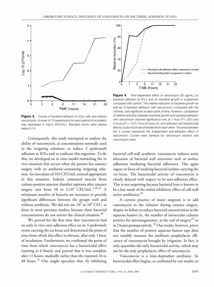
ResultsBacterial growth results with and without vanco-
mycin are presented in Figure 2. In the presence of
vancomycin, bacterial counts decreased over time. A
bactericidal effect of the vancomycin solution was found
after 1 hour of incubation (P Z .005), with a bacterial
reduction of 13% (compared with control without
Figure 2. Bacterial killing curves with and without vancomycin. A
mean of 10 experiments for each period of incubation was expressed
in log10 (CFU/mL). Standard errors were always below 0.12.
J CATARACT REFRACT SU
vancomycin), progressing to a minimum of a 3 log re-
duction (99.9%) after 11 hours (Figure 2 and Table 2).
Comparable effects were observed for the vancomycin
insert (Figure 2 and Table 3).
Analysis of bacterial adhesion proved that vanco-
mycin significantly reduced the counts of adhering
bacteria after 1 hour of incubation (P!.001) (Figure 3
and Table 2).
At 1 and 3 hours, vancomycin reduced the quantity
of bacteria adhering to IOLs by approximately 80%,
whereas the number of bacteria present in suspension
was reduced by only 13% and 36%, respectively
(P!.001 and P Z .017 at 1 and 3 hours). From
6 hours on, the anti-adhesion and bactericidal effects
could not be discriminated from each other (Figure 4)
because the difference between the 2 effects was too tiny.
After 6 hours of incubation, no bacteria were found on
IOL surfaces (Figure 3) and not enough bacteria were
found in broth suspension (Figure 2). Indeed, the
bactericidal effect of vancomycin became highly
relevant by then (1 log of bacterial growth reduction
in suspension at 6 hours, 3 log at 11 hours). The
comparison between the vancomycin solution and the
vancomycin insert revealed no difference regarding
bactericidal and anti-adhesion effects at any point in
time (PO.05).
Bacterial growth was identical in the solutions with
and without IOL (data not shown), showing that the
presence of an IOL had no effect on the planktonic
bacteria.
Results were also confirmed by SEM. A few
adhering bacteria per observation field at 1 and 3 hours
only, with a decrease over time, but always were
observed fewer than on control IOLs (data not shown).
DiscussionAll published studies state that coagulase-negative
Staphylococcus is the most common organism contam-
inating the anterior chamber after uneventful cataract
surgery.4,5,7,28 Not surprisingly, S epidermidis is also the
most common germ found in acute endophthalmitis
(50% to 60% of the cases).1 Vancomycin, known to be
highly effective against this group of bacteria, is the
antibiotic most frequently added to the irrigation fluid.
It is a glycopeptide antibiotic, which acts by inhibiting
the polymerization of the peptidoglycan, an essential
1053RG—VOL 31, MAY 2005
LABORATORY SCIENCE: INFLUENCE OF VANCOMYCIN ON BACTERIAL ADHESION TO IOLS
Table 2. Time-dependent effect of vancomycin (20 mg/ml) on bacterial growth in suspension and on bacterial adhesion to intraocular lenses
(IOLs).*
Bacterial Growthin IOL-Free Suspension(Bactericidal Effect)
Bacterial Adhesion to IOL(Anti-Adhesion Effect)
Comparison ofReduction Percentages
Time (H)Reduction of
Bacterial Growth (%) P ValueReduction of
Bacterial Adhesion (%) P Value P Value
1 13 .005 82 !.001 !.001
3 36 !.001 79 .002 .017
6 90 .002 94 .017 NS
11 99.9 !.001 99.7 .005 NS
16 R99.9 !.001 R99.9 !.001 NS
21 R99.9 !.001 R99.9 .002 NS
24 R99.9 !.001 R99.9 .01 NS
HZ hours; NSZ not significant
*Standard errors were always below 0.14.
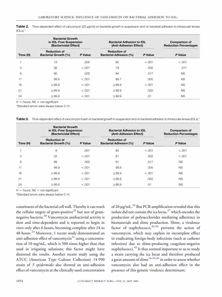
Table 3. Time-dependent effect of vancomycin insert on bacterial growth in suspension and on bacterial adhesion to intraocular lenses (IOLs).*
Bacterial Growthin IOL-Free Suspension(Bactericidal Effect)
Bacterial Adhesion to IOL(Anti-Adhesion Effect)
Comparison ofReduction Percentages
Time (H)Reduction of
Bacterial Growth (%) P ValueReduction of
Bacterial Adhesion (%) P Value P Value
1 8 .007 83 !.001 !.001
3 33 !.001 81 .002 !.001
6 89 .002 91 .017 NS
11 99.9 !.001 99.6 .005 NS
16 R99.9 !.001 R99.9 !.001 NS
21 R99.9 !.001 R99.9 .002 NS
24 R99.9 !.001 R99.9 .01 NS
HZ hours; NSZ not significant
*Standard errors were always below 0.14.
constituent of the bacterial cell wall. Thereby it can reach
the cellular targets of gram-positive29 but not of gram-
negative bacteria.30 Vancomycin antibacterial activity is
slow and time-dependent and is reported to begin in
vitro only after 6 hours, becoming complete after 24 to
48 hours.23 Moreover, 1 recent study demonstrated an
anti-adhesion effect of vancomycin31 using a concentra-
tion of 10 mg/mL, which is 500 times higher than that
used in irrigating solutions; this factor might have
distorted the results. Another recent study using the
ATCC (American Type Culture Collection) 14 990
strain of S epidermidis also showed an anti-adhesion
effect of vancomycin at the clinically used concentration
1054 J CATARACT REFRACT SU
of 20 mg/mL.32 But PCR amplification revealed that this
isolate did not contain the ica locus,27 which encodes the
production of polysaccharides mediating adherence to
biomaterials and slime production. Slime, a virulence
factor of staphylococci,33,34 prevents the action of
vancomycin, which may explain its incomplete effect
in eradicating foreign-body infections (such as catheter
infection) due to slime-producing coagulase-negative
staphylococci.35 It thus seemed important to us to study
a strain carrying the ica locus and therefore produced
a great amount of slime16,24,26 in order to assess whether
vancomycin also had an anti-adhesion effect in the
presence of this genetic virulence determinant.
RG—VOL 31, MAY 2005
LABORATORY SCIENCE: INFLUENCE OF VANCOMYCIN ON BACTERIAL ADHESION TO IOLS
Consequently, this study attempted to analyze the
ability of vancomycin, at concentrations normally used
in the irrigating solutions, to reduce S epidermidisadhesion to IOLs and to eradicate this organism. To do
this, we developed an in vitro model mimicking the in
vivo situation that occurs when the patient has cataract
surgery with an antibiotic-containing irrigating solu-
tion. An inoculum of 103 CFU/mL seemed appropriate
in this situation. Indeed, estimated inocula from
culture-positive anterior chamber aspirates after cataract
surgery vary from 10 to 2.102 CFU/mL.4,36,37 A
minimum number of bacteria are necessary to provide
significant differences between the groups with and
without antibiotic. We did not use 105 to 108 CFU, as
done in most previous studies, because these bacterial
concentrations do not mirror the clinical situation.30
We proved for the first time that vancomycin had
an early in vitro anti-adhesion effect on an S epidermidisstrain carrying the ica locus and determined the point of
time from which this effect is relevant, after only 1 hour
of incubation. Furthermore, we confirmed the point of
time from which vancomycin has a bactericidal effect
(starting at 6 hours) and proved that it was complete
after 11 hours, markedly earlier than the reported 24 to
48 hours.23 One might speculate that, by inhibiting
Figure 3. Curves of bacterial adhesion to IOLs with and without
vancomycin. A mean of 10 experiments for each period of incubation
was expressed in log10 (CFU/mL). Standard errors were always
below 0.14.
J CATARACT REFRACT SU
bacterial cell-wall synthesis, vancomycin induces some
alteration of bacterial wall structures such as surface
adhesions mediating bacterial adherence. This again
argues in favor of studying bacterial isolates carrying the
ica locus. The bactericidal activity of vancomycin is
clearly delayed with respect to its anti-adhesion effect.
This is not surprising because bacterial lysis is known to
be a late result of the initial inhibitory effect of cell-wall
active antibiotics.38
A current practice of many surgeons is to add
vancomycin to the infusion during cataract surgery,
despite its failure to reduce bacterial concentration in the
aqueous humor (ie, the number of intraocular cultures
positive for microorganisms), at the end of surgery23 or
at 2 hours postoperatively.22 Our results, however, prove
that the number of positive aqueous humor taps does
not suitably measure the antibiotic prophylactic effi-
ciency of vancomycin brought by irrigation. In fact, it
only quantifies the early bactericidal activity, which may
not be the only prophylactic effect of vancomycin.
Vancomycin is a time-dependent antibiotic. Its
bactericidal effect begins, as confirmed by our results, in
Figure 4. Time-dependent effect of vancomycin (20 mg/mL) on
bacterial adhesion to IOLs and on bacterial growth in suspension
compared with control. The relative reduction of bacterial growth as
well as of bacterial adhesion with vancomycin, compared with the
controls, was significant at each point of time. However, comparison
of relative reduction between bacterial growth and bacterial adhesion
with vancomycin showed significance only at 1 hour (P!.001) and
3 hours (PZ .017). From 6 hours on, anti-adhesion and bactericidal
effects could not be discriminated from each other. The area between
the 2 curves represents the independent anti-adhesion effect of
vancomycin. Curves were identical for vancomycin solution and
vancomycin insert.
1055RG—VOL 31, MAY 2005

LABORATORY SCIENCE: INFLUENCE OF VANCOMYCIN ON BACTERIAL ADHESION TO IOLS
vitro only after 6 hours of incubation. Because of this we
were able to assess the anti-adhesion effect of vanco-
mycin during the first 6 hours. It may be better to
harbor bacteria in the anterior chamber than on the IOL
surface where they are embedded within a slime layer.
Indeed, host defenses and antibiotics have trouble
penetrating this bacterial biofilm.39,40 Therefore, bac-
teria suspended in aqueous humor are less critical to
treat than those bound on an IOL surface. Nonetheless,
intracameral vancomycin used in irrigating liquids
shows a postoperative half-life of less than 2 hours,22,41
which means that its concentration becomes theoreti-
cally inferior to the MIC after 4 to 6 hours42 (the
estimated MIC for most bacteria responsible for
postoperative endophthalmitis is about 4 mg/mL).41
Thus, in a clinical situation, the antibiotic concentra-
tion decreases over time, which differs from our in vitro
model. Therefore, there is no evidence that contact time
with vancomycin is long enough in vivo for the anti-
adhesion effect to become clinically relevant. Moreover,
it has even been shown that vancomycin could
potentially increase bacterial adhesion if the drug
concentration fell below the MIC for the infecting
strain, by enhancing the biofilm matrix produced by
slime-positive coagulase-negative staphylococci on the
IOL surface.43,44 This could eventually worsen the
clinical situation. This absolutely merits further studies.
Because the contact time with the antibiotic
contained in the irrigating solution is definitely not
long enough to develop a relevant bactericidal effect, we
designed a biodegradable, biocompatible sterile insert to
overcome this drawback. Although only in vitro results
are available for now, we believe that placing this insert
in the capsular bag could, beyond its action closer to the
site in which germs are most critical, progressively
release the antibiotic, thus obtaining a longer and more
efficient effect. This vancomycin insert obtained in vitro
effects identical to those of a 20 mg/mL vancomycin
solution. Because the carried dose is 230 mg, while the
mean volume of the pseudophakic anterior chamber is
536 mL,45 our insert would keep a local concentration
above the MIC for at least 14 hours respecting a
postoperative half-life of vancomycin of less than 2
hours.22,41 Moreover, with the continuous curvilinear
capsulorhexis and the posterior chamber IOL, the bag is
widely isolated from the aqueous circulation by the
implanted IOL, which might increase the calculated
1056 J CATARACT REFRACT SU
duration by a lower release into the anterior chamber.46
This would also mean a weaker endothelial toxicity.46
This expected time of at least 14 hours would be
sufficient to develop a bactericidal effect as well as an
anti-adhesion one. These hypotheses have to be assessed
in vivo, using animal experiments in the future.
In conclusion, the risk–benefit ratio (ie, its effect on
bacterial adhesion growth) of supplementing irrigating
solutions with vancomycin seems very ambiguous.
Nevertheless, a sustained-release system would princi-
pally overcome the drawbacks of vancomycin as used in
addition to irrigating solutions by allowing both actions
of the antibiotic to develop. Furthermore, we demon-
strated that our in vitro model is suitable to study anti-
adhesion and bactericidal effects of antibiotics.
References1. Aaberg TM Jr, Flynn HW Jr, Schiffman J, Newton J.
Nosocomial acute-onset postoperative endophthalmitissurvey; a 10-year review of incidence and outcomes.Ophthalmology 1998; 105:1004–1010
2. Kresloff MS, Castellarin AA, Zarbin MA. Endophthal-mitis. Surv Ophthalmol 1998; 43:193–224
3. Salvanet-Bouccara A, Forestier F, Coscas G, et al. Bacte-rial endophthalmitis: ophthalmological results of a na-tional multicenter prospective survey. J Fr Ophtalmol1992; 15:669–678
4. Dickey JB, Thompson KD, Jay WM. Anterior chamberaspirate cultures after uncomplicated cataract surgery.Am J Ophthalmol 1991; 112:278–282
5. Leong JK, Shah R, McCluskey PJ, et al. Bacterial con-tamination of the anterior chamber during phacoemulsi-fication cataract surgery. J Cataract Refract Surg 2002;28:826–833
6. Mistlberger A, Ruckhofer J, Raithel E, et al. Anteriorchamber contamination during cataract surgery withintraocular lens implantation. J Cataract Refract Surg1997; 23:1064–1069
7. Srinivasan R, Tiroumal S, Kanungo R, Natarajan MK.Microbial contamination of the anterior chamber duringphacoemulsification. J Cataract Refract Surg 2002;28:2173–2176
8. Dilly PN, Sellors PJ. Bacterial adhesion to intraocularlenses. J Cataract Refract Surg 1989; 15:317–320
9. Vafidis GC, Marsh RJ, Stacey AR. Bacterial contamina-tion of intraocular lens surgery. Br J Ophthalmol 1984;68:520–523
10. Doyle A, Beigi B, Early A, et al. Adherence of bacteria tointraocular lenses: a prospective study. Br J Ophthalmol1995; 79:347–349
RG—VOL 31, MAY 2005
LABORATORY SCIENCE: INFLUENCE OF VANCOMYCIN ON BACTERIAL ADHESION TO IOLS
11. Cramton SE, Gerke C, Schnell NF, et al. The intercel-lular adhesion (ica) locus is present in Staphylococcusaureus and is required for biofilm formation. InfectImmun 1999; 67:5427–5433
12. Mack D. Molecular mechanisms of Staphylococcusepidermidis biofilm formation. J Hosp Infect 1999;43(Suppl):S113–S125
13. McKenney D, Hubner J, Muller E, et al. The ica locus ofStaphylococcus epidermidis encodes production of thecapsular polysaccharide/adhesion. Infect Immunol 1998;66:4711–4720
14. von Eiff C, Heilmann C, Peters G. New aspects in themolecular basis of polymer-associated infections due tostaphylococci. Eur J Clin Microbiol Infect Dis 1999;18:843–846
15. Griffiths PG, Elliot TS, McTaggart L. Adherence ofStaphylococcus epidermidis to intraocular lenses. Br JOphthalmol 1989; 73:402–406
16. Burillon C, Kodjikian L, Pellon G, et al. In vitro study ofbacterial adherence to different types of intraocularlenses. Drug Dev Ind Pharm 2002; 28:95–99
17. Cusumano A, Busin M, Spitznas M. Is chronic intraoc-ular inflammation after lens implantation of bacterialorigin? Ophthalmology 1991; 98:1703–1710
18. Ng EW, Barrett GD, Bowman R. In vitro bacterial adher-ence to hydrogel and poly(methyl methacrylate) intraoc-ular lenses. J Cataract Refract Surg 1996; 22:1331–1335
19. Menikoff JA, Speaker MG, Marmor M, Raskin EM.A case-control study of risk factors for postoperativeendophthalmitis. Ophthalmology 1991; 98:1761–1768
20. Raskin EM, Speaker MG, McCormick SA, et al. Influ-ence of haptic materials on the adherence of staphylo-cocci to intraocular lenses. Arch Ophthalmol 1993;111:250–253
21. Gills JP. Filters and antibiotics in irrigating solution forcataract surgery. J Cataract Refract Surg 1991; 17:385
22. Ferro J, De-Pablos M, Logrono M. Postoperative con-tamination after vancomycin and gentamicin duringphacoemulsification. Arch Ophthalmol 1997; 115:165–170
23. Feys J, Salvanet-Bouccara A, Emond JP, Dublanchet A.Vancomycin prophylaxis and intraocular contaminationduring cataract surgery. J Cataract Refract Surg 1997;23:894–897
24. Kodjikian L, Burillon C, Roques C, et al. Bacterial ad-herence of Staphylococcus epidermidis to intraocularlenses: a bioluminescence and scanning electron micros-copy study. Invest Ophthalmol Vis Sci 2003; 44:4388–4394
25. Kodjikian L, Burillon C, Chanloy C, et al. In vivo studyof bacterial adhesion to five types of intraocular lenses.Invest Ophthalmol Vis Sci 2002; 43:3717–3721
26. Lina G, Quaglia A, Reverdy ME, et al. Distribution ofgenes encoding resistance to macrolides, lincosamides,
J CATARACT REFRACT SU
and streptogramins among staphylococci. AntimicrobAgents Chemother 1999; 43:1062–1066
27. Kodjikian L, Burillon C, Lina G, et al. Biofilm formationon intraocular lenses by a clinical strain encoding icalocus: a scanning electron microscopy study. Invest Oph-thalmol Vis Sci 2003; 44:4382–4387
28. Egger SF, Huber-Spitzy V, Scholda C, et al. Bacterialcontamination during extracapsular cataract extraction:prospective study on 200 consecutive patients. Ophthal-mologica 1994; 208:77–81
29. Feys J, Emond JP, Salvanet-Bouccara A, DublanchetA. Bacteriological study of the intraocular fluid at theend of cataract surgery. J Fr Ophtalmol 1993; 16:501–505
30. Gritz DC, Cevallos AV, Smolin G, Whitcher JP Jr. Anti-biotic supplementation of intraocular irrigating solutions;an in vitro model of antibacterial action. Ophthalmology1996; 103:1204–1208; discussion 1208–1209
31. Das T, Sharma S, Muralidhar AV. Effect of vancomycinon Staphylococcus epidermidis adherence to poly(methylmethacrylate) intraocular lenses. J Cataract Refract Surg2002; 28:703–708
32. Abu el-Asrar AM, Kadry AA, Shibl AM, et al. Antibioticsin the irrigating solutions reduce Staphylococcus epider-midis adherence to intraocular lenses. Eye 2000; 14:225–230
33. Christensen GD, Simpson WA, Bisno AL, Beachey EH.Adherence of slime-producing strains of Staphylococcusepidermidis to smooth surfaces. Infect Immun 1982;37:318–326
34. Pfaller MA, Herwaldt LA. Laboratory, clinical, and epi-demiological aspects of coagulase-negative staphylococci.Clin Microbiol Rev 1988; 1:281–299
35. Farber BF, Kaplan MH, Clogston AG. Staphylococcusepidermidis extracted slime inhibits the antimicrobialaction of glycopeptide antibiotics. J Infect Dis 1990;161:37–40
36. Pospisil A, Pospisil F, Dupont MJ, et al. Bacterial con-tamination of the anterior chamber and cataract surgery.J Fr Ophtalmol 1993; 16:10–13
37. Samad A, Solomon LD, Miller MA, Mendelson J.Anterior chamber contamination after uncomplicatedphacoemulsification and intraocular lens implantation.Am J Ophthalmol 1995; 120:143–150
38. Tomasz A. The mechanism of the irreversible antimicro-bial effect of penicillins: how the beta-lactam antibioticskill and lyse bacteria. Ann Rev Microbiol 1979; 33:113–137
39. Evans RC, Holmes CJ. Effect of vancomycin hydrochlo-ride on Staphylococcus epidermidis biofilm associatedwith silicone elastomer. Antimicrob Agents Chemother1987; 31:889–894
40. Stewart PS, Costerton JW. Antibiotic resistance of bac-teria in biofilms. Lancet 2001; 358:135–138
1057RG—VOL 31, MAY 2005

LABORATORY SCIENCE: INFLUENCE OF VANCOMYCIN ON BACTERIAL ADHESION TO IOLS
41. Mendivil Soto A, Mendivil MP. The effect of topicalpovidone-iodine, intraocular vancomycin, or both onaqueous humor cultures at the time of cataract surgery.Am J Ophthalmol 2001; 131:293–300
42. Gordon YJ. Vancomycin prophylaxis and emerging re-sistance: are ophthalmologists the villains? The heroes?Am J Ophthalmol 2001; 131:371–376
43. Dunne WM Jr. Effects of subinhibitory concentrationsof vancomycin or cefamandole on biofilm production bycoagulase-negative staphylococci. Antimicrob AgentsChemother 1990; 34:390–393
1058 J CATARACT REFRACT SU
44. Wilcox M, Finch R, Smith D, et al. Effects of carbon di-oxide and sub-lethal levels of antibiotics on adherence ofcoagulase-negative staphylococci to polystyrene and sili-cone ruber. J Antimicrob Chemother 1991; 27:577–587
45. Lehmann OJ, Thompson JP, White LO, et al. Half-lifeof intracameral gentamicin after phacoemulsification.J Cataract Refract Surg 1997; 23:883–888
46. Gimbel H, Sun R, DeBrof B. Prophylactic intracameralantibiotics during cataract surgery: the incidence ofendophthalmitis and corneal endothelial cell loss. Eur JImplant Ref Surg 1994; 6:280–285
RG—VOL 31, MAY 2005