Procedure for Classification of Coarse Aggregates Based on ...

Planktonic Aggregates of Staphylococcus aureus Protectagainst Common AntibioticsJakob Haaber1", Marianne Thorup Cohn1¤", Dorte Frees1, Thorbjørn Joest Andersen2, Hanne Ingmer1*
1 Department of Veterinary Disease Biology, University of Copenhagen, Frederiksberg, Denmark, 2 Department of Geography and Geology, University of Copenhagen,
Copenhagen, Denmark
Abstract
Bacterial cells are mostly studied during planktonic growth although in their natural habitats they are often found incommunities such as biofilms with dramatically different physiological properties. We have examined another type ofcommunity namely cellular aggregates observed in strains of the human pathogen Staphylococcus aureus. By laser-diffraction particle–size analysis (LDA) we show, for strains forming visible aggregates, that the aggregation starts already inthe early exponential growth phase and proceeds until post-exponential phase where more than 90% of the population ispart of the aggregate community. Similar to some types of biofilm, the structural component of S. aureus aggregates is thepolysaccharide intercellular adhesin (PIA). Importantly, PIA production correlates with the level of aggregation whetheraltered through mutations or exposure to sub-inhibitory concentrations of selected antibiotics. While some properties ofaggregates resemble those of biofilms including increased mutation frequency and survival during antibiotic treatment,aggregated cells displayed higher metabolic activity than planktonic cells or cells in biofilm. Thus, our data indicate that theproperties of cells in aggregates differ in some aspects from those in biofilms. It is generally accepted that the biofilm lifestyle protects pathogens against antibiotics and the hostile environment of the host. We speculate that in aggregatecommunities S. aureus increases its tolerance to hazardous environments and that the combination of a biofilm-likeenvironment with mobility has substantial practical and clinical importance.
Citation: Haaber J, Cohn MT, Frees D, Andersen TJ, Ingmer H (2012) Planktonic Aggregates of Staphylococcus aureus Protect against Common Antibiotics. PLoSONE 7(7): e41075. doi:10.1371/journal.pone.0041075
Editor: Michael Otto, National Institutes of Health, United States of America
Received January 28, 2012; Accepted June 17, 2012; Published July 18, 2012
Copyright: � 2012 Haaber et al. This is an open-access article distributed under the terms of the Creative Commons Attribution License, which permitsunrestricted use, distribution, and reproduction in any medium, provided the original author and source are credited.
Funding: This work was supported by the Danish Reserch Council for Independent Research, grant number 274-08-0531, and the Lundbeck foundation, grantnumber R31-A2472. The funders had no role in study design, data collection and analysis, decision to publish, or preparation of the manuscript.
Competing Interests: The authors have declared that no competing interests exist.
* E-mail: [email protected]
" These authors are joint first authors on this work.
¤ Current address: Novozymes A/S, Bagsvaerd, Denmark
Introduction
Many bacterial species can grow either in the form of dispersed
single cells in liquid or as densely packed communities attached to
solid surfaces. Researchers generally refer to the latter growth form
as biofilm, which was defined by Costerton as ‘‘a structured
community of cells enclosed in a self-produced polymeric matrix
and adherent to an inert or living surface’’ [1]. The opportunistic,
human pathogen, Staphylococcus aureus can establish itself in biofilms
by colonizing natural body surfaces including lungs and heart
valves as well as abiotic surfaces such as medical implants [2,3]. As
cells present in biofilms are commonly protected against antibiotics
and host defense molecules, biofilm formation has serious clinical
consequences and is a significant contributor to the health care
problems associated with S. aureus [4–6].
Staphylococcal biofilms contain several matrix components
including extracellular DNA (eDNA), protein and polysaccharide
[2,7,8]. In S. aureus, eDNA is released from dead cells by controlled
cell lysis and the presence of eDNA is important in the very early
establishment of the biofilm [9,10]. At this early stage, S. aureus cell
surface proteins such as fibronectin and fibrinogen binding
proteins and Protein A are also contributing [11,12]. The
extracellular polysaccharide poly-N-acetyl-1,6-glucosamine
(PNAG) is often involved in biofilm formation and it is the most
characterized component of the biofilm matrix. PNAG is
synthesized by the products of the polysaccharide intercellular
adhesin (PIA) locus, icaADBC [13] and mediates adhesion to both
living and artificial surfaces [14,15]. The ica operon is present in
many clinical S. aureus strains and its expression has been shown to
be strongly induced in a device-related animal model, underscor-
ing the importance of biofilms during infection [16].
During establishment of an infection, formation of biofilm may
be seen as a survival strategy against host defenses and
antimicrobial therapy. It is well accepted that biofilms protect
the embedded cells against antimicrobial therapy [17–20] for
example through reduced exposure to the antimicrobial com-
pounds [21,22] or reduced metabolic activity that decreases
susceptibility to a range of antibiotics [20]. However, although a
biofilm can provide beneficial properties to the participating
bacteria it also traps the cells in a sessile community with limited
mobility. This limitation seems to have been circumvented by the
human, opportunistic pathogen, Pseudomonas aeruginosa that upon
growth in liquid culture forms large aggregates containing densely
packed viable cells and eDNA [23]. The properties of P. aeruginosa
cells present in aggregates resemble those of flow-cell biofilms by
their slow growth rate, their capacity to survive otherwise lethal
PLoS ONE | www.plosone.org 1 July 2012 | Volume 7 | Issue 7 | e41075

treatments with antibiotics and their ability to resist phagocytosis
[24]. Interestingly, non-attached aggregates are also observed in
the lungs of cystic fibrosis patients, in soft tissue fillers and in non-
healing wounds, indicating that they are of clinical relevance [24–
26].
Planktonic (non-attached) aggregation has also been observed
for other bacterial species. In the food borne pathogen,
Campylobacter jejuni, auto-aggregation is regularly observed but
dispersal is promoted in mutants with disrupted stress response
pathways [27,28]. Streptococcus pyogenes is another human pathogen
that forms planktonic aggregates in liquid culture and this property
is important for adherence, resistance to phagocytosis and
virulence [29]. S. aureus is known to grow in small clusters of 5–
20 cells but it also assembles in large aggregates that are visible to
the naked eye and are observed in both laboratory and clinical
settings [12,26]. While extensive research has focused on
staphylococcal biofilms only little knowledge exists on the
formation and properties of S. aureus planktonic aggregation.
Here, we characterize S. aureus aggregation and show that while
aggregates share many of the properties of surface-bound biofilms,
differences are also observed. In common to biofilms, the
aggregation process is influenced by sub-lethal concentrations of
antibiotics [30] and cells in aggregates are protected against
several unrelated and clinically relevant antimicrobial compounds
[19,20]. In contrast to biofilms, however, metabolic activity is high
in aggregates. We speculate that aggregates provide bacteria with
the benefits of a biofilm while maintaining mobility and that this
combination may contribute to the difficulties of eradicating S.
aureus infections.
Results
Staphylococcus Aureus Aggregates in SolutionSome strains of S. aureus are capable of forming large planktonic
aggregates that are visible to the naked eye when grown under
standard laboratory conditions [11,12,31]. From previous unpub-
lished work we had noticed four strains with very different
aggregation capabilities. These were two clinical isolates (15981
and SA564) and two laboratory strains (8325-4 and Newman),
where 8325-4 and 15981 form visible planktonic aggregates, while
Newman and SA564 do not (data not shown). With the aim of
investigating this phenomenon in more detail, we applied scanning
electron microscopy (SEM) to visualize aggregates in a post-
exponential culture of 8325-4 (Figure 1). Some aggregates were
more than 0.5 mm of size and consisted of a large numbers of cells
(Figure 1A). A closer look revealed pores and crevices in the
compact mass of cells (Figure 1B) and at even higher magnification
some cell-clusters were covered in a web-like structure as recently
observed in biofilms of P. aeruginosa [32] while other clusters of cells
remained un-covered (Figure 1C, D).
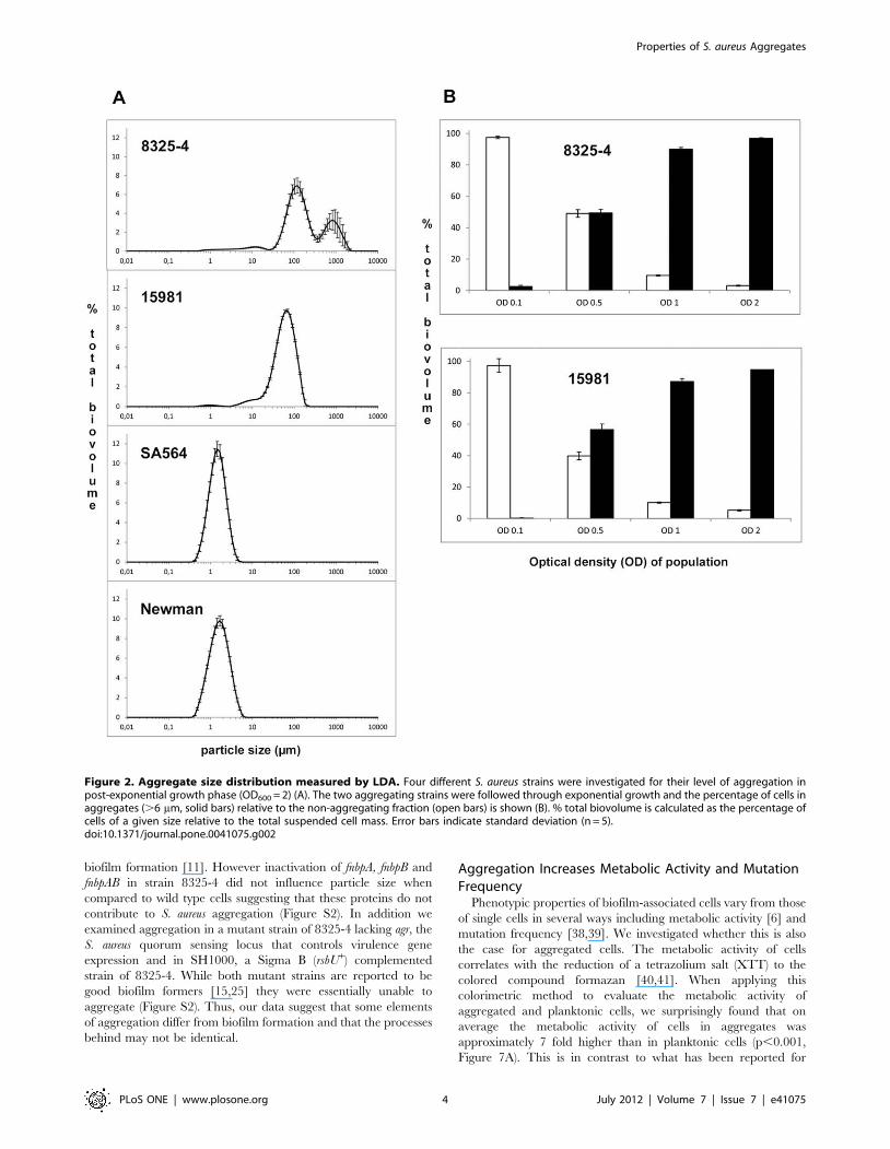
To quantify the extent of aggregation in liquid cultures of the
four S. aureus strains, we monitored the formation of planktonic S.
aureus aggregates using laser diffraction analysis (LDA) [23]. When
grown to post exponential growth phase (OD600 = 2) the majority
of cells of strains 8325-4 and 15981 were assembled in particles of
more than 6 mm in diameter, the minimum size chosen to define
an aggregate. For strains Newman and SA564 the average particle
size was 1–2 mm in diameter (Figure 2A) representing single and
double cells as observed by microscopy. Further, the LDA analysis
revealed that aggregate size varies between strains. For 8325-4
cultures two aggregate sizes of 100 and 800 mm in diameter
predominate while for strain 15981 the aggregates are approxi-
mate 60 mm in diameter (Figure 2A).
To address the structural robustness of the S. aureus aggregates
we applied increasing shear force as part of the LDA and found
that with gradual steps of stirring (50–500 rpm) and pump speed
(250–2500) no changes were observed in the size distribution until
medium shear force was applied (stir = 300 rpm,
pump = 1500 rpm). Even when exposed to maximum shear
(stir = 500 rpm, pump = 2500 rpm), the aggregates were still
surprisingly intact with a reduction in average aggregate size from
180 mm to 45 mm and maximum size from 480 mm to 240 mm
(Figure S1). Thus, under the assayed conditions the S. aureus
aggregates are structurally stable, which is in contrast to aggregates
formed by P. aeruginosa that are readily dissolved by whirly mixing
[24].
Aggregates are Formed already in the Exponential PhaseTo address the temporal development of aggregate formation
we monitored particle size of strains 8325-4 and 15981 at various
growth stages and found that while very little aggregation occurred
at OD600 of 0.1, already at OD600 of 0.5 around 50% of the cells
were assembled into aggregates (Figure 2B). Thus, for the tested S.
aureus strains, aggregation is not confined to the post-exponential
growth phase but occurs already at mid-exponential phase. The
size of the aggregates was two to three times larger for 8325-4 than
for 15981. Interestingly though, when applying the 6 mm threshold
for defining aggregates, the distribution of cells in the aggregated
versus dispersed fractions of cells was strikingly similar for strains
8325-4 and 15981 throughout the growth cycle (Figure 2B).
To address whether aggregates are formed by interconnected
daughter cells that are not separated after cell division or from
aggregation of single cells or smaller clusters we monitored
aggregation of fluorescent and non-fluorescent, isogenic 8325-4
cells after mixing (1:1) at OD600 = 0.01 [33]. The results show that
in the early growth stages (OD600 = 0.01 to 0.1) small grape-like
clusters are formed by <10 interconnected daughter cells
(Figure 3A). At OD600 = 0.1 the clusters of different clonal origin
begin to merge (Figure 3B) and from OD600 = 0.5 an increasing
number of large aggregates are observed that are composed of
both labeled and non-labeled cells (Figure 3C). Thus, our results
suggest that the large multi-cellular aggregates observed in post-
exponential growth phase arise from small cell clusters that merge
during exponential growth phase and end up glued together in
large multi-cellular structures.
Sub-inhibitory Concentrations of Antibiotics andEnvironmental Factors Influence Aggregation
Bacterial pathogens may be exposed to sub-lethal concentra-
tions of antibiotics as a consequence of uneven antibiotic
distribution during antimicrobial therapy, inadequate use of
antibiotics or from exposure to antimicrobials naturally produced
in soil and water environments [34–36]. Notably, we observed that
addition of sub-inhibitory concentrations (1/25 6MIC) of several
clinically relevant antibiotics influenced the aggregation properties
of the aggregating strains 8325-4 and 15981 (Figure 4). The RNA
polymerase inhibitor rifampicin stimulated aggregation while the
protein biosynthesis inhibitor erythromycin and the cell wall
synthesis inhibitor cefuroxime reduced aggregation. Further we
examined the aggregation ability in 43 S. aureus strains from our
culture collection comprising laboratory strains and clinical
isolates. We found that while significant differences were observed
between strains, exposure to either 2% NaCl, 35 mM glucose or
reduced oxygen tension induced aggregation in all strains tested
(data not shown) suggesting that the ability to aggregate is
omnipresent in S. aureus.
Properties of S. aureus Aggregates
PLoS ONE | www.plosone.org 2 July 2012 | Volume 7 | Issue 7 | e41075

The Polysaccharide Intercellular Adhesion (PIA) PromotesAggregate Formation
The cellular level of the polysaccharide intercellular adhesin
(PIA) produced by the enzymes encoded by the icaADBC operon
has been noted to influence dispersed growth of staphylococci in
liquid cultures [31,37]. To evaluate the contribution of PIA to
aggregation in S. aureus, we examined aggregate formation in an
isogenic ica mutant of 8325-4 during post-exponential growth. No
visible aggregates were observed and using LDA we could verify
that no particles larger than 6 mm were present in the culture
demonstrating that aggregates are not formed in the absence of
PIA (Figure S2). In an isogenic ica mutant of 15981 aggregation
was also abolished (data not shown).
To further investigate the composition of the extracellular
matrix in S. aureus aggregates we examined the resistance to the
action of either proteinase K, DNase or metaperiodate, which
degrade protein, DNA and polysaccharide, respectively. Only the
addition of metaperiodate resulted in disruption of aggregates
(Figure 5). This result indicates that extracellular polysaccharides
form the major adhesive component of the aggregates thus
supporting the genetic data stating the importance of the ica
operon.
The central role of PIA polysaccharide was further confirmed
by serological detection of PIA production in aggregating and non-
aggregating strains. Here we observed a clear correlation between
PIA production and aggregation (Figure 6A). Furthermore,
analysis of the PIA level in 8325-4 cultures treated with antibiotics
showed that rifampicin increased the cellular PIA level, while
cefuroxime and erythromycin decreased PIA production
(Figure 6B). The PIA levels correlated well with the level of
aggregation of 8325-4 treated with these antibiotics (Figure 4).
Collectively, these results indicate that the production of PIA is a
major contributor to planktonic aggregate formation of S. aureus
and that sub-inhibitory concentrations of antibiotics may affect
aggregation, possibly through modulation of PIA production.
In addition to PIA other cellular components such as the
fibronectin binding proteins have been reported to influence
Figure 1. Aggregate visualized by SEM. Aggregates from a post-exponential 8325-4 culture were fixed and visualized using Scanning ElectronMicroscopy (SEM). Panels A-D represent increasing magnification and red squares indicate the area magnified in the following panel. Overview of anaggregate, which is visible to the naked eye (A). Zooming in reveals pores and crevices in a topographical landscape of aggregated clusters (B). Athigher magnifications it is revealed that some clusters are embedded in a web-like matrix (black arrows) while some are not (white arrows) (C, D).doi:10.1371/journal.pone.0041075.g001
Properties of S. aureus Aggregates
PLoS ONE | www.plosone.org 3 July 2012 | Volume 7 | Issue 7 | e41075
biofilm formation [11]. However inactivation of fnbpA, fnbpB and
fnbpAB in strain 8325-4 did not influence particle size when
compared to wild type cells suggesting that these proteins do not
contribute to S. aureus aggregation (Figure S2). In addition we
examined aggregation in a mutant strain of 8325-4 lacking agr, the
S. aureus quorum sensing locus that controls virulence gene
expression and in SH1000, a Sigma B (rsbU+) complemented
strain of 8325-4. While both mutant strains are reported to be
good biofilm formers [15,25] they were essentially unable to
aggregate (Figure S2). Thus, our data suggest that some elements
of aggregation differ from biofilm formation and that the processes
behind may not be identical.
Aggregation Increases Metabolic Activity and MutationFrequency
Phenotypic properties of biofilm-associated cells vary from those
of single cells in several ways including metabolic activity [6] and
mutation frequency [38,39]. We investigated whether this is also
the case for aggregated cells. The metabolic activity of cells
correlates with the reduction of a tetrazolium salt (XTT) to the
colored compound formazan [40,41]. When applying this
colorimetric method to evaluate the metabolic activity of
aggregated and planktonic cells, we surprisingly found that on
average the metabolic activity of cells in aggregates was
approximately 7 fold higher than in planktonic cells (p,0.001,
Figure 7A). This is in contrast to what has been reported for
Figure 2. Aggregate size distribution measured by LDA. Four different S. aureus strains were investigated for their level of aggregation inpost-exponential growth phase (OD600 = 2) (A). The two aggregating strains were followed through exponential growth and the percentage of cells inaggregates (.6 mm, solid bars) relative to the non-aggregating fraction (open bars) is shown (B). % total biovolume is calculated as the percentage ofcells of a given size relative to the total suspended cell mass. Error bars indicate standard deviation (n = 5).doi:10.1371/journal.pone.0041075.g002
Properties of S. aureus Aggregates
PLoS ONE | www.plosone.org 4 July 2012 | Volume 7 | Issue 7 | e41075

biofilm associated cells, where oxygen and nutrient limitation
results in low metabolic activity [20]. Accordingly, we saw
approximately 4 fold lower metabolic activity in a biofilm
compared to dispersed cells (p,0.001) (Figure S3). When
comparing metabolic activity of aggregates with cells originating
from aggregates disrupted by sonication, no differences were
observed (data not shown, p = 0.84). Furthermore, when compar-
ing isogenic ica mutants of both 8325-4 and 15981 to aggregates
and dispersed cells, metabolic activity was significantly higher in
the aggregates than in dispersed wild type and ica mutant cells
(Figure S3, p,0.05). In biofilms, the metabolic activity is unevenly
distributed depending on oxygen and nutrient availability [42].
CLSM of LIVE/DEAD stained aggregates revealed that analo-
gous to biofilms, aggregates are unevenly composed of areas of
cells with low metabolic activity surrounded by metabolically
active cells (Figure 7B). Collectively, our results show that on
average cells in aggregates are markedly more metabolically active
compared to those in biofilms or planktonic cells.
Mutation frequency has previously been reported to be higher
in biofilms compared to single cells [38,39]. We determined the
mutation rate, calculated by the number of generated rifampicin-
resistant-mutants per total CFU, and found that it was 2 fold
greater in the aggregate fraction compared to the dispersed cell
fraction (Figure S4, p = 0.0015). Thus, aggregates resemble
biofilms by having a slightly increased mutation frequency
compared to planktonic cells.
Aggregation Promotes Protection against AntibioticKilling
Interestingly, we noted that the biomass of dispersed cells
increased during stationary phase compared to the aggregated
fraction (data not shown) indicating that cells might detach from
the aggregates to become dispersed as observed for P. aeruginosa
when experiencing starvation [23]. Therefore, we used an
experimental set up with antibiotic markers that enabled us to
follow cells from the two fractions after antibiotic exposure.
Aggregated and dispersed cells with different antibiotic markers
were mixed and exposed to 256 MIC of clinically relevant
antibiotics with different cellular targets (kanamycin, ciprofloxacin,
erythromycin and vancomycin). Importantly, antibiotic exposure
mediated increased shedding of cells from the aggregate fraction to
the dispersed fraction (data not shown, p,0.05). Furthermore, we
found that cells assembled in aggregates were protected against the
lethality of antibiotics and survived significantly better (p,0.05)
compared to dispersed cells (Figure 8). For kanamycin there was a
dramatic (1006) increase of survival in the aggregated cells
compared to dispersed cells, while for ciprofloxacin, vancomycin
and erythromycin the increased survival was 306, 146 and 86higher, respectively (Figure 8). However, if aggregates were
disrupted by sonication and the released cells were exposed to
kanamycin, the protection was completely abolished and the
susceptibility of the cells was comparable to the non-aggregated
cell fraction (data not shown, p,0.01). This is equivalent to
observations in P. aeruginosa in which tobramycin regained its
efficacy after disruption of the aggregates [24] and it indicates that
protection against antibiotics in aggregates in these two species is
mediated by a physical barrier provided by the aggregate matrix as
also observed for flocculating yeast [43].
Discussion
The ability of Staphylococcus aureus to form biofilms on host tissues
and medical implants is considered to be one of the most
important traits contributing to the health care problems
associated with the organism [11,44,45]. Recently it was proposed
for another opportunistic pathogen, P. aeruginosa, that non-attached
planktonic aggregates of cells may be considered dispersed biofilms
and that these planktonic aggregates have the same protective
properties as biofilms [24]. Sporadically, S. aureus has been
reported to form planktonic aggregates under laboratory condi-
tions [12,31] and in clinical infections [26,46] but so far this
property has received little attention.
We show here that the ability of S. aureus to form large
planktonic aggregates is highly dependent on strain and growth
conditions and is observed for both clinical and laboratory strains.
The aggregation process starts already in the early exponential
growth phase. At low cell densities S. aureus 8325-4 grows in clonal,
structured populations of up to approx. 20 cells whereas at greater
cell densities these structures merge into large aggregates
measuring up to 1000 mm in diameter.
The matrix component responsible for the extensive aggrega-
tion is a polysaccharide and is likely to be the extracellular
polysaccharide PIA, as Western blot analysis correlated the level of
aggregation to PIA production and absence of the ica operon
(encoding the PIA producing enzymes) completely abolished
Figure 3. Kinetic of aggregate formation. 8325-4 cells were mixed 1:1 with 8325-4 cells expressing YFP to OD600 of 0.01 and examined usingCLSM every 30 min through a growth cycle. Small clusters of cells dominate until OD600 = 0.1 (A) at which time they start to fuse (B) and form largeaggregates around OD600 = 0.5 (C). Note different sizes of scale bars.doi:10.1371/journal.pone.0041075.g003
Properties of S. aureus Aggregates
PLoS ONE | www.plosone.org 5 July 2012 | Volume 7 | Issue 7 | e41075

aggregation. The conserved ica operon is present in most clinical
isolates of S. aureus [45] and its expression is induced in exponential
growth phase [16,31] and during infection [16]. Furthermore,
clinical data show that S. aureus form PIA embedded multi-cellular
aggregates within the mucus of cystic fibrosis patients [45]. Taken
together, PIA is likely to contribute to both the aggregation process
and the pathogenesis of S. aureus.
Although little is still known about the environmental factors
that promote aggregation, we observed that different environ-
mental conditions (NaCl, glucose, O2) affected the aggregation
ability of many S. aureus sub-species, indicating that aggregation
may be a common feature of staphylococci. Interestingly, also sub-
inhibitory concentrations of antibiotics affected aggregation. As
the effects were correlated with corresponding changes in PIA
production we propose that sub-inhibitory concentrations of
antibiotics may affect aggregation of S. aureus by modulating the
PIA production. This may also be the case for other staphylococcal
species as antibiotics have been shown to stimulate ica expression
in Staphylococcus epidermidis [47].
The central role of PIA in formation of aggregates and some
types of biofilm indicated that the S. aureus aggregates may possess
properties resembling biofilms. The initial steps involved in biofilm
formation and aggregation, however, are likely to be different
since fibronectin binding proteins A and B promote biofilm
Figure 4. Aggregation is influenced by the presence of sub-inhibitory concentrations of antibiotics. 8325-4 (panel A) and 15981 (panelB) were cultivated to OD600 of 2 in TSB (solid circle) or in TSB added 1/256MIC of erythromycin (open triangles), cefuroxime (open circles) orrifampicin (open squares). The size distribution of planktonic aggregates was examined using LDA. Error bars indicate standard deviation (n = 5).doi:10.1371/journal.pone.0041075.g004
Properties of S. aureus Aggregates
PLoS ONE | www.plosone.org 6 July 2012 | Volume 7 | Issue 7 | e41075

formation but do not contribute to aggregation. Furthermore,
strong biofilm formers such as an agr mutant in 8325-4 and the
rsbU+ complemented SH1000 showed very limited aggregation
potential. On the other hand, protein A encoded by spa has
previously been shown to promote both biofilm and aggregation of
S. aureus [12] and supportive of this, we observed that a spa
mutation in strain 8325-4 abolished aggregation (results not
shown). However, the existence of both PIA-dependent [37] and
PIA-independent [48] biofilms may complicate the interpretations
of these results. Taken altogether, our results suggest that
formation of aggregates and biofilms may not be identical
processes.
Biofilms are generally considered to have low metabolic activity
[20] and accordingly, we found metabolic activity in surface
attached biofilms to be significantly lower than dispersed
planktonic cells. However, when we examined the metabolic
activity of the non-attached aggregated cells, we unexpectedly
found it to be 7–8 fold higher compared to dispersed cells. In
biofilms, dead cells have previously been proposed as source a of
nutrition for a growing surface layer of cells [48]. Confocal laser
scanning microscopy of LIVE/DEAD stained aggregates indeed
showed that in analogy to biofilms, cells with low membrane
potential dominate the center of these aggregates while a layer of
metabolically active cells coat the aggregates, which may explain
the increased metabolic activity observed in the aggregated
compared to the dispersed cell fraction. Isogenic ica mutants of
8325-4 and 15981 had low levels of metabolic activity, which was
comparable to the dispersed cell fraction of the respective wild
types emphasizing the significance of the aggregate life style for
high metabolic activity. Furthermore, following sonication, cells
from disrupted aggregates retained their high metabolic activity
and collectively, these results suggest that cells in aggregates are
experiencing more growth stimulating conditions than both
dispersed and biofilm-dwelling cells.
Another significant hallmark of biofilms is their resistance to
antimicrobial compounds [4,6]. Importantly, we found that the S.
aureus aggregates are tolerant to various antibiotics. The antibiotics
have very different cellular targets (protein-, DNA- and cell wall
synthesis) indicating that the mechanism conferring the tolerance
to the aggregated cells probably is general. Several mechanisms
have been suggested to explain the reduced killing of cells in
biofilms including reduced penetration of antibiotics into the
biofilm, decreased growth rate, persister cells and phenotypic
variants [49–52]. In contrast to the aggregates formed by P.
aeruginosa [24], the S. aureus aggregates are highly robust and
resistant to substantial shear forces suggesting that the density of
the matrix may offer protection against antimicrobial compounds.
Indeed, we found that disruption of the aggregates by sonication
completely abolished the protection against kanamycin. In
accordance with observations in P. aeruginosa and Saccharomyces
Figure 5. Polysaccharide constitutes the extracellular matrix of 8325-4 aggregates. Aggregates of 8325-4 cells were treated with DNase(tube 2), proteinase K (tube 3) or sodium metaperiodate (tube 5) at 37uC for 18 hours. Tubes 1 and 4 are untreated controls.doi:10.1371/journal.pone.0041075.g005
Figure 6. Production of the Polysaccharide Intercellular Adhesin (PIA). Dot blot and immune-detection was used to determine the amountof PIA produced by different strains in post-exponential growth phase (A) or by strain 8325-4 after exposure to 1/25 MIC of different antibiotics (B).doi:10.1371/journal.pone.0041075.g006
Properties of S. aureus Aggregates
PLoS ONE | www.plosone.org 7 July 2012 | Volume 7 | Issue 7 | e41075

cerevisiae this indicates that protection against antibiotics is a
consequence of a physical barrier provided by the aggregates
rather than the physiological state of the cells [24,43].
Social behavior is commonly observed for eukaryotic organisms
where aggregation appears to be a highly controlled process that
reflects adaptive behavior in response to adverse environmental
conditions [43,53]. Less is known about social behavior in
prokaryotic organisms but it is an intriguing speculation that
aggregation may be an adaptive response that allows S. aureus to
withstand host defenses and antimicrobial therapy. The results in
this study represent in vitro experiments and studies investigating
the clinical impact of the aggregation phenotype are highly
warranted. In conclusion, we have shown that S. aureus under
laboratory conditions is capable of forming large aggregates as a
consequence of enhanced production of the PIA polysaccharide.
We demonstrate similarities to surface bound biofilms including an
increased tolerance towards antibiotics, and we speculate that
aggregates due to the biofilm-like properties combined with
mobility may be of significant clinical importance.
Materials and Methods
Bacterial Strains and Culture ConditionsThe bacterial strains and plasmids used in this study are listed in
Table 1. Strains were grown in tryptic soy broth (TSB; Oxoid) or
on tryptic soy agar (TSA; Oxoid). Under standard growth
conditions cultures were grown in Erlenmeyer flasks with an
air:TSB volume ratio of 10:1. TSB was inoculated with cells from
TSA plates to OD600 of 0.01 and incubated at 37uC with agitation
(160 rpm) until visible aggregation occurred in late exponential
phase (approx. 4 h growth). When appropriate, 2% NaCl, 35 mM
glucose was supplemented to the TSB. For reduced O2 exposure,
cultures were grown in tightly sealed Erlenmeyer flasks to prevent
air exchange. Biofilm formation experiments were performed in
TSB supplemented with 35 mM glucose in Erlenmeyer flasks
incubated at 37uC with no agitation. If sonication was applied
before plating, cells were suspended in 1 ml 0.9% NaCl or TSB
and sonicated 15 pulses, 500 msec, 50% power using a Bandelin
sonopuls HD2070/UW2070 (Bandelin electronics, Germany)
apparatus. Large aggregates sedimented quickly when shaking
was stopped and therefore care was taken to manually agitate the
flasks to ensure an even distribution of aggregates in the flasks
immediately before sampling. All cultures containing aggregates
were handled using pipette tips with minimum 2.5 mm opening to
prevent shearing of the aggregates.
LDA-particle Sizing of Aggregates in Liquid CulturesLaser-diffraction analysis was performed essentially as reported
previously [23] (Text S1). As our standard condition on the
Malvern Mastersizer 2000 instrument we used a low stirring speed
of 50 rpm and pump speed of 250 rpm, which kept aggregates in
suspension and ensured an even flow of aggregates into the
detection chamber while still keeping shear forces at a minimum.
Figure 7. Metabolic activity is significantly higher in aggregated compared to dispersed cells. Metabolic activity was determined in apost-exponential (OD600 = 2) culture of 8325-4 by measuring reduced XTT (arbitrary units) normalized to mg dry weight (A). Error bars indicatestandard deviation (n = 3). The distribution of active cells (green) and cells with low membrane potential (red) in aggregates was determined usingLIVE/DEAD staining and CLSM (B).doi:10.1371/journal.pone.0041075.g007
Figure 8. Aggregated cells have increased survival afterantibiotic treatment. Cells in aggregates (solid bars) survive betterthan dispersed cells (open bars) following treatment with 256MIC ofkanamycin (KAN), ciprofloxacin (CIP), erythromycin (ERM) or vancomy-cin (VAN). Survival was calculated as CFU present in the aggregate anddispersed fractions after treatment with antibiotics relative to CFUmeasured before antibiotic exposure. Error bars indicate standarddeviation (n = 3).doi:10.1371/journal.pone.0041075.g008
Properties of S. aureus Aggregates
PLoS ONE | www.plosone.org 8 July 2012 | Volume 7 | Issue 7 | e41075

As the cell populations of both of the non-aggregating strains
(Newman and SA564) were in the size fraction ,6 mm, we used
this value as the cut-off to distinguish between dispersed cell and
aggregate fractions.
Procedure for Separation of Aggregates from DispersedCells
Ten mL culture was aliquoted into a 12 mL Falcon tube and
aggregates were allowed to sediment for 10 min before a quick
spin of 1400 rpm, 15 s was applied. The separation procedure is a
compromise since it was not possible to pellet all aggregates while
keeping dispersed cells in suspension. Therefore, the length and
speed of the quick spin step was calibrated to yield no visible cell
pellet using a fully dispersed culture of a non-aggregating strain
but a visible pellet when applied to an aggregating culture. When
analyzing the supernatant after the quick spin procedure of such a
culture it was revealed that all large aggregates (.50 mm) were
removed and the majority of the cells were in the size fraction
between single cells (1 mm) and 15 mm (Figure S5).
Fluorescent Tagging of S. aureusStandard S. aureus transformation procedure was used by first
amplifying PJEBAN3 in RN4220 (R2/M+) before electroporating
it into 8325-4 followed by selective plating on TSA plates
containing 5 mg/ml erythromycin to obtain 8325-4 expressing
yellow fluorescent protein (YFP). The same procedure was used to
introduce PJEBAN2 into 8325-4 to obtain cells expressing the
cyan fluorescent protein (CFP). However, the CFP signal was not
detectable in this strain and thus the strain carrying PJEBAN2 was
used as control in studies with mixed cultures.
Microscopy of AggregatesPrior to CLSM microscopy of non-fluorescent cellular aggre-
gates cells were stained using LIVE/DEADH BaclightTM Bacterial
Viability Kit (Invitrogen) according to the manufacturers’ recom-
mendations.
Confocal laser scanning microscopy was carried out using a
Leica SP5-X confocal laser scanning microscope (CLSM),
equipped with an argon and white light laser for excitation of
the flourophores (excitation/emission): SYTO 9 (480 nm/
500 nm), propidium iodide (490 nm/635 nm) and YFP
(514 nm/565 nm). Images were obtained by overlaying either 2
fluorescent channels alone or in combination with a transmission
(brightfield) channel. All images were generated and processed
using the LAS AF version 2.3.5 (Leica Microsystems, Germany).
Scanning Electron Microscopy (SEM) was performed as
previously described [54]. In short, aggregates were fixed in 2%
glutaraldehyde and postfixed in 1% OsO4. Samples were then
dehydrated in ethanol, critical point-dried using CO2, and sputter-
coated with gold according to standard procedures. Samples were
investigated using a Philips XL Feg30 scanning electron micro-
scope operated at 5 kV accelerating tension.
Degradation of Planktonic AggregatesAggregates were separated from dispersed cells before being
resuspended in 500 ml 50 mM sodium acetate buffer (pH 4.5) and
added 250 ml 10 mM sodium metaperiodate (Sigma) or in 500 ml
Tris with 100 mM NaCl (pH 7.5) and added either 100 ml DNase
1 U/ml (Fermentas) or 250 ml 100 mg/ml proteinase K (Sigma).
Samples were incubated at 37uC for 18 hours.
Table 1. Strains and plasmids used in the study.
Strains and plasmids Relevant characteristics Source and reference
Strains
8325-4 Wild type strain 8325 cured of phages w11, w12 and w13 [55]
RN4220 Restriction-defective derivative of RN450 [56]
SH1000 Functional rsbU derivative of 8325-4 rsbU+ [57]
15981 Clinical strain [58]
Newman Clinical isolate (ATCC 25904) [59]
SA564 Low passage clinical isolate from a patient with toxic shock syndrome [60]
ATC35556Dica::tet Does not produce PNAG/PIA [13]
8325-4Dica::tet Transduced from ATC35556Dica::tet This study
15981Dica::tet Does not produce PNAG/PIA [61]
DU5723 Protein A negative strain derivative of 8325-4 [63]
RN6911 RN6390Dagr::tet. A 8325-4 derivative not expressing the agr quorum sensing locus [62]
8325-4-pRMC2 8325-4 harbouring pRMC2 This study
8325-4-rifR Spontaneous rifampicin resistant mutant of 8325-4 This study
8325-4-CFP 8325-4 harbouring PJEBAN2 This study
8325-4-YFP 8325-4 harbouring PJEBAN3 This study
Plasmids
PJEBAN2 EmR, Mob+(IncP), oriR pAMb1, oriR pUC; Pdlt-cfp+ [33]
PJEBAN3 EmR, Mob+(IncP), oriR pAMb1, oriR pUC; Pdlt-yfp+ [33]
pRMC2 cat, bla; tetR,Pxyl/tet (1xtetO),E. coli/Staphylococcus shuttle vector, pALC2073 derivative [64]
doi:10.1371/journal.pone.0041075.t001
Properties of S. aureus Aggregates
PLoS ONE | www.plosone.org 9 July 2012 | Volume 7 | Issue 7 | e41075

Dot Blot Analysis of PIA LevelThe PIA level in S. aureus strains was detected as described by
Cramton et al., 1999 with few modifications [13] (Text S1).
Metabolic ActivityA colorimetric assay in which the colorless XTT (2,3-bis-(2-
methoxy-4-nitro-5-sulfophenyl)-2H-tetrazolium-5-carboxanilide)
(Sigma) is reduced to the water soluble formazan dye by
metabolically active cells was used to compare the metabolic
activity between biofilm cells, aggregated cells and dispersed cells
basically as described before [40,41]. Biofilm was scraped off the
glass surface of Erlenmeyer flasks following two times rinsing with
0.01 M PBS (pH 7.4) and from liquid cultures, the dispersed cell
and aggregate fractions were separated and harvested. From each
of the three fractions 2-10 mg cell mass was resuspended in 500 ml
of a filter-sterilized 0.5 mg/ml XTT and 50 mM menadione
(sigma) solution. Following 3 h incubation (37uC, 160 rpm
agitation) in the dark, the cells were harvested and reduction of
XTT was measured in the supernatant at 492 nm using an
eppendorf BioPhotometer Plus spectrophotometer. The pelleted
cells were dried (55uC, 18 h) to determine the dry weight. A
standard curve revealed a linear relationship between dry weight
and XTT reduction from at least 1 to 13 mg of dry weight cell
mass (data not shown).
Mutation FrequencyAggregating cultures were separated into aggregate and
dispersed cell fractions. Both fractions were centrifuged and
resuspended in 1 ml TSB. Cells were sonicated using 15 pulses,
500 msec, 50% power and OD600 was adjusted to 3. Cells were
plated on TSA with 0.1 mg/ml rifampicin to estimate the number
of spontaneous rifampicin-resistant mutants in the two fractions.
Furthermore, cells were plated on TSA to estimate total CFU in
the two fractions and mutation frequency was calculated as the
number of rifampicin-resistant mutants per total CFU.
MIC DeterminationCells were inoculated in TSB with added antibiotics to an
OD600 of 0.01 and incubated at 37uC. The assay was performed in
96 well microtiter plates (100 ml culture per well). The MIC was
defined as the lowest concentration from 2-fold dilution series of a
given antibiotic that prevented growth. The following MICs were
estimated: cefuroxime (CXM) = 4 mg/ml, erythromycin
(ERM) = 0.25 mg/ml, rifampicin (RIF) = 0.032 mg/ml, vancomy-
cin (VAN) = 2 mg/ml, kanamycin (KAN) = 0.8 mg/ml and cipro-
floxacin (CIP) = 1 mg/ml. MIC did not vary between the strains
used in the experiments (data not shown).
Susceptibility to AntibioticsCells from two 8325-4 derivatives resistant to chloramphenicol
and rifampicin, respectively were cultivated using standard growth
conditions until visible cellular aggregates occurred in both
(approx. 4 h). Six ml aggregated culture was separated into
aggregate and dispersed cell fractions. Both fractions were washed
in TSB and the aggregate fraction from one strain was mixed (1:1)
with the dispersed cell fraction from the other strain pre-adjusted
to OD600 = 0.1. Cultures were re-incubated until OD600 = 0.5
when 25 6 MIC of the antibiotics kanamycin (KAN), ciproflox-
acin (CIP), erythromycin (ERM) and vancomycin (VAN) was
added. Cultures were harvested after 0 h (control), 3 h (KAN and
CIP) or 18 h (ERM and VAN) of incubation with antibiotics,
depending on the killing kinetics of the antibiotic applied. To
harvest the cells, cultures were separated in aggregated and
dispersed cell fractions, centrifuged (6,300 G, 5 min) and resus-
pended in 1 ml 0.9% NaCl. Cells were then sonicated (15 pulses,
500 msec, 50% power) and plated on TSA; TSA with 10 mg/ml
chloramphenicol or TSA with 0.1 mg/ml rifampicin to estimate
total CFU, CFU of cells originating from the dispersed cell fraction
and CFU originating from the aggregate fraction, respectively.
Exponential cultures of the two strains included in the study were
equally sensitive to the used antibiotics and had same growth rate
(data not shown).
Statistical AnalysisAverages were compared using two-tailed student’s t-test.
Supporting Information
Figure S1 Aggregates are held together by strong forces.Aggregates from a post-exponential (OD600 = 2) culture of strain
8325-4 were analyzed using LDA. The aggregates were subjected
to increasing shear forces (from shear 1 to shear 5) represented by
increasing stirrer and pump speed. Error indicate standard
deviation (n = 5)
(TIF)
Figure S2 Aggregation of 8325-4 derivatives. Post-expo-
nential cultures of 8325-4 derivative strains were investigated by
LDA for their ability to aggregate. Error bars indicate standard
deviation (n = 5)
(TIF)
Figure S3 Metabolic activity in biofilm and ica mutants.Metabolic activity was determined and normalized to mg dry
weight in (A): an overnight culture of 8325-4 by measuring
reduced XTT (arbitrary units) of dispersed cells and dislodged
biofilm or (B): dispersed and aggregated cells of wild type 8325-4
and 15981 as well as their isogenic ica mutants. Error bars indicate
standard deviation (n = 3).
(TIF)
Figure S4 Mutation frequency is increased in aggregat-ing cells. The aggregate fraction of a post-exponential 8325-4
culture was separated from the dispersed fraction. Mutation
frequency was calculated as the number of rifampicin resistant
mutants per total CFU. Error bars indicate standard deviation
(n = 3).
(TIF)
Figure S5 Quickspin removes most aggregates fromsupernatant. A post-exponential culture of 8325-4 containing
visible aggregates was subjected to the quick spin procedure
(centrifugation 1400 rpm, 15s) and the supernatant was analyzed
using LDA. All aggregates .50 mm were removed and the
majority of the cells were present in the 1–15 mm size range. Error
bars represent standard deviation (n = 5).
(TIF)
Text S1 Supporting information, materials and meth-ods. Description of the LDA method and immune-detection
method.
(DOCX)
Acknowledgments
We thank Gerald Pier (Harvard Medical School) for the generous gift of
the PIA/PNAG antibody and Friedrich Gotz (University of Tubingen),
Greg Somerville (University of Nebraska), Simon Foster (University of
Sheffield), Markus Bischoff (University of Saarland Hospital) and Inigo
Lasa (University of Navarra) for providing strains. We thank Christel G.
Properties of S. aureus Aggregates
PLoS ONE | www.plosone.org 10 July 2012 | Volume 7 | Issue 7 | e41075

Buerholt, Vi Phuong Thi Nguyen, Piotr Binczyzki, Klaus Qvortrup and
Zhila Nikrozi for help with carrying out experiments.Author Contributions
Conceived and designed the experiments: MTC JH HI. Performed the
experiments: MTC JH. Analyzed the data: MTA JH DF HI. Contributed
reagents/materials/analysis tools: TJA. Wrote the paper: MTA JH DF HI.
References
1. Costerton JW, Stewart PS, Greenberg EP (1999) Bacterial biofilms: a common
cause of persistent infections. Science 284: 1318–1322.
2. Otto M (2008) Staphylococcal biofilms. Curr Top Microbiol Immunol 322: 207–
228.
3. Brady RA, Leid JG, Calhoun JH, Costerton JW, Shirtliff ME (2008)
Osteomyelitis and the role of biofilms in chronic infection. FEMS ImmunolMed Microbiol 52: 13–22.
4. del Pozo JL, Patel R (2007) The challenge of treating biofilm-associated bacterial
infections. Clin Pharmacol Ther 82: 204–209.
5. Thurlow LR, Hanke ML, Fritz T, Angle A, Aldrich A, et al. (2011)
Staphylococcus aureus biofilms prevent macrophage phagocytosis and attenuateinflammation in vivo. J Immunol 186: 6585–6596.
6. Nadell CD, Xavier JB, Foster KR (2009) The sociobiology of biofilms. FEMSMicrobiol Rev 33: 206–224.
7. Rice KC, Mann EE, Endres JL, Weiss EC, Cassat JE, et al. (2007) The cidAmurein hydrolase regulator contributes to DNA release and biofilm development
in Staphylococcus aureus. Proc Natl Acad Sci U S A 104: 8113–8118.
8. Izano EA, Amarante MA, Kher WB, Kaplan JB (2008) Differential roles of poly-N-acetylglucosamine surface polysaccharide and extracellular DNA in Staph-
ylococcus aureus and Staphylococcus epidermidis biofilms. Appl EnvironMicrobiol 74: 470–476.
9. Mann EE, Rice KC, Boles BR, Endres JL, Ranjit D, et al. (2009) Modulation ofeDNA release and degradation affects Staphylococcus aureus biofilm matura-
tion. PLoS ONE 4: e5822.
10. Rice KC, Firek BA, Nelson JB, Yang SJ, Patton TG, et al. (2003) The
Staphylococcus aureus cidAB operon: evaluation of its role in regulation of
murein hydrolase activity and penicillin tolerance. J Bacteriol 185: 2635–2643.
11. Vergara-Irigaray M, Valle J, Merino N, Latasa C, Garcia B, et al. (2009)
Relevant role of fibronectin-binding proteins in Staphylococcus aureus biofilm-associated foreign-body infections. Infect Immun 77: 3978–3991.
12. Merino N, Toledo-Arana A, Vergara-Irigaray M, Valle J, Solano C (2009)Protein A-mediated multicellular behavior in Staphylococcus aureus. J Bacteriol
191: 832–843.
13. Cramton SE, Gerke C, Schnell NF, Nichols WW, Gotz F (1999) The
intercellular adhesion (ica) locus is present in Staphylococcus aureus and is
required for biofilm formation. Infect Immun 67: 5427–5433.
14. Gotz F (2002) Staphylococcus and biofilms. Mol Microbiol 43: 1367–1378.
15. McKenney D, Hubner J, Muller E, Wang Y, Goldmann DA, et al. (1998) Theica locus of Staphylococcus epidermidis encodes production of the capsular
polysaccharide/adhesin. Infect Immun 66: 4711–4720.
16. Fluckiger U, Ulrich M, Steinhuber A, Doring G, Mack D, et al. (2005) Biofilm
formation, icaADBC transcription, and polysaccharide intercellular adhesin
synthesis by staphylococci in a device-related infection model. Infect Immun 73:1811–1819.
17. Aaron SD, Ferris W, Ramotar K, Vandemheen K, Chan F, et al. (2002) Singleand combination antibiotic susceptibilities of planktonic, adherent, and biofilm-
grown Pseudomonas aeruginosa isolates cultured from sputa of adults with cysticfibrosis. J Clin Microbiol 40: 4172–4179.
18. Parsek MR, Singh PK (2003) Bacterial biofilms: an emerging link to diseasepathogenesis. Annu Rev Microbiol 57: 677–701.
19. Hoyle BD, Costerton JW (1991) Bacterial resistance to antibiotics: the role of
biofilms. Prog Drug Res 37: 91–105.
20. Anderson GG, O’Toole GA (2008) Innate and induced resistance mechanisms
of bacterial biofilms. Curr Top Microbiol Immunol 322: 85–105.
21. Bjarnsholt T, Jensen PO, Burmolle M, Hentzer M, Haagensen JA, et al. (2005)
Pseudomonas aeruginosa tolerance to tobramycin, hydrogen peroxide andpolymorphonuclear leukocytes is quorum-sensing dependent. Microbiology 151:
373–383.
22. Aendekerk S, Diggle SP, Song Z, Hoiby N, Cornelis P, et al. (2005) The
MexGHI-OpmD multidrug efflux pump controls growth, antibiotic susceptibil-
ity and virulence in Pseudomonas aeruginosa via 4-quinolone-dependent cell-to-cell communication. Microbiology 151: 1113–1125.
23. Schleheck D, Barraud N, Klebensberger J, Webb JS, McDougald D, et al. (2009)Pseudomonas aeruginosa PAO1 preferentially grows as aggregates in liquid
batch cultures and disperses upon starvation. PLoS ONE 4: e5513.
24. Alhede M, Kragh KN, Qvortrup K, Allesen-Holm M, van Gennip M, et al.
(2011) Phenotypes of Non-Attached Pseudomonas aeruginosa AggregatesResemble Surface Attached Biofilm. PLoS One 6: e27943.
25. Bjarnsholt T, Jensen PO, Fiandaca MJ, Pedersen J, Hansen CR, et al. (2009)
Pseudomonas aeruginosa biofilms in the respiratory tract of cystic fibrosispatients. Pediatr Pulmonol 44: 547–558.
26. Burmolle M, Thomsen TR, Fazli M, Dige I, Christensen L, et al. (2010) Biofilmsin chronic infections - a matter of opportunity - monospecies biofilms in
multispecies infections. FEMS Immunol Med Microbiol 59: 324–336. FIM714[pii];10.1111/j.1574-695X.2010.00714.x [doi].
27. Andersen MT, Brondsted L, Pearson BM, Mulholland F, Parker M, et al. (2005)
Diverse roles for HspR in Campylobacter jejuni revealed by the proteome,transcriptome and phenotypic characterization of an hspR mutant. Microbiol-
ogy 151: 905–915.
28. Cohn MT, Ingmer H, Mulholland F, Jorgensen K, Wells JM, et al. (2007)
Contribution of conserved ATP-dependent proteases of Campylobacter jejuni to
stress tolerance and virulence. Appl Environ Microbiol 73: 7803–7813.
29. Frick IM, Morgelin M, Bjorck L (2000) Virulent aggregates of Streptococcus
pyogenes are generated by homophilic protein-protein interactions. MolMicrobiol 37: 1232–1247.
30. Mirani ZA, Jamil N (2010) Effect of sub-lethal doses of vancomycin and oxacillinon biofilm formation by vancomycin intermediate resistant Staphylococcus
aureus. J Basic Microbiol.
31. Seidl K, Goerke C, Wolz C, Mack D, Berger-Bachi B, et al. (2008)
Staphylococcus aureus CcpA affects biofilm formation. Infect Immun 76:
2044–2050.
32. van Gennip M, Christensen LD, Alhede M, Qvortrup K, Jensen PO, et al.
(2012) Interactions between polymorphonuclear leukocytes and P. aeruginosabiofilms on silicone implants in vivo. Infect Immun. IAI.06215-11 [pii];10.1128/
IAI.06215-11 [doi].
33. Andersen JB, Roldgaard BB, Lindner AB, Christensen BB, Licht TR (2006)
Construction of a multiple fluorescence labelling system for use in co-invasion
studies of Listeria monocytogenes. BMC Microbiol 6: 86.
34. Davies J, Spiegelman GB, Yim G (2006) The world of subinhibitory antibiotic
concentrations. Curr Opin Microbiol 9: 445–453.
35. Kummerer K (2009) Antibiotics in the aquatic environment–a review–part I.
Chemosphere 75: 417–434.
36. Chander Y, Kumar K, Goyal SM, Gupta SC (2005) Antibacterial activity of soil-
bound antibiotics. J Environ Qual 34: 1952–1957.
37. Heilmann C, Schweitzer O, Gerke C, Vanittanakom N, Mack D, et al. (1996)
Molecular basis of intercellular adhesion in the biofilm-forming Staphylococcusepidermidis. Mol Microbiol 20: 1083–1091.
38. Conibear TC, Collins SL, Webb JS (2009) Role of mutation in Pseudomonas
aeruginosa biofilm development. PLoS One 4: e6289.
39. Allegrucci M, Sauer K (2008) Formation of Streptococcus pneumoniae non-
phase-variable colony variants is due to increased mutation frequency presentunder biofilm growth conditions. J Bacteriol 190: 6330–6339.
40. Tunney MM, Ramage G, Field TR, Moriarty TF, Storey DG (2004) Rapidcolorimetric assay for antimicrobial susceptibility testing of Pseudomonas
aeruginosa. Antimicrob Agents Chemother 48: 1879–1881.
41. Smith K, Perez A, Ramage G, Gemmell CG, Lang S (2009) Comparison of
biofilm-associated cell survival following in vitro exposure of meticillin-resistant
Staphylococcus aureus biofilms to the antibiotics clindamycin, daptomycin,linezolid, tigecycline and vancomycin. Int J Antimicrob Agents 33: 374–378.
42. Xu KD, Stewart PS, Xia F, Huang CT, McFeters GA (1998) Spatialphysiological heterogeneity in Pseudomonas aeruginosa biofilm is determined
by oxygen availability. Appl Environ Microbiol 64: 4035–4039.
43. Smukalla S, Caldara M, Pochet N, Beauvais A, Guadagnini S, et al. (2008)
FLO1 is a variable green beard gene that drives biofilm-like cooperation in
budding yeast. Cell 135: 726–737.
44. Goerke C, Wolz C (2004) Regulatory and genomic plasticity of Staphylococcus
aureus during persistent colonization and infection. Int J Med Microbiol 294:195–202.
45. Goerke C, Wolz C (2010) Adaptation of Staphylococcus aureus to the cysticfibrosis lung. Int J Med Microbiol 300: 520–525.
46. Fazli M, Bjarnsholt T, Kirketerp-Moller K, Jorgensen B, Andersen AS, et al.(2009) Nonrandom distribution of Pseudomonas aeruginosa and Staphylococcus
aureus in chronic wounds. J Clin Microbiol 47: 4084–4089.
47. Rachid S, Ohlsen K, Witte W, Hacker J, Ziebuhr W (2000) Effect of
subinhibitory antibiotic concentrations on polysaccharide intercellular adhesin
expression in biofilm-forming Staphylococcus epidermidis. Antimicrob AgentsChemother 44: 3357–3363.
48. Resch A, Fehrenbacher B, Eisele K, Schaller M, Gotz F (2005) Phage releasefrom biofilm and planktonic Staphylococcus aureus cells. FEMS Microbiol Lett
252: 89–96.
49. Pamp SJ, Gjermansen M, Johansen HK, Tolker-Nielsen T (2008) Tolerance to
the antimicrobial peptide colistin in Pseudomonas aeruginosa biofilms is linked
to metabolically active cells, and depends on the pmr and mexAB-oprM genes.Mol Microbiol 68: 223–240.
50. Lewis K (2001) Riddle of biofilm resistance. Antimicrob Agents Chemother 45:999–1007.
51. Mah TF, O’Toole GA (2001) Mechanisms of biofilm resistance to antimicrobialagents. Trends Microbiol 9: 34–39.
52. Stewart PS (2002) Mechanisms of antibiotic resistance in bacterial biofilms.Int J Med Microbiol 292: 107–113.
Properties of S. aureus Aggregates
PLoS ONE | www.plosone.org 11 July 2012 | Volume 7 | Issue 7 | e41075

53. Queller DC, Ponte E, Bozzaro S, Strassmann JE (2003) Single-gene greenbeard
effects in the social amoeba Dictyostelium discoideum. Science 299: 105–106.
54. Qvortrup K, Rostgaard J, Bretlau P (1995) Surface morphology of the
endolymphatic duct in the rat. A scanning electron microscopy study. Ann
Otol Rhinol Laryngol 104: 120–126.
55. Novick R (1967) Properties of a cryptic high-frequency transducing phage in
Staphylococcus aureus. Virology 33: 155–166.
56. Kreiswirth BN, Lofdahl S, Betley MJ, O’Reilly M, Schlievert PM, et al. (1983)
The toxic shock syndrome exotoxin structural gene is not detectably transmitted
by a prophage. Nature 305: 709–712.
57. Horsburgh MJ, Aish JL, White IJ, Shaw L, Lithgow JK, et al. (2002) sigmaB
modulates virulence determinant expression and stress resistance: characteriza-
tion of a functional rsbU strain derived from Staphylococcus aureus 8325–4.
J Bacteriol 184: 5457–5467.
58. Valle J, Toledo-Arana A, Berasain C, Ghigo JM, Amorena B, et al. (2003) SarA
and not sigmaB is essential for biofilm development by Staphylococcus aureus.
Mol Microbiol 48: 1075–1087.
59. Duthie ES, Lorenz LL (1952) Staphylococcal coagulase; mode of action and
antigenicity. J Gen Microbiol 6: 95–107.60. Somerville GA, Chaussee MS, Morgan CI, Fitzgerald JR, Dorward DW, et al.
(2002) Staphylococcus aureus aconitase inactivation unexpectedly inhibits post-
exponential-phase growth and enhances stationary-phase survival. Infect Immun70: 6373–6382.
61. Toledo-Arana A, Merino N, Vergara-Irigaray M, Debarbouille M, Penades JR,et al. (2005) Staphylococcus aureus develops an alternative, ica-independent
biofilm in the absence of the arlRS two-component system. J Bacteriol 187:
5318–5329.62. Novick RP, Ross HF, Projan SJ, Kornblum J, Kreiswirth B, et al. (1993)
Synthesis of staphylococcal virulence factors is controlled by a regulatory RNAmolecule. EMBO J 12: 3967–3975.
63. Patel AH, Nowlan P, Weavers ED, Foster T (1987) Virulence of protein A-deficient and alpha-toxin-deficient mutants of Staphylococcus aureus isolated by
allele replacement. Infect Immun 55: 3103–3110.
64. Corrigan RM, Foster TJ (2009) An improved tetracycline-inducible expressionvector for Staphylococcus aureus. Plasmid 61: 126–129.
Properties of S. aureus Aggregates
PLoS ONE | www.plosone.org 12 July 2012 | Volume 7 | Issue 7 | e41075
Copyright © 2022 FDOKUMEN