How Can Spain Benefit from a Climate Deal in Copenhagen? (WP
Upload
independentCategory
view
2download
0
http://ict.sagepub.comIntegrative Cancer Therapies
DOI: 10.1177/1534735406294226 2006; 5; 350 Integr Cancer Ther
Gaddipati, Radha K. Maheshwari and Wayne B. Jonas Rajesh L. Thangapazham, N. V. Rajeshkumar, Anuj Sharma, Jim Warren, Anoop K. Singh, John A. Ives, Jaya P.
Effect of Homeopathic Treatment on Gene Expression in Copenhagen Rat Tumor Tissues
http://ict.sagepub.com/cgi/content/abstract/5/4/350 The online version of this article can be found at:
Published by:
http://www.sagepublications.com
can be found at:Integrative Cancer Therapies Additional services and information for
http://ict.sagepub.com/cgi/alerts Email Alerts:
http://ict.sagepub.com/subscriptions Subscriptions:
http://www.sagepub.com/journalsReprints.navReprints:
http://www.sagepub.com/journalsPermissions.navPermissions:
http://ict.sagepub.com/cgi/content/abstract/5/4/350#BIBLSAGE Journals Online and HighWire Press platforms):
(this article cites 18 articles hosted on the Citations
© 2006 SAGE Publications. All rights reserved. Not for commercial use or unauthorized distribution. by on April 5, 2007 http://ict.sagepub.comDownloaded from

Thangapazham et al
Effect of Homeopathic Treatment on GeneExpression in Copenhagen Rat Tumor Tissues
Rajesh L. Thangapazham, MS, N. V. Rajeshkumar, PhD, Anuj Sharma, MS,Jim Warren, MS, Anoop K. Singh, PhD, John A. Ives, PhD, Jaya P. Gaddipati, PhD,Radha K. Maheshwari, PhD, and Wayne B. Jonas, MD
cancer and concluded that identification of new ther-apeutic agents can be achieved by analyzing apoptoticchanges. The key regulators involved in apoptosis arewell characterized and include caspases, Bcl-2 family,tumor necrosis factor (TNF) receptor family, andother adapter proteins.3 Androgen-dependent prostatetumors undergo apoptosis in response to androgenablation and expression of Bcl-2, and caspases corre-late with the prostate cancer cell’s sensitivity to thetherapy. Prostate cancers respond to hormone abla-tion through apoptosis, which is regulated by severalgenes including the tumor suppressor gene p53 andproto-oncogene Bcl-2.4 Bcl-2 confers negative controlin the pathway of cellular suicide machinery. A Bcl-2homologous protein, Bax, promotes cell death bycompeting with Bcl-2. While Bax-Bax homodimers actas apoptosis inducers, Bcl-2-Bax heterodimer forma-tion evokes a survival signal for the cells. Both Bcl-2and Bax are transcriptional targets for the tumor sup-pressor protein, p53, which induces cell cycle arrestor apoptosis in response to DNA damage. Thus, eval-uation of novel therapies such as those in comple-mentary and alternative medicine should examinethese factors as possible mechanisms.
In a previous study conducted in our laboratory,we investigated the antitumor effect of a homeo-pathic treatment comprising Conium maculatum, Thujaoccidentalis, Sabal serrulata, and homeopathic Carcinosinon the prostate cancer line MAT-LyLu cells growth inCopenhagen rats. We found significant reduction inthe tumor incidence (23%), tumor volume (45%),and tumor weight (33%) in the homeopathy-treated
Background: Increasing evidence suggests that the inabilityto undergo apoptosis is an important factor in the develop-ment and progression of prostate cancer. Agents that induceapoptosis may inhibit tumor growth and provide therapeu-tic benefit. In a recent study, the authors found that certainhomeopathic treatments produced anticancer effects in ananimal model. In this study, the authors examined theimmunomodulating and apoptotic effects of these reme-dies. Materials and Methods: The authors investigated theeffect of a homeopathic treatment regimen containingConium maculatum, Sabal serrulata, Thuja occidentalis, anda MAT-LyLu Carcinosin nosode on the expression ofcytokines and genes that regulate apoptosis. This wasassessed in prostate cancer tissues, extracted from animalsresponsive to these drugs, using ribonuclease protectionassay or reverse transcription polymerase chain reaction.Results: There were no significant changes in mRNA levelsof the apoptotic genes bax, bcl-2, bcl-x, caspase-1, caspase-2, caspase-3, Fas, FasL, or the cytokines interleukin (IL)–1α,IL-1β, tumor necrosis factor (TNF)–β, IL-3, IL-4, IL-5, IL-6,IL-10, TNF-α, IL-2, and interferon-γ in prostate tumor andlung metastasis after treatment with homeopathic medi-cines. Conclusions: This study indicates that treatment withthe highly diluted homeopathic remedies does not alter thegene expression in primary prostate tumors or in lungmetastasis. The therapeutic effect of homeopathic treat-ments observed in the in vivo experiments cannot beexplained by mechanisms based on distinct alterations ingene expression related to apoptosis or cytokines. Futureresearch should explore subtle modulations in the expres-sion of multiple genes in different biological pathways.
Keywords: homeopathy; Carcinosin; MAT-LyLu; Copenhagenrats; apoptosis; cytokines; metastasis
Apoptosis and cell cycle progression are 2 intimatelylinked phenomena. The ability of tumor cells torespond to damage and eventually activate the apop-totic cascade determines the ultimate success of can-cer therapy.1 Tang and Porter2 summarized the dataon the relationship between apoptosis and prostate
group as compared with the control group. The study also revealed increased apoptosis and decreased
DOI: 10.1177/1534735406294226
350© 2006 SAGE Publications. All rights reserved. No
http://ict.sagDownloaded from
RLT, NVR, AS, JW, AKS, JPG, and RKM are in the Department ofPathology, Uniformed Services University of the Health Sciences,Bethesda, Maryland. RLT and AS are also at the Birla Institute ofTechnology and Science, Pilani, India. JAI and WBJ are at theSamueli Institute, Alexandria, Virginia.
Correspondence: Wayne B. Jonas, MD, Samueli Institute, 1700Diagonal Road, Suite 400, Alexandria, VA 22314. E-mail:[email protected].
INTEGRATIVE CANCER THERAPIES 5(4); 2006 pp. 350-355
t for commercial use or unauthorized distribution. by on April 5, 2007 epub.com
Homeopathy Treatment and Gene Expression
INTEGRATIVE CANCER THERAPIES 5(4); 2006 351
proliferation in MAT-LyLu cells inoculated intoCopenhagen rats in comparison to untreated controls.5
In the present study, we have analyzed the effect ofhomeopathic treatment on the expression of genesinvolved in apoptosis and cytokines in tumor andlung tissues from the homeopathy-treated animals toexplore possible mechanisms underlying homeo-pathic treatment. A few available studies have sug-gested immunoregulation by homeopathy in highdilutions.6
Materials and Methods
Homeopathic MedicinesHomeopathic medicines C maculatum, S serrulata,T occidentalis, Asterias, and Phytolacca with 30 c, 200 c,and 1000 c concentrations were obtained from Boiron(Simi Valley, Calif). Carcinosins (1000 c) were preparedfrom MAT-LyLu and MDA-MB-231 cells by WashingtonHomeopathic Products Inc (Bethesda, Md), as previ-ously described.7
Animal Treatment and Tissue CollectionTissues collected from the animals, in our in vivostudy with Copenhagen rats described earlier,7 wereused in the present study. Briefly, Copenhagen ratsinoculated with MAT-LyLu cells were fed with 100 µLof a homeopathic remedy regimen or succussedwater as controls once daily. The homeopathic treat-ment regimen contained T occidentalis (100 c) on thefirst and fourth day of the week, C maculatum (1000c) on the second and fifth day of the week, S serrulata(200 c) on the third and sixth day of the week, andMAT-LyLu Carcinosin (1000 c) on the seventh day ofthe week. Five weeks after cell inoculation, the exper-iment was concluded and animals were killed. At thetime of killing, tumors and lungs were removed,weighed, and immediately frozen for further analysis.
mRNA Analysis by RibonucleaseProtection AssayTo determine the effect of homeopathic treatmenton mRNA expression, tumor and lung tissue speci-mens were homogenized, and the total RNA was iso-lated using TRIzol (Invitrogen, Carlsbad, Calif). Theisolated RNA was quantitated by spectrophotometryand equalized, and the purity was checked on 1%formaldehyde agarose gel. mRNA expression oftreated and untreated cells was determined byribonuclease protection assay (RPA). mRNA levelsfor apoptotic genes and cytokines were estimatedusing RiboQuant multiprobe sets rAPO-1 (bax, bcl-2,bcl-x, caspase-1, caspase-2, caspase-3, Fas, FasL, L32,and glyceraldehyde 3-phosphate dehydrogenase[GAPDH]) and rCK-1 (interleukin [IL]–1α, IL-1β,
TNF-β, IL-3, IL-4, IL-5, IL-6, IL-10, TNF-α, IL-2, inter-feron [IFN]–γ, L-32, and GAPDH) using a kit fromBD Biosciences (San Diego, Calif), respectively. Theprotocols used for the RPA were according to themanufacturer’s instructions. Briefly, 20 µg of eachRNA sample was hybridized at 56°C for 12 to 14hours with a 32P-UTP–labeled probe. The probe wasprepared by transcribing the rat apoptosis templateset using T7 RNA polymerase. After hybridization,samples were subjected to RNase digestion for 45minutes at 30°C. The ribonuclease-protected bandswere then resolved on denaturing urea-polyacrylamidegels, followed by autoradiography. L32 and GAPDHmRNAs served as housekeeping gene controls in theassay to ensure equal loading of RNAs.
mRNA Estimations by ReverseTranscriptase Polymerase Chain ReactionFrom Laser-Controlled MicrodissectedTumor Tissue From Lung SpecimensWe also performed mRNA analysis based on laser-controlled microdissection (LCM) of defined regionsof the tissues. The LCM technique allowed for effi-cient isolation of tumor regions with no or very lowcontamination of surrounding tissue components,simultaneously leaving the intracellular structure andmolecules intact. Frozen lung tissue specimens fromanimal experiments7 were used for LCM using theArcturus PixCell IIe Laser Capture MicrodissectionSystem and the Histogene LCM Frozen SectionStaining Kit (Arcturus, Mountain View, Calif). TotalRNA was isolated from microdissected regions usingthe PicoPure RNA Isolation Kit (Arcturus, MountainView, Calif). Complementary DNA was synthesizedusing total RNA and the SuperScript First StrandSynthesis System for reverse transcriptase polymerasechain reaction (RT-PCR; Invitrogen, Carlsbad, Calif).Primers for caspase-1 and GAPDH were obtainedfrom Maxim Biotech Inc (South San Francisco,Calif). Caspase-3 primers were synthesized 5′ ACGGTA CGC GAA GAA AAG TGA C 3′ for sense and 5′TCC TGA CTT CGT ATT TCA GGG C 3′ for anti-sense. The PCR reaction mixture contained 10pmol/L of each primer pair and 2.5 U of Taq DNApolymerase (Invitrogen, Carlsbad, Calif). The linearrange of amplification of each gene was determinedby carrying out 18 to 35 cycles in increments of 3cycles each. Based on the results, amplification reac-tions were carried out in 25 to 33 customized cyclesfor each gene (95°C for 30 seconds, 55°C-60°C for 45seconds, and 72°C for 60 seconds) using 10% ofcDNA. Annealing temperatures used were 3°C to 5°Cless than the primer’s melting temperature. GAPDHwas used as an internal control. PCR products (10 µLeach) were analyzed by electrophoresis on 2%
© 2006 SAGE Publications. All rights reserved. Not for commercial use or unauthorized distribution. by on April 5, 2007 http://ict.sagepub.comDownloaded from
Thangapazham et al
352 INTEGRATIVE CANCER THERAPIES 5(4); 2006
agarose gels containing ethidium bromide. Densito-metric analysis of PCR products was performed withScanalyze software, and the quantitations were nor-malized to GAPDH.
Results
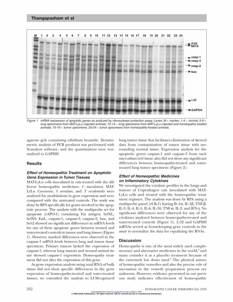
Effect of Homeopathic Treatment on ApoptoticGene Expression in Tumor TissuesMAT-LyLu cells inoculated in rats treated with the dif-ferent homeopathic medicines, C maculatum, MAT-LyLu Carcinosin, S serrulata, and T occidentalis wereanalyzed for modulations in gene expression and werecompared with the untreated controls. The study wasdone by RPA specifically for genes involved in the apop-totic process. The analysis with the multiprobe set forapoptosis (rAPO-1) containing Fas antigen, bclXL,bclXS, FasL, caspase-1, caspase-3, caspase-2, bax, andbcl-2 showed no significant differences in mRNA levelsfor any of these apoptotic genes between treated andwater-treated controls in tumor and lung tissues (Figure1). However, marked differences were observed in thecaspase-1 mRNA levels between lung and tumor tissuespecimens. Primary tumors lacked the expression ofcaspase-1, whereas lung tumors and normal animal tis-sue showed caspase-1 expression. Homeopathy treat-ment did not alter the expression of this gene.
As gene expression analysis using total RNA of bulktissue did not show specific differences in the geneexpression of homeopathy-treated and water-treatedtissues, we extended the analysis to LCM-captured
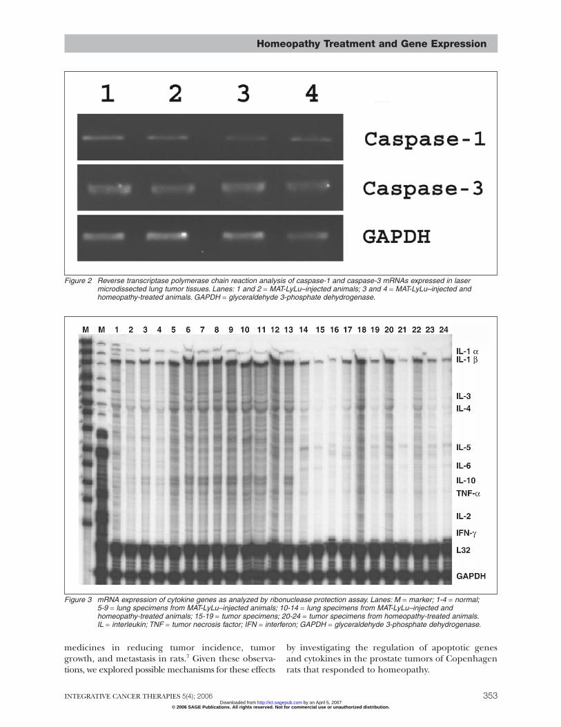
lung tumor tissue that facilitates elimination of skeweddata from contamination of tumor tissue with sur-rounding normal tissue. Expression analysis for theapoptotic genes caspase-1 and caspase-3 from suchmicrodissected tissue also did not show any significantdifferences between homeopathy-treated and water-treated lung tumor specimens (Figure 2).
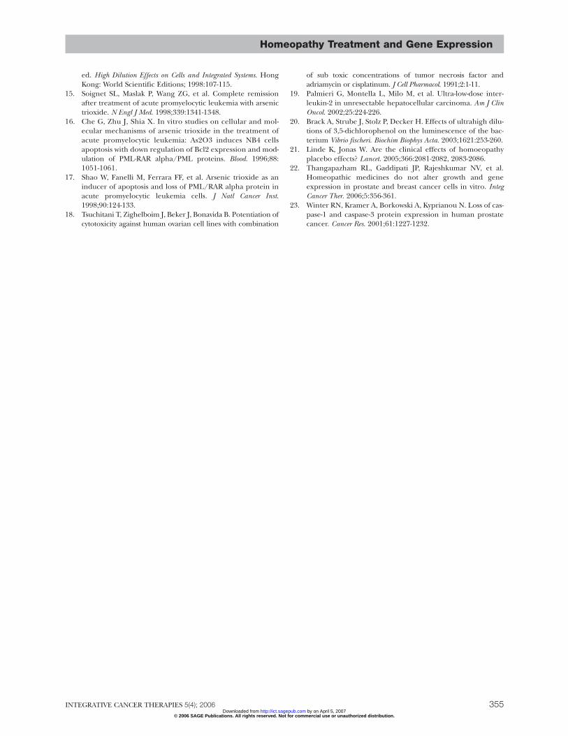
Effect of Homeopathic Medicineson Inflammatory CytokinesWe investigated the cytokine profiles in the lungs andtumors of Copenhagen rats inoculated with MAT-LyLu cells and treated with the homeopathic treat-ment regimen. The analysis was done by RPA using amultiprobe panel (rCK-1) having IL-1α, IL-1β, TNF-β,IL-3, IL-4, IL-5, IL-6, IL-10, TNF-α, IL-2, and IFN-γ. Nosignificant differences were observed for any of thecytokines analyzed between homeopathy-treated andwater-treated controls (Figure 3). L-32 and GAPDHmRNAs served as housekeeping gene controls in theassay to normalize the data for equalizing the RNAs.
DiscussionHomeopathy is one of the most widely used comple-mentary and alternative medicines in the world,8 andmany consider it as a placebo treatment because ofthe extremely low doses used.9 The physical natureof homeopathic remedies and also the precise role ofsuccussion in the remedy preparation process areunknown. However, evidence presented in our previ-ous study indicates effectiveness of homeopathic
Figure 1 mRNA expression of apoptotic genes as analyzed by ribonuclease protection assay. Lanes: M = marker; 1-4 = normal; 5-9 =lung specimens from MAT-LyLu–injected animals; 10-14 = lung specimens from MAT-LyLu–injected and homeopathy-treatedanimals; 15-19 = tumor specimens; 20-24 = tumor specimens from homeopathy-treated animals.
© 2006 SAGE Publications. All rights reserved. Not for commercial use or unauthorized distribution. by on April 5, 2007 http://ict.sagepub.comDownloaded from
Homeopathy Treatment and Gene Expression
INTEGRATIVE CANCER THERAPIES 5(4); 2006 353
medicines in reducing tumor incidence, tumorgrowth, and metastasis in rats.7 Given these observa-tions, we explored possible mechanisms for these effects
by investigating the regulation of apoptotic genesand cytokines in the prostate tumors of Copenhagenrats that responded to homeopathy.
Figure 2 Reverse transcriptase polymerase chain reaction analysis of caspase-1 and caspase-3 mRNAs expressed in lasermicrodissected lung tumor tissues. Lanes: 1 and 2 = MAT-LyLu–injected animals; 3 and 4 = MAT-LyLu–injected andhomeopathy-treated animals. GAPDH = glyceraldehyde 3-phosphate dehydrogenase.
Figure 3 mRNA expression of cytokine genes as analyzed by ribonuclease protection assay. Lanes: M = marker; 1-4 = normal;5-9 = lung specimens from MAT-LyLu–injected animals; 10-14 = lung specimens from MAT-LyLu–injected andhomeopathy-treated animals; 15-19 = tumor specimens; 20-24 = tumor specimens from homeopathy-treated animals.IL = interleukin; TNF = tumor necrosis factor; IFN = interferon; GAPDH = glyceraldehyde 3-phosphate dehydrogenase.
© 2006 SAGE Publications. All rights reserved. Not for commercial use or unauthorized distribution. by on April 5, 2007 http://ict.sagepub.comDownloaded from

Studies from several independent laboratories havealso indicated enhanced cellular adaptive process andanticancer effects with low doses of chemicals includ-ing some homeopathy medicines.10-12 Similarly, ultralowdoses of cadmium have been shown to induce theexpression of protective proteins13 and render the pro-tection of prostate cells from neoplastic transforma-tion to higher dose exposures.14 Arsenic trioxide hasbeen widely used in homeopathic medicine and hasbeen shown to induce incomplete differentiation,apoptosis, and degradation of oncogenic protein inacute promyeloycytic leukemia.15-17 Ultralow doses ofTNF and adriamycin/cisplatinum have been reportedto induce apoptosis in resistant human ovarian cancercells.18 Immunotherapy of hepatocellular carcinomapatients using an ultralow dose of IL-2 (1 MIU/d)induced tumor regression and prolonged the patients’survival.19 Recently, the toxicity of ultrahigh dilutionsof 3,5-dichlorophenol showed a significant inhibitoryeffect from these preparations using luminescent bac-teria testing.20 Analyses of reports from clinical trials ofhomeopathy have been mixed.9,21 While these studiesaddress the clinical effects of homeopathic medicines,they do not address mechanisms of action.
Treatment with high dilutions in our study did notshow any significant effect on the expression of genesanalyzed. In a separate study, treatment of human aswell as rat prostate cells with highly diluted homeo-pathic medicines also did not show any alterations intheir mRNA expression.22
During prostate tumorigenesis, losses in the proteinexpression of apoptotic genes such as caspase-1 andcaspase-3 have been reported.23 In the same study, RT-PCR analysis of mRNA expression did not showreduced expression in all the specimens analyzed, sug-gesting posttranscriptional deregulation. However, inour study, we found a reduction in the mRNA expres-sion of caspase-1 only in all primary tumor tissues andnot in lung tissues. The discrepancy in the expressionbetween primary tumors and lung tumors may indi-cate tissue-specific differences and probably differentmechanisms involved in the caspase-1 regulation. Also,homeopathic medicines did not alter caspase-1 expres-sion in either of these tissues.
ConclusionsIn conclusion, our experiments show that the inves-tigation of specific gene expression analysis for apoptosis and cytokine alteration may not providesufficient insights into the mechanisms of homeo-pathic treatment of prostate cancer. Investigation ofthe homeopathic treatment of cancer may requirenovel and sensitive methodologies such as globalgene array analysis to detect subtle differences to
account for the beneficial effects of homeopathictreatment in vivo. We are planning to study thechanges in protein levels using proteomics in thefuture.
AcknowledgmentsThis work was supported by grant G174LV fromthe Samueli Institute for Information Biology. Theopinions or assertions contained herein are the pri-vate views of the authors and should not be con-strued as official or necessarily reflecting the views ofthe Uniformed Services University of the HealthSciences or the Department of Defense. The authorsare thankful for the excellent technical assistance ofBrant Freed and to Cindy Crawford for manuscriptpreparation.
Thangapazham et al
354© 2006 SAGE Publications. All rights reserved. Not
http://ict.sagDownloaded from
References1. McCloskey D, Armstrong D, Jackisch C, Davidson N.
Programmed cell death in human breast cancer cells. RecentProg Horm Res. 1996;51:493-508.
2. Tang DG, Porter AT. Target to apoptosis: a hopeful weaponfor prostate cancer. Prostate. 1997;32:284-293.
3. Boedefeld WM II, Bland KI, Heslin MJ. Recent insights intoangiogenesis, apoptosis, invasion, and metastasis in colorectalcarcinoma. Ann Surg Oncol. 2003;10:839-851.
4. Johnson MI, Hamdy FC. Apoptosis regulating genes inprostate cancer. Oncol Rep. 1998;5:553-557.
5. Jonas WB, Gaddipati JP, Rajeshkumar NV, et al. In vitro andin vivo assessment of homeopathic treatment for prostatecancer. Paper presented at: Society of Integrative OncologyFirst International Conference; November 18, 2004; NewYork, NY.
6. Heine H, Schmolz M. Immunoregulation via “bystander sup-pression” needs minute amounts of substances—a basis forhomeopathic therapy? Med Hypotheses. 2000;54:392-393.
7. Jonas WB, Gaddipati JP, Rajeshkumar NV, et al. Can homeo-pathic treatment slow prostate cancer growth? Integ CancerTher. 2006;5:343-349.
8. Jonas WB. The homeopathy debate. J Altern Complement Med.2000;6:213-215.
9. Shang A, Huwiler-Muntener K, Nartey L, et al. Are the clini-cal effects of homoeopathy placebo effects? Comparativestudy of placebo-controlled trials of homoeopathy and allopa-thy. Lancet. 2005;366:726-732.
10. Cambar J, Delbancut A, Barrouillet M. Effects of metal dilu-tions on cells and integrated systems. In: Taddei-Ferretti C,ed. High Dilution Effects on Cells and Integrated Systems. HongKong: World Scientific Editions; 1998:45-62.
11. Wiegant F, van Rijn J, van Wijk R. Enhancement of the stressresponse by minute amounts of cadmium in sensitizedReuber H35 hepatoma cells. Toxicology. 1997;116:27-37.
12. Linde K, Jonas W B, Melchart D, Worku F, Wagner H, Eitel F.Critical review and meta-analysis of serial agitated dilutions inexperimental toxicology. Hum Exp Toxicol. 1994;13:481-492.
13. Goering P, Waalkes M, Klaassen C. Cadmium toxicity. In: GoyerR, Cherian M, eds. Handbook of Experimental Pharmacology;Toxicology of Metals, Biochemical Effects. New York, NY: Springer-Verlag; 1994.
14. Cambar J, Delbancut A, Barrouillet M. Effects of cadmium veryhigh dilutions in renal tubular cell cultures. In: Taddei-Ferretti C,
INTEGRATIVE CANCER THERAPIES 5(4); 2006 for commercial use or unauthorized distribution.
by on April 5, 2007 epub.com
Homeopathy Treatment and Gene Expression
ed. High Dilution Effects on Cells and Integrated Systems. HongKong: World Scientific Editions; 1998:107-115.
15. Soignet SL, Maslak P, Wang ZG, et al. Complete remissionafter treatment of acute promyelocytic leukemia with arsenictrioxide. N Engl J Med. 1998;339:1341-1348.
16. Che G, Zhu J, Shia X. In vitro studies on cellular and mol-ecular mechanisms of arsenic trioxide in the treatment ofacute promyelocytic leukemia: As2O3 induces NB4 cellsapoptosis with down regulation of Bcl2 expression and mod-ulation of PML-RAR alpha/PML proteins. Blood. 1996;88:1051-1061.
17. Shao W, Fanelli M, Ferrara FF, et al. Arsenic trioxide as aninducer of apoptosis and loss of PML/RAR alpha protein inacute promyelocytic leukemia cells. J Natl Cancer Inst.1998;90:124-133.
18. Tsuchitani T, Zighelboim J, Beker J, Bonavida B. Potentiation ofcytotoxicity against human ovarian cell lines with combination
INTEGRATIVE CANCER THERAPIES 5(4); 2006© 2006 SAGE Publications. All rights reserved. No
http://ict.saDownloaded from
of sub toxic concentrations of tumor necrosis factor andadriamycin or cisplatinum. J Cell Pharmacol. 1991;2:1-11.
19. Palmieri G, Montella L, Milo M, et al. Ultra-low-dose inter-leukin-2 in unresectable hepatocellular carcinoma. Am J ClinOncol. 2002;25:224-226.
20. Brack A, Strube J, Stolz P, Decker H. Effects of ultrahigh dilu-tions of 3,5-dichlorophenol on the luminescence of the bac-terium Vibrio fischeri. Biochim Biophys Acta. 2003;1621:253-260.
21. Linde K, Jonas W. Are the clinical effects of homoeopathyplacebo effects? Lancet. 2005;366:2081-2082, 2083-2086.
22. Thangapazham RL, Gaddipati JP, Rajeshkumar NV, et al.Homeopathic medicines do not alter growth and geneexpression in prostate and breast cancer cells in vitro. IntegCancer Ther. 2006;5:356-361.
23. Winter RN, Kramer A, Borkowski A, Kyprianou N. Loss of cas-pase-1 and caspase-3 protein expression in human prostatecancer. Cancer Res. 2001;61:1227-1232.
355 t for commercial use or unauthorized distribution.
by on April 5, 2007 gepub.com
Copyright © 2022 FDOKUMEN