This Bacterial Adhesion to Host Tissues
345
-
Upload
independent -
Category
Documents
-
view
3 -
download
0
Transcript of This Bacterial Adhesion to Host Tissues

This book is about the adhesion of bacteria to their human hosts. Althoughadhesion is essential for maintaining members of the normal microflorain/on their host, it is also the crucial first stage in any infectious disease. Itis important, therefore, to fully understand the mechanisms underlying bac-terial adhesion so that we may be able to develop methods of maintainingour normal (protective) microflora and of preventing pathogenic bacteriafrom initiating an infectious process. These topics are increasingly impor-tant because of the growing prevalence of antibiotic-resistant bacteria and,consequently, the need to develop alternative approaches for the preventionand treatment of infectious diseases. This book describes the bacterial struc-tures responsible for adhesion and the molecular mechanisms underlyingthe adhesion process. A unique feature is that it also deals with the conse-quences of adhesion for both the adherent bacterium and the host cell/tissue to which is has adhered. Researchers and graduate students in micro-biology and molecular medicine will find this book to be a valuable overviewof current research on this exciting and rapidly developing topic.
MICHAEL WILSON is Professor of Microbiology in the Faculty of ClinicalSciences, University College London, and Head of the Departmentof Microbiology at the Eastman Dental Institute, University College London.He is the co-editor of Community Structure and Co-operation in Biofilms(2000) and co-author of Bacterial Disease Mechanisms: An Introduction toCellular Microbiology (2002).
Bacterial Adhesion to Host Tissues Mechanisms and Consequences

Over the past decade, the rapid development of an array of techniques in thefields of cellular and molecular biology have transformed whole areas ofresearch across the biological sciences. Microbiology has perhaps been influ-enced most of all. Our understanding of microbial diversity and evolution-ary biology, and of how pathogenic bacteria and viruses interact with theiranimal and plant hosts at the molecular level, for example, have been revo-lutionized. Perhaps the most exciting recent advance in microbiology hasbeen the development of the interface discipline of Cellular Microbiology, afusion of classical microbiology, microbial molecular biology and eukaryoticcellular and molecular biology. Cellular Microbiology is revealing how path-ogenic bacteria interact with host cells in what is turning out to be a complexevolutionary battle of competing gene products. Molecular and cellularbiology are no longer discrete subject areas but vital tools and an integratedpart of current microbiological research. As part of this revolution in molec-ular biology, the genomes of a growing number of pathogenic and model bac-teria have been fully sequenced, with immense implications for our futureunderstanding of microorganisms at the molecular level.
Advances in Molecular and Cellular Microbiology is a series edited byresearchers active in these exciting and rapidly expanding fields. Each volumewill focus on a particular aspect of cellular or molecular microbiology, and willprovide an overview of the area, as well as examining current research. Thisseries will enable graduate students and researchers to keep up with therapidly diversifying literature in current microbiological research.
AMCM
ADVA
NCES
IN M
OLEC
ULAR
AND
CELL
ULAR
MIC
ROBI
OLOG
Y
Series Editors
Professor Brian HendersonUniversity College London
Professor Michael WilsonUniversity College London
Professor Sir Anthony CoatesSt George’s Hospital Medical School, London
Professor Michael CurtisSt Bartholemew’s and Royal London Hospital, London

Advances in Molecular and Cellular Microbiology 1
Bacterial Adhesion to Host TissuesMechanisms and Consequences
EDITED BY
Michael WilsonUniversity College London

Cambridge, New York, Melbourne, Madrid, Cape Town, Singapore, São Paulo
Cambridge University PressThe Edinburgh Building, Cambridge , United Kingdom
First published in print format
isbn-13 978-0-521-80107-2 hardback
isbn-13 978-0-511-06331-2 eBook (NetLibrary)
© Cambridge University Press 2002
2002
Information on this title: www.cambridge.org/9780521801072
This book is in copyright. Subject to statutory exception and to the provision ofrelevant collective licensing agreements, no reproduction of any part may take placewithout the written permission of Cambridge University Press.
isbn-10 0-511-06331-8 eBook (NetLibrary)
isbn-10 0-521-80107-9 hardback
Cambridge University Press has no responsibility for the persistence or accuracy ofs for external or third-party internet websites referred to in this book, and does notguarantee that any content on such websites is, or will remain, accurate or appropriate.
Published in the United States of America by Cambridge University Press, New York
www.cambridge.org
-
-
-
-

v
Contents
List of contributors vii
Preface xv
Part I Bacterial adhesins and adhesive structures 1
1 Surface protein adhesins of staphylococci 3
Timothy J. Foster
2 Mechanisms of utilization of host signalling molecules by
respiratory mucosal pathogens 27
Mumtaz Virji
3 Adhesive surface structures of oral streptococci 59
Roderick McNab, Pauline S. Handley and Howard F. Jenkinson
4 Regulation and function of phase variation in Escherichia coli 89
Ian Blomfield and Marjan van der Woude
5 Regulation of capsule expression 115
Clare Taylor and Ian S. Roberts
6 Role of pili in Haemophilus influenzae adherence, colonization
and disease 139
Janet R. Gilsdorf
v

Part II Effect of adhesion on bacterial structure and function 163
7 Transcriptional regulation of meningococcal gene expression upon
adhesion to target cells 165
Muhamed-Kheir Taha
8 Induction of protein secretion by Yersinia enterocolitica through
contact with eukaryotic cells 183
Dorothy E. Pierson
9 Functional modulation of pathogenic bacteria upon contact with
host target cells 203
Andreas U. Kresse, Frank Ebel and Carlos A. Guzmán
Part III Consequences of bacterial adhesion for the host 221
10 Adhesion, signal transduction and mucosal inflammation 223
Catharina Svanborg, Goran Bergsten, Hans Fischer, Bjorn Frendéus, Gabriela
Godaly, Erika Gustafsson, Long Hang, Maria Hedlund, Ann-Charlotte Lundstedt,
Martin Samuelsson, Patrik Samuelsson, Majlis Svensson and Bjorn Wullt
11 Adhesion of oral spirochaetes to host cells and its cytopathogenic
consequences 247
Richard P. Ellen
12 Interactions between enteropathogenic Escherichia coli and
epithelial cells 277
Elizabeth L. Hartland, Gad Frankel and Stuart Knutton
13 Host cell responses to Porphyromonas gingivalis and
Actinobacillus actinomycetemcomitans 299
Richard J. Lamont
Index 323
Plate section is between pp. 240 and 241.
con
ten
ts
vi

vii
Contributors
Goran BergstenDepartment of Microbiology, Immunology and Glycobiology (MIG)Institute of Laboratory MedicineLund UniversitySölvegatan 23S-223 62 LundSweden
Ian BlomfieldDepartment of BiosciencesUniversity of Kent at CanterburyCanterburyKent CT2 7NJUK
Frank EbelUnité de Génétique MoléculaireInstitut Pasteur25 rue du Dr. Roux75724 ParisFrance
Richard P. EllenFaculty of DentistryUniversity of Toronto124 Edward StreetTorontoOntarioCanada M5G 1G6
vii

con
trib
uto
rs
viii
Hans FischerDepartment of Microbiology, Immunology and Glycobiology (MIG)Institute of Laboratory MedicineLund UniversitySölvegatan 23S-223 62 LundSweden
Timothy J. FosterDepartment of MicrobiologyMoyne Institute of Preventive MedicineTrinity CollegeDublin 2Ireland
Gad FrankelDepartment of Biochemistry and Centre for Molecular Microbiology & InfectionImperial College of Science Technology and MedicineLondon SW7 2AZUK
Bjorn FrendéusDepartment of Microbiology, Immunology and Glycobiology (MIG)Institute of Laboratory MedicineLund UniversitySölvegatan 23S-223 62 LundSweden
Janet R. GilsdorfDepartment of Pediatrics and Communicable DiseasesUniversity of Michigan Medical SchoolAnn Arbor, MI 48109USA
Gabriela GodalyDepartment of Microbiology, Immunology and Glycobiology (MIG)Institute of Laboratory MedicineLund UniversitySölvegatan 23S-223 62 LundSweden
viii

con
tributo
rs
ix
Erika GustafssonDepartment of Microbiology, Immunology and Glycobiology (MIG)Institute of Laboratory MedicineLund UniversitySölvegatan 23S-223 62 LundSweden
Carlos A. GuzmánVaccine Research GroupDivision of MicrobiologyGBF-German Research Centre for BiotechnologyMascheroder Weg. 1D-38124 BraunschweigGermany
Pauline S. Handley1.800 Stopford BuildingSchool of Biological SciencesUniversity of ManchesterOxford RoadManchester M13 9PTUK
Long HangDepartment of Microbiology, Immunology and Glycobiology (MIG)Institute of Laboratory MedicineLund UniversitySölvegatan 23S-223 62 LundSweden
Elizabeth L. HartlandDepartment of MicrobiologyMonash UniversityClayton 3800VictoriaAustralia
ix

con
trib
uto
rs
x
Maria HedlundDepartment of Microbiology, Immunology and Glycobiology (MIG)Institute of Laboratory MedicineLund UniversitySölvegatan 23S-223 62 LundSweden
Howard F. JenkinsonDepartment of Oral and Dental ScienceUniversity of Bristol Dental SchoolBristol BS1 2LYUK
Stuart KnuttonInstitute of Child HealthUniversity of BirminghamBirmingham B4 6NHUK
Andreas U. KresseVaccine Research GroupDivision of MicrobiologyGBF-German Research Centre for BiotechnologyMascheroder Weg. 1D-38124 BraunschweigGermany
Richard J. LamontDepartment of Oral BiologyUniversity of WashingtonSeattle, WA 91895-7132USA
Ann-Charlotte LundstedtDepartment of Microbiology, Immunology and Glycobiology (MIG)Institute of Laboratory MedicineLund UniversitySölvegatan 23S-223 62 LundSweden

con
tributo
rs
xi
Roderick McNabDepartment of MicrobiologyEastman Dental Institute256 Grays Inn RoadUniversity College LondonLondon WC1X 8LDUK
Dorothy E. PiersonDepartment of Microbiology and ImmunologyC5181 Veterinary Medical CenterCornell University College of Veterinary MedicineIthaca, NY 14853USA
Ian S. Roberts1.800 Stopford BuildingSchool of Biological SciencesUniversity of ManchesterOxford RoadManchester M13 9PTUK
Martin SamuelssonDepartment of Microbiology, Immunology and Glycobiology (MIG)Institute of Laboratory MedicineLund UniversitySölvegatan 23S-223 62 LundSweden
Patrik SamuelssonDepartment of Microbiology, Immunology and Glycobiology (MIG)Institute of Laboratory MedicineLund UniversitySölvegatan 23S-223 62 LundSweden

con
trib
uto
rs
xii
Catharina SvanborgDepartment of Microbiology, Immunology and Glycobiology (MIG)Institute of Laboratory MedicineLund UniversitySölvegatan 23S-223 62 LundSweden
Majlis SvenssonDepartment of Microbiology, Immunology and Glycobiology (MIG)Institute of Laboratory MedicineLund UniversitySölvegatan 23S-223 62 LundSweden
Muhamed-Kheir TahaUnité des Neisseria and Centre National de Référence des Méningocoques(CNRM)Institut Pasteur28 rue du Dr Roux75724 Paris cedex 15France
Clare Taylor1.800 Stopford BuildingSchool of Biological SciencesUniversity of ManchesterOxford RoadManchester M13 9PTUK
Marjan van der WoudeDepartment of MicrobiologyUniversity of Pennsylvania202A Johnson Pavilion3610Hamilton WalkPhiladelphia, PA 19104USA

Mumtaz VirjiDepartment of Pathology and MicrobiologySchool of Medical SciencesUniversity of BristolBristol BS8 1TDUK
Bjorn WulltDepartment of Microbiology, Immunology and Glycobiology (MIG)Institute of Laboratory MedicineLund UniversitySölvegatan 23S-223 62 LundSweden co
ntribu
tors
xiii


Preface
Except when in utero, every human being is colonized by approximately 1014
microbes (mainly bacteria) that constitute the normal microflora. Apartfrom those organisms that are present in the lumen of the intestinal tract,all of these microbes maintain an association with their host by adheringto some cell, tissue or secretion. It is increasingly being realized that notonly is this normal microflora beneficial to its host (e.g. by providing vita-mins and protection from exogenous pathogens) but it is, indeed, essentialfor the host’s proper development, for example in the differentiation andmaturation of the intestinal tract, immune system, etc. A knowledge of howmembers of the normal microflora adhere to their host is, therefore, impor-tant in understanding the mutualistic association we know as Homosapiens.
Adhesion of a pathogenic organism to its host is also the first stage inany infectious disease and this truism provides another justification forstudying bacterial adhesion. Interest in this aspect of bacterial virulence isincreasing rapidly as our armamentarium of antibiotics dwindles in effec-tiveness owing to the development of resistance in major pathogens. It isbelieved (and strongly hoped) that research into bacterial adhesion mecha-nisms will identify new targets for therapeutic intervention.
This book is intended to update the reader in key areas of research in thefield of bacterial adhesion to host cells and tissues. It is divided into threeparts that deal with: (I) the mechanisms underlying the adhesion of bacteriato host structures, (II) the effect that adhesion to host cells has on bacteriaand (III) the consequences for the host cell (or tissue) of bacterial adhesion.The first part describes recent advances in our understanding of the adhe-sive structures and adhesins found in bacteria colonizing (and causingdisease in) a variety of habitats in a human being – the oral cavity, respiratory
xv

tract, gut, urinary tract, skin and internal tissues. The remaining two partsconcentrate on less well understood aspects of adhesion – the effects that thisprocess has on the adherent organism and on the cell/tissue to which it hasadhered. While we know very little about the former, our knowledge of whathappens to host cells following bacterial adhesion is increasing at a rapidrate. With regard to the latter, one frequent consequence of bacterial adhe-sion to a host cell is invasion of that cell. Only one chapter dealing with thistopic has been included (this focuses on organisms other than the classicalinvasive pathogens) as this huge subject will be covered in a later volume inthis series.
Michael Wilson
pref
ace
xvi

PART I Bacterial adhesins and adhesivestructures


CHAPTER 1
Surface protein adhesins of staphylococci
Timothy J. Foster
1.1 INTRODUCTION
Staphylococcus aureus is primarily an extracellular pathogen. In order toinitiate infection it adheres to components of the host extracellular matrix(ECM). Adherence is mediated by surface protein adhesins called MSCRAMMs(microbial surface components recognizing adhesive matrix molecules)(Patti et al., 1994a). In most cases the MSCRAMMs are covalently bound topeptidoglycan in the cell wall. However, there are several examples ofMSCRAMMs that are non-covalently associated with the wall. Coagulase-negative staphylococci also express MSCRAMMs. This chapter will discussthe mechanisms of attachment of proteins to the cell wall and will review theproperties of MSCRAMM proteins that have been characterized at themolecular level.
1.2 ANCHORING OF PROTEINS TO THE CELL WALL
Cell-wall-anchored proteins that are covalently bound to peptidoglycanare recognizable by a motif located at the C-terminus (Navarre andSchneewind, 1999). This comprises the sequence LPXTG (Leu-Pro-X-Thr-Gly) followed by hydrophobic residues that span the cytoplasmic membraneand by several positively charged residues. The positively charged residuesare required to hold the protein transiently in the membrane during secre-tion through the Sec secretome (Schneewind et al., 1993). The LPXTGsequence is recognized by an enzyme called sortase that cleaves LPXTGbetween the Thr and Gly residues (Navarre and Schneewind, 1994; Ton-Thatet al., 1999). The carboxyl group of the Thr is joined to the amino group ofthe branch peptide in nascent peptidoglycan. In the case of S. aureus, linkageoccurs to the NH
2group of the fifth Gly residue of the branch peptide, which
would otherwise form the interpeptide bridge of cross-linked peptidoglycan
3

(Ton-That and Schneewind, 1999; Ton-That et al., 1999). The wall-anchoredprotein becomes joined to the lipid-linked intermediate prior to its incorpo-ration into peptidoglycan (Ton-That et al., 1997). The covalently linkedprotein can be released from the cell only by enzymatic degradation of pep-tidoglycan. The glycine endopeptidase lysostaphin releases proteins ofhomogeneous size whereas muramidases release proteins of heterogeneoussize owing to varying amounts of peptidoglycan attached to the C-terminus(Schneewind et al., 1993).
The enzyme that catalyses the sorting reaction is sortase. The protein hasan N-terminal hydrophobic domain that provides anchorage to the outer faceof the cytoplasmic membrane (Mazmanian et al., 1999; Ton-That et al., 1999).Indeed it is likely that sortase is closely associated with the secretome becauseit must capture proteins as they are in the process of being secreted throughthe Sec pathway. Interestingly, a sortase-defective mutant can grow normallyin vitro, which shows that sortase is not an essential enzyme. The mutant isdefective in the expression of several LPXTG-anchored surface proteins andhas reduced virulence in murine infection models (Mazmanian et al., 2000).This indicates that sortase could be a novel target for antimicrobial agents.
1.3 CELL-WALL-ASSOCIATED PROTEINS
1.3.1 Protein A
Protein A (Spa) is the archetypal cell-wall-anchored protein of S. aureus.It is known primarily for its ability to bind the Fc region of immunoglobulin(Ig) G. Its structural organization is somewhat di¤erent from that of othersurface proteins in that the N-terminal signal sequence is followed bytandem repeats of five homologous IgG binding domains (Fig. 1.1; Uhlén etal., 1984). Each is composed of an approximately 60 amino acid residue unitthat forms three �-helices (Starovasnik et al., 1996). The structure of the sub-domain B in complex with the Fc region of IgG subclass 1 has been solvedby X-ray analysis of a co-crystal (Deisenhofer, 1981). The binding betweenthe two molecules involves nine amino acid residues in the IgG fragmentand 11 amino acid residues in the protein A domain (Gouda et al., 1998). Thebinding characteristics and specificity of the Spa–IgG interaction have beenanalysed in great detail (Langone, 1982).
One important role of Spa in staphylococcal infections is that it is anti-phagocytic. By binding to protein A on the bacterial surface, the Fc region ofIgG is not available for recognition by the Fc receptor on polymorphonuclearleukocytes (PMNLs) (Gemmell et al., 1990). A protein A-defective mutant
t. j
. fo
ster
4

was more avidly phagocytosed by PMNLs in the presence of normal serumopsonins than was the wild type, and the mutant was less virulent in murineinfection models (Patel et al., 1987). The recent observation that Spa canmediate adherence of bacteria to von Willebrand factor, an extracellularmatrix protein important in normal haemostasis, suggests that protein Amay have an additional role in the infection process (Hartlieb et al., 2000).
1.3.2 Fibronectin-binding proteins
Fibronectin (Fn) is a dimeric glycoprotein that occurs in a soluble formin body fluids and in a fibrillar form in the ECM (Hynes, 1993). A primaryfunction of insoluble Fn is to act as a substratum for the adhesion of cellsmediated by integrin receptors that bind to specific sites in the central partof Fn (Yamada, 1989). The primary binding site for staphylococcal Fn-binding protein is in the 29kDa N-terminal domain, which is composed offive type I modules (Sottile et al., 1991; Potts and Campbell, 1994).
Most strains of S. aureus express two related Fn-binding proteins FnBPAand FnBPB, which are encoded by closely linked genes (Signás et al., 1989;Jönsson et al., 1991). One survey of 163 isolates comprising carriage strains,as well as strains from invasive disease and orthopaedic device-related infec-tion, found that 77% had both fnbA and fnbB genes, while 23% had only fnbA
surface pro
tein ad
hesin
s of staph
loco
cci
5
S Xr
S A R
S A B B B
S A BB C W
W
W
FnBPA
CNA
ClfA
SpaXc
DDDD
R
E D A B C
Figure 1.1. Organization of surface proteins of S. aureus. The domain organization of the
fibronectin-binding protein A (FnBPA), the collagen-binding protein CNA, the
fibrinogen-binding protein (ClfA) and protein A (Spa). The signal sequences (S) are
removed during secretion across the cytoplasmic membrane. Region Xr of protein A is a
proline-rich octapeptide repeat that spans the cell wall. Region Xc is a non-repeated
(constant) region. Each protein has common features at the C-terminus indicated by the
cross-hatched box (LPXTG motif, hydrophobic region, and positively charged residues).
Regions W and R are peptidoglycan-spanning regions. (From Foster and Höök, 2000;
with permission from American Society for Microbiology Press.)

(Peacock et al., 2000). Strains from orthopaedic infections adhered to Fn at asignificantly higher level than did carriage strains or strains from non-device-related infections.
FnBPA and FnBPB have a structural organization similar to that ofFnBPs from streptococci (Fig. 1.1; Joh et al., 1994; Patti et al., 1994a; Fosterand Höök, 1998). The primary ligand-binding domain (D), which is almostidentical in FnBPA and FnBPB, is located very close to the cell-wall-spanningdomains (region W) and is composed of three to five repeats of an approxi-mately 40 residue motif. Synthetic peptides mimicking repeated units e¤ec-tively inhibit Fn binding to bacteria and bacterial attachment to immobilizedFn (Raja et al., 1990).
Studies with peptides and recombinant proteins expressing combina-tions of di¤erent FnBP D repeats indicate that the major interaction occursbetween FnBP repeat D3 and Fn modules 4 and 5, but that other discretesequences within the D region bind to di¤erent Fn type I module pairs (Johet al., 1998). The ligand-binding domain of FnBP reacts simultaneously atmultiple sites with Fn (Fig. 1.2). A consensus Fn-binding motif is presentwith each D repeat (McGavin et al., 1991, 1993). The interaction between theMSCRAMM and Fn involves structural rearrangements in the D repeatregion. The ligand-binding D repeat region has an unordered structure andacquires a defined conformation upon binding to the rigid type I modules ofFn (House-Pompeo et al., 1996). This conformational change is accompa-nied by the formation of neo-epitopes called ligand-induced binding site(LIBS) epitopes that can be demonstrated by monoclonal antibodies and byantibodies isolated from patients recovering from staphylococcal infection(Speziale et al., 1996). The antibodies that recognize the neo-epitopes do notinterfere with the MSCRAMM–Fn interaction but rather stabilize theFnBP–ligand complex and appear to promote Fn binding. The immunodom-inant non-LIBS epitopes in D1–D3 are confined to repeats D1 and D2 (Sunet al., 1997). They are very similar to each other and bind Fn with lower aªn-ity than does D3. The region in D3 corresponding to epitopes in D1–D2 con-tains the Fn-binding consensus but is otherwise divergent (8/21 di¤erentresidues). Antibodies in polyclonal sera preferentially react with the lowaªnity D1–D2 domains and block Fn binding to bacteria by no more than50%.
Staphylococcus aureus can invade cultured fibroblasts, endothelial andepithelial cells, and Fn plays a major role (Dziewanowska et al., 1999;Lammers et al., 1999; Peacock et al., 1999a; Sinha et al., 1999; Fowler et al.,2000). Bacteria either recruit soluble Fn or bind to Fn bound to the surfaceof host cells. Bacteria bind Fn via the type I modules at the N-terminus. Fn
t. j
. fo
ster
6

is bound to the �5�1
integrin on the surface of the host cell at the central FnRGD (Arg-Gly-Asp) motif-bearing module. Thus Fn forms a bridge betweenthe bacterial FnBP adhesin and the mammalian cell integrin (Fig. 1.3). Thisresults in stimulation of phagocytosis and bacteria become internalized.FnBP-defective mutants of S. aureus are not taken up, non-invasive bacteriathat acquire FnBP expression become invasive, and bacterial internalizationis blocked by soluble recombinant D repeat regions of FnBP and by anti-inte-grin function-blocking antibodies. The importance of internalization in vivois unclear, but it could be involved in bacterial escape from the bloodstreamand invasion of internal organs, in the initiation of invasive endocarditis, andin bacterial persistence.
FnBPs are considered to be important virulence factors in the initiation
surface pro
tein ad
hesin
s of staph
loco
cci
7
Y
D1-D2-D3
1 2 3 4 5
Y
Fibronectin type I modules
Figure 1.2. Interaction of the ligand-binding region of Fn-binding proteins with Fn. The
wavy line represents the ligand-binding D1–D2–D3 repeats of FnBPs, which do not have
secondary structure. The protein interacts with the type I modules of Fn and takes on a
discernible secondary structure with the formation of neo-epitopes (ligand-induced
binding site epitopes). (From Foster and Höök, 2000; with permission from American
Society for Microbiology Press.)

of foreign body infection. They promote bacterial adherence to immobilizedFn in vitro, and to implanted biomaterial that has been in long-term contactwith the host such as plastic coverslips implanted subcutaneously in guineapigs (Greene et al., 1995) and titanium screws implanted in the iliac bone ofguinea pigs (Fischer et al., 1996). In contrast, fibrinogen appears to be themajor adhesion-promoting factor in the conditioning layer on biomaterialthat had been in short-term contact with the host (Vaudaux et al., 1995;François et al., 2000). There are conflicting data concerning the ability ofFnBPs to promote infection in experimental animals. One study with aFnBP-defective mutant of strain 879 indicated that the bacterial MSCRAMMis important in promoting bacterial adhesion to damaged heart valve tissuein the rat model for endocarditis (Kuypers and Proctor, 1989), while anotherreport with mutants of strain 8325–4 found no e¤ect (Flock et al., 1996).
t. j
. fo
ster
8
Fibronectin
�5�
1- integrin
FnBP
S. aureus
Figure 1.3. Role of Fn in promoting bacterial attachment to mammalian cells. The N-
terminal type I modules of Fn are bound to the D1–D2–D3 region of FnBP attached to
the cell surface of S. aureus. The same molecule of Fn binds to the �5�1
integrin via the
centrally located RGD motif. This stimulates actin rearrangement and bacterial
internalization.

These contradictory data may reflect di¤erent bacterial strains (8325–4 isknown to express FnBPs poorly in vitro) or di¤erences in performing theinfection models.
1.3.3 Fibrinogen-binding proteins
Fibrinogen is a large protein of Mr 340000. It is composed of three poly-peptide chains (�, �, �) that are extensively linked by disulphide bonds toform an elongated dimeric structure (Ruggeri, 1993). It is the most abundantligand for the integrin �IIb/�3 (glycoprotein gpIIb/IIIa) on the surface ofplatelets. The binding of Fg to the integrin receptor on activated plateletsresults in platelet aggregation and the formation of platelet–fibrin thrombi(Hawiger, 1995). The C-terminal sequences of the �-, �- and �-chains formindependently folded globular domains. The three dimensional structure ofthe �-chain module is known (Spraggon et al., 1997; Doolittle et al., 1998)
Until recently it was thought that the ability of S. aureus to adhere to Fg-coated substrates and to form clumps in a solution containing Fg (e.g.plasma) was solely due to the clumping factor ClfA (McDevitt et al., 1994,1995). It is now known that S. aureus can express other Fg-binding adhesins:the ClfB protein, which is related to ClfA (Ní Eidhin et al., 1998); and the Fg-binding proteins, which can also interact with Fg via their A domains (Wannet al., 2000). The clfA and clfB genes are not allelic variants but are distinctgenes. They are not closely linked, in contrast to the fnbA and fnbB genes.
The structural organization of ClfA and ClfB is very similar (Fig. 1.4). Thesurface-exposed approximately 500 residue ligand-binding A domains arelinked to the cell wall via the R domain, which comprises mainly the Ser-Aspdipeptide repeats. The R domain appears to serve as a flexible stalk, allowingthe presentation of the A domain on the surface for ligand interactions(Hartford et al., 1997). FnBPA and FnBPB also possess N-terminal A domainsthat have sequence similarity with the A domains of ClfA and ClfB, and in thecase of FnBPA promote binding to Fg (Wann et al., 2000). Otherwise, theFnBP and Clf proteins have a completely di¤erent structural organization,apart from the typical cell-wall-anchoring signals at the extreme C-terminus.
Although the structural organization of ClfA and ClfB is similar, theamino acid sequences of the ligand-binding A domains are only 27% identi-cal. ClfA and ClfB bind to di¤erent sites in Fg. ClfA recognizes the flexiblepeptide that extends from the �-module at the C-terminus of the �-chain (Fig.1.5; McDevitt et al., 1997) whereas ClfB binds to the �-chain (Ní Eidhin et al.,1998). The A domain of FnBPA is approximately 25% identical with that ofClfA and binds to the same site in the Fg �-chain (Wann et al., 2000).
surface pro
tein ad
hesin
s of staph
loco
cci
9

The ClfA domain recognizes a site located at the extreme C-terminus ofthe �-chain of Fg (residues 399–411). These residues form a flexible exten-sion from the globular �-module (Fig. 1.5; Spraggon et al., 1997: Doolittle etal., 1998). A 17 amino acid residue synthetic peptide corresponding to the Fg�-chain residues 399–411 binds to a recombinant form of the A domain inan interaction that is inhibited by Ca2� (O’Connell et al., 1998). The A domaincontains a motif that is reminiscent of a Ca2�-binding EF-hand, and site-specific mutations in this motif resulted in a protein with lower aªnity forthe �-chain peptide and less sensitivity to Ca2�. Thus ClfA exhibits fibrino-gen-binding characteristics similar to those of the platelet integrin �IIb/�
3
(O’Connell et al., 1998). Both bind to the same site in Fg in interactions thatare a¤ected by Ca2�. The recombinant form of the ClfA A domain is a potentinhibitor of Fg-dependent platelet aggregation (McDevitt et al., 1997). Thiscould be a bacterial defence mechanism to prevent release of antimicrobialpeptides during degranulation (Yeaman et al., 1992) that might occur duringplatelet aggregation in the vicinity of colonizing bacteria.
t. j
. fo
ster
10
S
S
R
R
R
R
R
TYTFTDYVD
ClfA
ClfB
SdrC
SdrE
SdrD
M
M
M
M
M
S
S
S
SdrH
LPXTG
R3R1 A R2 R2 R2 R2 MPls
S RA C
V V
S
Figure 1.4. The Sdr multigene protein family in staphylococci. The domain organization
of di¤erent members of the Sdr protein family is shown. The approximately 500 residue
A domain of ClfA, ClfB, SdrC, SdrD, SdrE, SdrF and SdrG have a conserved sequence
TYTFTDYVD. SdrF and SdrG of S. epidermidis have an organization similar to that of the
SdrC, SdrD and SdrE proteins of S. aureus but are not shown. The A domain of Pls has
no sequence similarity to the A domains of the Clf–Sdr proteins, which are related by
about 25–30%.

The binding of ClfA to fibrinogen is progressively inhibited by Ca2� inthe range 1–10mM (O’Connell et al., 1998). The concentration of free Ca2�
in blood plasma is 1.3mM and is closely regulated at the threshold of theinhibitory range, although concentrations can vary more widely in extracel-lular spaces (Brown et al., 1995). However, at platelet-rich thrombi, and pos-sibly on the surface of freshly implanted biomaterial, the Ca2� concentrationappears to be considerably lower and may allow ClfA to bind fibrinogen.Thus, as bacteria circulate in plasma, they will tend to adhere to Fg/platelet-containing coagulation sites.
The ClfB region A binds to the Fg �-chain (Ní Eidhin et al., 1998) but theprecise binding site has not been defined. ClfB-promoted binding to immo-bilized Fg is also inhibited by millimolar concentrations of Ca2�. The ClfBprotein is expressed maximally during the early part of the exponential phaseof growth (Ní Eidhin et al., 1998; McAleese et al., 2001). Transcription (andhence translation) terminates before the culture reaches stationary phase, soClfB molecules become diluted amongst the progeny cells during theremaining cell divisions and some are released into the culture supernatantby cell wall turnover. Also, some of the ClfB protein is cleaved by the metal-loprotease aureolysin (McAleese et al., 2001). Protease cleavage results inloss of an N-terminal domain and the protein loses its ability to bind Fg. Incontrast, ClfA is expressed abundantly on cells from the stationary phase ofgrowth. It is also cleaved at a site similar to that of ClfA but does not loseligand-binding activity (McDevitt et al., 1995).
The ClfA protein is the primary adhesin of S. aureus for promoting
surface pro
tein ad
hesin
s of staph
loco
cci
11
D
Domain
���� ������������
�������� ClfA����
IIb ����
3
����M
����2
D
Domain
EDomain
Figure 1.5. Structure of fibrinogen. Schematic diagram showing the structural
organization of fibrinogen. The globular D domains comprise the C-terminal residues of
the �-, �- and �-chains. The C-terminus of the �-chain protrudes from the globular
�-module. Binding sites for ClfA and integrins are shown. The E domain contains the N-
terminal residues of �-, �- and �-chains cross-linked by disulphide bridges. (From Foster
and Höök, 2000; with permission from American Society for Microbiology Press.)

bacterial interactions with Fg. This is particularly the case for cells in the sta-tionary phase of growth where ClfB is absent. ClfA promotes bacterial attach-ment to immobilized Fg (McDevitt et al., 1994), to plasma clots formed invitro (Moreillon et al., 1995) and to plastic biomaterial that had been exposedto blood for short-term conditioning (Vaudaux et al., 1995; François et al.,2000). In addition, a ClfA� mutant had reduced ability to bind to damagedheart tissue in the rat endocarditis model (Moreillon et al., 1995). The infec-tion rate was lower in the ClfA� mutant but was restored in the mutant com-plemented with the wild-type clfA gene. However, reduced virulence was notmanifested at higher infection doses, indicating that other adhesins cancompensate for lack of ClfA. Similarly, when exponential phase cells thatlacked ClfA were used, the ClfB protein was shown to act as a bacterialadhesin and virulence factor (Ní Eidhin et al., 1998; Entenza et al., 2000).
ClfA has recently been shown to be an important virulence factor in themouse model of septic arthritis (Josefsson et al., 2001). The ClfA� mutantwas significantly less virulent in the bacteraemia phase of the infection,causing fewer mortalities and less weight loss than did the wild type. Themutant also caused much reduced tissue damage in infected joints as com-pared with wild-type infection. The e¤ect in joints was observed both in micethat were injected intravenously, where the bacteria may have been elimi-nated in the bloodstream by more eªcient phagocytosis, and also when bac-teria were injected directly into the joint. It is possible that reduced virulenceis linked to the observation that ClfA� mutants are more susceptible to pha-gocyotsis by PMNLs.
1.3.4 Proteins of the Sdr family in S. aureus
ClfA and ClfB are members of a larger family of structurally related surfaceproteins characterized by the R domain containing Ser-Asp dipeptide repeats.Strains of S. aureus contain a tandem array of related genes, sdrC, sdrD andsdrE. The proteins are predicted to have a structural organization similar to thatof ClfA and ClfB, except for an additional B repeat comprising 110–113 resi-dues located between the A domain and the R domain (Fig. 1.4; Josefsson et al.,1998a). The function of the B domain is not known. Each repeat has three highaªnity Ca2�-binding sites (Josefsson et al., 1998b). Bound Ca2� is required topromote the rigid rod-like structure of the B repeat array. It is possible that theB region acts as a non-flexible stalk, which, in combination with the more flex-ible R region, is required for surface display of the (putative) ligand-binding Adomain. The A domains of the Sdr proteins are of a size similar to that of theA domains of ClfA and ClfB and have about 30% sequence identity where any
t. j
. fo
ster
12

pairwise combination is compared. However, unlike ClfA and ClfB, the ligandsof SdrC, SdrD and SdrE have not been elucidated, with the exception of avariant of SdrD (Bbp) that binds to bone sialoprotein (Tung et al., 2000). In con-trast, SdrD, the A domain of which is about 75% identical with Bbp, does notbind bone sialoprotein. We have recently shown that SdrE, when expressed onthe surface of Lactococcus lactis, can promote platelet aggregation, most likelymediated by binding to a plasma protein that acts as a bridge between the bac-teria and a platelet receptor (O’Brien et al., 2001).
Some strains of methicillin-resistant S. aureus (MRSA) express a highmolecular weight protein Pls, which masks di¤erent surface proteins suchas Spa, Clf and FbBPs and prevents ligand binding (Hildén et al., 1996;Savolainen et al., 2001). The protein is susceptible to degradation by hostplasmin. It appears that the pls gene is closely linked to the mec genes andmay be part of the mec element in these strains. Protein Pls has a Ser-Aspdipeptide region R but is otherwise quite distinct from the typical ClfA-Sdrproteins. It carries two other repeated regions, R1 at the extreme N-terminusand R2 towards the C-terminus, specifying repeats of 12–14 and 128–129residues, respectively. They flank an approximately 400 residue A domain. Itis not known whether Pls has any ligand-binding activity.
1.3.5 Collagen-binding protein
Some strains of S. aureus express a collagen-binding protein called CNA.The presence of CNA is necessary and suªcient for bacteria to adhere to col-lagenous tissues such as cartilage (Switalski et al., 1993). The collagen-binding activity has been located in a 190 amino acid residue segment withinthe N-terminal A domain. The crystal structure of the subdomain has beensolved at 1.8Å (1 Å�0.1 nm) resolution (Symersky et al., 1997). The poly-peptide folds like a jelly roll in two �-sheets connected by a short �-helix. Atrench traverses one of the �-sheets, and molecular modelling suggests thatthis trench can accommodate a collagen triple helix. The collagen-bindingfunction of the trench was further demonstrated by site-directed mutagene-sis, where changes in single residues forming the walls of the trench resultedin proteins with reduced collagen-binding activity.
Biophysical analysis of the recombinant A domain of CNA suggest thatit is folded into an elongated structure rather than a sphere (Rich et al., 1998).This might suggests that the A domain is a mosaic protein composed ofseveral subdomains that are independently folded and have di¤erent func-tions. The A regions of ClfA and ClfB have similar physical properties (S.Perkins, E. Walsh, T. Foster and M. Höök, unpublished data).
surface pro
tein ad
hesin
s of staph
loco
cci
13

The collagen-binding protein is an important virulence factor in themouse septic arthritis model (Patti et al., 1994b). Fewer mice developedarthritis when injected with the CNA� mutant, as compared with thoseinfected with the wild type. While CNA contributes to the development ofarthritis, other bacterial components can compensate if it is lacking. This isclearly demonstrated in the experiments described above, demonstrating therole of ClfA in the same model. The strain used for those experiments didnot express CNA.
1.3.6 Other LPXTG-anchored proteins
The genome sequence of strain N315 was published recently (Kuroda etal., 2001) and the sequencing of four other strains of S. aureus is nearingcompletion. Bioinformatic analysis has revealed the existence of 10 previ-ously unknown genes capable of expressing proteins with the typical LPXTGmotif, followed by hydrophobic residues and positively charged residuestypical of the sorted/covalently linked family of surface proteins (M. Pallenand T.J. Foster, unpublished data). A challenge for investigators is to identifythe ligands bound by these proteins and their role in colonization and pathogenesis.
1.3.7 Non-covalently anchored proteins in Staphylococcusaureus
The Map protein of S. aureus is associated with the cell wall and surfaceof the organism but it does not have a cell-wall-spanning domain or a mem-brane anchor and LPXTG motif (McGavin et al., 1993; Jönsson et al., 1995).It contains a Sec-dependent signal sequence at the N-terminus. It can bequantitatively released from cells by treatment with LiCl and is thus not cova-lently anchored. The Map protein comprises six repeats of a 110 amino acidresidue motif with a central portion composed of a subdomain with highhomology to the peptide-binding groove of mammalian major histocompat-ibility complex class II (MHCII) molecules. Map is capable of interactingwith a variety of proteins and peptides, including many ECM proteins of thehost. Map is related to the extracellular adherence protein Eap, which can actas a transplantable substrate for promoting bacterial attachment to mammal-ian cells, to immobilized ECM components and possibly to implanted bio-material (Palma et al., 1999).
Elastic fibres are components of the mammalian ECM and are presentin abundance in tissues that require elasticity, such as skin, the lungs and the
t. j
. fo
ster
14

aorta. Mature elastin is a polymer of tropoelastin monomers that are secretedfrom mammalian cells prior to deposition in tissue and cross-linking viamodified lysine side chains (Mecham and Davis, 1994). Staphylococcusaureus expresses a surface-associated protein that promotes bacterial inter-actions with elastin at a region that is distinct from the binding site for themammalian elastin receptor (Wrenn et al., 1986; Park et al., 1991). Theinitial description of the elastin-binding protein (EbpS) reported a 25kDaprotein encoded by a 606bp gene (Park et al., 1996). We have recently shownthat EbpS in fact comprises 486 residues and is an integral membraneprotein (F. Roche, R. Downer, P. Park, R. Mecham and T. J. Foster, unpub-lished data). The elastin-binding domain is located at the N-terminus, asreported by Park et al. (1999) and is exposed at the surface. The C-terminuscontains a motif that is implicated in binding to peptidoglycan. Topologicalanalysis of EbpS using PhoA and LacZ fusions supports a model where boththe N-terminus and the C-terminus of EbpS are located on the outer face ofthe cytoplasmic membrane.
1.4 SURFACE PROTEIN ADHESINS OF COAGULASE-NEGATIVESTAPHYLOCOCCI
1.4.1 Staphylococcus epidermidis
Staphylococcus epidermidis has a propensity to form a biofilm onimplanted medical devices such as intravenous catheters (Peters et al.,1981). Biofilms comprise multiple layers of cells embedded in an amor-phous extracellular glycocalyx. The majority of cells in the biofilm have nocontact with the biomaterial surface. Many strains can produce a visibleadherent biofilm in test tubes or tissue culture plates. The hypothesis thatbiofilm formation is essential for pathogenicity is supported by severalstudies that correlated biofilm formation with ability to cause infection(Davenport et al., 1987; Diaz-Mitoma et al., 1987). There is debate as towhether adherence to naked polymer surfaces or to surfaces conditioned bydeposition of host proteins is most relevant in a clinical setting (discussedby Mack, 1999). It can be argued that colonization of a catheter prior toimplantation requires bacteria to adhere to naked polymer surfaces,whereas colonization of an already implanted device requires the ability tointeract with host proteins.
Biofilm formation in vitro can be separated into two phases: (i) primaryattachment to the polymer surface, and (ii) biofilm accumulation in multi-layered cell clusters, paralleled by glycocalyx production. Primary attachment
surface pro
tein ad
hesin
s of staph
loco
cci
15

is a complex process. It can vary due to the type of polymer and to the hydro-phobicity of the bacterial cell surface (for a review, see Mack, 1999). Severalbacterial factors have been implicated in attachment: (i) a capsular polysac-charide adhesin PS/A (Tojo et al., 1988), (ii) a 220kDa cell-surface-associatedfibrillar protein (Timmerman et al., 1991), and (iii) the major autolysin AtlE(Heilmann et al., 1997). Autolysin mutants failed to adhere to polymer sur-faces in vitro, but this could be due to pleiotropic alterations to the cell surfacecaused by the defect in cell wall turnover rather than AtlE itself being anadhesin. In addition, purified AltE bound to vitronectin in ligand aªnityblots, but AtlE was not shown directly to be an MSCRAMM.
The accumulation phase of biofilm formation requires the expression ofa polysaccharide intercellular adhesin (PIA). Transposon insertion mutantsdefective in PIA adhered to polymer surfaces but could not form multilay-ered aggregates (Mack et al., 1994; Heilmann et al., 1996). The polysaccha-ride is synthesized by the ica genes. Indeed, the cloned ica genes can beexpressed in another staphylococcal species, S. carnosus, and promoteexpression of PIA and cell aggregation.
Staphylococcus epidermidis can adhere to di¤erent host proteins that havebeen immobilized on polymer surfaces (Herrmann et al., 1988; Vaudaux etal., 1989), but only binding to Fg has been characterized at the molecularlevel. Staphylococcus epidermidis strains can express a Fg-binding surfaceprotein called Fbe (M. Nilsson et al., 1998; Pei et al., 1999;; Hartford et al.,2001) or SdrG (McCrea et al., 2000). SdrG/Fbe has the same sequence organ-ization as the Sdr proteins of S. aureus, comprising a 548 residue A domain,two B repeats, an R region containing the dipeptide repeat Ser-Asp followedby a cell-wall-anchoring domain at the C-terminus (Fig. 1.4). The A domainof SdrG/Fbe binds to the �-chain of Fg (Pei et al., 1999).
Staphylococcus epidermidis can express two other Sdr proteins (McCrea etal., 2000). SdrF, like SdrG, is a close relative of the S. aureus SdrCDE pro-teins. SdrH, however, is quite distinct. It contains a short A domain at the N-terminus, followed by an R domain comprising the dipeptide Ser-Asp and a277 residue domain C that contains a hydrophobic stretch at the C-terminus.As it lacks a typical LPXTG motif, it is not clear whether SdrH is anchoredto peptidoglycan. The functions of SdrF and SdrH are unknown.
1.4.2 Staphylococcus saprophiticus
This bacterium causes urinary tract infections in females. The Fn-binding activity and haemagglutinin were attributed to the autolysin Aas(Hell et al., 1998), which has sequence similarity to Alt of S. aureus and AltE
t. j
. fo
ster
16

of S. epidermidis. An Aas-defective mutant of S. saprophiticus lacked theability to bind Fn and to cause haemagglutination. The binding activity waslocalized in recombinant proteins to the repeat region R1–R3.
1.4.3 Staphylococcus schleiferi
Staphylococcus schleiferi subsp. schleiferi is an emerging nosocomialpathogen. The ability to bind Fn is a common trait of this organism and couldbe an important virulence factor. Western ligand blotting identified a FnBPof between 180 and 200kDa (Peacock et al., 1999b). It is likely that the Fn-binding region is very similar to that of the S. aureus FnBPA and FnBPBbecause polymerase chain reaction primers specific for region D1–D3, themajor Fn-binding region, amplified a fragment of the same size as in the S.aureus genes and binding of S. schleiferi was blocked by recombinant D1–D3protein
1.5 VACCINATION
There is accumulating experimental evidence that S. aureus infections canbe prevented by vaccination targeted at single surface protein antigens. Thusprotection against mastitis and endocarditis was achieved by immunizinganimals with FnBPs (Nelson et al., 1992; Rozalska and Wadström, 1993;Schennings et al., 1993). Immunization with the recombinant A domain ofCNA protected against septic death in mice (I.M. Nilsson et al., 1998).Protection was shown to be antibody mediated because mice were also pro-tected by passive immunization. Significant protection in the same model wasalso obtained by active immunization with the A domain of ClfA and by passivetransfer of rodent or human serum with high titre of anti-ClfA antibodies(Josefsson et al., 2001). It is not known whether protection is due to adhesion-blocking activity or to opsonization (or both). There is optimism, therefore, thathuman nosocomial S. aureus infections, particularly those caused by MRSA,can be combatted by passive immunization with intravenous immunoglobulinand ultimately with humanized monoclonal antibodies.
ACKNOWLEDGEMENTS
I acknowledge the Wellcome Trust for a project grant (50558), as well assupport from BioResearch Ireland, The Health Research Board, EnterpriseIreland and Inhibitex Inc. I also thank members of my group who have con-tributed unpublished data.
surface pro
tein ad
hesin
s of staph
loco
cci
17

REFERENCES
Brown, E.M., Vassilev, P.M. and Hebert, S.C. (1995). Calcium ions as extracellu-
lar messengers. Cell 83, 679–682.
Davenport, D.S., Massanari, R.M., Pfaller, M.A., Bale, M.J., Streed, S.A. and
Hierholzer, W.J. (1987). Usefulness of a test for slime production as a marker
for clinically significant infections with coagulase-negative staphylococci.
Journal of Infectious Diseases 153, 332–339.
Deisenhofer, J. (1981). Crystallographic refinement and atomic models of a
human Fc fragment and its complex with fragment B of protein A from
Staphylococcus aureus at 2.9 and 2.8 Å resolution. Biochemistry 20,
2361–2370.
Diaz-Mitoma, F., Harding, G.K.M., Hoban, D.J., Roberts, R.S. and Low, D.E.
(1987). Clinical significance of a test for slime production and ventriculope-
ritoneal shunt infections caused by coagulase-negative staphylococci.
Journal of Infectious Diseases 156, 555–560.
Doolittle, R.F., Spraggon, G. and Everse, S.J. (1998). Three-dimensional structu-
ral studies on fragments of fibrinogen and fibrin. Current Opinion in
Structural Biology 8, 792–798.
Dziewanowska, K., Patti, J.M., Deobald, C.F., Bayles, K.W., Trumble, W.R. and
Bohach, G.A. (1999). Fibronectin binding protein and host cell tyrosine
kinase are required for internalization of Staphylococcus aureus by epithelial
cells. Infection and Immunity 67, 4673–4678.
Entenza, J.M., Foster, T.J., Vaudaux, P., Francioli, P. and Moreillon, P. (2000).
Contribution of clumping factor B to the pathogenesis of experimental endo-
carditis due to Staphylococcus aureus. Infection and Immunity 68, 5443–5446.
Fischer, B., Vaudaux, P., Magnin, M., El Mestikawy, Y., Proctor., R.A., Lew, D.P.
and Vasey, H. (1996). Novel animal model for studying the molecular mech-
anisms of bacterial adhesion to bone-implanted metallic devices: role of
fibronectin in Staphylococcus aureus adhesion. Journal of Orthopaedic
Research 14, 914–920.
Flock, J.I., Hienz, S.A., Heimdahl, A. and Schennings, T. (1996). Reconsideration
of the role of fibronectin binding in endocarditis caused by Staphylococcus
aureus. Infection and Immunity 64, 1876–1878.
Foster, T.J. and Höök, M. (1998). Surface protein adhesins of Staphylococcus
aureus. Trends in Microbiology 6, 484–488.
Foster, T.J. and Höök, M. (2000). Molecular basis of adherence of Staphylococcus
aureus to biomaterials. In Infections Associated with Indwelling Medical
Devices, 3rd edn, ed. F.A. Waldvogel and A.L. Bisno, pp. 27–39. Washington,
DC: American Society for Microbiology Press.
t. j
. fo
ster
18

Fowler, T., Wann, E.R., Joh, D., Johansson, S., Foster, T.J. and Höök, M. (2000).
Cellular invasion by Staphylococcus aureus involves a fibronectin bridge
between the bacterial fibronectin-binding MSCRAMMs and host cell �1
integrins. European Journal of Cell Biology 79, 672–679.
François, P., Schrenzel, J., Stoerman-Chopard, C., Favre, H., Herrmann, M.,
Foster, T.J., Lew, D.P. and Vaudaux, P.E. (2000). Identification of plasma pro-
teins adsorbed on hemodialysis tubing that promote Staphylococus aureus
adhesion. Journal of Laboratory and Clinical Medicine 135, 32–42.
Gemmell, C.G., Tree, R., Patel, A., O’Reilly, M. and Foster T.J. (1990).
Susceptibility to opsonophagocytosis of protein A, alpha-haemolysin and
beta-toxin deficient mutants of Staphylococcus aureus isolated by allele-
replacement. Zentralblatt für Bakteriologie Supplement 21, 273–277.
Gouda, H., Shiraiski, M., Takahashi, H., Kalo, K., Torigoe, H., Arala, Y. and
Shimada, I. (1998). NMR study of the interaction between the B domain of
staphylococcal Protein A and the Fc portion of immunoglobulin G.
Biochemistry 37, 129–136.
Greene, C., McDevitt, D., François, P., Vaudaux, P.E., Lew, D.P. and Foster, T.J.
(1995). Adhesion properties of mutants of Staphylococcus aureus defective in
fibronectin-binding proteins and studies on the expression of the fnp genes.
Molecular Microbiology 17, 1143–1152.
Hartford, O., François, P., Vaudaux, P. and Foster, T.J. (1997). The dipeptide
repeat region of the fibrinogen-binding protein (clumping factor) is required
for functional expression of the fibrinogen-binding domain on the
Staphylococcus aureus cell surface. Molecular Microbiology 25, 1065–1107.
Hartford, O., O’Brien, L., Schofield, K., Wells, J. and Foster, T.J. (2001). The SdrG
protein of Staphylococcus epidermidis HB promotes bacterial adherence to
fibrinogen. Microbiology, in press.
Hartleib, J., Kohler, N., Dickinson, R.B., Chhatwal, G.S., Sixma, J.J., Foster, T.J.,
Peters, G., Kehrel, B.E. and Herrmann, M. (2000). Protein A is the von
Willebrand factor binding protein on Staphylococcus aureus. Evidence for a
novel function characterizing protein A as an MSCRAMM adhesin. Blood
96, 2149–2156.
Hawiger, J. (1995). Adhesive ends of fibrinogen and its anti-adhesive peptides:
the end of a saga? Seminars in Haematology 32, 99–109.
Heilmann, C., Schweitzer, O., Gerke, C., Vanittanakom, N., Mack, D. and Gotz,
F. (1996). Molecular basis of intercellular adhesion in the biofilm-forming
Staphylococcus epidermidis. Molecular Microbiology 20, 1083–1091.
Heilmann, C., Hussain, M., Peters, G. and Gotz, F. (1997). Evidence for autoly-
sin-mediated attachment of Staphylococcus epidermidis to a polystyrene
surface. Molecular Microbiology 24, 1013–1024.
surface pro
tein ad
hesin
s of staph
loco
cci
19

Hell, W., Meyer, H.-G. W. and Gatermann, S.G. (1998). Cloning of aas, a gene
encoding a Staphylococcus saprophiticus surface protein with adhesive and
autolytic properties. Molecular Microbiology 29, 871–881.
Herrmann, M., Vaudaux, P.E., Pittet, D., Auckenthaler, R., Lew, D.P., Schumacher-
Perdreau, F., Peters, G. and Waldvogel, F.A. (1988). Fibronectin, fibrinogen,
and laminin act as mediators of adherence of clinical staphylococcal isolates
to foreign material. Journal of Infectious Diseases 158, 693–701.
Hildén, P., Savolainen, K., Tyynelä, J., Vuento, M. and Kuusela, P. (1996).
Purification and characterisation of a plasmin-sensitive surface protein of
Staphylococcus aureus. European Journal of Biochemistry 236, 904–910.
House-Pompeo, K., Xu, Y., Joh, D., Speziale, P. and Höök, M. (1996).
Conformational changes in the fibronectin binding MSCRAMMs are
induced by ligand binding. Journal of Biological Chemistry 271, 1379–1384.
Hynes, R. (1993) Fibronectins. In Guidebook to the Extracellular Matrix and
Adhesion Proteins, ed. T. Kreis and R.Vale, pp. 56–58. Oxford: Oxford
University Press.
Joh, D., Speziale, P., Gurusiddappa, S., Manor, J. and Höök, M. (1998). Multiple
specificities of the staphylococcal and streptococcal fibronectin-binding
microbial surface components recognizing adhesive matrix molecules.
European Journal of Biochemistry 258, 897–905.
Joh, H.J., House-Pompeo, K., Patti, J., Gurusiddappa, S. and Höök, M. (1994).
Fibronectin receptors from Gram-positive bacteria: comparison of sites.
Biochemistry 33, 6086–6092.
Jönsson, K., Signäs, C., Müller, H.P. and Lindberg, M (1991). Two di¤erent genes
encode fibronectin binding proteins in Staphylococcus aureus. The complete
nucleotide sequence and characterization of the second gene. European
Journal of Biochemistry 202, 1041–1048.
Jönsson, K., McDevitt, D., Homonylo McGavin, M., Patti, J.M. and Höök, M.
(1995). Staphylococcus aureus expresses a major histocompatibility complex
class II analog. Journal of Biological Chemistry 270, 21457–21460.
Josefsson, E., McCrea, K.W., Ní Eidhin, D., O’Connell, D., Cox, J., Höök, M. and
Foster, T.J. (1998a). Three new members of the serine-aspartate repeat
protein multigene family of Staphylococcus aureus. Microbiology 144,
3387–3395.
Josefsson, E., O’Connell, D., Foster, T.J., Durussel, I. and Cox, J.A. (1998b). The
binding of calcium to the B-repeat segment of SdrD, a cell surface protein of
Staphylococcus aureus. Journal of Biological Chemistry 273, 31145–31152.
Josefsson, E., Hartford, O., Patti, J. and Foster T.J. (2001) Protection against
Staphylococcus aureus arthritis by vaccination with clumping factor A, a novel
virulence determinant. Journal of Infectious Diseases, in press.
t. j
. fo
ster
20

Kuroda, M., Ohta, T., Uchiyama, I., Baba, T., Yuzawa, H., Kobayashi, I. et al.
(2001). Whole genome sequencing of methicillin-resistant Staphylococcus
aureus. Lancet 357, 1225–1239.
Kuypers, J.M. and Proctor, R.A. (1989). Reduced adherence to traumatized rat
heart valves by a low-fibronectin-binding mutant of Staphylococcus aureus.
Infection and Immunity 57, 2306–2312.
Lammers, A., Nuijten, P.J.M. and Smith, H.E. (1999). The fibronectin binding
proteins of Staphylococcus aureus are required for adhesion to and invasion
of bovine mammary gland epithelial cells. FEMS Microbiology Letters 180,
103–109.
Langone, J.J. (1982). Protein A of Staphylococcus aureus and related immunoglob-
ulin receptors produced by streptococci and pneumococci. Advances in
Immunology 32, 157–252.
Mack, D. (1999). Molecular mechanisms of Staphylococcus epidermidis biofilm for-
mation. Journal of Hospital Infection 43, Supplement S113–S125.
Mack, D., Nedelmann, M., Krokotsch, A., Schwartzkopf, A., Heesemann, J. and
Laufs, R. (1994). Characterization of transposon mutants of biofilm-produc-
ing Staphylococcus epidermidis impaired in accumulation phase of biofilm
production: genetic identification of a hexosamine containing polysaccha-
ride intercellular adhesin. Infection and Immunity 62, 3244–3253.
Mazmanian, S.K., Liu, G., Ton-That, H. and Schneewind, O. (1999).
Staphylococcus aureus sortase, an enzyme that anchors surface proteins to the
cell wall. Science 285, 760–763.
Mazmanian, S.K., Liu, G., Jensen, E.R., Lenoy, E. and Schneewind, O. (2000).
Staphylococcus aureus sortase mutants defective in the display of surface pro-
teins and in the pathogenesis of animal infections. Proceedings of the National
Academy of Sciences, USA 97, 5510–5515.
McAleese, F.M., Walsh, E.J., Sieprawska, M., Potempa, J. and Foster, T.J. (2001).
Loss of clumping factor B fibrinogen binding activity by Staphylococcus
aureus involves cessation of transcription, shedding and cleavage by metal-
loprotease. Journal of Biological Chemistry, 276, 29969–29978.
McCrea, K.W., Hartford, O., Davis, S., Ní Eidhin, D., Lina, G., Speziale, P., Foster,
T.J. and Höök, M. (2000). The serine-asparate repeat (Sdr) protein family in
Staphylococcus epidermidis. Microbiology 146, 1535–1546.
McDevitt, D., François, P., Vaudaux, P. and Foster, T.J. (1994). Molecular charac-
terization of the fibrinogen receptor (clumping factor) of Staphylococcus
aureus. Molecular Microbiology 11, 237–248.
McDevitt, D., François, P., Vaudaux, P. and Foster, T.J. (1995). Identification of the
ligand-binding domain of the surface-located fibrinogen receptor (clumping
factor) of Staphylococcus aureus. Molecular Microbiology 16, 895–907.
surface pro
tein ad
hesin
s of staph
loco
cci
21

McDevitt, D., Nanavaty, T., House-Pompeo, K., Bell, E.C., Turner, N., McIntire,
L., Foster, T.J. and Höök, M. (1997). Characterization of the interaction
between the Staphylococcus aureus fibrinogen-binding MSCRAMM clump-
ing factor (ClfA) and fibrinogen. European Journal of Biochemistry 247,
416–424.
McGavin, M.J., Raucci, G., Gurusiddappa, S. and Hook, M. (1991). Fibronectin
binding determinants of the Staphylococcus aureus fibronectin receptor.
Journal of Biological Chemistry 266, 8343–8347.
McGavin, M.J., Gurusiddappa, S., Lindgren, P.E., Lindberg, M., Raucci, G. and
Höök, M. (1993). Fibronectin receptors from Streptococcus dysgalactiae and
Staphylococcus aureus. Involvement of conserved residues in ligand binding.
Journal of Biological Chemistry 268, 23946–23953.
Mecham, R.P. and Davis, E.C. (1994). Elastic fiber structure and assembly. In
Extracellular Matrix Assembly and Structure, ed. P.D. Yurchenco, D.E. Birk
and R.P. Mecham, pp. 281–314. San Diego: Academic Press.
Moreillon, P., Entenza, J.M., Francioli, P., McDevitt, D., Foster, T.J., François, P.
and Vaudaux, P. (1995). Role of Staphylococcus aureus coagulase and clump-
ing factor in the pathogenesis of experimental endocarditis. Infection and
Immunity 63, 4738–4743.
Navarre, W.W. and Schneewind, O. (1994). Proteolytic cleavage and cell wall
anchoring at the LPXTG motif of surface proteins in Gram-positive bacteria.
Molecular Microbiology 14, 115–121.
Navarre, W.W. and Schneewind, O. (1999). Surface proteins of Gram-positive bac-
teria and mechanisms of their targeting to the cell wall envelope.
Microbiology and Molecular Microbiology Reviews 63, 174–229.
Nelson, L., Flock, J.-I., Höök, M., Lindberg, M., Müller, H.P. and Wadström, T.
(1992). Adhesins in staphylococcal mastitis as vaccine components. Flemish
Veterinary Journal 62 (Supplement 1), 111–125.
Ní Eidhin, D., Perkins, S., François, P., Vaudaux, P., Höök, M. and Foster, T.J.
(1998). Clumping factor B (ClfB) a new surface-located fibrinogen-binding
adhesin of Staphylococcus aureus. Molecular Microbiology 30, 245–257.
Nilsson, I.M., Patti, J.M. Bremell, T., Höök, M. and Tarkowski, A. (1998).
Vaccination with a recombinant fragment of the collagen adhesin provides
protection against Staphylococcus aureus-mediated septic death. Journal of
Clinical Investigation 101, 2640–2649.
Nilsson, M., Frykberg, L., Flock, J.-I., Pei, L., Lindberg, M. and Guss, B. (1998). A
fibrinogen-binding protein from Staphylococcus epidermidis. Infection and
Immunity 66, 2666–2673.
O’Brien, L.M., Kerrigan, S., Foster, T.J. and Cox, D. (2001). Multiple mechanisms
for Staphylococcus aureus-induced platelet aggregation: roles for clumping
t. j
. fo
ster
22

factors A and B and serine-aspartate repeat protein E. Journal of Clinical
Investigation, in press.
O’Connell, D.P., Nanavaty, T., McDevitt, D., Gurusiddappa, S., Höök, M. and
Foster. T.J. (1998). The fibronectin-binding MSCRAMM (clumping factor) of
Staphylococcus aureus has an integrin-like Ca2�-dependent inhibitory site.
Journal of Biological Chemistry 273, 6821–6829.
Palma, M., Haggar, A. and Flock, J.-I. (1999). Adherence of Staphylococcus aureus
is enhanced by an endogenous secreted protein with broad binding activity.
Journal of Bacteriology 181, 2840–2845.
Park, P.W., Roberts, D.D., Grosso, L.E., Parks, W.C., Rosenbloom, J., Abrams,
W.R. and Mecham, R.P. (1991). Binding of elastin to Staphylococcus aureus.
Journal of Biological Chemistry 266, 23399–23406.
Park, P.W., Rosenbloom, J., Abrams, W.R., Rosenbloom, J. and Mecham, R.P.
(1996). Molecular cloning and expression of the gene for elastin binding
protein (EbpS) in Staphylococcus aureus. Journal of Biological Chemistry 271,
15803–15809.
Park, P.W., Broekelmann, T.J., Mecham, B.R. and Mecham, R.P. (1999).
Characterization of the elastin binding domain in the cell-surface 25-kDa
elastin-binding protein of Staphylococcus aureus. Journal of Biological
Chemistry 274, 2845–2850.
Patel, A.H., Nowlan, P., Weavers, E.D. and Foster, T.J. (1987). Virulence of protein
A-deficient and alpha-toxin-deficient mutants of Staphylococcus aureus iso-
lated by allele replacement. Infection and Immunity 55, 3103–3110.
Patti, J.M., Allen, B.A., McGavin, M.J. and Höök, M. (1994a). MSCRAMM-medi-
ated adherence of microorganisms to host tissues. Annual Reviews of
Microbiology 45, 585–617.
Patti, J.M., Bremell, T., Krajewska-Pietrasik, D., Abdelnour, A., Tarkowski, A.,
Ryden, C. and Höök (1994b). The Staphylococcus aureus collagen adhesin is
a virulence determinant in experimental septic arthritis. Infection and
Immunity 62, 152–161.
Peacock, S.J., Foster, T.J., Cameron, B. and Berendt, A.R. (1999a). Bacterial fibro-
nectin-binding proteins and endothelial cell surface fibronectin mediate
adherence of Staphylococcus aureus to resting human endothelial cells.
Microbiology 145, 3477–3486.
Peacock, S.J., Lina, G., Etienne, J. and Foster, T.J. (1999b). Staphylococcus schlei-
feri subsp. schleiferi expresses a fibronectin-binding protein. Journal of
Clinical Microbiology 67, 4272–4275.
Peacock, S.J., Day, N.P.J., Thomas, M.G., Berendt, A.R. and Foster, T.J. (2000).
Clinical isolates of Staphylococcus aureus exhibit diversity in fnb genes and
adhesion to human fibronectin. Journal of Hospital Infection 41, 23–31.
surface pro
tein ad
hesin
s of staph
loco
cci
23

Pei, L., Palma, M., Nilsson, M., Guss, B. and Flock, J.-I. (1999). Functional studies
of a fibrinogen binding protein from Staphylococcus epidermidis. Infection and
Immunity 67, 4525–4530.
Peters, G., Locci, R. and Pulverer, G. (1981). Microbial colonization of prosthetic
devices, II. Scanning electron microscopy of naturally infected intravenous
catheters. Zentralblatt für Bakteriologie, Mikrobiologie und Hygiene B 173,
293–299.
Potts, J. R. and Campbell, I.D. (1994). Fibronectin structure and assembly.
Current Opinion in Cell Biology 6, 648–655.
Raja, R.H., Raucci, G. and Höök, M. (1990). Peptide analogs to a fibronectin
receptor inhibit attachment of Staphylococcus aureus to fibronectin-coated
substrates. Infection and Immunity 58, 2593–2598.
Rich, R.L., Demeler, B., Ashby, K., Deivanayagam, C.C.S., Petrich, J.W., Patti,
J.M., Sthanam, V.L.N. and Höök, M. (1998). Domain structure of the
Staphylococcus aureus collagen adhesin. Biochemistry 37, 15423–15433.
Rozalska, B. and Wadström, T. (1993). Protective opsonic activity of antibodies
against fibronectin-binding proteins (FnBPs) of Staphylococcus aureus.
Scandinavian Journal of Immunology 37, 575–580.
Ruggeri, Z.M. (1993). Fibrinogen/fibrin. In Guidebook to the Extracellular Matrix
and Adhesion Proteins. ed. T. Kreis and R. Vale, pp. 52–53. Oxford: Oxford
University Press.
Savolainen, K,. Paulin, L., Westerlund-Wikström, B., Foster, T.J., Korhonen, T.K.
and Kuusela, P. (2001). Expression of pls, a game closely associated with the
mecA gene of methicillin-resistant Staphylococcus aureus prevents bacterial
adhesion in vitro. Infection and Immunity 69, 3013–3020.
Schennings, T., Heimdahl, A., Coster, K. and Flock, J.I. (1993). Immunization
with fibronectin binding protein from Staphylococcus aureus protects against
experimental endocarditis in rats. Microbial Pathogenesis 15, 227–236.
Schneewind, O., Mihaylova-Petkov, D. and Model, P. (1993). Cell wall sorting
signals in surface proteins of Gram-positive bacteria. EMBO Journal 12,
4803–4811.
Signás, C., Raucci, G., Jönsson, K., Lindgren, P.E., Anantharamaiah, G.M.,
Höök, M. and Lindberg, M. (1989). Nucleotide sequence of the gene for a
fibronectin-binding protein from Staphylococcus aureus: use of this peptide
sequence in the synthesis of biologically active peptides. Proceedings of the
National Academy of Sciences, USA 86, 699–703.
Sinha, B., François, P.P., Nüße, O., Foti, M., Hartford, O.M., Vaudaux, P., Foster,
T.J., Lew, D.P., Herrmann, M. and Krausse, K.H. (1999). Fibronectin-
binding protein acts as Staphylococcus aureus invasin via fibronectin bridg-
ing to integrin �5�1. Cellular Microbiology 1, 101–107.
t. j
. fo
ster
24

Sottile, J., Schwarzbauer, J., Selegue, J. and Mosher, D.F. (1991). Five type I
modules of fibronectin form a functional unit that binds to fibroblasts and
to Staphylococcus aureus. Journal of Biological Chemistry 266, 12840–12843.
Speziale, P., Joh, D., Visai, L., Bozzini, S., House-Pompeo, K., Lindberg, M. and
Höök, M. (1996). A monoclonal antibody enhances ligand binding of fibro-
nectin MSCRAMM (adhesin) from Streptococcus dysgalactiae. Journal of
Biological Chemistry 271, 1371–1378.
Spraggon, G., Everse, S.J. and Doolittle, R.F. (1997). Crystal structures of frag-
ment D from human fibrinogen and its crosslinked counterpart from fibrin.
Nature 389, 455–462.
Starovasnik, M.A., Skelton, N.J., O’Connell, M.P., Kelly, R.F., Reilly, D. and
Fairbrother, W.J. (1996). Solution structure of the E-domain of staphylococ-
cal protein A. Biochemistry 35, 1558–1569.
Sun, Q., Smith, G.M., Zahradka, C. and McGavin, M. (1997). Identification of D
motif epitopes in Staphylococcus aureus fibronectin-binding protein for the
production of antibody inhibitors of fibronectin binding. Infection and
Immunity 65, 537–543.
Switalski, L.M., Patti, J.M., Butcher, W., Gristina, A.G., Speziale, P. and Höök, M.
(1993). A collagen receptor on Staphylococcus aureus strains isolated from
patients with septic arthritis mediates adhesion to cartilage. Molecular
Microbiology 7, 99–107.
Symersky, J., Patti, J.M., Carson, M., House-Pompeo, K., Teale, M., Moore, D.,
Jin, L., Schneider, A., DeLucas, L.J., Höök, M. and Narayana, S.V.L. (1997).
Structure of the collagen-binding domain from a Staphylococcus aureus
adhesin. Nature Structural Biology 4, 833–838.
Timmerman, C.P., Fleer, A., Besnier, J.M., deGraaf, L., Cremers, F. and Verhoef,
J. (1991). Characterization of a proteinaceous adhesin of Staphylococcus epi-
dermidis which mediates attachment to polystyrene. Infection and Immunity
59, 4187–4197.
Tojo, M., Yamashita, N., Goldmann, D.A. and Pier, G.B. (1988). Isolation and
characterization of a capsular polysaccharide adhesin from Staphylococcus
epidermidis. Journal of Infectious Diseases 157, 713–722.
Ton-That, H. and Schneewind, O. (1999). Anchor structure of staphylococcal
surface proteins. IV. Inhibitors of the cell wall sorting reaction. Journal of
Biological Chemistry 34, 23416–24320.
Ton-That, H., Faull, K.F. and Schneewind, O. (1997). Anchor structure of staphylo-
coccal surface proteins. I. A branched peptide that links the carboxyl ter-
minus of proteins to the cell wall. Journal of Biological Chemistry 272,
22285–22292.
Ton-That, H., Lui, G., Mazmanian, S.K., Faull, K.F. and Schneewind, O. (1999).
surface pro
tein ad
hesin
s of staph
loco
cci
25

Purification and characterization of sortase, the transpeptidase that cleaves
surface proteins of Staphylococcus aureus at the LPXTG motif. Proceedings of
the National Academy of Sciences ,USA 96, 12424–12429.
Tung, H.-S., Guss, B., Hellman, U., Persson, L., Rubin, K. and Rydén, C. (2000).
A bone sialoprotein-binding protein from Staphylococcus aureus: a member
of the staphylococcal Sdr family. Biochemical Journal 345, 611–619.
Uhlén, M., Guss, B., Nilssön, B., Gatenbeck, S., Philipson, L. and Lindberg, M.
(1984). Complete sequence of the staphylococcal gene encoding protein A.
A gene evolved through multiple duplications. Journal of Biological Chemistry
259,1695–1702.
Vaudaux, P., Pittet, D., Haeberli, A., Huggler, E., Nydegger, U.E., Lew, D.P. and
Waldvogel, F.A. (1989). Host factors selectively increase staphylococcal
adherence on inserted catheters: a role for fibronectin and fibrinogen or
fibrin. Journal of Infectious Diseases 160, 865–875.
Vaudaux, P.E., François, P., Proctor, R.A., McDevitt, D., Foster, T.J., Albrecht,
R.M., Lew, D.P., Wabers, H. and Cooper, S.L. (1995). Use of adhesion-defec-
tive mutants of Staphylococcus aureus to define the role of specific plasma
proteins in promoting bacterial adhesion to canine arterio-venous shunts.
Infection and Immunity 63, 585–590.
Wann, E.R., Gurusiddappa, S. and Höök, M. (2000). A fibronectin-binding
MSCRAMM FnbpA of Staphylococcus aureus is a bifunctional protein that
also binds to fibrinogen. Journal of Biological Chemistry 275, 13863–13871.
Wrenn, D.S., Griªn, G.L., Senior, R.M. and Mecham, R.P. (1986).
Characterization of biologically active domains on elastin: identification of a
monoclonal antibody to a cell recognition site. Biochemistry 25, 5172–5176.
Yamada, K.M. (1989). Fibronectins: structure, function and receptors. Current
Opinion in Cell Biology 1, 956–963.
Yeaman, M.R., Puentes, S.M., Norman, D.C. and Bayer, A.S. (1992). Partial pur-
ification and staphylocidal activity of thrombin-induced platelet microbicidal
protein. Infection and Immunity 60, 1202–1209.
t. j
. fo
ster
26

CHAPTER 2
Mechanisms of utilization of host signallingmolecules by respiratory mucosal pathogens
Mumtaz Virji
2.1 INTRODUCTION
Microbes such as Neisseria meningitidis (meningococcus) and Haemophilusinfluenzae that reside in a single niche, the human upper respiratory tract,are particularly adept at genotypic plasticity and generate phenotypic variantsat high frequency. They elaborate multiple surface-expressed adhesiveligands, which undergo antigenic and phase variation with remarkable rapid-ity. Antigenic variation represents primary structural alteration, often arisingfrom genetic rearrangements, and phase variation represents ‘on’ or ‘o¤’mode of expression. The evolution of such variation as well as the redun-dancy in adhesins reflects the polymorphic nature of the host’s immuneresponse. The microbial counter-strategies accomplish not only immuneevasion but also tissue tropism.
Multiple microbial adhesins may act individually or in concert to inter-act with target cell molecules, enabling the bacterium primarily to achievethe fundamental requirement of colonization, i.e. adherence to mucosa.Many of the target molecules are involved in normal cell–cell communica-tions or in the reception of hormonal and other signals. As a result, target-ing of these signalling molecules additionally allows bacteria to manipulatehost cell functions, which may lead to intracellular location, transcytosis orparacytosis across the epithelial barrier. This chapter will outline somegeneral mechanisms of host cell targeting that determine between adhesionand invasion. Further, it will describe several major surface structures ofmeningococci and their interplay in host cell recognition or evasion. Twospecific receptor-targeting mechanisms are explored in detail, since theyprovide a paradigm to explain epidemiological evidence that implicates host-associated factors in increased microbial invasion.
The chapter is primarily set around meningococcal adhesion mecha-nisms but, since N. meningitidis and H. influenzae share a family of host
27

receptors, a comparative analysis of the mechanisms of targeting of thesemolecules and consequent events will be discussed at the end.
2.2 AN OVERVIEW OF PATHOGENESIS AND EPIDEMIOLOGY OFNEISSERIA MENINGITIDIS AND HAEMOPHILUS INFLUENZAE
2.2.1 Neisseria meningitidis
Neisseria meningitidis strains are isolated from the nasopharynx of up to30% of healthy individuals and may be classified as commensals of thehuman respiratory tract. Individual strains may produce one of several cap-sular chemotypes (serogroups), designated: A, B, C, 29E, H, I, K, L, W135,X, Y, and Z (Cartwright, 1995, p. 24). Of these A, B, and C are most com-monly encountered during disease. Serogroup A predominates in Africaand is responsible for epidemic spread whereas serogroups B and C prevailin the West and are associated with sporadic outbreaks. Capsules constitute‘shielding’ molecules of meningococci that enhance bacterial survival in dis-tinct environments. Capsule and lipopolysaccharides (LPS) sialylation andperhaps IgA protease also aid in evasion of the host immune mechanisms.However, acapsulate (non-groupable) meningococci are frequently isolatedfrom the nasopharynx of carriers and several genetic mechanisms areresponsible for phase variation of the capsule (Hammerschmidt et al.,1996a, b). Under some situations, meningococci cause serious disease con-ditions that can be fatal, demonstrating their considerable pathogenicpotential. This distinguishes meningococci from the other neisseriae thatcolonize the human nasopharynx but are rarely associated with disease. Theprecise bacterial factors responsible for the nature of outbreak and thereasons for geographical di¤erences are not clear. Within N. meningitidis iso-lates, some clones have been identified that are more often associated withdisease than others. Acquisition of such ‘more pathogenic’ clones duringendemic situations still induces illness only in 1% of individuals harbour-ing them (van Deuren et al., 2000). Invasive disease may arise as a result ofincreased host susceptibility, which in turn may be governed by severalfactors. It has long been recognized that bactericidal antibodies are impor-tant in defence against meningococci. Epidemiological studies also suggestthat other factors that damage mucosa such as smoking, prior infection ofthe host (e.g. respiratory viral infections in the winter months in the UK) orvery dry atmospheric conditions (in dry seasons in Africa) may also pre-dispose the host to meningococcal infection (Achtman, 1995; van Deuren etal., 2000).
m. v
irji
28

2.2.2 Haemophilus influenzae
Several species of the genus Haemophilus colonize the human respiratorytract. Up to 80% of healthy individuals may carry strains belonging to thespecies H. influenzae (Turk, 1984). Six serotypes (a–f) of capsulate H. influen-zae have been described, depending on the composition of the capsular poly-saccharide elaborated. These strains may become acapsulate in vivo and in vitroby genetic rearrangement (Hoiseth and Gilsdorf, 1988). However, in addition,many strains of the organisms lack the genetic information for capsulation andare truly acapsulate (non-typeable H. influenzae, NTHi) in vivo and in vitro andare more commonly isolated from the nasopharynx than typeable strains (THi)(Gyorkey et al., 1984; St Geme et al., 1994). Capsulate strains, especially thoseexpressing the type b capsule, are capable of producing serious conditions ininfants, such as bacteraemia and meningitis. Within NTHi strains, thosebelonging to the clonal biogroup aegyptius (Hi-aeg) are an important cause ofpurulent conjunctivitis and have been associated with Brazilian purpuric fever.This syndrome is characterized by a rapid development of fever and of petech-iae, purpura and vascular collapse, the characteristics normally assigned to N.meningitidis (Brenner et al., 1988). Other carriage and disease isolates of NTHiare genetically more diverse than Hi-aeg, are frequent colonizers of the humannasopharynx and are opportunistic pathogens. NTHi strains cause localized aswell as disseminated infections, including otitis media, pneumonia, endocar-ditis, bacteraemia and meningitis and also with acute recurrent and persistentinfections in patients with chronic obstructive pulmonary disease (COPD) andcystic fibrosis (CF). What determines recurrent infections by NTHi in patientswith COPD or CF, and multiple episodes of otitis media, which occur in chil-dren, is unclear. Interestingly, the frequency of colonization of the upper res-piratory tract by NTHi increases during respiratory viral infections, andbacteria occur within tissues and have been seen in tissue macrophages(Henderson et al., 1982; van Alphen et al., 1995; Foxwell et al., 1998).
Thus N. meningitidis and H. influenzae may colonize human respiratorymucosa without disease but have a pathogenic potential, which is realized infre-quently and is dependent on increased susceptibility of individuals. One hypoth-esis is that the underlying mechanisms of increased host susceptibility andbacterial persistence may involve host receptor molecules targeted by bacteriaand associated cell signalling cascades. Under normal situations, the microbeachieves successful colonization and survival in the niche but in certain circum-stances it leads to an undesired outcome for the host. Investigations of themechanisms of mucosal colonization and host cell targeting that may help toexplain epidemiological observations will be discussed below.
ho
st sign
als and
respiratory m
uco
sal patho
gen
s
29

2.3 POTENTIAL MOLECULAR TARGETS AND MECHANISMS OFLIGATION
Available targets on host cell surfaces for microbial adhesins includecell–cell or cell–matrix interaction molecules, for example adhesion recep-tors belonging to the integrin and immunoglobulin superfamilies, the selec-tins and the cadherins, which are subverted by many pathogenic organismswhose ligands often mimic the natural ligands. The interactions with thereceptor may occur at the normal host ligand recognition site and may utilizethe ligand recognition motif, for example the sequence RGD (Arg-Gly-Asp).This motif is present on a wide variety of microbial structures ranging fromviral proteins such as TAT of human immunodeficiency virus (Barillari et al.,1993) to bacterial proteins such as Bordetella pertussis filamentous haemag-glutinin (FHA) and pertactin (Sandros and Tuomanen, 1993), SpeB2 ofstreptococci (Stockbauer et al., 1999) and many others (Virji, 1996b). SinceRGD is a recognition sequence for several receptors (�
2, �3/�5
and some �1
integrin groups), the possession of this sequence has the potential to mediateinteractions with multiple integrin receptors. This mechanism can bedescribed as true ligand mimicry. Meningococci also possess ligands thatmimic structures of the host. It is becoming apparent from our recent inves-tigations that sialic acid structures present on meningococcal LPSs are rec-ognized specifically by certain host sialic acid-binding immunoglobulin-likelectins (Siglecs; Crocker et al. 1998) expressed on distinct subsets of haemo-poietic cells and involved in specific functions in haemopoietic cell biology(P.R. Crocker and M. Virji, unpublished data).
In an indirect manner, via a mechanism described as ‘pseudo ligandmimicry’, microbial adhesion to host ligands and to their natural receptorsoccurs by a bridging or sandwich mechanism. This mode of interaction hasseveral advantages in that adhesion to host ligands (which in many cases areextracellular matrix (ECM) proteins) allows an organism to adhere not onlyto the ECM, which may become exposed on the mucosae during damageresulting from mechanical injury or from infection by other microbes, butalso to the host cells via these proteins.
Complement deposition on the microbial surface may opsonise manymicrobes via CR3 (�m�
2-integrin), in addition to the non-integrin receptor
CR1. Interactions via CR3 that ensue on the coating of microbes by thenatural ligand iC3b apparently render the microbe resistant to intracellularkilling and some microbes specifically recruit CR3 ligand deposition. Forexample, Legionella pneumophila, which invades and grows within alveolarmacrophages and causes pneumonia, localizes complement components on
m. v
irji
30

its major outer membrane proteins (Bellinger-Kawahara and Horwitz, 1990).More recently it has been demonstrated that engagement of anti-CR3, iC3bor microbes with the CR3 receptor leads to negative signalling. In monocy-tes, suppression of interferon (IFN)-� induced tyrosine phosphorylation anddecreased interleukin (IL)-12 production was observed. It is suggested thatsuch signalling on interactions with CR3 may lead to the inhibition of TH1-dependent cell-mediated immunity and also to reduced nitric oxide and res-piratory burst in monocytes, thereby allowing microbes to survive withinintracellular compartments (Marth and Kelsall, 1997). Not all sandwich adhe-sion results in ligand/microbe internalization. It is reported that covering ofthe bacterial surface with fibronectin results in some cases in the organismhaving an extracellular location only (Isberg, 1991; see section 2.3.1).Meningococcal Opc protein is another example of a ligand that interacts withseveral target integrins via a sandwich mechanism (see section 2.5.1.2).
Other mechanisms exist in meningococci and H. influenzae that allowinteractions at receptor sites normally involved in the recognition of naturalligands. Presumably, these sites are exposed and readily accessible on thereceptors and/or because ligation at these sites results in e¤ective manipu-lation of host cellular signalling mechanisms by the microbe (see section2.5.2; Plates 2.1 and 2.2).
2.3.1 Factors that may determine between adhesion andinvasion
Studies on the mechanisms of microbial invasion of host cells have sug-gested that low aªnity interactions result in an extracellular location whereashigh aªnity engagement of the microbe with its receptor leads to cellularinvasion. High aªnity interactions may be achieved by distinct means. TheYersinia protein invasin possesses features that enable it to interact withseveral �
1integrins with aªnity that is higher than that of the natural ligand,
allowing eªcient competition with extracellular matrix proteins to which �1
integrins may be engaged. The result is ‘zippering’ – a process by which thehost cell forms a series of contacts over the surface of the microbe leading touptake (Isberg, 1991; Isberg and Tran Van Nhieu, 1994). High aªnity ofinteraction may be achieved also by engagement of multiple microbialligands with a single receptor, as described for Leishmania ligands gp63 andlipophosphoglycan, which interact with CR3, leading to cellular invasion(Talamas-Rohana et al., 1990). Additionally, receptor clustering achieved bycross-linking of ligands was also a requirement for the signal leading touptake (Isberg and Tran Van Nhieu, 1994). It is also suggested that high
ho
st sign
als and
respiratory m
uco
sal patho
gen
s
31

receptor density may achieve the same final end. Thus up-regulation of recep-tors during many viral and other (e.g. malarial) diseases may help to augmentwidespread invasion. The Opc protein of N. meningitidis, which interacts withintegrins via a sandwich mechanism, leads to eªcient invasion. It is possiblethat, in this case, high aªnity of interaction is achieved via a secondary ligandleading to multiple receptor occupancy (section 2.5.1.2).
In the case of meningococcal targeting of carcinoembryonic-antigen-related cell adhesion molecules (CEACAMs), the outer membrane Opa pro-teins may bind to several distinct members of the family of the cell adhesionmolecules (Plates 2.1 and 2.2). Some of these members are tethered to themembrane via glycosyl-phosphatidylinositol (GPI) anchors and, interest-ingly, interactions with these receptors result in high levels of adhesion butineªcient invasion. Other members of CEACAMs are transmembrane sig-nalling molecules and some contain immunoreceptor tyrosine-based inhib-itory motifs (ITIMs) in their cytoplasmic tails. Such interaction results incellular invasion and signalling, which has the potential to down-modulatee¤ector functions of phagocytes (section 2.5.2).
2.3.2 Receptor modulation
Modulation of signalling molecules occurs in response to circulatingcytokines and has been recorded for many receptors targeted by microbes. Inaddition, certain microbes induce up-regulation of their own receptors bysupplementary mechanisms. Bordetella pertussis appears to up-regulate CR3via FHA interactions involving leucocyte signal transduction complex (com-prising a �
3integrin and CD47, section 2.5.1.1) as well as via a selectin-like
function of pertussis toxin. Up-regulation of receptors has also been observedduring malaria infection. Brain endothelium from patients dying of malariaexpressed several of the implicated malarial receptors (CD36, intercellularadhesion molecule (ICAM) 1, endothelial leucocyte adhesion molecule(ELAM) 1, vascular cell adhesion molecule (VCAM) 1) not observed in unin-fected brain tissue and may result from increased levels of tumour necrosisfactor (TNF) � found in malaria patients (for a review, see Virji, 1996a).
2.4 NEISSERIA MENINGITIDIS INTERACTIONS WITH HUMANCELLS – INTERPLAY BETWEEN SURFACE-LOCATED STRUCTURES
Clinical observations of N. meningitidis pathogenesis suggest that dis-semination to the central nervous system (CNS) occurs via blood. Thereforebacteria must traverse the epithelial barrier of the nasopharynx and endothe-
m. v
irji
32

lial barriers of the vasculature to reach the CNS and other tissues. The pos-sible routes include direct intra- or intercellular translocation (transcytosisvs. paracytosis) in addition to carriage via phagocytic cells. How surfaceligands of meningococci participate in cellular interactions with various hostcells encountered by bacteria, leading to passage across cellular barriers, hasengaged many investigators and our current understanding of the participa-tion of some of the major surface structures is described below.
2.4.1 Capsule and lipopolysaccharides as masking agents
Since meningococci are typically capsulate organisms, with the capacityto alter their capsule expression, in considering their interactions with targetcells it is important to address the modulatory e¤ects of the capsule on thefunctions of surface ligands. In addition, LPS undergoes phase variation insialic acid expression on its terminal lacto-N-neotetraose (LNnT) structure(Jennings et al., 1995). Both the capsule and LPS a¤ect interactions mediatedvia the major outer membrane adhesive proteins.
Capsule and sialylated LPS are invariably expressed in disseminated iso-lates and are believed to protect the organism against antibody/complementand phagocytosis (Vogel and Frosch, 1999). They are also expressed by anumber of carrier isolates and may have functions that allow the organism toexist in the nasopharynx (enabling avoidance of mucosal immunity) or arephysically protective against external environment during transmissionbetween hosts (antidesiccation property of capsular polysaccharide).However, acapsulate meningococci are isolated frequently from the naso-pharynx (Cartwright, 1995, p. 127). In vitro studies show that adhesion, andparticularly invasion, of epithelial cells is enhanced (aided by some opacityproteins) in the absence of a capsule (Virji et al., 1992b, 1993a). This invitesthe hypothesis that loss of capsulation may help to establish long-termnasopharyngeal carriage where the intracellular state would potentiallyprovide protection from host defences. Intracellular meningococci have beenobserved within tonsillar tissue (Sim et al., 2000). Whether factors in thenasopharynx trigger down-modulation of capsulation is not known, but onestudy suggests that environmental factors may regulate capsule expression(Masson and Holbein, 1985). Recently, a contact-regulated gene, crg, has beendescribed, which is reported to be up-regulated following meningococcalcontact with host cells. Crg may act to control the expression of several genes,including down-modulation of capsule gene expression (see Chapter 7),although this remains to be demonstrated. It follows that dissemination fromthe site of colonization would require up-regulation of capsulation, since
ho
st sign
als and
respiratory m
uco
sal patho
gen
s
33

m. v
irji
34Strain MC58
Group B cap�
Pil�, Opc�,Opa�, LPS sia�
Cap-mutation in
sialyl-transferase
Opa�
selection ofphase variants
by colony opacity
L3 sia� LPS L8 sia� LPSvariant selection
by immunoblotting
Opc� Opc�
mutationof opc gene
Pil� Pil�
variany selectionby immunoblotting
Figure 2.1. Phase- and structurally variable components of N. meningitidis. (A) Negative
stain transmission electron micrograph of a piliated N. meningitidis isolate showing long
filamentous pili forming rope-like bundles. (B) Top: A three-dimensional molecular
model of a pilin of strain C311 based on that of N. gonorrhoeae MS11 pilin, which was
determined by X-ray crystallography (Parge et. al., 1995). The model was built with the
help of structural databases, and minimized using the program X-plor (A. Hadfield and
M. Virji, unpublished data).
Below: A diagram showing a cross-section through the pilus fibre based on the proposed
pilus assembly model of Parge et al. (1995). Virtually all the modifications are located on
the outside of the fibre. The positions of the glycans are indicated by asterisks. Thus pilus
fibre contains few exposed protein epitopes. Those that are exposed are the variable
domains of pili (V). (C)A light micrograph of agar-grown colonies of acapsulate non-

acapsulate bacteria are unlikely to survive in the blood. Alternatively, sinceblood provides an environment in which meningococci can grow rapidly, it ispossible that a small number of capsulate organisms, arising as a result ofnatural phase variation, will be selected for in the blood. In the case of H.influenzae, studies on the infant rat model of haemophilus bacteraemia andmeningitis have shown that bacteraemia may arise as a result of the survivalof a single organism in the bloodstream (Moxon and Murphy, 1978).
Meningococci from the nasopharynx often express the L8 LPS immuno-type that resists sialylation due to the absence of the LNnT structure(Cartwright, 1995, pp. 24, 128). In many in vitro studies it has been shownthat sialylation of LPS, like capsule, not only imparts resistance to immunemechanisms of the host (Vogel and Frosch, 1999) but also masks the func-tions of many outer membrane proteins (Virji et al., 1993a, 1995b; Virji,2000a). The interplay between surface polysaccharides and various adhesinsand invasins is a complex area of investigation with antigenically and phase-varying components adding to the complexity. In addition, as mentionedabove (section 2.3), LPS itself may act as a ligand for host receptors. Bothsialylated (P. Crocker and M. Virji, unpublished data) and asialylated (Harveyet al., 2000) LPSs may interact with specific receptors.
2.4.2 Adhesive proteins of meningococci
Amongst the major adhesive proteins elaborated by N. meningitidis arepili (fimbriae) and the outer membrane opacity proteins, Opa and Opc.The pili are long filamentous protein structures composed of multiplepilin subunits (Fig. 2.1). They are generally regarded as the most important
ho
st sign
als and
respiratory m
uco
sal patho
gen
s
35
piliated, Opa� meningococci. Opaque granular colonies (arrow) are those arising from
bacteria expressing the opacity protein Opc, in this case. Bacteria giving rise to
‘transparent’ colonies are Opc phase-o¤ variants. (D) Derivation of phenotypic variants of
the N. meningitidis serogroup B strain MC58. MC58 was isolated from the blood of a
patient with meningococcal infection during the Stroud (UK) outbreak in 1983–1985.
The parental phenotype contains the full attribute of the major virulence factors as
shown. By a combination of genetic manipulation and isolation of phase variants a family
of related derivatives was created for studies of the interplay between surface ligands
(section 2.4). Pil,pili; LPS sia� represents LPS containing terminal lacto-N-neotetraose
(LNnT) that is sialylated in this strain, giving rise to L3 LPS immunotype. L8
immunotype that lacks LNnT and sialylation can arise spontaneously. (NB: N.
meningitidis and H. influenzae express short oligosaccharide chains on their ‘LPS’ and are
also termed lipooligosaccharides: LOSs.) For d, e, f, see caption to Fig. 2.2.

adhesins in capsulate bacteria, owing to the fact that, whereas the capsulepartly or totally masks outer membrane ligands resulting in their reducedfunctional eªcacy, pili traverse the capsule and remain functional in fullycapsulate bacteria. Opa are structurally variable proteins and are encodedby a family of genes of which there are three to four in N. meningitidis iso-lates (Aho et al., 1991; Malorny et al., 1998). In the closely related N. gonor-rhoeae (gonococci), up to 12 homologous opa genes may be present.Distinct Opa proteins are characterized by variant extracellular domains.Of the four domains (loops 1–4) predicted to be exposed on the bacterialsurface, only loop 4 (the most proximal to the C-terminus) is conserved.Loop 1 maintains some structural similarities between Opa proteins andis termed semivariable (SV), whereas loops 2 and 3 are hypervariable (HV1and HV2) (Malorny et al., 1998; section 2.5). Opa proteins undergo anti-genic and phase variation at a high frequency. The two events are linkedsince the random on/o¤ switching of each opa locus generates the varia-tion of expression from none to several Opa proteins (Stern et al., 1986).Opc protein is expressed widely by many clinical isolates of meningococciand shares some physicochemical properties with the Opa proteins(Olyhoek et al., 1991). Opc is phase variable but structurally it is largelyinvariant.
2.4.3 Pili and their importance in multiple cellular targeting andin potentiation of cellular damage
Both carrier and disease isolates are usually piliated; however, pili arelost on non-selective subculture (Virji et al., 1995a), which suggests that piliare selected for in vivo. Pili have been implicated in mediating epithelialinteractions (Stephens and Farley, 1991) and were shown to mediate haem-agglutination (Trust et al., 1983). Pili also mediate adhesion both to humanumbilical vein and microvascular endothelial cells (Virji et al., 1991, 1993b,1995a; Merz and So, 2000), and cells of the human meninges (Hardy et al.,2000). One consequence of pilus-mediated adhesion to endothelial cells isincreased cellular damage, which is mediated primarily by LPS (Fig. 2.2) andis dependent on the presence of serum CD14 (Dunn et al., 1995; Dunn andVirji; 1996). Thus multiple ligation of receptors via LPS and pili augment cel-lular signalling events that result in cell death. These in vitro toxic e¤ectsreflect the acute toxicity of meningococci for endothelial cells observedduring vascular dissemination.
m. v
irji
36

2.4.4 Structure/function relationships of meningococcal pili,pilus-associated adhesins
Meningococci elaborate two structural classes of pili (I and II). However,no discernible functional di¤erence has been assigned to either class. Bothundergo antigenic variations, which alters their tissue tropism. Studies usingpilus adhesion variants (derived by single colony isolation, with or withoutprior selection on host cells) implied that structural variations in pilin a¤ectepithelial interactions significantly, but have less e¤ect on endothelial interac-tions (Virji et al., 1992a, 1993b; Merz and So, 2000; Virji, 2000). Thus the pilinsubunit may contain a human cellular binding domain, or at least has influ-ence on adhesion if mediated by an accessory protein such as PilC, which hasbeen implicated in cellular adhesion and in biogenesis in both meningococciand gonococci (Merz and So, 2000). At present, how pilin structural variationsmodulate PilC or other pilus-associated adhesion functions is not clear.Further analysis of PilC interactions is described in Chapter 7 in this volume.
Studies on adhesion variants of meningococci have revealed that menin-gococcal pili are subject to post-translational modifications (Virji et al.,1993b; Virji, 2000) and they contain unusual substitutions. A trisaccharidestructure (Gal�1–4, Gal�1–3-2,4-diacetamido-2,4,6-trideoxyhexose) is pre-sent on all variant pili of strain C311 (Stimson et al., 1995; Fig. 2.1). Furtherstudies have shown that, at a distinct site, meningococcal pili contain a,perhaps, unique substitution, �-glycerophosphate (Stimson et al., 1996). Thepili of N. meningitidis also contain epitopes that bind specifically to anti-phos-phorylcholine antibodies (Weiser et al., 1998). Interestingly, this epitope isnot found on the homologous pili of commensal neisserial strains (N. lac-tamica, N. flavescens, N. subflava), instead the moiety is present on their LPS.Thus the manner of phosphorylcholine (ChoP) expression distinguishescommensal strains from the pathogenic strains of Neisseria (Serino and Virji,2000). It has been reported that ChoP expressed on Streptococcus pneumon-iae may interact with platelet-activating factor receptor (Cundell et al., 1995)and signal for cellular invasion. It is possible that its presence on neisserialsurface structures could modulate cellular interactions. The functional con-sequences of pilin modifications are not understood at present.
2.4.5 Phenotypic requirements for interactions mediated viaouter membrane proteins
As in fully capsulate bacteria, only pili appear to be e¤ective in mediat-ing cellular adhesion to human epithelial and endothelial cells, acapsulate
ho
st sign
als and
respiratory m
uco
sal patho
gen
s
37

m. v
irji
38
Figure 2.2. Some mechanisms and consequences of receptor ligation by multiple
meningococcal ligands. (a–c) Light micrographs of Giemsa-stained human umbilical
vein endothelial cell monolayers demonstrating the synergistic e¤ect of LPS and pili in
mediating cytopathic damage. Confluent endothelial cell monolayers were inoculated
with piliated or non-piliated variants of a capsulate meningococcal strain or a capsulated
clinical isolate of H. influenzae. No toxicity was observed in the latter case (a). Under the
same condition, non-piliated meningococci caused some damage (arrow, b). However,
piliated variants of the same meningococcal strain produced a much increased cytotoxic
e¤ect (c). The primary factor responsible is LPS since the toxic e¤ect is dependent on
serum CD14 (Dunn and Virji, 1996). (d–f) Distinct modes of interaction of variants of N.
meningitidis strain MC58 with human endothelial cells observed by transmission electron
microscopy. The derivatives used here are shown in Fig. 2.1D. (d) The parental isolate (‘d’
Fig. 2.1D), despite expressing the opacity proteins, is not invasive in this case, due to the
presence of capsule. Adherence is mediated by pili that traverse the capsule. (e) Cellular

non-piliated derivatives of a serogroup A strain (C751) were used to demon-strate the invasive potential of the meningococcal proteins Opa and Opc(Virji et al., 1992b, 1993a). In addition, a library of variants and mutants(varying in expression of capsule, LPS, pili, Opa and Opc) created in a sero-group B strain (MC58) was also used to study the roles of outer membraneproteins (Fig 2.1). These studies demonstrated that Opc can act as an invasinin distinct serogroups, and that surface polysaccharides (capsule, LPS)inhibit Opc-mediated invasion. Interestingly, pili may potentiate Opc-medi-ated invasion of some cells (Virji et al., 1995b; Fig. 2.2e and f). As acapsu-late/asialylated phenotypes occur in the nasopharynx, and Opc and Opa areexpressed in many nasopharyngeal isolates, these proteins may be impor-tant, particularly in interactions with nasopharyngeal epithelial cells. Inmore recent studies we have also demonstrated that particular ligand–receptor pairs may bind with such high aªnity as to overcome the inhibitorye¤ect of the capsule on outer membrane protein function (see section 2.5.2).
2.5 INTERACTIONS OF MENINGOCOCCI WITH MULTIPLESIGNALLING MOLECULES
Several host cell receptors have been identified that may be targeted bydistinct meningococcal ligands, including the membrane cofactor proteinCD46 (by pili/pilC), heparan sulphate proteoglycans (by Opa and Opc),asialoglycoprotein receptor (asialylated LPS), Siglecs (sialylated LPS), extra-cellular matrix proteins (Opc and Opa), integrins (Opc and Opa), andCEACAMs (Opa). Of these, the last three will be discussed in greater detailbelow to explore how targeting of these receptors may increase bacterialpathogenic potential. Several recent reviews have described some of theother receptor interactions (Merz and So, 2000), although many of thedetails remain to be investigated. Engagement of multiple receptors byindependent adhesins, or ligation of individual receptors at multiple sites,
ho
st sign
als and
respiratory m
uco
sal patho
gen
s
39
invasion by acapsulate, asialylated, non-piliated, Opa� bacteria expressing the Opc
protein (‘e’ Fig. 2.ID). (f) Increased cellular invasion primarily mediated by Opc, in a
phenotype also expressing pili (‘f’ Fig. 2.1D). B, bacteria. (g–h) Immunofluorescence
micrographs demonstrating Opc-mediated interactions supported entirely by purified
vitronectin. Adherence of N. meningitidis inoculated in the absence of serum but in
media supplemented with vitronectin alone (g), or together with RGDS peptide (h).
Bacteria in excess of 100 can be seen covering the entire surface of a human endothelial
cell (arrow, g). Meningococci were labelled with anti-LPS antibody and detected with
rhodamine-conjugated secondary antibody.

often leads to augmentation or modification of signalling, leading to increasedinvasion or toxicity (Fig. 2.2).
2.5.1 Targeting of integrins
2.5.1.1 Integrin structure and function
Integrins are a superfamily of heterodimeric transmembrane moleculesconsisting of diverse �-chains (more than 18 have been described) and rela-tively more conserved �-chains (8 have been described). The nature of the �-chain defines the family of integrins. �
1, �3
and �5
are found on many celltypes and interact with matrix and other proteins and may mediate cell–celladhesion. The �
2family are leukocyte-associated integrins and include com-
plement receptors. As discussed in section 2.3, the sequence RGD (Arg-Gly-Asp) is a recognition sequence on many serum and ECM ligands ofintegrins. In addition, receptor specificity may be modified by RGD flankingsequences. Within the RGD motif, aspartic acid is important for recognitionby integrins (Ruoslahti and Pierschbacher, 1987) and some integrins that donot recognize the RGD sequence nevertheless require an acidic amino acid(Asp or Glu) for binding to ligands. Interestingly, in the case of the Yersiniainvasin that binds multiple �
1-integrins (the majority do not recognize
RGD), aspartic acid has been shown to be essential in adhesion (Isberg andTran Van Nhieu, 1994).
Integrins have short cytoplasmic tails that associate with cytoskeletalproteins such as �-actinin, talin, vinculin, paxillin and tensin in complexes(focal adhesions) and a¤ect cytoskeletal arrangement via actin microfila-ments. Thus integrins have the capacity to form a physical link between theECM and the cytoskeleton. Two principal mechanisms by which integrinsare activated to transduce signals involve conformational change and recep-tor clustering. Conformation-dependent activation is exemplified by throm-bin-mediated activation of gpIIb/IIIa (�IIb�3) integrin expressed on platelets.Thrombin induces intracellular signalling, leading to modulation of the con-formation of gpIIb/IIIa, which is then able to bind fibrinogen, leading toplatelet aggregation. This has been called ‘inside out’ signalling (Hughes andPfa¤, 1998).
Signal transduction via integrins may reside, at least in part, in cytoskel-etal rearrangement, which may physically bring together molecular com-plexes by forming focal adhesions. Inhibition of clustering, achieved bydrugs that prevent cytoskeletal rearrangement, also inhibits tyrosine kinases,suggesting that kinase activation may be mediated by receptor clustering. A
m. v
irji
40

tyrosine kinase, focal adhesion kinase (FAK) pp125FAK, is believed to play animportant role in integrin-mediated signal transduction. This kinase local-izes to focal adhesion complexes and is stimulated to autophosphorylate atTyr-397 upon integrin binding to the ECM. Studies using cytochalasin D(which inhibits cytoskeletal rearrangement) have shown that signal trans-duction via integrins requires cytoskeletal reorganization and involvesmitogen-activated protein (MAP) kinase activation in addition to pp125FAK
phosphorylation. FAK may interact with Src and recruit Src to focal adhe-sions, causing hyperphosphorylation of focal adhesion structures. There isalso evidence for association of other tyrosine kinases with integrins; forexample, in monocytes, Syk (Src family) may respond to integrin signalling.Certain integrins are associated with CD47, an unusual member of theimmunoglobulin superfamily, also known as IAP (integrin-associatedprotein). CD47 has unique signal-transduction properties, initiating hetero-trimeric G-protein signalling that augments the function of integrins of the�1, �2
and �3
families (Clark and Brugge, 1995; Richardson and Parsons,1995; Rosales et al., 1995; Brown and Frazier, 2001)
Stimulation of integrins may be translated into a variety of intracellularsignals that may lead to transcription of inflammatory mediators such as IL-1� and IL-8. Integrins may also act in concert with other receptor pathwaysto enhance or dampen signals. In particular, activation via growth factorreceptors requires adherence of cells to ECM via integrins. The small G-protein Rho appears to be critical in integrating signals induced by integrinsand growth factor receptors (Clark and Brugge, 1995).
A recent study has demonstrated that Borrelia hermsii, a spirochaeteresponsible for relapsing fever in humans, is able to alter the conformationof �IIb�3 (gpIIb/IIIa) integrin on resting platelets via a contact-dependentplatelet activation, thereby promoting its binding to activated integrins(Alugupalli et al., 2001).
2.5.1.2 Interactions of the outer membrane protein Opc with integrins:an example of pseudo ligand mimicry
2.5.1.2.1 RGD-dependent cellular invasion mediated by Opc
Opc, a basic protein, appears to have the capacity to bind to multipleECM components and serum proteins (Virji et al., 1994a). This, togetherwith the fact that interactions of Opc-expressing phenotype with the apicalsurface of polarized human endothelial cells require serum-derived factors(Virji et al., 1994b) and that pre-coating of bacteria with serum was suªcientto mediate interactions, suggested a sandwich mechanism of adhesion. The
ho
st sign
als and
respiratory m
uco
sal patho
gen
s
41
serum factors were shown to be RGD-containing proteins and RGDS but notRGES (Arg-Gly-Glu-Ser) peptides inhibited bacterial invasion of humanendothelial cells. Moreover, antibodies against the vitronectin receptor (�v�3,VNR) and also, to some extent, the fibronectin receptor (�
5�1, FNR) inhib-
ited adherence and invasion. Bacterial interactions could also be supportedby purified vitronectin in the absence of serum (Fig. 2.2g), supporting thenotion that Opc interacts with the natural ligands of RGD-recognizing inte-grins and, via the ligand, indirectly interacts at the ligand-binding site of thereceptor. This interaction is extremely e¤ective in mediating cellular inva-sion (Fig. 2.2e) (Virji et al., 1994b, 1995b).
In addition to utilization of the integrin ligands, further factors may beinvolved in the interactions via the vitronectin receptor. This was suggestedfrom the observations that cloned Opc does not confer invasive properties onEscherichia coli, even though the protein is surface expressed and is immuno-logically similar to that of N. meningitidis. By analogy with the complementreceptor CR3, which has been shown to interact simultaneously with C
3bi-
coated particles and with microbial glycolipids at distinct sites (Wright et al.,1989), VNR may require multiple ligand engagement. Indeed, the VNR alsoexhibits binding sites for ganglioside GD2 (Cheresh et al., 1987). Gangliosidesand LPS share structural similarities in that both are amphipathic with stronglyanionic hydrophilic groups and it is tempting to speculate that some mannerof LPS interaction with the VNR may be an additional factor required. However,it is also possible that the level of Opc expressed by E. coli is not optimum, sinceeªcient interactions of N. meningitidis via Opc require the protein to beexpressed at a high density on the bacterial surface (Virji et al., 1995b).
An interesting feature of Opc interaction was the requirement for hostcytoskeletal function. Attempts to inhibit host cell invasion by the use of cyto-chalasin D resulted in inhibition not only of invasion but also of total cellassociation. This observation is in contrast to cell adhesion mediated by N.meningitidis Opa proteins, which apparently increases in the presence ofcytochalasin D (Virji et al., 1993b).
2.5.2 Targeting of CEACAMs
2.5.2.1 CEACAMs: structure and biology
CEACAMs belong to the CEA family, a member of the immunoglobu-lin superfamily that includes clinically important tumour markers such asCEA (Plate 2.1). The antigen was first identified in human colonic carcinomatissue extracts, but since then it has been demonstrated in numerous normal
m. v
irji
42

human tissues. CEACAM1 (previously CD66a, biliary glycoprotein (BGP);Beauchemin et al., 1999) has the broadest tissue distribution and isexpressed on the apical surfaces of epithelial cells of human mucosa, cells ofmyeloid lineage as well as some endothelial cells. CEACAM1 belongs to atransmembrane subgroup and may contain either a short (CEACAM1-S) ora long (CEACAM1-L) cytoplasmic tail. In studies on respiratory pathogens,it is important to note that CEACAM expression on normal epithelial cells inoral, tonsillar and lung tissues has been reported. CEACAM1, especially theisoform with a long cytoplasmic tail (CEACAM1-L) that contains immuno-receptor tyrosine-based activation motif (ITAM)/ITIM-like motifs, has beenimplicated in signal transduction. In addition, CEACAM3 isoforms may alsocontain similar motifs whose tyrosine phosphorylation leads to protein tyro-sine kinase or phosphatase activation and stimulation or termination of sig-nalling (Prall et al 1996; Obrink 1997; Hammarström 1999). Beauchemin etal. (1997) have reported the association of CEACAM1 (BGP) with tyrosinephosphatases SHP-1 (Src-homology containing Tyr phosphatase-1) incolonic epithelial cells and CEACAM1-mediated signalling in these cellsresults in the inhibition of cell growth.
2.5.2.2 Opa protein targeting of CEACAMs
The identity of the receptor for Opa protein was obtained from studiesthat examined target cells (COS, African Green monkey kidney cells, lackingOpa receptors) that were transfected with cloned cDNA encoding several dis-tinct human cell surface molecules. These included constitutively expressedor inducible adhesion molecules. The studies led to several interesting obser-vations. First, they identified that CEACAM1 expression on transfected COScells resulted in increased adherence by meningococcal isolates. Second,they showed that some capsulate meningococci adhered to transfected cellsthat expressed CEACAM1 at high levels (Virji et al., 1996a; Plate 2.1). Thebinding epitope (adhesiotope) for Opa proteins was shown to be located atthe N-terminal IgV-like domain of the receptor (Plates 2.1 and 2.2).Polymorphonuclear phagocytes (PMNL) and some epithelial cells expressseveral members of the CEACAM family including CEACAM1 (Stanners etal., 1995; Teixeira et al., 1994). These have highly homologous N-terminaldomains, and Opa proteins of N. meningitidis were shown to mediate inter-actions with PMNL and epithelial cells via CEACAMs (Virji et al., 1996b).Studies on N. gonorrhoeae also confirmed that Opa proteins engage withCEACAMs on distinct target cells (Chen and Gotschlich, 1996; Virji et al.1996a,b; Gray-Owen et al., 1997).
ho
st sign
als and
respiratory m
uco
sal patho
gen
s
43
2.5.2.2.1 Opa/CEACAM interactions in commensal neisseriae
In addition to pathogenic neisseriae, in several commensal species ofNeisseria (Cn), genes similar to pathogenic opa genes have been reported.Recently, surface expression of Opa-like proteins was shown in many Cnstrains, for example N. lactamica, N. subflava, N. flavescens (Toleman et al.,2001). The Cn Opa proteins are structurally similar but are generally (but notalways) smaller and contain conserved regions that di¤er from the pathogenicOpa at many sites. Further, their SV and HV domains are often of di¤erentdimensions as compared with the equivalent domains of the pathogenicstrains, and may produce distinct surface conformations. This evolutionary dis-tance reflected in the Opa structure raised the question of whether commensalOpa proteins possess the capacity to target CEACAM molecules. Interestingly,within Opa-expressing commensal isolates, >75% had the capacity to targetCEACAM1. Therefore the receptor-targeting function is evolutionarily con-served and suggests a primary role of this interaction in neisserial colonizationof the human mucosae. The subtleties of receptor targeting and signalling thatmay di¤erentiate between commensal and pathogenic Opa interactions withtheir common receptors need to be explored. One observation that may di¤er-entiate between commensal and pathogenic Opa/CEACAM interaction is that,in general, the commensals bind with lower aªnity to CEACAMs as comparedwith N. meningitidis isolates (Toleman et al., 2001).
2.5.2.2.2 Opa proteins: tissue specificity vs. tropism
Within the neisserial gene pool, a large repertoire of variable surface-exposed HV and SV alleles are found. To investigate the extent to whichCEACAM could serve as a target for variant Opa proteins of Neisseria, wecarried out a large survey of clinical isolates. Numerous human mucosal anddisease isolates (including 50 strains each of gonococci and meningococci)were examined for their interactions with soluble CEACAM1-Fc. Specificadherence of the N-terminal domain of the receptor to c. 95% of Opa-expressing pathogenic Neisseria spp. was observed (Table 2.1). These studiesimply that the receptor is a target for a conserved domain present on themajority of Opa proteins of meningococci and gonococci, although thedomain remains to be identified. Alternatively, distinct Opa variable regionscontain complementary sequences with the capacity to target the receptor.
To investigate the e¤ect of variable domains of Opa proteins on receptorrecognition, we used variants of strain C751, since they share some variablesequences. They form two pairs (OpaA/B and OpaB/D) with only a singledi¤erence either in their HV1 or HV2 regions (Plate 2.2c). Using HeLa cells
m. v
irji
44

transfected with cDNA encoding CEA, CEACAM3 or CEACAM6, we observedthat all three Opa of C751 bound equally well to CEA but showed distincttropism for CEACAM3- and CEACAM6-expressing cells. Since C751Opa pro-teins make up distinct structures via combinations of their HV domains, HV1and HV2 regions appeared to be involved in tropism for the distinctCEACAMs. In further studies, site-directed mutants of surface-exposed resi-dues of CEACAM1 were used for identification of adhesiotopes on the recep-tor (Plates 2.1 and 2.2). The studies identified a number of critical amino acidresidues required for Opa attachment. The residues were scattered on theC/F/G strands and loops of the N-domain but formed a continuous adhesio-tope in the three-dimensional model (Plate 2.2). Binding of all Opa testedrequired Tyr-34 and Ile-91. Further, eªcient interaction of distinct Opa pro-teins were dependent on other residues located in close proximity to Tyr-34 onthe CFG face in the three-dimensional model. Studies from three laboratorieshave confirmed the critical binding regions of Opa proteins on CEACAMreceptors (Billker et al., 2000; Virji, 2000b). Using the N-domain mutants, wealso observed a striking influence of HV1 and HV2 regions of C751 Opas onreceptor targeting. For example, the mutant receptor with a single mutation atSer-32 on the beta strand ‘C’ was not recognized by an OpaA variant with a dis-tinct HV2, but OpaB and OpaD bound to the mutant receptor eªciently. Inanother case, a Gln-89 mutation on the F strand resulted in greatly reducedbinding of OpaD with a new HV1 region (Virji et al., 1999).
ho
st sign
als and
respiratory m
uco
sal patho
gen
s
45
Table 2.1. Reactivity of human mucosal isolates with CEACAM1
No. of CEACAM1–
Species No. of strains tested binding strains
Neisseria meningitidis 50 47
N. gonorrhoeae 50 48
Commensal Neisseria spp. 10 8
Typeable Haemophilus influenzae 24 18
Non-typeable H. influenzae 15 11
H. influenzae biogroup aegyptius 12 12
H. parainfluenzae 7 0
Pseudomonas aeruginosa 6 0
Streptococcus pneumoniae 18 0
Note: Data obtained from reactivity of whole cell lysates of bacteria in an
immuno-dot blot assay that used bacteria in solid phase and soluble chimeric
receptors (CEACAM1-human Fc) in liquid phase (Virji et al., 1999, 2000).

Overall, the studies identified a binding region in the N-domains ofmembers of the CEACAM family arranged around Tyr34. They indicatedthat at least two amino acid residues determine the primary receptor speci-ficity of the majority of Opa proteins. The tropism appears to be determinedby one to several amino acid residues located on the edges of this region andthese are di¤erently recognized by distinct Opa proteins. Thus there are dis-tinct Opa adhesiotopes on the N-domains of CEACAM molecules. The datasuggest that Opa proteins have evolved to generate a variety of hypervariabledomains that may not only help to avoid the host immune response butretain receptor binding capacity in such a way that they compensate for theheterogeneity in their target receptor family, thus increasing their host tissuerange. It remains to be seen whether commensal strains with Opa proteinsof greater structural and conformational repertoire provide a further insightinto the features that determine receptor tropism vs. specificity as well as col-onization vs. pathogenesis.
2.5.2.2.3 Opa signalling via CEACAMs
In addition to receptor tropism, studies of the interactions of menin-gococci with HeLa transfectants revealed distinct outcomes that dependedon the receptor expressed. Expression of the transmembrane receptorCEACAM3 (CGM1) correlated with invasion whereas that of GPI-anchored CEA, although supporting substantial adhesion, did not resultin significant internalization. A similar situation was apparent withCEACAM6 (NCA) (Virji et al., 1999). The observations are interpreted interms of the capacity of the receptor to transduce signals, although itshould be noted that, in transfected cells, individual CEA molecules maynot have complementary coupling partners for signal transduction and thesituation in host cells with simultaneous expression of both signalling andGPI-anchored members may be distinct. Interestingly, it has been sug-gested that molecules such as CEA may have evolved to reduce the burdenof microbes on the mucosa. Late evolutionary development of the GPI-anchored members, apical location and shedding/secretion of largeamounts of CEA in the gut suggest the possibility that CEA, which is tar-geted by enteric pathogens, may have developed as an arm of innateimmunity (Hammarström, 1999).
Signalling via Opa/CEACAM interactions has been studied mainly inprofessional phagocytes, and appears to involve Src kinases, Rac1, p21-acti-vated kinase (PAK) and Jun terminal kinase. The pathway di¤ers from theopsonic pathway of internalization via Fc�R, which results in activation ofcellular killing mechanisms. It is suggested that signalling via the ITIMs
m. v
irji
46

present in the CEACAM cytoplasmic domains may improve the chances ofgonococcal survival within the phagocyte (Hauck et al., 1998). Precisely whathappens in interactions at the epithelial surface is not clear; our currentinvestigations on meningococci and H. influenzae are discussed below.
2.5.2.3 Haemophilus influenzae targeting of CEACAMs
In THi, NTHi as well as Hi-aeg, several adhesins have been identified(St Geme, 1999). Many of these have been shown to target carbohydratereceptors in the host environment (Foxwell et al., 1998). Our recent studiesdemonstrated that H. influenzae targets CEACAMs and this binding appearsto involve protein–protein interactions.
On screening of multiple mucosal isolates to assess their potential forinteractions with CEACAMs, we observed that several strains of THi andNTHi were able to bind to the receptor. Haemophilus parainfluenzae andseveral other mucosal isolates did not recognize the receptor (Table 2.1). Thestudies indicated that, like N. meningitidis, H. influenzae primarily bound tothe N-terminal domain (Virji et al., 2000); however, unlike N. meningitidis, H.influenzae requires other extracellular domains of CEACAM1 for eªcientinteractions (D.J. Hill and M. Virji, unpublished data). The precise roles ofthe other domains in H. influenzae interactions are not clear but they mayhelp to determine a particular conformation of the N-domain favoured bythis organism for binding. Further analysis, using receptor moleculesmutated at distinct residues in the N-domain, indicated that the primarybinding sites of H. influenzae and N. meningitidis were closely positioned onthe protein (Plate 2.2). Indeed, the two species may compete for these recep-tors (Virji et al., 2000).
One of the ligands of H. influenzae that is able to recognize CEACAMreceptor constructs appears to be the outer membrane P5 protein (Hill et al.,2001). Interestingly, the P5 protein exhibits many structural features thatare similar to Opa proteins, for example variable surface-exposed loops.However, there is little sequence homology and no conserved stretches ofamino acid residues are present. It will be interesting to compare the fea-tures of Opa and P5 that confer the receptor-binding properties. A recentstudy has indicated that infection with the respiratory syncytial virus signifi-cantly enhances NTHi attachment to an A549 lung epithelial cell line andP5 proteins (‘P5-homologous fimbriae’) play a critical role (Jiang et al.,1999). Indeed, our studies have shown that A549 accommodate the H.influenzae strains that we tested via their CEACAM receptors, suggesting apotential role of CEACAMs in increasing susceptibility to infection by thisorganism.
ho
st sign
als and
respiratory m
uco
sal patho
gen
s
47

2.5.2.4 Potential outcomes of CEACAM targeting
2.5.2.4.1 Phagocytic interactions
Interactions with phagocytic cells, which could occur readily betweenacapsulate phenotypes and PMNL, may appear to be counter-productive.However, as mentioned above, it has been suggested that non-opsonicinteractions of N. gonorrhoeae Opa/PMNL involve host cell signalling path-ways that lead to bacterial localization in an intracellular niche that mayallow prolonged survival (Hauck et al., 1998). Although this remains to bedemonstrated, it is possible that bacteria able to survive for a relativelyshort period within the cells may be carried from site to site in a ‘Trojanhorse’ manner. Macrophage traªcking and the spread of mousepox virusduring experimental respiratory infection has been ascribed to this mech-anism. In situ hybridization studies have identified H. influenzae withinmacrophages in adenoid tissues (Forsgren et al., 1994). Since CEA mole-cules have also been identified on tissue macrophages (Obrink, 1997;Hammarström, 1999), their involvement in H. influenzae–macrophageinteractions is entirely possible. Whether PMNL or macrophages withengulfed H. influenzae help to disseminate the bacteria remains to beinvestigated.
2.5.2.4.2 Epithelial interactions
Targeting of transmembrane CEACAMs would also be expected to leadto the manipulation of host signalling mechanisms that could result ineither transcytosis or paracytosis and, by either mechanism, bacteria maytraverse the epithelial barriers. CEACAM-mediated traversal of colonic epi-thelial cells by a gonococcal strain has been reported (Wang et al., 1998). Wehave investigated the mechanisms by which meningococci and haemophi-lus strains utilize their common receptors on target cells to traverse the cel-lular barriers. The studies have shown that both bacteria are able tomanipulate the host cytoskeleton following ligation with CEACAMs (Plate2.2). Polymerized actin can be seen localized beneath bacterially inducedcaps in transfected cells as well as in human epithelial cell lines. However,the studies have also indicated that, further downstream, signalling divergesand H. influenzae and N. meningitidis traverse polarized monolayers in dis-tinct manners with distinct rates and modes of transmigration (Plate 2.2;D.J. Hill, J. Griªth and M. Virji, unpublished data). The molecular detailsremain to be defined.
m. v
irji
48

2.6 CONCLUSION
Of the critical determinants of receptor targeting that may lead to cellu-lar invasion, the strength of ligand–receptor ligation that may be achievedwhen receptors are expressed at high levels stands out as a key event thatmay determine increased host susceptibility to some bacterial infections.An important observation in favour of this argument is that, even in capsu-late bacteria, certain Opa proteins with high aªnity for CEACAM1 maye¤ectively bind to host cells expressing large numbers of these receptors.This demonstrated that even the inhibitory e¤ects of surface sialic acids maybe overcome when appropriate ligand/receptor pairs are present at therequired density. Thus targeting of cell adhesion molecules, which are up-regulated during inflammation (Dansky-Ullman et al., 1995) may be criticalto bacterial pathogenesis and may shift the balance from a carrier state todissemination. A low level constitutive expression of the receptor, forexample on epithelial cells, may favour attachment without invasion. Viralinfections or other conditions during which cytokines may be up-regulated,could result in increased expression of those signalling molecules targetedby bacteria, thereby increasing their potential to enter phagocytic cells aswell as mucosal epithelial cells. Massive invasion of epithelial cells could beinjurious to the host, while that of phagocytic cells could result in incom-plete elimination of bacteria and the possibility of transmission withinthem.
In summary, recent advances in the molecular mechanisms of menin-gococcal interactions with human target cells are beginning to providecertain clues that may explain epidemiological observations. Several poten-tial routes of invasion may exist. Acapsulate bacteria may target ECMexposed by epithelial damage following viral or other infections or they mayinvade epithelial cells. Further dissemination of such a phenotype requiresselection of capsulate bacteria arising from natural phase variation. However,epidemiological evidence suggests that disease in susceptible individualsoccurs soon after acquisition, with no prolonged carriage. The observationsthat some receptor–ligand interactions may occur in capsulate bacteriaprovide a feasible rationale for an alternative, perhaps not exclusive, mecha-nism. In this scenario, targeting of cell adhesion molecules that are up-reg-ulated by inflammatory cytokines may be the critical determinants ofmeningococcal invasion. Viral infections, or other conditions leading toinflammation, could result in increased expression of receptors that recruitmeningococci via one or more ligands. In a host with inadequate immuno-logical protection against meningococci, invasion would result in rapid
ho
st sign
als and
respiratory m
uco
sal patho
gen
s
49

growth and dissemination. This may constitute one of a number of mecha-nisms responsible in distinct circumstances for disease outbreaks.
In the case of H. influenzae also, the frequency of colonization of theupper respiratory tract by NTHi increases during respiratory viral infectionsthat stimulate the production of cytokines. Cytokine-stimulated increasedreceptor expression may also be responsible for the observed tendency of H.influenzae to invade tissue macrophages or migrate between cells. These pro-cesses, in turn, may be responsible for their escape from antimicrobials,leading to persistence in spite of antibiotic therapy. Thus, as with Neisseria,studies on Haemophilus interactions with signalling molecules that are up-regulated by inflammatory cytokines may help to define the basis ofincreased susceptibility to infections.
ACKNOWLEDGEMENTS
M.V. is an MRC Senior Fellow. The studies in the laboratory of M.V. weresupported by grants from the MRC, the National Meningitis Trust, theMeningitis Research Foundation and the Spencer Dayman MeningitisResearch Laboratories. I am grateful to Andrea Hadfield and Mark Jepson fortheir help with molecular modelling and confocal imaging, respectively.
REFERENCES
Achtman, M. (1995). Epidemic spread and antigenic variability of Neisseria
meningitidis. Trends in Microbiology 3, 186–192.
Aho, E.L., Dempsey, J.A., Hobbs, M.M., Klapper, D.G. and Cannon, J.G. (1991).
Characterization of the Opa (class-5) gene family of Neisseria meningitidis.
Molecular Microbiology 5, 1429–1437.
Alugupalli, K.R., Michelson, A.D., Barnard, M.R., Robbins, D., Coburn, J., Baker,
E.K., Ginsberg, M.H., Schwan, T.G. and Leong, J.M. (2001). Platelet activa-
tion by a relapsing fever spirochaete results in enhanced bacterium-platelet
interaction via integrin alphaIIbbeta3 activation. Molecular Microbiology 39,
330–341.
Barillari, G., Gendelman, R., Gallo, R.C. and Ensoli, B. (1993). The Tat protein of
human immunodeficiency virus type 1, a growth factor for AIDS, Kaposi
sarcoma and cytokine-activated vascular cells, induces adhesion of the same
cell types by using integrin receptors recognizing the RGD amino acid
sequence. Proceedings of the National Academy of Sciences, USA 90, 7941–7945.
Beauchemin, N., Kunath, T., Robitaille, J., Chow, B., Turbide, C., Daniels, E. and
Veillette, A. (1997). Association of biliary glycoprotein with protein tyrosine
m. v
irji
50

phosphatase SHP-1 in malignant colon epithelial cells. Oncogene 14,
783–790.
Beauchemin, N., Draber, P., Dveksler, G., Gold, P., Gray-Owen, S., Grunert, F.,
Hammarstrom S., Holmes, K.V., Karlsson, A. et al. (1999). Redefined
nomenclature for members of the carcinoembryonic antigen family.
Experimental Cell Research 252, 243–249.
Bellinger-Kawahara, C. and Horwitz, M.A. (1990). Complement component C3
fixes selectively to the major outer membrane protein (MOMP) of Legionella
pneumophila and mediates phagocytosis of liposome–MOMP complexes by
human monocytes. Journal of Experimental Medicine 172, 1201–1210.
Billker, O., Popp, A., Gray-Owen, S.D. and Meyer, T.F. (2000). The structural basis
of CEACAM-receptor targeting by neisserial Opa proteins. Trends in
Microbiology 8, 258–260.
Brenner, D.J., Mayer, L.W., Carlone, G.M., Harrison, L.H., Bibb, W.F.,
Brandileone, M.C., Sottnek, F.O., Irino, K., Reeves, M.W., Swenson, J.M., et
al. (1988). Biochemical, genetic, and epidemiologic characterization of
Haemophilus influenzae biogroup aegyptius (Haemophilus aegyptius) strains
associated with Brazilian purpuric fever. Journal of Clinical Microbiology 26,
1524–1534.
Brown, E.J. and Frazier, W.A. (2001). Integrin-associated protein (CD47) and its
ligands. Trends in Cell Biology 11, 130–135.
Cartwright K. (ed.) (1995). Meningococcal Disease. Chichester: John Wiley & Sons.
Chen, T. and Gotschlich, E.C. (1996) CGM1a antigen of neutrophils, a receptor
of gonococcal opacity proteins. Proceedings of the National Academy of
Sciences, 93, 14851–14856.
Cheresh, D.A., Pytela, R., Pierschbacher, M.D., Klier, F.G., Ruoslahti, E. and
Reisfeld, R.A. (1987). An Arg-Gly-Asp-directed receptor on the surface of
human melanoma cells exists in an divalent cation-dependent functional
complex with the disialoganglioside GD2. Journal of Cell Biology 105,
1163–1173.
Clark, E.A., and Brugge, J.S. (1995). Integrins and signal transduction pathways:
the road taken. Science 268, 233–239.
Crocker, P.R., Clark, E.A., Filbin M., Gordon, S., Jones, Y., Kehrl, J.H., Kelm, S.,
Le Douarin, N., Powell, L. et al. (1998). Siglecs: a family of sialic-acid binding
lectins. Glycobiology 8, v.
Cundell D.R., Gerard N.P., Gerard C., Idanpaan-Heikkila I. and Tuomanen E.I.
(1995). Streptococcus pneumoniae anchor to activated human cells by the
receptor for platelet-activating factor. Nature 377, 435–438.
Dansky-Ullman, C., Salgaller, M., Adams, S., Schlom, J. and Greiner, J.W. (1995).
Synergistic e¤ects of IL-6 and IFN-gamma on carcinoembryonic antigen
ho
st sign
als and
respiratory m
uco
sal patho
gen
s
51

(CEA) and HLA expression by human colorectal carcinoma cells: role for
endogenous IFN-beta. Cytokine 7, 118–129.
Dunn, K.L.R. and Virji, M. (1996). Neisseria meningitidis toxicity for cultured
human endothelial cells requires soluble CD14. In Pathogenic Neisseria, ed.
W.D. Zollinger, C.E. Frasch, and C.D. Deal, pp. 275–276. Bethesda, MD,
NIH.
Dunn, K.L.R., Virji, M. and Moxon, E.R. (1995). Investigations into the molecu-
lar basis of meningococcal toxicity for human endothelial and epithelial
cells: the synergistic e¤ect of LPS and pili. Microbial Pathogenesis 18, 81–96.
Forsgren, J., Samuelson, A., Ahlin, A., Jonasson, J., Rynnel-Dagoo, B. and
Lindberg, A. (1994). Haemophilus influenzae resides and multiplies intracel-
lularly in human adenoid tissue as demonstrated by in situ hybridization
and bacterial viability assay. Infection and Immunity 62, 673–679.
Foxwell, A.R., Kyd, J.M. and Cripps, A.W. (1998). Nontypeable Haemophilus
influenzae: pathogenesis and prevention. Microbiological Molecular Biology
Review 62, 294–308.
Gray-Owen, S.D., Dehio, C., Haude, A., Grunert, F. and Meyer, T.F. (1997). CD66
carcinoembryonic antigens mediate interactions between Opa-expressing
Neisseria gonorrhoeae and human polymorphonuclear phagocytes, EMBO
Journal 16, 3435–3445.
Gyorkey, F., Musher, D., Gyorkey, P., Goree, A. and Baughn, R. (1984).
Nontypable Haemophilus influenzae are unencapsulated both in vivo and in
vitro. Journal of Infectious Diseases 149, 518–522.
Hammarström, S. (1999). The carcinoembryonic antigen (CEA) family: struc-
tures, suggested functions and expression in normal and malignant tissues.
Seminars in Cancer Biology 9, 67–81.
Hammerschmidt, S., Hilse, R., van Putten, J.P., Gerardy-Schahn, R., Unkmeir,
A. and Frosch, M. (1996a). Modulation of cell surface sialic acid expression
in Neisseria meningitidis via a transposable genetic element. EMBO Journal
15, 192–198.
Hammerschmidt, S., Muller, A., Sillmann, H., Muhlenho¤, M., Borrow, R., Fox,
A., van Putten, J., Zollinger, W.D., Gerardy-Schahn, R., and Frosch, M.
(1996b). Capsule phase variation in Neisseria meningitidis serogroup B by
slipped-strand mispairing in the polysialyltransferase gene (SiaD): correla-
tion with bacterial invasion and the outbreak of meningococcal disease.
Molecular Microbiology 20, 1211–1220.
Hardy, S.J., Christodoulides, M., Weller, R.O. and Heckels, J.E. (2000).
Interactions of Neisseria meningitidis with cells of the human meninges.
Molecular Microbiology 36, 817–829.
Harvey, H.A., Porat, N., Campbell, C.A., Jennings, M., Gibson, B.W., Phillips,
m. v
irji
52

N.J., Apicella, M.A. and Blake, M.S. (2000). Gonococcal lipooligosaccharide
is a ligand for the asialoglycoprotein receptor on human sperm. Molecular
Microbiology 36, 1059–1070.
Hauck, C.R., Meyer, T.F., Lang, F. and Gulbins, E. (1998). CD66-mediated phag-
ocytosis of Opa(52) Neisseria gonorrhoeae requires a Src-like tyrosine kinase-
and Rac1-dependent signalling pathway. EMBO Journal, 17, 443–454.
Henderson, F.W., Collier, A.M., Sanyal, M.A., Watkins, J.M., Fairclough, D.L.,
Clyde, W.A. Jr and Denny, F.W. (1982). A longitudinal study of respiratory
viruses and bacteria in the etiology of acute otitis media with e¤usion. New
England Journal of Medicine 306, 1377–1383.
Hill, D.J., Toleman, M.A., Evans, D.J., Villullas, S., Van Alphen, L. and Virji, M.
(2001). The variable P5 proteins of typable and non-typable Haemophilus
influenzae target human CEACAM1. Molecular Microbiology 39, 850–862.
Hoiseth, S.K. and Gilsdorf, J.R. (1988). The relationship between type b and non-
typable Haemophilus influenzae isolated from the same patient. Journal of
Infectious Diseases 158, 643–645.
Hughes, P.E. and Pfa¤, M. (1998). Integrin aªnity modulation. Trends in Cell
Biology 8, 359–364.
Isberg, R.R. (1991). Discrimination between intracellular uptake and surface
adhesion of bacterial pathogens. Science 252, 934–938.
Isberg, R.R. and Tran Van Nhieu, G. (1994). Binding and internalization of micro-
organisms by integrin receptors. Trends in Microbiology 2, 10–14.
Jennings, M.P., Hood, D.W., Peak, I.R.A., Virji, M. and Moxon, E.R. (1995).
Molecular analysis of a locus for the biosynthesis and phase variable expres-
sion of the lacto-N-neotetraose terminal LPS structure in Neisseria mening-
itidis. Molecular Microbiology 18, 729–740.
Jiang, Z., Nagata, N., Molina, E., Bakaletz, L.O., Hawkins, H. and Patel, J.A.
(1999). Fimbria-mediated enhanced attachment of nontypeable
Haemophilus influenzae to respiratory syncytial virus-infected respiratory
epithelial cells. Infection and Immunity 67, 187–192.
Malorny, B., Morelli, G., Kusecek, B., Kolberg, J. and Achtman, M. (1998).
Sequence diversity, predicted two-dimensional protein structure, and epitope
mapping of neisserial Opa proteins. Journal of Bacteriology 180, 1323–1330.
Marth, T. and Kelsall, B.L. (1997). Regulation of interleukin-12 by complement
receptor � signaling. Journal of Experimental Medicine 185, 1987–1995.
Masson, L. and Holbein, B.E. (1985). Influence of environmental conditions on
serogroup B Neisseria meningitidis capsular polysaccharide levels. In The
Pathogenic Neisseriae, ed. G.K. Schoolnik, G.F. Brooks, S. Falkow, C.E.
Frasch, J.S. Knapp, J.A. McCutchan and S.A. Morse, pp. 571–578.
Washington, DC: ASM Press.
ho
st sign
als and
respiratory m
uco
sal patho
gen
s
53

Merz, A.J. and So, M. (2000). Interactions of pathogenic neisseriae with epithelial
cell membranes. Annual Review of Cell and Developmental Biology 16, 423–457.
Moxon, E.R. and Murphy, P.A. (1978). Haemophilus influenzae bacteraemia and
meningitis resulting from survival of a single organism. Proceedings of the
National Academy of Sciences, USA 75, 1534–1536.
Obrink, B. (1997). CEA adhesion molecules: multifunctional proteins with
signal-regulatory properties. Current Opinion in Cell Biology 9, 616–626.
Olyhoek, A.J.M., Sarkari, J., Bopp, M., Morelli, G. and Achtman, M. (1991).
Cloning and expression in Escherichia coli of opc, the gene for an unusual
class 5 outer-membrane protein from Neisseria meningitidis. Microbial
Pathogenesis 11, 249–257.
Parge, H.E., Forest, K.T., Hickey, M.J., Christensen, D.A., Getzo¤, E.D. and
Tainer, J.A. (1995). Structure of the fibre-forming protein pilin at 2.6 Å reso-
lution. Nature 378, 32–38.
Prall, F., Nollau, P., Neumaier, M., Haubeck, H.D., Drzeniek, Z., Helmchen, U.,
Loning, T. and Wagener, C. (1996). CD66a (BGP), an adhesion molecule of
the carcinoembryonic antigen family is expressed in epithelium, endothe-
lium and myeloid cells in a wide range of normal human tissues. Journal of
Histochemistry and Cytochemistry 44, 35–41.
Richardson, A. and Parsons, J.T. (1995). Signal transduction through integrins: a
central role for focal adhesion kinase? BioEssays 17, 229–236.
Rosales, C., O’Brien, V., Kornberg, L. and Juliano, R. (1995). Signal transduction
by cell adhesion receptors. Biochimica et Biophysica Acta 1142, 77–98.
Ruoslahti, E. and Pierschbacher, M.D. (1987). New perspectives in cell adhesion:
RGD and integrins. Science 238, 491–497.
Sandros, J. and Tuomanen, E. (1993). Attachment factors of Bordetella pertussis:
mimicry of eukaryotic cell recognition molecules. Trends in Microbiology 1,
192–196.
Serino, L. and Virji, M. (2000). Phosphorylcholine decoration of LPS di¤eren-
tiates commensal neisseriae from pathogenic strains: identification of licA-
type genes in commensal neisseriae. Molecular Microbiology 35, 1550–1559.
Sim, R.J., Harrison, M.M., Moxon, E.R. and Tang, C.M. (2000). Underestimation
of meningococci in tonsillar tissue by nasopharyngeal swabbing. Lancet 11,
1653–1654.
St Geme J.W. III (1999). Molecular determinants of the interactions between
Haemophilus influenzae and human cells. American Journal of Respiratory and
Critical Care Medicine 154, S192–S196.
St Geme, J.W. III, Takala, A., Esko, E. and Falkow, S. (1994). Evidence for capsule
gene sequences among pharyngeal isolates of nontypeable Haemophilus
influenzae. Journal of Infectious Diseases 169, 337–342.
m. v
irji
54

Stanners, C.P., DeMarte, L., Rojas, M., Gold, P. and Fuks, A. (1995). Opposite
functions for 2 classes of genes of the human carcinoembryonic antigen
family. Tumor Biology 16, 23–31.
Stephens, D.S. and Farley, M.M. (1991). Pathogenic events during infection of the
human nasopharynx with Neisseria meningitidis and Haemophilus influenzae.
Reviews of Infectious Diseases 13, 22–33.
Stern, A., Brown, M., Nickel, P. and Meyer, T.F. (1986). Opacity genes in Neisseria
gonorrhoeae – control of phase and antigenic variation. Cell 47, 61–71.
Stimson, E., Virji, M., Makepeace, K., Dell, A., Morris, H.R., Payne, G., Saunders,
J.R., Jennings, M.P., Barker, S., Panico, M., Blench, I. and Moxon, E.R.
(1995). Meningococcal pilin: a glycoprotein substituted with digalactosyl 2,4-
diacetamido-2,4,6-trideoxyhexose. Molecular Microbiology 17, 1201–1214.
Stimson, E., Virji, M., Panico, M., Blench, I., Barker, S., Moxon, E.R., Dell, A. and
Morris, H.R. (1996). Discovery of a novel protein modification: �-glycerophos-
phate is a substituent of meningococcal pilin. Biochemical Journal 316, 29–33.
Stockbauer, K,E., Magoun, L., Liu, M., Burns, E.H. Jr, Gubba, S., Renish, S., Pan,
X., Bodary, S.C., Baker, E., Coburn, J., Leong, J.M. and Musser, J.M. (1999).
A natural variant of the cysteine protease virulence factor of group A
Streptococcus with an arginine-glycine-aspartic acid (RGD) motif preferen-
tially binds human integrins �v�3 and �IIb�3. Proceedings of the National
Academy of Sciences, USA 96, 242–247.
Talamas-Rohana, P., Wright, S.D., Lennartz, M.R. and Russell, D.G. (1990).
Lipophosphoglycan from Leishmania mexicana promastigotes binds to
members of the CR3, p150,95 and LFA-1 family of leukocyte integrins.
Journal of Immunology 144, 4817–4824.
Teixeira, A.M., Fawcett, J., Simmons, D.L. and Watt, S.M. (1994). The N-domain
of the biliary glycoprotein (BGP) adhesion molecule mediates homotypic
binding–domain interactions and epitope analysis of BGPc. Blood 84,
211–219.
Toleman, M., Aho, E. and Virji, M. (2001). Expression of pathogen-like Opa adhe-
sins in commensal Neisseria: genetic and functional analysis. Cellular
Microbiology 3, 33–44.
Trust T.J., Gillespie R.M., Bhatti A.R. and White L.A. (1983). Di¤erences in the
adhesive properties of Neissseria meningitidis for human buccal epithilial
cells and erythrocytes. Infection and Immunity 41, 106–113.
Turk, D.C. (1984). The pathogenicity of Haemophilus influenzae. Journal of
Medical Microbiology 18, 1–16.
van Alphen, L., Jansen, H.M. and Dankert, J. (1995). Virulence factors in the col-
onization and persistence of bacteria in the airways. American Journal of
Respiratory and Critical Care Medicine 151, 2094–2099.
ho
st sign
als and
respiratory m
uco
sal patho
gen
s
55

van Deuren, M., Brandtzaeg, P. and van der Meer, J.W.M. (2000). Update on
meningococcal disease with emphasis on pathogenesis and clinical manage-
ment. Clinical Microbiology Review 13, 144–166.
Virji, M. (1996a). Adhesion receptors in microbial pathogenesis. In Molecular
Mechanisms of Adhesion Molecules, ed. M. Horton, pp. 99–129. Chichester:
John Wiley and Sons.
Virji, M. (1996b). Microbial utilisation of human signalling molecules.
Microbiology 142. 3319–3336.
Virji, M. (2000a). Glycans in meningococcal pathogenesis and the enigma of the
molecular decorations of neisserial pili. In Glycomicrobiogy, ed. R. Boyle, pp.
31–66. London: Plenum Press.
Virji, M. (2000b). The structural basis of CEACAM-receptor targeting by neisse-
rial Opa proteins. Trends in Microbiology, 8, 260–261.
Virji, M., Kayhty, H., Ferguson, D.J.P., Alexandrescu, C., Heckels, J.E. and
Moxon, E.R. (1991). The role of pili in the interactions of pathogenic Neisseria
with cultured human endothelial cells. Molecular Microbiology 5, 1831–1841.
Virji, M., Alexandrescu, C., Ferguson, D.J.P., Saunders, J.R. and Moxon, E.R.
(1992a). Variations in the expression of pili: the e¤ect on adherence of
Neisseria meningitidis to human epithelial and endothelial cells. Molecular
Microbiology 6, 1271–1279.
Virji, M., Makepeace, K., Ferguson, D.J.P., Achtman, M., Sarkari, J. and Moxon,
E.R. (1992b). Expression of the Opc protein correlates with invasion of epi-
thelial and endothelial cells by Neisseria meningitidis. Molecular Microbiology
6, 2785–2795.
Virji, M., Makepeace, K., Ferguson, D.J.P., Achtman, M. and Moxon, E.R. (1993a).
Meningococcal Opa and Opc proteins: role in colonisation and invasion of
human epithelial and endothelial cells. Molecular Microbiology 10, 499–510.
Virji, M., Saunders, J.R., Sims, G., Makepeace, K., Maskell, D. and Ferguson,
D.J.P. (1993b). Pilus-facilitated adherence of Neisseria meningitidis to human
epithelial and endothelial cells: modulation of adherence phenotype occurs
concurrently with changes in amino acid sequence and the glycosylation
status of pilin. Molecular Microbiology 10, 1013–1028.
Virji, M., Makepeace, K. and Moxon, E.R. (1994a). Meningococcal outer mem-
brane protein Opc mediates interactions with multiple extracellular matrix
components. In Neisseria 94, ed. J.S. Evans, S.E. Yost, M.C.J. Maiden and
I.M. Feavers, pp. 263–264. Potters Bar, England: NIBSC.
Virji, M., Makepeace, K. and Moxon, E.R. (1994b). Distinct mechanisms of inter-
action of Opc-expressing meningococci at apical and basolateral surfaces of
human endothelial cells; the role of integrins in apical interactions.
Molecular Microbiology 14, 173–184.
m. v
irji
56

Virji, M., Makepeace, K., Peak, I., Payne, G., Saunders, J.R., Ferguson, D.J.P. and
Moxon, E.R. (1995a). Functional implications of the expression of PilC pro-
teins in meningococci. Molecular Microbiology 16, 1087–1097.
Virji, M., Makepeace, K., Peak, I.R.A., Ferguson, D.J.P., Jennings, M.P. and
Moxon, E.R. (1995b). Opc- and pilus- dependent interactions of meningo-
cocci with human endothelial cells: molecular mechanisms and modulation
by surface polysaccharides. Molecular Microbiology 18, 741–754.
Virji, M., Watt, S.M., Barker, S., Makepeace, K. and Doyonnas, R. (1996a). The N-
domain of the human CD66a adhesion molecule is a target for Opa proteins
of Neisseria meningitidis and Neisseria gonorrhoeae. Molecular Microbiology 22,
929–939.
Virji, M., Makepeace, K., Ferguson, D.J.P. and Watt, S.M. (1996b).
Carcinoembryonic antigens (CD66) on epithelial cells and neutrophils are
receptors for Opa proteins of pathogenic neisseriae. Molecular Microbiology
22, 941–950.
Virji, M., Evans, D., Hadfield, A., Grunert, F., Teixeira, A. M. and Watt, S. M.
(1999). Critical determinants of host receptor targeting by Neisseria meningit-
idis and Neisseria gonorrhoeae: identification of Opa adhesiotopes on the N-
domain of CD66molecules. Molecular Microbiology 34, 538–551.
Virji, M., Evans, D., Griªth, J., Hill, D., Serino, L., Hadfield, A. and Watt, S.M.
(2000). Carcinoembryonic antigens are targeted by diverse strains of typable
and non-typable Haemophilus influenza. Molecular Microbiology 36, 784–795.
Vogel, U. and Frosch, M. (1999). Mechanisms of neisserial serum resistance.
Molecular Microbiology 32, 1133–1139.
Wang, J., Gray-Owen, S.D., Knorre, A., Meyer, T.F. and Dehio, C. (1998). Opa
binding to cellular CD66 receptors mediates the transcellular traversal of
Neisseria gonorrhoeae across polarized T84 epithelial cell monolayers.
Molecular Microbiology 30, 657–671.
Weiser, J.N., Goldberg, J.B., Pan, N., Wilson, L. and Virji, M. (1998). The phos-
phorylcholine epitope undergoes phase variation on a 43-kilodalton protein
in Pseudomonas aeruginosa and on pili of Neisseria meningitidis and Neisseria
gonorrhoeae. Infection and Immunity 66, 4263–4267.
Wright, S.D., Levin, S.M., Jong, M.T., Chad, Z. and Kabbash, L.G. (1989). CR3
(CD11b/CD18) expresses one binding site for Arg-Gly-Asp-containing pep-
tides and a second site for bacterial lipopolysaccharide. Journal of
Experimental Medicine 169, 175–183.
ho
st sign
als and
respiratory m
uco
sal patho
gen
s
57


CHAPTER 3
Adhesive surface structures of oral streptococci
Roderick McNab, Pauline S. Handley and Howard F. Jenkinson
3.1 INTRODUCTION
3.1.1 The oral cavity
The human oral cavity is home to a large and diverse microbial popu-lation. The mouth provides a wide range of di¤erent habitats for bacterialcolonization and growth, including hard, non-shedding surfaces (the teeth)as well as various epithelial surfaces such as the tongue dorsum and buccalmucosa. The composition of the bacterial flora at these di¤erent sites canvary considerably, reflecting the range of surfaces for attachment and envi-ronmental conditions that are available for growth. Nevertheless, strepto-cocci, which comprise some 20% of the human normal oral flora, can beisolated from most oral sites (Nyvad and Kilian, 1987; Frandsen et al.,1991). Streptococci, together with Actinomyces species, are the predominantorganisms found in the early stages of biofilm (i.e. dental plaque) forma-tion on tooth surfaces, and the interactions of these initial species with thesalivary pellicle may define the strength of biofilm adhesion (Busscher etal., 1995). Furthermore, the streptococci participate in a wide range of intra-and inter-generic coaggregation interactions that help to establish earlybiofilm communities (Kolenbrander and London, 1993; Kolenbrander,2000). Consequently, streptococci play an important role in plaque develop-ment.
Oral streptococcal species have recently undergone considerable taxo-nomic upheaval (see Whiley and Beighton, 1998). This has resulted not onlyin the reclassification of species and the description of new species, but alsothe grammatical correction of Latin species epithets (thus Streptococcus cristahas been renamed S. cristatus, and Streptococcus parasanguis is now S. para-sanguinis). For this chapter we have chosen to retain the epithets in long-termusage. Currently, four species groups of oral streptococci have beendescribed. These are the mitis, mutans, salivarius and anginosus groups. Itis important to note that members of these groups are not necessarily
59

restricted to the oral cavity, but may also be found at other body sites, and areoften associated with disease.
3.1.2 Oral streptococcal adhesion
The growth and survival of streptococci within the oral cavity aredependent, at least in part, on bacterial adhesion to oral surfaces coatedwith salivary proteins and glycoproteins, and to other adherent bacteria(Whittaker et al., 1996). Since adhesion is a prerequisite of oral coloniza-tion, it is not surprising that most oral streptococci express arrays of adhe-sins on their cell surfaces and consequently exhibit a wide range ofadhesion properties (Jenkinson and Lamont, 1997). Thus the substratesto which streptococci can bind include salivary and serum components,host cells, tissue matrix components and other microbial cells. Theexpression of multiple adhesins would allow enhanced binding throughco-operativity, and also allow a range of surfaces carrying the cognatereceptors to be colonized. Conversely, the range of adhesins expressed willalso account, by-and-large, for the tissue specific tropisms displayed bysome oral streptococci. For example, S. sanguis, a colonizer of the toothsurface, is not found in the oral cavity of infants until the teeth erupt(Carlsson et al., 1970; Caulfield et al., 2000), and is rapidly lost from themouth of edentulous persons not wearing dentures (Carlsson et al., 1969).In contrast, S. salivarius, which colonizes the tongue and other mucosalsurfaces, can be detected as early as three weeks after birth (Smith et al.,1993).
Molecular genetic techniques have allowed great advances to be made inthe understanding of the composition, structure and function(s) of cellsurface-located adhesins, particularly amongst members of the mitis group(including S. mitis, S. sanguis, S. parasanguis, S. oralis, S. gordonii and S. pneu-moniae) and mutans group (including S. mutans and S. sobrinus) streptococci(for a review, see Jenkinson and Lamont, 1997). Thus a number of genesencoding cell surface-associated proteins that function in adhesion havebeen cloned, sequenced and studied in detail (Table 3.1). In many cases, therole of these proteins in adhesion and colonization has been assessed usingisogenic knockout mutant strains. So, what do we know about the adhesinsof oral streptococci? In general, surface proteins of oral streptococci can bedivided into two categories based on the means by which the protein isanchored at the cell surface. Thus a surface protein may be anchored by cova-lent modification at the C- or the N-terminus.
The principle mechanism of cell surface anchorage of adhesins
r. m
cnab
et
al.
60

Tabl
e 3
.1. A
dhes
ins
of o
ral s
trep
toco
cci f
or w
hich
gen
e se
quen
ces
are
avai
labl
e
Surf
ace
Size
Adh
esin
anch
orag
e(k
Da)
Spec
ies
Subs
trat
e(s)
Ref
eren
ces
An
tige
n I/
II f
amily
LP
XT
G w
all a
nch
or�160–175
S. m
utan
s, S
. sob
rinu
s,
Saliv
ary
com
pon
ents
, Je
nki
nso
n a
nd
Dem
uth
,
(Ag
I/II
, P1
(Spa
P),
S. g
ordo
nii,
S. s
angu
is,
colla
gen
, A. n
aesl
undi
i, 1997; L
ove
et a
l., 1997
PA
c, S
r, Sp
aA,
S. o
ralis
P. g
ingi
valis
, S. m
utan
s,
SspA
, Ssp
B, S
oaA
)C
. alb
ican
s
Csh
ALP
XT
G w
all a
nch
or�259
S. g
ordo
nii
Fibr
onec
tin
, A. n
aesl
undi
i,H
olm
es e
t al.,
1996;
S. o
ralis
, C. a
lbic
ans
McN
ab e
t al.,
1996
Fap1
LPX
TG
wal
l an
chor
�261
S. p
aras
angu
isSa
livar
y co
mpo
nen
t(s)
Wu
an
d Fi
ves-
Tayl
or, 1999
Em
bN
ot y
et k
now
n�215
S. d
efec
tivu
sE
xtra
cellu
lar
mat
rix
Man
gan
elli
and
van
de
Rijn
, 1999
Gbp
CLP
XT
G w
all a
nch
or�164
S. m
utan
sG
luca
nSa
to e
t al.,
1997
Wap
ALP
XT
G w
all a
nch
or�148
S. m
utan
sG
luca
nQ
ian
an
d D
ao, 1993
Gtf
G
C-t
erm
inal
14
�174
S. g
ordo
nii
Hu
man
en
doth
elia
l cel
lsV
acca
-Sm
ith
et a
l., 1994;
(glu
cosy
ltra
nsf
eras
e)am
ino
acid
res
idu
esV
icke
rman
an
d C
lew
ell,
1997

involves the covalent attachment of protein to peptidoglycan via a special-ized C-terminal signal. Over 40 streptococcal cell surface proteins havenow been identified that carry this cell-wall-sorting signal, including themajority of known adhesins of oral streptococci (Fischetti, 1996; Navarreand Schneewind, 1999). The cell-wall-sorting signal comprises thesequence motif LPXTG (Lys-Pro-X-Thr-Gly, where X can be any aminoacid) followed by a hydrophobic sequence of approximately 20 amino acidresidues and a short, charged cytoplasmic tail. The latter two componentsserve as a ‘stop transfer’ signal to protein translocation. An enzyme,sortase, then is proposed to cleave between the threonine and glycine res-idues within the LPXTG motif, and the free carboxyl group of threonine issubsequently amide-linked to a free amino group present within the pep-tidoglycan cross-bridge (Schneewind et al., 1995; Ton-That et al., 2000).Cell-wall-anchored proteins in this group have a characteristic moleculararchitecture and are frequently modular in design, comprising a mosaic ofrepetitive and non-repetitive amino acid sequences (Navarre andSchneewind, 1999). Oral streptococcal adhesins that possess a C-terminalwall anchor include CshA of S. gordonii and Fap1 of S. parasanguis thatform large fibrillar structures (Table3. 1). Insertional inactivation of thegene encoding sortase in S. gordonii resulted in reduced adhesion ofmutant cells to fibronectin and impaired colonization of the murine oralcavity (Bolken et al., 2001), properties conferred, at least in part, by wall-anchored CshA fibrils (McNab et al., 1994, 1996). Similar experiments inStaphylococcus aureus resulted in a failure of cells to process and displaynormally wall-anchored proteins, and this was associated with a corre-sponding defect in their ability to establish infection in experimental mice(Mazmanian et al., 2000).
Lipoproteins are anchored within the outer leaflet of the cytoplasmicmembrane by lipid modification of an N-terminal cysteine residue (Braunand Wu, 1994). Virtually all of the streptococcal lipoproteins characterized todate are substrate-binding proteins serving ATP-binding cassette (ABC)transport systems (Sutcli¤e and Russell, 1995). Nevertheless, some lipopro-teins have been implicated in adhesion of oral streptococci to various sub-strata (Jenkinson, 1994).
Other surface anchor mechanisms do exist amongst streptococci. Forexample, a family of pneumococcal proteins, some of which function asadhesins, are held at the cell surface non-covalently via interaction of C-terminal amino acid residue repeat blocks with choline-containing tei-choic acid or lipoteichoic acid (Yother and White, 1994; Rosenow et al.,1997).
r. m
cnab
et
al.
62

3.2 ORAL STREPTOCOCCAL FIBRILS AND FIMBRIAE
Although most oral streptococci produce surface structures, it has not,in the majority of instances, been possible to establish unequivocally a linkbetween these structures and bacterial adhesion. However, molecular evi-dence is now accumulating for a link between adhesion and the presence ofa range of surface structures on oral streptococci, particularly members ofthe mitis group streptococci (Table 3.2). Two classes of surface structure,fibrils and fimbriae, have been described on oral streptococci on the basis ofmorphological descriptions of bacterial surfaces visualized by electronmicroscopy. Fimbriae are thin, flexible structures up to 3�m in length, butwith a defined width of 3–5 nm (Fig. 3.1a). Fimbriae are found on 50% ofStreptococcus salivarius strains (Handley et al., 1984) and on S. parasanguis(formerly classified as S. sanguis) (Elder et al., 1982) but have not beenreported consistently on other mitis-group streptococci (Handley et al.,1985). A wealth of information is available on the structure, assembly andfunction of adhesive fimbriae of non-oral bacteria (for a review, see Soto andHultgren, 1999). By contrast, fimbriae of oral streptococci remain relativelypoorly defined in molecular terms. Nevertheless, adhesive fimbriae havebeen described in some detail for S. parasanguis (see below) and for certainother oral bacteria including Gram-negative periodontal pathogensPorphyromonas gingivalis, Prevotella spp. and Actinobacillus actinomycetem-comitans, and for the commensal Gram-positive bacterium Actinomyces naes-lundii (for reviews, see Hamada et al., 1998; Handley et al., 1999).
A large proportion of oral streptococcal isolates display surface struc-tures that are morphologically distinct from fimbriae (Handley, 1990).These structures, termed fibrils, appear more rigid and are shorter thanfimbriae, usually extending up to 200 nm from the cell surface (Fig. 3.1b).The length, distribution and density of fibrils on the surface of oral strep-tococci may vary in a species- or strain-dependent manner, and frequentlya strain may express two or more fibril types, each with characteristic lengthand properties. Fibrils are seen on strains of S. salivarius (Handley et al.,1984), mitis group streptococci (Handley et al., 1985), and on S. mutans(Hogg et al., 1981). On some strains, fibrils are arranged in prominent polaror lateral tuft arrangements (Handley et al., 1991; Jameson et al., 1995) (Fig.3.1c).
Evidence is accumulating, as will be described here, which suggests thatthe morphological di¤erences between fibrils and fimbriae of oral bacteria,observed by electron microscopy, may reflect di¤erences in composition andorganization of the proteinaceous components of these structures.
adh
esive surface stru
ctures o
f oral strepto
cocci
63

Tabl
e 3
.2. P
rote
in c
ompo
siti
on o
f ora
l str
epto
cocc
al s
urfa
ce s
truc
ture
s
Pro
tein
Stru
ctu
re ty
pe a
nd
Mol
mas
s
dim
ensi
ons
(kD
a)Sp
ecie
s an
d st
rain
Adh
esio
n s
ubs
trat
e(s)
Ref
eren
ces
Csh
AFi
bril,
61
nm
lon
g�259
S. g
ordo
nii D
L1-C
hal
lisFi
bron
ecti
n, A
. nae
slun
dii,
Hol
mes
et a
l., 1996;
S. o
ralis
, C. a
lbic
ans
McN
ab e
t al.,
1996
Fap1
Fim
bria
, 3–4
nm
wid
e,
�261
S. p
aras
angu
is F
W213
Saliv
ary
com
pon
ent(
s)W
u a
nd
Five
s-Ta
ylor
,
216
nm
lon
g1999
Em
bFi
bril,
�90
nm
lon
g�215
S. d
efec
tivu
s N
VS-47
Ext
race
llula
r m
atri
xM
anga
nel
li an
d va
n d
e
Rijn
, 1999
P1
(Ag
I/II
) Fi
mbr
ia, 100–200
nm
lon
g�175
(P1)
S. m
utan
s T
H16
Saliv
ary
com
pon
ents
Per
ron
e et
al.,
1997
GT
F�157
(GT
F)
AgB
(VB
P)
Fibr
il, 91
nm
lon
g�320
S. s
aliv
ariu
s H
BV
. par
vula
Wee
rkam
p et
al.,
1986a,
b
AgC
(HA
F)
Fibr
il, 72
nm
lon
g�220–280
S. s
aliv
ariu
s H
BB
EC
s, e
ryth
rocy
tes,
W
eerk
amp
et a
l., 1986a,
b
saliv
ary
com
pon
ent(
s)
LFP
Fibr
il, �200
nm
lon
g�300
S. s
angu
is12
Saliv
ary
com
pon
ent(
s),
Mor
ris
et a
l., 1987;
BE
Cs
Will
cox
et a
l., 1989
TE
Ps
Tuft
fibr
il, 290
nm
lon
g�175–250
S. o
ralis
CN3410
Not
kn
own
Jam
eson
et a
l., 1995
�(m
ult
iple
)
Not
es: B
EC
s, b
ucc
al e
pith
elia
l cel
ls; G
TF
, glu
cosy
ltra
nsf
eras
e; H
AF
, hos
t-as
soci
atio
n f
acto
r; L
FP
, lon
g f i
bril
prot
ein
; TE
Ps,
tryp
sin
-
extr
acte
d po
lype
ptid
es; V
BP
, Vei
llone
lla-b
indi
ng
prot
ein
.

adh
esive surface stru
ctures o
f oral strepto
cocci
65
Figure 3.1. Cells were negatively stained with 1% (w/v) methylamine tungstate. (a) S.
salivarius CHR (Lancefield K� strain) carries flexible, peritrichous fimbriae 3.0–4.0 nm
wide and 0.5–1.0�m long. 176610. (b) S. salivarius HB (Lancefield K� strain) carries a
dense fringe of peritrichous fibrils. Sparse long fibrils (168 (5.0) nm) project through a
dense mass of shorter fibrils (90 (4.0) nm). 117450. (a) and (b) are reprinted with
permission from Handley et al., 1984.) (c) S. oralis CN3410 carries a lateral tuft of fibrils.
Long fibrils (289 (15) nm long) project through the shorter, more densely packed tuft
fibrils (159 (5) nm long). 67860.

3.3 FIBRILS OF STREPTOCOCCUS GORDONII
Perhaps the best evidence linking large cell-wall-anchored proteins,surface structures and adhesion has come from studies on the CshA adhe-sive polypeptide of S. gordonii. CshA polypeptide (259kDa), which is cova-lently attached to the streptococcal cell wall via its C-terminus, appears to bethe structural and functional component of short fibrils on the surface of S.gordonii (McNab et al., 1999). CshA polypeptide can be divided into four sec-tions (Fig. 3.2): (i) a 41 residue N-terminal signal sequence directing export;(ii) a non-repetitive region (residues 42–878); (iii) an extensive amino acidrepeat block region (residues 879–2417), containing 13 repeat blocks of 101residues, and three incomplete repeat blocks, and (iv) a C-terminal cell wallanchor domain (residues 2418–2508) that is responsible for covalent attach-ment of CshA to cell wall peptidoglycan as described above. The mature poly-peptide has numerous potential sites for N-linked glycosylation (thesequence Asn-X[Ser/Thr]), although there is currently no evidence tosuggest that CshA is extensively glycosylated. Gene inactivation experimentshave demonstrated that CshA contributes significantly to the cell surfacehydrophobicity of S. gordonii and additionally mediates attachment of strep-tococcal cells to other oral microorganisms (Actinomyces naeslundii, S. oralis,Candida albicans) and to immobilized human fibronectin (McNab et al.,1994, 1996). The adhesion-mediating sequences of CshA have been locatedto the N-terminal non-repetitive region on the basis of adhesion-inhibitionexperiments (McNab et al., 1996). Antibodies raised to a recombinant 93kDapolypeptide representing the N-terminal non-repetitive region inhibited thebinding of S. gordonii to A. naeslundii and to human fibronectin in a dose-dependent manner. Electron microscopic analysis of negatively stained cellsof S. gordonii DL1-Challis revealed thin, sparsely distributed fibrils that pro-jected 60.7 (14.5) nm from the cell surface (Fig. 3.3a). The fibrils boundCshA specific antibodies (Fig. 3.3b) and were absent from isogenic mutantsof S. gordonii in which the cshA gene was inactivated. Furthermore, when thecshA gene was cloned and expressed in Enterococcus faecalis JH2–2, whichdoes not normally carry fibrils, thin cell surface structures of 70.3 (9.1) nmwere expressed (Fig. 3.3c). These fibrils were morphologically identical withnative S. gordonii fibrils, and bound CshA-specific antibodies (Fig. 3.3d)(McNab et al., 1999). Recombinant E. faecalis concomitantly acquired surfaceand adhesion properties associated with CshA fibrils on S. gordonii. Thus therecombinant E. faecalis cells demonstrated significantly increased cellsurface hydrophobicity and adhesion to human fibronectin, and acquired theability to co-aggregate with A. naeslundii T14V.
r. m
cnab
et
al.
66

Csh
A 2
508
aaS
. gor
doni
i
Fap
1 25
52 a
aS
. par
asan
guis
Em
b >
2055
aa
S. d
efec
tivu
s
Fim
P 5
33 a
aA
. nae
slun
dii
SS
SS
SS
SS
AAA
NR
NR
NR
R R
[E/V
/I]S
Figu
re 3
.2. S
chem
atic
dia
gram
of
the
feat
ure
s of
fibr
il- o
r fi
mbr
ial-a
ssoc
iate
d pr
otei
ns
of o
ral s
trep
toco
cci.
Pro
tein
fea
ture
s de
scri
bed
i n th
e te
xt h
ave
been
labe
lled
belo
w e
ach
pro
tein
(aa,
am
ino
acid
res
idu
es; S
S, s
ign
al s
equ
ence
; NR
, non
-rep
etit
ive
regi
on; R
, am
ino
acid
rep
eat b
loc k
reg
ion
; A, L
PX
TG
mot
if-
con
tain
ing
anch
or d
omai
n; [
E/V/
I]S,
dip
epti
de r
epea
t reg
ion
; see
text
). A
. nae
slun
diiT14V
type
1fi
mbr
ial s
ubu
nit
, Fim
P, h
as b
een
incl
ude
d fo
r
com
pari
son
.

r. m
cnab
et
al.
68
Figure 3.3. Cells were stained with 1% methylamine tungstate (MT) only (a and c) or with
1% MT together with CshA-specific antibodies and 10 nm gold-conjugated secondary
antibody (b and d). (a) S. gordonii DL1-Challis with sparse, 60.7 (14.5) nm long, fibrils.
Fibrils sometimes have globular ends (see arrowhead). 102060. (b) S. gordonii DL1-
Challis showing gold particles and antiserum labelling the CshA molecules, 61.3 (19.2)
nm from the cell surface. Some fibrils are visible that have not been labelled. 149940.
(c) E. faecalis recombinant strain OB516 expresses S. gordonii CshA polypeptide and
presents dense, 70.3 (9.1) nm long, peritrichous fibrils. 101640. (d) E. faecalis OB516
cell probed with CshA-specific antiserum demonstrates gold particles 61.7 (12.2) nm
from the cell surface. Fibrils are masked by the gold and antiserum. 102060. (a–d) are
reproduced with permission from McNab et al., 1999.)

adh
esive surface stru
ctures o
f oral strepto
cocci
69
Figure 3.4. Cells were stained with 1% methylamine tungstate. (a) S. sanguis GW2 carries
peritrichous fibrils (75.3 (22.3) nm). 156600. (Courtesy of Emma Harrison.) (b) S.
sanguis FC1 carries a range of peritrichous short and long surface structures. 148770.
(Courtesy of David Elliott.)

These results provide evidence that the cell-wall-anchored polypeptideCshA contains all the necessary structural information for the assembly ofadhesive fibrils in an appropriate host, although currently it is not knownwhether each fibril is composed of one or multiple CshA molecules. For S.gordonii and E. faecalis expressing CshA, antibodies raised to the N-terminalregion of CshA bound at a distance of 60–70 nm from the cell surface. Thisdistance correlated with that measured for the ends or tips of fibrils visual-ized by negative staining (McNab et al., 1999). Furthermore, some of theCshA fibrils on E. faecalis have globular ends, similar to those noted in micro-graphs of purified fibrillar proteins AgB and AgC of S. salivarius (Weerkampet al., 1986b; see below). These observations have led to the hypothesis thatthe N-terminal non-repetitive region, which carries the adhesive domain(s)of CshA, is held distal from the cell surface, possibly as a folded globulardomain, via the extensive amino acid repeat block region. However, analysisof the sequence composition of the amino acid repeat region suggests thatCshA is unlikely to adopt the alpha-helical coiled-coil structure of fibrous Mand M-like proteins from S. pyogenes (Fischetti, 1989), nor is it likely toassume a collagen-like structure despite the high glycine and proline contentof this region. Instead, it is speculated that the repeat region adopts an open,extended conformation, with the N-terminal non-repetitive region formingan adhesive tip domain (McNab et al., 1999).
The correlation between CshA and fibrils may extend to other mitisgroup streptococci including strains of S. oralis and S. sanguis. Many of thesestrains possess short (48–75 nm) sparse fibrils (Fig. 3.4) (Handley et al., 1985,1999) and also express large surface proteins that are antigenically related toCshA of S. gordonii (McNab et al., 1995). A number of these strains wereinvestigated by immunoelectron microscopy using CshA-specific antiserum.In most cases, gold particles were distributed at a distance from the cellsurface that corresponded to the tips of the short fibrils (D. Elliott, P.S.Handley and R. McNab, unpublished data) as was seen for S. gordonii DL1-Challis.
3.4 FIMBRIAE OF STREPTOCOCCUS PARASANGUIS
Streptococcus parasanguis strain FW213 expresses a dense array of peri-trichous fimbriae (Fig. 3.5a), which is an unusual feature in oral strepto-cocci. Spontaneous afimbriate mutants of FW213 no longer adhere tosaliva-coated hydroxylapatite (SHA, a model of the tooth surface) (Fives-Taylor and Thompson, 1985) and antibodies raised to purified fimbriaeblock adhesion of S. parasanguis to SHA (Fachon-Kalweit et al., 1985). It has
r. m
cnab
et
al.
70

not proved possible to dissociate these fimbriae into constituent subunits(Elder and Fives-Taylor, 1986). Two putative adhesin genes, fimA and fap1,have now been identified in S. parasanguis and their products appear to beassociated with the structure and function of adhesive fimbriae. The firstgene to be cloned, fimA, encoded a 35kDa protein that was shown by
adh
esive surface stru
ctures o
f oral strepto
cocci
71
Figure 3.5. Cells were stained with 1% methylamine tungstate. (a) S. parasanguis FW213
with peritrichous, flexible fimbriae (see arrowheads), 3–4 nm wide and 216 (28) nm
long. 151380. Reproduced with permission from Harty et al. (1990). (b) S. parasanguis
fap1� strain VT1393. No fimbriae are present; however, sparse fibrils are now visible.
(Courtesy of Nicola Mordan.) 150075.

immunoelectron microscopy to be associated with the tips of fimbriae(Fenno et al., 1995). FimA is a lipoprotein and the fimA gene is part of anoperon that demonstrates significant sequence identity with operons encod-ing manganese-specific ABC transporter systems in S. pneumoniae(Dinthilac et al., 1997) and S. gordonii (Kolenbrander et al., 1998). By homol-ogy to these systems, FimA may be the metal ion-binding component of anABC transporter. While fimA mutants are deficient in binding to fibrin(Burnette-Curley et al., 1995), it is apparent that FimA is not required for theformation of fimbriae, since isogenic fimA mutants of S. parasanguispossess fimbriae (Fenno et al., 1995).
Recent evidence indicates that the S. parasanguis fimbriae are composedof a novel protein, designated Fap1, that has an unusual, highly repetitivesequence (Wu et al., 1998; Wu and Fives-Taylor, 1999). The large Fap1protein (2552 residues) comprises four distinct regions (Fig. 3.2): (i) a 50residue signal sequence; (ii) a non-repetitive region of approximately 400residues that is interrupted by a run of 28 repeats of the dipeptide sequence[Glu/Val/Ile]Ser; (iii) an extensive region containing 1000 copies of thedipeptide repeat unit; (iv) a C-terminal wall anchor domain that contains theconsensus LPXTG sequence motif for covalent attachment to cell-surfacepeptidoglycan. The N-terminal non-repetitive region has been reported tocarry the adhesive epitope(s) for salivary receptor(s) (Wu and Fives-Taylor,1999). Insertional inactivation of fap1 resulted in loss of fimbriae (Fig. 3.5b)and reduced adhesion of cells to SHA (Wu and Fives-Taylor, 1999). Lack offimbriae on Fap1� mutant cells made it easier to visualize short, sparse,fibril-like structures on S. parasanguis that are currently of unknown com-position and function (Fig. 3.5b). In contrast to the e¤ect of cshA gene inac-tivation on S. gordonii cell surface properties, loss of fimbriae did not a¤ectcell surface hydrophobicity of S. parasanguis (Wu and Fives-Taylor, 1999). Amutant, in which the wall anchor domain of Fap1 was deleted, secreted atruncated Fap1 protein into the extracellular medium. Mutant cells lackedfimbriae and had properties similar to those of cells of a null mutant, indi-cating that anchorage of Fap1 on the cell surface is required for fimbriae for-mation.
The functional significance of the dipeptide repeats is not understood.Similar dipeptide repeats (X-Ser) occur extensively in a number ofStaphylococcus aureus wall-anchored proteins including the fibrinogen-binding clumping factors ClfA and ClfB (McDevitt et al., 1994; Ní Eidhin etal., 1998; see Chapter 1). The dipeptide repeats of ClfA are required for span-ning the cell wall peptidoglycan and presenting the N-terminal fibrinogen-binding domain distal from the cell surface (Hartford et al., 1997).
r. m
cnab
et
al.
72

It is not clear how Fap1 polypeptides assemble to form fimbriae of S. par-asanguis, although a mechanism similar to that involved in curli formationin Escherichia coli (Hammar et al., 1996) has been proposed. The assembly ofcurli fibres occurs by the precipitation of secreted CsgA (curlin) monomersinto thin aggregative fibres. This reaction requires cell-surface-attachedCsgB polypeptide that acts as a nucleator for CsgA polymerization, althoughCsgB is also incorporated as a minor component of the fibres (Bian andNormark, 1997). Curli structures are very stable and cannot easily be disso-ciated into constituent monomers. In S. parasanguis, however, a precipitationmodel has to take into account that each Fap1molecule has a cell wall anchormotif. Thus a mechanism must be evoked whereby sortase-cleaved Fap1molecules are protected from cell wall anchorage and instead are secreted forpolymerization into fimbriae.
An alternative method for Fap1 polymerization may be derived fromanalysis of fimbrial assembly in A. naeslundii that may involve covalentattachment of fimbrial subunits via the LPXTG motif (Yeung et al., 1998). Asfor S. parasanguis, it has not proved possible to dissociate actinomyces fim-briae into constituent subunits. Nevertheless, molecular analysis has iden-tified the genes encoding fimbrial subunits of type 1 (FimP) and type 2(FimA) fimbriae of A. naeslundii T14V, and of type 2 (FimA) fimbriae of A.naeslundii WVU45 (Yeung, 1999). The fimbrial subunits (approximately 57kDa) all possess, in addition to a signal sequence directing protein export, atypical wall anchor domain containing the consensus LPXTG motif.Antiserum that is specific to the C-terminal 20 amino acid residues of FimAof A. naeslundii T14V reacts with recombinant FimA purified from E. coli, aswould be expected. However, this antiserum does not react with isolated fim-briae, suggesting that the FimA components have been processed at the C-terminus, presumably at the LPXTG motif. Gene inactivation experimentshave implicated the open reading frame (ORF) lying immediately 3� to thefimA and fimP fimbrial subunit genes in the assembly of fimbriae (Yeung andRagsdale, 1997; Yeung et al., 1998). Thus, although mutants in which theadjacent ORF is inactivated express fimbrial subunits that can be detected incell wall preparations, these units do not assemble into fimbriae and conse-quently cells lack the associated adhesive properties. The gene immediately3� to fimA, orf365, encodes a protein with significant sequence similarity tosortase proteins (Pallen et al., 2001). The data were used to propose thehypothesis that, once cleaved at the LPXTG motif in the wall anchor, sub-units were covalently linked to another subunit either directly or else via apeptidoglycan fragment that has yet to be detected in purified fimbriae. Thishypothesis is still highly speculative, but would account for the inability to
adh
esive surface stru
ctures o
f oral strepto
cocci
73

resolve individual FimA subunits using detergents or chaotropic agents.What is becoming clear, however, is that surface anchoring via covalentattachment to cell wall peptidoglycan may be only one of the possible fatesfor a protein possessing an LPXTG wall anchor motif.
3.5 SURFACE STRUCTURES OF NUTRITIONALLY VARIANTSTREPTOCOCCI
Nutritionally variant streptococci (NVS) are members of normal oralflora of humans but are also responsible for up to 10% of infective endocar-ditis cases. As the name suggests, NVS have unusual nutritional require-ments, and are unable to grow in most normal laboratory media without theaddition of pyroxidal hydrochloride or l-cysteine. Consequently, NVS may bean important cause of the blood culture-negative form of endocarditis, whichis often associated with a poor clinical outcome. Three serotypes are recog-nized, with serotype I (S. defectivus) and serotype II (S. adjacens) togethercomprising 97% of isolates. These strains have recently been placed in a newgenus called Abiotrophia, based on 16 S ribosomal DNA sequence analysis(Kawamura et al., 1995).
Bacterial adhesion to damaged heart valves is a primary step in infectiveendocarditis, and substrates for attachment include the platelet–fibrinmatrix as well as extracellular tissue matrix (ECM) components exposed onthe damaged tissue (Herzberg, 1996). A large (>200kDa) surface protein ofS. defectivus was shown to mediate adhesion of cells to ECM (Tart and van deRijn, 1993). Cloning and partial sequencing of the gene encoding Emb hasidentified an ORF that may be at least 14kb (Manganelli and van de Rijn,1999). The structure of Emb revealed by sequencing is shown in Fig. 3.2. Theincomplete 2055 amino acid residue sequence is dominated by repeat blocksof 77 amino acid residues at the C-terminus of the protein. At least 50 repeatsare predicted to be present in Emb, indicating that the protein may be greaterthan 500kDa in size (Manganelli and van de Rijn, 1999). The N-terminus ofthe protein possesses a signal sequence directing export of the protein, andbetween this and the repeat region is a stretch of approximately 600 aminoacid residues of non-repetitive sequence that carries the adhesive domain(s)of Emb. Thus antibodies specific for the non-repetitive region demonstrated90% inhibition of S. defectivus binding to ECM. In contrast, antibodies spe-cific for the repeat region blocked adhesion by only 10% (Manganelli and vande Rijn, 1999). Sequence is not available for the C-terminus of the protein,so it is not clear whether an LPXTG-containing wall anchor domain ispresent. Nevertheless, Emb demonstrates the typical modular design of
r. m
cnab
et
al.
74

C-terminal wall-anchored proteins, and is surface associated and releasedfrom whole cells only following digestion of the cell wall with mutanolysinand lysozyme (Tart and van de Rijn, 1993). The repeats show homology torepeats of proteins in other pathogenic streptococci and are predicted toadopt an alpha-helical coiled-coil structure, at least for the first half of each
adh
esive surface stru
ctures o
f oral strepto
cocci
75
Figure 3.6. Cells were stained with 1% methylamine tungstate. (a) S. defectivus strain
NVS-47 with dense peritrichous fibrils. 150075. (b) S. defectivus mutant strain WFemb1
demonstrates significantly reduced fibril density. (Courtesy Nicola Mordan.) 150075.

repeat. This is in contrast to other fibrillar proteins such as M and M-like pro-teins of S. pyogenes, where the coiled-coil structure is conserved along almostthe entire length of the protein. Nevertheless, the data suggest that the N-ter-minal non-repetitive region of the mature Emb protein may be held distalfrom the cell surface by a sca¤old of amino acid repeat blocks (Manganelliand van de Rijn, 1999).
The fibrillar nature of Emb was confirmed by electron microscopic anal-ysis of S. defectivus wild-type and isogenic Emb� mutant cells (Fig. 3.6). Wild-type cells possess relatively densely distributed fibrils approximately 90 nmin length (Fig. 3.6a). These fibrils were absent from the surface of isogenicEmb� cells, although sparse surface structures were revealed on some cells(Fig. 3.6b).
3.6 EXPORT OF FUNCTIONAL SURFACE STRUCTURES
Whether or not these large proteins polymerize to form multimericsurface structures, a pattern is emerging whereby extensive amino acidrepeat blocks, which can constitute over two-thirds of the mature polypeptidebut may not themselves function in adhesion, serve to present adhesive non-repetitive domains distal from the cell surface. A similar molecular architec-ture is presented by an ever-growing list of streptococcal and staphylococcalsurface proteins (Navarre and Schneewind, 1999), although with the excep-tion of M protein of S. pyogenes (Fischetti, 1989) and aggregation substanceof Enterococcus faecalis (Olmsted et al., 1993) detailed electron microscopicstudies have not been performed. There are no obvious sequence similaritiesbetween the repeat block regions of CshA, Fap1 or Emb, and secondary struc-ture predictions di¤er for each. How do the repeats fold to produce the char-acteristic fibril-like structures visualized by electron microscopy? Thisquestion remains to be addressed and provides a strong challenge to struc-tural microbiologists.
A second question that arises relates to the translocation and surfacelocation of these structures. The current model for cell wall anchoring viasortase and the LPXTG sequence motif invokes the covalent attachment oftranslocated proteins to lipid II (carrier)-linked peptidoglycan precursormolecules that are subsequently incorporated into the cell wall (Ton-Thatand Schneewind, 1999). An obvious consequence arising from this theoryis that cell wall anchoring of proteins by sortase would be expected to berestricted to the site of new wall synthesis. In Gram-positive cocci, the siteof new wall synthesis is the septal region (Cole and Hahn, 1962; Higgins &Shockman, 1970). Consequently, older wall is present towards the ends of
r. m
cnab
et
al.
76

dividing cells and, upon separation, daughter cell hemispheres compriseone-half new wall and one-half old wall, separated by the nascent cross-wall.Indeed, early immunofluorescence studies on the presentation of Mprotein at the surface of S. pyogenes cells indicated that M protein was incor-porated at the site of new wall synthesis (Cole and Hahn, 1962). In contrast,the CshA fibrils of S. gordonii or recombinant E. faecalis normally exhibit anasymmetric distribution over the cell surface. Thus fibrils are more denselyassociated with the ends of cells (corresponding to older wall) and onlysparsely distributed in the septal region, the site of new wall synthesis. Thissuggests that CshA fibrils are inserted into older, preformed wall.Alternatively, CshA polypeptides may be inserted at the site of new wall syn-thesis, but may only fold to present fibril structures, and thus to exposeCshA for antibody binding, once the cell wall has ‘aged’ somewhat and isconsequently distal to the septal region. It seems likely, nevertheless, thataccessory protein(s) are required for presentation of CshA fibrils at olderwall. However, the mechanism would not be CshA specific, since E. faecalis-expressed CshA structures demonstrate the same polar bias (McNab et al.,1999). Indeed, E. faecalis produces a fibrillar protein, termed ‘aggregationsubstance’, in response to pheromone induction that is inserted preferen-tially into old wall (Wanner et al., 1989; Olmsted et al., 1993). Aggregationsubstance has a typical C-terminal wall anchor domain containing theLPXTG motif.
The genes that encode fimbrial subunits in Gram-negative bacteria arefrequently organized into operons that include also genes for chaperonesand export/assembly proteins. In contrast, it is clear that both cshA and fap1genes are monocistronic. Furthermore, flanking cshA in S. gordonii DL1-Challis are aldB, encoding acetolactate dehydrogenase, and flpA, encoding aprotein that shows homology to a putative fibronectin-binding protein of S.pyogenes (McNab and Jenkinson, 1998). Gene inactivation of flpA did nota¤ect cell surface presentation of CshA at late exponential phase of growth(McNab et al., 1994). It seems likely, therefore, that any gene(s) encodingaccessory protein(s) involved in the anchorage of proteins within mature wallare at genetic loci distinct from the genes encoding structural (sub)units. Theisolation and analysis of mutants impaired in surface expression of proteinsand/or structures may prove useful in elucidating the pathways involved inthe surface presentation of fibrils and fimbriae. The mechanism of cell wallanchoring of the large structures present in lateral or polar tufts of mitisgroup streptococci is also not yet known but, again, a mechanism must beevoked for location-specific insertion and/or anchorage of the fibrils in theseremarkable surface features.
adh
esive surface stru
ctures o
f oral strepto
cocci
77

3.7 OTHER PROTEIN STRUCTURE RELATIONSHIPS
CshA, Fap1 and Emb remain, at the time of writing, the best-describedadhesive structures of oral streptococci in molecular terms. Nonetheless,there are other surface structures on most oral streptococci isolates, many ofwhich are known to be involved in adhesion. These lack either detailedmolecular analysis or else the association between protein and surface struc-ture has not yet been confirmed. These examples illustrate well the complex-ity of cell wall structures of oral streptococci and the continued challenge ofidentifying their functions and compositions.
3.7.1 Surface structures of Streptococcus mutans
The predominant adhesin of S. mutans is the high molecular mass cell-wall-anchored protein variously known as antigen I/II, antigen B, SpaA,SpaP, or Pac (for a review, see Jenkinson and Demuth, 1997). This protein ispresent on all of the mutans group streptococci, but it is now clear thatantigen I/II-like polypeptides are produced by virtually all species of strepto-cocci that are indigenous to the human oral cavity. The antigen I/II polypep-tides are multifunctional and exhibit diverse binding properties. Thusantigen I/II polypeptides bind to salivary agglutinin glycoprotein andmediate the attachment of streptococci to other oral microorganisms and tocollagen (Jenkinson and Demuth, 1997; Love et al., 1997). Antigen I/II poly-peptides (165–170kDa) are held at the cell surface via an LPXTG-containingwall anchor motif but have a more complex modular design than either CshAor Fap1 described above. Distinct domains within the linear sequence ofantigen I/II polypeptides have been associated with di¤erent adhesive func-tions. Gene inactivation of spaP, encoding antigen I/II, in S. mutans strainNG8 resulted in the loss of a fibrillar fuzzy coat from the streptococcal cellsurface (Lee et al., 1989).
Fontana et al. (1995) have described the presence of peritrichous fim-brial-like structures 100–200 nm in length that protruded from a ‘fuzzy coat’of constant width around cells of S. mutans serotype c strain TH16. A ‘fim-brial-enriched preparation’ was obtained by a shearing technique and yieldeda number of proteins including one that reacted with antiserum to antigenI/II polypeptide, and a second protein that was recognized by antiserum toglucosyltransferase (Perrone et al., 1997). It is not clear whether the fimbriaeon the surface of S. mutans TH16 comprise the antigen I/II protein, eitheralone or complexed with other cell-wall-anchored polypeptides. Nevertheless,antigen I/II polypeptides SspA or SspB of S. gordonii DL1-Challis are not
r. m
cnab
et
al.
78

thought to be fibrillar, since mutant cells of S. gordonii no longer expressingthe CshA fibrils were devoid of surface structures yet still anchored SspA andSspB at the cell surface (McNab et al., 1999).
3.7.2 Streptococcus salivarius fibrils
Streptococcus salivarius isolates can be divided into Lancefield K� strainsthat possess a dense peritrichous fringe of fibrils, and Lancefield K� strainsthat lack fibrils but instead express longer, flexible peritrichous fimbriae(Handley et al., 1984). The fibrillar K� strains generally exhibit a wider rangeof adhesive interactions than the fimbriate K� strains (Weerkamp andMcBride, 1980; Handley et al., 1987). Thus K� strains can participate ininterbacterial coaggregation with Veillonella parvula and with Fusobacteriumnucleatum. A third category of adhesin mediates host-related adhesion andaggregation including adhesion to buccal epithelial cells (BECs), saliva-induced aggregation, and haemagglutination. In contrast, fimbriate K�
strains show reduced aªnity for red blood cells and are not aggregated bysaliva, although these strains do undergo intergeneric coaggregation(Weerkamp and McBride, 1980; Handley et al., 1987).
Most S. salivarius K� strains have a dense fringe of short fibrils (76–111nm in length, depending on the strain) through which project sparse longerfibrils (159–209 nm). The isolation and study of adhesion-deficient mutants ofS. salivarius HB, a typical Lancefield K� strain, has revealed a complex pictureof the cell surface in which the dense peritrichous fringe consists of three dis-tinct classes of fibril that di¤er in length and composition (Weerkamp et al.,1986a,b). Two cell surface protein antigens exhibiting adhesive functions werepurified and characterized from S. salivarius HB. Antigen B (AgB), a 320kDaglycoprotein, is the Veillonella-binding protein (VBP), while antigen C (AgC), aglycoprotein of 220–280kDa, is responsible for adhesion to BECs, haemagglu-tination and salivary aggregation and is known as the host association factor(HAF). Both AgB, which constitutes the 91 nm long fibril subclass, and AgC,which constitutes the 72 nm long fibril subclass, are released from whole cellsof S. salivarius HB by mutanolysin treatment. Purified proteins have a fibrillarmorphology when visualized by low angle rotary shading (Weerkamp et al.,1986b). The locations of the adhesive domain(s) on VBP and HAF fibrils arenot known. However, given the dense packing of the fibrils within the shortfringe, it seems likely that the adhesive domains reside at the tips of the fibrilstructures. It is also unclear how the shorter AgC (VBP) fibrils function in adhe-sion when coexpressed with the longer AgB (HAF) fibrils. The structure andfunction of the longer fibrils (178 nm in S. salivarius strain HB) is not known.
adh
esive surface stru
ctures o
f oral strepto
cocci
79

3.7.3 Streptococcus sanguis 12 fibrils
Cells of S. sanguis strain 12 carry three distinct types of surface structure(Morris et al., 1987). Short fibrils (approximately 70 nm long) and long fibrils(approximately 200 nm long) are found on most cells and demonstrate apreference for the poles of cells. Occasional cells carry fimbriae that have adefined and constant width of 3–4 nm and are up to 1�m in length. Freezethaw followed by shear treatment of whole cells of S. sanguis 12 released longfibrils which, when analysed by sodium dodecylsulphate–polyacrylamide gelelectrophoresis (SDS-PAGE), demonstrated a single protein band in excessof 300kDa. This long fibril protein (LFP) was shown to comprise a glycopro-tein that consisted of approximately equal amounts of protein and carbohy-drate. LFP could be released from whole cells by mutanolysin treatment,which may indicate that the protein is anchored at the cell surface via a mech-anism that involves peptidoglycan. Antibodies to a purified preparation offibrils reacted with LFP and also blocked adhesion of S. sanguis to SHA by85%. Furthermore, monoclonal antibodies to LFA specifically recognized thelong fibrils when cells of S. sanguis 12 were analysed by immunoelectronmicroscopy. Finally, a naturally occurring mutant, 12na, lacked both long andshort fibrils and showed a 50% reduction in adhesion to BECs (Willcox et al.,1989). These cells also lacked LFP (Morris et al., 1987), further linking LFPwith cell surface fibrils and adhesive properties. However, these associationshave not been confirmed by molecular methods.
3.7.4 Fibril tufts
A number of strains of the mitis group of streptococci have been shownto possess tufts of fibrils rather than a peritrichous fibril fringe. This groupincludes S. mitis, S. oralis and S. crista strains, and many have two or morelengths of fibril in the tuft (Handley et al., 1999).
The lateral fibril tufts of S. crista strains are involved in specific co-aggre-gation reactions with Corynebacterium matruchotii and with Fusobacteriumnucleatum (Lancy et al., 1983) to form characteristic corn-cob formationswhere the streptococci adorn the central filamentous rod (Listgarten et al.,1973). The fibrils probably contain protein component(s), since proteasetreatment removes fibril tufts completely (Hesketh et al., 1987), although theS. crista fibril tufts react with antiserum to the glycerolphosphate backboneof lipoteichoic acid (Mouton et al., 1980).
No further biochemical data are available for the adhesive tuft fibrils ofS. crista. However, Jameson et al. (1995) investigated the nature of the fibril
r. m
cnab
et
al.
80

tufts of S. oralis CN3410. The cells produce lateral tufts of short (approxi-mately 160 nm), densely packed fine fibrils through which protrude longer(290 nm) sparser fibrils. Protease treatment appeared to degrade the proteincomponents of short fibrils, but released high molecular mass (>175 kDa)trypsin-resistant proteins that were components of the long fibrils (Jamesonet al., 1995). An adhesive function for the fibril tufts of S. oralis CN3410 hasnot yet been demonstrated.
3.8 CONCLUSIONS
The sequence of each new gene encoding a streptococcal surface struc-ture reveals a very di¤erent polypeptide sequence. Nevertheless, each dem-onstrates a common molecular architecture that is exemplified by CshA ofS. gordonii, where adhesion-mediating epitopes reside within the N-terminalnon-repetitive region of the polypeptide that is held distal from the cellsurface by an extensive amino acid repeat block region. A number of ques-tions remain to be answered, including how the individual regions of eachpolypeptide fold to generate the functional domains of these surface append-ages and whether additional gene products are required for their transloca-tion and assembly, perhaps into higher-order structures, at the cell surface.
ACKNOWLEDGEMENTS
We thank Ivo van de Rijn for providing the S. defectivus strains and forpermission to use the electron micrographs of wild-type and mutant strains,and Eunice Froeliger for provision of S. parasanguis fap1� strain. We alsothank Nicola Mordan, David Elliott and Emma Harrison for their expert elec-tron microscopic work.
REFERENCES
Bian, Z. and Normark, S. (1997). Nucleator function of CsgB for the assembly of
adhesive surface organelles in Escherichia coli. EMBO Journal 16, 5827–5836.
Bolken, T.C., Franke, C.A., Jones, K.F., Zeller, G.O., Jones, C.H., Dutton, E.K. and
Hruby, D.E. (2001). Inactivation of the srtA gene in Streptococcus gordonii
inhibits cell wall anchoring of surface proteins and decreases in vitro and in
vivo adhesion. Infection and Immunity 69, 75–80.
Braun, L. and Wu, H.C. (1994). Lipoproteins, structure, function, biosynthesis
and model for protein export. In Bacterial Cell Wall, ed. by J.-M. Ghuysen and
R. Hackenbeck, pp. 319–341. New York: Elsevier Science.
adh
esive surface stru
ctures o
f oral strepto
cocci
81

Burnette-Curley, D., Wells, V., Viscount, H., Munro, C.L., Fenno, J.C., Fives-
Taylor, P. and Macrina, F.L. (1995). FimA, a major virulence factor associated
with Streptococcus parasanguis endocarditis. Infection and Immunity 63,
4669–4674.
Busscher, H.J., Bos, R and van der Mei, H.C. (1995). Initial microbial adhesion
is a determinant for the strength of biofilm adhesion. FEMS Microbiology
Letters 128, 229–234.
Carlsson, J., Söderholm, G. and Almfeldt I. (1969). Prevalence of Streptococcus
sanguis and Streptococcus mutans in the mouth of persons wearing full den-
tures. Archives of Oral Biology 14, 243–249.
Carlsson, J., Grahnen, H., Jonsson, G. and Wickner, S. (1970). Establishment of
Streptococcus sanguis in the mouths of infants. Archives of Oral Biology 15,
1143–1148.
Caufield, P.W., Dasanayake, A.P., Li, Y., Pan, Y., Hsu, J. and Hardin, J.M. (2000).
Natural history of Streptococcus sanguinis in the oral cavity of infants: evi-
dence for a discrete window of infectivity. Infection and Immunity 68,
4018–4023.
Cole, R.M. and Hahn, J.J. (1962). Cell wall replication in Streptococcus pyogenes.
Science 135, 722–724.
Dinthilac, A., Alloing, G., Granadel, C. and Claverys, J.P. (1997). Competence
and virulence of Streptococcus pneumoniae: Adc and PsaA mutants exhibit a
requirement for Zn and Mn resulting from inactivation of putative ABC
metal permeases. Molecular Microbiology 25, 727–739.
Elder, B.L. and Fives-Taylor, P. (1986). Characterization of monoclonal antibod-
ies specific for adhesion: isolation of an adhesin of Streptococcus sanguis
FW213. Infection and Immunity 54, 421–427.
Elder, B.L., Boraker, D. and Fives-Taylor, P. (1982). Whole bacterial cell enzyme
linked immunosorbent assay for Streptococcus sanguis fimbrial antigens.
Journal of Clinical Microbiology 16, 141–144.
Fachon-Kalweit, S., Elder, B.L. and Fives-Taylor, P. (1985). Antibodies that bind to
fimbriae block adhesion of Streptococcus sanguis to saliva-coated hydroxyapa-
tite. Infection and Immunity 48, 617–624.
Fenno, J.C., Shaikh, A., Spatafora, G. and Fives-Taylor, P. (1995). The fimA locus
of Streptococcus parasanguis encodes an ATP-binding membrane transport
system. Molecular Microbiology 15, 849–863.
Fischetti, V.A. (1989). Streptococcal M protein: molecular design and biological
behavior. Clinical Microbiological Review 2, 285–314.
Fischetti, V.A. (1996). Gram-positive commensal bacteria deliver antigens to elicit
mucosal and systemic immunity. ASM News 62, 405–410.
Fives-Taylor, P.M. and Thompson, D.W. (1985). Surface properties of
r. m
cnab
et
al.
82

Streptococcus sanguis FW213mutants non-adherent to saliva-coated hydroxy-
apatite. Infection and Immunity 47 752–759.
Fontana, M., Gfell, L.E. and Gregory, R.L. (1995). Characterization of prepara-
tions enriched for Streptococcus mutans fimbriae: salivary immunoglobulin
A antibodies in caries-free and caries-active subjects. Clinical and Diagnostic
Laboratory Immunology 2, 719–725.
Frandsen, E.V.G., Pedrazzoli, V. and Kilian, M. (1991). Ecology of viridans strep-
tococci in the oral cavity and pharynx. Oral Microbiology and Immunology 6,
129–133.
Hamada, S., Amano, A., Kimura, S., Nakagawa, I., Kawabata, S. and Morisaki, I.
(1998). The importance of fimbriae in the virulence and ecology of some oral
bacteria. Oral Microbiology and Immunology 13, 129–138.
Hammar, M., Bian, Z. and Normark, S. (1996). Nucleator-dependent intercellu-
lar assembly of adhesive curli organelles in Escherichia coli. Proceedings of the
National Academy of Sciences, USA 93, 6562–6566.
Handley, P.S. (1990). Structure, composition and functions of surface structures
on oral bacteria. Biofouling 2, 239–264.
Handley, P.S., Carter, P. and Fielding, J. (1984). Streptococcus salivarius strains
carry either fibrils or fimbriae on the cell surface. Journal of Bacteriology 157,
64–72.
Handley, P.S., Carter, P., Wyatt, J.E. and Hesketh, L. (1985). Surface structures
(peritrichous fibrils and tufts of fibrils) found on Streptococcus sanguis strains
may be related to their ability to coaggregate with other oral genera. Infection
and Immunity 47, 217–227.
Handley, P.S., Harty, D.W.S., Wyatt, J.E., Brown, C.R., Doran, J.P. and Gibbs,
A.C.C. (1987). A comparison of the adhesion, coaggregation and hydropho-
bicity properties of fibrillar and fimbriate strains of Streptococcus salivarius.
Journal of General Microbiology 133, 3207–3217.
Handley, P.S., Coykendale, A., Beighton, D., Hardie, J.M. and Whiley, R.A.
(1991). Streptococcus crista sp. nov., a viridans streptococcus with tufted
fibrils, isolated from the human oral cavity and throat. International Journal
of Systematic Bacteriology 41, 543–547.
Handley, P.S., McNab, R. and Jenkinson, H.F. (1999). Adhesive surface struc-
tures on oral bacteria. In Dental Plaque Revisited: Oral Biofilms in Health
and Disease, ed. H.N. Newman and M. Wilson, pp. 145–170. Cardi¤:
BioLine.
Hartford, O., François, P., Vaudaux, P. and Foster, T.J. (1997). The dipeptide
repeat region of the fibrinogen-binding protein (clumping factor) is required
for functional expression of the fibrinogen-binding domain on the
Staphylococcus aureus cell surface. Molecular Microbiology 25, 1065–1076.
adh
esive surface stru
ctures o
f oral strepto
cocci
83

Harty, D.W.S., Willcox, M.D.P., Wyatt, J.E., Oyston, P.C.F. and Handley, P.S.
(1990). The surface ultrastructure and adhesive properties of a fimbriate
Streptococcus sanguis strain and six non-fimbriate mutants. Biofouling 2,
75–86.
Herzberg, M.C. (1996). Platelet-streptococcal interactions in endocarditis.
Critical Reviews in Oral Biology and Medicine 7, 222–236.
Hesketh, L.M., Wyatt, J.E. and Handley, P.S. (1987). E¤ect of protease on cell
surface structure, hydrophobicity and adhesion of tufted strains of
Streptococcus sanguis biotypes I and II. Microbios 50, 131–145.
Higgins, M.L. and Shockman, G.D. (1970). Model for cell wall growth of
Streptococcus faecalis. Journal of Bacteriology 101, 643–648.
Hogg, S.D., Handley, P.S. and Embery, G. (1981). Surface fibrils may be respon-
sible for the salivary glycoprotein-mediated aggregation of the oral bacte-
rium Streptococcus sanguis. Archives of Oral Biology 26, 945–949.
Holmes, A.R., McNab, R. and Jenkinson, H.F. (1996). Candida albicans binding
to the oral bacterium Streptococcus gordonii involves multiple adhesin-recep-
tor interactions. Infection and Immunity 64, 4680–4685.
Jameson, M.W., Jenkinson, H.F., Parnell, K. and Handley, P.S. (1995).
Polypeptides associated with tufts of cell-surface fibrils in an oral
Streptococcus. Microbiology 141, 2729–2738.
Jenkinson, H.F. (1994). Adherence and accumulation of oral streptococci. Trends
in Microbiology 2, 209–212.
Jenkinson, H.F. and Demuth, D.R. (1997). Structure, function and immunoge-
nicity of streptococcal antigen I/II polypeptides. Molecular Microbiology 23,
183–190.
Jenkinson, H.F. and Lamont, R.J. (1997). Streptococcal adhesion and coloniza-
tion. Critical Reviews in Oral Biology and Medicine 8, 175–200.
Kawamura, Y., Hou, X.G., Sultana, F., Liu, S., Yamamoto, H. and Ezaki, T. (1995).
Transfer of Streptococcus adjacens and Streptococcus defectivus to Abiotrophia
gen. nov. as Abiotrophia adjacens comb. nov. and Abiotrophia defectiva comb.
nov., respectively. International Journal of Systematic Bacteriology 45,
798–803.
Kolenbrander, P.E. (2000). Oral microbial communities: biofilms, interactions,
and genetic systems. Annual Review of Microbiology 54, 413–437.
Kolenbrander, P.E. and London, J. (1993). Adhere today, here tomorrow: oral bac-
terial adherence. Journal of Bacteriology 175, 3247–3252.
Kolenbrander, P.K., Andersen, R.A., Baker, R.A. and Jenkinson, H.F. (1998). The
adhesion-associated sca operon in Streptococcus gordonii encodes an indu-
cible high-aªnity ABC transporter for Mn2� uptake. Journal of Bacteriology
180, 290–295.
r. m
cnab
et
al.
84

Lancy, P., DiRienzo, J.M., Appelbaum, B., Rosan, B. and Holt, S.C. (1983). Corn-
cob formation between Fusobacterium nucleatum and Streptococcus sanguis.
Infection and Immunity 40, 303–309.
Lee, S.F., Progulske-Fox, A., Erdos, G.W., Piacentini, D.A., Ayakawa, G.Y.,
Crowley, P.J. and Bleiweis, A.S. (1989). Construction and characterization of
isogenic mutants of Streptococcus mutans deficient in major surface protein
antigen P1 (I/II). Infection and Immunity 57, 3306–3313.
Listgarten, M.A., Mayo, H. and Amsterdam, M. (1973). Ultrastructure of the
attachment device between coccal and filamentous microorganisms in ‘corn-
cob’ formations of dental plaque. Archives of Oral Biology 18, 651–656.
Love, R.M., McMillan, M.D. and Jenkinson, H.F. (1997). Invasion of dentinal
tubules by oral streptococci is associated with collagen recognition medi-
ated by antigen I/II family of polypeptides. Infection and Immunity 65,
5157–5164.
Manganelli, R. and van de Rijn, I. (1999). Characterization of emb, a gene encod-
ing the major adhesin of Streptococcus defectivus. Infection and Immunity 67,
50–56.
Mazmanian, S.K., Liu, G., Jensen, E.R., Lenoy, E. and Schneewind, O. (2000).
Staphylococcus aurues sortase mutants defective in the display of surface pro-
teins and in the pathogenesis of animal infections. Proceedings of the National
Academy of Sciences, USA 97, 5510–5515.
McDevitt, D., François, P., Vaudaux, P. and Foster, T.J. (1994). Molecular charac-
terization of the clumping factor (fibrinogen receptor) of Staphylococcus
aureus. Molecular Microbiology 11, 237–248.
McNab, R. and Jenkinson, H.F. (1998). Altered adherence properties of a
Streptococcus gordonii hppA (oligopeptide permease) mutant result from
transcriptional e¤ects on cshA adhesin gene expression. Microbiology 144,
127–136.
McNab, R., Jenkinson, H.F., Loach, D.M. and Tannock, G.W. (1994). Cell-surface-
associated polypeptides CshA and CshB of high molecular mass are coloni-
zation determinants in the oral bacterium Streptococcus gordonii. Molecular
Microbiology 14, 743–754.
McNab, R., Tannock, G.W. and Jenkinson, H.F. (1995). Characterization of CshA,
a high molecular mass adhesin of Streptococcus gordonii. Developments in
Biological Standardization 85, 371–375.
McNab, R., Holmes, A.R., Clarke, J.C., Tannock, G.W. and Jenkinson, H.F.
(1996). Cell surface polypeptide CshA mediates binding of Streptococcus gor-
donii to other oral bacteria and to immobilized fibronectin. Infection and
Immunity 64, 4204–4210.
McNab, R., Forbes, H., Handley, P.S., Loach, D.M., Tannock, G.W. and
adh
esive surface stru
ctures o
f oral strepto
cocci
85

Jenkinson, H.F. (1999). Cell wall-anchored CshA polypeptide (259 kilodal-
tons) in Streptococcus gordonii forms surface fibrils that confer hydrophobic
and adhesive properties. Journal of Bacteriology 181, 3087–3095.
Morris, E.J., Ganeshkumar, N., Song, M. and McBride, B.C. (1987). Identification
and preliminary characterization of a Streptococcus sanguis fibrillar glycopro-
tein. Journal of Bacteriology 169, 164–171.
Mouton, C., Reynolds, H.S. and Genko, R.J. (1980). Characterization of tufted
streptococci isolated from the ‘corn cob’ configuration of human dental
plaque. Infection and Immunity 27, 235–245.
Navarre, W.W. and Schneewind, O. (1999). Surface proteins of Gram-positive bac-
teria and mechanisms of their targeting to the cell wall envelope.
Microbiology and Molecular Biology Reviews 63, 174–229.
Ní Eidhin, D., Perkins, S., François, P., Vaudaux, P., Höök, M. and Foster T.J.
(1998). Clumping factor B (ClfB), a new surface-located fibrinogen-binding
adhesin of Staphylococcus aureus. Molecular Microbiology 30, 245–257.
Nyvad, B. and Kilian, M. (1987). Microbiology of the early colonization of human
enamel and root surfaces in vivo. Scandinavian Journal of Dental Research 95,
369–380.
Olmsted, S.B., Erlandsen, S.L., Dunny, G.M. and Wells, C.L. (1993). High-reso-
lution visualization by field emission scanning electron microscopy of
Enterococcus faecalis surface proteins encoded by the pheromone-inducible
conjugative plasmid pCF10. Journal of Bacteriology 175, 6229–6237.
Pallen, M.J., Lam, A.C., Antonio, M. and Dunbar, K. (2001). An embarrassment
of sortases – a richness of substrates? Trends in Microbiology 9, 97–101.
Perrone, M., Gfell, L.E., Fontana, M. and Gregory, R.L. (1997). Antigenic charac-
terization of fimbria preparations from Streptococcus mutans isolates from
caries-free and caries-susceptible subjects. Clinical and Diagnostic Laboratory
Immunology 4, 291–296.
Qian, H. and Dao, M.L. (1993). Inactivation of the Streptococcus mutans wall-asso-
ciated protein A gene (wapA) results in a decrease in sucrose-dependent
adherence and aggregation. Infection and Immunity 61, 5021–5028.
Rosenow, C., Ryan, P., Weiser, J.N., Johnson, S., Fontan, P., Ortqvist, A. and
Masure, H.R. (1997). Contribution of novel choline-binding proteins to
adherence, colonization and immunogenicity of Streptococcus pneumoniae.
Molecular Microbiology 25, 819–829.
Sato, Y., Yamamoto, Y. and Kizaki, H. (1997). Cloning and sequence analysis of
the gbpC gene encoding a novel glucan-binding protein of Streptococcus
mutans. Infection and Immunity 65, 668–675.
Schneewind, O., Fowler, A. and Faull, K.F. (1995). Structure of the cell wall
anchor of surface proteins in Staphylococcus aureus. Science 268, 103–106.
r. m
cnab
et
al.
86
Smith, D.J., Anderson, J.M., King, W.F., van Houte, J. and Taubman, M.A. (1993).
Oral streptococcal colonization of infants. Oral Microbiology and Immunology
8, 1–4.
Soto, G.E. and Hultgren, S.J. (1999). Bacterial adhesins: common themes and vari-
ations in architecture and assembly. Journal of Bacteriology 181, 1059–1071.
Sutcli¤e, I.C. and Russell, R.R.B. (1995). Lipoproteins of gram-positive bacteria.
Journal of Bacteriology 177, 1123–1128.
Tart, R.C. and van de Rijn, I. (1993). Identification of the surface component of
Streptococcus defectivus that mediates extracellular matrix adherence.
Infection and Immunity 61, 4994–5000.
Ton-That, H. and Schneewind, O. (1999). Anchor structure of staphylococcal
surface proteins. IV. Inhibitors of the cell wall sorting reaction. Journal of
Biological Chemistry 274, 24316–24320.
Ton-That, H., Liu, G., Mazmanian, S.K., Faull, K.F. and Schneewind, O.
(2000). Anchoring of surface proteins to the cell wall of Staphylococcus
aureus. Sortase catalyzed in vitro transpeptidation reaction using LPXTG
peptide and NH2-Gly
3substrates. Journal of Biological Chemistry 275,
9876–9881.
Vacca-Smith, A.M., Jones, C.A., Levine, M.J. and Stinson, M.W. (1994).
Glucosyltransferase mediates adhesion of Streptococcus gordonii to human
endothelial cells in vitro. Infection and Immunity 62, 2187–2194.
Vickerman, M.M. and Clewell, D.B. (1997). Deletions in the carboxy-terminal
region of Streptococcus gordonii glucosyltransferase a¤ect cell-associated
enzyme activity and sucrose-associated accumulation of growing cells.
Applied Environmental Microbiology 63, 1667–1673.
Wanner, G., Formanek, H., Galli, D. and Wirth, R. (1989). Localization of aggre-
gation substance of Enterococcus faecalis after induction by sex pheromones.
An ultrastructural comparison using immunolabelling, transmission and
high resolution scanning electron microscopic techniques. Archives of
Microbiology 151, 491–497.
Weerkamp, A.H. and McBride, B.C. (1980). Characterisation of the adherence
properties of Streptococcus salivarius. Infection and Immunity 29, 459–468.
Weerkamp, A.H., Handley, P.S., Baars, A. and Slot, J.W. (1986a). Negative stain-
ing and immunoelectron microscopy of adhesion-deficient mutants of
Streptococcus salivarius reveal that adhesive protein antigens are separate
classes of cell surface fibril. Journal of Bacteriology 165, 746–755.
Weerkamp, A.H., van der Mei, H.C. and Liem, R.S.B. (1986b). Structural prop-
erties of fibrillar proteins isolated from the cell surface and cytoplasm of
Streptococcus salivarius (K�) cells and nonadhesive mutants. Journal of
Bacteriology 165, 756–762.
adh
esive surface stru
ctures o
f oral strepto
cocci
87

Whiley, R.A. and Beighton, D. (1998). Current classification of the oral strepto-
cocci. Oral Microbiology and Immunology 13, 195–216.
Whittaker, C.J., Klier, C.M. and Kolenbrander, P.E. (1996). Mechanisms of adhe-
sion by oral bacteria. Annual Review of Microbiology 50, 513–552.
Willcox, M.D.P., Wyatt, J.E. and Handley, P.S. (1989). A comparison of the adhe-
sive properties and surface ultrastructure of the fibrillar Streptococcus sanguis
12 and an adhesion deficient non-fibrillar mutant 12 na. Journal of Applied
Bacteriology 66, 291–299.
Wu, H. and Fives-Taylor, P.M. (1999). Identification of dipeptide repeats and a
cell wall sorting signal in the fimbriae-associated adhesin, Fap1, of
Streptococcus parasanguis. Molecular Microbiology 34, 1070–1081.
Wu, H., Mintz, K.P., Ladha, M. and Fives-Taylor, P.M. (1998). Isolation and char-
acterization of Fap1, a fimbriae-associated adhesin of Streptococcus parasan-
guis FW213. Molecular Microbiology 28, 487–500.
Yeung, M.K. (1999). Molecular and genetic analyses of Actinomyces spp. Critical
Reviews of Oral Biology and Medicine 10, 120–138.
Yeung, M.K. and Ragsdale, P.A. (1997). Synthesis and function of Actinomyces
naeslundii T14V type 1 fimbriae require the expression of additional fimbria-
associated genes. Infection and Immunity 65, 2629–2639.
Yeung, M.K., Donkersloot, J.A., Cisar, J.O. and Ragsdale, P.A. (1998).
Identification of a gene involved in assembly of Actinomyces naeslundii T14V
type 2 fimbriae. Infection and Immunity 66, 1482–1491.
Yother, J. and White, J.M. (1994). Novel surface attachment mechanism of the
Streptococcus pneumoniae protein PspA. Journal of Bacteriology 176,
2976–2985.
r. m
cnab
et
al.
88

CHAPTER 4
Regulation and function of phase variation inEscherichia coli
Ian Blomfield and Marjan van der Woude
4.1 INTRODUCTION
Expression of the majority of proteins and other cellular constituentswithin bacteria such as Escherichia coli appears to be homogeneous withinthe bacterial population, changing only in response to specific alterations inthe environment. Moreover, for most cellular properties, environmentalchange appears to elicit a uniform response from each cell. However, for asubset of genes, expression is restricted to a fraction of the population. In thismore unusual mode of gene regulation, termed phase variation, individualcells switch between expressing (ON) and non-expressing (OFF) states rever-sibly at frequencies that can be as high as one switch per cell per generationto one switch per 10000 cells per generation. Thus, at the level of an indi-vidual cell, it appears that, in molecular terms, one or more of the steps thatare required for phase switching occurs at a low frequency. Furthermore,phase variation appears to be a random or stochastic process. According tothis hypothesis, the molecular events leading to phase switching occur withequal probability for each cell in a given phase over the course of the cellcycle. As described in more detail below, phase variation is nevertheless reg-ulated in most (if not all) phase variable systems in E. coli in response to spe-cific environmental signals. Where regulation does occur, it seems likely thatswitching still occurs at random, even though the regulation results inswitching at a di¤erent frequency. In the related phenomenon of antigenicvariation, di¤erent cells express variants of the same protein or other macro-molecule.
Phase variation has been observed in a wide range of bacteria, and hasbeen reported to a¤ect the expression of many cell surface structures such asadhesins, lipopolysaccharide (LPS) and capsules (for an excellent recentreview, see Henderson et al., 1999). However, phase variation can also a¤ectproperties such as DNA restriction and modification (Dybvig et al., 1998).
89

The demonstration that a specific property undergoes phase variation isoften provided using techniques (such as immunofluorescence microscopy)that detect cell surface structures preferentially. It is possible that phase vari-ation is more widespread among cytoplasmic and periplasmic factors thanhas been reported, simply because the phenomenon is more diªcult todetect in these cases.
Thus far in E. coli, a variety of di¤erent fimbriae have been found to becontrolled by phase variation, as has the expression of the cell agglutinationfactor Ag43. Progress has been made in uncovering the molecular mecha-nisms that control phase variation both in E. coli and in other bacteria. Onlyin one instance in E. coli (type 1 fimbriation) is phase variation determinedby site-specific recombination. In the other characterized phase variationsystems in this microorganism, control is mediated via the action of the DNAmethylating enzyme Dam in conjunction with a DNA-binding protein.However, the mechanism of control of phase variation of some fimbrialsystems in E. coli remains to be determined. In other bacterial species,slipped strand mispairing and general recombination are commonly occur-ring regulatory mechanisms (for a recent review, see Henderson et al., 1999),but as yet these have not been found to occur in E. coli.
It seems likely that phase variation is a strategy to enhance bacterial sur-vival in what is not only an inhospitable world but also an unpredictable one(Dybvig, 1993; van der Woude et al., 1996; Henderson et al., 1999; Norris andBaumler, 1999). According to this hypothesis, the bacterial cell is unable toforesee whether or not expression of a specific property will be a net benefit.Benefit might include the ability to bind to the mucosal surface, whereas riskmay entail encounter with the host defences (both cell mediated andhumoral), or simply the wasted metabolic cost of producing a cell structurethat is not needed. In the face of this uncertainty, a bacterial population pro-ducing both phase ON and phase OFF cells will be prepared for all eventual-ities. In this chapter, the detailed mechanisms that control phase variation inE. coli will be described. In addition, possible functions for phase variationwill be discussed.
4.2 MECHANISMS OF CONTROL OF PHASE VARIATION INESCHERICHIA COLI
4.2.1 DNA methylation
A regulatory mechanism of phase variation has been identified in E. colithat requires DNA modification, specifically methylation. This mechanism
i. b
lom
fiel
d a
nd
m. v
an d
er w
ou
de
90

di¤ers from all other characterized mechanisms in that a change in DNAsequence is not required to acquire a switch in expression state. Regulationof phase variation of the pyelonephritis-associated pilus operon (pap) is theparadigm for DNA methylation-dependent phase variation. Phase variationof pap requires deoxyadenosine methylase (Dam) and the leucine-responsiveregulatory protein (Lrp) (Blyn et al., 1990; Braaten et al., 1992; van der Woudeet al., 1992). Dam is the maintenance methylase in E. coli, being required forcellular processes such as mismatch repair and timing of the initiation ofreplication of the chromosome (Marinus, 1996). Dam mediates the transferof a methyl group from S-adenosine-l-methionine (SAM) to the adenine ofthe target sequence 5�-GATC. No cognate restriction enzyme exists for Dam,nor does there appear to be a demethylating enzyme. Lrp is a global regula-tory protein that is involved in transcriptional activation and repression of awide variety of genes, ranging from those encoding enzymes involved inmetabolic processes to structural genes, such as those for fimbriae (for areview, see Calvo and Matthews, 1994).
Phase variation of pap is the reversible switching between an express-ing phase (ON) and a non-expressing phase (OFF). Transcription from thepBA promoter is essential for expression of Pap fimbriae (Fig. 4.1) (Blynet al., 1989). Lrp is both an activator and a repressor of pBA transcription,and its role in regulating pap transcription depends on which of two Lrp-binding regions in the pap regulatory region is occupied (Fig. 4.1)(Braaten et al., 1992; van der Woude et al., 1995; Weyand and Low, 2000).When Lrp binding occurs at the pBA promoter distal region, Lrp func-tions as an activator of transcription and this is required for the ON phase(Braaten et al., 1992; van der Woude et al., 1995). In contrast, Lrp bindingat a promoter proximal site that overlaps the pBA promoter results intranscriptional repression, which results in the OFF phase (Weyand andLow, 2000).
The alternation between activation and repression of transcription and,therefore, phase variation of the pap operon, is due to translocation of Lrpbetween its two binding sites in the pap regulatory region. Thus the OFF toON transition requires translocation of Lrp from the pBA promoter-proximalregion to the promoter-distal binding region. The OFF to ON switch is facil-itated by interaction of the operon-specific protein PapI with Lrp, whichincreases the aªnity of Lrp for the distal region (Kaltenbach et al., 1995; Nouet al., 1995). Whether PapI remains associated with Lrp in the ON phase isnot known. In addition, the methylation pattern of the pap GATC sequencesis also essential for phase variation via a¤ecting Lrp binding, as describedbelow.
regu
lation
and
fun
ction
of ph
ase variation
in e. co
li
91

i. b
lom
fiel
d a
nd
m. v
an d
er w
ou
de
92
---
- -
(a)
(b) 3
3
Figure 4.1. An overview of the genetic elements in the pap regulatory region. In (a), the
location of the Dam target sites, GATCdist and GATCprox, the binding sites for Lrp, CAP
and PapB, as well as the pI and pBA promoters are indicated. In (b), the sites occupied by
regulatory proteins and RNA polymerase (RNAP) in the ON and OFF phases,
respectively, are indicated by bars, with identification of the protein by the label (see text
for details). The methylation states of GATCprox and GATCdist in the ON and OFF phases
are shown as well. Note that the DNA sequence does not change between an ON and an
OFF phase.

Both the promoter proximal and promoter distal binding regions of Lrpcontain a Dam target sequence, designated 5�-GATCdist and 5�-GATCprox,respectively. In vivo and in vitro analyses have indicated that Lrp binding ateither region is necessary and suªcient to block Dam-dependent methyla-tion of the GATC sequence contained in the occupied Lrp-binding region(Blyn et al., 1990; Braaten et al., 1991, 1992, 1994; van der Woude et al., 1998;Weyand and Low, 2000). The GATC sequence that is not in the occupied Lrp-binding region is accessible to Dam and is methylated (Braaten et al., 1994;Weyand and Low, 2000). Thus Lrp-dependent blocking of methylation of theGATC sequences results in the characteristic converse methylation statesfound in cells in the ON and OFF phase (Fig. 4.1b) (Blyn et al., 1990; Braatenet al., 1994). These di¤erential methylation states not only are characteristicof, but are essential for, pap phase variation. In an isolate with a GATCdist
point mutation that renders the site permanently non-methylated, the phe-notype is locked ON (Braaten et al., 1994). These data suggest that GATCdist
must be non-methylated for PapI-mediated Lrp translocation to theupstream binding site, which leads to Lrp-dependent activation of pBA andthe ON phase. In agreement with this, the aªnity of the Lrp binding for thisregion is decreased if the GATCdist site is methylated, as indicated from invitro analyses of protein-DNA interactions (Braaten et al., 1994; Nou et al.,1993, 1995). The switch in expression state is thought to occur immediatelyfollowing DNA replication when DNA, including the pap GATC sequences,is temporarily hemimethylated (Nou et al., 1995). To maintain the ON stateafter translocation has occurred to the GATCdist region, methylation of theGATCprox sequence is required. This is indicated by a locked OFF phenotypeof an isolate containing a point mutation in the GATCprox site, which rendersit permanently non-methylated (Braaten et al., 1994). Thus opposite methyl-ation states of the two GATC sequences in the pap regulatory region arerequired for maintenance of the respective expression states in pap phasevariation.
Phase variation of Pap is under the control of a complex autoregula-tory loop mediated by the DNA-binding protein PapB, which is encoded bythe first gene transcribed from the pBA promoter. PapB has a high as wellas a low aªnity binding site in the pap operon. The high aªnity bindingsite lies upstream from pI, and occupation of this element activates pI. Incontrast, the low aªnity binding site overlaps with the pBA promoter, andPapB binding to this sequence inhibits initiation from pBA (Fig. 4.1) (Bågaet al., 1985). Thus, at low levels of PapB, the regulator binds upstream frompI to activate transcription from pI. The resulting rise in levels of PapI facil-itates an increase in Lrp-dependent activation of transcription at pBA. The
regu
lation
and
fun
ction
of ph
ase variation
in e. co
li
93

increase in PapB causes a positive feedback loop for pBA transcriptionuntil the DNA binding protein reaches higher levels, and the low aªnitybinding site upstream from pBA becomes occupied. Binding to this siteleads to repression of transcription from pBA, and thus a decrease in PapBsynthesis, initiating a regulatory loop that results in a decrease in the fre-quency of switching to the ON phase (Båga et al., 1985; Forsman et al.,1989).
Basic features of this pap regulatory mechanism are shared with afamily of fimbrial operons that have been named the pap-like group of fim-brial operons. These operons have in common that 11 and 8 bp of DNAsequence containing the GATCdist and GATCprox sites, respectively, are con-served and each operon encodes a homologue to the PapI regulatoryprotein, as well as for a PapB homologue (van der Woude et al., 1992).Indeed, it was shown that the amino acid residudes in PapB that are essen-tial for oligomerization and DNA binding are conserved among the familyof PapB-like proteins (Xia and Uhlin, 1999). On the basis of the DNAsequence homology of the regulatory region, the sfa, daa, fae, clp and prffimbrial operons, as well as the afa operon encoding the afimbrial adhesin,are included in this family. Expression of the sfa and daa operons have beenshown to be under the control of phase variation in a Dam- and Lrp-depen-dent manner (van der Woude and Low, 1994). However, not all operons thatcontain the conserved elements are controlled by phase variation. Forexample, even though the fae operon encoding K88 fimbriae includes twoGATC sequences within a conserved region, and its expression is Dam andLrp dependent, it is not control by phase variation. In addition, the faehomologue of PapI, FaeA, mediates repression instead of activation oftranscription. Two IS1 sequences in the regulatory region, and the presenceof a third GATC sequence contained within the Lrp-binding region, seemto contribute to this altered Dam- and Lrp-dependent regulation (Huismanet al., 1994; Huisman and de Graaf, 1995). Thus di¤erences in regulatoryelements outside the conserved regions may a¤ect expression and the rolesof Dam and Lrp in regulation. Other variations have also been described,such as an e¤ect of leucine and alanine on regulation of the clp operon(Martin, 1996). Recently it was shown that a pap-like regulatory mechanismof phase variation also exists in Salmonella typhimurium but that additionalfactors are involved in transcriptional activation (Nicholson and Low, 2000).It seems likely that other variations on the pap-like regulatory theme will beidentified.
A Dam-dependent phase variation mechanism also regulates expressionof the outer membrane protein Ag43 in E. coli, but is clearly distinct from the
i. b
lom
fiel
d a
nd
m. v
an d
er w
ou
de
94

Pap phase variation mechanism. Ag43 expression is very common among E.coli isolates. Expression results in autoaggregation of cells and is requiredfor mature biofilm formation under nutrient poor growth conditions(Diderichsen, 1980; Ca¤rey and Owen, 1989; Henderson et al., 1997; Daneseet al., 2000). Phase variation of Ag43, which is encoded by the agn43 gene,requires Dam and the global regulator OxyR (Henderson and Owen, 1999).OxyR is a DNA-binding protein that becomes oxidized in the presence ofhydrogen peroxide. In the oxidized form, the regulator activates transcrip-tion of genes involved in the survival of oxidative stress induced by hydrogenperoxide. The reduced form of OxyR also can bind DNA and is known torepress transcription at some operons (for a review, see Zheng and Storz,2000).
A 39 bp stretch in the agn regulatory region contains three GATCsequences and shows high homology to an OxyR binding site. This region isimmediately downstream from a sequence that is required for phase varia-tion and contains a �70 promoter (Fig. 4.2) (M. van der Woude, unpublisheddata). OxyR is a repressor of agn43 transcription, as is evident from the factthat in an oxyR mutant background phase variation is abrogated and all cellsare in the expressing phase (ON) (Henderson and Owen, 1999; Haagmansand van der Woude, 2000). The precise mechanism of OxyR-dependentrepression is not known. Several lines of evidence indicate that transcription,and thus the ON phase, is a result of abrogation of OxyR-dependent repres-sion, and that this is mediated by methylation of the three GATC sequencesin the regulatory region. First, the phenotype of cells with a high intracellu-lar level of Dam resembles that of an oxyR mutant, in that Ag43 expressionand transcription are locked in the ON phase. Second, the methylation stateof the agn43 GATC sequences on the chromosome varies with the expres-sion state. In cells that are in the ON phase, the three GATC sequences aremethylated, whereas in cells in the OFF phase they are non-methylated. Thelatter methylation protection requires a functional OxyR protein (Haagmansand van der Woude, 2000). Finally, OxyR binds to non-methylated agn DNAin vitro, but not to methylated agn43 DNA (Haagmans and van der Woude,2000). This is true for the oxidized form of OxyR as well as mutantOxyR(C119S), which has the properties of the reduced form (Kullik et al.,1995; Haagmans and van der Woude, 2000). A model for agn43 phase vari-ation is shown in Fig. 4.2, indicating that Dam is required for agn43transcription by abrogating OxyR-mediated repression. However, Dam isalso required for full transcriptional activation in an oxyR mutant back-ground for an as yet unidentified reason (Haagmans and van der Woude,2000). No other regulators have been identified for agn43 transcription, and
regu
lation
and
fun
ction
of ph
ase variation
in e. co
li
95

DNA upstream from the �35 sequence of the identified �70 promoter is notrequired for transcription or phase variation. Whether oxidative stress playsa role in agn43 regulation as it does for other members of the OxyR regulonis not clear. OxyR(C119S) complements an oxyR mutant for transcriptionalrepression in vivo, suggesting that oxidized OxyR is not required for phasevariation, but preliminary data also indicate that sustained oxidative stressleads to a bias in the switch frequency of agn43 towards the ON phase(Haagmans and van der Woude, 2000; M. van der Woude, unpublisheddata).
i. b
lom
fiel
d a
nd
m. v
an d
er w
ou
de
96
3 3 3
Figure 4.2. Model for Dam- and OxyR-dependent Ag43 phase variation. The location of
the three genetic elements known to be required for agn43 phase variation and
transcription, specifically RNA polymerase (RNAP), three Dam target sites and the
proposed OxyR-binding site, are shown. Indicated is the proposed occupation of these
sites and the methylation state of the three GATC sequences in the ON and OFF phases,
respectively. See text for more details. Note that the DNA sequence does not change
between an ON and an OFF phase.

4.2.2 DNA inversion and the phase variation of type 1fimbriation
In contrast to the situation for other fimbrial systems in E. coli for whichthe mechanism of phase variation has been elucidated, switching in expres-sion of type 1 fimbriation (fim) is controlled by site-specific recombination(Abraham et al., 1985). Transcription of the fimbrial structural genes initi-ates at a promoter that lies within a short (314 bp) invertible element of DNA,and fimbriae are expressed when the element is in one orientation (ON), butnot the other (OFF) (Olsen and Klemm, 1994). Surprisingly, however, theinvertible region (also known as the fim switch) in the ON orientation is nec-essary, but not suªcient for, fimbrial expression (McClain et al., 1993). Thusmutants that contain the fim switch locked in the ON orientation continue toalternate between fimbrial and non-fimbrial phases. Although the mecha-nism of this second level of control has not been characterized in detail,replacement of the fim promoter with the tac promoter does not circumventthis e¤ect. Thus the phase variation of fim expression is controlled at bothtranscriptional and post-transcriptional initiation steps.
Inversion of the fim element is catalysed by two site-specific recombi-nases, FimB and FimE, which appear to act independently to bring about therecombination reactions (Klemm, 1986; McClain et al., 1991; Gally et al.,1996). In addition, both FimB and FimE recombinations are stimulated byIHF (integration host factor) and by Lrp (Dorman and Higgins, 1987;Eisenstein et al., 1987; Blomfield et al., 1993). The fim recombinases, whichare members of the lambda integrase family of site-specific recombinases,are encoded by genes that lie upstream from the fim switch in the ON orien-tation (Fig. 4.3a) (Klemm, 1986). Like other site-specific recombinases, FimBand FimE bind to half-binding sites that flank, and overlap with, the actualsites of strand cleavage and exchange (which lie somewhere within theinverted repeats 5�-TTGGGGCCA, termed IRL and IRR) (Gally et al., 1996).Whereas FimB is able to catalyse inversion of the fim switch in either direc-tion, FimE exhibits a high degree of specificity for inversion from the ON tothe OFF orientation (Klemm, 1986). The specificity of FimE is determinedby di¤erences in the nucleotide sequences of the recombinase half-bindingsites, although the mechanism by which this controls specificity remainsundetermined (Kulasekara and Blomfield, 1999; Smith and Dorman, 1999).As described in more detail below, however, the specificity associated withfimE is also a¤ected by di¤erential expression of the recombinase dependingon the orientation of the invertible element. An additional important featureof the fim invertible element is that the �10 region of the fimbrial promoter
regu
lation
and
fun
ction
of ph
ase variation
in e. co
li
97

i. b
lom
fiel
d a
nd
m. v
an d
er w
ou
de
98 Inverted repeat regions contained in FimB- or FimE-binding site
bp
-
-
(a)
(b)
Figure 4.3. An overview of the genetic elements in the fim regulatory region and of the
ON and OFF phases. In (a), an overview of the genetic organization of the fimB, fimE and
fimA genes with their promoters is shown, as well as a detail of the fimS region. The
location of the �10, �35 region of the RNA polymerase-binding site and the known
binding sites for IHF and Lrp, as well as the inverted repeat regions right and left (IRR
and IRL, respectively), are indicated. In (b), the genetic organization of the fim region in
the ON and OFF phases is shown. Note that the DNA region flanked by IRR and IRL has
been inverted. See text for details. The binding sites proposed to be occupied by
regulatory proteins in the ON and OFF phases are indicated by a heavy bar.

overlaps with the internal half-binding site for the fim recombinases. Not sur-prisingly, both FimB and FimE inhibit transcription from the fimbrial pro-moter (Dove and Dorman, 1996; Smith and Dorman, 1999). Furthermore, itseems likely that recombination of the fim switch and transcription of thefimbrial subunit genes are mutually exclusive.
Although the steps of strand cleavage and exchange that are requisite forrecombination to occur have not been determined for either FimB or FimE,both of these proteins contain a tetrad of conserved amino acid residues thatare known to be important for catalysis by members of the lambda integrasefamily of site-specific recombinases (Arg-47 and Arg-41, His-141 and His-136, Arg-144 and Arg-139 and Tyr-176 and Tyr-171 for FimB and FimE,respectively) (Smith and Dorman, 1999; Burns et al., 2000). Mutations ineach of these conserved residues in FimB and FimE do indeed result in a lossof recombinase activity. On the basis of similarity to the lambda integraseand other recombinases of this family, FimB and FimE are expected to cata-lyse recombination via the formation of a Holliday intermediate (for a review,see Nash, 1996). Members of the integrase family form transient 3�-DNA-tyrosine covalent complexes at one side of the region of DNA homology(Pargellis et al., 1988). Free 5�-hydroxyl ends on each strand then mount anucleophilic attack on the complementary strand to displace theprotein–DNA covalent linkage to complete the first round of strandexchanges (Pargellis et al., 1988). Following this, branch migration of theDNA in the overlap region (expected to be all or part of the region of nucle-otide identity between IRL and IRR of fim), and then a second round ofstrand breakage and rejoining, completes the recombination reaction. Animportant feature of the fim switch is that, like many other site-specific rec-ombinases and their substrates, mutations that destroy sequence homologyin the overlap region at IRL and IRR of the fim switch block recombination(McClain et al., 1993; Nash, 1996; Kulasekara and Blomfield, 1999). Fromthe practical point of view, this property has been particularly useful insofaras it has allowed the construction of mutants in which the fim switch is‘locked’ in either the ON or OFF orientation in strains that are wild type forthe fim recombinases.
For inversion of the fim switch to occur, the sites of strand exchange andcleavage must presumably become juxtaposed to form a synaptic complex atsome stage in the recombination. IHF and Lrp, both of which can stimulatethe fim inversion, bind to sites that lie within the invertible element (Fig.4.3b) (Gally et al., 1996). Moreover, both Lrp and IHF introduce hairpin-likeloops into DNA and, provided that these protein-induced bends are in phase,it seems likely that these DNA-binding proteins act in concert to facilitate
regu
lation
and
fun
ction
of ph
ase variation
in e. co
li
99

synapses and hence recombination (Blomfield et al., 1993) (Fig. 4.3b). As isdiscussed in more detail below, it also seems likely that factors that a¤ect thetight structural organization of the fim switch play a role in controlling therecombination reaction in response to at least some environmental signals.In addition to IHF and Lrp, fim phase variation is also a¤ected by the abun-dant nucleoid-associated protein H-NS (histone-like non-structural protein)(Spears et al, 1986). In contrast to the other DNA-binding proteins, however,H-NS appears to inhibit the action of both of the fim recombinases. How H-NS controls the fim inversion is somewhat unclear. Thus, although H-NSdoes bind to the fim switch region, mutations that abolish the DNA-bindingactivity of H-NS still inhibit fim recombination (Donato and Kawula, 1999).Moreover, H-NS also inhibits the expression of both fimB and fimE (Olsenand Klemm, 1994; Donato et al., 1997). On the basis of these results, it seemslikely that H-NS plays a complex role in controlling fim phase variation, exert-ing both direct and indirect e¤ects on the system and perhaps acting at multi-ple steps in the recombination reactions themselves.
In addition to di¤erences in specificity noted above, the activities asso-ciated with fimB and fimE also di¤er markedly; whereas fimB promotesrecombination in both directions at quite low frequencies (around 10�3 percell per generation), fimE is associated with inversion from the ON to OFFphases at very high frequencies indeed (up to 0.8 per cell per generation)(Blomfield et al., 1991; Gally et al., 1993). Thus, under typical laboratorygrowth conditions (rich, aerated media incubated at 37 °C), the prevailingactivity of fimE ensures that only a small fraction of cells are in the fimbriatephase. It is important to note that production of di¤erent colony morphologytypes depending on the phase of type 1 fimbrial expression is, at least in E.coli K-12, a phenotype that is typically associated with mutations in fimE(Blomfield et al., 1991). Wild-type bacteria switch from the fimbriate to afim-briate phase far too rapidly on rich agar medium to produce phase-variantcolonies.
The existence of the two fim recombinases, di¤ering as they do both inspecificity and activity, leads to the idea that FimB and FimE are antagonists,and that the balance in levels of the two proteins controls the overall fre-quency of phase switching (Klemm, 1986). However, FimE recombinaseactivity is undetectable, and levels of fimE mRNA reduced many fold in phaseOFF cells (Kulasekara and Blomfield, 1999; H.D. Kulasekara and I.C.Blomfield, unpublished data). The mechanism of this ‘orientational control’remains to be determined, but could involve changes in fimE transcriptioninitiation, or in mRNA degradation, depending on the orientation of the fim-invertible element. Irrespective, it seems that the fim recombinases are
i. b
lom
fiel
d a
nd
m. v
an d
er w
ou
de
100

unlikely to compete for binding to the fim switch in the OFF orientation.Notwithstanding these observations, however, transcription from withinfimE inhibits FimB recombination (O’Gara and Dorman, 2000). Thus factorsthat activate fimE transcription should both stimulate FimE-mediated phasevariation from the ON to OFF phase, and block FimB-mediated inversion ofthe fim switch from the OFF orientation to the ON orientation in the firstplace.
4.3 THE REGULATION OF PHASE VARIATION
The regulation of gene expression is an adaptive response that allows thebacterium to control the synthesis of its constituents appropriately to max-imize growth and/or survival in a wide range of situations. A fundamentalfeature of the phase-variable systems studied in most detail in E. coli is thatthey are all controlled in response to specific changes in the environment.Identifying the environmental signals that control phase-variable systemsseems likely to o¤er insights into the biological function of the structuresthat are under this regulatory control mechanism. Moreover, these studiesmay also help us to determine the function of phase variation itself.
As noted earlier, the molecular events that determine phase variationmust occur at very low frequencies. Understanding how phase variation isregulated, then, should lead to the identity of the key rate-limiting steps thatare required for phase variation to occur. A detailed review of the regulationof phase variation in E. coli lies outside of the scope of this chapter, but theexamples below should serve to illustrate the principles involved.
4.3.1 Regulation of pap in response to carbon source andtemperature
Pap fimbriae are not expressed in the presence of glucose, but areexpressed in media containing a poor carbon source such as glycerol. Thisregulation requires binding of the catabolite-activating protein (CAP) incomplex with cyclic AMP (cAMP) to a site centred 215 bp upstream from thepBA promoter (Fig. 4.1) (Goransson and Uhlin, 1984; Båga et al., 1985;Goransson et al., 1989; Weyand et al., 2001). In a detailed in vivo and in vitroanalysis of transcription originating at pBA, Weyand and co-workers haveshown that cAMP-CAP is required for activation of the pBA promoter, inde-pendently of a possible requirement of cAMP-CAP for PapI expression.Activation of Pap by CAP requires a functional AR1 region in the pBA prox-imal subunit of the CAP dimer (Weyand et al., 2001). The AR1 region was
regu
lation
and
fun
ction
of ph
ase variation
in e. co
li
101

shown previously to interact with RNA polymerase in class I CAP-dependentpromoters (Zhou et al., 1993). For Pap, the AR1 region is required for activa-tion even in the absence of H-NS (Weyand et al., 2001). Thus CAP plays amore direct role in pap regulation than in disrupting H-NS-mediated repres-sion (Forsman et al., 1992). In addition, cAMP-CAP-dependent activation ofpBA requires a whole number of helical turns between the CAP-binding siteand the pBA promoter, and is thus helical phase dependent. This cAMP-CAP-dependent activation also requires Lrp, but CAP and Lrp bind indepen-dently to the pap region. Together, these data show that CAP is directlyinvolved in transcriptional activation of pBA, possibly through a direct inter-action of CAP with RNA polymerase (Weyand et al., 2001). Through thePapB-mediated autoregulatory loop described above, cAMP-CAP will alsofacilitate a switch to the ON phase.
Like the expression of many other virulence factors, Pap is a¤ected bytemperature. At 37 °C Pap phase variation occurs, whereas at temperaturesbelow 26 °C Pap is not expressed owing to transcriptional regulation(Goransson and Uhlin, 1984; Blyn et al., 1989). This temperature-dependentrepression of transcription requires the histone-like protein H-NS(Goransson et al., 1990; White-Ziegler et al., 1998). White-Ziegler and co-workers showed that H-NS binds the pap regulatory region in vitro (White-Ziegler et al., 1998). Transcription of pap is suppressed within onegeneration following a temperature downshift. Furthermore, over a periodof 10 generations following a temperature shift, the DNA methylation stateof the GATC sequences of pap changes gradually from the pattern character-istic of the ON phase to that of the OFF phase. These data imply that pBA isinactivated at low temperature by H-NS even in cells that retain the methyl-ation pattern characteristic of ON phase cells. In addition, these data suggestthat H-NS mediates repression by blocking Lrp binding at the GATCdist
region and thereby prevents the Lrp translocation that is required for theswitch to the ON phase (White-Ziegler et al., 1998). An hns mutation alsoa¤ects expression of pap under growth conditions with varying osmolarity,oxygen levels, and medium composition (White-Ziegler et al., 2000). H-NSis not required for phase variation, and it remains to be determined whetherthese e¤ects are direct or indirect (van der Woude et al., 1995).
4.3.2 Regulation of the fim inversion in response to thebranched-chain amino acids and alanine
The DNA inversion that controls the phase variation of type 1 fimbria-tion in E.coli is, unlike Pap phase variation, stimulated by the presence of the
i. b
lom
fiel
d a
nd
m. v
an d
er w
ou
de
102

branched-chain amino acids (leucine, and to a lesser extent, isoleucine andvaline) and alanine in growth media (Gally et al., 1993). Remarkably, bothFimB and FimE recombination are a¤ected, so that inversion from OFF toON and from ON to OFF is activated. Although the physiological significanceof the regulation is unclear, the amino acids involved are a major constituentof type 1 fimbriae (40% of the major structural component, FimA (Klemm,1984)). Since type 1 fimbriation must impose a significant drain on the cel-lular pools of alanine and the branched-chain amino acids, it is tempting tospeculate that the regulation serves to control consumption of these nutri-ents. Perhaps consistent with this hypothesis is the fact that fim is part of theLrp regulon (Blomfield et al., 1993). As described in more detail below,control of fim by the branched-chain amino acids and leucine involveschanges in the interaction of the global regulatory protein Lrp with the fimswitch (Roesch and Blomfield, 1998).
Lrp binds with high aªnity to the fim-invertible element, and mutationsthat diminish binding of the regulator to two adjacent sites within the fimswitch (sites 1 and 2, Fig. 4.3a) in vitro, produce a parallel decrease in recom-bination in vivo (Gally et al., 1994). In contrast, a mutation that blocks Lrpbinding to a third site (site 3), proximal to site 1, stimulates recombination(Roesch and Blomfield, 1998). Loss of Lrp binding to site 3 results in a loss(for fimB) or a decrease (for fimE) in amino acid control of recombination(Roesch and Blomfield, 1998). Lrp binds co-operatively to all three siteswithin the fim switch (Lrp complex 1), but, in the presence of the aminoacids, also forms an alternative nucleoprotein complex in which the regula-tor is bound to sites 1 and 2 only (Lrp complex 2).
Lrp complex 2 seems to be more proficient at supporting inversion ofthe fim switch than is complex 1, and the stimulation of inversion by alanineand the branched-chain amino acids may simply reflect this fact (Roesch andBlomfield, 1998). If the binding of Lrp and IHF within the fim switch servesto loop the DNA to bring the sites of recombination into a juxtaposition tofavour productive synapses, then Lrp complex 2may do this more e¤ectivelythan complex 1 (Blomfield et al., 1997). If this hypothesis is correct, it seemslikely that synapse formation is a rate-limiting step in the fim recombina-tions, and its control an important regulatory event in fim phase variation.
4.3.3 REGULATORY CROSS-TALK BETWEEN FIMBRIALOPERONS
The genomes of E. coli isolates often encode type 1 fimbriae, as well asone or more members of the pap-like family of fimbriae. For example, the
regu
lation
and
fun
ction
of ph
ase variation
in e. co
li
103

sfa and pap operons often coexist in isolates of uropathogenic E. coli(Archambaud et al., 1988). On the basis of our current understanding of theregulatory mechanisms it seems possible, and even likely, that phase varia-tion of two members of the pap-like family of operons in a single cell are co-ordinated. For example, it is known that artificial changes in levels of theDNA methylase Dam repress expression of pap as well as of daa and sfa (vander Woude and Low, 1994). Thus, if levels of the methylase are controlled,then such regulation might produce co-ordinate regulation in fimbrialexpression in E. coli.
Co-ordinating regulation may also occur through cross-talk that does notrequire a common global regulator. Cross-talk between the operons of thepap-like family may be mediated through PapB and PapI homologues, sincefunctional complementation has been shown to occur (Goransson et al.,1989; van der Woude and Low, 1994). Deletion of the pap-related prf operondecreased expression of the sfa operon in a pathogenic isolate, suggestingthat this decrease occurs in natural isolates (Morschhäuser et al., 1994). Inaddition, inversion of the fim element in the Nissle 1917 natural isolate fromthe OFF to ON was observed even in the absence of the fimB and fimE rec-ombinases (Stentebjerg-Olesen et al., 1999). This suggests that the phasevariation of type 1 fimbriae can be mediated by other, non-fim-encoded site-specific recombinases.
It has been known for some time that the expression of pap and type 1fimbriae is co-ordinately controlled (Nowicki et al., 1984). More recently, ithas been shown that the cross-regulation is mediated by the pap encodedDNA-binding protein PapB (Xia et al., 2000). As discussed above, PapB is anactivator of the pI promoter in pap, thereby facilitating the switch to the ONphase of the pap operon, as well as its own transcription. Xia et al. (2000)showed that PapB also modulates both fimB and fimE recombination tofavour inversion to the OFF phase. The fim regulatory region contains PapBbinding sites, and FimB-mediated recombination is decreased in vitro in thepresence of PapB.
4.4 THE FUNCTION OF PHASE VARIATION OF ADHESINS
Control of a specific subset of genes by phase variation seems likely toallow survival or optimum growth in environments in which key elementsin the bacterium’s milieu change entirely at random. Thus, faced with theinability to judge whether or not expression of a specific phenotype will bean advantage, bacteria produce a mixed population in which at least somecells should thrive. If this hypothesis is indeed correct, then it seems that the
i. b
lom
fiel
d a
nd
m. v
an d
er w
ou
de
104

expression of phase-variable phenotypes must be an advantage to the cell insome circumstances, but a distinct disadvantage to the cell in others. After all,if expression never conferred a disadvantage to the cell, then why not expressthe gene(s) concerned constitutively or solely in response to environmentalcues? In the case of fimbrial adhesins, the advantages of attachment to thehost mucosa are well documented, but in what situation might expression ofthe organelles be detrimental to the cell?
It has generally been assumed that phase variation is a mechanism ofimmune avoidance, allowing bacteria to evade the alternative pathway forcomplement activation and opsonin-enhanced phagocytosis. Since theseantibacterial defence mechanisms kill bacteria, it is not diªcult to under-stand how suppressing the expression of an antigen by phase variationmight enhance survival of pathogenic microorganisms. In what is perhapsthe clearest example of how phase variation is important in immuneevasion, Norris and Baumler (1999) showed recently that phase variation offimbrial antigens contributes to coexistence of multiple serotypes ofSalmonella. Nevertheless, it is important to understand that the expressionof many adhesins, and of Ag43, is also controlled by phase variation in com-mensal strains of E. coli. Such organisms may elicit the expression of secre-tory IgA via a non-T-cell-dependent pathway. However, although relativelylittle is known about the e¤ects of the host’s immune response on com-mensal E. coli, such a response appears unlikely to be bactericidal. SecretoryIgA could block adhesin–receptor interactions by steric hindrance, so thatproduction of the bacterial antigen would be futile. If so, then the major dis-advantage of producing the surface structures concerned in the presence ofneutralizing antibodies may be nothing more than wasted metabolicresources.
In some instances phase variation plays a role in cellular specializationas a key step in the interaction with the mammalian host. Although yet to bereported in E. coli, phase variation leads to the co-ordinate control of a set ofcellular properties in both Bordetella, mediated by the BvgAS regulators. Inthis organism, the Bvg� phase is required for virulence whereas the Bvg�
phase is needed for survival under nutrient deprivation (Stibitz et al., 1989;Cotter and Miller, 1994; Akerley et al., 1995). In other situations it seemslikely that phase variation allows temporal control of gene expression whenthere is a requirement for an alternation in expression of a particular struc-ture. McCormick et al. (1993) suggested that the expression of type 1 fim-briae in E. coli is an impediment to bacterial cell penetration of the mucosa,even thought the adhesin helps in attachment to the epithelial cell layer onceit is reached. Furthermore, it also seems likely that phase variation from the
regu
lation
and
fun
ction
of ph
ase variation
in e. co
li
105

ON-to-OFF phase could serve to allow detachment from a primary site of col-onization to facilitate spread to a new location, or even to enhance intracel-lular growth following invasion.
Bearing in mind the ideas discussed in this chapter, how can we inter-pret the fact that many phase-variable systems in E. coli are regulated inresponse to specific signals in the environment? For example, why shouldpap phase variation be stimulated in the OFF to ON direction, but not in theopposite direction, in the absence of glucose? Even if phase variation hasevolved as a strategy to counteract variability in the environment, it stillremains possible that microorganisms can, by monitoring key signals in thehost milieu, acquire limited information about the probable benefits ofswitching the genes concerned on or o¤. According to this hypothesis, theenhanced phase variation of pap to the OFF phase in the presence of glucoseoccurs because the probable net benefit of producing the adhesin is lower inenvironments in which the sugar is present. Likewise, the increased frequen-cies of phase switching from the OFF to the ON phase that is seen for mostfimbrial types in E. coli at 37 °C, as opposed to lower temperatures, seems tobe an obvious adaptive response to growth in the mammalian host.
In one instance identified thus far (the control of the fim switch by thebranched-chain amino acids and alanine), the environmental signal stimu-lates the frequency of phase switching in both directions. The existence ofsuch a pattern of control seems to indicate that phase variation of type 1 fim-briation per se is important. Thus, as suggested above, the phase variation oftype 1 fimbriation may be one step in a pathway in which both fimbriate andafimbriate cells participate to allow colonization of the mammalian host.
4.5. SUMMARY AND FUTURE DIRECTIONS
Phase variation in E. coli is commonly determined by the action of Damin combination with DNA-binding proteins, at very specific Dam target sitesin the regulatory region of the controlled operon. More unusually in thisorganism, this mode of gene regulation is determined by site-specific recom-bination. Thus far in E. coli, the identified examples of phase variation havebeen restricted to the control of cell surface adhesins (fimbriae and an auto-agglutinin, Ag43). However, phase-variable systems are not easy to identify,particularly when they control factors other than cell surface structures, andwhen the frequency of switching is so rapid that bacteria do not form ON andOFF colony types on agar plates. Are there many unidentified phase-variablesystems in E. coli and, if so, how will we recognize them? Phase variation,which produces heterogeneity in the bacterial population, may simply be a
i. b
lom
fiel
d a
nd
m. v
an d
er w
ou
de
106

strategy to ensure that a fraction of the population is prepared for di¤erenteventualities at any given time. However, it seems likely that it also allows, atleast in some instances, temporal control of gene expression when first onephase and then the other participate in di¤erent stages in host–parasite inter-actions. To what extent does phase variation play a role in temporal regulationin the di¤erent systems identified thus far? Moreover, if temporal control is akey function of phase variation, are additional factors co-ordinately controlled,and, if so, what are these? Phase variation seems to be a strategy to counter-act unpredictability in the environment; yet the systems studied in E. coli arestill a¤ected by environmental factors such as temperature, carbon sourceand other nutrients. This being the case, it appears that bacteria alter the fre-quencies of phase variation in the light of the likely net benefit/disadvantageof producing the structures concerned. Phase variation does not seem to beregulated as a general rule in most bacterial species, however, and control ofthe process in E. coli could reflect the complex niche occupied by this micro-organism. What exactly are the factors (both positive and negative) that E. coli(including commensal strains), as well as other bacteria, encounter within themammalian host that drives phase variation? Phase variation appears to bean important property for bacteria, and the answers to these questions shouldenable us to develop better strategies to combat bacterial infection.
REFERENCES
Abraham, J.M., Freitag, C.S., Clements, J.R. and Eisenstein, B.I. (1985). An
invertible element of DNA controls phase variation of type 1 fimbriae of
Escherichia coli. Proceedings of the National Academy of Sciences, USA 82,
5724–5727.
Akerley, B.J., Cotter, P.A. and Miller, J.F. (1995). Ectopic expression of the flagel-
lar regulon alters development of the Bordetella–host interaction. Cell 80,
611–620.
Archambaud, M., Courcoux, P. and Labigne-Roussel, A. (1988). Detection by
molecular hybridization of pap, afa, and sfa adherence systems in Escherichia
coli strains associated with urinary and enteral infections. Annales de l’Institut
Pasteur Microbiologie 39, 575–588.
Båga, M., Goransson, M., Normark, S. and Uhlin, B.E. (1985). Transcriptional
activation of a pap pilus virulence operon from uropathogenic Escherichia
coli. EMBO Journal 4, 3887–3893.
Blomfield, I.C., McClain, M.S., Princ, J.A., Calie, P.J. and Eisenstein, B.I. (1991).
Type 1 fimbriation and fimE mutants of Escherichia coli K-12. Journal of
Bacteriology 173, 5298–5307.
regu
lation
and
fun
ction
of ph
ase variation
in e. co
li
107

Blomfield, I.C., Calie, P.J., Eberhardt, K.J., McClain, M.S. and Eisenstein, B.I.
(1993). Lrp stimulates phase variation of type 1 fimbriation in Escherichia coli
K-12. Journal of Bacteriology 175, 27–36.
Blomfield, I.C., Kulasekara, D.H. and Eisenstein, B.I. (1997). Integration host
factor stimulates both FimB- and FimE-mediated site-specific DNA inver-
sion that controls phase variation of type 1 fimbriae expression in Escherichia
coli. Molecular Microbiology 23, 705–713.
Blyn, L.B., Braaten, B.A., White-Ziegler, C.A., Rolfson, D.A. and Low, D.A. (1989).
Phase-variation of pyelonephritis-associated pili in Escherichia coli: evidence
for transcriptional regulation. EMBO Journal 8, 613–620.
Blyn, L.B., Braaten, B.A. and Low, D.A. (1990). Regulation of pap pilin phase vari-
ation by a mechanism involving di¤erential Dam methylation states. EMBO
Journal 9, 4045–4054.
Braaten, B.A., Blyn, L.B., Skinner, B.S. and Low, D.A. (1991). Evidence for a meth-
ylation-blocking factor (mbf) locus involved in pap pilus expression and
phase variation in Escherichia coli. Journal of Bacteriology 173, 1789–1800.
Braaten, B.A., Platko, J.V., van der Woude, M.W., Simons, B.H., de Graaf, F.K.,
Calvo, J.M. and Low, D.A. (1992). Leucine-responsive regulatory protein (Lrp)
controls the expression of both the pap and fan pili operons in Escherichia coli.
Proceedings of the National Academy of Sciences, USA 89 4250–4254.
Braaten, B.A., Nou, X., Kaltenbach, L.S. and Low, D.A. (1994). Methylation pat-
terns in pap regulatory DNA control pyelonephritis-associated pili phase
variation in E. coli. Cell 76, 577–588.
Burns, L.S., Smith, S.G.J. and Dorman, C.J. (2000). Interaction of the FimB inte-
grase with the fimS invertible DNA element in Escherichia coli in vivo and in
vitro. Journal of Bacteriology 182, 2953–2959.
Ca¤rey, P. and Owen, P. (1989). Purification and N-terminal sequence of the �
subunit of antigen 43, a unique protein complex associated with the outer
membrane of Escherichia coli. Journal of Bacteriology 171, 3634–3640.
Calvo, J.M. and Matthews, R.G. (1994). The leucine-responsive regulatory
protein, a global regulator of metabolism in Escherichia coli. Microbiological
Reviews 58, 466–490.
Cotter, P.A. and Miller, J.F. (1994). BvgAS-mediated signal transduction: analysis
of phase-locked regulatory mutants of Bordetella bronchiseptica in a rabbit
model. Infection and Immunity 62, 3381–3390.
Danese, P.N., Pratt, L.A., Dove, S. and Kolter, R. (2000). The outer-membrane
protein, Ag43, mediates cell-to-cell interactions within E. coli biofilms.
Molecular Microbiology 37, 424–432.
Diderichsen, B. (1980). flu, a metastable gene controlling surface properties of
Escherichia coli. Journal of Bacteriology 141, 858–867.
i. b
lom
fiel
d a
nd
m. v
an d
er w
ou
de
108

Donato, G.M. and Kawula, T.H. (1999). Phenotypic analysis of random hns muta-
tions di¤erentiate DNA-binding activity from properties of fimA promoter
inversion modulation and bacterial motility. Journal of Bacteriology 181,
941–948.
Donato, G.M., Lelivelt, M.J. and Kawula, T.H. (1997). Promoter-specific repres-
sion of fimB expression by the Escherichia coli nuleoid-associated protein H-
NS. Journal of Bacteriology 179, 6618–6625.
Dorman, C.J. and Higgins, C.F. (1987). Fimbrial phase variation in Escherichia
coli: dependence on integration host factor and homologies to other site-spe-
cific recombinases. Journal of Bacteriology 169, 3840–3843.
Dove, S.L. and Dorman, C.J. (1996). Multicopy fimB gene expression in
Escherichia coli: binding to the inverted repeats in vivo, e¤ect of fimA
transcription and DNA inversion. Molecular Microbiology 21, 1161–1173.
Dybvig, K. (1993). DNA rearrangements and phenotypic switching in prokar-
yotes. Molecular Microbiology 10, 465–471.
Dybvig, K., Ramakrishnan, S. and French, C.T. (1998). A family of phase-variable
restriction enzymes with di¤ering specificities generated by high-frequency
gene rearrangements. Proceedings of the National Academy of Sciences, USA
95, 13923–13928.
Eisenstein, B.I., Sweet, D.S., Vaughn, V. and Friedman, D.I. (1987). Integration
host factor is required for the DNA inversion that controls phase variation
in Escherichia coli. Proceedings of the National Academy of Sciences, USA 84,
6506–6510.
Forsman, K., Gorannson, M. and Uhlin, B.E. (1989). Autoregulation and multi-
ple DNA interactions by a transcriptional regulatory protein in E. coli pili bio-
genesis. EMBO Journal, 8, 1271–1277.
Forsman, K., Sonden, B., Goransson, M. and Uhlin, B.E. (1992). Antirepression
function in Escherichia coli for the cAMP–cAMP receptor protein transcrip-
tional activator. Proceedings of the National Academy of Sciences, USA 89,
9880–9884.
Gally, D.L., Bogan, J.A., Eisenstein, B.I. and Blomfield, I.C. (1993).
Environmental regulation of the fim switch controlling type 1 fimbrial phase
variation in Escherichia coli K-12: e¤ects of temperature and media. Journal
of Bacteriology 175, 6186–6193.
Gally, D.L., Rucker, T.J. and Blomfield, I.C. (1994). The leucine-responsive regula-
tory protein binds to the fim switch to control phase variation of type 1 fim-
brial expression in Escherichia coli K-12. Journal of Bacteriology 176, 5665–5672.
Gally, D.L., Leathart, J. and Blomfield, I.C. (1996). Interaction of FimB and FimE
with the fim switch that controls the phase variation of type 1 fimbriation in
Escherichia coli. Molecular Microbiology 21, 725–738.
regu
lation
and
fun
ction
of ph
ase variation
in e. co
li
109
Goransson, M. and Uhlin, B.E. (1984). Environmental temperature regulates
transcription of a virulence pili operon in E. coli. EMBO Journal 3,
2885–2888.
Goransson, M., Forsman, P., Nilsson, P. and Uhlin, B.E. (1989). Upstream acti-
vating sequences that are shared by two divergently transcribed operons
mediate cAMP-CRP regulation of pilus-adhesin in Escherichia coli. Molecular
Microbiology 3, 1557–1565.
Goransson, M., Sonden, B., Nilsson, P., Dagberg, B., Forsman, K., Emanuelsson,
K. and Uhlin, B.E. (1990). Transcriptional silencing and thermoregulation of
gene expression in Escherichia coli. Nature 344, 682–685.
Haagmans, W. and van der Woude, M. (2000). Phase variation of Ag43 in E. coli:
Dam-dependent methylation abrogates OxyR binding and OxyR-mediated
repression of transcription. Molecular Microbiology 35, 877–887.
Henderson, I.R. and Owen, P. (1999). The major phase-variable outer membrane
protein of Escherichia coli structurally resembles the immunoglobulin A1
protease class of exported protein and is regulated by a novel mechanism
involving Dam and OxyR. Journal of Bacteriology 181, 2132–2141.
Henderson, I.R., Meehan, M. and Owen, P. (1997). Antigen 43, a phase-variable
bipartite outer membrane protein, determines colony morphology and auto-
aggregation in Escherichia coli K-12. FEMS Microbiology Letters 149,
115–200.
Henderson, I.R., Owen, P. and Nataro, J.P. (1999). Molecular switches – the ON
and OFF of bacterial phase variation. Molecular Microbiology 33, 919–932.
Huisman, T.T. and de Graaf, F.K. (1995). Negative control of fae (K88) expression
by the ‘global’ regulator Lrp is modulated by the ‘local’ regulator FaeA and
a¤ected by DNA methylation. Molecular Microbiology 16, 943–953.
Huisman, T.T., Bakker, D., Klaasen, P. and de Graaf, F.K. (1994). Leucine-respon-
sive regulatory protein, IS1 insertions, and the negative regulator FaeA
control the expression of the fae (K88). operon in Escherichia coli. Molecular
Microbiology 11, 525–536.
Kaltenbach, L.K., Braaten, B.A. and Low, D.A. (1995). Specific binding of PapI to
Lrp-pap DNA complexes. Journal of Bacteriology 177, 6449–6455.
Klemm, P. (1984). The fimA gene encoding the type-1 fimbrial subunit of
Escherichia coli. European Journal of Biochemistry 143, 395–399.
Klemm, P. (1986). Two regulatory fim genes, fimB and fimE control the phase vari-
ation of type 1 fimbriae in Escherichia coli. EMBO Journal, 5, 1389–1393.
Kulasekara, H.D. and Blomfield, I.C. (1999). The molecular basis for the speci-
ficity of fimE in the phase variation of type 1 fimbriae of Escherichia coli K-12.
Molecular Microbiology 31, 1171–1181.
Kullik, I., Toledano, M.B., Tartaglis, L.A. and Storz, G. (1995). Mutational analy-
i. b
lom
fiel
d a
nd
m. v
an d
er w
ou
de
110

sis of the redox-sensitive transcriptional regulator OxyR: regions important
for oxidation and transcriptional activation. Journal of Bacteriology 177,
1275–1284.
Marinus, M.G. (1996). Methylation of DNA. In Escherichia coli and Salmonella:
Cellular and Molecular Biology, 2nd edn, ed. F.C. Neidhardt, R. Curtis III, J.L.
Ingraham, E.C.C. Lin, K.B. Low, B. Magasanik, W.S. Rezniko¤, M. Riley, M.
Schaechter and H.E. Umbarger, pp. 782–791. Washington, DC: ASM Press.
Martin, C. (1996). The clp (CS31A) operon is negatively controlled by Lrp, Clp,
and l-alanine at the transcriptional level. Molecular Microbiology 21, 281–292.
McClain, M.S., Blomfield, I.C. and Eisenstein, B.I. (1991). Roles of fimB and fimE
in site-specific inversion associated with phase variation of type 1 fimbriae
in Escherichia coli. Journal of Bacteriology 173, 5308–5314.
McClain, M.S., Blomfield, I.C., Eberhardt, K.J. and Eisenstein, B.I. (1993).
Inversion-independent phase variation of type 1 fimbriae in Escherichia coli.
Journal of Bacteriology 175, 4335–4344.
McCormick, B.A., Klemm, P., Krogfelt, K.A., Burgho¤, R.L., Pallesen, L., Laux,
D.C. and Cohen, P.S. (1993). Escherichia coli F-18 phase locked ‘on’ for
expression of type 1 fimbriae is a poor colonizer of the streptomycin-treated
mouse large intestine. Microbial Pathogenesis 14, 33–43.
Morschhäuser, J., Vetter, V., Emödy, L. and Hacker, J. (1994). Adhesin regulatory
genes within large, unstable DNA regions of pathogenic Escherichia coli:
cross-talk between di¤erent adhesin gene clusters. Molecular Microbiology
11, 555–566.
Nash, H.A. (1996). Site-specific recombination: Integration, excision, resolution
and inversion of defined DNA segments. In Escherichia coli and Salmonella:
Cellular and Molecular Biology, 2nd edn, ed. F.C. Neidhardt, R. Curtis III, J.L.
Ingraham, E.C.C. Lin, K.B. Low, B. Magasanik, W.S. Rezniko¤, M. Riley, M.
Schaechter and H.E. Umbarger, pp. 2363–2376. Washington, DC: ASM Press.
Nicholson, B. and Low, D. (2000). DNA methylation-dependent regulation of Pef
expression in Salmonella typhimurium. Molecular Microbiology 35, 728–742.
Norris, T.L. and Baumler, A.J. (1999). Phase variation of the lpf operon is a mech-
anism to evade cross-immunity between Salmonella serovars. Proceedings of
the National Academy of Sciences, USA 96, 13393–13398.
Nou, X., Skinner, B., Braaten, B., Blyn, L., Hirsch, D. and Low, D. (1993).
Regulation of pyelonephritis-associated pili phase variation in Escherichia
coli: binding of the PapI and Lrp regulatory proteins is controlled by DNA
methylation. Molecular Microbiology 7, 545–553.
Nou, X., Braaten, B., Kaltenbach, L. and Low, D. (1995). Di¤erential binding of
Lrp to two sets of pap DNA binding sites mediated by PapI regulates Pap
phase variation in Escherichia coli. EMBO Journal 14, 5785–5797.
regu
lation
and
fun
ction
of ph
ase variation
in e. co
li
111

Nowicki, B., Rhen, M., Väisänen-Rhen, V., Pere, A. and Korhonen, T.K. (1984).
Immunofluorescence study of fimbrial phase variation in Escherichia coli
KS71. Infection and Immunity 160, 691–695.
O’Gara, J.P. and Dorman, C.J. (2000). E¤ects of local transcription and H-NS on
inversion of the fim switch of Escherichia coli. Molecular Microbiology 36,
457–466.
Olsen, P.B. and Klemm, P. (1994). Localization of promoters in the fim gene
cluster and the e¤ect of H-NS on the transcription of fimB and fimE. FEMS
Microbiology Letters 116, 95–100.
Pargellis, C.A., Nunes-Düby, S.E., Moitoso de Vargas, L. and Landy, A. (1988).
Suicide recombination substrates yield covalent lambda integrase–DNA
complexes and lead to identification of the active site tyrosine. Journal of
Biological Chemistry 263, 7678–7685.
Roesch, P.L. and Blomfield, I.C. (1998). Leucine alters the interaction of the
leucine-responsive regulatory protein (Lrp) with the fim switch to stimulate
site-specific recombination in Escherichia coli. Molecular Microbiology 27,
751–761.
Smith, S.G.J. and Dorman, C.J. (1999). Functional analysis of the FimE integrase
of Escherichia coli K-12: isolation of mutant derivatives with altered DNA
inversion preferences. Molecular Microbiology 34, 965–979.
Spears, P.A., Schauer, D.A. and Orndor¤, P.E. (1986). Metastable regulation of
type 1 piliation in Escherichia coli and isolation and characterization of a phe-
notypically stable mutant. Journal of Bacteriology 168, 179–185.
Stentebjerg-Olesen, B., Chakraborty, T. and Klemm, P. (1999). Type 1 fimbriation
and phase switching in a natural Escherichia coli fimB null strain, Nissle 1917.
Journal of Bacteriology 181, 7470–7478.
Stibitz, S., Aaronson, W., Monack, D. and Falkow, S. (1989). Phase-variation in
Bordetella pertussis by frameshift mutation in a gene for a novel two-compo-
nent system. Nature 338, 266–269.
van der Woude, M.W. and Low, D.A. (1994). Leucine-responsive regulatory
protein and deoxyadenosine methylase control the phase variation and
expression of the sfa and daa pili operons in Escherichia coli. Molecular
Microbiology 11, 605–618.
van der Woude, M.W., Braaten, B.A. and Low, D.A. (1992). Evidence for a global
regulatory control of pilus expression in Escherichia coli by Lrp and DNA
methylation: model building based on analysis of pap. Molecular Microbiology
6, 2429–2435.
van der Woude, M.W., Kaltenbach, L.S. and Low, D.A. (1995). Leucine-responsive
regulatory protein plays dual roles as both an activator and a repressor of the
Escherichia coli pap fimbrial operon. Molecular Microbiology 17, 303–312.
i. b
lom
fiel
d a
nd
m. v
an d
er w
ou
de
112

van der Woude, M.W., Braaten, B. and Low, D. (1996). Epigenetic phase variation
of the pap operon in Escherichia coli. Trends in Microbiology 4, 5–9.
van der Woude, M.W., Hale, W.B. and Low, D.A. (1998). Formation of DNA meth-
ylation patterns: nonmethylated GATC sequences in gut and pap operons.
Journal of Bacteriology 180, 5913–5920.
Weyand, N. and Low, D. (2000). Lrp is suªcient for the establishment of the
phase OFF pap DNA methylation pattern and repression of pap transcrip-
tion in vitro. Journal of Biological Chemistry 275, 3192–3200.
Weyand, N., Braaten, B., van der Woude, M., Tucker, J. and Low, D. (2001). The
essential role of the promoter-proximal subunit of CAP in pap phase varia-
tion: Lrp- and helical phase-dependent activation of papBA by CAP from
�215. Molecular Microbiology 39, 1504–1522.
White-Ziegler, C.A., Angus Hill, M.L., Braaten, B.A., van der Woude, M.W. and
Low, D.A. (1998). Thermoregulation of E. coli pap transcription: H-NS is a
temperature-dependent DNA methylation blocking factor. Molecular
Microbiology 28, 1121–1138.
White-Ziegler, C.A., Villapakkam, A., Ronaszeki, K. and Young, S. (2000). H-NS
controls pap and daa fimbrial transcription in Escherichia coli in response to
multiple environmental cues. Journal of Bacteriology 182, 6391–6400.
Xia, Y. and Uhlin, B.E. (1999). Mutational analysis of the PapB transcriptional
regulator in Escherichia coli. Regions important for DNA binding and olig-
omerization. Journal of Biological Chemistry 274, 19723–19730.
Xia, Y., Gally, D., Forsman-Semb, K. and Uhlin, B.E. (2000). Regulatory cross-talk
between adhesin operons in Escherichia coli: inhibition of type 1 fimbriae
expression by the PapB protein. EMBO Journal 19, 1450–1457.
Zheng, M. and Storz, G. (2000). Redox sensing by prokaryotic transcription
factors. Biochemical Pharmacology 59, 1–6.
Zhou, Y., Zhang, X. and Ebright, R.H. (1993). Identification of the activating
region of catabolite gene activator protein (CAP): isolation and characteriza-
tion of mutants of CAP specifically defective in transcription activation.
Proceedings of the National Academy of Sciences, USA 90, 6081–6085.
regu
lation
and
fun
ction
of ph
ase variation
in e. co
li
113


CHAPTER 5
The regulation of capsule expression
Clare Taylor and Ian S. Roberts
5.1 BACTERIAL CAPSULES
The production of extracellular polysaccharide (EPS) molecules is acommon feature of many bacteria (Whitfield and Valvano, 1993; Roberts,1996). These molecules may be linked to the cell surface and organized intoa discrete structure termed the capsule or, alternatively, may comprise anamorphous slime layer that is easily sloughed o¤ from the cell surface. Inessence, EPS provides a hydrated negatively charged gel that surrounds thebacterium and it is the physicochemical properties of this gel that accountfor the biological properties of bacterial capsules. A striking feature of bacte-rial capsular polysaccharides is their diversity, both in terms of componentsugars and the glycosidic bond between repeating sugar residues. Evenwithin a single bacterial species, there can be enormous structural diversity.For instance, in the case of Streptococcus pneumoniae there are in excess of 90capsular serotypes. This diversity has important implications for the designof vaccine formulations that are based on capsular polysaccharides.Paradoxically, amongst this array of structural diversity, there are capsularpolysaccharide molecules that are conserved across di¤erent bacterialspecies, such as the Escherichia coli K1 and Neisseria meningitidis serogroupB capsular polysaccharide (Jennings, 1990). Both the diversity of capsularpolysaccharides and the conservation of certain polysaccharide structuresacross species barriers raise questions about the evolution of capsule geneclusters and the selective pressures that drive structural diversity.
For nearly 70 years, from the pioneering experiments of F. Griªth onthe transformation of avirulent unencapsulated pneumococci to encapsula-tion and virulence (Griªth, 1928), it has been known that the expression ofa capsule is an essential virulence factor. In invasive bacterial infections,interactions between the capsule and the host’s immune system may be vitalin deciding the outcome of an infection (Moxon and Kroll, 1990). In the
115

absence of specific antibody, a capsule o¤ers protection against the non-specific arm of the host’s immune system by conferring increased resistanceto complement-mediated killing and complement-mediated opsonophago-cytosis (Michalek et al., 1988; Moxon and Kroll, 1990). As a consequence ofsimilarity to polysaccharide moieties present within the host (Vann et al.,1981; Finne, 1982), a small set of capsular polysaccharides are poorly immu-nogenic and elicit a poor antibody response in infected individuals. As such,these capsules also confer some measure of resistance to the host’s adaptivehumoral response and cannot be used as possible vaccine candidates.
In addition to mediating interactions with the host, it has been sug-gested that expression of a hydrated capsule around the cell surface mayprotect the bacteria from the harmful e¤ects of desiccation and aid in thetransmission of encapsulated pathogens from one host to the next (Ophirand Gutnick, 1994). This may be particularly important in highly host-adapted pathogens for which there are no alternative hosts and which areunable to survive in the environment.
5.2 BACTERIAL CAPSULES AND ADHESION
The polysaccharide capsule represents the outermost layer of the cellthat mediates interactions between the bacterium and its immediate envi-ronment. In the case of abiotic surfaces, it has been demonstrated that EPSmay promote the formation of biofilms and stimulate interspecies coaggre-gation, thereby enhancing the colonization of a variety of ecological niches.These include the colonization of industrial pipelines, food preparationmachinery, waterpipes, indwelling catheters and prostheses (Costerton et al.,1987). In such instances, the extracellular polysaccharide may present apermeability barrier to decontaminating agents and antibiotics and hinderthe e¤ective eradication of the bacteria (Costerton et al., 1999).
5.2.1 Staphylococcus epidermidis
In the case of Staph. epidermidis, the expression of a capsular polysaccha-ride has been shown to be vital for initial attachment to abiotic and medicalsurfaces (Müller et al., 1993a,b; Shiro et al., 1994). The polysaccharidetermed PS/A is a polymer of �-1,6-linked N-acetylglucosamine residues sub-stituted at the amino group with succinate residues (McKenney et al., 1998).The identification of a second lower molecular weight polysaccharide intra-cellular adhesin (PIA) believed to be involved in intercellular adhesion(Heilmann et al., 1996; Ziebuhr et al., 1997) has lead to some confusion with
c. t
aylo
r an
d i
. s. r
obe
rts
116

respect to the role of PS/A in biofilm formation. However, it is now clear thatexpression of PS/A is correlated with initial adherence of Staph. epidermidisto biomaterials, while the development of cellular aggregates is associatedwith the establishment of a biofilm (McKenney et al., 1998). The expressionof PS/A also conveys resistance to phagocytosis and virulence in animalmodels of infection (Shiro et al., 1995). In the case of other human patho-gens, the role of capsular polysaccharides in the initial adhesion to abioticsurfaces is less well proven. In the case of Pseudomonas aeruginosa, Vibriocholerae El Tor and Escherichia coli, the expression of a capsular polysaccha-ride has been implicated post initial adhesion in stabilizing the three-dimensional stratified biofilm architecture rather than being essential forbacterial attachment (Davies et al., 1993; Watnick and Kolter, 1999; Daneseet al., 2000).
The regulation of polysaccharide capsule expression during the di¤erentstages of a successful infection cycle represents a paradox to invasive patho-gens of humans. On the one hand, during the initial stages of colonizationwhen bacteria–host cell interactions are vital for adhesion, the expression ofa polysaccharide capsule may be disadvantageous by hindering the recep-tor–ligand interactions vital for binding to host tissue. Indeed, it has beendemonstrated for a number of human pathogens that capsule-minusmutants bind to epithelial cell lines to a greater degree than does the encap-sulated progenitor strain (Runnels and Moon, 1984; Virji et al., 1992;Stephens et al., 1993; St Geme and Cutter, 1996). However, following trans-gression of the epithelial surface and the development of a subsequent bac-teraemia during dissemination from the initial focus of infection, thenmaximum capsule expression will be essential to confer resistance to non-specific host defences (Moxon and Kroll, 1990). As such, an ability to down-or up-regulate the level of cell surface capsular polysaccharide will be vital inensuring the successful outcome of di¤erent stages of an infection.
5.2.2 Streptococcus pyogenes
Adhesion to host tissues is likely to be a multifactorial process involvinga number of bacterial cell surface structures that may act co-operatively in thebinding of bacteria to host cells. Capsules have been implicated in the adhe-sion of a number of human pathogens to host tissue and successful coloni-zation of infected animals. Streptococcus pyogenes or group A Streptococcus(GAS) is responsible for a large number of infections including streptococ-cal pharyngitis, skin infections, acute rheumatic fever, streptococcal toxicshock syndrome and invasive syndromes such as necrotizing fasciitis (Hoge
regu
lation
of capsu
le expression
117

et al., 1993; Kaul et al., 1997). Colonization of the pharynx is not only the firststage in the development of pharyngitis, but also serves as a possible reser-voir for GAS, from which it can cause invasive infections and be dissemi-nated to other non-carriers. As a consequence, colonization of pharyngealepithelial cells is a vital stage in the life cycle of GAS. It has been demon-strated that the hyaluronic acid capsule of GAS can bind to the CD44mole-cule on the surface of keratinocytes and act as a receptor for colonization ofthe pharynx by GAS (Cywes et al., 2000). The binding in vitro to keratinocytesand pharyngeal colonization of mice could be blocked by exogenous hyalu-ronic acid and by the administration of anti-CD44 monoclonal antibody(Cywes et al., 2000). In addition, transgenic mice lacking CD44were not col-onized by GAS and GAS failed to adhere to keratinocytes lacking CD44(Cywes et al., 2000). These data provide strong evidence for a role for thehyaluronic acid capsule in the adhesion of GAS to host tissue and the colo-nization of the orthopharynx. A number of putative adhesins have beendescribed for GAS (Jenkinson and Lamont, 1997) and it is likely that adhe-sion will be multifactorial involving cell surface structures in addition to thehyaluronic acid capsule. Indeed, acapsular mutants of GAS do bind to thepharynx via CD44-independent mechanisms, although, in contrast toencapsulated strains, the acapsular mutants were rapidly cleared, probablyas a consequence of increased sensitivity to phagocytosis. It is likely that thehyaluronic acid capsule is involved in the initial interactions with the CD44receptor and, subsequently, other receptor–ligand interactions involving anumber of cell surface adhesins may then stabilize the interactions (Cyweset al., 2000). Following tissue damage, CD44 expression is increased and itis possible that this provides additional binding sites for hyaluronic acidcapsule-mediated attachment of GAS during invasion of damaged hosttissue. Hyaluronic acid capsule expression is regulated by the CsrR/CsrStwo-component system (Levin and Wessels, 1998), with CsrR acting torepress transcription of the has (capsule biosynthesis) operon by binding tohas operon promoter (Bernish and van de Rijn 1999). The role of theCsrR/CsrS two-component system in mediating hyaluronic acid capsuleexpression following adhesion to, and invasion of, pharyngeal host tissue isnot, as yet, clear.
5.2.3 Klebsiella pneumoniae
In K. pneumoniae, it has been shown that expression of a polysaccharidecapsule is essential for the colonization of the large intestine of mice and thatan isogenic capsule-minus mutant was rapidly out-competed by the encap-
c. t
aylo
r an
d i
. s. r
obe
rts
118

sulated wild-type strain (Favre-Bonte et al., 1999b). Analysis of the interac-tion between K. pneumoniae and epithelial cell lines in vitro demonstrated, aspredicted, that a capsule-minus mutant adhered to a greater extent than theencapsulated wild-type strain to epithelial cells (Favre-Bonte et al., 1999a).However, surprisingly, when adhesion to a mucus-producing cell line wascompared, then the encapsulated strain adhered to a greater extent than didthe capsule-minus mutant (Favre-Bonte et al., 1999a). One interpretation ofthese data is that the capsule is required for the initial steps of colonizationby interacting with the mucus layer and this interaction is vital for success-ful colonization in vivo. Subsequently, the interaction between the bacteriaand the underlying epithelial cell is inhibited, but not abolished, by the pres-ence of a polysaccharide capsule. This suggests that, following initial inter-action with the mucus layer, capsule expression may be down-regulated tofacilitate interactions between bacterial cell surface adhesins and the under-lying epithelial cell. The observation that adhesin expression is inversely cor-related to that of capsule expression (Favre-Bonte et al., 1995, 1999a)supports the notion that there is some form of co-ordinated regulation of theexpression of these cell surface structures to maximize adhesion. In K. pneu-moniae, capsule expression is regulated by the Rcs system, with additionalinput in the highly mucoid strain from the ancillary activator RmpA(Wacharotayankun et al., 1993). The central feature of this regulatory circuitis a two-component regulatory system in which RcsC is the sensor and RcsBis the response regulator whose activity is modulated by phosphorylation anddephosphorylation by the RcsC protein in response to environmental stimuli(Fig. 5.1) (Gottesman, 1995). The activity of the phosphorylated RcsB proteinis potentiated by the unstable RcsA protein, the availability of which is regu-lated at the level of transcription and also by proteolytic degradation by theLon protease (Fig. 5.1) (Gottesman, 1995). Superimposed on this basic frame-work, there are a number of other inputs which impinge on the regulation ofthe capsule gene cps expression (Fig. 5.1). It is clear that such a myriad ofinteracting regulatory pathways provides an opportunity to fine-tune theexpression of capsular polysaccharide in response to environmental signals.In the case of colanic acid (slime) expression in E. coli, which is the beststudied Rcs-regulated system (Gottesman, 1995), temperature has been iden-tified as the key environmental factor, with reduced colanic acid expression at37 °C. However, at 37 °C K. pneumoniae expresses copious amounts of capsu-lar polysaccharide indicating that, in this case, the RcsBC regulatory circuit isnot responding to growth at 37 °C to down-regulate capsule expression. Onepossibility is that following the initial capsule-mediated interaction with hostmucus, the bacteria respond to the new environment (mucus) via the
regu
lation
of capsu
le expression
119

EP
S
P
RcsC
RcsF
RcsB
RcsA
RcsB
*
CA
PS
ULE
GE
NE
S
BIO
SY
NTH
ET
IC C
OM
PLE
X
+ve
Eff
ecto
rsH
U im
bal
ance
incr
ease
s R
csA
Dsr
A s
mal
l RN
A a
nti
sile
nce
r an
tag
on
ist
of
H-N
SIn
tera
ctio
n b
etw
een
DjlA
an
d R
csC
�ve
Eff
ecto
rsR
csA
cle
avag
e b
y th
e Lo
n p
rote
ase
H-N
S r
epre
sses
Rcs
A t
ran
scri
pti
on
.
Ad
dit
ion
al R
csB
-dep
end
ent
acti
vato
rsT
viA
, ac
tiva
tio
n o
f S
al. t
yph
i
Vi
cap
sule
gen
esR
mp
A a
ctiv
atio
n o
f K
. pn
eum
onia
e ca
psu
le g
enes
Ph
osp
ho
ryla
tio
n/d
eph
osp
ho
ryla
tio
nR
csC
kin
ase
acti
vity
Alt
ern
ativ
e ac
tiva
tio
n o
f R
csB
?
Inn
erm
emb
ran
e
En
viro
nm
enta
lst
imu
li
Figu
re 5
.1. A
sch
emat
ic
repr
esen
tati
on o
f th
e R
cs r
egu
lato
ry
path
way
. Th
e co
nse
rved
cor
e of
the
path
way
was
est
ablis
hed
by
stu
dies
on c
olan
ic a
cid
expr
essi
on in
Esc
heri
chia
col
i (G
otte
sman
, 1995).
On
top
of th
is c
ore
fram
ewor
k,
addi
tion
al r
egu
lato
ry in
puts
fro
m
oth
er m
icro
orga
nis
ms
are
show
n.
�ve
, neg
ativ
e; �
ve, p
osit
ive;
Rcs
B*,
phos
phor
ylat
ed a
ctiv
ated
Rcs
B; E
PS,
extr
acel
lula
r po
lysa
cch
arid
e; H
U,
his
ton
e-lik
e pr
otei
n; H
-NS,
his
ton
e-
like
non
-str
uct
ura
l pro
tein
.
Rcs system and reduce capsule expression. The concomitant increase inadhesin expression would have the net e¤ect of enhancing bacteria–epithelial cell interactions essential for long-term colonization. There may bea role for the Rcs system in co-ordinating the regulation of these di¤erent cellsurface structures in K. pneumoniae in response to environmental changes,but it is still conjecture and needs to be established experimentally.
5.2.4 Salmonella typhi
In the case of expression of the Sal. typhi Vi capsular polysaccharide,invasion proteins and flagellin are all di¤erentially regulated by the RscBCtwo-component system (Arricau et al., 1998). In this case, under conditionsof low osmolarity that enhance Vi antigen expression, the transcription of theiagA, invF and sipB genes encoding invasion-associated proteins is negativelyregulated by RcsB, probably acting in conjunction with TviA protein, whichactivates Vi capsule gene expression (Virlogeux et al., 1996). In addition,under these conditions the increased expression of a Vi capsule inhibits thesecretion of the Sip proteins and flagellin (Arricau et al., 1998). However,increasing osmolarity in the growth medium results in enhanced transcrip-tion of the iagA, invF and sipB genes, with reduced transcription of the Vicapsule biosynthesis genes (Arricau et al., 1998). One interpretation of thisco-ordinated regulation of capsule, motility and invasion genes is that itpermits the adaptation of Sal. typhi to the di¤erent environments encoun-tered during the infection cycle. When outside the host in a low osmolarityenvironment (wastewater for instance), the Vi capsule is switched on to o¤erprotection from environmental insults, while functions required for hostinvasion are switched o¤. Upon ingestion into the host, Sal. typhi in the intes-tinal lumen will be exposed to increased osmolarity, causing a decease in Vicapsule expression and a concomitant increase in expression of the genesessential for adhesion and invasion of host cells. At this stage, it is unclearwhat role, if any, the Vi antigen may play in mediating the interactionbetween Sal. typhi and the mucus surrounding host epithelial cells.Subsequently, following exit from the epithelial cells, blood-borne carriage ofSal. typhi will occur. The osmolarity of blood is in the order of 150mM (Millerand Mekalanos, 1988), under which conditions Vi capsule expression wouldbe increased while expression of invasion-associated proteins woulddecrease. The net e¤ect of this will be to protect the systemic Sal. typhi fromthe host’s specific and non-specific defences. As a consequence, the RcsBCsystem provides a method for the co-ordinated expression of cell surfacestructures involved in adhesion and survival in Sal. typhi.
regu
lation
of capsu
le expression
121
5.2.5 Neisseria meningitidis
In terms of capsule expression in N. meningitidis, a similar paradoxexists. Specifically, how to regulate the level of capsule expression appositefor initial mucosal surface colonization and then subsequent systemic infec-tion. Colonization is mediated by pili and two outer membrane proteins, Opaand Opc, and adhesion is enhanced by a loss or reduction in capsule expres-sion (Virji, 2000). Capsule expression in N. meningitidis is modulated in atleast three ways. First, phase variation through a poly(dC) repeat that intro-duces a �1/�1 frameshift in the siaD gene, causing premature translation(Hammerschmidt et al., 1996b). Second, Rho-dependent intracistronictranscription termination in the siaD gene (Lavitola et al., 1999). Third, bythe site-specific insertion of IS1301 in the siaA gene encoding CMP-NeuNAcsynthetase (Hammerschmidt et al., 1996a). The consequence of the insertionof IS1301 in the siaA gene is to abolish capsule expression and sialylation ofthe LOS molecules, both of which will promote interaction with epithelialcells. This stochastic approach to gene regulation, in which host selectiondrives the emergence of mutated/reverted pathogens expressing the appro-priate phenotype, is an approach typical of many highly adapted humanpathogens. These pathogens appear to rely less on complex regulatorycontrol mechanisms for sensing and responding to environmental stimuli,which probably reflects the relatively small number of environments towhich these pathogens are exposed and in which they survive.
5.3 ESCHERICHIA COLI CAPSULES AND THEIR ROLE INADHESION
Escherichia coli produces more than 80 chemically and serologically dis-tinct capsules, called K antigens (Jann and Jann, 1992). These capsules havebeen separated into four groups, 1, 2, 3 and 4 (Table 5.1), on the basis ofcapsule gene organization, regulation of expression and the biosyntheticmechanism (Whitfield and Roberts, 1999). The expression of particular Kantigens is associated with specific infections, with the majority of extra-intestinal isolates of E. coli expressing group 2 capsules (Roberts, 1996).Group 2 K antigens have been most intensively studied, both in terms oftheir role in disease and the biochemistry and genetics of expression. Thischapter will focus on E. coli group 2 capsules and the reader is referred torecent reviews that describe in detail the other E. coli capsule groups(Roberts, 2000; Whitfield et al., 2000).
Group 2 capsules are very heterogeneous in composition and, in terms
c. t
aylo
r an
d i
. s. r
obe
rts
122

Tabl
e 5
.1C
lass
ifica
tion
of E
. col
icap
sule
s
Gro
up
Ch
arac
teri
stic
12
34
Form
er K
an
tige
n g
rou
pIA
III/
II o
r II
IIB
(O a
nti
gen
cap
sule
s)
Coe
xpre
ssed
wit
h O
Li
mit
ed r
ange
Man
yM
any
Oft
en 08, 09
but
sero
grou
ps(08, 09, 020, 0101)
som
etim
es n
one
Coe
xpre
ssed
wit
h c
olan
ic a
cid
No
Yes
Yes
Yes
Th
erm
osta
bilit
yYe
sN
oN
oYe
s
Term
inal
lipi
d m
oiet
yLi
pid
A-c
ore
in K
LPS;
�
-Gly
cero
phos
phat
e�
-Gly
cero
phos
phat
eLi
pid
A-c
ore
in K
LPS;
un
know
n f
or c
apsu
lar
un
know
n f
or c
apsu
lar
K-a
nti
gen
K a
nti
gen
Dir
ecti
on o
f ch
ain
gro
wth
Red
uci
ng
term
inu
sN
on-r
edu
cin
g te
rmin
us
Non
-red
uci
ng
term
inu
s?R
edu
cin
g te
rmin
us
Pol
ymer
izat
ion
sys
tem
Wzy
dep
ende
nt
Pro
cess
ive
Pro
cess
ive?
Wzy
-dep
ende
nt
Tran
spla
sma
mem
bran
e ex
port
Wzx
AB
C- 2
expo
rter
AB
C-2
expo
rter
Wzx
Ele
vate
d le
vels
of
CM
P-K
do
syn
thet
ase
at 37
°Cn
oye
sn
on
o
Gen
etic
locu
scp
sn
ear
his
and
rfb
kps
nea
r se
rAkp
sn
ear
serA
rfb
nea
r hi
s
Th
erm
oreg
ula
ted
– N
oYe
sN
oN
o
(i.e
. not
exp
ress
ed b
elow
20
°C)
Mod
el s
yste
mSe
roty
pe K30
Sero
type
s K1, K5
Sero
type
s K10, K54
Sero
type
s K40, 0111
Sim
ilar
toK
lebs
iella
, Erw
inia
Nei
sser
ia, H
aem
ophi
lus
Nei
sser
ia, H
aem
ophi
lus
Man
y ge
ner
a
Not
e:K
LPS,
low
mol
ecu
lar
wei
ght K
an
tige
n o
ligos
acch
arid
e.

of structure and cell surface assembly, they closely resemble the capsularpolysaccharides of N. meningitidis and Haemophilus influenzae. Group 2 cap-sular polysaccharides are linked via their reducing terminus to �-glycero-phosphatidic acid, which is believed to play a role in the formation andstabilization of the capsule structure, possibly by anchoring the polysaccha-ride to the outer membrane via hydrophobic interactions (Jann and Jann,1990). The most studied E. coli group 2 K antigens are K1 and K5, both ofwhich are frequently found among isolates causing extra-intestinal infec-tions. Escherichia coli K1 isolates are commonly associated with neonatalmeningitis, while E. coli K5 strains are implicated in sepsis and urinary tractinfections (Gransden et al., 1990). Both polymers closely resemble host poly-saccharide moieties. The K1 polysaccharide is a polymer of �-2,8-linked N-acetyl neuraminic acid which is a mimic of sialic acid structures present onhost glyco-conjugates (Finne, 1982), while the K5 polymer of -4)-�GlcA-(1,4)-�GlcNAc-(1– is identical with N-acetyl-heparosan a non-sulphated pre-cursor of heparin (Vann et al., 1981). The similarity between these capsularpolysaccharides and host molecules results in a poor antibody response inindividuals with E. coli K1 or K5 infections (Jennings, 1990).
Studies using gnotobiotic rats have demonstrated that expression of a K5capsule enhances persistence of E. coli in the large intestine of the rat (Heriaset al., 1997). The likely mechanism by which the expression of a K5 capsuleconfers a selective advantage in colonization is, as yet, unclear. No increasein adherence to mucus was observed when isogenic K5� and K5� strainswere compared (Herias et al., 1997), suggesting that the e¤ect of the K5capsule is not in enhancing the interaction with the mucosal epithelium. Itwill be interesting to see whether the expression of other group 2 capsularpolysaccharides confers an advantage to E. coli in the colonization of the hostlarge intestine.
5.4 THE GENETIC ORGANIZATION AND REGULATION OFESCHERICHIA COLI GROUP 2 CAPSULE GENE CLUSTERS
The cloning and analysis of a large number of E. coli group 2 capsulegene clusters established that group 2 capsule gene clusters have a conservedmodular genetic organization consisting of three regions 1, 2 and 3 (Fig. 5.2)(Silver et al., 1984; Roberts et al., 1986, 1988; Boulnois et al., 1987; Roberts,1996). This modular organization, first demonstrated with E. coli group 2capsule gene clusters, now appears to be applicable to capsule gene clustersfrom other bacteria (Roberts, 1996). Regions 1 and 3 are conserved in all ofthe group 2 capsule gene clusters analysed and encode proteins involved in
c. t
aylo
r an
d i
. s. r
obe
rts
124

kp
s F E
D
U
C
S
T
M
kfi
D
C
B
A
H-N
S B
ipA
Rfa
H
Pro
cess
ing
IHF
H- N
S
1 2
3
P1
P3
2 kb
Bip
A
Figu
re 5
.2. D
iagr
amm
atic
rep
rese
nta
tion
of
the
Esc
heri
chia
col
i K5
caps
ule
gen
e cl
ust
er. T
he
nu
mbe
rs a
t th
e to
p re
fer
to th
e th
ree
fun
ctio
nal
reg
ion
s
pres
ent i
n E
. col
i gro
up 2
caps
ule
gen
es c
lust
ers,
wit
h th
e re
gion
2ge
nes
sh
aded
. P1
and
P3
repr
esen
t th
e re
gion
1an
d re
gion
3pr
omot
ers
and
the
arro
ws
den
ote
the
maj
or tr
ansc
ript
s. T
he
diam
ond
wit
hin
kps
F id
enti
fies
the
intr
agen
ic, R
ho-
depe
nde
nt t
ran
scri
ptio
nal
term
inat
or, w
hile
the
stem
–loo
p st
ruct
ure
� |de
not
es th
e JU
MP
star
t seq
uen
ce.
the transport of group 2 polysaccharides from their site of synthesis on theinner face of the cytoplasmic membrane onto the cell surface. Region 2 isserotype specific and encodes enzymes for the polymerization of the polysac-charide molecule and, where necessary, for the biosynthesis of the specificmonosaccharide components that make up the polysaccharide. The size ofthe specific region 2 is variable and, in part, reflects the complexity of thepolysaccharide to be synthesized (Boulnois et al., 1992). The region 2 DNAof the K5 and K1 capsule gene clusters have a high (66%) A�T content ascompared to that of regions 1 (50%) and 3 (57%) (Roberts, 1996). This istypical of genes that encode enzymes for polysaccharide biosynthesis(Roberts, 1995) and would suggest that group 2 capsule diversity has beenachieved, in part, through the acquisition of di¤erent region 2 sequences.Amplification by polymerase chain reaction (PCR) of sequences betweenregions 1 and 2, and between regions 2 and 3 from a number of group 2capsule gene clusters, failed to find any evidence for insertion sequences orsite-specific recombination events playing a role in this process (Roberts,1996). Rather, the acquisition of new region 2 sequences may occur throughhomologous recombination between the flanking regions 1 and 3 of anincoming and resident capsule gene cluster. The observation that 3�-ends ofthe kpsS and kpsT genes that flank either side of region 2 (Fig. 5.2) show thegreatest divergence amongst the conserved region 1 and 3 kps genes (Roberts,1996) supports this hypothesis for acquiring and losing region 2 sequences.The mechanism by which region 2 diversity, and therefore the diversity of E.coli group 2 capsular polysaccharides, has been achieved is still unknown.
Region 1 contains six genes, kpsFEDUCS, organized in a singletranscriptional unit (Fig. 5.2). The functions performed by these proteins inthe transport of group 2 capsular polysaccharides are shown in Table 5.2. Asingle E. coli �70 promoter is located 225bp 5� of kpsF (Simpson et al., 1996).Analysis of the promoter identified three integration host factor (IHF)binding site consensus sequences. One of these is located 80bp 5� to theinitiation codon of kpsF, while the other two are 60bp and 110bp 5� to thetranscription start point. Gel retardation experiments using PCR fragmentsspanning the region 1 promoter have confirmed that IHF binds to the pro-moter (Rowe et al., 2000). The observation that mutations in the himA andhimD genes lead to a 20% reduction in expression of the KpsE protein(Simpson et al., 1996) confirm that IHF plays a role in regulating the expres-sion of region 1 at 37 °C. Transcription from the region 1 promoter generatesan 8.0kb polycistronic transcript that is subsequently processed to give astable 1.3kb kpsS-specific transcript. (Simpson et al., 1996). The processingof this transcript would appear to be independent of either RNaseIII or
c. t
aylo
r an
d i
. s. r
obe
rts
126

RNaseE (Simpson et al., 1996). The processing of mRNA has been impli-cated in the di¤erential expression of bacterial genes (Bilge et al., 1993; Klug,1993), and it is possible that the generation of a separate kpsS-specific tran-script may enable the di¤erential expression of KpsS from the other region1 proteins. An intragenic Rho-dependent transcriptional terminator islocated with the kpsF gene. Such intragenic terminators have been impli-cated in regulating transcription in response to physiological stress
regu
lation
of capsu
le expression
127
Table 5.2. The functions of Kps proteins in group 2 capsular polysaccharideexpression
Protein Function Reference
KpsF Regulator of capsule expression? Cieslewicz and Vimr,
1997
KpsE Inner membrane protein involved Arrecubieta et al., 2001
in the transport of group 2
polysaccharides across the
periplasmic space
KpsD Periplasmic protein that cycles Arrecubieta et al., 2001
between the inner and outer
membrane in the export of group
2 polysaccharides across the
periplasmic space
KpsU CMP-Kdo synthetase Pazzani et al., 1993
KpsC, KpsS Synthesis and attachment of Rigg et al., 1998
phosphatidyl-Kdo to the reducing
end of the nascent polysaccharide
chain prior to export across the
inner membrane
KpsT ATPase component of the inner Bliss and Silver, 1996
membrane ABC transporter for the
export of group 2 polysaccharides
across the inner membrane
KpsM Integral inner membrane component Bliss and Silver, 1996
of the ABC transporter for the export
of group 2 polysaccharides across the
inner membrane.

(Richardson, 1991). In the case of region 1, under conditions of physiologi-cal stress in which the mRNA message is not being eªciently translated,transcription would cease at the intragenic terminator within kpsF, therebyswitching o¤ expression of region 1. The observation that mutations inregion 1 genes that abolish polysaccharide export out of the cell reduce mem-brane transferase activity (Bronner et al., 1993) means that the overall e¤ectof terminating transcription within kpsF would be to reduce capsule expres-sion under physiological stressful conditions.
Region 3 contains two genes, kpsT and kpsM organized in a singletranscriptional unit (Fig. 5.2) (Bliss and Silver, 1996; Roberts, 1996). The pro-moter has been mapped to 741bp 5� to the initiation codon of the kpsM geneand the promoter has a typical E. coli �70-10 consensus sequence but no �35region (Stevens et al., 1997). No consensus binding sequences for other �factors were detectable and no IHF binding sites were present in the region3 promoter (Stevens et al., 1997). However a cis-acting regulatory sequencetermed ops, which is essential for the action of RfaH, was identified 33bp 5�to the initiation codon of the kpsM gene (Stevens et al., 1997). The opssequence of GGCGGTAG is contained within a larger 39bp regulatoryelement called JUMPstart (Just Upstream from Many Polysaccharide-associated gene starts) (Hobbs and Reeves, 1994). RfaH regulates a numberof gene clusters in E. coli, including the cps, hly, rfa, rfb and tra operons (Baileyet al., 1997; Whitfield and Roberts, 1999). RfaH is a homologue of the essen-tial transcription elongation factor, NusG, that is required for Rho-dependenttranscription termination and bacteriophage N-mediated antitermination.RfaH is believed to act as a transcriptional elongation factor that permitstranscription to proceed over long distances. As such, mutations in rfaHresult in increased transcription polarity throughout RfaH-regulatedoperons without a¤ecting the initiation from the operon promoters (Baileyet al., 1997). It is believed that ops sequence in the nascent mRNA moleculerecruits RfaH, and possibly other proteins, to the transcription complex topromote transcriptional elongation. Recently, it has been proposed that thelarger JUMPstart sequence may permit the formation of stem–loop struc-tures in the 5� mRNA that mediate the interactions between the mRNA mole-cule and RfaH (Marolda and Valvano, 1998). The observations that either amutation in the rfaH gene or deletion of the JUMPstart sequence abolishedK5 and K1 capsule production confirmed a role for RfaH in regulating group2 capsule expression in E. coli (Stevens et al., 1997). Analysis of the pheno-type of an rfaH mutant demonstrated that expression of region 2 genes wasdramatically reduced and, by quantitative reverse transcriptase (RT)/PCR, itwas possible to show that this e¤ect was due to a reduction in readthrough
c. t
aylo
r an
d i
. s. r
obe
rts
128

transcription across a Rho-dependent terminator in the kpsT-kfiA junction(Fig. 5.2) (Stevens et al., 1997). This is in keeping with RfaH regulating group2 capsule expression by permitting transcription originating from the region3 promoter to proceed through into region 2. The co-regulation of a numberof cell surface factors by RfaH is curious and it will be interesting to see howthe expression of rfaH is regulated and how this relates to the expression ofparticular cell surface structures under specific environmental conditions.
The genetic organization of region 2 is serotype specific. In the case ofthe K5 capsule gene cluster, region 2 comprises four genes kfiABCD (Petit etal., 1995), while there are six genes in region 2 of the K1 capsule gene cluster(Bliss and Silver, 1996). In both cases, transcription of region 2 is in the samedirection as that of region 3, which is important in permitting the regulationof region 2 expression by RfaH (Stevens et al., 1997; Whitfield and Roberts,1999). In the K5 capsule gene cluster, promoters have been mapped, 5� tokfiA, kfiB and kfiC genes. Transcription from the kfiA promoter generates apolycistronic transcript of 8.0kb, while transcription from the kfiB or kfiCpromoter results in transcripts of 6.5 and 3.0kb, respectively (Petit et al.,1995). This transcriptional organization is surprising, since it generatestranscripts with two large untranslated intergenic regions, a gap of 340bpbetween the kfiA and kfiB genes and a gap of 1293bp between kfiB and kfiCgenes – both of which appear to be untranslated (Petit et al., 1995). The role,if any, of these regions in the mRNA molecule in regulating expression ofthe region 2 genes is currently unknown. The three region 2 promoters arenot temperature regulated, with equivalent transcription at both 37 °C and18 °C (Roberts, 1996). However, the region 2 promoters are weak and gener-ate low levels of expression of the region 2 genes, which, in the absence ofRfaH-mediated readthrough transcription from the region 3 promoter, isinsuªcient for synthesis of detectable K5 polysaccharide (Stevens et al.,1997). This complex pattern of transcription raises the question of the roleof these promoters in the expression of the K5 capsule. One possibility is thatthese promoters play a role in fine-tuning the expression of the kfi genes, orin allowing the bacteria to respond rapidly to temperature changes by main-taining a pool of kfi-specific mRNA. Equally, it is possible that these promot-ers play no role in regulating kfi gene expression, rather they may beremnants following the evolution of the K5 capsule gene cluster and theacquisition of the K5-specific region 2. This process may have occurredeither in a single event from another bacterial species in which these promot-ers were functionally important, or in a piecemeal fashion, with each incom-ing region 2 gene(s) bringing with it its own promoter. Ultimately, providedthe transcription of the acquired K5 region 2 was in the same direction as
regu
lation
of capsu
le expression
129

that of region 3, then whatever promoters were also inherited would be irrel-evant. If the region 2 promoters play no functional role, it would suggest thatacquisition by E. coli of the K5 region 2 was a relatively recent event.
Expression of group 2 capsules is temperature regulated, with capsuleexpression at 37 °C but not at 18 °C. Transcription from the region, 1 and 3promoters is temperature regulated, with no transcription detectable at 180C (Cieslewicz and Vimr, 1996; Simpson et al., 1996). Temperature regula-tion is in part controlled by the global regulatory protein H-NS (histone-likenon-structural protein), since hns mutants show detectable transcriptionfrom the regions 1 and 3 promoters at 18 0C, albeit lower than that seen at37 °C (S. Rowe and I.S. Roberts, unpublished data). This is analogous to theH-NS-mediated thermoregulation of the virB promoter in Shigella flexneri(Dorman and Porter, 1998). In this system, activation of the virB promoterhas an absolute requirement for the AraC-like protein VirF (Dorman andPorter, 1998). Whether an AraC-like transcriptional activator is involved inmediating transcription from the regions 1 and 3 promoters is as yet unclear.
Recently, it has been shown that mutations in bipA result in an increasein transcription of the capsule genes at 20 °C and a reduction in transcrip-tion of the capsule genes at 37 °C (Rowe et al., 2000). Although this pheno-type is analogous to that of a hns mutant, the e¤ects of a bipA mutationcannot be accounted for by loss of H-NS function that is unaltered in a bipAmutant (Rowe et al., 2000). In enteropathogenic E. coli (EPEC), bipAmutants do not trigger cytoskeletal rearrangements in host cells typical ofEPEC, are hypersensitive to the host defence protein BPI, (bacterici-dal/p
–ermeability increasing protein) and demonstrate increased mobility
and flagella expression (Farris et al., 1998). BipA is a GTPase with homologyto elongation factor G (EF-G) and the TetO resistance protein, both of whichinteract with the ribosome (Farris et al., 1998). This has led to the sugges-tion that BipA may represent a new class of regulators that function at thelevel of the ribosome by regulating translation elongation (Farris et al.,1998). If this is the case, then the e¤ects on transcription from the regions1 and 3 promoters at 37 °C and 20 °C are fascinating and are currently underinvestigation.
At 37 °C the situation is further complicated by the interaction of IHFwith the region 1 promoter. IHF tends to act as a facilitator, potentiating theactivity of other regulatory proteins (Freundlich et al., 1992) and, as such, itis likely that IHF interacts with as yet unidentified transcriptional activatorsthat control transcription from the regions 1 and 3 promoters at 37 °C. Thelack of any IHF consensus binding sequences in the region 3 promoter(Stevens et al., 1997) confirms that there is no absolute requirement for IHF.
c. t
aylo
r an
d i
. s. r
obe
rts
130

Therefore, in summary, the control of expression of group 2 capsulegene clusters in E. coli is complex, involving several overlapping regulatorycircuits (Fig. 5.2). The temperature regulation is achieved by temperature-dependent transcription from the regions 1 and 3 promoters (Fig. 5.2). Thisis mediated, at least in part, by H-NS and BipA. Superimposed on this systemat 37 °C is IHF acting at the region 1 promoter, the intragenic terminatorwithin the kpsF gene and the processing of the region 1 mRNA to generatea stable kpsS-specific transcript. In addition, RfaH acts to allow transcriptionfrom the region 3 promoter to extend into region 2 and thereby results insuªcient expression of region 2 genes for capsular polysaccharide biosyn-thesis. The net e¤ect of this is that expression of group 2 capsule gene clus-ters is regulated by two convergent promoters (Fig. 5.2). However, there arestill many unanswered questions concerning the regulation of group 2capsule gene clusters. In particular, what are the functions of the largeuntranslated regions of 225bp and 741bp in the 5� end of the respectiveregions 1 and 3mRNA molecules? How are changes in temperature sensedand transduced to induce changes in gene expression? How is the expressionof group 2 capsule gene clusters mediated in response to attachment to, andinteraction with, host cells?
5.5. CONCLUSIONS
In summary, it is clear that the expression of a polysaccharide capsuleplays a key role in the colonization of host animals. In certain cases, thecapsule plays a significant role in mediating the initial bacteria–host interac-tions that are essential for any subsequent receptor–ligand interactions andintimate contact with the host epithelial cell. As such, encapsulation may par-ticipate directly in the initial attachment process. In addition, encapsulationis vital in resisting the subsequent host responses to attachment and therebyis essential for the maintenance of the adhered bacteria. Where a significantbiofilm is established, then the expression of extracellular polysaccharideswill be instrumental in the development of the correct three dimensionalspatial architecture of the biofilm. Understanding the regulation of capsuleexpression under these di¤erent environmental conditions encounteredduring an infection is a fascinating challenge.
ACKNOWLEDGEMENTS
The work in the laboratory of I.S.R. is supported by the BBSRC, theWellcome Trust and the EU framework V.
regu
lation
of capsu
le expression
131

REFERENCES
Arrecubieta, C., Hammarton, T.C., Barrett, B., Chareonsudjai, S., Hodson, N.,
Rainey, D. and Roberts. I.S. (2001). The transport of group 2 capsular
polysaccharides across the periplasmic space in Escherichia coli: roles
for the KpsE and KpsD proteins. Journal of Biological Chemistry 276,
4245–4250.
Arricau, N., Hermant, D., Waxin, H., Ecobichon, C., Du¤ey, P.S. and Popo¤, M.
Y. (1998). The RcsB–RcsC regulatory system of Salmonella typhi di¤erentially
modulates the expression of invasion proteins, flagellin and Vi antigen in
response to osmolarity. Molecular Microbiology 29, 835–850.
Bailey, M.J., Hughes, C. and Koronakis, V. (1997). RfaH and the ops element,
components of a novel system controlling bacterial transcription elongation.
Molecular Microbiology 26, 845–851.
Bernish, B. and van de Rijn, I. (1999). Characterization of a two-component
system in Streptococcus pyogenes which is involved in regulation of hyaluronic
acid production. Journal of Biological Chemistry 274, 4786–493.
Bilge, S.S., Apostol, J.M., Fullner, K.J. and Moseley, S.L. (1993). Transcription
organisation of the F1854 fimbrial adhesin determinant of Escherichia coli.
Molecular Microbiology 7, 993–1006.
Bliss, J.M. and Silver, R.P. (1996). Coating the surface: a model for expression of
capsular polysialic acid in Escherichia coli K1. Molecular Microbiology 21,
221–231.
Boulnois, G.J., Roberts, I.S., Hodge, R., Hardy, K., Jann, K. and Timmis, K.N.
(1987). Analysis of the K1 capsule biosynthesis genes of Escherichia coli: defi-
nition of three functional regions for capsule production. Molecular and
General Genetics 208, 242–246.
Boulnois, G.J., Drake, R., Pearce, R., and Roberts, I.S. (1992). Genome diversity
at the serA-linked capsule locus in Escherichia coli. FEMS Microbiology Letters
100, 121–124.
Bronner, D., Sieberth, V., Pazzani, C., Roberts, I.S., Boulnois, G.J., Jann, B. and
Jann, K. (1993). Expression of the capsular K5 polysaccharide of Escherichia
coli: biochemical and electron microscopic analyses of mutants with defects
in region 1 of the K5 gene cluster. Journal of Bacteriology 175, 5984–5992.
Cieslewicz, M. and Vimr, E. (1996). Thermoregulation of kpsF, the first region 1
gene in the kps locus for polysialic acid biosynthesis in Escherichia coli K1.
Journal of Bacteriology 178, 3212–3220.
Cieslewicz, M. and Vimr, E. (1997). Reduced polysialic acid capsule expression in
Escherichia coli K1 mutants with chromosomal defects in kpsF. Molecular
Microbiology 26, 237–249.
c. t
aylo
r an
d i
. s. r
obe
rts
132

Costerton, J.W., Chjeng, K.-J., Geesey, G.G., Ladd, T.I., Nickel, J.C., Dasgupta, M.
and Marrie, T. (1987). Bacterial biofilms in nature and disease. Annual
Review of Microbiology 41, 435–464.
Costerton J.W., Stewart, P.S. and Greenberg E.P. (1999). Bacterial biofilms: a
common cause of persistent infections. Science 284, 1318–1322.
Cywes, C., Stamenkovic, I. and Wessels, M.R. (2000). CD44 as a receptor for col-
onization of the pharynx by group A Streptococcus. Journal of Clinical
Investigation 106, 995–1002.
Danese, P.N., Pratt, L.A., Dove, S.L. and Kolter, R. (2000). The outer membrane
protein, antigen 43, mediates cell-to-cell interactions within Escherichia coli
biofilms. Molecular Microbiology 37, 424–432.
Davies, D.G., Chakrabarty, A.M. and Geesey, G.G. (1993). Exopolysaccharide pro-
duction in biofilms: substratum activation of alginate gene expression by
Pseudomonas aeruginosa. Applied and Environmental Microbiology 59,
1181–1186.
Dorman, C.J. and Porter, M.E. (1998).The Shigella virulence gene regulatory
cascade: a paradigm of bacterial gene control mechanisms. Molecular
Microbiology 29, 677–684.
Farris, M., Grant, A., Richardson, T.B. and O’Connor, C.D. (1998). BipA: a tyro-
sine-phosphorylated GTPase that mediates interactions between entero-
pathogenic Escherichia coli (EPEC) and epithelial cells. Molecular Microbiology
28, 265–279.
Favre-Bonte, S., Darfeuille, A. and Forestier, C. (1995). Aggregative adherance of
Klebsiella pneumoniae to human intestine-407 cell line. Infection and
Immunity 63, 1318–1338.
Favre-Bonte, S., Joly, B. and Forestier, C. (1999a). Consequences of reduction of
Klebsiella pneumoniae capsule expression on interactions of this bacterium
with epithelial cells. Infection and Immunity 67, 554–561.
Favre-Bonte, S., Licht, T.R., Forestier, C. and Krogfelt, K.A. (1999b). Klebsiella
pneumoniae capsule expression is necessary for colonisation of the large
intestines of streptomycin treated mice. Infection and Immunity 67,
6152–6156.
Finne, J. (1982). Occurrence of unique polysialosyl carbohydrate units in glyco-
proteins in developing brains. Journal of Biological Chemistry 257,
11966–11970.
Freundlich, M., Ramani, N., Mathew, E., Sirko, A. and Tsui, P. (1992). The role of
integration host factor in gene expression in Escherichia coli. Molecular
Microbiology 6, 2557–2563.
Gottesman, S. (1995). Regulation of capsule synthesis: modifications of the two-
component paradigm by an accessory unstable regulator. In Two Component
regu
lation
of capsu
le expression
133

Signal Transduction, ed. J.A. Hoch and T.J. Silhavy, pp. 253–262. Washington,
DC: ASM Press.
Gransden, W.R., Eykyn, S.J., Phillips, I. and Rowe, B. (1990). Bacteremia due to
Escherichia coli: a study of 861 episodes. Reviews in Infectious Disease. 12,
1008–1018.
Griªth, F. (1928). The significance of pneumococcal types. Journal of Hygiene 27,
113–159.
Hammerschmidt, S., Hilse, R., van Putten, J.P., Gerardy-Schahn, R., Unkmeir,
A. and Frosch, M. (1996a). Modulation of cell surface sialic acid expression
in Neisseria meningitidis via a transposable genetic element. EMBO Journal
15, 192–198.
Hammerschmidt, S., Muller, A., Sillmann, H., Muhleno¤, M., Borrow, R., Fox,
A., van Putten, J.P., Zollinger, W.D., Gerardy-Schahn, R. and Frosch, M.
(1996b). Capsule phase variation in Neisseria meningitidis serogroup B by
slipped strand mispairing in the polysialyl transferase gene (siaD): correla-
tion with bacterial invasion and the outbreak of meningococcal disease.
Molecular Microbiology 20, 1211–1220.
Heilmann, C., Schweitzer, O., Gerke, C., Vanittanakom, N., Mack, D. and Gotz,
F. (1996). Molecular basis of intercellular adhesion in the biofilm-forming
Staphylococcus epidermidis. Molecular Microbiology 20, 1083–1091.
Herias, M.V., Midtvedt, T., Hanson, L.A. and Wold, A.E. (1997). Escherichia coli
K5 capsule expression enhances colonisation of the large intestine in gno-
tobiotic rat. Infection and Immunity 65, 531–536.
Hobbs, M. and Reeves, P.R. (1994). The JUMPstart sequence: a 39bp element
common to several polysaccharide gene clusters. Molecular Microbiology 12,
855–856.
Hoge, C.W., Schwartz, B., Talkington, D.F., Breiman, R.F., MacNeill, E. M. and
Englender S.J. (1993). The changing epidemiology of invasive group A strep-
tococcal infections and the emergence of streptococcal toxic shock-like syn-
drome. A retrospective population-based study. Journal of the American
Medical Association 269, 384–389.
Jann, B. and Jann, K. (1990). Structure and biosynthesis of the capsular antigens
of Escherichia coli. In Bacterial capsules, Current Topics in Microbiology and
Immunology 150, 19–42.
Jann, K. and Jann, B. (1992). Capsules of Escherichia coli, expression and biolog-
ical significance. Canadian Journal of Microbiology 38, 705–710.
Jenkinson, H.F. and Lamont, R.J. (1997). Streptococcal adhesion and colonisa-
tion. Critical Reviews in Oral Biology 8, 175–200.
Jennings, H.J. (1990). Capsular polysaccharides as vaccine candidates. Current
Topics in Microbiology and Immunology 150, 97–128.
c. t
aylo
r an
d i
. s. r
obe
rts
134

Kaul, R., McGeer, A., Low, D.E., Green, K. and Schwartz, B. (1997). Population-
based surveillance for group A streptococcal necrotizing fasciitis: clinical
features, prognostic indicators, and microbiologic analysis of seventy-seven
cases. Ontario Group A Streptococcal Study. American Journal of Medicine
103, 18–24.
Klug, G. (1993). The role of mRNA degradation in regulated expression of bacte-
rial photosynthesis genes. Molecular Microbiology 9, 1–7.
Lavitola, A., Bucci, C., Salvatore, P., Maresca, G., Bruni, C.B. and Alifano, P.
(1999). Intracistronic transcription termination in polysialyltransferase gene
(siaD) a¤ects phase variation in Neisseria meningitidis. Molecular Microbiology
33, 119–127.
Levin, J.C. and Wessels, M.R. (1998). Identification of csrR/csrS, a genetic locus
that regulates hyaluronic acid capsule synthesis in group A Streptococcus.
Molecular Microbiology 30, 209–219.
Marolda, C.L. and Valvano, M.A. (1998). Promoter region of the Escherichia coli
O7-specific lipopolysaccharide gene cluster: structural and functional char-
acterization of an upstream untranslated mRNA sequence. Journal of
Bacteriology 180, 3070–3079.
McKenney, D., Hubner, J., Muller, E., Wang, Y., Goldman, D.A. and Pier, G.B.
(1998). The ica locus of Staphylococcus epidermidis encodes production of the
capsular polysaccharide/adhesin. Infection and Immunity 66, 4711–4720.
Michalek, M., Mold, C. and Brenner, E. (1988). Inhibition of the alternative
pathway of human complement by structural analogues of sialic acid.
Journal of Immunology 140, 1588–1599.
Miller, V.L. and Mekalanos, J.J. (1988). A novel suicide vector and its use in con-
struction of insertion mutations: osmoregulation of outer-membrane pro-
teins and virulence determinants in Vibrio cholerae requires toxR. Journal of
Bacteriology 170, 2575–2583.
Moxon, E.R. and Kroll, J.S. (1990). The role of bacterial polysaccharide capsules as
virulence factors. Current Topics in Microbiology and Immunology 150, 65–86.
Müller, E., Huebner, J., Gutierrez, N., Takeda, S., Goldman, D.A. and Pier, G.B.
(1993a). Isolation and characterisation of transposon mutants of
Staphylococcus epidermidis deficient in capsular polysaccharide and slime.
Infection and Immunity 61, 551–558.
Müller, E., Takeda, S., Shiro, H., Goldman, D.A. and Pier, G.B. (1993b).
Occurrence of capsular polysaccharide adhesin among clinical isolates of
coagulase-negative staphylococci. Journal of Infectious Disease 168, 1211–1218.
Ophir, T. and Gutnick, D. (1994). A role for exopolysaccharides in the protection
of micro-organisms from dessication. Applied and Environmental
Microbiology 60, 740–745.
regu
lation
of capsu
le expression
135

Pazzani, C., Rosenow, C., Boulnois, G.J., Bronner, D., Jann, K. and Roberts, I.S.
(1993). Molecular analysis of region 1 of the Escherichia coli K5 antigen gene
cluster: a region encoding proteins involved in cell surface expression of cap-
sular polysaccharide. Journal of Bacteriology 175, 5978–5983.
Petit, C., Rigg, G.P., Pazzani, C., Smith, A., Sieberth, V., Stevens, M., Boulnois,
G., Jann, K. and Roberts, I.S. (1995). Region 2 of the Escherichia coli K5
capsule gene cluster encoding proteins for the biosynthesis of the K5 poly-
saccharide. Molecular Microbiology 17, 611–620.
Richardson, J.P. (1991). Preventing the synthesis of unused transcripts by Rho
factor. Cell 64, 1047–1049.
Rigg, G.P., Barrett, B. and Roberts, I.S. (1998). The localization of KpsC, S and T
and KfiA, C and D proteins involved in the biosynthesis of the Escherichia
coli K5 capsular polysaccharide: evidence for a membrane-bound complex.
Microbiology 144, 2905–2914.
Roberts, I.S. (1995). Bacterial polysaccharides in sickness and in health.
Microbiology 141, 2023–2031.
Roberts, I.S. (1996). The biochemistry and genetics of capsular polysaccharide
production in bacteria. Annual Reviews in Microbiology 50, 285–315.
Roberts, I.S. (2000). The expression of polysaccharide capsules in Escherichia coli:
a molecular genetic perspective. In Glycomicrobiology, ed. R. Doyle, pp
441–464. New York: Kluwer Academic/Plenum Publishers.
Roberts, I.S., Mountford, R., High, N., Bitter-Suerman, D., Jann, K., Timmis,
K.N. and Boulnois, G.J. (1986). Molecular cloning and analysis of the genes
for production of the K5, K7, K12, and K92 capsular polysaccharides of
Escherichia coli. Journal of Bacteriology 168, 1228–1233.
Roberts, I.S., Mountford, R., Hodge, R., Jann, K. and Boulnois, G.J. (1988). Common
organization of gene clusters for the production of di¤erent capsular polysac-
charides (K antigens) in Escherichia coli. Journal of Bacteriology 170, 1305–1310.
Rowe, S., Hodson, N., Griªths, G. and Roberts, I.S. (2000). Regulation of the
Escherichia coli K5 capsule gene cluster; evidence for the role of H-NS, BipA
and IHF in the regulation of group II capsule gene clusters in pathogenic E.
coli. Journal of Bacteriology 182, 2741–2745.
Runnels, P.L. and Moon, H.W. (1984). Capsule reduces adherence of enterotoxi-
genic Escherichia coli to isolated intestinal epithelial cells of pigs. Infection
and Immunity 65, 737–740.
Shiro, H., Muller, E., Gutierrez, N., Boisot, S., Grout, M., Tosterton, T.D.,
Goldman, D.A., and Pier, G.B. (1994). Transposon mutants of Staphylococcus
epidermidis deficient in elaboration of capsular polysaccharide/adhesin and
slime are avirulent in a rabbit model of endocarditis. Journal of Infectious
Diseases 169, 1042–1049.
c. t
aylo
r an
d i
. s. r
obe
rts
136

Shiro, H., Meluleni, G., Groll, A., Müller, E., Tosterton, T.D., Goldman, D.A. and
Pier, G.B. (1995). The pathogenic role of Staphylococcus epidermidis capsular
polysaccharide/adhesin in a low-innoculum rabbit model of prosthetic valve
endocarditis. Circulation 92, 2715–2722.
Silver, R.P., Vann, W.F., and Aaronson, W. (1984). Genetic and molecular analy-
ses of Escherichia coli K1 antigen genes. Journal of Bacteriology 157, 568–575.
Simpson, D.A., Hammarton, T.C. and Roberts, I.S. (1996). Transcriptional organ-
ization and regulation of expression of region 1 of the Escherichia coli K5
capsule gene cluster. Journal of Bacteriology 178, 6466–6474.
St Geme, J. W. III and Cutter, D. (1996). Influence of pili, fibrils and capsule on
in vitro adherence by Haemophilus influenzae type b. Molecular Microbiology
21, 21–31.
Stephens, D.S., Spellman, P.A. and Swartley, J.S. (1993). E¤ect of the (�2–8)
linked polysialic acid capsule on adherence of Neisseria meningitidis to
human mucosal cells. Journal of Infectious Diseases 167, 475–479.
Stevens, M.P., Clarke, B.R. and Roberts, I.S. (1997). Regulation of the Escherichia
coli K5 capsule gene cluster by transcription antitermination. Molecular
Microbiology 24, 1001–1012.
Vann, W.F., Schmidt, M., Jann, B., and Jann, K. (1981). The structure of the cap-
sular polysaccharide (K5 antigen) of urinary tract infective Escherichia coli
O10:k5:H4. A polymer similar to desulfo-heparin. European Journal of
Biochemistry 116, 359–364.
Virji, M. (2000). Glycans in meningococcal pathogenesis. In Glycomicrobiology,
ed. R. Doyle, pp. 31–65. New York: Kluwer Academic/Plenum Publishers.
Virji, M., Makepeace, K., Peak, I.R.A., Fergusson, D.J.P., Jennings, M.P. and
Moxon, E.R. (1992). Expression of the Opc protein correlates with invasion
of epithelial and endothelial cells by Neisseria meningitidis. Molecular
Microbiology 6, 2785–2795.
Virlogeux, I., Waxin, H., Ecobichon, C., Lee, J.O. and Popo¤, M.Y. (1996).
Characterization of the rcsA and rcsB genes from Salmonella typhi: rcsB
through tviA is involved in regulation of Vi antigen synthesis. Journal of
Bacteriology 178, 1691–1698.
Wacharotayankun, R., Arakawa, Y., Ohta, M., Tanaka, K., Akashi, T., Mori, M. and
Kato, N. (1993). Enhancement of extracapsular polysaccharide synthesis in
Klebsiella pneumoniae by RmpA2, which shows homology to NtrC and FixJ.
Infection and Immunity 61, 3164–3174.
Watnick, P.I., and Kolter, R. (1999). Steps in the development of a Vibrio cholerae
El Tor biofilm. Molecular Microbiology 34, 586–595.
Whitfield, C. and Roberts, I.S. (1999). Structure, assembly and regulation of expres-
sion of capsules in Escherichia coli. Molecular Microbiology 31, 1307–1319.
regu
lation
of capsu
le expression
137

Whitfield, C. and Valvano, M.A. (1993). Biosynthesis and expression of cell
surface polysaccharides in Gram-negative bacteria. Advances in Microbiology
and Physiology 35, 135–246.
Whitfield, C., Drummelsmith, J., Rahn, A. and Wugeditsch, T. (2000).
Biosynthesis and regulation of expression of group 1 capsules in Escherichia
coli and related extracellular polysaccharides in other bacteria. In
Glycomicrobiology, ed. R. Doyle, pp. 275–297. New York: Kluwer
Academic/Plenum Publishers.
Ziebuhr, W., Heilmann, C., Gotz, F., Meyer, P., Wilms, K., Straube, E. and
Hacker, J. (1997). Detection of the intercellular adhesion gene cluster (ica)
and phase variation in Staphylococcus epidermidis blood culture strains and
mucosal isolates. Infection and Immunity 65, 890–896.
c. t
aylo
r an
d i
. s. r
obe
rts
138

CHAPTER 6
Role of pili in Haemophilus influenzaeadherence, colonization and disease
Janet R. Gilsdorf
6.1 INTRODUCTION
Haemophilus influenzae is a Gram-negative bacillus that causes signifi-cant human disease. This organism is classified on the basis of the presenceand serological specificity (serotypes a through f) of its polysaccharidecapsule. Systemic infections such as bacteraemia, meningitis, septic arthri-tis and pneumonia in young children are caused primarily by H. influenzaepossessing the type b capsule (Hib), while respiratory infections such aspneumonia in patients with chronic pulmonary disease, otitis media, sinus-itis, and bronchitis are caused primarily by non-encapsulated strains (the so-called non-typeable H. influenzae).
The organism lives exclusively on human mucosal surfaces, most com-monly in the nasopharynx and occasionally on the conjunctivae or in thefemale genital tract, where it is carried asymptomatically. Respiratory tractcolonization rates in children vary between 30% and 100% (Gratten et al.,1989; Trottier et al., 1989; Harabuchi et al., 1994; Faden et al., 1995; Fontanalset al., 2000; St Sauver et al., 2000), with point prevalence rates of about 25%(Bou et al., 2000) to 77–84% (St Sauver et al., 2000), and are higher amongchildren as compared with adults (Turk, 1984).
The pathogenesis of H. influenzae infections is a complex, and incom-pletely understood, process. Transmission to new hosts is presumed to be viainfected respiratory droplets, and the bacterial factors important in success-ful transmission are poorly defined (Lipsitch and Moxon, 1997). Upon entryinto a new host, the organism adheres to respiratory tract structures bymeans of a number of adhesins that prevent removal of the bacteria by res-piratory tract cleansing mechanisms such as mucin trapping and ciliaryaction. Among the bacterial factors that have been shown to facilitate adher-ence of the organism to respiratory tract tissues are fimbrial structures, suchas haemagglutinating pili (sometimes called fimbriae) (Gilsdorf et al., 1997),
139

P5 fimbriae (Bakaletz et al., 1988; Sirakova et al., 1994), and Hia (St Gemeand Cutter, 1995; St Geme et al., 1996a), as well as non-fimbrial structures,such as the high molecular adhesins HMW1 and HMW2 (St Geme et al.,1993; Barenkamp and St Geme, 1996), Hap (St Geme et al., 1994) a 46 kDasurface protein (Busse et al., 1997), and OapA (Weiser et al., 1995; Weiser,2000). Of these adhesins, pili were the first described and have been mostextensively studied.
6.2 PREVALENCE OF PILIATION OF HAEMOPHILUSINFLUENZAE STRAINS
The prevalance of pili on H. influenzae clinical isolates varies, dependingon the source of the organisms studied and the technique used for determin-ing pili. Upon subculture, most clinical isolates do not express pili. Within thepopulation of bacteria from clinical isolates, however, piliated variants may beidentified, selected for, and enriched by their agglutination of human eryth-rocytes (Connor and Loeb, 1983; Gilsdorf et al., 1992), which is strongly cor-related with the presence of pili (Guerina et al., 1982; LiPuma and Gilsdorf,1988). Of 25Hib strains isolated from cerebral spinal fluid (Stull et al., 1984),22 could be enriched for haemagglutination. Gilsdorf et al. found that 12 of38 randomly selected non-typeable isolates expressed haemagglutinating pili(Gilsdorf et al., 1992), four de novo and eight more after red blood cell selec-tion. On electron microscopy, Scott and Old (1981) detected fimbrial struc-tures on three of six respiratory and four of five conjunctival isolates. Krasanet al. (2000) studied nasopharyngeal and middle ear isolates from 17 childrenwith non-typeable acute otitis media due to H. influenzae and, by electronmicroscopy and Southern hybridization analysis, found that two of the 17nasopharyngeal isolates and none of the 17middle ear isolates expressed pili,in spite of the presence of the entire pilus gene cluster in the ear isolates fromthe two patients with piliated nasopharyngeal isolates. On the other hand, StGeme et al. (1991) identified pilus genes in all eight H. influenzae biogroupaegyptius (a subset of H. influenzae associated with conjunctivitis) strainsstudied; pili were expressed (as shown on electron microscopy and antibodyreactivity) on four of these strains. Using Southern blotting with probesderived from Hib strains, Geluk et al. (1998) demonstrated pilus genes in18% of non-typeable strains tested, ranging from 8% of strains colonizinghealthy individuals, 13% of patients with otitis media, to 13–50% of patientswith chronic bronchitis. These techniques may underestimate the presenceof pilus genes because of strain-to-strain sequence variation, particularly ifhigh stringency conditions were used in the assays.
j. r
. gil
sdo
rf
140

Apicella et al. (1984) evaluated 21H. influenzae strains from the sputa ofadult patients; two of the strains possessed fimbrial structures on electronmicroscopy. Interestingly, 40–50% of the bacterial cells from the fresh iso-lates possessed these structures, and, after five serial passages on laboratorymedia, <1% of the cells did, demonstrating that pilus expression may be lostduring passage on laboratory media. The number of pili per bacterium variesfrom none on non-piliated isolates to at least 100 on some piliated isolates(Scott and Old, 1981; Stull et al., 1984; Gilsdorf et al., 1992).
The data showing rapid loss of pili with laboratory passage and the abilityof the bacteria to regain pilus expression with red blood enrichment con-firms that, like the pili of other Gram-negative organisms, H. influenzae piliare phase variable. Environmental factors that may regulate the phase varia-tion of pilus expression in this organism have not been identified.
6.3 HAEMOPHILUS INFLUENZAE PILUS STRUCTURE
Brinton et al. (1989) described four families of H. influenzae pili on thebasis of their appearance on electron microscopy and characterized them onthe basis of width and length. Only pili of the long, thick phenotype (LKP)were associated with haemagglutination. Electron microscopic measure-ments have shown haemagglutinating pili to be 4.7–18.0 nm in diameterand 200–1500 nm in length (Scott and Old, 1981; Stull et al., 1984; Guerinaet al., 1985) (Fig. 6.1). When examined by most investigators by electronmicroscopy, the pili appear in a uniform, peritrichous distribution aroundthe bacterial surface (Scott and Old, 1981; Pichichero et al., 1982; Stull et al.,1984; Brinton et al., 1989; Gilsdorf et al., 1992). Apicella et al. (1984),however, described polar distribution of pili.
Comparison of piliated and non-piliated variants of the same H. influen-zae strains by sodium dodecyl sulphate-polyacrylamide gel electrophoresis(SDS-PAGE) analysis of their outer membrane proteins revealed an approx-imately 24 kDa band unique to piliated strains (Pichichero et al., 1982; Stullet al., 1984; Gilsdorf et al., 1989a, 1992), which represents pilin, the majorstructural subunit of pili. Pili have been purified by mechanically shearingthem from the bacterial cells, followed by repeated cycles of pilus precipita-tion at low pH and pilus resolubilization at high pH, on the basis of theability of soluble pili to reassemble into insoluble pili under pilus-specificionic strength and pH conditions (Stull et al., 1984; Guerina et al., 1985;Brinton et al., 1989; Armes and Forney, 1990). Amino acid analysis of pur-ified pili of H. influenzae strain Eagan revealed the pilin subunits to possessa molecular mass of 21152 Da with 196 amino acid residues (Armes and
role o
f pili in h
. influ
enzae in
fection
141

Forney, 1990). This sequence determination was corroborated by the aminoacid sequence derived by gene analysis (Forney et al., 1991). These data areconsistent with the structural models of other bacterial pili, in which pilussubunits (pilins) are assembled into long, multimeric pilus structures (pili)on the bacterial surface.
On negative stain electron microscopy of pili of H. influenzae strainEagan, Stull et al. (1984) observed that pili possess a hollow core. On highmagnification, quick-freeze, deep-etch electron microscopy, St Geme et al.(1996b) showed that pili are two-stranded, helical structures with regularlyspaced horizontal striations with cross-over repeats. In addition, they possessa short, thin fibrillum at the tip, reminiscent of the fibrillar structure at thetip of Escherichia coli pap pili.
Early analysis of H. influenzae pili suggested that these proteins arecomplex structures, composed of three proteins (McCrea et al., 1994, 1997;Watson et al., 1994; St Geme et al., 1996b). Pilins (also known as HifA) arethe major structural subunit. HifD proteins are located at the pilus tip inshort, thin, fibrillar structures, and HifE, also located at the pilus tip, pos-sesses the epithelial cell adhesive domain (McCrea et al., 1997).
Haemophilus influenzae pilins are members of so-called class I majorfimbrial subunits (Low et al., 1992), and, as such, possess the structural fea-
j. r
. gil
sdo
rf
142
Figure 6.1. Transmission electron micrographs of H. influenzae strains stained with
phosphotungstic acid. (A) Non-piliated strain AAr176, 41600. (B) Piliated strain
AAr176, 31200.

tures common to these proteins, including two similarly placed cysteine res-idues in the N-terminal region (McCrea et al., 1994). Recent analysis of thepredicted two-dimensional structure of pilins of a variety of Gram-negativestrains (Girardeau et al., 2000) demonstrated that H. influenzae pilins arestructurally related to the other class I major fimbrial subunits, sharing sim-ilarities in their secondary structure, and comprise one (type IE) of sevensubfamilies of these fimbrial subunits.
Biogenesis of H. influenzae pili, like that of other members of the classI family such as type 1 and pap pili of E. coli, is dependent on a periplasmicchaperone (called HifB), which is a member of the E. coli pap pilus chape-rone PapD family of immunoglobulin-like chaperones (Holmgren et al.,1992; Hultgren et al., 1993; St Geme et al., 1996b). Although the crystal struc-tures of the proteins involved in H. influenzae pilus biogenesis have not beensolved to date, presumably the structure of HifB will resemble that of PapD(Sauer et al., 1999) and will function similarly to PapD in pilus biogenesis,with the C-termini of the structural subunits binding to the conserved creviceof the PapD cleft via �-zipper interactions. In this configuration, PapD capsthe interactive surfaces of the subunits, preventing their inappropriate aggre-gation and proteolytic degradation (Kuehn et al., 1991, 1993). Crystal struc-tural analysis of PapD complexed with PapK (a tip adapter of E. coli Pap pili)demonstrated both donor strand complementation, in which the G1� strandof PapD completes the immunoglobulin-like folding of the subunit by pro-viding the integral G7� strand, as well as donor strand exchange, in whichsubunit strands complete the folding of neighbouring subunits (Sauer et al.,1999; Barnhart et al., 2000). Modelling of the H. influenzae HifA subunitstructure based on the E. coli PapD-PapK stucture showed high levels ofsequence conservation in regions predicted to interact with HifB, the H.influenzae chaperone (Girardeau et al., 2000; Krasan et al., 2000).
6.4 FUNCTION OF HAEMOPHILUS INFLUENZAE PILI INADHERENCE AND COLONIZATION
The ability of H. influenzae to agglutinate human erythrocytes was firstdemonstrated in 1950 (Davis et al., 1950), and red cell agglutination was sub-sequently shown to correlate strongly with adherence to human buccal epi-thelial cells and with the presence of pili detected by electron microscopy(Scott and Old, 1981; Guerina et al., 1982). In 1986, van Alphen et al. iden-tified the Anton antigen (now called AnWj antigen) as the red cell receptorfor piliated H. influenzae. AnWj is borne on CD44, a member of the cartil-age link protein family that is expressed on almost all tissues (Telen, 1995).
role o
f pili in h
. influ
enzae in
fection
143

AnWj is present on the red cells of most individuals, except those possess-ing the dominant In(Lu) gene, which inhibits its expression (Gilsdorf et al.,1989b). Expression of AnWj by red cells is developmentally regulated, as itis not expressed by cord erythrocytes (van Alphen et al., 1986), is present inonly a minority of babies during the first 15 days of age, and is present in themajority of babies by age 40 days. AnWj, or a similar molecule that also bindsHaemophilus influenzae pili, is also expressed on the buccal epithelial cells ofhuman individuals whose erythrocytes express AnWj (van Alphen et al.,1987). H. influenzae possessing pili of varying antigenic specificities recog-nize the same red cell receptor, AnWj, suggesting that the adhesin is con-served among antigenically distinct pili (Gilsdorf et al., 1989b).
Although the red cell and buccal cell pilus receptors appear to berelated to the AnWj and CD44 antigens, the precise pilus-binding sites onhuman cells remain to be defined. Several investigators (van Alphen et al.,1991; Gilsdorf et al., 1996) have demonstrated that sialylated glycosphingo-lipids, including the gangliosides GM1, GM2, GM3 and GD1a, inhibitpilus-mediated binding of the organism to human respiratory cells. In addi-tion, Fakih et al. (1997) showed by thin-layer chromatography that piliatedH. influenzae did not bind to desialylated HEp-2 gangliosides. Thus H.influenzae pilus receptors on human epithelial cells appear to possess sial-ylated ganglioside moities. Electron microscopy of human respiratorytissues in organ culture that were infected with piliated and non-piliated H.influenzae revealed that piliated strains preferentially attached to non-cili-ated cells (Loeb et al., 1988; Farley et al., 1990) and to damaged cells (Readet al., 1991).
Early studies of adherence of H. influenzae documented the ability of pilito mediate binding of the organism to human respiratory cells, using humanbuccal epithelial cells as the model (Guerina et al., 1982; Gilsdorf andFerrieri, 1984). Subsequent adherence studies have demonstrated cell typespecificity of pilus-mediated binding: pili facilitate binding of the organismto human buccal epithelial cells and human bronchial epithelial cells but notto HeLa cells (of human cervical carcinoma origin), A549 cells (of humanalveolar epithelial carcinoma origin), human nasal epithelial cells, humantracheal fibroblasts, KB cells (of human oral epithelial carcinoma origin),HEp-2 cells (of human laryngeal carcinoma origin), Chang epithelial cells (ofhuman conjunctival origin), HaCaT cells (human keratinocytes), and humanforeskin fibroblasts (Sable et al., 1985; Gilsdorf et al., 1996; St Geme andCutter, 1996). Furthermore, pili do not mediate adherence to animal cells,including Bu¤alo green monkey cells, Madin–Darby canine kidney cells,mink epithelial cells, rat buccal or nasal epithelial cells (Kaplan et al., 1983;
j. r
. gil
sdo
rf
144

Sable et al., 1985; Gilsdorf et al., 1996), which is synchronous with the obser-vation that H. influenzae lives only in the respiratory tracts of humans.
In addition to its role in adherence to human respiratory cells, pili havealso been shown to facilitate adherence of the organism to human respira-tory mucins (Kubiet et al., 2000), which may be an additional mechanism bywhich the organism adheres to respiratory tract tissues.
Although the contribution of pili to the adherence of H. influenzae to res-piratory epithelial cells has been well established, their contribution to theestablishment or maintenance of respiratory tract colonization is more diª-cult to document, particularly in the absence of adherence, a prerequisite ofcolonization, to respiratory cells of animal origin. Nevertheless, pili havebeen shown to facilitate the initiation of nasopharyngeal colonization of rats(Anderson et al., 1985) and rhesus monkeys (Weber et al., 1991).
6.5 FUNCTION OF PILI IN DISEASE DUE TO HAEMOPHILUSINFLUENZAE
Haemophilus influenzae appears to be able to invade respiratory tissuesby both intracellular and intercellular routes, but pili seem to play little rolein these processes (Farley et al., 1990; Gilsdorf et al., 1996; Ketterer et al.,1999). Virkola et al. (2000), however, showed that pili from a type b strain ofthe organism bound to the heparin-binding extracellular matrix proteinsfibronectin and heparin-binding growth-associated molecule. Indeed, fol-lowing intranasal, intraperitoneal or intravascular inoculation of the organ-ism, rats receiving non-piliated cells were more likely to develop bacteraemiaand meningitis, and to die than those receiving piliated cells (Kaplan et al.,1983; Gutierrez et al., 1990; Miyazaki et al., 1999). Furthermore, the organ-isms recovered from the blood of animals inoculated with piliated strainswere non-piliated. This observation suggests that piliated cells are clearedfrom the bloodstream more eªciently than are non-piliated cells. Two pos-sible mechanisms have been proposed to explain this increased clearance –increased antibody killing of piliated cells and increased opsonophagocyto-sis of piliated cells. Indeed, non-piliated cells have been shown to be moreresistant to human serum and to bind complement less eªciently than dopiliated cells (Miyazaki et al., 1999). In addition, antibodies directed againstnon-pilus surface antigens bind to piliated H. influenzae more readily thanto non-piliated cells, and these antibodies show greater complement-mediated bactericidal activity against piliated bacteria than against non-piliated organisms (Gilsdorf et al., 1993). Also, piliated H. influenzaestimulates enhanced opsonization-dependent phagocytosis by neutrophils
role o
f pili in h
. influ
enzae in
fection
145

as compared with non-piliated cells (Tosi et al., 1985). Thus, although piliappear to play an important role in the adherence of the organism to thehuman respiratory tract and possibly to facilitate respiratory tract coloniza-tion, they appear to play little or no role in invasion of the respiratory tractand to predispose the organisms to increased serum and neutrophilic killing.
Few studies have investigated the role of pili in the development of acuteotitis media due to H. influenzae. Recently, however, Melhus et al. (1998)showed that pili appeared to play no role in the frequency or intensity of acuteotitis media in a rat model using direct instillation of the organism into themiddle ear space.
While the major role of pili in respiratory cell adherence is, most likely,to prevent the organism from being washed away by respiratory secretions,pilus-mediated adherence to respiratory cells may stimulate other biologicalprocesses related to immunity or disease. Clemans et al. (2000) demon-strated that respiratory epithelial cells respond to stimulation with the organ-ism by secreting interleukins 6 and 8. Although lipopolysaccharide (LOS)accounted for only a portion of the stimulation, bacteria–epithelial cell inter-actions with the adhesins pili, Hia, HMW 1 or 2, or Hap could not explainthe non-LOS stimulated response.
6.6 GENETICS OF HAEMOPHILUS INFLUENZAE PILI
Haemophilus influenzae pili are generated from the products of fivegenes, hifA to E, located contiguously in the pilus gene cluster betweenhomologues of the E. coli housekeeping genes purE and pepN (van Ham etal., 1989, 1994; Watson et al., 1994; McCrea et al., 1997; Gilsdorf, 1998) (Fig.6.2). Most piliated strains possess one copy of the gene cluster; the exceptionis the H. influenzae biogroup aegyptius (Hae) strains, which are associatedwith conjunctivitis and include the Brazilian purpuric fever strains. Haestrains possess two complete pilus gene clusters (Read et al., 1996), althoughvariation in individual genes between these copies occurs (Read et al., 1998).One copy is inserted in the same genomic location as the gene clusters ofother H. influenzae strains – between the E. coli purE and pepN homologues– while the other copy is inserted between the E. coli pmbA and hpt homo-logues. The genomic DNA of H. influenzae strain Rd has been completelysequenced and does not contain the pilus gene cluster (Fleischmann et al.,1995).
hifA, located upstream from purE, is approximately 650 bp in length andencodes the major pilus subunit pilin protein HifA. Each pilin possesses aleader sequence of between 18 and 20 amino acid residues, followed by a
j. r
. gil
sdo
rf
146

=
Dir
ectio
n of
tran
scri
ptio
n
pur
E
Hyp
erva
riab
le r
egio
n
hi
fA
hifB
h
ifC
hif
Dhi
fE p
epN
-(T
A) n-
Bid
irec
tiona
l
pro
mot
er
hic
A h
icB
Figu
re 6
.2. T
he
hifp
ilus
gen
e cl
ust
er o
f H
. infl
uenz
ae.

mature protein of between 191 and 196 amino acid residues (Clemans et al.,1998). Haemophilus influenzae pilins show sequence homology to pilins of E.coli F17, type 1c, and Pap pili as well as pili of Klebsiella pneumoniae, Bordetellapertussis and Serratia marcescens (Gilsdorf et al., 1990a). Among di¤erentstrains of the organism, hifA shows significant amino acid sequence hetero-geneity, ranging from 59% to 100% identity by pairwise comparison(Clemans et al., 1998). When hifAs of 26 strains were subjected to restrictionfragment length polymorphism (RFLP) analysis using the restrictionenzymes AluI and RsaI, 10 unique patterns were identified, with moderatecorrelation between serotype and hifA genotype. On the basis of the adher-ence of E. coli transformed with hifA gene sequences, some investigatorssuggest that an epithelial cell-binding region of H. influenzae pili resides onHifA (van Ham et al., 1995).
Upstream from hifA, but transcribed in the opposite direction, is hifB,which is 711 bp in length and encodes a 214 amino acid residue protein withstrong nucleotide sequence homology to the immunoglobulin-like E. colichaperone protein, PapD (Hultgren et al., 1993). Like other bacterial piluschaperones, HifB binds to pilus subunits, including pilin (HifA) and HifD(St Geme et al., 1996b), capping these proteins to prevent premature assem-bly and stabilizing them against degradation. HifB possesses the Arg-8 andLys-112 residues critical to the binding pocket domain that lends stability tothe chaperone–subunit complex (Kuehn et al., 1993). Haemophilus influen-zae with mutations in hifB express neither pilin (HifA) in periplasmic andwhole cell lysates nor pili on their surfaces and lack haemagglutinating activ-ity (McCrea et al., 1994; van Ham et al., 1994; Watson et al., 1994; St Gemeet al., 1996b). HifB proteins show a high level of amino acid sequence homol-ogy (95–98%) among di¤erent strains of the organism (Gilsdorf et al., 1997).
Downstream from hifB, and transcribed in the same direction, is a 2514bp open reading frame, hifC, that encodes a 837 amino acid residue proteinwith sequence homology to members of the pilus assembly platform (usher)protein family, including FhaA of Bordetella pertussis, MrkC of Klebsiella pneu-moniae, and FimD and PapC of E. coli (Watson et al., 1994). The usher proteinsescort pilus subunits from chaperone–subunit complexes into pili as theybecome assembled, directing the correct order of the subunits (Dodson et al.,1993). Mutations in hifC result in bacteria that produce pilins (HifAs), but notpili. In addition, these mutants do not agglutinate human red cells, suggest-ing that the function of the pilus adhesin has been altered (Watson et al., 1994),possibly by failure of the HifE adhesin to be properly incorporated into theassembling pili. HifC proteins show a high level of amino acid sequencehomology (97–98%) among strains of the organism (Gilsdorf et al., 1997).
j. r
. gil
sdo
rf
148

Immediately (16 bp) downstream from hifC is a 645 bp open readingframe, hifD, that encodes a 216 amino acid residue lipoprotein-like protein(van Ham et al., 1994). Its gene product, HifD, is incorporated into pili, andis located at the pilus tip (St Geme et al., 1996b; McCrea et al., 1997). Mutantspossessing in-frame deletions in hifD express pilins (HifAs), but are unableto assemble pilins into pili or to haemagglutinate (McCrea et al., 1994). HifDhas significant amino acid sequence homology to HifA, but not to pilus pro-teins of other organisms (McCrea et al., 1994). The function of HifD has notbeen defined, but may serve adapter/initiator functions, similar to those ofthe PapK and PapF pilus tip proteins of E. coli (Hultgren et al., 1993). HifDproteins show a modest level of amino acid sequence homology (74–92%)among di¤erent strains of the organism (McCrea et al., 1998).
Downstream from hifD (27 bp) is a 1305 bp open reading frame, hifE,that encodes a 435 amino acid residue protein located at the pilus tip(McCrea et al., 1994; 1997). Mutations in hifE result in mutants that expresspilin, show reduced pilus expression, and exhibit no haemagglutination norbuccal epithelial cell adherence (McCrea et al., 1994; van Ham et al., 1994).These observations, plus the data showing that antibodies raised againstHifE completely block binding of piliated H. influenzae to human red cellswhile antisera against HifA only partially blocks red cell binding (McCrea etal., 1997), provide compelling evidence that the epithelial cell-bindingdomain resides in HifE. HifE proteins show a modest level of amino acidsequence homology (35–99%) among strains of the organism (McCrea et al.,1998; Read et al., 1998).
Hif A, D and E, the subunits of mature, surface-assembled pili, show rel-atively high amino acid sequence conservation at their C-termini, andpossess the chaperone-binding motif common to class I pili (Hacker, 1990;Low et al., 1992; McCrea et al., 1994), including the penultimate tyrosine res-idues that fit into the cleft between the two chaperone �-barrel domains andthe alternating hydrophobic residues in the C-terminal domain that, in inter-actions with complementary structures on the chaperone, form the �-zippermotif (McCrea et al., 1994). Mutation of the penultimate tyrosine residue ofHifA abrogates its binding to HifB (Krasan et al., 2000). In addition, Hif A,D and E all possess the glycine located 14 residues from their C-termini thatappears to be important in subunit–subunit interactions (Clemans et al.,1998; McCrea et al., 1998). Mutation of this glycine residue of HifA abrogatesHifA oligomerization, but has no impact on HifA-HifB binding (Krasan etal., 2000).
Three recent studies have detailed by DNA sequence analysis the genomicregions surrounding the pilus gene cluster from a variety of H. influenzae
role o
f pili in h
. influ
enzae in
fection
149

(Geluk et al., 1998; Mhlanga-Mutangadura et al., 1998; Read et al., 2000). Allof these analyses have documented the hypervariable, complex genetic organ-ization of these regions, identifying insertions and/or deletions and multipledirect and inverted repeat sequences. In addition, several strains displayedpartial deletions of HifE (seen in all type f strains tested) and HifD. The asso-ciation of these genetic rearrangements with repeat sequences suggest thatthese sequences may be recombination hot spots (Geluk et al., 1998; Read etal., 2000). Analysis of C�G content of the pilus gene cluster shows it to be39%, similar to that of the H. influenzae genome (Fleischmann et al., 1995),suggesting that it is not a ‘pathogenicity island’, nor has it been incorporatedrecently, in evolutionary terms, into H. influenzae from another bacterialspecies. Approximately 90% of strains of the organism possess open readingframes, named hicA and hicB, located between purE and hifA (Mhlanga-Mutangadura et al., 1998; Read et al., 2000); whether the gene products ofthese coding regions are expressed is unclear, as are their function.
Like some other bacterial pili, H. influenzae pili exhibit phase variation,with a piliated to non-piliated switch frequency of 310�4 per bacteriumper generation and a non-piliated to piliated switch frequency of 710�4
per bacterium per generation (Farley et al., 1990). The genetic mechanismgoverning pilus phase variation in this organism (van Ham et al., 1993)appears to be slipped-strand mispairing occurring at a sequence of TArepeats in the bidirectional pilus promoter located between hifA and hifB.Haemophilus influenzae possessing 10, 11, or 12 TA repeats express pili,while those expressing nine repeats do not. Geluk et al. (1988) showed thatred cell enrichment of non-piliated H. influenzae possessing nine TA repeatsyielded piliated variants that possessed 10 TA repeats. In addition, one strainpossessing four TA repeats was non-piliated. These data suggest that theoverlapping �10 and �35 sequences in this region are optimally orientedfor eªcient transcription when the promoter region contains 10 to 12 TArepeats and fails to transcribe with nine or fewer TA repeats.
6.7 STRAIN TO STRAIN VARIATION OF HAEMOPHILUSINFLUENZAE PILI
Haemophilus influenzae pili, like other surface structures of the organ-ism, di¤er greatly from strain to strain, a characteristic that may facilitateavoidance of local immunity and allow its survival in its environmentalniche, which is restricted to the antibody-laden, human nasopharyngealmucosa (Gilsdorf, 1998; Weiser, 2000). Rather than responding to changingenvironmental cues through two-component or other complex regulatory
j. r
. gil
sdo
rf
150

systems, the organism appears to rely on the principles of population genet-ics and evolutionary selection to survive. The large variety of genetic rear-rangements described for the pilus gene cluster suggests a stochastic processin which gene rearrangements occur frequently, potentially arming eachpopulation of organisms with a number of variant types that possess survi-val advantage under varied circumstances.
Non-piliated organisms may lack the entire pilus gene cluster, maypossess deletions in the pilus cluster genes, or may lack the optimal numberof TA repeats in the hifA-hifB promoter region to e¤ectively allow transcrip-tion of the pilus genes (i.e. in the phase-variable ‘o¤’ position) (Geluk et al.,1998; Mhlanga-Mutangadura et al., 1998; Read et al., 2000). Type f strains,which appear to be fairly clonal on the basis of their HifA sequences(Clemans et al., 1998), possess a conserved deletion encompassing most ofhifE (Read et al., 2000). On the other hand, Brazilian purpuric fever strains,a subset of H. influenzae biogroup aegyptius, show complete duplication ofthe pilus gene cluster, which explains why these strains are commonly pili-ated – phase variation of both clusters to the ‘o¤’ motif is required for thenon-piliated phenotype. The circumstances for selection and persistence ofthese genetic mutations are unclear. Data showing that piliated isolates areselected against during mucosal colonization and bacteraemia (Kaplan et al.,1983; Weber et al., 1991; Miyazaki et al., 1999) suggest that pili may play arole in transmission from host to host or in the early stages of microbialadherence to, and colonization of, mucosal sites.
In addition to heterogeneity of pilus genes from strain to strain, theorganism demonstrates mosaicism among genes in the pilus cluster.Comparison of RFLP patterns of hifA and hifD/E from a number of strainsrevealed discordance – i.e. among strains whose hifA patterns were alike,their hifE patterns were variable and vice versa (McCrea et al., 1998). Thisapparent ‘mixing and matching’ of pilus genes may be explained by horizon-tal gene transfer in which DNA is taken up by the organism by natural trans-formation (a well-known characteristic of H. influenzae) and incorporatedinto the bacterial genome by homologous recombination. Data showingsimilar rearrangements in the purE-hifA region among strains from dispar-ate genetic backgrounds, such as typeable and non-typeable strains, as wellas very di¤erent rearrangements in this region among type b strains thathave identical hifE genes, is further evidence of possible horizontal genetransfer (Read et al., 2000). Mosaicism within pilus genes, facilitated by hor-izontal transfer, may explain regional similarities of hifE gene sequenceamong strains with di¤erent leader sequences (McCrea et al., 1998).
Besides demonstrating genetic heterogeneity, pili also demonstrate
role o
f pili in h
. influ
enzae in
fection
151

antigenic heterogeneity. Antibodies raised to Hib strain Eagan denaturedpilins (HifA) – the 24 kDa protein cut from SDS-PAGE gels – bound to pilinsof all type b and non-typeable strains tested (Gilsdorf et al., 1992). On theother hand, antibodies raised to Hib strain Eagan native pili (which aredirected at pilin/ HifA) (Forney et al., 1992; McCrea et al., 1997) bound to50% of 22 piliated Hib strains, while antibodies raised to Hib strain M43native pili bound to 82% of 22 piliated Hib strains; neither of these antiserabound to 12 piliated non-typeable strains (Gilsdorf et al., 1992). Furthermore,these antibodies raised to native pili did not bind to denatured pilins of anystrains, including the homologous strains, on Western blot assays, demon-strating that their antigenic specificity was directed at conformational epi-topes not present on the denatured antigens (Gilsdorf et al., 1990b).
Antisera raised to native HifD and HifE of Hib strain Eagan bound to allpiliated type b strains and to none of the non-typeable strains tested, suggest-ing that HifE epitopes are conserved on type b strains and heterogeneous onnon-typeable strains (McCrea et al., 1998). This finding corroborates thefinding that the RFLP patterns of hifEs from type b strains were identical,while those from non-typeable strains were variable (McCrea et al., 1998).
6.8 HAEMOPHILUS INFLUENZAE PILUS VACCINES
Because pili are located on the bacterial surface, are immunogenic, andare the target of bactericidal antibodies (LiPuma and Gilsdorf, 1988), they areattractive candidates for vaccines that inhibit bacterial adherence to the res-piratory tract and/or stimulate bactericidal antibodies, thus preventing infec-tion and disease. Indeed, antibodies against H. influenzae pili inhibit theadherence of strains possessing immunologically homologous pili (LiPumaand Gilsdorf, 1988; Forney et al., 1992). In animal studies using purified pilias the immunogen, 40% of vaccinated chinchillas, compared with 93% ofcontrol animals, developed otitis media after intrabullar challenge with thehomologous strain (Karasic et al., 1989). Similarly, after nasopharyngealchallenge, 17% of vaccinated chinchillas, compared with 75% of controlanimals, developed otitis media. This vaccine has not progressed in develop-ment; on the basis of antibody binding data, it would not be expected toprotect against challenge with heterologous strains.
6.9 THE FUTURE
Haemophilus influenzae is a highly adaptable organism, well suited forsurvival in its unique environment. The genetic mechanisms that foster this
j. r
. gil
sdo
rf
152

adaptability are being defined. Pili, which exhibit many of the basic mecha-nisms that underly the diversity of the organism’s pathogenic factors, consti-tute an excellent model for further understanding important evolutionaryaspects of bacterial survival.
Over the past two decades, considerable progress has been made inunderstanding the structure and function of H. influenzae pili. Yet, impor-tant questions remain. What is the epithelial cell receptor to which pili bind?What is the epithelial cell-binding domain on HifE? Do pili, or their peptides,have a place as vaccine antigens? What host epithelial cell responses do pilistimulate, if any? What are the forces that drive the dramatic heterogeneityof pili? What are the bacterial mechanisms that facilitate this heterogeneity?Future studies will undoubtedly lead to answers to these important ques-tions.
ACKNOWLEDGEMENTS
The author is supported, in part, by Public Health Service grant AI25630from the National Institute of Allergy and Infectious Diseases and would liketo thank Dr Carl Marrs, Dr Kirk McCrea, and Dr Graham Krasan for insight-ful comments.
REFERENCES
Anderson, P.W., Pichichero, M.E. and Connor, E.M. (1985). Enhanced nasopha-
ryngeal colonization of rats by piliated Haemophilus influenzae type b.
Infection and Immunity 48, 565–568.
Apicella, M.A., Shero, M., Dudas, K.C., Stack, R.R., Klohs, W., LaScolea, L.J.,
Murphy, T.F. and Mylotte, J.M. (1984). Fimbriation of Haemophilus species
isolated from the respiratory tract of adults. Journal of Infectious Diseases 150,
40–43.
Armes, L.G. and Forney, L.J. (1990). The complete primary structure of pilin from
Haemophilus influenzae type b strain Eagan. Journal of Protein Chemistry 9,
45–52.
Bakaletz, L.O., Tallan, B.M., Hoepf, T., DeMaria, T.F., Birck, H.G. and Lim, D.J.
(1988). Frequence of fimbriation of nontypable Haemophilus influenzae and
its ability to adhere to chinchilla and human respiratory epithelium. Infection
and Immunity 56, 331–335.
Barenkamp, S.J. and St Geme, J.W. III (1996). Identification of a second family
of high-molecular-weight adhesion proteins expressed by non-typable
Haemophilus influenzae. Molecular Microbiology 19, 1215–1223.
role o
f pili in h
. influ
enzae in
fection
153

Barnhart, M.M., Pinkner, J.S., Soto, G.E., Sauer, F.G., Langermann, S.,
Waksman, G., Frieden, C. and Hultgren S.J. (2000). PapD-like chaperones
provide the missing information for folding of pilin proteins. Proceedings of
the National Academy of Sciences, USA 97, 7709–7714.
Bou, R., Dominguez, A., Fontanals, D., Sanfeliu, I., Pons, I., Renau, J., Pineda,
V., Lobera, E., Latorre, C., Majo, M. and Salleras, L. (2000). Prevalence of
Haemophilus influenzae pharyngeal carriers in the school population of
Catalonia. European Journal of Epidemiology 16, 521–526.
Brinton, C. C. Jr, Carter, M.J., Derber, D.B., Kar, S., Kramarik, J.A., To, A.C., To,
S.C. and Wood, S.W. (1989). Design and development of pilus vaccines for
Haemophilus influenzae diseases. Pediatric Infectious Disease Journal 8,
S54–S61.
Busse, J., Hartmann, E. and Lingwood, C.A. (1997). Receptor aªnity purification
of a lipid-binding adhesion from Haemophilus influenzae. Journal of Infectious
Diseases 175, 77–83.
Clemans, D.L., Marrs, C.F., Patel, M., Duncan, M. and Gilsdorf, J. R. (1998).
Comparative analysis of Haemophilus influenzae hifA (pilin) genes. Infection
and Immunity 66, 656–663.
Clemans, D.L., Bauer, R.J., Hanson, J.A., Hobbs, M.V., St Geme, J.W. III, Marrs,
C.F. and Gilsdorf, J.R. (2000). Induction of proinflammatory cytokines from
human respiratory epithelial cells after stimulation by nontypeable
Haemophilus influenzae. Infection and Immunity 68, 4430–4440.
Connor, E.M. and Loeb, M.R. (1983). A hemadsorption method for detection of
colonies of Haemophilus influenzae type b expressing fimbriae. Journal of
Infectious Diseases 148, 855–860.
Davis, D.J., Pittman, M. and Griªtts, J.J. (1950). Haemagglutination by the
Koch–Weeks bacillus (Hemophilus aegyptius). Journal of Bacteriology 59,
427–431.
Dodson, K.W., Jacob-Dubuisson, F., Striker, R.T. and Hultgren, S.J. (1993). Outer-
membrane PapC molecular usher discriminately recognizes periplasmic
chaperone–pilus subunit complexes. Proceedings of the National Academy of
Sciences, USA 90, 3670–3674.
Faden, H., Du¤y, L., Williams, A., Krystofik, D.A. and Wolf, J. (1995).
Epidemiology of nasopharyngeal colonization with nontypeable
Haemophilus influenzae in the first 2 years of life. Journal of Infectious Diseases
172, 132–135.
Fakih, M.G., Murphy, T. F., Pattoli, M.A. and Berenson, C.S. (1997). Specific
binding of Haemophilus influenzae to minor gangliosides of human respira-
tory epithelial cells. Infection and Immunity 65, 1965–1700.
Farley, M.M., Stephens, D.S., Kaplan, S.L. and Mason, E.O. Jr (1990). Pilus-and
j. r
. gil
sdo
rf
154

non-pilus-mediated interactions of Haemophilus influenzae type b with
human erythrocytes and human nasopharyngeal mucosa. Journal of
Infectious Diseases 161, 274–280.
Fleischmann, R.D., Adams, M.D., White, O., Clayton, R.A., Kirkness, E.F.,
Kerlavage, A.R., Bult, C.J., Tomb, J.F., Dougherty, B.A. and Merrick, J.M.
(1995). Whole-genome random sequencing and assembly of Haemophilus
influenzae. Science 269, 496–512.
Fontanals, D., Bou, R., Pons, I., Sanfeliu, I., Dominguez, A., Pineda, V., Renau,
J., Munoz, C., Latorre, C. and Sanches, F. (2000). Prevalence of Haemophilus
influenzae carriers in the Catalan preschool population. European Journal of
Clinical Microbiology and Infectious Diseases 19, 301–304.
Forney, L.J., Marrs, C.F., Bektesh, S.L. and Gilsdorf, J.R. (1991). Comparison and
analysis of the nucleotide sequences of pilin genes from Haemophilus
influenzae type b strains Eagan and M43. Infection and Immunity 59,
1991–1996.
Forney, L.J., Gilsdorf, J.R. and Wong, D.C. (1992). E¤ect of pili-specific antibod-
ies on the adherence of Haemophilus influenzae type b to human buccal cells.
Journal of Infectious Diseases 165, 464–470.
Geluk, F., Eijk, P.P., van Ham, S.M., Jansen, H.M. and van Alphen, L. (1998). The
fimbria gene cluster of nonencapsulated Haemophilus influenzae. Infection
and Immunity 66, 406–417.
Gilsdorf, J. R. (1998). Antigenic diversity and gene polymorphisms in
Haemophilus influenzae. Infection and Immunity 66, 5053–5059.
Gilsdorf, J.R. and Ferrieri, P. (1984). Adherence of Haemophilus influenzae to
human epithelial cells. Scandinavian Journal of Infectious Diseases 16,
271–278.
Gilsdorf, J.R., Forney, L.J. and LiPuma, J. J. (1989a). Reactivity of Haemophilus
influenzae type b anti-pili antibodies. Microbial Pathogenesis 7, 311–316.
Gilsdorf, J.R., Judd, W.J. and Cinat, M. (1989b). Relationship of Haemophilus
influenzae type b pilus structure and adherence to human erythrocytes.
Infection and Immunity 57, 3259–3260.
Gilsdorf, J.R., Marrs, C.F., McCrea, K.W. and Forney, L.J. (1990a). Cloning,
expression, and sequence analysis of the Haemophilus influenzae type b
strain M43p+ pilin gene. Infection and Immunity 58, 1065–1072.
Gilsdorf, J.R., McCrea, K.W. and Forney, L. (1990b). Conserved and nonconserved
epitopes among Haemophilus influenzae type b pili. Infection and Immunity
58, 2252–2257.
Gilsdorf, J.R., Chang, H.Y., McCrea, K.W. and Bakaletz, L.O. (1992). Comparison
of haemagglutinating pili of Haemophilus influenzae type b with similar struc-
tures of nontypeable H. influenzae. Infection and Immunity 60, 374–379.
role o
f pili in h
. influ
enzae in
fection
155

Gilsdorf, J.R., Tucci, M., Forney, L.J., Watson, W., Marrs, C.F. and Hansen, E.J.
(1993). Paradoxical e¤ect of pilus expression on binding of antibodies by
Haemophilus influenzae. Infection and Immunity 61, 3375–3381.
Gilsdorf, J.R., Tucci, M. and Marrs, C.F. (1996). Role of pili in Haemophilus
influenzae adherence to, and internalization by, respiratory cells. Pediatric
Research 39, 343–348.
Gilsdorf, J.R., McCrea, K.W. and Marrs, C.F. (1997). Role of pili in Haemophilus
influenzae adherence and colonization. Infection and Immunity 65,
2997–3002.
Girardeau, J.P., Bertin, Y. and Callebaut, I. (2000). Conserved structural features
in class I major fimbrial subunits (Pilin) in gram-negative bacteria.
Molecular basis of classification in seven subfamilies and identification of
intrasubfamily sequence signature motifs which might be implicated in
quaternary structure. Journal of Molecular Evolution 50, 424–442.
Gratten, M.J., Montgomery, G.G., Gratten, H., Siwi, H., Poli, A. and Koki, G.
(1989). Multiple colonization of the upper respiratory tract of Papua New
Guinea children with Haemophilus influenzae and Streptococcus pneumoniae.
Southeast Asian Journal of Tropical Medicine and Public Health 20, 501–509.
Guerina, N.G., Langermann, S., Clegg, H.W., Kessler, T.W., Goldman, D.A. and
Gilsdorf, J.R. (1982). Adherence of piliated Haemophilus influenzae type b to
human oropharyngeal cells. Journal of Infectious Diseases 146, 564.
Guerina, N.G., Langermann, S., Schoolnik, G.K., Kessler, T.W. and Goldmann,
D.A. (1985). Purification and characterization of Haemophilus influenzae pili,
and their structural and serological relatedness to Escherichia coli P and
mannose-sensitive pili. Journal of Experimental Medicine 161, 145–159.
Gutierrez, M.K., Jo¤e, L.S., Forney, L.J. and Glode, M.P. (1990). E¤ect of pili on
the frequency of bacteremia following challenge with Haemophilus influen-
zae type b (HIB) in the infant rat. Abstracts of the 30th ICAAC American
Society for Microbiology, Washington, DC 4, 93.
Hacker, J. (1990). Genetic determinants coding for fimbriae and adhesins of extra-
intestinal Escherichia coli. Current Topics in Microbiology and Immunology 151, 1–27.
Harabuchi, Y., Faden, H., Yamanaka, N., Du¤y, L., Wolf, J. and Krystofik, D.
(1994). Nasopharyngeal colonization with nontypeable Haemophilus influen-
zae and recurrent otitis media. Journal of Infectious Diseases 170, 862–866.
Holmgren, A., Kuehn, M.J., Branden, C.I. and Hultgren, S.J. (1992). Conserved
immunoglobulin-like features in a family of periplasmic pilus chaperones
in bacteria. EMBO Journal 11, 1617–1622.
Hultgren, S.J., Abraham, S., Caparon, M., Falk, P., St Geme, J.W. III and
Normark, S. (1993). Pilus and nonpilus bacterial adhesins: assembly and
function in cell recognition. Cell 73, 887–901.
j. r
. gil
sdo
rf
156

Kaplan, S.L., Mason, E. O. Jr and Wiedermann, B.L. (1983). Role of adherence in
the pathogenesis of Haemophilus influenzae type b infection in infant rats.
Infection and Immunity 42, 612–617.
Karasic, R.B., Beste, D.J., To, S.C., Doyle, W.J., Wood, S.W., Carter, M.J., To, A.C.,
Tanpowpong, K., Bluestone, C.D. and Brinton, C.C. Jr (1989). Evaluation of
pilus vaccines for prevention of experimental otitis media caused by nontyp-
able Haemophilus influenzae. Pediatric Infectious Disease Journal 81 (1 Suppl),
S62–65.
Ketterer, M.R., Shao, J.Q., Hornick, D.B., Buscher, B., Bandi, V.K. and Apicella,
M.A. (1999). Infection of primary human bronchial epithelial cells by
Haemophilus influenzae: macropinocytosis as a mechanism of airway epithe-
lial cell entry. Infection and Immunity 67, 4161–4170.
Krasan, G.P., Sauer, F.G., Cutter, D., Farley, M.M., Gilsdorf, J.R., Hultgren, S.J.
and St Geme, J.W. III (2000). Evidence for donor strand complementation in
the biogenesis of Haemophilus influenzae haemagglutinating pili. Molecular
Microbiology 35, 1335–1347.
Kubiet, M., Ramphal, R., Weber, A. and Smith, A. (2000). Pilus-mediated adher-
ence of Haemophilus influenzae to human respiratory mucins. Infection and
Immunity 68, 3362–3367.
Kuehn, M.J., Normark, S. and Hultgren, S.J. (1991). Immunoglobulin-like PapD
chaparone caps and uncaps interactive surfaces of nascently translocated
pilus subunits. Proceedings of the National Academy of Sciences, USA 88,
10586–10590.
Kuehn, M.J., Ogg, D.J., Kihlberg, J., Slonim, L.N., Flemmer, K., Bergfors, T. and
Hultgren S.J. (1993). Structural basis of pilus subunit recognition by the
PapD chaperone. Science 262, 1234–1241.
Lipsitch, M. and Moxon, E.R. (1997). Virulence and transmissibility of pathogens:
what is the relationship? Trends in Microbiology 5, 31–37.
LiPuma, J.J. and Gilsdorf, J.R. (1988). Structural and serological relatedness of
Haemophilus influenzae type b pili. Infection and Immunity 56, 1051–1056.
Loeb, M R., Connor, E. and Penney, D. (1988). A comparison of the adherence of
fimbriated and nonfimbriated Haemophilus influenzae type b to human ade-
noids in organ culture. Infection and Immunity 56, 484–489.
Low, D., Braaten, B. and VanDerWoude, M. (1992). Fimbriae. In Escherichia coli
and Salmonella typhimurium. Cellular and Molecular Biology, ed. J. L.
Ingraham, K. B. Low, B. Magasanik, M. Schaechter and H. E. Umbarger, pp.
146–157. Washington, DC: Society for Microbiology.
McCrea, K.W., Watson, W.J., Gilsdorf, J.R. and Marrs, C.F. (1994). Identification
of hifD and hifE in the pilus gene cluster of Haemophilus influenzae type b
strain Eagan. Infection and Immunity 62, 4922–4928.
role o
f pili in h
. influ
enzae in
fection
157

McCrea, K.W., Watson, W.J., Gilsdorf, J.R. and Marrs, C.F. (1997). Identification
of two minor subunits in the pilus of Haemophilus influenzae. Journal of
Bacteriology 179, 4227–4231.
McCrea, K.W., St Sauver, J.M., Marrs, C.F., Clemans, D. and Gilsdorf, J.R. (1998).
Immunologic and structural relationships of the minor pilus subunits among
Haemophilus influenzae isolates. Infection and Immunity 66, 4788–4796.
Melhus, A., Hermansson, A., Forsgren, A. and Prellner, K. (1998). Intra-and
interstrain di¤erences of virulence among nontypeable Haemophilus influen-
zae strains. Acta Pathologica, Microbiologica et Immunologica Scandinavica
106, 858–868.
Mhlanga-Mutangadura, T., Morlin, G., Smith, A.L., Eisenstark, A. and Golomb,
M. (1998). Evolution of the major pilus gene cluster of Haemophilus influen-
zae. Journal of Bacteriology 180, 4693–4703.
Miyazaki, S., Matsumoto, T., Furuya, N., Tateda, K. and Yamaguchi, K. (1999).
The pathogenic role of fimbriae of Haemophilus influenzae type b in murine
bacteraemia and meningitis. Journal of Medical Microbiology 48, 383–388.
Pichichero, M.E., Loeb, M., Anderson, P. and Smith, D.H. (1982). Do pili play a
role in pathogenicity of Haemophilus influenzae type B? Lancet 2, 960–962.
Read, R.C., Wilson, R., Rutman, A., Lund, V., Todd, H.C., Brain, A.P.R., Je¤rey,
P.K. and Cole, P.J. (1991). Interaction of nontypable Haemophilus influenzae
with human respiratory mucosa in vitro. Journal of Infectious Diseases 163,
549–558.
Read, T.D., Dowdell, M., Satola, S.W. and Farley, M.M. (1996). Duplication of
pilus gene complexes of Haemophilus influenzae biogroup aegyptius. Journal
of Bacteriology 178, 6564–6570.
Read, T.D., Satola, S. W., Opdyke, J.A. and Farley, M.M. (1998). Copy number of
pilus gene clusters in Haemophilus influenzae and variation of the hifE pilin
gene. Infection and Immunity 66, 1622–1631.
Read, T.D., Satala, S.W. and Farley, M.M. (2000). Nucleotide sequence analysis of
hypervariable junctions of Haemophilus influenzae pilus gene clusters.
Infection and Immunity 68, 6896–6902.
Sable, N.S., Connor, E.M., Hall, C.B. and Loeb, M.R. (1985). Variable adherence
of fimbriated Haemophilus influenzae type b to human cells. Infection and
Immunity 48, 119–123.
Sauer, F.G., Futterer, K., Pinkner, J.S., Dodson, K.W., Hultgren, S.J. and
Waksman, G. (1999). Structural basis of chaperone function and pilus bio-
genesis. Science 285, 1058–1061.
Scott, S.S. and Old, D.C. (1981). Mannose-resistant and eluting (MRE) haemag-
glutinins, fimbriae and surface structure in strains of Haemophilus. FEMS
Microbiology Letters 10, 235–240.
j. r
. gil
sdo
rf
158

Sirakova T. K.P., Murwin D., Billy, J., Leake, E., Lim, D., DeMaria, T. and Bakaletz,
L. (1994). Role of fimbriae expressed by nontypeable Haemophilus influenzae
in pathogenesis of and protection against otitis media and relatedness of the
fimbrin subunit to outer membrane Protein A. Infection and Immunity 62,
2002–2020.
St Geme, J.W. III and Cutter, D. (1995). Evidence that surface fibrils expressed by
Haemophilus influenzae type b promote attachment to human epithelial cells.
Molecular Microbiology 15, 77–85.
St Geme, J.W. III and Cutter, D. (1996). Influence of pili, fibrils, and capsule on
in vitro adherence by Haemophilus influenzae type b. Molecular Microbiology
21, 21–31.
St Geme, J.W. III, Gilsdorf, J.R. and Falkow, S. (1991). Surface structures and
adherence properties of diverse strains of Haemophilus influenzae biogroup
aegyptius. Infection and Immunity 59, 3366–3371.
St Geme, J.W. III, Falkow, S. and Barenkamp, S.J. (1993). High-molecular-weight
proteins of nontypable Haemophilus influenzae mediate attachment to
human epithelial cells. Proceedings of the National Academy of Sciences, USA
90, 2875–2879.
St Geme, J.W. III, de la Morena, M.L. and Falkow, S. (1994). A Haemophilus
influenzae IgA protease-like protein promotes intimate interaction with
human epithelial cells. Molecular Microbiology 14, 217–233.
St Geme, J.W. III, Cutter, D. and Barenkamp, S.J. (1996a). Characterization of the
genetic locus encoding Haemophilus influenzae type b surface fibrils. Journal
of Bacteriology 178, 6281–6287.
St Geme, J.W. III, Pinkner, J.S. III, Krasan, G.P., Heuser, J., Bullitt, E., Smith,
A.L. and Hultgren, S.J. (1996b). Haemophilus influenzae pili are composite
structures assembled via the HifB chaperone. Proceedings of the National
Academy of Sciences, USA 93, 11913–11918.
St Sauver, J., Marrs, C.F., Foxman, B., Somsel, P., Madera, R. and Gilsdorf, J.R.
(2000). Risk factors for otitis media and carriage of multiple strains of
Haemophilus influenzae and Streptococcus pneumoniae. Emerging Infectious
Diseases 6, 1–9.
Stull, T.L., Mendelman, P.M., Haas, J.E., Schoenborn, M.A., Mack, K.D. and
Smith, A. L. (1984). Characterization of Haemophilus influenzae type b fim-
briae. Infection and Immunity 46, 787–796.
Telen, M.J. (1995). Lutheran antigens, DC44-related antigens, and Lutheran reg-
ulatory genes. Transfusion Clinique et Biologique 2, 291–301.
Tosi, M.F., Anderson, D.C., Barrish, J., Mason, E.O. Jr and Kaplan, S.L. (1985).
E¤ect of piliation on interactions of Haemophilus influenzae type b with
human polymorphonuclear leukocytes. Infection and Immunity 47, 780–785.
role o
f pili in h
. influ
enzae in
fection
159

Trottier, S., Stenberg, K. and Svanborg-Eden, C. (1989). Turnover of nontypable
Haemophilus influenzae in the nasopharynges of healthy children. Journal of
Clinical Microbiology 27, 2175–2179.
Turk, D.C. (1984). The pathogenicity of Haemophilus influenzae. Journal of
Medical Microbiology 18, 1–16.
van Alphen, L., Poole, J. and Overbeeke, M. (1986). The anton blood group
antigen is the erythrocyte receptor for Haemophilus influenzae. FEMS
Microbiology Letters 37, 69–71.
van Alphen, L., Poole, J., Geelen, L. and Zanen, H.C. (1987). The erythrocyte and
epithelial cell receptors for Haemophilus influenzae are expressed indepen-
dently. Infection and Immunity 55, 2355–2358.
van Alphen, L., Geelen-van den Broek, L., Blaas, L., van Ham, M. and Dankert, J.
(1991). Blocking of fimbria-mediated adherence of Haemophilus influenzae
by sialyl gangliosides. Infection and Immunity 59, 4473–4477.
van Ham, S.M., Mooi, F.R., Sindhunata, M.G., Maris, W.R. and van Alphen, L.
(1989). Cloning and expression in Escherichia coli of Haemophilus influenzae
fimbrial genes establishes adherence to oropharyngeal epithelial cells.
EMBO Journal 8, 3535–3540.
van Ham, S.M., van Alphen, L., Mooi, F.R. and van Putten, J.P. (1993). Phase
variation of H. influenzae fimbriae: transcriptional control of two diver-
gent genes through a variable combined promoter region. Cell 73,
1187–1196.
van Ham, S.M., van Alphen, L., Mooi, F.R. and van Putten, J.P. (1994). The fim-
brial gene cluster of Haemophilus influenzae type b. Molecular Microbiology
13, 673–684.
van Ham, S.M., van Alphen, L., Mooi, F.R. and van Putten, J.P.M (1995).
Contribution of the major and minor subunits of fimbria-mediated adher-
ence of Haemophilus influenzae to human epithelial cells and erythrocytes.
Infection and Immunity 63, 4883–4889.
Virkola, R., Brummer, M., Rauvala, H., van Alphen, L. and Korhonen, T.K. (2000).
Interaction of fimbriae of Haemophilus influenzae type b with heparin-
binding extracellular matrix proteins. Infection and Immunity 68,
5696–5701.
Watson, W.J., Gilsdorf, J.R., Tucci, M.A., McCrea, K.W., Forney, L.J. and Marrs,
C.F. (1994). Identification of a gene essential for piliation in Haemophilus
influenzae type b with homology to the pilus assembly platform genes of
Gram-negative bacteria. Infection and Immunity, 62, 468–475.
Weber, A., Harris, K., Lohrke, S., Forney, L. and Smith, A.L. (1991). Inability to
express fimbriae results in impaired ability of Haemophilus influenzae b to
colonize the nasopharynx. Infection and Immunity 59, 4724–4728.
j. r
. gil
sdo
rf
160

Weiser, J.N. (2000). The generation of diversity by Haemophilus influenzae. Trends
in Microbiology 8, 433–436
Weiser, J.N., Chong, S.T.H., Greenberg, D. and Fong, W. (1995). Identification
and characterization of a cell envelope protein of Haemophilus influenzae
contributing to phase variation in colony opacity and nasopharyngeal colo-
nization. Molecular Microbiology 17, 555–564.
role o
f pili in h
. influ
enzae in
fection
161


PART II Effect of adhesion on bacterialstructure and function


CHAPTER 7
Transcriptional regulation of meningococcalgene expression upon adhesion to target cells
Muhamed-Kheir Taha
7.1 INTRODUCTION
Initial interactions between pathogenic bacteria and target cells arecrucial events in cell infection. Bacteria have developed various regulatorynetworks to interact with their hosts. These mechanisms tend to favour bac-terial survival and multiplication in infectious sites.
In extracellular bacteria such as Neisseria meningitidis and Neisseriagonorrhoeae, several structures have evolved to permit eªcient adhesion totarget cells. These two species are exclusive human pathogens. Neisseriameningitidis–host cell interactions alternate between asymptomatic car-riage and invasive infections. The organism provokes septicaemia and thenmay invade the subarachnoidal space and causes meningitis. Other local-izations are also observed, such as arthritis. Meningococcal infectionsoccur as sporadic or epidemic cases. Factors that provoke meningococcalepidemics have not been fully elucidated. They are thought to be related toboth host and bacteria as well as to the environment. Meningococcal strainsinvolved in epidemics are usually di¤erent from those isolated from spo-radic cases. Using genetic typing approaches, epidemic clones can be clus-tered into a few groups (clonal complexes). The percentage of healthycarriers in the general population is about 10%; however, very few individ-uals develop invasive infections. Moreover, meningococcal strains isolatedfrom carriers are highly heterogeneous and di¤er genetically from invasivestrains.
Bacteria first adhere to the epithelium of the nasopharynx and may thenbe translocated into the bloodstream. To gain entry into the cerebrospinalfluid (CSF), N. meningitidis has to adhere to endothelial cells and to cross theblood–CSF barrier. During the septicaemia phase, the organism interactswith components of the host immune system. Meningococci, as well asendotoxin released by the bacteria, are potent inducers of the inflammatory
165
response. The production of cytokines, and particulary tumour necrosisfactor (TNF)�, is a key element in this process and in the outcome of menin-gococcal infection. Subsequently, injury to endothelial cells leads to capillaryleakage. Coagulopathy and intravascular thrombosis also result from inap-propriate activation of the coagulation system. Bacterium–host cell interac-tion is therefore essential for N. meningitidis pathogenesis and adhesion ofthe organism to epithelial and endothelial cells are crucial steps in the infec-tion process.
7.2 MENINGOCOCCAL ADHESION: DESCRIPTIVE VIEW
This chapter will focus particularly on the modulation of the expressionof bacterial structures during adhesion. Modifications of target cells uponbacterial adhesion will not be extensively described. Bacterial adhesion totarget cells often involves a multistep pathway consisting of (i) initial adhe-sion and (ii) intimate adhesion involving close contact between the organismand the target cell.
7.2.1 Structures involved in initial adhesion
Pili, filamentous structures on the bacterial surface, are composedmainly of protein subunits called pilin. Meningococcal pili are type IV piliand play a key role in mediating interactions with host cells. Non-piliated bac-teria are not able to adhere eªciently to target cells. Pilin is encoded by thechromosomal gene pilE. It undergoes phase and antigenic variation, phe-nomena that are implicated in many aspects of meningococcal virulence.Pilin antigenic variation has been shown to modulate adhesiveness tohuman epithelial cells in both N. meningitidis and the closely-related speciesN. gonorrhoeae (Rudel et al., 1992; Virji et al., 1992; Nassif et al., 1993; Jonssonet al., 1994).
Pili make an essential contribution to N. meningitidis adhesion (particu-larly in capsulated meningococci) by allowing initial (localized) adhesion totarget cells. Pili are the only structures able to transverse the capsule. Thenegatively charged meningococcal capsule is expected to hinder bacte-rium–host cell contact. Indeed, capsule- and/or lipo-oligosaccharide (LOS)-associated sialic acid have been reported to interfere negatively withadhesion of N. meningitidis (see below).
Two homologous proteins, PilC1 and PilC2, are also key elements in thestructure of pili, since the production of at least one PilC protein is requiredfor pilus assembly. In addition, PilC1 (but not PilC2) modulates adhesive-
m.-
k. t
aha
166

ness, most likely by being the adhesin (Jonsson et al., 1991; Nassif et al.,1994; Rudel et al., 1995). These proteins have been shown to be the pilus tip-located adhesins in N. gonorrhoeae (Rudel et al., 1992, 1995). However, PilCproteins have also been reported to be localized in the outer membrane ofmeningococci (Rahman et al., 1997). In N. meningitidis, mutants that do notexpress PilC1 are non-adhesive, PilC2 alone is not capable of promoting bac-terial interaction with cells (Nassif et al., 1994). This suggests that in thisorganism, unlike N. gonorrhoeae, only PilC1 recognizes the eukaryoticreceptor. The PilC proteins are subject to phase variation. The mechanismresponsible for this event results from changes in the number of nucleotideresidues within a tract of poly(G) located in the signal peptide-encodingsequence (Jonsson et al., 1991). Variations of both pilC and pilE are able tomodulate meningococcal pilus-mediated adhesion. Several components oftarget cells have been proposed to participate in pilus-dependent interac-tions between bacteria and cells, for example CD46, a complement regula-tory glycoprotein. Subsequently, bacterial attachment causes a transientincrease in cytosolic free Ca2� in target cells (Källström et al., 1998).Antibodies directed against CD46 and purified CD46 are able to blockbinding of meningococci to target cells. Moreover, piliated but not non-pili-ated Neisseria are able to bind to transfected Chinese hamster ovary cellsexpressing human CD46 (Källström et al., 1997). In a recent study, neisse-rial type IV pili were reported to be required for cortical plaque formation inepithelial cells (Merz et al., 1999). These plaques are enriched with cellularcomponents that are involved in bacterial adhesion to target cells. Thisrecruitment of cellular components in plaques upon pili-mediated initialadhesion may increase the concentration of local receptor(s) at the surfaceof target cells. Subsequently, other adhesin-receptor pairs could bind eª-ciently (Merz et al., 1999).
7.2.2 Structures involved in intimate adhesion
During this step, membranes of bacteria and target cells come into closecontact and microvilli seem to disappear from the surface of infected cells.Several meningococcal surface structures have been proposed to play a roleduring this step. Surface adhesins have been reported, for example the Opcouter membrane protein and Opa outer membrane protein families ( Sternet al., 1986; Achtman et al., 1988; Aho et al., 1991). Opc and Opa share manyfeatures and may confer colonial opacity. The expression of the genes encod-ing these proteins undergoes phase variation. For opa genes, the mechanismresponsible for this event results from changes in the number of pentamer
men
ing
oco
ccal gen
e expression
and
adh
esion
167

repeats, CTCTT, within the signal peptide-encoding sequence (Stern et al.,1986). The expression of opc is modified by changes in the number of nucle-otide residues within a tract of poly(C) located within the promoter region(Sarkari et al., 1994).
Cellular receptors for Opc and Opa have been reported. The Opa familyof neisserial proteins may interact with several members of the CD66 family(Virji et al., 1996; Gray-Owen et al., 1997; Dehio et al., 1998). CD66 (CEA,carcinoembryonic antigen) seems to mediate interactions between Opa-expressing Neisseria and human cells such as endothelial cells, polymorpho-nuclear phagocytes, and certain epithelial cells (Virji et al., 1996). However,certain opa proteins are also able to use proteoglycans as substrates for bac-terial adhesion (Duensing and van Putten, 1997). Opc was reported to inter-act with integrins on endothelial cells via interaction with serum vitronectin(Virji et al., 1994). Moreover, meningococci producing Opc also bind epithe-lial cell proteoglycan receptors (de Vries et al., 1998). Opc� and/or Opa�
meningococci can be isolated from cases of invasive meningococcal infec-tions, indicating that other structures could also participate in intimate adhe-sion. Multiple glycolipid-binding adhesins have been reported in pathogenicNeisseria; however, further characterization is needed to elucidate their rolein the adhesion process (Paruchuri et al., 1990).
7.3 MENINGOCOCCAL ADHESION: MECHANISTIC VIEW
If all surface structures of N. meningitidis were present at the same timeduring the entire period of its interaction with a host cell, this interactionwould not be eªcient as steric hindrance would obviously prevent intimateadhesion. Capsule and/or LOS-associated sialic acid have been reported tointerfere negatively with meningococcal adhesion (Virji et al., 1992; de Vrieset al., 1996; Hammerschmidt et al., 1996; Hardy et al., 2000). Indeed, it hasbeen reported that an Opc-expressing galE mutant (capsulated but lackingsialic acid on LOS) was able to bind a soluble proteoglycan receptor but wasunable to interact eªciently with this receptor on the epithelial cell surface(de Vries et al., 1998). An Opc-expressing siaD mutant (non-capsulated butexpressing sialic acid on LOS) was unable to interact with the soluble prote-oglycan receptor or with this receptor on the epithelial cell surface. siaA andcps mutants (non-capsulated and lacking sialic acid on LOS) were able to bindthe soluble proteoglycan receptor and to interact eªciently with epithelialcell surfaces (de Vries et al., 1998). While capsulated non-piliated strains arenot able to adhere eªciently to target cells, non-capsulated meningococci areable to adhere in a pili-independent manner.
m.-
k. t
aha
168

Neisseria meningitidis interacts with several cellular barriers during infec-tion (epithelial and endothelial cells). These steps are separated by a dissemi-nation step in the blood stream. It could be hypothesized that modulation ofthe expression of genes encoding adhesion-involved structures is crucialduring this organism’s pathophysiological processes.
Contact between capsulated N. meningitidis and viable target cells seemsto be a signal that promotes e¤ective adhesion. This adhesion requires thepresence of pili and PilC1. Indeed, adhesion of the organism to viable epi-thelial or endothelial cells is more eªcient than adhesion to glutaraldehyde-treated cells (Taha et al., 1998). Moreover, the adhesion level (the percentageof cell-associated bacteria) of capsulated meningococci seems to increase asbacterium–host cell contact progresses (Fig. 7.1). These data indicate thatadhesion could be induced upon cell contact.
men
ing
oco
ccal gen
e expression
and
adh
esion
169
10864200
10
20
30
40
50
Time (h)
Cel
l-as
soci
ated
bac
teri
a/ b
acte
ria
in t
he
sup
ern
atan
t (�
100)
0
10
20
30
40
50
Viable bacteriaKilled bacteria
Figure 7.1. Left: Extent of adhesion of N. meningitidis (strain 8013) to Hec-1-B epithelial
cells. Infection was performed with a multiplicity of infection of 1:10 as described by
Deghmane et al. (2000). At 1 hour, 4 hours and 9 hours post infection, cells were lifted o¤
the plates and the extent of adhesion was measured. Right: Extent of adhesion of N.
meningitidis (strain 8013) to Hec-1-B epithelial cells. Infection was performed as above
using live or killed bacteria (glutaraldehyde-fixed bacteria).
7.3.1 Optimal initial adhesion in meningococci necessitates theinduction of the expression of pilC1 but not that of pilE
In spite of the fact that PilC1 and PilC2 are highly homologous (80%identity), only PilC1 is involved in meningococcal adhesion. While the C-ter-minal halves of these two proteins are almost identical, sequence diversity islocated in the N-terminal parts of both proteins. The functional di¤erencebetween PilC1 and PilC2 could be then due to this sequence diversity.Moreover, pilC1 and pilC2 are also distinguishable at the transcriptionallevel, since they are controlled by distinct promoters (Fig. 7.2). The di¤erencebetween the organization of the regulatory regions of pilC1 and pilC2 loci isin a good agreement with the di¤erent functions of these proteins. pilC2 isexpressed from a classical �35/�10 promoter, while pilC1 can be tran-scribed from three promoters. One corresponds to PC1.1 and is in fact iden-tical with the pilC2 promoter. Two others (PC1.2 and PC1.3) are locatedfurther downstream in a DNA region (size 150bp) not found in pilC2. PC1.3seems to be the major transcription start point (TSP) for pilC1. Transcriptionfrom PC1.2 is quite minor. No canonical consensus sequence was foundupstream from PC1.3 (Taha et al., 1996, 1998).
The use of reporter genes (such as lacZ, which encodes �-galactosidase)has facilitated the monitoring of the expression of meningococcal genesinvolved in initial adhesion. The expression of pilC1, but not that of pilC2,seems to be induced early, and temporarily, upon contact with viable cells butnot with glutaraldehyde-treated cells (Taha et al., 1998). Furthermore, noinduction of pilC1 expression was observed in the presence of extracts of epi-thelial cells obtained by sonication. This induction depends on the expres-sion of pilC1 from the major TSP that is located in the specific fragment inthe promoter region of pilC1 that is absent from pilC2 (Fig. 7.2; Taha et al.,1996, 1998). The inactivation of this TSP abolished the induction of theexpression of pilC1 upon contact of the bacteria with the host cells andcaused a dramatic reduction in adhesion. These data indicate that the up-reg-ulation of pilC1 expression is at the transcriptional level and is a result ofphysical contact between the meningococcus and the surface of a viabletarget cell. The fragment in the promoter region involved in the induction ofthe transcription of pilC1 upon contact with target cells has been namedCREN for contact regulatory element of Neisseria. One obvious mechanismby which this up-regulation might control adhesiveness is by increasing pil-iation. The expression of pilE does not seem to be induced upon contact withcells, and the pilE promoter region does not harbour a CREN-like element(Taha et al., 1996; Deghmane et al., 2000). Taken together, these data demon-
m.-
k. t
aha
170

strate that the up-regulation of pilC1 does not increase meningococcal pilus-mediated adhesion by increasing the number of pili, but suggest that thise¤ect is linked directly to the adhesive function of PilC1. An intriguingfinding is the fact that up-regulation of PilC1 does not modify the piliationof meningococci adhering to cells, thus raising the question of the mecha-nism by which the increasing level of PilC1 up-regulates adhesiveness. Oneexplanation is that a basal level of PilC1may be suªcient to promote the for-mation of pili whereas a higher production could be required to localizePilC1 in the pilus and eventually at the tip of the fibre. This would explainwhy PilC1 of bacteria grown on standard media were reported as being asso-ciated with the outer membrane and not with the pili (Rahman et al., 1997),in contrast to a previous report (Rudel et al., 1995).
In meningococci, the CREN element upstream from the pilC1 gene isconserved in di¤erent strains belonging to di¤erent genetic lineages butseems to be absent in commensal Neisseria strains (Deghmane et al., 2000).As bacterial adhesion to target cells seems to involve several genes, the
men
ing
oco
ccal gen
e expression
and
adh
esion
171ATG
PC1.3
orfCREN
PC1.1
ATG
P2
orfCREN
P1
crgA
pilC1
ATG
orf pilC2
PC2
Figure 7.2. Schematic representation of crgA, pilC1 and pilC2 loci in N. meningitidis. The
open reading frames (orf ) and the start codons are indicated. The transcription start
points (P1 and P2 for crgA, PC1.1 and PC1.3 for pilC1 and PC2 for pilC2) are also
indicated by arrows. CREN (crgA and pilC1) is in a box. The figure is not drawn to scale.

presence of such a regulatory fragment in the pilC1 promoter region mightsuggest that other meningococcal genes may possess this fragment andcould be controlled co-ordinately during bacterium–host cell interactions.
7.3.2 Intimate adhesion: how to switch on?
Once initial adhesion has been established, a subsequent negative feed-back on the expression of pilC1 is then observed and the level of expressionof pilC1 declines to its basal level at a late (intimate) step of bacterial adhe-sion (Taha et al., 1998). pilE expression also seems to be down-regulatedduring intimate adhesion as judged by the pilE-lacZ transcriptional fusion.The level of pilin protein (encoded by the pilE gene) is also reduced and piliseem to disappear (Fig. 7.3; Pujol et al., 1999). During the intimate adhesionstep, structures involved in initial adhesion seem to be modulated at thetranscriptional level (down-regulation for pilE and negative feedback forpilC1). If this is a prerequisite to switch to intimate adhesion, it could behypothesized that a regulatory protein may be induced upon cell contact andcan then act on the expression of pilE, pilC1 and probably other genes inter-fering with initial adhesion. To be induced by cell contact, the gene encodingsuch a regulator may be under the control of a CREN-like element.Consistent with this hypothesis is the fact that hybridization experimentsusing the CREN fragment as a probe suggest that CREN homologues arepresent in several chromosomal loci (Deghmane et al., 2000).
crgA (contact-regulated g–ene A) is a gene encoding a protein of 299
amino acid residues that is under the control of a CREN-like element.Promoter mapping of crgA detected two TSPs: P1 and P2. They reside withinthe CREN-like element in the promoter region of crgA (Fig. 7.2). As for pilC1,crgA is also up-regulated by target cell contact by the induction of transcrip-tion from P2 (Fig. 7.2; Deghmane et al., 2000). CrgA shares a strong homol-ogy (between 47% and 77%) with LysR-type transcriptional regulators(LTTR), possibly the most common type of transcriptional regulator in pro-karyotes. LTTRs are involved in very diverse biological functions such asamino acid biosynthesis and regulation of virulence factors (Schell, 1993).CrgA seems to be a good candidate for the control of the switch betweeninitial and intimate adhesion.
Insertional inactivation of crgA results in a dramatic reduction of menin-gococcal adhesion to target cells, particularly at the late stage of meningococ-cal–host cell interaction (intimate adhesion) (Deghmane et al., 2000). Whilethe wild-type strain was able to establish intimate adhesion, the crgA mutantshowed a quite di¤erent interaction with target cells. Indeed, the crgA
m.-
k. t
aha
172

mutant was unable to adhere intimately to cells. Bacteria were always distantfrom the cell surface; the bacterial and host cell membranes were neverobserved to come into direct contact and microvilli were still present at thesurface of the cells (Fig. 7.4; Deghmane et al. 2000).
7.3.3 CrgA: mode of action
If CrgA is to control the switch from initial to intimate adhesion it shouldbe able to bind to the promoter regions of genes involved in initial and/orintimate adhesion. Indeed, purified CrgA was used in gel mobility shiftassays and shown to be able to bind to pilC1, pilE, sia and crgA promoters(Deghmane et al., 2000; A. Deghmane and M.-K. Taha, unpublished data).sia genes are involved in the biosynthesis of sialic acid-containing capsules.
Alignment of the DNA sequences of these four promoters and analysis
men
ing
oco
ccal gen
e expression
and
adh
esion
173
10864200
100
200
300
400
500
Bacteria alone
Bacteria�cells
Time (h)
ß
-Gal
acto
sid
ase
acti
vit
y
Bacter
ia alo
neBac
teria
�ce
lls
Pilin
pilE-lacZ transcriptional fusion
Western blo t
Figure 7.3. pilE expression is repressed during bacterium–host cell contact. Left: Levels of
�-galactosidase activity in N. meningitidis harbouring a transcriptional pilE-lacZ fusion.
Hec-1-B epithelial cells were infected with a multiplicity of infection of 1:10 as described
by Deghmane et al. (2000). At 1 h, 4 h and 9 h post infection, cells were lifted o¤ the
plates and the level of �-galactosidase was measured. Right: Western blot analysis to
detect the level of pilin in N. meningitidis during bacterium–host cell contact. Hec-1-B
epithelial cells were infected for 9 hours. Cells were lifted o¤ the plates and Western blot
analysis was performed using anti-pilin antibodies. Comparison was made with the same
number of bacteria that were grown in the absence of epithelial cells.

Crg
A
sia
(cap
sule
), p
ilE
(pi
li),
crgA
, pi
lC1
(neg
ativ
e fe
edba
ck)
Reg
ulat
or(s
)?
-
?+G
ene(
s) in
volv
ed in
intim
ate
adhe
sion
?
+
Initi
al a
dhes
ion
Intim
ate
adhe
sion
pilC
1cr
gA
Nei
sser
ia
men
ingi
tidi
s
Mic
rovi
lli
Targ
et e
pith
elia
l cel
l
CR
EN
dep
ende
nt
Figu
re 7
.4. A
mod
el f
or th
e sw
itch
fro
m
init
ial t
o in
tim
ate
adh
esio
n. E
lect
ron
mic
rogr
aph
s of
Hec
-1-B
epi
thel
ial c
ells
infe
cted
for
1h
our
(lef
t) o
r 9
hou
rs (r
igh
t)
by N
. men
ingi
tidi
s.le
ft: O
ne
bact
eriu
m is
seen
in c
lose
pro
xim
ity
to th
e ce
ll
mem
bran
e bu
t is
still
sep
arat
ed f
rom
the
cell
surf
ace
by a
spa
ce in
wh
ich
mic
rovi
lli
are
also
obs
erve
d. r
ight
: Th
e ba
cter
ium
is
on th
e ce
ll su
rfac
e in
clo
se c
onta
ct w
ith
the
cell
(in
tim
ate
adh
esio
n).
Not
e th
e
disa
ppea
ran
ce o
f m
icro
villi
. Con
tact
(in
itia
l adh
esio
n) w
ith
the
targ
et c
ell i
s a
sign
al th
at in
duce
s th
e ex
pres
sion
of
cert
ain
bac
teri
al g
enes
(su
ch a
s pi
lC1
and
crgA
) in
a C
RE
N-d
epen
den
t man
ner
.
On
ce in
duce
d, C
rgA
dow
n-r
egu
late
s pi
li
and
caps
ule
an
d h
as a
neg
ativ
e fe
edba
ck
e¤ec
t on
the
expr
essi
on o
f pi
lC1.
Inti
mat
e
adh
esio
n c
ould
then
occ
ur
byu
nm
aski
ng
of th
e st
ruct
ure
s in
volv
ed. C
rgA
cou
ld
also
sti
mu
late
the
expr
essi
on o
f ge
ne(
s)
invo
lved
in in
tim
ate
adh
esio
n. T
hes
e
gen
es r
emai
n to
be
iden
tifi
ed.

by gel mobility shift assays using di¤erent promoter fragments enabled theidentification of minimal CrgA-binding sequences in these promoters.Interestingly, these latter fragments were found to contain several copies ofthe T-N11-A motif, which has been suggested to be the recognition site forLTTRs (Schell, 1993). Footprinting analysis using purified CrgA and targetpromoter regions confirmed the binding site of CrgA (A. Deghmane andM.-K. Taha, unpublished data). The CrgA binding site is located immediatelyupstream from the CREN element in both pilC1 and crgA (no CREN elementis present in the pilE and sia promoter regions).
CrgA binds to its own promoter and has a negative autoregulatory e¤ect(Deghmane et al., 2000). In a crgA mutant, the expression of pilC1 (as judgedby the pilC1-lacZ transcriptional fusion) showed an increase in �-galactosidase activity in host cell-associated bacteria, reflecting the inductionof pilC1. However, in contrast to the wild-type strain, this induction wasmaintained throughout the adhesion of the crgA mutant (no negative feed-back). These results suggest that at least part of the e¤ect of crgA inactivation(diminution of adhesion and absence of intimate adhesion) might be due tothe absence of the negative feedback regulation on pilC1 after initial adhe-sion to target cells. CrgA could, therefore, be a negative regulator of pilC1during the intimate phase of bacterial adhesion.
In meningococci, crgA seems to be expressed at a very low level in theabsence of contact with target cells. However, it is noteworthy that the expres-sion of crgA, like that of pilC1, is induced upon bacterium–host cell contact.The nature of the signal responsible for the induction of the expression ofcrgA and pilC1, as well as the regulatory protein(s) required, remain to bedetermined. However, this induction depends on the presence of CREN inthe promoter region. Once crgA is induced, it seems to repress the expres-sion of pilC1 after the initial phase of induction. The fact that CrgA is able tobind to the pilC1 promoter immediately upstream from CREN favours thishypothesis. Indeed, the CrgA-binding site is distinct from the sequence nec-essary for transcriptional stimulation by bacterium–host cell contact (Taha etal., 1998; Deghmane et al., 2000). CrgA may modulate the function of CRENby binding DNA in the near vicinity of the regulatory element.
As CrgA binds to the promoter region of sia, and as the capsule is knownto hinder bacterium–host cell contact, it is tempting to hypothesize thatcapsule synthesis is also down-regulated during intimate meningococ-cus–host cell interaction and, most likely, the expression of pilE. The e¤ectof crgA on the expression of the capsule of other serogroups (not containingsialic acid, such as serogroup A) remains to be tested.
Trans-acting regulators of the LTTR family that are able to modulate the
men
ing
oco
ccal gen
e expression
and
adh
esion
175

expression of virulence factors have been reported in several bacteria. Oneexample is Salmonella typhimurium SpvR, which regulates the expression ofspv virulence genes involved in spleen invasion (Caldwell and Gulig, 1991;Coynault et al., 1992). LTTRs are thought to act in a co-inducer-responsivemanner in order to permit optimal bacterial survival and adaptation to envi-ronmental factors (Schell, 1993). Co-inducers are usually small compoundsthat are specific to a particular environmental condition. Such a co-inducerremains to be identified in N. meningitidis.
7.4 BACTERIUM–HOST CELL CROSS-TALK: THE BACTERIAL SIDEOF THE STORY
Adhesion of N. meningitidis to target cells provokes bacterium–host cellcross-talk. A new aspect of this meningococcus–host cell interaction is thefact that the bacterium also undergoes an adaptive response through a signaltransduction pathway involving a network of regulators acting in cascade(Fig. 7.4). CrgA could be a member of this network and could be involved ina co-ordinated regulation of bacterial genes such as pilC1, pilE and sia. Otherbacterial regulators of this pathway remain to be identified, as well as othertargets of CrgA regulation.
Negative feedback of pilC1might be necessary for bacterial adhesion toprogress further to intimate adhesion. This latter may occur by the unmask-ing of the structures involved. The fact that CrgA binds to the sia gene pro-moter suggests that CrgA may directly repress capsule synthesis duringintimate adhesion. However, capsule diminution may be a consequence ofintimate adhesion rather than occurring prior to it. Alternatively, CrgA, onceinduced, might stimulate the expression of gene(s) involved in intimateadhesion. These genes remain to be identified, and the purification of CrgAshould facilitate this identification. According to the second hypothesis,CrgA might act as both a positive and a negative regulator of the transcrip-tion of target genes. Regulators of the LTTR family are very diverse. Ingeneral, they are positive transcriptional regulators with a negative autoreg-ulatory e¤ect (Schell, 1993). However, several LTTRs can act as repressors oreven as repressor-activators, such as the TfdS protein encoded by pJP4 inAlcaligenes eutrophus (Kaphammer and Olsen, 1990). The identification ofcrgA, which seems to be required for intimate adhesion, confirms that inti-mate adhesion is dissociable from initial pilus-mediated meningococcalattachment to target cells (Pujol et al., 1999; Deghmane et al., 2000). The factthat crgA is needed for the negative feedback of pilC1 does not conflict withthe observed reduction in adhesion by the crgA mutant. Indeed, the reduc-
m.-
k. t
aha
176

tion in adhesion level is observed after 9 hours of meningococcus–host cellinteraction. After 1 hour of infection, the crgA mutant and the wild-typestrain showed comparable levels of induction of pilC1 and comparable levelsof adhesion to epithelial cells. CrgA would only reduce the level of PilC1 toits initial level. pilC1 induction may be necessary in the early initial (focal)adhesion but its high level may hinder intimate contact.
Target cells (particularly the cell cortex) seem to undergo several dra-matic modifications (morphological and biochemical changes) such as lossof microvilli and carbohydrate-containing surface structures, possibly as aresult of signal transduction in the cell. Whether these modifications areinduced by N. meningitidis or provoked by di¤erent physiological states of thetarget cells remains to be analysed (Deghmane et al., 2000). These di¤erentcellular states may be necessary for the bacteria to be able to infect targetcells.
Neisseria gonorrhoeae, which is closely related to N. meningitidis, has beenshown to induce the production of inflammatory cytokines by epithelial cells.This induction is dependent on the activation of the transcriptional factorNF-�B and requires the adhesion of N. gonorrhoeae to epithelial cells(Naumann et al., 1997). Moreover, we have recently shown that piliated(adhesive) meningococci, but not non-piliated (not adhesive) meningococci,are able to induce the expression of the TNF-� encoding gene in target cells(Taha, 2000). This induction could be provoked by a pilus-activated signaltransduction pathway in target cells. Interestingly, cortical plaque formationin meningococcus-infected epithelial cells also required pilus-mediatedadhesion (Merz et al., 1999). Pili may play an inducer role in bacterial adap-tation upon cell contact. Complex signalling pathways have evolved in manybacterial systems to permit such an adaptation. Microbial adaptation to theenvironment may resemble the physiological and genetic di¤erentiationsthat are usually ascribed to higher organisms (Rosenzweig and Adams,1994).
In pathogenic Neisseria, DNA rearrangements have been shown to allowthe appearance of genetically di¤erent bacteria. These phenomena wouldenable the bacteria to escape the host immune response and to modulate bac-terial adhesion and invasion to di¤erent cellular lines (Nassif and So, 1995).These DNA arrangements appear at a relatively low frequency of 10�2 to 10�3
per cell per generation. Alternatively, a central and pleiotropic regulatorysystem could allow these pathogens to adapt in a more co-ordinated andresponsive manner to environmental changes. In the latter case, allmembers of the bacterial population could be involved in this response.
Contact with cells has recently been recognized as a signal for the
men
ing
oco
ccal gen
e expression
and
adh
esion
177

transcription of bacterial genes (Cornelis, 1997). In uropathogenicEscherichia coli, the expression of a gene essential for the response to ironstarvation has been shown to be induced by the interaction of P pili with theirreceptors (Zhang and Normark, 1996). In Yersinia pseudotuberculosis, thebacterium–host cell contact modulates the expression of Yop proteinsthrough a type III secretion system (Pettersson et al., 1996).
Epithelial cells are located at the interface between the external environ-ment and the host. Pilus-mediated adhesion could be responsible for target-ing the interaction of bacteria to these cells (Abraham et al., 1998). Thisinteraction would be expected to facilitate the passage of bacteria to internalcompartments of the host. The activation through signal transduction path-ways of bacteria and these strategically located cells is therefore a key elementin neisserial pathogenesis.
Adhesion involves a sophisticated sequence of signalling pathways inboth the host and the bacterium. Several issues remain to be elucidated, inparticular the signal(s) recognized by bacteria upon contact with target cells.Moreover, the identification of other regulatory proteins involved in signal-ling pathways may help in understanding Neisseria–host cell interactions.Indeed, several regulatory loci have been revealed among the neisserialgenomic sequences (Parkhill et al., 2000; Tettelin et al., 2000). For example,density-dependent interactions (quorum sensing) may control modulationof gene expression. Extensive adaptation within meningococcal populationsmay confer upon them many advantages during their interactions with hostcells.
ACKNOWLEDGEMENTS
I thank Jean-Michel Alonso for useful discussions and his generoussupport. The work in the Neisseria Unit is funded by the Institut Pasteur.
REFERENCES
Abraham, S.N., Jonsson, A.B. and Normark, S. (1998). Fimbriae-mediated host-
pathogen cross-talk. Current Opinion in Microbiology 1, 75–81.
Achtman, M., Neibert, M., Crowe, B.A., Strittmatter, W., Kusecek, B., Weyse, E.,
Walsh, M.J., Slawig, B., Morelli, G. et al. (1988). Purification and character-
ization of eight class 5 outer membrane protein variants from a clone of
Neisseria meningitidis serogroup A. Journal of Experimental Medicine 168,
507–525.
Aho, E.L., Dempsey, J.A., Hobbs, M.M., Klapper, D.G. and Cannon, J.G. (1991).
m.-
k. t
aha
178
Characterization of the opa (class 5) gene family of Neisseria meningitidis.
Molecular Microbiology 5, 1429–1437.
Caldwell, A.L. and Gulig, P.A. (1991). The Salmonella typhimurium virulence
plasmid encodes a positive regulator of a plasmid-encoded virulence gene.
Journal of Bacteriology 173, 7176–7185.
Cornelis, G. (1997). Contact with eukaryotic cells: a new signal triggering bacte-
rial gene expression. Trends in Microbiology 5, 43–45.
Coynault, C., Robbe-Saule, V., Popo¤, M.Y. and Norel, F. (1992). Growth phase
and SpvR regulation of transcription of Salmonella typhimurium spvABC vir-
ulence genes. Microbial Pathogenesis 13, 133–143.
de Vries, F.P., van Der Ende, A., van Putten, J.P. and Dankert, J. (1996). Invasion
of primary nasopharyngeal epithelial cells by Neisseria meningitidis is con-
trolled by phase variation of multiple surface antigens. Infection and
Immunity 64, 2998–3006.
de Vries, F.P., Cole, R., Dankert, J., Frosch, M. and van Putten, J.P. (1998).
Neisseria meningitidis producing the Opc adhesin binds epithelial cell prote-
oglycan receptors. Molecular Microbiology 27, 1203–1212.
Deghmane, A., Petit, S., Topilko, A., Pereira, Y., Giorgini, G., Larribe, M. and
Taha, M.-K. (2000). Intimate adhesion of Neisseria meningitidis to human epi-
thelial cells is under the control of crgA gene, a novel LysR-type transcrip-
tional regulator. EMBO Journal 19, 1068–1078.
Dehio, C., Gray-Owen, S.D. and Meyer, T.F. (1998). The role of neisserial Opa pro-
teins in interactions with host cells. Trends in Microbiology 6, 489–495.
Duensing,, T.D. and van Putten, J.P. (1997). Vitronectin mediates internalization
of Neisseria gonorrhoeae by Chinese hamster ovary cells. Infection and
Immunity 65, 964–970.
Gray-Owen, S.D., Dehio, C., Haude, A., Grunert, F. and Meyer, T.F. (1997). CD66
carcinoembryonic antigens mediate interactions between Opa-expressing
Neisseria gonorrhoeae and human polymorphonuclear phagocytes. EMBO
Journal 16, 3435–3445.
Hammerschmidt, S., Hilse, R., van Putten, J.P., Gerardy-Schahn, R., Unkmeir,
A., Frosch, M. (1996). Modulation of cell surface sialic acid expression in
Neisseria meningitidis via a transposable genetic element. EMBO Journal 15,
192–198.
Hardy, S.J., Christodoulides, M., Weller, R.O. and Heckels, J.E. (2000).
Interactions of Neisseria meningitidis with cells of the human meninges.
Molecular Microbiology 36,817–829.
Jonsson, A.B., Nyberg, G. and Normark, S. (1991). Phase variation of gonococcal
pili by frameshift mutation in pilC, a novel gene for pilus assembly. EMBO
Journal 10, 477–488.
men
ing
oco
ccal gen
e expression
and
adh
esion
179

Jonsson, A.B., Ilver, D., Falk, P., Pepose, J. and Normark, S. (1994). Sequence
changes in the pilus subunit lead to tropism variation of Neisseria gonor-
rhoeae to human tissue. Molecular Microbiology 13, 403–416.
Källström, H., Liszewski, M.K., Atkinson, J.P. and Jonsson, A.B. (1997).
Membrane cofactor protein (MCP or CD46) is a cellular pilus receptor for
pathogenic Neisseria. Molecular Microbiology 25: 639–647.
Källström, H., Islam, M.S., Berggren, P.O. and Jonsson, A.B. (1998). Cell signal-
ing by the type IV pili of pathogenic Neisseria. Journal of Biological Chemistry
273, 21777–21782.
Kaphammer, B. and Olsen, R. H. (1990). Cloning and characterization of tfdS, the
repressor-activator gene of tfdB, from the 2,4-dichlorophenoxyacetic acid cat-
abolic plasmid pJP4. Journal of Bacteriology 172, 5856–5862.
Merz, A.J., Enns, C.A. and So, M. (1999). Type IV pili of pathogenic neisseriae
elicit cortical plaque formation in epithelial cells. Molecular Microbiology 32,
1316–1332.
Nassif, X. and So, M. (1995). Interactions of pathogenic neisseria with non-
phagocytic cells. Clinical Microbiological Reviews 8, 376–388.
Nassif, X., Lowy, J., Stenberg, P., O’Gaora, P., Ganji, A. and So, M. (1993).
Antigenic variation of pilin regulates adhesion of Neisseria meningitidis to
human epithelial cells. Molecular Microbiology 8, 719–725.
Nassif, X., Beretti, J.C., Lowy, J., Stenberg, P., O’Gaora, P., Pefifer, J., Normark,
S. and So, M. (1994). Roles of pilin and PilC in adhesion of Neisseria menin-
gitidis to human epithelial and endothelial cells. Proceedings of the National
Academy of Sciences USA 91, 3769–3773.
Naumann, M., Webler, S., Bartsch, C., Wieland, B. and Meyer, T.F. (1997).
Neisseria gonorrhoeae epithelial cell interaction leads to the activation of the
transcription factor nuclear factor �B and activator protein 1 and the induc-
tion of inflammatory cytokines. Journal of Experimental Medicine 186,
247–258.
Parkhill, J., Achtman, M., James, K.D., Bentley, S.D., Churcher, C., Klee, S.R.,
Morelli, G., Basham, D., Brown, D. et al. (2000). Complete DNA sequence of
a serogroup A strain of Neisseria meningitidis Z2491. Nature 404, 502–506.
Paruchuri, D.K., Seifert, H.S., Ajioka, R.S., Karlsson, K.A. and So, M. (1990).
Identification and characterization of a Neisseria gonorrhoeae gene encoding
a glycolipid-binding adhesin. Proceedings of the National Academy of Sciences,
USA 87, 333–337.
Pettersson, J., Nordfelth, R., Dubinina, E., Bergman, T., Gustafsson, M.,
Magnusson, K.E. and Wolf-Watz, H. (1996). Modulation of virulence factor
expression by pathogen target cell contact. Science 273, 1231–1233.
Pujol, C., Eugène, E., Marceau, M. and Nassif, X. (1999). The meningococcal PilT
m.-
k. t
aha
180

protein is required for induction of intimate attachment to epithelial cells
following pilus-mediated adhesion. Proceedings of the National Academy of
Sciences, USA 96, 4017–4022.
Rahman, M., Källström, H., Normark, S. and Jonsson, A.B. (1997). PilC of path-
ogenic Neisseria is associated with the bacterial cell surface. Molecular
Microbiology 25, 11–25.
Rosenzweig, F. and Adams, J. (1994). Microbial adaptation to a changeable envi-
ronment: cell–cell interactions mediate physiological and genetic di¤erenti-
ation. BioEssays 16, 715–717.
Rudel, T., van Putten, J.P., Gibbs, C.P., Haas, R., and Meyer, T.F. (1992).
Interaction of two variable proteins (PilE and PilC) required for pilus-
mediated adherence of Neisseria gonorrhoeae to human epithelial cells.
Molecular Microbiology 6, 3439–3450.
Rudel, T., Scheuerpflug, I. and Meyer, T.F. (1995). Neisseria PilC protein identified
as type-4 pilus tip-located adhesion. Nature 373, 357–359.
Sarkari, J., Pandit, N., Moxon, E.R. and Achtman, M. (1994). Variable expression
of the Opc outer membrane protein in Neisseria meningitidis is caused by size
variation of a promoter containing poly-cytidine. Molecular Microbiology 13,
207–217.
Schell, M.A. (1993). Molecular biology of the LysR family of transcriptional reg-
ulators. Annual Review of Microbiology 47, 597–626.
Stern, A., Brown, M., Nickel, P. and Meyer, T.F. (1986). Opacity genes in Neisseria
gonorrhoeae: control of phase and antigenic variation. Cell 47, 61–71.
Taha, M.-K. (2000). Neisseria meningitidis induces the expression of the TNF-alpha
gene in endothelial cells. Cytokine 12, 21–25.
Taha, M.-K., Giorgini, D. and Nassif, X. (1996). The pilA regulatory gene mod-
ulates the pilus-mediated adhesion of Neisseria meningitidis by controling the
transcription of pilC1. Molecular Microbiology 19, 1073–1084.
Taha, M-.K., Morand, P.C., Pereira, Y., Eugène, E., Giorgini, D., Larribe, M. and
Nassif, X. (1998). Pilus-mediated adhesion of Neisseria meningitidis: the
essential role of cell contact-dependent transcriptional upregulation of the
PilC1 protein. Molecular Microbiology 28, 1153–1163.
Tettelin, H., Saunders, N.J., Heidelberg, J., Je¤ries, A.C., Nelson, K.E., Eisen, J.A.,
Ketchum, K.A., Hood, D.W., Peden, J.F. et al. (2000). Complete genome
sequence of Neisseria meningitidis serogroup B strain MC58. Science 287,
1809–1815.
Virji, M., Alexandrescu, C., Ferguson, D.J., Saunders, J.R. and Moxon, E.R.
(1992). Variations in the expression of pili: the e¤ect on adherence of
Neisseria meningitidis to human epithelial and endothelial cells. Molecular
Microbiology 6, 1271–1279.
men
ing
oco
ccal gen
e expression
and
adh
esion
181

Virji, M., Makepeace, K. and Moxon, E.R. (1994). Distinct mechanisms of inter-
actions of Opc-expressing meningococci at apical and basolateral surfaces of
human endothelial cells; the role of integrins in apical interactions.
Molecular Microbiology 14, 173–184.
Virji, M., Makepeace, K., Ferguson, D.J. and Watt, S.M. (1996). Carcinoembryonic
antigens (CD66) on epithelial cells and neutrophils are receptors for Opa
proteins of pathogenic neisseriae. Molecular Microbiology 22, 941–950.
Zhang, J.P. and Normark, S. (1996). Induction of gene expression in Escherichia
coli after pilus-mediated adherence. Science 273, 1234–1236.
m.-
k. t
aha
182

CHAPTER 8
Induction of protein secretion by Yersiniaenterocolitica through contact with eukaryotic cells
Dorothy E. Pierson
8.1 INTRODUCTION
Contact-dependent secretion pathways, also known as type III secretionpathways, have been identified in a large number of animal and plant path-ogenic bacteria (Van Gijsegem et al., 1993; Cornelis and Van Gijsegem,2000). The designation ‘contact dependent’ comes from the observation thatproteins are secreted from bacteria after they contact their particular eukar-yotic cell target (Ginocchio et al., 1994; Ménard et al., 1994; Rosqvist et al.,1994; Wol¤ et al., 1998; Vallis et al., 1999; van Dijk et al., 1999). The nametype III di¤erentiates this secretion pathway from the four other secretionpathways that have been identified thus far in Gram-negative bacteria (forrecent reviews, see Henderson et al., 1998; Wandersman, 1998; Burns,1999; Stathopoulos et al., 2000; Thanassi and Hultgren, 2000; ). Many of thecomponents of the type III secretion pathways of Gram-negative bacteriaresemble components of the flagellar assembly apparatus found in thesesame organisms (Minamino and Macnab, 1999; Bennett and Hughes,2000).
Yersinia enterocolitica, a pathogen of a variety of mammals, has threeapparently independent type III secretion pathways that are involved in vir-ulence. The best studied of these (and probably the best-studied type IIIsecretion pathway in all bacteria), the plasmid-encoded Ysc secretionpathway, is found in all three pathogenic species of Yersinia – Y. enterocolit-ica, Y. pestis and Y. pseudotuberculosis (for a review, see Cornelis and VanGijsegem, 2000). This contact-dependent secretion pathway is involved inthe secretion of 11 proteins, termed Yops, most of which are translocated intohost cells, where they alter host cell functions, resulting in the inhibition ofantimicrobial activities of phagocytic cells (for a review, see Cornelis, 2000).The action of individual e¤ector Yops on the mammalian cell has been wellcovered in several recent reviews (e.g. Bliska, 2000; Cornelis, 2000) and will
183

not be discussed here. The other two type III secretion pathways in Y. ente-rocolitica, the Ysa and flagellar type III secretion systems are less well char-acterized. The Ysa secretion pathway secretes at least eight proteins (Halleret al., 2000). The functions of these secreted proteins are not known individ-ually; however, it is clear that this pathway is required for virulence, possiblyin the early stages of infection, as strains containing mutations in thispathway are slightly attenuated for virulence when given orally, but are as vir-ulent as parental strains when given intraperitoneally (Haller et al., 2000).The flagellar type III secretion pathway secretes 14 proteins (Young et al.,1999). One of the targets of this pathway has been identified thus far, a phos-pholipase, YplA, which contributes to host colonization by Y. enterocolitica(Schmiel et al., 1998; Young et al., 1999). Evidence to date suggests thesethree secretion pathways act independently. The in vitro signals that inducesecretion di¤er for each pathway (Portnoy et al., 1981; Straley and Brubaker,1981; Young et al., 1999; Haller et al., 2000). Mutants in the Ysc secretionpathway are unaltered in secretion of the targets of the Ysa secretion pathway(Haller et al., 2000). Finally, a mutant in one component of the Ysa secretionpathway is unaltered in Yop secretion as well as in phospholipase activity ormotility, two targets of the flagellar secretion pathway (Haller et al., 2000).
As described above, type III secretion pathways are also referred to ascontact dependent because, in the cases that have been examined, proteinsecretion occurs when bacteria come in contact with eukaryotic cells. Theeukaryotic cell signal sensed by adherent bacteria that lets them know theyare attached has not been established. More is known about the bacterialfactors that sense this contact as well as the consequences of this contact ongene expression and protein secretion. The Ysa and flagellar secretion path-ways have not been demonstrated to be contact dependent. Therefore, thischapter will focus on the Ysc secretion pathway and the consequences of hostcontact on its expression and activity. Because there are excellent reviews onthe function of the secreted proteins, I will not touch on this topic. The workdescribed comes from studies on all three pathogenic species of Yersinia.Gene names given are those for Y. enterocolitica, the names of their counter-parts in Y. pestis and Y. pseudotuberculosis (where di¤erent) are given in paren-theses.
Over 20 di¤erent proteins make up the Ysc secretion machinery (Hueck,1998; Cheng and Schneewind, 2000a; Cornelis and Van Gijsegem, 2000).These proteins make up a complex structure crossing the inner and outermembrane of the bacterium, resulting in secretion of components from thecell without a periplasmic intermediate (Van Gijsegem et al., 1993). The Yscproteins and their location and roles in production of the secretory appara-
d. e
. pie
rso
n
184

tus are discussed below and reviewed in greater detail elsewhere (Hueck,1998; Cheng and Schneewind, 2000a; Cornelis and Van Gijsegem, 2000).Several lines of evidence support the hypothesis that secretion and translo-cation are coupled in Yersinia species. A strain that overexpresses a negativeregulator of pore size, YopQ (YopK), is blocked for Yop secretion (Hölmstromet al., 1997). In addition, in the presence of eukaryotic cells, secretion ispolarized – it occurs only where bacteria are in contact with cells, whensecreted proteins will be translocated into the host cell (Rosqvist et al., 1994;Persson et al., 1995). Thus it appears that, upon contact, most secreted pro-teins are released only at the site of contact with the host cell, where they aretranslocated into the host cell. Only upon opening of both channels, thesecretion channel and the translocation channel, are Yops released, thusensuring their contact with their targets inside the eukaryotic cell.Expression of Yops is also induced by contact, ensuring a continuing supplyof e¤ectors that target the host cell (Pettersson et al., 1996). In vitro, expres-sion and secretion of Yops can be induced by growth at 37 °C in the absenceof calcium (referred to as the low calcium response; for a review, see Straley,1988). Much of what we know about how the bacterium senses the eukar-yotic cell, resulting in activation of expression and secretion, comes fromstudies of mutants that are altered in the expression and secretion of Yops inresponse to calcium in vitro (Portnoy et al., 1983; Goguen et al., 1984; Yotherand Goguen, 1985; Straley and Bowmer, 1986).
This chapter will focus primarily on the roles of five major players in theregulation of expression and secretion of Yops upon contact with host cells,YopN (LcrE), LcrG, LcrV, YscM (LcrQ) and YopD. Some of these are alsoinvolved in the translocation process, either directly (YopD; Rosqvist et al.,1991a,b; Hartland et al., 1994; Sory and Cornelis, 1994) or indirectly (LcrV;Nilles et al., 1998; Sarker et al., 1998; Pettersson et al., 1999), whereas othersare themselves translocated (YopD; Francis and Wolf-Watz, 1998; and YscM;Cambronne et al., 2000). Two of the major players act independently of trans-location (YopN; Boland et al., 1996; and LcrG; Nilles et al., 1997).
8.2 THE PLAYERS
8.2.1 The Ysc secretion machinery
As mentioned above, the Ysc secretion apparatus is made up of over 20proteins that form a complex that traverses the two membranes of the organ-ism. YscC and perhaps YscW are in the outer membrane (Plano and Straley,1995; Koster et al., 1997), YscD, R, U, V, and probably YscJ, S, and T are in
con
tact-ind
uced
protein
secretion
by yersinia
185

the inner membrane (Michiels et al., 1991; Plano et al., 1991; Allaoui et al.,1994; Plano and Straley, 1995; Payne and Straley, 1998). The locations ofsome of the other components in this apparatus have not been established.In addition to the complex, there are associated proteins found in the bacte-rial cytoplasm. These include the putative ATPase that is thought to providethe energy for translocation, YscN (Woestyn et al., 1994), as well as severalchaperones that bind to intracytoplasmic Yops (Wattiau and Cornelis, 1993;Wattiau et al., 1994; Day and Plano, 1998, 2000; Iriarte and Cornelis, 1998;Jackson et al., 1998; ). Each chaperone binds only one or two specific Yoppartners. There are likely to be multiple functions of these chaperone/Yopinteractions. Evidence from chaperone mutants suggests that they have arole in preventing degradation of their partner Yops (Frithz-Lindsten et al.,1995). A number of these chaperones aid in secretion of their partner Yops(Cheng et al., 1997) and in some cases appear to help translocation as well(Lee et al., 1998). Finally, it has been suggested that the chaperone bindingof Yops inside the bacterial cytoplasm prevents their premature associationwith the components of the translocation apparatus, which would renderthem secretion-incompetent (Woestyn et al., 1996).
The Ysc secretion machinery genes are expressed in bacteria grown at37 °C, the host temperature (Cornelis et al., 1989). Thus the secretion appa-ratus is in place when bacteria contact host cells. Upon contact, the secretionapparatus opens and the majority of Yops are translocated into the host cellcytoplasm. Several of the Yops, LcrV, and three Ysc proteins (YscO, YscP andYscX) are secreted into the extracellular milieu where either they are directlyinvolved in the translocation process (the translocase components, as well asproposed members of an ‘injectisome’ that helps introduce proteins into thehost cell) or they are involved in the regulation of secretion or translocationin some other manner (Lawton et al., 1963; Payne and Straley, 1998; Lee andSchneewind, 1999; Day and Plano, 2000; Stainier et al., 2000).
8.2.2 The translocation apparatus
Two of the Yops – YopB and YopD – are proposed to form the translo-case that allows Yops to enter the eukaryotic cell. Strains containing a muta-tion in either gene do not inject their Yops inside host cells, as detected bythree methods. First, the activity of particular Yops on host cell function (e.g.YopE is cytotoxic) has been measured in yopB and yopD mutants in associa-tion with host cells (Rosqvist et al., 1991a,b; Hartland et al., 1994; Håkanssonet al., 1996a,b; Iriarte and Cornelis, 1998). The second method that has beenused to detect the translocation of Yops is operon fusions of yop genes to the
d. e
. pie
rso
n
186

cya gene of Bordetella pertussis (Sory and Cornelis, 1994; Sory et al., 1995;Boland et al., 1996; Håkansson et al., 1996b; Iriarte and Cornelis, 1998).Bordetella pertussis adenylate cyclase, the product of cya, is calmodulin acti-vated (Wol¤ et al., 1980). Thus adenylate cyclase activity is only detectedwhen proteins are inside host cells, in the presence of calmodulin (Sory andCornelis, 1994). Finally, Yop translocation has been detected by immunoflu-orescence (Persson et al., 1995; Håkansson et al., 1996a; Skrzypek et al.,1998). YopB is proposed to form a pore; it shares homology with several pore-forming toxins in the RTX family (Håkansson et al., 1993). In addition, YopBhas been shown to have contact-dependent cytolytic activity against macro-phages and sheep erythrocytes (Håkansson et al., 1996b; Neyt and Cornelis,1999a). YopD interacts with YopB and has been suggested to be part of thepore (Håkansson et al., 1993; Neyt and Cornelis, 1999a,b). YopD is also trans-located into the eukaryotic cell cytoplasm, leading to the suggestion that itmay be an extracellular chaperone that helps some or all of the e¤ector Yopsto enter the eukaryotic cell through the YopB pore (Francis and Wolf-Watz,1998). It should be noted that recent data from Schneewind and colleaguesexamining Yop location by digitonin extraction of eukaryotic cells suggeststhat YopB is not involved in translocation and that YopD has its e¤ects ontranslocation from its location inside the bacterial cell (Lee and Schneewind,1999). These contradictory results are probably due to the di¤erent methodsused to examine localization of Yops and will need to be examined further toresolve the discrepancies.
8.2.3 The regulators
8.2.3.1 YopN (LcrE)
The YopN protein is found on the bacterial cell surface (Forsberg et al.,1991). It is secreted through the Ysc channel and requires two chaperones,SycN and YscB, for its secretion (Day and Plano, 1998; Jackson et al., 1998).Because of its location and the phenotypes of yopN mutants discussed below,YopN is proposed to be the plug for the secretion channel. Additionally, it isproposed to be the surface receptor for eukaryotic cells; the binding of YopNto its putative receptor(s) is thought to unblock the secretion channel whenthe bacterium has contacted a host cell (Rosqvist et al., 1994). yopN mutantsexpress and secrete Yops whether eukaryotic cells are present or not(Forsberg et al., 1991). In the yopN mutant, secretion is random; althoughsome Yops are translocated into eukaryotic cells in contact with yopNmutants, the majority of Yops are secreted into the medium by these strains
con
tact-ind
uced
protein
secretion
by yersinia
187

(Rosqvist et al., 1994; Persson et al., 1995; Boland et al., 1996). YopN is thusresponsible for the polarized release of Yops from the bacterial cell, whichallows their translocation into the host cell. The host molecule(s) with whichYopN interacts to signal to the bacterium that it has contacted a host cell arenot known. Experiments designed to address this question using a glutathi-one S-transferase (GST)-YopN fusion to pull out molecules from HeLa cellsthat bind YopN were unsuccessful due to the instability of the GST-YopNfusion protein (Boyd et al., 1998).
8.2.3.2 LcrG
lcrG mutants have the same phenotype as yopN mutants: they constitu-tively express and secrete Yops at 37 °C (Skrzypek and Straley, 1993). Thus,like YopN, LcrG is a negative regulator of secretion, it prevents Yop expres-sion and release in the absence of host cells. The location of LcrG is less welldefined than that of YopN. The majority of LcrG appears to be inside the bac-terial cytoplasm, leading to the suggestion that LcrG is the intrabacterial plugfor the Ysc secretion channel (Nilles et al., 1997). Some LcrG is also secretedupon contact with eukaryotic cells (Skrzypek and Straley, 1993). The secretedLcrG tends to remain associated with the cell, leading to the alternative sug-gestion that LcrG is found on the bacterial surface in association with YopN,helping to block the secretion channel there (Skrzypek and Straley, 1993).Experiments using a GST-LcrG fusion to pick out molecules from HeLa cellsthat bind LcrG pulled out heparin sulphate proteoglycans (Boyd et al., 1998).Heparin does not inhibit expression and secretion of Yops; however, it doesa¤ect their translocation into the cell, perhaps suggesting an additional rolefor LcrG in translocation (Boyd et al., 1998). These results suggest that LcrGmay have di¤ering roles inside and outside the cell. In this model, intrabac-terial LcrG blocks secretion through the Ysc channel in the absence of hostcell contact, whereas surface-exposed LcrG is required for the translocationof Yops into the host cell.
8.2.3.3 LcrV
LcrV (also known as V antigen) appears to have multiple roles in Yersinia.LcrV is a secreted e¤ector that has the ability to suppress the immunesystem. LcrV prevents the production of the pro-inflammatory cytokinestumor necrosis factor (TNF)� and interferon (IFN)� (Nakajima et al., 1995)and inhibits neutrophil chemotaxis (Welkos et al., 1998). Much of thesecreted LcrV is found located on the bacterial cell surface in discrete focieven before contact with eukaryotic cells (Fields et al., 1999; Pettersson et al.,1999). LcrV has been shown to a¤ect the abundance and secretion of YopB
d. e
. pie
rso
n
188

(Nilles et al., 1998), which may explain why lcrV mutants are defective in thetranslocation of Yops into the eukaryotic cell (Nilles et al., 1998; Pettersson etal., 1999; Lee et al., 2000). Antibodies raised against LcrV are protectiveagainst Yersinia infection (Une and Brubaker, 1984). At least some of thisprotection is likely to be due to the inhibition of suppression of inflamma-tion that is induced by LcrV. An alternative explanation for protection bythese antibodies is that they inhibit Yop translocation. Some, but not all, ofthese protective antibodies do indeed inhibit Yop translocation into host cells(Fields et al., 1999; Pettersson et al., 1999). In addition to being a secretede¤ector of the Ysc secretion system, LcrV is found in the cytoplasm (Straleyand Brubaker, 1981), where it appears to a¤ect expression and secretionthrough the Ysc channel via its e¤ects on LcrG (Nilles et al., 1997). LcrV bindsLcrG and the two can be cross-linked by a variety of chemical cross-linkers(Nilles et al., 1997). lcrVlcrG double mutants have the same phenotype aslcrG mutants, they are down-regulated for Yop expression and secretion,indicating that lcrG is epistatic to lcrV (Skrzypek and Straley, 1995). Asexpected from its role as a negative regulator, overexpression of LcrG inYersinia results in an inhibition of Yop expression and secretion (Nilles et al.,1998). However, concurrent overexpression of LcrV in the presence of excessLcrG results in a wild-type phenotype: Yops are expressed and secreted inresponse to contact with cells or in media lacking calcium (Nilles et al., 1998).Taken together, these observations suggest that LcrV has its actions throughthe inhibition of the negative e¤ects of LcrG. It is proposed that by bindingLcrG, LcrV prevents LcrG from binding to the Ysc secretion machinery andblocking secretion.
8.2.3.4 YscM (LcrQ)
Yersinia enterocolitica has two YscM proteins, YscM1 and YscM2 (Stainieret al., 1997). They are homologous to a single protein produced by Y. pestisand Y. pseudotuberculosis, LcrQ (Rimpiläinen et al., 1992). Overexpression ofYscM1 or YscM2 inhibits Yop synthesis and yscM1 yscM2 double mutantsconstitutively express yop genes (Stainier et al., 1997). However, Yop secre-tion and translocation are still dependent upon the presence of eukaryoticcells in yscM1 yscM2 mutants (Stainier et al., 1997). So, although the YscMand LcrQ proteins are negative regulators like YopN and LcrG, the YscM andLcrQ proteins have their e¤ects inside the cell. Operon fusions of yopH to thecat gene demonstrate that the e¤ect of the YscM proteins on yop expressionis at the level of transcription (Stainier et al., 1997). This control is indirect,yscM1 alone does not prevent transcription, it requires other genes on the vir-ulence plasmid (Stainier et al., 1997). There is no evidence that the YscM and
con
tact-ind
uced
protein
secretion
by yersinia
189

LcrQ proteins bind DNA and they are thought to act through their e¤ects onsome other protein (Stainier et al., 1997; Cambronne et al., 2000). The YscMand LcrQ proteins are secreted from bacteria when they contact eukaryoticcells (Stainier et al., 1997). Digitonin extraction experiments suggest thatYscM1 and LcrQ are translocated into the eukaryotic cell cytoplasm, butYscM2 is not translocated (Cambronne et al., 2000). The secretion of LcrQ israpid: secretion can be detected within three minutes of a shift to mediumwithout calcium and no LcrQ can be detected inside the bacterium by fiveminutes after the shift (Pettersson et al., 1996). The secretion of the YscM andLcrQ proteins leads to rapid expression of yop genes (Pettersson et al., 1996;Stainier et al., 1997). This secretion of a negative regulator by the Ysc secre-tion machinery is similar to the secretion of the anti-sigma factors that neg-atively regulate flagellar gene expression by the flagellar apparatus in avariety of bacteria, which results in an increase in flagellar gene expression(Hughes et al., 1993; Kutsukake, 1994). The ultimate result of secretioncontrol of the negative regulator of gene expression in Yersinia is that Yopsare down-regulated until bacteria contact host cells, when they are needed tocarry out their anti-host activities.
8.2.3.5 YopD
As mentioned above, YopD appears to be part of the translocase thatbrings the Yop e¤ectors into the eukaryotic cell (Håkansson et al., 1993; Neytand Cornelis, 1999a,b) and has been proposed to be the extracellular chape-rone that aids in this process (Francis and Wolf-Watz, 1998). In addition toits role in translocation, YopD has a role in the regulation of yop gene expres-sion. The role of YopD in regulation was first noticed in an examination ofphenotypes of mutants that were defective in production of the intracellularchaperone required for the stabilization and secretion of YopD, SycD (LcrH)(Wattiau et al., 1994). sycD mutants constitutively express Yops (Price andStraley, 1989; Bergman et al., 1991), raising the possibility that SycD or YopDare involved in yop gene regulation. yopD mutants also constitutively expressYops, although as expected, since YopD is important in formation of thetranslocase, secreted Yops are not translocated into the eukaryotic cell(Williams and Straley, 1998). The high-level expression of Yops occurs eventhough LcrQ, the negative regulator of gene expression, is present in thecytoplasm. As mentioned above, when LcrQ is overexpressed from a heterol-ogous promoter in a wild-type strain, it down-regulates yop expression(Stainier et al., 1997). However, yop gene expression is constitutive whenLcrQ is overexpressed in a yopD mutant (Williams and Straley, 1998). Thus,although this negative regulator is present at high levels it did not down-
d. e
. pie
rso
n
190

regulate yop gene expression in the yopD mutant, suggesting that YopD isrequired for the negative regulatory activity of YscM (LcrQ).
8.2.4 Other components
8.2.4.1 Regulation of gene expression
As described above, yop gene expression is induced in bacteria at 37 °Cin the presence of eukaryotic cells (or absence of calcium in vitro). The regu-lation of yop gene expression by temperature is controlled by a positive acti-vator, VirF (LcrF) (Cornelis et al., 1989). The activity of VirF is, in turn,controlled by a regulator of chromatin structure, YmoA (Cornelis et al.,1991). VirF is a member of the AraC family of transcriptional activators(Gallegos et al., 1997; Cornelis et al., 1989). The virF gene itself is tempera-ture regulated; virF gene transcription is induced at 37 °C (Cornelis et al.,1989). Overexpression of virF at 28 °C under control of a heterologous pro-moter is insuªcient to induce yop gene transcription a temperature shift to37 °C is required. This temperature dependence is controlled by the histone-like protein YmoA (Lambert de Rouvroit et al., 1992). In a ymoA mutant, yopgene transcription is activated by overexpression of VirF. Expression of yopgenes at 37 °C thus requires chromatin structure changes that allow VirF tobind and activate transcription (Cornelis, 1993).
8.2.4.2 Control of translocation
In Salmonella enterica, Shigella flexneri and enterotoxigenic Escherichiacoli (ETEC), cell surface appendages can be observed by electron microscopyin bacteria producing type III secretion machinery but not in mutants thatare lacking a type III secretion system (Knutton et al., 1998; Kubori et al.,1998, 2000; Tamano et al., 2000). These cell surface appendages show somesimilarities to flagella, in particular in the region of attachment to the bacte-rial cell surface (Kubori et al., 1998). As it has been shown that some compo-nents of the type III secretion pathways resemble the flagellar assemblymachinery, the similarities in appearance of the macromolecular structuresmight be expected. It has been proposed that these cell surface appendagesare the injectisomes that introduce the type III e¤ector proteins from thesebacteria into their host cells (Knutton et al., 1998; Kubori et al., 1998; Tamanoet al., 2000). Electron microscopic analysis of the surface of Yersinia specieshas not revealed similar macromolecular structures (Stainier et al., 2000).However, several proteins in Yersinia are predicted to be part of an injecti-some structure due to their surface location and/or the translocation-defect
con
tact-ind
uced
protein
secretion
by yersinia
191

phenotypes seen in mutants lacking these proteins. Potential components ofan injectisome would include some of the previously described proteins thata¤ect translocation, including LcrV, LcrG, YopB, and YopD. Additionally,TyeA and YscP in particular have been proposed to be components of a cellsurface-localized injectisome structure in Yersinia (Iriarte et al., 1998;Stainier et al., 2000). tyeA mutants are defective in translocation of a subsetof Yop e¤ectors (Iriarte et al., 1998; Cheng and Schneewind, 2000b).Interestingly, only the Yops that have intrabacterial chaperones are a¤ectedby a tyeA mutant, suggesting that one role of TyeA might be in removing thechaperone from the protein being secreted and translocated (Iriarte et al.,1998). The TyeA protein binds both YopN and YopD, leading to the sugges-tion that TyeA may serve as a tether between these two molecules, allowingthe coupling of secretion (induced by the release of the YopN block uponcontact) and translocation (aided by the YopD extracellular chaperone/trans-locase component) (Iriarte et al., 1998; Cheng and Schneewind, 2000b).YscP is required for Yop secretion (Stainier et al., 2000). The designation ofYscP as a member of the injectisome comes from the observations that theprotein is located on the bacterial cell surface and that it can be removed bysimple mechanical shearing, a procedure that removes extracellular orga-nelles such as flagella and pili (Stainier et al., 2000). Due to their extracellu-lar location, two components of the secretion machinery, YscO and YscX,have also been proposed to be members of the injectisome (Iriarte andCornelis, 1999; Day and Plano, 2000), but this possibility has not been wellinvestigated.
8.3 THE MODEL
As shown in Figure 8.1, bacteria that have just entered the host producethe Ysc secretion machinery and low levels of the Yop e¤ectors, as well as themain players in the control of secretion described above. The expression ofthis system is positively regulated by VirF at the host temperature of 37 °C.No secretion occurs through the Ysc secretion channel because YopN blockssecretion from the outside of the cell and LcrG blocks secretion from theinside. Low levels of LcrV are found on the bacterial surface in discrete foci;this LcrV is probably associated with the secretion channel. TyeA is alsofound on the surface; because it is able to bind YopN it is presumed to be inassociation with this protein. Inside the host, the VirF activator can bind toyop genes and induce their transcription only if the negative regulatory signalis removed. This negative regulatory signal is controlled by YscM1, YscM2and YopD. As there is no evidence that these molecules bind DNA, they must
d. e
. pie
rso
n
192

con
tact-ind
uced
protein
secretion
by yersinia
193
(a)
(b)
(c)
Figure 8.1. Model for induction
of protein secretion by Yersinia
upon contact with host cells.
This process is described in
greater detail in the text. (a)
Before coming into close
contact with a eukaryotic cell,
Yersinia inside the host express
the Ysc secretion machinery but
proteins are secreted only at a
low level due to the presence of
YopN and LcrG, which plug the
machinery. (b) Upon contact,
sensed by YopN, the secretion
pore opens and the negative
regulators YscM1 and YscM2 are
released from the cell. This
release results in a lifting of the
repression, and the yop and lcrV
genes are expressed. (c) LcrV
protein binds LcrG, removing it
from the Ysc machinery, further
opening the secretion channel.
Proteins involved in injection of
Yops into the host cell are
secreted and perhaps assembled
into a macromolecular
injectisome structure on the
bacterial cell surface. Yops are
secreted directly from the
bacterium into the host cell
through this injectisome
structure. HSP, heparin
sulphate proteoglycans.

work through another molecule(s) to repress yop gene expression. This othermolecule (or molecules (‘?’ in Fig. 8.1]) may be an activator like VirF, inwhich case YscM1, YscM2 and YopD negatively regulate its activity as shownin Fig. 8.1. Alternatively, this other molecule could be a repressor which isactivated by the YscM1, YscM2, YopD negative regulatory system. Bacteriathat come into intimate contact with a host cell through the action of adhe-sins such as YadA, Inv, Ail and pH6 antigen (Isberg and Falkow, 1985; Millerand Falkow, 1988; Bliska et al., 1993; Yang and Isberg, 1993; Yang et al., 1996)sense this contact through the YopN protein attached to the external portionof the Ysc secretion machinery (Fig. 8.1b). This binding of YopN to its recep-tor leads to opening of the channel that has contacted the cell through YopN,and the negative regulator YscM is released from the bacterium. The level ofsecretion from the channel at this point is low because the LcrG protein isstill blocking secretion from the inside of the bacterium. The removal of thenegative regulator leads to the up-regulation of yop and lcrV gene expression.Increased levels of LcrV bind LcrG and remove it from its location blockingthe inside of the channel (Fig. 8.1c). LcrV also aids in YopB secretion. Thesecretion of YopB and YopD results in the formation of the pore throughwhich the e¤ector Yops are translocated. Small amounts of LcrG aresecreted, aiding in translocation in some unknown manner. This transloca-tion is dependent upon the binding of heparin sulphate proteoglycans.Additionally YscO, YscP and YscX (not shown) are released into the extracel-lular space, where they may be involved in forming an injectisome. The fullopening of the secretion channel and formation of the injectisome and trans-location pore results in full secretion and translocation of the YscM proteinssuch that yop and lcrV gene expression is elevated to even higher levels.Ultimately, high levels of Yops are introduced into the eukaryotic cell cyto-plasm, where they poison normal cell functions, and secreted LcrV sup-presses the immune system. The bacterium senses its contact with a host celland only upon this contact does it release the proteins that can have theire¤ects on host cell function. This tight coupling of gene expression, secre-tion and translocation is useful to the bacterium as it allows the organism tosecrete the Yop e¤ector molecules when the target of the molecule (insidethe eukaryotic cell) is readily available.
REFERENCES
Allaoui, A., Woestyn, S., Sluiters, C. and Cornelis, G.R. (1994). YscU, a Yersinia
enterocolitica inner membrane protein involved in Yop secretion. Journal of
Bacteriology 176, 4534–4542.
d. e
. pie
rso
n
194

Bennett, J.C.Q. and Hughes, C. (2000). From flagellum assembly to virulence: the
extended family of type III export chaperones. Trends in Microbiology 8, 202–204.
Bergman, T., Håkansson, S., Forsberg, Å., Norlander, L., Macellaro, A., Backman,
A., Bolin, I. and Wolf-Watz, H. (1991). Analysis of the V antigen lcrGVH-
yopBD operon of Yersinia pseudotuberculosis: evidence for a regulatory role of
LcrH and LcrV. Journal of Bacteriology 173, 1607–1616.
Bliska, J.B. (2000). Yop e¤ectors of Yersinia spp. and actin rearrangements. Trends
in Microbiology 8, 205–208.
Bliska, J.B., Copass, M.C. and Falkow, S. (1993). The Yersinia pseudotuberculosis
adhesin yadA mediates intimate bacterial attachment to and entry into HEp-
2 cells. Infection and Immunity 61, 3914–3921.
Boland, A., Sory, M.-P., Iriarte, M., Kerbourch, C., Wattiau, P. and Cornelis, G. R.
(1996). Status of YopM and YopN in the Yersinia Yop virulon: YopM of
Yersinia enterocolitica is internalized inside the cytosol of PU5–1.8 macro-
phages by the YopB, D, N delivery apparatus. EMBO Journal 15, 5191–5201.
Boyd, A.P., Sory, M.-P., Iriarte, M. and Cornelis, G.R. (1998). Heparin interferes
with translocation of Yop proteins into HeLa cells and binds to LcrG, a reg-
ulatory component of the Yersinia Yop apparatus. Molecular Microbiology 27,
425–436.
Burns, D.L. (1999). Biochemistry of type IV secretion. Current Opinion in
Microbiology 2, 25–29.
Cambronne, E.D., Cheng, L.W. and Schneewind, O. (2000). LcrQ/YscM1, regula-
tors of the Yersinia yop virulon, are injected into host cells by a chaperone-
dependent mechanism. Molecular Microbiology 37, 263–273.
Cheng, L.W. and Schneewind, O. (2000a). Type III machines of Gram-negative
bacteria: delivering the goods. Trends in Microbiology 8, 214–220.
Cheng, L.W. and Schneewind, O. (2000b). Yersinia enterocolitica TyeA, an intra-
cellular regulator of the type III machinery is required for specific targeting
of YopE, YopH, YopM and YopN into the cytosol of eukaryotic cells. Journal
of Bacteriology 182, 3183–3190.
Cheng, L.W., Anderson, D.M. and Schneewind, O. (1997). Two independent type
III secretion mechanisms for YopE in Yersinia enterocolitica. Molecular
Microbiology 24, 757–765.
Cornelis, G.R. (1993). Role of the transcriptional activator VirF and the histone-
like protein YmoA in the thermoregulation of virulence functions in
Yersiniae. Zentralblatt für Bakteriologie 278, 149–164.
Cornelis, G.R. (2000). Molecular and cell biology aspects of plague. Proceedings of
the National Academy of Sciences, USA 97, 8778–8783.
Cornelis, G.R. and Van Gijsegem, E. (2000). Assembly and function of type III
secretion systems. Annual Review of Microbiology 54, 735–774.
con
tact-ind
uced
protein
secretion
by yersinia
195

Cornelis, G.R., Sluiters, C., Lambert de Rouvroit, D. and Michiels, T. (1989).
Homology between VirF, the transcriptional activator of the Yersinia viru-
lence regulon, and AraC, the Escherichia coli arabinose operon regulator.
Journal of Bacteriology 171, 254–262.
Cornelis, G.R., Sluiters, C., Delor, I., Geib, D., Kaniga, K., Lambert de Rouvroit,
C., Sory, M.-P., Vanooteghem, J.-C. and Michiels, T. (1991). ymoA, a Yersinia
enterocolitica chromosomal gene modulating the expression of virulence
functions. Molecular Microbiology 5, 1023–1034.
Day, J.B. and Plano, G.V. (1998). A complex composed of SycN and YscB func-
tions as a specific chaperon for YopN in Yersinia pestis. Molecular Microbiology
30, 777–788.
Day, J.B. and Plano, G.V. (2000). The Yersinia pestis YscY protein directly binds
YscX, a secreted component of the type III secretion machinery. Journal of
Bacteriology 182, 1834–1843.
Fields, K.A., Nilles, M.L., Cowan, C. and Straley, S.C. (1999). Virulence role of V
antigen of Yersinia pestis at the bacterial surface. Infection and Immunity 67,
5395–5408.
Forsberg, Å., Viitanen, A.-M., Skurnik, M. and Wolf-Watz, H. (1991). The surface-
located YopN protein is involved in calcium signal transduction in Yersinia
pseudotuberculosis. Molecular Microbiology 5, 977–986.
Francis, M.S. and Wolf-Watz, H. (1998). YopD of Yersinia pseudotuberculosis is
translocated into the cytosol of HeLa epithelial cells: evidence of a structural
domain necessary for translocation. Molecular Microbiology 29, 799–813.
Frithz-Lindsten, E., Rosqvist, R., Johansson, L. and Forsberg, Å. (1995). The
chaperone-like protein YerA of Yersinia pseudotuberculosis stabilizes YopE in
the cytoplasm but is dispensable for targeting to the secretion loci. Molecular
Microbiology 16, 635–647.
Gallegos, M.T., Schleif, R., Bairoch, A., Hofmann, K. and Ramos, J.L. (1997).
AraC/XylS family of transcriptional regulators. Microbiology and Molecular
Biology Reviews 61, 393–410.
Ginocchio, C.C., Olmsted, S.B., Wells, C.L. and Galán, J.E. (1994). Contact with
epithelial cells induces the formation of surface appendages on Salmonella
typhimurium. Cell 76, 714–724.
Goguen, J.D., Yother, J. and Straley, S.C. (1984). Genetic analysis of the low
calcium response in Yersinia pestis Mu d1(Ap lac) insertion mutants. Journal
of Bacteriology 160, 842–848.
Håkansson, S., Bergman, T., Vanooteghem, J.C., Cornelis, G. and Wolf-Watz, H.
(1993). YopB and YopD constitute a novel class of Yersinia Yop proteins.
Infection and Immunity 61, 71–80.
Håkansson, S., Galyov, E.E., Rosqvist, R. and Wolf-Watz, H. (1996a). The Yersinia
d. e
. pie
rso
n
196

YpkA Ser/Thr kinase is translocated and subsequently targeted to the inner
surface of the HeLa cell plasma membrane. Molecular Microbiology 20,
593–603.
Håkansson, S., Schesser, K., Persson, C., Galyov, E.E., Rosqvist, R., Homble, F.
and Wolf-Watz, H. (1996b). The YopB protein of Yersinia pseudotuberculosis is
essential for the translocation of Yop e¤ector proteins across the target cell
plasma membrane and displays a contact-dependent membrane disrupting
activity. EMBO Journal 15, 5812–5823.
Haller, J.C., Carlson, S., Pederson, K.J. and Pierson, D.E. (2000). A chromoso-
mally-encoded type III secretion pathway in Yersinia enterocolitica is impor-
tant in virulence. Molecular Microbiology 36, 1436–1446.
Hartland, E.L., Green, S.P., Philipps, W.A. and Robins-Browne, R.M. (1994).
Essential role of YopD in inhibition of the respiratory burst of macrophages
by Yersinia enterocolitica. Infection and Immunity 62, 4445–4453.
Henderson, I.R., Navarro-Garcia, F. and Nataro, J.P. (1998). The great escape:
structure and function of autotransporter proteins. Trends in Microbiology 6,
370–378.
Hölmstrom, A., Pettersson, J., Rosqvist, R., Håkansson, S., Tafazoli, F., Fällman,
M., Magnusson, K.-E., Wolf-Watz, H. and Forsberg, Å. (1997). YopK of
Yersinia pseudotuberculosis controls translocation of Yop e¤ectors across the
eukaryotic cell membrane. Molecular Microbiology 24, 73–91.
Hueck, C.J. (1998). Type III protein secretion systems in bacterial pathogens of
animals and plants. Microbiology and Molecular Biology Review 62, 379–433.
Hughes, K.T., Gillen, K.L., Semon, M.J. and Karlinsey, J.E. (1993). Sensing struc-
tural intermediates in bacterial flagellar assembly by export of a negative reg-
ulator. Science 262, 1277–1280.
Iriarte, M. and Cornelis, G.R. (1998). YopT, a new Yersinia Yop e¤ector protein,
a¤ects the cytoskeleton of host cells. Molecular Microbiology 29, 915–929.
Iriarte, M. and Cornelis, G.R. (1999). Identification of SycN, YscX and YscY, three
new elements of the Yersinia yop virulon. Journal of Bacteriology 181,
675–680.
Iriarte, M., Sory, M.-P., Boland, A., Boyd, A.P., Mills, S.D., Lambermont, I. and
Cornelis, G.R. (1998). TyeA, a protein involved in control of Yop release and
translocation of Yersinia Yop e¤ectors. EMBO Journal 17, 1907–1918.
Isberg, R.R. and Falkow, S. (1985). A single genetic locus encoded by Yersinia
pseudotuberculosis permits invasion of cultured animal cells by Escherichia
coli K-12. Nature 317, 262–264.
Jackson, M.W., Day, J.B. and Plano, G.V. (1998). YscB of Yersinia pestis functions
as a specific chaperone for YopN. Journal of Bacteriology 180, 4912–4921.
Knutton, S., Rosenshine, I., Pallen, M.J., Nisan, I., Neves, B.C., Bain, C., Wol¤,
con
tact-ind
uced
protein
secretion
by yersinia
197

C., Dougan, G. and Frankel, G. (1998). A novel EspA-associated surface orga-
nelle of enteropathogenic Escherichia coli involved in protein translocation
into epithelial cells. EMBO Journal 17, 2166–2176.
Koster, M., Bitter, W., de Cock, H., Allaoui, A., Cornelis, G. R. and Tommassen,
J. (1997). The outer membrane component, YscC, of the Yop secretion
machinery of Yersinia enterocolitica forms a ring-shaped multimeric
complex. Molecular Microbiology 26, 789–797.
Kubori, R., Matsushima, Y., Nakamura, D., Uralil, J., Lara-Tejero, M., Sukhan, A.,
Galán, J.E. and Aizawa, S.-I. (1998). Supramolecular structure of the Salmonella
typhimurium type III protein secretion system. Science 280, 602–605.
Kubori, R., Sukhan, A., Aizawa, S.-I. and Galán, J.E. (2000). Molecular character-
ization and assembly of the needle complex of the Salmonella typhimurium
type III protein secretion system. Proceedings of the National Academy of
Sciences, USA 97, 10225–10230.
Kutsukake, K. (1994). Excretion of the anti-sigma factor through a flagellar sub-
structure couples flagellar gene expression with flagellar assembly in
Salmonella typhimurium. Molecular and General Genetics 243, 605–612.
Lambert de Rouvroit, C., Sluiters, C. and Cornelis, G.R. (1992). Role of the
transcriptional activator, VirF, and temperature in the expression of the pYV
plasmid genes of Yersinia enterocolitica. Molecular Microbiology 6, 395–409.
Lawton, W.D., Erdman, R.I. and Surgalla, M.J. (1963). Biosynthesis and purifica-
tion of V and W antigens in Pasteurella pestis. Journal of Immunology 91,
179–184.
Lee, V.T. and Schneewind, O. (1999). Type III machines of pathogenic yersiniae
secrete virulence factors into the extracellular milieu. Molecular Microbiology
31, 1619–1629.
Lee, V.T., Anderson, D.M. and Schneewind, O. (1998). Targeting of Yersinia Yop
proteins into the cytosol of HeLa cells: one-step translocation of YopE across
bacterial and eukaryotic membranes is dependent on SycE chaperone.
Molecular Microbiology 28, 593–601.
Lee, V.T., Tam, C. and Schneewind, O. (2000). LcrV, a substrate for Yersinia ente-
rocolitica type III secretion, is required for toxin targeting into the cytosol of
HeLa cells. Journal of Biological Chemistry 275, 36869–36875.
Ménard, R., Sansonetti, P. and Parsot, C. (1994). The secretion of the Shigella flex-
neri Ipa invasins is activated by epithelial cells and controlled by IpaB and
IpaD. EMBO Journal 13, 5293–5302.
Michiels, T., Vanooteghem, J.C., Lambert de Rouvroit, C., China, B., Gustin, A.,
Boudry, P. and Cornelis, G.R. (1991). Analysis of virC, an operon involved in
the secretion of Yop proteins by Yersinia enterocolitica. Journal of Bacteriology
173, 4994–5009.
d. e
. pie
rso
n
198

Miller, V.L. and Falkow, S. (1988). Evidence for two genetic loci in Yersinia enteroc-
olitica that can promote invasion of epithelial cells. Infection and Immunity
56, 1242–1248.
Minamino, R. and Macnab, R.M. (1999). Components of the Salmonella flagellar
export apparatus and classification of export substrates. Journal of
Bacteriology 181, 1388–1394.
Nakajima, R., Motin, V.L. and Brubaker, R.R. (1995). Suppression of cytokines in
mice by protein A–V antigen fusion peptide and restoration of synthesis by
active immunization. Infection and Immunity 63, 3021–3029.
Neyt, C. and Cornelis, G.R. (1999a). Insertion of a Yop translocation pore into the
macrophage plasma membrane by Yersinia enterocolitica: requirement for trans-
locators YopB and YopD, but not LcrG. Molecular Microbiology 33, 971–981.
Neyt, C. and Cornelis, G.R. (1999b). Role of SycD, the chaperone of the Yersinia
Yop translocators YopB and YopD. Molecular Microbiology 31, 143–156.
Nilles, M.L., Williams, A.W., Skrzypek, E. and Straley, S.C. (1997). Yersinia pestis
LcrV forms a stable complex with LcrG and may have a secretion-related reg-
ulatory role in the low-Ca2� response. Journal of Bacteriology 179, 1307–1316.
Nilles, M.L., Fields, K.A. and Straley, S.C. (1998). The V antigen of Yersinia pestis
regulates Yop vectorial targeting as well as Yop secretion through e¤ects on
YopB and LcrG. Journal of Bacteriology 180, 3410–3420.
Payne, P.L. and Straley, S.C. (1998). YscO of Yersinia pestis is a mobile core com-
ponent of the Yop secretion system. Journal of Bacteriology 180, 3882–3890.
Persson, C., Nordfelth, R., Hölmstrom, A., Håkansson, S., Rosqvist, R. and Wolf-
Watz, H. (1995). Cell-surface-bound Yersinia translocate the protein tyrosine
phosphatase YopH by a polarized mechanism into the target cell. Molecular
Microbiology 18, 135–150.
Pettersson, J., Nordfelth, R., Dubinina, E., Bergman, T., Gustafsson, M.,
Magnusson, K. E. and Wolf-Watz, H. (1996). Modulation of virulence factor
expression by pathogen target cell contact. Science 273, 1231–1233.
Pettersson, J., Hölmstrom, A., Hill, J., Leary, S., Frithz-Lindsten, E., von Euler-
Matell, A., Carlsson, W., Titball, R., Forsberg, Å. and Wolf-Watz, H. (1999).
The V-antigen of Yersinia is surface exposed before target cell contact and
involved in virulence protein translocation. Molecular Microbiology 32,
961–976.
Plano, G.V. and Straley, S.C. (1995). Mutations in yscC, yscD, and yscG prevent
high-level expression and secretion of V antigen and Yops in Yersinia pestis.
Journal of Bacteriology 177, 3843–3854.
Plano, G.V., Barve, S.S. and Straley, S.C. (1991). LcrD, a membrane-bound regu-
lator of the Yersinia pestis low-calcium response. Journal of Bacteriology 173,
7293–7303.
con
tact-ind
uced
protein
secretion
by yersinia
199

Portnoy, D.A., Moseley, S.L. and Falkow, S. (1981). Characterization of plasmids
and plasmid-associated determinants of Yersinia enterocolitica pathogenesis.
Infection and Immunity 31, 775–782.
Portnoy, D.A., Blank, H.F., Kingsbury, D.T. and Falkow, S. (1983). Genetic analy-
sis of essential plasmid determinants of pathogenicity in Yersinia pestis.
Journal of Infectious Diseases 148: 297–304.
Price, S.B. and Straley, S.C. (1989). lcrH, a gene necessary for virulence of Yersinia
pestis and for the normal response of Y. pestis to ATP and calcium. Infection
and Immunity 57, 1491–1498.
Rimpiläinen, M., Forsberg, Å. and Wolf-Watz, H. (1992). A novel protein, LcrQ,
involved in the low-calcium response of Yersinia pseudotuberculosis shows
extensive homology to YopH. Journal of Bacteriology 174, 3355–3363.
Rosqvist, R., Forsberg, Å. and Wolf-Watz, H. (1991a). Intracellular targeting of
the Yersinia YopE cytotoxin in mammalian cells induces actin microfilament
disruption. Infection and Immunity 59, 4562–4569.
Rosqvist, R., Forsberg, Å. and Wolf-Watz, H. (1991b). Microinjection of the
Yersinia YopE cytotoxin in mammalian cells induces actin microfilament dis-
ruption. Biochemical Society Transactions 19, 1131–1132.
Rosqvist, R., Magnusson, K.-E. and Wolf-Watz, H. (1994). Target cell contact trig-
gers expression and polarized transfer of Yersinia YopE cytotoxin into mam-
malian cells. EMBO Journal 13, 964–972.
Sarker, M.R., Neyt, C., Stainier, I. and Cornelis, G.R. (1998). The Yersinia Yop
virulon: LcrV is required for extrusion of the translocators YopB and YopD.
Journal of Bacteriology 180, 1207–1214.
Schmiel, D.H., Wagar, E., Karamanou, L., Weeks, D. and Miller, V.L. (1998).
Phospholipase A of Yersinia enterocolitica contributes to pathogenesis in a
mouse model. Infection and Immunity 66, 3941–3951.
Skrzypek, E. and Straley, S.C. (1993). LcrG, a secreted protein involved in nega-
tive regulation of the low-calcium response in Yersinia pestis. Journal of
Bacteriology 175, 3520–3528.
Skrzypek, E. and Straley, S.C. (1995). Di¤erential e¤ects of deletions in lcrV on
secretion of V antigen, regulation of the low-Ca2� response, and virulence of
Yersinia pestis. Journal of Bacteriology 177, 2530–2542.
Skrzypek, E., Cowan, C. and Straley, S.C. (1998). Targeting of the Yersinia pestis
YopM protein into HeLa cells and intracellular traªcking to the nucleus.
Molecular Microbiology 30, 1051–1065.
Sory, M.-P. and Cornelis, G.R. (1994). Translocation of a hybrid YopE-adenylate
cyclase from Yersinia enterocolitica into HeLa cells. Molecular Microbiology 14,
583–594.
Sory, M.-P., Boland, A., Lambermont, I. and Cornelis, G.R. (1995). Identification
d. e
. pie
rso
n
200

of the YopE and YopH domains required for secretion and internalization
into the cytosol of macrophages, using the cyaA gene fusion approach.
Proceedings of the National Academy of Sciences, USA 92, 11998–12002.
Stainier, I., Iriarte, M. and Cornelis, G.R. (1997). YscM1 and YscM2, two Yersinia
enterocolitica proteins causing downregulation of yop transcription.
Molecular Microbiology 26, 833–843.
Stainier, I., Bleves, S., Josenhans, C., Karmani, L., Kerbourch, C., Tötemeyer, S.,
Boyd, A. and Cornelis, G.R. (2000). YscP, a Yersinia protein required for Yop
secretion that is surface exposed, and released in low Ca2�. Molecular
Microbiology 37, 1005–1018.
Stathopoulos, C., Hendrixson, D.R., Thanassi, D.G., Hultgren, S.J., St Geme,
J.W. III and Curtiss, R. III (2000). Secretion of virulence determinants by the
general secretory pathway in Gram-negative pathogens: an evolving story.
Microbes and Infection 2, 1061–1072.
Straley, S.C. (1988). The plasmid-encoded outer-membrane proteins of Yersinia
pestis. Review of Infectious Diseases 10, S323–S326.
Straley, S.C. and Bowmer, W.S. (1986). Virulence genes regulated at the transcrip-
tional level by Ca2� in Yersinia pestis include structural genes for outer mem-
brane proteins. Infection and Immunity 51, 445–454.
Straley, S.C. and Brubaker, R.R. (1981). Cytoplasmic and membrane proteins of yer-
siniae cultivated under conditions simulating mammalian intracellular envi-
ronment. Proceedings of the National Academy of Sciences, USA 78, 1224–1228.
Tamano, K., Aizawa, S.-I., Katayama, E., Nonaka, T., Imajoh-Ohmi, S., Kuwae, A.,
Nagai, S. and Sasakawa, C. (2000). Supramolecular structure of the Shigella
type III secretion machinery: the needle part is changeable in length and
essential for delivery of e¤ectors. EMBO Journal 19, 3876–3887.
Thanassi, D.G. and Hultgren, S.J. (2000). Multiple pathways allow protein secre-
tion across the bacterial outer membrane. Current Opinion in Cell Biology 12,
420–430.
Une, T. and Brubaker, R.R. (1984). Roles of V antigen in promoting virulence and
immunity in yersiniae. Journal of Immunology 133, 2226–2230.
Vallis, A.J., Yahr, T.L., Barbieri, J.T. and Frank, D.W. (1999). Regulation of ExoS
production and secretion by Pseudomonas aeruginosa in response to tissue
culture conditions. Infection and Immunity 67, 914–920.
van Dijk, K., Fouts, D.E., Rehn, A.H., Hill, A.R., Collmer, A. and Alfano, J.R.
(1999). The Avr (e¤ector) proteins HrmA (HopPsyA) and AvrPto are
secreted in culture from Pseudomonas syringae pathovars via the Hrp (type
III) protein secretion system in a temperature- and pH-sensitive manner.
Journal of Bacteriology 181, 4790–4797.
Van Gijsegem, F., Genin, S. and Boucher, C. (1993). Conservation of secretion
con
tact-ind
uced
protein
secretion
by yersinia
201

pathways for pathogenicity determinants of plant and animal bacteria.
Trends in Microbiology 1, 175–180.
Wandersman, C. (1998). Protein and peptide secretion by ABC exporters.
Research in Microbiology 149, 163–170.
Wattiau, P. and Cornelis, G.R. (1993). SycE, a chaperone-like protein of Yersinia ente-
rocolitica involved in the secretion of YopE. Molecular Microbiology 8, 123–131.
Wattiau, P., Bernier, B., Deslee, P., Michiels, T. and Cornelis, G.R. (1994).
Individual chaperones required for Yop secretion by Yersinia. Proceedings of
the National Academy of Sciences, USA 91, 10493–10497.
Welkos, S., Friedlander, A., McDowell, D., Weeks, J. and Tobery, S. (1998). V
antigen of Yersinia pestis inhibits neutrophil chemotaxis. Microbial
Pathogenesis 24, 185–196.
Williams, A.W. and Straley, S.C. (1998). YopD of Yersinia pestis plays a role in neg-
ative regulation of the low-calcium response in addition to its role in trans-
location of Yops. Journal of Bacteriology 180, 350–358.
Woestyn, S., Allaoui, A., Wattiau, P. and Cornelis, G.R. (1994). YscN, the putative
energizer of the Yersinia Yop secretion machinery. Journal of Bacteriology 176,
1561–1569.
Woestyn, S., Sory, M.-P., Boland, A., Lequenne, O. and Cornelis, G.R. (1996). The
cytosolic SycE and SycH chaperones of Yersinia protect the region of YopE
and YopH involved in translocation across eukaryotic cell membranes.
Molecular Microbiology 20, 1261–1271.
Wol¤, C., Nisan, I., Hanski, E., Frankel, G. and Rosenshine, I. (1998). Protein
translocation into host epithelial cells by infecting enteropathogenic
Escherichia coli. Molecular Microbiology 28, 143–155.
Wol¤, J., Cook, G.H., Goldhammer, A.R. and Berkowtiz, S.A. (1980). Calmodulin
activates prokaryotic adenylate cyclase. Proceedings of the National Academy of
Sciences, USA 77, 3841–3844.
Yang, Y. and Isberg, R.R. (1993). Cellular internalization in the absence of invasin
expression is promoted by the Yersinia pseudotuberculosis yadA product.
Infection and Immunity 61, 3907–3913.
Yang, Y., Merriam, J.J., Mueller, J.P. and Isberg, R.R. (1996). The psa locus is
responsible for thermoinducible binding of Yersinia pseudotuberculosis to cul-
tured cells. Infection and Immunity 64, 2483–2489.
Yother, J. and Goguen, J.D. (1985). Isolation and characterization of Ca2�-blind
mutants of Yersinia pestis. Journal of Bacteriology 164, 704–711.
Young, G.M., Schmiel, D.H. and Miller, V.L. (1999). A new pathway for the secre-
tion of virulence factors by bacteria: the flagellar export apparatus functions
as a protein-secretion system. Proceedings of the National Academy of Sciences,
USA 96, 6456–6461.
d. e
. pie
rso
n
202

CHAPTER 9
Functional modulation of pathogenic bacteriaupon contact with host target cells
Andreas U. Kresse, Frank Ebel and Carlos A. Guzmán
9.1 INTRODUCTION
The list of prokaryotic microorganisms able to cause illness or death inanimals and plants is long. In fact, infectious diseases constitute the mostcommon cause of human morbidity and death in the world. Our fight againstthese pathogens, however, started to become a serious scientific businessonly in our very recent history. The experience gathered during the pastcentury has already demonstrated that even the initial hopes of the antibio-tic era, namely to save uncountable lives by eradicating or limiting bacterialinfections, turned out to be idealistic dreams of a scientific generation ratherthan deliverable promises. We now realize that the concept of either a patho-gen-free world or the existence of a universally active therapeutic tool belongsto the realms of science fiction.
The treatment of infected patients has been rendered complicated by theconstant emergence of multidrug-resistant strains. This is further impairedby the appearance of novel infectious agents that are not treatable by conven-tional therapies. Although it is expected that, as a result of the combinede¤orts of pharmaceutical companies, we will see new antibiotics comingonto the market, it has become clear that this strategy to combat pathogenicbacteria is insuªcient and has limited value. In fact, a single point mutationin a gene from a member of the microbial community can destroy theoutcome of many years of research and development. The identification ofnovel molecular targets for therapeutic intervention, in combination withhigh throughput screening for novel active compounds, will have a key rolein the discovery of new tools to address these problems. However, it is essen-tial that we first of all gain a better understanding of microbial pathogenesis,in order to identify the critical bottlenecks during the infection process. Thefine modulation of pathogenesis-related functions and structures duringinfection constitutes a critical physiological event for infectious agents,
203

providing a golden opportunity to devise novel strategies to attack pathogens,and thereby counteracting their attempts to colonize host target tissues.
9.2 THE NEED FOR A TIGHT MODULATION OF BACTERIALVIRULENCE
Di¤erent portals of entry into the human body are exploited by di¤erentpathogens. On the one hand, some bacteria cause infections of the skin orlung epithelium that may progress to the underlying tissues. However, pre-existing injury or host predisposition are usually a prerequisite. An examplefor this group of opportunistic pathogens is Pseudomonas aeruginosa, whichis naturally present in the environment but triggers disease in immuno-compromised individuals or patients su¤ering from cystic fibrosis. On theother hand, a large number of pathogens target the intestinal tract as this rep-resents the largest interface between the host and its external environment.To reach the relatively friendly environment of their target areas in the gut,bacteria have to survive harsh conditions in the stomach, overcome protec-tive barriers (e.g. bile salts) and compete with the commensal flora. A largeinoculum, enclosure within and protection by food particles, and the pres-ence of alkaline substances that may temporarily increase the pH in thestomach are factors that facilitate an infection by otherwise-susceptible bac-teria. Interestingly, some pathogens have developed extremely sophisticatedstrategies to tolerate low pH. Examples of such highly infectious pathogensare enterohaemorrhagic Escherichia coli (EHEC) and Shigella spp., for whichan incredibly small inoculum (10–100 bacteria) is suªcient for them toreach their targets and promote disease, overcoming the non-specific hostclearance mechanisms.
The pathway from the external environment to a specific habitat withinthe host can be long, dangerous and plagued by stress factors. In order tosurvive, a microorganism has to adapt itself to rapidly changing environmen-tal conditions. Certain virulence factors may be necessary to ensure survivalof a pathogen within a particular habitat, but it has been shown that theiruntimely expression may have a devastating e¤ect on the pathogen (Akerleyand Miller, 1996). Virulence factors are often regulated at the transcriptionallevel, but also they may be directly activated or else assembled from storedcomponents to form functional complexes. In all cases, this takes place inresponse to a variety of environmental signals and is orchestrated by anextremely complex cascade of regulatory factors (Mekalanos, 1992; Finlayand Falkow, 1997).
The precise regulation of the pathogenic repertoire is essential for most
a. u
. kre
sse
et a
l.
204

bacterial pathogens and especially those that transit across several, and com-pletely di¤erent, habitats on their way to and through the host. A finely tunedregulation of protein expression requires the ability to sense external stimuliin order to identify the actual environment in which the pathogen finds itself.The major means by which this is accomplished is through the presence ofresponse regulators that are located in the bacterial envelope and can sensea wide variety of signals (e.g. temperature, oxygen concentration, osmolarity,ion and nutrient gradients, and cell density) and respond accordingly by acti-vating the expression of appropriate genes. The larger the variety of environ-ments a given pathogen has to encounter, the greater is its need to be able torecognize its actual location. A pathogen like Helicobacter, which is special-ized to colonize gastric epithelial cells, may achieve a clear identification ofits habitat by sensing temperature and pH, whereas an organism likeSalmonella typhi, which breaches the gut mucosal barrier spreading system-ically via infected macrophages, is faced with a variety of situations and hab-itats, thereby needing to integrate multiple signals to accurately identify theactual environment in order to activate the appropriate set of genes.
9.3 TYPE III SECRETION SYSTEMS ARE COMPLEX STRUCTURESWHICH ARE MODULATED DURING INFECTION
The bacterial flagellum enables motility and spread of the organism.However, this organelle is generated at the expense of restricted energy andnutrient resources and under certain conditions the possession of an activeflagellum may even be harmful to the organism. Hence, flagella can be excel-lent targets for the host immune system or they may impair bacterial adhe-sion to host cells. Therefore, in many pathogens, the expression of flagella istightly regulated, although the means by which such organisms down-regulate the production of, or even shed, these organelles, are currentlypoorly understood. However, the situation is more clear for Caulobacter cres-centus, in which an active cell cycle-controlled degradation of a flagellarmotor protein has been reported that triggers the subsequent release of theorganelle (Jenal and Shapiro, 1996).
The so-called type III secretion system, which is found in many Gram-negative bacteria such as EHEC, enteropathogenic E. coli (EPEC), Shigella,Yersinia and Salmonella, facilitates the export of proteins in a one-step mech-anism from the bacterial cytosol to either the surrounding medium (secre-tion) or into the cytoplasm of a cell of the infected host (translocation).Proteins of the type III secretion system show striking homologies to compo-nents of the flagella basal body, and both systems share a similar structural
fun
ction
al mo
du
lation
of path
og
enic bacteria
205

organization (Stephens and Shapiro, 1996; Kubori et al., 1998; Blocker et al.,1999). Because of this, it has been proposed that the flagellum basal body andthe pathogenic injection apparatus are designated ‘type III systems a and b’,respectively (Stephens and Shapiro, 1996). However, for the sake of conven-ience we will use the term ‘type III secretion system’ only for the injectionapparatus of pathogenic bacteria.
The core of the type III secretion machinery in the bacterial envelopeappears to be similar in most pathogens. In contrast, the surface-exposedportions can di¤er significantly and may comprise specialized structuresthat reflect di¤erent strategies to gain intimate contact with the target hostcell surface (Ebel et al., 1998; Knutton et al., 1998; Kubori et al., 1998). Likethe flagellum, type III secretion systems are assembled and disassembled ina tightly regulated manner, thus providing an excellent model to study thefunctional and structural modulation of pathogenic bacteria upon contactwith target cells.
9.4 THE TYPE III SECRETION MACHINERY OF EHEC ISINVOLVED IN THE ATTACHMENT TO, AND MODIFICATION OF,HOST CELLS
Our work in the last few years has focused on the assembly, structure andfunction of the type III secretion system of EHEC, which belongs to the largergroup of Shiga toxin-producing E. coli (STEC). This microorganism can causefood-borne epidemics of gastrointestinal illnesses that range from mild diar-rhoea to severe diseases such as haemorrhagic colitis and haemolytic-uremicsyndrome (Boyce et al., 1995; Nataro and Kaper, 1998). The primary sourceof EHEC infections in humans is livestock, particularly contaminated meator dairy products (Renwick et al. 1994; Gallien et al., 1997). Interestingly,animals carrying EHEC are often asymptomatic, but it is so far unclearwhether EHEC actually might be a commensal in these hosts. Although con-taminated food is the major source of infection, EHEC are also able to survivefor a long time in the environment, for example lakes or drinking water res-ervoirs (Swerdlow et al., 1992; Keene et al., 1994). They can also survive at alow pH and have a low infective dose – between 10 and 100 viable cells(Gorden and Small, 1993). Treatment of EHEC infections is complicated, asthe administration of antibiotics can actually worsen the clinical symptomsdue to the induction of the massive release of Shiga toxins. Therefore animproved knowledge of the function and regulation of the type III secretionsystem, which is essential for this pathogen to establish an infection in thegut, may facilitate the discovery of alternative therapeutic approaches.
a. u
. kre
sse
et a
l.
206

After their passage through the stomach, EHEC must quickly adhere tothe intestinal epithelium to avoid being shed. The colonization of the duode-num by EHEC resembles the colonization of the small intestine by the closelyrelated EPEC, both infections being associated with histopathologicalchanges known as ‘attaching and e¤acing’ (A/E) lesions (Moon et al., 1983;Tzipori et al., 1995). Bacteria penetrate the mucus layer in an unknownmanner, establish direct contact with the epithelial cells, induce an e¤ace-ment of the microvilli, attach intimately, and trigger a locally restricted reor-ganization of the cytoskeleton (Donnenberg et al., 1997). This massiverecruitment of cellular cytoskeletal elements leads to the formation of pro-truding pedestal-like structures on which the organisms reside (see Plate 9.1).
9.5 THE LINKAGE BETWEEN THE TYPE III SECRETIONMACHINERY AND THE FORMATION OF A/E LESIONS
Most of the factors required to produce A/E lesions are encoded by alarge chromosomal locus called LEE (locus of enterocyte e¤acement). Thispathogenicity island comprises (i) the genes for a type III secretion system,(ii) the outer membrane protein intimin (eae), (iii) the secreted proteinsEspA, EspD, EspB, and EspF, and (iv) the translocated intimin receptor Tir(for STEC, this was originally designated EspE). Tir is injected (translocated)into eukaryotic cells by bacteria via their type III-system and inserts itself intothe cellular membrane, where it binds to intimin (Deibel et al., 1998).Evidence suggests that Tir is also directly involved in the reorganization offilamentous actin, thereby linking the attached pathogen directly to the cyto-skeleton (Goosney et al., 2000). Genetic experiments in which the LEEelement was transferred to E. coli K12 showed that the LEEEPEC is suªcientto confer the capacity to cause A/E lesions (McDaniel and Kaper, 1997),whereas the LEEEHEC is not (Elliott et al., 1999). This might in part be due tothe lack of a so far unknown essential accessory gene located outside of theLEEEHEC or an altered transcriptional regulation of factors encoded by thepathogenicity island.
Analysis of the LEE-encoded proteins secreted under di¤erent in vitroconditions revealed that they are not (or only poorly) expressed after growthat low temperatures or in bacterial media (e.g. Luria broth). In contrast,supernatants collected after bacterial growth in cell culture media at 37 °Ccontain nearly exclusively the three LEE-encoded proteins EspA, EspB andEspD (Jarvis et al., 1995; Ebel et al., 1996). We and others have demonstrated(by microscopy) that the EspA protein assembles into thread-like (Fig. 9.1and 9.2) surface structures (Ebel et al., 1998; Knutton et al., 1998). Similar
fun
ction
al mo
du
lation
of path
og
enic bacteria
207

A
DB
C1
�m
1 �
m2
�m
500
nm
Fig.
9.1
. Sca
nn
ing
elec
tron
mic
rosc
opic
an
alys
is o
f su
rfac
e ap
pen
dage
s fr
om th
e E
HE
C s
trai
n E
DL 933. S
urf
ace
appe
nda
ges
con
trib
ute
to th
e in
itia
l con
tact
betw
een
bac
teri
a an
d eu
kary
otic
cel
ls. B
acte
rial
cel
ls a
re in
terc
onn
ecte
d vi
a ri
gid
fila
men
tou
s ap
pen
dage
s (A
to C
, see
arr
owh
eads
). So
me
fila
men
ts s
how
rou
nd
thic
ken
ings
(B).
Th
e in
itia
l con
tact
bet
wee
n b
acte
ria
and
euka
ryot
ic c
ells
is e
stab
lish
ed v
ia m
icro
villi
(D, s
ee a
rrow
) an
d th
e fi
lam
ento
us
appe
nda
ges
(D, s
ee a
rrow
hea
d).

type III-dependent filaments have so far been described only for the plantpathogen Pseudomonas syringae (Roine et al., 1997). Using isogenic deletionmutants, it was shown that the EspA-filaments are essential for the translo-cation of the e¤ector proteins EspB and Tir, whereas they are not requiredfor the in vitro secretion (Ebel et al., 1998; Knutton et al., 1998; Wol¤ et al.,
fun
ction
al mo
du
lation
of path
og
enic bacteria
209
A
B C
D
2 �m
2 �m
Fig. 9.2. Scanning electron microscopic analysis of EDL933 infected cells. Some of the
bacterial filamentous appendages involved in bacteria–host cell contact via microvilli can
be more than 2 �m in length (A and B, see arrowheads). However, shorter filaments in
direct contact with the cellular surface (C) or thick filaments with round enlargements
(D) are also apparent.

1998). EspD was shown to be inserted into the host cell membrane and adeletion in the espD gene led to the formation of shorter, or even abrogated,EspA-filaments, which were not functional with respect to protein transloca-tion (Knutton et al., 1998; Kresse et al., 1999). Both EspB and EspD sharestructural features with some pore-forming proteins, and a haemolytic activ-ity has been demonstrated for these proteins (Wawara et al., 1999; Shaw etal., 2001). All of these data can be integrated into a model in which (i) EspBand EspD are located at the tip of the EspA filament, (ii) they insert into theeukaryotic membrane, and (iii) they form a pore that represents the mostdistal part of the injection apparatus and the channel that allows transloca-tion into the host cell. Interestingly, translocation of the EspB protein hasalso been shown, which might therefore serve di¤erent functions duringinfection (Wol¤ et al., 1998).
For EPEC (as described in greater detail in Chapter 12) the initial attach-ment to host cells is, at least in in vitro tissue culture models of infection, facil-itated by type IV pili. These so-called bundle-forming pili (Bfp) also supportthe formation of microcolonies by cross-linking individual bacteria into athree-dimensional network (Donnenberg et al., 1992; Rosenshine et al.,1996). EHEC do not possess Bfp (Gunzburg et al., 1995), which certainly con-tributes to the longer incubation time that is needed to initiate attachmentand the formation of microcolonies. The absence of additional adhesive struc-tures in EHEC enabled us to hypothesize and then demonstrate that theEspA-filaments are bi-functional, being not only an important part of theinjection apparatus, but also playing a crucial role in the initial binding ofEHEC to host cells (Ebel et al., 1998; Kresse et al., 2000). Mutations in espAand espD both abrogated attachment of EHEC to HeLa cells, whereas similarmutations in espB reduced, but did not abolish, binding, suggesting that theEspD located at the tip of the filament may embed itself into the target mem-brane to establish a first physical link between the pathogen and its host cell.
9.6 THE ESP OPERON AND ITS TRANSCRIPTIONALREGULATION
The genes encoding the putative components of the functional EHEC fil-ament, EspA, B, and D, are clustered and part of an operon (Beltrametti etal., 1999). This transcriptional linkage enables co-regulation of componentsthat build up a structure that is believed to be the ‘injection needle’ of thetranslocation machinery. We have analysed the stimuli that control expres-sion of the esp operon using a fusion of lacZ to the esp promoter, which islocalized 94bp upstream from the espA gene. Consistent with results
a. u
. kre
sse
et a
l.
210

obtained at the level of protein expression, we found that the esp promoter isactivated during growth at 37 °C in tissue culture medium (Beltrametti et al.,1999). These conditions might somehow mimic an environmental cue, butHepes, which is commonly used to bu¤er these media, also triggers theinduction of the esp promoter in a pH-independent manner (Beltrametti etal., 1999). Type III secretion is often referred to as a contact-induced translo-cation process. Consistently, we found that the physical contact to target cellsis an important stimulus that leads to a 5- to 10-fold induction of the esp pro-moter (Beltrametti et al., 1999).
Further studies revealed additional environmental stimuli that influ-enced the activation of the esp promoter. The potential role in regulation ofmicronutrients that are available in the intestine was also analysed. Ca2� wasfound to strongly induce the esp promoter, supporting an earlier notion thatcalcium is essential for signal transduction events triggered by EHEC toinduce the rearrangement of cytoskeletal proteins (Ismaili et al., 1995). Arecent study, however, provided evidence that the formation of A/E lesions isnot accompanied by Ca2� influxes (Bain et al., 1998). Nevertheless, theresults of the transcriptional studies are in agreement with the general roleplayed by Ca2� in the regulation of virulence genes from other pathogenicmicroorganisms (Mekalanos, 1992; Puente et al., 1996; Kenny et al., 1997).Similar activation levels were observed when media were supplemented withmanganese instead of calcium ions (Beltrametti et al., 1999). Regulation ofvirulence factors by Mn2� has already been shown for unrelated pathogenssuch as Streptococcus spp. and Yersinia spp. (Dintilhac et al., 1997; Bearden etal., 1998). In all these cases, surface proteins were a¤ected, suggestingcommon underlying processes, although the precise role of Mn2� in theinduction of the esp promoter remains to be investigated in more detail.
Surprisingly, when temperature was tested as a potential physiologicalparameter, a relatively weak e¤ect on the activity of the esp promoter wasobserved. However, when combined with high osmolarity, a significantincrement in promoter activation was achieved (Beltrametti et al., 1999).Although EHEC can be confronted with any of these stimuli outside the host,the combination of 37 °C and high osmotic pressure represents an excellentindication that bacteria have reached their target site within the host intes-tine. Interestingly, a similar global activation pattern in response to the testedstimuli was observed also for the pas gene (Beltrametti et al., 2000), strength-ening the concept of common regulatory cascades for the expression of vir-ulence factors in EHEC. In fact, the Pas protein seems to play a key role inpathogenesis, since it is apparently required for the secretion of Esp proteins(Kresse et al., 1998).
fun
ction
al mo
du
lation
of path
og
enic bacteria
211

9.7 REGULATORY PROTEINS THAT CONTROL THE esp OPERON
In EPEC the expression of the esp genes as well as those coding for theadhesin intimin and other virulence factors (e.g. Bfp pilus) are controlled bythe per locus, which is located on the 60 MDa plasmid pMAR2. The pres-ence of this plasmid is required in EPEC to achieve full virulence (Gomez-Duarte and Kaper, 1995). So far, a similar regulator has not been identifiedin EHEC, but the presence of a megaplasmid, which is di¤erent frompMAR2, results in a significant increment in the activation of the esp operon(Beltrametti et al., 1999). This suggests a regulatory pattern in which the acti-vation of the esp promoter is fine-tuned by a product(s) encoded by this meg-aplasmid.
Which other regulatory factors are involved in the expression of the pro-teins encoded by LEE that contribute to the initial attachment? The resultsobtained with an rpoS mutant of EHEC and the high degree of homologybetween putative consensus sequences located upstream from the espoperon and the �S-dependent promoter of osmE (Conter et al., 1997) led tothe hypothesis that esp transcription is �S dependent. However, activation ofthe esp promoter preferentially occurs during the exponential phase ofgrowth. On the other hand, the role of �S is more complex than that of otheralternative sigma factors, since it plays a role under various inducing condi-tions of slow growth, such as those observed during the stationary phase orosmotic shock (Hengge-Aronis et al., 1993; Hengge-Aronis, 1996). Althoughthe basal expression levels of the reporter were strongly reduced in the rpoSmutant, osmoinduction was still preserved. Interestingly, Tanaka et al. (1993)showed that several promoters can be recognized by both E�70 and E�S RNApolymerase holoenzymes. Therefore, �S-independent transcription of the esppromoter may be directly dependent on E�70. The esp promoter also exhibitshomology with the bfpA promoter of EPEC (Beltrametti et al., 1999), whichhas been suggested to be �70 dependent. It is intriguing that promotersdriving the expression of di¤erent proteins involved in the synthesis ofsurface appendages that are required for the initial bacterial attachment havecommon motifs.
The H-NS protein is involved in the regulation of many genes activatedby environmental signals (Atlung and Ingmer, 1997). Our initial studiesdemonstrated that the levels of transcription of the esp promoter are signifi-cantly increased in an hns mutant (Beltrametti et al., 1999). The presence ofthis regulator usually results in 2- to 20-fold repression, which is strongerwhen H-NS both acts at the promoter level and a¤ects the expression of pos-itive regulators (Atlung and Ingmer, 1997). Therefore, in the hns mutant, the
a. u
. kre
sse
et a
l.
212

observed influence of H-NS in activation of the esp promoter can beexplained by (i) hyperexpression of the �S factor, which is repressed by H-NS(Atlung and Ingmer, 1997), and (ii) a direct e¤ect on the promoter itself,since putative H-NS-binding regions have been identified. Subsequentstudies from Ogierman et al. (2000) resulted in the characterization of a LEEgene coding for an EHEC H-NS homologue, Ler, which is not only involvedin the expression of proteins that are essential for the A/E lesions (e.g. thetype III secretion system, Esp proteins, Tir and intimin), but also other genesnot required for A/E lesions, which are located within and outside LEE(Elliott et al., 2000). In EPEC, Ler is believed to be positively regulated by themegaplasmid-encoded Per, thereby explaining one of the main di¤erencesbetween EPEC and EHEC in the regulatory cascades leading to the expres-sion of LEE-encoded virulence factors (Elliott et al., 2000; Ogierman et al.,2000). This dual regulatory cascade resembles the fine-tuning of the type IIIsecretion system and secreted proteins by VirF and VirB in Shigella flexneri(Adler et al., 1989; Dorman and Porter, 1998).
9.8 SWITCHING PROPERTIES IN THE RIGHT PLACE AT THERIGHT TIME
After their passage through the stomach, EHEC encounter their pre-ferred habitat within the host and the expression of LEE-encoded pathogenicfactors is activated. The biosynthesis of EspA filaments is necessary to enablebacterial attachment to the host tissues. The luminal surface of the intestinalepithelial cells is highly organized and microvilli are the major architecturalelement. They can represent a morphological barrier for the formation ofactin pedestals, thereby hindering the colonization process. EspA filamentsmay enable EHEC both to establish distant contact with epithelial cells andto promote a subsequent alteration of the cellular surface by the injection ofe¤ector proteins. This notion is in agreement with the recent finding thattype III-mediated haemolysis of red blood cells by EHEC and EPEC does notrequire an intimate physical contact (Shaw et al., 2001). This is in contrast tothe Shigella and Yersinia systems in which such intimate contact has first tobe established by centrifugation (Håkansson et al., 1996; Blocker et al., 1999).The fact that EPEC are able to induce haemolysis at a distance, suggests thatthe translocation apparatus is fully functional at this stage, although translo-cation into red blood cells has not yet been demonstrated.
A very interesting finding is that the EspA filaments disappear when bac-teria are in intimate contact with their target cells (Knutton et al., 1998). Howtheir disassembly takes place is unknown, but it is tempting to speculate that
fun
ction
al mo
du
lation
of path
og
enic bacteria
213

these structures may be retractable, as has been reported for type IV pili (Walland Kaiser, 1999). The ability to actively reduce the length of the EspA fila-ments, once the initial contact has been made, would (i) reduce the distanceto the target cell surface and lead to an intimate contact, (ii) enable the bac-teria to recycle a certain percentage of their secreted proteins, and (iii) mini-mize epitope presentation to the host defence system by removing surfacestructures that are not needed any more. The phenotypic changes in thisorganelle are accompanied by a sequential up- and down-regulation of theesp promoter. This shows the tight time frame in which the EspA filamentsare formed to fulfil the requirements of initial adherence, protein transloca-tion and intimate attachment, and their subsequent disappearance to mini-mize the exposition of potential antigens and the additional energetic costassociated with their synthesis. However, it is important to keep in mind thatthe colonization of the intestinal epithelium by EHEC is a dynamic processcharacterized by the formation and growth of microcolonies. Consequently,bacterial multiplication will provide the need for establishing new transloca-tion machinery in daughter cells. However, due to the already intimateattachment, the required quantities of Esp proteins might be much smallerduring this phase than in the initial infection.
Interestingly, it has been shown that intimin also disappears in a laterphase of infection when actin pedestals have already been formed (Knuttonet al., 1997) and that bacterial de novo protein synthesis is not required tomaintain these structures. Taken together, these data suggest a co-ordinatedinduction and subsequent stepwise down-regulation of LEE-encoded prod-ucts during the formation of the A/E lesion, indicating that bacteria are ableto sense the morphological changes that they are promoting in the infectedhost cells.
9.9 CONCLUSIONS
We are at the dawn of an era in which we will gain a global understand-ing of the modulation of pathogenic factors and functions during infection.During the complex cross-talk between pathogens and target cells, theexpression of the relevant genes is tightly controlled and fine-tuned. The typeIII secretion apparatus provides an excellent model to study this vital physio-logical process in pathogenic bacteria. The mechanisms for the assemblyand disassembly of the type III machinery are still an enigma, and even theorganization and functional modulation of EspA filaments are not yet fullyunderstood. Further studies are needed to investigate the stability of the typeIII apparatus and its potential substructures. It would also be important to
a. u
. kre
sse
et a
l.
214

elucidate whether the disappearance of EspA filaments is linked to a disas-sembly of the secretion apparatus or if the intimately attached bacteriasimply do not need elongated filaments.
The emerging picture shows us that the products encoded by the LEE areintegrated into extremely complex regulatory networks. However, the co-ordi-nated expression of two type III secretion systems in Salmonella provides anexample of an even more complex system. Salmonella pathogenicity island 1(SPI1) is required for the invasion of epithelial cells, whereas SPI2 is essen-tial for the intracellular survival in macrophages (Hensel, 2000). These twopathogenicity islands seem to be expressed in a highly organized manner toavoid unproductive interference of di¤erent type III-mediated processes. Ithas been shown that mutations in SPI2 substantially reduce expression ofSPI1 genes (Deiwick et al., 1998) and that genes encoding secreted e¤ectorproteins are not necessarily located on the same island as their secretionsystem (Mirold et al., 1999). Di¤erent gene clusters coding for pathogenicfunctions have been independently acquired by horizontal gene transfer andare located at di¤erent topologies within the genome. These genes have beenfinally integrated into global regulatory networks that orchestrate their har-monious expression and integration during the complex dynamics of theinfection process. The clear understanding of the regulatory cascades andtheir interplay during infection remains as a major challenge for futureresearch in the field of bacterial pathogenesis. The fulfilment of this taskwould enable us not only to understand the pathophysiology of bacterial infec-tions but also to identify new molecular targets for therapeutic interventions.
ACKNOWLEDGEMENTS
We thank M. Rohde for providing the electron microscopy pictures pre-sented in this work.
REFERENCES
Adler, B., Sasakawa, C., Tobe, T., Makino, K., Komatsu, K. and Yoshikawa, M.
(1989). A dual transcriptional activation system for the 230kb plasmid genes
coding for virulence-associated antigens of Shigella flexneri. Molecular
Microbiology 3, 627–635.
Akerley, B.J. and Miller, J.F. (1996). Understanding signal transduction during
bacterial infection. Trends in Microbiology 4, 141–146.
Atlung, T. and Ingmer, H. (1997). H-NS: a modulator of environmentally regu-
lated gene expression. Molecular Microbiology 24, 7–17.
fun
ction
al mo
du
lation
of path
og
enic bacteria
215

Bain, C., Keller, R., Collington, G.K., Trabulsi, L.R. and Knutton, S. (1998).
Increased levels of intracellular calcium are not required for the formation
of attaching and e¤acing lesions by enteropathogenic and enterohaemor-
rhagic Escherichia coli. Infection and Immunity 66, 3900–3908.
Bearden, S.W., Staggs, T.M. and Perry, R.D. (1998). An ABC transporter system
of Yersinia pestis allows utilization of chelated iron by Escherichia coli SAB11.
Journal of Bacteriology 180, 1135–1147.
Beltrametti, F., Kresse, A.U. and Guzmán, C.A. (1999). Transcriptional regula-
tion of the esp genes of enterohemorrhagic Escherichia coli. Journal of
Bacteriology 181, 3409–3418.
Beltrametti, F., Kresse, A.U. and Guzmán, C.A. (2000). Transcriptional regula-
tion of the pas gene of enterohemorrhagic Escherichia coli. FEMS
Microbiology Letters, 184, 119–125.
Blocker, A., Gounon, P., Larquet, E., Niebuhr, K., Cabiaux, V., Parsot, C. and
Sansonetti, P. (1999). The tripartite type III secreton of Shigella flexneri inserts
IpaB and IpaC into host membranes. Journal of Cellular Biology 147, 683–693.
Boyce, T.G., Swerdlow, D.L. and Griªn, P.M. (1995). Escherichia coli O157:H7
and the hemolytic-uremic syndrome. New England Journal of Medicine 333,
364–368.
Conter, A., Menchon, C. and Gutierrez, C. (1997). Role of DNA supercoiling and
RpoS sigma factor in the osmotic and growth phase-dependent induction of
the gene osmE of Escherichia coli K12. Journal of Molecular Biology 273, 75–83.
Deibel, C., Kramer, S., Chakraborty, T. and Ebel, F. (1998). EspE, a novel secreted
protein of attaching and e¤acing bacteria, is directly translocated into
infected host cells, where it appears as a tyrosine-phosphorylated 90kDa
protein. Molecular Microbiology 28, 463–474.
Deiwick, J., Nikolaus, T., Shea, J.E., Gleeson, C., Holden, D.W. and Hensel, M.
(1998). Mutations in Salmonella pathogenicity island 2 (SPI2) genes a¤ect-
ing transcription of SPI1 genes and resistance to antimicrobial agents.
Journal of Bacteriology 180, 4775–4780.
Dintilhac, A., Alloing, G., Granadel, C. and Claverys, J. (1997). Competence and
virulence of Streptococcus pneumoniae: Adc and PsaA mutants exhibit a
requirement for Zn and Mn resulting from inactivation of putative ABC
metal permeases. Molecular Microbiology 25, 727–739.
Donnenberg, M.S., Giròn, J.A., Nataro, J.P. and Kaper, J.B. (1992). A plasmid-
encoded type IV fimbrial gene of enteropathogenic Escherichia coli associated
with localized adherence. Molecular Microbiology 6, 3427–3437.
Donnenberg, M.S., Kaper, J.B. and Finlay, B.B. (1997). Interaction between enter-
opathogenic Escherichia coli and host epithelial cells. Trends in Microbiology
5, 109–114.
a. u
. kre
sse
et a
l.
216

Dorman, C.J. and Porter, M.E. (1998). The Shigella virulence gene regulatory
cascade: a paradigm of bacterial gene control mechanisms. Molecular
Microbiology 29, 677–684.
Ebel, F., Deibel, C., Kresse, A.U., Guzmán, C.A. and Chakraborty, T. (1996).
Temperature- and medium-dependent secretion of proteins by Shiga toxin-
producing Escherichia coli. Infection and Immunity 64, 4472–4479.
Ebel, F., Podzadel, T., Rohde, M., Kresse, A.U., Krämer, S., Deibel, C., Guzmán,
C.A. and Chakraborty, T. (1998). Initial binding of Shiga toxin-producing
Escherichia coli to host cells and subsequent induction of actin rearrange-
ments depend on filamentous EspA-containing surface appendages.
Molecular Microbiology 30,147–161.
Elliott, S.J., Yu, J. and Kaper, J.B. (1999). The cloned locus of enterocyte e¤ace-
ment from enterohemorrhagic Escherichia coli O157:H7 is unable to confer
the attaching and e¤acing phenotype upon E. coli K-12. Infection and
Immunity 67, 4260–4263.
Elliott, S.J., Sperandio, V., Giron, J.A., Shin, S., Mellies J.L., Wainwright, L.,
Hutcheson, S.W., McDaniel, T.K. and Kaper, J.B. (2000). The locus of enter-
ocyte e¤acement (LEE)-encoded regulator controls expression of both LEE-
and non-LEE-encoded virulence factors in enteropathogenic and entero-
hemorrhagic Escherichia coli. Infection and Immunity 68, 6115–6126.
Finlay, B.B. and Falkow, S. (1997). Common themes in microbial pathogenicity
revisited. Microbiology and Molecular Biology Reviews 61, 136–139.
Gallien, P., Richter, H., Klie, H., Timm, M., Karch, H., Perlberg, K.W., Steinruck,
H., Riemer, S., Djuren, M. and Protz, D. (1997). Detection of STEC and epi-
demiological investigations in surrounding of a HUS patient. Berliner und
Muenchner Tieraerztliche Wochenschrift 110, 342–346.
Gomez-Duarte, O. and Kaper, J.B. (1995). A plasmid-encoded regulatory region
activates chromosomal eaeA expression in enteropathogenic Escherichia coli.
Infection and Immunity 63, 1767–1776.
Goosney, D.L., DeVinney, R., Pfuetzner, R.A., Frey, E.A., Strynadka, N.C. and
Finlay, B.B. (2000). Enteropathogenic E. coli translocated intimin receptor
Tir interacts directly with alpha-actinin. Current Biology 10, 735–738.
Gorden, J. and Small, P.L.C. (1993). Acid resistance in enteric bacteria. Infection
and Immunity 61, 364–367.
Gunzburg, S.T., Tornieporth, N.G. and Riley, L.W. (1995). Identification of enter-
opathogenic Escherichia coli by PCR-based detection of the bundle-forming
pilus gene. Journal of Clinical Microbiology 33, 1375–1377.
Håkansson, S., Schesser, K., Persson, C., Galyov, E.E., Rosqvist, R. and Wolf-
Watz, H. (1996). The YopB protein of Yersinia pseudotuberculosis is essential
for the translocation of Yop e¤ector proteins across the target cell membrane
fun
ction
al mo
du
lation
of path
og
enic bacteria
217

and displays a contact-dependent membrane disrupting activity. EMBO
Journal 15, 5812–5823.
Hengge-Aronis, R. (1996). Back to the log phase: �S as a global regulator in the
osmotic control of gene expression in Escherichia coli. Molecular Microbiology
21, 887–893.
Hengge-Aronis, R., Lange, R., Henneberg, N. and Fisher, D. (1993). Osmotic reg-
ulation of rpoS-dependent genes in Escherichia coli. Journal of Bacteriology
175, 259–265.
Hensel, M. (2000). Salmonella pathogenicity island 2. Molecular Microbiology 36,
1015–1023.
Ismaili, A., Philpott, D.J., Dytoc, M.T. and Sherman, P.M. (1995). Signal trans-
duction responses following adhesion of verocytotoxin-producing
Escherichia coli. Infection and Immunity 63, 3316–3326.
Jarvis, K.G., Giròn, J.A., Jerse, A.E., McDaniel, T.K., Donnenberg, M.S. and
Kaper, J.B. (1995). Enteropathogenic Escherichia coli contains a putative type
III secretion system necessary for the export of proteins involved in attach-
ing and e¤acing lesion formation. Proceedings of the National Academy of
Sciences, USA 92, 7996–8000.
Jenal, U. and Shapiro, L. (1996). Cell cycle-controlled proteolysis of a flagellar
motor protein that is asymmetrically distributed in the Caulobacter predivi-
sional cell. EMBO Journal 15, 2393–2406.
Keene, W.E., McAnulty, J.M. and Hoesly, F.C. (1994). A swimming-associated
outbreak of hemorrhagic colitis caused by Escherichia coli O157:H7 and
Shigella sonnei. New England Journal of Medicine 331, 579–584.
Kenny, B., De Vinney, R., Stein, M., Reinsheid, D.J., Frey, E.A. and Finlay, B.B.
(1997). Enteropathogenic Escherichia coli (EPEC) transfer its receptor for inti-
mate adherence to mammalian cells. Cell 91, 511–520.
Knutton, S., Adu-Bobie, J., Bain, C., Phillips, A.D., Dougan, G. and Frankel, G.
(1997). Down regulation of intimin expression during attaching and e¤ac-
ing Escherichia coli adhesion. Infection and Immunity 65, 1644–1652.
Knutton S., Rosenshine, I., Pallen, M.J., Nisan, I., Neves, B.C., Bain, C., Wol¤,
C., Dougan, G. and Frankel, G. (1998). A novel EspA-associated surface orga-
nelle of enteropathogenic Escherichia coli involved in protein translocation
into epithelial cells. EMBO Journal 17, 2166–2176.
Kresse, A.U., Schulze, K., Deibel, C., Ebel, F., Rohde, M., Chakraborty, T. and
Guzmán, C.A. (1998). Pas, a novel protein required for protein secretion and
attaching and e¤acing activities of enterohemorrhagic Escherichia coli.
Journal of Bacteriology 180, 4370–4379.
Kresse A.U., Rohde, M. and Guzmán, C.A. (1999). The EspD protein of entero-
hemorrhagic Escherichia coli is required for the formation of bacterial surface
a. u
. kre
sse
et a
l.
218

appendages and is incorporated in the cytoplasmic membranes of target
cells. Infection and Immunity 67, 4834–4842.
Kresse, A.U., Beltrametti, F., Müller, A., Ebel, F. and Guzmán, C.A. (2000).
Characterization of SepL of enterohemorrhagic Escherichia coli. Journal of
Bacteriology 182, 6490–6498.
Kubori, T., Matsushima, Y., Nakamura, D., Uralil, J., Lara-Tejero, M., Sukhan, A.,
Galan, J.E. and Aizawa, S.I. (1998). Supramolecular structure of the
Salmonella typhimurium type III secretion system, Science 280, 602–605.
McDaniel, T.K. and Kaper, J.B. (1997). A cloned pathogenicity island from enter-
opathogenic Escherichia coli confers the attaching and e¤acing phenotype on
E. coli K-12. Molecular Microbiology 23, 399–407.
Mekalanos, J.J. (1992). Environmental signals controlling expression of virulence
determinants in bacteria. Journal of Bacteriology 174, 1–7.
Mirold, S., Rabsch, W., Rohde, M., Stender, S., Tschäpe, H., Rüssmann, H., Jgwe,
E. and Hardt, W.D. (1999). Isolation of a temperent bacteriophage encoding
the type III e¤ector protein SopE from an epidemic Salmonella typhimurium
strain. Proceedings of the National Academy of Sciences, USA 96, 9845–9850.
Moon, H.V., Whipp, S.C., Argenzio, R.A., Levine, M.M. and Ginnella, R.A.
(1983). Attaching and e¤acing activities of rabbit and human enteropatho-
genic Escherichia coli in pig and rabbit intestines. Infection and Immunity 41,
1340–1351.
Nataro, J.P. and Kaper, J.B. (1998). Diarrheagenic Escherichia coli. Clinical
Microbiology Reviews 11, 142–201.
Ogierman, M.A., Paton, A.W. and Paton, J.C. (2000). Up-regulation of both
intimin and eae-independent adherence of Shiga toxigenic Escherichia coli
O157 by ler and phenotypic impact of a naturally occurring ler mutation.
Infection and Immunity 68, 5344–5353.
Puente, J.L., Bieber, D., Ramer, S.W., Murray, W. and Schoolnik, G.K. (1996). The
bundle-forming pili of enteropathogenic Escherichia coli: transcriptional reg-
ulation by environmental signals. Molecular Microbiology 20, 87–100.
Renwick, S.A., Wilson, J.B., Clarke, R.C., Lior, H., Borczyck, A., Spika, J.S., Rahn,
K., McFadden, K., Brouwer, A. and Copps, A. (1994). Evidence of direct trans-
mission of Escherichia coli O157:H7 infection between calves and a human
— Ontario. Canada Communicable Disease Report 20, 73–75.
Roine, E., Wei, W., Yuan, J., Nurmiaho-Lassila, E.-L., Kalkkinen, N.,
Romantschuk, M. and He, S. Y. (1997). Hrp pilus: an hrp-dependent bacte-
rial surface appendage produced by Pseudomonas syringae pv. tomato
DC3000. Proceedings of the National Academy of Sciences, USA 94, 3459–3464.
Rosenshine, I., Ruschkowski, S., Stein, M., Reinscheid, D.J., Mills, S.D. and
Finlay, B.B. (1996). A pathogenic bacterium triggers epithelial signals to
fun
ction
al mo
du
lation
of path
og
enic bacteria
219

form a functional bacterial receptor that mediates actin pseudopod forma-
tion. EMBO Journal 15, 2613–2624.
Shaw, R.K., Daniell, S., Ebel, F., Frankel, G. and Knutton, S. (2001). EspA
filament-mediated protein translocation into red blood cells. Cellular
Microbiology 3, 213–222.
Stephens, C. and Shapiro, L. (1996). Bacterial pathogenesis: delivering of the
payload. Current Biology 6, 927–930.
Swerdlow, D.L., Woodru¤, B.A., Brady, R.C., Griªn, P.M., Tippen, S., Donnell,
H.D., Jr., Geldreich, E., Payne, B.J., Meyer, A., Jr, Wells, J.G. et al. (1992). A
waterborne outbreak in Missouri of Escherichia coli O157:H7 associated with
bloody diarrhea and death. Annals of Internal Medicine 117, 812–819.
Tanaka, K., Takayanagi, Y., Fujita, N., Ishihama, A. and Takahashi, H. (1993).
Heterogeneity of the principal � factor in Escherichia coli: the rpoS gene
product �38 is a secondary principal � factor of RNA polymerase in station-
ary-phase. Proceedings of the National Academy of Sciences, USA, 90,
3511–3515.
Tzipori, S., Gunzer, F., Donnenberg, M.S., de Montigny, L., Kaper, J.B. and
Donohue Rolfe, A. (1995). The role of the eaeA gene in diarrhea and neuro-
logical complications in a gnotobiotic piglet model of enterohemorrhagic
Escherichia coli infection. Infection and Immunity 63, 3621–3627.
Wall, D. and Kaiser, D. (1999). Type IV pili and cell motility. Molecular
Microbiology 32, 1–10.
Wawara, J., Finlay, B.B. and Kenny, B. (1999). Type III secretion-dependent hemo-
lytic activity of enteropathogenic Escherichia coli. Infection and Immunity 67,
5538–5540.
Wol¤, C., Nisan, I., Hanski, E., Frankel, G. and Rosenshine, I. (1998). Protein
translocation into host epithelial cells by infecting enteropathogenic
Escherichia coli. Molecular Microbiology 28, 143–155.
a. u
. kre
sse
et a
l.
220

PART III Consequences of bacterialadhesion for the host


CHAPTER 10
Adhesion, signal transduction and mucosalinflammation
Catharina Svanborg, Goran Bergsten, Hans Fischer, Björn Frendéus,Gabriela Godaly, Erika Gustafsson, Long Hang, Maria Hedlund, Ann-Charl0tte Lundstedt, Martin Samuelsson, Patrik Samuelsson, MajlisSvensson and Björn Wullt
10.1 INTRODUCTION
Mucosal surfaces continue where the skin leaves o¤, completing the boun-daries between the environment and internal tissues. It is to the mucosal barrierthat pathogens first attach, and the ensuing molecular interactions determinewhether the pathogens will cause infection or if health will be maintained.
The di¤erent cell populations in the mucosal barrier are equipped tosense and respond to the molecular contents in the lumen and to translatethis molecular information into signals that can reach local or distant tissuesites. Inflammatory cascades are often the first to be activated by bacteria orviruses, and explain many aspects of acute disease. It is not the presence ofmicrobes in the tissues that makes us sick. Vast numbers of bacteria maycolonize di¤erent sites in the body, and viruses may persist for long periodsof time, while the host remains perfectly healthy. It is the host response tothe infecting agents that causes the symptoms and tissue damage.Inflammatory mediators thus provide a direct link between the microbe andthe host in disease pathogenesis (for a review, see Svanborg et al., 1999).
It is commonly accepted that the most virulent bacteria cause the mostsevere acute symptoms and long-term e¤ects, but the mechanisms used totrigger acute disease manifestations are less clear. Pathogens elicit thestrongest host response, even though members of the indigenous microfloracontain many of the same molecules. This is quite puzzling, but at least twofactors may explain the di¤erence. First, pathogens may have a broader rep-ertoire of host-activating molecules than do the commensals or they mayexpress molecular variants with a better specificity for the host responsepathways. Second, the pathogens may present the host-activating moleculesmore eªciently to the cells in the mucosa (Smith and Linggood, 1972;Svanborg et al., 1999; Hedlund et al., 2001). Finally, it has been suggestedthat commensals inactivate the host response in order to enhance their ownsurvival (Neish et al. 2000).
223

Bacterial attachment serves a number of basic functions. Early studiesemphasized the importance of attachment for bacterial persistence at sitesof colonization or infection (Freter, 1969; Smith and Linggood, 1972;Gibbons, 1973). The attached state was thought to promote nutrient uptakeand to allow bacteria to multiply (Zobell, 1943). The e¤ect on persistence,however, does not explain why adherence is linked to the severity of infec-tion. Smith and Linggood (1972) showed that adherence promotes the deliv-ery of toxins, and we have demonstrated that pathogens use adherence as adirect tissue attack mechanism, to initiate the local host response to infec-tion (Svanborg-Edén et al., 1976; Linder et al., 1988; Svanborg et al., 1999).
c. s
van
borg
et
al.
224
signalling and activation
Figure 10.1. The ‘two-signal model’ for host response induction by attaching bacteria.
Signal 1: Bacterial attachment to epithelial cell receptors triggers cell activation through
specific transmembrane signalling pathways. Patients with a dysfunctional first signal
have no response, leading to an asymptomatic carrier state.
Signal 2: Cell activation causes inflammation through the activation of the second
signal. Chemokines are secreted and inflammatory cells are recruited to the mucosa.
Adherence and the fimbrial receptor specificity direct the cellular infiltrate through the
repertoire of mediators that is produced as a consequence. In the patients with a well-
functioning inflammatory response the cells first migrate into and then exit from the
tissues, and in the process infection is cleared. These inividuals may develop symptoms
of acute disease but no tissue pathology. Defective inflammation leads to tissue damage
and renal scarring, owing to poor bacterial clearance and tissue destruction by cells
trapped in the tissues (see Plate 10.2).

Figure 10.1 illustrates the ‘two-signal’ model for host response inductionby attaching bacteria. Attachment of bacterial adhesins to cell surface recep-tors triggers the first signal. Specific signal transduction pathways are acti-vated, and host response-activating virulence factors are delivered to thetissues. This interaction determines whether bacteria will remain surfaceassociated, cross the mucosal barrier through or between the cells, or exporttheir virulence products to modify the behaviour of the cells. The secondsignal involves the release of pro-inflammatory mediators by the activatedcells. Chemokines recruit inflammatory cells and chemokine receptorsdirect their interaction with the mucosal barrier. The inflammation isamplified by the recruited cells and their mediators determine subsequentsteps in the inflammatory process.
The e¤ects of bacterial adhesion depend on the host. In the ‘high respon-ders’ both the first and the second signal are very active. If the resultinginflammatory response is fully functional, the patient may develop transientsymptoms and clear the infection, but if the response is dysfunctional, clear-ance is impaired and chronic tissue pathology may follow (see below).
The ‘low responders’, on the other hand, have a suppressed ‘first signal’,and do not mount an inflammatory response or a defence. This allows bacteriato establish without causing inflammation, and the patients become asympto-matic carriers, due to their refractoriness to activation by attaching bacteria.
This chapter describes how bacterial attachment influences the signaltransduction pathways involved in cell activation and directs pathogenesisthrough the inflammatory response, and indirectly the resistance to mucosalinfection. We will use P fimbriated Escherichia coli urinary tract infection(UTI), as a model to illustrate the link between bacterial attachment and cellactivation. Many mechanisms are common to the di¤erent fimbrial types onuropathogens, and to mucosal pathogens infecting other sites.
10.2 THE ‘FIRST SIGNAL’: ATTACHMENT – MECHANISMS ANDCONSEQUENCES FOR CELL ACTIVATION
Adherence is a virulence factor for uropathogenic E. coli. Strains causingsevere disease (acute pyelonephritis, urosepsis) are more highly adhesivethan strains causing less severe forms of UTI (asymptomatic bacteriuria,ABU) or in the faecal flora of healthy controls (Fig. 10.2). In epidemiologicalstudies, P fimbriae show the strongest association with acute disease severity,with at least 90% of acute pyelonephritis isolates but fewer than 20% of ABUstrains expressing this phenotype (Svanborg-Edén et al. 1976; Svanborg-Edénand Hansson, 1978; Leºer and Svanborg-Edén, 1981; Plos et al., 1995).
adh
esion
, sign
al transd
uctio
n an
d in
flamm
ation
225


Figu
re 10.2
. Bac
teri
al a
dher
ence
: mec
han
ism
s an
d ep
idem
iolo
gica
l cor
rela
tes.
(a) A
dher
ence
to u
ro-e
pith
elia
l cel
ls in
vit
ro is
ass
ocia
ted
wit
h th
e se
veri
ty o
f
UT
I. I
sola
tes
from
pat
ien
ts w
ith
acu
te p
yelo
nep
hri
tis
adh
ere
mos
t eª
cien
tly,
acu
te c
ysti
tis
stra
ins
show
inte
rmed
iate
adh
eren
ce a
nd
AB
U s
trai
ns
are
poor
ly a
dhes
ive,
as
are
isol
ates
fro
m th
e fa
ecal
flor
a of
hea
lth
y ch
ildre
n (S
van
borg
-Edé
net
al.,
1976, 1978).
(b) F
imbr
iae
are
the
bact
eria
l su
rfac
e or
gan
elle
s th
at m
edia
te a
dher
ence
to d
i¤er
ent c
ell t
ypes
(Du
guid
et a
l., 1955; B
rin
ton
, 1965; S
van
borg
-Edé
net
al., 1978; K
orh
onen
et a
l., 1982),
incl
udi
ng
uro
-epi
thel
ial c
ells
. Tra
nsm
issi
on e
lect
ron
mic
rogr
aph
of
E. c
oli83972
pap�
afte
r pl
atin
um
sh
adow
ing.
(c) M
olec
ula
r m
echa
nis
m o
f P
fim
bria
e-m
edia
ted
adhe
ren
ce to
uro
-epi
thel
ial c
ells
. (Le
ft pa
nel )
P fi
mbr
iae-
med
iate
d at
tach
men
t to
a sq
uam
ous
epit
helia
l cel
l fro
m h
um
an u
rin
e. (R
ight
pan
el) P
fim
bria
e re
cogn
ize
spec
ific
rece
ptor
epi
tope
s co
mpo
sed
of o
ligos
acch
arid
e se
quen
ces
in th
e gl
obo
seri
es o
f
glyc
osph
ingo
lipid
s. T
he fi
mbr
ial a
dhes
in is
sho
wn
at t
he ti
p of
the
fim
bria
l rod
, ext
endi
ng
tow
ards
the
epit
helia
l cel
l su
rfac
e. P
fim
bria
e bi
nd
spec
ifica
lly to
Gal
�1–4
Gal
�-c
onta
inin
g re
cept
or e
pito
pes.
(Dat
a fr
om L
eºer
an
d Sv
anbo
rg-E
dén
, 1980. U
sed
wit
h pe
rmis
sion
fro
m th
e S
wed
ish
Med
ical
Jou
rnal
.)
(d) p
apG
adh
esin
seq
uen
ces
in r
elat
ion
to d
isea
se s
ever
ity.
Th
e co
lum
ns
desc
ribe
the
prop
orti
on o
f pa
p�st
rain
s in
eac
h d
iagn
osti
c gr
oup,
det
ecte
d by
prob
es e
nco
mpa
ssin
g th
e en
tire
pap
ope
ron
. Th
e sh
aded
are
as in
dica
te th
e pr
opor
tion
of
stra
ins
carr
yin
g ea
ch G
adh
esin
, as
dete
cted
by
poly
mer
ase
chai
n
reac
tion
wit
h p
apG
-spe
cifi
c pr
imer
s. T
he
papG
IA2
sequ
ence
s pr
edom
inat
ed a
mon
g th
e ac
ute
pye
lon
eph
riti
s st
rain
s, m
any
of w
hic
h c
arri
ed m
ult
iple
pap
copi
es. T
he
prsG
J96se
quen
ces
wer
e re
lati
vely
mor
e co
mm
on in
acu
te c
ysti
tis,
bu
t wer
e fo
un
d on
ly in
abo
ut 25%
of
thes
e st
rain
s. T
he
papG
J96
adh
esin
,
wh
ich
has
bee
n u
sed
as a
mod
el b
y se
vera
l gro
ups
(Hu
llet
al.,
1981; N
orm
ark
et a
l., 1983),
is a
typi
cal a
nd
rare
am
ong
dise
ase
isol
ates
. (D
ata
from
Plo
set
al., 1990; P
los,
1992; J
ohan
son
et a
l., 1993.)

10.2.1 Receptor specificity of P fimbriae
For many years, adherence was regarded as a non-specific process,involving simply charge and hydrophobicity. We were unable to reconcile ourfindings with this concept, as uropathogens showed di¤erential adherencedepending on the individual host and tissue where the cells had beenobtained. We demonstrated that adherence is a specific process involvingbacterial surface ligands and host cell receptors, and that fimbriae-associatedlectins use cell surface glycoconjugate receptors to target sites where they ini-tiate infection (C. Svanborg, oral presentation, 1979; Leºer and Svanborg-Edén, 1980; Svanborg-Edén et al., 1981).
P fimbriae attach to epithelial cells through specific receptors composedof Gal�1–4Gal�-oligosaccharide sequences in the globoseries of glycosphin-golipids (GSLs) (Leºer and Svanborg-Edén, 1980). The receptor specificityof P fimbriae has been extensively characterized. P fimbriated bacteria fail toadhere to cells from host individuals of blood group P (Källenius et al., 1980;Leºer and Svanborg-Edén 1980), who lack these receptors, but bind toreceptor-coated inert surfaces, showing that the receptors are necessary, andsuªcient for attachment. Furthermore, in vivo attachment is inhibited bysoluble receptor analogues (Svanborg-Edén et al., 1982).
10.2.2 P fimbriae, colonization and host response induction
P fimbriae augment the virulence of uropathogenic E. coli at di¤erentstages during the pathogenesis of UTIs. Prior to infection of the urinarytract, P fimbriated strains establish in the intestinal flora of UTI-pronepatients and spread more eªciently than other bacteria to the urinary tract(Wold et al., 1988; Plos et al., 1995). P fimbriae enhance the establishment ofbacteriuria and the cytokine response to E. coli in the murine urinary tract(Hagberg et al., 1983; Hedges et al., 1991; Hedlund et al., 1996; Godaly et al.,1998; Frendéus et al., 2001; Wullt et al., 2001).
The role of P fimbriae in bacterial persistence and host response induc-tion has recently been studied in the human urinary tract (Wullt et al., 2000,2001). As carriage of avirulent bacteria may prevent symptomatic superin-fections, we have developed a protocol for deliberate colonization of thehuman urinary tract with an avirulent ABU strain, and have used P fimbri-ated transformants of this strain for comparison. The pap� transformantexpressed P fimbriae, as shown by electron microscopy and receptor-specifichaemagglutination assays (Fig. 10.3). Patients with recurrent UTI wereinvited to participate, and were inoculated intravesically with 105
c. s
van
borg
et
al.
228

adh
esion
, sign
al transd
uctio
n an
d in
flamm
ation
229
(
(
(
p
p
p
+
p
c.f.u
./ml
a b
c
Figure 10.3. Role of P fimbriae in bacterial persistence and host response induction in
the human urinary tract. ABU has been shown to prevent symptomatic UTI (Hanson et
al., 1972; Lindberg, 1975). In the patients who do not develop ABU, a protocol for
deliberate colonization has been developed. An avirulent ABU strain was used (�) and it
was transformed with the pap sequences in an attempt to enhance bacterial
establishment (–�–). The pap� transformant expressed P fimbriae, as shown by electron
microscopy. (a) Patients with recurrent UTI were inoculated intravesically with 105
c.f.u./ml of either strain on three consecutive days (denoted by arrows). The figure
illustrates the kinetics of bacterial establishment for the P fimbriated transformant and
the non-fimbriated ABU strain. (b) The columns show cumulative bacterial counts from
the first three days. Open columns, non-fimbriated ABU strain; shaded columns,
fimbriated transformant. (c) Host response induction, quantified by urine neutrophil
counts and IL-6 and IL-8 concentrations in the urine. The P fimbriated transformant was
shown to trigger the local host responses more eªciently than the ABU host strain. (Data
from Wullt et al., 2000, 2001.)

colony-forming units (c.f.u.) of the ABU strain or the P fimbriated transfor-mant per millilitre. The P fimbriated transformants established bacteriuriamore rapidly than did the ABU host strain, and triggered the local hostresponses more eªciently, as shown by the neutrophil, interleukin (IL)-6and IL-8 measurements (Fig. 10.3d). These results demonstrate that P fim-briae fulfil the molecular Koch postulates as a colonization enhancer andhost response inducer in the human urinary tract.
10.2.3 Mechanisms of cell activation
Two general mechanisms have been proposed to explain the linkbetween attachment and inflammation. First, the coupling of fimbriae totheir receptors activates receptor-determined signalling pathways in the hostcell (de Man et al., 1989; Hedges et al., 1992; Hedlund et al., 1996, 1998,1999, 2001). Second, fimbriated bacteria deliver to the tissues other viru-lence factors that themselves lack specific targeting mechanisms, and theensuing tissue responses are due mainly to the delivered virulence factorwith attachment as a facilitator.
10.2.3.1 Fimbriae, transmembrane signalling and cell activation
P fimbriae are directly involved in host cell activation. Coupling of fim-briated bacteria or isolated fimbriae to their receptors elicits a cytokineresponse in the target cell (Plate 10.1).
10.2.3.1.1 P fimbriae and transmembrane signalling through ceramide
Membrane sphingolipids play an important role in signal transduction.While the carbohydrate head groups are essential for ligand recognition, themembrane domain determines signalling and cell activation. After bindingof P fimbriae to their Gal�1–4Gal� receptor epitopes, ceramide is released(Hedlund et al., 1996) and downstream signalling involves Ser/Thr kinases,consistent with the ceramide signalling pathway being activated.
Other exogenous ligands such as vitamin D3, IL-1, tumour necrosisfactor (TNF) or interferon (IFN)-� (Kim et al., 1991) release ceramide fromsphingomyelin and the released ceramide activates CAPP (ceramide-activated protein phosphatase), a heterotrimeric protein phosphatase, andSer/Thr specific protein kinases (Schütze et al., 1992). P fimbriae di¤er fromthese activators by binding directly to ceramide-anchored receptors and trig-gering the release of ceramide from the glycosphingolipid (Hedlund et al.,1996, 1998).
Receptor fragmentation into ceramide and free oligosaccharide may rep-
c. s
van
borg
et
al.
230

resent a highly eªcient strategy of the host defence (Hedlund et al., 1996,1998). Release of the carbohydrate receptor makes the bacteria ‘lose theirgrip’, and the host cells probably remain refractory to further adherence untilnew receptors are expressed. Soluble receptors may competitively inhibitfurther attachment as shown by the prevention of experimental UTI byreceptor analogues in vivo (Svanborg-Edén et al., 1982). Inflammation is thesecond and more obvious advantage to the host defence, as released cera-mide may activate mediators involved in the recruitment of inflammatorycells that clear the infection.
10.2.3.1.2 P fimbriae recruit Toll-like receptor 4 as a co-receptor
Recent studies have demonstrated that P fimbriae recruit Toll-like recep-tor 4 (TLR4) as co-receptor in cell activation (Svanborg et al., 2001; Frendéuset al., 2001). The involvement of TLR4 was first suggested from results ofexperimental UTI in TLR4-deficient mice (Shahin et al., 1987; Svanborg-Edén et al., 1987), carrying a mutation in TLR4 that renders them unrespon-sive to lipopolysaccharide (LPS) (Poltorak et al., 1998). Recombinant Pfimbriated strains triggered inflammation in C3H/HeN mice but responseswere virtually absent in the TLR4-deficient mice. The possible relevance tohuman disease was shown by expression in uro-epithelial cells of severalTLR mRNAs, including TLR4, and by the up-regulation of TLR4 followingexposure to the P fimbriated strains. By confocal microscopy, TLR4 wasshown to co-localize with the GSL receptors in caveoli. We proposed that Pfimbriae bind to the receptor GSLs and recruit TLR4 for signal transduction(Fig. 10.4) (Frendéus et al., 2001).
LPS is thought to be the principal component of Gram-negative bacteriathat alerts the host to systemic infection. LPS-binding protein transfers LPSto a binding site on CD14, and cell activation occurs through the recruitmentof TLRs (Ulevitch and Tobias, 1995) (Fig. 10.4). Most epithelial cells areCD14-negative, however, and do not become activated by free LPS but theyrespond to whole Gram-negative bacteria and secrete cytokines and otherpro-inflammatory mediators (Hedges et al., 1992; Hedlund et al., 1996,1999). LPS might still play a role in this response, if delivered to CD14-independent signalling pathways.
The possible involvement of LPS in P fimbrial responses was investi-gated by mutational inactivation of lipid A. The msbB gene encodes an acyltransferase that couples myristic acid to the lipid IVA precursor (Somervilleet al., 1996). Bacteria expressing the mutated lipid A lose their toxicity forCD14-positive cells. We used mutant strains expressing normal (msbB�) ormutated (msbB�) lipidA as hosts for recombinant plasmids carrying the pap
adh
esion
, sign
al transd
uctio
n an
d in
flamm
ation
231

c. s
van
borg
et
al.
232
(a)
(b)

sequences encoding P fimbriae. The recombinant strains expressing P fim-briae in the two LPS backgrounds were then used for cell activation. The Pfimbriated strains elicited strong cellular responses, but the non-fimbriatedcontrols were poor host response inducers, demonstrating the importance ofthe fimbriae for cell activation in this model (Hedlund et al., 1999; Frendéuset al., 2001). Responses to the P fimbriated transformants were independentof the msbB mutation. Furthermore, known inhibitors of LPS responses inCD14-positive cells showed no inhibitory e¤ect.
The results suggest that the TLR4-dependent signalling pathway allowsP fimbriae to overcome the LPS refractoriness of the CD14-negative epithe-lial cells, and that P fimbriae recruit TLR4 by molecular mechanisms di¤er-ent from those involved in cell activation by LPS in CD14-positive cells.
10.2.3.2 Difference in LPS delivery between P and type 1 fimbriatedEscherichia coli
Type 1 fimbriae recognize mannosylated glycoprotein receptors on avariety of cells (Fig. 10.4). Uroplakins are one candidate on epithelial cells,and the Tamm–Horsfall protein may bind type 1 fimbriae in the lumen, butthe exact structure of the uro-epithelial cell receptors is not known (Orskovet al., 1980; Wu et al., 1996). Like P fimbriae, type 1 fimbriae mediate attach-ment and activate the production of inflammatory mediators in the target cell
adh
esion
, sign
al transd
uctio
n an
d in
flamm
ation
233
Figure 10.4. Di¤erence in TLR4 recruitment and LPS delivery between P and type 1
fimbriae. (a) The uro-epithelial cells are CD14-negative. Still, their response to P fimbriae
is TLR4 dependent, but with no detectable influence of LPS. Addition of LPS inhibitors
such as polymyxin B or BPI did not reduce the cellular responses to P fimbriated E. coli.
Also, mutational inactivation of the msbB sequences that encode lipidA myristoylation
and reduce the responses to Gram-negative bacteria in CD14-positive cells had no e¤ect
on responses to P fimbriated bacteria (Hedlund et al., 1999; Frendéus et al., 2000). The
results suggest that P fimbriae overcome the LPS refractoriness of the epithelial cells and
activate them through mechanisms that converge at the level of TLR4. Interestingly, both
the LPS receptor CD14 and the glycosphingolipid receptor for P fimbriae are lipid
anchored, lack a transmembrane signalling domain and may thus activate TLR4 via
similar mechanisms. (b) The epithelial cell responses to type 1 fimbriated E. coli are
partially TLR4 and LPS dependent. Type 1 fimbriated bacteria deliver a weak LPS/TLR4
signal accounting for about 25% of the cellular response. The remaining response is
lectin-dependent, requiring the binding to mannosylated glycoprotein receptors
(Hedlund et al., 2001). Both responses are inhibited by �-methyl-d-mannoside, which
blocks the binding of type 1 fimbriae to their receptors, demonstrating the importance of
adhesion for the LPS-dependent as well as the lectin-dependent signal.

(Fig. 10.4). They contribute to virulence in the murine urinary tract, asshown by the loss of virulence in adhesin-negative fimH gene knockouts(Connell et al., 1996).
Type 1 fimbriae use di¤erent mechanisms for cell activation. Theytrigger both a lectin-dependent/TLR4-independent and an LPS/TLR4-depen-dent signal (Fig. 10.5). Mutational inactivation of lipid A caused about a 20%reduction in the host response, and polymyxin B and bacterial permeability-inducing (BPI) protein gave a similar reduction of cellular responses to type1 fimbriated bacteria. The type 1 fimbriated bacteria elicited a strong inflam-matory response in TLR4-proficient mice, but the responses were slower andlower in the TLR4-deficient mice. Still, the type 1 fimbriated strains causeda significant and fimbriae-dependent response also in these mice, which wasnot seen for the P fimbriae. These results demonstrate that type 1 fimbriatedE. coli trigger both an LPS-dependent and a lectin-dependent cytokineresponse (Hedlund et al., 2001).
These studies illustrate that bacterial fimbriae target LPS and othermicrobial molecules to the host cells and that they activate host responses viaLPS-dependent and LPS-independent mechanisms. LPS is released from thesurface of Gram-negative bacteria as membrane ‘blebs’ containing most ofthe outer membrane components. Host cells probably see such membraneblebs rather than LPS monomers. It is not just a question of the moleculesinvolved and their concentration at the cell surface, but of the molecularcontext into which these molecules are delivered to the cell surface.Fimbriae-mediated attachment provides an interesting example where thedelivery mechanism itself determines the pathway of cell activation andwhere an additional microbial product may contribute to this process if deliv-ered to the appropriate target.
10.2.3.3 Host receptor variation
There exist great individual di¤erences in the expression of receptors forP fimbriae, and they influence the susceptibility to infection. Patients proneto UTI show a higher density of epithelial cell receptors (Stamey and Sexton,1975). Furthermore individuals of blood-group P
1run an increased risk of
developing recurrent pyelonephritis (Lomberg et al., 1986), and the A1P1
blood group predisposes to infection with bacteria recognizing the globo Areceptor (Lindstedt et al., 1991). One may predict that a host lacking recep-tors would be resistant to UTI, but there are too few receptor-negative indi-viduals to enable investigation of this hypothesis.
c. s
van
borg
et
al.
234

10.2.4 Summary of the first signal
Mucosal responses to infectious agents depend on the delivery of micro-bial components and on the activation of signalling pathways in the respond-ing cell(s). Fimbriae-mediated attachment is an important mechanism fortissue targeting of Gram-negative bacteria, and enhances mucosal inflam-mation. While fimbriae themselves can trigger cell activation, they also dockthe bacteria to sites where receptors are expressed, and may initiate the hostresponse. Furthermore, attachment is the delivery mechanism for other vir-ulence-associated molecules, causing, for example, membrane perturbation,invasion or apoptosis.
10.3 THE SECOND SIGNAL: EPITHELIAL CELL MEDIATORS ANDTHE INFLAMMATORY RESPONSE
10.3.1 Epithelial cell cytokine production
For a long time epithelial cells were regarded mainly as mechanicalbuilding blocks in the mucosal barrier, with the sole duty to secrete immu-noglobulin (Ig) A. We proposed that the link between attachment and diseaseseverity could be explained if the bacteria were able to elicit a response in theepithelial cells. We proposed that epithelial cells sense external danger, andshowed that uro-epithelial cells produce an array of mediators that transmitsignals across the mucosal barrier to adjacent cells or underlying tissues (fora review, see Svanborg et al., 1999). More specifically, bacteria elicit mucosalcytokine production. By enhancing epithelial cell cytokine responses, bacte-rial adherence provides a direct link between adherence, inflammation anddisease severity (de Man et al., 1989; Hedges et al., 1990, 1991; Linder et al.,1991; Agace et al., 1993a; Svensson et al., 1994).
10.3.1.1 Attachment and the chemokine repertoire
Attachment influences the magnitude of the chemokine response as wellas the uro-epithelial chemokine repertoire (Fig. 10.5). The type 1 fimbriated bac-teria stimulated chemokines fairly selective for neutrophils, including IL-8 andGRO-�, while P fimbriated bacteria stimulated a response that should favourthe recruitment of lymphocytes and monocytes, in addition to neutrophils.
The in vitro data on the chemokine repertoire were confirmed by clini-cal studies. The CXC chemokines IL-8, IP-10, GRO-� and ENA-78 werefound in the urine of most patients with febrile UTI, as were the CC chemo-kines MCP-1, MIP-1� and RANTES. The CXC or CC chemokine responses
adh
esion
, sign
al transd
uctio
n an
d in
flamm
ation
235

pap�
prs�
pap�
fim�
fimH�fim�
wild type
control
Sec
rete
d pr
otei
nm
RN
A
Figu
re 10.5
. Fim
bria
e in
flu
ence
the
chem
okin
e re
pert
oire
of
uro
-epi
thel
ial c
ells
. A498
kidn
ey c
ells
wer
e ch
alle
nge
d w
ith
bac
teri
a ex
pres
sin
g ty
pe 1
or P
fim
bria
e an
d th
e ch
emok
ine
resp
onse
was
qu
anti
fied
in c
ult
ure
su
pern
atan
ts, o
r by
RN
A d
ispl
ay. T
he
left
pan
el s
how
s th
e cl
asse
s of
ch
emok
ines
pro
duce
d
by th
e ce
lls. T
he
gel s
how
s m
RN
A s
peci
es in
con
trol
cel
ls a
f ter
bac
teri
al c
hal
len
ge (d
ata
afte
r 12
hou
rs).
EN
A, e
pith
elia
l cel
l-der
ived
neu
trop
hil-
acti
vati
ng
prot
ein
; GR
O, g
row
th-r
elat
ed o
nco
gen
e; G
AD
PH
, gly
cera
ldeh
yde-3-p
hos
phat
e de
hyd
roge
nas
e; I
L, in
terl
euki
n; I
P, i
nte
rfer
on- �
-indu
cibl
e pr
otei
n; M
CP
,
mon
ocyt
e ch
emoa
ttra
ctan
t pro
tein
; MIG
, mon
okin
e in
duci
ble
by in
terf
eron
- �; M
IP, m
acro
phag
e in
flam
mat
ory
prot
ein
; RA
NT
ES,
reg
ula
ted
upo
n
acti
vati
on, n
orm
al T
cel
l exp
ress
ed a
nd
secr
eted
.
of the patients were also shown to di¤er with the pap genotype of the infect-ing strain. Patients infected with the pap� E. coli isolates had higher MCP-1responses, and patients infected with prs� strains had higher RANTESresponses (Otto et al., 1999; G. Godaly et al., unpublished data).
10.3.1.2 Infection up-regulates chemokine receptor expression
Chemokines mediate their biological e¤ects by binding to chemokinereceptors belonging to the large serpentine receptor family with seven trans-membrane loops, linked to a G-protein for signal transduction (Baggiolini etal., 1997; Damaj et al., 1996; Laudanna et al., 1998). Human uro-epithelialcells express both CXCR1 and CXCR2 (Godaly et al., 2000). Thus the molec-ular basis for CXCR-dependent neutrophil–epithelial cell interactions is inplace along the mucosal lining of the human urinary tract. Experimentalinfection caused a rapid increase in the expression of the murine IL-8 recep-tor by epithelial cells in kidney and bladders in vivo. CXCR1, but not CXCR2,was shown to account for the increased binding of IL-8 to infected cells andfor the increased neutrophil migration across infected cell layers in vitro. Theepithelial cells thus present IL-8 to incoming neutrophils and lead themthrough the cell layer into the lumen (Fig. 10.5).
10.3.2 Neutrophil recruitment to the urinary tract
The recruitment of neutrophils to the urinary tract results in so-called‘pyuria’. The di¤erent steps involved in neutrophil migration to the urinarytract (Agace et al., 1993a,b; Godaly et al., 1997) have been identified, usingthe Transwell model system (Parkos et al., 1991; McCormick et al., 1993).Bacteria first stimulate the epithelial cells to secrete chemokines, and a gra-dient is established. Neutrophils leave the bloodstream, migrate through thetissues and cross the epithelial barrier into the lumen, and IL-8 appears to bethe main force that drives neutrophil migration across epithelial cell layers.In the mouse UTI model, MIP-2was shown to be one IL-8 equivalent, direct-ing neutrophil migration through the lamina propria and across the epithe-lium (Hang et al., 1999).
10.3.2.1 Dysfunctional neutrophil migration in the mIL-8Rh knockoutmice.
The in vivo relevance of epithelial CXCR expression was studied in IL-8Rh knockout (KO) mice. While no single rodent homologue for human IL-8 has been identified (Cochran et al., 1983; Tekamp-Olson et al., 1990) severalCXC and CC chemokines are produced in response to UTI in vivo (Hang et
adh
esion
, sign
al transd
uctio
n an
d in
flamm
ation
237

al., 1999). These chemokines converge on the single murine IL-8 receptorhomologue, which was deleted in the mIL-8Rh KO mouse (Cacalano et al.,1994). A dysfunctional neutrophil response was found in the IL-8R KO miceas compared with the Balb/c controls. The IL-8R mutant mice had an intactmucosal chemokine response to infection and, by immunohistochemistry,neutrophils were shown to accumulate in bladder and kidney tissue (Godalyet al., 2000; Hang et al., 2000). The neutrophils were unable to cross the epi-thelium into the lumen, resulting in low urine neutrophil numbers at alltimes (Plate 10.2). The influx of neutrophils showed that the chemotactic gra-dient was intact despite the lack of IL-8 receptors, and demonstrated that theepithelium formed a virtually impermeable barrier when the chemokinereceptor was absent.
10.3.2.2 Innate defences are important in the urinary tract
Specific immune functions and innate host defences have converged atmucosal sites (for a review, see Kagno¤ and Kiyono, 1996). While specificimmunity can be particularly e¤ective in preventing systemic infections byinvading pathogens, innate immune mechanisms provide the most potentdefence against UTI, and neutrophils are important local e¤ectors of themucosal defence (Shahin et al., 1987; Haraoka et al., 1999). The work wasbased on earlier observations in C3H/HeJ (lpsd, lpsd) mice that lack a neu-trophil response to UTI, and fail to clear bacteria from the tissues (Shahin etal., 1987; Svanborg-Edén et al., 1987). To prove that neutrophils were the crit-ical e¤ector cells, C3H/HeN mice were depleted of neutrophils with thegranulocyte-specific antibody RB6-8C5. As a consequence, bacterial clear-ance from kidneys and bladders was drastically impaired (Haraoka et al.,1999). The results demonstrate that neutrophils are essential for bacterialclearance from the urinary tract and that the neutrophil recruitment defi-ciency in C3H/HeJ mice explains their susceptibility to Gram-negativemucosal infection.
10.3.2.3 The IL-8R deficiency confers susceptibility to acutepyelonephritis and renal scarring
Even though the IL-8 receptor KO mice recruited neutrophils into thetissues, they were highly susceptible to infection (Plate 10.2b) (Frendéus etal., 2000). The mutant mice were unable to clear the bacteria from kidneyand bladder tissues and eventually developed bacteraemia and symptoms ofsystemic disease. This is the first case of an animal model where disruptionof a single gene causes a disease closely resembling acute pyelonephritis inhumans, as well as long-term tissue pathology with the characteristics of
c. s
van
borg
et
al.
238

renal scarring. These results emphasize the role of innate immunity, neu-trophils and IL-8 receptor function in resistance to acute urinary tract infec-tion and in renal tissue integrity.
10.3.2.4 Neutrophils recognize type 1 but not P fimbriated bacteria
Fimbriae influence the interactions of E. coli with neutrophils and thesusceptibility to phagocytosis and killing. Type 1 fimbriae bind neutrophils,utilizing mannosylated glycoproteins as receptors, elicit an oxidative burst,and bacteria are taken into the cells and killed depending on other factorssuch as the capsule, outer membrane proteins LPS and P fimbriae bindpoorly to human neutrophils, owing to the paucity of GSL receptors on thesecells (Svanborg-Edén et al., 1984; Tewari et al., 1994), and the bacteria arephagocytosed quite poorly (Öhman et al., 1982; Goetz, 1989).
Taken together with the chemokine response profile, the results suggestthat while bacteria with type 1 fimbriae mainly trigger a cellular responseleading to their own destruction, bacteria with P fimbriae enhance their vir-ulence by activating a cellular infiltrate that they can evade.
10.3.3 Summary of the second signal
Epithelial cells secrete inflammatory mediators that set the stage for sub-sequent recruitment of inflammatory and lymphoid cells. Attachment regu-lates this response by influencing the chemokine repertoire and chemokinereceptor expression.
Neutrophils are essential for the antibacterial defence, and IL-8 and IL-8 receptors are crucial in order for neutrophils to cross the epithelial barriers.In the absence of functional chemokine receptors, mice become highly sus-ceptible to infection and develop systemic disease resembling acute pyelo-nephritis in humans (Frendéus et al., 2000). Neutrophils accumulate in thetissues and the animals develop renal scarring (Hang et al., 2000). Thesefindings illustrate the need to regulate neutrophil migration across epithelialbarriers and explain why resident cells have developed mechanisms for di¤e-rential expression of chemokines and their receptors.
10.4 LOW CXCR1 EXPRESSION IN CHILDREN WITH ACUTEPYELONEPHRITIS
The phenotypes of the mIL-8Rh KO mice suggested that susceptibilityto UTI in humans might reflect poor IL-8 receptor expression. This was,indeed, shown to be the case (Frendéus et al., 2000). Children with recurrent
adh
esion
, sign
al transd
uctio
n an
d in
flamm
ation
239

UTI and acute pyelonephritis were compared with age-matched childrenwithout a history of UTI. Samples for CXCR1 quantification were obtainedtwice during an infection-free interval, with one year in between. CXCR1expression was examined by fluorescence-activated cell sorting (FACS) usingmonoclonal antibodies, and mRNA levels were determined by RNA display.The children with recurrent UTI and acute pyelonephritis were shown tohave reduced CXCR1 expression, and their CXCR1 mRNA levels were lowas compared with the controls (Plate 10.3). Ongoing studies have suggestedthat the UTI-prone children have sequence heterogeneity in the CXCR1 pro-moter region as compared to controls.
These results suggest that the susceptibility to attaching bacteria can beregulated by mutations influencing the expression of molecules involved inhost defence. The results illustrate how attachment triggers mucosal inflam-mation and determines the disease through control of the host response.
REFERENCES
Agace, W., Hedges, S., Andersson, U., Andersson, J., Ceska, M. and Svanborg,
C. (1993a). Selective cytokine production by epithelial cells following expo-
sure to Escherichia coli. Infection and Immunity 61, 602–609.
Agace, W., Hedges, S.R., Ceska, M. and Svanborg, C. (1993b). Interleukin-8 and
the neutrophil response to mucosal gram-negative infection. Journal of
Clinical Investigation 92, 780–785.
Baggiolini, M., Dewald, B., Moser, B. (1997). Human chemokines: an update.
Annual Review of Immunology 15, 675–705.
Beutler, B. (2000). Tlr4: central component of the sole mammalian LPS sensor.
Current Opinion in Immunology 12, 20–26.
Brinton, C. (1965). The structure, function, synthesis, and genetic control of bac-
terial pili, and a molecular model of DNA and RNA transport in Gram-
negative bacteria. Transactions of the New York Academy of Sciences 27,
1003–1054.
Cacalano, G., Le, J., Kikly, K., Ryan, A.M., Pitts-Meek, S., Hultgren, B., Wood, W.I.
and Moore, M.W. (1994). Neutrophil and B cell expansion in mice that lack
the murine IL-8 receptor homolog. Science 265, 682–684.
Cochran, B.H., Re¤el, A.C. and Stiles, C.D. (1983). Molecular cloning of gene
sequences regulated by platelet-derived growth factor. Cell 33, 939–947.
Connell, I., Agace, W., Klemm, P., Schembri, M., Marild, S. and Svanborg, C.
(1996). Type 1 fimbrial expression enhances Escherichia coli virulence for the
urinary tract. Proceedings of the National Academy of Sciences, USA 93,
9827–9832.
c. s
van
borg
et
al.
240

Damaj, B.B., McColl, S.R., Mahana, W., Crouch, M.F. and Naccache, P.H. (1996).
Physical association of Gi2alpha with interleukin-8 receptors. Journal of
Biological Chemistry 271, 12783–12789.
de Man, P., van Kooten, C., Aarden, L., Engberg, I., Linder, H. and Svanborg-
Edén, C. (1989). Interleukin-6 induced at mucosal surfaces by Gram-
negative bacterial infection. Infection and Immunity 57, 3383–3388.
Duguid, J., Smith, I., Dempster, G. and Edmunds, P. (1955). Non-flagellar fila-
mentous appendages (‘fimbriae’) and haemagglutinating activity in
Bacterium coli. Journal of Pathology and Bacteriology 70, 335–348.
Frendéus, B., Godaly, G., Hang, L., Karpman, D. and Svanborg, C. (2000). IL-8R
deficiency confers susceptibility to acute experimental pyelonephritis and
may have a human counterpart. Journal of Experimental Medicine 192,
881–890
Frendéus, B., Wachtler, C., Hedlund, M., Fischer, H., Samuelsson, P., Svensson,
M. and Svanborg, C. (2001). Escherichia coli P fimbriae utilize the TLR4
receptor pathway for cell activation. Molecular Microbiology 40, 37–51.
Freter, R. (1969). Studies on the mechanism of action of intestinal antibody in
experimental cholera. Texas Reports on Biology and Medicine 27, 299–316.
Gibbons, R. (1973). Bacterial adherence in infection and immunity. Reviews in
Microbiology 4, 49–60.
Godaly, G., Proudfoot, A.E., O¤ord, R.E., Svanborg, C. and Agace, W.W. (1997).
Role of epithelial interleukin-8 (IL-8) and neutrophil IL-8 receptor A in
Escherichia coli-induced transuroepithelial neutrophil migration. Infection
and Immunity 65, 3451–3456.
Godaly, G., Frendéus, B., Proudfoot, A., Svensson, M., Klemm, P. and Svanborg,
C. (1998). Role of fimbriae-mediated adherence for neutrophil migration
across Escherichia coli-infected epithelial cell layers. Molecular Microbiology
30, 725–735.
Godaly, G., Hang, L., Frendéus, B. and Svanborg, C. (2000). Transepithelial neu-
trophil migration is CXCR1 dependent in vitro and is defective in IL-8 recep-
tor knock out mice. Journal of Immunology 165, 5287–5294.
Goetz, M.B. (1989). Priming of polymorphonuclear neutrophilic leukocyte oxida-
tive activity by type 1 pili from Escherichia coli. Journal of Infectious Diseases
159, 533–542.
Hagberg, L., Engberg, I., Freter, R., Lam, J., Olling, S. and Svanborg-Edén, C.
(1983). Ascending, unobstructed urinary tract infection in mice caused by
pyelonephritogenic Escherichia coli of human origin. Infection and Immunity
40, 273–283.
Hang, L., Haraoka, M., Agace, W.W., Leºer, H., Burdick, M., Strieter, R. and
Svanborg, C. (1999). Macrophage inflammatory protein-2 is required for
adh
esion
, sign
al transd
uctio
n an
d in
flamm
ation
241

neutrophil passage across the epithelial barrier of the infected urinary tract.
Journal of Immunology 162, 3037–3044.
Hang, L., Frendéus, B., Godaly, G. and Svanborg, C. (2000). Il-8 receptor KO mice
have subepithelial neutrophil entrapment and renal scarring following acute
pyelonephritis. Journal of Infectious Diseases 182, 1738–1748.
Hanson, L.A., Holmgren, J., Jodal, U., Lincoln, K. and Lindberg, U. (1972).
Asymptomatic bacteriuria – a serious disease? British Medical Journal, 2, 530.
Haraoka, M., Hang, L., Frendéus, B., Godaly, G., Burdick, M., Strieter, R. and
Svanborg, C. (1999). Neutrophil recruitment and resistance to urinary tract
infection. Journal of Infectious Diseases 180, 1220–1229.
Hedges, S., de Man, P., Linder, H., Kooten, C. V. and Svanborg-Edén, C. (1990).
Interleukin-6 is secreted by epithelial cells in response to Gram-negative bac-
terial challenge. In Advances in Mucosal Immunology, Proceedings of the
Fifth International Congress of Mucosal Immunology, ed. T.T. MacDonald,
S. Challacombe, P. Bland, C. Stokes, A. Heatley and A. Mowat, pp. 144–148.
London: Kluwer.
Hedges, S., Anderson, P., Lidin-Janson, G., de Man, P. and Svanborg, C. (1991).
Interleukin-6 response to deliberate colonization of the human urinary tract
with Gram-negative bacteria. Infection and Immunity 59, 421–427.
Hedges, S., Svensson, M. and Svanborg, C. (1992). Interleukin-6 response of epi-
thelial cell lines to bacterial stimulation in vitro. Infection and Immunity 60,
1295–1301.
Hedlund, M., Svensson, M., Nilsson, Å., Duan, R. and Svanborg, C. (1996). Role
of the ceramide signalling pathway in cytokine responses to P fimbriated
Escherichia coli. Journal of Experimental Medicine 183, 1–8.
Hedlund, M., Nilsson, Å., Duan, R.D. and Svanborg, C. (1998). Sphingomyelin,
glycosphingolipids and ceramide signalling in cells exposed to P fimbriated
Escherichia coli. Molecular Microbiology 29, 1297–1306.
Hedlund, M., Wachtler, C., Johansson, E., Hang, L., Somerville, J.E., Darveau,
R.P. and Svanborg, C. (1999). P fimbriae dependent lipopolysaccharide inde-
pendent activation of epithelial cytokine responses. Molecular Microbiology
33, 693–703.
Hedlund, M., Frendéus, B., Wachtler, C., Hang, L., Fischer, H. and Svanborg, C.
(2001). Type 1 fimbriae deliver an LPS- and TLR4-dependent activation
signal to CD14-negative cells. Molecular Microbiology 39, 542–552.
Hull, R., Gill, R., Hsu, P., Minshew, B. and Falkow, S. (1981). Construction and
expression of recombinant plasmids encoding type 1 or d- mannose resist-
ant pili from the urinary tract infection Escherichia coli isolate. Infection and
Immunity 33, 933–938.
Johanson, I.M., Plos, K., Marklund, B.I. and Svanborg, C. (1993). Pap, papG and
c. s
van
borg
et
al.
242

prsG DNA sequences in Escherichia coli from the fecal flora and the urinary
tract. Microbial Pathogenesis 15, 121–129.
Kagno¤, M. and Kiyono, H. (1996). Mucosal Immunology. San Diego: Academic
Press.
Källenius, G., Möllby, R., Svensson, S.B., Winberg, J., Lundblad, S., Svensson, S.
and Cedergren, B. (1980). The Pk antigen as a receptor for the haemaggluti-
nation of pyelonephritogenic E. coli. FEMS Microbiology Letters 7, 297–302.
Kim, M. Y., Linardic, C., Obeid, L. and Hannun, Y. (1991). Identification of sphin-
gomyelin turnover as an e¤ector mechanism for the action of tumor necro-
sis factor alpha and gamma-interferon. Specific role in cell di¤erentiation.
Journal of Biological Chemistry 266, 484–489.
Korhonen, T.K., Vaisanen, V., Saxen, H., Hultberg, H. and Svenson, S.B. (1982).
P-antigen-recognizing fimbriae from human uropathogenic Escherichia coli
strains. Infection and Immunity 37, 286–291.
Laudanna, C., Mochly-Rosen, D., Liron, T., Constantin, G. and Butcher, E.C.
(1998). Evidence of zeta protein kinase C involvement in polymorphonuclear
neutrophil integrin-dependent adhesion and chemotaxis. Journal of
Biological Chemistry 273, 30306–30315.
Leºer, H. and Svanborg-Edén, C. (1980). Chemical identification of a glyco-
sphingolipid receptor for Escherichia coli attaching to human urinary tract
epithelial cells and agglutinating human erythrocytes. FEMS Microbiology
Letters 8, 127–134.
Leºer, H. and Svanborg-Edén, C. (1981). Glycolipid receptors for uropathogenic
Escherichia coli on human erythrocytes and uroepithelial cells. Infection and
Immunity 34, 920–929.
Lindberg, U. (1975). Asymptomatic bacteriuria in school girls. V. The clinical
course and response to treatment. Acta Paediatrica Scandinavica 64,
718–724.
Linder, H., Engberg, I., Mattsby Baltzer, I., Jann, K. and Svanborg-Edén, C.
(1988). Induction of inflammation by Escherichia coli on the mucosal level:
requirement for adherence and endotoxin. Infection and Immunity 56,
1309–1313.
Linder, H., Engberg, I., Hoschützky, H., Mattsby Baltzer, I. and Svanborg-Edén,
C. (1991). Adhesion dependent activation of mucosal IL-6 production.
Infection and Immunity 59, 4357–4362.
Lindstedt, R., Larson, G., Falk, P., Jodal, U., Leºer, H. and Svanborg-Edén, C.
(1991). The receptor repertoire defines the host range for attaching
Escherichia coli recognizing globo-A. Infection and Immunity 59, 1086–1092.
Lomberg, H., Cedergren, B., Leºer, H., Nilsson, B., Carlström, A.-S. and
Svanborg-Edén, C. (1986). Influence of blood group on the availability of
adh
esion
, sign
al transd
uctio
n an
d in
flamm
ation
243
receptors for attachment of uropathogenic Escherichia coli. Infection and
Immunity 51, 919–926.
McCormick, B., Colgan, S., Delp-Archer, C., Miller, S. and Madara, J. (1993).
Salmonella typhimurium attachment to human intestinal epithelial monolay-
ers: transcellular signalling to subepithelial neutrophils. Journal of Cellular
Biology 123, 895–907.
Neish, A.S., Gewirtz, A.T., Zeng, H., Young, A.N., Hobert, M.E., Karmali, V., Rao,
A.S. and Madara, J.L. (2000). Prokaryotic regulation of epithelial responses by
inhibition of IkappaB-alpha ubiquitination. Science 289, (5484):1560–1563.
Normark, S., Lark, D., Hull, R., Norgren, M., Båga, M., O´Hanley, P., Schoolnik,
G. and Falkow, S. (1983). Genetics of digalactose-binding adhesion from a
uropathogenic Escherichia coli. Infection and Immunity 41, 942–949.
Öhman, L., Hed, J. and Stendahl, O. (1982). Interaction between human poly-
morphonuclear leukocytes and two di¤erent strains of type 1 fimbriae-
bearing Escherichia coli. Journal of Infectious Diseases 146, 751–757.
Orskov, I., Ferencz, A. and Orskov, F. (1980). Tamm–Horsfall protein or uromu-
coid is the normal urinary slime that traps type 1 fimbriated Escherichia coli.
Lancet 1, 887.
Otto, G., Braconier, J., Andreasson, A. and Svanborg, C. (1999). Interleukin-6 and
disease severity in patients with bacteremic and nonbacteremic febrile
urinary tract infection. Journal of Infectious Diseases 179, 172–179.
Parkos, C., Delp, C., Armin Arnout, M. and Madara, J. (1991). Neutrophil migra-
tion across cultured intestinal epithelium. Journal of Clinical Investigation 88,
1605–1612.
Plos, K. (1992). P-fimbrial Genotype, Phenotype and Clonal Variation in Escherichia
coli from the Urinary Tract and the Fecal Flora. Göteborg.
Plos, K., Carter, T., Hull, S., Hull, R. and Svanborg-Edén, C. (1990). Frequency
and organization of pap homologous DNA in relation to clinical origin of
uropathogenic Escherichia coli. Journal of Infectious Diseases 161, 518–524.
Plos, K., Connell, H., Jodal, U., Marklund, B. I., Marild, S., Wettergren, B. and
Svanborg, C. (1995). Intestinal carriage of P fimbriated Escherichia coli and
the susceptibility to urinary tract infection in young children. Journal of
Infectious Diseases 171, 625–631.
Poltorak, A., He, X., Smirnova, I., Liu, M.Y., Van Hu¤el, C., Du, X., Birdwell, D.,
Alejos, E., Silva, M. and Galanos, C. (1998). Defective LPS signaling in
C3H/HeJ and C57BL/10ScCr mice: mutations in Tlr4 gene. Science 282,
2085–2088.
Schütze, S., Pottho¤, K., Machleidt, T., Berkovic, C., Wiegman, K. and Krönke,
M. (1992). TNF activates NF-�B by phosphatidylcholine-specific phospholi-
pase C-induced ‘acidic’ sphingomyelin breakdown. Cell 71, 765–776.
c. s
van
borg
et
al.
244

Shahin, R.D., Engberg, I., Hagberg, L. and Svanborg-Edén, C. (1987). Neutrophil
recruitment and bacterial clearance correlated with LPS responsiveness in
local Gram-negative infection. Journal of Immunology 138, 3475–3480.
Smith, H.W. and Linggood, M.A. (1972). Further observations on Escherichia coli
enterotoxins with particular regard to those produced by atypical piglet
strains and by calf and lamb strains: the transmissible nature of these enter-
otoxins and of a K antigen possessed by calf and lamb strains. Journal of
Medical Microbiology 5, 243–250.
Somerville, J.E.J., Cassiano, L., Bainbridge, B., Cunningham, M.D. and Darveau,
R.P. (1996). A novel Escherichia coli lipid A mutant that produces an anti-
inflammatory lipopolysaccharide. Journal of Clinical Investigation 97, 359–365.
Stamey, T. and Sexton, C. (1975). The role of vaginal colonization with
Enterobacteriacea in recurrent urinary tract infections. Journal of Urology
113, 214–217.
Svanborg, C., Godaly, G. and Hedlund, M. (1999). Cytokine responses during
mucosal infections: role in disease pathogenesis and host defence. Current
Opinion in Microbiology 2, 99–105.
Svanborg, C., Frendéus, B., Godaly, G., Hang, L., Hedlund,M. and Wachtler, C.
(2001). Mucosal host response to urinary tract infection. Journal of Infectious
Diseases 183 (Supplement 1) 61–65.
Svanborg-Edén, C. and Hansson, H.A. (1978). Escherichia coli pili as possible
mediators of attachment to human urinary tract epithelial cells. Infection and
immunity 21, 229–237.
Svanborg-Edén, C., Hanson, L.A., Jodal, U., Lindberg, U. and Sohl-Åkerlund, A.
(1976). Variable adherence to normal urinary tract epithelial cells of
Escherichia coli strains associated with various forms of urinary tract infec-
tions. Lancet ii, 490–492.
Svanborg-Edén, C., Eriksson, B., Hanson, L.Å., Jodal, U., Kaijser, B., Lidin
Jamson, G., Lindberg, U. and Olling, S. (1978). Adhesion to normal human
uroepithelial cells of Escherichia coli of children with various forms of urinary
tract infections. Journal of Pediatrics 93, 398–403.
Svanborg-Edén, C., Hagberg, L., Hanson, L.A., Korhonen, T., Leºer, H. and
Olling, S (1981). Adhesion of Escherichia coli in urinary tract infection. In
Adhesion and Microorganism Pathogenicity, Ciba Foundation Symposium 80,
pp. 161–187. London: Pitman Medical Ltd.
Svanborg-Edén, C., Freter, R., Hagberg, L., Hull, R., Hull, S., Leºer, H. and
Schoolnik, G. (1982). Inhibition of experimental ascending urinary tract
infection by an epithelial cell-surface receptor analogue. Nature 298,
560–562.
Svanborg-Edén, C., Bjursten, L.M., Hull, R., Hull, S., Magnusson, K.E.,
adh
esion
, sign
al transd
uctio
n an
d in
flamm
ation
245

Moldovano, Z. and Leºer, H. (1984). Influence of adhesins on the interac-
tion of Escherichia coli with human phagocytes. Infection and Immunity 44,
672–680.
Svanborg-Edén, C., Hagberg, L., Hull, R., Hull, S., Magnusson, K.E. and Ohman,
L. (1987). Bacterial virulence versus host resistance in the urinary tracts of
mice. Infection and Immunity 55, 1224–1232.
Svensson, M., Lindstedt, R., Radin, N. and Svanborg, C. (1994). Epithelial glycos-
phingolipid expression as a determinant of bacterial adherence and cytokine
production. Infection and Immunity 62, 4404–4410.
Tekamp-Olson, P., Gallegos, C., Bauer, D., McClain, J., Sherry, B., Fabre, M., van
Deventer, S. and Cerami, A. (1990). Cloning and characterization of cDNAs
for murine macrophage inflammatory protein 2 and its human homologues.
Journal of Experimental Medicine 172, 911–919.
Tewari, R., Ikeda, T., Malaviya, R., MacGregor, J.I., Little, J.R., Hultgren, S.J. and
Abraham, S.N. (1994). The PapG tip adhesin of P fimbriae protects
Escherichia coli from neutrophil bactericidal activity. Infection and Immunity
62, 5296–5304.
Ulevitch, R.J. and Tobias, P.S. (1995). Receptor-dependent mechanisms of cell
stimulation by bacterial endotoxin. Annual Reviews of Immunology 14,
437–457.
Wold, A., Thorssén, M., Hull, S. and Svanborg-Edén, C. (1988). Attachment of
Escherichia coli via mannose or Gal�1–4Gal� containing receptors to human
colonic epithelial cells. Infection and Immunity 56, 2531–2537.
Wu, X.R., Sun, T.T. and Medina, J.J. (1996). In vitro binding of type 1-fimbriated
Escherichia coli to uroplakins Ia and Ib: relation to urinary tract infections.
Proceedings of the National Academy of Sciences, USA 93, 9630–9635.
Wullt, B., Bergsten, G., Connell, H., Röllano, P., Gebratsedik, N., Hang, L. and
Svanborg, C. (2000). P fimbriae enhance the early establishment of
Escherichia coli in the human urinary tract. Molecular Microbiology 38,
456–464.
Wullt, B., Bergsten, G., Connell, H., Rollano, P., Gebratsedik, N., Hang, L. and
Svanborg, C. (2001). P-fimbriae trigger mucosal responses to Escherichia coli
in the human urinary tract. Cellular Microbiology 3, 255–264.
Zobell, C. (1943). The e¤ect of solid surfaces on bacterial activity. Journal of
Bacteriology 46, 39–56.
c. s
van
borg
et
al.
246

CHAPTER 11
Adhesion of oral spirochaetes to host cells andits cytopathogenic consequences
Richard P. Ellen
11.1 INTRODUCTION
Oral spirochaetes colonize the gingival crevice and periodontal pocketadjacent to inflamed gingival tissues as their primary habitat. They are foundmost often as part of a loosely adherent mass of motile bacteria at the inter-face of the biofilm known as subgingival dental plaque and the sulcular andjunctional epithelium lining the pocket. This location o¤ers the opportunityto co-adhere with other bacteria and to adhere to a variety of host cells andextracellular matrix (ECM) components. Spirochaetes are motile, and theycan translocate between epithelial cells, through the basement membrane,and even into the viscous ECM of the lamina propria of the gingiva. En route,whole cells or fragments shed from their outer sheath may become attachedto epithelial cells, gingival fibroblasts, endothelial cells, and leukocytes, andthereby may initiate cascades of cellular responses that a¤ect the normalfunctions of host cells and the remodelling of the periodontal tissues. Thusthe adhesion of oral spirochaetes to ECM components and cells probablycontributes to the exacerbation of periodontal lesions and to the chronicityof lesions by interfering with wound healing in sites where infections are notsuªciently suppressed by host defences or by therapy.
Most oral spirochaetes have not yet been cultivated in the microbiologylaboratory (Qui et al., 1994). Until the advent of methodology based onmolecular genetics, there was little understanding of the great diversity rep-resented by this group, which was traditionally classified by cell size and bythe number of periplasmic flagella originating at the poles of these long,slender bacteria. Only 10 species have been cultivated and described inenough phenotypic detail to be assigned species names according to conven-tions in nomenclature. All have been classified in the genus Treponema. Yetover 50 clusters, or phylotypes, in 10 major groups have been described onthe basis of 16 S rRNA sequence diversity (Choi et al., 1996; Dewhirst et al.,
247

2000). Presumably, most humans are colonized early in life by a variety ofthese phylotypes. It apparently takes the enriched, anaerobic environment ofchronically inflamed periodontal pockets to support the emergence of themost diverse variety of spirochaetes at a population density suªcient fordetection and discrimination (Choi et al., 1994; Riviere et al., 1995; Moter etal., 1998; Willis et al., 1999).
Treponema denticola is the most frequently isolated species of oral spiro-chaete and the species most successfully cultivated long term in the labora-tory (Chan et al., 1993). Therefore it has become a convenient modelorganism for the investigation of mechanisms of treponemal adhesion tohost ligands and pathways by which host cells respond to oral treponemes.Moreover, routine cultivation of T. denticola provided the opportunity for it toserve as a model vector for the first studies of genetic transformation andconstruction of genetic mutants in treponemes (Chi et al., 1999). The com-plete genome of the type strain ATCC 35405 is presently being sequenced(see URL http://www.tigr.org/cgi-bin/BlastSearch/blast.cgi?). This chapterwill examine the state of knowledge of the mechanisms and cytopathogenicconsequences of adhesion of oral treponemes to ECM components and tohuman cells. By necessity, almost all the information was derived from theliterature on T. denticola (for reviews, see Thomas, 1996; Fenno and McBride,1998). New investigations that include some of the other cultivable oralspecies are just beginning to emerge, and these will appear in the chapterwhere relevant.
11.2 SURFACE STRUCTURE OF TREPONEMA DENTICOLA
Like all spirochaetes, treponemes are spiral-shaped, motile bacteria.External to the cytoplasmic membrane are a periplasm and an outer sheaththat are analogous to the periplasm and outer membrane of Gram-negativebacteria. Flagella, varying in number according to species, originate at eachpole and extend in the periplasm for more than half the cell length; the fla-gella from opposite poles often overlap at mid cell. Treponema denticola is asmall spirochaete that usually has two flagella originating from each pole(Fig. 11.1). The structural proteins of the flagella themselves, the proteinsthat anchor the flagella, the proteins that function in flagellar export, and theproteins that act as motors for flagellar rotation and thereby control the direc-tion of cellular movement are comparable to those with similar functions inother bacteria (Heinzerling et al., 1997; Li et al., 2000). The outer sheath iscomposed of a complex lipid-carbohydrate polymer, either lipopolysaccha-ride or lipo-oligosaccharide, and associated outer membrane protein
r. p
. ell
en
248
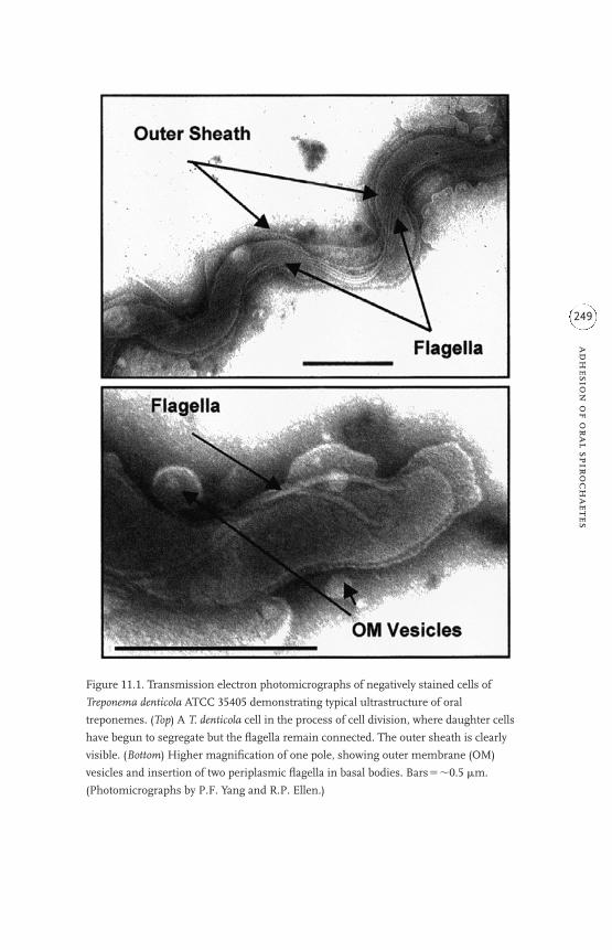
adh
esion
of o
ral spiroch
aetes
249
Figure 11.1. Transmission electron photomicrographs of negatively stained cells of
Treponema denticola ATCC 35405 demonstrating typical ultrastructure of oral
treponemes. (Top) A T. denticola cell in the process of cell division, where daughter cells
have begun to segregate but the flagella remain connected. The outer sheath is clearly
visible. (Bottom) Higher magnification of one pole, showing outer membrane (OM)
vesicles and insertion of two periplasmic flagella in basal bodies. Bars��0.5 �m.
(Photomicrographs by P.F. Yang and R.P. Ellen.)

complexes. Very few outer sheath proteins of T. denticola have been charac-terized at the molecular level. The best known of these are the major surfaceprotein (Msp) (Haapasalo et al., 1992; Fenno et al, 1996) and an associatedserine protease that has been called the chymotrypsin-like protease or, morerecently, dentilisin by di¤erent laboratories (Uitto et al., 1988; Grenier et al.,1990; Ishihara et al., 1996).
There is electron microscopy (EM), immuno-EM and functional evi-dence that the Msp complex is displayed on the bacterial surface (Masudaand Kawata, 1982; Egli et al., 1993), although the extent of its surface expo-sure has been questioned in one recent publication (Caimano et al., 1999).Extracted native Msp is detected as a high molecular mass complex, oligo-meric polypeptide by polyacrylamide gel electrophoresis (PAGE) whenunboiled and usually as a 53kDa polypeptide when denatured by boiling insodium dodecyl sulphate (SDS). A 64kDa putative adhesin has also beenidentified in the outer sheath of some T. denticola strains (Weinberg and Holt,1991) and may be analogous in structure–function relationships to Msp. Theouter sheath chymotrypsin-like protease is encoded by the gene prtP(Ishihara et al., 1996). The term ‘dentilisin’ was chosen because of the con-served deduced amino acid residues that comprise and flank its putative cat-alytic residues, which resemble those of the subtilisin family of serineproteases. The protease runs in SDS-PAGE as a minimum 91–95kDacomplex; the complex is much larger in unboiled samples (Uitto et al., 1988;Grenier et al., 1990; Rosen et al., 1995). The complex in the type strain ATCC35405 contains dentilisin, estimated at 72kDa from its deduced amino acidsequence, and a 43kDa protein of unknown function that is encoded by agene immediately upstream from prtP (Ishihara et al., 1996). Fragments ofthe outer sheath of T. denticola are known to shed from the surface. Vesicleswith proteolytic activity are also thought to derive from the outer sheath (Fig.11.1).
11.3 ADHESION OF TREPONEMES TO EXTRACELLULAR MATRIX
Oral treponemes encounter ECM components on the surface of hostcells, in the intercellular spaces between epithelial cells, in the basementmembrane, and in the extracellular compartment of the lamina propria anddeeper connective tissues of the gingiva. Knowledge of their binding andadhesion to these components is derived primarily from in vitro experimen-tal assays using T. denticola. This organism most likely evolved to bind toECM components both as a mechanism for evading removal by bathing hostfluids and as a way to forage for substrates that could be degraded by prote-
r. p
. ell
en
250

olysis to satisfy its nutritional requirement for peptides. Indeed, some of theECM proteins to which T. denticola binds are known to be degraded by den-tilisin, and this activity has been shown to promote the movement of T. den-ticola cells through in vitro molecular complexes mimicking the mammalianbasement membrane (Grenier et al., 1990). The ECM components bound byT. denticola represent a diverse range of molecular structures. These havebeen identified using assays that detect either the soluble ECM moleculesthat can be bound by the bacteria, or various polypeptides extracted from thebacteria, or the adhesion of whole T. denticola cells (or extracted proteins) toinanimate surfaces that had been preconditioned by the adsorption of ECMcomponents. The distinction is subtle but significant; macromolecules thatare bound to inanimate or cell surfaces may undergo conformationalchanges that a¤ect bacterial adhesion.
Like many wild-type bacteria, the surface of T. denticola cells is moder-ately hydrophobic (Fiehn, 1991); cells in aqueous suspensions of T. denticolavary in their net negative surface charge (Cowan et al., 1994). Thereforewhole cells of the organism are able to associate transiently or even bind toinanimate surfaces depending on their wettability. Treponema denticola cellsbind avidly to hydrophobic plastics and non-wettable silicon surfaces condi-tioned with methyl groups (Ellen et al., 1998). The cells lie flat upon contact.The cells bind less readily to wettable hydrophilic surfaces; those cells thatdo adhere bind tenuously, mostly in a polar orientation and possibly medi-ated by ionic interactions. In contrast, T. denticola adheres very well to inan-imate surfaces that have been preconditioned with human macromoleculesthat form extracellular matrices, and such coated surfaces favour a polarorientation of the adherent cells (Fig. 11.2). The organism binds selectivelyto fibronectin (Fn), laminin (Lam), fibrinogen, and hyaluronan-coated sur-faces but binds relatively poorly to surfaces conditioned with bovine serumalbumin (BSA) and inconsistently to type IV or other collagens (Haapasaloet al., 1991, 1996). Its adhesion to the native ECM conditioning componentsis usually tip-oriented (Dawson and Ellen, 1990, 1994), similar to the polaradhesion to Fn previously reported for Treponema pallidum.
In most cases, the identity of the adhesin(s) and the exact mechanismsof adhesion are unknown. Marked inhibition of adhesion to Lam and otherECM proteins when the bacteria were pretreated with either mixed glycosi-dases or the sulphydryl reagents p-chloromercuribenzoic acid and oxidizedglutathione suggests that surface protein SH groups and/or carbohydrateresidues may be involved. So far, the only non-proteolytic outer sheathprotein known to adhere to the ECM components listed is Msp. Both nativeand recombinant Msp (rMsp) bind to Lam- and Fn-coated plastic (Haapasalo
adh
esion
of o
ral spiroch
aetes
251

et al., 1991; Fenno et al., 1996). The degree of binding is diminished in thepresence of the soluble form of these ECM components. Yet, rMsp does notblock the adhesion of T. denticola cells, probably due to the presence of other,yet to be identified, outer sheath adhesins that recognize the substrates.Similarly, very little is known about potential domains of ECM componentsthat may serve as the ligand for Msp or other T. denticola adhesins. The wholebacterium can bind to an immobilized, elastase-generated 140kDa �-chainfragment of Lam that contains its eukaryotic cell attachment domain(Haapasalo et al., 1991). Lam and Fn contain a similar cell-binding domainthat consists of RGD(Arg-Gly-Asp) residues. Treponema denticola cells canadhere in polar orientation to plastic coated with RGDS-containing hepta-
r. p
. ell
en
252
Figure 11.2. Scanning electron photomicrograph of a T. denticola cell adhering by one
pole to a latex microsphere coated with fibronectin. Bar��0.5 �m. (Reprinted from
Trends in Microbiology, 2, Ellen, R.P., Dawson, J.R. and Yang, P.F.: Treponema denticola as
a model for polar adhesion and cytopathogenicity of spirochetes, pp. 114–119, Copyright
1994, with permission of Elsevier Science.).

peptides but not to control peptides. However, the putative target peptides insolution do not inhibit the adhesion of whole cells to Fn (Dawson and Ellen,1990).
A novel aspect of polar adhesion to Fn-conditioned surfaces is the time-dependent concentration of Fn-binding adhesins at one pole of the bacte-rium during the adhesion process. Treponema denticola cells usuallyapproach Fn-coated surfaces laterally or obliquely and then slowly ‘stand up’into a polar orientation. Dawson and Ellen (1994) have shown by ligand–goldelectron microscopy that T. denticola cells that contact Fn-coated nitrocellu-lose membranes have a disproportionate labelling of Fn[gold]-binding adhe-sins at one pole as compared with cells that are distant to, or prevented fromcontact with, the Fn-coated surface (Fig. 11.3). Cells contacting bu¤er-treatedor Lam-coated surfaces show no redistribution of Fn-binding adhesins.These observations lend support to the contention that treponemal proteinsare free to move in the outer sheath of the bacterium (Charon et al., 1981)and imply that a process analogous to ‘capping’ of specific ligand receptorsin eukaryotes occurs in prokaryotic cells. Clustering of adhesins would tendto increase the avidity of the interaction between the bacterium and the spe-cific substrate.
Much of the work on T. denticola adhesion to ECM components has con-centrated on binding assays in which the ligand is first bound to a surface,probably because native ECM components in tissues associate with cellsurface receptors such as integrins and with each other. When extracted pro-teins of T. denticola are first dissociated, electrophoresed by PAGE, trans-ferred to membranes and then exposed to Fn in solution, immunoreactiveFn can be detected bound to many of the polypeptide bands (Umemoto et al.,1993). Few have been characterized, and it is likely that several of the posi-tive bands that migrate di¤erentially actually reflect variously-sized molecu-lar species or degradation products of a single protein. Treponema denticolauses ECM proteins as targets for adhesion in order to resist removal bybathing fluids, but it also probably binds soluble substrates to satisfy itsnutritional needs. Thus the substrate recognition and binding domains ofsurface-associated enzymes, for example subtilisin, may serve concomitantlyor transiently as adhesins. In addition, several strains of T. denticola have agene for OppA, a 70kDa putative surface binding protein of an ATP-bindingcassette type of transporter (Fenno et al., 2000). Treponema denticola OppAevidently binds plasma Fn and soluble plasminogen but binds to neither sub-strate when it is immobilized on a surface, nor to epithelial cells. OppA isalso found in Treponema vincentii. Treponema denticola also has an operon thatencodes MglBAC (Lépine and Ellen, 2000), another putative ATP-binding
adh
esion
of o
ral spiroch
aetes
253

r. p
. ell
en
254
Figure 11.3. Transmission electron photomicrographs of a T. denticola ATCC 33520 cell
that had migrated through methyl cellulose into contact with a fibronectin-coated filter.
After washing, the bacteria were exposed to fibronectin (Fn)-gold probes (10 nm). The
probes concentrated towards one pole of the spirochaete (top) relative to the other pole
(bottom). Bars��0.5 �m. (Photomicrographs by J.R. Dawson and R.P. Ellen.)

cassette implicated in glucose/galactose binding in enteric bacteria and fla-gellar export in other bacteria, including T. pallidum. MglB and MglA alsohave 40–55% amino acid similarity to several Spa and Ysc surface proteinsof the ATP-binding and type III secretion pathways of enteropathogens.
11.4 ADHESION TO EPITHELIAL CELLS AND DISRUPTION OFEPITHELIAL BARRIER FUNCTION
In their natural habitat, oral treponemes interface directly with the strat-ified squamous, sulcular and junctional epithelium of the gingival creviceand inflamed periodontal pocket. Their adhesion to a variety of squamous-like epithelial cell lines in vitro has been studied by several investigators(Olsen, 1984; Baehni, 1986; Keulers et al., 1993; Walker et al., 1999).Recently, there have been detailed investigations of the cytopathogenic con-sequences of the adhesion of T. denticola to: (i) human KB cells (ATCC CL-17) (DeFilippo et al., 1995), which were originally derived from an epidermoidcarcinoma; (ii) HeLa cells (Mathers et al., 1996);, (iii) human Hep-2 cells (Koet al., 1998b; Ellen et al., 2000), which are of laryngeal origin; and (iv) celllines established from explants of porcine periodontal ligament epithelialcell rests (Uitto et al., 1995). Together, these studies have clearly demon-strated the potential of T. denticola to breach epithelial barriers (Fig. 11.4).
When epithelial monolayers are challenged with T. denticola supsensions,the bacteria attach selectively to a subpopulation of cells. Consequently, cyto-pathic changes are detected in some cells in the monolayer almost immedi-ately, but most cells remain viable even though they may progress through aseries of cellular responses a¤ecting their cytoskeleton and intercellularjunctional complexes. In time, an increasing proportion of the cells detachfrom the extracellular substratum. The detached cells are usually dead, whilethose that remain attached apparently remain viable, even though they mayexperience profound rearrangement of their cytoskeleton, disassembly ofactin filaments, diminished cytokeratin and desmoplakin II expression anddemonstrate rather marked abnormalities in volume regulation, which is anactin-dependent function (DeFilippo et al., 1995; Uitto et al., 1995). AlthoughT. denticola cells can be detected inside some epithelial cells and endothelialcells during in vitro challenge assays (Uitto et al., 1995; Peters et al., 1999),there is no evidence that their uptake represents an evolved survival or viru-lence factor that is analogous to the survival strategies reported for manyother mucosal pathogens. Indeed, intracellular T. denticola cells are seen invarious stages of degradation inside vesicular organelles of the epithelialcells (Uitto et al., 1995).
adh
esion
of o
ral spiroch
aetes
255

Figu
re 11.4
. Hyp
oth
etic
al m
odel
illu
stra
tin
g th
e co
nse
quen
ces
of th
e ad
hes
ion
of
oral
trep
onem
es to
str
atifi
ed s
quam
ous
epit
hel
ium
of
the
g in
giva
l
crev
ice,
bas
ed m
ostl
y on
res
earc
h u
sin
g T.
den
tico
la. P
rote
olyt
ic e
nzy
mes
su
ch th
e ch
ymot
ryps
in-li
ke e
nzy
me
(su
btili
sin
, Prt
P) a
nd
oth
er e
nzy
mes
that
degr
ade
inte
rcel
lula
r m
atri
x (c
hon
droi
tin
ase
(C-a
se) a
nd
hya
lulr
onid
ase
(H-a
se))
dis
rupt
bar
rier
fu
nct
ion
that
is n
orm
ally
mai
nta
ined
by
inte
rcel
lula
r
adh
esio
n m
olec
ule
s an
d as
soci
ated
jun
ctio
nal
pro
tein
s. T
he
trep
onem
es m
igra
te b
etw
een
the
epit
hel
ial c
ells
, deg
rade
mat
rix,
an
d pe
net
rate
thro
ugh
the
base
men
t mem
bran
e in
to th
e la
min
a pr
opri
a of
the
gin
giva
. Adh
esio
n o
f tr
epon
emes
to e
xpos
ed e
pith
elia
l cel
ls in
duce
s cy
tosk
elet
al r
earr
ange
men
t, lo
ss o
f
volu
me
regu
lati
on, s
hri
nka
ge, a
nd
dim
inis
hed
exp
ress
ion
of
jun
ctio
nal
pro
tein
s, w
hic
h a
lso
con
trib
ute
to w
iden
ed in
terc
ellu
lar
spac
es. M
sp b
inds
to
epit
hel
ial c
ells
an
d m
ay tr
ansp
ose
into
the
plas
ma
mem
bran
e to
per
turb
ion
flu
xes.
Msp
is a
lso
cyto
toxi
c to
som
e ce
lls (⊗
). B
oth
wh
ole
trep
onem
es a
nd
Prt
P m
ay b
e ta
ken
up
into
the
cells
. Th
e tr
epon
emes
are
evi
den
tly
degr
aded
in in
trac
ellu
lar
vesi
cles
.

Transcytosis of T. denticola and other treponemes may be an infrequentevent, even in cases when spirochaetes are detected in the gingival connec-tive tissues of periodontal lesions. Treponema denticola is able to pass throughmodel mucosal barriers in vitro but not as readily as some other species oftreponemes isolated from periodontal lesions (Riviere et al., 1991). Severalspecies of oral treponemes produce enzymes with hyaluronidase, chondroi-tinase, and wide-range peptidase activities that would be expected to degradeextracellular matrices in the epithelium and connective tissues (Mikx et al.,1992; Scott et al., 1996b). Moreover, T. denticola and outer membraneextracts with proteolytic activity have profound detrimental e¤ects on epithe-lial junctions in vitro, both in typical confluent monolayers and in a multi-layer system that mimics stratified epithelium (Uitto et al., 1995; Ko et al.,1998b). Proteolytic protein complexes or vesicles that are shed from thewhole bacteria can penetrate between epithelial cells coincident with bacte-rial abrogation of epithelial barrier function (Uitto et al., 1995). The nativechymotrypsin-like serine protease (PrtP; dentilisin), within complexes ofouter membrane extracts or in purified preparations, clearly has the abilityto eliminate junctional resistance to electrical conductivity and to the di¤u-sion of molecules that are normally excluded (Uitto et al., 1995; Ko et al.,1998b). Insertional inactivation of the prtP gene, resulting in a mutantdevoid of the prolylphenylalanine peptidase activity typical of serine pro-teases, prevents the disruptive activity of T. denticola outer membraneextracts on epithelial resistance (Ellen et al., 2000). Direct degradation of theintercellular matrix and substratum by dentilisin probably accounts for thetime- and concentration-dependent detachment of epithelial cells in culture.Hep-2 cells seeded in the presence of moderate concentrations of an outermembrane extract of wild-type T. denticola 35405 never reach confluence,whereas cells cultured with similar extracts from the PrtP-defective mutantgrow normally into contact (Ellen et al., 2000). Confluent Hep-2 cells that areexposed to T. denticola outer membrane extracts also experience a biphasicresponse in electrical impedance signals that reflect cellular micromotion. Atfirst, their micromotion increases, which may reflect sublethal actin filamentrearrangement, cellular shape changes, and shrinkage due to altered actinassembly or proteolysis of intercellular contacts; then micromotion appar-ently ceases (Ko et al., 1998b). Thus T. denticola a¤ects physiological proper-ties of epithelial cells that would normally maintain exclusive epithelialbarriers and migration into confluence that would promote wound healing.
In vitro studies using confluent monolayers emphasize the acute patho-genic e¤ects of T. denticola adhesion on epithelial cells, many of which canbe attributed to the bacterium’s proteolytic activity. Yet, other cell surface
adh
esion
of o
ral spiroch
aetes
257

molecules such as its non-proteolytic outer sheath proteins may have toxice¤ects. Prominent among these is Msp, which is cytotoxic for a variety ofmammalian cell types (Fenno et al., 1998a). Both native and rMsp have pore-forming properties that have been documented using HeLa cells. Exposureof cultured HeLa cells to Msp induces transient fluctuations in membranepotential that may be caused by the transposition of Msp into the plasmamembrane (Mathers et al., 1996) (Fig. 11.4). Msp is known to have the prop-erties of a non-exclusive outer membrane porin when tested in a standard invitro model (Egli et al., 1993; Fenno et al., 1998a). Transient retention of itsporin activity when incorporated into the plasma membrane of human epi-thelial cells would be expected to a¤ect ion fluxes that normally regulate cel-lular functions. Indeed, the Msp-induced ion-permeable channels found inHeLa cells appear to be inwardly unidirectional and open only intermittently.Some other bacterial porins have been shown to transpose to the target cellplasma membrane and to cause acute elevation of intracellular Ca2� concen-tration. Msp has recently been found to display analogous e¤ects in gingivalfibroblasts, as discussed in section 11.5.
11.5 ADHESION TO FIBROBLASTS AND THE PERTURBATION OFSIGNALLING PATHWAYS FOR ACTIN ASSEMBLY
Fibroblasts are the key cells that maintain homeostasis of non-mineralized collagenous tissues, and they provide a useful model for study-ing cytopathogenic e¤ects of periodontal pathogens on non-immune cellsthat are important in inflammatory responses and wound repair in thegingiva. Similar to their adhesion to ECM components and epithelial cells,oral treponemes adhere in a polar orientation to cultured gingival fibroblasts(Fig. 11.5), and they are occasionally engulfed into the intracellular environ-ment. For example, although almost all adherent T. denticola cells remainextracellular, intact bacteria have been observed in some fibroblasts by usingdouble label immunofluorescence techniques, electron microscopy and con-focal microscopy (Ellen et al., 1994a). The biological implications of thisinfrequent invasion are unknown, as oral treponemes have not yet beenreported to exist intracellularly in fibroblasts in the gingiva.
Many of the fibroblast’s key functions in maintaining the homeostasisof the gingival tissues involve its cytoskeleton. The cells migrate alongcollagen-rich ECM networks using tractional forces generated by cycles ofactin-binding focal complex assembly and adhesion of integrins to ECMligands. Thus their locomotion in response to chemotactic factors would beperturbed by microorganisms that degraded the ECM at the fibroblast
r. p
. ell
en
258

surface or upset the intracellular signalling pathways that regulate the cyto-skeleton. Physiological turnover of collagen in the gingiva is maintained at acontrolled rate by the phagocytosis of collagen fibrils, intracellular degrada-tion of the fibrils, and the synthesis of new collagen by fibroblasts. Collagenphagocytosis depends on actin assembly. It should be sensitive to bacterialfactors that bind to the cells and that interrupt the normal balance of signal-ling molecules, such as the phosphoinositides (PIs) and calcium that regu-late the activity of actin binding and severing proteins.
Cultured human gingival fibroblasts (HGFs) challenged by suspensionsof T. denticola undergo cytoskeletal rearrangement, round up, shrink, andeventually detach from the ECM substratum (Baehni et al, 1992; Weinbergand Holt, 1990) (Figs. 11.6 and 11.7). These cytopathic changes occur asyn-chronously in HGF cultures, beginning with a few cells within minutes andeventually involving most of them. The changes are dependent on time andbacterial cell density, and they are a¤ected by whether the HGFs are chal-lenged as sparsely spaced cells soon after plating or as monolayers that havegrown into confluence over a few days. Gross cellular distortion and detach-ment of HGFs from the substratum may result directly from ECM degrada-tion by bacterial proteases, especially dentilisin, as the serine proteaseinhibitor phenylmethylsulphonyl fluoride (PMSF) inhibits HGF detachment
adh
esion
of o
ral spiroch
aetes
259Figure 11.5. Treponema denticola ATCC 35405 cells bound in a polar orientation to the
dorsal surface of a human gingival fibroblast. The image represents the x–z plane
derived from a three-dimensional reconstruction from x–y optical sections of a fibroblast
stained by indirect immunofluorescence with polyclonal rabbit antibodies raised against
T. denticola ATCC 35405 followed by TRITC-conjugated goat anti-rabbit �-globulin to
label T. denticola cells, and by fluorescein isothiocyanate-conjugated concanavalin A to
highlight the HGF plasma membrane. (Confocal microscopy image by I. Buivids and
R.P. Ellen.)

r. p
. ell
en
260
Figure 11.6. Fluorescence microscopy of rhodamine-phalloidin–stained human gingival
fibroblasts that had been challenged with an outer membrane extract of T. denticola ATCC
35405 (bottom) compared with the control that had been exposed to the culture medium
vehicle (top). Note the disruption of actin filaments in the originally prominent stress
fibres and the assembly of actin filaments in a dense perinuclear positon and subjacent
to the plasma membrane (arrows). (Photomicrographs by M. Song and R.P. Ellen.)

by T. denticola (Baehni et al., 1992). Treponema denticola can degrade endog-enous fibronectin that decorates the surface of cultured HGFs (Ellen et al.,1994b) and this may account for some of the initial rounding-up that hasbeen observed. Yet inhibition of serine protease activity by PMSF does notreduce the adhesion of T. denticola to HGFs (Ellen et al., 1994b) or the dis-ruption of stress fibre networks in HGFs (Yang et al., 1998). Likewise, inser-tional inactivation of the prtP gene does not diminish stress fibre disruptionin HGFs by T. denticola whole cells (Ellen et al., 2000). It is the stress fibresthat normally interface via integrins with the ECM components of the sub-stratum. Many of the cytopathic e¤ects of T. denticola whole cells can bemediated by extracts of its outer membrane (OM). While highly proteolytic,these extracts contain other non-proteolytic proteins that may account forsome of the impact on intracellular signalling pathways a¤ecting the cyto-skeleton and thereby perturb important physiological functions of gingivalfibroblasts.
Treponema denticola OM has profound e¤ects on the actin-dependentphagocytosis of collagen by HGFs, a function crucial for the normal turn-over of collagen in the gingiva (Battikhi et al., 1999). Even short-term, tran-sient exposure of HGFs to a concentration of OM that has no detrimentale¤ect on their viablility, cell size and attachment to the substratum can causea great increase in the uptake of collagen-coated beads in vitro. The rapidresponse, detected within 15minutes of exposure to the OM, suggests thatT. denticola OM components exert their e¤ects on pathways that cause rear-rangement of actin oligomers near the plasma membrane rather than trig-gering signalling pathways that would require genetic regulation of proteinexpression.
Investigations of intracellular signalling pathways a¤ected by binding ofT. denticola surface components have concentrated on calcium flux and bio-chemical determination of total inositol phosphates as a downstream indica-tor of perturbation of membrane PI metabolism (Fig. 11.7). Because thecalcium responses of gingival fibroblasts are inherently sensitive to mechan-ical forces on the plasma membrane, OM extracts have served as the bacte-rial challenge to avoid confounding e¤ects caused by the motility of tetheredtreponemes (Ko et al., 1998a). Transients in intracellular calcium concentra-tion ([Ca2�]i) were measured in fura-2-loaded HGFs by ratio fluorimetry.Exposure to T. denticola OM causes immediate but short-lived calcium oscil-lations in resting HGFs. These last for about 30minutes, followed by a pro-longed period of quiescence in which few, if any, calcium fluctuations can bedetected. In addition, T. denticola OM is a rather potent inhibitor of HGFcalcium responses to well-established chemical agonists and to mechanical
adh
esion
of o
ral spiroch
aetes
261

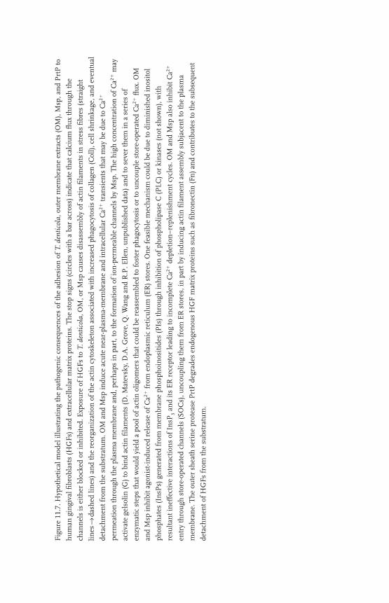
Figu
re 11.7
. Hyp
oth
etic
al m
odel
illu
stra
tin
g th
e pa
thog
enic
con
sequ
ence
s of
the
adh
esio
n o
f T.
den
tico
la, o
ute
r m
embr
ane
extr
acts
(OM
), M
sp, a
nd
Prt
P to
hu
man
gin
giva
l fibr
obla
sts
(HG
Fs) a
nd
extr
acel
lula
r m
atri
x pr
otei
ns.
Th
e st
op s
ign
s (c
ircl
es w
ith
a b
ar a
cros
s) in
dica
te th
at c
alci
um
flu
x th
rou
gh th
e
chan
nel
s is
eit
her
blo
cked
or
inh
ibit
ed. E
xpos
ure
of
HG
Fs to
T. d
enti
cola
, OM
, or
Msp
cau
ses
disa
ssem
bly
of a
ctin
fila
men
ts in
str
ess
fibr
es (s
trai
ght
lines
→da
shed
lin
es) a
nd
the
reor
gan
izat
ion
of
the
acti
n c
ytos
kele
ton
ass
ocia
ted
wit
h in
crea
sed
phag
ocyt
osis
of
colla
gen
(Col
l), c
ell s
hri
nka
ge, a
nd
even
tual
deta
chm
ent f
rom
the
subs
trat
um
. OM
an
d M
sp in
duce
acu
te n
ear-
plas
ma-
mem
bran
e an
d in
trac
ellu
lar
Ca2
�tr
ansi
ents
that
may
be
due
to C
a2�
perm
eati
on th
rou
gh th
e pl
asm
a m
embr
ane
and,
per
hap
s in
par
t, to
the
form
atio
n o
f io
n-p
erm
eabl
e ch
ann
els
by M
sp. T
he
hig
h c
once
ntr
atio
n o
f C
a2�
may
acti
vate
gel
solin
(G) t
o bi
nd
acti
n fi
lam
ents
(D. M
atev
sky,
D.A
. Gro
ve, Q
. Wan
g an
d R
.P. E
llen
, un
publ
ish
ed d
ata)
an
d to
sev
er th
em in
a s
erie
s of
enzy
mat
ic s
teps
that
wou
ld y
ield
a p
ool o
f ac
tin
olig
omer
s th
at c
ould
be
reas
sem
bled
to f
oste
r ph
agoc
ytos
is o
r to
un
cou
ple
stor
e-op
erat
ed C
a2�
flu
x. O
M
and
Msp
inh
ibit
ago
nis
t-in
duce
d re
leas
e of
Ca2
�fr
om e
ndo
plas
mic
ret
icu
lum
(ER
) sto
res.
On
e fe
asib
le m
ech
anis
m c
ould
be
due
to d
imin
ish
ed in
osit
ol
phos
phat
es (I
nsP
s) g
ener
ated
fro
m m
embr
ane
phos
phoi
nos
itid
es (P
Is) t
hro
ugh
inh
ibit
ion
of
phos
phol
ipas
e C
(PLC
) or
kin
ases
(not
sh
own
), w
ith
resu
ltan
t in
e¤ec
tive
inte
ract
ion
s of
In
sP3
and
its
ER
rec
epto
r le
adin
g to
inco
mpl
ete
Ca2
�de
plet
ion
–rep
len
ish
men
t cyc
les.
OM
an
d M
sp a
lso
inh
ibit
Ca2
�
entr
y th
rou
gh s
tore
-ope
rate
d ch
ann
els
(SO
Cs)
, un
cou
plin
g th
em f
rom
ER
sto
res,
in p
art b
y in
duci
ng
acti
n fi
lam
ent a
ssem
bly
subj
acen
t to
the
plas
ma
mem
bran
e. T
he
oute
r sh
eath
ser
ine
prot
ease
Prt
P d
egra
des
endo
gen
ous
HG
F m
atri
x pr
otei
ns
such
as
fibr
onec
tin
(Fn
) an
d co
ntr
ibu
tes
to th
e su
bseq
uen
t
deta
chm
ent o
f H
GFs
fro
m th
e su
bstr
atu
m.

force. It blocks both ATP- and thapsigargin-induced release of Ca2� fromendoplasmic reticulum (ER) stores. It inhibits the increase in [Ca2�]i follow-ing physical stretching of the plasma membrane, a rise that is usually causedby a combination of Ca2� influx through stretch-activated channels and store-operated calcium channels (SOCs) that are linked to calcium release fromthe ER. Indeed, preliminary experiments examining the e¤ect of T. denticolaOM on store-operated Ca2� flux suggest that the OM inhibits Ca2� entrythrough putative SOCs. Moreover, both T. denticola whole cells and OM causea diminished inositol phosphate (InsP) response in HGFs to agonists inserum and to ATP (Yang et al., 1998). Inositol 1,4,5-trisphosphate (InsP
3) is
a key factor in the release and replenishment of Ca2� in the ER stores. Thecomponents of the T. denticola OM responsible for calcium perturbation inHGFs have not been fully elucidated. The fact that its inhibition of stretch-activated calcium transients is insensitive to PMSF, an inhibitor of serineproteases, but sensitive to heat at 60 °C, a temperature at which dentilisin’speptidase activity is stable, suggests that the OM’s calcium perturbing activ-ity is not related to the protease PrtP (Ko et al., 1998a).
The major surface protein, Msp, is apparently one of the OM compo-nents that may cause profound e¤ects of T. denticola on store-operatedcalcium flux in HGFs. Recently, Wang and co-workers (Wang et al., 2001) havereported that native Msp complex (i) binds and aggregates on the HGFsurface, (ii) induces [Ca2�]i transients near the plasma membrane that areprobably due to persistent Ca2� permeation through the plasma membrane,(iii) blocks ATP- and thapsigargin-induced release of Ca2� from the ER, and(iv) inhibits influx of extracellular Ca2� through SOCs following thapsigargindepletion of ER stores (Fig. 11.7). Msp also induces redistribution of actin fil-aments, including assembly of filamentous actin subjacent to the plasmamembrane, a sign that it may inhibit conformational coupling of SOCs andthe ER Ca2� stores (Berridge et al., 2000). These findings are consistent withthe novel findings of Mathers and co-workers (1996) that Msp can have pro-found e¤ects on cation flux in epithelial cells. Fluctuations in cytosolic Ca2�
may initiate a cascade of intracellular responses that mediate the cytoskeletalrearrangement that is so common in human cells exposed to T. denticola. Yetcontinuous exposure of HGFs to Msp complex is required for cytotoxic activ-ity resulting in significant cell death (Fenno et al., 1998a; Wang et al., 2001).
11.6 HAEMAGGLUTINATION AND HAEMOLYSIS
Even mildly inflamed periodontal tissues tend to bleed, enriching theenvironment of the periodontal pocket with erythrocytes and contents of
r. p
. ell
en
264

haemolysed erythrocytes. Treponemes are able to bind and agglutinate redblood cells (RBCs). Treponema denticola agglutinates RBCs from humanblood types A, B and O, as well as rabbit, horse, and bovine sources(Grenier, 1991; Mikx and Keulers, 1992). The haemagglutinating activity iscell associated, influenced by bacterial surface charge, sensitive to heat andproteolysis of the bacteria, and strongest in cells harvested in late exponen-tial and stationary phases of growth (Mikx and Keulers, 1992; Cowan et al.,1994). Although reports of haemagglutination inhibitors di¤er amonglaboratories, it is likely that the treponemal haemagglutinin is a protein. Itmay also be a surface-associated proteolytic enzyme, as soluble protein sub-strates and inhibitors of trypsin-like and chymotrypsin-like proteases alldiminish the haemagglutinating activity of T. denticola, T. vincentii and T.socranskii (Eggert et al., 1995). As arginine can also inhibit haemagglutina-tion of these treponemes, as well as that of some other oral bacteria, Eggertand co-workers (1995) have hypothesized that the bacteria may bindsurface proteins of erythrocytes, and likewise other proteins, via the highly-charged guanidinium group of arginine. Sensitivity of haemagglutinationby T. denticola to periodate (Mikx and Keulers, 1992) suggests that carbohy-drate-containing ligands may also be involved, but sugar specificity has notyet been established.
There is evidently a close association between haemagglutination andhaemolytic activities of T. denticola (Grenier, 1991). Thus oral spirochaetesmay have evolved a way to bind erythrocytes in their environment andthereby foster the lysis of cells that would provide enriched exposure toiron-containing compounds that may, in turn, be bound by outer sheathproteins (Scott et al., 1996a). Recently, a cysteine-dependent haemolysinhas been described for T. denticola (Chu et al., 1999; Kurzban et al., 1999).Treponema denticola expresses a 46kDa cytosolic l-cysteine desulphydrase,named cystalysin, that both chemically oxidizes haemoglobin in the pres-ence of cysteine and degrades RBC membrane proteins, including spectrin,thereby a¤ecting the integrity of the plasma membrane. Chu and co-workers (1999) characterized cystalysin as a putative pyridoxal-5-phos-phate-containing enzyme with cystathionase activity, optimum underreduced, H
2S-enriched conditions like those that occur in periodontal
pockets. There are no known haemolysins that are structurally homolo-gous. The crystal structure of recombinant cystalysin has recently beenresolved and analysed to relate its active site architecture to a putative mech-anism for the catabolism of sulphur-containing amino acid substrates(Krupka et al., 2000).
adh
esion
of o
ral spiroch
aetes
265

11.7 INTERACTIONS WITH LEUKOCYTES AND MACROPHAGES
The polymorphonuclear leukocyte (PMNL) is a key cell of innateimmune responses in the periodontal environment; PMNLs migratethrough the junctional epithelium in response to gradients of surface com-ponents that shed from bacteria at the surface of the subgingival biofilm(Darveau, 2000). Whole cells of T. denticola are readily phagocytosed aftercontacting PMNLs in vitro (Lingaas et al., 1983; Olsen et al., 1984; Boehringeret al., 1986). Yet, without causing disruption of the cells, they can inhibit oxi-dative bursts of PMNLs (Loesche et al., 1988; Sela et al., 1988) and triggerrelease and activation of PMNL-derived proteolytic enzymes that would beexpected to contribute to connective tissue degradation in periodontallesions (Ding et al., 1997). LPS, peptidoglycan and a 53kDa outer membraneprotein, probably Msp, cause PMNLs to discharge the matrix metalloprotei-nases MMP-8 and MMP-9; Msp is particularly active in the release of MMP-8 (Ding et al., 1996). Latent forms of MMPs from both PMNLs andfibroblasts can also be activated by the chymotrypsin-like serine protease(PrtP) of T. denticola (Sorsa et al., 1992).
Treponemes express many lipoproteins that are associated with their cyto-plasmic and outer membranes. An enriched lipoprotein fraction and the lipo-oligosaccharides of T. denticola ATCC 35404 have been shown to have manybiological e¤ects on PMNLs and macrophages (Sela et al., 1997; Rosen et al.,1999). The lipoproteins can cause enhanced luminol-dependent chemilumi-nescence and release of lysozyme from PMNLs and modulate these responsesto the experimental agonist N-formyl-methionyl-leucyl-phenylalanine(FMLP). They and the lipo-oligosaccharides induce expression of the proin-flammatory cytokines tumour necrosis factor � and interleukin 1 frommacrophages, which are thought to mediate significant tissue-destructivesignalling cascades in periodontal disease. In addition to modulating theinnate immunity activities of these key cells, T. denticola is also known to sup-press monocyte-dependent mitogenic responses in lymphocytes (Shenker etal., 1984). Therefore the binding of whole T. denticola cells and fragmentsshed from their surface to gingival inflammatory cells has the potential toalter host–parasite interactions important in the pathogenesis of periodon-titis.
11.8 INTERGENERIC CO-AGGREGATION
The inflamed periodontal pocket is an ideal environment for the emer-gence of fastidious anaerobes like the treponemes that rely on complex food
r. p
. ell
en
266

webs for sustenance. Indeed, the most striking feature of suspended subgin-gival debris viewed by phase-contrast or darkfield microscopy is the abun-dance of highly motile spirochaetes surrounding the bacterial biofilm andtethered to it by one pole (Fig. 11.8). Like many bacteria that comprise oralbiofilms, T. denticola is known to coaggregate with bacteria in other genera.Samples derived from inflamed sites of destructive periodontitis oftencontain a complex of at least three major proteolytic species: Porphyromonasgingivalis, Bacteroides forsythus and T. denticola. Together, these speciesaccount for very broad substrate degradation, and the individual bacteria inthe biofilm probably derive their required peptides through ‘community pro-teolysis’. Treponema denticola, Treponema medium and Treponema socranskiisubsp. socranskii coaggregate avidly with P. gingivalis over a wide pH range(Grenier, 1992; Onagawa et al., 1994; Umemoto et al., 1999). These andseveral other oral treponemes co-aggregate with Fusobacterium species(Kolenbrander et al., 1995). There has been one report that the co-aggregation reactions of T. denticola and P. gingivalis are sensitive to heatingthe T. denticola partner (Grenier, 1992); yet, other reports agree that the co-adhesion of T. denticola with either P. gingivalis or fusobacteria is resistant toheat. Co-aggregation with P. gingivalis may be inhibited by d-galactosamineand arginine under some conditions (Grenier, 1992). Co-aggregation of T.denticola with fusobacteria is evidently sensitive to galactosamine but not toarginine. The actual surface adhesins responsible for T. denticola’s interge-neric co-aggregation reactions are unknown. Inactivation of the prtP gene,which eliminates the serine protease activity of dentilisin, concomitantlyknocks out the ability of the mutant T. denticola strain to coaggregate withfusobacteria (Ishihara et al., 1998). However, PrtP-deficient mutants of T.denticola have many other pleiotropic e¤ects, such as lower expression ofother surface proteins (e.g. Msp) and diminshed hydrophobicity that mayaccount for the altered co-aggregation (Fenno et al., 1998b; Ishihara et al.,1998).
11.9 TREPONEMES, FASTIDIOUS HANGERS-ON IN A COMPLEXCOMMUNITY
Treponemes require a complex assortment of nutrients for energy andgrowth, and they colonize an environment enriched with a diverse array ofsolutes and complex molecules. Their need of peptides has probably driventhe evolution of their proteases and peptidases. In the case of T. denticola,expression of PrtP in its outer sheath probably provides the bacterium anopportunity to adhere with the specificity of an enzyme to protein substrates.
adh
esion
of o
ral spiroch
aetes
267

r. p
. ell
en
268
Figure 11.8. Phase-contrast micrograph of subgingival debris taken from a periodontal
pocket of a subject with periodontitis. Numerous spirochaetes (arrows) are tethered to the
surface of the dense bacterial biofilm (dental plaque) at the interface that would be
exposed to the sulcular and junctional epithelium. (Photomicrograph by R.P. Ellen.
Adapted from Ten Cate, 1994.)

The use of sulphur as an electron acceptor in treponemes and other anaer-obes may have led to T. denticola’s ability to bind via sulphydryl groups andto interact with sulphated macromolecules in the extracellular matrix andcytokeratins of epithelial cells. The aªnity of Msp for matrix proteins and itsclose association with PrtP in the outer sheath is an additional example of aconnection between adhesion specificity and nutrient acquisition.Haemagglutination, haemolysis, and haemin-binding would be another. Tobe fastidious in a complex environment like the periodontal pocket has prob-ably driven oral treponemes to adhere to a variety of cell surfaces and extra-cellular ligands so that they may hang on to a fragile lifestyle in a harshclimate. This chapter has summarized the pathogenic consequences of themost obvious adhesion functions that oral treponemes have evolved for theirown survival.
ACKNOWLEDGEMENT
Original research cited in this chapter was supported by grant MT-5619to R.P.E. from the Canadian Institutes of Health Research.
REFERENCES
Baehni, P. (1986). Interactions between plaque microorganisms and human oral
epithelial cells. In Borderland Between Caries and Periodontal Disease, 3rd
European Symposium, ed. T. Lehner and G. Cimasoni, pp. 143–153.
Geneva: Editions Médecine et Hygiène.
Baehni, P.C., Song, M., McCulloch, C.A.G. and Ellen, R.P. (1992). Treponema den-
ticola induces actin rearrangement and detachment of human gingival fibro-
blasts. Infection and Immunity 60, 3360–3368.
Battikhi, T., Lee, W., McCulloch, C.A.G. and Ellen, R.P. (1999). Treponema denti-
cola outer membrane enhances the phagocytosis of collagen-coated beads by
gingival fibroblasts. Infection and Immunity 67, 1220–1226.
Berridge, M.J., Lipp, P. and Bootman, M.D. (2000). The calcium entry pas de deux.
Science 287, 1604–1605.
Boehringer, H., Berthold, P.H. and Taichman, N.S. (1986). Studies on the inter-
action of human neutrophils with plaque spirochetes. Journal of Periodontal
Research 21, 195–209.
Caimano, M.J., Bourell, K.W., Bannister, T.D., Cox, D.L. and Radolf, J.D. (1999). The
Treponema denticola major sheath protein is predominantly periplasmic and
has only limited surface exposure. Infection and Immunity 67, 4072–4083.
Chan, E.C.S., Siboo, R., Keng, T., Psarra, N., Hurley, R., Cheng, S.-L. and Iugovaz,
adh
esion
of o
ral spiroch
aetes
269

I. (1993). Treponema denticola (ex Brumpt 1925) sp. Nov., nom. Rev., and iden-
tification of new spirochete isolates from periodontal pockets. International
Journal of Systematic Bacteriology 43, 196–203.
Charon, N.W., Lawrence, C.W. and O’Brien, S. (1981). Movement of antibody-
coated latex beads attached to the spirochete Leptospira interrogans.
Proceedings of the National Academy of Sciences, USA 78, 7166–7170.
Chi, B., Chauhan, S. and Kuramitsu, H. (1999). Development of a system for
expressing heterologous genes in the oral spirochete Treponema denticola
and its use in expression of the Treponema pallidum flaA gene. Infection and
Immunity 67, 3653–3656.
Choi, B.K., Paster, B.J., Dewhirst, F.E. and Gobel, U.B. (1994). Diversity of culti-
vable and uncultivable oral spirochetes from a patient with severe destruc-
tive periodontitis. Infection and Immunity 62, 1889–1895.
Choi, B.K., Wyss, C. and Göbel, U.B. (1996). Phylogenetic analysis of pathogen
related oral spirochetes. Journal of Clinical Microbiology 34, 1922–1925.
Chu, L., Ebersole, J.L., Kurzban, G.P. and Holt, S.C. (1999). Cystalysin, a 46-kDa
l-cysteine desulfhydrase from Treponema denticola: biochemical and bio-
physical characterization. Clinical Infectious Diseases 28, 442–450.
Cowan, M.M., Mikx, F.H.M. and Busscher, H.J. (1994). Electrophoretic mobility
and haemagglutination of Treponema denticola ATCC 33520. Colloids and
Surfaces B: Biointerfaces 2, 407–410.
Darveau, R. (2000). Oral innate host defense response: interactions with micro-
bial communities and their role in the development of disease. In Oral
Bacterial Ecology, the Molecular Basis, ed. H.K. Kuramitsu and R.P. Ellen, pp.
169–218. Wymondham, Norfolk: Horizon Scientific Press.
Dawson, J.R. and Ellen, R.P. (1990). Tip-oriented adherence of Treponema denti-
cola to fibronectin. Infection and Immunity 58, 3924–3928.
Dawson, J.R. and Ellen, R.P. (1994). Clustering of fibronectin adhesins toward
Treponema denticola tips upon contact with immobilized fibronectin.
Infection and Immunity 62, 2214–2221.
DeFilippo, A.B., Ellen, R.P. and McCulloch, C.A.G. (1995). Induction of cytoskel-
etal rearrangments and loss of volume regulation in epithleial cells by
Treponema denticola. Archives of Oral Biology 40, 199–207.
Dewhirst, F.E., Tamer, M.A., Ericson, R.E., Lau, C.N., Levanos, V.A., Boches, S.K.,
Galvin, J.L. and Paster, B.J. (2000). The diversity of periodontal spirochetes
by 16S rRNA analysis. Oral Microbiology and Immunology 15, 196–202.
Ding, Y., Uitto, V.-J., Haapasalo, M, Lounatmaa, K., Konttinen, Y.T., Salo, T.,
Grenier, D. and Sorsa, T. (1996). Membrane components of Treponema den-
ticola trigger proteinase release from human polymorphonuclear leukocytes.
Journal of Dental Research 75, 1986–1993.
r. p
. ell
en
270

Ding, Y., Haapasalo, M., Kerosuo, E., Lounatmaa, K., Kotiranta, A. and Sorsa, T.
(1997). Release and activation of human neutrophil matrix metallo- and
serine proteinases during phagocytosis of Fusobacterium nucleatum,
Porphyromonas gingivalis, and Treponema denticola. Journal of Clinical
Periodontology 24, 237–248.
Eggert, F.-M., Chan, E.C.S., Klitorinos, A. and Flowerdew, G. (1995). Arginine is
a common ligand for haemagglutination and protein binding by organisms
inhabiting mucosal surfaces. Microbial Ecology in Oral Health and Disease 8,
267–280.
Egli, C., Leung, W.K., Müller, K.H., Hancock, R.E.W. and McBride, B.C. (1993).
Pore-forming properties of the major 53 kilodalton surface antigen from the
outer sheath of Treponema denticola. Infection and Immunity 61, 1694–1699.
Ellen, R.P., Dawson, J.R. and Yang, P.F. (1994a). Treponema denticola as a model
for polar adhesion and cytopathogenicity of spirochetes. Trends in
Microbiology 2, 114–119.
Ellen, R.P., Song, M. and McCulloch, C.A.G. (1994b). Degradation of endoge-
nous plasma membrane fibronectin concomitant with Treponema denticola
35405 adhesion to gingival fibroblasts. Infection and Immunity 62,
3033–3037.
Ellen, R.P., Wikström, M., Grove, D.A., Song, M. and Elwing, H. (1998). Polar
adhesion of Treponema denticola on wettability gradient surfaces. Colloids and
Surfaces. B: Biointerfaces 11, 177–186.
Ellen, R.P., Ko, K.S.-C., Lo, C.-M., Grove, D.L. and Ishihara, K. (2000). Insertional
inactivation of the prtP gene of Treponema denticola confirms dentilisin’s dis-
ruption of epithelial junctions. Journal of Molecular Microbiology and
Biotechnology 2, 581–586.
Fenno, J.C. and McBride, B.C. (1998). Virulence factors of oral treponemes.
Anaerobe 4, 1–17.
Fenno, J.C., Müller, K.-H. and McBride, B.C. (1996). Sequence analysis, expres-
sion, and binding activity of recombinant major outer sheath protein (Msp)
of Treponema denticola. Journal of Bacteriology 178, 2489–2497.
Fenno, J.C., Hannam, P.M., Leung, W.K., Tamura, M., Uitto, V.-J. and McBride,
B.C. (1998a). Cytopathic e¤ects of the major surface protein and the chymo-
trypsin-like protease of Treponema denticola. Infection and Immunity 66,
1869–1877.
Fenno, J.C., Wong, G.W., Hannam, P.M. and McBride, B.C. (1998b).
Mutagenesis of outer membrane virulence determinants of the oral spiro-
chete Treponema denticola. FEMS Microbiology Letters 163, 209–215.
Fenno, J.S., Tamura, M., Hannam, P.M., Wong, G.W., Chan, R.A. and McBride,
B.C. (2000). Identification of a Treponema denticola OppA homologue that
adh
esion
of o
ral spiroch
aetes
271

binds host proteins present in the subgingival environment. Infection and
Immunity 68, 1884–1892.
Fiehn, N.-E. (1991). Surface hydrophobicity of small oral spirochetes. Acta
Odontologica Scandinavica 49, 1–6.
Grenier, D. (1991). Characteristics of hemolytic and hemagglutinating activities
of Treponema denticola. Oral Microbiology and Immunology 6, 246–249.
Grenier, D. (1992). Demonstration of a bimodal coaggregation reaction between
Porphyromonas gingivalis and Treponema denticola. Oral Microbiology and
Immunology 7, 280–284.
Grenier, D., Uitto, V.-J. and McBride, B.C. (1990). Cellular location of a Treponema
denticola chymotrypsin like protease and importance of the protease in
migration through the basement membrane. Infection and Immunity 58,
347–351.
Haapasalo, M., Singh, U., McBride, B.C. and Uitto, V.-J. (1991). Sulfhydryl-
dependent attachment of Treponema denticola to laminin and other proteins.
Infection and Immunity 59, 4230–4237.
Haapasalo, M., Müller, K.-H., Uitto, B.-J., Leung, W.K. and McBride, B.C. (1992).
Characterization, cloning, and binding properties of the major 53-kilodalton
Treponema denticola surface antigen. Infection and Immunity 60, 2058–2065.
Haapasalo, M., Hannam, P., McBride, B.C. and Uitto, V.-J. (1996). Hyaluronan,
a possible ligand mediating Treponema denticola binding to periodontal
tissue. Oral Microbiology and Immunology 11, 156–160.
Heinzerling, H.F., Olivares, M. and Burne, R.A. (1997). Genetic and transcrip-
tional analysis of flgB flagellar operon constituents in the oral spirochete
Treponema denticola and their heterologous expression in enteric bacteria.
Infection and Immunity 65, 2041–2051.
Ishihara, K., Miura, T., Kuramitsu, H.K. and Okuda, K. (1996). Characterization
of the Treponema denticola prtP gene encoding a prolyl-phenylalanine-spe-
cific protease (dentilisin). Infection and Immunity 64, 5178–5186.
Ishihara, K., Kuramitsu, H.K., Miura, T. and Okuda, K. (1998). Dentilisin activity
a¤ects the organization of the outer sheath of Treponema denticola. Journal of
Bacteriology 180, 3837–3844.
Keulers, R.A., Maltha, J.C., Mikx, F.H.M. and Wolters-Lutgerhorst, J.M. (1993).
Attachment of Treponema denticola strains to monolayers of epithelial cells
of di¤erent origin. Oral Microbiology and Immunology 8, 84–88.
Ko, K.S.-C., Glogauer, M., McCulloch, C.A.G. and Ellen, R.P. (1998a). Treponema
denticola outer membrane inhibits calcium flux in gingival fibroblasts.
Infection and Immunity 66, 703–709.
Ko, S.-C., Lo, C.-M., Ferrier, J., Hannam, P., Tamura, M., McBride, B.C. and Ellen,
R.P. (1998b). Cell–substrate impedance analysis of epithelial cell shape and
r. p
. ell
en
272

micromotion upon challenge with bacterial proteins that perturb extracellu-
lar matrix and cytoskeleton. Journal of Microbiological Methods 34, 125–132.
Kolenbrander, P.E., Parrish, K.D., Andersen, R.N. and Greenberg, E.P. (1995).
Intergeneric coaggregation of oral Treponema spp. with Fusobacterium spp.
and intrageneric coaggregation among Fusobacterium spp. Infection and
Immunity 63, 4584–4588.
Krupka, H.I., Huber, R., Holt, S.C. and Clausen, T. (2000). Crystal structure of
cystalysin from Treponema denticola: a pyridoxal 5�-phosphate-dependent
protein acting as a haemolytic enzyme. EMBO Journal 19, 3168–3178.
Kurzban, G.P., Chu, L., Ebersole, J.L. and Holt, S.C. (1999). Sulfhemoglobin for-
mation in human erythrocytes by cystalysin, an l-cysteine desulfhydrase
from Treponema denticola. Oral Microbiology and Immunology 14, 153–164.
Lépine G. and Ellen, R.P. (2000). mglA and mglB of Treponema denticola; similar-
ity to ABC transport and spa genes. DNA Sequence 11, 419–431.
Li, C., Motaleb, M.A., Sal, M., Goldstein, F. and Charon, N.W. (2000). Spirochete
periplasmic flagella and motility. Journal of Molecular Microbiology and
Biotechnology 2, 345–354.
Lingaas, E., Olsen, I, Midtvedt, T. and Hurlen, B. (1983). Demonstration of the in
vitro phagocytosis of Treponema denticola by human polymorphonuclear
neutrophils. Acta Pathologica, Microbiologica et Immunologica Scandinavica
[B] 91, 333–337.
Loesche, W.J., Robinson, J.P., Flynn, M., Hudson, J.L. and Duque, R.E. (1988).
Reduced oxidative function in gingival crevicular neutrophils in periodontal
disease. Infection and Immunity 56, 156–160.
Masuda, K. and Kawata, T. (1982). Isolation, properties, and reassembly of outer
sheath carrying a polygonal array from an oral treponeme. Journal of
Bacteriology 150, 1405–1413.
Mathers, D.A., Leung, W.K., Fenno, J.C., Hong, Y. and McBride, B.C. (1996). The
major surface protein complex of Treponema denticola depolarizes and
induces ion channels in HeLa cell membranes. Infection and Immunity 64,
2904–2910.
Mikx, F.H.M. and Keulers, R.A. (1992). Haemagglutination activity of Treponema
denticola grown in serum-free medium in continuous culture. Infection and
Immunity 60, 1761–1766.
Mikx, F.H.M., Jacobs, F. and Satumalay, C. (1992). Cell-bound peptidase activities
of Treponema denticola ATCC 33520 in continuous culture. Journal of General
Microbiology 138, 1837–1842.
Moter, A.C., Hoenig, B.K., Choi, B.K. and Göbel, U.B. (1998). Molecular epidem-
iology of oral treponemes associated with periodontal disease. Journal of
Clinical Microbiology 36, 1399–1403.
adh
esion
of o
ral spiroch
aetes
273

Olsen, I. (1984). Attachment of Treponema denticola to cultured human epithelial
cells. Scandinavian Journal of Dental Research 92, 55–63.
Olsen, I., Ligaas, E., Hurlen, B. and Midtvedt, T. (1984). Scanning and transmis-
sion electron microscopy of the phagocytosis of Treponema denticola and
Escherichia coli by human neutrophils in vitro. Scandinavian Journal of Dental
Research 92, 282–293.
Onagawa, M., Ishihara, K. and Okuda, K. (1994). Coaggregation between
Porphyromonas gingivalis and Treponema denticola. Bulletin of Tokyo Dental
College 35, 171–181.
Peters, S.R., Valdez, M., Riviere, G. and Thomas, D.D. (1999). Adherence to and
penetration through endothelial cells by oral treponemes. Oral Microbiology
and Immunology 14, 379–383.
Qui, Y.-S., Klitorinos, A., Rahal, M.D., Siboo, R. and Chan, E.C.S. (1994).
Enumeration of viable oral spirochetes from periodontal pockets. Oral
Microbiology and Immunology 9, 301–304.
Riviere, G.R., Weisz, K.S., Adams, D.F. and Thomas, D.D. (1991). Pathogen-
related oral spirochetes from dental plaque are invasive. Infection and
Immunity 59, 3377–3380.
Riviere, G.R., Smith, K.S., Carranza, N., Jr, Tzagaroulaki, E., Kay, S.L. and Dock,
M. (1995). Subgingival distribution of Treponema denticola, Treponema
socranskii, and pathogen-related oral spirochetes: prevalence and relationship
to periodontal status of sample sites. Journal of Periodontology 66, 829–837.
Rosen, G., Naor, R., Rahamim, E., Yishai, R. and Sela, M.N. (1995). Proteases of
Treponema denticola outer sheath and extracellular vesicles. Infection and
Immunity 63, 3973–3979.
Rosen, G., Sela, M.N., Naor, R., Halabi, A., Barak, V. and Shapira, L. (1999).
Activation of murine macrophages by lipoprotein and lipooligosaccharide of
Treponema denticola. Infection and Immunity 67, 1180–1186.
Scott, D., Chan, E.C. and Siboo, R. (1996a). Iron acquisition by oral hemolytic spi-
rochetes: isolation of a hemin-binding protein and identification of iron
reductase activity. Canadian Journal of Microbiology 42, 1072–1079.
Scott, D., Siboo, R., Chan, E.C.S. and Siboo, R. (1996b). An extracellular enzyme
with hyaluronidase and chondroitinase activities from some oral anaerobic
spirochetes. Microbiology 142, 2567–2576.
Sela, M.N., Weinberg, A., Borinsky, R., Holt, S.C. and Dishon, T. (1988).
Inhibition of superoxide production in human polymorphonuclear leuko-
cytes by treponemal factors. Infection and Immunity 56, 589–594.
Sela, M.N., Bolotin, A., Naor, R., Weinberg, A. and Rosen, G. (1997). Lipoproteins
of Treponema denticola: their e¤ect on human polymorphonuclear neutro-
phils. Journal of Periodontal Research 32, 455–466.
r. p
. ell
en
274

Shenker, B.J., Listgartedn M.A. and Taichman, N.S. (1984). Suppression of
human lymphocytic responses by oral spirochetes: a monocyte-dependent
phenomenon. Journal of Immunology 132, 2039–2045.
Sorsa, T. Ingman, T., Suomalainen, K., Haapasalo, M., Konttinen, Y.T., Lindy, O.,
Saari, H. and Uitto, V.-J. (1992). Identification of proteases from periodonto-
pathogenic bacteria as activators of latent human neutrophil and fibroblast-
type interstitial collagenases. Infection and Immunity 60, 4491–4495.
Ten Cate, A.R. (ed.) (1994). Oral Histology: Development, Structure and Function,
4th edn. St Louis: Mosby.
Thomas, D.D. (1996). Aspects of adherence of oral spirochetes. Critical Reviews of
Oral Biology and Medicine 7, 4–11.
Uitto, V.-J., Grenier, D., Chan, E.C. and McBride, B.C. (1988). Isolation of a chy-
motrypsin like enzyme from Treponema denticola. Infection and Immunity 56,
2717–2722.
Uitto, V.-J., Pan, Y.-M., Leung, W.K., Larjava, H., Ellen, R.P., Finlay, B.B. and
McBride, B.C. (1995). Cytopathic e¤ects of Treponema denticola chymotryp-
sin-like proteinase on migrating and stratified epithelial cells. Infection and
Immunity 63, 3401–3410.
Umemoto, T., Nakatani, Y., Nakamura, Y. and Namikawa, I. (1993). Fibronectin-
binding proteins of a human oral spirochete Treponema denticola.
Microbiology and Immunology 37, 75–78.
Umemoto, T., Yoshimura, F., Kureshiro, H., Hayashi, J., Noguchi, T. and Ogawa,
T. (1999). Fimbria-mediated coaggregation between human oral anaerobes
Treponema medium and Porphyromonas gigivalis. Microbiology and
Immunology 43, 837–845.
Walker, S.G., Ebersole, J.L. and Holt, S.C. (1999). Studies on the binding of
Treponema pectinovorum to HEp-2 epithelial cells. Oral Microbiology and
Immunology 14, 165–171.
Wang, Q., Ko, S.C., Kapus, A., McCulloch, C.A.G. and Ellen, R.P. (2001). A spi-
rochete surface protein uncouples store-operated calcium channels in fibro-
blasts. A novel cytotoxic mechanism. Journal of Biological Chemistry 276,
23056–23064
Weinberg, A. and Holt, S.C. (1990). Interaction of Treponema denticola TD-4, GM-
1, and MS25 with human gingival fibroblasts. Infection and Immunity 58,
1720–1729.
Weinberg, A. and Holt, S.C. (1991). Chemical and biological activities of a 64-kilo-
dalton outer sheath protein from Treponema denticola strains. Journal of
Bacteriology 173, 6935–6947.
Willis, S.G., Smith, K.S., Dunn, V.L., Gapter, L.A., Riviere, K.H. and Riviere, G.R.
(1999). Identification of seven Treponema species in health- and disease-
adh
esion
of o
ral spiroch
aetes
275

associated dental plaque by nested PCR. Journal of Clinical Microbiology 37,
867–869.
Yang, P.F., Song, M., Grove, D.A. and Ellen, R.P. (1998). Filamentous actin dis-
ruption and diminished inositol phosphate response in gingival fibroblasts
caused by Treponema denticola. Infection and Immunity 66, 696–702.
r. p
. ell
en
276

CHAPTER 12
Interactions between enteropathogenicEscherichia coli and epithelial cells
Elizabeth L. Hartland, Gad Frankel and Stuart Knutton
12.1 INTRODUCTION
Each year diarrhoeal diseases contribute to the deaths of more than 2million people in developing countries, most of whom are children.Enteropathogenic Escherichia coli (EPEC) is an important cause of diarrhoeain young children and makes a major contribution to infant morbidity andmortality in the developing world. A striking feature of EPEC diarrhoea is theage-dependent susceptibility of patients. Infections occur primarily in chil-dren less than 2 years of age and symptoms are usually acute but may be verysevere and protracted (Nataro and Kaper, 1998). Patients routinely experi-ence profuse watery diarrhoea, but vomiting and low grade fever are alsocommon symptoms. EPEC rarely causes diarrhoea in adults and in volun-teers only at very high doses (Donnenberg et al., 1993; Tacket et al., 2000)
EPEC is principally a pathogen of the small bowel and one of several gas-trointestinal pathogens of humans and animals able to cause distinctivelesions in the gut, termed attaching and e¤acing (A/E) lesions. This groupof A/E pathogens includes the closely related human pathogen enterohaem-orrhagic E. coli (EHEC) (see Chapter 9), and the animal pathogens, rabbitenteropathogenic E. coli (REPEC) and Citrobacter rodentium (Nataro andKaper, 1998). The remarkable histopathology of A/E lesions can be observedin intestinal biopsies from patients infected with EPEC and other hostspecies. The lesions are characterized by localized destruction of intestinalmicrovilli, intimate attachment of the bacteria to the host cell surface and theformation of pedestal-like structures underneath tightly adherent bacteria(Fig. 12.1) (Frankel et al., 1998; Vallance and Finlay, 2000).
A/E lesion formation is a dynamic process involving substantial rear-rangement and reassembly of host cytoskeletal proteins. Lesions may bereproduced in vitro using cultured epithelial cells (Moon et al., 1983). In thissystem, pedestal-like structures can extend up to 10�m away from the apical
277

surface of the epithelial cell and video microscopy has revealed that the ped-estals can bend and change length while remaining attached to the cellsurface (Sanger et al., 1996). The body of the pedestal beneath adherent bac-teria is rich in cytoskeletal proteins and contains an abundance of polymer-ized, filamentous actin (F-actin) (Knutton et al., 1989). In epithelial cells, thecharacteristic accumulation of filamentous actin beneath adherent EPEC canbe visualized with fluorescently labelled phalloidin, a compound that bindsspecifically to filamentous actin. This fluorescent actin-staining (FAS) test iswidely used as an in vitro assessment of A/E lesion formation in cultured epi-thelial cells infected with EPEC (Knutton et al., 1989). Other cytoskeletal pro-teins present in the pedestal include �-actinin, ezrin and talin, each of whichplays a role in the cross-linking of actin microfilaments (Finlay et al., 1992).
12.2 THE LOCUS FOR ENTEROCYTE EFFACEMENT AND PROTEINSECRETION
The bacterial genes required for A/E lesion formation by EPEC arepresent in a pathogenicity island termed the locus for enterocyte e¤acement(LEE) (Fig. 12.2) (McDaniel et al., 1995). Transfer of the LEE pathogenicityisland from EPEC to E. coli K12 confers on the latter the ability to form A/Elesions on cultured epithelial cells (McDaniel and Kaper, 1997). DNAsequence analysis of LEE has revealed that the G�C base composition of thisregion is only 38.4% while the G�C content of the E. coli chromosome is
e. l
. har
tlan
d e
t al
.
278
EPEC
a bFigure 12.1. (a) Scanning and (b) transmission electron micrographs, showing EPEC
infected human intestinal mucosa and illustrating key features of A/E histopathology
including localized destruction of brush border microvilli, intimate bacterial attachment
and pedestal formation. Bars: (a) 0.2�m; (b) 0.5�m).

LE
E1
LE
E2
LE
E3
LE
E4
tir/
eae
op
ero
nes
pes
c
orf
A D
B
FR
ST
U
C
J
V
N
D
F
r1
r2
ler
3
5
r3
11
r6
r8
12
1
6
18 1
9 t
ir
e
ae
2
3
2
7 29
2 4
10
ces
D
sep
Z15
r
10
ce
sTse
pQ
0
500
0
1
0000
150
00
2
0000
250
00
3
0000
350
00
bp
typ
e III
sec
reto
n
typ
e III
sec
rete
d p
rote
ins
chap
ero
ne
reg
ula
tor
inti
mat
e ad
hes
inp
hen
oty
pe
un
know
n
Figu
re 12.2
. Th
e lo
cus
of e
nte
rocy
te e
¤ac
emen
t (LE
E) o
f E
. col
i E2348/69
show
ing
the
arra
nge
men
t of
poly
cist
ron
ic o
pero
ns
(th
in a
rrow
s), t
he 41
open
read
ing
fram
es (O
RFs
) (la
rge
arro
ws)
an
d th
eir
nom
encl
atu
re a
s es
p, e
sc, c
es, t
iran
d ea
e; r
emai
nin
g or
fs a
re d
esig
nat
ed a
s or
f or
rorf
depe
ndi
ng
on th
e
dire
ctio
n o
f tr
ansc
ript
ion
rel
ativ
e to
eae
.

significantly higher (50.8%). This divergence implies that EPEC acquiredLEE by horizontal gene transfer from a non-E. coli source (Elliott et al., 1998).Molecular evolutionary studies speculate that this event took place severaltimes throughout the development of clonal groups of A/E pathogens (Reidet al., 2000).
The LEE region contains 41 open reading frames organized into severalpolycistronic operons (Fig. 12.2) (Elliott et al., 1998). Most of the genes inLEE code for a type III secretion system that directs the secretion of severalother LEE-encoded proteins, the EPEC secreted proteins or Esps. Theseinclude EspA, D, B, F, G and Tir/EspE (Elliott et al., 1998). The LEE regionalso contains the eae gene, which codes for an outer membrane proteinadhesin, intimin (Jerse et al., 1990). Type III secretion systems are found ina number of human, animal and plant pathogens including species ofBordetella, Burkholderia, Chlamydia, Erwinia, Pseudomonas, Salmonella,Shigella, Xanthomonas and Yersinia (Hueck, 1998). These systems aredesigned to secrete and translocate virulence proteins across the bacterial cellenvelope and the plasma membrane of the target eukaryotic cell into the cellcytoplasm. Recent electron microscopical studies of the Shigella andSalmonella (SP1) type III secretion systems have revealed the existence of amacromolecular complex that spans both bacterial membranes and consistsof a basal structure with two upper and two lower rings connected by a cylin-drical structure, and a needle-like projection that extends out from the bac-terial cell surface (Kubori et al., 1998; Blocker et al., 1999; Tamano et al.,2000). Many of the proteins that are believed to form part of the LEE-encodedtype III secretion apparatus exhibit sequence homology with the prototypetype III system of Yersinia and thus their role in secretion is assumed fromsequence similarity (see Chapter 8). At this stage, the function of very few ofthese proteins (designated Esc) has been established experimentally.
Like other type III secretion systems, export of some, if not all, Espsinvolves a system of unique chaperones. Two chaperones have been iden-tified to date: CesD, which is involved in the secretion of EspD; and CesT,which is required for the secretion of Tir (Hueck, 1998; Wainwright andKaper, 1998; Abe et al., 1999; Elliott et al., 1999). Both chaperones binddirectly to their cognate proteins and stabilize the protein prior to secretion.CesT is a small, acidic protein (15kDa, predicted pI 4.3) and thus has the fea-tures of a classic type III chaperone (Abe et al., 1999; Elliott et al., 1999). Bycontrast, the 17.5kDa CesD protein is more basic (predicted pI 7.1). As wellas being deficient in the secretion of EspD, cesD mutants are also unable toeªciently secrete EspB. However, CesD does not appear to interact withEspB and so the reason for this secretion deficiency is unknown (Wainwright
e. l
. har
tlan
d e
t al
.
280

and Kaper, 1998). Genes encoding the Esps and type III secretion compo-nents are organized into four polycistronic operons designated LEE1–4. Afifth operon comprises tir, cesT and eae (Fig. 12.2) (Friedberg et al., 1999). Atleast three factors regulate the expression of these transcriptional units, theplasmid-encoded regulator (Per), the LEE-encoded regulator (Ler) and theintegration host factor (IHF). The Per regulon is present on a large plasmid(the EAF plasmid), which also carries the genes coding for a type IV pilusknown as the bundle-forming pilus or BFP (Nataro et al., 1987; see section12.7). The Per regulon consists of three components, PerA, B and C, of whichPerA is an AraC-like protein and PerB shows homology to eukaryotic DNAbinding proteins (Gomez-Duarte and Kaper, 1995). No homologues of PerChave been identified so far. Unlike other AraC homologues, PerA acts inconcert with PerB and PerC to induce full transcriptional activation of targetgenes (Gomez-Duarte and Kaper, 1995). The Per locus acts primarily onLEE1, and in particular on the first open reading frame of LEE1, Ler (Mellieset al., 1999). Ler is similar to the H-NS (histone-like non-structural protein)binding proteins that together form a family of DNA-binding proteins(Elliott et al., 2000). Following activation by Per, Ler then initiates transcrip-tion of LEE2, 3, 4 and the tir operon. A recent study showed that site-directedmutagenesis of hydrophobic residues in the amphipathic region of Ler abol-ished the ability of Ler to activate LEE2 and bind to LEE2 regulatory region(Sperandio et al., 2000). Ler is also regulated by the DNA-binding protein,IHF (Friedberg et al., 1999). In addition, Per, Ler and LEE2 are controlled glo-bally by quorum sensing (Sperandio et al., 1999). Taken as a whole, expres-sion of the LEE operons is modulated at several levels by di¤erent controllingelements that together form a kind of regulatory cascade.
12.3 INTIMIN
The eae gene was the first locus to be associated with A/E lesion forma-tion. eae was identified following the screening of TnphoA mutants by FAS(Jerse et al., 1990). The product of eae, intimin, is a 94kDa outer membraneprotein adhesin (Jerse and Kaper, 1991). Experimental infection of humanvolunteers with an eae mutant of EPEC and data from several animal modelshave shown that intimin is essential for colonization of the host but not forthe elaboration of diarrhoea (Donnenberg et al., 1993). Overall, the intiminsfrom di¤erent A/E pathogens show a high degree of similarity and alsoexhibit homology to the invasin protein of Yersinia (Isberg et al., 1987). TheN-terminal region comprising the first 700 amino acid residues is highlyconserved and exhibits around 94% identity. Greatest sequence divergence
interactio
ns betw
een e. co
li and
epithelial cells
281

is found in the C-terminal region comprising the last 280 residues whichshows only 49% identity (Frankel et al., 1994). Based on sequence variationin the C-terminal 280 residues, we now recognize a family of intimins thatincludes at least five antigenic groups, �, �, �, � and � (Adu-Bobie et al.,1998). Di¤erent intimins are associated with di¤erent clonal groups of A/Epathogens; for example, the prototype O127:H7 EPEC strain expressesintimin �, while the closely related A/E pathogen, O157:H7 EHEC,expresses intimin � (Adu-Bobie et al., 1998).
The cell-binding activity of intimin is contained within the C-terminal280 amino acid residues (Int280) (Frankel et al., 1995). Although thisdomain exhibits most variation, the intimins share a number of conservedresidues. Like invasin, cell-binding activity depends on the formation of adisulphide link between two conserved cysteine residues, Cys-860 and Cys-937. Disruption of this 77 amino acid residue loop by site-directed mutagen-esis of Cys-937 to alanine or serine (Int280C/S) abrogates cell-bindingfunction (Frankel et al., 1995). Purified Int280 but not Int280C/S is capableof binding to the surface of epithelial cells and inducing ultrastructuralchanges in the cell, although cell binding by intimin alone is not suªcientto cause A/E lesions (Frankel et al., 1995). In vitro, Int280 induces elongatedprotrusions of the HEp-2 epithelial cell membrane in the early stages ofinfection. These abnormal extensions of the host cell membrane have beentermed microvillus-like processes (MLPs) and their number and lengthreduce as A/E lesion formation progresses (Phillips et al., 2000). In this par-ticular study, latex beads coated with Int280 were also capable of generatingMLPs, demonstrating that intimin stimulates substantial remodelling of thehost cell surface.
Infection of mice with C. rodentium expressing intimin � or intimin �results in gross thickening of the colonic epithelium (Frankel et al., 1996b).This marked pathology coincides with a strong T helper cell 1 (TH1) mucosalimmune response and closely resembles mouse models of inflammatorybowel disease (Higgins et al., 1999b). Dead bacteria expressing intiminmediate severe colonic epithelial cell hyperplasia if given intrarectally to miceand drive a mucosal TH1 response. The mechanism behind the immuno-modulatory properties of intimin are unknown, as intimin does not appearto be directly mitogenic for T-lymphocytes of the lamina propria (Higgins etal., 1999a), although intimin can bind directly to T-cells through �
1-integrins
(Frankel et al., 1996a).Intimin also potentially contributes to tissue tropism and, to some
extent, host specificity. Infection of paediatric small intestinal human in vitroorgan cultures (IVOCs) show that O127:H6 EPEC, which expresses intimin
e. l
. har
tlan
d e
t al
.
282

�, will colonize the entire surface of the small intestinal mucosa, includingthe proximal and distal small intestine and Peyer’s patches, while O157:H7EHEC, which expresses intimin �, is restricted to the follicle-associated epi-thelium of Peyer’s patches. Intimin exchange studies have shown that thistissue tropism can be attributed to the intimin type (Phillips and Frankel,2000). Similar studies have been performed in animal models of infection(Tzipori et al., 1995; Hartland et al., 2000b). An eae null mutant of C. roden-tium that normally expresses intimin � was engineered to express intimin �from O157:H7 EHEC and intimin � from O127:H6 EHEC. Citrobacter roden-tium expressing intimin � colonized mice as eªciently as the wild typewhereas C. rodentium expressing intimin � was unable to colonize mice(Hartland et al., 2000b). These data suggest that di¤erent intimins havedi¤erent binding specificities and/or aªnities.
Recently, the three-dimensional structure of Int280 was determined bymultidimensional nuclear magnetic resonance and X-ray crystallography(Kelly et al., 1999; Luo et al., 2000). The derived structure showed that theInt280 molecule comprises three discrete domains, two immunoglobulin-like domains and a C-type, lectin-like module that contains the 77 amino acidresidue disulphide loop (Kelly et al., 1999). While the presence of a lectin-like structure at the tip of intimin suggests this part of the protein may inter-act with a carbohydrate moiety, the absence of a critical calcium-bindingmotif does not support a role for a protein–carbohydrate interaction.Nevertheless, although the molecular mechanism by which intimin medi-ates ultrastructural changes on the cell surface is unknown, intimin presum-ably interacts with a surface carbohydrate or protein receptor encoded by thehost cell.
12.4 INTIMATE ADHERENCE
Intimate adherence of EPEC to the host epithelial cell surface is a defin-ing feature of A/E lesions. As well as mediating a direct e¤ect on the host cellthrough an as yet uncharacterized interaction, intimin also binds to the LEE-encoded protein, Tir (the translocated intimin receptor), also termed EspE(Kenny et al., 1997; Deibel et al., 1998). Tir is a 78kDa bacterial protein thatis translocated into the host epithelial cell where it localizes to the cell mem-brane (Kenny et al., 1997). Tir contains two putative transmembranedomains and is predicted to have a hairpin loop topology in the host cellmembrane, with the middle region exposed extracellularly and the N- and C-terminal regions located intracellularly (Hartland et al., 1999; Kenny, 1999).Protein–protein interactions occur between the most distal, lectin-like
interactio
ns betw
een e. co
li and
epithelial cells
283
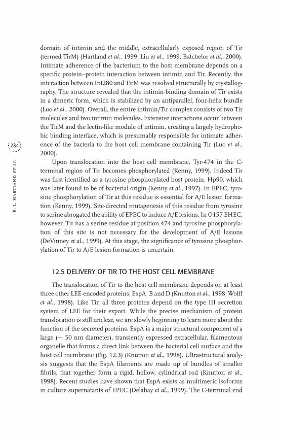
domain of intimin and the middle, extracellularly exposed region of Tir(termed TirM) (Hartland et al., 1999; Liu et al., 1999; Batchelor et al., 2000).Intimate adherence of the bacterium to the host membrane depends on aspecific protein–protein interaction between intimin and Tir. Recently, theinteraction between Int280 and TirM was resolved structurally by crystallog-raphy. The structure revealed that the intimin-binding domain of Tir existsin a dimeric form, which is stabilized by an antiparallel, four-helix bundle(Luo et al., 2000). Overall, the entire intimin/Tir complex consists of two Tirmolecules and two intimin molecules. Extensive interactions occur betweenthe TirM and the lectin-like module of intimin, creating a largely hydropho-bic binding interface, which is presumably responsible for intimate adher-ence of the bacteria to the host cell membrane containing Tir (Luo et al.,2000).
Upon translocation into the host cell membrane, Tyr-474 in the C-terminal region of Tir becomes phosphorylated (Kenny, 1999). Indeed Tirwas first identified as a tyrosine phosphorylated host protein, Hp90, whichwas later found to be of bacterial origin (Kenny et al., 1997). In EPEC, tyro-sine phosphorylation of Tir at this residue is essential for A/E lesion forma-tion (Kenny, 1999). Site-directed mutagenesis of this residue from tyrosineto serine abrogated the ability of EPEC to induce A/E lesions. In O157 EHEC,however, Tir has a serine residue at position 474 and tyrosine phosphoryla-tion of this site is not necessary for the development of A/E lesions(DeVinney et al., 1999). At this stage, the significance of tyrosine phosphor-ylation of Tir to A/E lesion formation is uncertain.
12.5 DELIVERY OF TIR TO THE HOST CELL MEMBRANE
The translocation of Tir to the host cell membrane depends on at leastthree other LEE-encoded proteins, EspA, B and D (Knutton et al., 1998; Wol¤et al., 1998). Like Tir, all three proteins depend on the type III secretionsystem of LEE for their export. While the precise mechanism of proteintranslocation is still unclear, we are slowly beginning to learn more about thefunction of the secreted proteins. EspA is a major structural component of alarge (� 50 nm diameter), transiently expressed extracellular, filamentousorganelle that forms a direct link between the bacterial cell surface and thehost cell membrane (Fig. 12.3) (Knutton et al., 1998). Ultrastructural analy-sis suggests that the EspA filaments are made up of bundles of smallerfibrils, that together form a rigid, hollow, cylindrical rod (Knutton et al.,1998). Recent studies have shown that EspA exists as multimeric isoformsin culture supernatants of EPEC (Delahay et al., 1999). The C-terminal end
e. l
. har
tlan
d e
t al
.
284

of EspA is strongly predicted to contain an alpha-helical region with a pro-pensity to form a coiled-coil interaction. Non-conservative site-directedmutagenesis of hydrophobic amino acid residues predicted to be essentialfor coiled-coil formation abrogated the ability of EspA to form multimers,assemble filaments and induce A/E lesions (Delahay et al., 1999). This sug-gests that, similar to the well-characterized coiled-coil region of flagellins(Hyman and Trachtenberg, 1991), coiled-coil interactions are important forEspA filament assembly and/or stability. Interestingly, espD mutants secreteonly low levels of EspA and are unable to produce mature EspA filaments,instead producing short, vestigial filaments (Knutton et al., 1998; Kresse etal., 1999). Although this suggests that EspD has a structural role in filamentassembly, EspD is not required for EspA multimerization and, at this stage,no staining of the filaments has been demonstrated with antisera to EspDand no protein–protein associations between EspA and EspD have beenobserved. Nevertheless, it is possible that EspD forms a minor component ofthe filament.
espB mutants produce normal EspA filaments and antibodies to EspB donot stain the filament, indicating that EspB does not play a integral role infilament construction (Knutton et al., 1998). EspB does, however, show a spe-cific and close association with the filament and can be co-purified with EspAfilaments by immunoprecipitation (Hartland et al., 2000a). Although EspBis essential for protein translocation, this protein is not required for attach-ment of EspA filaments to the host cell surface (Hartland et al., 2000a). Thusthe association between the EspA filament and EspB may represent thefilament’s transition from an adhesive function to a role in protein translo-cation.
interactio
ns betw
een e. co
li and
epithelial cells
285
a bFigure 12.3. (a) Scanning electron micrograph and (b) EspA immunofluorescence
showing that EspA filaments form a direct link between the bacterium and the host cell
membrane, in this example between the bacterium and human red blood cells (arrows).
Bars: (a) 0.5�m; (b) 1�m.

EP
EC
inn
er m
emb
ran
e
EP
EC
out
er m
embr
ane
EP
EC
cyt
osol
host
cel
l mem
bran
e
host
cel
l cyt
osol
Esp
B/D
gen
erat
edpo
re
Esp
A f
ilam
ent
nee
dle
co
mp
lex
Esc
N
Esc
Esc
Esc
Esc
Tir
Tir
Tir
inti
min
typ
e III
sec
reto
n
Figu
re 12.4
. Mod
el o
f E
PE
C p
rote
in tr
ansl
ocat
ion
. Th
e pr
opos
ed p
rote
in tr
ansl
ocat
ion
app
arat
us
(tra
nsl
ocon
) con
sist
s of
por
es in
the
bact
e ria
l en
velo
pe
(typ
e II
I se
cret
on) a
nd
in th
e h
ost c
ell m
embr
ane
(Esp
B/D
-gen
erat
ed p
ore)
con
nec
ted
by a
hol
low
Esp
A fi
lam
ent,
ther
eby
prov
idin
g a
con
tin
uou
s ch
ann
el
from
the
bact
eria
l to
the
hos
t cel
l cyt
osol
. Th
e tr
ansl
ocon
is u
sed
to tr
ansl
ocat
e T
ir in
to th
e h
ost c
ell,
wh
ere
it b
ecom
es in
sert
ed in
to th
e h
ost c
ell
mem
bran
e. S
ubs
equ
ent r
etra
ctio
n/e
limin
atio
n o
f E
spA
fila
men
ts a
llow
s in
tim
in-T
ir in
tera
ctio
n a
nd
pede
stal
for
mat
ion
.

Although the molecular basis of protein translocation has not yet beenelucidated for any type III secretion system, each model predicts the pres-ence of pore-forming proteins of bacterial origin in both the bacterial outermembrane and the host plasma membrane, which are connected by aneedle-like structure (Fig. 12.4) (Frankel et al., 1998). EspD and EspB showweak homology with YopB and YopD, respectively, of Yersinia (Håkansson etal., 1993). Like EspD and EspB, YopB and YopD are hydrophobic, type IIIsecreted proteins that are inserted into the host cell membrane during infec-tion (Neyt and Cornelis, 1999). YopB and YopD are pore-forming proteinsthat together form a channel in the host cell membrane through which othere¤ector proteins enter the cytoplasm of the target cell (Tardy et al., 1999).Pore formation by the type III secreted proteins of Yersinia spp. and Shigellaspp. has been correlated with the ability of the bacteria to mediate contact-dependent haemolysis of red blood cells (RBCs) (Håkansson et al., 1996;Blocker et al., 1999). EspD and EspB are both essential for protein transloca-tion and mediate EPEC-induced haemolysis of RBCs, suggesting that theymay also be pore-forming proteins (Warawa et al., 1999). Recently, Shaw etal. (2001) showed that EspD was the only bacterial protein detected in themembrane of RBCs following infection with EPEC. By contrast, EspB did notappear to be associated with the RBC membrane, suggesting that EspD playsa dominant role in pore formation. An investigation of protein–protein inter-actions found that EspD interacts with itself, suggesting that the pore maycomprise EspD multimers (S.J. Daniell et al, unpublished data).Interestingly, interactions between EspD and other secreted proteins havenot been observed thus far. Given its absence from the RBC membrane andapparent lack of interaction with EspD, the precise role of EspB in the for-mation of a membrane pore, and indeed protein translocation, remainsunclear.
12.6 HOST CELL CYTOSKELETAL AND SIGNALLING CHANGES
A number of bacterial pathogens mediate a direct e¤ect on the host cellcytoskeleton during infection. The development of an A/E lesion followinginfection of the epithelium with EPEC reflects gross ultrastructural changesin the host cell underneath, and around, adherent bacteria. Cytoskeletal reor-ganization involves the recruitment of several cytoskeletal proteins to thebody of the pedestal, including actin, �-actinin, talin and ezrin (Finlay et al.,1992). Although the precise cellular processes that govern cytoskeletal rear-rangement during infection with EPEC are unknown, recently the N-terminal region of Tir was shown to interact directly with �-actinin (Goosney
interactio
ns betw
een e. co
li and
epithelial cells
287

et al., 2000). �-Actinin is one of the cytoskeletal proteins recruited to the ped-estal during A/E lesion formation. The N-terminal region of Tir correspondsto an area of the protein that is located inside the host cell (Hartland et al.,1999), and provides the first demonstration of a direct link between an EPEC-derived factor and a host cell target protein. This interaction occurred inde-pendently of tyrosine phosphorylation of Tir, unlike the recruitment of othercytoskeletal proteins (actin, vasodilator-stimulated phosphoprotein (VASP)and N-Wiscott–Aldrich syndrome protein (N-WASP) where tyrosine phos-phorylation of Tir appears to be an essential step (Goosney et al., 2000).
As well as its putative role in the formation of a membrane pore, EspBhas been suggested to have an independent e¤ect on the cytoskeleton of thetarget cell. EspB is translocated to both the host cell membrane and cytosolicfractions, and transfection of EspB into the cytosol of epithelial cells inducesthe reorganization of actin stress fibres (Taylor et al., 1999). This suggeststhat EspB may harbour some cytoskeletal-altering activity of its own,although no host cell targets have been identified to date. EPEC has also beenshown to induce a number of signalling changes in the host cell that mayindirectly lead to cytoskeletal alterations. Attachment of the bacteria to theepithelial cell surface leads to inositol phosphate fluxes and activation ofprotein kinase C (PKC) and phospholipase C-� (PLC-�) (Crane and Oh,1997). These cellular responses occur only if the bacteria express an intacttype III secretion system and intimin. Hence, intimate attachment isrequired to induce the signalling changes.
12.7 LOCALIZED ADHERENCE AND MICROCOLONYFORMATION
In addition to A/E lesions, EPEC adheres to the surface of epithelial cellsin characteristic microcolonies. This pattern of attachment is termed local-ized adherence (LA) and denotes the adhesion of bacteria to host cells inaggregates or clusters. The bacterial microcolonies are believed to be essen-tial for eªcient colonization of the host and subsequent dispersal of thepathogen. Localized adherence and microcolony formation by EPECdepends on the expression of a type IV pilus called the bundle-forming pilus,BFP (Giron et al., 1991; Donnenberg et al., 1992). While the role and timingof microcolony formation in vivo remains uncertain, recent studies withIVOCs suggest that BFP mediates microcolony formation subsequent to inti-mate attachment and the development of A/E lesions (Hicks et al., 1998),rather than initiating attachment of the bacteria to the host epithelium priorto A/E lesion formation.
e. l
. har
tlan
d e
t al
.
288

Production and assembly of BFP depends on a cluster of 14 genes, whichare present on the large, 50–70MDa EAF plasmids (Donnenberg et al., 1992;Stone et al., 1996). The major pilin subunit (BFP pilin or bundlin) is encodedby the first gene of the cluster, bfpA. BFP pilin is produced as a pre-protein thatundergoes proteolytic cleavage by the bfpP-encoded peptidase into a matureform (Zhang et al., 1994). BFP production is regulated by the plasmid-encodedPer (also known as BfpTWV) (Tobe et al., 1996). Human volunteer studieshave shown that BFP is essential for colonization of the host intestine (Bieberet al., 1998); bfpA mutants caused significantly less diarrhoea than wild-typeEPEC in experimentally infected adults. In addition, a bfpF mutant that lacksthe ability to disperse from microcolonies was 200 times less virulent than thewild-type strain in the same infection model, suggesting that microcolony dis-persal is an important virulence trait. In vitro studies have shown that BFPundergoes marked changes in quaternary structure as the infection proceeds.The pilus changes from a thin to a thick bundle structure that results indisruption of the interactions between bacteria and the subsequent release ofbacteria from the microcolony (Fig. 12.5) (Knutton et al., 1999). This transfor-mation relies on BfpF, a putative nucleotide-binding factor (Anantha et al.,1998), which is either directly or indirectly involved in the shift from thin tothick bundles. BfpF is not required for thin filament production but may actas an energizer for pilus retraction in a manner analogous to PilT ofPseudomonas aeruginosa (Knutton et al., 1999). Thus, through the structuraltransformation of the pilus, BFP is able to promote initial bacterial aggrega-tion and formation of the microcolony and then dispersal of bacteria alreadyprimed to form new A/E lesions and microcolonies at fresh sites in the gut.
interactio
ns betw
een e. co
li and
epithelial cells
289
a bFigure 12.5. Scanning electron micrographs illustrating bundle-forming pilus (BFP)-
mediated microcolony formation and dispersal. (a) Thin BFP filaments initially promote
bacteria–bacteria interaction and microcolony formation; (b) rearrangement to a network
of much thicker BFP filaments promotes release of bacteria and dispersal of the
microcolony. Bar, 0.5�m.

12.8 CONCLUSIONS
Although our understanding of the mechanisms employed by EPEC tointeract with the host intestinal epithelium has improved dramatically inrecent years, the precise mechanism of EPEC-induced diarrhoea remainsobscure. Features of EPEC diarrhoea suggest the existence of a toxin but todate no toxigenic factors have been isolated. Extensive e¤acement of absorp-tive microvilli due to A/E lesions may contribute to fluid loss but the shortincubation period between ingestion and onset of diarrhoea (less than 4hours) suggests active fluid secretion by the intestine (Nataro and Kaper,1998). One possible mechanism involves EPEC-induced alterations in tran-sepithelial electric resistance that result from disruption of epithelial tightjunctions due to phosphorylation of myosin light chain (Philpott et al., 1996;Yuhan et al., 1997). Adherence by EPEC also stimulates some secretion ofchloride ions by epithelial cells (Collington et al., 1998). These modes ofinterference with the intestinal barrier may lead to increased permeability ofthe gut and contribute to EPEC-induced diarrhoea, although host factorssuch as the release of inflammatory mediators may also play a role.
A/E lesion formation and intimate adherence of EPEC to the host epi-thelium is a complex, multistage process. The LEE-encoded secreted pro-teins and intimin represent a potent set of virulence determinants that act toundermine the structure of the host cell cytoskeleton and e¤ect tight adher-ence of the bacteria to the intestinal epithelium. Gradually, through the useof animal models and powerful tools in molecular biology, cell biology,microscopy and structural biology, we are beginning to unravel the individ-ual and combined roles of the LEE-encoded proteins and other virulencefactors in pathogenesis. A more complete understanding of the molecularbasis of protein translocation and subsequent bacterial reorganization of thehost cell cytoskeleton would aid the design of strategies to intervene in thedisease process.
ACKNOWLEDGEMENTS
Research in the authors’ laboratories is supported by the WellcomeTrust. E.L.H. is the recipient of a Royal Society/NHMRC Howard Florey fel-lowship.
REFERENCES
Abe, A., de Grado, M., Pfuetzner, R.A., Sanchez-Sanmartin, C., Devinney, R.,
Puente, J.L., Strynadka, N.C. and Finlay, B.B. (1999). Enteropathogenic
e. l
. har
tlan
d e
t al
.
290
Escherichia coli translocated intimin receptor, Tir, requires a specific chape-
rone for stable secretion. Molecular Microbiology 33, 1162–1175.
Adu-Bobie, J., Frankel, G., Bain, C., Goncalves, A.G., Trabulsi, L.R., Douce, G.,
Knutton, S. and Dougan, G. (1998). Detection of intimins alpha, beta,
gamma, and delta, four intimin derivatives expressed by attaching and e¤ac-
ing microbial pathogens. Journal of Clinical Microbiology 36, 662–668.
Anantha, R.P., Stone, K.D. and Donnenberg, M.S. (1998). Role of BfpF, a
member of the PilT family of putative nucleotide-binding proteins, in type
IV pilus biogenesis and in interactions between enteropathogenic
Escherichia coli and host cells. Infection and Immunity 66, 122–131.
Batchelor, M., Prasannan, S., Daniell, S., Reece, S., Connerton, I., Bloomberg, G.,
Dougan, G., Frankel, G. and Matthews, S. (2000). Structural basis for recog-
nition of the translocated intimin receptor (Tir) by intimin from enteropath-
ogenic Escherichia coli. EMBO Journal 19, 2452–2464.
Bieber, D., Ramer, S.W., Wu, C.Y., Murray, W.J., Tobe, T., Fernandez, R. and
Schoolnik, G.K. (1998). Type IV pili, transient bacterial aggregates, and vir-
ulence of enteropathogenic Escherichia coli. Science 280, 2114–2118.
Blocker, A., Gounon, P., Larquet, E., Niebuhr, K., Cabiaux, V., Parsot, C. and
Sansonetti, P. (1999). The tripartite type III secreton of Shigella flexneri inserts
IpaB and IpaC into host membranes. Journal of Cell Biology 147, 683–693.
Collington, G.K., Booth, I.W. and Knutton, S. (1998). Rapid modulation of elec-
trolyte transport in Caco-2 cell monolayers by enteropathogenic Escherichia
coli (EPEC) infection. Gut 42, 200–207.
Crane, J.K. and Oh, J.S. (1997). Activation of host cell protein kinase C by enter-
opathogenic Escherichia coli. Infection and Immunity 65, 3277–3285.
Deibel, C., Kramer, S., Chakraborty, T. and Ebel, F. (1998). EspE, a novel secreted
protein of attaching and e¤acing bacteria, is directly translocated into
infected host cells, where it appears as a tyrosine-phosphorylated 90kDa
protein. Molecular Microbiology 28, 463–474.
Delahay, R.M., Knutton, S., Shaw, R.K., Hartland, E.L., Pallen, M.J. and Frankel,
G. (1999). The coiled-coil domain of EspA is essential for the assembly of the
type III secretion translocon on the surface of enteropathogenic Escherichia
coli. Journal of Biological Chemistry 274, 35969–35974.
DeVinney, R., Stein, M., Reinscheid, D., Abe, A., Ruschkowski, S. and Finlay, B.B.
(1999). Enterohemorrhagic Escherichia coli O157:H7 produces Tir, which is
translocated to the host cell membrane but is not tyrosine phosphorylated.
Infection and Immunity 67, 2389–2398.
Donnenberg, M.S., Giron, J.A., Nataro, J.P. and Kaper, J.B. (1992). A plasmid-
encoded type IV fimbrial gene of enteropathogenic Escherichia coli associated
with localized adherence. Molecular Microbiology 6, 3427–3437.
interactio
ns betw
een e. co
li and
epithelial cells
291

Donnenberg, M.S., Tacket, C.O., James, S.P., Losonsky, G., Nataro, J.P.,
Wasserman, S.S., Kaper, J.B. and Levine, M.M. (1993). Role of the eaeA gene
in experimental enteropathogenic Escherichia coli infection. Journal of
Clinical Investigation 92, 1412–1417.
Elliott, S.J., Wainwright, L.A., McDaniel, T.K., Jarvis, K.G., Deng, Y.K., Lai, L.C.,
McNamara, B.P., Donnenberg, M.S. and Kaper, J.B. (1998). The complete
sequence of the locus of enterocyte e¤acement (LEE) from enteropathogenic
Escherichia coli E2348/69. Molecular Microbiology 28, 1–4.
Elliott, S.J., Hutcheson, S.W., Dubois, M.S., Mellies, J.L., Wainwright, L.A.,
Batchelor, M., Frankel, G., Knutton, S. and Kaper, J.B. (1999). Identification
of CesT, a chaperone for the type III secretion of Tir in enteropathogenic
Escherichia coli. Molecular Microbiology 33, 1176–1189.
Elliott, S.J., Sperandio, V., Giron, J.A., Shin, S., Mellies, J.L., Wainwright, L.,
Hutcheson, S.W., McDaniel, T.K. and Kaper, J.B. (2000). The locus of
enterocyte e¤acement (LEE)-encoded regulator controls expression of
both LEE- and non-LEE-encoded virulence factors in enteropathogenic
and enterohemorrhagic Escherichia coli. Infection and Immunity 68,
6115–6126.
Finlay, B.B., Rosenshine, I., Donnenberg, M.S. and Kaper, J.B. (1992).
Cytoskeletal composition of attaching and e¤acing lesions associated with
enteropathogenic Escherichia coli adherence to HeLa cells. Infection and
Immunity 60, 2541–2543.
Frankel, G., Candy, D.C., Everest, P. and Dougan, G. (1994). Characterization of
the C-terminal domains of intimin-like proteins of enteropathogenic and
enterohemorrhagic Escherichia coli, Citrobacter freundii, and Hafnia alvei.
Infection and Immunity 62, 1835–1842.
Frankel, G., Candy, D.C., Fabiani, E., Adu-Bobie, J., Gil, S., Novakova, M.,
Phillips, A.D. and Dougan, G. (1995). Molecular characterization of a
carboxy-terminal eukaryotic-cell-binding domain of intimin from entero-
pathogenic Escherichia coli. Infection and Immunity 63, 4323–4328.
Frankel, G., Lider, O., Hershkoviz, R., Mould, A.P., Kachalsky, S.G., Candy,
D.C.A., Cahalon, L., Humphries, M.J. and Dougan, G. (1996a). The cell-
binding domain of intimin from enteropathogenic Escherichia coli binds to
beta1 integrins. Journal of Biological Chemistry 271, 20359–20364.
Frankel, G., Phillips, A.D., Novakova, M., Field, H., Candy, D.C., Schauer, D.B.,
Douce, G. and Dougan, G. (1996b). Intimin from enteropathogenic
Escherichia coli restores murine virulence to a Citrobacter rodentium eaeA
mutant: induction of an immunoglobulin A response to intimin and EspB.
Infection and Immunity 64, 5315–5325.
Frankel, G., Phillips, A.D., Rosenshine, I., Dougan, G., Kaper, J.B. and Knutton,
e. l
. har
tlan
d e
t al
.
292

S. (1998). Enteropathogenic and enterohaemorrhagic Escherichia coli: more
subversive elements. Molecular Microbiology 30, 911–921.
Friedberg, D., Umanski, T., Fang, Y. and Rosenshine, I. (1999). Hierarchy in the
expression of the locus of enterocyte e¤acement genes of enteropathogenic
Escherichia coli. Molecular Microbiology 34, 941–952.
Giron, J.A., Ho, A.S. and Schoolnik, G.K. (1991). An inducible bundle-forming
pilus of enteropathogenic Escherichia coli. Science 254, 710–713.
Gomez-Duarte, O.G. and Kaper, J.B. (1995). A plasmid-encoded regulatory
region activates chromosomal eaeA expression in enteropathogenic
Escherichia coli. Infection and Immunity 63, 1767–1776.
Goosney, D.L., DeVinney, R., Pfuetzner, R.A., Frey, E.A., Strynadka, N.C. and
Finlay, B.B. (2000). Enteropathogenic Escherichia coli translocated intimin
receptor, Tir, interacts directly with alpha-actinin. Current Biology 10, 735–738.
Håkansson, S., Bergman, T., Vanooteghem, J.C., Cornelis, G. and Wolf-Watz, H.
(1993). YopB and YopD constitute a novel class of Yersinia Yop proteins.
Infection and Immunity 61, 71–80.
Håkansson, S., Schesser, K., Persson, C., Galyov, E.E., Rosqvist, R., Homble, F.
and Wolf-Watz, H. (1996). The YopB protein of Yersinia pseudotuberculosis is
essential for the translocation of Yop e¤ector proteins across the target cell
plasma membrane and displays a contact-dependent membrane disrupting
activity. EMBO Journal 15, 5812–5823.
Hartland, E.L., Batchelor, M., Delahay, R.M., Hale, C., Matthews, S., Dougan, G.,
Knutton, S., Connerton, I. and Frankel, G. (1999). Binding of intimin from
enteropathogenic Escherichia coli to Tir and to host cells. Molecular
Microbiology 32, 151–158.
Hartland, E.L., Daniell, S.J., Delahay, R.M., Neves, B.C., Wallis, T., Shaw, R.K.,
Hale, C., Knutton, S. and Frankel, G. (2000a). The type III protein transloca-
tion system of enteropathogenic Escherichia coli involves EspA–EspB protein
interactions. Molecular Microbiology 35, 1483–1492.
Hartland, E.L., Huter, V., Higgins, L.M., Goncalves, N.S., Dougan, G., Phillips,
A.D., MacDonald, T.T. and Frankel, G. (2000b). Expression of intimin
gamma from enterohemorrhagic Escherichia coli in Citrobacter rodentium.
Infection and Immunity 68, 4637–4646.
Hicks, S., Frankel, G., Kaper, J.B., Dougan, G. and Phillips, A.D. (1998). Role of
intimin and bundle-forming pili in enteropathogenic Escherichia coli adhe-
sion to pediatric intestinal tissue in vitro. Infection and Immunity 66,
1570–1578.
Higgins, L.M., Frankel, G., Connerton, I., Goncalves, N.S., Dougan, G. and
MacDonald, T.T. (1999a). Role of bacterial intimin in colonic hyperplasia and
inflammation. Science 285, 588–591.
interactio
ns betw
een e. co
li and
epithelial cells
293

Higgins, L.M., Frankel, G., Douce, G., Dougan, G. and MacDonald, T.T. (1999b).
Citrobacter rodentium infection in mice elicits a mucosal Th1 cytokine
response and lesions similar to those in murine inflammatory bowel
disease. Infection and Immunity 67, 3031–3039.
Hueck, C.J. (1998). Type III protein secretion systems in bacterial pathogens of
animals and plants. Microbiology and Molecular Biology Reviews 62, 379–433.
Hyman, H.C. and Trachtenberg, S. (1991). Point mutations that lock Salmonella
typhimurium flagellar filaments in the straight right-handed and left-handed
forms and their relation to filament superhelicity. Journal of Molecular Biology
220, 79–88.
Isberg, R.R., Voorhis, D.L. and Falkow, S. (1987). Identification of invasin: a
protein that allows enteric bacteria to penetrate cultured mammalian cells.
Cell 50, 769–778.
Jerse, A.E. and Kaper, J.B. (1991). The eae gene of enteropathogenic Escherichia
coli encodes a 94– kilodalton membrane protein, the expression of which is
influenced by the EAF plasmid. Infection and Immunity 59, 4302–4309.
Jerse, A.E., Yu, J., Tall, B.D. and Kaper, J.B. (1990). A genetic locus of enteropath-
ogenic Escherichia coli necessary for the production of attaching and e¤acing
lesions on tissue culture cells. Proceedings of the National Academy of Sciences,
USA 87, 7839–7843.
Kelly, G., Prasannan, S., Daniell, S., Fleming, K., Frankel, G., Dougan, G.,
Connerton, I. and Matthews, S. (1999). Structure of the cell-adhesion frag-
ment of intimin from enteropathogenic Escherichia coli. Nature Structural
Biology 6, 313–318.
Kenny, B. (1999). Phosphorylation of tyrosine 474 of the enteropathogenic
Escherichia coli (EPEC) Tir receptor molecule is essential for actin nucleating
activity and is preceded by additional host modifications. Molecular
Microbiology 31, 1229–1241.
Kenny, B., DeVinney, R., Stein, M., Reinscheid, D.J., Frey, E.A. and Finlay, B.B.
(1997). Enteropathogenic Escherichia coli (EPEC) transfers its receptor for
intimate adherence into mammalian cells. Cell 91, 511–520.
Knutton, S., Baldwin, T., Williams, P.H. and McNeish, A.S. (1989). Actin accu-
mulation at sites of bacterial adhesion to tissue culture cells: basis of a new
diagnostic test for enteropathogenic and enterohemorrhagic Escherichia coli.
Infection and Immunity 57, 1290–1298.
Knutton, S., Rosenshine, I., Pallen, M.J., Nisan, I., Neves, B.C., Bain, C., Wol¤,
C., Dougan, G. and Frankel, G. (1998). A novel EspA-associated surface orga-
nelle of enteropathogenic Escherichia coli involved in protein translocation
into epithelial cells. EMBO Journal 17, 2166–2176.
Knutton, S., Shaw, R.K., Anantha, R.P., Donnenberg, M.S. and Zorgani, A.A.
e. l
. har
tlan
d e
t al
.
294

(1999). The type IV bundle-forming pilus of enteropathogenic Escherichia
coli undergoes dramatic alterations in structure associated with bacterial
adherence, aggregation and dispersal. Molecular Microbiology 33, 499–509.
Kresse, A.U., Rohde, M. and Guzmán, C.A. (1999). The EspD protein of entero-
hemorrhagic Escherichia coli is required for the formation of bacterial surface
appendages and is incorporated in the cytoplasmic membranes of target
cells. Infection and Immunity 67, 4834–4842.
Kubori, T., Matsushima, Y., Nakamura, D., Uralil, J., Lara-Tejero, M., Sukhan, A.,
Galan, J.E. and Aizawa, S.I. (1998). Supramolecular structure of the
Salmonella typhimurium type III protein secretion system. Science 280,
602–625.
Liu, H., Magoun, L., Luperchio, S., Schauer, D.B. and Leong, J.M. (1999). The Tir-
binding region of enterohaemorrhagic Escherichia coli intimin is suªcient
to trigger actin condensation after bacterial-induced host cell signalling.
Molecular Microbiology 34, 67–81.
Luo, Y., Frey, E.A., Pfuetzner, R.A., Creagh, A.L., Knoechel, D.G., Haynes, C.A.,
Finlay, B.B. and Strynadka, N.C. (2000). Crystal structure of enteropatho-
genic Escherichia coli intimin-receptor complex. Nature 405, 1073–1077.
McDaniel, T.K. and Kaper, J.B. (1997). A cloned pathogenicity island from enter-
opathogenic Escherichia coli confers the attaching and e¤acing phenotype on
E. coli K-12. Molecular Microbiology 23, 399–407.
McDaniel, T.K., Jarvis, K.G., Donnenberg, M.S. and Kaper, J.B. (1995). A genetic
locus of enterocyte e¤acement conserved among diverse enterobacterial
pathogens. Proceedings of the National Academy of Sciences, USA 92,
1664–1668.
Mellies, J.L., Elliott, S.J., Sperandio, V., Donnenberg, M.S. and Kaper, J.B. (1999).
The Per regulon of enteropathogenic Escherichia coli: identification of a reg-
ulatory cascade and a novel transcriptional activator, the locus of enterocyte
e¤acement (LEE)-encoded regulator (Ler). Molecular Microbiology 33,
296–306.
Moon, H.W., Whipp, S.C., Argenzio, R.A., Levine, M.M. and Giannella, R.A.
(1983). Attaching and e¤acing activities of rabbit and human enteropatho-
genic Escherichia coli in pig and rabbit intestines. Infection and Immunity 41,
1340–1351.
Nataro, J.P. and Kaper, J.B. (1998). Diarrheagenic Escherichia coli. Clinical
Microbiology Reviews 11, 142–201.
Nataro, J.P., Maher, K.O., Mackie, P. and Kaper, J.B. (1987). Characterization of
plasmids encoding the adherence factor of enteropathogenic Escherichia coli.
Infection and Immunity 55, 2370–2377.
Neyt, C. and Cornelis, G.R. (1999). Insertion of a Yop translocation pore into the
interactio
ns betw
een e. co
li and
epithelial cells
295

macrophage plasma membrane by Yersinia enterocolitica: requirement for
translocators YopB and YopD, but not LcrG. Molecular Microbiology 33,
971–981.
Phillips, A.D. and Frankel, G. (2000). Intimin-mediated tissue specificity in
enteropathogenic Escherichia coli interaction with human intestinal organ
cultures. Journal of Infectious Diseases 181, 1496–500.
Phillips, A.D., Giron, J., Hicks, S., Dougan, G. and Frankel, G. (2000). Intimin
from enteropathogenic Escherichia coli mediates remodelling of the eukar-
yotic cell surface. Microbiology 146, 1333–1344.
Philpott, D.J., McKay, D.M., Sherman, P.M. and Perdue, M.H. (1996). Infection
of T84 cells with enteropathogenic Escherichia coli alters barrier and trans-
port functions. American Journal of Physiology 270, G634–G645.
Reid, S.D., Herbelin, C.J., Bumbaugh, A.C., Selander, R.K. and Whittam, T.S.
(2000). Parallel evolution of virulence in pathogenic Escherichia coli. Nature
406, 64–67.
Sanger, J.M., Chang, R., Ashton, F., Kaper, J.B. and Sager, J.W. (1996). Novel form
of actin motility transports bacteria on the surface of infected cells. Cell
Motility and the Cytoskeleton 34, 279–287.
Shaw, R.K., Daniell, S., Ebel, F., Frankel, G. and Knutton, S. (2001). EspA fila-
ment-mediated protein translocation into red blood cells. Cellular
Microbiology 3, 213–222.
Sperandio, V., Mellies, J.L., Nguyen, W., Shin, S. and Kaper, J.B. (1999). Quorum
sensing controls expression of the type III secretion gene transcription and
protein secretion in enterohemorrhagic and enteropathogenic Escherichia
coli. Proceedings of the National Academy of Sciences, USA 96, 15196–15201.
Sperandio, V., Mellies, J.L., Delahay, R.M., Frankel, G., Crawford, J.A., Nguyen,
W. and Kaper, J.B. (2000). Activation of enteropathogenic E. coli (EPEC) LEE2
and LEE3 operons by Ler. Molecular Microbiology 38, 781–793.
Stone, K.D., Zhang, H.Z., Carlson, L.K. and Donnenberg, M.S. (1996). A cluster
of fourteen genes from enteropathogenic Escherichia coli is suªcient for the
biogenesis of a type IV pilus. Molecular Microbiology 20, 325–337.
Tacket, C.O., Sztein, M.B., Losonsky, G., Abe, A., Finlay, B.B., McNamara, B.P.,
Fantry, G.T., James, S.P., Nataro, J.P., Levine, M.M. and Donnenberg, M.S.
(2000). Role of EspB in experimental human enteropathogenic Escherichia
coli infection. Infection and Immunity 68, 3689–3695.
Tamano, K., Aizawa, S. I., Katayama, E., Nonaka, T., Imajoh-Ohmi, S., Kuwae, A.,
Nagai, S. and Sasakawa, C. (2000). Supramolecular structure of the Shigella
type III secretion machinery: the needle part is changeable in length and
essential for delivery of e¤ectors. EMBO Journal 19, 3876–3887.
Tardy, F., Homble, F., Neyt, C., Wattiez, R., Cornelis, G.R., Ruysschaert, J.M. and
e. l
. har
tlan
d e
t al
.
296

Cabiaux, V. (1999). Yersinia enterocolitica type III secretion-translocation
system: channel formation by secreted Yops. EMBO Journal 18, 6793–6799.
Taylor, K.A., Luther, P.W. and Donnenberg, M.S. (1999). Expression of the EspB
protein of enteropathogenic Escherichia coli within HeLa cells a¤ects stress
fibers and cellular morphology. Infection and Immunity 67, 120–125.
Tobe, T., Schoolnik, G.K., Sohel, I., Bustamante, V.H. and Puente, J.L. (1996).
Cloning and characterization of bfpTVW, genes required for the transcrip-
tional activation of bfpA in enteropathogenic Escherichia coli. Molecular
Microbiology 21, 963–975.
Tzipori, S., Gunzer, F., Donnenberg, M.S., de Montigny, L., Kaper, J.B. and
Donohue-Rolfe, A. (1995). The role of the eaeA gene in diarrhoea and neuro-
logical complications in a gnotobiotic piglet model of enterohemorrhagic
Escherichia coli infection. Infection and Immunity 63, 3621–3627.
Vallance, B.A. and Finlay, B.B. (2000). Exploitation of host cells by enteropatho-
genic Escherichia coli. Proceedings of the National Academy of Sciences, USA 97,
8799–8806.
Wainwright, L.A. and Kaper, J.B. (1998). EspB and EspD require a specific chape-
rone for proper secretion from enteropathogenic Escherichia coli. Molecular
Microbiology 27, 1247–1260.
Warawa, J., Finlay, B.B. and Kenny, B. (1999). Type III secretion-dependent hemo-
lytic activity of enteropathogenic Escherichia coli. Infection and Immunity 67,
5538–5540.
Wol¤, C., Nisan, I., Hanski, E., Frankel, G. and Rosenshine, I. (1998). Protein
translocation into host epithelial cells by infecting enteropathogenic
Escherichia coli. Molecular Microbiology 28, 143–155.
Yuhan, R., Koutsouris, A., Savkovic, S.D. and Hecht, G. (1997). Enteropathogenic
Escherichia coli-induced myosin light chain phosphorylation alters intestinal
epithelial permeability. Gastroenterology 113, 1873–1882.
Zhang, H.Z., Lory, S. and Donnenberg, M.S. (1994). A plasmid-encoded prepilin
peptidase gene from enteropathogenic Escherichia coli. Journal of Bacteriology
176, 6885–6891.
interactio
ns betw
een e. co
li and
epithelial cells
297

CHAPTER 13
Host cell responses to Porphyromonasgingivalis and Actinobacillusactinomycetemcomitans
Richard J. Lamont
13.1 INTRODUCTION
The area of contact between the teeth and the gums (gingiva) is an ana-tomically unique region that comprises mineralized tissue embedded in epi-thelium and exposed to a microbially abundant environment. The small(1–4mm deep) gap between the surfaces of the tooth and the gingiva isknown as the gingival sulcus or crevice. The gingiva is highly vascularizedand the crevice is lined with sulcular epithelial cells that di¤er from oral epi-thelial cells by exhibiting less keratinization. Apically, sulcular epitheliumbecomes junctional epithelium that is characterized by a lack of keratiniza-tion, limited di¤erentiation and a relatively permeable structure. It is thisjunctional epithelium that directly interposes between the gingiva and thetooth surface (Fig. 13.1). In destructive periodontal disease there is migra-tion of the junctional epithelium resulting in enlargement of the crevice intoa deeper periodontal pocket that contains inflammatory cells such as neu-trophils and T-cells. The gingiva itself also contains immune cells includingB-cells, T-cells and dendritic cells. The microbiota of the gingival area in bothhealth and disease is complex, with at least 500 species of bacteria present inthe gingival crevice. Although many of these have pathogenic potential, thestrongest causal associations have been demonstrated betweenPorphyromonas gingivalis and severe adult periodontitis, and betweenActinobacillus actinomycetemcomitans and localized juvenile periodontitis.
Many factors contribute to the maintenance or disruption of the ecolog-ical balance in the subgingival area. The immunological status of the host,the relative and absolute numbers of specific organisms or groups of organ-isms, and environmental parameters such as tobacco use, all play a role indetermining gingival health or disease (Socransky and Ha¤ajee, 1992).Periodontal pathogens such as P. gingivalis and A. actinomycetemcomitanscan, therefore, be present in the absence of disease (Greenstein and
299
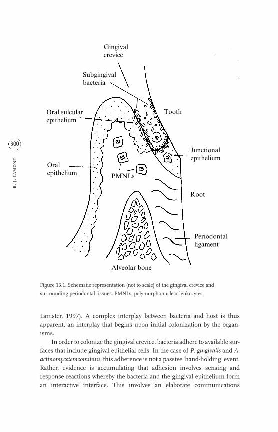
Lamster, 1997). A complex interplay between bacteria and host is thusapparent, an interplay that begins upon initial colonization by the organ-isms.
In order to colonize the gingival crevice, bacteria adhere to available sur-faces that include gingival epithelial cells. In the case of P. gingivalis and A.actinomycetemcomitans, this adherence is not a passive ‘hand-holding’ event.Rather, evidence is accumulating that adhesion involves sensing andresponse reactions whereby the bacteria and the gingival epithelium forman interactive interface. This involves an elaborate communications
r. j
. lam
on
t
300
Alveolar bone
Root
Periodontalligament
Oralepithelium
Oral sulcularepithelium
Junctionalepithelium
PMNLs
Gingivalcrevice
Subgingivalbacteria
Tooth
Figure 13.1. Schematic representation (not to scale) of the gingival crevice and
surrounding periodontal tissues. PMNLs, polymorphonuclear leukocytes.

network that generates and transmits signals among bacteria, epithelialcells and the underlying cells in the periodontal tissues. Bacteria possess avariety of adhesins, with di¤ering receptor specificities and aªnities, thatcan potentially impinge to varying degrees upon diverse receptor-dependenthost cell biochemical pathways. The consequences of this adhesion processinclude uptake of bacteria within the host cells and modification of pheno-typic properties that are important for tissue integrity and maintenance ofthe local innate host defence mechanisms that serve to control the micro-bial challenge.
13.2 ADHESINS DISPLAYED BY P. GINGIVALIS AND A.ACTINOMYCETEMCOMITANS
As adherence is an important early step in defining the nature of hostcell responses to P. gingivalis and A. actinomycetemcomitans, we will begin ourstory with a description of the adhesins of these organisms and their mech-anisms of action.
13.2.1 Porphyromonas gingivalis
Porphyromonas gingivalis can bind to host cells including epithelial cells,endothelial cells, fibroblasts, and erythrocytes, and to components of theextracellular matrix namely laminin, elastin, fibronectin, type I collagen,thrombospondin and vitronectin (Kontani et al., 1997; Lamont andJenkinson, 1998; Nakamura et al., 1999; Sojar et al., 1999; Dorn et al., 2000).Adhesion is multimodal, involving fimbriae, outer membrane proteins andproteinases, molecules that are inextricably linked at both the transcriptionaland post-translational levels.
The major fimbriae of P. gingivalis constitute a unique class of theseGram-negative organelles and comprised an �43kDa fimbrillin (FimA)monomer. Fimbrillin possesses a number of binding domains for individualsubstrate recognition. The functional domain of FimA for epithelial cellsspans amino acid residues 49–90 whereas the domains for fibronectin-binding are located within amino acid residues 126–146 along with318–337, and involve a conserved VXXXA sequence (Sojar et al., 1995,1999). Although the fimA gene is monocistronic, immediately downstreamare four genes whose products may be associated with the mature fimbriae(Watanabe et al., 1996). The fimA upstream region contains functionallyactive �70-like promoter consensus sequences along with a potential UPelement (Xie and Lamont, 1999). AT-rich sequences upstream from the RNA
ho
st cell respon
ses to p. g
ing
ivalis
301

polymerase-binding sites are involved in positive regulation of transcrip-tional activity. Environmental cues to which fimA responds include temper-ature, haemin concentration and salivary molecules (Xie et al., 1997),parameters with relevance to conditions in the oral cavity. The fimA gene canalso be positively autoregulated by the FimA protein (Xie et al., 2000).
Although the primary function of proteinases secreted by the asacchar-olytic P. gingivalis is the provision of nutrients, proteinases are also involvedboth directly and indirectly in adhesion. At least eight distinct proteinasesare produced by P. gingivalis (for reviews, see Potempa et al., 1995;Kuramitsu, 1998; Curtis et al., 1999) and direct enzyme–substrate interac-tions can e¤ect adhesion, albeit short lived. Possibly more importantly, theC-terminal coding regions of the Arg-X- and Lys-X-specific proteases RgpAand Kgp contain extensive amino acid repeat blocks that have up to 90%identity with sequences that are also found in the HagA, D and E haemag-glutinin proteins (discussed below) (Barkocy-Gallagher et al., 1996). Theseregions may thus allow direct binding to human cell surface receptors. Anadditional adherence-related activity of proteinases is the partial degradationof substrates that subsequently exposes epitopes for adhesin recognition.Hydrolysis of fibronectin or other matrix proteins by the Arg-X-specific pro-teases RgpA and RgpB displays C-terminal Arg residues that mediate fim-briae-dependent binding (Kontani et al., 1996). RgpA and RgpB alsocontribute to the fimbriae-mediated adhesive process by processing theleader peptide from the fimbrillin precursor (Nakayama et al., 1996), and byup-regulating transcription of the fimA gene (Tokuda et al., 1996; Xie et al.,2000).
In addition to the haemagglutinin-associated activities of the RgpA andKgp proteinases, five hag genes encoding haemagglutinins have now beensequenced. The hagA, hagD and hagE genes encode polypeptides with73–93% identical amino acid sequences, while hagB and hagC genes are atdistinct chromosomal loci and encode 39kDa polypeptides that are 98.6%identical (Progulske-Fox et al., 1995; Lépine and Progulske-Fox, 1996). Aminimal peptide motif PVQNLT has recently been shown to be associatedwith haemagglutinating activity and is found within the proteinase–haemag-glutinin sequences (Shibita et al., 1999) and at multiple chromosomal sites(Barokocy-Gallagher et al., 1996).
13.2.2 Actinobacillus actinomycetemcomitans
Actinobacillus actinomycetemcomitans can also adhere to host cells and tomatrix components such as fibronectin, collagen and laminin (Meyer and
r. j
. lam
on
t
302

Fives-Taylor 1994; Alugupalli et al., 1996; Mintz and Fives-Taylor, 1999).Adhesion is associated with fimbriae, outer membrane proteins, and extra-cellular vesicles and amorphous material (Fives-Taylor et al., 1999). Fimbriaeare found only on recent clinical isolates; laboratory subculture results in lossof fimbrial expression (Scannapieco et al., 1987; Rosan et al., 1988). Althoughthe mechanism of this transition is unknown, progress is being made on thegenetics of fimbrial structure and assembly. The fimbrial subunit appears tobe a 6.5kDa protein (Flp) that bears homology to the Neisseria gonorrhoeaetype IV fimbriae, which are synthesized via the general protein secretionpathway for fimbrial assembly (Hultgren et al., 1996; Inoue et al., 1998). Theflp gene comprises part of an operon that includes genes that may be involvedin protein secretion and fimbrial assembly (Haase et al., 1999). Whether thefimbrial structural component per se mediates adhesion to host cells, orwhether fimbria-associated proteins (Ishihara et al., 1997) act as adhesins,remains to be determined.
Adhesion to epithelial cells is also associated both with extracellular ves-icles that bud from outer membrane extrusions and with extracellular amor-phous material that may be predominantly protein or glycoprotein. Thesecomponents may also enhance binding of other adherence-deficient strains(Meyer and Fives-Taylor, 1993, 1994). The functional adhesin has yet to beidentified; however, an �34kDa OmpA-like outer membrane protein canbind laminin (Alugupalli et al., 1996).
13.3 UPTAKE OF BACTERIA BY HOST CELLS
Perhaps the most dramatic outcome of the interaction between P. gingiv-alis or A. actinomycetemcomitans and host cells is the internalization of bacte-rial cells. Porphyromonas gingivalis can invade epithelial cells, endothelial cellsand dendritic cells (for a review, see Lamont and Jenkinson, 1998).Interestingly, although the overall mechanistic basis is similar in these cellsystems, the signal transduction pathways activated by the organism and theintracellular locations of the bacteria di¤er according to cell type. Invasion byA. actinomycetemcomitans has been demonstrated in primary and trans-formed epithelial cells (for a review, see Fives-Taylor et al., 1999). For bothorganisms, viable bacterial cells are required for this active bacterially drivenprocess. The ability to induce self-uptake by non-professional phagocytic cellsis a property of a number of important pathogens including Salmonella,Shigella, Listeria and Yersinia, and is considered to be an important virulencedeterminant. An intracellular location may benefit bacteria by providing anutritionally rich environment that is largely sheltered from the host immune
ho
st cell respon
ses to p. g
ing
ivalis
303

response. Moreover, whether inadvertently or by design, the perturbation ofhost cell information flow that occurs during the invasion process can alsocompromise the normal phenotypic properties of the cell. The molecularbasis of the invasive mechanisms of P. gingivalis and A. actinomycetemcomi-tans is under investigation in a number of laboratories and has the potentialto provide insights into not only bacterial pathogenicity but also eukaryoticcell biology.
13.3.1 Porphyromonas gingivalis
13.3.1.1 Epithelial cells
Studies of P. gingivalis invasion of epithelial cells have, in the main, uti-lized two models: primary gingival epithelial cells and transformed oral epi-thelial lines such as KB cells. Invasion of P. gingivalis was first demonstratedin primary gingival epithelial cells (Lamont et al., 1992). These cells are cul-tured from basal epithelial cells extracted from gingival explants and can bemaintained in culture for several generations. Immunohistochemical stain-ing has shown that the cells are non-di¤erentiated and non-cornified, fea-tures of junctional epithelium (Oda and Whatsin, 1990). Thus, although notderived from junctional epithelium, these epithelial cells demonstratesimilar properties and provide a relevant ex vivo model for the events thatoccur at the base of the gingival crevice.
When in contact with primary gingival epithelial cells, P. gingivalis isinduced to secrete a novel set of extracellular proteins (Park and Lamont,1998). Such contact-dependent protein secretion is generally indicative ofthe presence of a type III protein secretion apparatus. Type III secretionmachines are utilized by Gram-negative bacteria to secrete e¤ector proteinsdirectly into the cytoplasm of the host cell (Cheng and Schneewind, 2000).The e¤ector molecules can exhibit, for example, phosphatase or kinase activ-ity that allows the pathogen to interfere directly with host signalling path-ways (DeVinney et al., 2000). However, a search of the P. gingivalis genomedatabase (http://www.tigr.org) reveals that P. gingivalis does not possessobvious structural equivalents of components of the type III secretionsystem. None the less, some functional equivalence is implied by the findingthat one of the contact-dependent secreted proteins of P. gingivalis bearshomology to bacterial phosphoserine phosphatases (Laidig et al., 2001). Theextent to which this protein is translocated into the host cell and is functionaltherein remains to be determined.
r. j
. lam
on
t
304

Following proximate association with the epithelial cells, adhesion isrealized. As mentioned above, P. gingivalis binding to epithelial cells ismultimodal. However, studies with fimbriae-deficient mutants andmanipulations of fimA expression suggest that the FimA-mediated com-ponent of adhesion is necessary (although probably not suªcient) for sub-sequent invasion (Weinberg et al., 1997; Xie et al., 1997). The P. gingivaliscysteine proteases (RpgA and Kgp) contribute to optimal invasion, pos-sibly by exposing cryptitopes in epithelial cell receptors for fimbrial recog-nition (Park and Lamont, 1998). Sensitivity of the invasion process to theinhibitors cytochalasin D and nocodazole provides indirect evidence thatboth actin microfilament and microtubule rearrangements are requiredfor the membrane invaginations that bring the bacteria into the host cell(Lamont et al., 1995). The whole process is remarkably rapid and eªcient.Invasion is complete within 15 minutes (Belton et al., 1999) and conven-tional antibiotic protection assays reveal that laboratory strains can invadeto around 10% of the initial inoculum, with clinical isolates exhibiting over20% invasion (Lamont et al., 1995). Moreover, direct fluorescent imageanalysis indicates that these percentages may be an underestimate of thenumber of internalized bacteria (Belton et al., 1999). Once inside the cells,the bacteria are not confined to a membrane-bound vacuole and congre-gate in the perinuclear region (Belton et al., 1999) (Plate 13.1). The ulti-mate metabolic fate of bacteria and epithelial cells in vivo is uncertain. Thebacteria, however, remain viable and are capable of intracellular replication(Lamont et al., 1995). The epithelial cells do not undergo necrotic or apop-totic cell death, although the cells contract and there is condensation of theactin cytoskeleton after prolonged cohabitation with P. gingivalis (Belton etal., 1999). The invasion process is represented diagrammatically in Fig.13.2.
Invasion of transformed cells such as KB cells by P. gingivalis is some-what less eªcient, with values less than 0.1% of the initial inoculum gener-ally reported (Duncan et al., 1993; Sandros et al., 1993, 1994; Njoroge et al.,1997). This may be a consequence of the alterations in signal transductionpathways and surface protein expression that accompany transformation(Hynes et al., 1978; Cantley et al., 1991). Initial adherence is FimA mediated(Njoroge et al., 1997); however, engulfment of bacteria then occurs by classicreceptor-mediated endocytosis (Sandros et al., 1996) and bacteria can befound both free in the cytoplasm and contained within membrane-boundvacuoles (Sandros et al., 1993; Njoroge et al., 1997). Bacteria do remain viableand can replicate within the KB cells (Madianos et al., 1996).
ho
st cell respon
ses to p. g
ing
ivalis
305

Fim
A
48 k
Da
MF
MT
Ca2�
Ca-
gate
d ch
anne
l(s)
MA
P-k
inas
e fa
mily
P
Secr
eted
pro
tein
se.
g. p
hosp
hata
se
P. g
ingi
valis
Pro
teas
esP
hosp
hata
se
Ca2�
stor
es
Ca2�
CM N
M
Dis
rupt
ion
of n
ucle
ar t
rans
crip
tion
fac
tor
acti
vity
and
mod
ulat
ion
of g
ene
expr
essi
on (
e.g.
IL
-8)

Figu
re 13.2
. Mod
el o
f cu
rren
tly
un
ders
tood
P. g
ingi
valis
inte
ract
ion
s w
ith
pri
mar
y gi
ngi
val e
pith
elia
l cel
ls. P
orph
yrom
onas
gin
giva
lisce
lls b
ind
thro
ugh
adh
esin
s on
fim
bria
e (e
.g. F
imA
) to
surf
ace
rece
ptor
s on
gin
giva
l cel
ls (e
.g. t
he 48
kDa
rece
ptor
for
Fim
A).
Mic
rotu
bule
s an
d m
icro
fila
men
ts a
re
rear
ran
ged
to f
acili
tate
inva
gin
atio
n o
f th
e m
embr
ane
that
res
ult
s in
the
engu
lfm
ent o
f ba
cter
ial c
ells
. Bac
teri
a ra
pidl
y lo
cate
in th
e pe
rin
ucl
ear
area
wh
ere
they
rep
licat
e. C
alci
um
ion
s ar
e re
leas
ed f
rom
intr
acel
lula
r st
ores
an
d ot
her
sig
nal
ling
mol
ecu
les
such
as
the
MA
P-k
inas
e fa
mily
can
be
phos
phor
ylat
ed/d
eph
osph
oryl
ated
or
degr
aded
. Gen
e ex
pres
sion
in th
e ep
ith
elia
l cel
ls is
ult
imat
ely
a¤ec
ted.
Abb
revi
atio
ns:
Ca,
cal
ciu
m; C
M, c
ytop
lasm
ic
mem
bran
e; I
L-8, i
nte
rleu
kin
8; M
F, a
ctin
mic
rofi
lam
ents
; MT,
tubu
lin m
icro
tubu
les;
NM
, nu
clea
r m
embr
ane;
P, p
hos
phat
e;→
path
way
wit
h p
oten
tial
inte
rmed
iate
ste
ps;·
·····>
,tran
sloc
atio
n;-
---->
,rel
ease
;<·-·
-·-·>
, rev
ersi
ble
asso
ciat
ion
. (R
epro
duce
d in
mod
ified
for
m, L
amon
t an
d Je
nki
nso
n, 1998.)

13.3.1.2 Endothelial cells
Invasion of bovine and human heart and aortic endothelial cells by P.gingivalis has been established (Deshpande et al., 1998; Dorn et al., 1999).Initial attachment requires FimA (Deshpande et al., 1998); however, the pres-ence of FimA is not suªcient to direct maximal invasion (Dorn et al., 2000),indicating the need for additional adhesins and/or invasins. Invasionrequires microfilament and microtubule remodelling (Deshpande et al.,1998) and, once inside the cells, the bacteria are present in multimembra-nous vacuoles that resemble autophagosomes (Dorn et al., 1999).Porphyromonas gingivalis can thus potentially gain access to cells of vascularwalls, where the induction of autophagocytic pathways may alter the proper-ties of the cells. Invasion of vascular endothelial cells may also provide aportal for bacterial entry into the bloodstream and subsequent systemicspread.
13.3.1.3 Dendritic cells
Dendritic cells are antigen-presenting cells that can activate lymphocytesincluding, distinctively, naive T-cells (Banchereau and Steinman, 1998). Animmunological role for dendritic cells in chronic periodontal disease waspostulated by DiFranco et al. (1985), and later Saglie et al. (1987) reported anincreased number of dendritic cells in sites of diseased oral epithelium con-taining intragingival bacteria. These perceptive and provocative observationswere largely neglected until a recent revival of the concept (Cutler et al.,1999). Evidence has also been presented that P. gingivalis can internalizewithin cultured dendritic cells and that invasion is associated with sensitiza-tion and activation of the dendritic cells (Cutler et al., 1999). This process hasparallels with contact hypersensitivity responses that may therefore play arole in periodontal diseases.
13.3.2 Actinobacillus actinomycetemcomitans
Actinobacillus actinomycetemcomitans invasion has been studied mostextensively in the KB cell model where internalization occurs by a dynamicmultistep process (Fives-Taylor et al., 1999). Initial attachment to the trans-ferrin receptor appears to be the primary stimulus for invasion (Meyer et al.,1997), although binding to integrins may constitute a secondary entrypathway (Meyer et al., 1997; Fives-Taylor et al., 1999). Adhesion inducese¤acement of the microvilli and the bacteria enter through ruºed aperturesin the cell membrane (Meyer et al., 1996). Entry of most strains requiresrestructuring of actin microfilaments that translocate from the periphery of
r. j
. lam
on
t
308

the cell to a focus surrounding bacterial cells (Fives-Taylor et al., 1995). Otherstrains internalize through actin-independent receptor-mediated endocyto-sis (Brissette and Fives-Taylor, 1999). Internal bacteria are initially con-strained within a host-derived membrane vacuole, but this membrane issoon broken down and the bacteria are present in the cytoplasm where theycan replicate (Sreenivasan et al., 1993). The molecule(s) required for lysis ofthe vacuole membrane are unknown; however, A. actinomycetemcomitansproduces a phospholipase C, an enzyme with potential membrane lytic abil-ities (Fives-Taylor et al., 1999). Leukotoxin, a member of the RTX family ofpore-forming cytolysins (Hritz et al., 1996) could also be involved, althougha study of the invasive ability of clinical isolates of A. actinomycetemcomitansindicated that leukotoxin production is more associated with non-invasivethan with invasive strains (Lépine et al., 1998). Interestingly, once in the cyto-plasm, intracellular replication is more rapid than division in laboratorygrowth medium (Meyer et al., 1996). The events that follow A. actinomyce-temcomitans entry provide one of the most dramatic examples of bacterialorchestration of host cell function. Actinobacillus actinomycetemcomitansinduces the formation of surface membrane protrusions through which theorganism can migrate and enter into adjacent cells (Plate 13.2). The forma-tion of these protrusions is consequent to bacterial interaction with the plus-ends of microtubules and movement through them may be driven bybacterial cell division (Fives-Taylor et al., 1999; Meyer et al., 1999). Thisunique process will facilitate penetration of the organism through the gingi-val tissues. The invasion processes are represented diagrammatically in Fig.13.3.
13.3.3 Host cell signalling and phenotypic consequencesassociated with bacterial entry
What has become apparent from the study of a variety of invasive patho-gens is that the process of bacterial entry into non-phagocytic host cells isdependent upon the bacteria seizing control of host intracellular communi-cation pathways. Although it is diªcult to discriminate signalling processesthat are strictly required for invasion from those that are a response to thestress of a microbial onslaught, any alteration of normal information flowmay have consequences for the status of the host cell.
In primary gingival epithelial cells, invasive P. gingivalis induces a tran-sient increase in cytosolic [Ca2�], as a result of release of Ca2� from intracel-lular stores (Izutsu et al., 1996). Such calcium ion fluxes are likely to beimportant in many signalling events and may converge on calcium-gated ion
ho
st cell respon
ses to p. g
ing
ivalis
309

(a)
MF
MF
Pho
spho
lipas
e C
MT
Pho
spho
lipas
e C
(b)
Epi
thel
ial c
ells
A. a
ctin
omyc
etem
com
itan
s
Figu
re 13.3
. Mod
el o
f cu
rren
tly
un
ders
tood
A. a
ctin
omyc
etem
com
itan
sin
tera
ctio
ns
wit
h e
pith
elia
l cel
ls. A
ctin
obac
illus
act
inom
ycet
emco
mit
ans
cells
bin
d
thro
ugh
adh
esin
s su
ch a
s fi
mbr
iae,
ou
ter
mem
bran
e pr
otei
ns,
an
d ex
trac
ellu
lar
vesi
cles
an
d am
orph
ous
mat
eria
l, to
su
rfac
e re
cept
ors
on g
ingi
val c
ells
such
as
the
tran
sfer
rin
rec
epto
r. T
he
epit
hel
ial c
ell m
embr
ane
ruº
es a
nd
e¤ac
es, a
nd
inva
gin
atio
ns
engu
lf th
e ba
cter
ia, w
hic
h t h
en b
ecom
e in
tern
aliz
ed
wit
hin
a m
embr
ane
vesi
cle.
In
vasi
on c
an b
e (a
) act
in d
epen
den
t, re
sult
ing
in a
n a
ctin
foc
us
arou
nd
the
bact
eria
, or
(b) a
ctin
i nde
pen
den
t. T
he
bact
eria
l
cells
des
troy
the
mem
bran
e ve
sicl
es (p
ossi
bly
by s
ecre
tion
of
phos
phol
ipas
e C
) rel
easi
ng
the
bact
eria
into
the
cyto
plas
m, w
her
e th
ey g
row
an
d di
vide
rapi
dly.
Bac
teri
a be
com
e lo
caliz
ed a
t mem
bran
e pr
otru
sion
s, th
rou
gh w
hic
h th
ey e
nte
r ad
join
ing
epit
hel
ial c
ells
in a
mic
rotu
bul e
-dep
ende
nt p
roce
ss.
Abb
revi
atio
ns:
MF
, act
in m
icro
fila
men
ts; M
T, tu
bulin
mic
rotu
bule
s;---
-->, r
elea
se.

channels in the cytoplasmic membrane, the cytoskeletal apparatus, ornuclear transcription factors. Of similar importance is the observation thatinvasive P. gingivalis causes tyrosine phosphorylation of a eukaroytic 43kDaprotein, possibly an extracellular signal-regulated protein kinase (ERK)(Sandros et al., 1996). ERKs are members of the mitogen-activated protein(MAP) kinase family that are involved in multiple intracellular signallingpathways. The collective results of the subversion of epithelial cell intracel-lular pathways by P. gingivalis can have phenotypic e¤ects with immediaterelevance to the disease process. Transcription and secretion of interleukin(IL)-8 (a potent neutrophil chemokine) by gingival epithelial cells is inhibitedfollowing P. gingivalis invasion. Porphyromonas gingivalis is even able toantagonize IL-8 secretion following stimulation of epithelial cells bycommon plaque commensals (Darveau et al., 1998). Reduced expression ofepithelial cell intercellular adhesion molecule (ICAM)-1may also contributeto down-regulation of the innate host response (Madianos et al., 1997).Regulation of matrix metalloproteinase (MMP) production by gingival epi-thelial cells is disrupted following contact with P. gingivalis (Fravalo et al.,1996), thus interfering with extracellular matrix repair and reorganization.These activities are distinct from, but probably complementary to, the directaction of proteolytic enzymes that will be delivered in close proximity to theirsubstrates during the adhesion and entry process. Porphyromonas gingivalisproteinases can also activate and up-regulate the transcription of MMPenzymes (DeCarlo et al., 1997, 1998). Furthermore, P. gingivalis proteinasescan degrade IL-8 and other cytokines (Fletcher et al., 1997; Darveau et al.,1998; Yun et al., 1999; Zhang et al., 1999) along with occludin, cadherins andintegrins, proteins important in maintaining the barrier function of the epi-thelium (Katz et al., 2000).
Less is known concerning the outcome of the invasive interactionbetween A. actinomycetemcomitans and epithelial cells. Recent studies showthat gingival epithelial cells secrete IL-8 and up-regulate ICAM-1 in responseto A. actinomycetemcomitans invasion (Huang et al., 1998a,b). Thus the initialencounter with the innate immune response di¤ers between A. actinomyce-temcomitans and P. gingivalis. Whereas P. gingivalis has stealth-like propertiesthat avoid immune recognition, A. actinomycetemcomitans is a more overtpathogen, readily recognized by the sentinels of mucosal immunity. Thesedistinguishing features may have relevance to the di¤ering clinical presen-tation of periodontal diseases associated with the two organisms, as dis-cussed below.
ho
st cell respon
ses to p. g
ing
ivalis
311

13.3.4 Relevance to health and disease
Invasion of gingival tissues by periodontal organisms such as P. gingiv-alis and A. actinomycetemcomitans has been recognized, although not univer-sally appreciated, for some time. Early immunofluorescence studies revealedthe presence of both organisms within cells from biopsy tissue (Saglie et al.,1988). In addition, A. actinomycetemcomitans was cultivated in large numbersfrom similar gingival biopsy samples (Christersson et al., 1987). Comparisonof healthy and diseased sites showed that bacterial penetration is a character-istic of diseased tissue (Pekovic and Fillery, 1984; Saglie et al., 1986). Morerecent immunological studies have also provided supportive evidence for theability of P. gingivalis to localize within the cells of the gingiva (Noiri et al.,1997; Culter et al., 1999). On an elementary level then, invasion, be it inter-or intracellular, may be an extension of subgingival colonization that exacer-bates tissue destruction or serves as an occult bacterial reservoir for reactiva-tion of disease episodes following cell death and release of bacteria. Whilethis may be true to some extent, the significance of intracellular invasion islikely to be more complex. In the first instance, epithelial cells invaded witheither P. gingivalis or A. actinomycetemcomitans do not show any immediatesigns of cell death (Madianos et al., 1996; Fives-Taylor et al., 1999; Katz et al.,2000). Even primary gingival epithelial cells that have a finite life span inculture can tolerate high levels of intracellular P. gingivalis without loss ofviability over at least a 24 hour period (Belton et al., 1999). Given that gingi-val epithelial cells turn over about every 5–7 days in vivo, a significant periodof their existence could be in association with internalized bacteria. Indeed,one could advance an alternative hypothesis that bacterial entry is a meansby which epithelial cells sequester pathogenic organisms that are then dis-posed of following normal cell cycling. However, there are several observa-tions that suggest a role for invasion in the pathogenic process rather thanin the maintenance of gingival health. Invasive P. gingivalis induce down-reg-ulation of IL-8 production by epithelial cells and also antagonize IL-8 secre-tion following stimulation by common plaque constituents such asFusobacterium nucleatum (Darveau et al., 1998). In clinically healthy tissue,IL-8 forms a gradient of expression that is highest at the bacteria–epitheliainterface and decreases deeper in the gingiva (Tonetti et al., 1994). This gra-dient directs neutrophils to sites of bacterial accumulation thereby protect-ing the tissues from the bacteria and from neutrophil-mediated damage.Low-level expression of these inflammatory mediators is thus important inensuring gingival health. Inhibition of IL-8 accumulation by P. gingivalis atsites of bacterial invasion could have a debilitating e¤ect on innate host
r. j
. lam
on
t
312

defense in the periodontium, where bacterial exposure is constant. The hostwould no longer be able to detect the presence of bacteria and direct leuko-cytes for their removal. The ensuing overgrowth of bacteria would then contrib-ute to a burst of disease activity. Furthermore, a delay in polymorphonuclearleukocyte (PMNL) recruitment from the vasculature could cause prematurerelease of lytic enzymes and contribute to tissue destruction (Van Dyke,1984). None the less, host PMNLs and other defence mechanisms do even-tually become mobilized, as evidenced by the pyogenic nature of P. gingiv-alis-associated periodontal diseases. The overgrowth of subgingival plaquebacteria, or of P. gingivalis itself, that ensues after initial immune suppres-sion may trigger reactivation of the immune response. Alternatively, or con-comitantly, the encounter with di¤erent host cells as the infectionprogresses may result in a more vigorous immune response. For example,P. gingivalis invasion of dendritic cells results in maturation, increased co-stimulatory molecule expression and stimulatory activity for T-cells. Themigration and proliferation of P. gingivalis-specific e¤ector T-cells could beone means by which the immune system gears up in periodontal disease(Cutler et al., 1999). In the case of A. actinomycetemcomitans, the initialinteraction between invasive bacteria and epithelial cells is accompanied byan increase in levels of innate response e¤ectors, including IL-8. However,A. actinomycetemcomitans may be able to avoid the encounter with theimmune system by remaining intracellular and spreading from cell to cellthrough the interconnecting intercellular protrusions. The immuneresponse may then burn out leaving a situation, as observed clinically,whereby some A. actinomycetemcomitans infections do not involve signifi-cant inflammation.
Although tissue destruction in periodontal diseases is limited to the sup-porting structures of the teeth, epidemiological evidence has emergedrecently for an association between periodontal infections and serious sys-temic diseases including coronary artery disease (Scannapieco and Genco,1999). Several observations provide a credible, though as yet very prelimi-nary, basis for a causal link between infections with periodontal organismssuch as P. gingivalis and heart disease. Porphyromonas gingivalis has beendetected in carotid and coronary atheromas (Chiu, 1999), and the organismcan induce platelet aggregation, which is associated with thrombus forma-tion (Herzberg et al., 1994). While common dental procedures, even vigor-ous tooth brushing, can lead to the presence of oral bacteria in thebloodstream, it is also possible that tissue and cell invasion by P. gingivalis orA. actinomycetemcomitans in the highly vascularized gingiva may be a meansby which these bacteria can gain access to the circulating blood and establish
ho
st cell respon
ses to p. g
ing
ivalis
313

infections at remote sites. Once located at sites such as the heart vessel walls,the invasion of endothelial cells (Deshpande et al., 1998; Dorn et al., 1999)could constitute a chronic insult to arterial walls. Injured, or activated, endo-thelial cells may demonstrate a variety of artherogenic properties, includingincreased pro-coagulant activity, secretion of vasoactive and inflammatorymediators, and expression of adhesion molecules (Deshpande et al.,1998–9). If verified by additional experimentation, these bacterial–endothelial cell interactions could contribute to the pathology of cardiovas-cular disease.
It is likely that we are only beginning to uncover the full range of conse-quences of the interactions between invasive oral bacteria and host cells. Inthe future, high throughput techniques such as DNA array analysis and prot-eomics can be expected to reveal a greater range of responses of both bacte-ria and host cells to their coexistence. An understanding of thesesensing-response mechanisms and their biological implications will make asignificant contribution to the aetiology of periodontal diseases and possiblyprovide insight into serious systemic diseases.
ACKNOWLEDGEMENTS
Thanks go to Gwyneth Lamont for art work.
REFERENCES
Alugupalli, K.R., Kalfas, S. and Forsgren, A. (1996). Laminin binding to a heat-
modifiable outer membrane protein of Actinobacillus actinomycetemcomi-
tans. Oral Microbiology and Immunology 11, 326–331.
Banchereau, J. and Steinman, R.M. (1998). Dendritic cells and the control of
immunity. Nature 392, 245–252.
Barkocy-Gallagher, G.A., Han, N., Patti, J.M., Whitlock, J., Progulske-Fox, A. and
Lantz, M.S. (1996). Analysis of the prtP gene encoding porphypain, a cys-
teine proteinase of Porphyromonas gingivalis. Journal of Bacteriology 178,
2734–2741.
Belton, C.M., Izutsu, K.T., Goodwin, P.C., Park, Y. and Lamont, R.J. (1999).
Fluorescence image analysis of the association between Porphyromonas gin-
givalis and gingival epithelial cells. Cellular Microbiology 1, 215–224.
Brissette, C.A. and Fives-Taylor, P.M. (1999). Actinobacillus actinomycetemcomi-
tans may utilize either actin-dependent or actin-independent mechanisms of
invasion. Oral Microbiology and Immunology 14, 137–142.
Cantley, L.C., Auger, K.R., Carpenter, C., Duckworth, B., Graziani, A., Kapeller,
r. j
. lam
on
t
314

R. and Solto¤, S. (1991). Oncogenes and signal transduction. Cell 64,
281–302.
Cheng, L.W. and Schneewind, O. (2000). Type III machines of Gram-negative
bacteria: delivering the goods. Trends in Microbiology 8, 214–220.
Chiu, B. (1999). Multiple infections in carotid atherosclerotic plaques. American
Heart Journal 138, S534–S536.
Christersson, L.A., Wikesjo, U.M., Albini, B., Zambon, J.J. and Genco, R.J.
(1987). Tissue localization of Actinobacillus actinomycetemcomitans in human
periodontitis. II. Correlation between immunofluorescence and culture
techniques. Journal of Periodontology 58, 540–545.
Curtis, M.A., Kuramitsu, H.K., Lantz, M., Macrina, F.L., Nakayama, K., Potempa,
J., Reynolds, E.C. and Aduse-Opoku, J. (1999). Molecular genetics and
nomenclature of proteases of Porphyromonas gingivalis. Journal of Periodontal
Research 34, 464–472.
Cutler C.W., Jotwani, R., Palucka, K.A., Davoust, J., Bell, D. and Banchereau, J.
(1999). Evidence and a novel hypothesis for the role of dendritic cells and
Porphyromonas gingivalis in adult periodontitis. Journal of Periodontal
Research 34, 406–412.
Darveau, R.P., Belton, C.M., Reife, R.A. and Lamont, R.J. (1998). Local chemo-
kine paralysis: a novel pathogenic mechanism of Porphyromonas gingivalis.
Infection and Immunity 66, 1660–1665.
DeCarlo, A.A., Windsor, L.J., Bodden, M.K., Harber, G.J., Birkedal-Hansen, B.
and Birkedal-Hansen, H. (1997). Activation and novel processing of matrix
metalloproteinases by thiol-proteinase from the oral anaerobe Porphyromonas
gingivalis. Journal of Dental Research 76, 1260–1270.
DeCarlo, A.A., Grenett, H.E., Harber, G.J., Windsor, L.J., Bodden, M.K., Birkedal-
Hansen, B. and Birkedal-Hansen, H. (1998). Induction of matrix metallo-
proteinases and a collagen-degrading phenotype in fibroblasts and epithelial
cells by a secreted Porphyromonas gingivalis proteinase. Journal of Periodontal
Research 33, 408–420.
Deshpande, R.G., Khan, M.B. and Genco, C.A. (1998). Invasion of aortic and
heart endothelial cells by Porphyromonas gingivalis. Infection and Immunity
66, 5337–5343.
Deshpande, R.G., Khan, M. and Genco, C.A. (1998–99). Invasion strategies of the
oral pathogen Porphyromonas gingivalis: implications for cardiovascular
disease. Invasion and Metastasis 18, 57–69.
DeVinney, I., Steele-Mortimer, I. and Finlay, B.B. (2000). Phosphatases and
kinases delivered to the host cell by bacterial pathogens. Trends in Microbiology
8, 29–33.
DiFranco, C.F., Toto, P.D., Rowden, G., Gargiulo, A.W., Keene, J.J. and Connelly,
ho
st cell respon
ses to p. g
ing
ivalis
315

E. (1985). Identification of Langerhans cells in human gingival epithelium.
Journal of Periodontology 56, 48–54.
Dorn, B.R., Dunn, W.A. and Progulske-Fox, A. (1999). Invasion of human coro-
nary artery cells by periodontal pathogens. Infection and Immunity 67,
5792–5798.
Dorn B.R., Burks, J.N., Seifert, K.N. and Progulske-Fox, A. (2000). Invasion of
endothelial and epithelial cells by strains of Porphyromonas gingivalis. FEMS
Microbiology Letters 187, 139–144.
Duncan, M.J., Nakao, S., Skobe, Z. and Xie, H. (1993). Interactions of
Porphyromonas gingivalis with epithelial cells. Infection and Immunity 61,
2260–2265.
Fives-Taylor, P.M., Meyer, D.H. and Mintz, K.P. (1995). Characteristics of
Actinobacillus actinomycetemcomitans invasion of and adhesion to cultured
epithelial cells. Advances in Dental Research 9, 55–62.
Fives-Taylor, P.M., Meyer, D.H., Mintz, K.P. and Brissette, C. (1999). Virulence
factors of Actinobacillus actinomycetemcomitans. Periodontology 2000 20,
136–167.
Fletcher, J., Reddi, K., Poole, S., Nair, S., Henderson, B., Tabona, P. and Wilson,
M. (1997). Interactions between periodontopathogenic bacteria and cyto-
kines. Journal of Periodontal Research 32, 200–205.
Fravalo, P., Menard, C. and Bonnaure-Mallet, M. (1996). E¤ect of Porphyromonas
gingivalis on epithelial cell MMP-9 type IV collagenase production. Infection
and Immunity 64, 4940–4945.
Greenstein, G. and Lamster, I. (1997). Bacterial transmission in periodontal dis-
eases: a critical review. Journal of Periodontology 68, 421–431.
Haase, E.M., Zmuda, J.L. and Scannapieco, F.A. (1999). Identification and
molecular analysis of rough-colony-specific outer membrane proteins of
Actinobacillus actinomycetemcomitans. Infection and Immunity 67,
2901–2908.
Herzberg, M.C., MacFarlane, G.D., Liu, P. and Erickson, P.R. (1994). The plate-
let as an inflammatory cell in periodontal diseases: the interactions with
Porphyromonas gingivalis. In Molecular Pathogenesis of Periodontal Disease, ed.
R. Genco, S. Hamada, T. Lehner, J. McGhee and S. Mergenhagen, pp.
247–256, Washington, DC: ASM Press.
Hritz, M., Fisher, E. and Demuth, D.R. (1996). Di¤erential regulation of the leu-
kotoxin operon in highly leukotoxic and minimally leukotoxic strains of
Actinobacillus actinomycetemcomitans. Infection and Immunity 64,
2724–2729.
Huang, G.T., Haake, S.K., Kim, J.W. and Park, N. (1998a). Di¤erential expression
of interleukin-8 and intercellular adhesion molecule-1 by human gingival
r. j
. lam
on
t
316

epithelial cells in response to Actinobacillus actinomycetemcomitans or
Porphyromonas gingivalis infection. Oral Microbiology and Immunology 13,
301–309.
Huang, G.T., Haake, S.K. and Park, N. (1998b). Gingival epithelial cells increase
interleukin-8 secretion in response to Actinobacillus actinomycetemcomitans
challenge. Journal of Periodontology 69, 1105–1110.
Hultgren, S.J., Jones, C.H. and Normark, S. (1996). Bacterial adhesins and their
assembly. In Escherichia coli and Salmonella, ed. F.C. Neidhardt, 2nd edn,
pp. 2730–2756, Washington, DC: ASM Press.
Hynes, R., Ali, I., Destree, A., Mautner, V., Perkins, M., Senger, D., Wagner, D.
and Smith, K. (1978). A large glycoprotein lost from the surfaces of trans-
formed cells. Annals of the New York Academy of Sciences 312, 317–343.
Inoue, T., Tanimoto, I., Ohta, H., Kato, K., Murayama, Y. and Fukui, K. (1998).
Molecular characterization of low-molecular-weight component protein,
Flp, in Actinobacillus actinomycetemcomitans fimbriae. Microbiology and
Immunology 42, 253–258.
Ishihara, K., Honma, K., Miura, T., Kato, T. and Okuda, K. (1997). Cloning and
sequence analysis of the fimbriae associated protein (fap) gene from
Actinobacillus actinomycetemcomitans. Microbial Pathogenesis 23, 63–69.
Izutsu, K.T., Belton, C.M., Chan, A., Fatherazi, S., Kanter, J.P., Park, Y. and
Lamont, R.J. (1996). Involvement of calcium in interactions between gingi-
val epithelial cells and Porphyromonas gingivalis. FEMS Microbiology Letters
144, 145–150.
Katz, J., Sambandam, V., Wu, J.H., Michalek, S.M. and Balkovetz, D.F. (2000).
Characterization of Porphyromonas gingivalis-induced degradation of epithe-
lial cell junctional complexes. Infection and Immunity 68, 1441–1449.
Kontani, M., Ono, H., Shibata, H., Okamura, Y., Tanaka, T., Fujiwara, T., Kimura,
S. and Hamada, S. (1996). Cysteine protease of Porphyromonas gingivalis 381
enhances binding of fimbriae to cultured human fibroblasts and matrix pro-
teins. Infection and Immunity 64, 756–762.
Kontani, M., Kimura, S., Nakagawa, I. and Hamada, S. (1997). Adherence of
Porphyromonas gingivalis to matrix proteins via a fimbrial cryptic receptor
exposed by its own arginine-specific protease. Molecular Microbiology 24,
1179–1187.
Kuramitsu, H.K. (1998). Proteases of Porphyromonas gingivalis: what don’t they
do? Oral Microbiology and Immunology 13, 263–270.
Laidig, K.E., Chen, W., Park, Y., Park, K., Yates, J.R., Lamont, R.J. and Hackett, M.
(2001). Searching the Porphyromonas gingivalis genome with peptide frag-
mentation mass spectra. The Analyst 126, 52–57.
Lamont, R.J. and Jenkinson, H.F. (1998). Life below the gum line: pathogenic
ho
st cell respon
ses to p. g
ing
ivalis
317

mechanisms of Porphyromonas gingivalis. Microbiology and Molecular Biology
Reviews 62, 1244–1263.
Lamont, R.J., Oda, D., Persson, R.E. and Persson, G.R. (1992). Interaction of
Porphyromonas gingivalis with gingival epithelial cells maintained in culture.
Oral Microbiology and Immunology 7, 364–367.
Lamont, R.J., Chan, A., Belton, C.M., Izutsu, K.T., Vasel, D. and Weinberg, A.
(1995). Porphyromonas gingivalis invasion of gingival epithelial cells. Infection
and Immunity 63, 3878–3885.
Lépine, G., and Progulske-Fox, A. (1996). Duplication and di¤erential expression
of hemagglutinin genes in Porphyromonas gingivalis. Oral Microbiology and
Immunology 11, 65–78.
Lépine, G., Caudry, S., DiRienzo, J.M. and Ellen, R.P. (1998). Epithelial cell inva-
sion by Actinobacillus actinomycetemcomitans strains from restriction frag-
ment-length polymorphism groups associated with juvenile periodontitis or
carrier status. Oral Microbiology and Immunology 13, 341–347.
Madianos, P.N., Papapanou, P.N., Nannmark, U., Dahlen, G. and Sandros, J.
(1996). Porphyromonas gingivalis FDC381 multiplies and persists within
human oral epithelial cells in vitro. Infection and Immunity 64, 660–664.
Madianos, P.N., Papapanou, P.N. and Sandros, J. (1997). Porphyromonas gingiv-
alis infection of oral epithelium inhibits neutrophil transepithelial migra-
tion. Infection and Immunity 65, 3983–3990.
Meyer, D.H. and Fives-Taylor, P.M. (1993). Evidence that extracellular compo-
nents function in adherence of Actinobacillus actinomycetemcomitans to epi-
thelial cells. Infection and Immunity 61, 4933–4936.
Meyer, D.H. and Fives-Taylor, P.M. (1994). Characteristics of adherence of
Actinobacillus actinomycetemcomitans to epithelial cells. Infection and
Immunity 62, 928–935.
Meyer, D.H., Lippmann, J.E. and Fives-Taylor, P.M. (1996). Invasion of epithelial
cells by Actinobacillus actinomycetemcomitans: a dynamic, multistep process.
Infection and Immunity 64, 2988–2997.
Meyer, D.H., Mintz, K.P. and Fives-Taylor, P.M. (1997). Models of invasion of
enteric and periodontal pathogens into epithelial cells: a comparative analy-
sis. Critical Reviews of Oral Biology and Medicine 8, 389–409.
Meyer, D.H., Rose, J.E., Lippmann, J.E. and Fives-Taylor, P.M. (1999).
Microtubules are associated with intracellular movement and spread of the
periodontopathogen Actinobacillus actinomycetemcomitans. Infection and
Immunity 67, 6518–6525.
Mintz, K.P, and Fives-Taylor, P.M. (1999). Binding of the periodontal pathogen
Actinobacillus actinomycetemcomitans to extracellular matrix proteins. Oral
Microbiology and Immunology 14, 109–116.
r. j
. lam
on
t
318

Nakamura, T., Amano, A., Nakagawa, I. and Hamada, S. (1999). Specific interac-
tions between Porphyromonas gingivalis fimbriae and human extracellular
matrix proteins. FEMS Microbiology Letters 175, 267–272.
Nakayama, K., Yoshimura, F., Kadowaki, T. and Yamamoto, K. (1996).
Involvement of arginine-specific cysteine proteinase (Arg-gingipain) in
fimbriation of Porphyromonas gingivalis. Journal of Bacteriology 178,
2818–2824.
Njoroge, T., Genco, R.J., Sojar, H.T. and Genco, C.A. (1997). A role for fimbriae
in Porphyromanas gingivalis invasion of oral epithelial cells. Infection and
Immunity 65, 1980–1984.
Noiri, Y., Ozaki, K., Nakae, H., Matsuo, T. and Ebisu, S. (1997). An immunohis-
tochemical study on the localization of Porphyromonas gingivalis,
Campylobacter rectus and Actinomyces viscosus in human periodontal pockets.
Journal of Periodontal Research 32, 598–607.
Oda, D. and Whatsin, E. (1990). Human oral epithelial cell culture. I. Improved
conditions for reproducible culture in serum-free medium. In Vitro Cell
Developmental Biology 26, 589–595.
Park, Y. and Lamont, R.J. (1998). Contact-dependent protein secretion in
Porphyromonas gingivalis. Infection and Immunity 66, 4777–4782.
Pekovic, D.D. and Fillery, E.D. (1984). Identification of bacteria in immunopath-
ological mechanisms of human periodontal diseases. Journal of Periodontal
Research 19, 329–351.
Potempa, J., Pavlo¤, N. and Travis, J. (1995). Porphyromonas gingivalis: a protei-
nase/gene accounting audit. Trends in Microbiology 3, 430–434.
Progulske-Fox, A., Tumwasorn, S., Lepine, G., Whitlock, J., Savett, D., Ferretti,
J.J. and Banas, J.A. (1995). The cloning, expression and sequence analysis of
second Porphyromonas gingivalis gene that encodes for a protein involved in
haemagglutination. Oral Microbiology and Immunology 10, 311–318.
Rosan, B., Slots, J., Lamont, R.J., Listgarten, M.A. and Nelson, G.M. (1988).
Actinobacillus actinomycetemcomitans fimbriae. Oral Microbiology and
Immunology 3, 58–63.
Saglie, F.R., Smith, C.T., Newman, M.G., Carranza, F.A., Pertuiset, J.H., Cheng,
L., Auil, E. and Nisengard, R.J. (1986). The presence of bacteria in oral epi-
thelium in periodontal disease. II. Immunohistochemical identification of
bacteria. Journal of Periodontology 57, 492–500.
Saglie, F.R., Pertuiset, J.H., Smith, C.T., Nestor, M.G., Carranza, F.A., Newman,
M.G., Rezende, M.T. and Nisengard, R. (1987). The presence of bacteria in
the oral epithelium in periodontal disease. III. Correlation with Langerhans
cells. Journal of Periodontology 58, 417–422.
Saglie, F.R., Marfany, A. and Camargo, P. (1988). Intragingival occurrence of
ho
st cell respon
ses to p. g
ing
ivalis
319

Actinobacillus actinomycetemcomitans and Bacteroides gingivalis in active
destructive periodontal lesions. Journal of Periodontology 59, 259–265.
Sandros, J., Papapanou, P.N. and Dahlen, G. (1993). Porphyromonas gingivalis
invades oral epithelial cells in vitro. Journal of Periodontal Research 28,
219–226.
Sandros, J., Papapanou, P.N., Nannmark, U. and Dahlen, G. (1994).
Porphyromonas gingivalis invades human pocket epithelium in vitro. Journal
of Periodontal Research 29, 62–69.
Sandros, J., Madianos, P.N. and Papapanou, P.N. (1996). Cellular events concur-
rent with Porphyromonas gingivalis invasion of oral epithelium in vitro.
European Journal of Oral Sciences 104, 363–371.
Scannapieco, F.A. and Genco, R.J. (1999). Association of periodontal infections
with atherosclerotic and pulmonary diseases. Journal of Periodontal Research
34, 340–345.
Scannapieco, F.A, Millar, S.J., Reynolds, H.S., Zambon, J.J. and Levine, M.J.
(1987). E¤ect of anaerobiosis on the surface ultrastructure and surface pro-
teins of Actinobacillus actinomycetemcomitans (Haemophilus actinomycetem-
comitans). Infection and Immunity 55, 2320–2323.
Shibita Y., Hayakawa, M., Takiguchi, H., Shiroza, T. and Abiko, Y. (1999).
Determination and characterization of the hemagglutinin-associated short
motifs found in Porphyromonas gingivalis multiple gene products. Journal of
Biological Chemistry 274, 5012–5020.
Socransky S.S. and Ha¤ajee, A.D. (1992). The bacterial etiology of destructive
periodontal disease: current concepts. Journal of Periodontology 63, 322–331.
Sojar, H.T., Lee, J-Y. and Genco, R.J. (1995). Fibronectin binding domain of P. gin-
givalis fimbriae. Biochemical and Biophysical Research Communications 216,
785–792.
Sojar, H.T., Han, Y., Hamada, N., Sharma, A. and Genco, R.J. (1999). Role of the
amino-terminal region of Porphyromonas gingivalis fimbriae in adherence to
epithelial cells. Infection and Immunity 67, 6173–6176.
Sreenivasan, P.K., Meyer, D.H. and Fives-Taylor, P.M. (1993). Requirements for
invasion of epithelial cells by Actinobacillus actinomycetemcomitans. Infection
and Immunity 61, 1239–1245.
Tokuda, M., Duncan, M., Cho, M.I. and Kuramitsu, H.K. (1996). Role of
Porphyromonas gingivalis protease activity in colonization of oral surfaces.
Infection and Immunity 64, 4067–4073.
Tonetti, M.S., Imboden, M.A., Gerber, L., Lang, N.P., Laissue, J. and Mueller, C.
(1994). Localized expression of mRNA for phagocyte-specific chemotactic
cytokines in human periodontal infections. Infection and Immunity 62,
4005–4014.
r. j
. lam
on
t
320

Van Dyke, T.E. (1984). Neutrophil receptor modulation in the pathogenesis of
periodontal diseases. Journal of Dental Research 63, 452–454.
Watanabe, K., Onoe, T., Ozeki, M., Shimizu, Y., Sakayori, T., Nakamura, H. and
Yoshimura, F. (1996). Sequence and product analyses of the four genes
downstream from the fimbrillin gene (fimA) of the oral anaerobe
Porphyromonas gingivalis. Microbiology and Immunobiology 40, 725–734.
Weinberg, A., Belton, C.A., Park, Y. and Lamont, R.J. (1997). Role of fimbriae in
Porphyromonas gingivalis invasion of gingival epithelial cells. Infection and
Immunity 65, 313–316.
Xie, H. and Lamont, R.J. (1999). Promoter architecture of the Porphyromonas gin-
givalis fimbrillin gene. Infection and Immunity 67, 3227–3235.
Xie, H., Cai, S. and Lamont, R.J. (1997). Environmental regulation of fimbrial
gene expression in Porphyromonas gingivalis. Infection and Immunity 65,
2265–2271.
Xie, H., Chung, W., Park, Y. and Lamont, R.J. (2000). Regulation of the
Porphyromonas gingivalis fimA (fimbrillin) gene. Infection and Immunity 68,
6574–6579.
Yun, P.L., DeCarlo, A.A. and Hunter, N. (1999). Modulation of major histocom-
patibility complex protein expression by human gamma interferon mediated
by cysteine proteinase-adhesin polyproteins of Porphyromonas gingivalis.
Infection and Immunity 67, 2986–2995.
Zhang, J., Dong, H., Kashket, S. and Duncan, M.J. (1999). IL-8 degradation by
Porphyromonas gingivalis proteases. Microbial Pathogenesis 26, 275–280.
ho
st cell respon
ses to p. g
ing
ivalis
321


Actinobacillus actinomycetemcomitans299
adhesins 302–3cytokine induction 313invasion of host cells 308–9
Actinomyces naeslundii 60, 63, 66, 67, 73Ag43 protein of E. coli 90, 94–6, 106antigenic variation
in mucosal pathogens 27, 89in N. meningitidis 35, 37, 166
AnWj antigen and adhesion of H.influenzae 143–4
ATP-binding cassette transport systems62, 72
attaching and effacing lesioninduced by EHEC 207–10induced by EPEC 277–8
autolysins 16–17
biofilmsand capsules 116formed by oral bacteria 59, 247, 266formed by Staph. epidermidis 15–16,
116–17biomaterials
adhesion of staphylococci to 5, 8, 11, 12,15–16, 117
bundle-forming pili 210, 281, 288–9bundlin 289
capsule 115–16of E. coli
classification 123genetic organization and regulation124–31
structure 122–4of H. influenzae 29of K. pneumoniae 118–21
of N. meningitidis 28, 33–5, 122of Sal. typhi 121of Staph. epidermidis 16, 116–17of Strep. pyogenes 117–18
catabolite-activating protein 101–2CD14, and adhesion of N. meningitidis 36,
231–3CD44
and adhesion of H. influenzae 143–4and adhesion of Strep. pyogenes 118
CD46, and adhesion of N. meningitidis 167CEACAMs (carcinoembryonic antigen-
related cellular adhesionmolecules)
and adhesion of H. influenzae 47and binding of other Neisseria spp. 44and binding of other organisms 45and role in bacteria–epithelial cell
interactions 48and role in bacteria–phagocyte
interactions 48and binding of N. meningitidis 43as a receptor for Opa protein 43–6, 168signalling via 46–7structure 42–3
ceramide signalling pathway 230–1chaperones
in biogenesis of pili 143, 148in type III secretion systems
of E. coli 280–1of Yersinia spp. 186, 187, 190, 192
chemokine receptors 237–40chemokines 235–7Citrobacter rodentium 277, 282–3clumping factor
ClfA 9–12, 13, 17ClfB 9–12, 13of Staph. aureus 9–12, 72
323
Index

CNA 13–14co-aggregation
involving Treponema spp. 266–7of oral bacteria 59, 79, 80
collagen-binding proteins 13–14complement receptors 30–1, 32CREN (contact regulatory element of
Neisseria) 170–2crgA (contact-regulated gene A) 172–8CshA (cell surface hydrophobin A) 62,
66–70, 82export of 77
CsrR/CsrS two-component system 118curli of E. coli 73cystalysin 265cystic fibrosis 29cytokine receptor gene knockout mice
237–9cytokines
bacterial degradation of 311induction of neutrophil migration by
237–9production as a consequence of bacterial
adhesion 235involving N. gonorrhoeae 177involving N. meningitidis 177involving T. denticola 266involving type I fimbriated E. coli
235–7production as a consequence of bacterial
invasion 311, 313
Dam (deoxyadenosine methylase) 91–6dendritic cells 308dental plaque 59, 247dentilisin 250–1, 257, 264DNA inversion, in phase variation of type I
fimbriae 97–101DNA methylation, in phase variation of
Pap fimbriae 90–6
EHEC, see enterohaemorrhagic E. colielastin-binding proteins, of Staph. aureus
15
Emb protein of Strep. defectivus 74–6, 82endocarditis, and streptococci 74endothelial cells
and N. meningitidis 33, 36–7, 41–2, 165and P. gingivalis 308and Staph. aureus 6
enterohaemorrhagic E. coli (EHEC) 206formation of A/E lesions 207–13type III secretion system 207–13
enteropathogenic E. coli (EPEC) 277initial adhesion to host cell 281–3type III secretion system of 283–8
EPEC, see enteropathogenic E. coli
Escherichia colifunction of phase variation in adhesins
104–7mechanisms of phase variation
in outer membrane proteins 94–6in pili 90–4in type I fimbriae 97–101
regulation of phase variation 101–4see also uropathogenic E. coli
esp operon 210–13Esp proteins 207–13
in filament formation 284–7regulation 210–13
export of surface structures of streptococci76–7
extracellular matrixadhesion of A. actinomycetemcomitans
302–3adhesion of N. meningitidis 41adhesion of P. gingivalis 301adhesion of spirochaetes 247adhesion of staphylococci 3, 14–15adhesion of streptococci 73–5
Fap1 protein of Strep. parasanguis 72–4, 82FAS (fluorescent actin-staining) test 278fibrils
export of 76–7of mitis group streptococci 80–1of nutritionally variant streptococci
75–6of oral streptococci 63–5of Strep. gordonii 66–70, 77of Strep. salivarius 79of Strep. sanguis 80tufts of 80–1
fibrinogen 9–12, 16, 251fibrinogen-binding proteins
of Staph. aureus 9–12of Staph. epidermidis 16
fibroblasts, adhesion of T. denticola to258–64
fibronectin 5–8, 17, 62, 251–2, 301–2fibronectin-binding proteins
of P. gingivalis 301–2of Staph. aureus 5–9of Staph. schleiferi 17of Strep. gordonii 66of T. denticola 251–4
fim recombinases 97–101fim switch 97–101FimA protein
of Actinomyces naeslundii 73of E. coli 98, 103of P. gingivalis 301–2, 305–6of Strep. parasanguis 71–74
FimB protein of E. coli 97–101
ind
ex
324

fimbriaeof A. actinomycetemcomitans 303of E. coli
and neutrophil recognition 239P fimbriae
and colonization of urinary tract228–30
and signal induction in host cells230–3
and urinary tract infections 225–9receptors for 228, 231–2, 234
type I fimbriaehost cell activation by 232–4receptors for 232–3
of oral streptococci 63–5of P. gingivalis 301–2of Strep. mutans 78–9, 82of Strep. parasanguis 70–4
Fap1 72–4, 82FimA 71–74
FimE protein of E. coli 97–101flagellar type III secretion system 184FnBPA (fibronectin-binding protein A)
5–8, 9FnBPB (fibronectin-binding protein B)
5–8, 9
gangliosidesand adhesion of H. influenzae 144and vitronectin receptor 42
haemagglutininof H. influenzae 141–4of P. gingivalis 302of Staph. saprophiticus 16–17of Strep. salivarius 78of T. denticola 264–5
Haemophilus influenzaeCEACAMs as receptors for 45, 47epidemiology 29, 139non-typeable 29, 140pathogenesis 29, 139pili
function 143–6genetics 146–50Hif proteins 146–50prevalence of piliation 140–1structure 141–3variation 150–2
vaccination against 152histone-like non-structural protein (H-NS)
and capsule gene expression 130and phase variation of fimbriae 100,
102
and type III secretion 212–13hydrophobicity
of Strep. gordonii 65
of Strep. parasanguis 72of T. denticola 251
ICAM-1 (intercellular adhesion molecule 1)down-regulation by P. gingivalis 311up-regulation by A.
actinomycetemcomitans 311immune evasion
by P. gingivalis 311, 312–13by T. denticola 266by Yersinia spp. 188due to capsules 33–5, 115–16, 121, 124due to LPS 33–5due to phase variation 104–5due to sialylation 28
injectisomeof other bacteria 191of Yersinia spp. 186, 191–2, 194
integration host factorand capsule gene expression 126–31and phase variation of fimbriae 97–100and type III secretion 281
integrinsand adhesion/invasion of N.
meningitidis 31, 41–2and bacterial invasion 31and complement deposition 30and cytoskeletal rearrangement 40–1and induction of cytokine release 41and signal transduction 40–1binding of intimin to 282binding of staphylococci to 7–8, 9, 10
intimin 207, 281–3invasion of host cells
by A. actinomycetemcomitans 308–9by N. meningitidis 41–2by P. gingivalis 304–8by Staph. aureus 6–8by Yersinia spp. 31
JUMPstart 128
Klebsiella pneumoniae 118–21, 148regulation of capsule expression 119–21
Kps proteins of E. coli 126–9
laminin 251–2, 302–3LEE (locus of enterocyte effacement)
in EHEC 207–10, 212–14in EPEC 278–81
Legionella pneumophila 30–1Ler (LEE-encoded regulator) 281leukotoxin 309ligand mimicry 30lipopolysaccharide
of E. coli 231–4of H. influenzae 146
ind
ex
325

lipopolysaccharide (cont.)of N. meningitidis 30, 33–5, 39, 42
lipoproteinsFimA 71–4of oral streptococci 62
lipoteichoic acid 62, 80localized adherence of EPEC 288–9long fibril protein of Strep. sanguis 80, 82LPS-binding protein 231LPXTG motif
and adhesion of oral streptococci 60–2and adhesion of Staph. aureus 3–4, 14in antigen I/II of Strep. mutans 79in fimbriae of Strep. parasanguis 72
Lrp (leucine-responsive regulatory protein)91–4, 97–100, 102–3
LysR-type transcriptional regulators 172,175–6
Map protein of Staph. aureus 14–15MSCRAMMs (microbial surface
components recognizingadhesive matrix molecules) 3,16
Msp (major surface protein) of T. denticola250–1, 258
nasopharynxcolonization by H. influenzae 29, 139colonization by N. meningitidis 28, 33
Neisseria meningitidisCEACAMs as receptors 42–7epidemiology 28host receptors for 39–47integrins as receptors 40–2invasion of host cells 41–2outer membrane proteins 37–9, 167–8pathogenesis 28pili 35–7, 166–7, 170–6surface structures 32–5, 166–8
neutrophil recruitmentin the oral cavity 266, 312–13in urinary tract infections 237–9
nutritionally variant streptococci 74–6adhesion to extracellular matrix 74endocarditis due to 74
opacity proteins of N. meningitidis 34–6,37–9, 167–8
binding to CEACAMs 42–7, 168binding to integrins 42phase variation in 167–8role in invasion of host cells 41–2
OxyR 95–96
P5 protein of H. influenzae 47P fimbriae of E. coli 228
pap (pyelonephritis-associated pilus)operon 91–4
regulation of 101–2pathogenicity island
of E. coli 207, 278of Salmonella spp. 215
Per (plasmid-encoded regulator) 281periodontitis 266, 267, 299–300phase variation
function of 104–7in Bordetella spp. 105in E. coli
cross-talk between fimbrial operons103–4
Pap fimbriae 90–6regulation of 101–2
type I fimbriae 97–101regulation of 102–3
in H. influenzae 150in N. meningitidis 28, 33, 36, 122in Salmonella spp. 105
phosphorylcholine 37PIA (polysaccharide intercellular adhesin)
16, 116–17PilC proteins of N. meningitidis 166–7,
170–2pili
of E. coli 210, 281, 288–9of H. influenzae
function in adherence 143–5genetic organization 146–50phase variation 150prevalence 140–1role in disease 145–6strain variation 150–2structure 141–3
of N. meningitidis 34–9, 166–7antigenic variation 37, 166phase variation 167post-translational modification 37receptors for 39regulation 170–6structure 166–7
pilinof E. coli 289of H. influenzae 141–3, 146–50, 152of N. meningitidis 34–5, 37, 166, 172
Pls protein of Staph. aureus 13Porphyromonas gingivalis 267, 299
fimbrial adhesin 301–2haemagglutinins as adhesins 302invasion of dendritic cells 308invasion of endothelial cells 308invasion of epithelial cells 304–7manipulation of host cell signalling
pathways 309–11proteases as adhesins 302
ind
ex
326

protein A 4–5PS/A (capsular polysaccharide/adhesin)
116–17Pseudomonas aeruginosa
and CEACAM1 45as an opportunistic pathogen 204capsule 117pili 289
pyelonephritis 225, 234, 238, 239–40
quorum sensingand type III secretion 281in Neisseria spp. 178
Rcs regulatory system of K. pneumoniae119–21
receptor modulation 32RfaH and capsule gene regulation
128–31RGD motif
and invasion by N. meningitidis 41–2in bacterial adhesins 30, 40in fibronectin 7
Salmonella typhi 121, 205capsule 121
Sdr proteinsof Staph. aureus 12–13of Staph. epidermidis 16
septic arthritis 12, 14sialylation of bacterial surface molecules
28, 33–5site-specific recombination, in phase
variation of type I fimbriae97–101
sortase 3–4, 62, 76Staphylococcus aureus
collagen-binding proteins 13–14fibrinogen-binding proteins 9–12, 72fibronectin-binding proteins 5–9invasion of host cells 6–7non-covalently anchored surface
proteins 14–15protein A 4–5Sdr proteins 12–13
Staphylococcus epidermidis 15–16Staphylococcus saprophyticus 16–17Staphylococcus schleiferi 17Streptococcus defectivus 61, 68, 74–6Streptococcus gordonii
export of surface structures 77fibrils of 65–70, 82
Streptococcus mutans, surface structures of78–9, 82
Streptococcus parasanguis, fimbriae of 67,70–4, 82
Streptococcus salivarius, fibrils of 79, 82Streptococcus sanguis, fibrils of 69, 80, 82
teichoic acid 62Tir (translocated intimin receptor) 207–9,
283–7tissue tropism
of E. coli 282–3of Neisseria spp. 44–6of oral streptococci 60
TLR4 231–4Toll-like receptors 231–4transcytosis 257Treponema denticola 248
adhesion to epithelial cells 255–8adhesion to extracellular matrix
250–5adhesion to fibroblasts 258–64
induction of cytoskeletal changes259–60
interference with signalling pathways261–4
haemagglutination by 264–5haemolytic activity 265interaction with phagocytes 266surface structure 248–50
Treponema spp. 247type III secretion systems 183, 205
in EHEC 206–7effector proteins 207–10pore formation in host cell 210protein translocation and EspA
filaments 207–10regulation of 210–13
in EPEC 280–1host cell cytoskeletal reorganization287–8
initial adhesion to host cell 281–3intimate adherence 283–4protein translocation and EspA
filaments 284–7in Salmonella spp. 215in Yersinia spp. 178, 183–4
control of translocation 191–2operation of the system 192–4regulation of gene expression 191regulators
LcrG 188LcrV 188–9YopD 190–1YopN 187–8YscM 189–90
translocation apparatus 186–7Yop effectors 185, 192–4Ysc secretion machinery 185–6
type IV pili of E. coli 210, 281, 288–9
urinary tract infectionsdue to E. coli 225due to Staph. saprophiticus 16
ind
ex
327

uropathogenic E. coliinduction of inflammatory response
235–40P fimbriae 225
mechanism of cell activation by 230–3receptors for 228role in activation of host response228–30
role in adhesion 228–30type I fimbriae 232–4
uroplakins 233
vaccinationagainst H. influenzae infections 152against Staph. aureus infections 17
Veillonella-binding protein of Strep.salivarius 79
Vi antigen of Sal. typhi 121vitronectin
and adhesion of N. meningitidis 39, 42,168
and adhesion of P. gingivalis 301and adhesion of Staph. epidermidis 16
vitronectin receptor 42
Yersinia enterocolitica, type III secretion in183–5
operation of system 192–4regulators
LcrG 188LcrV 188–9YopD 190–1YopN 187–8YscM 189–90
secretion machinery 185–6translocation apparatus 186–7
Yersinia pestis 183Yersinia pseudotuberculosis 178, 183Ysa secretion pathway 184
zippering 31
ind
ex
328