Temporally- and spatially-regulated transcriptional activity of the nicotinic acetylcholine receptor...
25
Temporally- and spatially-regulated transcriptional activity of the nicotinic acetylcholine receptor β4 subunit gene promoter Lei Brüschweiler-Li 1,a , Yuly F. Fuentes Medel 1,2 , Michael D. Scofield 1,3 , Ellen B. T. Trang 1 , and Sarah A. Binke 1 1 Brudnick Neuropsychiatric Research Institute, Department of Psychiatry, University of Massachusetts Medical School, Worcester, MA 01604 2 Program in Neuroscience, University of Massachusetts Medical School, Worcester, MA 01605 3 Interdisciplinary Graduate Program, University of Massachusetts Medical School, Worcester, MA 01605 Abstract Signaling through nicotinic acetylcholine (nACh) receptors underlies a diverse array of behaviors. In order for appropriate signaling to occur via nACh receptors, it is necessary for the genes encoding the receptor subunits to be expressed in a highly regulated temporal and spatial manner. Here we report a transgenic mouse approach to characterize the transcriptional regulation of the gene encoding the nACh receptor β4 subunit. nACh receptors containing this subunit play critical roles in both the central and peripheral nervous systems. We demonstrate that a 2.3-kilobase pair fragment of the β4 5′-flanking region is capable of directing reporter gene expression in transgenic animals. Importantly, the transcriptional activity of the promoter region is cell-type-specific and developmentally regulated and overlaps to a great extent with endogenous β4 mRNA expression. These data indicate that the 2.3-kilobase pair fragment contains transcriptional regulatory elements critical for appropriate β4 subunit gene expression. Keywords nicotinic receptor; gene expression; transcription Signaling through neuronal nicotinic acetylcholine (nACh) receptors underlies several fundamental biological processes both during development and in the adult (Albuquerque et al., 2009). In the central nervous system, presynaptic nACh receptors modulate release of most classical neurotransmitters including norepinephrine, ACh, glutamate and GABA (Albuquerque et al., 2009; Dani and Bertrand, 2007; Engelman and MacDermott, 2004). Postsynaptic nACh receptors are intimately involved in fast ACh-mediated synaptic transmission in addition to activity-dependent gene expression, which is critical for synaptic © 2009 IBRO. Published by Elsevier Ltd. All rights reserved. Corresponding author: Paul D. Gardner, Brudnick Neuropsychiatric Research Institute, Department of Psychiatry, University of Massachusetts Medical School, 303 Belmont Street, Worcester, MA 01604, USA. Telephone: 508-856-4035; Fax: 508-856-4130. [email protected].. a Present address: Department of Chemistry and Biochemistry, National High Magnetic Field Laboratory, Florida State University, Tallahassee, FL, 32310. Publisher's Disclaimer: This is a PDF file of an unedited manuscript that has been accepted for publication. As a service to our customers we are providing this early version of the manuscript. The manuscript will undergo copyediting, typesetting, and review of the resulting proof before it is published in its final citable form. Please note that during the production process errors may be discovered which could affect the content, and all legal disclaimers that apply to the journal pertain. NIH Public Access Author Manuscript Neuroscience. Author manuscript; available in PMC 2011 March 31. Published in final edited form as: Neuroscience. 2010 March 31; 166(3): 864–877. doi:10.1016/j.neuroscience.2010.01.026. NIH-PA Author Manuscript NIH-PA Author Manuscript NIH-PA Author Manuscript
Transcript of Temporally- and spatially-regulated transcriptional activity of the nicotinic acetylcholine receptor...

Temporally- and spatially-regulated transcriptional activity of thenicotinic acetylcholine receptor β4 subunit gene promoter
Lei Brüschweiler-Li1,a, Yuly F. Fuentes Medel1,2, Michael D. Scofield1,3, Ellen B. T.Trang1, and Sarah A. Binke11Brudnick Neuropsychiatric Research Institute, Department of Psychiatry, University ofMassachusetts Medical School, Worcester, MA 016042Program in Neuroscience, University of Massachusetts Medical School, Worcester, MA 016053Interdisciplinary Graduate Program, University of Massachusetts Medical School, Worcester, MA01605
AbstractSignaling through nicotinic acetylcholine (nACh) receptors underlies a diverse array of behaviors.In order for appropriate signaling to occur via nACh receptors, it is necessary for the genes encodingthe receptor subunits to be expressed in a highly regulated temporal and spatial manner. Here wereport a transgenic mouse approach to characterize the transcriptional regulation of the gene encodingthe nACh receptor β4 subunit. nACh receptors containing this subunit play critical roles in both thecentral and peripheral nervous systems. We demonstrate that a 2.3-kilobase pair fragment of the β45′-flanking region is capable of directing reporter gene expression in transgenic animals. Importantly,the transcriptional activity of the promoter region is cell-type-specific and developmentally regulatedand overlaps to a great extent with endogenous β4 mRNA expression. These data indicate that the2.3-kilobase pair fragment contains transcriptional regulatory elements critical for appropriate β4subunit gene expression.
Keywordsnicotinic receptor; gene expression; transcription
Signaling through neuronal nicotinic acetylcholine (nACh) receptors underlies severalfundamental biological processes both during development and in the adult (Albuquerque etal., 2009). In the central nervous system, presynaptic nACh receptors modulate release of mostclassical neurotransmitters including norepinephrine, ACh, glutamate and GABA(Albuquerque et al., 2009; Dani and Bertrand, 2007; Engelman and MacDermott, 2004).Postsynaptic nACh receptors are intimately involved in fast ACh-mediated synaptictransmission in addition to activity-dependent gene expression, which is critical for synaptic
© 2009 IBRO. Published by Elsevier Ltd. All rights reserved.Corresponding author: Paul D. Gardner, Brudnick Neuropsychiatric Research Institute, Department of Psychiatry, University ofMassachusetts Medical School, 303 Belmont Street, Worcester, MA 01604, USA. Telephone: 508-856-4035; Fax: [email protected] address: Department of Chemistry and Biochemistry, National High Magnetic Field Laboratory, Florida State University,Tallahassee, FL, 32310.Publisher's Disclaimer: This is a PDF file of an unedited manuscript that has been accepted for publication. As a service to our customerswe are providing this early version of the manuscript. The manuscript will undergo copyediting, typesetting, and review of the resultingproof before it is published in its final citable form. Please note that during the production process errors may be discovered which couldaffect the content, and all legal disclaimers that apply to the journal pertain.
NIH Public AccessAuthor ManuscriptNeuroscience. Author manuscript; available in PMC 2011 March 31.
Published in final edited form as:Neuroscience. 2010 March 31; 166(3): 864–877. doi:10.1016/j.neuroscience.2010.01.026.
NIH
-PA Author Manuscript
NIH
-PA Author Manuscript
NIH
-PA Author Manuscript

plasticity (Albuquerque et al., 2009; Dani and Bertrand, 2007; Hu et al., 2002; Ji et al., 2001).Within the peripheral nervous system, nACh receptors mediate fast excitatory transmission inmost, if not all, autonomic ganglia and are involved in modulating visceral and somatic sensorytransmission (Genzen et al., 2001; Hu and Li, 1997; Steen and Reeh, 1993; Sucher et al.,1990; Wang et al., 2002). More recently, numerous studies have revealed the expression ofnACh receptors on non-neuronal cells and evidence is accumulating indicating that thereceptors play crucial roles in signal transduction underlying many physiological processesoutside the nervous system (Gahring and Rogers, 2006; Spindel, 2003; Wessler andKirkpatrick, 2008). The importance of nACh receptor-mediated signaling is reflected in themany pathologies in which cholinergic signal transduction is compromised. For example,significant alterations in nACh receptor expression and function have been documented inseveral diseases such as Alzheimer's disease, autosomal dominant nocturnal frontal lobeepilepsy, schizophrenia, Parkinson's disease, Tourette's disease and megacystismicrocolon-intestinal hypoperistalsis syndrome (De Fusco et al., 2000; Isacson et al., 2002; Lena andChangeux, 1997; Perl et al., 2003; Perry et al., 2001; Richardson et al., 2001; Silver et al.,2001; Steinlein et al., 1995; Teaktong et al., 2003; Whitehouse et al., 1988). In addition, nAChreceptors are key players in the initial steps and subsequent downstream health consequencesof nicotine addiction (Kedmi et al., 2004; Laviolette and van der Kooy, 2004). Significantly,there is a growing awareness that nACh receptors may directly contribute to the pathogenesisof lung cancer (Catassi et al., 2008; Egleton et al., 2008; Schuller, 2008; Schuller, 2009; Songet al., 2008).
Neuronal nACh receptors are pentameric ligand-gated ion channels assembled from a familyof subunits that include α2 – α10 and β2 – β4 (Albuquerque et al., 2009). The α2 – α6 subunitscan form functional receptors in combination with the β2 – β4 subunits while the α7 – α10subunits are capable of forming homomeric nACh receptors. In addition, the α9 and α10subunits form unique heteromeric receptors (Elgoyhen et al., 2001; Lustig et al., 2001; Tarandaet al., 2009) as do the α7 and β2 subunits (Liu et al., 2009). Importantly, each nACh receptorsubtype exhibits distinct electrophysiological and pharmacological properties, reflecting thebroad array of signaling pathways in which nACh receptors are involved (Albuquerque et al.,2009). The functional diversity exhibited by the neuronal nACh receptor family is aconsequence, in part, of the differential expression of the various subunit genes leading to theincorporation of distinct subunits into mature receptors. A major goal in the field is to uncoverthe molecular mechanisms underlying the temporal and spatial expression of the nACh receptorsubunit genes.
Several laboratories, including our own, have focused on elucidating the transcriptionalmechanisms regulating expression of the α3, α5 and β4 subunit genes. These genes areparticularly interesting from a regulatory point-of-view as they are tightly clustered in thegenome (Boulter et al., 1990), perhaps reflecting their coordinate regulation. Several lines ofevidence support this hypothesis. First, the three subunits have extensively overlappingexpression patterns (Azam et al., 2002; Dineley-Miller and Patrick, 1992; Gahring et al.,2004; Genzen et al., 2001; Hellström-Lindahl et al., 1998; Keiger et al., 2003; Liu et al.,1998; Rust et al., 1994; Vincler and Eisenach, 2003; Winzer-Serhan and Leslie, 1997; Zoli etal., 1995). Second, the promoter regions of the α3, α5 and β4 subunit genes can all directlyinteract with and be trans-activated by the widely expressed transcription factors Sp1 and Sp3(Bigger et al., 1996; Bigger et al., 1997; Boyd, 1996; Campos-Caro et al., 1999; Valor et al.,2002; Yang et al., 1995) and the more spatially-restricted regulatory factors Sox10 and SCIP/Tst-1/Oct-6 (Fyodorov and Deneris, 1996; Liu et al., 1999; Yang et al., 1994). Third, α3, α5and β4 mRNA levels are coordinately up-regulated during neural development (Corriveau andBerg, 1993; Levey et al., 1995; Levey and Jacob, 1996) and coordinately down-regulatedfollowing denervation (Zhou et al., 1998). Finally, two transcriptional regulatory elements,β43′ and CNR4, have been shown to play key roles in directing expression of the clustered
Brüschweiler-Li et al. Page 2
Neuroscience. Author manuscript; available in PMC 2011 March 31.
NIH
-PA Author Manuscript
NIH
-PA Author Manuscript
NIH
-PA Author Manuscript

nACh receptor genes in a tissue-specific manner with β43′ being important for expression inthe adrenal gland and CNR4 being critical for expression in the pineal gland and superiorcervical ganglion (Xu et al., 2006). CNR4 also appears to play a role in directing nACh receptorexpression in the brain (Xu et al., 2006).
While these advances have shed considerable light on the molecular details of nACh receptorexpression, it is likely that cis elements in addition to β43′ and CNR4 are involved in regulatingclustered gene expression in the nervous system given that neither of these elements completelyrecapitulated receptor gene expression in vivo (Xu et al., 2006). Here we describe a transgenicmouse approach in which a 2.3-kilobase pair region of the nACh receptor β4 subunit 5′-flankingDNA was used to drive expression of β-galactosidase (β-gal). This region of the β4 subunitgene contains several regulatory elements we previously showed to direct cell-type-specificexpression in vitro (Liu et al., 1999). The present work demonstrates that this region also directsspatially restricted expression in vivo and further, that this regulatory function isdevelopmentally regulated.
Experimental ProceduresConstruction of the β4 promoter/β-galactosidase transgene
A 2,346-base pair SstI/HindIII fragment of the rat β4 subunit gene was treated with Klenowfragment of DNA polymerase I to generate blunt ends. This fragment spans nucleotides −2208to +137 relative to the β4 transcription initiation site (Hu et al., 1994). The blunt-ended SstI/HindIII fragment was subcloned into the β-gal expression vector, pSG-MAR, which had beendigested with SmaI, to generate the construct pSGB4SH (Fig. 1B). pSG-MAR contains the β-gal coding sequence, a nuclear localization signal (NLS) as well as the 5′ matrix attachmentregion (MAR) of the chick lysozyme gene (Fuchs et al., 2002). The NLS was included tofacilitate comparison between β-gal activity with β4 RNA expression and to reduce backgroundstaining (Mercer et al., 1991). The MAR was included to insulate the transgene from insertionpositional effects (Phi-Van and Stratling, 1996). The construct was verified by nucleotidesequencing (Nucleic Acid Facility, University of Massachusetts Medical School).
Cell culture and transfectionThe rat pheochromocytoma cell line, PC12 (Greene and Tischler, 1976), was cultured andtransfected as previously described (Liu et al., 1999). Briefly, the transfections were done usinga liposome-mediated approach (Lipofectamine, Invitrogen, California, USA). The cells weretransfected with pSGB4SH and a luciferase expression construct, pGL-Promoter (PromegaCorporation, Wisconsin, USA). The cells were differentiated with 100 ng/ml nerve growthfactor (NGF; Upstate Biotechnologies, Inc., New York, USA) for 2 days following transfectionand then harvested and assayed for β-gal (Galacto-Star, Applied Biosystems, California, USA)and luciferase (Luciferase Assay System, Promega) activities in a Lumimark microplateluminometer (Bio-Rad, California, USA). To correct for differences in transfection efficienciesbetween dishes, the β-gal activity in each sample was normalized to the luciferase activity inthat same sample.
Generation of transgenic micepSGB4SH was digested with NotI to release the β4/β-gal transgene (Fig. 1B). Followingagarose gel electrophoresis, the transgene fragment was excised and the DNA was extractedfrom the gel using a QIAquick Gel Extraction Kit (QIAGEN, California, USA). The purifiedDNA was injected into pronuclei followed by implantation into pseudopregnant females. TheC57BL/6 × SJL F2 hybrid mouse strain was used for all transgenic experiments. Injection ofDNA and all subsequent steps up to and including the generation of founder animals wereperformed by the University of Massachusetts Medical School Transgenic Animal Modeling
Brüschweiler-Li et al. Page 3
Neuroscience. Author manuscript; available in PMC 2011 March 31.
NIH
-PA Author Manuscript
NIH
-PA Author Manuscript
NIH
-PA Author Manuscript

Core. Transgenic founders were identified by polymerase chain reaction. Founders were matedwith C57BL/6 × SJL F2 hybrid mice to establish transgenic lines. Adequate measures weretaken to minimize pain and discomfort to the animals. All procedures were conducted inaccordance with the rules of the Institutional Animal Care and Use Committee of the Universityof Massachusetts Medical School.
Determination of transgene copy numberTransgene copy number of the 4 transgenic lines was determined using absolute quantification-based real-time PCR (Yuan et al., 2007). PCR reactions were performed with primers designedto amplify a fragment of the β-gal coding sequence present in the β4 promoter/β-gal transgene.The sequence of the upper strand primer was 5′-GATTTCCATGTTGCCACTCGCTTTA-3′while that of the lower strand primer was 5′-TTCAGCAGCAGCAGACCATTTTCAA-3′. AllPCR reactions were set up in triplicate and included 100 nanograms of genomic DNA astemplate. Two positive control samples of known copy number (a generous gift from RicardoMedina) were used in this analysis. These control samples were isolated from transgenicanimals that contain a targeted β-gal coding sequence. One of these animals has 1 copy of theβ-gal transgene and the other has 2 copies. The value obtained for the single copy positivecontrol sample was set as 1 in each experiment. This value was used to estimate the copynumber for all other samples including the control sample with 2 copies. In quadruplicate assaysrun with the 2-copy control, transgene copy numbers ranged from 2.005 to 2.274 with anaverage value of 2.07. In order to determine copy number for our 4 β4/β-gal transgenic lines,3 DNA samples from each line were run in qPCR experiments. The quantities derived fromthe standard curve for each set of 3 animals were averaged and then divided by the value forthe 1-copy positive control.
Histochemical analysis of transgenic miceTwo ages of transgenic mice were studied: embryonic day (ED) 18.5 and postnatal day (PD)30. Mice were anesthetized with pentobarbital and perfused transcardially with cold 0.1 Msodium phosphate buffer/2 mM MgCl2 followed by fixative (cold 4% paraformaldehyde).Tissues were then dissected and post-fixed for 5-6 hours (ED18.5) or 4 hours (PD30). Fixedtissues were transferred to 30% sucrose/2 mM MgCl2, in 1X phosphate-buffered saline (PBS)and incubated at 4°C overnight. Tissues were embedded in Tissue-Tek (Miles, Indiana, USA)and quick frozen on dry ice. If not used immediately, the samples were stored at −70°C.Sectioning was done on a Leica CM3050S cryostat at −30°C generating either 14 μm (ED18.5)or 25 μm (PD30) thick sections that were transferred directly onto Superfrost glass slides(Fisher, Pennsylvania, USA). Slides were air-dried at room temperature, washed with sodiumphosphate buffer and then incubated overnight at 37°C with β-gal staining solution (0.1 MNaHPO4, 0.1 M NaH2PO4, 2 mM MgCl2, 0.1% sodium deoxycholate, 0.02% NP-40, 10 mMK3(Fe)CN6, 10 mM K4(Fe)CN6, 1 mg/ml X-gal). The slides were subsequently washed with1X PBS and incubated with distilled water either for 1 hour (ED18.5) or overnight (PD30).Slides were then counter-stained with Neutral Red (1% w/v in 37 mM sodium acetate),dehydrated through a graded series of ethanol (50%, 70%, 90% and 100%) and cleared withxylene. The slides were air-dried overnight at room temperature in a fume hood followed bythe application of cover slips. Microscopy was done using a Zeiss Axiovert 200M microscopewith a high resolution Retiga 1300R CCD camera and Slidebook image analysis software.Anatomical analysis was done with the aid of Kaufman (Kaufman, 1995) and Paxinos andFranklin (Paxinos and Franklin, 2001).
Triple ImmunofluorescenceSlides were prepared as described above for the PD30 animals (25-μm sections). The sampleswere washed twice for 10 minutes in 1X PBS then permeablized for 20 minutes in 0.1% Triton
Brüschweiler-Li et al. Page 4
Neuroscience. Author manuscript; available in PMC 2011 March 31.
NIH
-PA Author Manuscript
NIH
-PA Author Manuscript
NIH
-PA Author Manuscript

X-100 in PBS. Following 3 washes in PBS, the sections were incubated with 1% BSA in PBSfor 3 hours. The BSA solution was removed and the sections were incubated overnight withprimary antibodies. The antibodies and their dilutions were as follows: rabbit anti-β-gal (1:100;MP Biomedicals, Ohio, USA), mouse anti-neuron-specific nuclear marker (NeuN; 1:100;Millipore, Massachusetts, USA) and chicken anti-glial fibrillary acid protein (GFAP; 1:500;Millipore). The dilutions were made with 1% BSA in PBS. After antibody incubation, theslides were washed 5 times for 6 minutes with 1X PBS followed by a 2-hour incubation witha 1:100 dilution of each secondary antibody. The following antibodies were used (all fromInvitrogen, California, USA): Alexa 488 (goat anti-rabbit), Alexa 350 (goat anti-mouse IgG1)and Alexa 594 (goat anti-chicken). The sections were washed 5 times for 10 minutes with 1XPBS and mounted with Vectashield (Vector Laboratories, California, USA). Microscopy wasdone as described above.
Results and DiscussionIn vitro functional characterization of the β4/β-galactosidase transgenic construct
An approximately 2.3-kilobase pair fragment of the nACh receptor β4 subunit 5′-flanking DNA(Fig. 1A) was subcloned into the β-gal expression vector, pSG-MAR (Fuchs et al., 2002)generating the construct pSGB4SH (Fig. 1B). This region of the β4 promoter contains twotranscriptional regulatory elements we previously characterized in vitro in the context of aluciferase-based reporter vector. These elements are referred to as a CT box and a CA boxbased on their nucleotide compositions (Fig. 1C). The 19-base pair CT box contains 3 repeatsof the sequence 5′-CCCT-3′ (Hu et al., 1995) and directly interacts with the regulatory factorsheterogeneous nuclear ribonucleoprotein K and Purα (Du et al., 1998;Du et al., 1997). The CAbox (5′-CCACCCC-3′) directly interacts with the general transcription factors Sp1 and Sp3(Bigger et al., 1996;Bigger et al., 1997) and the more spatially-restricted factor Sox10 (Liu etal., 1999). To confirm that the transcriptional activity of pSGB4SH, a β-gal-based system, issimilar to that of the previous β4/luciferase constructs, transient transfections were carried outusing the rat pheochromocytoma cell line, PC12 (Greene and Tischler, 1976). pSGB4SH wastransfected into parallel cultures of PC12 cells with one set of cells being treated with NGF for2 days following transfection and the other set being used as untreated controls. As shown inFigure 1D, there was a significant increase in β-gal activity following NGF treatment reflectingan increase in β4 promoter activity. These data are consistent with our previous work usingthe luciferase-based reporter vector (Hu et al., 1994;Liu et al., 1999) and indicated that the newconstruct was suitable for transgenic work.
β4 promoter activity in ED18.5 transgenic mouse embryosTo determine whether the 2.3-kilobase pair β4 promoter region functions in vivo and if so, ina cell-type-specific manner reflecting endogenous β4 mRNA expression, the β4/β-galtransgene was excised from the pSGB4SH backbone and used to generate several founder linesof mice (see Experimental Procedures for details). Nine founder lines were obtained. Of these,1 died unexpectedly, 1 showed no β-gal staining, 2 did not breed well and 1 showed extremelyintense β-gal staining throughout the body and was not pursued. The remaining 4 lines (lines30, 39, 54 and 447) were analyzed for β-gal staining at ED18.5 and PD30 as described below.Using a quantitative PCR approach (Yuan et al., 2007), transgene copy number was determinedfor each line and revealed that line 30 has approximately 7 copies, line 39 has 26 copies, line54 has 12 copies and line 447 has 10 copies (see Experimental Procedures for details).
In general, β-gal staining was substantially less intense in embryos as compared to adults forall four founder lines indicating a developmental effect on β4 promoter activity. In fact, founderlines 30 and 39 showed almost no detectable staining in the central nervous system at ED18.5but showed extensive staining at 1 month of age (see below). The two other lines (lines 54 and
Brüschweiler-Li et al. Page 5
Neuroscience. Author manuscript; available in PMC 2011 March 31.
NIH
-PA Author Manuscript
NIH
-PA Author Manuscript
NIH
-PA Author Manuscript

447) however, did exhibit significant β-gal staining early in development, but again, thestaining was more intense in older animals (see below). As shown in Figures 2 - 4, the β4promoter region directs rather striking cell-type-specific expression of the β-gal gene in ED18.5transgenic embryos of founder lines 54 and 447. In the central nervous system, there is verystrong β-gal staining in the cortex (intermediate and ventricular zones, Fig. 2A), the medullaoblongata (Fig. 2C), the pons (Fig. 2E) and the spinal cord (Fig. 2G, I and K). Less intensestaining is seen in other regions of the central nervous system including the piriform cortex,subiculum, lateral and medial habenular nuclei and the pontine area (Table 1). These resultsare consistent with in situ work done by others characterizing expression of endogenous β4mRNA (Winzer-Serhan and Leslie, 1997;Zoli et al., 1995). In addition to these regions,endogenous β4 mRNA has been detected in the pineal gland. Our transgene construct did notyield β-gal expression in this region. This is most likely due to the absence of CNR4, atranscriptional regulatory region shown by the Deneris group to be important for pineal glandexpression of the clustered nACh receptor genes (Xu et al., 2006). CNR4 is located severalkilobase pairs upstream of the β4 promoter region being tested in this work (Xu et al., 2006).
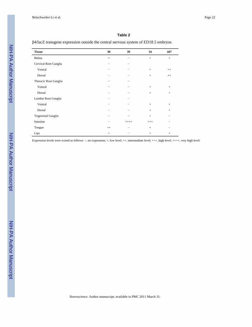
Outside of the central nervous system, there was remarkable β-gal staining in the spinal ganglia(Fig. 3), the intestine (Fig. 4A) and the tongue (Fig. 4C) of ED18.5 transgenic animals. Therewere lower levels of β-gal staining in the retina, trigeminal ganglia and lips (Table 2). Again,for the most part, these results are consistent with β4 mRNA localization studies (Winzer-Serhan and Leslie, 1997;Zoli et al., 1995). Two notable exceptions are the intestine and thelips, for which we are not aware of any reports describing the presence of β4 mRNA in ED18.5animals. The presence of β-gal staining in these areas may be due to positional effects ontransgene expression or it could be due to the lack of a cell-type-specific repressor element wepreviously identified in an intron of the nACh receptor α3 subunit gene, which we showedrepresses β4 promoter activity in cells that do not express the β4 subunit gene (Fuentes Medeland Gardner, 2007).
β4 promoter activity in the brains of transgenic PD30 miceAs mentioned above, in contrast to embryonic animals, all four founder lines exhibitedsignificant β-gal staining at one month of age. Analysis of β-gal staining revealed broad andsubstantial β4 promoter activity in the brains of transgenic animals. In particular, there wassignificant staining in the cortex, hippocampus, thalamus, amygdala, medial habenula and thecolliculus. For the most part, the broad β-gal staining (Table 3) is consistent with previousstudies that determined β4 subunit mRNA expression using in situ hybridization (Azam et al.,2002;Dineley-Miller and Patrick, 1992;Winzer-Serhan and Leslie, 1997). In addition, thepresent results are consistent with immunostaining work that indicated widely dispersedexpression of the nACh receptor β4 subunit protein in the adult mouse nervous system (Gahringet al., 2004). As discussed by Gahring et al., the regulated widespread expression of the β4subunit gene likely reflects its role in a variety of diverse functions in the nervous system.
Forebrain/cortex—As with nACh receptor β4 mRNA (Dineley-Miller and Patrick, 1992;Winzer-Serhan and Leslie, 1997) and protein (Gahring et al., 2004), significant β4 promoteractivity was detected in layer 5 of the cortex (Fig. 5A) and the piriform cortex (Fig. 5C). Slightlylower promoter activity was observed in the frontal association cortex and caudate putamen(Fig. 5E and G, respectively) with there being lower levels in the cingulate/retrosplenial cortex(Fig. 5I). There was also substantial β4 promoter activity in other regions of the forebrain/cortex (Table 3) including the olfactory bulb, agranular insular cortex, auditory cortex,cingulated/retrosplenial cortex, piriform cortex and the visual cortex, amongst others. Inaddition, there were regions where β4 promoter activity was seen for which we know of noreports of β4 mRNA expression, such as the entorhinal, orbital and prelimbic cortices (Table3). Again, this may be due to the lack of other transcriptional regulatory elements in the β4/
Brüschweiler-Li et al. Page 6
Neuroscience. Author manuscript; available in PMC 2011 March 31.
NIH
-PA Author Manuscript
NIH
-PA Author Manuscript
NIH
-PA Author Manuscript

β-gal transgene, such as the cell-type-specific repressor element discussed above.Alternatively, it could be due to positional effects on transgene expression.
Hippocampus/Subiculum—The hippocampus is an interesting region in which to studynACh receptor β4 expression, as it is known that β4 expression in this structure significantlyvaries across mouse strains (Gahring et al., 2004). However, it is clear that across strains, thereis β4 expression, both mRNA and protein, in the dentate gyrus, CA1 field, CA3 field and thesubiculum (Dineley-Miller and Patrick, 1992; Gahring et al., 2004; Winzer-Serhan and Leslie,1997). Consistent with these observations, we see strong β4 promoter activity in these sameregions (Fig. 6A and Table 3).
Habenular Nuclei—The habenula is the region of highest nACh receptor β4 subunitexpression in the brain (Dineley-Miller and Patrick, 1992; Duvoisin et al., 1989; Gahring etal., 2004; Winzer-Serhan and Leslie, 1997). As expected, β4 promoter activity was quite strongin the medial habenula, but was lower in the lateral habenula (Fig. 6C). Interestingly, β4promoter activity was much higher in PD30 animals versus ED18.5 animals. This is in contrastto β4 mRNA levels - which are high throughout development (Winzer-Serhan and Leslie,1997; Zoli et al., 1995) - and may reflect the absence of other regulatory elements in the β4/β-gal transgene, for example, CNR4, that may be active early in development (Xu et al.,2006).
Thalamus/Hypothalamus—As discussed above, significant β4 promoter activity wasobserved in the medial habenula, a region that expresses high levels of β4 mRNA. In addition,the β4 subunit is also expressed in the paraventricular thalamic nucleus, medial geniculatenucleus, the posterior thalamic nuclear group, the retroparafascicular nucleus, the inferiorcolliculus and the ventral anterior and ventral posteromedial thalamic nuclei (Gahring et al.,2004). We detected β4 promoter activity in these same regions (Fig. 6C and E and Table 3).With the exception of these structures and the interpeduncular nucleus (see below), nAChreceptor β4 expression is either very low or undetectable in the thalamus and hypothalamus(Dineley-Miller and Patrick, 1992; Duvoisin et al., 1989; Gahring et al., 2004; Winzer-Serhanand Leslie, 1997). Thus, it was surprising to observe significant β4 promoter activity in bothof these regions (Table 3). Again, this could be due to the absence of the cell-type-specificrepressor element mentioned earlier. Interestingly, while β4 mRNA and protein expression ishigh in the interpeduncular nucleus (Dineley-Miller and Patrick, 1992; Gahring et al., 2004;Winzer-Serhan and Leslie, 1997), we detected no β4 promoter activity in this region (notshown). However, Deneris and colleagues reported that CNR4 drives high transgeneexpression in the interpeduncular nucleus (Xu et al., 2006). The absence of CNR4 in the β4/β-gal transgene used in the current study may explain why no β4 promoter activity was seenin this nucleus.
Amygdala and Pons—Two other brain regions of the PD30 mouse where significant β4promoter activity was detected are the amygdala and the pons. In particular, strong β4 promoteractivity was seen in the posteromedial cortical amygdaloid nucleus (Fig. 6G) and theposterolateral cortical amygdaloid nucleus (Table 3). Lower, yet significant, β4 promoteractivity was observed in the pons, most notably in the laterodorsal tegmental nucleus, the lateralmammillary nucleus and the lateral parabrachial nucleus (ventral part) as well as in the mediallemniscus (Table 3). To our knowledge, the amygdala and pons have not been reported toexpress the nACh receptor β4 subunit, once more raising the issue as to why β4 promoteractivity is seen in these two brain regions in multiple founder lines. As before, one possibilityis the lack of a cell-type-specific repressor element in the β4/β-gal transgene (Fuentes Medeland Gardner, 2007), although it is possible that the endogenous promoter is subject toregulation, negative in this case, to which the transgene promoter is immune.
Brüschweiler-Li et al. Page 7
Neuroscience. Author manuscript; available in PMC 2011 March 31.
NIH
-PA Author Manuscript
NIH
-PA Author Manuscript
NIH
-PA Author Manuscript

The β4 promoter is active in neuronsExpression of nACh receptor β4 subunit protein has been detected in non-neuronal cells withinthe brain (Gahring et al., 2004). In order to determine if the β4 promoter activity we observedthroughout the brain is or is not neuron-specific, triple immunofluorescence was carried outusing anti-GFAP to label glial cells, anti-NeuN to label neurons and anti-β-gal to label β-gal-expressing cells. As shown in Figure 7, in each region tested, β-gal labeling over-lapped thatof NeuN but not GFAP, indicating that the transgene β4 promoter activity is neuron-specific.This suggests that other regulatory elements are involved in the non-neuronal expression ofthe β4 subunit described by Gahring et al. (2004). Alternatively, post-transcriptional orepigenetic regulatory mechanisms may be at work in non-neuronal cells.
In summary, we have used a transgenic mouse model to study the transcriptional activity ofthe nACh receptor β4 subunit gene promoter. As described above, the activity of the β4/β-galtransgene used in this study paralleled, for the most part, endogenous β4 subunit mRNAexpression both spatially and temporally. These results indicate that the 2.3-kilobase pairfragment of the nACh receptor β4 subunit 5′-flanking DNA contains transcriptional regulatoryelements critical for appropriate cell-type-specific and developmentally regulated expressionof the β4 subunit gene. Although the precise regulatory elements within the 2.3-kilobase pairregion responsible for the in vivo activity of the β4 promoter have not been determined, strongcandidates are the CT and CA boxes that we previously characterized in cell culture systems.The significance of these elements in vivo is currently being pursued. However, it is clear thatthe 2.3-kilobase pair fragment does not contain all of the required positive regulatory elementsthat govern expression of the β4 gene, as there are regions of the brain that express relativelyhigh levels of the β4 gene but in which no transgene activity was detected. In some cases, asdescribed above, this may be due to the absence of specific regulatory elements in the transgeneused in the present study. For example, the CNR4 element identified by the Deneris group andshown to drive transgene expression in the pineal gland, adrenal gland and specific regions ofthe brain, was not part of the transgene construct. It is also possible that other, yet to be identifiedpositive regulatory elements are also required for appropriate β4 gene expression. In additionto the β4 promoter activity that corresponds to endogenous β4 mRNA expression, we alsodetected promoter activity in brain regions that have not been reported to express the β4 subunit.These observations likely underscore the importance of negative regulatory elements in β4gene expression and may be a consequence of the absence of a cell-type-specific repressor inthe transgene used in this study. We previously demonstrated in vitro that this repressor,referred to as “α3 intron 5”, exerts its transcriptional effects in non-β4-expressing cells but isrelatively inactive in β4-expressing cells (Fuentes Medel and Gardner, 2007).
Finally, it is important to note that at least two other modes of regulation are likely to play keyroles in expression of the nACh receptor β4 subunit gene (as well as other nACh receptorsubunit genes). First, in addition to the cis-acting elements described thus far, chromatinremodeling most probably is critical for appropriate β4 gene expression. This is a relativelyunderstudied area in terms of nACh receptor gene expression and is a focus of current researchefforts. Second, post-transcriptional mechanisms are another level of control of nACh receptorexpression, particularly in response to extracellular stimuli. One such mechanism may involvesmall 21-24 nucleotide long regulatory molecules, referred to as microRNAs (miRNAs)(Ambros, 2004). miRNAs are predicted to regulate the majority of mammalian protein-codinggenes, typically by binding to complementary sites in the 3′-untranslated regions of targetmRNAs and guiding them to an RNA-induced silencing complex (Kosik, 2006). Interestingly,a miRNA, miR-1, has been reported to target nACh receptors in C. elegans, leading to changesin nACh receptor properties (Simon et al., 2008). We are investigating whether miRNAs playa role in the expression of mammalian nACh receptor genes.
Brüschweiler-Li et al. Page 8
Neuroscience. Author manuscript; available in PMC 2011 March 31.
NIH
-PA Author Manuscript
NIH
-PA Author Manuscript
NIH
-PA Author Manuscript

AcknowledgmentsWe thank Steve Jones, Joe Gosselin and their colleagues in the UMMS Transgenic Animal Modeling Core for theiroutstanding services in generating the majority of the transgenic mice used in this study. We also thank Haley Melikianfor her help with the microscopy, Monica Carrasco for help with the immunofluorescence, Stéphane Germain for thekind gift of pSG-MAR, Ricardo Medina for providing useful advice and reagents for determining transgene copynumber and Irena N. Melnikova for critical review of the manuscript. We acknowledge E.I. Ginns, D. Faryna and C.Dickinson for generating founder line 447. This work was supported by Grant Number R01NS030243 from theNational Institute of Neurological Disorders and Stroke. The content is solely the responsibility of the authors anddoes not necessarily represent the official views of the National Institute of Neurological Disorders and Stroke or theNational Institutes of Health.
Abbreviations
nACh nicotinic acetylcholine
β-gal β-galactosidase
NLS nuclear localization signal
MAR matrix attachment region
NGF nerve growth factor
ED embryonic day
PD postnatal day
PBS phosphate-buffered saline
NeuN neuron-specific nuclear marker
GFAP glial fibrillary acid protein
miRNAs microRNAs
IZ intermediate zone
VZ ventricular zone
M medulla oblongata
P pons
DRG dorsal root ganglion
CDRG cervical dorsal root ganglion
LRG lumber root ganglion
CRG cervical root ganglion
Pir piriform cortex
FrA frontal association cortex
Cpu caudate putamen
Cg/Rs cingulate/retrosplenial cortex
DG dentate gyrus
MHb medial habenula
LHb lateral habenula
PV paraventricular thalamic nucleus
VA ventral anterior thalamic nucleus
Brüschweiler-Li et al. Page 9
Neuroscience. Author manuscript; available in PMC 2011 March 31.
NIH
-PA Author Manuscript
NIH
-PA Author Manuscript
NIH
-PA Author Manuscript
PMCo posteromedial cortical amygdaloid nucleus
ReferencesAlbuquerque EX, Pereira EFR, Alkondon M, Rogers SW. Mammalian nicotinic acetylcholine receptors:
From structure to function. Physiol Rev 2009;89:73–120. [PubMed: 19126755]Ambros V. The functions of animal microRNAs. Nature 2004;431:350–355. [PubMed: 15372042]Azam L, Winzer-Serhan UH, Chen Y, Leslie FM. Expression of neuronal nicotinic acetylcholine receptor
subunit mRNAs within midbrain dopamine neurons. J Comp Neurol 2002;444:260–274. [PubMed:11840479]
Bigger CB, Casanova EA, Gardner PD. Transcriptional regulation of neuronal nicotinic acetylcholinereceptor genes. Functional interactions between Sp1 and the rat beta4 subunit gene promoter. J BiolChem 1996;271:32842–32848. [PubMed: 8955122]
Bigger CB, Melnikova IN, Gardner PD. Sp1 and Sp3 regulate expression of the neuronal nicotinicacetylcholine receptor β4 subunit gene. J Biol Chem 1997;272:25976–25982. [PubMed: 9325332]
Boulter J, O'Shea-Greenfield A, Duvoisin RM, Connolly JG, Wada E, Jensen A, Gardner PD, BallivetM, Deneris ES, McKinnon D, et al. Alpha 3, alpha 5, and beta 4: three members of the rat neuronalnicotinic acetylcholine receptor-related gene family form a gene cluster. J Biol Chem 1990;265:4472–4482. [PubMed: 1689727]
Boyd RT. Transcriptional regulation and cell specificity determinants of the rat nicotinic acetylcholinereceptor alpha 3 gene. Neurosci Lett 1996;208:73–76. [PubMed: 8859893]
Campos-Caro A, Carrasco-Serrano C, Valor LM, Viniegra S, Ballesta JJ, Criado M. Multiple functionalSp1 domains in the minimal promoter region of the neuronal nicotinic receptor alpha5 subunit gene.J Biol Chem 1999;274:4693–4701. [PubMed: 9988706]
Catassi A, Servent D, Paleari L, Cesario A, Russo P. Multiple roles of nicotine on cell proliferation andinhibition of apoptosis: implications on lung carcinogenesis. Mutat Res 2008;659:221–231. [PubMed:18495523]
Corriveau RA, Berg DK. Coexpression of multiple acetylcholine receptor genes in neurons: quantitationof transcripts during development. J Neurosci 1993;13:2662–2671. [PubMed: 8501530]
Dani JA, Bertrand D. Nicotinic acetylcholine receptors and nicotinic cholinergic mechanisms of thecentral nervous system. Annu Rev Pharmacol Toxicol 2007;47:699–729. [PubMed: 17009926]
De Fusco M, Becchetti A, Patrignani A, Annesi G, Gambardella A, Quattrone A, Ballabio A, Wanke E,Casari G. The nicotinic receptor beta 2 subunit is mutant in nocturnal frontal lobe epilepsy. Nat Genet2000;26:275–276. [PubMed: 11062464]
Dineley-Miller K, Patrick J. Gene transcripts for the nicotinic acetylcholine receptor subunit, beta4, aredistributed in multiple areas of the rat central nervous system. Brain Res Mol Brain Res 1992;16:339–344. [PubMed: 1337943]
Du Q, Melnikova IN, Gardner PD. Differential effects of heterogeneous nuclear ribonucleoprotein K onSp1- and Sp3-mediated transcriptional activation of a neuronal nicotinic acetylcholine receptorpromoter. J Biol Chem 1998;273:19877–19883. [PubMed: 9677424]
Du Q, Tomkinson AE, Gardner PD. Transcriptional regulation of neuronal nicotinic acetylcholinereceptor genes. A possible role for the DNA-binding protein Puralpha. J Biol Chem 1997;272:14990–14995. [PubMed: 9169473]
Duvoisin RM, Deneris ES, Patrick J, Heinemann S. The functional diversity of the neuronal nicotinicacetylcholine receptors is increased by a novel subunit: beta 4. Neuron 1989;3:487–496. [PubMed:2642007]
Egleton RD, Brown KC, Dasgupta P. Nicotinic acetylcholine receptors in cancer: multiple roles inproliferation and inhibition of apoptosis. Trends Pharmacol Sci 2008;29:151–158. [PubMed:18262664]
Elgoyhen AB, Vetter DE, Katz E, Rothlin CV, Heinemann SF, Boulter J. alpha10: a determinant ofnicotinic cholinergic receptor function in mammalian vestibular and cochlear mechanosensory haircells. Proc Natl Acad Sci U S A 2001;98:3501–3506. [PubMed: 11248107]
Brüschweiler-Li et al. Page 10
Neuroscience. Author manuscript; available in PMC 2011 March 31.
NIH
-PA Author Manuscript
NIH
-PA Author Manuscript
NIH
-PA Author Manuscript
Engelman HS, MacDermott AB. Presynaptic ionotropic receptors and control of transmitter release. NatRev Neurosci 2004;5:135–145. [PubMed: 14735116]
Fuchs S, Germain S, Philippe J, Corvol P, Pinet F. Expression of renin in large arteries outside the kidneyrevealed by human renin promoter/LacZ transgenic mouse. Am J Pathol 2002;161:717–725.[PubMed: 12163396]
Fuentes Medel YF, Gardner PD. Transcriptional repression by a conserved intronic sequence in thenicotinic receptor α3 subunit gene. J Biol Chem 2007;282:19062–19070. [PubMed: 17504758]
Fyodorov D, Deneris E. The POU domain of SCIP/Tst-1/Oct-6 is sufficient for activation of anacetylcholine receptor promoter. Mol Cell Biol 1996;16:5004–5014. [PubMed: 8756659]
Gahring LC, Persiyanov K, Rogers SW. Neuronal and astrocyte expression of nicotinic receptor subunitβ4 in the adult mouse brain. J Comp Neurol 2004;468:322–333. [PubMed: 14681928]
Gahring LC, Rogers SW. Neuronal nicotinic acetylcholine receptor expression and function onnonneuronal cells. AAPS Journal 2006;7:E885–E894. [PubMed: 16594641]
Genzen JR, Van Cleve W, McGehee DS. Dorsal root ganglion neurons express multiple nicotinicacetylcholine receptor subtypes. J Neurophysiol 2001;86:1773–1782. [PubMed: 11600638]
Greene LA, Tischler AS. Establishment of a noradrenergic clonal line of rat adrenal pheochromocytomacells which respond to nerve growth factor. Proc Natl Acad Sci U S A 1976;73:2424–2428. [PubMed:1065897]
Hellström-Lindahl E, Gorbounova O, Seiger Å, Mousavi M, Nordberg A. Regional distribution ofnicotinic receptors during prenatal development of human brain and spinal cord. Develop Brain Res1998;108:147–160.
Hu HZ, Li ZW. Modulation of nicotinic ACh-, GABAA- and 5-HT3-receptor functions by external H-7,a protein kinase inhibitor, in rat sensory neurones. Br J Pharmacol 1997;122:1195–1201. [PubMed:9401786]
Hu M, Bigger CB, Gardner PD. A novel regulatory element of a nicotinic acetylcholine receptor geneinteracts with a DNA binding activity enriched in rat brain. J Biol Chem 1995;270:4497–4502.[PubMed: 7876217]
Hu M, Liu QS, Chang KT, Berg DK. Nicotinic regulation of CREB activation in hippocampal neuronsby glutamatergic and nonglutamatergic pathways. Mol Cell Neurosci 2002;21:616–625. [PubMed:12504594]
Hu M, Whiting Theobald NL, Gardner PD. Nerve growth factor increases the transcriptional activity ofthe rat neuronal nicotinic acetylcholine receptor beta 4 subunit promoter in transfected PC12 cells.J Neurochem 1994;62:392–395. [PubMed: 7505316]
Isacson O, Seo H, Lin L, Albeck D, Granholm AC. Alzheimer's disease and Down's syndrome: roles ofAPP, trophic factors and ACh. Trends Neurosci 2002;25:79–84. [PubMed: 11814559]
Ji D, Lape R, Dani JA. Timing and location of nicotinic activity enhances or depresses hippocampalsynaptic plasticity. Neuron 2001;31:131–141. [PubMed: 11498056]
Kaufman, MH. The Atlas of Mouse Development. revised edition. Academic Press; San Diego: 1995.Kedmi M, Beaudet AL, Orr-Urtreger A. Mice lacking neuronal nicotinic acetylcholine receptor β4-
subunit and mice lacking both α5- and β4-subunits are highly resistant to nicotine-induced seizures.Physiol Genomics 2004;17:221–229. [PubMed: 14996991]
Keiger CJH, Prevette D, Conroy WG, Oppenheim RW. Developmental expression of nicotinic receptorsin the chick and human spinal cord. J Comp Neurol 2003;455:86–99. [PubMed: 12454998]
Kosik K. The neuronal microRNA system. Nat Rev Neurosci 2006;7:911–920. [PubMed: 17115073]Laviolette SR, van der Kooy D. The neurobiology of nicotine addiction: bridging the gap from molecules
to behaviour. Nat Rev Neurosci 2004;5:55–65. [PubMed: 14708004]Lena C, Changeux JP. Pathological mutations of nicotinic receptors and nicotine-based therapies for brain
disorders. Curr Opin Neurobiol 1997;7:674–682. [PubMed: 9384554]Levey MS, Brumwell CL, Dryer SE, Jacob MH. Innervation and target tissue interactions differentially
regulate acetylcholine receptor subunit mRNA levels in developing neurons in situ. Neuron1995;14:153–162. [PubMed: 7826633]
Brüschweiler-Li et al. Page 11
Neuroscience. Author manuscript; available in PMC 2011 March 31.
NIH
-PA Author Manuscript
NIH
-PA Author Manuscript
NIH
-PA Author Manuscript

Levey MS, Jacob MH. Changes in the regulatory effects of cell-cell interactions on neuronal AChRsubunit transcript levels after synapse formation. J Neurosci 1996;16:6878–6885. [PubMed:8824326]
Liu L, Chang GQ, Jiao YQ, Simon SA. Neuronal nicotinic acetylcholine receptors in rat trigeminalganglia. Brain Res 1998;809:238–245. [PubMed: 9853116]
Liu Q, Huang Y, Xue F, Simard A, DeChon J, Li G, Zhang J, Lucero L, Wang M, Sierks M, et al. Anovel nicotinic acetylcholine receptor subtype in basal forebrain cholinergic neurons with highsensitivity to amyloid peptides. J Neurosci 2009;29:918–929. [PubMed: 19176801]
Liu Q, Melnikova IN, Hu M, Gardner PD. Cell type-specific activation of neuronal nicotinic acetylcholinereceptor subunit genes by Sox10. J Neurosci 1999;19:9747–9755. [PubMed: 10559384]
Lustig LR, Peng H, Hiel H, Yamamoto T, Fuchs PA. Molecular cloning and mapping of the humannicotinic acetylcholine receptor alpha10 (CHRNA10). Genomics 2001;73:272–283. [PubMed:11350119]
Mercer EH, Hoyle GW, Kapur RP, Brinster RL, Palmiter RD. The dopamine beta-hydroxylase genepromoter directs expression of E. coli lacZ to sympathetic and other neurons in adult transgenic mice.Neuron 1991;7:703–716. [PubMed: 1742021]
Paxinos, G.; Franklin, KBJ. The Mouse Brain in Stereotaxic Coordinates. 2nd ed.. Academic Press; SanDiego: 2001.
Perl O, Ilani T, Strous RD, Lapidus R, Fuchs S. The alpha7 nicotinic acetylcholine receptor inschizophrenia: decreased mRNA levels in peripheral blood lymphocytes. Faseb J 2003;17:1948–1950. [PubMed: 12897067]
Perry EK, Martin-Ruiz CM, Court JA. Nicotinic receptor subtypes in human brain related to aging anddementia. Alcohol 2001;24:63–68. [PubMed: 11522424]
Phi-Van L, Stratling WH. Dissection of the ability of the chicken lysozyme gene 5′ matrix attachmentregion to stimulate transgene expression and to dampen position effects. Biochemistry1996;35:10735–10742. [PubMed: 8718863]
Richardson CE, Morgan JM, Jasani B, Green JT, Rhodes J, Williams GT, Lindstrom J, Wonnacott S,Thomas GA, Smith V. Megacystis-microcolon-intestinal hypoperistalsis syndrome and the absenceof the alpha3 nicotinic acetylcholine receptor subunit. Gastroenterology 2001;121:350–357.[PubMed: 11487544]
Rust G, Burgunder JM, Lauterburg TE, Cachelin AB. Expression of neuronal nicotinic acetylcholinereceptor subunit genes in the rat autonomic nervous system. Eur J Neurosci 1994;6:478–485.[PubMed: 8019684]
Schuller HM. Neurotransmission and cancer: implications for prevention and therapy. Anti-Cancer Drugs2008;19:655–671. [PubMed: 18594207]
Schuller HM. Is cancer triggered by altered signalling of nicotinic acetylcholine receptors? Nat RevCancer 2009;9:195–205. [PubMed: 19194381]
Silver AA, Shytle RD, Philipp MK, Wilkinson BJ, McConville B, Sanberg PR. Transdermal nicotine andhaloperidol in Tourette's disorder: a double-blind placebo-controlled study. J Clin Psychiatry2001;62:707–714. [PubMed: 11681767]
Simon D, Madison J, Conery A, Thompson-Peer K, Soskis M, Ruvkun G, Kaplan J, Kim J. ThemicroRNA miR-1 regulates a MEF-2-dependent retrograde signal at neuromuscular junctions. Cell2008;133:903–915. [PubMed: 18510933]
Song P, Sekhon HS, Fu XW, Maier M, Jia Y, Duan J, Proskosil BJ, Gravett C, Lindstrom J, Mark GP,et al. Activated cholinergic signaling provides a target in squamous cell lung carcinoma. Cancer Res2008;68:4693–4700. [PubMed: 18559515]
Spindel ER. Neuronal nicotinic acetylcholine receptors: not just in brain. Am J Physiol Lung Cell MolPhysiol 2003;285:L1201–L1202. [PubMed: 14604850]
Steen KH, Reeh PW. Actions of cholinergic agonists and antagonists on sensory nerve endings in ratskin, in vitro. J Neurophysiol 1993;70:397–405. [PubMed: 8103089]
Steinlein OK, Mulley JC, Propping P, Wallace RH, Phillips HA, Sutherland GR, Scheffer IE, BerkovicSF. A missense mutation in the neuronal nicotinic acetylcholine receptor alpha 4 subunit is associatedwith autosomal dominant nocturnal frontal lobe epilepsy. Nat Genet 1995;11:201–203. [PubMed:7550350]
Brüschweiler-Li et al. Page 12
Neuroscience. Author manuscript; available in PMC 2011 March 31.
NIH
-PA Author Manuscript
NIH
-PA Author Manuscript
NIH
-PA Author Manuscript

Sucher NJ, Cheng TP, Lipton SA. Neural nicotinic acetylcholine responses in sensory neurons frompostnatal rat. Brain Res 1990;533:248–254. [PubMed: 2289141]
Taranda J, Maison SF, Ballestero JA, Katz E, Savino J, Vetter DE, Boulter J, Liberman MC, Fuchs PA,Elgoyhen AB. A point mutation in the hair cell nicotinic cholinergic receptor prolongs cochlearinhibition and enhances noise protection. PLOS Biol 2009;7:1–13.
Teaktong T, Graham A, Court J, Perry R, Jaros E, Johnson M, Hall R, Perry E. Alzheimer's disease isassociated with a selective increase in alpha7 nicotinic acetylcholine receptor immunoreactivity inastrocytes. Glia 2003;41:207–211. [PubMed: 12509811]
Valor LM, Campos-Caro A, Carrasco-Serrano C, Ortiz JA, Ballesta JJ, Criado M. Transcription factorsNF-Y and Sp1 are important determinants of the promoter activity of the bovine and human neuronalnicotinic receptor beta 4 subunit genes. J Biol Chem 2002;277:8866–8876. [PubMed: 11742001]
Vincler MA, Eisenach JC. Immunocytochemical localization of the α3, α4, α5, α7, β2, β3 and β4 nicotinicacetylcholine receptor subunits in the locus coeruleus of the rat. Brain Res 2003;974:25–36.[PubMed: 12742621]
Wang N, Orr-Urtreger A, Korczyn AD. The role of neuronal nicotinic acetylcholine receptor subunits inautonomic ganglia: lessons from knockout mice. Prog Neurobiol 2002;68:341–360. [PubMed:12531234]
Wessler I, Kirkpatrick CJ. Acetylcholine beyond neurons: the non-neuronal cholinergic system inhumans. Brit J Pharmacol 2008;154:1558–1571. [PubMed: 18500366]
Whitehouse PJ, Martino AM, Marcus KA, Zweig RM, Singer HS, Price DL, Kellar KJ. Reductions inacetylcholine and nicotine binding in several degenerative diseases. Arch Neurol 1988;45:722–724.[PubMed: 3390025]
Winzer-Serhan UH, Leslie FM. Codistribution of nicotinic acetylcholine receptor subunit alpha3 andbeta4 mRNAs during rat brain development. J Comp Neurol 1997;386:540–554. [PubMed: 9378850]
Xu X, Scott MM, Deneris ES. Shared long-range regulatory elements coordinate expression of a genecluster encoding nicotinic receptor heteromeric subtypes. Mol Cell Biol 2006;26:5636–5649.[PubMed: 16847319]
Yang X, Fyodorov D, Deneris ES. Transcriptional analysis of acetylcholine receptor alpha 3 genepromoter motifs that bind Sp1 and AP2. J Biol Chem 1995;270:8514–8520. [PubMed: 7721749]
Yang X, McDonough J, Fyodorov D, Morris M, Wang F, Deneris ES. Characterization of an acetylcholinereceptor alpha 3 gene promoter and its activation by the POU domain factor SCIP/Tst-1. J Biol Chem1994;269:10252–10264. [PubMed: 8144606]
Yuan JS, Burris J, Stewart NR, Mentewab A, Stewart NS. Statiistical tools for transgene copy numberestimation based on real-time PCR. BMC Bioinformatics 2007;8:S6. [PubMed: 18047729]
Zhou Y, Deneris E, Zigmond RE. Differential regulation of levels of nicotinic receptor subunit transcriptsin adult sympathetic neurons after axotomy. J Neurobiol 1998;34:164–178. [PubMed: 9468387]
Zoli M, Le Novere N, Hill JA Jr. Changeux JP. Developmental regulation of nicotinic ACh receptorsubunit mRNAs in the rat central and peripheral nervous systems. J Neurosci 1995;15:1912–1939.[PubMed: 7891142]
Brüschweiler-Li et al. Page 13
Neuroscience. Author manuscript; available in PMC 2011 March 31.
NIH
-PA Author Manuscript
NIH
-PA Author Manuscript
NIH
-PA Author Manuscript

Figure 1.The nACh receptor β4 subunit promoter region used for transgenesis. A. The β4/α3/α5 genecluster. The 2.3-kb SacI/HindIII fragment of the β4 promoter region is indicated on the left.Arrows indicate direction of transcription. The nucleotide positions of the SacI and HindIIIsites are relative to the major transcription initiation site. B. Structure of the linearizedconstruct, pSGB4SH, used to generate transgenic animals. MAR = matrix attachment region,NLS = nuclear localization sequence. C. Nucleotide sequences of the CT and CA boxes of theβ4 promoter region. D. The vector, pSGMAR, and pSGB4SH were transfected into PC12 cellsalong with a luciferase construct in which the SV40 promoter drives expression of the fireflyluciferase gene. Cells were treated with NGF (100 ng/ml for 2 days) as indicated. β-Gal activitywas normalized to luciferase activity to correct for differences in transfection efficiencies. Errorbars represent standard deviations of the means.
Brüschweiler-Li et al. Page 14
Neuroscience. Author manuscript; available in PMC 2011 March 31.
NIH
-PA Author Manuscript
NIH
-PA Author Manuscript
NIH
-PA Author Manuscript

Figure 2.nACh receptor β4 subunit promoter activity in the central nervous system of ED18.5 transgenicanimals. Sagittal sections of transgenic (A, C, E, G, I and K) and non-transgenic (B, D, F, H,J and L) ED18.5 mouse brain and spinal cord are shown. The sections were stained for β-galactivity and counter-stained with Neutral Red. Note that β-gal staining is restricted to nucleias the β4/lacZ transgene contains a nuclear localization signal (see Fig. 1B). A and B, cortex(IZ = intermediate zone, VZ = ventricular zone); C and D, medulla oblongata (M); E and F,pons (P); G and H, upper cervical region of the spinal cord; I and J, mid-thoracic region of thespinal cord; K and L, mid-lumbar region of the spinal cord. Arrows in panels G, I and K indicateβ-gal-expressing cells.
Brüschweiler-Li et al. Page 15
Neuroscience. Author manuscript; available in PMC 2011 March 31.
NIH
-PA Author Manuscript
NIH
-PA Author Manuscript
NIH
-PA Author Manuscript

Figure 3.nACh receptor β4 subunit promoter activity in spinal ganglia of ED18.5 transgenic animals.Sagittal sections of transgenic ED18.5 animals were stained for β-gal activity and counter-stained with Neutral Red. A, dorsal root ganglion (DRG); B, cervical dorsal root ganglion(CDRG); C, lumbar root ganglion (LRG); D, cervical root ganglion (CRG). Arrows in panelsC and D indicate β-gal-expressing cells.
Brüschweiler-Li et al. Page 16
Neuroscience. Author manuscript; available in PMC 2011 March 31.
NIH
-PA Author Manuscript
NIH
-PA Author Manuscript
NIH
-PA Author Manuscript

Figure 4.nACh receptor β4 subunit promoter activity in the intestine and tongue of ED18.5 transgenicanimals. Sagittal sections of transgenic (A and C) and non-transgenic (B and D) ED18.5animals were stained for β-gal activity and counter-stained with Neutral Red. A and B,intestine; C and D, tongue. β-gal staining of the tongue appears to be restricted to or near thelongitudinal intrinsic muscle (arrows indicate β-gal-expressing cells).
Brüschweiler-Li et al. Page 17
Neuroscience. Author manuscript; available in PMC 2011 March 31.
NIH
-PA Author Manuscript
NIH
-PA Author Manuscript
NIH
-PA Author Manuscript

Figure 5.nACh receptor β4 subunit promoter activity in the forebrain and cortical regions of PD30transgenic animals. Coronal sections of transgenic (A, C, E, G and I) and non-transgenic (B,D, F, H and J) PD30 mouse brains are shown. The sections were stained for β-gal activity andcounter-stained with Neutral Red. A and B, layer 5 of the cortex (“5)”; C and D, piriform cortex(Pir); E and F, frontal association cortex (FrA); G and H, caudate putamen (Cpu); I and J,cingulate/retrosplenial cortex (Cg/Rs). Arrows in panels E, G and I indicate β-gal-expressingcells
Brüschweiler-Li et al. Page 18
Neuroscience. Author manuscript; available in PMC 2011 March 31.
NIH
-PA Author Manuscript
NIH
-PA Author Manuscript
NIH
-PA Author Manuscript

Figure 6.nACh receptor β4 subunit promoter activity in the central nervous system of PD30 transgenicanimals. Coronal sections of transgenic (A, C, E and G) and non-transgenic (B, D, F and H)PD30 mouse brains are shown. The sections were stained for β-gal activity and counter-stainedwith Neutral Red. A and B, hippocampus (CA1) and dentate gyrus (DG); C and D, medial(MHb) and lateral (LHb) habenula and paraventricular thalamic nucleus (PV); E and F, ventralanterior thalamic nucleus (VA); G and H, posteromedial cortical amygdaloid nucleus (PMCo).Arrows in panels A, C and G indicate β-gal-expressing cells.
Brüschweiler-Li et al. Page 19
Neuroscience. Author manuscript; available in PMC 2011 March 31.
NIH
-PA Author Manuscript
NIH
-PA Author Manuscript
NIH
-PA Author Manuscript

Figure 7.The nACh receptor β4 subunit promoter directs neuron-specific gene expression. Tripleimmunofluorescence was done on coronal sections of transgenic (TG) and non-transgenic(WT) PD30 mouse brains. A, cortex; B, thalamus; C, amygdala. GFAP = glial fibrillary acidprotein (yellow arrows), NeuN = neuron-specific nuclear marker, β-gal = β-galactosidase.Overlap of the three immunostained sections indicates specific β-gal staining in neuronal nuclei(white arrows). β-gal histochemistry (right panels) was done to confirm β-gal activity in thetransgenic animals. Note: There was no GFAP staining in the thalamus (panel B).
Brüschweiler-Li et al. Page 20
Neuroscience. Author manuscript; available in PMC 2011 March 31.
NIH
-PA Author Manuscript
NIH
-PA Author Manuscript
NIH
-PA Author Manuscript

NIH
-PA Author Manuscript
NIH
-PA Author Manuscript
NIH
-PA Author Manuscript
Brüschweiler-Li et al. Page 21
Table 1
β4/lacZ transgene expression in the central nervous system of ED18.5 embryos
Central Nervous System Region 30 39 54 447
Cerebral Cortex − − ++++ ++
Piriform Cortex − − + +
Subiculum − − ++ +
Lateral Habenula − − + +
Medial Habenula − − + +
Medulla Oblongata − − ++ +
Anterior Pontine Area − − ++ +
Posterior Pontine Area − − ++ +
Pons − − ++ ++
Pineal Gland − − − −
Thalamus − − − +
Ventral Spinal Cord − − + ++
Dorsal Spinal Cord − − + ++
Expression levels were scored as follows: −, no expression; +, low level; ++, intermediate level; ++++, very high level.
Neuroscience. Author manuscript; available in PMC 2011 March 31.
NIH
-PA Author Manuscript
NIH
-PA Author Manuscript
NIH
-PA Author Manuscript
Brüschweiler-Li et al. Page 22
Table 2
β4/lacZ transgene expression outside the central nervous system of ED18.5 embryos
Tissue 30 39 54 447
Retina + − + +
Cervical Root Ganglia − −
Ventral − − + ++
Dorsal − − + ++
Thoracic Root Ganglia − −
Ventral − − + +
Dorsal − − + +
Lumbar Root Ganglia − −
Ventral − − + +
Dorsal − − + +
Trigeminal Ganglia − − + −
Intestine − ++++ +++ −
Tongue ++ − + −
Lips + − + +
Expression levels were scored as follows: −, no expression; +, low level; ++, intermediate level; +++, high level; ++++, very high level.
Neuroscience. Author manuscript; available in PMC 2011 March 31.

NIH
-PA Author Manuscript
NIH
-PA Author Manuscript
NIH
-PA Author Manuscript
Brüschweiler-Li et al. Page 23
Table 3
β4/lacZ transgene expression in the central nervous system of PD30 animals
Brain Region 30 39 54 447
Forebrain/Cortex
layer 1 − + + +
layer 2 − + + −
layer 3 − − + +
layer 5 + ++ ++++ ++
anterior olfactory nucleus − ++ + +
basal nucleus (Meynert) + + − +
caudate putamen (striatum) + + ++ ++
lateral globus pallidus − + + +
olfactory bulb + + ++ +
olfactory tubercle + − + ++
agranular insular cortex + + +++ +
auditory cortex − + ++ +
cingulate cortex, area 1 − + ++ ++
cingulate/retrosplenial cortex + + + −
cortex-amygdala transition zone − + + +
dorsal endopiriform nucleus + + ++++ ++
entorhinal cortex + + +++ +
frontal association cortex + + ++ ++
granular insular cortex − + + +
orbital cortex + + +++ ++
primary motor cortex − + ++ ++
secondary motor cortex + + ++ ++
piriform cortex ++ +++ ++++ +++
perirhinal cortex + + +++ +
prelimbic cortex + − +++ ++
retrosplenial agranular cortex + + + +
primary somatosensory cortex + + ++ ++
secondary somatosensory cortex + + ++ +
temporal association cortex − + +++ +
primary visual cortex + + + ++
secondary visual cortex − + + +
Hippocampus/Corpus Callosum
parasubiculum − + + +
presubiculum − + + +
subiculum − ++ ++++ +
molecular layer of the dentate gyrus − ++++ ++ ++
oriens layer of the hippocampus − ++ + +
Habenular Nuclei
lateral habenular nucleus + + + +
Neuroscience. Author manuscript; available in PMC 2011 March 31.

NIH
-PA Author Manuscript
NIH
-PA Author Manuscript
NIH
-PA Author Manuscript
Brüschweiler-Li et al. Page 24
Brain Region 30 39 54 447
medial habenular nucleus − +++ ++ +
Preoptic Area
lateral preoptic area + + + +
medial preoptic area + + + +
median preoptic nucleus + + - +
medial preoptic nucleus, medial part − + + +
Thalamus
anteromedial thalamic nucleus − ++ + +
basomedial amygdaloid nucleus, anterior part + + ++ +
caudal interstitial nucleus of the medial, longitudinal − + ++ +
fasciculus
central medial thalamic nucleus − + + +
dysgranular insular cortex − + ++ +
dorsolateral orbital cortex + + ++ +
dorsal penduncular cortex + + + ++
gustatory thalamic nucleus + + + −
laterodorsal thalamic nucleus, dorsomedial part + + + −
lateral posterior thalamic nucleus, mediorostral part − + + +
mediodorsal thalamic nucleus + + + +
medial geniculate nucleus − ++ ++ +
posterior thalamic nuclear group − + ++++ +
paratenial thalamic nucleus − + + +
paraventricular thalamic nucleus + + ++ +
precommissural nucleus + + + +
reuniens thalamic nucleus − + + +
stria medullaris of the thalamus − + + +
ventral anterior thalamic nucleus + + + +
ventrolateral thalamic nucleus − + + +++
ventromedial thalamic nucleus − + + +
ventral posteromedial thalamic nucleus − + + +
Hypothalamus
dorsomedial hypothalamic nucleus ++ + + −
lateral hypothalamic area +++ + + +
medial forebrain bundle + + + −
perifornical nucleus + + + −
posterior hypothalamic area + + + −
ventromedial hypothalamic nucleus + + + +
Amygdala
anterior amygdaloid area − + + +
anterior cortical amygaloid nucleus − + + +
medial amygdaloid nucleus + + + +
posterolateral cortical amygdaloid nucleus − ++ +++ ++
Neuroscience. Author manuscript; available in PMC 2011 March 31.

NIH
-PA Author Manuscript
NIH
-PA Author Manuscript
NIH
-PA Author Manuscript
Brüschweiler-Li et al. Page 25
Brain Region 30 39 54 447
posteromedial cortical amygdaloid nucleus + ++ ++ +
Other Midbrain Structures
dorsolateral periaqueductal gray + + − +
deep mesencephalic nucleus + + + −
inferior colliculus + + ++ +
retroparafascicular nucleus + + + −
subbrachial nucleus + + + +
superior colliculus + + − +
Pons
laterodorsal tegmental nucleus + + + −
lateral mammillary nucleus + + + −
lateral parabrachial nucleus, ventral part + + − +
medial lemniscus + ++ − +
Expression levels were scored as follows: −, no expression; +, low level; ++, intermediate level; +++, high level; ++++, very high level.
Neuroscience. Author manuscript; available in PMC 2011 March 31.