Genetic Dissection and Functional Studies in Hirschsprung ...
Upload
independentCategory
view
0download
0
Neuron, Vol. 15, 205-211, July, 1995, Copyright 0 1995 by Cell Press
Molecular Dissection of Subunit Interfaces in the Acetylcholine Receptor: Identification of Determinants of a=Conotoxin M l Selectivity Steven M. Sine,” Hans-Jurgen Kreienkamp,t Nina Bren,’ Robert Maeda,t and Palmer Taylort *Receptor Biology Laboratory Department of Physiology and Biophysics Mayo Foundation Rochester, Minnesota 55905 fDepartment of Pharmacology, 0636 University of California, San Diego La Jolla, California 92093
Summary
The acetylcholine receptor from vertebrate skeletal muscle is a pentamer of homologous subunits with composition a&G. Its two ligand binding sites, formed at a-y and a-6 interfaces, differ in their affinities for ago- nists and competitive antagonists, owing to different contributions of the y and 6 subunits. To identify por- tions of the y and 6 subunits that contribute to the binding sites, the experiments described here use y-6 subunit chimeras and site-specific mutants to de- termine the basis of the 1 O,OOO-fold selectivity of cono- toxin Ml for the sites. Three distinct regions of the extracellular domain were found to contribute to cono- toxin Ml selectivity, each containing a single residue responsible for the contribution of that region. Resi- dues K34, Sl 11, and F172 of the y subunit confer low affinity to the a-y binding site, whereas the corre- sponding residues of the 6 subunit, S36, Y113, and 1176, confer high affinity to the a-6 site. Identification of three separate determinants of ligand selectivity suggests a limited model of the folding pattern of the extracellular domain of the subunits.
Introduction
The acetylcholine receptor (AChR) from vertebrate skele- tal muscle is an integral membrane protein that binds nerve-released ACh to elicit rapid changes in transmem- brane permeability to small cations. The AChR contains two ligand binding sites within a pentamer of homologous subunits with composition a&S. Each subunit contains an amino-terminal extracellular domain of approximately 210 amino acids, followed by four candidate transmem- brane domains. Although the two binding sites originally appeared to reside entirely within the extracellular do- mains of the a subunits, substantial evidence has accumu- lated to show that the binding sites reside at a-y and a-6 subunit interfaces. In the native AChR, the ligand bind- ing sites show very different affinities for ACh (Sine et al., 1990) and several types of competitive antagonists (Neubig and Cohen, 1979; Sine and Taylor, 1981; Kreien- kamp et al., 1994). The molecular basis for the different affinities is thought to reside in the portions of the y and 6 subunits that contact the two a subunits to form the
binding sites (Blount and Merlie, 1989; Pedersen and Co- hen, 1990; Sine and Claudio, 1991).
Sine (1993) recently identified three residues in the ex- tracellular domain of the y and S subunits responsible for site selectivity of the competitive antagonist dimethyl-d- tubocurarine (DMT). One of these residues, Y117 of the y subunit, contributes to stabilization of the DMT-receptor complex through quaternary ammonium-aromatic inter- actions (Fu and Sine, 1994).
a-Conotoxins are a class of peptide neurotoxins found in venom of fish-eating marine snails that potently inhibit neuromuscular transmission (Myers et al., 1993). We re- cently demonstrated that the a-conotoxin from Conus magnus, conotoxin Ml, shows marked site selectivity in binding to the mouse AChR (Kreienkamp et al., 1994). Unlike DMT, which binds more tightly to the site at the a-y interface, conotoxin Ml binds more tightly to the site at the a-6 interface. Also, whereas the affinities of the two sites differ by approximately 80-fold for DMT, they differ by nearly lO,OOO-fold for conotoxin Ml. Owing to these differences in site selectivity, DMT and conotoxin Ml most likely interact with different residues in they and 6 subunits at the binding sites.
To gain further insight into the architecture of the ligand binding sites, the work described here examines the struc- tural basis of conotoxin Ml selectivity for the two binding sites. By constructing chimeras composed of segments of the y and the 6 subunits, we identify three residues at equivalent positionsof each subunitthat govern selectivity of conotoxin Ml for the two binding sites.
Results
a-Conotoxin Ml binding was measured by its competition against the initial rate of 1z51-labeled a-bungarotoxin bind- ing to cell-surface receptors composed of a, 6, and either y, 6, or chimeric subunits. Omitting the complementary y or 6 subunit leads to formation of pentamers that contain a second copy of the non-a/8 subunit, thus potentially cre- ating two identical binding sites (Sine and Claudio, 1991). Figure 1 shows that AChRs containing only a-y sites bind conotoxin Ml with low affinity, whereas AChRs containing only a-6 sites bind with high affinity. Moreover, the chi- mera 622511, which contains 6 sequence from the amino terminus to the first transmembrane domain Ml, produces AChRs with pure S-like affinity. Thus, the determinants of conotoxin Ml selectivity lie in the major extracellular portion of the subunit.
To localize selectivity determinants within the major ex- tracellular domain, segments of approximately 50 resi- dues of the y subunit were replaced by homologous seg- ments of 6 sequence (Figure 2). Inserting the segment of 6 sequence either at the amino terminus or just preceding transmembrane domain Ml increases conotoxin Ml affin- ity 15- to 30-fold relative to that of the native y subunit. However, when these two segments of 6 sequence are combined, as in the fourth chimera (656y1716225y), the
Neuron 206
Figure 1. Concentration Dependence of Conotoxin Ml Binding to Cell Surface AChRs
The following combinations of subunits were coexpressed: a, 5, and y (open circles); a, 5, and 6 (open squares); a, 6, and 62251 (closed squares); and o, 5, and 657y1716225y (closed circles). Conotoxin Ml binding was measured by its competition against the initial rate of a-bungarotoxin binding, as described in Experimental Procedures, and is plotted as the rate of toxin binding in the presence of conotoxin Ml divided by the rate in its absence (k&k,,). The curves are fits to the Hill equation for these data. For all the data, the fitted dissociation constants had the following means and standard deviations: ap-u. Kd = 7.6 f 1.3 KM; @3-S, Kd = 2.6 f 1.3 nM; +6225y, Kd = 2.5 f 0.5 nM; +657y171S225y, Kd = 0.133 f 0.016 WM.
affinity is only slightly greater than with chimeras con- taining either segment alone. Also, the absolute affinity conferred by 656~1716225~ is still intermediate to that of the y and the S subunits (see Figure l), suggesting the presence of additional determinants of conotoxin Ml se- lectivity between segments 1-57 and 171-225. Thus, three distinct regions of the major extracellular domain, labeled I-III, contribute to conotoxin Ml selectivity.
To search for determinants within amino-terminal region I, chimeraswere constructed in which the chimerajunction was stepped in the two possible amino- to carboxyl- terminal directions (Figure 3): from S to y (upper panel) and from y to S (lower panel). In both sets of chimeras, the affinity for conotoxin Ml changes when the subunit junction is stepped past position 34, which harbors K in the y subunit and S in the corresponding position of the S subunit (unless otherwise specified, position numbers correspond to they subunit). When the junction is stepped from S to y, the affinity change is about 30-fold, whereas when it is stepped from y to 6, the change is only 3-fold.
As described below, this asymmetry is due to interaction between residue 34 and a determinant of conotoxin Ml selectivity contained in region Ill. The chimeraS31ydiffers from the other members of the series of S-y chimeras by the presence of a potential glycosylation site at position 30 (Figure 3). However, the presence of this nearby glyco- sylation site does not affect conotoxin Ml selectivity.
The intermediate affinity conferred by chimera S56y- 1716225~ indicates that a major determinant of conotoxin Ml selectivity lies between residues 56 and 171; this re- gion, labeled region II in Figure 1, contains the disulfide loop found in all AChR subunits. When S sequence is ex- tended from position 56 to 118, as in 6118~171 S225~, con- otoxin Ml affinity increases to approach that of the native S subunit (Figure4), narrowing the region containing selec- tivity determinants to between residues 56 and 118. Sev- eral additional chimeras were constructed with junctions between positions 56 and 118, but none formed surface receptors when cotransfected with a and f3 subunits, so it was not possible to further localize determinants using chimeras. Therefore, a portion of this segment, 104-l 18, was examined by introducing point mutations into the y subunit at positions where the y and S subunits differ (Fig- ure 4). Segment 104-118 is also significant because it contains a major determinant of DMT selectivity, the Y to T difference at position 117 (Sine, 1993). yY117 produces high affinity at the a-y site by associating with one of two quaternary ammonium groups in DMT (Fu and Sine, 1994). The point mutation yS111Y markedly increases conotoxin Ml affinity, whereas mutations at three other candidate positions have little effect. The increase in affin- ity produced by yS111 Y is nearly 2 orders of magnitude, similar to that caused by the entire segment 56-l 18. Thus, the S to Y difference at position 111, which precedes the conserved disulfide loop, is likely to be the major selectivity determinant in region II.
Region Ill begins approximately 30 residues carboxyl- terminal to the conserved disulfide loop. We examined region Ill by changing the position of the amino terminal y-S junction of chimera ~1716225~. In this base chimera, the presence of S sequence between positions 171 and 225 increases conotoxin Ml affinity 16-fold relative to that
Log Kd (chImera)/ Kd(gamma)
Figure 2. Effect of y-5 Subunit Chimeras on Conotoxin Ml Binding Affinity At left are shown schematic drawings of the chi- meras. Shaded segments represent 6 se- quences, and unshaded segments represent y sequences. Chimeras are designated as fol- lows. The first symbol is the subunit from which amino-terminal sequence is taken, the follow- ing number is the position of the chimera junc- tion, and the following symbol is the subunit from which carboxyl-terminal sequence is taken. Marked above the chimeras are posi- tions of the conserved disulfide loop (SS) and transmembrane domains Ml-M4.
For data shown at right, the dissociation constant for conotoxin Ml binding was determined for each chimera as in Figure 1 and divided by the dissociation constant determined for the a&y, AChR in each experiment. The dashed vertical lines mark the mean ratios of dissociation constants for a&y, and a&& AChRs, and the error bars indicate + SD (13 determinations for a&y2 and 7 determinations for a$&). The results from these chimeras reveal three distinct regions, labeled I, II, and III, likely to harbor determinants of conotoxin Ml selectivity.

a-Conotoxin-Nrcotinic Receptor Interactions 207
of the y subunit (see Figure 2). When the subunit junction is stepped past position 172, conotoxin Ml affinity de- creases sharply to a value close to that of the native y subunit (Figure 5). Stepping the junction further toward the carboxyl terminus produces no further change in cono- toxin Ml affinity. Thus, position 172, which harbors F in y and I in the equivalent position of 6, is the major selectivity determinant in postdisulfide region Ill.
The preceding results identify three residues at equiva- lent positions of the y and 6 subunits likely to determine conotoxin Ml selectivity. To confirm these assignments, point mutations were introduced into the three key deter- minant positionsof both subunits (Figure 6). For each point mutation, residues at the three positions are shown, with mutant residues bold and underlined. In the y subunit, simultaneous mutation of all three residues to their 6 coun- terparts, as in r(SYl), increases conotoxin Ml affinity by
3 orders of magnitude, to a value close to that of the native 6 subunit. The converse triple mutant, G(KSF), decreases conotoxin Ml affinity to that of the native y subunit. These results confirm that the three residues identified here are the major determinants of conotoxin Ml selectivity for the two AChR binding sites.
The observation that the triple point mutations transfer conotoxin Ml selectivity from one subunit to the other sug- gests that the a-carbon backbones of the y and 6 subunits each contribute similar scaffolds to the binding site, and that the side chains of the three conotoxin Ml determi- nants arise from equivalent positions in each scaffold. However, point mutations at single determinant positions produce quantitatively different changes in affinity in the two subunits (Figure 6). In the y subunit, Sl 11 Y increases conotoxin affinity by 60-fold, whereas the homologous mu- tation in the 6 subunit, Y1135, decreases affinity by only
Figure 3. Molecular Dissection of AminoTer- minal Region I
At left are schematic drawings of base chimeras, with sequences preceding and following the chimera junction given below; 5 subunit resi- dues are bold and underlined, and y subunit residues are in plain text. The column to the left gives the position of the amino-terminal chi- mera junction. For data shown at right, conotoxin Ml binding for each chimera was determined and plotted as in Figure 2. The upper set of data shows dissection of region I with 6-y chimeras, whereas the lower set shows dissection with y-S chimeras. In both sets of chimeras, cono- toxin Ml affinity changes where lysine and ser- ine are exchanged at position 34 of the y sub- unit and position 36 of the 6 subunit (arrows).
Figure 4. Molecular Dissection of Predisulfide Region II
Upper portion shows two chimeras and their effect on conotoxin Ml binding affinity. Middle portion is a comparison of y and S se- quences within part of the region that differs in the above two chimeras. Lower portion shows point mutations at candi- date positions in they subunit and their effect on conotoxin Ml affinity. Nonmutant residues are indicated by dashes, and mutant residues are bold and underlined. Note the marked in- crease in affinity caused by the presence of tyrosine at position 111 (arrow).
Log Kd (chImera)/ Kd(gamma)

Neuron 206
Log Kd (chimera)/ Kd(gamma)
1 O-fold Similarly, yF1721 increases affinity by 16-fold, but the converse mutation, 61178F, has little effect in decreas- ing affinity.
In addition to subunit-dependent contributions of single determinants, interactions between determinants I and Ill are observed that again depend on the subunit. Whereas yK34S does not lead to expression of surface AChR, the double mutation y(K34S + F1721) produces amounts of surface AChR exceeding those of the native y subunit (186% f 6% of a&& The converse point mutation, SS36K, produces amounts of surface AChR similar to those of the native 6 subunit (92% f 18% of a$&), but the double mutation 6(S36K + 1178F) shows no further increase in expression (76% f 22% of az6Sz), contrary to the results with the y subunit. The effect of yK34S on conotoxin Ml affinity could not be determined because of lack of expression, but the double mutation, y(K34S + F1721), increases conotoxin Ml affinity 3-fold relative to yF1721. The converse point mutation, 6S36K, produces a similar change in affinity, a decrease of about 5fold, but when SS36K is combined with the apparently silent Sl178F, conotoxin Ml affinity decreases by nearly lOOO- fold. These observations suggest that the three determi- nants of conotoxin Ml selectivity are not independent, but are in close proximity and interact to confer the affinity of each binding site. Further, a particular matching of resi- dues distant in the linear sequence appears to be required to achieve fidelity of assembly sufficient for cell surface expression.
Discussion
The experiments described here use y/6 subunit chimeras to identify residues that confer the remarkably large lO,OOO-fold selectivity of conotoxin Ml for the two binding sites in the AChR. The three selectivity determinants iden- tified lie in the major extracellular domain of the subunit, each separated by about 60 residues. Although our experi- ments cannot distinguish direct from indirect interactions with conotoxin Ml, the proximity of determinants II and III to previously identified points of ligand interaction sug- gests direct interactions with these residues. The pres- ence at the binding site of three residues distant in the linear sequence would mean at least three distinct regions of the y and S subunits converge to the subunit interface. Because exchange of these three residues transfers se- lectivity from one subunit to the other, the a-carbon back-
Figure 5. Molecular Dissection of Postdisul- fide Region Ill At left, the base chimera is shown above (yl716225~), with sequences preceding and following the chimera junction shown below. The column to the left gives the position of the amino-terminal chimera junction. At right isconotoxin Ml bindingaffinityforeach chimera, as presented in Figure 2. Note the increase in conotoxin Ml affinity where isoleu- tine and phenylalanine are exchanged at posi- tron 172 of the y subumt (arrow).
bones of the y and 6 subunits appear to contribute similar scaffolds to the binding sites. Further, because their indi- vidual contributions depend on each other and on the sub- unit in which they reside, the side chains of the determi- nants appear to interact to form the surface of the non-a subunit interface.
Determinant I, located at position 34 of the y subunit and position 360f the 6 subunit, is the most amino-terminal residue so far identified to contribute to either ligand selec- tivity or expression efficiency. It is conserved as lysine in all species of y subunits, but it is either serine or alanine among 6 subunits. In both the a and 6 subunits, the corre- sponding residue is glycine, which is conserved across all species of these subunits. Considered together with the overall findings of this study, conservation at position 34 suggests a role for these residues in formation of sub- unit interfaces. The low affinity of the y subunit, which
Figure 6. Point Mutations at Positrons of the y and 6 Subunits That DetermIne Conotoxin Ml Selectivity
Mutant subunits are designated according to residues present at the three key conotoxin Ml selectivrty positions, I, II, and Ill, with mutant residues bold and underlined. For each point mutation, conotoxin Ml affinity IS presented as in Figure 2
a-Conotoxin-Nicotinic Receptor Interactions 209
harbors lysine at position 34, is consistent with repulsion of one or more of three positive charges in conotoxin Ml and thus suggests a direct interaction.
Our results also show that the contribution of determi- nant I depends on determinant Ill, which is F172 in y and I1 78 in 6. This interdependence is important for expression efficiency where yK34S does not form surface receptors when coexpressed with a and 8 subunits, but y(K34S + F1721) produces amounts of surface receptor similar to thoseofthenativeysubunit. Interdependence isalsoseen in conotoxin Ml selectivity, in which 6S36K decreases conotoxin affinity 5fold and 61178F has no effect, but com- bining both mutations decreases conotoxin Ml affinity by 1 OOO-fold. This 1 OOO-fold decrease imparted by the 6 sub- unit contrasts with only a 45fold increase caused by the converse double mutation in the y subunit. Interdepen- dence is also subunit specific, in that it is most important for expression efficiency in the y subunit, but it is more important for conotoxin Ml affinity in the 6 subunit. Com- plementarity between determinants I and III suggests that these two residues come into close apposition at the non-a surface of the binding site.
Determinant II, located at position 111 of the y subunit and position 113 of the S subunit, lies among a cluster of residues that have been shown to contribute to either li- gand selectivity or expression efficiency. Determinant II is not a conserved residue in either y or 6 subunits, being either serine, tyrosine, or arginine in different species, so it is not surprising that it affects ligand selectivity rather than expression efficiency. The high affinity produced by tyrosine suggests stabilization of a positively charged amino group in conotoxin Ml. In studies of selectivity of conotoxin Ml and Gl for the Torpedo AChR, Hann et al. (1994) and Utkin et al. (1994) demonstrated a 150- to 200- fold preference of the a-y site over the a-6 site, a selectiv- ityopposite to that seen in the mouse AChR. Together with our identification of determinant II, this opposite selectivity suggests a tyrosine-cation interaction, because in Tor- pedo, determinant II is tyrosine in the high affinity y sub- unit, whereas it is arginine in the low affinity 6 subunit.
Determinant II is near the pair of residues that contribute to selectivity of DMT, I1 16, and Y117 in the y subunit and V118 and Tl19 in equivalent positions of the 6 subunit (Sine, 1993). Studies of side chain specificity showed that yY117 stabilizes one of two quaternary ammonium groups in DMT, and that the other quaternary group is stabilized by Y198 of the a subunit (Fu and Sine, 1994). Also, we recently showed that R117 of the 8 subunit is essential for expression of cell surface pentamers, suggesting a role for this residue in formation of subunit interfaces (Kreienkamp et al., 1995). Similarly, residues 106 and 115 of the E subunit promote its association with the a subunit, thus affecting efficiency of assembly (Gu et al., 1991), and two groupsof residues in this region of the glycine receptor affect subunit stoichiometry (Kuhse et al., 1993). Thus, determinant II is near residues that affect DMT selectivity and subunit assembly, suggesting that it too is at the sub- unit interface, where it can stabilize positively charged groups in conotoxin Ml.
Determinant III is located at position 172 of they subunit
and position 178 of the S subunit, again near residues
that contribute to ligand binding. Determinant Ill is not
conserved among y subunits, being either phenylalanine, glutamate, or histidine. The electron-rich character of these residues could allow stabilizing interactions with K34, which might underlie the synergy between these two residues in affecting conotoxin Ml selectivity and expres- sion efficiency. Determinant III is isoleucine in 6 subunits from all species, suggesting the need for hydrophobicity at this position. Just amino-terminal to determinant III are residues in corresponding positions of the subunits, $161 and SK1 63, which also contribute to DMT selectivity (Sine, 1993) though interaction of these residues with DMT ap- pears to be allosteric (Fu and Sine, 1994). Also nearby is SD180, which reacts with a cross-linking reagent tethered to one of the vicinal cysteines at positions 192 and 193 of the a subunit (Czajkowski and Karlin, 1995) and contri- butes to agonist affinity (Czajkowski et al., 1993). The weight of the evidence thus suggests that determinant Ill is also at the subunit interface, where it can contribute to conotoxin Ml selectivity. A novel feature of determinant Ill is its interaction with determinant I, some 140 residues away in the linear sequence. Matching of these two resi- dues determines their combined contribution to conotoxin Ml selectivity, as well as their effect on expression effi- ciency, suggesting a role for these residues in determining specificity of subunit association.
The determinants of conotoxin Ml selectivity identified here can be combined with affinity labeling and site- directed mutagenesis studies to map residues to the two faces of the homologous subunits, which we term counter- clockwise (+) and clockwise (-) (Figure 7; Table 1; see Czajkowski et al., 1993). On the presumed (+) face of the a subunit, reduction of the bridged cysteines 192 and 193 allows labeling by the site-specific reagent MBTA (N-[4- maleimido]benzyltrimethylammonium iodide; Kao, et al., 1984). Also on the (+) face, tyrosine 190 covalently reacts with the coral toxin lophotoxin (Abramson et al., 1989); tyrosine 93, tryptophan 149, tyrosine 151, tyrosine 190, and cysteines 192-193 react with the photolytic labeling agent DDF (p-N,Ndimethylaminobenzenediazonium; Den- nis et al., 1988; Galzi et al., 1990); and tyrosine 190, cys- teine 192, and tyrosine 198 react with nicotine following
Figure 7. Schematic Diagram of the AChR Depicting the Circular Ar- rangement of the Subunits and the (+) and (-) Surfaces of the Subunit Interfaces
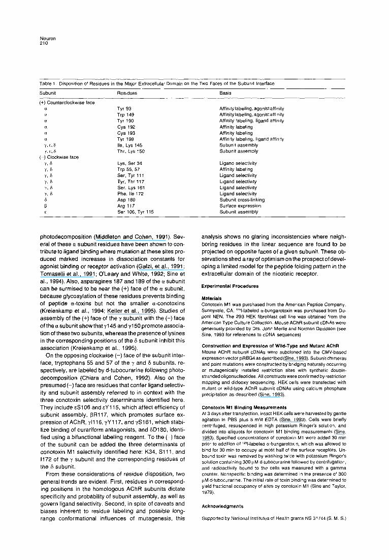
Neuron 210
Table 1. Disposition of Residues in the Major Extracellular Domain on the Two Faces of the Subunit Interface
Subunit Residues Basis
(+) Counterclockwise face a a a a a a Y! & 8 Y,E,S
(-) Clockwise face Y3 6 Y, fJ Y, 6 Ys 6 Y, 6 Yj 6 6 P E
Tyr 93 Trp 149 Tyr 190 cys 192 cys 193 Tyr 198 Ile, Lys 145 Thr, Lys 150
Lys, Ser 34 Trp 55, 57 Ser, Tyr 111 Tyr, Thr 117 Ser, Lys 161 Phe, Ile 172 Asp 180 Arg 117 Ser 106, Tyr 115
Affinity labeling, agonistaffinity Affinitylabeling,agonistaffinity Affinity labeling, ligand affinity Affinity labeling Affinity labeling Affinity labeling, ligand affinity Subunit assembly Subunit assembly
Ligand selectivity Affinity labeling Ligand selectivity Ligand selectivity Ligand selectivity Ligand selectivity Subunit cross-linking Surface expression Subunit assembly
photodecomposit ion (Middleton and Cohen, 1991). Sev- eral of these a subunit residues have been shown to con- tribute to ligand binding where mutation at these sites pro- duced marked increases in dissociation constants for agonist binding or receptor activation (Galzi, et al., 1991; Tomaselli et al., 1991; O ’Leary and White, 1992; Sine et al., 1994). Also, asparagines 187 and 189 of the a subunit can be surmised to be near the (+) face of the a subunit, because glycosylation of these residues prevents binding of peptide a-toxins but not the smaller a-conotoxins (Kreienkamp et al., 1994; Keller et al., 1995). Studies of assembly of the (t) face of the y subunit with the (-) face of the a subunit show that ~145 and ~150 promote associa- tion of these two subunits, whereas the presence of lysines in the corresponding positions of the S subunit inhibit this association (Kreienkamp et al., 1995).
On the opposing clockwise (-) face of the subunit inter- face, tryptophans 55 and 57 of the y and S subunits, re- spectively, are labeled by d-tubocurarine following photo- decomposition (Chiara and Cohen, 1992). Also on the presumed (-) face are residues that confer ligand selectiv- ity and subunit assembly referred to in context with the three conotoxin selectivity determinants identified here. They include cS106 and EY 115, which affect efficiency of subunit assembly, f3R117, which promotes surface ex- pression of AChR, ~1116, yY 117, and $3161, which stabi- lize binding of curariform antagonists, and SD180, identi- fied using a bifunctional labeling reagent. To the (-) face of the subunit can be added the three determinants of conotoxin Ml selectivity identified here: K34, Sill, and 1172 of the y subunit and the corresponding residues of the 6 subunit.
From these considerations of residue disposition, two general trends are evident. First, residues in correspond- ing positions in the homologous AChR subunits dictate specificity and probability of subunit assembly, as well as govern ligand selectivity. Second, in spite of caveats and biases inherent to residue labeling and possible long- range conformational influences of mutagenesis, this
analysis shows no glaring inconsistencies where neigh- boring residues in the linear sequence are found to be projected on opposite faces of a given subunit. These ob- servations shed a ray of optimism on the prospect of devel- oping a limited model for the peptide folding pattern in the extracellular domain of the nicotinic receptor.
Experimental Procedures
Materials Conotoxin Ml was purchased from the American Peptide Company, Sunnyvale, CA. ‘251-labeled a-bungarotoxin was purchased from Du- pont NEN. The 293 HEK fibroblast cell line was obtained from the American Type Culture Collection. Mouse AChR subunit cDNAs were generously provided by Drs. John Merlie and Norman Davidson (see Sine, 1993 for references to cDNA sequences).
Construction and Expression of Wild-Type and Mutant AChR Mouse AChR subunit cDNAs were subcloned into the CMV-based expression vector pRBG4 as described (Sine, 1993). Subunit chimeras and point mutations were constructed by bridging naturally occurring or mutagenically installed restriction sites with synthetic double- stranded oligonucleotides. All constructs were confirmed by restriction mapping and dideoxy sequencing. HEK cells were transfected with mutant or wild-type AChR subunit cDNAs using calcium phosphate precipitation as described (Sine, 1993).
Conotoxin Ml Binding Measurements At 3 days after transfection, intact HEK cells were harvested by gentle agitation in PBS plus 5 mM EDTA (Sine, 1993). Cells were briefly centrifuged, resuspended in high potassium Ringer’s solution, and divided into aliquots for conotoxin Ml binding measurements (Sine, 1993). Specified concentrations of conotoxin Ml were added 30 min prior to addition of 1Z51-labeled a-bungarotoxin, which was allowed to bind for 30 min to occupy at most half of the surface receptors Un- bound toxin was removed by washing twice with potassium Ringers solution containing 300 FM d-tubocurarine followed by centrifugation, and radioactivity bound to the cells was measured with a gamma counter. Nonspecific binding was determined in the presence of 300 trM d-tubocurarine The initial rate of toxin binding was determined to yield fractional occupancy of sites by conotoxin Ml (Sine and Taylor, 1979).
Acknowledgments
Supported by Natronal lnstrtutes of Health grants NS 31744 (S. M. S.)

a-Conotoxin-Nicotinic Receptor Interactions 211
and GM 24437 and GM 18360 (P. T.) and a Deutsche Forshungsgem- einschaft Fellowship (H.J. K.).
The costs of publication of this article were defrayed in part by the payment of page charges. This article must therefore be hereby marked “advertisement” in accordance with 16 USC Section 1734 solely to indicate this fact.
Received March 31, 1995; revised May 3, 1995
References
Abramson, S. N., Li, Y., Culver, P., and Taylor, P. (1989). An analog of lophotoxin reacts covalently with tyrosine 190 in the a-subunit of the nicotinic acetylcholine receptor. J. Biol. Chem. 264, 12666-12672.
Blount, P., and Merlie, J. (1989). Molecular basis of the two nonequiva- lent ligand binding sites of the muscle nicotinic acetylcholine receptor. Neuron 3, 349-357.
Chiara, D. C., and Cohen, J. B. (1992). Identification of amino acids contributing to high andlow affinityd-tubocurarinesiteson theTorpedo nicotinic acetylcholine receptor subunits. Biophys. J. 67, A106. Czajkowski, C., and Karlin, A. (1995). Structureofthe nicotinic receptor acetylcholine-binding site: identification of acidic residues in the delta subunit within 0.9 nm of the a subunit binding site disulfide. J. Biol. Chem. 270,3160-3164. Czajkowski, C., Kaufmann, G., and Karlin, A. (1993). Negatively charged amino acid residues in the nicotinic receptor 6 subunit that contribute to the binding of acetycholine. Proc. Natl. Acad. Sci. USA 90, 6285-6289. Dennis, M., Giraudat, J., Kotzyba-Hibert, F., Goeldner, M., Hirth, C., Chang, J. Y., Lazure, C., Chretien, M., and Changeux, J. P. (1988). Amino acids of the Torpedo marmorata acetylcholine receptor alpha subunit labeled by a photoaffinity ligand for the acetylcholine binding site. Biochemistry 27, 2346-2357.
Fu, D.-X., and Sine, S. M. (1994). Competitive antagonists bridge the a-y subunit interface of the acetylcholine receptor through quaternary ammonium-aromatic interactions. J. Biol. Chem. 269, 26152-26157. Galzi, J. L., Revah, F., Black, D., Goeldner, M., Hirth, C., and Changeux, J. P. (1990). Identification of a novel amino acid alpha- tyrosine 93 within the cholrnergic ligand-binding sites of the acetylcho- line receptor by photoaffinity labeling. J. Biol. Chem. 265, 10430- 10437.
Galzi, J. L.. Bertrand, D., Devillers-Theiry, A., Revah, F., Bertrand, S., and Changeux, J. P. (1991). Functional significance of aromaticamino acids from three peptide loops of the alpha 7 neuronal nicotinic recep- tor site investigated by site-directed mutagenesis. FEBS Lett. 294, 198-202.
Gu, Y., Forsayeth, J., Verrall, S., Yu, X., and Hall, 2. (1991). Assembly of the mammalian muscle acetylcholine receptor in transfected COS cells. J. Biol. Chem. 174, 799-807.
Hann, Ft. M., Pagan, 0. R., and Eterovic, V. A. (1994). The a-conotoxins GI and Ml distinguish between the nicotinic acetylcholine receptor agonist sites while SI does not. Biochemistry 33, 14058-14063. Kao, P. N., Dwork, A. J., Kaldany, R. Ft., Silver, M. L., Wideman, J., Stein, S., and Karlin, A. (1984). Identification of the alpha subunit half-cystine specifically labeled by an affinity reagent for the acetylcho- line receptor binding site. J. Biol. Chem. 259, 11662-I 1665.
Keller, S. H., Kreienkamp, H.-J., Kawanishi, C., and Taylor, P. (1995). Molecular determinants conferring a-toxin resistance in recombinant DNA-derived acetylcholine receptors. J. Biol. Chem. 270, 4165-4171, Kreienkamp, H.-J., Sine, S. M., Maeda, R. K., and Taylor, P. (1994). Glycosylation sites selectively interfere with a-toxin binding to the nico- tinic acetylcholine receptor. J. Biol. Chem. 269, 8108-8114.
Kreienkamp, H.-J., Meada, R., Sine, S. M., and Taylor, P. (1995). Intersubunit contacts governing assembly of the mammalian nicotinic acetylcholine receptor. Neuron 74, 635-644.
Kuhse. J., Laube, B., Magalei, D., and Betz, H. (1993). Assembly of the inhibitory glycine receptor: identification of amino acid sequence motifs governing subunit stoichiometry. Neuron 17, 1049-1056.
Middleton, R. E., and Cohen, J. B. (1991). Mapping oftheacetylcholine
binding site of the nicotinic acetylcholine receptor: [3H]nicotine as an agonist photoaffinity label. Biochemistry 30, 6987-6997. Myers, R. A., Cruz. L. J., Rivier, J. E., and Olivera, B. M. (1993). Conus peptides as chemical probes for receptors and ion channels, Chem. Rev. 93, 1923-1936.
Neubig, R. R., and Cohen, J. E3. (1979). Equilibrium binding of PH] tubocurarine and [?f] acetylcholine by Torpedo postsynaptic mem- branes: stoichiometry and ligand interactions. Biochemistry 16,5464- 5475.
O’Leary. M. E., and White, M. M. (1992). Mutational analysis of ligand- induced activation of the Torpedo acetylcholine receptor. J. Biol. Chem. 267, 8360-8365.
Pedersen, S. E., and Cohen, J. B. (1990). d-tubocurarine binding sites are located at a-y and a-6 subunit interfaces of the nicotinic acetylcho- line receptor. Proc. Natl. Acad. Sci. USA 87, 2785-2789.
Sine, S. M. (1993). Molecular dissection of subunit interfaces in the acetylcholine receptor: identification of residues that determine curare selectivity. Proc. Natl. Acad. Sci. USA 90, 9436-9440.
Sine, S. M., andTaylor, P. (1979). Functional consequencesofagonist- mediated state transitions in the cholinergic receptor. J. Biol. Chem. 254, 3315-3325.
Sine, S. M., and Taylor, P. (1981). Relationships between reversible antagonist occupancy and the functional capacity of the acetylcholine receptor. J. Biol. Chem. 256, 6692-6699. Sine, S. M., Claudia, T., and Sigworth, F. J. (1990). Activation of Tor- pedo acetylcholine receptor expressed in mouse fibroblasts: single channel current kinetics reveal distinct agonist binding affinities. J. Gen. Physiol. 96, 395-437. Sine, S. M., and Claudio, T. (1991). y- and &subunits regulate the affinity and cooperativity of ligand binding to the acetylcholine recep- tor. J. Biol. Chem. 266, 19369-19377.
Sine, S. M., Quiram. P., Papanikolaou, F., Kreienkamp, H.J., and Taylor, P. (1994). Conserved tyrosines in the alpha subunit of the nicotinic acetylcholine receptor stabilize quaternary ammonium groups of agonists and antagonists. J. Biol. Chem. 269, 8808-6816. Tomaselli, G. F., McLaughlin, J. T., Jurman, M. E., Hawrot, E., and Yellen, G. (1991). Mutations affecting agonist sensitivity of the nicotinic acetylcholine receptor. Biophys. J. 60, 721-727.
Utkin, Y. N., Kobayashi, Y., Hucho, F., and Tsatlin, V. I. (1994). Rela- tionships between the binding sites for an a-conotoxin and snake venom neurotoxins in the nicotinic acetylcholine receptor from Tor- pedo californica. Toxicon 32, 1153-l 157.
Copyright © 2022 FDOKUMEN