Promoter DNA Hypermethylation and Gene Repression in Undifferentiated Arabidopsis Cells
10
Promoter DNA Hypermethylation and Gene Repression in Undifferentiated Arabidopsis Cells Marı ´a Berdasco 1,2,3 , Rube ´n Alca ´ zar 4 , Marı ´a Victoria Garcı´a-Ortiz 5 , Esteban Ballestar 1 , Agustı´n F. Ferna ´ ndez 1 , Teresa Rolda ´ n-Arjona 5 , Antonio F. Tiburcio 4 , Teresa Altabella 4 , Nicolas Buisine 6 , Hadi Quesneville 7 , Antoine Baudry 8 , Loı¨c Lepiniec 8 , Miguel Alaminos 9 , Roberto Rodrı´guez 2 , Alan Lloyd 10 , Vincent Colot 6 , Judith Bender 11 , Marı´a Jesu ´ s Canal 2 , Manel Esteller 1,3 , Mario F. Fraga 1,12 * 1 Cancer Epigenetics Laboratory, Molecular Pathology Programme, Spanish National Cancer Centre (CNIO), Madrid, Spain, 2 Plant Epigenetics Laboratory, University of Oviedo, Oviedo, Spain, 3 Cancer Epigenetics and Biology Program (PEBC), Catalan Institute of Oncology (ICO), Barcelona, Catalonia, Spain, 4 Universitat de Barcelona, Facultat de Farmacia, Unitat de Fisiologia Vegetal, Barcelona, Spain, 5 Departamento de Gene ´ tica, Universidad de Co ´ rdoba, Co ´ rdoba, Spain, 6 Unite ´ de Recherche en Ge ´nomique Ve ´ge ´ tale, CNRS UMR8114, INRA UMR1165, Universite ´ d’Evry Val d’Essonne, Evry, France, 7 Unite ´ de Recherches en Ge ´ nomique-Info, INRA, Evry, France, 8 Laboratoire de Biologie des Semences, Unite ´ Mixte de Recherche, 204 Institut National de la Recherche Agronomique, Paris-Grignon, Versailles, France, 9 Department of Histology, University of Granada, Granada, Spain, 10 Molecular Cell and Developmental Biology, The Institute for Cellular and Molecular Biology, The University of Texas at Austin, Austin, Texas, United States of America, 11 Department of Biochemistry and Molecular Biology, Johns Hopkins University Bloomberg School of Public Health, Baltimore, Maryland, United States of America, 12 Department of Immunology and Oncology, Centro Nacional de Biotecnologı ´a/CSIC, Cantoblanco, Madrid, Spain Abstract Maintaining and acquiring the pluripotent cell state in plants is critical to tissue regeneration and vegetative multiplication. Histone-based epigenetic mechanisms are important for regulating this undifferentiated state. Here we report the use of genetic and pharmacological experimental approaches to show that Arabidopsis cell suspensions and calluses specifically repress some genes as a result of promoter DNA hypermethylation. We found that promoters of the MAPK12, GSTU10 and BXL1 genes become hypermethylated in callus cells and that hypermethylation also affects the TTG1, GSTF5, SUVH8, fimbrin and CCD7 genes in cell suspensions. Promoter hypermethylation in undifferentiated cells was associated with histone hypoacetylation and primarily occurred at CpG sites. Accordingly, we found that the process specifically depends on MET1 and DRM2 methyltransferases, as demonstrated with DNA methyltransferase mutants. Our results suggest that promoter DNA methylation may be another important epigenetic mechanism for the establishment and/or maintenance of the undifferentiated state in plant cells. Citation: Berdasco M, Alca ´zar R, Garcı ´a-Ortiz MV, Ballestar E, Ferna ´ ndez AF, et al. (2008) Promoter DNA Hypermethylation and Gene Repression in Undifferentiated Arabidopsis Cells. PLoS ONE 3(10): e3306. doi:10.1371/journal.pone.0003306 Editor: Frederic Berger, Temasek Life Sciences Laboratory, Singapore Received May 6, 2008; Accepted September 1, 2008; Published October 1, 2008 Copyright: ß 2008 Berdasco et al. This is an open-access article distributed under the terms of the Creative Commons Attribution License, which permits unrestricted use, distribution, and reproduction in any medium, provided the original author and source are credited. Funding: This work was supported by the Health (FIS01-04) (PI061267), Education and Science (I+D+I MCYT08-03, FU2004-02073/BMC and Consolider MEC09-05) Departments of the Spanish Government, the European Grant TRANSFOG LSHC-CT-2004-503438, and the Spanish Association Against Cancer (AECC). M.B. is funded by the Association Against Cancer (AECC). Competing Interests: The authors have declared that no competing interests exist. * E-mail: [email protected] Introduction The ability of mature plant cells to regenerate a whole organism is probably the most remarkable growth attribute of plant cells that distinguishes them from mammalian cells [1]. The basis of such a capacity in plants lies in the availability of undifferentiated cells that can subsequently differentiate into all the cell types present in a mature organism [2–4]. The exact molecular processes involved in the maintenance and/or induction of cell undifferentiation in plants are still poorly understood. In recent years, the epigenetic mechanisms that control chromatin structure and function, including DNA methylation and histone modification, have emerged as key factors in the regulation of cell growth and differentiation and, thereby, the nuclear reprogramming necessary for dedifferentiation [4]. The presence of a well-defined DNA methylation pattern in plants is necessary for the normal growth and development of the organism [5–9]. DNA methylation in plants has two functions: to protect the genome by inactivating transposable elements, and to control the expression of single-copy genes, such as the FWA transcription factor, the BALL pathogen-resistance gene (BAL), and the phosphoribosylanthranilate isomerase (PAI) family of tryptophan biosynthesis genes (reviewed in [6]). Recent pioneering reports [7,9–11] defining the DNA methylome of A. thaliana have reinforced the significance of the two aforementioned tasks of DNA methylation as an epigenetic marker in this model and its relevance as a central coordinator of epigenetic memory. In mammals, promoter DNA methylation-dependent gene regulation has been found to have important roles in development and differentiation. This is exemplified by the observations that human embryonic stem cells have specific epigenetic signatures [12–14], many tissue-specific genes present promoter DNA methylation-dependent regulation (reviewed in [15,16]), and finally, aberrant hypermethylation-mediated repression of genes involved in cell differentiation results in malignant transformation (reviewed in [17,18]). In plants, however, there is little information about DNA methylation-dependent mechanisms involved in the control of cell differentiation. Previous work in plants found that PLoS ONE | www.plosone.org 1 October 2008 | Volume 3 | Issue 10 | e3306
Transcript of Promoter DNA Hypermethylation and Gene Repression in Undifferentiated Arabidopsis Cells
Promoter DNA Hypermethylation and Gene Repressionin Undifferentiated Arabidopsis CellsMarıa Berdasco1,2,3, Ruben Alcazar4, Marıa Victoria Garcıa-Ortiz5, Esteban Ballestar1, Agustın F.
Fernandez1, Teresa Roldan-Arjona5, Antonio F. Tiburcio4, Teresa Altabella4, Nicolas Buisine6, Hadi
Quesneville7, Antoine Baudry8, Loıc Lepiniec8, Miguel Alaminos9, Roberto Rodrıguez2, Alan Lloyd10,
Vincent Colot6, Judith Bender11, Marıa Jesus Canal2, Manel Esteller1,3, Mario F. Fraga1,12*
1 Cancer Epigenetics Laboratory, Molecular Pathology Programme, Spanish National Cancer Centre (CNIO), Madrid, Spain, 2 Plant Epigenetics Laboratory, University of
Oviedo, Oviedo, Spain, 3 Cancer Epigenetics and Biology Program (PEBC), Catalan Institute of Oncology (ICO), Barcelona, Catalonia, Spain, 4 Universitat de Barcelona,
Facultat de Farmacia, Unitat de Fisiologia Vegetal, Barcelona, Spain, 5 Departamento de Genetica, Universidad de Cordoba, Cordoba, Spain, 6 Unite de Recherche en
Genomique Vegetale, CNRS UMR8114, INRA UMR1165, Universite d’Evry Val d’Essonne, Evry, France, 7 Unite de Recherches en Genomique-Info, INRA, Evry, France,
8 Laboratoire de Biologie des Semences, Unite Mixte de Recherche, 204 Institut National de la Recherche Agronomique, Paris-Grignon, Versailles, France, 9 Department of
Histology, University of Granada, Granada, Spain, 10 Molecular Cell and Developmental Biology, The Institute for Cellular and Molecular Biology, The University of Texas at
Austin, Austin, Texas, United States of America, 11 Department of Biochemistry and Molecular Biology, Johns Hopkins University Bloomberg School of Public Health,
Baltimore, Maryland, United States of America, 12 Department of Immunology and Oncology, Centro Nacional de Biotecnologıa/CSIC, Cantoblanco, Madrid, Spain
Abstract
Maintaining and acquiring the pluripotent cell state in plants is critical to tissue regeneration and vegetative multiplication.Histone-based epigenetic mechanisms are important for regulating this undifferentiated state. Here we report the use ofgenetic and pharmacological experimental approaches to show that Arabidopsis cell suspensions and calluses specificallyrepress some genes as a result of promoter DNA hypermethylation. We found that promoters of the MAPK12, GSTU10 andBXL1 genes become hypermethylated in callus cells and that hypermethylation also affects the TTG1, GSTF5, SUVH8, fimbrinand CCD7 genes in cell suspensions. Promoter hypermethylation in undifferentiated cells was associated with histonehypoacetylation and primarily occurred at CpG sites. Accordingly, we found that the process specifically depends on MET1and DRM2 methyltransferases, as demonstrated with DNA methyltransferase mutants. Our results suggest that promoterDNA methylation may be another important epigenetic mechanism for the establishment and/or maintenance of theundifferentiated state in plant cells.
Citation: Berdasco M, Alcazar R, Garcıa-Ortiz MV, Ballestar E, Fernandez AF, et al. (2008) Promoter DNA Hypermethylation and Gene Repression inUndifferentiated Arabidopsis Cells. PLoS ONE 3(10): e3306. doi:10.1371/journal.pone.0003306
Editor: Frederic Berger, Temasek Life Sciences Laboratory, Singapore
Received May 6, 2008; Accepted September 1, 2008; Published October 1, 2008
Copyright: � 2008 Berdasco et al. This is an open-access article distributed under the terms of the Creative Commons Attribution License, which permitsunrestricted use, distribution, and reproduction in any medium, provided the original author and source are credited.
Funding: This work was supported by the Health (FIS01-04) (PI061267), Education and Science (I+D+I MCYT08-03, FU2004-02073/BMC and Consolider MEC09-05)Departments of the Spanish Government, the European Grant TRANSFOG LSHC-CT-2004-503438, and the Spanish Association Against Cancer (AECC). M.B. isfunded by the Association Against Cancer (AECC).
Competing Interests: The authors have declared that no competing interests exist.
* E-mail: [email protected]
Introduction
The ability of mature plant cells to regenerate a whole organism
is probably the most remarkable growth attribute of plant cells that
distinguishes them from mammalian cells [1]. The basis of such a
capacity in plants lies in the availability of undifferentiated cells
that can subsequently differentiate into all the cell types present in
a mature organism [2–4]. The exact molecular processes involved
in the maintenance and/or induction of cell undifferentiation in
plants are still poorly understood.
In recent years, the epigenetic mechanisms that control
chromatin structure and function, including DNA methylation
and histone modification, have emerged as key factors in the
regulation of cell growth and differentiation and, thereby, the
nuclear reprogramming necessary for dedifferentiation [4]. The
presence of a well-defined DNA methylation pattern in plants is
necessary for the normal growth and development of the organism
[5–9]. DNA methylation in plants has two functions: to protect the
genome by inactivating transposable elements, and to control the
expression of single-copy genes, such as the FWA transcription
factor, the BALL pathogen-resistance gene (BAL), and the
phosphoribosylanthranilate isomerase (PAI) family of tryptophan
biosynthesis genes (reviewed in [6]). Recent pioneering reports
[7,9–11] defining the DNA methylome of A. thaliana have
reinforced the significance of the two aforementioned tasks of
DNA methylation as an epigenetic marker in this model and its
relevance as a central coordinator of epigenetic memory.
In mammals, promoter DNA methylation-dependent gene
regulation has been found to have important roles in development
and differentiation. This is exemplified by the observations that
human embryonic stem cells have specific epigenetic signatures
[12–14], many tissue-specific genes present promoter DNA
methylation-dependent regulation (reviewed in [15,16]), and
finally, aberrant hypermethylation-mediated repression of genes
involved in cell differentiation results in malignant transformation
(reviewed in [17,18]). In plants, however, there is little information
about DNA methylation-dependent mechanisms involved in the
control of cell differentiation. Previous work in plants found that
PLoS ONE | www.plosone.org 1 October 2008 | Volume 3 | Issue 10 | e3306

the loss of the differentiated state in protoplasts is accompanied by
global changes in DNA methylation [19,20] and histone
modification [21] that alter heterochromatin distribution and
organization [1,3,22], chromatin decondensation [20,23], and
disrupt the nucleolus [19]. Intriguingly, a recent study found that
protoplast dedifferentiation is accompanied by a dramatic
centromeric heterochromatin decondensation that is not accom-
panied by changes in DNA methylation or H3K9 dimethylation
[24]. With respect to promoter DNA methylation in plants,
however, undifferentiation has only been associated with the
hypomethylation-dependent upregulation of a few members of the
NAC (NAM/ATAF1/CUC2) domain family [19], and there has
been no report of genes whose repression by promoter
hypermethylation contributes to the maintenance or establishment
of the undifferentiated state. To address these gaps in our
knowledge by looking for genes regulated by promoter methyla-
tion in undifferentiated cells, we adopted two experimental
strategies: a genetic approach, using Arabidopsis cells deficient in
different members of the family of plant DNA methyltransferases,
and a pharmacological approach, treating undifferentiated
Arabidopsis cell suspensions (ACS) with the demethylating drug 5-
aza-29-deoxycytidine (ADC), followed by expression microarray
analysis in a similar way to that previously done in whole plants
[8]. Using these approaches, we identified three and five
hypermethylated genes in callus and cell suspensions, respectively.
Promoter hypermethylation primarily occurred at CpG sites and
specifically depended on MET1 and DRM2 methyltransferases.
Our results suggest that promoter DNA methylation may be
another important epigenetic mechanism for the establishment
and/or maintenance of the undifferentiated state in plant cells.
Materials and Methods
Generation of calluses from DNA methyltransferase KOsand culture conditions
Seeds of Arabidopsis thaliana (L.) ecotype Wassilewskija wild type
and mutants (met1, cmt3, drm2, drm2 cmt3) were surface-sterilized
and placed on agar tubes with 10 ml of germination medium
containing MS basal medium (mineral salts, vitamins, and
micronutrients), 3% sucrose, 0.8% agar, pH 5.8. After stratifica-
tion at 4uC for 2 days in darkness, seeds were transferred to a
growth room and maintained under fluorescent lights (16 h light/
8 h dark illumination regime) at 24uC for 10 days. Explants from
roots and leaves (approximately 1 cm long) were excised in sterile
conditions and sections were put in 60 mm Petri dishes with callus-
induction medium (MS basal medium supplemented with
1 mg L21 2,4- dichlorophenoxyacetic acid (2,4-D), 3% sucrose,
0.8% agar, pH 5.8) at 24uC with a 16:8 h photoperiod. Nucleic
acid was extracted after 20 days of culture once calluses had been
established.
Cell suspensions of Arabidopsis (Col 0) were obtained as
previously described [25]. Cells were grown in Murashige &
Skoog (MS) salts medium supplemented with 3% sucrose (w/v),
0.5 mg L21 naphthalene acetic acid, and 0.1 mg L21 kinetin at
pH 5.8 in a 16 h light/8 h dark regime at 22uC. For demethyl-
ation treatments 5-aza-2-deoxycytidine (5 mM) was added.
Affymetrix GeneChipsTotal RNA was prepared using TRIZOLH (Invitrogen,
Carlsbad, CA) and further purified using RNeasy columns
(Qiagen, GmbH) according to the manufacturer’s instructions.
The integrity of RNA was monitored by denaturing agarose gel
electrophoresis in 16 MOPS. Biotinylated target RNA was
prepared from 5 mg of total RNA using the Affymetrix protocol.
Briefly, double-stranded cDNA was prepared from the RNA
template using a modified oligo-dT primer containing a 59 T7
RNA polymerase promoter sequence and the Superscript Choice
System for cDNA Synthesis (Invitrogen). cDNA was then used as
the template in an in vitro transcription reaction. The resulting
biotinylated-cRNA ‘‘target’’ was purified on an affinity resin of the
GeneChip Sample Cleanup Module Kit (Affymetrix, Santa Clara,
CA), randomly fragmented and hybridized on the GeneChip
Arabidopsis ATH1 Genome Array (Affymetrix). The hybridization
reactions were processed and scanned according to the standard
Affymetrix protocols. All arrays were globally scaled to a target-
intensity value of 600 and then the scaling factor, background,
noise, and percentage presence were calculated according to the
Affymetrix Data Mining Tool protocols (Affymetrix). All resulting
datasets were filtered using the absolute call metric (present or
absent) implemented within Microsoft Access (Microsoft Corpo-
ration, Redmond, WA). Two biological replicates were done for
each sample. The expression profiles for the demethylation
treatment and DNA methyltransferase mutants were compared
in scatterplots.
Quantification of global 5-methylcytosine content5-methylcytosine (5 mC) content was determined by high-
performance capillary electrophoresis (HPCE) as previously
described [26]. Briefly, genomic DNA samples were boiled,
treated with nuclease P1 (Sigma) for 16 h at 37uC, and with
alkaline phosphatase (Sigma) for an additional 2 h at 37uC. After
hydrolysis, total cytosine and 5 mC content were measured by
capillary electrophoresis using a P/ACE MDQ system (Beckman-
Coulter). Relative 5 mC content was expressed as a percentage of
the total (methylated and non-methylated) cytosine content.
Methylation at CpG sites was quantified as previously described
[27]. In brief, DNA was first digested with XbaI (20 units mg21) for
2 h at 37uC. For methylation with SssI, 5 ml of cut DNA were
added to a mixture containing 1 ml Tris-HCl, 1 M (pH 8.0), 2 ml
NE-Buffer 2 106 (New England Biolabs), 10 ml S-adenosyl-L-
[methyl-3H]methionine (14.4 Ci mmol21, 70 mM; Amersham),
and 2 ml SssI methylase (2 units ml21; New England Biolabs). For
methylation with dam methylase, the same amount of DNA was
added to the following mixture: 1 ml Tris-HCl, 1 M (pH 7.5), 2 ml
NE-Buffer for dam methylase 106 (New England Biolabs), 0.5 ml
water, 10 ml S-adenosyl-L-[methyl-3H]methionine, and 1.5 ml dam
methylase (8 units ml21; New England Biolabs). Both reaction
mixtures were incubated for 4 h at 37uC and the incorporation of
radioactivity was measured using a 1414 liquid scintillation
counter (PerkinElmer, Inc.). The relative percentage of methylated
CpG was estimated as [1 – (ratio SssI/dam/3.2)]6100.
Analysis of sequence-specific DNA methylationThe methylation status of specific genomic DNA sequences was
established by bisulfite genomic sequencing as previously described
[28]. Each of these was automatically sequenced in the twelve
colonies to measure the methylation status of every single CpG
dinucleotide for subsequent statistical analysis. Primers for bilsufite
sequencing were designed using Methyl Primer Express SoftwareH(Applied Biosystems). Primer sequences and PCR conditions for
methylation analysis are summarized in Table S1.
Semi-quantitative and quantitative RT-PCR expressionanalyses
We reverse-transcribed total RNA (2 mg) treated with Dnase I
(Ambion) using oligo (dT) 20 primer with ThermoScript TM RT-
PCR (Invitrogen). We carried out semi-quantitative PCR reactions
Promoter Methylation in Plants
PLoS ONE | www.plosone.org 2 October 2008 | Volume 3 | Issue 10 | e3306

in a final volume of 16 ml containing 106 PCR buffer (Ecogen),
50 mM of MgCl2, 2 mM of dNTP, 1 mM of each primer and 3 U
of EcoStart DNA polymerase (Ecogen). We used 100 ng of cDNA
for PCR amplification, and amplified all of the sequences with
multiple cycle numbers (25–27 cycles) to determine the appropri-
ate conditions for distinguishing semi-quantitative differences in
expression levels. For quantitative RT-PCR analysis, PCR
amplifications were performed as followed: 0,20 mg of cDNA, 5
pM of each primer and SYBRGreen PCR Master Mix (Applied
Biosystems). Three measurements were analyzed using the Prism
7700 Sequence Detection (Applied Biosystems). Relative quanti-
fications were performed for all genes and ACTIN was used as
loading control. Primer sequences and PCR conditions for semi-
quantitative and quantitative RT-PCR analysis are summarized in
Table S1.
Chromatin immunoprecipitationChromatin immunoprecipitation (ChIP) assays were performed
as previously described [28] using commercial antiacetylated
histones H3 and H4 and antimethylK4 H3 antibodies (Upstate
Biotechnologies). Chromatin was sheared to an average length of
0.2–0.5 kb for this analysis. PCR amplification was performed in
25 ml with primers specific to each of the analyzed promoters. The
sensitivity of PCR amplification of each promoter was evaluated
by serial dilution of total DNA collected after sonication (input
fraction). Primer sequences and PCR conditions are summarized
in Table S1. All samples were analyzed in triplicate.
Results
Genetic identification of putative DNA methylation-dependent transcriptionally repressed genes inArabidopsis callus
To look for genes repressed by promoter hypermethylation in
callus Arabidopsis cells, we used Arabidopsis plants deficient in the
different plant DNA methyltransferases: met1 mutant (MET1 2/
2), cmt3 mutant (CMT3 2/2), drm2 mutant (DRM2 (2/2), and
cmt3/drm2 double mutant (DKO). We produced callus cells from
all the mutants using 1 mg L21 2,4- dichlorophenoxyacetic acid
(2,4-D) and measured changes in gene expression relative to
differentiated root cells using microarray chips containing around
22,500 transcripts (Arabidopsis ATH1 Genome array 1 GENE-
CHIPH AFFYMETRIXH) (Figure 1A). A sequence of criteria was
used to select genes dependent on each DNA methyltransferase.
First, genes downregulated in the WT callus relative to WT plants
were identified in order to select putative promoter hypermethyla-
tion-dependent undifferentiation-associated genes. We used the U
rank-statistic test, which ignores the magnitude of the expression
values but distinguishes samples with higher and lower levels of
expression. The statistic was determined for each comparison and
genes with values of U = 0 were selected. A fold-change value,
given by the ratio of the means of the two comparison groups was
calculated, and genes with at least twice the average expression in
WT plants as in WT callus were selected. Second, from the
selected genes, we picked those genes that were upregulated
(according to the two-step criteria described above) in callus of
each DNA methyltransferase mutant relative to the WT callus in
order to associate gene regulation with a specific plant DNA
methyltransferase. Third, of the candidate genes obtained under
the previous criteria, we discarded those common to at least two
mutants. Using these criteria we identified 96 genes for cmt3, 235
for drm2, 134 for the double mutant DKO, and, strikingly, 505 for
the met1 mutant (Figure 1B). This may be evidence of a role for
MET1 methyltransferase in promoter hypermethylation in
Arabidopsis callus cells. The observation is also consistent with the
fact that callus induction was less efficient using cells from the met1
mutant. Callus from MET1 deficient cells presented low
proliferation rates and maintained differentiation-associated fea-
tures like green pigmentation (Figure 1A).
To determine whether promoter hypermethylation is involved
in the selective repression of all DNA methyltransferase-specific
loci, we investigated the genomic DNA methylation status of the
promoter region of twenty candidates for the met1 mutant, one
candidate for the cmt3 mutant, and nine candidates for the drm2
mutant (Table S2). The number of genes selected for each mutant
was proportional to the number of candidates identified with each
mutant. The selection of the 30 genes from the total of 836
candidates identified with the three DNA methyltransferase
mutants was made on the basis of their putative role in cell
differentiation and cell division (Table S3). We designed specific
primers for DNA modified with sodium bisulfite around the
transcription start site of each gene (Table S1). Of the 30 genes
analyzed, three, GSTU10 (At1g74590), MAPK12 (At2g46070) and
BXL1 (At5g49360), presented a higher methylated cytosine
content in the CpG sites of the WT callus. We studied the origin
of the CpG hypermethylation and compared the degree of DNA
methylation in calluses from WT and methyltransferase mutants.
We observed a strong decrease of CpG methylation levels in met1
mutants but not in cmt3 and dko callus (Figure 1C). drm2 mutants
also presented a decrease of CpG methylation within the promoter
region of MAPK12 (Figure 1C). As expected, differentiated tissues
such as roots, shoots or leaves from WT plants were completely
unmethylated (Figure 1C). The remaining 27 selected genes were
completely unmethylated in all the tissues analyzed (Figure S1).
Interestingly, promoter hypermethylation of GSTU10, MAPK12
and BXL1 was associated with gene repression (Figure 1D). These
results suggest that hypermethylation of GSTU10, MAPK12 and
BXL1 in WT callus results from a MET1/DRM2-dependent
mechanism. MAPK12, which was initially identified as a DRM2-
dependent gene, was less methylated in drm2 mutants than in WT
callus, but the drm2 mutant retained several methylated CpGs.
Moreover, the met1 mutant completely failed to hypermethylate
the MAPK12 promoter after callus induction, which suggests that
even though DRM2 might be important for establishing
dedifferentiation-associated promoter hypermethylation, MET1
could be more important still.
Pharmacological identification of putative DNAmethylation-dependent transcriptionally repressedgenes in Arabidopsis cell suspensions
As a second approach to identify genes repressed by promoter
DNA methylation in undifferentiated Arabidopsis cells we generated
Arabidopsis cell suspensions by in vitro culture of seedling-excised roots
in the presence of auxins and cytokinins [25]. We then used the
demethylating agent ADC (5 mM) for 4 days to reactivate putative
genes epigenetically silenced in the Arabidopsis cell suspensions
(Figure 2A). The efficiency of the DNA demethylating treatment was
measured by HPCE [26]. This revealed a 51.3% relative decrease in
global DNA methylation (Figure 2B, left). We also used the methyl-
acceptor assay [27] and found that 21.2% of the cytosines within the
dinucleotide CpG became unmethylated after treatment with the
DNA demethylating drug (Figure 2B, right).
After ADC treatment, we measured changes in gene expression
using the same microarray chips as described in the previous
section. To detect genes with significantly higher expression in
ADC-treated cells than in wild-type cells, and to take both
magnitude and rank into account, we used the two-step criteria
described above. Analysis of the expression microarray data
Promoter Methylation in Plants
PLoS ONE | www.plosone.org 3 October 2008 | Volume 3 | Issue 10 | e3306

revealed that changes in expression after treatment with the
demethylating drug involved the overexpression of genes in most
cases (71.9%, 1,794 genes with U = 0 and at least twice the average
level of expression) (Figure 2C). Of the 1,794 candidate sequences
upregulated after treatment with the DNA demethylating agent,
1,719 corresponded to single-copy genes and 75 to transposons
(Figure 2A). A substantial number of transposable elements (11/
75) belonged to the CACTA-like transposase family [29].
Promoter hypermethylation and transcriptional silencingof single-copy genes in Arabidopsis cell suspensions
We analyzed the possible DNA methylation-associated silencing
of the fourteen selected single-copy genes by bisulfite genomic
sequencing of multiple clones using primers located around the
transcriptional start site of each candidate gene (Table S4). We
selected these genes using a dual criterion: genes statistically
upregulated after the treatment with ADC but, at the same time,
with a role in growth and differentiation (Table S3). In the
Arabidopsis cell suspensions we observed dense DNA hypermethy-
lation around the transcription start site of five genes: TTG1
(At5g24520), GSTF5 (At1g02940), SUVH8 (At2g24740), fimbrin
(At2g04750) and CCD7 (At2g44990) (Figure 3A). The nine
remaining sequences analyzed were completely unmethylated
(Figure S2). DNA methylation of TTG1, GSTF5, SUVH8, fimbrin
and CCD7 in the cell suspensions was found mostly in the CpG
dinucleotide, and not in the CpNpG and CpNpN motifs. The
same regions of these five genes were completely unmethylated in
differentiated tissues of Arabidopsis, such as leaf, root, and shoot
(Figure 3A). It is worth considering the difference in the DNA
methylation status of the aforementioned five single-copy genes
and the transposons in our Arabidopsis system. While we found that
TTG1, GSTF5, SUVH8, fimbrin and CCD7 presented specific CpG
hypermethylation in the cell suspensions, but were fully unmethy-
lated in all the differentiated tissues studied, the CACTA-like
transposons (At1g43840, At2g13160, At2g12980) exhibited dense
methylation of both CpG and CpNpG motifs independently of
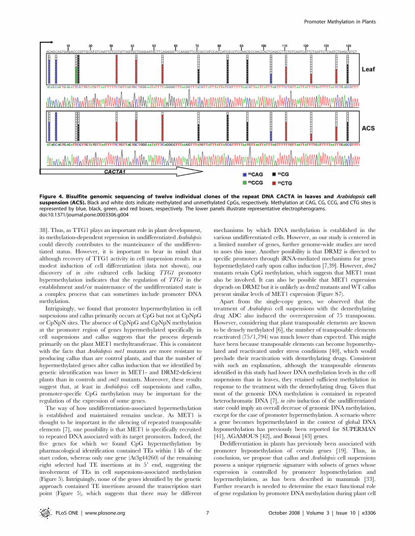
their tissue differentiation status (Table S5 and Figure 4). These
results highlight the distinctive and specific CpG hypermethylation
events occurring in the promoter region of single-copy genes and
Figure 1. Promoter hypermethylation of Arabidopsis callus specifically depends on MET1 and DRM2 methyltransferase activity. (A)Callus induction from DNA methyltransferases mutants. Upper panels, morphological aspect of entire plants grown from WT and DNAmethyltransferase mutant seeds. Lower panels, growth rates of WT and mutant callus generated after treatment with 2,4-D of root explants. (B)Number of candidate genes susceptible to DNA methylation-dependent regulation obtained from each DNA methyltransferase mutant using thetwo-step criteria described in the Results section. (C) Bisulfite sequencing of twelve individual clones of the GSTU10, MAPK12 and BXL1 promoters inWT and DNA methyltransferase mutants. (D) Relationship between levels of GSTU10, MAPK12 and BXL1 expression and promoter DNAhypermethylation. Transcript levels of both genes were analyzed by quantitative RT-PCR and results are expressed as a relative enrichment of thehypomethylated samples (roots and methyltransferase mutants) versus the hypermethylated samples (WT callus).doi:10.1371/journal.pone.0003306.g001
Promoter Methylation in Plants
PLoS ONE | www.plosone.org 4 October 2008 | Volume 3 | Issue 10 | e3306

imply that dedifferentiation-associated promoter hypermethylation
in Arabidopsis depends on MET1 and DRM2 methyltransferases.
The promoter hypermethylation of TTG1, GSTF5, SUVH8,
fimbrin and CCD7 was always associated with transcriptional
silencing, and expression could be restored with the use of the
DNA demethylating agent ADC (Figure 3B). Chromatin immu-
noprecipitation analysis with antibodies raised against histone
modifications associated with transcriptional activation [30]
demonstrated a loss of acetylated histones H3 and H4 and
trimethylated lysine 4 histone H3 within all of the hypermethy-
lated promoters (TTG1, GSTF5, and SUVH8) in cell suspensions
compared with differentiated tissues (Figure 3C).
In this model of Arabidopsis cell suspensions, the undifferentiated
and highly proliferative state is known to depend on the presence of
phytohormones (NAA and Kinetin) in the culture medium [31]. For
this reason, we wondered whether their removal might lead, in
addition to the differentiation and cell growth arrest described herein
(Figure S3A), to the loss of promoter hypermethylation. Bisulfite
genomic sequencing showed that depletion of just one of the
phytohormones was sufficient to induce a loss of CpG methylation in
all three promoters studied (Figure S3B). To asses whether changes
in phytohormone- induced DNA methylation were associated with
changes in MET1 expression we analyzed the levels of MET1
transcripts by quantitative RT-PCR in Arabidopsis cell suspensions
exposed or not to NAA and/or Kinetin. Our results showed that
removal of any of the phytohormones, did not result in any
significant change in MET1 expression (Figure S3C) which suggest
that other mechanisms such as control of its enzymatic activity or
recruitment to specific promoters could be involved.
To establish whether the specific genes that become hyper-
methylated in dedifferentiated cells depend on the type of
phytohormone used to maintain the undifferentiated state, we
compared the DNA methylation status of two hypermethylated
genes in callus cells obtained with 2,4-D (GSTU10 and MAPK12)
and the Arabidopsis cell suspensions maintained with kinetin and
NAA. We found that GSTU10 and MAPK12 were also densely
methylated in the Arabidopsis cell suspensions (Figure S4). In
contrast, the promoter region of three genes identified in the well-
established Arabidopsis cell suspensions (TTG1, GSTF5, and
SUVH8) remained unmethylated in the early 2,4-D-induced callus
(Figure S5). The lack of promoter hypermethylation of TTG1,
GSTF5, and SUVH8 genes in cells soon after the induction of callus
prompted us to question whether the hypermethylation-mediated
repression of these three genes is indispensable to the maintenance
of the undifferentiated state. To address this, we monitored the
promoter hypermethylation of the TTG1 gene in Arabidopsis cell
suspensions of different passage number. Intriguingly, we found a
drop in CpG island methylation levels of the TTG1 gene after 400
passages (Figure S6), in a similar manner to that described for
other methylated DNA sequences in mammals [32]. This finding,
in conjunction with the unmethylated status of TTG1 in callus,
suggest that other mechanisms apart from DNA methylation
Figure 2. Reactivation of hypermethylated genes in Arabidopsis cell suspensions (ACS) using demethylating drugs. (A) Flowchart foridentification of hypermethylated growth-associated genes. We used Arabidopsis cell suspensions after 5 mM ADC treatments followed by cRNAhybridization to a 22,500-oligonucleotide microarray. We obtained over 1,794 unique sequences overexpressed after treatments. Of these, 1,719corresponded to known genes and 75 to repeat elements. We selected fourteen genes to test for promoter hypermethylation by direct bisulfitesequencing; five of them were found to be methylated in Arabidopsis cell suspensions but not in differentiated tissues such as roots and shoots. (B)Quantification of DNA methylation as: overall 5-methyl-cytosine (5 mC) using high-performance capillary electrophoresis (Left panel), and percentageof methylated CpGs using the methyl acceptor assay (Right panel). (C) Reactivation of genes in Arabidopsis cell suspensions after treatment with thedemethylating drug ADC. Upper panel, scatterplots showing expression profiles of control cells and cells treated with ADC obtained by AffymetrixGeneChip technology; Lower panel, relative percentage of overexpressed and repressed genes after treatment with ADC.doi:10.1371/journal.pone.0003306.g002
Promoter Methylation in Plants
PLoS ONE | www.plosone.org 5 October 2008 | Volume 3 | Issue 10 | e3306

might be involved in the regulation of TTG1 in undifferentiated
Arabidopsis cells.
Discussion
Promoter DNA methylation-dependent gene regulation in
mammalian cells has important roles during development and
differentiation. For example, human embryonic stem cells have
specific epigenetic signatures [12–14], many tissue-specific genes
present promoter DNA methylation-dependent regulation [15,16],
and, finally, aberrant hypermethylation-mediated repression of
genes involved in cell differentiation results in malignant
transformation [17,18]. In addition to hypermethylated genes,
undifferentiated mammalian cells also feature genes that are
hypomethylated with respect to their differentiated counterparts
[33]. In plants, cells acquiring pluripotency have been described as
exhibiting hypomethylation-dependent upregulation of several
members of the NAC (NAM/ATAF1/CUC2) domain family
[19]. In the present study we have demonstrated that Arabidopsis
calluses and cell suspensions can use promoter DNA methylation
to repress specific single-copy genes that are unmethylated and
expressed in differentiated cells from various origins. However, it is
important to state that this epigenetic mechanism is not a general
response to cell culture as just a minor fraction of the genes
become hypermethylated in callus and cell suspensions.
We found that promoters of the MAPK12, GSTU10 and BXL1
genes are densely hypermethylated in callus and cell suspensions,
whilst the TTG1, GSTF5 and SUVH8 genes become occasionally
hypermethylated only in cell suspensions. Interestingly, the role of
TTG1 in cell fate and differentiation is very well documented [34–
Figure 3. Genomic DNA methylation status of reactivated genes in leaf and Arabidopsis cell suspensions (ACS). (A) Bisulfite genomicsequencing of twelve clones of the TTG1 (three different regions), GSTF5, SUVH8, Fimbrin and CCD7 promoters. In the schematic representations ofthe methylation status of each CpG dinucleotides black and white dots indicate methylated and unmethylated CpGs, respectively. ACS IMP:Arabidopsis cell suspensions at intermediate passage. (B) Expression profiles of reactivated genes in leaves and Arabidopsis cell suspensionsdetermined by quantitative RT-PCR assays. TTG1, GSTF5, SUVH8, Fimbrin and CCD7 are more strongly expressed in leaf tissues. Expression in cellsuspensions can be restored by treatments with the demethylating drug ADC. ACTIN was used as a control. (*) the expression level of TTG1 in leafs is80-fold higher than in ACS. (**) the expression level of GSTF5 and CCD7 in leafs is 40-fold higher than in ACS. (C) Promoter CpG islandhypermethylation is associated with changes of histone modifications. Chromatin immunoprecipitation analysis of the histone-modification status ofthe promoters of the TTG1, GSTF5, and SUVH8 genes. NAB is the control without antibody. The promoter region of ACTIN is used as a control. AcH3,acetylated histone H3; AcH4, acetylated histone H4; 3mK4, trimethyl-lysine 3 histone H3.doi:10.1371/journal.pone.0003306.g003
Promoter Methylation in Plants
PLoS ONE | www.plosone.org 6 October 2008 | Volume 3 | Issue 10 | e3306
38]. Thus, as TTG1 plays an important role in plant development,
its methylation-dependent repression in undifferentiated Arabidopsis
could directly contributes to the manteinance of the undifferen-
tiated status. However, it is important to bear in mind that
although recovery of TTG1 activity in cell suspension results in a
modest induction of cell differentiation (data not shown), our
discovery of in vitro cultured cells lacking TTG1 promoter
hypermethylation indicates that the regulation of TTG1 in the
establishment and/or maintenance of the undifferentiated state is
a complex process that can sometimes include promoter DNA
methylation.
Intriguingly, we found that promoter hypermethylation in cell
suspensions and callus primarily occurs at CpG but not at CpNpG
or CpNpN sites. The absence of CpNpG and CpNpN methylation
at the promoter region of genes hypermethylated specifically in
cell suspensions and callus suggests that the process depends
primarily on the plant MET1 methyltransferase. This is consistent
with the facts that Arabidopsis met1 mutants are more resistant to
producing callus than are control plants, and that the number of
hypermethylated genes after callus induction that we identified by
genetic identification was lower in MET1- and DRM2-deficient
plants than in controls and cmt3 mutants. Moreover, these results
suggest that, at least in Arabidopsis cell suspensions and callus,
promoter-specific CpG methylation may be important for the
regulation of the expression of some genes.
The way of how undifferentiation-associated hypermethylation
is established and maintained remains unclear. As MET1 is
thought to be important in the silencing of repeated transposable
elements [7], one possibility is that MET1 is specifically recruited
to repeated DNA associated with its target promoters. Indeed, the
five genes for which we found CpG hypermethylation by
pharmacological identification contained TEs within 1 kb of the
start codon, whereas only one gene (At3g44260) of the remaining
eight selected had TE insertions at its 59 end, suggesting the
involvement of TEs in cell suspensions-associated methylation
(Figure 5). Intriguingly, none of the genes identified by the genetic
approach contained TE insertions around the transcription start
point (Figure 5), which suggests that there may be different
mechanisms by which DNA methylation is established in the
various undifferentiated cells. However, as our study is centered in
a limited number of genes, further genome-wide studies are need
to asses this issue. Another possibility is that DRM2 is directed to
specific promoters through iRNA-mediated mechanisms for genes
hypermethylated early upon callus induction [7,39]. However, drm2
mutants retain CpG methylation, which suggests that MET1 must
also be involved. It can also be possible that MET1 expression
depends on DRM2 but it is unlikely as drm2 mutants and WT callus
present similar levels of MET1 expression (Figure S7).
Apart from the single-copy genes, we observed that the
treatment of Arabidopsis cell suspensions with the demethylating
drug ADC also induced the overexpression of 75 transposons.
However, considering that plant transposable elements are known
to be densely methylated [6], the number of transposable elements
reactivated (75/1,794) was much lower than expected. This might
have been because transposable elements can become hypomethy-
lated and reactivated under stress conditions [40], which would
preclude their reactivation with demethylating drugs. Consistent
with such an explanation, although the transposable elements
identified in this study had lower DNA methylation levels in the cell
suspensions than in leaves, they retained sufficient methylation in
response to the treatment with the demethylating drug. Given that
most of the genomic DNA methylation is contained in repeated
heterochromatic DNA [7], in vitro induction of the undifferentiated
state could imply an overall decrease of genomic DNA methylation,
except for the case of promoter hypermethylation. A scenario where
a gene becomes hypermethylated in the context of global DNA
hypomethylation has previously been reported for SUPERMAN
[41], AGAMOUS [42], and Bonsai [43] genes.
Dedifferentiation in plants has previously been associated with
promoter hypomethylation of certain genes [19]. Thus, in
conclusion, we propose that callus and Arabidopsis cell suspensions
possess a unique epigenetic signature with subsets of genes whose
expression is controlled by promoter hypomethylation and
hypermethylation, as has been described in mammals [33].
Further research is needed to determine the exact functional role
of gene regulation by promoter DNA methylation during plant cell
Figure 4. Bisulfite genomic sequencing of twelve individual clones of the repeat DNA CACTA in leaves and Arabidopsis cellsuspension (ACS). Black and white dots indicate methylated and unmethylated CpGs, respectively. Methylation at CAG, CG, CCG, and CTG sites isrepresented by blue, black, green, and red boxes, respectively. The lower panels illustrate representative electropherograms.doi:10.1371/journal.pone.0003306.g004
Promoter Methylation in Plants
PLoS ONE | www.plosone.org 7 October 2008 | Volume 3 | Issue 10 | e3306

Figure 5. Gbrowse view of gene, transposable element and small RNA annotation of 20 kb segments centered on the 2 CpG islandcontaining genes selected with the genetic (GSTU10 and MAPK12) and pharmacologic (TTG1 and SUVH8) approaches. Gene annotationis from TAIR (v7). TE annotation was performed using a novel detection pipeline (HQ, NB, and VC, manuscript in preparation). Small RNA data weredirectly imported from the ASRP database (http://asrp.cgrb.oregonstate.edu/cgi-bin/gbrowse/thaliana-v5) and corresponded to deep sequencing ofsmall RNAs extracted from wild type plants (seedlings, leaves, and flowers).doi:10.1371/journal.pone.0003306.g005
Promoter Methylation in Plants
PLoS ONE | www.plosone.org 8 October 2008 | Volume 3 | Issue 10 | e3306

differentiation, and in the establishment and maintenance of the
undifferentiated state.
Supporting Information
Figure S1 Bisulfite genomic sequencing of twelve individual
clones of nine representative genes unmethylated in leaves and
Arabidopsis wild type callus. Black and white dots indicate
methylated and unmethylated CpGs, respectively. Lower panels,
representative electropherograms.
Found at: doi:10.1371/journal.pone.0003306.s001 (0.05 MB PDF)
Figure S2 Bisulfite genomic sequencing of twelve individual
clones of six representative genes unmethylated in leaves and
Arabidopsis cell suspensions (ACS). Black and white dots indicate
methylated and unmethylated CpGs, respectively. Lower panels,
representative electropherograms.
Found at: doi:10.1371/journal.pone.0003306.s002 (0.04 MB PDF)
Figure S3 Promoter hypermethylation in cell suspensions is
dependent on combined phytohormone action and is associated
with histone hypoacetylation and gene repression. Bisulfite
genomic sequencing of the TTG1, GSTF5, and SUVH8
promoters in the presence or absence of the demethylating drug
ADC, and the phytohormones NAA and kinetin in Arabidopsis
cell suspensions. (A) Quantification of growth rates and the relative
percentage of methylated CpGs. (B) Schematic representations of
the methylation status of each CpG dinucleotide. Black and white
dots indicate methylated and unmethylated CpGs, respectively.
(C) Analysis of MET1 expression in Arabidopsis cell suspensions
(ACS) growing in normal culture medium, in presence or in
absence of phytohormones (kinetin or NAA). Transcript levels
were analyzed by quantitative RT-PCR and results are expressed
as a value relative to the expression in control ACS.
Found at: doi:10.1371/journal.pone.0003306.s003 (0.04 MB PDF)
Figure S4 Promoter DNA methylation of MAPK12 and
GSTU10 genes was also analyzed after different numbers of
passages in Arabidopsis cell suspensions (ACS): IMP, intermediate
passages; LP, late passages. Black and white dots indicate
methylated and unmethylated CpGs, respectively.
Found at: doi:10.1371/journal.pone.0003306.s004 (0.02 MB PDF)
Figure S5 Bisulfite genomic sequencing of twelve individual
clones of TTG1 (A), GSTF5 (B), and SUVH8 (C) genes in cell
suspensions derived from WT and DNA methyltransferase
mutants of Arabidopsis thaliana (L.). Black and white dots indicate
methylated and unmethylated CpGs, respectively.
Found at: doi:10.1371/journal.pone.0003306.s005 (0.03 MB PDF)
Figure S6 Bisulfite genomic sequencing of twelve clones of the
TTG1 (three regions) 59-regulatory region in Arabidopsis cell
suspensions at 300, 330, and 400 passages. Schematic represen-
tations of the methylation status of each CpG dinucleotide. Black
and white dots indicate methylated and unmethylated CpGs,
respectively.
Found at: doi:10.1371/journal.pone.0003306.s006 (0.03 MB PDF)
Figure S7 Transcription levels of MET1 in WT callus and drm2
mutant. Transcript levels were analyzed by quantitative RT-PCR
and results are expressed as a value relative to the expression in
WT callus.
Found at: doi:10.1371/journal.pone.0003306.s007 (0.01 MB PDF)
Table S1 Primer sequences and annealing temperatures.
Found at: doi:10.1371/journal.pone.0003306.s008 (0.02 MB PDF)
Table S2 Genetic identification of methylated genes in dedif-
ferentiated Arabidopsis cells.
Found at: doi:10.1371/journal.pone.0003306.s009 (0.02 MB PDF)
Table S3 List of candidate genes selected for validation with
bisulfite sequencing analysis.
Found at: doi:10.1371/journal.pone.0003306.s010 (0.03 MB PDF)
Table S4 List of Arabidopsis thaliana genes upregulated after
treatment with ADC.
Found at: doi:10.1371/journal.pone.0003306.s011 (0.02 MB PDF)
Table S5 Sequence context of some of the DNA methylation
found in Arabidopsis cell suspensions and leaves.
Found at: doi:10.1371/journal.pone.0003306.s012 (0.01 MB PDF)
Author Contributions
Conceived and designed the experiments: MB RR MJC MFF. Performed
the experiments: MB RA MVGO EB AF TA. Analyzed the data: MB AF
NB HQ MA VC. Contributed reagents/materials/analysis tools: RA
MVGO TRA AT NB HQ AB LL AL JB. Wrote the paper: RR MJC ME
MFF.
References
1. Grafi G (2004) How cells dedifferentiate: a lesson from plants. Dev Biol 268: 1–6.
2. Doerner P (2006) Plant meristems: what you see is what you get? Curr Biol 16:
R56–58.
3. Grafi G, Avivi Y (2004) Stem cells: a lesson from dedifferentiation. TrendsBiotechnol 22: 388–389.
4. Costa S, Shaw P (2007) ‘Open minded’ cells: how cells can change fate. Trends
Cell Biol 17: 101–106.
5. Lippman Z, Gendrel AV, Black M, Vaughn MW, Dedhia N, et al. (2004) Role
of transposable elements in heterochromatin and epigenetic control. Nature 430:
471–476.
6. Chan SW, Henderson IR, Jacobsen SE (2005) Gardening the genome: DNA
methylation in Arabidopsis thaliana. Nat Rev Genet 6: 351–360.
7. Zhang X, Yazaki J, Sundaresan A, Cokus S, Chan SW, et al. (2006) Genome-wide high-resolution mapping and functional analysis of DNA methylation in
arabidopsis. Cell 126: 1189–1201.
8. Chan SW, Zhang X, Bernatavichute YV, Jacobsen SE (2006) Two-step recruitmentof RNA-directed DNA methylation to tandem repeats. PLoS Biol 4: e363.
9. Zilberman D, Gehring M, Tran RK, Ballinger T, Henikoff S (2007) Genome-
wide analysis of Arabidopsis thaliana DNA methylation uncovers aninterdependence between methylation and transcription. Nat Genet 39: 61–69.
10. Vaughn MW, Tanurd IcM, Lippman Z, Jiang H, Carrasquillo R, et al. (2007)
Epigenetic Natural Variation in Arabidopsis thaliana. PLoS Biol 5: e174.
11. Mathieu O, Reinders J, Caikovski M, Smathajitt C, Paszkowski J (2007)
Transgenerational Stability of the Arabidopsis Epigenome Is Coordinated by
CG Methylation. Cell 130: 851–862.
12. Ohm JE, McGarvey KM, Yu X, Cheng L, Schuebel KE, et al. (2007) A stemcell-like chromatin pattern may predispose tumor suppressor genes to DNA
hypermethylation and heritable silencing. Nat Genet 39: 237–242.
13. Widschwendter M, Fiegl H, Egle D, Mueller-Holzner E, Spizzo G, et al. (2007)
Epigenetic stem cell signature in cancer. Nat Genet 39: 157–158.
14. Schlesinger Y, Straussman R, Keshet I, Farkash S, Hecht M, et al. (2007)
Polycomb-mediated methylation on Lys27 of histone H3 pre-marks genes for denovo methylation in cancer. Nat Genet 39: 232–236.
15. Fraga MF, Agrelo R, Esteller M (2007) Cross-talk between aging and cancer: the
epigenetic language. Ann N Y Acad Sci 1100: 60–74.
16. Poulsen P, Esteller M, Vaag A, Fraga MF (2007) The epigenetic basis of twin
discordance in age-related diseases. Pediatr Res 61: 38R–42R.
17. Feinberg AP, Tycko B (2004) The history of cancer epigenetics. Nat Rev Cancer
4: 143–153.
18. Esteller M (2002) CpG island hypermethylation and tumor suppressor genes: abooming present, a brighter future. Oncogene 21: 5427–5440.
19. Avivi Y, Morad V, Ben-Meir H, Zhao J, Kashkush K, et al. (2004)Reorganization of specific chromosomal domains and activation of silent genes
in plant cells acquiring pluripotentiality. Dev Dyn 230: 12–22.
20. Koukalova B, Fojtova M, Lim KY, Fulnecek J, Leitch AR, et al. (2005)
Dedifferentiation of tobacco cells is associated with ribosomal RNA genehypomethylation, increased transcription, and chromatin alterations. Plant
Physiol 139: 275–286.
21. Williams L, Zhao J, Morozova N, Li Y, Avivi Y, et al. (2003) Chromatin
reorganization accompanying cellular dedifferentiation is associated with
Promoter Methylation in Plants
PLoS ONE | www.plosone.org 9 October 2008 | Volume 3 | Issue 10 | e3306

modifications of histone H3, redistribution of HP1, and activation of E2F-target
genes. Dev Dyn 228: 113–120.
22. Grafi G, Ben-Meir H, Avivi Y, Moshe M, Dahan Y, et al. (2007) Histone
methylation controls telomerase-independent telomere lengthening in cells
undergoing dedifferentiation. Dev Biol 306: 838–846.
23. Zhao J, Morozova N, Williams L, Libs L, Avivi Y, et al. (2001) Two phases of
chromatin decondensation during dedifferentiation of plant cells: distinction
between competence for cell fate switch and a commitment for S phase. J Biol
Chem 276: 22772–22778.
24. Tessadori F, Chupeau MC, Chupeau Y, Knip M, Germann S, et al. (2007)
Large-scale dissociation and sequential reassembly of pericentric heterochroma-
tin in dedifferentiated Arabidopsis cells. J Cell Sci 120: 1200–1208.
25. Mathur J, Szabados L, Schaefer S, Grunenberg B, Lossow A, et al. (1998) Gene
identification with sequenced T-DNA tags generated by transformation of
Arabidopsis cell suspension. Plant J 13: 707–716.
26. Fraga MF, Uriol E, Borja Diego L, Berdasco M, Esteller M, et al. (2002) High-
performance capillary electrophoretic method for the quantification of 5-methyl
29-deoxycytidine in genomic DNA: application to plant, animal and human
cancer tissues. Electrophoresis 23: 1677–1681.
27. De Smet C, De Backer O, Faraoni I, Lurquin C, Brasseur F, et al. (1996) The
activation of human gene MAGE-1 in tumor cells is correlated with genome-
wide demethylation. Proc Natl Acad Sci U S A 93: 7149–7153.
28. Fraga MF, Ballestar E, Villar-Garea A, Boix-Chornet M, Espada J, et al. (2005)
Loss of acetylation at Lys16 and trimethylation at Lys20 of histone H4 is a
common hallmark of human cancer. Nat Genet 37: 391–400.
29. Kato M, Miura A, Bender J, Jacobsen SE, Kakutani T (2003) Role of CG and
non-CG methylation in immobilization of transposons in Arabidopsis. Curr Biol
13: 421–426.
30. Bender J (2004) Chromatin-based silencing mechanisms. Curr Opin Plant Biol
7: 521–526.
31. Vanyushin BF (2006) DNA methylation in plants. Curr Top Microbiol Immunol
301: 67–122.
32. Gonzalo S, Garcia-Cao M, Fraga MF, Schotta G, Peters AH, et al. (2005) Role
of the RB1 family in stabilizing histone methylation at constitutive heterochro-matin. Nat Cell Biol 7: 420–428.
33. Bibikova M, Chudin E, Wu B, Zhou L, Garcia EW, et al. (2006) Human
embryonic stem cells have a unique epigenetic signature. Genome Res 16:1075–1083.
34. Koornneef M (1981) The complex syndrome of TTG mutants. Arabid Inf Serv18: 45–51.
35. Galway ME, Masucci JD, Lloyd AM, Walbot V, Davis RW, et al. (1994) The
TTG gene is required to specify epidermal cell fate and cell patterning in theArabidopsis root. Dev Biol 166: 740–754.
36. Payne CT, Zhang F, Lloyd AM (2000) GL3 encodes a bHLH protein thatregulates trichome development in arabidopsis through interaction with GL1
and TTG1. Genetics 156: 1349–1362.37. Zhang F, Gonzalez A, Zhao M, Payne CT, Lloyd A (2003) A network of
redundant bHLH proteins functions in all TTG1-dependent pathways of
Arabidopsis. Development 130: 4859–4869.38. Baudry A, Heim MA, Dubreucq B, Caboche M, Weisshaar B, et al. (2004) TT2,
TT8, and TTG1 synergistically specify the expression of BANYULS andproanthocyanidin biosynthesis in Arabidopsis thaliana. Plant J 39: 366–380.
39. Huettel B, Kanno T, Daxinger L, Bucher E, van der Winden J, et al. (2007)
RNA-directed DNA methylation mediated by DRD1 and Pol IVb: a versatilepathway for transcriptional gene silencing in plants. Biochim Biophys Acta 1769:
358–374.40. Grandbastien MA, Audeon C, Bonnivard E, Casacuberta JM, Chalhoub B, et
al. (2005) Stress activation and genomic impact of Tnt1 retrotransposons inSolanaceae. Cytogenet Genome Res 110: 229–241.
41. Jacobsen SE, Meyerowitz EM (1997) Hypermethylated SUPERMAN epigenetic
alleles in arabidopsis. Science 277: 1100–1103.42. Jacobsen SE, Sakai H, Finnegan EJ, Cao X, Meyerowitz EM (2000) Ectopic
hypermethylation of flower-specific genes in Arabidopsis. Curr Biol 10: 179–186.43. Saze H, Kakutani T (2007) Heritable epigenetic mutation of a transposon-
flanked Arabidopsis gene due to lack of the chromatin-remodeling factor
DDM1. Embo J 26: 3641–3652.
Promoter Methylation in Plants
PLoS ONE | www.plosone.org 10 October 2008 | Volume 3 | Issue 10 | e3306