Cyclin-dependent kinase activity retains the shoot apical meristem cells in an undifferentiated...
12
Cyclin-dependent kinase activity maintains the shoot apical meristem cells in an undifferentiated state Tarik Gaamouche 1,2,† , Carmem-Lara de O. Manes 1,2,†,‡ , Dorota Kwiatkowska 3 , Barbara Berckmans 1,2 , Rachel Koumproglou 4 , Sara Maes 1,2 , Tom Beeckman 1,2 , Teva Vernoux 5 , John H. Doonan 4 , Jan Traas 5 , Dirk Inze ´ 1,2 and Lieven De Veylder 1,2,* 1 Department of Plant Systems Biology, VIB, 9052 Gent, Belgium, 2 Department of Plant Biotechnology and Genetics, Ghent University, 9052 Gent, Belgium, 3 Department of Biophysics and Morphogenesis of Plants, University of Silesia, 40-032 Katowice, Poland, 4 John Innes Centre, Norwich Research Park, Colney, Norwich, NR4 7UH, UK, and 5 Laboratoire de Reproduction et De ´ velopement des Plantes, Institut National de la Recherche Agronomique, 69364 Lyon Cedex 07, France Received 28 May 2010; accepted 6 July 2010. * For correspondence (fax +32 9 3313809; e-mail [email protected]). † These authors contributed equally to this work. ‡ Present address: Universite ´ Pierre et Marie Curie, Unite ´ Mixte de Recherche 7621, Oce ´ anographie Microbienne, Observatoire Oce ´ anologique, 66651 Banyuls sur Mer, France. SUMMARY As the shoot apex produces most of the cells that comprise the aerial part of the plant, perfect orchestration between cell division rates and fate specification is essential for normal organ formation and plant development. However, the inter-dependence of cell-cycle machinery and meristem-organizing genes is still poorly understood. To investigate this mechanism, we specifically inhibited the cell-cycle machinery in the shoot apex by expression of a dominant negative allele of the A-type cyclin-dependent kinase (CDK) CDKA;1 in meristematic cells. A decrease in the cell division rate within the SHOOT MERISTEMLESS domain of the shoot apex dramatically affected plant growth and development. Within the meristem, a subset of cells was driven into the differentiation pathway, as indicated by premature cell expansion and onset of endo-reduplication. Although the meristem structure and expression patterns of the meristem identity genes were maintained in most plants, the reduced CDK activity caused splitting of the meristem in some plants. This phenotype correlated with the level of expression of the dominant negative CDKA;1 allele. Therefore, we propose a threshold model in which the effect of the cell-cycle machinery on meristem organization is determined by the level of CDK activity. Keywords: Arabidopsis thaliana, cell cycle, CDK, endo-reduplication, organogenesis, shoot apical meristem. INTRODUCTION An important attribute of growing plants is their ability to coordinate the events of cell division and cell differentiation. A prominent example is the shoot apex, in which cell divi- sions occur in the shoot apical meristem (SAM) and lateral organ primordia, whereas cell differentiation happens in meristem-derived cells that will form stem and lateral organ tissues. At the organizational level, the dome-shaped SAM is divided into functionally distinct domains that are charac- terized by different division rates. A central zone (CZ) at the summit harbors slowly dividing cells that can be distin- guished from the surrounding peripheral zone (PZ) and the rib meristem (RM) located beneath, which both exhibit more frequent cell divisions. The PZ contains cells that will be incorporated into the lateral organs. The CZ consists of stem cells that replenish themselves. During primordium forma- tion, PZ cells become the founder cells of lateral organs, after which these cells are replaced by dividing pluripotent cells from the CZ. As a consequence, the SAM can maintain itself as a stable self-sustaining structure (Carraro et al., 2006). The self-perpetuation of the SAM is controlled by a group of well-characterized genes. The SHOOT MERISTEMLESS (STM) gene is expressed throughout the CZ, PZ and RM, and keeps them in the indeterminate state of cells within the SAM (Long et al., 1996; Long and Barton, 1998). By contrast, expression of the WUSCHEL (WUS) gene is confined to a 26 ª 2010 The Authors Journal compilation ª 2010 Blackwell Publishing Ltd The Plant Journal (2010) 64, 26–37 doi: 10.1111/j.1365-313X.2010.04317.x
-
Upload
independent -
Category
Documents
-
view
2 -
download
0
Transcript of Cyclin-dependent kinase activity retains the shoot apical meristem cells in an undifferentiated...

Cyclin-dependent kinase activity maintains the shoot apicalmeristem cells in an undifferentiated state
Tarik Gaamouche1,2,†, Carmem-Lara de O. Manes1,2,†,‡, Dorota Kwiatkowska3, Barbara Berckmans1,2, Rachel Koumproglou4,
Sara Maes1,2, Tom Beeckman1,2, Teva Vernoux5, John H. Doonan4, Jan Traas5, Dirk Inze1,2 and Lieven De Veylder1,2,*
1Department of Plant Systems Biology, VIB, 9052 Gent, Belgium,2Department of Plant Biotechnology and Genetics, Ghent University, 9052 Gent, Belgium,3Department of Biophysics and Morphogenesis of Plants, University of Silesia, 40-032 Katowice, Poland,4John Innes Centre, Norwich Research Park, Colney, Norwich, NR4 7UH, UK, and5Laboratoire de Reproduction et Developement des Plantes, Institut National de la Recherche Agronomique,
69364 Lyon Cedex 07, France
Received 28 May 2010; accepted 6 July 2010.*For correspondence (fax +32 9 3313809; e-mail [email protected]).†These authors contributed equally to this work.‡Present address: Universite Pierre et Marie Curie, Unite Mixte de Recherche 7621, Oceanographie Microbienne, Observatoire Oceanologique, 66651 Banyuls sur
Mer, France.
SUMMARY
As the shoot apex produces most of the cells that comprise the aerial part of the plant, perfect orchestration
between cell division rates and fate specification is essential for normal organ formation and plant
development. However, the inter-dependence of cell-cycle machinery and meristem-organizing genes is still
poorly understood. To investigate this mechanism, we specifically inhibited the cell-cycle machinery in the
shoot apex by expression of a dominant negative allele of the A-type cyclin-dependent kinase (CDK) CDKA;1 in
meristematic cells. A decrease in the cell division rate within the SHOOT MERISTEMLESS domain of the shoot
apex dramatically affected plant growth and development. Within the meristem, a subset of cells was driven
into the differentiation pathway, as indicated by premature cell expansion and onset of endo-reduplication.
Although the meristem structure and expression patterns of the meristem identity genes were maintained in
most plants, the reduced CDK activity caused splitting of the meristem in some plants. This phenotype
correlated with the level of expression of the dominant negative CDKA;1 allele. Therefore, we propose a
threshold model in which the effect of the cell-cycle machinery on meristem organization is determined by the
level of CDK activity.
Keywords: Arabidopsis thaliana, cell cycle, CDK, endo-reduplication, organogenesis, shoot apical meristem.
INTRODUCTION
An important attribute of growing plants is their ability to
coordinate the events of cell division and cell differentiation.
A prominent example is the shoot apex, in which cell divi-
sions occur in the shoot apical meristem (SAM) and lateral
organ primordia, whereas cell differentiation happens in
meristem-derived cells that will form stem and lateral organ
tissues. At the organizational level, the dome-shaped SAM is
divided into functionally distinct domains that are charac-
terized by different division rates. A central zone (CZ) at the
summit harbors slowly dividing cells that can be distin-
guished from the surrounding peripheral zone (PZ) and the
rib meristem (RM) located beneath, which both exhibit more
frequent cell divisions. The PZ contains cells that will be
incorporated into the lateral organs. The CZ consists of stem
cells that replenish themselves. During primordium forma-
tion, PZ cells become the founder cells of lateral organs,
after which these cells are replaced by dividing pluripotent
cells from the CZ. As a consequence, the SAM can maintain
itself as a stable self-sustaining structure (Carraro et al.,
2006).
The self-perpetuation of the SAM is controlled by a group
of well-characterized genes. The SHOOT MERISTEMLESS
(STM) gene is expressed throughout the CZ, PZ and RM, and
keeps them in the indeterminate state of cells within the
SAM (Long et al., 1996; Long and Barton, 1998). By contrast,
expression of the WUSCHEL (WUS) gene is confined to a
26 ª 2010 The AuthorsJournal compilation ª 2010 Blackwell Publishing Ltd
The Plant Journal (2010) 64, 26–37 doi: 10.1111/j.1365-313X.2010.04317.x

few cells in the proximal part of the CZ (the organizing center),
and maintains the stem cell identity of neighbor cells (Mayer
et al., 1998). When ectopically expressed, the combined
action of STM and WUS is sufficient to trigger the formation
of ectopic SAMs and organogenesis (Gallois et al., 2002;
Lenhard et al., 2002). WUS expression depends on the
CLAVATA (CLV) signaling pathway, and is negatively con-
trolled by the CLV1, CLV2 and CLV3 genes, which are
expressed in overlapping domains of the CZ (CLV1 and
CLV3) and other SAM zones (CLV2) (Clark et al., 1997; Fletcher
et al., 1999). In clv mutants, enlargement of the SAM appears
to result from ectopic WUS expression in a proximal region
of the CZ (Schoof et al., 2000). Moreover, over-expression of
CLV3 inhibits WUS expression (Brand et al., 2000). These
findings have led to a model in which stem cell maintenance
is regulated by a negative feedback loop mediated by the
WUS and CLV3 genes, in which the organizing center triggers
the distally located neighbors to specialize as stem cells, that
in turn signal back to restrict the size of the organizing center
(Brand et al., 2000; Schoof et al., 2000; Ito et al., 2006; Kondo
et al., 2006).
Meristem regulators have been proposed to act partially
through direct regulation of cell division (Laufs et al., 1998;
Reddy et al., 2004; Reddy and Meyerowitz, 2005), a mech-
anism that is conserved among eukaryotes. Cell-cycle tran-
sitions are controlled by heterodimeric serine–threonine
protein kinases, consisting of a catalytic cyclin-dependent
kinase (CDK) and a regulatory cyclin (CYC) subunit.
Although mRNA in situ hybridization can detect expression
of most CDK and cyclin genes in the SAM (de Almeida Engler
et al., 2009), it is still unclear how the cell division machinery
is controlled by the meristem patterning genes and whether
the cell-cycle machinery itself has an impact on meristem
organization. On the one hand, manipulation of the cell
division rate by over-expression of CYCA3;2, CYCD3;1 or the
fission yeast gene cdc25 provokes additional cell divisions
within the SAM, but without disturbing its organization
(Wyrzykowska et al., 2002; Dewitte et al., 2003). On the other
hand, over-expression or down-regulation of the mitotis-
specific B2-type CDKs results in severe meristematic defects
and disorganization of the SAM (Andersen et al., 2008).
Here, we used an alternative approach to inhibit cell-cycle
activity at the SAM by means of a dominant negative allele
(CDKA;1.N146) of CDKA;1 that is the major cell division-
controlling CDK in Arabidopsis thaliana and is needed at
both the G1/S and G2/M transition points (Porceddu et al.,
2001; Joubes et al., 2004). Its requirement in cell division has
been demonstrated by a reduced cell division rate in plants
that have low CDKA activity and by an arrest of the second
mitotic division during male gametogenesis in null mutants
(Hemerly et al., 1995; Iwakawa et al., 2006; Nowack et al.,
2006). As CDKA;1.N146 expression reduces the overall CDK
activity through competition with the endogenous CDKs for
cyclin subunits (Hemerly et al., 1995; Joubes et al., 2004),
we assessed the effects of a reduced cell division rate
on meristem functioning by specifically expressing
CDKA;1.N146 at the SAM under the control of the STM
promoter. Inhibition of CDK activity caused premature
differentiation of some meristem cells and induced several
morphological alterations, but did not have a drastic impact
on the meristem organization. Only some of the plants
displayed altered apex morphology, visible by meristem
splitting. The size of this population was found to correlate
with the level of expression of CDKA;1.N146. We propose
that the meristematic organization is relatively stable
against changes in cell division rates and is affected only
when the CDK activity drops below a hypothetical threshold
level, thereby reconciling previous contradictory observa-
tions on the link between cell division and meristem
organization.
RESULTS
Expression of a dominant negative CDKA;1 allele under the
control of the STM promoter perturbs post-embryonic
development
To investigate the role of CDK activity in cell fate determi-
nation within the SAM, we generated transgenic plants that
(a) (b) (c)
Figure 1. Molecular characterization of transgenic lines harboring the CDKA;1.N146 mutant allele under the control of the STM promoter.
(a) Expression of the STM promoter in the SAM as visualized by confocal microscopy of 1-week-old PSTM:YFP-histone H4 seedlings.
(b) Quantitative real-time expression analysis of CDKA;1.N146 in PSTM:CDKA;1.N146 lines B3-4 and B3-5, and Col-0 plants. The values are means of three
independent replicates. The ACTIN2, EF1-a and CYTOCHROME B5 expression levels were used as a reference.
(c) CDK kinase activity in 8-day-old wild-type (Col-0) versus PSTM:CDKA;1.N146 lines with histone H1 as the substrate. The kinase activity level of the control plants
was arbitrary set to 100.
CDK dose-dependent control of meristem activity 27
ª 2010 The AuthorsJournal compilation ª 2010 Blackwell Publishing Ltd, The Plant Journal, (2010), 64, 26–37
expressed the dominant negative CDKA;1.N146 allele under
the control of the SHOOT MERISTEMLESS (STM) promoter
(PSTM). The promoter used corresponded to the 4.5 kb 5’
untranslated region of the STM gene and mimicked the
expression pattern of the endogenous STM gene (Figure 1a).
Homozygous PSTM:CDKA;1.N146 lines were selected for
molecular and phenotypic analyzes. Expression of the
transgene in the plant apices was confirmed by RT-PCR for at
least six independent lines. For downstream analysis, we
focused on two independent lines homozygous for a single
T-DNA locus, CDKA;1.N146 B3-4 and CDKA;1.N146 B3-5
(Figure 1b). Although these two lines showed an almost
twofold difference in the level of CDKA;1.N146 expression,
the CDK activity was equally reduced by approximately 50%
in both lines (Figure 1c). As both lines show identical phe-
notypes (see below), corresponding to their equal inhibition
of CDK activity, we assumed that a proportion of the detected
CDKA;1.N146 transcripts were not translated in the
CDKA;1.N146 B3-4 line, perhaps because of the presence of a
truncated T-DNA locus.
Homozygous PSTM:CDKA;1.N146 transgenic lines from
the T3 generation were germinated on non-selective med-
ium and scored for phenotypes. During the vegetative phase
of development, various phenotypes were observed within a
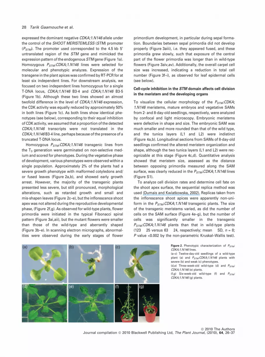
single population. Approximately 2% of the plants had a
severe growth phenotype with malformed cotyledons and/
or fused leaves (Figure 2a,b), and showed early growth
arrest. However, the majority of the transgenic plants
presented less severe, but still pronounced, morphological
alterations, such as retarded growth and small and
mis-shapen leaves (Figure 2c–e), but the inflorescence shoot
apex was not altered during the reproductive developmental
phase, (Figure 2f,g). As observed for wild-type plants, flower
primordia were initiated in the typical Fibonacci spiral
pattern (Figure 3ai,aii), but the mutant flowers were smaller
than those of the wild-type and aberrantly shaped
(Figure 3b–e). In scanning electron micrographs, abnormal-
ities were observed during the early stages of flower
primordium development, in particular during sepal forma-
tion. Boundaries between sepal primordia did not develop
properly (Figure 3aiii), i.e. they appeared fused, and these
primordia grew slowly, such that exposure of the central
part of the flower primordia was longer than in wild-type
flowers (Figure 3aiv,av). Additionally, the overall carpel cell
size was increased, indicating a reduction in total cell
number (Figure 3f–i), as observed for leaf epidermal cells
(see below).
Cell-cycle inhibition in the STM domain affects cell division
in the meristem and the developing organs
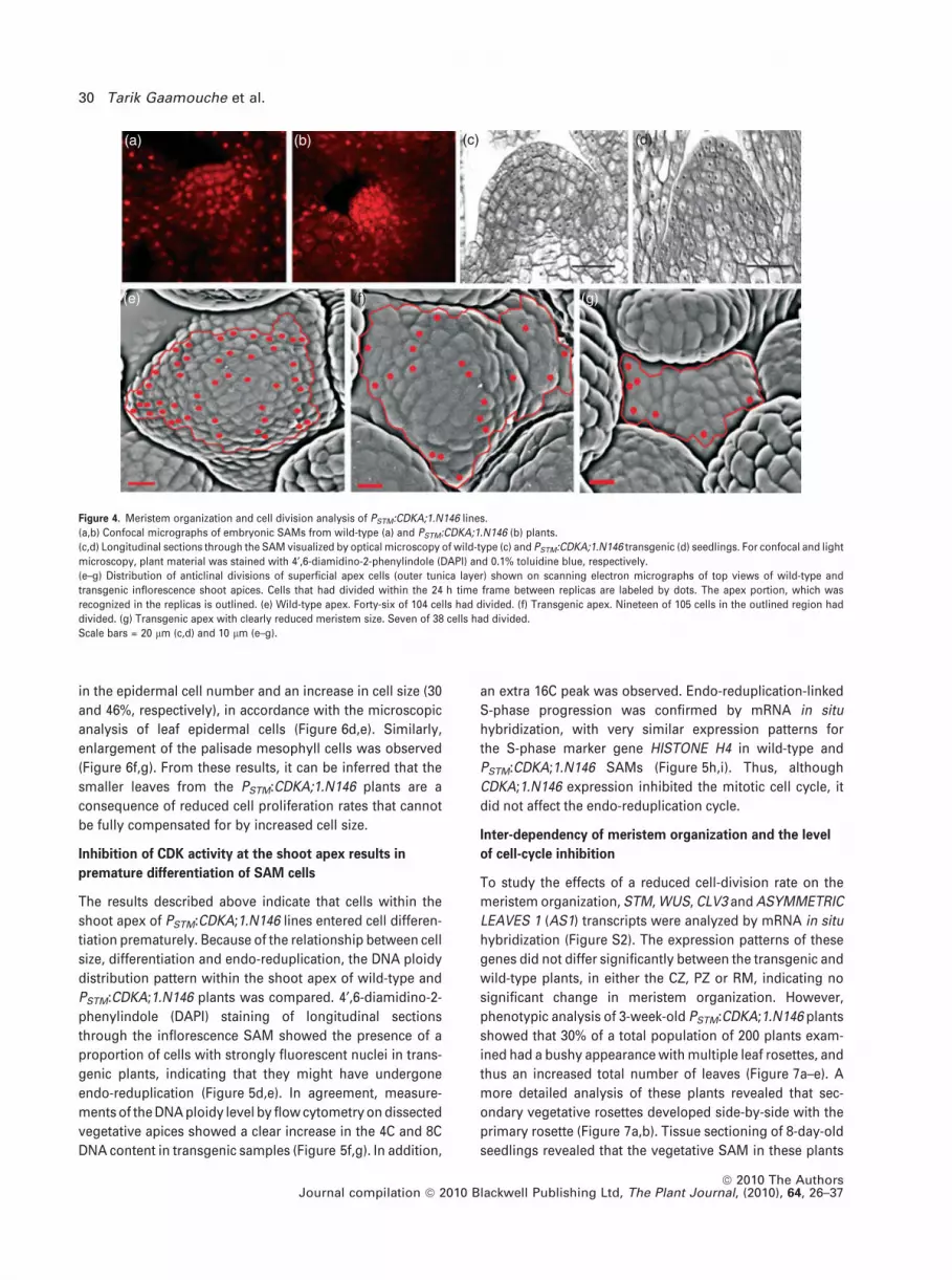
To visualize the cellular morphology of the PSTM:CDKA;
1.N146 meristems, mature embryos and vegetative SAMs
from 2- and 8-day-old seedlings, respectively, were analyzed
by confocal and light microscopy. Embryonic meristems
were defective in shape and size. The embryonic SAM was
much smaller and more rounded than that of the wild type,
and the tunica layers (L1 and L2) were indistinct
(Figure 4a,b). Longitudinal sections from SAMs of 8-day-old
seedlings confirmed the altered meristem organization and
shape, although the two tunica layers (L1 and L2) were rec-
ognizable at this stage (Figure 4c,d). Quantitative analysis
showed that meristem size, assessed as the distance
between opposing primordia measured along the SAM
surface, was clearly reduced in the PSTM:CDKA;1.N146 lines
(Figure S1).
To analyze cell division rates and determine cell fate on
the shoot apex surface, the sequential replica method was
used (Dumais and Kwiatkowska, 2002). Replicas taken from
the inflorescence shoot apices were apparently non-uni-
form in the PSTM:CDKA;1.N146 transgenic plants. The size
of the transgenic meristems varied, as did the number of
cells on the SAM surface (Figure 4e–g), but the number of
cells was significantly smaller in the transgenic
PSTM:CDKA;1.N146 plants than that in wild-type plants
(123 � 25 versus 63 � 24, respectively; mean � SD, n = 8;
P value <0.002 by the non-parametric Kruskal–Wallis test).
(a) (b)
(d) (e) (f) (g)
(c) Figure 2. Phenotypic characterization of PSTM:
CDKA;1.N146 lines.
(a–c) Twelve-day-old seedlings of a wild-type
plant (a) and PSTM:CDKA;1.N146 plants with
severe (b) and weak (c) phenotypes.
(d,e) Three-week-old wild-type (d) and PSTM:
CDKA;1.N146 (e) plants.
(f,g) Six-week-old wild-type (f) and PSTM:
CDKA;1.N146 (g) plants.
28 Tarik Gaamouche et al.
ª 2010 The AuthorsJournal compilation ª 2010 Blackwell Publishing Ltd, The Plant Journal, (2010), 64, 26–37

The cell division frequency, determined by comparison of
cell numbers in two consecutive sequential replicas, was
also variable, and was lower in transgenic than in wild-type
SAMs (Figure 4e–g). A striking feature of the
PSTM:CDKA;1.N146 inflorescence meristems was the non-
homogeneous size of the cells, i.e. some cells were
apparently larger than others (Figure 5a–c). These
cells did not divide, but expanded (Figure 5a–c; cells
labeled by asterisks and circles). Cells were elongated
along the meristem radius, unlike typical isodiametric
meristematic cells. This shape most probably resulted
from anisotropic growth that is specific to the meristem
periphery, i.e. faster growth along the meristem radius
than along its circumference. Atypical cells were also
found on the adaxial flower primordium boundary: these
cells were elongated along the periphery (i.e. along the
circumference), which often appeared wrinkled (Figure 5a–
c), in contrast to the wild-type apices (Figure 4e). The
wrinkled appearance was due to deep grooves (creases) in
the meristem surface that occur at anticlinal walls in
contact with superficial meristem cells. These deep
grooves are a distinctive feature of differentiating cells,
such as cells on the sepal surface (Figure 3aii).
The aberrant leaf phenotype of the PSTM:CDKA;1.N146
transgenic plants suggested that inhibition of cell division
within the STM domain of the SAM has a dramatic effect on
the cell cycle during plant development. To test this
hypothesis, we analyzed the leaves in more detail. The first
true mature leaves showed a decrease in the surface area of
the leaf lamina of 24 and 32% in the CDKA;1.N146 B3-4
and B3-5 lines, respectively, compared to control plants
(Figure 6a). To investigate whether this reduction in leaf size
was caused by inhibition of cell division or cell expansion,
the mean cell area of abaxial epidermal cells was determined
from drawing-tubus images, and the total cell number in the
leaf epidermis was calculated from the ratio of the leaf
lamina surface area to the mean cell area. Significant
differences were observed between wild-type and trans-
genic lines (Figure 6b,c). Both CDKA;1.N146 lines displayed
a statistically significant reduction (43 and 37%, respectively)
(a)
(f) (g) (h) (i)
(b) (c)
(d) (e)(i)
(iii) (iv) (v)
(ii)
Figure 3. Floral and inflorescence phenotypes of PSTM:CDKA;1.N146 lines.
(ai,aii) Arrangement of flower primordia and aberrations in primordium development in PSTM:CDKA;1.N146 lines. Top views of inflorescence shoot apices of wild-
type (ai) and transgenic (aii) plants surrounded by flower primordia (labeled P1–P9, numbered from the youngest primordium to the oldest). (aiii–av) Close-up views
of abnormally developed flower primordia representing consecutive stages of early sepal formation. (aiii) Six-plastochron-old primordium. No clear boundaries
(asterisks) are visible between sepals of two pairs (S1 and S2). (aiv) Six-plastochron-old primordium with apparent sepals of two pairs (S1 and S2) with a developed
boundary. (av) Seven-plastochron-old primordium with developed sepals of two pairs (S1 and S2), but exhibiting retarded growth. At this stage in the wild-type, the
sepals start to or completely overtop the central portion of the flower primordium [compare with P7 in (ai)].
(b,d) Wild-type flowers.
(c,e) Mutant flowers of small size and abnormal shape.
(f–i) Scanning electron microscopy of carpels. (f) Wild-type carpel. (h) Mutant carpel. Epidermal cells of wild-type (g) and mutant (i) carpels.
Scale bars = 30 lm (ai,aii), 10 lm (aiii–av), 3 mm (f,h) and 800 lm (g,i).
CDK dose-dependent control of meristem activity 29
ª 2010 The AuthorsJournal compilation ª 2010 Blackwell Publishing Ltd, The Plant Journal, (2010), 64, 26–37
in the epidermal cell number and an increase in cell size (30
and 46%, respectively), in accordance with the microscopic
analysis of leaf epidermal cells (Figure 6d,e). Similarly,
enlargement of the palisade mesophyll cells was observed
(Figure 6f,g). From these results, it can be inferred that the
smaller leaves from the PSTM:CDKA;1.N146 plants are a
consequence of reduced cell proliferation rates that cannot
be fully compensated for by increased cell size.
Inhibition of CDK activity at the shoot apex results in
premature differentiation of SAM cells
The results described above indicate that cells within the
shoot apex of PSTM:CDKA;1.N146 lines entered cell differen-
tiation prematurely. Because of the relationship between cell
size, differentiation and endo-reduplication, the DNA ploidy
distribution pattern within the shoot apex of wild-type and
PSTM:CDKA;1.N146 plants was compared. 4¢,6-diamidino-2-
phenylindole (DAPI) staining of longitudinal sections
through the inflorescence SAM showed the presence of a
proportion of cells with strongly fluorescent nuclei in trans-
genic plants, indicating that they might have undergone
endo-reduplication (Figure 5d,e). In agreement, measure-
ments of the DNA ploidy level by flow cytometry on dissected
vegetative apices showed a clear increase in the 4C and 8C
DNA content in transgenic samples (Figure 5f,g). In addition,
an extra 16C peak was observed. Endo-reduplication-linked
S-phase progression was confirmed by mRNA in situ
hybridization, with very similar expression patterns for
the S-phase marker gene HISTONE H4 in wild-type and
PSTM:CDKA;1.N146 SAMs (Figure 5h,i). Thus, although
CDKA;1.N146 expression inhibited the mitotic cell cycle, it
did not affect the endo-reduplication cycle.
Inter-dependency of meristem organization and the level
of cell-cycle inhibition
To study the effects of a reduced cell-division rate on the
meristem organization, STM, WUS, CLV3 and ASYMMETRIC
LEAVES 1 (AS1) transcripts were analyzed by mRNA in situ
hybridization (Figure S2). The expression patterns of these
genes did not differ significantly between the transgenic and
wild-type plants, in either the CZ, PZ or RM, indicating no
significant change in meristem organization. However,
phenotypic analysis of 3-week-old PSTM:CDKA;1.N146 plants
showed that 30% of a total population of 200 plants exam-
ined had a bushy appearance with multiple leaf rosettes, and
thus an increased total number of leaves (Figure 7a–e). A
more detailed analysis of these plants revealed that sec-
ondary vegetative rosettes developed side-by-side with the
primary rosette (Figure 7a,b). Tissue sectioning of 8-day-old
seedlings revealed that the vegetative SAM in these plants
(a)
(e) (f) (g)
(b) (c) (d)
Figure 4. Meristem organization and cell division analysis of PSTM:CDKA;1.N146 lines.
(a,b) Confocal micrographs of embryonic SAMs from wild-type (a) and PSTM:CDKA;1.N146 (b) plants.
(c,d) Longitudinal sections through the SAM visualized by optical microscopy of wild-type (c) and PSTM:CDKA;1.N146 transgenic (d) seedlings. For confocal and light
microscopy, plant material was stained with 4¢,6-diamidino-2-phenylindole (DAPI) and 0.1% toluidine blue, respectively.
(e–g) Distribution of anticlinal divisions of superficial apex cells (outer tunica layer) shown on scanning electron micrographs of top views of wild-type and
transgenic inflorescence shoot apices. Cells that had divided within the 24 h time frame between replicas are labeled by dots. The apex portion, which was
recognized in the replicas is outlined. (e) Wild-type apex. Forty-six of 104 cells had divided. (f) Transgenic apex. Nineteen of 105 cells in the outlined region had
divided. (g) Transgenic apex with clearly reduced meristem size. Seven of 38 cells had divided.
Scale bars = 20 lm (c,d) and 10 lm (e–g).
30 Tarik Gaamouche et al.
ª 2010 The AuthorsJournal compilation ª 2010 Blackwell Publishing Ltd, The Plant Journal, (2010), 64, 26–37

was split, resulting in formation of shoot apices with two
fused domes (Figure 7f). During ontogenesis, the flower
organs were small and the siliques were extremely short
with few seeds inside (data not shown). Also, the epidermis
of the carpels consisted of enlarged, irregularly shaped cells
(Figure 7g,h), an indication of strongly inhibited cell divi-
sion. As the carpel cell division phenotype was more severe
in plants with a split meristem than in plants with a single
(a) (d) (e)
(f)
(h) (i)
(g)(b)
(c)
Figure 5. DNA replication and cell expansion in
PSTM:CDKA;1.N146 SAMs.
(a–c) Developmental sequences of a transgenic
inflorescence shoot apex. Primordia (P1–P5) are
numbered from the youngest primordium to the
oldest. Three stages (replicas) are shown, taken
at 24 h intervals. Asterisks and circles indicate
cells that appeared normal in the first (a) or the
second stage (b), but did not divide within the
next 24 h, although their size increased consid-
erably (c). Deep wrinkles were apparent between
some cells at the primordium P3 boundary
(indicated by arrowheads).
(d,e) DAPI-stained longitudinal sections of
7-week-old inflorescence shoot meristems from
wild-type (d) and PSTM:CDKA;1.N146 plants.
Arrows point to enlarged nuclei (e).
(f,g) DNA ploidy distribution for nuclei isolated
from 8-day-old seedling shoot meristems of
wild-type (f) and PSTM:CDKA;1.N146 transgenic
plants (g).
(h,i) Histone H4 expression pattern in wild-type
(h) and PSTM:CDKA;1.N146 transgenic plants (i).
Scale bar = 10 lm (a–c) and 30 lm (d,e).
(a)
(d) (e) (f) (g)
(b) (c)
Figure 6. Cell and leaf lamina size analysis of mature PSTM:CDKA;1.N146 leaves compared to those of the wild-type.
(a–c) Leaf surface area (a), cell number (b) and abaxial epidermis cell size (c). All measurements were obtained on 3-week-old first leaves. Values are means for 6–10
plants (*P = 0.06, **P = 0.01, ***P = 0.001, ****P = 0.0001, *****P = 0.00001, Student’s t test, for comparison with wild-type).
(d–g) Microscopy analysis of adaxial epidermis cells and palisade mesophyll cells from wild-type (d,f) and a transgenic line (e,g). Scale bar = 25 lm.
CDK dose-dependent control of meristem activity 31
ª 2010 The AuthorsJournal compilation ª 2010 Blackwell Publishing Ltd, The Plant Journal, (2010), 64, 26–37

meristem, we hypothesized that the shoot apex phenotype
correlated with the level of PSTM:CDKA;1.N146 activity. To
test this hypothesis, we compared the number of plants with
at least two SAMs under long-day and continuous light
conditions. In both the wild-type and transgenic plants, an
increase in day length did not significantly affect the
expression level of endogenous CDKA;1 (Figure 7i), but
triggered an increase in the level of expression of STM
(Figure 7j), as reported previously (Geier et al., 2008).
Accordingly, the transcription level of the CDKA;1.N146
transgene was increased under continuous light conditions
in the PSTM:CDKA;1.N146 plants (Figure 7k). Additionally,
under continuous light conditions, the number of plants
with multiple rosettes increased by approximately 20%
(a)
(c) (f)
(i) (j)
(k) (l)
(b) (d)
(e)
(g) (h)
Figure 7. Phenotypic analysis of PSTM:CDKA;1.N146 plants exhibiting a severe phenotype.
(a) Three-week-old plant with two rosettes developing side by side.
(b) Twelve-day-old seedling with two young rosettes.
(c) Six-week-old plant with a bushy appearance.
(d,e) Dissected rosette leaves (cut to flatten the leaf lamina) of wild-type (d) and PSTM:CDKA;1.N146 transgenic plants (e).
(f) Longitudinal section through the vegetative SAM of a PSTM:CDKA;1.N146 transgenic line. The SAM had two domes.
(g,h) Scanning electron micrographs of a PSTM:CDKA;1.N146 carpel (g) and its epidermal cells (h). Scale bars = 3 mm (g) and 800 lm (h).
(i–k) Quantitative real-time expression analysis of CDKA;1 (i), STM (j) and CDKA;1.N146 (k) in 1-week-old wild-type and PSTM:CDKA;1N146 seedlings grown either
under long-day (LD; 16 h light) or continuous light (CL) conditions. The values are the means of three independent replicates. The ACTIN2, EF1-a and CYTOCHROME
B5 expression levels were used as a reference. Expression levels observed in Col-0 (LD) (i,j) or CDKA;1.N146 (LD) (k) were arbitrary set to 1.
(l) Percentage of transgenic plants exhibiting double rosettes after 3 weeks of growth under LD and CL photoperiods (n = 300).
**P = 0.05 and ***P = 0.007 (Student’s t test, LD versus CL).
32 Tarik Gaamouche et al.
ª 2010 The AuthorsJournal compilation ª 2010 Blackwell Publishing Ltd, The Plant Journal, (2010), 64, 26–37

(Figure 7l), indirectly implying a correlation between the
appearance of multiple apices and the level of cell-cycle
inhibition.
As independent proof that the CDKA;1.N146 expression
level affects meristem organization, the number of plants
with multiple meristems was compared in homozygous
versus hemizygous PSTM:CDKA;1.N146 populations. Hemi-
zygous lines were obtained by pollinating wild-type plants
with pollen from the PSTM:CDKA;1.N146 homozygous
plants. Genotyping the offspring confirmed a high success
rate (>90%) for cross-pollination. The hemizygous
PSTM:CDKA;1.N146 plants had lost the typical malformed
leaf phenotype of the homozygous plants (Figure 8a,b),
corresponding with a decrease in the CDKA;1.N146 expres-
sion level (Figure 8c), and the number of plants with two or
more rosettes was significantly reduced (Figure 8d). The
reduction in the number of plants with multiple meristems
was accompanied by a decrease in the CDKA;1.N146 tran-
script level of >80%, rather than the theoretically expected
50% in hemizygous plants. This observation is probably due
to a reduction in the relative abundance of STM-expressing
cells as a consequence of the decrease in SAM number in the
hemizygous lines.
DISCUSSION
Although it is generally accepted that CDK/CYC complexes
perform fundamental control of cell division in plants, the
manner by which the cell-cycle machinery is integrated into
the meristem organization is still poorly understood. Here,
we have confirmed a link between cell-cycle control and
meristem function by specifically inhibiting cell divisions in
the SAM. Cell divisions in the vegetative SAM are necessary
for SAM self-perpetuation, a process in which the general
shape and size of the SAM are maintained despite the mer-
istem continuously providing cells that are incorporated into
leaf primordia. Strikingly, although the cell cycle was aimed
at inhibition in the SAM exclusively, plants expressing
PSTM:CDKA;1.N146 showed clear morphological alterations,
such as mis-shapen leaves. The phenotypic changes were
accompanied by a reduction in cell number and an increase
in cell size. Developmental defects because of mis-expres-
sion of cell-cycle regulatory genes have been described
previously (Riou-Khamlichi et al., 1999; De Veylder et al.,
2001; Weingartner et al., 2004), but, because of the use of
ubiquitously expressed promoters, the influence of the
malfunctioning meristem on the overall plant phenotype
could not be determined. In other cases, the effects on
meristem organization were so strong that they hampered
post-meristematic development, as observed upon over-
expression or knockout of B2-type CDKs (Andersen et al.,
2008). Although the possibility cannot be totally excluded
that a residual amount of CDKA;1.N146 protein may remain
in the cells after they have left the meristem (because of
protein stability), and thus contribute to the reduced leaf cell
number, the data presented indicate that a reduced cell
division rate within the meristem exerts a severe negative
effect on cell divisions outside the meristem. As noted pre-
viously, early stages of leaf primordium development can
take place without any cell division (Foard and Haber, 1961;
(a)
(c)
(d)
(b)
Figure 8. Phenotypic comparison of PSTM:CDKA;1N146 plants.
(a,b) Three-week-old homozygous (a) and hemizygous (b) PSTM:CDKA;1.N146
plants.
(c) CDKA;1.N146 expression levels measured by quantitative real-time PCR.
The values are means of three independent replicates. The ACTIN2, EF1-a and
CYTOCHROME B5 expression levels were used as a reference. Expression
levels observed in Col-0 were arbitrarily set to 1.
(d) Percentage of transgenic plants exhibiting double rosettes after 3 weeks of
growth (n = 200, ****P < 0.0001, Student’s t test, for comparison with
hemizygous plants).
CDK dose-dependent control of meristem activity 33
ª 2010 The AuthorsJournal compilation ª 2010 Blackwell Publishing Ltd, The Plant Journal, (2010), 64, 26–37
Haber, 1962), supporting the ‘organismal’ theory, which
states that the frequency and pattern of cell divisions do not
drive organogenesis, but are secondary to an intrinsic
developmental plan. Nevertheless, because the cell number
increases exponentially during the initial stages of leaf
development (De Veylder et al., 2001; Boudolf et al., 2004),
primordia consisting initially of a strongly reduced number
of cells are expected to eventually develop in leaves made
of fewer cells.
Alternatively, the reduction in cell number in leaves might
be due to the decrease in CDK activity when the cells leave
the meristem. In Arabidopsis, leaf cells exit the cell cycle in
an apex-to-base direction (Donnelly et al., 1999). If this
gradient coincides with a basipetal gradient in CDK activity,
cells will probably exit the cell cycle when the level of CDK
activity drops below a critical threshold level required to
drive mitosis. We propose that primordia with a lower initial
CDK level leave the cell cycle before those with a higher
level, eventually resulting in a reduction in leaf cell number
in PSTM:CDKA;1.N146 plants. Earlier cell-cycle exit and
differentiation are substantiated by an increase in the overall
DNA ploidy level of PSTM:CDKA;1.N146 leaf cells (data not
shown), because the onset of endo-reduplication correlates
with exit from the mitotic cell cycle (Boudolf et al., 2004;
Verkest et al., 2005; Vlieghe et al., 2005; Dewitte et al., 2007).
A similar increase in the DNA ploidy level was observed in
the case of meristem-specific over-expression of the CDKA;1
inhibitor-encoding gene ICK2/KRP2 (Verkest et al., 2005).
The expression of ICK2/KRP2 has been postulated to reduce
the level of CDKA;1 activity below a threshold level neces-
sary for mitotic cell divisions, but that is still high enough to
enable repeated rounds of DNA replication. Similarly,
expression of the dominant negative CDKA;1 allele probably
has a profound effect on the mitotic cell cycle, but not on the
endo-reduplication cycle, triggering an early mitosis-to-
endocycle transition, and eventually a higher DNA ploidy
level. This process may take place at the SAM, because the
enlarged cells at the meristem correlate with an increase in
the number of cells with an enlarged nucleus. As endo-
reduplicating cells rarely divide, premature differentiation of
cells within the meristem might also have a significant
negative effect on the final cell number within the organism.
With regard to the abnormal leaf morphology in
PSTM:CDKA;1.N146 plants, some of the SAM cells that
entered the endo-reduplication cycle prematurely might be
assumed to be incorporated into the leaf primordium.
Together the general reduction in the cell number of
primordia and the endocycling of some primordium cells
might obstruct leaf organogenesis.
Despite the clear defects in plant development and the
cellular phenotypes within the meristem, the majority of the
PSTM:CDKA;1.N146 plants had no clearly altered meristem
organization. In situ hybridization and expression analysis of
SAM regulatory genes revealed no difference between
transgenic and wild-type plants. The absence of visible
meristem organization phenotype appears to contradict
other published data. Local over-expression of the RETINO-
BLASTOMA-RELATED 1 (RBR1) gene at the SAM triggered
differentiation of meristematic cells together with loss of
expression of meristem identity markers (Wyrzykowska
et al., 2006). Analogously, treatment of meristems with
microtubule-depolymerizing drugs caused meristem cells
to differentiate (Grandjean et al., 2004), while loss of
expression or over-expression of B2-type CDKs resulted in
a total loss of meristem integrity (Andersen et al., 2008). In
contrast, local stimulation of cell division rates by over-
expression of CYCA3;2 and expression of fission yeast
cdc25, or inhibition of cell proliferation by knock-out of
D3-type cyclin, had none or only mild effects on meristem
organization (Wyrzykowska et al., 2002; Dewitte et al., 2007).
Surprisingly, upon over-expression of CDKA;1.N146 in the
STM domain, a sub-population of plants had a split SAM in
the vegetative phase, indicating some meristematic defects
in this sub-population of plants. This phenotype resembles
that obtained by surgical manipulations of the SAM in which
destruction of the CZ resulted in regeneration of new SAMs
from the PZ (Reinhardt et al., 2003). The split-meristem
phenotype was correlated with the PSTM:CDKA;1.N146
expression level, suggesting a dose-dependent relationship
between inhibition of cell division and the effect on meri-
stem organization. Although the product of the dominant
negative CDKA;1.N146 allele probably specifically targets
the A-type CDKs, the reduced cell proliferation rate caused
by a decrease in CDKA;1 activity will affect other CDKs. Thus,
rather than attributing the meristem defects solely to
CDKA;1, we assume that the overall effect on CDK activity
is responsible for the observed phenotypes. We propose a
CDK activity threshold model in which weak CDK inhibition
suppresses the cell division activity without affecting mer-
istem organization, whereas strong inactivation of CDK
leads to a loss of meristem identity (Figure 9), which may
be followed by new meristem formation. This hypothesis
reconciles the apparently conflicting data available in the
literature, in which the presence or absence of clear meri-
stem organization phenotypes might be correlated with the
strength of cell-cycle inhibition. The loss-of-meristem orga-
nization phenotype of B2-type CDK mutants might be
a consequence of the severe inhibition of cell division
(Andersen et al., 2008). By contrast, plants lacking the
D3-type cyclin show only a mildly defective cell cycle
without clear meristem patterning alterations (Dewitte et al.,
2007). The model implies that high CDK activity within the
meristem is necessary to maintain cells in an undifferenti-
ated state, as supported by the induction of differentiation of
root stem cells by co-suppression of an F-type CDK-activat-
ing kinase (Umeda et al., 2000). Similarly, in a leaf callus
culture, continuous CDK activity is important to maintain
cells in an undifferentiated state (Yamaguchi et al., 2003).
34 Tarik Gaamouche et al.
ª 2010 The AuthorsJournal compilation ª 2010 Blackwell Publishing Ltd, The Plant Journal, (2010), 64, 26–37

Hence, probably only substantial changes in the cell division
rates or the proliferation status of meristem cells will result
in a change in meristem organization. We postulate that, in
the case of the SAM of the PSTM:CDKA;1.N146 transgenic
line, the CDK level is always close to the putative threshold,
and subtle differences below or above this threshold are
sufficient to trigger apparently different phenotypes.
EXPERIMENTAL PROCEDURES
Plant material and growth conditions
The STM promoter (PSTM) was amplified from the F24O1 BAC cloneas a 4.5 kb fragment, as described previously (Verkest et al., 2005),and cloned into the pBIN+ vector, into which a cassette containingthe CDKA;1.N146 allele (Hemerly et al., 1995) together with a 3’ NOSterminator was introduced. Arabidopsis thaliana (L.) Heyhn. plants(ecotype Columbia, Col-0) were grown at 22 � 2�C under a 16 h/8 hlight/dark photoperiod (65 lE m)2 sec)1) on agar-solidified culturemedium (half-strength Murashige and Skoog medium, 0.5 g/L MES,10 g/L sucrose, 0.8% plant tissue culture agar), except where indi-cated. Agrobacterium tumefaciens-mediated transformation wasperformed by floral dip as described previously (Clough and Bent,1998). Transgenic lines were selected on kanamycin-containingmedium.
Sequential replicas of inflorescence meristems
To obtain sequential replicas, plants were grown under short-dayconditions (11 h/13 h light/dark) for 13 weeks, followed by 4 weeksof long-day conditions (16 h/8 h light/dark). Replicas were takenfrom inflorescence apices of main shoots or axillary shoots (30–150 mm long) as described previously (Williams and Green, 1988;Kwiatkowska, 2004). Sequences were taken every 24 h for 2–4 daysfrom inflorescence apices of transgenic plants. Previously obtained
sequential replicas of Col-0 (Kwiatkowska, 2004, 2006) were used forcomparison. All the electron micrographs for cell identification andcounting were improved by means of the MATLAB image pro-cessing toolbox (http://www.mathworks.com/products/matlab/), toenable more precise recognition of the anticlinal walls of surfacecells.
In situ hybridization
The RNA of 12-day-old seedlings from wild-type and mutant linesfixed in FAA (10% formaldehyde, 50% EtOH, 5% glacial acetic acid)was hybridized in situ. Full-length riboprobes were synthesized forthe STM (At1G62360), WUS (At2G17950), CLV3 (At2G27250), AS1(At2G37630), H4 (At1G07820) and CDKB1 (At3G54180) genes by PCRamplification with universal primers specific to the gene-containingvectors. Samples were processed as described previously (Dreaet al., 2005), with the following modifications: the slides were pro-cessed using the automated slide processor InsituPro VSi (http://www.intavis.com) from the hybridization step to signal detection,hybridization and post-hybridization washes were performed at55�C, samples were incubated in 1:1250 diluted anti-digoxigeninantibody conjugated to alkaline phosphatase (http://www.roche.com) for 1.5 h, and the signal was developed using Western Bluestabilized substrate (http://www.promega.com). Sections werephotographed using a Nikon E800 microscope with a digital camera(http://www.nikoninstruments.com).
Real-time quantitative PCR analysis
For real-time quantitative PCR, total RNA was isolated from a pool of200 plantlets at 7 days after germination using an RNeasy plant minikit (http://www.qiagen.com). For each sample, 1 lg was reverse-transcribed into cDNA using the SuperScript first-strand synthesissystem for RT-PCR (http://www.invitrogen.com). QuantitativeRT-PCR amplification of the cDNA was performed using a LightCy-clerTM 480 (http://www.roche.com), with the gene-specific primers5¢-ATTGCGTATTGCCACTCTCATAGG-3¢ and 3¢-TCCTGACAGGGATACCGAATGC-5¢ for CDKA;1 (At3G48750), 5¢-CGGAGGACATGCAGTTTGTA-3¢ and 3¢-AAAGCATGGTGGAGGAGATG-5¢ for STM,and 5¢-AAGCTTTTGAGATCAGTTTCTTG-3¢ and 3¢-ACTTTATTGCCAAATGTTTGAA-5¢ for the CDKA;1.N146 transgene. Differences inthe amount of target cDNA used as input were normalized againstthree reference genes amplified using primers 5¢-GGCTCCTCTTAACCCAAAGGC-3¢ and 3¢-CACACCATCACCAGAATCCAGC-5¢for ACTIN2 (At3G46520), 5¢-CTGGAGGTTTTGAGGCTGGTAT-3¢ and3¢-CCAAGGGTGAAAGCAAGAAGA-5¢ for EF1-a (At5G60390), and5¢-CGACACTGCAAGGGACATGA-3¢ and 3¢-ACGTATGTCCTAGTTGCTGGAACA-5¢ for CYTOCHROME B5 (AT5G53560).
Kinase assay
Eight-day-old plants were harvested for total protein extraction andkinase activity tests. CDK-like proteins were purified from totalprotein extracts on p10CKS1 Sepharose beads (De Veylder et al.,1999), and their ability to phosphorylate histone H1 was determinedas described previously (Azzi et al., 1994; Corellou et al., 2000).Radioactive histone H1 was quantified using a STORM Phospho-Imager and ImageQuant software (http://www.gehealthcare.com).
Flow cytometric analysis
Frozen plant material was chopped with a razor blade in 800 ll ofnucleus extraction solution (http://www.partec.com), supplementedwith 200 ll of staining solution (http://www.partec.com). Thehomogenate was filtered through a 30 lm mesh. The nuclei were
Figure 9. CDK activity threshold model for meristem organization.
High CDK activity is required to maintain cells within the SAM in an
undifferentiated state. A decrease in CDK activity has no effect on meristem
organization unless it drops below a hypothetical threshold level, below
which the meristematic cells exit the cell cycle affecting meristem identity and
organization.
CDK dose-dependent control of meristem activity 35
ª 2010 The AuthorsJournal compilation ª 2010 Blackwell Publishing Ltd, The Plant Journal, (2010), 64, 26–37
analyzed using a CyFlow cytometer and FloMax software (http://www.partec.com).
Microscopic analysis
Plastic embedding with Technovit 7100 resin (http://www.kulzer-technik.de) and sectioning were performed according to the man-ufacturer’s protocol. Serial sections (5 lm) were stained with 0.1%toluidine blue and mounted using Depex-mounting medium (http://uk.vwr.com). Sections were photographed under a Diaplan lightmicroscope (http://www.leica-microsystems.com). The nuclearDNA was visualized as described previously (Bemis and Torii, 2007).Slides were observed under a 40· objective after 4¢,6-diamidino-2-phenylindole (DAPI) staining and using an appropriate filter on anAxioImager M1 equipped with an AxioCam HRm camera (http://www.zeiss.com). Digital images were acquired and analyzed usingAxioVision 4.6 software. Final images were obtained using Photo-shop 6.0 (http://www.adobe.com/). Dissected embryos of seedlings2 and 7 days after germination were analyzed by confocal micros-copy as described previously (Autran et al., 2002).
ACKNOWLEDGEMENTS
We thank all members of the Cell Cycle Group and the SY-STEMtraining network for useful discussions and suggestions, and Mar-tine De Cock for help in preparing the manuscript. This work wassupported by grants from the Interuniversity Attraction PolesProgram (IUAP VI/33), initiated by the Belgian State Science PolicyOffice, and the European Union SY-STEM (MRTN-GT-2004-005336).L.D.V. is a postdoctoral fellow of the Fonds voor WetenschappelijkOnderzoek – Vlaanderen (FWO).
SUPPORTING INFORMATION
Additional Supporting Information may be found in the onlineversion of this article:Figure S1. Distance between opposing leaf primordia measuredalong the meristem surface.Figure S2. Spatial expression pattern analysis of SAM patterninggenes.Please note: As a service to our authors and readers, this journalprovides supporting information supplied by the authors. Suchmaterials are peer-reviewed and may be re-organized for onlinedelivery, but are not copy-edited or typeset. Technical supportissues arising from supporting information (other than missingfiles) should be addressed to the authors.
REFERENCES
de Almeida Engler, J., De Veylder, L., De Groodt, R. et al. (2009) Systematic
analysis of cell cycle gene expression during Arabidopsis development.
Plant J. 59, 645–660.
Andersen, S.U., Buechel, S., Zhao, Z., Ljung, K., Novak, O., Busch, W.,
Schuster, C. and Lohmann, J.U. (2008) Requirement of B2-type cyclin-
dependent kinases for meristem integrity in Arabidopsis thaliana. Plant
Cell, 20, 88–100.
Autran, D., Jonak, C., Belcram, K., Beemster, G.T.S., Kronenberger, J.,
Grandjean, O., Inze, D. and Traas, J. (2002) Cell numbers and leaf devel-
opment in Arabidopsis. A functional analysis of the STRUWWELPETER
gene. EMBO J. 21, 6036–6049.
Azzi, L., Meijer, L., Ostvold, A.-C., Lew, J. and Wang, J.H. (1994) Purification of
a 15-kDa cdk4- and cdk5-binding protein. J. Biol. Chem. 269, 13279–13288.
Bemis, S.M. and Torii, K.U. (2007) Autonomy of cell proliferation and devel-
opmental programs during Arabidopsis aboveground organ morphogen-
esis. Dev. Biol. 304, 367–381.
Boudolf, V., Barroco, R., de Almeida Engler, J., Verkest, A., Beeckman, T.,
Naudts, M., Inze, D. and De Veylder, L. (2004) B1-type cyclin-dependent
kinases are essential for the formation of stomatal complexes in Arabid-
opsis thaliana. Plant Cell, 16, 945–955.
Brand, U., Fletcher, J.C., Hobe, M., Meyerowitz, E.M. and Simon, R. (2000)
Dependence of stem cell fate in Arabidopsis on a feedback loop regulated
by CLV3 activity. Science, 289, 617–619.
Carraro, N., Peaucelle, A., Laufs, P. and Traas, J. (2006) Cell differentiation
and organ initiation at the shoot apical meristem. Plant Mol. Biol. 60,
811–826.
Clark, S.E., Williams, R.W. and Meyerowitz, E.M. (1997) The CLAVATA1 gene
encodes a putative receptor kinase that controls shoot and floral meristem
size in Arabidopsis. Cell, 89, 575–585.
Clough, S.J. and Bent, A.F. (1998) Floral dip: a simplified method for Agro-
bacterium-mediated transformation of Arabidopsis thaliana. Plant J. 16,
735–743.
Corellou, F., Bisgrove, S.R., Kropf, D.L., Meijer, L., Kloareg, B. and Bouget,
F.-Y. (2000) A S/M DNA replication checkpoint prevents nuclear and cyto-
plasmic events of cell division including centrosomal axis alignment and
inhibits activation of cyclin-dependent kinase-like proteins in fucoid
zygotes. Development, 127, 1651–1660.
De Veylder, L., De Almeida Engler, J., Burssens, S., Manevski, A., Lescure, B.,
Van Montagu, M., Engler, G. and Inze, D. (1999) A new D-type cyclin of
Arabidopsis thaliana expressed during lateral root primordia formation.
Planta, 208, 453–462.
De Veylder, L., Beeckman, T., Beemster, G.T.S., Krols, L., Terras, F., Landrieu,
I., Van Der Schueren, E., Maes, S., Naudts, M. and Inze, D. (2001) Functional
analysis of cyclin-dependent kinase inhibitors of Arabidopsis. Plant Cell 13,
1653–1667.
Dewitte, W., Riou-Khamlichi, C., Scofield, S., Healy, J.M.S., Jacqmard, A.,
Kilby, N.J. and Murray, J.A.H. (2003) Altered cell cycle distribution,
hyperplasia, and inhibited differentiation in Arabidopsis caused by the
D-type cyclin CYCD3. Plant Cell, 15, 79–92.
Dewitte, W., Scofield, S., Alcasabas, A.A. et al. (2007) Arabidopsis
CYCD3 D-type cyclins link cell proliferation and endocycles and are rate-
limiting for cytokinin responses. Proc. Natl. Acad. Sci. USA, 104, 14537–
14542.
Donnelly, P.M., Bonetta, D., Tsukaya, H., Dengler, R.E. and Dengler, N.G.
(1999) Cell cycling and cell enlargement in developing leaves of Arabid-
opsis. Dev. Biol. 215, 407–419.
Drea, S., Corsar, J., Crawford, B., Shaw, P., Dolan, L. and Doonan, J.H. (2005)
A streamlined method for systematic, high resolution in situ analysis of
mRNA distribution in plants. Plant Methods, 1, 8.1–8.11.
Dumais, J. and Kwiatkowska, D. (2002) Analysis of surface growth in shoot
apices. Plant J. 31, 229–241.
Fletcher, J.C., Brand, U., Running, M.P., Simon, R. and Meyerowitz, E.M.
(1999) Signaling of cell fate decisions by CLAVATA3 in Arabidopsis shoot
systems. Science, 283, 1911–1914.
Foard, D.E. and Haber, A.H. (1961) Anatomic studies of gamma-irradiated
wheat growing without cell division. Am. J. Bot. 48, 438–446.
Gallois, J.-L., Woodward, C., Reddy, G.V. and Sablowski, R. (2002) Combined
SHOOT MERISTEMLESS and WUSCHEL trigger ectopic organogenesis in
Arabidopsis. Development, 129, 3207–3217.
Geier, F., Lohmann, J.U., Gerstung, M., Maier, A.T., Timmer, J. and Fleck, C.
(2008) A quantitative and dynamic model for plant stem cell regulation.
PLoS ONE, 3, e3553.1–e3553.10.
Grandjean, O., Vernoux, T., Laufs, P., Belcram, K., Mizukami, Y. and Traas, J.
(2004) In vivo analysis of cell division, cell growth, and differentiation at the
shoot apical meristem in Arabidopsis. Plant Cell, 16, 74–87.
Haber, A.H. (1962) Nonessentiality of concurrent cell divisons for degree of
polarization of leaf growth. I. Studies with radiation-induced mitotic inhi-
bition. Am. J. Bot. 49, 583–589.
Hemerly, A., de Almeida Engler, J., Bergounioux, C., Van Montagu, M.,
Engler, G., Inze, D. and Ferreira, P. (1995) Dominant negative mutants of
the Cdc2 kinase uncouple cell division from iterative plant development.
EMBO J. 14, 3925–3936.
Ito, Y., Nakanomyo, I., Motose, H., Iwamoto, K., Sawa, S., Dohmae, N. and
Fukuda, H. (2006) Dodeca-CLE peptides as suppressors of plant stem cell
differentiation. Science, 313, 842–845.
Iwakawa, H., Shinmyo, A. and Sekine, M. (2006) Arabidopsis CDKA;1, a cdc2
homologue, controls proliferation of generative cells in male gametogen-
esis. Plant J. 45, 819–831.
Joubes, J., De Schutter, K., Verkest, A., Inze, D. and De Veylder, L. (2004)
Conditional, recombinase-mediated expression of genes in plant cell cul-
tures. Plant J. 37, 889–896.
36 Tarik Gaamouche et al.
ª 2010 The AuthorsJournal compilation ª 2010 Blackwell Publishing Ltd, The Plant Journal, (2010), 64, 26–37

Kondo, T., Sawa, S., Kinoshita, A., Mizuno, S., Kakimoto, T., Fukuda, H. and
Sakagami, Y. (2006) A plant peptide encoded by CLV3 identified by in situ
MALDI-TOF MS analysis. Science, 313, 845–848.
Kwiatkowska, D. (2004) Surface growth at the reproductive shoot apex of
Arabidopsis thaliana pin-formed 1 and wild type. J. Exp. Bot. 55, 1021–
1032.
Kwiatkowska, D. (2006) Flower primordium formation at the Arabidopsis
shoot apex: quantitative analysis of surface geometry and growth. J. Exp.
Bot. 57, 571–580.
Laufs, P., Grandjean, O., Jonak, C., Kieu, K. and Traas, J. (1998) Cellular
parameters of the shoot apical meristem in Arabidopsis. Plant Cell, 10,
1375–1389.
Lenhard, M., Jurgens, G. and Laux, T. (2002) The WUSCHEL and SHOOT-
MERISTEMLESS genes fulfil complementary roles in Arabidopsis shoot
meristem regulation. Development, 129, 3195–3206.
Long, J.A. and Barton, M.K. (1998) The development of apical embryonic
pattern in Arabidopsis. Development, 125, 3027–3035.
Long, J.A., Moan, E.I., Medford, J.I. and Barton, M.K. (1996) A member of the
KNOTTED class of homeodomain proteins encoded by the STM gene of
Arabidopsis. Nature, 379, 66–69.
Mayer, K.F.X., Schoof, H., Haecker, A., Lenhard, M., Jurgens, G. and Laux, T.
(1998) Role of WUSCHEL in regulating stem cell fate in the Arabidopsis
shoot meristem. Cell, 95, 805–815.
Nowack, M.K., Grini, P.E., Jakoby, M.J., Lafos, M., Koncz, C. and Schnittger,
A. (2006) A positive signal from the fertilization of the egg cell sets off
endosperm proliferation in angiosperm embryogenesis. Nat. Genet. 38,
63–67.
Porceddu, A., Stals, H., Reichheld, J.-P., Segers, G., De Veylder, L., De Pinho
Barroco, R., Casteels, P., Van Montagu, M., Inze, D. and Mironov, V. (2001)
A plant-specific cyclin-dependent kinase is involved in the control of G2/M
progression in plants. J. Biol. Chem. 276, 36354–36360.
Reddy, G.V. and Meyerowitz, E.M. (2005) Stem-cell homeostasis and growth
dynamics can be uncoupled in the Arabidopsis shoot apex. Science, 310,
663–667.
Reddy, G.V., Heisler, M.G., Ehrhardt, D.W. and Meyerowitz, E.M. (2004) Real-
time lineage analysis reveals oriented cell divisions associated with mor-
phogenesis at the shoot apex of Arabidopsis thaliana. Development, 131,
4225–4237.
Reinhardt, D., Pesce, E.-R., Stieger, P., Mandel, T., Baltensperger, K., Bennett,
M., Traas, J., Friml, J. and Kuhlemeier, C. (2003) Regulation of phyllotaxis
by polar auxin transport. Nature, 426, 255–260.
Riou-Khamlichi, C., Huntley, R., Jacqmard, A. and Murray, J.A.H. (1999)
Cytokinin activation of Arabidopsis cell division through a D-type cyclin.
Science, 283, 1541–1544.
Schoof, H., Lenhard, M., Haecker, A., Mayer, K.F.X., Jurgens, G. and Laux, T.
(2000) The stem cell population of Arabidopsis shoot meristem is main-
tained by a regulatory loop between the CLAVATA and WUSCHEL genes.
Cell, 100, 635–644.
Umeda, M., Umeda-Hara, C. and Uchimiya, H. (2000) A cyclin-dependent
kinase-activating kinase regulates differentiation of root initial cells in
Arabidopsis. Proc. Natl. Acad. Sci. USA, 97, 13396–13400.
Verkest, A., de O. Manes, C.-L., Vercruysse, S., Maes, S., Van Der Schueren, E.,
Beeckman, T., Genschik, P., Kuiper, M., Inze, D. and De Veylder, L. (2005)
The cyclin-dependent kinase inhibitor KRP2 controls the onset of the
endoreduplication cycle during Arabidopsis leaf development through
inhibition of mitotic CDKA;1 kinase complexes. Plant Cell, 17, 1723–1736.
Vlieghe, K., Boudolf, V., Beemster, G.T.S., Maes, S., Magyar, Z., Atanassova,
A., de Almeida Engler, J., De Groodt, R., Inze, D. and De Veylder, L. (2005)
The DP-E2F-like DEL1 gene controls the endocycle in Arabidopsis thaliana.
Curr. Biol. 15, 59–63.
Weingartner, M., Criqui, M.-C., Meszaros, T., Binarova, P., Schmit, A.-C.,
Helfer, A., Derevier, A., Erhardt, M., Bogre, L. and Genschik, P. (2004)
Expression of a nondegradable cyclin B1 affects plant development and
leads to endomitosis by inhibiting the formation of phragmoplast. Plant
Cell, 16, 643–657.
Williams, M.H. and Green, P.B. (1988) Sequential scanning electron micro-
scopy of a growing plant meristem. Protoplasma, 147, 77–79.
Wyrzykowska, J., Pien, S., Shen, W.H. and Fleming, A.J. (2002) Manipulation
of leaf shape by modulation of cell division. Development, 129, 957–964.
Wyrzykowska, J., Schorderet, M., Pien, S., Gruissem, W. and Fleming, A.J.
(2006) Induction of differentiation in the shoot apical meristem by transient
overexpression of a retinoblastoma-related protein. Plant Physiol. 141,
1338–1348.
Yamaguchi, M., Kato, H., Yoshida, S., Yamamura, S., Uchimiya, H. and
Umeda, M. (2003) Control of in vitro organogenesis by cyclin-dependent
kinase activities in plants. Proc. Natl. Acad. Sci. USA, 100, 8019–8023.
CDK dose-dependent control of meristem activity 37
ª 2010 The AuthorsJournal compilation ª 2010 Blackwell Publishing Ltd, The Plant Journal, (2010), 64, 26–37