Copper induced upregulation of apoptosis related genes in zebrafish (Danio rerio) gill
Upload
independentCategory
view
0download
0
DOI 10.1105/tpc.10.9.1427 1998;10;1427-1437Plant Cell
Didier Reinhardt, Franz Wittwer, Therese Mandel and Cris KuhlemeierMeristem
Localized Upregulation of a New Expansin Gene Predicts the Site of Leaf Formation in the Tomato
This information is current as of October 23, 2013
References http://www.plantcell.org/content/10/9/1427.full.html#ref-list-1
This article cites 32 articles, 19 of which can be accessed free at:
Permissions https://www.copyright.com/ccc/openurl.do?sid=pd_hw1532298X&issn=1532298X&WT.mc_id=pd_hw1532298X
eTOCs http://www.plantcell.org/cgi/alerts/ctmain
Sign up for eTOCs at:
CiteTrack Alerts http://www.plantcell.org/cgi/alerts/ctmain
Sign up for CiteTrack Alerts at:
Subscription Information http://www.aspb.org/publications/subscriptions.cfm
is available at:Plant Physiology and The Plant CellSubscription Information for
ADVANCING THE SCIENCE OF PLANT BIOLOGY © American Society of Plant Biologists
The Plant Cell, Vol. 10, 1427–1437, September 1998, www.plantcell.org © 1998 American Society of Plant Physiologists
Localized Upregulation of a New Expansin Gene Predicts the Site of Leaf Formation in the Tomato Meristem
Didier Reinhardt, Franz Wittwer, Therese Mandel, and Cris Kuhlemeier
1
Institute of Plant Physiology, University of Berne, Altenbergrain 21, CH-3013 Berne, Switzerland
Expansins are extracellular proteins that increase plant cell wall extensibility in vitro and are thought to be involved incell expansion. We showed in a previous study that administration of an exogenous expansin protein can trigger the ini-tiation of leaflike structures on the shoot apical meristem of tomato. Here, we studied the expression patterns of twotomato expansin genes,
LeExp2
and
LeExp18. LeExp2
is preferentially expressed in expanding tissues, whereas
LeExp18
is expressed preferentially in tissues with meristematic activity. In situ hybridization experiments showed that
LeExp18
expression is elevated in a group of cells, called I
1
, which is the site of incipient leaf primordium initiation.Thus,
LeExp18
expression is a molecular marker for leaf initiation, predicting the site of primordium formation at a timebefore histological changes can be detected. We propose a model for the regulation of phyllotaxis that postulates acrucial role for expansin in leaf primordium initiation.
INTRODUCTION
In tomato, leaves are positioned in a spiral phyllotaxis, with adivergence angle of
z
135
8
. This highly regular pattern origi-nates from the apex of the plant, where leaf primordia areinitiated at the peripheral zone of the shoot apical meristem(reviewed in Steeves and Sussex, 1989; Lyndon, 1990;Jean, 1994). At predictable sites on the meristem, groups ofcells become engaged in the formation of a bulge that un-dergoes morphogenesis to become a leaf (Poethig, 1997).All three layers that have been described in the shoot apicalmeristem, that is, the epidermal (L1), the subepidermal (L2),and the corpus (L3) layers, contribute to the leaf (Szymkowiakand Sussex, 1996).
What are the cellular events that induce the local bulgingof meristem tissue? Primordium initiation could be drivenby a local increase in the rate of cell division or by a reorien-tation of the plane of cell division, followed by expansionand differentiation processes in the proliferating tissue(Meyerowitz, 1997). Alternatively, bulging could be driven bylocal expansion of primordium initials, followed by divisionof the expanded cells.
Several lines of evidence suggest that local growth pro-cesses in plants can be initiated by cell expansion, with celldivision being a secondary event (reviewed in Jacobs,1997). There is also evidence concerning the importance ofcell expansion in the earliest steps of primordium formation.First, when cell division in wheat seedlings was inhibited by
g
irradiation, primordium initiation continued through localcell expansion (Foard, 1971). Second, transformed tobacco
plants with downregulated cell cycle activity had smallermeristems with fewer cells than the controls had, as was ex-pected, but they formed leaves of almost normal shape andsize, and at normal rates. In the mature leaves, decreasedcell number was compensated by increased cell size(Hemerly et al., 1995). From these experiments, we con-cluded that regulation of cell expansion rather than cell divi-sion determines the rate of primordium initiation and thefinal shape and size of the plant and its organs.
Although the role of hormones in the regulation of cellexpansion is well documented (Kende and Zeevaart, 1997),the downstream events that drive and control the actual ex-pansion process are not understood in detail (reviewed inCosgrove, 1997). Recently, a cell wall protein that exhibits invitro wall expansion activity was identified and purified(McQueen-Mason et al., 1992), and it has been designated“expansin.” Because no hydrolytic activity could be at-tributed to expansin, it is thought that expansins act bydisrupting hydrogen bonds between microfibrils and thesurrounding matrix in the cell wall, thus allowing them to slip(McQueen-Mason and Cosgrove, 1994). In a previous study,we tested the hypothesis that changes in the wall extensibil-ity of meristem cells can initiate primordium formation. Us-ing purified expansin, we showed that the exogenousapplication of this protein can induce local growth on themeristem, resulting in the formation of bulges that resembleearly primordia (Fleming et al., 1997). In some cases, thesebulges grew out to leaflike structures with several character-istics of young leaves, such as formation of trichomes, ex-pression of a leaf marker gene, dorsoventrality, and theability to influence the positioning of future primordia. There-fore, exogenously applied expansin is sufficient to mimic at
1
To whom correspondence should be addressed. E-mail [email protected]; fax 41-31-332-20-59.
1428 The Plant Cell
least some aspects of the endogenous processes involvedin leaf primordium initiation.
Expansins are encoded by gene families with up to adozen members (Cosgrove, 1997). Some of them are ex-pressed in several organs and tissue types, whereas othersshow very tight specificity in their spatial and temporal ex-pression patterns (Cho and Kende, 1997; Rose et al., 1997),suggesting that regulation of cell wall extensibility could becontrolled at least in part by differential regulation of the dif-ferent expansin genes. Although purified expansin proteinfrom a given species can be active on cell walls from differ-ent plants, suggesting that specificity is low (McQueen-Mason et al., 1992), it is not known whether the different ex-pansin genes found in a given species encode proteins withdifferent “loosening activities” or with different specificitiestoward different components of the cell wall.
Here, we report the cloning of a tomato expansin cDNA,
LeExp18. LeExp18
is highly expressed in meristematic tissues.The expression of
LeExp18
in the shoot apical meristem cor-relates with areas of incipient leaf primordium formation andis thus the earliest positive molecular marker for leaf primor-dium initiation described to date. Considering the in vitro andin vivo activities of expansin,
LeExp18
may play a crucial rolein the temporal and spatial regulation of primordium initiation.
RESULTS
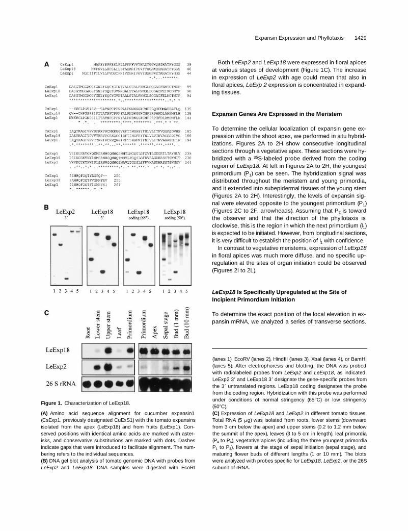
Cloning of a Tomato Expansin cDNA and Genomic Analysis of Tomato Expansins
Exogenously applied expansin protein can initiate primor-dium formation on the shoot apical meristem of tomatoplants (Fleming et al., 1997). To study the function of endog-enous expansin in primordium initiation, we decided toclone expansin cDNAs that are expressed in the apex of to-mato plants and to study their regulation. Using degenerateprimers matching conserved sequences in the coding regionof the cucumber
expansin1
gene, we amplified a fragment ofa putative tomato expansin by reverse transcription–poly-merase chain reaction. The amplification product was usedas a probe to isolate a full-length cDNA, named
LeExp18.LeExp18
represents a new tomato expansin gene exhibitingall of the basic features of expansin, as described byShcherban et al. (1995). The open reading frame of the
LeExp18
cDNA codes for a polypeptide of 255 amino acids(Figure 1A). The predicted protein contains a putative signalsequence of 18 amino acids and all of the cysteines andtryptophans that are conserved between expansins (Figure1A; Shcherban et al., 1995).
LeExp18
is most closely relatedto the fruit-specific
LeExp1
(Rose et al., 1997), with 79%amino acid identity.
Genomic DNA gel blot analysis was performed to deter-mine the number of expansin genes in the tomato genome.
Probes were derived from
LeExp18
and from
LeExp2
, a par-tial cDNA isolated from leaf RNA (kindly provided by S.McQueen-Mason, University of York, York, UK, who is work-ing on a complete analysis of the expansin gene family in to-mato). Probes derived from the 3
9
noncoding regions of
LeExp2
and
LeExp18
detected single bands and occasionalweaker bands (Figure 1B). Most likely,
LeExp2
and
LeExp18
are single-copy genes. The patterns of the bands detectedwith the two probes are different, confirming that bothprobes are gene specific.
A probe containing part of the coding region of
LeExp18
was used to estimate the number of
LeExp18
-related genesin the tomato genome. After hybridization and washing athigh stringency, we observed a banding pattern that wasindistinguishable from the pattern seen with the
LeExp18
3
9
probe (Figure 1B, third gel; cf. with second gel). After low-stringency hybridization with the
LeExp18
coding regionprobe, we detected several additional bands in the differentsamples, none of which comigrated with
LeExp2
bands (Fig-ure 1B, fourth gel; cf. with first gel). Thus,
LeExp18
and
LeExp2
are sufficiently divergent even in the coding regionto prevent cross-hybridization. We conclude that
LeExp18
isa member of a small subfamily of expansins within a largerexpansin family.
LeExp2
and
LeExp18
Are Differentially Regulated
Expression levels of
LeExp2
and
LeExp18
mRNAs were de-termined by RNA gel blot analysis by using the probes de-rived from their 3
9
noncoding regions. Samples wereisolated from the root, stem, expanding leaves, leaf primor-dia (P
4
to P
6
), vegetative apex (including the three youngestleaf primordia, P
1
to P
3
), and developing flowers at variousstages.
LeExp18
was detected primarily in apical tissuessuch as the vegetative apex, in developing flowers, in leafprimordia, and in the upper stem. Notably,
LeExp18
expres-sion was low in leaves and in the lower stem tissue (Figure1C). In contrast,
LeExp2
was expressed primarily in thestem, in maturing flowers, in expanding leaves, and in leafprimordia; however,
LeExp2
was not expressed in the vege-tative apex. No expression of either
LeExp2
or
LeExp18
could be detected in roots (Figure 1C).From the experiments described above, we conclude that
LeExp2
expression coincides primarily with expansion(leaves, stems, and flowers), whereas
LeExp18
expression isassociated with meristematic activity (apex, leaf primordia,and developing flowers), although there is some overlappingexpression of both expansins in leaf primordia, flowers, andthe stem. In this context, it is interesting that all of theseplant parts combine expansion and meristematic activity.The stem retains meristematic activity in the cambium, andthe young leaves of tomato are known to retain meristematic features for prolonged times during leaflet formation (Harevenet al., 1996; Chen et al., 1997).
Expansin Expression and Phyllotaxis 1429
Both
LeExp2
and
LeExp18
were expressed in floral apicesat various stages of development (Figure 1C). The increasein expression of
LeExp2
with age could mean that also infloral apices,
LeExp 2
expression is concentrated in expand-ing tissues.
Expansin Genes Are Expressed in the Meristem
To determine the cellular localization of expansin gene ex-pression within the shoot apex, we performed in situ hybrid-izations. Figures 2A to 2H show consecutive longitudinalsections through a vegetative apex. These sections were hy-bridized with a
35
S-labeled probe derived from the codingregion of
LeExp18.
At left in Figures 2A to 2H, the youngestprimordium (P
1
) can be seen. The hybridization signal wasdistributed throughout the meristem and young primordia,and it extended into subepidermal tissues of the young stem(Figures 2A to 2H). Interestingly, the levels of expansin sig-nal were elevated opposite to the youngest primordium (P
1
)(Figures 2C to 2F, arrowheads). Assuming that P
2
is towardthe observer and that the direction of the phyllotaxis isclockwise, this is the region in which the next primordium (I
1
)is expected to be initiated. However, from longitudinal sections,it is very difficult to establish the position of I
1
with confidence.In contrast to vegetative meristems, expression of
LeExp18
in floral apices was much more diffuse, and no specific up-regulation at the sites of organ initiation could be observed(Figures 2I to 2L).
LeExp18
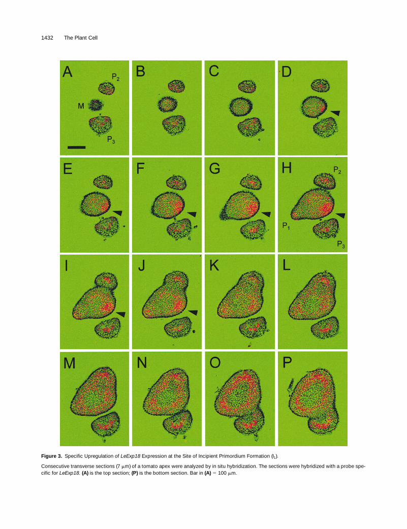
Is Specifically Upregulated at the Site of Incipient Primordium Initiation
To determine the exact position of the local elevation in ex-pansin mRNA, we analyzed a series of transverse sections.
(lanes 1), EcoRV (lanes 2), HindIII (lanes 3), XbaI (lanes 4), or BamHI(lanes 5). After electrophoresis and blotting, the DNA was probedwith radiolabeled probes from
LeExp2
and
LeExp18
, as indicated.LeExp2 3
9
and LeExp18 3
9
designate the gene-specific probes fromthe 3
9
untranslated regions. LeExp18 coding designates the probefrom the coding region. Hybridization with this probe was performedunder conditions of normal stringency (65
8
C) or low stringency(50
8
C).
(C)
Expression of
LeExp18
and
LeExp2
in different tomato tissues.Total RNA (5
m
g) was isolated from roots, lower stems (downwardfrom 3 cm below the apex) and upper stems (0.2 to 1.2 mm belowthe summit of the apex), leaves (3 to 5 cm in length), leaf primordia(P
4
to P
6
), vegetative apices (including the three youngest primordiaP
1
to P
3
), flowers at the stage of sepal initiation (sepal stage), andmaturing flower buds of different lengths (1 or 10 mm). The blotswere analyzed with probes specific for
LeExp18
,
LeExp2
, or the 26Ssubunit of rRNA.
Figure 1.
Characterization of LeExp18.
(A)
Amino acid sequence alignment for cucumber expansin1(CsExp1, previously designated CuExS1) with the tomato expansinsisolated from the apex (LeExp18) and from fruits (LeExp1). Con-served positions with identical amino acids are marked with aster-isks, and conservative substitutions are marked with dots. Dashesindicate gaps that were introduced to facilitate alignment. The num-bering refers to the individual sequences.
(B)
DNA gel blot analysis of tomato genomic DNA with probes from
LeExp2
and
LeExp18.
DNA samples were digested with EcoRI

1430 The Plant Cell
Figures 3A to 3P show a series of consecutive cross-sec-tions of a vegetative tomato apex that were hybridized witha
35
S-labeled probe derived from the
LeExp18
3
9
end. Theoldest primordium (P
3
) is in the lower half of the images (in-dicated in Figures 3A and 3H). The younger primordia (P
2
and P
1
) follow in a counterclockwise, spiral phyllotaxis, witha divergence angle of
z
135
8
, so that the site of incipient pri-mordium initiation (I
1
) can be predicted (indicated by arrow-heads in Figures 3D to 3J). In the sections from the top ofthe apex (Figures 3A to 3C),
LeExp18
was only expressed
Figure 2. Expression of Expansin in the Vegetative Apex and in Developing Flowers of Tomato.
(A) to (H) Consecutive longitudinal sections (7 mm) of a vegetative apex were analyzed by in situ hybridization. The sections were hybridized witha probe derived from the coding region of LeExp18. The sections exhibit an apex with the meristem dome in the center (M in [A]) and the young-est primordium (indicated with P1 in [A] and [D]) to the left. The site of increased signal levels is indicated by arrowheads in (C) to (F). Bar in (A) 5100 mm.(I) to (L) Longitudinal sections of tomato flowers at different stages of development were analyzed by in situ hybridization, using the same probeas given in (A) to (H). A, axillary meristem; Ca, carpel primordium; F, floral meristem; I, inflorescence meristem; Pe, petal primordium; Se, sepalprimordium; St, stamen primordium. Bar in (I) 5 100 mm.
Expansin Expression and Phyllotaxis 1431
at low levels. Low levels of expansin signal could also beseen throughout the meristem, with a concentration of sig-nal localized at I
1
. The elevated level is present in seven con-secutive sections (Figures 3D to 3J). Considering that thethickness of the sections is 7
m
m, we can estimate that theregion of elevated expansin expression corresponds to anapproximately circular area on the flank of the meristem witha diameter of
z
50 mm.The tissue with elevated expansin expression comprises
all three meristem layers (cf. Figures 2D and 3H) and ex-tends almost to the center of the meristem proper. Apartfrom the meristem, LeExp18 was also expressed in primor-dia and in the cortical tissues of the young stem (Figures 3Lto 3P). The expression pattern in primordia comprised aU-shaped area that included the central tissues forming thevasculature and the adaxial margins of the leaf primordia,where leaflets and the leaf blade develop. The ring-shapedregion of expansin expression in the stem coincides withdeveloping vascular bundles and may reflect procambiumactivity. Therefore, high expression of LeExp18 coincideswith tissues showing meristematic activity, such as theshoot apical meristem, the leaf margins, and the procam-bium, whereas expanding tissues, such as the central pithtissue in the stem and the adaxial and abaxial side of the ra-chis, exhibit low levels of LeExp18 expression. This is incontrast to the idea that expansin expression is associatedwith cell elongation in tissues such as leaves, hypocotyls,and grass internodes (Cho and Kende, 1997; Cosgrove,1997). LeExp18 may therefore fulfill a specific function inmeristematic tissues, particularly in leaf primordium initiation.
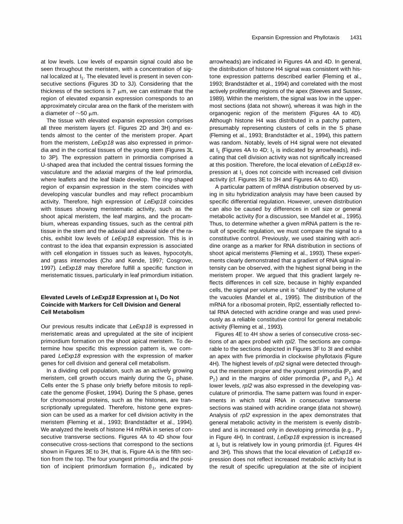
Elevated Levels of LeExp18 Expression at I1 Do Not Coincide with Markers for Cell Division and GeneralCell Metabolism
Our previous results indicate that LeExp18 is expressed inmeristematic areas and upregulated at the site of incipientprimordium formation on the shoot apical meristem. To de-termine how specific this expression pattern is, we com-pared LeExp18 expression with the expression of markergenes for cell division and general cell metabolism.
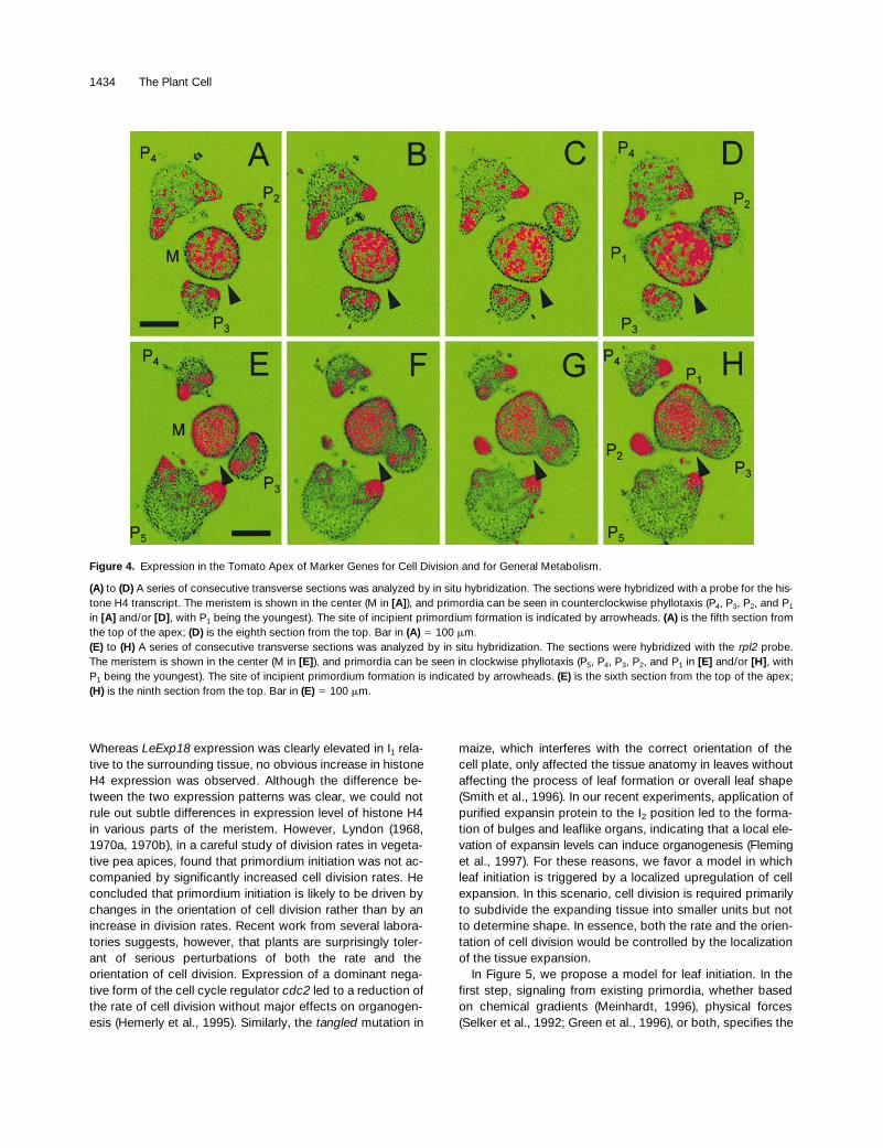
In a dividing cell population, such as an actively growingmeristem, cell growth occurs mainly during the G1 phase.Cells enter the S phase only briefly before mitosis to repli-cate the genome (Fosket, 1994). During the S phase, genesfor chromosomal proteins, such as the histones, are tran-scriptionally upregulated. Therefore, histone gene expres-sion can be used as a marker for cell division activity in themeristem (Fleming et al., 1993; Brandstädter et al., 1994).We analyzed the levels of histone H4 mRNA in series of con-secutive transverse sections. Figures 4A to 4D show fourconsecutive cross-sections that correspond to the sectionsshown in Figures 3E to 3H, that is, Figure 4A is the fifth sec-tion from the top. The four youngest primordia and the posi-tion of incipient primordium formation (I 1, indicated by
arrowheads) are indicated in Figures 4A and 4D. In general,the distribution of histone H4 signal was consistent with his-tone expression patterns described earlier (Fleming et al.,1993; Brandstädter et al., 1994) and correlated with the mostactively proliferating regions of the apex (Steeves and Sussex,1989). Within the meristem, the signal was low in the upper-most sections (data not shown), whereas it was high in theorganogenic region of the meristem (Figures 4A to 4D).Although histone H4 was distributed in a patchy pattern,presumably representing clusters of cells in the S phase(Fleming et al., 1993; Brandstädter et al., 1994), this patternwas random. Notably, levels of H4 signal were not elevatedat I1 (Figures 4A to 4D; I1 is indicated by arrowheads), indi-cating that cell division activity was not significally increasedat this position. Therefore, the local elevation of LeExp18 ex-pression at I1 does not coincide with increased cell divisionactivity (cf. Figures 3E to 3H and Figures 4A to 4D).
A particular pattern of mRNA distribution observed by us-ing in situ hybridization analysis may have been caused byspecific differential regulation. However, uneven distributioncan also be caused by differences in cell size or generalmetabolic activity (for a discussion, see Mandel et al., 1995).Thus, to determine whether a given mRNA pattern is the re-sult of specific regulation, we must compare the signal to aconstitutive control. Previously, we used staining with acri-dine orange as a marker for RNA distribution in sections ofshoot apical meristems (Fleming et al., 1993). These experi-ments clearly demonstrated that a gradient of RNA signal in-tensity can be observed, with the highest signal being in themeristem proper. We argued that this gradient largely re-flects differences in cell size, because in highly expandedcells, the signal per volume unit is “diluted” by the volume ofthe vacuoles (Mandel et al., 1995). The distribution of themRNA for a ribosomal protein, Rpl2, essentially reflected to-tal RNA detected with acridine orange and was used previ-ously as a reliable constitutive control for general metabolicactivity (Fleming et al., 1993).
Figures 4E to 4H show a series of consecutive cross-sec-tions of an apex probed with rpl2. The sections are compa-rable to the sections depicted in Figures 3F to 3I and exhibitan apex with five primordia in clockwise phyllotaxis (Figure4H). The highest levels of rpl2 signal were detected through-out the meristem proper and the youngest primordia (P1 andP2) and in the margins of older primordia (P4 and P5). Atlower levels, rpl2 was also expressed in the developing vas-culature of primordia. The same pattern was found in exper-iments in which total RNA in consecutive transversesections was stained with acridine orange (data not shown).Analysis of rpl2 expression in the apex demonstrates thatgeneral metabolic activity in the meristem is evenly distrib-uted and is increased only in developing primordia (e.g., P2
in Figure 4H). In contrast, LeExp18 expression is increasedat I1 but is relatively low in young primordia (cf. Figures 4Hand 3H). This shows that the local elevation of LeExp18 ex-pression does not reflect increased metabolic activity but isthe result of specific upregulation at the site of incipient
1432 The Plant Cell
Figure 3. Specific Upregulation of LeExp18 Expression at the Site of Incipient Primordium Formation (I1).
Consecutive transverse sections (7 mm) of a tomato apex were analyzed by in situ hybridization. The sections were hybridized with a probe spe-cific for LeExp18. (A) is the top section; (P) is the bottom section. Bar in (A) 5 100 mm.
Expansin Expression and Phyllotaxis 1433
primordium formation. Therefore, we postulate that the pat-tern of LeExp18 expression reflects the regulation of phyllo-taxis in the meristem and that LeExp18 serves a specificfunction in leaf primordium initiation.
DISCUSSION
Differential Expression of Expansin Genes
We have isolated a tomato expansin cDNA, LeExp18, thatrepresents a member of a small multigene subfamily of thetomato expansin genes. The tomato expansin family alsoincludes LeExp2, an expansin cDNA isolated from leaves,and the fruit-specific LeExp1 cDNA, which may play a roleduring fruit softening (Rose et al., 1997). The existence ofmultiple expansin genes could signify that the individualproteins have distinct biochemical properties. Currently, how-ever, neither sequence comparisons nor the limited functionaldata available indicate that expansins have distinct bio-chemical specificities; therefore, the major significance ofthe presence of multiple genes may be that distinctly ex-pressed genes can regulate cell expansion in place and time.
The RNA gel blots shown in Figure 1C demonstrate thatthe LeExp2 and LeExp18 genes are differentially expressed.LeExp2 is not expressed in the shoot apical meristem but ispredominantly active in more mature tissues. In flowers,LeExp2 expression increases during maturation. In contrast,LeExp18 expression is highest in the shoot apex and in flow-ers, with low expression in maturing leaves. Our research in-terest centers on the vegetative shoot apical meristem;therefore, we analyzed the expression of LeExp18 in this or-gan. Expansin gene expression was compared with the ex-pression pattern of the housekeeping genes encodingribosomal protein Rpl2 and histone H4. A comparison withhousekeeping genes is essential, because differences in cellsize and general metabolic activity can cause large apparentdeviations from a uniform signal intensity (for a detailed dis-cussion, see Mandel et al., 1995).
As can be seen from the hybridization patterns obtainedwith rpl2 (Figures 4E to 4H), the small cells present in theshoot apical meristem, the young leaf primordia, and themeristematic zones at the margins of older primordia displaythe highest signal density. In contrast, the larger cells of themidrib and the stem pith tissue, for instance, have fewer sil-
ver grains per square micrometer. The distribution of the hy-bridization signal coincides with the staining pattern seenwith acridine orange, which is a general dye for nucleic ac-ids (data not shown). Thus, the signal distribution seen withrpl2 reflects the general RNA distribution in the shoot apexand serves as a useful baseline against which the expres-sion of other genes can be compared. Histone H4 is ex-pressed in patches of cells throughout the apex, presumablyreflecting those cells that are in S phase and actively divid-ing. It can also be seen that the areas of high cell divisionactivity are the same areas that have high rpl2 expression.Thus, rpl2 is a general marker for small cells with high meta-bolic activity, whereas histone H4 expression defines thesubset of the rpl2-expressing cells that are in S phase.
In the apex, the expression patterns of LeExp18 observedin in situ experiments exhibited two main features. First,LeExp18 expression in general coincided with small, rapidlydividing cells in the meristem, in leaf primordia, and in thevascular tissues of the young stem. Second, LeExp18 ex-pression was elevated at the site of incipient primordiumformation (I1) when compared with other regions in the mer-istem. This distribution was surprising considering the pos-tulated function of expansins in cell elongation and cellenlargement, as discussed below (Cosgrove, 1997). Expres-sion in the floral meristems was more diffuse, and no specificupregulation at the site of incipient primordium formationcould be observed (Figures 2I to 2L).
Role of Expansin in Leaf Primordium Formation
The action of expansin and the expression of expansingenes are commonly associated with cell expansion in rap-idly growing tissues, for example, in the unidirectional elon-gation of hypocotyls (Cosgrove, 1997) and grass internodes(Cho and Kende, 1997) or in expanding leaves (Keller andCosgrove, 1995). This association may well hold for LeExp2,which is highly expressed in stems, maturing leaves, andflowers but is absent from meristematic tissues. However,LeExp18-expressing cells are small and actively dividing,and in these cells, upregulation of expansin expression isaccompanied by isodiametrical cell expansion and cell divi-sion.
The differences in expression pattern of LeExp18 and his-tone H4 invite a discussion of the relative importance of cellexpansion versus cell division in primordium initiation.
(A) to (C) Sections from the top of the apex, exhibiting the apical meristem dome (M in [A]) and two primordia (P3 and P2 in [A]). The youngestprimordium cannot be seen at this level.(D) to (J) Sections including the region of primordium initiation. The sections represent the meristem in the center and the three youngest primor-dia in counterclockwise phyllotaxis (P3, P2, and P1 in [H]), with P1 being the youngest primordium. The site of incipient primordium formation isindicated by arrowheads in (D) to (J).(K) to (P) Sections from the lower apex, representing young stem tissue.
Figure 3. (continued).
1434 The Plant Cell
Whereas LeExp18 expression was clearly elevated in I1 rela-tive to the surrounding tissue, no obvious increase in histoneH4 expression was observed. Although the difference be-tween the two expression patterns was clear, we could notrule out subtle differences in expression level of histone H4in various parts of the meristem. However, Lyndon (1968,1970a, 1970b), in a careful study of division rates in vegeta-tive pea apices, found that primordium initiation was not ac-companied by significantly increased cell division rates. Heconcluded that primordium initiation is likely to be driven bychanges in the orientation of cell division rather than by anincrease in division rates. Recent work from several labora-tories suggests, however, that plants are surprisingly toler-ant of serious perturbations of both the rate and theorientation of cell division. Expression of a dominant nega-tive form of the cell cycle regulator cdc2 led to a reduction ofthe rate of cell division without major effects on organogen-esis (Hemerly et al., 1995). Similarly, the tangled mutation in
maize, which interferes with the correct orientation of thecell plate, only affected the tissue anatomy in leaves withoutaffecting the process of leaf formation or overall leaf shape(Smith et al., 1996). In our recent experiments, application ofpurified expansin protein to the I2 position led to the forma-tion of bulges and leaflike organs, indicating that a local ele-vation of expansin levels can induce organogenesis (Fleminget al., 1997). For these reasons, we favor a model in whichleaf initiation is triggered by a localized upregulation of cellexpansion. In this scenario, cell division is required primarilyto subdivide the expanding tissue into smaller units but notto determine shape. In essence, both the rate and the orien-tation of cell division would be controlled by the localizationof the tissue expansion.
In Figure 5, we propose a model for leaf initiation. In thefirst step, signaling from existing primordia, whether basedon chemical gradients (Meinhardt, 1996), physical forces(Selker et al., 1992; Green et al., 1996), or both, specifies the
Figure 4. Expression in the Tomato Apex of Marker Genes for Cell Division and for General Metabolism.
(A) to (D) A series of consecutive transverse sections was analyzed by in situ hybridization. The sections were hybridized with a probe for the his-tone H4 transcript. The meristem is shown in the center (M in [A]), and primordia can be seen in counterclockwise phyllotaxis (P4, P3, P2, and P1
in [A] and/or [D], with P1 being the youngest). The site of incipient primordium formation is indicated by arrowheads. (A) is the fifth section fromthe top of the apex; (D) is the eighth section from the top. Bar in (A) 5 100 mm.(E) to (H) A series of consecutive transverse sections was analyzed by in situ hybridization. The sections were hybridized with the rpl2 probe.The meristem is shown in the center (M in [E]), and primordia can be seen in clockwise phyllotaxis (P5, P4, P3, P2, and P1 in [E] and/or [H], withP1 being the youngest). The site of incipient primordium formation is indicated by arrowheads. (E) is the sixth section from the top of the apex;(H) is the ninth section from the top. Bar in (E) 5 100 mm.
Expansin Expression and Phyllotaxis 1435
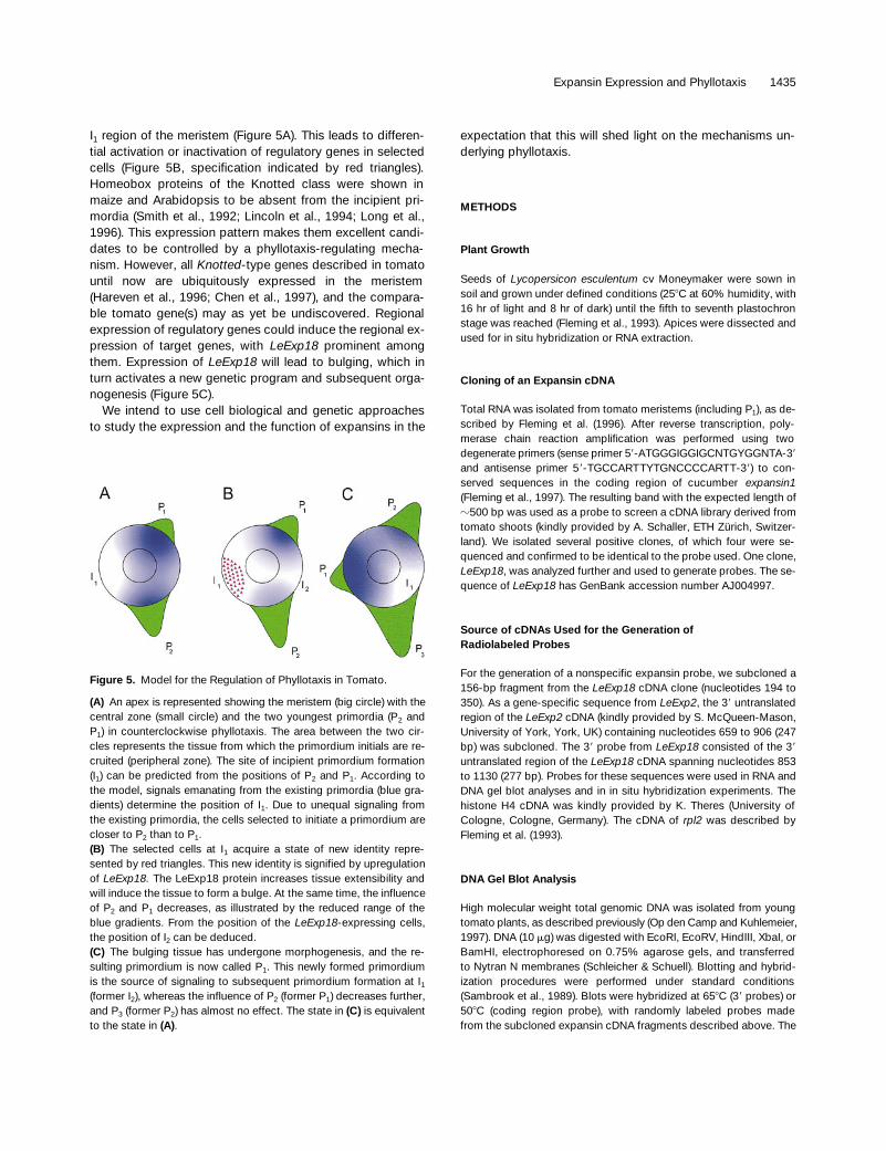
I1 region of the meristem (Figure 5A). This leads to differen-tial activation or inactivation of regulatory genes in selectedcells (Figure 5B, specification indicated by red triangles).Homeobox proteins of the Knotted class were shown inmaize and Arabidopsis to be absent from the incipient pri-mordia (Smith et al., 1992; Lincoln et al., 1994; Long et al.,1996). This expression pattern makes them excellent candi-dates to be controlled by a phyllotaxis-regulating mecha-nism. However, all Knotted-type genes described in tomatountil now are ubiquitously expressed in the meristem(Hareven et al., 1996; Chen et al., 1997), and the compara-ble tomato gene(s) may as yet be undiscovered. Regionalexpression of regulatory genes could induce the regional ex-pression of target genes, with LeExp18 prominent amongthem. Expression of LeExp18 will lead to bulging, which inturn activates a new genetic program and subsequent orga-nogenesis (Figure 5C).
We intend to use cell biological and genetic approachesto study the expression and the function of expansins in the
expectation that this will shed light on the mechanisms un-derlying phyllotaxis.
METHODS
Plant Growth
Seeds of Lycopersicon esculentum cv Moneymaker were sown insoil and grown under defined conditions (258C at 60% humidity, with16 hr of light and 8 hr of dark) until the fifth to seventh plastochronstage was reached (Fleming et al., 1993). Apices were dissected andused for in situ hybridization or RNA extraction.
Cloning of an Expansin cDNA
Total RNA was isolated from tomato meristems (including P1), as de-scribed by Fleming et al. (1996). After reverse transcription, poly-merase chain reaction amplification was performed using twodegenerate primers (sense primer 59-ATGGGIGGIGCNTGYGGNTA-39
and antisense primer 59-TGCCARTTYTGNCCCCARTT-39) to con-served sequences in the coding region of cucumber expansin1(Fleming et al., 1997). The resulting band with the expected length ofz500 bp was used as a probe to screen a cDNA library derived fromtomato shoots (kindly provided by A. Schaller, ETH Zürich, Switzer-land). We isolated several positive clones, of which four were se-quenced and confirmed to be identical to the probe used. One clone,LeExp18, was analyzed further and used to generate probes. The se-quence of LeExp18 has GenBank accession number AJ004997.
Source of cDNAs Used for the Generation ofRadiolabeled Probes
For the generation of a nonspecific expansin probe, we subcloned a156-bp fragment from the LeExp18 cDNA clone (nucleotides 194 to350). As a gene-specific sequence from LeExp2, the 39 untranslatedregion of the LeExp2 cDNA (kindly provided by S. McQueen-Mason,University of York, York, UK) containing nucleotides 659 to 906 (247bp) was subcloned. The 39 probe from LeExp18 consisted of the 39
untranslated region of the LeExp18 cDNA spanning nucleotides 853to 1130 (277 bp). Probes for these sequences were used in RNA andDNA gel blot analyses and in in situ hybridization experiments. Thehistone H4 cDNA was kindly provided by K. Theres (University ofCologne, Cologne, Germany). The cDNA of rpl2 was described byFleming et al. (1993).
DNA Gel Blot Analysis
High molecular weight total genomic DNA was isolated from youngtomato plants, as described previously (Op den Camp and Kuhlemeier,1997). DNA (10 mg) was digested with EcoRI, EcoRV, HindIII, XbaI, orBamHI, electrophoresed on 0.75% agarose gels, and transferredto Nytran N membranes (Schleicher & Schuell). Blotting and hybrid-ization procedures were performed under standard conditions(Sambrook et al., 1989). Blots were hybridized at 658C (39 probes) or508C (coding region probe), with randomly labeled probes madefrom the subcloned expansin cDNA fragments described above. The
Figure 5. Model for the Regulation of Phyllotaxis in Tomato.
(A) An apex is represented showing the meristem (big circle) with thecentral zone (small circle) and the two youngest primordia (P2 andP1) in counterclockwise phyllotaxis. The area between the two cir-cles represents the tissue from which the primordium initials are re-cruited (peripheral zone). The site of incipient primordium formation(I1) can be predicted from the positions of P2 and P1. According tothe model, signals emanating from the existing primordia (blue gra-dients) determine the position of I1. Due to unequal signaling fromthe existing primordia, the cells selected to initiate a primordium arecloser to P2 than to P1.(B) The selected cells at I1 acquire a state of new identity repre-sented by red triangles. This new identity is signified by upregulationof LeExp18. The LeExp18 protein increases tissue extensibility andwill induce the tissue to form a bulge. At the same time, the influenceof P2 and P1 decreases, as illustrated by the reduced range of theblue gradients. From the position of the LeExp18-expressing cells,the position of I2 can be deduced.(C) The bulging tissue has undergone morphogenesis, and the re-sulting primordium is now called P1. This newly formed primordiumis the source of signaling to subsequent primordium formation at I1(former I2), whereas the influence of P2 (former P1) decreases further,and P3 (former P2) has almost no effect. The state in (C) is equivalentto the state in (A).
1436 The Plant Cell
final wash was in 0.2 3 SSC (1 3 SSC is 0.15 M NaCl and 0.015 Msodium citrate) and 0.1% SDS at 658C (39 probes) or 508C (coding re-gion probe).
RNA Gel Blot Analysis
RNA gel blot analysis was performed essentially as described by Opden Camp and Kuhlemeier (1997). Total RNA from various parts oftomato plants was isolated as described by Fleming et al. (1993). Toisolate 5 mg of total RNA, we extracted 150 apices (including thethree youngest primordia) and 450 primordia (P4 to P6). RNA wasquantified spectrophotometrically at 260 nm, and 5-mg portions wererun on a 1.0% agarose–glyoxal gel after glyoxylation. RNA blottingand hybridization procedures were performed under standard condi-tions (Sambrook et al., 1989). Blots were hybridized at 658C, withrandomly labeled probes from the expansin cDNAs described aboveor with a probe from a rRNA (Fleming et al., 1993). The final wash wasin 0.1 3 SSC and 0.1% SDS at 658C.
In Situ Hybridizations
In situ hybridization experiments were performed according to theprotocol described by Fleming et al. (1993), with minor modifications.From embedded tomato apices, we cut consecutive longitudinal ortransverse sections (7 mm) and used them for in situ hybridization.Before exposure, slides were treated with a solution of 50 mg/mLRNase A (rather than 1 mg/mL) and finally washed in 2 3 SSC atroom temperature and in 0.1 3 SSC at 428, each for 30 min. After de-velopment, the slides were stained in toluidine blue and viewed on anLSM 310 microscope (Carl Zeiss AG, Oberkochen, Germany). Im-ages were taken under bright-field light (shown in false green color)and overlayed with epifluorescence images taken under polarizedlight exhibiting the silver grain signal (shown in false red color). In allcases, control hybridizations were performed with the correspondingsense probes, and in all instances, the signal obtained was negligiblecompared with that obtained using the antisense probe.
ACKNOWLEDGMENTS
We thank Andreas Schaller for kindly providing a tomato shoot cDNAlibrary, Simon McQueen-Mason for supplying a cDNA clone forLeExp2, Klaus Theres for a histone H4 cDNA, and Isabelle Dupuis,Andrew Fleming, and Jeroen Stuurman for critical reading of themanuscript. This work was supported in part by a grant from theSwiss National Science Foundation.
Received February 23, 1998; accepted July 9, 1998.
REFERENCES
Brandstädter, J., Rossbach, C., and Theres, K. (1994). The pat-tern of histone H4 expression in the tomato shoot apex changesduring development. Planta 192, 69–74.
Chen, J.-J., Janssen, B.-J., Williams, A., and Sinha, N. (1997). Agene fusion at a homeobox locus: Alterations in leaf shape andimplications for morphological evolution. Plant Cell 9, 1289–1304.
Cho, H.-T., and Kende, H. (1997). Expression of expansin genes iscorrelated with growth in deepwater rice. Plant Cell 9, 1661–1671.
Cosgrove, D.J. (1997). Relaxation in a high-stress environment: Themolecular bases of extensible cell walls and cell enlargement.Plant Cell 9, 1031–1041.
Fleming, A.J., Mandel, T., Roth, I., and Kuhlemeier, C. (1993). Thepatterns of gene expression in the tomato shoot apical meristem.Plant Cell 5, 297–309.
Fleming, A.J., Manzara, T., Gruissem, W., and Kuhlemeier, C.(1996). Fluorescent imaging of GUS activity and RT-PCR analy-sis of gene expression in the shoot apical meristem. Plant J. 10,745–754.
Fleming, A.J., McQueen-Mason, S., Mandel, T., and Kuhlemeier,C. (1997). Induction of leaf primordia by the cell wall proteinexpansin. Science 276, 1415–1418.
Foard, D.E. (1971). The initial protrusion of a leaf primordium canform without concurrent periclinal cell divisions. Can. J. Bot. 49,694–702.
Fosket, D.E. (1994). Plant Growth and Development. (San Diego,CA: Academic Press).
Green, P.B., Steele, C.S., and Rennich, S.C. (1996). Phyllotacticpatterns: A biophysical mechanism for their origin. Ann. Bot. 77,515–527.
Hareven, D., Gutfinger, T., Parnis, A., Eshed, Y., and Lifschitz, E.(1996). The making of a compound leaf: Genetic manipulation ofleaf architecture in tomato. Cell 84, 735–744.
Hemerly, A., de Almeida-Engler, J., Bergounioux, C., Van Montagu,M., Engler, G., Inzé, D., and Ferreira, P. (1995). Dominant nega-tive mutants of the Cdc2 kinase uncouple cell division from itera-tive plant development. EMBO J. 14, 3925–3936.
Jacobs, T. (1997). Why do plant cells divide? Plant Cell 9, 1021–1029.
Jean, R.V. (1994). Phyllotaxis: A Systematic Study in Plant Morpho-genesis. (Cambridge, UK: Cambridge University Press).
Keller, E., and Cosgrove, D.J. (1995). Expansins in growing tomatoleaves. Plant J. 8, 795–802.
Kende, H., and Zeevaart, J.A.D. (1997). The five “classical” planthormones. Plant Cell 9, 1197–1210.
Lincoln, C., Long, J., Yamaguchi, J., Serikawa, K., and Hake, S.(1994). A knotted1-like homeobox gene in Arabidopsis is ex-pressed in the vegetative meristem and dramatically alters leafmorphology when overexpressed in transgenic plants. Plant Cell6, 1859–1876.
Long, J.A., Moan, E.I., Medford, J.I., and Barton, M.K. (1996). Amember of the KNOTTED class of homeodomain proteins encodedby the STM gene of Arabidopsis. Nature 379, 66–69.
Lyndon, R.F. (1968). Changes in volume and cell number in the dif-ferent regions of the shoot apex of Pisum during a single plasto-chron. Ann. Bot. 32, 371–390.
Lyndon, R.F. (1970a). Rates of cell division in the shoot apical mer-istem of Pisum. Ann. Bot. 34, 1–17.
Lyndon, R.F. (1970b). Planes of cell division and growth in the shootapex of Pisum. Ann. Bot. 34, 19–28.
Expansin Expression and Phyllotaxis 1437
Lyndon, R.F. (1990). Plant Development: The Cellular Basis. (Lon-don: Unwin Hyman).
Mandel, T., Fleming, A.J., Krähenbühl, R., and Kuhlemeier, C.(1995). Definition of constitutive gene expression in plants: Thetranslation initiation factor 4A gene as a model. Plant Mol. Biol.29, 995–1004.
McQueen-Mason, S.J., and Cosgrove, D.J. (1994). Disruption ofhydrogen bonding between plant cell wall polymers by proteinsthat induce wall extension. Proc. Natl. Acad. Sci. USA 91,6574–6578.
McQueen-Mason, S.J., Durachko, D.M., and Cosgrove, D.J.(1992). Two endogenous proteins that induce cell wall extensionin plants. Plant Cell 4, 1425–1433.
Meinhardt, H. (1996). Models of biological pattern formation: Com-mon mechanism in plant and animal development. Int. J. Dev.Biol. 40, 123–134.
Meyerowitz, E.M. (1997). Genetic control of cell division patterns indeveloping plants. Cell 88, 299–308.
Op den Camp, R.G.L., and Kuhlemeier, C. (1997). Aldehyde dehy-drogenase in tobacco pollen. Plant Mol. Biol. 35, 355–365.
Poethig, R.S. (1997). Leaf morphogenesis in flowering plants. PlantCell 9, 1077–1087.
Rose, J.K.C, Lee, H.H., and Bennett, A.B. (1997). Expression of adivergent expansin gene is fruit-specific and ripening-regulated.Proc. Natl. Acad. Sci. USA 94, 5955–5960.
Sambrook, J., Fritsch, E.F., and Maniatis, T. (1989). MolecularCloning: A Laboratory Manual, 2nd ed. (Cold Spring Harbor, NY:Cold Spring Harbor Laboratory Press).
Selker, J.M.L., Steucek, G.L., and Green, P.B. (1992). Biophysicalmechanisms for morphogenetic progression at the shoot apex.Dev. Biol. 153, 29–43.
Shcherban, T.Y., Shi, J., Durachko, D.M., Guiltinan, M.J.,McQueen-Mason, S.J., Shieh, M., and Cosgrove, D.J. (1995).Molecular cloning and sequence analysis of expansins—A highlyconserved, multigene family of proteins that mediate cell wallextension in plants. Proc. Natl. Acad. Sci. USA 92, 9245–9249.
Smith, L.G., Greene, B., Veit, B., and Hake, S. (1992). A dominantmutation in the maize homeobox gene, knotted-1, causes itsectopic expression in leaf cells with altered fates. Development116, 21–30.
Smith, L.G., Hake, S., and Sylvester, A.W. (1996). The tangled-1mutation alters cell division orientations throughout maize leafdevelopment without altering leaf shape. Development 122,481–489.
Steeves, T.A., and Sussex, I.M. (1989). Patterns in Plant Develop-ment. (New York: Cambridge University Press).
Szymkowiak, E.J., and Sussex, I.M. (1996). What chimeras can tellus about plant development. Annu. Rev. Plant Physiol. Plant Mol.Biol. 47, 351–376.
Copyright © 2022 FDOKUMEN


![Regeneration Of Three Sweet Potato [Ipomoea Batatas(L.)] Accessions in Ghana via Meristem And Nodal culture](https://static.fdokumen.com/doc/165x107/631af948d43f4e176304af45/regeneration-of-three-sweet-potato-ipomoea-batatasl-accessions-in-ghana-via.jpg)