Parasite Killing in Malaria NonVector Mosquito Anopheles culicifacies Species B: Implication of...
12
Parasite Killing in Malaria Non-Vector Mosquito Anopheles culicifacies Species B: Implication of Nitric Oxide Synthase Upregulation Sonam Vijay 1 , Manmeet Rawat 1 , Tridibes Adak 2 , Rajnikant Dixit 3 , Nutan Nanda 4 , Harish Srivastava 5 , Joginder K. Sharma 6 , Godavarthi B. K. S. Prasad 7 , Arun Sharma 1 * 1 Protein Biochemistry and Structural Biology Laboratory, National Institute of Malaria Research (ICMR), Dwarka, New Delhi, India, 2 Vector Biology Laboratory, National Institute of Malaria Research (ICMR), Dwarka, New Delhi, India, 3 Host Parasite Interaction Group, National Institute of Malaria Research (ICMR), Dwarka, New Delhi, India, 4 Molecular Entomology Laboratory, National Institute of Malaria Research (ICMR), Dwarka, New Delhi, India, 5 Entomology Laboratory, National Institute of Malaria Research (ICMR) Field Unit, Civil Hospital, Nadiad, Gujarat, India, 6 Molecular Diagnostics Laboratory, Institute of Cytology and Preventive Oncology (ICMR), Noida, India, 7 Department of Biotechnology, Jiwaji University, Gwalior, India Abstract Background: Anopheles culicifacies, the main vector of human malaria in rural India, is a complex of five sibling species. Despite being phylogenetically related, a naturally selected subgroup species B of this sibling species complex is found to be a poor vector of malaria. We have attempted to understand the differences between vector and non-vector Anopheles culicifacies mosquitoes in terms of transcriptionally activated nitric oxide synthase (AcNOS) physiologies to elucidate the mechanism of refractoriness. Identification of the differences between genes and gene products that may impart refractory phenotype can facilitate development of novel malaria transmission blocking strategies. Methodology/Principal Findings: We conducted a study on phylogenetically related susceptible (species A) and refractory (species B) sibling species of An. culicifacies mosquitoes to characterize biochemical and molecular differences in AcNOS gene and gene elements and their ability to inhibit oocyst growth. We demonstrate that in species B, AcNOS specific activity and nitrite/nitrates in mid-guts and haemolymph were higher as compared to species A after invasion of the mid-gut by P. vivax at the beginning and during the course of blood feeding. Semiquantitative RT-PCR and real time PCR data of AcNOS concluded that this gene is more abundantly expressed in midgut of species B than in species A and is transcriptionally upregulated post blood meals. Dietary feeding of L-NAME along with blood meals significantly inhibited midgut AcNOS activity leading to an increase in oocyst production in An. culicifacies species B. Conclusions/Significance: We hypothesize that upregulation of mosquito innate cytotoxicity due to NOS in refractory strain to Plasmodium vivax infection may contribute to natural refractoriness in An. culicifacies mosquito population. This innate capacity of refractory mosquitoes could represent the ancestral function of the mosquito immune system against the parasite and could be utilized to understand the molecular basis of refractoriness in planning effective vector control strategies. Citation: Vijay S, Rawat M, Adak T, Dixit R, Nanda N, et al. (2011) Parasite Killing in Malaria Non-Vector Mosquito Anopheles culicifacies Species B: Implication of Nitric Oxide Synthase Upregulation. PLoS ONE 6(4): e18400. doi:10.1371/journal.pone.0018400 Editor: Georges Snounou, Universite ´ Pierre et Marie Curie, France Received August 12, 2010; Accepted March 7, 2011; Published April 4, 2011 Copyright: ß 2011 Vijay et al. This is an open-access article distributed under the terms of the Creative Commons Attribution License, which permits unrestricted use, distribution, and reproduction in any medium, provided the original author and source are credited. Funding: Financial support to the work was provided by Indian Council of Medical Research. The funders had no role in study design, data collection and analysis, decision to publish, or preparation of the manuscript. Competing Interests: The authors have declared that no competing interests exist. * E-mail: [email protected] Introduction The mosquito Anopheles culicifacies Giles 1901 is the most important vector of malaria in India and is responsible for nearly 65 percent of total 2–3 million malaria cases reported annually [1]. The An. culicifacies species complex comprises of five sibling species provisionally designated as species A, B, C, D and E [2]. Sibling species are phylogenetically closely related to each other, are morphologically indistinguishable, and can crossbreed in captivity. Distinct biological variations are reported to exist among different members of the An. culicifacies sibling species complex with respect to variation in disease-transmission potential and variation in susceptibility to Plasmodium [2,3,4]: some are refractory and block transmission of parasite [5,6]. Thus naturally evolved and genetically selected refractory strains are important for the study of mechanisms that mediate Plasmodium killing [7,8,9]. The whole genome analysis and application of genetic and molecular biological techniques to research on mosquitoes has broadened the scope for the development of disease control strategies [10,11]. Advances in the molecular genetic manipula- tions of insect species have led to speculation that malaria could be controlled through genetic alterations of Anopheline mosquitoes rendered refractory to Plasmodium growth and differentiation can be used for development of novel control strategies [12,13,14,15]. Among limited studies carried out so far on malaria refractory mosquito, most are restricted to animal parasite models. A specific strain of An. gambiae originally selected to be refractory to P. cynomolgi was found to have limited refractoriness to human PLoS ONE | www.plosone.org 1 April 2011 | Volume 6 | Issue 4 | e18400
-
Upload
independent -
Category
Documents
-
view
0 -
download
0
Transcript of Parasite Killing in Malaria NonVector Mosquito Anopheles culicifacies Species B: Implication of...

Parasite Killing in Malaria Non-Vector MosquitoAnopheles culicifacies Species B: Implication of NitricOxide Synthase UpregulationSonam Vijay1, Manmeet Rawat1, Tridibes Adak2, Rajnikant Dixit3, Nutan Nanda4, Harish Srivastava5,
Joginder K. Sharma6, Godavarthi B. K. S. Prasad7, Arun Sharma1*
1 Protein Biochemistry and Structural Biology Laboratory, National Institute of Malaria Research (ICMR), Dwarka, New Delhi, India, 2 Vector Biology Laboratory, National
Institute of Malaria Research (ICMR), Dwarka, New Delhi, India, 3 Host Parasite Interaction Group, National Institute of Malaria Research (ICMR), Dwarka, New Delhi, India,
4 Molecular Entomology Laboratory, National Institute of Malaria Research (ICMR), Dwarka, New Delhi, India, 5 Entomology Laboratory, National Institute of Malaria
Research (ICMR) Field Unit, Civil Hospital, Nadiad, Gujarat, India, 6 Molecular Diagnostics Laboratory, Institute of Cytology and Preventive Oncology (ICMR), Noida, India,
7 Department of Biotechnology, Jiwaji University, Gwalior, India
Abstract
Background: Anopheles culicifacies, the main vector of human malaria in rural India, is a complex of five sibling species.Despite being phylogenetically related, a naturally selected subgroup species B of this sibling species complex is found tobe a poor vector of malaria. We have attempted to understand the differences between vector and non-vector Anophelesculicifacies mosquitoes in terms of transcriptionally activated nitric oxide synthase (AcNOS) physiologies to elucidate themechanism of refractoriness. Identification of the differences between genes and gene products that may impart refractoryphenotype can facilitate development of novel malaria transmission blocking strategies.
Methodology/Principal Findings: We conducted a study on phylogenetically related susceptible (species A) and refractory(species B) sibling species of An. culicifacies mosquitoes to characterize biochemical and molecular differences in AcNOSgene and gene elements and their ability to inhibit oocyst growth. We demonstrate that in species B, AcNOS specific activityand nitrite/nitrates in mid-guts and haemolymph were higher as compared to species A after invasion of the mid-gut by P.vivax at the beginning and during the course of blood feeding. Semiquantitative RT-PCR and real time PCR data of AcNOSconcluded that this gene is more abundantly expressed in midgut of species B than in species A and is transcriptionallyupregulated post blood meals. Dietary feeding of L-NAME along with blood meals significantly inhibited midgut AcNOSactivity leading to an increase in oocyst production in An. culicifacies species B.
Conclusions/Significance: We hypothesize that upregulation of mosquito innate cytotoxicity due to NOS in refractory strainto Plasmodium vivax infection may contribute to natural refractoriness in An. culicifacies mosquito population. This innatecapacity of refractory mosquitoes could represent the ancestral function of the mosquito immune system against the parasiteand could be utilized to understand the molecular basis of refractoriness in planning effective vector control strategies.
Citation: Vijay S, Rawat M, Adak T, Dixit R, Nanda N, et al. (2011) Parasite Killing in Malaria Non-Vector Mosquito Anopheles culicifacies Species B: Implication ofNitric Oxide Synthase Upregulation. PLoS ONE 6(4): e18400. doi:10.1371/journal.pone.0018400
Editor: Georges Snounou, Universite Pierre et Marie Curie, France
Received August 12, 2010; Accepted March 7, 2011; Published April 4, 2011
Copyright: � 2011 Vijay et al. This is an open-access article distributed under the terms of the Creative Commons Attribution License, which permitsunrestricted use, distribution, and reproduction in any medium, provided the original author and source are credited.
Funding: Financial support to the work was provided by Indian Council of Medical Research. The funders had no role in study design, data collection andanalysis, decision to publish, or preparation of the manuscript.
Competing Interests: The authors have declared that no competing interests exist.
* E-mail: [email protected]
Introduction
The mosquito Anopheles culicifacies Giles 1901 is the most
important vector of malaria in India and is responsible for nearly
65 percent of total 2–3 million malaria cases reported annually [1].
The An. culicifacies species complex comprises of five sibling species
provisionally designated as species A, B, C, D and E [2]. Sibling
species are phylogenetically closely related to each other, are
morphologically indistinguishable, and can crossbreed in captivity.
Distinct biological variations are reported to exist among different
members of the An. culicifacies sibling species complex with respect to
variation in disease-transmission potential and variation in
susceptibility to Plasmodium [2,3,4]: some are refractory and block
transmission of parasite [5,6]. Thus naturally evolved and
genetically selected refractory strains are important for the study
of mechanisms that mediate Plasmodium killing [7,8,9].
The whole genome analysis and application of genetic and
molecular biological techniques to research on mosquitoes has
broadened the scope for the development of disease control
strategies [10,11]. Advances in the molecular genetic manipula-
tions of insect species have led to speculation that malaria could be
controlled through genetic alterations of Anopheline mosquitoes
rendered refractory to Plasmodium growth and differentiation can
be used for development of novel control strategies [12,13,14,15].
Among limited studies carried out so far on malaria refractory
mosquito, most are restricted to animal parasite models. A specific
strain of An. gambiae originally selected to be refractory to P.
cynomolgi was found to have limited refractoriness to human
PLoS ONE | www.plosone.org 1 April 2011 | Volume 6 | Issue 4 | e18400

malaria parasite P. falciparum [7]. Furthermore, a strain of An.
stephensi selected for refractoriness to P. falciparum transmission
showed no detectable resistance to other Plasmodium species [16].
However, none of these strains were found to be completely
refractory to any of the human Plasmodium. While assessing natural
susceptibility of An. culicifacies sensu lato from different geographical
areas against P. vivax infection, Adak et al [17] reported the
isolation of a naturally occurring field strain of An. culicifacies that is
100% refractory to P. vivax and partially resistant to P. falciparum
and P. vinckei (rodent parasite) [6,17]. This iso-female line has been
identified as An. culicifacies species B and may serve as a model for
the study of biochemical and molecular novel innate immune
responsive strategies for mechanisms of malaria refractoriness.
To date the molecular basis of refractoriness and more generally
parasite recognition and killing are not well understood.
Plasmodium undergoes a complex sporogonic development in the
midgut and salivary glands of the mosquito. During their passage
through a mosquito vector, malaria parasites undergo several
developmental transformations including that from a motile
zygote, the ookinete, to a sessile oocyst that develops beneath
the basal lamina of the midgut epithelium. This developmental
cycle can be blocked by the innate cellular immune responses of
the mosquito thereby resulting in the elimination of parasite in the
mosquito. It has long been recognized that mosquitoes possess
highly effective innate defense mechanisms of both cellular and
humoral nature [18,19,20,21]. Recent studies have documented a
variety of additional immune responses, both cellular and
humoral, and secretion and activation of antimicrobial peptides,
proteins and enzymes [22] as manifested by transcriptional
activation of the infection-responsive genes [15] but no specific
cyto-toxic mechanism has been described for any mosquito strains.
In some of the naturally selected mosquito refractory strains these
responses may result in the complete blockage of parasite
development. Recent studies have suggested the mosquito
refractoriness to be manifested in sequential steps namely parasite
recognition and parasite killing followed by melanization for
disposal of dead parasites [23]. It has been shown that interference
with physiological responses may affect the immune activity
readout e.g PPO activating enzymes, but how these responses are
coordinated and regulated is not yet known. Therefore, the drive
to identify novel control strategies has focused on identifying the
genes and gene products that may impart refractory phenotype
manifested by immune responses for killing of parasites at
developmental stages in the mosquito [20,24].
Nitric oxide (NO), a multifunctional free radical and non
specific cytotoxic antiparasitic molecule [25,26] has been strongly
suggested as an important component of innate immunity in
midgut lumen. NO is also produced from the midgut epithelial
cells via the oxidative deamination of L-arginine to L-citrulline
which is catalyzed by nitric oxide synthase (NOS) [27]. It was
shown [28] that a NOS gene in An. stephensi is transcriptionally
activated at a modest level after malaria infection to limit the
development of parasites; the early induction partly occurs in the
mid-gut, but the origin of late induction has not been
characterized. Induction of AsNOS expression is proportional to
the intensity of parasite infection and is detectable in the mid-gut
by 6 h post infection [29,30,31]. Early induction is critical to
inhibition of parasite development: dietary provision of the NOS
inhibitor N_-nitro-L-arginine, with a half life in blood of 3 to 6 h
[29], resulted in significantly higher parasite infection intensities
than did the inactive enantiomer N_-nitro-D-arginine [28].
Furthermore, basal level of NOS is required for the survival of
early stage Plasmodium, but elevated level of NOS during later
stage of oocyst development acts as major oocyst limiting factor
[25]. The NO-mediated defense of An. stephensi [26] is analogous
to mammalian NO-mediated inactivation of liver-invading
sporozoites and blood-stage gametocytes [32] indicating that
mosquitoes and mammals share a conserved anti-parasite
defense. Interestingly, though recently other immune molecules
like TEP1, APL family member proteins have been implicated for
refractory mechanism [33,34] the dynamic role of NOS in
parasite killing has not been explored to examine the mechanism
of refractoriness.
In the present study, we investigate the plausible mechanism of
refractoriness to P. vivax in the malaria non vector mosquito species
of An. culicifacies species B through NOS physiologies. Our goal is to
understand and develop alternate tools for altering the vector
competence of An. culicifacies which requires the understanding the
mechanism of vectorial resistance to the malaria parasite including
biochemical and molecular studies of vector parasite interactions.
Interruption of transmission cycle in the mosquito by key toxic
molecules namely nitrates and nitrites that are required for
successful killing of the parasite in the vector would require detailed
knowledge of the complex interplay between Plasmodium and its
mosquito vector. Our research provides the first description of the
NO responses of An. culicifacies against the human malaria parasite P.
vivax during its interaction with the mosquito midgut and predicts
the existence of Plasmodium-specific NOS mechanisms of gene
induction in An. culicifacies refractory species B. We show that
refractoriness may be due to cytotoxic killing of parasitic stages in
the mosquito midgut lumen. Our data suggest that mosquito innate
immune system may affect refractoriness via upregulation of AcNOS
pathway and NO may contribute to killing of parasite stages. Our
results demonstrated that AcNOS may be another effector gene in
addition to prophenoloxidase pathways to block the development of
the malaria parasite in An. culicifacies mosquito’s midgut lumen and
thus may elucidate a novel putative mechanism of refractoriness.
Materials and Methods
Study DesignThe study was carried out on susceptible (Species A) and
refractory (Species B) An. culicifacies sibling species to evaluate a
plausible role of NOS gene and gene elements in biochemical and
molecular terms. This study was performed in three sequential
steps namely biochemical (NOS specific activity, Nitrite and
Nitrate assay), oocyst kinetics (oocyst growth) and molecular (PCR,
semiquantitative RT-PCR, real time PCR) at different periods of
infected blood feeding.
The study was conducted under the protocol reviewed and
approved by the institutional Scientific Advisory Committee (SAC)
of National Institute of Malaria Research (NIMR). Written
informed consent was obtained from all the volunteers prior to
the collection of P. vivax positive blood samples collected for
mosquito feeding.
Establishment of refractory strainThe iso-female line that was identified as sibling species B and
designated as P. vivax refractory strain was established as described
by Adak et al [17]. Briefly, Indoor resting wild An. culicifacies sensu lato
adult females were collected from human dwellings by hand catch
method and transported to laboratory. Few adult female
mosquitoes from each F1 iso-female progeny were identified to
sibling species using species-specific diagnostic inversion genotypes
as described [35]. At least 50 to 60 iso-female lines of species B
from a particular geographical locality were pooled together to
establish a strain. Further, progenies of a single iso-female line
originated from Haldwani, Uttaranchal state, 29o 239 N, 79o 309 E
Parasite Killing in Malaria Non-Vector Mosquito
PLoS ONE | www.plosone.org 2 April 2011 | Volume 6 | Issue 4 | e18400
was found to be 100% refractory against P. vivax infection were
selected.
Mosquito rearingCyclic colonies of Species A (S) and Species B (R) strains of
malaria vector, Anopheles culicifacies, were reared and propagated in
an insectary at National Institute of Malaria Research, Delhi as
described by Adak et al [5]. Female mosquitoes were offered rabbit
blood for ovarian development. Following hatching, larvae were
reared in enamel trays containing de-chlorinated water and fed on
powdered dog biscuits and brewer’s yeast tablets in 3:2 ratios.
Blood feeding strategyAbout 1–2 ml of P. vivax infected blood was drawn from
consenting volunteer patient (aged $16 yrs) having mature P. vivax
gametocytes density ranging between 0.05 to 0.5% following
human use protocol approved by the Human Ethical Committee
of the Centre as described by Adak et al [5]. Thin blood smears
prepared from Plasmodium vivax positive blood were fixed in
methanol and stained in JSB stain. The slides were examined
under oil immersion lens of compound microscope (Carl-Zeiss,
Germany) for the presence of various stages of parasite.
Three to four day old mosquitoes were starved by depriving
them of raisin and glucose pads for 12–16 hours. Approximately
100–200 starved mosquitoes of both species A and species B were
held in cages separately and divided into three groups namely:
sugar fed (SF), uninfected blood fed (UBF) and P. vivax infected
blood Fed (IBF). SF mosquitoes were maintained (glucose pad)
under appropriate conditions and were treated as controls. For
batch of UBF and IBF mosquitoes uninfected blood and
gametocyte positive P. vivax infected blood was placed in the cage
for 2 hour for feeding the female adult mosquitoes via a
membrane feeding apparatus essentially following the method as
described by Adak et al., [5]. After 2 hour of feeding, infected
blood was removed from the cage. After 30 min. unfed and
partially fed mosquitoes were removed from each cohort and only
fully engorged mosquitoes were kept upto 14 days securely in
30630630 cms cloth cages for dissection of mosquito midguts for
subsequent examination of sporogonic development. Mosquitoes
were maintained on glucose water soaked sterile cotton balls and
changed daily, until dissection.
Midgut and haemolymph preparationMinimum of 50% of the surviving An. culicifacies mosquitoes
from each feeding experiment were dissected on ice in PBS
(phosphate-buffered saline). Midgut tissue samples and haemo-
lymph samples were simultaneously isolated from individually
dissected mosquitoes and were pooled (25 samples) from sugar fed
(SF, 0 day) and UBF and IBF mosquitoes (1, 3, 7, 9–10, and 14–15
days). Midguts were opened by a longitudinal incision and
haemolymph was directly collected and pooled. Midgut tissues
were thoroughly rinsed three times in ice-cold PBS to remove all
traces of peritrophic matrix and gut contents. Pooled haemolymph
and dissected mid-guts tissues were sonicated on ice (three pulses
for 20 sec). After sonication homogenized tissue was centrifuged at
1500 xg for 10 min to remove cell debris. Supernatant was
collected and stored at 270uC for further analysis. Protein
estimation was carried out by Lowry’s method [36].
Biochemical StudiesInhibition on AcNOS activity. Specific activities of AcNOS
in midgut lysates of SF (0 days), UBF, IBF mosquitoes in two
replicates were measured as a rate of conversion of Arg to Cit at 1,
3, 7 and 9–14 days pBM using nitric oxide colorimetric assay
(Roche). The concentration of midgut samples were calculated
from the standard curve and have been then compared by
determining mmoles/mg protein/unit time.
For AcNOS inhibition experiments and to diminish the possible
effects of mid-gut microflora on NOS expression and NO
generation, mosquitoes were provided with 10% sugar solution
and gentamicin-soaked (50 mg/ml in water) sterile cotton for two
days before blood feeding. The NOS inhibitor, L-NAME (1 mg/
ml) was added to the blood in one feeder, D-NAME (1 mg/ml) the
inactive isomer, to the second feeder and P. vivax infected blood
alone was added to the third feeder, as a control. Fully engorged
mosquitoes were separated and transferred to cages supplied with
10% glucose solution and maintained. Midguts from 25
mosquitoes per group were dissected at different time points (1,
3, 7, and 9–14 days). Specific activities of AcNOS in midgut of
infected blood fed mosquitoes with and without treatments in two
replicates were measured.
Determination of nitrite/nitrate levelsProduction of NO was assessed by measuring the accumulation
of nitrite/nitrate (NO2-/NO3
-) in the dissected haemolymph and
mid-gut from sugar fed (0 days), uninfected blood fed (UBF) and P.
vivax infected blood fed mosquitoes (IBF) of sensitive (Sp. A) and
refractory (Sp. B) species at 1, 3, 7, 9–10 and 14–15 days pBM by
using a modified HPLC microassay method developed in our
laboratory [37]. Statistical differences among mean absorbance of
two replicates in uninfected and infected species at different days
were analyzed using 2 ways ANOVA (p,0.05).
Oocyst kinetics studiesEffects of NO on ookinetes viability: oocysts counts and
dynamics. Two replicates of three groups of mosquitoes were
provided with sugar cubes and gentamicin (50 mg/ml) in water for
three days before blood feeding. On day 0, L-NAME and D-
NAME was added to the infected blood before mosquito feeding
through a membrane feeding device; a third aliquot was left as an
untreated control. Minimum of 50% of the surviving An. culicifacies
mosquitoes from each feeding experiment, fed on the same blood
isolate were dissected on day ‘9’ and ‘10’ in normal saline (0.65%
NaCl) and stained in a drop of 0.5% mercurochrome [38].
Subsequently, midguts were dissected and individual midguts were
removed amd placed under a small piece of cover glass and
examined for the presence of infection in the midgut under an x 10
interference phase contrast objective of Axiophot Zeiss
microscope. Morphology was examined for P. vivax live oocysts
and number of oocysts was counted.
Statistical analysis. Differential infection among these two
strains was assessed using two outcome measures by comparing
two indicators; the percent gut positivity for oocysts /encapsulated
parasites (oocyst rate) and geometric mean (GM) number of
oocysts/encapsulated parasites per gut (oocyst density) among all
the infected mosquitoes as described earlier [17]. The oocyst
prevalence was analyzed with the Chi-square Fishers exact test
with Yates correction, and the oocyst density with the Kruskal-
Wallis non-parametric ANOVA. Data of average number of
oocysts were analyzed by Tukey’s test (a.0.05 for L-NAME v D-
NAME experiments).
Molecular StudiesIsolation of genomic DNA and PCR analysis of Anopheles
culicifacies. Genomic DNA (gDNA) was isolated from the
midgut tissue of sensitive (Sp. A) and refractory (Sp.B) strains of
mosquito by the method as described [39]. PCR assay was carried
Parasite Killing in Malaria Non-Vector Mosquito
PLoS ONE | www.plosone.org 3 April 2011 | Volume 6 | Issue 4 | e18400

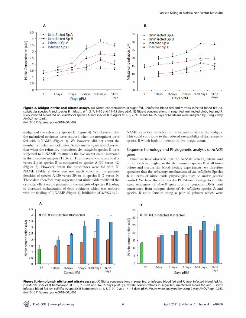
out to differentiate the sequence variations between susceptible
and refractory sibling species of An. culicifacies. In order to test the
homology of nitric oxide synthase (NOS) enzyme in various strains
of An. culicifacies against An. stephensi, only those sequences were
selected using BLAST, which encoded co-factors of (NOS)
enzyme. We have designed primer complementary to An.
stephensi exon 1 region (200 bp) encoding co-factor heme
DOMAIN. Primers specific for those sequences were designed
manually and the quality of the oligos was checked using web
based software Primer 3. (http://frodo.wi.mit.edu/cgi-bin/
primer3/primer3_www.cgi). Primer sequences of forward and
backward region were 59 ATGAGGACCAACTATCGGG 39 and
59 GCCTTGGTGACAATGCTC 39 respectively. Selected PCR
products (200 bp) amplified from genomic DNA in mosquito pools
were cloned into the pUC18 at HINDIII sites and sequenced and
a comparative analysis of the nucleotide sequence from both
strains to others reported NOS was done using the ClustalW
BLAST homology analysis. (MacVector Version 7.0).
Dissections and RNA extractions. The mosquitoes were
dissected in a drop of ice-cold sterile DEPC-treated water and
midgut from 25–30 mosquitoes was pooled in 100 ml DEPC
treated PBS buffer at 270uC. The tissues were ground to
homogeneity with a batterydriven hand-held homogenizer using
DEPC-treated sterile grinder tips and processed for RNA
extraction. Total RNA was isolated from dissected 25–30
midguts of each sugarfed (SF), uninfected blood fed (UBF) and
P. vivax infected blood fed (IBF) mosquitoes at 0, 1, 3, 7 and 9–14
days pBM using RNeasy micro kit (Qiagen).
Semi-quantitative RT-PCR analysis. The relative transcript
abundance and expression of NOS in naive mosquitoes (An.
culicifacies species A and species B) was evaluated by semiquantitative
RT-PCR both in uninfected (UBF) and P. vivax infected blood fed
(IBF) mosquitoes using primers designed against An. stephensi at
various intervals of feeding as described previously. First strand
cDNA was synthesized by using oligo (dT) primer and transcriptor
Reverse transcriptase (Roche) according to manufacturer protocol.
AcNOS fragment was amplified by 35 cycles of PCR (94uC for
1 min, 50uC for 1 min and 72uC for 1.00 min) with the forward and
backward primers (same as used in PCR). Transcript abundance of
RNA in the midgut tissues of An. culicifacies species A and B were
normalized to the ribosome S7 protein gene fragment and only 25
cycles were used for PCR amplification in identical environment to
obtain a 200 bp amplicon. After amplification 10 ml PCR products
was analyzed by 1.8% agarose gel electrophoresis. The gel image
was photographed by video gel documentation system (Bio-Rad).
The semiquantitative RT-PCR images were evaluated and pixels in
respective bands were quantified using IPLab gel software for a 12
bit image. The results were expressed as a ratio calculated from the
integrated signal of AcNOS amplicon bands over ribosomal protein
S7 gene amplicon bands. For the mid-gut expression analysis, data
from three replicates were analysed by 2 way ANOVA.
Real Time RT-PCR analysis. Real time RT-PCR was
performed using SYBR Green RT-PCR kit (Roche Diagnostics,
USA) and Light Cycler 480 system (Roche Diagnostics, USA) to
measure relative transcript levels of AcNOS in An. culicifacies sp A
and An. culicifacies sp B mosquitoes. cDNA of both the species was
reverse-transcribed from 500 ng total RNA using oligo(dT) primer
and transcriptor Reverse transcriptase (Roche), following the
manufacturer’s instructions. Assays used a total reaction volume of
20 mL incorporating 9 ml master mix with SYBR green I and
10 mM of each primer and 2 ml cDNA. Signals were normalized to
the ribosome S7 protein gene fragment. The same conditions have
been taken for normalizer gene S7 RNA polymerase having
forward primer sequence 5’ GGTGTTCGGTTCCAAGGTGA 3’
and reverse primer sequence 5’ GGTGGTCTGCTGGTTCTT-
ATCC 3’. The forward and backward sequences of AcNOS primers
for PCR are 5’ ATGAGGACCAACTATCGGG 3’ and 5’
GCCTTGGTGACAATGCTC 3’ and PCR conditions were:
Initial denaturation and renaturation step were same for all
primers, i.e., 95uC for 5 min and 95uC for 30 sec, 50uC for
40 sec, 72uC for 30 sec of 40 cycles respectively. The fluorescence
acquisition temperature was 72uC for all genes. This assay was
performed thrice to minimize variations due to sample handling.
Amplification specificity was further validated by melting curve
analysis, generated at the end of each PCR reaction. A non-
template control (NTC) was run with every assay. Normalized data
were used to quantitate relative levels of a RNA in species A and
species B. The threshold cycles (Ct) were recorded for AcNOS and S-
7 amplicons during each experiment. Difference between the Ct of
S-7 and AcNOS or DCt was determined and the relative abundance
of AcNOS was calculated in different treatments using Comparative
Ct method using the formula 2-DDCt [40].
Results
Effect of P. vivax infection on AcNOS activityIn our endeavor to investigate the role of nitric oxide mediated
killing of the P. vivax parasite in the An. culicifacies species B, we
measured the specific activities of the nitric oxide synthase enzyme
in both the sensitive (An. culicifacies species A) and refractory
mosquitoes (An. culicifacies species B). We assessed the activities of
AcNOS post blood meal at 1, 3, 7, 9–14 days in mosquitoes by
feeding uninfected blood (Figure 1A). 0 day old sugar fed mosquito
midguts were taken as a control. In these experiments, the
enhanced activity of AcNOS even at day 0 in refractory species
suggested that NO physiology may play a role in refractoriness.
Data from blood feeding experiments showed that the AcNOS
specific activity of the enzyme is increased with the day’s pBM in
both species A and species B. However, the increase was much
more in species B as compared to species A. Compared to midgut
AcNOS activitiy in An. culicifacies species A (34.8 mmoles/
mgprotein/min, p,0.001), refractory species An. culicifacies species
B exhibited a far more increase (60 mmoles/mgprotein/min,
p,0.001) at 9–14 days pBM (Figure 1A). Thus, these data
suggested that nitric oxide mechanism could contribute to the
refractory phenotype of the An. culicifacies species B mosquitoes.
To assess the ability of P. vivax parasite to induce the AcNOS
activity, we have also measured and compared the specific
activities of the AcNOS in both the sensitive species A and
refractory species B by feeding P. vivax infected blood meal at 1, 3,
7, 9–14 days by membrane feeding method. Data from infected
blood feeding experiments showed that the AcNOS specific
activity of the enzyme is rapidly increased with the days post blood
meal. Compared to AcNOS activitiy in An. culicifacies species A
(26.4 mmoles/mgprotein/min, at day 1 vs. 48.2 mmoles/mgpro-
tein/min, at day 9–14; p,0.001), An. culicifacies species B exhibited
a far more increase (35.9 mmoles/mgprotein/min, at day 1 vs.
93.3 mmoles/mgprotein/min, at day 9–14; p,0.001) post infected
blood meal (Figure 1B).
We also tested the effect of L-NAME, a known inhibitor of nitric
oxide synthase enzyme, to inhibit this AcNOS activity by feeding
this inhibitor simultaneously with the infected blood meals to the
mosquito’s midguts. The enzyme activity was found to be
markedly inhibited by this L-NAME at all days (1, 3, 7, and 9–
14 days) of blood feeding in both species A and species B. The
significant inhibition in the specific activity (mmoles/mgprotein/
min) was however observed in the refractory species B at 9–14
days (93.3 vs. 30.9 p,0.001) (Figure 1B). Thus, these data
Parasite Killing in Malaria Non-Vector Mosquito
PLoS ONE | www.plosone.org 4 April 2011 | Volume 6 | Issue 4 | e18400

suggested that induction of the nitric oxide synthase activity is
blood and parasite induced which is inhibited by L-NAME.
Higher level of NOS during Plasmodium infection, further indicate
that refractory strains are under chronic state of hostile cyto-toxic
stress because of the production of reactive cytotoxic free radicals
viz NO2-, NO3
- etc. This induction of the enzyme activity in
mosquito midguts of An. culicifacies species B may be an inheritable
trait of refractory species and may lead to an increased production
of reactive nitrogen species namely nitrate and nitrites which are
the stable reaction products of NO and could contribute to the
killing of the parasite in midguts leading to the refractory
phenotype of the An. culicifacies species B mosquitoes.
Effect of infection on mid-gut and haemolymph NO2- and
NO3- levels
To investigate this possibility further, we have measured the
differences in nitrate and nitrite (NO2- and NO3
-) within the An.
culicifacies mid-gut and hemolymph during early sporogonic
development of P. vivax under semi-natural conditions of
transmission in both sensitive (Species A) and refractory species
(Species B). NO2- and NO3
- concentrations in Anopheles species B
were found to be significantly higher than the concentrations in
species A. Our data shows that mid-gut levels of NO2- and NO3
-
were significantly higher in Plasmodium-infected mosquitoes than in
uninfected mosquitoes at all time points, with the greatest relative
difference at 9 days pBM (Figure 2A and 2B). Levels at 9–10 days
pBM may be correlated with induction and a higher specific
activity in infected mosquitoes at this time (Figure 1B). At 14 days
pBM nitrate levels in species A & B were 15.11 6 1.73 mM and
28.6 6 1.54 mM respectively and nitrite levels were 1.98 6
0.32 mM and 3.48 6 0.18 mM respectively. NO2-/NO3
- midgut
concentration was appeared to be increased in infected mosquitoes
at different time points (Figure 2) and same pattern was observed
in AcNOS enzyme activity above, which indicates the direct
correlation between these two biochemical assays.
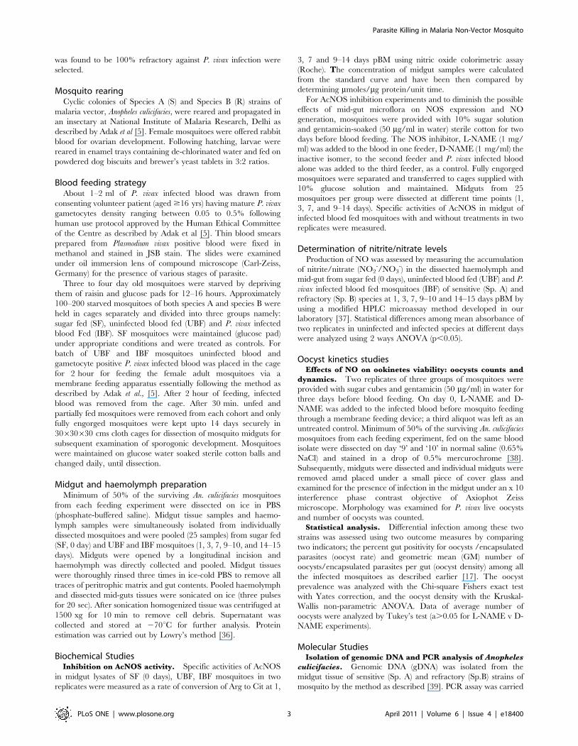
We have also determined the hemolymph NO2- and NO3
-
concentration in SF (0 day), UBF and IBF An. culicifacies B at
different time periods (1, 3, 7, 9–10 and 14–15 days).
Hemolymph NO2- and NO3
- concentration of blood fed P. vivax
infected species B were found to be higher than uninfected
mosquitoes This concentration of NO2- and NO3
- were found to
be time dependent and were proportionately higher in Plasmodium
infected mosquitoes than in uninfected mosquitoes at all time
points with the greatest relative difference at 9–10 days pBM
(Figure 3A and 3B).
Oocyst kineticsWe tested the ability of An. culicifacies species B to support
development of P. vivax. An. culicifacies species A mosquitoes were
used as a reference. Three to four-day-old female mosquitoes were
fed via a membrane with gametocytes infected P. vivax and 9-10
days later their midguts were dissected and the presence of oocyst
and the total parasites per midgut were recorded. The results from
two independent feeding experiments showed 0 of 22 (0/22) and 7
of 39 (7/39) midguts infected (0% and 17.9% oocyst infection
prevalence, respectively) in An. culicifacies species B and the
corresponding median oocyst densities were 0.0 and 2.0 in both
experiments (Table 1). In the paired feedings of An. culicifacies
species A 16/48 and 14/34 midguts had at least some viable
oocyst (33.3% and 41.1% oocyst infection prevalence, respectively)
with corresponding median oocyst densities of 9.0 and 12.0. The
live oocyst density was found to be much lower (P,0.001) in An.
culicifacies species B than in An. culicifacies species A. The
distributions of oocyst densities varied significantly (P,0.01)
between the two mosquito species, as one-third of An. culicifacies
species B were observed to have virtually no oocysts whereas
almost every An. culicifacies species A midgut had one or more
(Table 1). Clearance of pre-oocyst parasitic stages and melaniza-
tion of ookinetes are established important immune reactions of
mosquitoes against Plasmodium indicating that the inherent
mosquito immunity may be contributing to the reduced
susceptibility of An. culicifacies species B.
To support this conclusion, we examined the effect of L-NAME,
which appeared to interfere with induced AcNOS (Figure 1B) on P.
vivax development in mosquitoes. No much difference in
survivorship was observed across the treatments in species A.
We assessed the mosquito immunity in terms of NOS physiology
by measuring the effect of NOS inhibitor L-NAME on the
melanized ookinetee density in refractory species to determine
whether this is correlated to the killing of the oocysts in the
Figure 1. Specific activity in P. vivax uninfected and infected blood fed mosquitoes (with and without inhibitor). (A) Anophelesculicifacies nitric oxide synthase (AcNOS) specific activity in sugar fed (0 days) and uninfected blood fed mosquitoes midgut samples at different timepoints: 1, 3, 7, 9–14 days pBM in species A and species B. (B) An. culicifacies nitric oxide synthase (AcNOS) specific activity in P. vivax infected blood fedmosquitoes midgut samples at different time points: 1, 3, 7, 9–14 days pBM in species A and species B with and without L-NAME. L-NAME: N-nitro-L-arginine methyl ester, pBM:post blood meal.doi:10.1371/journal.pone.0018400.g001
Parasite Killing in Malaria Non-Vector Mosquito
PLoS ONE | www.plosone.org 5 April 2011 | Volume 6 | Issue 4 | e18400
midguts of the refractory species B (Figure 4). We observed that
the melanized ookinetes were reduced when the mosquitoes were
fed with L-NAME (Figure 4). We however, did not count the
number of melanized ookinetes. Simultaneously, we also observed
that when the refractory mosquitoes An. culicifacies species B were
subjected to L-NAME treatments the live oocyst count increased
in the mosquito midguts (Table 2). This increase was substantial (7
verses 21) in species B as compared to species A (30 verses 42)
(Figure 5). However, when the mosquitoes were fed with D-
NAME (Table 2) there was not much effect on the parasite
densities of species A (30 verses 39) or in species B (7 verses 9).
These data therefore may suggested that nitric oxide mediated the
cytotoxic effect on the parasites in the midguts of species B leading
to increased melanization of dead ookinetes which was reduced
with the feeding of L-NAME (Figure 4). Inhibition of AcNOS by L-
NAME leads to a reduction of nitrate and nitrites in the midguts.
This could contribute to the reduced susceptibility of An. culicifacies
species B which leads to increase in live oocyst count.
Sequence homology and Phylogenetic analysis of AcNOSgene
Since we have observed that the AcNOS activity, nitrate and
nitrite levels are higher in the An. culicifacies species B at all times
before and during the blood feeding experiments, we therefore
speculate that the refractory mechanisms of An. culicifacies Species
B in terms of nitric oxide physiologies may be under genetic
control. We have therefore used a PCR-based strategy to amplify
exon sequences of AcNOS gene from a genomic DNA pool
constructed from midguts tissue of An. culicifacies species A and
species B adult females using a pair of primers which were
Figure 3. Hemolymph nitrite and nitrate assays. (A) Nitrite concentrations in sugar fed, uninfected blood fed and P. vivax infected blood fed An.culicifacies species B hemolymph at 1, 3, 7, 9–10 and 14–15 days pBM. (B) Nitrate concentrations in sugar fed, uninfected blood fed and P. vivaxinfected blood fed An. culicifacies species B hemolymph at 1, 3, 7, 9–10 and 14–15 days pBM. Means were analyzed by using 2 way ANOVA (p,0.05).doi:10.1371/journal.pone.0018400.g003
Figure 2. Midgut nitrite and nitrate assays. (A) Nitrite concentrations in sugar fed, uninfected blood fed and P. vivax infected blood fed An.culicifacies species A and species B midguts at 1, 3, 7, 9–10 and 14–15 days pBM. (B) Nitrate concentrations in sugar fed, uninfected blood fed and P.vivax infected blood fed An. culicifacies species A and species B midguts at 1, 3, 7, 9–10 and 14–15 days pBM. Means were analyzed by using 2 wayANOVA (p,0.05).doi:10.1371/journal.pone.0018400.g002
Parasite Killing in Malaria Non-Vector Mosquito
PLoS ONE | www.plosone.org 6 April 2011 | Volume 6 | Issue 4 | e18400

designed from the conserved cofactor-binding domain of NOS
sequences from Anopheles stephensi.
Our data on sequencing and ClustalW alignment of the
amplified fragments revealed a high degree of sequence similarity
(100%) between An. culicifacies sp.A and sp.B for AcNOS genes
(accession number: FJ172998 and FJ172999 respectively)
(Figure 6). The amplified sequence encodes a 200 residue region
(Figure 6A) which revealed 33–100% identical at the amino acid
level to the corresponding region of these known NOS sequences,
as well as 100% identical to the recently isolated An. stephensi NOS
sequence (Figure 6B). A phylogenetic tree (Figure 6C) was
constructed on the basis of alignment of the partial AcNOS amino
acid sequence and the corresponding homologous regions of
several invertebrate and vertebrate NOS. Hence in the resulting
dendrogram An. culicifacies species A and species B were in same
clade and vertebrate NOS were in the different clade i.e. obtained
by the Neighbor-joining method (Figure 6C). The deduced amino
acid sequences of Monodelphis domestic NOS, Drosophila melanogaster
NOS, Rattus norvegicus NOS, Homo sapiens NOS, Anopheles stephensi
NOS and Anopheles culicifacies species A and species B NOS show
the highest level of homology to vertebrate neuronal NOS,
followed by decreasing homologies to vertebrate endothelial and
inducible NOS genes.
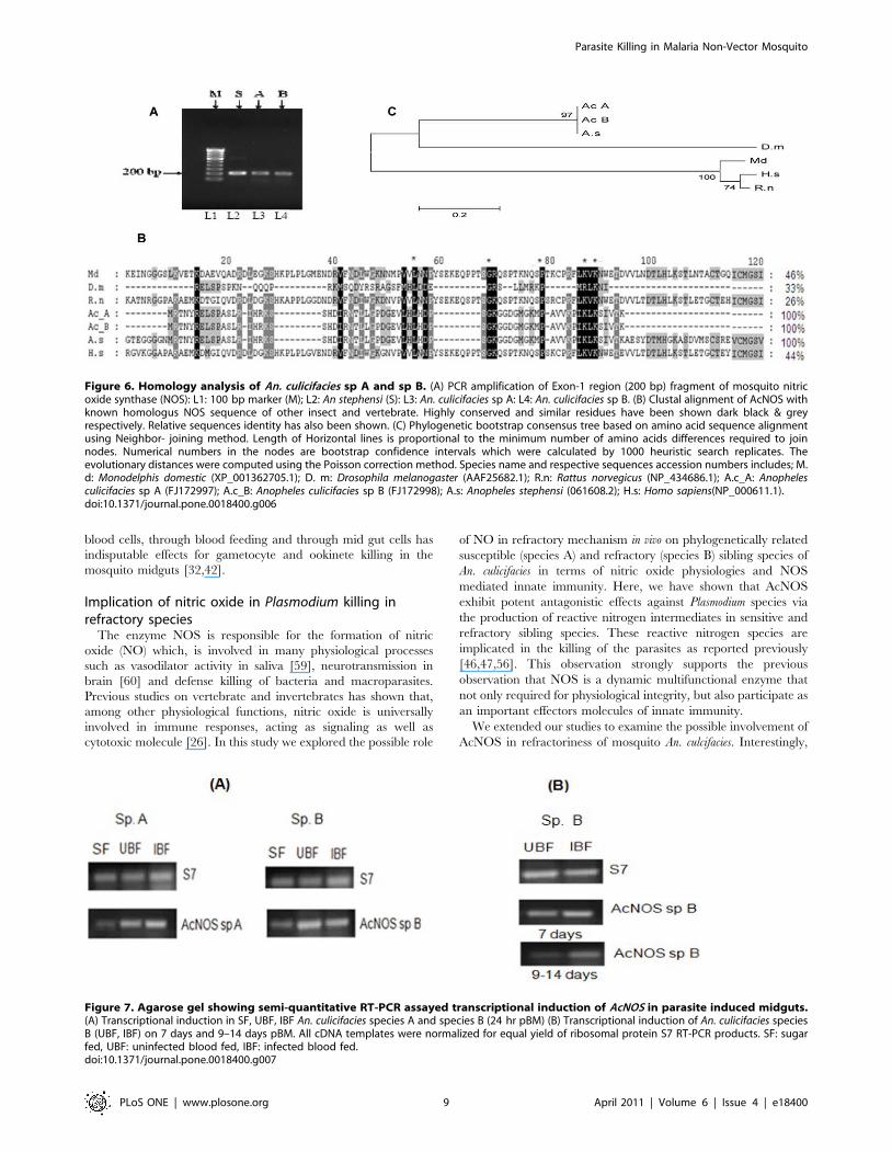
Differential expression of Nitric oxide synthase (AcNOS)Since it is not possible to correlate An. culicifacies species A and
An. culicifacies species B using PCR as both the species were found
to be identical, the relative expression level of AcNOS transcript in
both P. vivax infected sibling species was analyzed by using
primers designed against An. stephensi phenotypes by semi-
quantitative RT-PCR (Figure 7A). First we measured the basal
level of NOS expression pattern by semi-quantitative RT-PCR in
the midgut tissue collected at 24 hr post feeding using
constitutively expressing S7 Ribosome protein gene as an internal
control. As expected, in comparison to species A, we observed an
increased NOS expression observed in An. culicifacies species B
(Figure 7A).
NOS induction was seen at all stages of parasite development in
both species A and species B. Interestingly, NOS expression was
found to be much more in Anopheles species B at 7 days in
comparison to the 9–14 days (Figure 7B). At 9–14 days pBM,
however, when AcNOS expression was induced in infected
mosquitoes, the specific activity was 2-fold higher in infected
mosquitoes. Increase levels of NOS expression in mid-guts of
species B mosquitoes fed on P. vivax infected blood containing
parasites and gametocytes may reveal that early sporogonic stages
of P. vivax are able to increase NOS expression from day 1 to day
14. AcNOS was expressed constitutively and was transcriptionally
upregulated post blood meals in the refractory species B. A
proportional increase in transcript abundance of 12 fold from day
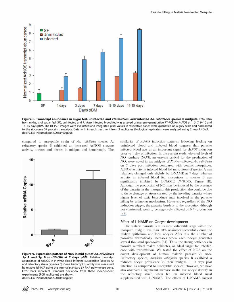
1 to day 14 was observed (Figure 8) through out the blood feeding
experiments in P. vivax infected mosquitoes species B.
Finally for a comparative study of the relative abundance of the
NOS transcripts in An. culicifacies susceptible and refractory species,
a real-time RT-PCR analysis was undertaken on samples from
midgut tissues at 7 days pBM. Temporal expression of AcNOS was
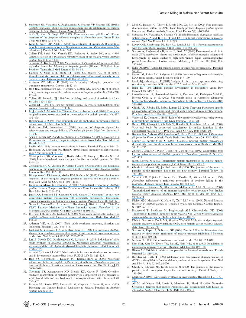
monitored by real time PCR after feeding of mosquitoes (Figure 9).
The threshold values (Ct) show that NOS transcript in midgut
tissue was 21.13 for An. culicifacies species A and 18.56 for An.
culicifacies species B. The lower Ct value of NOS for species B
indicates that NOS is more abundant in the midgut tissue of
species B. Thus, in An. culicifacies, unexpectedly we observed 11
fold induction of NOS gene in refractory species as compared to
susceptible species.
Table 1. Oocyst prevalence and Plasmodium vivax infection density in An. culicifacies species A and species B.
Experiment. Species n Oocyst prevalence (%) P Oocyst density Range P
1 An. culicifacies species A 48 27.08 (13/48) - 9.0 1–43 -
An. culicifacies species B 22 0 (0/22) ns 0.0 0 ,0.01
2 An. culicifacies species A 34 41.17 (14/34) - 12.0 1–48 -
An. culicifacies species B 39 17.94 (7/39) ,0.001 2 1–5 ,0.01
Mosquito mid-guts were examined for P. vivax live oocysts 9–14 days post infection. Oocyst prevalence is the percentage of mosquitoes displaying at least one liveoocyst, and Oocyst density is the median number of oocysts in infected mosquitoes infection of An. culicifacies species B was not observed in experiment 1. The oocystprevalence was analyzed with the chi-square Fishers exact test with Yates correction, and the oocyst density with the Kruskal –Wallis non-parametric ANOVA. n: numberof mosquitoes, ns: not significant.doi:10.1371/journal.pone.0018400.t001
Figure 4. Plasmodium parasite killing in An. culicifacies (species Aand species B). (A) Melanized ookinetes (arrows) of P. vivax insensitive An. culicifacies species A. (B) Melanized ookinetes (arrows) of P.vivax in refractory An. culicifacies species B while crossing the An.culicifacies midgut. (C) Melanized ookinete in the midguts of An.culicifacies species B (refractory) infected with P. vivax treated with NOSinhibitor L-NAME. Four independent paired experiments were per-formed. Treatment with L-NAME decreased the melanized ookinetescount (C) in comparison to without treatment (B).doi:10.1371/journal.pone.0018400.g004
Parasite Killing in Malaria Non-Vector Mosquito
PLoS ONE | www.plosone.org 7 April 2011 | Volume 6 | Issue 4 | e18400

Discussion
The Plasmodium undergoes a complex developmental interplay
during its lifecycle in mosquitoes. This interaction between vector
and parasite is essential for malaria transmission. The capacity of
mosquitoes to transmit malaria is determined by numerous factors
such as their longevity, feeding preference, and permissiveness to
parasite development in the mosquito midgut [41]. It is clear that
species specific mosquito-parasite strain combinations are essential
for the development of parasite in the mosquitoes. To this end, not
all mosquito-parasite strain combinations are compatible: some
Plasmodium strains are unable to develop in certain refractory
mosquito strains because of innate immune system in mosquitoes
that eliminate all ingested Plasmodia [12,16,22].
Most of the recent research efforts have focused mainly on
blocking parasite development in mosquitoes by targeting
gametocytes [32], ookinettes [42] in the mid guts of the
mosquitoes. Research concerning mid gut biology exploits parasite
entry mechanisms [43], melanization [44], and transcriptionally
activated mosquitoes immune response related antiplosmodial
genes [45] and free radicals namely ROS, RNS and nitric oxide
[46,47]. Advances in molecular biology have allowed us to
characterize mosquitoes that reduce Plasmodium transmission [14].
The development of genetically modified Anopheles mosquitoes and
transformation and availability of mosquitoes that exhibit
enhanced refractoriness to Plasmodium spp. is regarded as a model
for potential strategies for control of malaria transmission [48,49].
Refractory speciesNaturally occurring, malaria resistant strains are termed as
refractory mosquitoes and these are very rare in the field. Low
occurrence of such resistant phenotypes is attributed to acute
selection pressures on the innate immune system of mosquitoes.
Anopheles culicifaces, a rural Indian vector of malaria is a complex of
five sibling species, of which species B has been known to be a
refractory species [17]. Sibling species are phylogenetically closely
related to each other, are morphologically indistinguishable, and
can crossbreed in captivity; however, they vary greatly in their
capacity to transmit human malaria. The isolation of a wild An.
culicifaces strain B which is refractory to Plasmodium vivax infection
[6,17] presents an opportunity to use this laboratory model of
infection for research related to understanding the molecular basis
of refractoriness.
Induction of parasite killingMalaria parasites are often killed by various intracellular events
that may include nutrient deprivation, lysis or melanization, drug
treatments and the presence of nitric oxide (NO) and reactive
oxygen species (ROS) and nitrogen species (RNS) in the mosquito
midgut epithelium, which are controlled by reactions of the
mosquito innate immune system leading to refractoriness in
mosquitoes. Large losses in parasite numbers occurs at the
ookinete-to-oocyst transition stage of its life cycle [50,51]. These
losses are correlated with transcriptional activation of innate
immunity genes by malaria infection during invasion of epithelial
tissues and translocation to the salivary glands [52]. Three
molecules with recognition functions, TEP1, LR1M1 and APL1
also have parasite killing activity however, the mechanism is
incompletely understood [33,34,53]. The search for antiparasite
effectors in the refractory species Anopheles has identified some
promising targets in the immune-responsive mosquito, including
melanotic encapsulation [44,52,54], antimicrobial peptides such as
defensins [55], nitric oxide synthase [56]. NO or its derivatives,
play a role in the immunological reaction of the host defense
against the parasites [57]. NO has also been known to produce
nitrosative stress which may lead to apoptosis by activation of
mitochondrial apoptotic pathway [58]. These toxic molecules may
thus act triggers of apoptosis and elimination in Plasmodium killing
in refractory mosquitoes. The availability of NO through white
Table 2. Effect of L-NAME and D-NAME treatment on oocyst count in Anopheles culicifacies species A and species B.
Experiments Oocyst number/Midgut
Sugar + Gentamicin (50 mg/ml) L-NAME (1 mg/ml) D-NAME (1 mg/ml)
An. culicifacies species A 30 6 3.4 (47) 42 6 2.9 (49) 39 6 3.8 (50)
An. culicifacies species B 7 6 0.5a (22) 21 6 1.7b (26) 9 6 0.6a (25)
P. vivax infected blood fed mosquitoes were maintained on sugar and L-NAME or D-NAME treated or untreated water until dissection at 7 days pBM to count midgutoocysts. Figures in the table are depicted as average no. of oocysts per gut 6 SEM. Values in parentheses depict the total no. of midguts dissected. Data were analyzedby Tukey’s test (a.0.05 for L-NAME v D-NAME experiments, b,0.05 for L-NAME v D-NAME experiments). Significant differences are indicated by different letters. pBM:post blood meal, NAME: Nw-nitro-arginine methyl ester, SEM: standard error mean.doi:10.1371/journal.pone.0018400.t002
Figure 5. Live oocyst density in the midguts of An. culicifaciesspecies A and species B infected with P. vivax. The geometricmeans 6 SD of the pooled data from the two independent experimentsare shown (Biological replicates). The live oocyst densities (purple bars)were 3063.4 for An. culicifacies species A (n = 47) and 760.5 for An.culicifacies species B (blue bars) (n = 22; P,0.001), and the oocystdensities were 42 62.9 and 2161.7, respectively (P,0.001) followingthe L-NAME treatments (n: number of midguts). Oocyst mortalityincreased with the L-NAME treatment in both species A and species B.doi:10.1371/journal.pone.0018400.g005
Parasite Killing in Malaria Non-Vector Mosquito
PLoS ONE | www.plosone.org 8 April 2011 | Volume 6 | Issue 4 | e18400
blood cells, through blood feeding and through mid gut cells has
indisputable effects for gametocyte and ookinete killing in the
mosquito midguts [32,42].
Implication of nitric oxide in Plasmodium killing inrefractory species
The enzyme NOS is responsible for the formation of nitric
oxide (NO) which, is involved in many physiological processes
such as vasodilator activity in saliva [59], neurotransmission in
brain [60] and defense killing of bacteria and macroparasites.
Previous studies on vertebrate and invertebrates has shown that,
among other physiological functions, nitric oxide is universally
involved in immune responses, acting as signaling as well as
cytotoxic molecule [26]. In this study we explored the possible role
of NO in refractory mechanism in vivo on phylogenetically related
susceptible (species A) and refractory (species B) sibling species of
An. culicifacies in terms of nitric oxide physiologies and NOS
mediated innate immunity. Here, we have shown that AcNOS
exhibit potent antagonistic effects against Plasmodium species via
the production of reactive nitrogen intermediates in sensitive and
refractory sibling species. These reactive nitrogen species are
implicated in the killing of the parasites as reported previously
[46,47,56]. This observation strongly supports the previous
observation that NOS is a dynamic multifunctional enzyme that
not only required for physiological integrity, but also participate as
an important effectors molecules of innate immunity.
We extended our studies to examine the possible involvement of
AcNOS in refractoriness of mosquito An. culcifacies. Interestingly,
Figure 7. Agarose gel showing semi-quantitative RT-PCR assayed transcriptional induction of AcNOS in parasite induced midguts.(A) Transcriptional induction in SF, UBF, IBF An. culicifacies species A and species B (24 hr pBM) (B) Transcriptional induction of An. culicifacies speciesB (UBF, IBF) on 7 days and 9–14 days pBM. All cDNA templates were normalized for equal yield of ribosomal protein S7 RT-PCR products. SF: sugarfed, UBF: uninfected blood fed, IBF: infected blood fed.doi:10.1371/journal.pone.0018400.g007
Figure 6. Homology analysis of An. culicifacies sp A and sp B. (A) PCR amplification of Exon-1 region (200 bp) fragment of mosquito nitricoxide synthase (NOS): L1: 100 bp marker (M); L2: An stephensi (S): L3: An. culicifacies sp A: L4: An. culicifacies sp B. (B) Clustal alignment of AcNOS withknown homologus NOS sequence of other insect and vertebrate. Highly conserved and similar residues have been shown dark black & greyrespectively. Relative sequences identity has also been shown. (C) Phylogenetic bootstrap consensus tree based on amino acid sequence alignmentusing Neighbor- joining method. Length of Horizontal lines is proportional to the minimum number of amino acids differences required to joinnodes. Numerical numbers in the nodes are bootstrap confidence intervals which were calculated by 1000 heuristic search replicates. Theevolutionary distances were computed using the Poisson correction method. Species name and respective sequences accession numbers includes; M.d: Monodelphis domestic (XP_001362705.1); D. m: Drosophila melanogaster (AAF25682.1); R.n: Rattus norvegicus (NP_434686.1); A.c_A: Anophelesculicifacies sp A (FJ172997); A.c_B: Anopheles culicifacies sp B (FJ172998); A.s: Anopheles stephensi (061608.2); H.s: Homo sapiens(NP_000611.1).doi:10.1371/journal.pone.0018400.g006
Parasite Killing in Malaria Non-Vector Mosquito
PLoS ONE | www.plosone.org 9 April 2011 | Volume 6 | Issue 4 | e18400
compared to susceptible strain of An. culicifacies species A,
refractory species B exhibited an increased AcNOS enzyme
activity, nitrates and nitrites in midguts and hemolymph. The
similarity of AcNOS induction patterns following feeding on
uninfected blood and infected blood suggests that parasite
infected blood acts as an important signal for AcNOS induction
prior to 1 day of infection. In the current study, elevated levels of
NO synthase (NOS), an enzyme critical for the production of
NO, were noted in the midguts of P. vivax-infected An. culicifacies
on 7 days post infection compared with control mosquitoes.
AcNOS activity in infected blood fed mosquitoes of species A was
relatively changed only slightly by L-NAME at 7 days, whereas
activity in infected blood fed mosquitoes in species B was
significantly inhibited by L-NAME (P,0.005, Figure 1B).
Although the production of NO may be induced by the presence
of the parasite in the mosquito, this production also could be due
to tissue damage or stress created by the invading parasite where
higher level of toxic byproducts may involved in the parasite
killing by unknown mechanism. However, regardless of the NO
induction trigger, the parasite burdens in the mosquito, although
not eliminated, seem to be negatively affected by NO production
[25].
Effect of L-NAME on Oocyst developmentThe malaria parasite is at its most vulnerable stage within the
mosquito midgut, less than 10% ookinetes successfully cross the
midgut epithelium and form oocysts. After this, the number of
parasites dramatically increases when each oocyst generates
several thousand sporozoites [61]. Thus, the strong bottleneck in
parasite numbers makes ookinetes, an ideal target for interfer-
ence with transmission. We tested the effect of NOS on the
oocyst development of human malaria parasite P. vivax.
Refractory species, Anopheles culicifacies species B exhibited a
reduced oocyst prevelence in their midguts 9–10 days post
infection as compared to susceptible species. However, we have
also observed a significant increase in the live oocyst density in
the refractory strain when fed on infected blood meal
supplemented with L-NAME. The effects of L-NAME suggest
Figure 9. Expression pattern of NOS in mid gut of An. culicifaciesSp A and Sp B (n = 25–30) at 7 days pBM. Relative transcriptabundance of AcNOS in P. vivax blood infected susceptible (species A)and refractory strain (species B). Gene transcript quantity was measuredby relative RT-PCR using the internal standard S7 RNA polymerase gene.Error bars represent standard deviation from three independentexperiments (PCR replicates) are shown.doi:10.1371/journal.pone.0018400.g009
Figure 8. Transcript abundance in sugar fed, uninfected and Plasmodium vivax infected An. culicifacies species B midguts. Total RNAfrom midguts of sugar fed (SF), uninfected and P. vivax infected blood fed was assayed using semi-quantitative RT-PCR for AcNOS at 1, 3, 7, 9–10 and14–15 days pBM. The RT-PCR images were evaluated and integrated pixel values in respective bands were quantified on a grey scale and normalizedto the ribosome S7 protein transcripts. Data with in each treatment from 3 replicates (biological replicates) were analyzed using 2 way ANOVA.doi:10.1371/journal.pone.0018400.g008
Parasite Killing in Malaria Non-Vector Mosquito
PLoS ONE | www.plosone.org 10 April 2011 | Volume 6 | Issue 4 | e18400

that parasites are targeted before or during the oocyst
development, a hypothesis which seems to be consistent with
the demonstrated susceptibility of parasites to NO damage [56].
NOS-specific inhibitor specifically fully rescued the susceptibility
phenotype in refractory mosquitoes, causing an approximate 4-
fold increase in the oocyst density, and 71% increase in oocyst
prevalence. The increase in the number of oocysts observed in
response to infected blood in An. culicifacies species A could be an
amplified midgut response to parasite infection: however, this
midgut response seems to be significant in An. culicifacies species
B. Furthermore, feeding of NOS inhibitor L-NAME, increases
mean oocyst infections while D-NAME, the inactive isomer has
no effect (Table 2). Luckhart et al [28] has reported similar effect
when An. stephensi mosquitoes were fed on L-NAME and D-
NAME. A decreased oocyst infection in An. culicifacies species B
(Table 2) may be attributed to the susceptibility of P. vivax to
Anopheles culicifacies midgut stages to nitric oxide (NO). These data
clearly demonstrated that AcNOS could be an important player in
the development of refractory nature of the mosquito and need
to be explored further.
Transcriptional upregulation of AcNOS geneOur knowledge of NOS function in invertebrates is still limited
[62], and the gene has only been cloned from a few insect species
[28,60]. In view of the multiple physiological roles of NO, it is
quite possible that the effects on NOS will prove to be related to
the success of parasite infection [28]. Our sequence analysis
results of the amplified fragment of AcNOS gene revealed a high
degree of sequence similarity between An. culicifacies species A and
species B. This was not surprising as the two species are very
closely related in the evolutionary scale and genetic introgression
has likely taken place for some time after their separation and
may be regarded more as a reflection of gene ancestry than
functional activity [54].
To evaluate the temporal expression of AcNOS gene in An.
culicifaces species A and species B experiments were designed to
determine whether P. vivax parasite could transcriptionally induce
the upregulation of NOS gene by semiquantitative RT-PCR and
real time PCR analysis. Our data have concluded that AcNOS
gene is more abundantly expressed in midgut of species B than in
species A. We show that NOS expression is transcriptionally
upregulated in the midgut in response to Plasmodium infection and
induction of NOS is proportional to the intensity of infection
[63]. Our results show that An. culicifaces does have a shared
evolutionary history with P. vivax and thus in principle host
resistance mechanisms, including AcNOS, could have been
selected by the parasite, although to date this have not been
proven. We hypothesize that upregulation of mosquito innate
cytotoxicity due to NOS in refractory strain to Plasmodium vivax
infection may contribute to natural refractoriness in An. culicifacies
mosquito population. This innate capacity of refractory mosqui-
toes could represent the ancestral function of the mosquito
immune system against the parasite and could be utilized to
understand the molecular basis of refractoriness in planning
effective vector control strategies. A better understanding of the
natural mechanisms of host defense against the Plasmodium
parasite may provide new targets for therapeutic intervention in
this disease.
ConclusionsAttention has largely focused on mosquitoes innate immune
responses that may lead to cytotoxic killing, lysis and melanization
of the parasites. NO, RNS and ROS are known to kill parasites in
oxidation-reduction reactions and may play a role in the refractory
mechanisms. However, the importance of NOS activity in
inducing cytotoxic killing of the parasites needs to be ascertained
in order to show the feasibility to augment the activity of NOS for
killing of the parasites. In this study, we have utilized a naturally
selected non-vector An. culicifacies species B in conjunction with the
An. culicifacies species A as a model vector system, to understand the
differences contributing to its reduced vectorial capacity. The
phenotype of this refractory An. culicifacies species B strain is
identical to that of other An. culicifacies sibling species complex.
Dissecting the molecular basis of refractoriness in Anopheles
culicifacies model system may pave the way to novel disease control
mechanisms. Experimental evidences relating to increased NOS
activity and reduced oocyst development do suggest the evolu-
tionary significance of the existence of a cytotoxic innate immunity
system operating in refractory species. It is therefore, tempting to
speculate that persistent interaction of An. culicifacies with P. vivax
might have led to an evolutionary co adaptation between the
mosquito immune responses and this parasite, whereas the
refractory phenotype could represent the ancestral function of
the mosquito immune system against the parasite. This immune
system could operate via triggering of mosquito AcNOS activity by
the Plasmodium vivax dependent manner. The latter implying that
AcNOS cytotoxic mechanism of parasite killing in the mosquito
midgut lumen could be an important transmission blocking
strategy. Based on the present data on Anopheles culicifacies NOS
which has been shown to be transcriptionally regulated, we believe
that the study of this gene is a promising approach to unravel yet
unknown NOS-dependent production of nitric oxide elements,
leading to a better insight in different aspects of insect physiology
in terms of refractoriness. Future research will aim to determine if
any of these changes or any cofactors to the enzyme AcNOS can
enhance or otherwise alter the function of this AcNOS gene and
gene elements, thus contributing to the refractoriness phenotype
for novel malaria control strategies.
Acknowledgments
We are grateful to all scientists and entomology teams whose contributions
to the study of An. culicifacies sibling species and their molecular forms made
this study possible; we especially thank Bhanu Arya, Technical Officer and
Mrs. Poonam Gupta, Technical Assistant for HPLC analysis and
enzymatic analysis and experimental work. We also thank Alakh Deo
Prasad, Insect Collector for help with sample collections and mosquito
dissections and Mr. Pratap and Laxman for help in mosquito membrane
feeding experiments. We are grateful to Dr. Shashi Sharma of ICPO,
NOIDA for useful and enlightening discussions regarding statistical
analysis.
Author Contributions
Conceived and designed the experiments: AS RD TA. Performed the
experiments: SV MR NN HS. Analyzed the data: SV RD JS AS GBKS.
Provided facilities and scientific environment for experimental work and
wrote the paper: AS. All authors read and approved the final manuscript.
References
1. Sharma VP (1999) Current scenario of malaria in India. Parassitologia 41: 349–353.
2. Subbarao SK (1998) Anopheline species complexes in South-east Asia,
Technical publication, SEARO No.18, World Health Organization Regional
Office for South-East Asia, New Delhi.
3. Subbarao SK, Adak T, Vasantha K, Joshi H, Raghvendra K, et al. (1988)
Susceptibility of Anopheles culicifacies species A and B to Plasmodium vivax and
Plasmodium falciparum as determined by immunoradiometric assays. Trans R Soc
Trop Med Hyg 82: 394–397.
Parasite Killing in Malaria Non-Vector Mosquito
PLoS ONE | www.plosone.org 11 April 2011 | Volume 6 | Issue 4 | e18400
4. Subbarao SK, Vasantha K, Raghavendra K, Sharma VP, Sharma GK (1988a)
Anopheles culicifacies: sibling species composition and its relationship to malariaincidence. J. Am. Mosq. Control Assoc 4: 29–33.
5. Adak T, Kaur S, Singh OP (1999) Comparative susceptibility of different
members of the Anopheles culicifacies complex to Plasmodium vivax. Trans R SocTrop Med Hyg 93: 573–577.
6. Kaur S, Singh OP, Adak T (2000) Susceptibility of species A, B and C ofAnopheles culicifacies complex to Plasmodiumyoelii yoelii and Plasmodium vinckei petteri
infections. J Parasitol 86: 1345–1348.
7. Collins FH, Sakai RK, Vernick KD, Paskewitz S, Seeley DC, et al. (1986)Genetic selection of a Plasmodium-refractory strain of the malaria vector Anopheles
gambiae. Sci 234: 607–610.8. Schwartz A, Koella JC (2002) Melanization of Plasmodium falciparum and C-25
sephadex beads by field-caught Anopheles gambiae (Diptera, Culicidae) fromSouthern Tanzania. J Med Entomol 39: 84–88.
9. Blandin S, Shiao S-H, Moita LF, Janse CJ, Waters AP, et al. (2004)
Complement-like protein TEP1 is a determinant of vectorial capacity in themalaria vector Anopheles gambiae. Cell 116: 661–670.
10. Atkinson PW, Michel K (2002) what’s buzzing? Mosquito genomics andtransgenic mosquitoes. Genesis 32: 42–48.
11. Holt RA, Subramanian GM, Halpern A, Sutton GG, Charlab R, et al. (2002)
The genome sequence of the malaria mosquito Anopheles gambiae. Sci 298;5591):129–49.
12. Collins FH, Besansky NJ (1994) Vector biology and control of malaria in Africa.Sci 264: 1874–1875.
13. Curtis CF (1994) The case for malaria control by genetic manipulation of itsvectors. Parasitol Today 10: 371–374.
14. Ito J, Ghosh A, Moreira LA, Wimmer EA, Jacobs-Lorena M (2002) Transgenic
anopheline mosquitoes impaired in transmission of a malaria parasite. Nat 417:452–455.
15. Dimopoulos G (2003) Insect immunity and its implication in mosquito-malariainteractions. Cell Microbiol 5: 3–14.
16. Feldmann AM, Ponnudurai T (1989) Selection of Anopheles stephensi for
refractoriness and susceptibility to Plasmodium falciparum. Med. Vet Entomol 3:41–52.
17. Adak T, Singh OP, Nanda N, Sharma VP, Subbarao SK (2006) Isolation of aPlasmodium vivax refractory Anopheles culicifacies strain from India. Trop Med Intl
Health 2: 1–7.18. Lackie AM (1988) Immune mechanisms in insects. Parasitol Today 4: 98–105.
19. Hoffmann JA, Reichhart JM, Hetru C (1996) Innate immunity in higher insects.
Curr Opin Immunol 8: 8–13.20. Christophides GK, Zdobnov E, Barillas-Mury C, Birney E, Blandin S, et al.
(2002) Immunity-related genes and gene families in Anopheles gambiae. Sci 298:159–165.
21. Christophides GK, Vlachou D, Kafatos FC (2004) Comparative and functional
genomics of the innate immune system in the malaria vector Anopheles gambiae.Immunol Rev 198: 127–148.
22. Dimopoulos G, Richman A, Muller HM, Kafatos FC (1997) Molecular immuneresponses of the mosquito Anopheles gambiae to bacteria and malaria parasites.
Proc Natl Acad Sci USA 94: 11508–11513.23. Blandin SA, Marois E, Levashina EA (2008) Antimalarial Responses in Anopheles
gambiae: From a Complement-like Protein to a Complement-like Pathway. Cell
Host Microbe 3: 6364–374.24. James AA, Beerntsen BT, Capurro M deL, Coates CJ, Coleman J, et al. (1999)
Controlling malaria transmission with genetically engineered, Plasmodium-resistant mosquitoes: milestones in a model system. Parassitologia 41: 461–471.
25. Gupta L, Molina-Cruz A, Kumar S, Rodrigues J, Dixit R, et al. (2009) The
STAT Pathway Mediates Late-Phase Immunity against Plasmodium in theMosquito Anopheles gambiae. Cell Host Microbe 5: 498–507.
26. Peterson TM, Gow AJ, Luckhart S (2007) Nitric oxide metabolites induced inAnopheles stephensi control malaria parasite infection. Free Radic Biol Med 42:
132–42.
27. Alderton WK, et al. (2001) Nitric oxide synthases: structure, function andinhibition. Biochem J 357: 593–615.
28. Luckhart S, Vodovotz Y, Cui L, Rosenberg R (1998) The mosquito Anopheles
stephensi limits malaria parasite development with inducible synthesis of nitric
oxide. Proc Natl Acad Sci USA 95: 5700–5705.29. Lim J, Gowda DC, Krishnegowda G, Luckhart S (2005) Induction of nitric
oxide synthase in Anopheles stephensi by Plasmodium falciparum: mechanism of
signaling and the role of parasite glycosylphosphatidylinositols. Infect Immun 73:2778–2789.
30. Ascenzi P, Gradoni L (2002) Nitric oxide limits parasite development in vectorsand in invertebrate intermediate hosts. IUBMB Life 53: 121–123.
31. Han YS, Thompson J, Kafatos FC, Barillas-Mury C (2000) Molecular
interactions between Anopheles stephensi midgut cells and Plasmodium berghei: thetime bomb theory of ookinete invasion of mosquitoes. Eur Mol Bio Org J 19:
6030–6040.32. Naotunne TS, Karunaweera ND, Mendis KN, Carter R (1993) Cytokine-
mediated inactivation of malarial gametocytes is dependent on the presence ofwhite blood cells and involved reactive nitrogen intermediates. Immunol 78:
555–562.
33. Blandin SA, Sattler RW, Lamacchia M, Gagneur J, Lycett G, et al. (2009)Dissecting the Genetic Basis of Resistance to Malaria Parasites in Anopheles
gambiae. Sci 326: 147.
34. Mitri C, Jacques JC, Thiery I, Riehle MM, Xu J, et al. (2009) Fine pathogen
discrimination within the APL1 Gene family protects Anopheles gambiae against
Human and Rodent malaria Species. PLoS Pathog 5(9): e1000576.
35. Subbarao SK, Vasantha K, Sharma VP (1988b) Responses of Anopheles culicifacies
sibling species A and B to DDT and HCH in India: implications in malaria
control. Med Vet Entomol 2: 219–223.
36. Lowry OH, Rosebrough NJ, Farr AL, Randall RJ (1951) Protein measurement
with the folin phenol reagent. J Biol Chem 193: 265–75.
37. Sharma A, Raghavendra K, Adak T, Dash AP (2008) Determination of nitric
oxide (NO) metabolites, nitrate and nitrite in An. culicifacies mosquito midgut and
hemolymph by anion exchange high-performance liquid chromatography:
plausible mechanism of refractoriness. Malaria. J 7: 71. doi: 10.1186/1475-
2875-7-71.
38. Eyles DE (1950) A stain for malaria oocysts in temporary preparations. J Parasitol
36: 501.
39. Henry JM, Raina AK, Ridgway RL (1990) Isolation of high-molecular-weight
DNA from insects. Analyt Biochem 185: 147–150.
40. Livak, KJ, Schmittgen TD (2001) Analysis of relative gene expression data using
real time quantitative PCR and the 2-DDCt method. Met 25, 402.
41. Beier JC (1998) Malaria parasite development in mosquitoes. Annu Rev
Entomol 43: 519–543.
42. Lanz-Mendoza H, Hernandez-Martinez S, Ku-Lopez M, Rodriguez Mdel C,
Herrera-Ortiz A, et al. (2002) Superoxide anion in Anopheles albimanus
hemolymph and midgut is toxic to Plasmodium berghei ookinetes. J Parasitol 88:
702–706.
43. Ghosh AK, Ribolla PE, Jacbos-Lorena M (2001) Targeting Plasmodium ligands
on mosquito salivary glands and midgut with a phage display peptide library.
Proc Natl Acad Sci USA 98: 13278–81.
44. Soderhall K, Cerenius L (1998) Role of the prophenoloxidase-activating system
in invertebrate immunity. Curr Opin Immunol 19: 23–28.
45. Baxter RH, Chang CI, Chelliah Y, Blandin S, Levashina EA, et al. (2007)
Structural basis for conserved complement factor-like function in the
antimalarial protein TEP1. Proc Natl Acad Sci USA 104: 11615–20.
46. Rockett KA, Awburn MM, Cowden WB, Clark IA (1991) Killing of Plasmodium
falciparum In Vitro by Nitric Oxide Derivatives. Infect Imm 59: 3280–3283.
47. Kumar S, Barillas-Mury C (2005) Ookinete-induced midgut peroxidases
detonate the time bomb in Anopheline mosquitoes. Insect Biochem Mol Biol
35: 721–727.
48. Zheng L, Cornel AJ, Wang R, Erfle H, Voss H, et al. (1997) Quantitative trait
loci for refractoriness of Anopheles gambiae to Plasmodium cynomolgi B. Sci 276:
425–428.
49. Jacobs-Lorena M (2003) Interrupting malaria transmission by genetic manip-
ulation of anopheline mosquitoes. J Vect Borne Dis 40: 73–77.
50. Ghosh A, Edwards MJ, Jacobs-Lorena M (2000) The journey of the malaria
parasite in the mosquito: hopes for the new century. Parasitol Today 16:
196–201.
51. Vernick KD, Fujioka H, Seeley DC, Tandler B, Aikawa M, et al. (1995)
Plasmodium gallinaceum: a refractory mechanism of ookinete killing in the
mosquito, Anopheles gambiae. Exp Parasitol 80: 583–595.
52. Rodrigues J, Agarwal N, Sharma A, Malhotra P, Adak T, et al. (2007)
Transcriptional analysis of an immune-responsive serine protease from Indian
malarial vector, Anopheles culicifacies. BMC Mol Bio 8: 33. doi: 10.1186/1471-
2199-8-33.
53. Riehle MM, Markianos K, Niare O, Xu J, Li J, et al. (2006) Natural Malaria
Infection in Anopheles gambiae Is Regulated by a Single Genomic Control Region.
Sci 312: 577–579.
54. Habtewold T, Povelones M, Blagborough AM, Christophides GK (2008)
Transmission Blocking Immunity in the Malaria Non-Vector Mosquito Anopheles
quadriannulatus Species A. PLoS Pathog 4(5): e1000070.
55. Dixit R, Sharma A, Patole MS, Shouche YS (2008) Molecular and phylogenetic
analysis of a novel salivary defensin cDNA from malaria vector Anopheles stephensi.
Acta Trop 106: 75–79.
56. Sharma A, Eapen A, Subbarao SK (2004) Parasite killing in Plasmodium vivax
malaria by nitric oxide: implication of aspartic protease inhibition. J Biochem
(Tokyo) 136: 329–34.
57. Nathan C (1995) Natural-resistance and nitric oxide. Cell 82: 873–876.
58. Kim KM, Kim PK, Kwon YG, Bai SK, Nam WD, et al. (2002) Regulation of
apoptosis by nitrosative stress. J Biochem Mol Biol 35: 127–133.
59. Rivero A (2006) Nitric oxide: an antiparasitic molecule of invertebrates. Trends
Parasitol 22: 219–225.
60. Regulski M, Tully T (1995) Molecular and biochemical characterization of
dNOS: a Drosophila Ca2+/calmodulin-dependent nitric oxide synthase. Proc Natl
Acad Sci U S A 92: 9072–9076.
61. Ghosh A, Edwards MJ, Jacobs-Lorena M (2000) The journey of the malaria
parasite in the mosquito: hopes for the new century. Parasitol Today 16:
196–201.
62. Martinez A (1995) Nitric oxide synthase in invertebrates. Histochem J 27: 770–
776.
63. Ali M, Al-Olayan EM, Lewis S, Matthews H, Hurd H (2010) Naturally
Occurring Triggers that Induce Apoptosis-Like Programmed Cell Death in
Plasmodium berghei Ookinetes. PLoS ONE 5(9): e12634.
Parasite Killing in Malaria Non-Vector Mosquito
PLoS ONE | www.plosone.org 12 April 2011 | Volume 6 | Issue 4 | e18400