Gibberellin Signaling in the Endodermis Controls Arabidopsis Root Meristem Size
6
Current Biology 19, 1194–1199, July 28, 2009 ª2009 Elsevier Ltd All rights reserved DOI 10.1016/j.cub.2009.06.023 Report Gibberellin Signaling in the Endodermis Controls Arabidopsis Root Meristem Size Susana Ubeda-Toma ´s, 1,9, * Ferna ´ n Federici, 2,9 Ilda Casimiro, 3 Gerrit T.S. Beemster, 4,5,6 Rishikesh Bhalerao, 7 Ranjan Swarup, 1 Peter Doerner, 8 Jim Haseloff, 2 and Malcolm J. Bennett 1, * 1 Centre for Plant Integrative Biology, University of Nottingham, Nottingham LE12 5RD, UK 2 Department of Plant Sciences, University of Cambridge, Cambridge CB2 3EA, UK 3 Universidad de Extremadura, Facultad de Ciencias, Badajoz 06071, Spain 4 Department of Plant Systems Biology, Flanders Institute for Biotechnology (VIB), 9052 Gent, Belgium 5 Department of Biology, University of Antwerp, 2020 Antwerp, Belgium 6 Department of Plant Biotechnology and Genetics, Gent University, 9052 Gent, Belgium 7 Umea ˚ Plant Science Centre, Department of Forest Genetics and Plant Physiology, SLU, SE-901 83 Umea ˚ , Sweden 8 Institute for Molecular Plant Sciences, School of Biological Sciences, University of Edinburgh, Edinburgh EH9 3JH, UK Summary Plant growth is driven by cell proliferation and elongation [1]. The hormone gibberellin (GA) regulates Arabidopsis root growth [2–5] by controlling cell elongation [6], but it is currently unknown whether GA also controls root cell prolif- eration. Here we show that GA biosynthetic mutants are unable to increase their cell production rate and meristem size after germination. GA signals the degradation of the DELLA growth repressor proteins [7–12] GAI and RGA, promoting root cell production. Targeting the expression of gai (a non-GA-degradable mutant form of GAI) in the root meristem disrupts cell proliferation. Moreover, express- ing gai in dividing endodermal cells was sufficient to block root meristem enlargement. We report a novel function for GA regulating cell proliferation where this signal acts by removing DELLA in a subset of, rather than all, meristem cells. We suggest that the GA-regulated rate of expansion of dividing endodermal cells dictates the equivalent rate in other root tissues. Cells must double in size prior to dividing but cannot do so independently, because they are physically restrained by adjacent tissues with which they share cell walls. Our study highlights the importance of probing regu- latory mechanisms linking molecular- and cellular-scale processes with tissue and organ growth responses. Results and Discussion A germinating seedling must obtain anchorage, water, and nutrients after emerging from its seed coat. Vigorous root growth is essential if the seedling is to rapidly secure these resources. Root length is determined by the number of dividing cells and their final cell size [1]. Root cells first undergo repeated rounds of division in the root proximal meristem and then subsequently experience rapid cell expansion in the elon- gation-differentiation zone (EDZ; Figure 1A). In order to maxi- mize root growth after germination, seedlings could increase either root cell production rate or final cell size or both. In the model plant Arabidopsis thaliana, root growth rate rapidly increases after seed germination (Figure 1B; Figure S1A available online). We initially investigated the cellular basis for the increased root growth, measuring mature cell length, cell production in the meristem, and root meristem size. Mature cell size was essentially constant, whereas the rate of root cell production increased proportional to the root growth rate; wild-type roots exhibit a doubling in cell production rate over a 4 day period after germination (Figure 1C; Figures S1D and S1F). Increased cell production can be due to an increasing number of cells in the meristem because extensive variations in cell division rates are relatively rare [13]. Indeed, our measurements indicate that root meristem size doubles during the first days after germination (DAG), then plateaus by 4–6 DAG, depending on the Arabidopsis accession (Figures S1D–S1F). Similarly, cortical cell numbers in the root meristem reached a constant number of between 40 and 60 cells, depending on the Arabidopsis accession (Figures S1E and S1G). Hence, the acceleration in Arabidopsis root growth is correlated with increasing root meristem size and numbers of meristematic cells (Figure 1B; Figure S1). The plant hormone gibberellin (GA) represents an important regulator of Arabidopsis root growth [2–5]. GA has recently been reported to regulate root cell elongation [6]. However, it is currently unclear whether GA also controls root cell produc- tion. To investigate this possibility, we used pharmacological and genetic approaches to test the role of GA in regulation of root meristem size. Reduction of endogenous GA levels by treating wild-type seedlings with Paclobutrazol (PAC, an inhibitor of GA biosynthesis [14]) results in a reduced root growth rate (Figure S1A). Detailed microscopy measurements revealed that PAC treatment caused a reduction in root meri- stem size (Figures S1D–S1G) and mature cell length (Figures S1B and S1C). Subsequently, roots of GA biosynthetic mutants ga1-3 [15] and ga3ox1/ga3ox2 [16] were analyzed. The ga1-3 mutant failed to increase cell production rate, essentially remaining static (Figure 1C). Similarly, a reduction in cell production rate was also detected in ga3ox1/ga3ox2 mutant (Figure S1K). Both mutants exhibited a smaller root meristem size compared to wild-type (Figures 1B–1D; Figure S1H). However, GA3 treatment was able to fully rescue root meristem size in the ga1-3, ga3ox1/ga3ox2 mutants and PAC-treated seedlings (Figure 1B; Figures S1D–S1H). Conversely, removal of 7- to 8-day-old ga1-3 seedlings from GA3-supplemented media resulted in a reduction in root meristem size (Figure S1L). Hence, GA levels appear to be required to promote and main- tain the increase in root growth rate through control of root meristem size. To investigate the relationship between gibberellins and root cell division, we monitored how changes in GA levels affect the expression of the mitotic cyclin CycB1;1 (marks G2/M-phase *Correspondence: [email protected] (S.U-T.), [email protected] (M.J.B.) 9 These authors contributed equally to this work
Transcript of Gibberellin Signaling in the Endodermis Controls Arabidopsis Root Meristem Size
Current Biology 19, 1194–1199, July 28, 2009 ª2009 Elsevier Ltd All rights reserved DOI 10.1016/j.cub.2009.06.023
ReportGibberellin Signaling in the EndodermisControls Arabidopsis Root Meristem Size
Susana Ubeda-Tomas,1,9,* Fernan Federici,2,9 Ilda Casimiro,3
Gerrit T.S. Beemster,4,5,6 Rishikesh Bhalerao,7
Ranjan Swarup,1 Peter Doerner,8 Jim Haseloff,2
and Malcolm J. Bennett1,*1Centre for Plant Integrative Biology, University of Nottingham,Nottingham LE12 5RD, UK2Department of Plant Sciences, University of Cambridge,Cambridge CB2 3EA, UK3Universidad de Extremadura, Facultad de Ciencias, Badajoz06071, Spain4Department of Plant Systems Biology, Flanders Institute forBiotechnology (VIB), 9052 Gent, Belgium5Department of Biology, University of Antwerp, 2020 Antwerp,Belgium6Department of Plant Biotechnology and Genetics, GentUniversity, 9052 Gent, Belgium7Umea Plant Science Centre, Department of Forest Geneticsand Plant Physiology, SLU, SE-901 83 Umea, Sweden8Institute for Molecular Plant Sciences, School of BiologicalSciences, University of Edinburgh, Edinburgh EH9 3JH, UK
Summary
Plant growth is driven by cell proliferation and elongation
[1]. The hormone gibberellin (GA) regulates Arabidopsisroot growth [2–5] by controlling cell elongation [6], but it is
currently unknown whether GA also controls root cell prolif-
eration. Here we show that GA biosynthetic mutants areunable to increase their cell production rate and meristem
size after germination. GA signals the degradation of theDELLA growth repressor proteins [7–12] GAI and RGA,
promoting root cell production. Targeting the expressionof gai (a non-GA-degradable mutant form of GAI) in the
root meristem disrupts cell proliferation. Moreover, express-ing gai in dividing endodermal cells was sufficient to block
root meristem enlargement. We report a novel function forGA regulating cell proliferation where this signal acts by
removing DELLA in a subset of, rather than all, meristemcells. We suggest that the GA-regulated rate of expansion
of dividing endodermal cells dictates the equivalent rate inother root tissues. Cells must double in size prior to dividing
but cannot do so independently, because they are physicallyrestrained by adjacent tissues with which they share cell
walls. Our study highlights the importance of probing regu-latory mechanisms linking molecular- and cellular-scale
processes with tissue and organ growth responses.
Results and Discussion
A germinating seedling must obtain anchorage, water, andnutrients after emerging from its seed coat. Vigorous rootgrowth is essential if the seedling is to rapidly secure these
*Correspondence: [email protected] (S.U-T.),
[email protected] (M.J.B.)9These authors contributed equally to this work
resources. Root length is determined by the number ofdividing cells and their final cell size [1]. Root cells first undergorepeated rounds of division in the root proximal meristem andthen subsequently experience rapid cell expansion in the elon-gation-differentiation zone (EDZ; Figure 1A). In order to maxi-mize root growth after germination, seedlings could increaseeither root cell production rate or final cell size or both.
In the model plant Arabidopsis thaliana, root growth raterapidly increases after seed germination (Figure 1B; Figure S1Aavailable online). We initially investigated the cellular basis forthe increased root growth, measuring mature cell length, cellproduction in the meristem, and root meristem size. Maturecell size was essentially constant, whereas the rate of rootcell production increased proportional to the root growthrate; wild-type roots exhibit a doubling in cell production rateover a 4 day period after germination (Figure 1C; FiguresS1D and S1F). Increased cell production can be due to anincreasing number of cells in the meristem because extensivevariations in cell division rates are relatively rare [13]. Indeed,our measurements indicate that root meristem size doublesduring the first days after germination (DAG), then plateausby 4–6 DAG, depending on the Arabidopsis accession (FiguresS1D–S1F). Similarly, cortical cell numbers in the root meristemreached a constant number of between 40 and 60 cells,depending on the Arabidopsis accession (Figures S1E andS1G). Hence, the acceleration in Arabidopsis root growth iscorrelated with increasing root meristem size and numbersof meristematic cells (Figure 1B; Figure S1).
The plant hormone gibberellin (GA) represents an importantregulator of Arabidopsis root growth [2–5]. GA has recentlybeen reported to regulate root cell elongation [6]. However, itis currently unclear whether GA also controls root cell produc-tion. To investigate this possibility, we used pharmacologicaland genetic approaches to test the role of GA in regulation ofroot meristem size. Reduction of endogenous GA levels bytreating wild-type seedlings with Paclobutrazol (PAC, aninhibitor of GA biosynthesis [14]) results in a reduced rootgrowth rate (Figure S1A). Detailed microscopy measurementsrevealed that PAC treatment caused a reduction in root meri-stem size (Figures S1D–S1G) and mature cell length (FiguresS1B and S1C). Subsequently, roots of GA biosynthetic mutantsga1-3 [15] and ga3ox1/ga3ox2 [16] were analyzed. The ga1-3mutant failed to increase cell production rate, essentiallyremaining static (Figure 1C). Similarly, a reduction in cellproduction rate was also detected in ga3ox1/ga3ox2 mutant(Figure S1K). Both mutants exhibited a smaller root meristemsize compared to wild-type (Figures 1B–1D; Figure S1H).However, GA3 treatment was able to fully rescue root meristemsize in the ga1-3, ga3ox1/ga3ox2 mutants and PAC-treatedseedlings (Figure 1B; Figures S1D–S1H). Conversely, removalof 7- to 8-day-old ga1-3 seedlings from GA3-supplementedmedia resulted in a reduction in root meristem size (Figure S1L).Hence, GA levels appear to be required to promote and main-tain the increase in root growth rate through control of rootmeristem size.
To investigate the relationship between gibberellins and rootcell division, we monitored how changes in GA levels affect theexpression of the mitotic cyclin CycB1;1 (marks G2/M-phase
Endodermal GA Response Controls Root Meristem Size1195
A B D
C
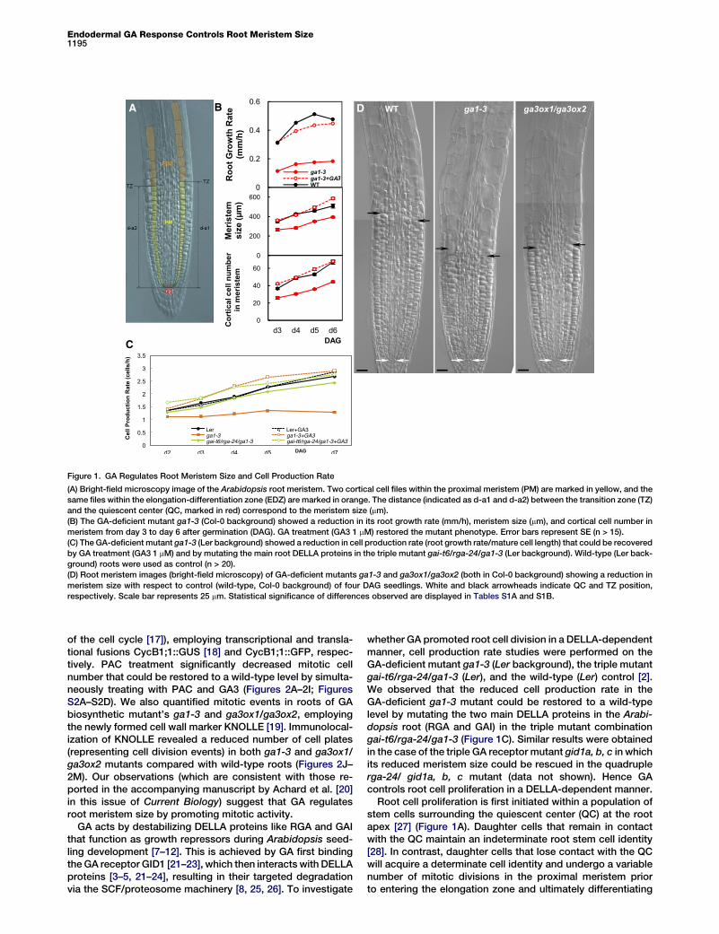
Figure 1. GA Regulates Root Meristem Size and Cell Production Rate
(A) Bright-field microscopy image of the Arabidopsis root meristem. Two cortical cell files within the proximal meristem (PM) are marked in yellow, and the
same files within the elongation-differentiation zone (EDZ) are marked in orange. The distance (indicated as d-a1 and d-a2) between the transition zone (TZ)
and the quiescent center (QC, marked in red) correspond to the meristem size (mm).
(B) The GA-deficient mutant ga1-3 (Col-0 background) showed a reduction in its root growth rate (mm/h), meristem size (mm), and cortical cell number in
meristem from day 3 to day 6 after germination (DAG). GA treatment (GA3 1 mM) restored the mutant phenotype. Error bars represent SE (n > 15).
(C) The GA-deficient mutant ga1-3 (Ler background) showed a reduction in cell production rate (root growth rate/mature cell length) that could be recovered
by GA treatment (GA3 1 mM) and by mutating the main root DELLA proteins in the triple mutant gai-t6/rga-24/ga1-3 (Ler background). Wild-type (Ler back-
ground) roots were used as control (n > 20).
(D) Root meristem images (bright-field microscopy) of GA-deficient mutants ga1-3 and ga3ox1/ga3ox2 (both in Col-0 background) showing a reduction in
meristem size with respect to control (wild-type, Col-0 background) of four DAG seedlings. White and black arrowheads indicate QC and TZ position,
respectively. Scale bar represents 25 mm. Statistical significance of differences observed are displayed in Tables S1A and S1B.
of the cell cycle [17]), employing transcriptional and transla-tional fusions CycB1;1::GUS [18] and CycB1;1::GFP, respec-tively. PAC treatment significantly decreased mitotic cellnumber that could be restored to a wild-type level by simulta-neously treating with PAC and GA3 (Figures 2A–2I; FiguresS2A–S2D). We also quantified mitotic events in roots of GAbiosynthetic mutant’s ga1-3 and ga3ox1/ga3ox2, employingthe newly formed cell wall marker KNOLLE [19]. Immunolocal-ization of KNOLLE revealed a reduced number of cell plates(representing cell division events) in both ga1-3 and ga3ox1/ga3ox2 mutants compared with wild-type roots (Figures 2J–2M). Our observations (which are consistent with those re-ported in the accompanying manuscript by Achard et al. [20]in this issue of Current Biology) suggest that GA regulatesroot meristem size by promoting mitotic activity.
GA acts by destabilizing DELLA proteins like RGA and GAIthat function as growth repressors during Arabidopsis seed-ling development [7–12]. This is achieved by GA first bindingthe GA receptor GID1 [21–23], which then interacts with DELLAproteins [3–5, 21–24], resulting in their targeted degradationvia the SCF/proteosome machinery [8, 25, 26]. To investigate
whether GA promoted root cell division in a DELLA-dependentmanner, cell production rate studies were performed on theGA-deficient mutant ga1-3 (Ler background), the triple mutantgai-t6/rga-24/ga1-3 (Ler), and the wild-type (Ler) control [2].We observed that the reduced cell production rate in theGA-deficient ga1-3 mutant could be restored to a wild-typelevel by mutating the two main DELLA proteins in the Arabi-dopsis root (RGA and GAI) in the triple mutant combinationgai-t6/rga-24/ga1-3 (Figure 1C). Similar results were obtainedin the case of the triple GA receptor mutant gid1a, b, c in whichits reduced meristem size could be rescued in the quadruplerga-24/ gid1a, b, c mutant (data not shown). Hence GAcontrols root cell proliferation in a DELLA-dependent manner.
Root cell proliferation is first initiated within a population ofstem cells surrounding the quiescent center (QC) at the rootapex [27] (Figure 1A). Daughter cells that remain in contactwith the QC maintain an indeterminate root stem cell identity[28]. In contrast, daughter cells that lose contact with the QCwill acquire a determinate cell identity and undergo a variablenumber of mitotic divisions in the proximal meristem priorto entering the elongation zone and ultimately differentiating
Current Biology Vol 19 No 141196
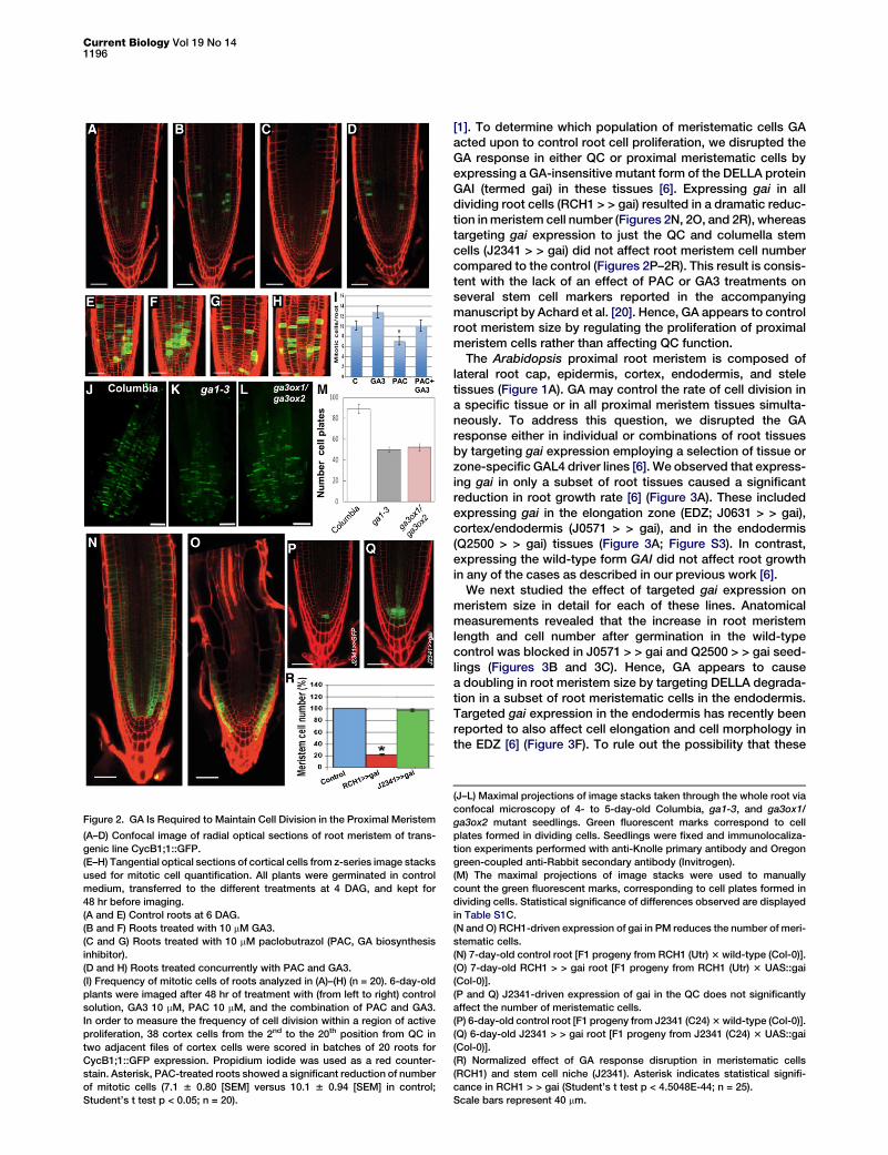
Figure 2. GA Is Required to Maintain Cell Division in the Proximal Meristem
(A–D) Confocal image of radial optical sections of root meristem of trans-
genic line CycB1;1::GFP.
(E–H) Tangential optical sections of cortical cells from z-series image stacks
used for mitotic cell quantification. All plants were germinated in control
medium, transferred to the different treatments at 4 DAG, and kept for
48 hr before imaging.
(A and E) Control roots at 6 DAG.
(B and F) Roots treated with 10 mM GA3.
(C and G) Roots treated with 10 mM paclobutrazol (PAC, GA biosynthesis
inhibitor).
(D and H) Roots treated concurrently with PAC and GA3.
(I) Frequency of mitotic cells of roots analyzed in (A)–(H) (n = 20). 6-day-old
plants were imaged after 48 hr of treatment with (from left to right) control
solution, GA3 10 mM, PAC 10 mM, and the combination of PAC and GA3.
In order to measure the frequency of cell division within a region of active
proliferation, 38 cortex cells from the 2nd to the 20th position from QC in
two adjacent files of cortex cells were scored in batches of 20 roots for
CycB1;1::GFP expression. Propidium iodide was used as a red counter-
stain. Asterisk, PAC-treated roots showed a significant reduction of number
of mitotic cells (7.1 6 0.80 [SEM] versus 10.1 6 0.94 [SEM] in control;
Student’s t test p < 0.05; n = 20).
[1]. To determine which population of meristematic cells GAacted upon to control root cell proliferation, we disrupted theGA response in either QC or proximal meristematic cells byexpressing a GA-insensitive mutant form of the DELLA proteinGAI (termed gai) in these tissues [6]. Expressing gai in alldividing root cells (RCH1 > > gai) resulted in a dramatic reduc-tion in meristem cell number (Figures 2N, 2O, and 2R), whereastargeting gai expression to just the QC and columella stemcells (J2341 > > gai) did not affect root meristem cell numbercompared to the control (Figures 2P–2R). This result is consis-tent with the lack of an effect of PAC or GA3 treatments onseveral stem cell markers reported in the accompanyingmanuscript by Achard et al. [20]. Hence, GA appears to controlroot meristem size by regulating the proliferation of proximalmeristem cells rather than affecting QC function.
The Arabidopsis proximal root meristem is composed oflateral root cap, epidermis, cortex, endodermis, and steletissues (Figure 1A). GA may control the rate of cell division ina specific tissue or in all proximal meristem tissues simulta-neously. To address this question, we disrupted the GAresponse either in individual or combinations of root tissuesby targeting gai expression employing a selection of tissue orzone-specific GAL4 driver lines [6]. We observed that express-ing gai in only a subset of root tissues caused a significantreduction in root growth rate [6] (Figure 3A). These includedexpressing gai in the elongation zone (EDZ; J0631 > > gai),cortex/endodermis (J0571 > > gai), and in the endodermis(Q2500 > > gai) tissues (Figure 3A; Figure S3). In contrast,expressing the wild-type form GAI did not affect root growthin any of the cases as described in our previous work [6].
We next studied the effect of targeted gai expression onmeristem size in detail for each of these lines. Anatomicalmeasurements revealed that the increase in root meristemlength and cell number after germination in the wild-typecontrol was blocked in J0571 > > gai and Q2500 > > gai seed-lings (Figures 3B and 3C). Hence, GA appears to causea doubling in root meristem size by targeting DELLA degrada-tion in a subset of root meristematic cells in the endodermis.Targeted gai expression in the endodermis has recently beenreported to also affect cell elongation and cell morphology inthe EDZ [6] (Figure 3F). To rule out the possibility that these
(J–L) Maximal projections of image stacks taken through the whole root via
confocal microscopy of 4- to 5-day-old Columbia, ga1-3, and ga3ox1/
ga3ox2 mutant seedlings. Green fluorescent marks correspond to cell
plates formed in dividing cells. Seedlings were fixed and immunolocaliza-
tion experiments performed with anti-Knolle primary antibody and Oregon
green-coupled anti-Rabbit secondary antibody (Invitrogen).
(M) The maximal projections of image stacks were used to manually
count the green fluorescent marks, corresponding to cell plates formed in
dividing cells. Statistical significance of differences observed are displayed
in Table S1C.
(N and O) RCH1-driven expression of gai in PM reduces the number of meri-
stematic cells.
(N) 7-day-old control root [F1 progeny from RCH1 (Utr) 3 wild-type (Col-0)].
(O) 7-day-old RCH1 > > gai root [F1 progeny from RCH1 (Utr) 3 UAS::gai
(Col-0)].
(P and Q) J2341-driven expression of gai in the QC does not significantly
affect the number of meristematic cells.
(P) 6-day-old control root [F1 progeny from J2341 (C24) 3 wild-type (Col-0)].
(Q) 6-day-old J2341 > > gai root [F1 progeny from J2341 (C24) 3 UAS::gai
(Col-0)].
(R) Normalized effect of GA response disruption in meristematic cells
(RCH1) and stem cell niche (J2341). Asterisk indicates statistical signifi-
cance in RCH1 > > gai (Student’s t test p < 4.5048E-44; n = 25).
Scale bars represent 40 mm.
Endodermal GA Response Controls Root Meristem Size1197
D
E
F
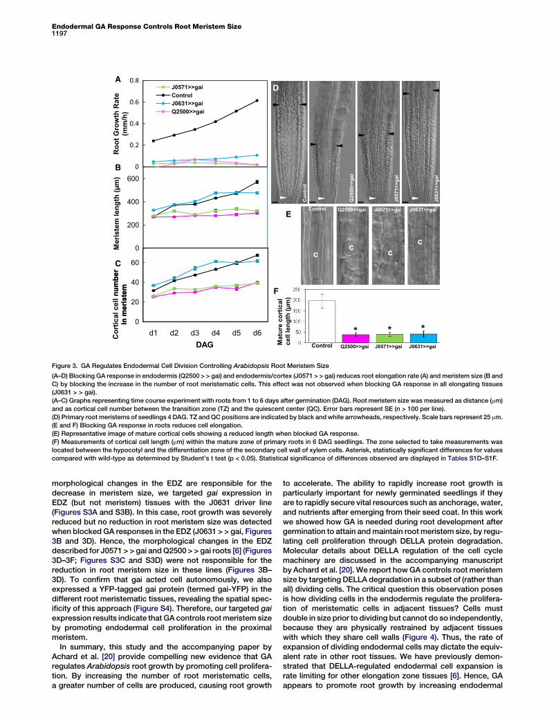
Figure 3. GA Regulates Endodermal Cell Division Controlling Arabidopsis Root Meristem Size
(A–D) Blocking GA response in endodermis (Q2500 > > gai) and endodermis/cortex (J0571 > > gai) reduces root elongation rate (A) and meristem size (B and
C) by blocking the increase in the number of root meristematic cells. This effect was not observed when blocking GA response in all elongating tissues
(J0631 > > gai).
(A–C) Graphs representing time course experiment with roots from 1 to 6 days after germination (DAG). Root meristem size was measured as distance (mm)
and as cortical cell number between the transition zone (TZ) and the quiescent center (QC). Error bars represent SE (n > 100 per line).
(D) Primary root meristems of seedlings 4 DAG. TZ and QC positions are indicated by black and white arrowheads, respectively. Scale bars represent 25 mm.
(E and F) Blocking GA response in roots reduces cell elongation.
(E) Representative image of mature cortical cells showing a reduced length when blocked GA response.
(F) Measurements of cortical cell length (mm) within the mature zone of primary roots in 6 DAG seedlings. The zone selected to take measurements was
located between the hypocotyl and the differentiation zone of the secondary cell wall of xylem cells. Asterisk, statistically significant differences for values
compared with wild-type as determined by Student’s t test (p < 0.05). Statistical significance of differences observed are displayed in Tables S1D–S1F.
morphological changes in the EDZ are responsible for thedecrease in meristem size, we targeted gai expression inEDZ (but not meristem) tissues with the J0631 driver line(Figures S3A and S3B). In this case, root growth was severelyreduced but no reduction in root meristem size was detectedwhen blocked GA responses in the EDZ (J0631 > > gai, Figures3B and 3D). Hence, the morphological changes in the EDZdescribed for J0571 > > gai and Q2500 > > gai roots [6] (Figures3D–3F; Figures S3C and S3D) were not responsible for thereduction in root meristem size in these lines (Figures 3B–3D). To confirm that gai acted cell autonomously, we alsoexpressed a YFP-tagged gai protein (termed gai-YFP) in thedifferent root meristematic tissues, revealing the spatial spec-ificity of this approach (Figure S4). Therefore, our targeted gaiexpression results indicate that GA controls root meristem sizeby promoting endodermal cell proliferation in the proximalmeristem.
In summary, this study and the accompanying paper byAchard et al. [20] provide compelling new evidence that GAregulates Arabidopsis root growth by promoting cell prolifera-tion. By increasing the number of root meristematic cells,a greater number of cells are produced, causing root growth
to accelerate. The ability to rapidly increase root growth isparticularly important for newly germinated seedlings if theyare to rapidly secure vital resources such as anchorage, water,and nutrients after emerging from their seed coat. In this workwe showed how GA is needed during root development aftergermination to attain and maintain root meristem size, by regu-lating cell proliferation through DELLA protein degradation.Molecular details about DELLA regulation of the cell cyclemachinery are discussed in the accompanying manuscriptby Achard et al. [20]. We report how GA controls root meristemsize by targeting DELLA degradation in a subset of (rather thanall) dividing cells. The critical question this observation posesis how dividing cells in the endodermis regulate the prolifera-tion of meristematic cells in adjacent tissues? Cells mustdouble in size prior to dividing but cannot do so independently,because they are physically restrained by adjacent tissueswith which they share cell walls (Figure 4). Thus, the rate ofexpansion of dividing endodermal cells may dictate the equiv-alent rate in other root tissues. We have previously demon-strated that DELLA-regulated endodermal cell expansion israte limiting for other elongation zone tissues [6]. Hence, GAappears to promote root growth by increasing endodermal
Current Biology Vol 19 No 141198
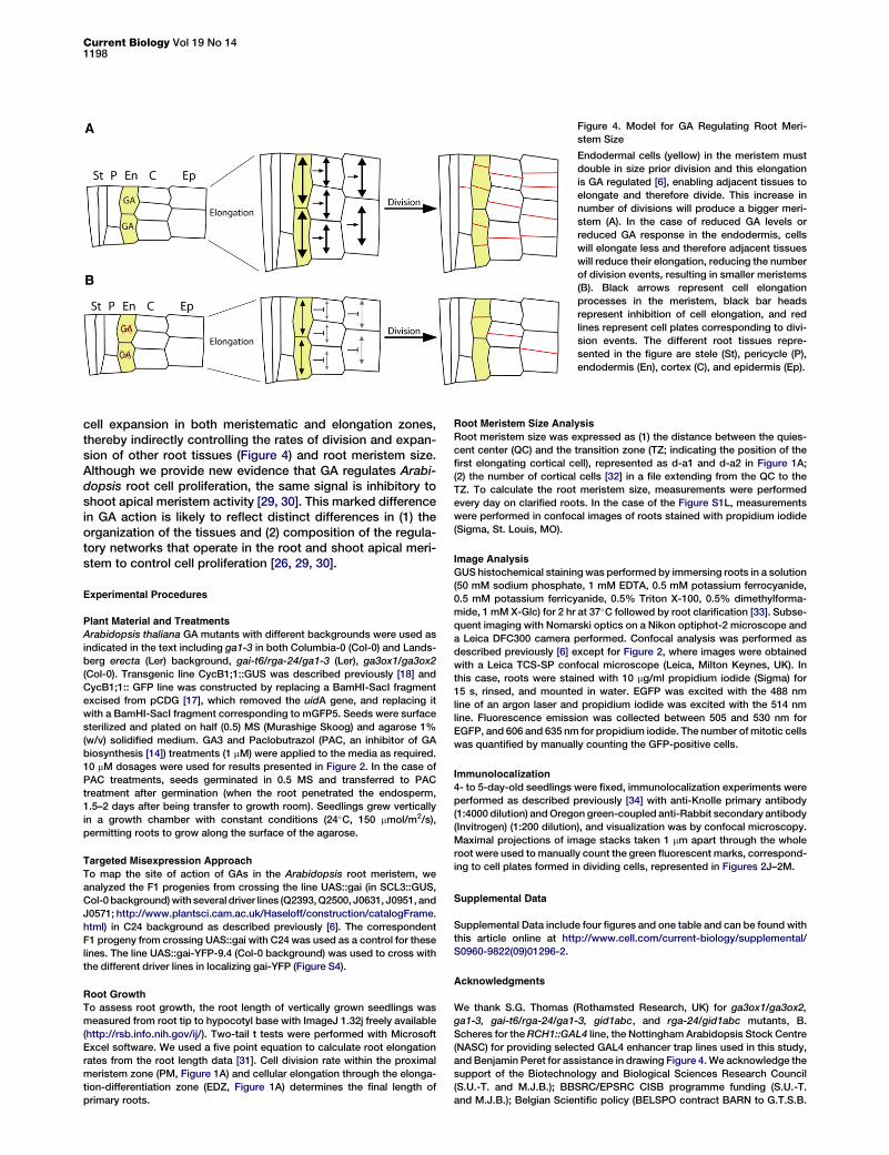
Figure 4. Model for GA Regulating Root Meri-
stem Size
Endodermal cells (yellow) in the meristem must
double in size prior division and this elongation
is GA regulated [6], enabling adjacent tissues to
elongate and therefore divide. This increase in
number of divisions will produce a bigger meri-
stem (A). In the case of reduced GA levels or
reduced GA response in the endodermis, cells
will elongate less and therefore adjacent tissues
will reduce their elongation, reducing the number
of division events, resulting in smaller meristems
(B). Black arrows represent cell elongation
processes in the meristem, black bar heads
represent inhibition of cell elongation, and red
lines represent cell plates corresponding to divi-
sion events. The different root tissues repre-
sented in the figure are stele (St), pericycle (P),
endodermis (En), cortex (C), and epidermis (Ep).
cell expansion in both meristematic and elongation zones,thereby indirectly controlling the rates of division and expan-sion of other root tissues (Figure 4) and root meristem size.Although we provide new evidence that GA regulates Arabi-dopsis root cell proliferation, the same signal is inhibitory toshoot apical meristem activity [29, 30]. This marked differencein GA action is likely to reflect distinct differences in (1) theorganization of the tissues and (2) composition of the regula-tory networks that operate in the root and shoot apical meri-stem to control cell proliferation [26, 29, 30].
Experimental Procedures
Plant Material and Treatments
Arabidopsis thaliana GA mutants with different backgrounds were used as
indicated in the text including ga1-3 in both Columbia-0 (Col-0) and Lands-
berg erecta (Ler) background, gai-t6/rga-24/ga1-3 (Ler), ga3ox1/ga3ox2
(Col-0). Transgenic line CycB1;1::GUS was described previously [18] and
CycB1;1:: GFP line was constructed by replacing a BamHI-SacI fragment
excised from pCDG [17], which removed the uidA gene, and replacing it
with a BamHI-SacI fragment corresponding to mGFP5. Seeds were surface
sterilized and plated on half (0.5) MS (Murashige Skoog) and agarose 1%
(w/v) solidified medium. GA3 and Paclobutrazol (PAC, an inhibitor of GA
biosynthesis [14]) treatments (1 mM) were applied to the media as required.
10 mM dosages were used for results presented in Figure 2. In the case of
PAC treatments, seeds germinated in 0.5 MS and transferred to PAC
treatment after germination (when the root penetrated the endosperm,
1.5–2 days after being transfer to growth room). Seedlings grew vertically
in a growth chamber with constant conditions (24�C, 150 mmol/m2/s),
permitting roots to grow along the surface of the agarose.
Targeted Misexpression Approach
To map the site of action of GAs in the Arabidopsis root meristem, we
analyzed the F1 progenies from crossing the line UAS::gai (in SCL3::GUS,
Col-0 background) with several driver lines (Q2393, Q2500, J0631, J0951, and
J0571; http://www.plantsci.cam.ac.uk/Haseloff/construction/catalogFrame.
html) in C24 background as described previously [6]. The correspondent
F1 progeny from crossing UAS::gai with C24 was used as a control for these
lines. The line UAS::gai-YFP-9.4 (Col-0 background) was used to cross with
the different driver lines in localizing gai-YFP (Figure S4).
Root Growth
To assess root growth, the root length of vertically grown seedlings was
measured from root tip to hypocotyl base with ImageJ 1.32j freely available
(http://rsb.info.nih.gov/ij/). Two-tail t tests were performed with Microsoft
Excel software. We used a five point equation to calculate root elongation
rates from the root length data [31]. Cell division rate within the proximal
meristem zone (PM, Figure 1A) and cellular elongation through the elonga-
tion-differentiation zone (EDZ, Figure 1A) determines the final length of
primary roots.
Root Meristem Size Analysis
Root meristem size was expressed as (1) the distance between the quies-
cent center (QC) and the transition zone (TZ; indicating the position of the
first elongating cortical cell), represented as d-a1 and d-a2 in Figure 1A;
(2) the number of cortical cells [32] in a file extending from the QC to the
TZ. To calculate the root meristem size, measurements were performed
every day on clarified roots. In the case of the Figure S1L, measurements
were performed in confocal images of roots stained with propidium iodide
(Sigma, St. Louis, MO).
Image Analysis
GUS histochemical staining was performed by immersing roots in a solution
(50 mM sodium phosphate, 1 mM EDTA, 0.5 mM potassium ferrocyanide,
0.5 mM potassium ferricyanide, 0.5% Triton X-100, 0.5% dimethylforma-
mide, 1 mM X-Glc) for 2 hr at 37�C followed by root clarification [33]. Subse-
quent imaging with Nomarski optics on a Nikon optiphot-2 microscope and
a Leica DFC300 camera performed. Confocal analysis was performed as
described previously [6] except for Figure 2, where images were obtained
with a Leica TCS-SP confocal microscope (Leica, Milton Keynes, UK). In
this case, roots were stained with 10 mg/ml propidium iodide (Sigma) for
15 s, rinsed, and mounted in water. EGFP was excited with the 488 nm
line of an argon laser and propidium iodide was excited with the 514 nm
line. Fluorescence emission was collected between 505 and 530 nm for
EGFP, and 606 and 635 nm for propidium iodide. The number of mitotic cells
was quantified by manually counting the GFP-positive cells.
Immunolocalization
4- to 5-day-old seedlings were fixed, immunolocalization experiments were
performed as described previously [34] with anti-Knolle primary antibody
(1:4000 dilution) and Oregon green-coupled anti-Rabbit secondary antibody
(Invitrogen) (1:200 dilution), and visualization was by confocal microscopy.
Maximal projections of image stacks taken 1 mm apart through the whole
root were used to manually count the green fluorescent marks, correspond-
ing to cell plates formed in dividing cells, represented in Figures 2J–2M.
Supplemental Data
Supplemental Data include four figures and one table and can be found with
this article online at http://www.cell.com/current-biology/supplemental/
S0960-9822(09)01296-2.
Acknowledgments
We thank S.G. Thomas (Rothamsted Research, UK) for ga3ox1/ga3ox2,
ga1-3, gai-t6/rga-24/ga1-3, gid1abc, and rga-24/gid1abc mutants, B.
Scheres for the RCH1::GAL4 line, the Nottingham Arabidopsis Stock Centre
(NASC) for providing selected GAL4 enhancer trap lines used in this study,
and Benjamin Peret for assistance in drawing Figure 4. We acknowledge the
support of the Biotechnology and Biological Sciences Research Council
(S.U.-T. and M.J.B.); BBSRC/EPSRC CISB programme funding (S.U.-T.
and M.J.B.); Belgian Scientific policy (BELSPO contract BARN to G.T.S.B.
Endodermal GA Response Controls Root Meristem Size1199
and M.J.B.); Ayuda de movilidad GRU0790 to I.C.; and Bill and Melinda
Gates Foundation (Gates Cambridge Scholarship to F.F.).
Received: March 6, 2009
Revised: June 2, 2009
Accepted: June 3, 2009
Published online: July 2, 2009
References
1. Beemster, G.T.S., and Baskin, T.I. (1998). Analysis of cell division and
elongation underlying the developmental acceleration of root growth
in Arabidopsis thaliana. Plant Physiol. 116, 1515–1526.
2. Fu, X., and Harberd, N.P. (2003). Auxin promotes Arabidopsis root
growth by modulating gibberellin response. Nature 421, 740–743.
3. Griffiths, J., Murase, K., Rieu, I., Zentella, R., Zhang, Z.L., Powers, S.J.,
Gong, F., Phillips, A.L., Hedden, P., Sun, T.P., et al. (2006). Genetic
characterization and functional analysis of the GID1 gibberellin recep-
tors in Arabidopsis. Plant Cell 18, 3399–3414.
4. Willige, B.C., Ghosh, S., Nill, C., Zourelidou, M., Dohmann, E.M., Maier,
A., and Schwechheimer, C. (2007). The DELLA domain of GA INSENSI-
TIVE mediates the interaction with the GA INSENSITIVE DWARF1A
gibberellin receptor of Arabidopsis. Plant Cell 19, 1209–1220.
5. Ueguchi-Tanaka, M., Nakajima, M., Katoh, E., Ohmiya, H., Asano, K.,
Saji, S., Hongyu, X., Ashikari, M., Kitano, H., Yamaguchi, I., et al.
(2007). Molecular interactions of a soluble gibberellin receptor, GID1,
with a rice DELLA protein, SLR1, and gibberellin. Plant Cell 19, 2140–
2155.
6. Ubeda-Tomas, S., Swarup, R., Coates, J., Swarup, K., Laplaze, L.,
Beemster, G.T., Hedden, P., Bhalerao, R., and Bennett, M.J. (2008).
Root growth in Arabidopsis requires gibberellin/DELLA signalling in
the endodermis. Nat. Cell Biol. 10, 625–628.
7. Peng, J., Richards, D.E., Hartley, N.M., Murphy, G.P., Devos, K.M.,
Flintham, J.E., Beales, J., Fish, L.J., Worland, A.J., Pelica, F., et al.
(1999). ‘Green revolution’ genes encode mutant gibberellin response
modulators. Nature 400, 256–261.
8. Silverstone, A.L., Jung, H.S., Dill, A., Kawaide, H., Kamiya, Y., and Sun,
T.P. (2001). Repressing a repressor: Gibberellin-induced rapid reduc-
tion of the RGA protein in Arabidopsis. Plant Cell 13, 1555–1566.
9. Peng, J., Carol, P., Richards, D.E., King, K.E., Cowling, R.J., Murphy,
G.P., and Harberd, N.P. (1997). The Arabidopsis GAI gene defines
a signaling pathway that negatively regulates gibberellin responses.
Genes Dev. 11, 3194–3205.
10. King, K.E., Moritz, T., and Harberd, N.P. (2001). Gibberellins are not
required for normal stem growth in Arabidopsis thaliana in the absence
of GAI and RGA. Genetics 159, 767–776.
11. Dill, A., and Sun, T.P. (2001). Synergistic derepression of gibberellin
signalling by removing RGA and GAI function in Arabidopsis thaliana.
Genetics 159, 777–785.
12. Cheng, H., Qin, L., Lee, S., Fu, X., Richards, D.E., Cao, D., Luo, D.,
Harberd, N.P., and Peng, J. (2004). Gibberellin regulates Arabidopsis
floral development via suppression of DELLA protein function. Develop-
ment 131, 1055–1064.
13. Baskin, T.I. (2000). On the constancy of cell division rate in the root
meristem. Plant Mol. Biol. 43, 545–554.
14. Olszewski, N., Sun, T.P., and Gubler, F. (2002). Gibberellin signaling:
Biosynthesis, catabolism, and response pathways. Plant Cell Suppl.
14, S61–S80.
15. Sun, T.P., and Kamiya, Y. (1994). The Arabidopsis GA1 locus encodes
the cyclase ent-kaurene synthetase A of gibberellin biosynthesis. Plant
Cell 6, 1509–1518.
16. Mitchum, M.G., Yamaguchi, S., Hanada, A., Kuwahara, A., Yoshioka, Y.,
Kato, T., Tabata, S., Kamiya, Y., and Sun, T.P. (2006). Distinct and over-
lapping roles of two gibberellin 3-oxidases in Arabidopsis development.
Plant J. 45, 804–818.
17. Ferreira, P.C.G., Hemerly, A.S., de Almeida-Engler, J., Van Montagu, M.,
Engler, G., and Inze, D. (1994). Developmental expression of the Arabi-
dopsis cyclin gene cyc1At. Plant Cell 6, 1763–1774.
18. Colon-Carmona, A., You, R., Haimovitch-Gal, T., and Doerner, P. (1999).
Technical advance: spatio-temporal analysis of mitotic activity with
a labile cyclin-GUS fusion protein. Plant J. 20, 503–508.
19. Lauber, M.H., Waizenegger, I., Steinmann, T., Schwarz, H., Mayer, U.,
Hwang, I., Lukowitz, W., and Jurgens, G. (1997). The Arabidopsis
KNOLLE protein is a cytokinesis-specific syntaxin. J. Cell Biol. 139,
1485–1493.
20. Achard, P., Gusti, A., Cheminant, S., Alioua, M., Dhondt, S., Coppens, F.,
Beemster, G.T.S., and Genschik, P. (2009). Gibberellin signaling
controls cell proliferation rate in Arabidopsis. Curr. Biol. 19, this issue,
1188–1193.
21. Ueguchi-Tanaka, M., Ashikari, M., Nakajima, M., Itoh, H., Katoh, E.,
Kobayashi, M., Chow, T.Y., Hsing, Y.I., Kitano, H., Yamaguchi, I., et al.
(2005). GIBBERELLIN INSENSITIVE DWARF1 encodes a soluble
receptor for gibberellin. Nature 437, 693–698.
22. Nakajima, M., Shimada, A., Takashi, Y., Kim, Y.C., Park, S.H., Ueguchi-
Tanaka, M., Suzuki, H., Katoh, E., Iuchi, S., Kobayashi, M., et al. (2006).
Identification and characterization of Arabidopsis gibberellin receptors.
Plant J. 46, 880–889.
23. Shimada, A., Ueguchi-Tanaka, M., Nakatsu, T., Nakajima, M., Naoe, Y.,
Ohmiya, H., Kato, H., and Matsuoka, M. (2008). Structural basis for
gibberellin recognition by its receptor GID1. Nature 456, 520–523.
24. Murase, K., Hirano, Y., Sun, T.P., and Hakoshima, T. (2008). Gibberellin-
induced DELLA recognition by the gibberellin receptor GID1. Nature
456, 459–463.
25. Fu, X., Richards, D.E., Ait-Ali, T., Hynes, L.W., Ougham, H., Peng, J., and
Harberd, N.P. (2002). Gibberellin-mediated proteasome-dependent
degradation of the barley DELLA protein SLN1 repressor. Plant Cell
14, 3191–3200.
26. Dill, A., Thomas, S.G., Hu, J., Steber, C.M., and Sun, T.P. (2004). The
Arabidopsis F-box protein SLEEPY1 targets gibberellin signaling
repressors for gibberellin-induced degradation. Plant Cell 16, 1392–
1405.
27. Scheres, B. (2007). Stem-cell niches: Nursery rhymes across kingdoms.
Nat. Rev. Mol. Cell Biol. 8, 345–354.
28. van den Berg, C., Willemsen, V., Hendriks, G., Weisbeek, P., and
Scheres, B. (1997). Short-range control of cell differentiation in the
Arabidopsis root meristem. Nature 390, 287–289.
29. Hay, A., Kaur, H., Phillips, A., Hedden, P., Hake, S., and Tsiantis, M.
(2002). The gibberellin pathway mediates KNOTTED1-type homeobox
function in plants with different body parts. Curr. Biol. 12, 1557–1565.
30. Jasinski, S., Piazza, P., Craft, J., Hay, A., Wooley, L., Rieu, I., Phillips, A.,
Hedden, P., and Tsiantis, M. (2005). KNOX1 action in Arabidopsis is
mediated by coordinate regulation of cytokinin and gibberellin activi-
ties. Curr. Biol. 15, 1560–1565.
31. Erickson, R.O. (1976). Modeling of plant growth. Annu. Rev. Plant Phys-
iol. 27, 407–434.
32. Dello Ioio, R., Linhares, F.S., Scacchi, E., Casamitjana-Martinez, E.,
Heidstra, R., Costantino, P., and Sabatini, S. (2007). Cytokinins
determine Arabidopsis root-meristem size by controlling cell differenti-
ation. Curr. Biol. 17, 678–682.
33. Malamy, J.E., and Benfey, P.N. (1997). Organization and cell differentia-
tion in lateral roots of Arabidopsis thaliana. Development 124, 33–44.
34. Swarup, R., Kramer, E.M., Perry, P., Knox, K., Leyser, O.H.M., Haseloff,
J., Beemster, G.T.S., Bhalerao, R., and Bennett, M.J. (2005). Root grav-
itropism requires lateral root cap and epidermal cells for transport and
response to a mobile auxin signal. Nat. Cell Biol. 7, 1057–1065.


![Regeneration Of Three Sweet Potato [Ipomoea Batatas(L.)] Accessions in Ghana via Meristem And Nodal culture](https://static.fdokumen.com/doc/165x107/631af948d43f4e176304af45/regeneration-of-three-sweet-potato-ipomoea-batatasl-accessions-in-ghana-via.jpg)