Gene expression map of the Arabidopsis shoot apical meristem stem cell niche
Upload
tuebingen-mpgCategory
view
0download
0
Positional Information by Dif
Current Biology 21, 1918–1923, November 22, 2011 ª2011 Elsevier Ltd All rights reserved DOI 10.1016/j.cub.2011.10.002
Reportferential
Endocytosis Splits Auxin Responseto Drive Arabidopsis Root Meristem Growth
Luca Santuari,1,4 Emanuele Scacchi,1,4
Antia Rodriguez-Villalon,1 Paula Salinas,1
Esther M.N. Dohmann,1 Geraldine Brunoud,2 Teva Vernoux,2
Richard S. Smith,3 and Christian S. Hardtke1,*1Department of Plant Molecular Biology, University ofLausanne, Biophore Building, CH-1015 Lausanne, Switzerland2Laboratoire de Reproduction et Developpement des Plantes,CNRS, INRA, ENS Lyon, UCBL, Universite de Lyon,69364 Lyon, France3Institute of Plant Sciences, University of Bern,Altenbergrain 21, Bern CH-3013, Switzerland
Summary
In the Arabidopsis root meristem, polar auxin transportcreates a transcriptional auxin response gradient that peaks
at the stem cell niche and gradually decreases as stem celldaughters divide and differentiate [1–3]. The amplitude and
extent of this gradient are essential for both stem cell main-tenance and root meristem growth [4, 5]. To investigate why
expression of some auxin-responsive genes, such as theessential root meristem growth regulator BREVIS RADIX
(BRX) [6], deviates from this gradient, we combined ex-perimental and computational approaches. We created
cellular-level root meristem models that accurately repro-duce distribution of nuclear auxin activity and allowdynamic
modeling of regulatory processes to guide experimentation.
Expression profiles deviating from the auxin gradient couldonly be modeled after intersection of auxin activity with the
observed differential endocytosis pattern and positive auto-regulatory feedback through plasma-membrane-to-nucleus
transfer of BRX. Because BRX is required for expression ofcertain auxin response factor targets, our data suggest a
cell-type-specific endocytosis-dependent input into tran-scriptional auxin perception. This input sustains expression
of a subset of auxin-responsive genes across the root meri-stem’s division and transition zones and is essential for
meristem growth. Thus, the endocytosis pattern providesspecific positional information to modulate auxin response.
Results and Discussion
Differential distribution of auxin by polar auxin transport (PAT)regulates cellular characteristics such as proliferation, elonga-tion, and differentiation and depends on nonuniform (polar)plasma membrane localization of PIN-FORMED (PIN) auxinefflux carriers [1]. Along the root meristem, PAT creates anauxin response gradient, which peaks at the stem cell nicheand decreases as stem cell daughters proliferate in themeristematic zone [2, 3]. Their eventual differentiation andelongation are timed by complex dynamic hormone pathwaycrosstalk in the so-called transition zone, which creates adevelopmental window in young roots to permit meristem
4These authors contributed equally to this work
*Correspondence: [email protected]
growth [4–6]. The BREVIS RADIX (BRX) gene is essential forthis process and is expressed in developing vasculature,mainly in the protophloem [6, 7]. In brx mutants, suspendedmeristem growth coincides with diminished auxin responseas BRX limits auxin-responsive transcription, likely throughmodulation of auxin response factor (ARF) activity and down-stream effects on brassinosteroid levels [6, 7]. Interestingly,BRX is itself auxin responsive and targeted by the ARFMONOPTEROS (MP). Paradoxically, however, the expressionprofile of BRX as well as other MP targets does not alwaysfollow the described auxin response gradient across the meri-stematic and transition zones [6] (Figure 1A). In this study, wecombined experimental and modeling approaches to investi-gate this phenomenon.In a first step, we created a cellular-level Arabidopsis root-
meristem model, centered across the two protophloems,where BRX expression is most prominent and robust.Throughout our study, confocal microscopy allowed unequiv-ocal identification of the protophloem strands as a result oftheir unique cell wall staining characteristics after modifiedpseudo-Schiff propidium iodide and/or propidium iodidestaining [6, 8] and because of their distinct morphology ascompared to the xylem strands (see Figure S1A available on-line). Because the phloem poles are not planar, a slightlycurved plane through a serial stack of confocal sections wasextracted using a Bezier surface. Its segmentation rendereda two-dimensional model of cellular structure (Figure 1B),from which an idealized variant was derived (Figure 1C) (seeSupplemental Experimental Procedures for model details).The model’s PAT topography after incorporation of the previ-ously reported PIN distribution [9] dynamically recreated thestemcell niche auxinmaximumand gradient once steady statewas reached after feeding auxin through the vasculature (Fig-ure 1C; Movie S1A). However, modeled cellular auxin concen-tration gradually increased toward the elongation zone, afeature that had not been observed previously [9]. Graduallyincreased auxin concentration toward the elongation zonewas also not observed in auxin measurements [2]; however,because the modeled relative increase is comparatively small,this could reflect the limited cellular and technical resolution ofthis approach. Recreation of a grid model similar to Grieneisenet al. [9] reproduced this feature if the parameter of the ratiobetween passive and active (PIN-mediated) auxin transportwas adjusted (Figure S1B). To obtain a clearer picture of auxinactivity across the meristem, we estimated it in vivo using theconstitutively expressed fluorescent DII-VENUS reporter [10].DII-VENUS is degraded upon auxin-triggered interaction withnuclear auxin receptors [11–13], which are ubiquitously ex-pressed throughout the root [14]. DII-VENUS abundance thuscorrelates inversely with cellular auxin activity and shouldreflect auxin concentration. Interestingly, experimentally ob-served DII-VENUS fluorescence intensity matched with ourmodeled auxin distribution pattern. An exception was the pro-tophloem (and the protoxylem, which was however not con-sidered separately in our model), where relative to neighboringtissue layers, increased auxin activity was indicated (Figures1D and 1E; Figure S1C). This experimental finding could bere-created by addition of the protophloem-specific auxin influx
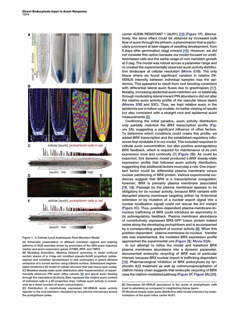
Figure 1. A Cellular-Level Arabidopsis Root Meristem Model
(A) Schematic presentation of different meristem regions and staining
patterns of GUS reporters driven by promoters of the DR5 auxin response
marker and auxin-responsive genes ATHB8, BRX, and TMO5.
(B) Modeling illustration. Marking bilateral symmetry in serial confocal
section stacks of a 3-day-old modified pseudo-Schiff propidium iodide-
stained root meristem (protophloem in red; protoxylem in green) allowed
extraction of a curved section using a Bezier surface. Subsequent segmen-
tation rendered a 2Dmodel of cellular structure that was tissue-type coded.
(C) Modeled steady-state auxin distribution after implementation of experi-
mentally observed PIN auxin efflux carriers [9] and apical auxin feeding
through the vasculature (bottom). Bars represent the relative auxin content
of individual cells in a 2D projection (top). Cellular auxin activity is consid-
ered as a direct function of auxin concentration.
(D) Distribution of constitutively expressed DII-VENUS auxin activity
reporter in the root meristem, visualized by two-photon microscopy across
the protophloem poles.
Direct Endocytosis Input in Auxin Response1919
carrier AUXIN RESISTANT 1 (AUX1) [15] (Figure 1F). Alterna-tively, the same effect could be obtained by increased bulkflowof auxin through the phloem, a phenomenon that is partic-ularly prominent at later stages of seedling development, from5 days after germination (dag) onward [16]. However, we didnot consider this option because our model focused on undif-ferentiated cells and the earlier stage of root meristem growthat 3 dag. The model was robust across a parameter range andre-created the experimentally observed auxin activity distribu-tion landscape at cellular resolution (Movie S1B). The onlytissue where we found significant variation in relative DII-VENUS intensity between individual samples was the epi-dermis. This appeared to result from root bending consistentwith differential lateral auxin fluxes due to gravitropism [17].Notably, increasing epidermal auxin retention uni- or bilaterallythroughmodulating lateral inward PIN abundance did not alterthe relative auxin activity profile of the vascular tissue layers(Movies S3B and S3C). Thus, we kept relative auxin in theepidermis low in follow-upmodels, for better viewing of resultsbut also consistent with a straight root and epidermal auxinmeasurements [2].Confirming the initial paradox, auxin activity distribution
only partially matched the BRX transcription profile (Fig-ure 2A), suggesting a significant influence of other factors.To determine which conditions could create this profile, weadded BRX transcription and the established regulatory influ-ences that modulate it to our model. This included response tocellular auxin concentration, but also positive autoregulatoryBRX feedback, which is required for maintenance of its ownexpression level and continuity [7] (Figure 2B). As could beexpected, this dynamic model produced a BRX steady-stateexpression profile that followed auxin activity distribution,suggesting that additional factors must play a role. One impor-tant factor could be differential plasma membrane versusnuclear partitioning of BRX protein. Various experimental evi-dences suggest that BRX is a transcriptional coregulator;however, BRX is primarily plasma membrane associated[18, 19]. Passage by the plasma membrane appears to beobligatory for its nuclear activity, because BRX variants withdisrupted plasma membrane targeting (either by N-terminalextension or by mutation of a nuclear export signal into anuclear localization signal) could not rescue the brx mutant(Figure 2C). Thus, position-dependent plasma-membrane-to-nucleus trafficking of BRX could introduce an asymmetry toits autoregulatory feedback. Plasma membrane abundanceof constitutively expressed BRX-GFP fusion protein indeedvaries along the developing protophloem and is accompaniedby a corresponding gradient of nuclear activity [6]. When thisposition-dependent plasma-membrane-to-nucleus transferrate was implemented, the modeled BRX expression profileapproached the experimental one (Figure 2E; Movie S2A).In our attempt to refine the model and transform BRX
plasma membrane abundance into a dynamic parameter,documented endocytic recycling of BRX was of particularinterest, because BRX nuclear import is trafficking dependent[19]. Pharmacological inhibition of BRX endocytosis by tyr-phostin A23 treatment as well as coimmunoprecipitation ofclathrin heavy chain suggests that endocytic recycling of BRXuses the clathrin-mediated pathway (Figure 2F; Figure 3A) [20].
(E) Decreased DII-VENUS abundance in the nuclei of protophloem cells
(next to asterisks) as compared to neighboring tissue layers.
(F) Modeled steady-state auxin distribution after model extension by imple-
mentation of the auxin influx carrier AUX1.
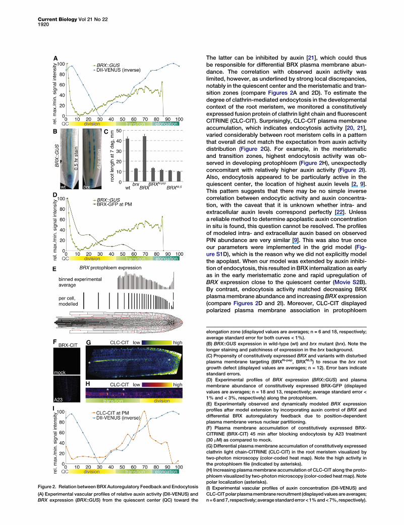
Figure 2. Relation between BRX Autoregulatory Feedback and Endocytosis
(A) Experimental vascular profiles of relative auxin activity (DII-VENUS) and
BRX expression (BRX::GUS) from the quiescent center (QC) toward the
Current Biology Vol 21 No 221920
The latter can be inhibited by auxin [21], which could thusbe responsible for differential BRX plasma membrane abun-dance. The correlation with observed auxin activity waslimited, however, as underlined by strong local discrepancies,notably in the quiescent center and the meristematic and tran-sition zones (compare Figures 2A and 2D). To estimate thedegree of clathrin-mediated endocytosis in the developmentalcontext of the root meristem, we monitored a constitutivelyexpressed fusion protein of clathrin light chain and fluorescentCITRINE (CLC-CIT). Surprisingly, CLC-CIT plasma membraneaccumulation, which indicates endocytosis activity [20, 21],varied considerably between root meristem cells in a patternthat overall did not match the expectation from auxin activitydistribution (Figure 2G). For example, in the meristematicand transition zones, highest endocytosis activity was ob-served in developing protophloem (Figure 2H), unexpectedlyconcomitant with relatively higher auxin activity (Figure 2I).Also, endocytosis appeared to be particularly active in thequiescent center, the location of highest auxin levels [2, 9].This pattern suggests that there may be no simple inversecorrelation between endocytic activity and auxin concentra-tion, with the caveat that it is unknown whether intra- andextracellular auxin levels correspond perfectly [22]. Unlessa reliable method to determine apoplastic auxin concentrationin situ is found, this question cannot be resolved. The profilesof modeled intra- and extracellular auxin based on observedPIN abundance are very similar [9]. This was also true onceour parameters were implemented in the grid model (Fig-ure S1D), which is the reason why we did not explicitly modelthe apoplast. When our model was extended by auxin inhibi-tion of endocytosis, this resulted in BRX internalization as earlyas in the early meristematic zone and rapid upregulation ofBRX expression close to the quiescent center (Movie S2B).By contrast, endocytosis activity matched decreasing BRXplasmamembrane abundance and increasingBRX expression(compare Figures 2D and 2I). Moreover, CLC-CIT displayedpolarized plasma membrane association in protophloem
elongation zone (displayed values are averages; n = 6 and 18, respectively;
average standard error for both curves < 1%).
(B) BRX::GUS expression in wild-type (wt) and brx mutant (brx). Note the
longer staining and patchiness of expression in the brx background.
(C) Propensity of constitutively expressed BRX and variants with disturbed
plasma membrane targeting (BRXN-pep, BRXNLS) to rescue the brx root
growth defect (displayed values are averages; n = 12). Error bars indicate
standard errors.
(D) Experimental profiles of BRX expression (BRX::GUS) and plasma
membrane abundance of constitutively expressed BRX-GFP (displayed
values are averages; n = 18 and 13, respectively; average standard error <
1% and < 3%, respectively) along the protophloem.
(E) Experimentally observed and dynamically modeled BRX expression
profiles after model extension by incorporating auxin control of BRX and
differential BRX autoregulatory feedback due to position-dependent
plasma membrane versus nuclear partitioning.
(F) Plasma membrane accumulation of constitutively expressed BRX-
CITRINE (BRX-CIT) 45 min after blocking endocytosis by A23 treatment
(30 mM) as compared to mock.
(G) Differential plasma membrane accumulation of constitutively expressed
clathrin light chain-CITRINE (CLC-CIT) in the root meristem visualized by
two-photon microscopy (color-coded heat map). Note the high activity in
the protophloem file (indicated by asterisks).
(H) Increasing plasmamembrane accumulation of CLC-CIT along the proto-
phloem visualized by two-photonmicroscopy (color-coded heatmap). Note
polar localization (asterisks).
(I) Experimental vascular profiles of auxin concentration (DII-VENUS) and
CLC-CITpolarplasmamembrane recruitment (displayedvaluesareaverages;
n=6and7, respectively; averagestandarderror<1%and<7%, respectively).
A
cellular [auxin], protophloem cells in red
auxin flux, protophloem cells in red
B
C
DII-VENUS, mock-treated DII-VENUS, NPA-treated
D
binned experimental average
per
prot
ophl
oem
cel
l, m
odel
ed
BRX protophloem expression,
cellular [auxin], protophloem cells in red
active cytosolic BRX
CLC plasma membrane activity
nuclear BRX
BRX plasma membrane abundance
BRX transcription
proteinunique
BRX-GFP PM-GFPpeptide count in IP
heavy clathrin chain At3g08530
heavy clathrin chain At3g11130
peptides
23 23 10
2 02
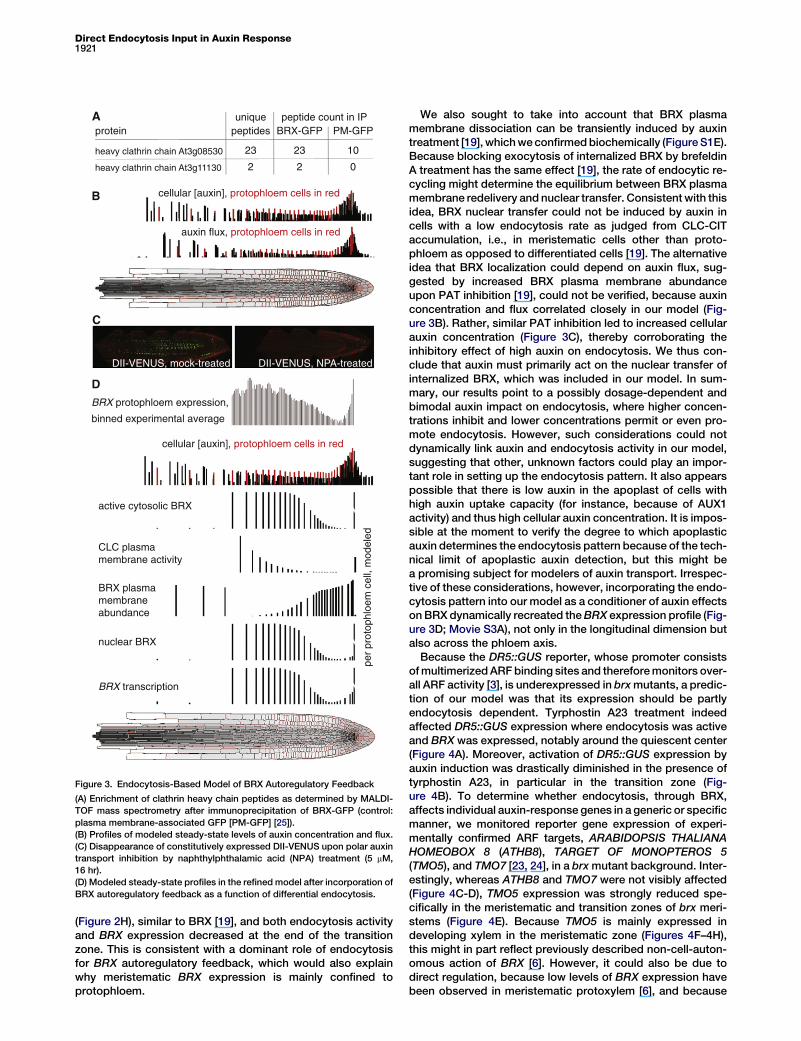
Figure 3. Endocytosis-Based Model of BRX Autoregulatory Feedback
(A) Enrichment of clathrin heavy chain peptides as determined by MALDI-
TOF mass spectrometry after immunoprecipitation of BRX-GFP (control:
plasma membrane-associated GFP [PM-GFP] [25]).
(B) Profiles of modeled steady-state levels of auxin concentration and flux.
(C) Disappearance of constitutively expressed DII-VENUS upon polar auxin
transport inhibition by naphthylphthalamic acid (NPA) treatment (5 mM,
16 hr).
(D) Modeled steady-state profiles in the refinedmodel after incorporation of
BRX autoregulatory feedback as a function of differential endocytosis.
Direct Endocytosis Input in Auxin Response1921
(Figure 2H), similar to BRX [19], and both endocytosis activityand BRX expression decreased at the end of the transitionzone. This is consistent with a dominant role of endocytosisfor BRX autoregulatory feedback, which would also explainwhy meristematic BRX expression is mainly confined toprotophloem.
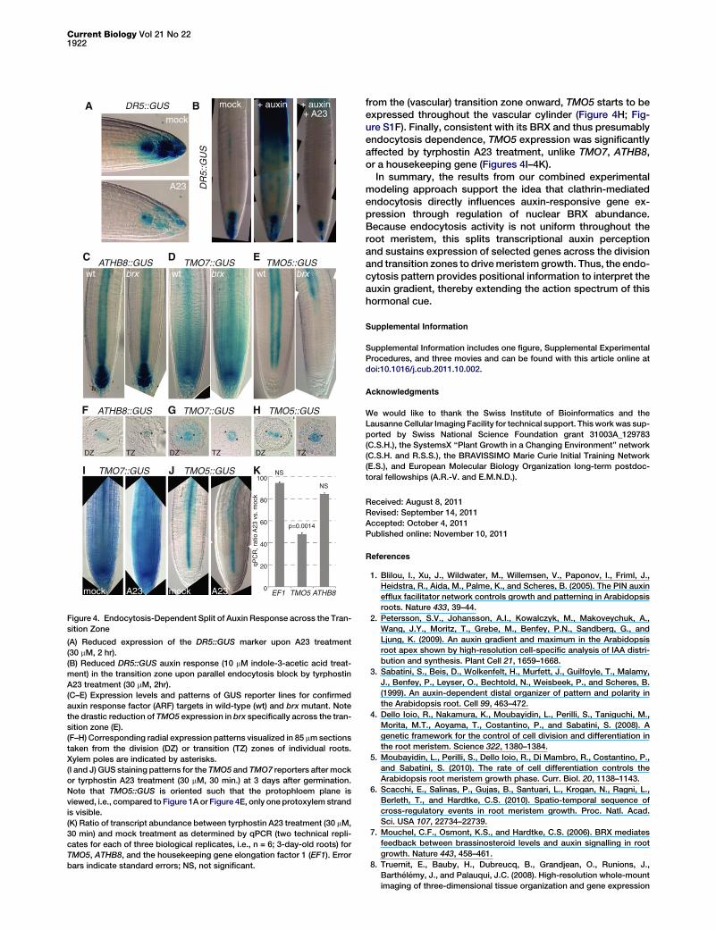
We also sought to take into account that BRX plasmamembrane dissociation can be transiently induced by auxintreatment [19], whichweconfirmedbiochemically (FigureS1E).Because blocking exocytosis of internalized BRX by brefeldinA treatment has the same effect [19], the rate of endocytic re-cycling might determine the equilibrium between BRX plasmamembrane redelivery and nuclear transfer. Consistent with thisidea, BRX nuclear transfer could not be induced by auxin incells with a low endocytosis rate as judged from CLC-CITaccumulation, i.e., in meristematic cells other than proto-phloem as opposed to differentiated cells [19]. The alternativeidea that BRX localization could depend on auxin flux, sug-gested by increased BRX plasma membrane abundanceupon PAT inhibition [19], could not be verified, because auxinconcentration and flux correlated closely in our model (Fig-ure 3B). Rather, similar PAT inhibition led to increased cellularauxin concentration (Figure 3C), thereby corroborating theinhibitory effect of high auxin on endocytosis. We thus con-clude that auxin must primarily act on the nuclear transfer ofinternalized BRX, which was included in our model. In sum-mary, our results point to a possibly dosage-dependent andbimodal auxin impact on endocytosis, where higher concen-trations inhibit and lower concentrations permit or even pro-mote endocytosis. However, such considerations could notdynamically link auxin and endocytosis activity in our model,suggesting that other, unknown factors could play an impor-tant role in setting up the endocytosis pattern. It also appearspossible that there is low auxin in the apoplast of cells withhigh auxin uptake capacity (for instance, because of AUX1activity) and thus high cellular auxin concentration. It is impos-sible at the moment to verify the degree to which apoplasticauxin determines the endocytosis pattern because of the tech-nical limit of apoplastic auxin detection, but this might bea promising subject for modelers of auxin transport. Irrespec-tive of these considerations, however, incorporating the endo-cytosis pattern into our model as a conditioner of auxin effectsonBRX dynamically recreated theBRX expression profile (Fig-ure 3D; Movie S3A), not only in the longitudinal dimension butalso across the phloem axis.Because the DR5::GUS reporter, whose promoter consists
ofmultimerizedARFbinding sites and thereforemonitors over-all ARF activity [3], is underexpressed in brxmutants, a predic-tion of our model was that its expression should be partlyendocytosis dependent. Tyrphostin A23 treatment indeedaffected DR5::GUS expression where endocytosis was activeand BRX was expressed, notably around the quiescent center(Figure 4A). Moreover, activation of DR5::GUS expression byauxin induction was drastically diminished in the presence oftyrphostin A23, in particular in the transition zone (Fig-ure 4B). To determine whether endocytosis, through BRX,affects individual auxin-response genes in a generic or specificmanner, we monitored reporter gene expression of experi-mentally confirmed ARF targets, ARABIDOPSIS THALIANAHOMEOBOX 8 (ATHB8), TARGET OF MONOPTEROS 5(TMO5), and TMO7 [23, 24], in a brxmutant background. Inter-estingly, whereas ATHB8 and TMO7 were not visibly affected(Figure 4C-D), TMO5 expression was strongly reduced spe-cifically in the meristematic and transition zones of brx meri-stems (Figure 4E). Because TMO5 is mainly expressed indeveloping xylem in the meristematic zone (Figures 4F–4H),this might in part reflect previously described non-cell-auton-omous action of BRX [6]. However, it could also be due todirect regulation, because low levels of BRX expression havebeen observed in meristematic protoxylem [6], and because
DR
5::G
US
ATHB8::GUS TMO7::GUS TMO5::GUS
A
wt wt wtbrx brx brx
EDC
mock + auxin + auxin+ A23
BDR5::GUS
A23
mock
ATHB8::GUS TMO7::GUS TMO5::GUSHGF
DZ DZDZTZ TZ TZ
100
20
40
60
80
0
TMO7::GUS TMO5::GUSJI
A23mock A23mock
qPC
R, r
atio
A23
vs.
moc
k
TMO5 ATHB8EF1
NS
NS
p=0.0014
K
**
* * **
** * ***
Figure 4. Endocytosis-Dependent Split of Auxin Response across the Tran-
sition Zone
(A) Reduced expression of the DR5::GUS marker upon A23 treatment
(30 mM, 2 hr).
(B) Reduced DR5::GUS auxin response (10 mM indole-3-acetic acid treat-
ment) in the transition zone upon parallel endocytosis block by tyrphostin
A23 treatment (30 mM, 2hr).
(C–E) Expression levels and patterns of GUS reporter lines for confirmed
auxin response factor (ARF) targets in wild-type (wt) and brx mutant. Note
the drastic reduction of TMO5 expression in brx specifically across the tran-
sition zone (E).
(F–H) Corresponding radial expression patterns visualized in 85 mmsections
taken from the division (DZ) or transition (TZ) zones of individual roots.
Xylem poles are indicated by asterisks.
(I and J) GUS staining patterns for the TMO5 and TMO7 reporters after mock
or tyrphostin A23 treatment (30 mM, 30 min.) at 3 days after germination.
Note that TMO5::GUS is oriented such that the protophloem plane is
viewed, i.e., compared to Figure 1A or Figure 4E, only one protoxylem strand
is visible.
(K) Ratio of transcript abundance between tyrphostin A23 treatment (30 mM,
30 min) and mock treatment as determined by qPCR (two technical repli-
cates for each of three biological replicates, i.e., n = 6; 3-day-old roots) for
TMO5, ATHB8, and the housekeeping gene elongation factor 1 (EF1). Error
bars indicate standard errors; NS, not significant.
Current Biology Vol 21 No 221922
from the (vascular) transition zone onward, TMO5 starts to beexpressed throughout the vascular cylinder (Figure 4H; Fig-ure S1F). Finally, consistent with its BRX and thus presumablyendocytosis dependence, TMO5 expression was significantlyaffected by tyrphostin A23 treatment, unlike TMO7, ATHB8,or a housekeeping gene (Figures 4I–4K).In summary, the results from our combined experimental
modeling approach support the idea that clathrin-mediatedendocytosis directly influences auxin-responsive gene ex-pression through regulation of nuclear BRX abundance.Because endocytosis activity is not uniform throughout theroot meristem, this splits transcriptional auxin perceptionand sustains expression of selected genes across the divisionand transition zones to drivemeristem growth. Thus, the endo-cytosis pattern provides positional information to interpret theauxin gradient, thereby extending the action spectrum of thishormonal cue.
Supplemental Information
Supplemental Information includes one figure, Supplemental Experimental
Procedures, and three movies and can be found with this article online at
doi:10.1016/j.cub.2011.10.002.
Acknowledgments
We would like to thank the Swiss Institute of Bioinformatics and the
Lausanne Cellular Imaging Facility for technical support. This work was sup-
ported by Swiss National Science Foundation grant 31003A_129783
(C.S.H.), the SystemsX ‘‘Plant Growth in a Changing Environment’’ network
(C.S.H. and R.S.S.), the BRAVISSIMO Marie Curie Initial Training Network
(E.S.), and European Molecular Biology Organization long-term postdoc-
toral fellowships (A.R.-V. and E.M.N.D.).
Received: August 8, 2011
Revised: September 14, 2011
Accepted: October 4, 2011
Published online: November 10, 2011
References
1. Blilou, I., Xu, J., Wildwater, M., Willemsen, V., Paponov, I., Friml, J.,
Heidstra, R., Aida, M., Palme, K., and Scheres, B. (2005). The PIN auxin
efflux facilitator network controls growth and patterning in Arabidopsis
roots. Nature 433, 39–44.
2. Petersson, S.V., Johansson, A.I., Kowalczyk, M., Makoveychuk, A.,
Wang, J.Y., Moritz, T., Grebe, M., Benfey, P.N., Sandberg, G., and
Ljung, K. (2009). An auxin gradient and maximum in the Arabidopsis
root apex shown by high-resolution cell-specific analysis of IAA distri-
bution and synthesis. Plant Cell 21, 1659–1668.
3. Sabatini, S., Beis, D., Wolkenfelt, H., Murfett, J., Guilfoyle, T., Malamy,
J., Benfey, P., Leyser, O., Bechtold, N., Weisbeek, P., and Scheres, B.
(1999). An auxin-dependent distal organizer of pattern and polarity in
the Arabidopsis root. Cell 99, 463–472.
4. Dello Ioio, R., Nakamura, K., Moubayidin, L., Perilli, S., Taniguchi, M.,
Morita, M.T., Aoyama, T., Costantino, P., and Sabatini, S. (2008). A
genetic framework for the control of cell division and differentiation in
the root meristem. Science 322, 1380–1384.
5. Moubayidin, L., Perilli, S., Dello Ioio, R., Di Mambro, R., Costantino, P.,
and Sabatini, S. (2010). The rate of cell differentiation controls the
Arabidopsis root meristem growth phase. Curr. Biol. 20, 1138–1143.
6. Scacchi, E., Salinas, P., Gujas, B., Santuari, L., Krogan, N., Ragni, L.,
Berleth, T., and Hardtke, C.S. (2010). Spatio-temporal sequence of
cross-regulatory events in root meristem growth. Proc. Natl. Acad.
Sci. USA 107, 22734–22739.
7. Mouchel, C.F., Osmont, K.S., and Hardtke, C.S. (2006). BRX mediates
feedback between brassinosteroid levels and auxin signalling in root
growth. Nature 443, 458–461.
8. Truernit, E., Bauby, H., Dubreucq, B., Grandjean, O., Runions, J.,
Barthelemy, J., and Palauqui, J.C. (2008). High-resolution whole-mount
imaging of three-dimensional tissue organization and gene expression
Direct Endocytosis Input in Auxin Response1923
enables the study of Phloem development and structure in Arabidopsis.
Plant Cell 20, 1494–1503.
9. Grieneisen, V.A., Xu, J., Maree, A.F., Hogeweg, P., and Scheres, B.
(2007). Auxin transport is sufficient to generate amaximum and gradient
guiding root growth. Nature 449, 1008–1013.
10. Vernoux, T., Brunoud, G., Farcot, E., Morin, V., Van den Daele, H.,
Legrand, J., Oliva, M., Das, P., Larrieu, A., Wells, D., et al. (2011). The
auxin signalling network translates dynamic input into robust patterning
at the shoot apex. Mol. Syst. Biol. 7, 508.
11. Benjamins, R., and Scheres, B. (2008). Auxin: the looping star in plant
development. Annu. Rev. Plant Biol. 59, 443–465.
12. Dharmasiri, N., Dharmasiri, S., and Estelle, M. (2005). The F-box protein
TIR1 is an auxin receptor. Nature 435, 441–445.
13. Kepinski, S., and Leyser, O. (2005). The Arabidopsis F-box protein TIR1
is an auxin receptor. Nature 435, 446–451.
14. Parry, G., Calderon-Villalobos, L.I., Prigge, M., Peret, B., Dharmasiri, S.,
Itoh, H., Lechner, E., Gray, W.M., Bennett, M., and Estelle, M. (2009).
Complex regulation of the TIR1/AFB family of auxin receptors. Proc.
Natl. Acad. Sci. USA 106, 22540–22545.
15. Swarup, R., Friml, J., Marchant, A., Ljung, K., Sandberg, G., Palme, K.,
and Bennett, M. (2001). Localization of the auxin permease AUX1
suggests two functionally distinct hormone transport pathways operate
in the Arabidopsis root apex. Genes Dev. 15, 2648–2653.
16. Petrasek, J., and Friml, J. (2009). Auxin transport routes in plant
development. Development 136, 2675–2688.
17. Wisniewska, J., Xu, J., Seifertova, D., Brewer, P.B., Ruzicka, K., Blilou, I.,
Rouquie, D., Benkova, E., Scheres, B., and Friml, J. (2006). Polar PIN
localization directs auxin flow in plants. Science 312, 883.
18. Mouchel, C.F., Briggs, G.C., and Hardtke, C.S. (2004). Natural genetic
variation in Arabidopsis identifies BREVIS RADIX, a novel regulator of
cell proliferation and elongation in the root. Genes Dev. 18, 700–714.
19. Scacchi, E.,Osmont,K.S.,Beuchat, J., Salinas,P.,Navarrete-Gomez,M.,
Trigueros, M., Ferrandiz, C., and Hardtke, C.S. (2009). Dynamic, auxin-
responsive plasma membrane-to-nucleus movement of Arabidopsis
BRX. Development 136, 2059–2067.
20. Doherty, G.J., and McMahon, H.T. (2009). Mechanisms of endocytosis.
Annu. Rev. Biochem. 78, 857–902.
21. Robert, S., Kleine-Vehn, J., Barbez, E., Sauer, M., Paciorek, T., Baster,
P., Vanneste, S., Zhang, J., Simon, S., �Covanova, M., et al. (2010).
ABP1 mediates auxin inhibition of clathrin-dependent endocytosis in
Arabidopsis. Cell 143, 111–121.
22. Kramer, E.M. (2009). Auxin-regulated cell polarity: an inside job? Trends
Plant Sci. 14, 242–247.
23. Donner, T.J., Sherr, I., and Scarpella, E. (2009). Regulation of prepro-
cambial cell state acquisition by auxin signaling in Arabidopsis leaves.
Development 136, 3235–3246.
24. Schlereth, A., Moller, B., Liu, W., Kientz, M., Flipse, J., Rademacher,
E.H., Schmid, M., Jurgens, G., and Weijers, D. (2010). MONOPTEROS
controls embryonic root initiation by regulating a mobile transcription
factor. Nature 464, 913–916.
25. Cutler, S.R., Ehrhardt, D.W., Griffitts, J.S., and Somerville, C.R. (2000).
Random GFP:cDNA fusions enable visualization of subcellular struc-
tures in cells of Arabidopsis at a high frequency. Proc. Natl. Acad. Sci.
USA 97, 3718–3723.
Copyright © 2022 FDOKUMEN