Ponticulin plays a role in the positional stabilization of pseudopods
12
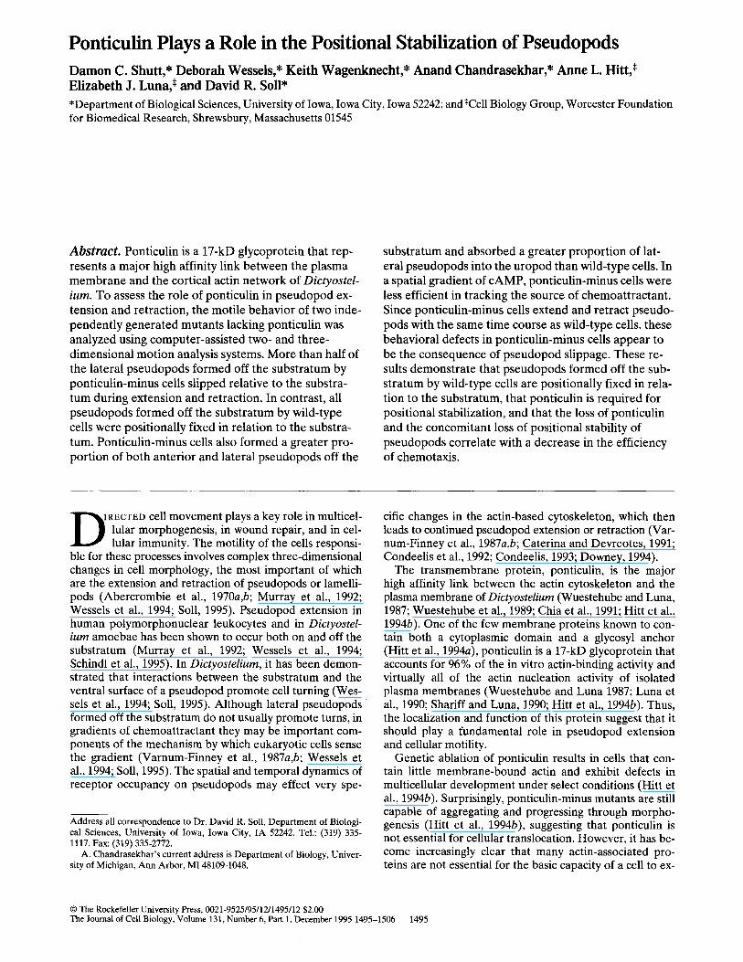
Ponticulin Plays a Role in the Positional Stabilization of Pseudopods Damon C. Shutt,* Deborah Wessels,* Keith Wagenknecht,* Anand Chandrasekhar,* Anne L. Hitt, * Elizabeth J. Luna,* and David R. Soll* • Department of Biological Sciences, University of Iowa, Iowa City, Iowa 52242; and*Cell Biology Group, Worcester Foundation for Biomedical Research, Shrewsbury, Massachusetts 01545 Abstract. Ponticulin is a 17-kD glycoprotein that rep- resents a major high affinity link between the plasma membrane and the cortical actin network of Dictyostel- ium. To assess the role of ponticulin in pseudopod ex- tension and retraction, the motile behavior of two inde- pendently generated mutants lacking ponticulin was analyzed using computer-assisted two- and three- dimensional motion analysis systems. More than half of the lateral pseudopods formed off the substratum by ponticulin-minus cells slipped relative to the substra- tum during extension and retraction. In contrast, all pseudopods formed off the substratum by wild-type cells were positionally fixed in relation to the substra- tum. Ponticulin-minus cells also formed a greater pro- portion of both anterior and lateral pseudopods off the substratum and absorbed a greater proportion of lat- eral pseudopods into the uropod than wild-type cells. In a spatial gradient of cAMP, ponticulin-minus cells were less efficient in tracking the source of chemoattractant. Since ponticulin-minus cells extend and retract pseudo- pods with the same time course as wild-type cells, these behavioral defects in ponticulin-minus cells appear to be the consequence of pseudopod slippage. These re- suits demonstrate that pseudopods formed off the sub- stratum by wild-type cells are positionally fixed in rela- tion to the substratum, that ponticulin is required for positional stabilization, and that the loss of ponticulin and the concomitant loss of positional stability of pseudopods correlate with a decrease in the efficiency of chemotaxis. D tRECTEDcell movement plays a key role in multicel- lular morphogenesis, in wound repair, and in cel- lular immunity. The motility of the cells responsi- ble for these processes involves complex three-dimensional changes in cell morphology, the most important of which are the extension and retraction of pseudopods or lamelli- pods (Abercrombie et al., 1970a,b; Murray et al., 1992; Wessels et al., 1994; Soil, 1995). Pseudopod extension in human polymorphonuclear leukocytes and in Dictyostel- ium amoebae has been shown to occur both on and off the substratum (Murray et al., 1992; Wessels et al., 1994; Schindl et al., 1995). In Dictyostelium, it has been demon- strated that interactions between the substratum and the ventral surface of a pseudopod promote cell turning (Wes- sels et al., 1994; Soil, 1995). Although lateral pseudopods formed off the substratum do not usually promote turns, in gradients of chemoattractant they may be important com- ponents of the mechanism by which eukaryotic cells sense the gradient (Varnum-Finney et al., 1987a,b; Wessels et al., 1994; Soil, 1995). The spatial and temporal dynamics of receptor occupancy on pseudopods may effect very spe- Address all correspondence to Dr. David R. Soil, Department of Biologi- cal Sciences, University of Iowa, Iowa City, IA 52242. Tel.: (319) 335- 1117. Fax: (319) 335-2772. A. Chandrasekhar's current address is Department of Biology, Univer- sity of Michigan, Ann Arbor, MI 48109-1048. cific changes in the actin-based cytoskeleton, which then leads to continued pseudopod extension or retraction (Var- num-Finney et al., 1987a,b; Caterina and Devreotes, 1991; Condeelis et al., 1992; Condeelis, 1993; Downey, 1994). The transmembrane protein, ponticulin, is the major high affinity link between the actin cytoskeleton and the plasma membrane of Dictyostelium (Wuestehube and Luna, 1987; Wuestehube et al., 1989; Chia et al., 1991; Hitt et al., 1994b). One of the few membrane proteins known to con- tain both a cytoplasmic domain and a glycosyl anchor (Hitt et al., 1994a), ponticulin is a 17-kD glycoprotein that accounts for 96% of the in vitro actin-binding activity and virtually all of the actin nucleation activity of isolated plasma membranes (Wuestehube and Luna 1987; Luna et al., 1990; Shariff and Luna, 1990; Hitt et al., 1994b). Thus, the localization and function of this protein suggest that it should play a fundamental role in pseudopod extension and cellular motility. Genetic ablation of ponticulin results in cells that con- tain little membrane-bound actin and exhibit defects in multicellular development under select conditions (Hitt et al., 1994b). Surprisingly, ponticulin-minus mutants are still capable of aggregating and progressing through morpho- genesis (Hitt et al., 1994b), suggesting that ponticulin is not essential for cellular translocation. However, it has be- come increasingly clear that many actin-associated pro- teins are not essential for the basic capacity of a cell to ex- © The Rockefeller University Press, 0021-9525/95/12/1495/12 $2.00 The Journal of Cell Biology, Volume 131, Number 6, Part 1, December 1995 1495-1506 1495
-
Upload
independent -
Category
Documents
-
view
2 -
download
0
Transcript of Ponticulin plays a role in the positional stabilization of pseudopods
Ponticulin Plays a Role in the Positional Stabilization of Pseudopods Damon C. Shutt,* Deborah Wessels,* Keith Wagenknecht,* Anand Chandrasekhar,* Anne L. Hitt, * Elizabeth J. Luna,* and David R. Soll*
• Department of Biological Sciences, University of Iowa, Iowa City, Iowa 52242; and*Cell Biology Group, Worcester Foundation for Biomedical Research, Shrewsbury, Massachusetts 01545
Abstract. Ponticulin is a 17-kD glycoprotein that rep- resents a major high affinity link between the plasma membrane and the cortical actin network of Dictyostel- ium. To assess the role of ponticulin in pseudopod ex- tension and retraction, the motile behavior of two inde- pendently generated mutants lacking ponticulin was analyzed using computer-assisted two- and three- dimensional motion analysis systems. More than half of the lateral pseudopods formed off the substratum by ponticulin-minus cells slipped relative to the substra- tum during extension and retraction. In contrast, all pseudopods formed off the substratum by wild-type cells were positionally fixed in relation to the substra- tum. Ponticulin-minus cells also formed a greater pro- portion of both anterior and lateral pseudopods off the
substratum and absorbed a greater proportion of lat- eral pseudopods into the uropod than wild-type cells. In a spatial gradient of cAMP, ponticulin-minus cells were less efficient in tracking the source of chemoattractant. Since ponticulin-minus cells extend and retract pseudo- pods with the same time course as wild-type cells, these behavioral defects in ponticulin-minus cells appear to be the consequence of pseudopod slippage. These re- suits demonstrate that pseudopods formed off the sub- stratum by wild-type cells are positionally fixed in rela- tion to the substratum, that ponticulin is required for positional stabilization, and that the loss of ponticulin and the concomitant loss of positional stability of pseudopods correlate with a decrease in the efficiency of chemotaxis.
D tRECTED cell movement plays a key role in multicel-
lular morphogenesis, in wound repair, and in cel- lular immunity. The motility of the cells responsi-
ble for these processes involves complex three-dimensional changes in cell morphology, the most important of which are the extension and retraction of pseudopods or lamelli- pods (Abercrombie et al., 1970a,b; Murray et al., 1992; Wessels et al., 1994; Soil, 1995). Pseudopod extension in human polymorphonuclear leukocytes and in Dictyostel- ium amoebae has been shown to occur both on and off the substratum (Murray et al., 1992; Wessels et al., 1994; Schindl et al., 1995). In Dictyostelium, it has been demon- strated that interactions between the substratum and the ventral surface of a pseudopod promote cell turning (Wes- sels et al., 1994; Soil, 1995). Although lateral pseudopods formed off the substratum do not usually promote turns, in gradients of chemoattractant they may be important com- ponents of the mechanism by which eukaryotic cells sense the gradient (Varnum-Finney et al., 1987a,b; Wessels et al., 1994; Soil, 1995). The spatial and temporal dynamics of receptor occupancy on pseudopods may effect very spe-
Address all correspondence to Dr. David R. Soil, Department of Biologi- cal Sciences, University of Iowa, Iowa City, IA 52242. Tel.: (319) 335- 1117. Fax: (319) 335-2772.
A. Chandrasekhar's current address is Department of Biology, Univer- sity of Michigan, Ann Arbor, MI 48109-1048.
cific changes in the actin-based cytoskeleton, which then leads to continued pseudopod extension or retraction (Var- num-Finney et al., 1987a,b; Caterina and Devreotes, 1991; Condeelis et al., 1992; Condeelis, 1993; Downey, 1994).
The transmembrane protein, ponticulin, is the major high affinity link between the actin cytoskeleton and the plasma membrane of Dictyostelium (Wuestehube and Luna, 1987; Wuestehube et al., 1989; Chia et al., 1991; Hitt et al., 1994b). One of the few membrane proteins known to con- tain both a cytoplasmic domain and a glycosyl anchor (Hitt et al., 1994a), ponticulin is a 17-kD glycoprotein that accounts for 96% of the in vitro actin-binding activity and virtually all of the actin nucleation activity of isolated plasma membranes (Wuestehube and Luna 1987; Luna et al., 1990; Shariff and Luna, 1990; Hitt et al., 1994b). Thus, the localization and function of this protein suggest that it should play a fundamental role in pseudopod extension and cellular motility.
Genetic ablation of ponticulin results in cells that con- tain little membrane-bound actin and exhibit defects in multicellular development under select conditions (Hitt et al., 1994b). Surprisingly, ponticulin-minus mutants are still capable of aggregating and progressing through morpho- genesis (Hitt et al., 1994b), suggesting that ponticulin is not essential for cellular translocation. However, it has be- come increasingly clear that many actin-associated pro- teins are not essential for the basic capacity of a cell to ex-
© The Rockefeller University Press, 0021-9525/95/12/1495/12 $2.00 The Journal of Cell Biology, Volume 131, Number 6, Part 1, December 1995 1495-1506 1495
tend a pseudopod and crawl, but are required for the orderly and highly regulated temporal and spatial dynam- ics of anterior and lateral pseudopod extension and retrac- tion (for review see Soil, 1995). The selective absence of such proteins, effected by gene disruption, can result in ab- normalities in the frequency, order, rate of growth and 3-D geometry of pseudopod extension and retraction (Wessels et al., 1988, 1989, 1991, 1996; Wessels and Soil, 1990; Titus et al., 1993). These abnormalities, in turn, can impact sig- nificantly on the efficiency of translocation, chemotaxis, and multicellular morphogenesis (DeLozanne and Spu- dich, 1987; Knecht and Loomis, 1987; Wessels et al., 1988; Condeelis, 1993; Doolittle et al., 1995; Sheldon and Knecht, 1995).
To assess directly the role of ponticulin in single cell mo- tility and chemotaxis, we have used computer-assisted two- and three-dimensional motion analysis systems (Soil, 1988, 1995; Soil et al., 1988; Murray et al., 1992; Wessels et al., 1994; Chandrasekhar et al., 1995) to characterize the motile behavior of aggregation-competent ponticulin-minus cells in buffer and in spatial gradients of cAMP. We have discovered that all lateral pseudopods formed off the sub- stratum by wild-type cells in buffer are fixed positionally in relation to the substratum during extension and retrac- tion, while more than half of the lateral pseudopods formed by ponticulin-minus cells in buffer are not fixed and slip posteriorly along the cell axis. Slippage is very rapid, and results in an abnormally high proportion of pseudopods that are retracted into the uropod rather than into the main cell body. Ponticulin-minus cells also form an abnor- mally high proportion of both anterior and lateral pseudo- pods off the substratum, and these cells are less efficient at chemotaxing in a spatial gradient of the chemoattractant, cAMP. Our results demonstrate that ponticulin plays a novel role in the control of pseudopod position relative to the substratum, and that this spatial stability is an impor- tant component of normal cell motility and chemotaxis.
Materials and Methods
Ponticulin-Minus Mutants The genesis of the ponticulin-minus mutants, Tfl.1 and Tf24.1, in Dictyo- stelium discoideum strain Ax3K was described in detail in a previous re- port (Hitt et al., 1994a). In brief, mutants were generated by homologous recombination with a construct in which a neomycin-resistance cassette was inserted after nucleotide 9 of the ponticulin coding sequence. The sin- gle-copy gene encoding ponticulin was disrupted in both of the indepen- dently generated transformant cell lines, as demonstrated by Southern blot hybridization and by the absence of a diagnostic 2.5-kb PCR product (Hitt et al., 1994a). Northern and Western analyses of Tfl.1 and Tf24.1 confirmed the absence of detectable ponticulin message and ponticulin protein, respectively (Hitt et al., 1994a). Both mutant cell lines were sub- cloned twice in HL-5 medium (Cocucci and Sussman, 1970) and stored as frozen stocks in HL-5 medium containing 10% dimethylsulfoxide.
Development of Wild-type and Mutant Strains Cells from frozen cultures were grown in suspension in HL-5 medium at 22°C and serially transferred for no longer than 3 wk. To initiate develop- ment, cells from the mid-log phase of growth were washed free of nutrient medium with a buffered salt solution (BSS) 1 consisting of 20 mM KCI, 2.4
1. Abbreviations used in thispaper: BSS, buffered salt solution; CI, chemo- tactic index; DIC, differential interference contrast.
mM MgCI2, 0.34 mM streptomycin sulfate, 20 mM KH2PO4, and 20 mM Na2HPO4, pH 6.4 (Sussman, 1987). Cells in BSS were dispersed at high density (5 x 10⁶ cells per cm 2) on a black filter (29; Whatman Inc., Clifton, N J) supported by two prefilters (Millipore Corp., Bedford, MA) saturated with the same solution (Soil, 1987). These conditions, in which cells are multilayered, have been shown to result in highly synchronous develop- ment and highly reproducible developmental timing of Ax3 cells (Soll, 1979). Unless otherwise noted, cells of both wild-type and mutant strains were removed at the ripple stage, which represents the onset of aggrega- tion under these conditions (Soil, 1979), by gently pipetting BSS across the surface of the development filter. The cells were pelleted and washed, and the final cell suspension, in BSS, was gently mixed and adjusted to a con- centration of 3 x 10⁴ cells per ml for inoculation into a perfusion or chemotaxis chamber. Growth cultures of strain Ax2 and strain Ax3-clone RC3 were initiated from desiccated cultures of spores and then developed in the same manner as described above.
Two-Dimensional Analysis of Cell Behavior and Pseudopod Dynamics A 0.3-ml aliquot of cell suspension was inoculated into a Dvorak-Stotler chamber (Nicholson Precision Instruments, Inc., Gaithersburg, MD), the chamber closed, and the cells allowed to settle to the bottom glass cham- ber wall to reinitiate motile behavior. The resulting density on the cham- ber wall was ~33 cells/mm 2. The chamber was then clamped to the stage of an inverted microscope (ICM 405; Carl Zeiss, Inc., Thornwood, NY) equipped with differential interference contrast (DIC) optics for high magnification (x630) recordings. The chamber was perfused with BSS, and the behavior of single cells not in contact with each other videore- corded on half or three-quarter inch tape according to methods previously described (Wessels et al., 1989; Wessels and Soil, 1990). Low magnifica- tion videorecorded cell images were automatically digitized and high mag- nification cell images were manually digitized into the data file of the D[AS program (Wessels et al., 1992; Sylwester et al., 1993, 1995). 2-D mo- tility and morphology parameters were computed according to formulas and methods previously described (Soil, 1988, 1995; Soil et al., 1988). In- stantaneous velocity for a cell in frame (n) was computed by first drawing a line from the cell centroid in the previous frame (n - 1) to the cell c e n -
t r o i d in the subsequent frame (n + 1), and dividing the length of the line by 2At, where At is the time interval between analyzed frames. Positive flow was computed as the percentage of area contained in the expansion zones of difference pictures, generated by overlapping the images in frames n and n - 1 (Soll, 1995). Roundness was computed as (4rr x area/ perimeter 2) x 100. A perfect circle has a roundness measure of 100% and a straight line a roundness measure of 0%. 2-D centroid tracks were ana- lyzed for turning frequency by dividing total translocation time by the number of turns persisting for ~>32 s that caused the direction of the cell centroid to change by/>30 °.
Definition of a Pseudopod In the 2-D analysis of the spatial and temporal dynamics of pseudopod ex- tension and retraction, an anterior pseudopod was defined as a cell exten- sion that formed at the anterior end of the main cell body in the approxi- mate direction (_ 30 °) of the long cell axis, which attained a minimum area of 4 i~m 2 and which was initially free of particulate cytoplasm. The anterior end of a cell was determined from the long axis of the cell estab- lished during the preceding translocation step in the behavior cycle (Wes- sels et al., 1994). A lateral pseudopod was defined as an extension which formed from the side of the cell body in a direction 30 ° to 90 ° from the long cell axis, which attained a minimum area of 4 ~m 2 and which was ini- tially free of particulate cytoplasm. Again, the angle of pseudopod expan- sion was determined from the long axis of the cell established during the preceding translocation step. Comparisons of the videotaped segments of cells recorded through DIC optics and the digitized images of the same cells demonstrated that in all cases pseudopods defined by the preceding criteria in digitized images represented cell extensions in which the apical zones were free of particles. As pseudopods grew, the apical ends re- mained particle free, but the proximal regions filled with particulate cyto- plasm. The apical zones of particle-free cytoplasm corresponded to the F-actin-filled zones of pseudopods, as determined by staining with fluo- rescein-conjugated phaUoidin (Wessels et al., 1989). During pseudopod retraction, the pseudopods were primarily filled with particulate cyto- plasm.
The Journal of Cell Biology, Volume 131, 1995 1496

Three-Dimensional Analysis of Cell Behavior and Pseudopod Dynamics
The methods for optically sectioning living cells, reconstructing 3-D im- ages and wrapping the images for analysis with 3-D DIAS software have been described elsewhere (Murray et al., 1992; Wessels et al., 1994; Soll, 1995). In brief, cells were inoculated into a Dvorak-Stotler chamber, and the chamber was positioned on the stage of a Zeiss ICM405 inverted mi- croscope equipped with DIC optics and perfused with BSS. To obtain op- tical sections, the plane of focus was automatically raised in 1-p~m incre- ments at 0.2-s intervals using a newly developed microstepper motor regulated by a Macintosh-based operating program. Sectioning of a cell was complete in 2 s and repeated every 5 s. Perimeters of the in-focus por- tion of each section were manually digitized into the 3-D DIAS data file.
To illustrate this technique, the original videorecorded DIC images and the manually digitized perimeters of in-focus portions are presented in Fig. 1 A for one 3-D image (i.e., at a single time point). Pseudo-3-D repre- sentations of the stacked optical sections are presented at progressive an- gles in Fig. 1 B. A more representative rendition of the reconstructed cell was then generated by reinserting the in-focus portion of each image into its digitized perimeter, stacking these images and presenting them as pseudo-3-D renditions at progressive angles in Fig. 1 C. Noise removal and dilation-erosion techniques were used to unify perimeters, when nec- essary, in each optical section, and final shapes were generated using 13-splines (SOU, 1995). The final shapes in the stack of optical sections were first connected to form transparent caged images (Fig. 1 D), then wrapped and smoothed to generate nontransparent 3-D reconstructions that most closely reflect the original images (Fig. 1 E). Smoothed images of cells re- constructed at 5-s intervals were then viewed as dynamic animations on the polarizing screen of a stereo monitor (4337; Tektronix, Beavcrton, OR). The final 3-D reconstructions were used to quantitate 3-D param- eters.
In 3-D analyses of pseudopod extension and retraction, the criteria used to define anterior and lateral pseudopods were similar to those used in 2-D analyses. However, instead of using minimum area as a criterion, a minimum vol of 6 i~m 3 was used, and instead of using 2-D shape criteria, 3-D shape criteria were used to interpret extensions. Comparisons of 3-D reconstructions with the original DIC optical sections demonstrated that pseudopods defined by 3-D criteria conformed to the same criteria used in defining a pseudopod in 2-D images.
Pseudopod volume was measured in p~m 3 by encapsulating the 3-D pro- jection in a box which abutted the cell body and then summing the faceted blocks contained in a faceted image of the pseudopod (Wessels et al., 1996). Rates of expansion or contraction were computed in ~m 3 per 5 s.
Error Estimates
To estimate the error in 3-D measurements due to movement of the cell or pseudopod during optical sectioning, we first computed the distance an average cell would move in the course of 2 s, the time required to generate a 3-D image. For an average cell moving 10 ~rn/min, the distance moved in 2 s is 0.33 p.m. Since elongate cells are, on average, 20 ~m in length, the directional skew (the distance error between the bottom and top section due to movement during optical reconstruction) is ~2%. For a pseudopod growing at an average rate of 30 ~zm/min with an average height of 4 IJ, m, the average number of optical sections necessary for reconstruction is four, and the average time for reconstruction is 0.8 s. In this case, the di- rectional skew would be ~10% between the bottom and top sections of the reconstruction.
To estimate volume errors due to missing portions of the pseudopod in optical sections, we modeled a pseudopod as a parabola, then positioned the parabola in the worst positions for analysis (i.e., with the highest and lowest optical sections just missing the top and bottom edges of the pseudopod, respectively). For an average-sized pseudopod 5 i~m in length and 4 p.m in height, the average error is 12%. This low error is due to an automatic interpolation algorithm in the 3D-DIAS program which com- pletes shapes for volume measurements.
To test the reliability and accuracy of in-focus outlining, an Ax3K cell was fixed on a gridded coverslip and optically sectioned at l -~m incre- ments using the DIC method. The cell was then stained with FITC-conju- gated phalloidin, which stains F-actin, but more importantly, provides a high resolution image of F-actin just under the plasma membrane and, therefore, a very accurate outline of the cell. The original cell optically sectioned by DIC was found on the gridded coverslip and optically resec- tioned by confocal microscopy. In the confocal microscopy sectioning pro-
cedure, 40 sections at 0.2-1~m increments were collected. The in-focus pe- rimeters of the DIC sections and the perimeters of the confocal sections were then both digitized into the DIAS data bank. Both the reconstructed DIC and confocal pseudo-3-D images were then generated, and their vol- umes compared. The reconstructed images were visually similar and the difference in computed volume was only 0.4%. The only difference be- tween the two reconstructions was the resolution of a small number of stained filopodia in the confocal reconstruction.
Analysis of Chemotaxis in a Gradient Chamber
To assess the chemotactic index (CI) of individual parental Ax3K and m u -
Figure 1. Optica l sec t ion ing and c o m p u t e r - g e n e r a t e d recons t ruc - t ion of a t r ans loca t ing cell at a s ingle t ime point . (A) T h e or iginal s e q u e n c e of optical sec t ions ob ta ined by dif ferent ia l i n t e r f e rence con t ras t mic roscopy and m a n u a l l y d r awn p e r i m e t e r s o f the in- focus image. Sect ions were r eco rded at 0.2-s in tervals an d were s e p a r a t e d in the z-axis by 1 txm. 0 is at the glass s u b s t r a t u m an d 9 is 9 p~m above the s u b s t r a t u m . (B) T h e digit ized pe r ime t e r s of the in-focus por t ions of the optical sec t ions a re filled and s t acked to c rea te a p s e u d o 3-D recons t ruc t ion . This r econs t ruc t ion is t h e n ro t a t ed for views initially f r o m the side and progress ive ly t o ward the dorsa l surface. (C) E a c h digit ized p e r i m e t e r of t h e in-focus por t ion of the optical sec t ions are refi l led wi th only tha t po r t i on o f the sec t ion which was in focus, and these filled pe r im e t e r s are s t acked and v iewed f rom progress ive ly dorsa l angles. This pro- v ides a m o r e realistic recons t ruc t ion . (D) T h e p la tes in the image in B are c o n n e c t e d by facets . Th i s t r a n s p a r e n t image is u sed for c o m p u t i n g 3-D m o r p h o l o g y and mot i l i ty p a r a m e t e r s (Soil, 1995). Two views are p r e s e n t e d of the face ted image . (E) T h e face ted image is t h e n s m o o t h e d and w r a p p e d to ob ta in a n o n t r a n s p a r e n t image which m o s t closely r ep r e sen t s the original cell. Two views are p r e s e n t e d o f the w r a p p e d image , a, an te r io r end of cell; u, u ropod .
Shuttet al. Ponticulin Is a Pseudopod Anchor 1497

tant cells in a spatial gradient of the chemoanractant cAMP, a dilute sus- pension of cells in BSS was dispersed on the bridge of a gradient chamber which consisted of a 2-mm plexiglass bridge bordered on either side by parallel troughs that were 2 mm wide and 1 mm deep (Zigmond, 1977; Varnum and Soil, 1984; Varnum-Finney et al., 1987b). BSS containing 1.0 ~M cAMP was placed in the trough serving as the "source," and BSS alone was placed in the trough serving as the "sink" (Varnum and Soil, 1984; Varnum-Finney et al., 1987b). Cells were allowed to acclimate for 4 min and were then videorecorded for 12 min. The CI of a cell was com- puted from the centroid track as the directional distance (net distance to- ward the source) divided by the total distance traveled (McCutcheon, 1946; Varnum and Soil, 1984). A CI of 0 reflects no net movement to- wards or away from the source of attractant; a CI of +1.0 reflects persis- tent movement directly towards the source (i.e., up the gradient); and a CI of -1.0 reflects persistent movement directly away from the source (i.e., down the gradient). All cells analyzed were separated from other cells on the bridge by at least two cell body lengths.
Results
Developmental Regulation of Motility
Since the rate of single cell motility is regulated by the de- velopmental program of Dictyostelium (Varnum et al., 1986), we first identified conditions that permitted com- parison of wild-type Ax3K and ponticulin-minus mutant cells at comparable developmental stages. In a previous report (Hitt et al., 1994b), ponticulin-minus cells were demonstrated to exhibit a shorter preaggregation period than Ax3K cells when developed at low density on agar containing Sorensen's buffer (14.6 mM KH2PO4, 2.0 mM Na2HPO4, pH 6.0). We found that when cells were plated four deep on development filters saturated with BSS (Fig. 2) or were developed on agar with Sorensen's buffer con- taining 2.4 mM MgC12 (data not shown), the preaggrega- tion period and timing of subsequent stages of develop- ment were similar for wild-type and mutant cells. In BSS, Ax3K and Tf24.1 cells progressed simultaneously through the ripple (R), loose aggregate (LA) and tight aggregate (TA) stages at 7.0, 8.5, and 9.5 h, respectively (Fig.2). This latter developmental protocol, therefore, provided the temporal parallelism needed for a valid comparison of be- havioral phenotypes, and was used in all studies reported here.
The developmental regulation of motility in both wild- type and mutant cells was also similar through the first 9 h of development under these conditions. In Fig. 2, the mean instantaneous velocity of individual Ax3K and Tf24.1 cells are plotted as functions of developmental time. The mean instantaneous velocity of both cell types was ~2 ixm/min during the first 6 h of development, increased to peak val- ues of 9.5 and 11.5 ixm/min, respectively, at 7 h of develop- ment, and then decreased to 3.5 txm/min at 9 h of develop- ment. These results are highly similar to those originally reported for wild-type strain Ax3-clone RC3 (Varnum et al., 1986). Subsequent comparisons of wild-type and ponti- culin-minus cells were performed with ripple stage cells re- moved from filters after 7 h of development (Fig. 2, R), at peak instantaneous velocity for all three strains.
Two-Dimensional Motility and Shape Parameters
We initially considered two possible outcomes of a pond- culin-minus phenotype. First, we considered the possibility that a cell lacking ponticulin and, therefore, most of the
,..., 13q .E 124 I~114 71o-i
9.
6- 5- 4-
1- - - O -
/ \ / \
t
R LA TA
TIME (at)
Figure 2. Developmental regulation of single cell motility in wild-type Ax3K and mutant Tf24.1 cultures. Cells were grown to late log phase in nutrient medium and were dispersed on devel- opment filters at high cell density in BSS (Soil, 1987). At various time points, developing cultures were disaggregated, single cells were inoculated into a Dvorak-Stotler chamber at low density, and the chamber was perfused with buffer, Cell behavior was video-recorded for 10-min periods at high magnification through DIC optics. The perimeters of 25 randomly selected ceils at each time point were digitized into the 2D-DIAS data file at 4-s inter- vals, and their average instantaneous velocities were computed over the 10-min period of analysis. Each data point represents the mean _+ standard deviation of the average instantaneous veloci- ties of the 25 analyzed cells. The developmental stages of Ax3K and Tf24.1 cells were identical in these high-density cultures: R, ripple stage (onset of aggregation); LA, loose aggregate stage; TA, tight aggregate stage. --I~, Ax3K; -~5)--, Tf24.1.
high affinity linkages between actin and the plasma mem- brane (Hitt et al., 1994b), would translocate at a slower ve- locity than wild-type cells if the ponticulin-based connec- tions were required for pseudopod extension. Alternatively, we considered it possible that a cell lacking ponticulin might translocate at a faster velocity than wild-type cells if ponti- culin-actin connections have to be disrupted for normal pseudopod extension.
A 2-D analysis of general motility parameters suggests that ponticulin-minus cells translocate at instantaneous ve- locities similar to or slightly higher than those of wild-type Ax3K cells (Table I). Both the mean instantaneous veloc- ity and the mean positive flow, a measure of area displace- ment with time (Soil et al., 1988; Soil, 1988, 1995), were slightly greater for mutant cells than for wild-type ceils, al- though the differences were statistically significant only for Tfl.1 cells vs. wild-type cells (Table I). There were no significant differences in the velocity cycle (Wessels et al., 1994). The mean period between velocity peaks of Ax3K cells was 1.23 _+ 0.29 min (n = 24), and that of Tfl.1 and Tf24.1 cells 1.06 --- 0.20 (n = 33) and 1.44 _+ 0.75 (n = 21) min, respectively. The differences between mutant and wild-type values were in both cases insignificant (P > 0.35). There was also no significant difference between wild-type and mutant cell shape (Table I). Mean maximum length, mean maximum width, mean area, and mean roundness of wild-type and mutant cells were statistically indistinguish- able, with values very similar to those previously reported for other wild-type strains of Dictyostelium (Wessels et al., 1991; Cox et al., 1992). We, therefore, conclude that ponti- culin-minus and wild-type cells crawl in buffer with similar instantaneous velocities, velocity cycles, and general shape.
The Journal of Cell Biology, Volume 131, 1995 1498

Table I. Motility and Morphology Measurements of Parental Ax3K and Ponticulin-Minus Transformants (Tfl.1, Tf24.1) Translocating in Buffer
Mean. Mean Mean Mean Mean Mean Inst. Vel. Pos. Flow Max. Len. Max. Wid. Area Round.
izm/min % area bun prn txm 2 %
A x 3 K (parental) 9.2 -+ 2.2 15.9 -+ 3.1 19.2 -- 3.8 9.7 - 1.8 97.9 -+ 26.9 43.0 ± 9.8 (n = 26)
T f l . l 11.1 + 3.0 19.5 -+ 6.4 21.7 + 4.1 9.6 ± 1.6 105.9 ± 25.7 38.8 ± 9.4 (n = 23)
Tf24.1 10.9 ± 4.4 16.1 ± 5.7 21.5 ± 6.6 9.3 -+ 1.0 115.8 + 38.1 44.7 ± 6.8 (n = 13)
P values ~ A x 3 K vs. Tf l .1 0.02 0.02 0.04 NS NS NS
A x 3 K vs. Tf24.1 NS NS NS NS NS NS
*Determinations were made from 2-D cell perimeters manually digitized at 4-s intervals. *See Materials and Methods for explanations of Parameters. (Mean Inst. Vel.), Mean Instantaneous Velocity; (Mean Pos. Flow), Mean Positive Flow; (Mean Max. Len.), Mean Maximum Length; (Mean Max. Wid.), Mean Maximum Width; Mean Area; Mean Round, (Mean Roundness). All values represent the mean and standard deviation for the number of cells (n) noted. *The Student t test was used to assess significance. A P value/>0.05 was considered not significant.
Formation of Lateral and Anterior Pseudopods
Computer-generated 2-D tracks of the digitized perime- ters of ponticulin-minus cells translocating in buffer gave the impression of being slightly wider, on average, than tracks of wild-type cells. Because the width of a perimeter track is affected by the number, shape, and dynamics of lateral pseudopods, and because the majority of lateral pseudopods formed by wild-type cells in buffer are ex- tended off the substratum and are then retracted (Wessels et al., 1994; Soll, 1995), aberrations in the dynamics of lat- eral pseudopods could affect the width of a perimeter track without significantly affecting instantaneous veloc- ity. To explore the possibility of abnormalities in the shape or position of lateral pseudopods formed by ponticulin- minus cells, we analyzed pseudopod formation in three di- mensions (Murray et al., 1992; Wessels et al., 1994, 1996; Soll, 1995).
In dynamic 3-D reconstructions, we observed that wild- type Ax3K cells and ponticulin-minus cells of strain Tfl.1
and Tf24.1 extended lateral pseudopods at comparable frequencies (Table IIA). However, lateral pseudopods formed by Tfl.1 and Tf24.1 cells were significantly more likely to be formed off the substratum than were lateral pseudopods formed by wild-type cells. Only 10-15% of the lateral pseudopods of ponticulin-minus cells were formed on the substratum, i.e., with their ventral surface in contact with the substratum (Table IIA). By contrast, 40% of the lateral pseudopods of wild-type cells were formed on the substratum. This latter proportion is very similar to that previously reported for cells of the wild-type strain Ax3, clone RC3 (Wessels et al., 1994). Therefore, the pro- portion of lateral pseudopods formed on the substratum by ponticulin-minus cells was approximately three times smaller than that of wild-type cells.
Since lateral pseudopods formed on the substratum have a greater propensity for initiating sharp turns than do lat- eral pseudopods formed off the substratum (Wessels et al., 1994), one would expect wild-type cells translocating in
Table II. The Proportion of Lateral and Anterior Pseudopods Formed on and off the Substratum by Ax3K, Tfl.1, and Tf24.1 Cells Translocating in Buffer
Strain No. cells Pseudopods Pseudopods Proportion formed off substratum Proportion formed on substratum
A. La t e r a l P s e u d o p o d s No. lateral Lateral per 5 min.
A x 3 K 15 93 6.2 ± 2.4 60 ± 23% 40% ± 23 Tf l .1 4 31 7.8 --- 3.6 90 - 8% 10% z 8
Tf24.1 5 39 7.8 ± 0.4 85 ± 14% 15% - 14
P values*
A x 3 K vs Tf l .1 NS 0.02 Ax3k vs Tf24.1 NS 0.04
B. A n t e r i o r P s e u d o p o d s
No. anterior Anterior per 5 min.
A x 3 K 6 22 3.8 ± 0.7 40 ± 25% 60% ± 25% Tf l .1 4 18 4.2 ± 1.1 73 ± 15% 27% ± 15% Tf24.1 6 25 4.5 --- 1.0 80 ±- 12% 22% ± 12%
P values* A x 3 K vs. T f l . 1 NS 0.02 A x 3 K vs. Tf24,1 NS 0.04
Determinations were made from dynamic three-dimensional reconstructions generated at 5-s intervals. *Student t-test was used to assess significance; similar results were obtained with the nonparametric Mann-Whitney test. A P value/>0.05 was considered not significant.
Shutt et al. Ponticulin Is a Pseudopod Anchor 1499
buffer to turn more often than mutant cells, since a greater proport ion of their lateral pseudopods are formed on the substratum. We, therefore, counted the number of sharp turns in centroid tracks which persisted for/>32 s. The re- sults supported the above prediction. While centroid tracks of Ax3K cells contained turns at a frequency of 0.74 +_ 0.20 per rain (n = 15), the centroid tracks of ponticulin-minus cells (Tfl.1 and Tf24.1) contained turns at a frequency of 0.45 -+ 0.34 (n = 25). The difference was significant, with a P < 0.01.
We also examined the proport ion of anterior pseudo- pods formed on and off the substratum. Again, the pro- portion of anterior pseudopods formed on the substratum by ponticulin-minus cells was only about one-third that observed for wild-type cells (Table IIB). These results sug- gest that at least some of the defects described above for the more easily characterized lateral pseudopods also are present in anterior pseudopods.
Fates of Lateral Pseudopods
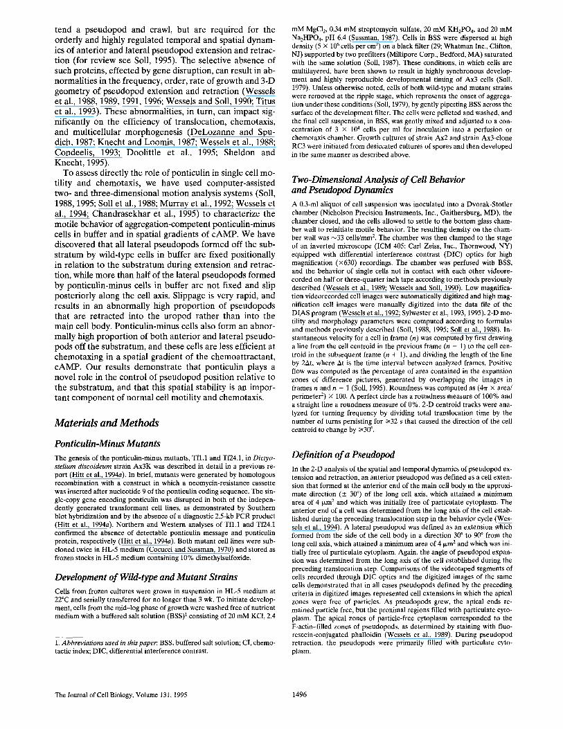
The ultimate fates of lateral pseudopods formed off the substratum differed between wild-type and ponticulin- minus cells. The majority of the lateral pseudopods formed off the substratum by Ax3K cells were retracted back into the main cell body. An example of normal pseudopod re- traction by an Ax3K cell is presented in the sequence of wrapped 3-D reconstructions shown in Fig. 3. At 0 s, this cell had no lateral pseudopods, a tapered anterior end which had lifted off the substratum, and a slightly bifur- cated uropod. Between 0 and 5 s, the cell extended a lat- eral pseudopod (rl) from the right side of the anterior por- tion of the main cell body. As is evident in the horizontal view at 5 s (bottom), the ventral surface of pseudopod r l
was not in contact with the substratum. Pseudopod r l was fully extended at 10 s (not shown) and was retracted into the main cell body between 10 and 20 s, as the cell again extended the original anterior pseudopod along the sub- stratum. Since retraction was completed at the main cell body, the uropod retained its original shape. Throughout the process of pseudopod extension and retraction, the ventral surface of pseudopod r l did not contact the sub- stratum.
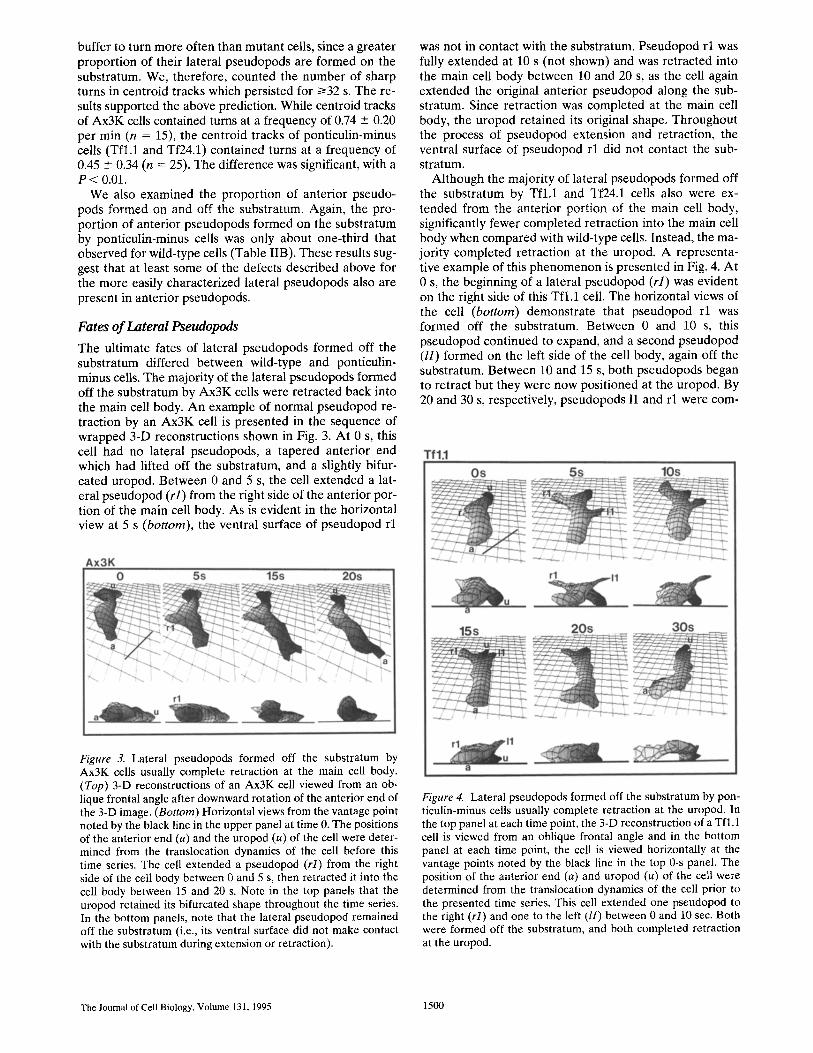
Although the majority of lateral pseudopods formed off the substratum by Tfl,1 and Tf24.1 cells also were ex- tended from the anterior portion of the main cell body, significantly fewer completed retraction into the main cell body when compared with wild-type cells. Instead, the ma- jority completed retraction at the uropod. A representa- tive example of this phenomenon is presented in Fig. 4. At 0 s, the beginning of a lateral pseudopod (rl) was evident on the right side of this Tfl.1 cell. The horizontal views of the cell (bottom) demonstrate that pseudopod rl was formed off the substratum. Between 0 and 10 s, this pseudopod continued to expand, and a second pseudopod (//) formed on the left side of the cell body, again off the substratum. Between 10 and 15 s, both pseudopods began to retract but they were now positioned at the uropod. By 20 and 30 s, respectively, pseudopods 11 and r l were corn-
Figure 3. Lateral pseudopods formed off the substratum by Ax3K cells usually complete retraction at the main cell body. (Top) 3-D reconstructions of an Ax3K cell viewed from an ob- lique frontal angle after downward rotation of the anterior end of the 3-D image. (Bottom) Horizontal views from the vantage point noted by the black line in the upper panel at time 0. The positions of the anterior end (a) and the uropod (u) of the cell were deter- mined from the translocation dynamics of the cell before this time series. The cell extended a pseudopod (rl) from the right side of the cell body between 0 and 5 s, then retracted it into the cell body between 15 and 20 s. Note in the top panels that the uropod retained its bifurcated shape throughout the time series. In the bottom panels, note that the lateral pseudopod remained off the substratum (i.e., its ventral surface did not make contact with the substratum during extension or retraction).
Figure 4. Lateral pseudopods formed off the substratum by pon- ticulin-minus cells usually complete retraction at the uropod. In the top panel at each time point, the 3-D reconstruction of a Tfl.l cell is viewed from an oblique frontal angle and in the bottom panel at each time point, the cell is viewed horizontally at the vantage points noted by the black line in the top 0-s panel. The position of the anterior end (a) and uropod (u) of the cell were determined from the translocation dynamics of the cell prior to the presented time series. This cell extended one pseudopod to the right (rl) and one to the left (H) between 0 and 10 sec. Both were formed off the substratum, and both completed retraction at the uropod.
The Journal of Cell Biology. Volume 131, 1995 1500

Table IlL Rates of Expansion and Contraction of Lateral Pseudopods Formed off the Substratum in Wild-type and Ponticulin-Minus Cells
No. Avg. Rate of Expansion Avg. Rate of Retraction Strain Pseudopods Expansion Period Retraction Period
tun3~5 s s Izm3/5 s s
A x 3 K 15 15 --- 10 29 - 22 8 ± 5 45 - 27 Tf l .1 + Tf24.1 15 11 - 18 34 ± 20 5 ± 4 39 ± 16
P value* NS NS NS NS
Measurements are based on a 3-D analysis. *Student t-test was used to assess significance. A P value/>0.05 was considered not significant.
Figure 5. Histograms of the proportions of lateral pseudopods formed off the substratum by wild-type (Ax3K) and mutant (Tfl.1, Tf24.1) cells that were retracted into the main cell body or absorbed into the uropod. (A) A 3-D analysis of 20 Ax3K, 28 Tfl.1, and 33 Tf24.1 pseudopods. (B) A 2-D analysis of 58 Ax3K, 59 Tfl.1, and 63 Tf24.1 pseudopods. Means and standard devia- tions for the proportion of retracted pseudopods completing re- tractions at the main cell body for Ax3K, Tfl.1, and Tf24.1 cells in the 3-D study were 81 _+ 24, 23 - 19, and 36 -+ 17%, and at the uropod 19 _ 24, 77 _ 19, and 64 _+ 17%. Means and standard de- viations for the proportion completing retraction at the main cell body for Ax3K, Tfl.1, and Tf24.1 in the 3-D study were 83 +_ 14, 25 _ 15, and 45 -+ 21%, and at the uropod 17 -+ 14, 75 -+ 15, and 55 ___21%. The differences in the proportions of pseudopods re- tracted into the main cell body between Ax3K and Tfl.1 and be- tween Ax3K and Tf24.1 were statistically significant, with P val- ues <0.05.
pletely absorbed. The uropod, in this case, changed its shape as a consequence of absorption. Such shape changes were always observed concommitant with absorption at the uropod, suggesting that they are diagnostic for this phe- nomenon.
The difference between wild-type and mutant cells in the position along the cell body at which pseudopod retraction is completed was quantified using 3-D (Fig. 5 A) and 2-D (Fig. 5 B) analyses. In both cases, ~80% of the lateral pseudopods formed off the substratum by wild-type Ax3K cells were retracted into main cell body. In marked con- trast, up to 75% of the pseudopods extended off the sub- stratum by ponticulin-minus cells completed retraction at the uropod.
Pseudopods of Ponticulin-Minus Cells Slip Posteriorly
One explanation for the positional difference in lateral pseudopod retraction (Fig. 5) is that pseudopods in ponti- culin-minus cells may exhibit a longer average lifespan due to a decrease in the rate of pseudopod extension and/or re- traction. For a cell rapidly translocating in buffer, a longer- lived lateral pseudopod would be more likely to remain extended as the cell translocated anteriorly, and the conse- quence would be completion of retraction at the uropod. To test this possibility, we analyzed the rates and periods of expansion and retraction of lateral pseudopods (Table III). The results showed no significant differences between Ax3K and ponticulin-minus cells in the rate of expansion, the rate of retraction, or the longevity of lateral pseudopods formed off the substratum.
Another explanation for the positional difference in lat- eral pseudopod retraction (Fig. 5) is that the lateral pseudopods of ponticulin-minus cells may be positionally unstable, (i.e., more inclined to slip posteriorly relative to the cell body and/or the substratum), and, therefore, more likely to complete retraction at the uropod. In wild-type cells, we discovered that the position of every lateral pseudopod formed off the substratum remained fixed in relation to the substratum through the entire extension and retraction process, even during translocation of the cell (Fig. 6; Table IV). This positional invariance was ob- served both for the majority of lateral pseudopods, which were retracted into the main cell body (Fig. 6 A, r2), and for the majority of lateral pseudopods, which completed retraction at the uropod (Fig. 6 A, rl). Positional invari- ance of each lateral pseudopod formed off the substratum was observed not only for the Ax3K parental strain in this study, but also for strain Ax3, clone RC3 (Table IV, Fig. 6 B), and strain Ax2 (Table IV), two other laboratory strains of Dictyostelium discoideum that have been used in previous computer-assisted analyses of cell motility (Wes- sels et al., 1989, 1992, 1994). Of 104 lateral pseudopods formed by 14 wild-type axenic cells, 100% remained fixed in relation to the substratum, i.e., none showed positional slippage (Table IV).
In marked contrast, lateral pseudopods formed off the substratum by ponticulin-minus cells were not positionally fixed relative to the substratum (Fig. 7; Table IV). Half or more of these lateral pseudopods changed positions in re- lation to the substratum during expansion and retraction (Table IV). Most of these unfixed pseudopods slipped pos- teriorly; none were observed to slip towards the front of the cell. In the case of pseudopod rl , formed by the repre- sentative Tfl.1 cell in Fig. 7, the distance that the base of the pseudopod slipped in relation to the substratum (noted by a dotted line at 10 s) was 6.25 I~m, which represents a minimum retrograde velocity of 75 p~m/min. Pseudopod 11 on this same cell slipped 5 ~m in the same 5-s period and an additional 5 p~m in the next 5-s period, which represents a minimum retrograde velocity of 60 ~m/min. Of 76 lateral pseudopods formed by 11 ponticulin-minus cells, over half slipped and in all cases they did so in the posterior direc- tion (Table IV). Every mutant cell analyzed for a period of 3-10 min exhibited slippage of one or more pseudopods (Table IV).
Pseudopod slippage in ponticulin-minus cells also oc- curred in the z-axis (Fig. 8). In some cases, lateral pseudo- pods that were formed off the substratum by ponticulin-
Shutt et al. Ponticulin Is a Pseudopod Anchor 1501

Table IV. Lateral Pseudopod Slippage in Ponticulin-Minus (Tfl.1, Tf24.1) Cells
Percentage Percentage cells No. lateral pseudopod exhibiting
Strain No. cells pseudopods slippage slippage*
Ax3K 5 53 0 0 Ax3(RC3) 4 26 0 0 Ax2 5 25 0 0 Tf l . 1 6 47 49 100 Tf24.1 5 29 55 100
The relationship of the base of each pseudopod to the substratum was determined dur- ing extension and retraction from 2-D perimeters hand-digitized at 4-s intervals. *The proportion of cells exhibiting at least one case of lateral pseudopod slippage dur- ing the period of analysis, which was between 3 and i0 min per cell.
repor ted propor t ions of sl ippage by lateral pseudopods of Tfl .1 and Tf24.1 cells in Table IV are probably underest i- mates.
Chemotactic Inefficiency o f Ponticulin-Minus Cells
Latera l pseudopods that are ex tended off the subst ra tum have been suggested to play a key role in sensing chemo- tactic gradients (Varnum-Finney et al., 1987b; Wessels et al., 1994; Soll, 1995) and in initiating turns in the correct direct ion (i.e., in the direct ion of increasing c A M P concen- tration). Thus, cells with lateral pseudopods that frequently slip posteriorly and/or dorsally might be expected to be less efficient in tracking a spatial gradient (see Discussion).
Figure 6. The base of each lateral pseudopod formed by wild- type cells remains fixed relative to the substratum during both ex- tension and retraction. (A) A representative Ax3K cell; (B) A representative Ax3-clone RC3 cell. The reconstructions are viewed dorsally so that the position of the base of each pseudo- pod can be established and marked by an arrow on the underly- ing grid. The positions of the anterior end (a) and uropod (u) of the cell at 0 s of each time sequence were determined from the translocation dynamics of the cell before the presented time se- ries. The Ax3K cell in A had already formed a lateral pseudopod to its right (rl) before 0 s and another to its right (r2) between 0 and 5 s. The Ax3-RC3 cell in B formed a lateral pseudopod to its left (H) between 0 and 5 s.
minus cells appeared to be absorbed into the main cell body when viewed dorsal ly (Fig. 8, top). However , when the same cells were viewed from a horizontal vantage point, it was clear that the pseudopod had ro ta ted to the dorsal sur- face of the cell and then s l ipped poster ior ly to the u ropod (Fig. 8, bottom). Thus, the top views of the representa t ive Tf24.1 cell in Fig. 8 failed to discriminate between pseudo- pod retract ion and circumferential s l ippage to the dorsal surface of the cell body. In the case of the cell in Fig. 8, the pseudopod on the dorsal surface of the cell s l ipped 7.5 ixm toward the poster ior end of the cell (Fig. 8, dotted line) be- tween 15 and 25 s, which represents a re t rograde velocity of 45 p~m/min. A t 25 s, this dorsal pseudopod was posi- t ioned at the uropod. Since the propor t ion of pseudopods which s l ipped poster ior ly (Table IV) was de te rmined from 2-D reconstruct ions in which dorsal s l ippage such as that demons t ra ted in Fig. 7 would not have been detected, the
Figure 7. Lateral pseudopods formed by ponticulin-minus cells are not anchored to the substratum and slip posteriorly. The an- terior end (a) and uropod (u) of this representative Tfl.1 cell were determined from the translocation dynamics before the pre- sented time series. The reconstructions are viewed dorsally. Ar- rows denote the original position of the base of each pseudopod in relation to the substratum on the underlying grid. This repre- sentative Tfl.1 cell formed one pseudopod to the right (rl) and one pseudopod to the left (H) between 0 and 5 s. Both pseudo- pods then slipped posteriorly, and the distances that they slipped are noted by dotted lines. This figure represents the same cell and time series viewed from oblique and horizontal angles in Fig. 3, which showed that neither rl nor 11 contacted the substratum.
The Journal of Cell Biology, Volume 131, 1995 1502

Figure 8. In some cases, lateral pseudopods formed off the sub- stratum by ponticulin-minus cells rotate to the dorsal surface of the cells as they slip posteriorly. In the top panel at each time point, the re- construction of the representative Tf24.1 cell is viewed dorsally, and in the bottom panel at each time point, the cell is viewed horizon- tally from the vantage point noted by the black line in the top panel at 0 s. This particular ponticulin- minus cell had formed a pseudo- pod to its left (ll) before 0 s. In the dorsal views, this pseudopod appeared to be retracted between
5 and 15 s; in the horizontal views, it is clear that the pseudopod first slipped circumferentially to the dorsal surface of the cell and then slipped posteriorly. Black arrow, the original position of the pseudopod in relation to the substratum. Dotted lines, the distance it slips in relation to the substratum.
The mean CI of ponticulin-minus cells was significantly less than that of wild-type cells (Fig. 9). The mean CI for Ax3K cells was +0.45 _ 0.29 (n = 66), while that for Tfl .1 was +0.30 --- 0.29 (n = 48) and that for Tf24.1 was +0.30 +- 0.30 (n = 52). The difference be tween the mean CIs of Ax3K and Tfl .1 cells, and the CIs of Ax3K and Tf24.1 cells were highly significant. In the former case, the P value was 0.006 and in the la t ter case 0.007. In contrast , the P value for the mean CIs of Tfl .1 and Tf24.1 was 0.986, demonst ra t ing an ext remely high similari ty be tween the two mutant strains. The difference in the distr ibutions of CIs of wild-type and mutant cells is demons t ra ted in the his togram in Fig. 9, in which the pooled da ta for the two mutants is compared with that of wild-type cells. Perhaps even more impor tan t than the shift in the average CI is the dramat ic decrease in the propor t ion of mutant cells that achieve high end CIs. While 33% of wild-type cells exhib- i ted CIs of 0.6-0.8, only 17% of mutant cells did so, and while 8% of wild-type cells exhibited CIs of 0.8-1.0, no mu- tant cells were this efficient.
The decrease in the chemotact ic efficiency of ponticulin- minus cells appears to be due to an increase in the frequency of sharp turns away from the direct ion of the gradient (i.e., away from the direct ion of increasing concentra t ion of chemoat t rac tant ) . In Fig. 10, A and B, the per imeter and centroid tracks of a wild-type cell and a ponticul in-minus cell with comparab le ins tantaneous velocities and very high CIs are compared. Al though both cells were moving up the spatial gradient , the mutant cell made two small lat- eral deviat ions that lowered its chemotact ic index. In each case, the deviat ion was due to a mis taken turn (i.e., a turn away from the direct ion of the source) genera ted by exten- sion of a la teral pseudopod. These deviat ions genera ted kinks in the centroid t rack (Fig. 10 B). A n examinat ion of the t racks of the 10 Ax3K and 10 pont icul in-minus cells with the highest CIs demons t ra ted that mutant cells, on average, made more lateral turning mistakes. In Fig. 9, C and D, per imete r and centroid tracks are presented for a wild-type cell and ponticulin-minus cell with similar instan- taneous velocit ies and CIs close to the median. Again, the net direct ion of migrat ion of the wild-type and the mutant cell is in the direct ion of the source of chemoat t rac tant ,
but the mutant cell made more turning mistakes than the wild-type cell. A n examinat ion of the 10 Ax3K and 10 ponticul in-minus cells with CIs closest to the mean of each group demons t ra ted that mutant cells, on average, made more lateral turning mistakes.
Discussion
We demonst ra te here that la teral pseudopods formed off the subst ra tum by wild-type Dictyostelium amoebae trans- locating in buffer remain fixed in relat ion to the substra- tum as they are ex tended and retracted, even though the main cell body may continue to t ranslocate anteriorly. Be- cause the ventral surfaces of la teral pseudopods formed off the subs t ra tum are not in contact with the substratum,
4 0 -
0 Z 35 ~ I I A x 3 K !
= 4 i ~ 2 5 - -
2 0 . . . . .
1 5 . . . .
10
<0.0 0.O to >0.2 to >0.4 to >0,6 to >0.8 to 0.2 0.4 0.6 0.8 1 ,O
CHEMOTACTIC INDEX
Figure 9. The high end of chemotactic indices is underrepre- sented in ponticulin-minus cells chemotaxing in a spatial gradient of the chemoattractant, cAMP. Ax3K (filled bars) and ponticu- lin-minus cells (hatched bars) were distributed on the bridge of a gradient chamber bordered by a trough filled with buffer only and a trough filled with buffer plus 1 IxM cAMP. After 4 min, cel- lular behavior was recorded, and the CI of each cell was com- puted according to the formula described in Materials and Meth- ods. Data were pooled from six separate experiments with Ax3K cells and from four separate experiments with each of the pon- ticulin-minus cell lines. The mean CI +_ SD and the number of an- alyzed cells for Ax3K and the pooled mutant cells were 0.45 _ 0.29 (n = 66) and 0.30 -+ 0.29 (n = 100), respectively. The difference between the means is highly significant (P < 0.01) determined both by the T test and the Mann-Whitney nonparametric test.
S h u t t e t a l . Ponticulin Is a Pseudopod Anchor 1 5 0 3

A W..T.cI=o.84 B Mut.cI,G77 )
C W.T.cl=0.50 D Mut.cl,~40
Figure 10. Perimeter and centroid plots of a wild-type cell (.4) and a ponticulin-minus cell (B) with comparable instantaneous velocities and very high chemotactic indices (CI), and of a wild- type cell (C) and a ponticulin-minus cell (D) with comparable in- stantaneous velocities and CIs close to the median.
these pseudopods must be anchored to the substratum through interactions between the substratum and cortical and/or plasma membrane proteins in that portion of the cell body from which the pseudopod extends. Presumably, these interactions involve actin-based supramolecular cy- toskeletal structures that connect the substratum with the plasma membrane, the cortex of the cell body and the cor- tex of the pseudopod.
We also have demonstrated here that ponticulin, a trans- membrane protein responsible for most of the high affinity binding between F-actin and the Dictyostelium plasma membrane (Wuestehube and Luna, 1987; Wuestehube et al,, 1989; Chia et al., 1991; Hitt et al., 1994a,b), is required for positionally fixing lateral pseudopods relative to the substratum, since the lateral pseudopods formed off the substratum by these mutant cells undergo frequent and dramatic slippage. Slippage can occur during both exten- sion and retraction, and occurs primarily in the posterior direction. Pseudopods can also slip circumferentially to the top of a cell, and then slip posteriorly. Such circumferen- tial slippage may be responsible for the significant increase in the proportion of both lateral and anterior pseudopods formed off the substratum by ponticulin-minus cells, since pseudopods that slip dorsally have less chance of contact- ing and subsequently extending along the substratum (Wes- sels et al., 1994).
This basic behavioral defect of ponticulin-minus cells is quite specific to this mutant. Ponticulin-minus cells trans-
locating in buffer still extend pseudopods with the same frequency and growth dynamics as wild-type cells, and re- tract pseudopods with the same dynamics as wild-type cells. They exhibit the same periodicity in their velocity cycle as wild-type cells and can readily sense a gradient of chemoat- tractant, although with less efficiency. Their basic behav- ioral defect is, specifically, the loss of the positional stabili- zation of lateral pseudopods in relation to the substratum and subsequent pseudopod slippage. This mutant pheno- type leads to a number of additional behavioral defects and contrasts markedly with that of myosin IA and B mu- tants, which are defective primarily in the frequency of pseudopods formed on the substratum (Wessels et al., 1991, 1996; Titus et al., 1993). It also contrasts with that of ABP-120 mutants, which are defective in the frequency of pseudopod formation and the rate and extent of pseudo- pod expansion (Cox et al., 1992; Cox, D., D. Wessels, D. R. Soil, J. Hartwig, and J. Condeelis, manuscript submitted for publication), with that of myolI mutants, which are de- fective in polarity, the original position of pseudopod ex- tension, and the rate and extent of pseudopod growth (Wessels et al., 1988; Wessels and Soil, 1990), and with that of the discoidinless mutants, which are capable of rapid translocation in spite of their aberrant blunt morphology and loss of a conventional tapered uropod (Alexander et al., 1992).
Pseudopod slippage in ponticulin-minus cells correlates with a number of behavioral defects for cells translocating in buffer and for cells chemotaxing in a spatial gradient of cAMP. All of these defects may be consequences of slip- page. First, ponticulin-minus cells translocating in buffer complete the process of pseudopod retraction at a more posterior cellular position than wild-type cells. In wild- type cells, most lateral pseudopods formed off the substra- tum extend from the anterior portion of the main cell body and are retracted primarily into the middle or posterior portion of the main cell body, just anterior to the uropod. This appears to be a consequence of the forward movement of the cell and the fixed position of the pseudopod in relation to the substratum. In wild-type cells, the morphological in- tegrity of the uropod is, therefore, maintained throughout the 3-D behavior cycle of most cells (Wessels et al., 1994; Soil, 1995). In marked contrast, most pseudopods formed off the substratum by ponticulin-minus cells complete the retraction process at the uropod, with an accompanying characteristic alteration of uropod morphology. Pseudo- pod slippage obviously accounts for posteriorization of the retraction process in these mutants.
Ponticulin-minus cells translocating in buffer also exhibit altered turning behavior. Pseudopods formed by wild-type cells off the substratum have a greater propensity for being retracted and are less likely to initiate a sharp turn than pseudopods formed on the substratum (Wessels et al., 1994). Ponticulin-minus cells form a far greater proportion of their pseudopods off the substratum, which most likely explains why they translocate in buffer with a lower fre- quency of turning.
Finally, ponticulin-minus cells exhibit a decrease in the efficiency of chemotaxis. This decrease is due to an increase in turning mistakes made by ponticulin-minus cells moving up a spatial gradient of the chemoattractant cAMP. Super- ficially, this observation appears to contradict the observa-
The Joumal of Cell Biology, Volume 131, 1995 1504

tions made on turning in buffer, namely that slippage leads to a decrease in turning. However, inappropriate pseudo- pod slippage would be expected to lead to a depression in chemotactic efficiency since there is strong reason to be- lieve that the spatial stability of lateral pseudopods is nec- essary for sensing a spatial gradient of chemoattractant and reorienting (Varnum-Finney et al., 1987b; Soil et al., 1993; Soil, 1995). Cells moving towards the source of a spa- tial gradient form lateral pseudopods at roughly one-third the frequency of cells moving in buffer, and the fewer lat- eral pseudopods which do form generate turns at a lower frequency (Varnum-Finney et al., 1987b), suggesting that they are formed primarily off the substratum and, there- fore, have a greater propensity for being retracted. In ad- dition, cells oriented at an angle towards the source of chemoattractant form as many lateral pseudopods towards as away from the source, but turn into the former two to three times as frequently as they do into the latter (Var- num-Finney et al., 1987b). These results suggest that lat- eral pseudopods formed by cells in a spatial gradient sense the direction of the gradient and either fall to the substra- tum to initiate a turn when sensing a positive gradient or remain off the substratum and are retracted when sensing a negative gradient (Soil, 1995). Within this context, the loss of the positional stability of pseudopods by ponticulin- minus cells would diminish the efficiency of chemotaxis by three possible mechanisms. First, if a lateral pseudopod rotates to the dorsal surface of a cell, it would be perpen- dicular to the gradient of chemoattractant and, therefore, incapable of sensing it. This outcome would depress the average chemotactic index because cells veering off track would produce fewer compensating sharp turns (Soil et al., 1993). Second, if a lateral pseudopod slipped posteriorly along the cell axis, it would also move through the gradient in a direction of decreasing concentration, which would confuse a temporal and/or spatial mechanism of gradient assessment (Soil et al., 1993; Soil, 1995). Again, this out- come would depress the average chemotactic index by de- creasing the efficiency of reorientation. Third, if a lateral pseudopod slipped to the substratum during sensing, it would initiate an inappropriate turn, and result in a de- pression in chemotactic index. It is likely that all three out- comes contribute to the decrease in chemotactic efficiency observed in ponticulin-minus cells.
Therefore, the loss of the positional stability of pseudo- pods and the corresponding decrease in chemotactic effi- ciency of ponticulin-minus cells suggests that ponticulin is involved in at least one step in the chemotactic signal- response pathway of Dictyostelium. One intriguing possi- bility is that ponticulin is involved in stabilizing a sensing pseudopod as it extends through a spatial gradient of at- tractant off the substratum, and that a pseudopod which senses an increasing concentration of attractant (i.e., is ex- tending up a concentration gradient towards the source of attractant) falls to the substratum to initiate a turn by de- stabilizing the pseudopod through the regulation of pon- ticulin.
Posterior slippage of lateral pseudopods along the long axis of cells shares some morphological similarities to the capping of cell surface receptors. However, these two phe- nomena occur at dramatically different velocities. Lateral pseudopods formed by ponticulin-minus mutants slip pos-
teriorly at N50-60 tzm/min, which is about an order of magnitude faster than the velocity with which Dictyostel- ium amoebae cap beads coated with lectin (Pasternak et al., 1989; Jay and Elson, 1992). The velocity of lateral pseudopod slippage is also at least five times faster than the mean instantaneous velocity of cellular translocation. The velocity of pseudopod slippage is, however, compara- ble to the instantaneous velocities of intraceUular particles moving within the central cytoplasm of rapidly translocat- ing wild-type Dictyostelium amoebae (Wessels and Soil, 1990), but these particles were observed to move primarily towards the front of the cell. Since the velocity of these forward moving particles was roughly five times that of cellular translocation, there would either have to be disso- lution of the particles at the anterior edge of the particu- late cytoplasm or a retrograde compensatory current of particles, perhaps in the cytoplasmic cortex, to account for the fact that particles do not accumulate in the front of a cell. The absence of posterior slippage of lateral pseudo- pods in wild-type cells suggests that ponticulin is integral to a plasma membrane-associated cytoskeletal structure that normally resists the suggested rearward flow of corti- cal cytoplasm. Posterior pseudopod slippage in ponticulin- minus cells is consistent with the contraction-hydraulic (Mast, 1926; Jahn, 1964) and cortical flow (Bray and White, 1988) models of cellular translocation, in which fountains of submembranous cytoplasm (Grebecki, 1994) move rear- ward in regions of the cell not attached to the substratum.
The absence of posterior pseudopod slippage in wild- type cells suggests that ponticulin is involved in a plasma membrane-associated cytoskeletal structure (membrane skeleton) that normally resists the putative rearward flow of cytoplasm. Ponticulin-based anchorage of lateral pseudo- pods relative to the substratum is consistent with the gen- eral role played by membrane skeleton proteins in sepa- rating regions of the plasma membrane into discrete functional domains (for review see Luna and Hitt, 1992). Another implication of our results is that pseudopods, al- though dynamic in nature, may be thought of as plasma membrane domains that are stabilized and regulated by membrane skeleton proteins, such as ponticulin. In con- junction with other recently described regions of mem- brane specialization in motile cells (for review see Sheetz, 1995), our observations suggest that the membrane skele- tons of motile cells are at least as complex as, though much less well understood than, the membrane skeletons of more static cells such as erythrocytes and epithelial cells.
Because the formation of pseudopods on and off the substratum and the periodicity of the motility cycle of Dic- tyosteliurn amoebae translocating in buffer are strikingly similar to several behavioral aspects of polymorphonu- clear leukocytes (Murray et al., 1992) and of T cells and gi- ant HIV-induced T cell syncytia (Sylwester et al., 1993, 1995), a final implication of our results is that precise tem- poral and spatial regulation of pseudopod dynamics may be a general requirement for the taxis of many chemotacti- cally responsive mammalian cells. Therefore, proteins with structural and functional similarities to Dictyostelium pon- ticulin may play critical roles in morphogenesis, the cellu- lar immune system, and other biological processes in which amoeboid cells must move in a directed fashion through extension of actin-based pseudopods.
Shutt et al. Ponticulin Is a Pseudopod Anchor 1505

The authors are indebted to Dr. Edward Voss for helping run the 3D- DIAS program and for computing 3D-DIAS errors, and to Jim Trepka for developing the precision stepper motor for optical sectioning.
This research was supported in part by grant H D 18577 from the Na- tional Institutes of Health (NIH) to D. R. Soil, by a grant from the Iowa Economic Development Commission to D. R. Soil to develop 3D-DIAS~ by a grant from Wallace Technologies to D. R. Soil to develop the mi- crostepper motor, and by grant GM33048 from NIH to E. J. Luna.
Received for publication 4 May 1995 and in revised form 29 September 1995.
References
Abercrombie, M., J. E. M. Heaysman, and S. M. Pegrurn. 1970a. The locomo- tion of fibroblasts in culture. I. Movements of the leading edge. Exp. Cell Res. 59:393-398.
Abercrombie, M,, J. E. M. Heasyman, and S. M. Pegrum. 1970b. The locomo- tion of fibroblasts in culture, II. Ruffling. Exp. Cell Res. 60:437--444,
Alexander, S., L. Sydow, D. Wessels, and D. R. Soil. 1992. Discoidin proteins of Dictyostelium are necessary for normal cytoskeletal organization and cellu- lar morphology during translocation. Differentiation. 51:149-161.
Bray, D., and J. G. White. 1988. Cortical flow in animal ceils. Science (Wash. DC). 239:883--888.
Caterina, M. J., and P. N. Devreotes. 1991. Molecular insights into eukaryotic chemotaxis. FASEB Z 5:3078-3085.
Chandrasekhar, A., D. Wessels, and D. R. Soil. 1995. A mutation that abolishes cGMP phosphodiesterase activity in Dictyostelium selectively affects cell be- havior in the back of the chemotactic wave. Dev. Biol. 169:109-122.
Chia, C. P., A. L. Hilt, and E. J. Luna. 1991. Direct binding of F-actin to pontic- ulin, an integral plasma membrane glycoprotein. Cell Motil. Cytoskeleton. 18:164-179.
Cocucci, S., and M. Sussman. 1970. RNA in cytoplasmic and nuclear fractions of cellular slime mold amebae. J. Cell Biol. 45:399-407.
Condeelis, J. 1993, Life at the leading edge: the formation of cell protrusions. Annu. Rev. Cell Biol. 9:411--444.
Condeelis, J., J. Jones, and J. E. Segall. 1992. Chemotaxis of metastatic tumor cell. Clues to mechanisms from the Dictyostelium paradigm. Cancer Metasta- sis Rev. 11:55-68.
Cox, D., J. Condeelis, D. Wessels, D. R. Soil, H. Kern, and D. A. Knecht. 1992. Targeted disruption of the ABP-120 gene leads to cells with altered motility. J. Cell BioL 116:943-955.
DeLozanne, A., and J. A. Spudich. 1987. Disruption of the Dictyostelium myo- sin heavy chain gene by homologous recombination. Science (Wash. DC). 236:1086-1091.
Doolittle, K. W., I. Reddy, and J. G. McNally. 1995.3D analysis of cell move- ment during normal and mysoin II null cell morphogenesis in Dictyostelium. Dev. BioL 167:118-129.
Downey, G. P. 1994. Mechanisms of leukocyte motility and chemotaxis. Curr. Opin. lmmunoL 6:113-124.
Grebecki, A. 1994. Membrane and cytoskeleton flow in motile ceils with em- phasis on the contribution of free-living amoebae. Int. Rev. Cytol. 148:37-80.
Hitt, A. L, T. H. Lu, and E. J. Luna. 1994a. Ponticulin is an atypical membrane protein. Z Cell Biol. 126:1421-1431.
Hitt, A. L., J. H. Hartwig, and E. J. Luna. 1994b. Ponticulin is the major high af- finity link between the plasma membrane and the cortical actin network in Dictyostelium. Z Cell Biol. 126:1433-1444.
.lahn, T. L. 1964. Relative motion in Amoeba proteus. In Primitive Motile Sys- tems in Cell Biology. R. D. Allen and N. Kamiya, editors. Academic Press, Inc, New York. 279-302.
Jay, P. J., and E. L. Elson. 1992. Surface particle transport mechanism indepen- dent of myosin II in Dictyostelium. Nature (Lond.). 356:438-440.
Knecht, D. A., and W. F. Loomis. 1987. Antisense RNA inactivation of myosin heavy chain gene expression in Dictyostelium discoideum. Science (Wash. DC). 236:1081-1086.
Luna, E. J, and A. L. Hitt. 1992. Cytoskeleton-plasma membrane interactions. Science (Wash. DC). 258:955-964.
Luna, E. J., L. J. Wuestehube, C. P. Chia, A. Shariff, A. L. Hitt, and H. M. In- galls. 1990. Ponticulin, a developmentally-regulated plasma membrane gly- coprotein, mediates actin binding and nucleation. Dev. Genet. 11:354-361.
Mast, S. O. 1926. Structure, movement, locomotion and stimulation in Amoeba. J. Morphol. Physiol. 41:347-425.
McCutcheon, M. 1946. Chemotaxis in leukocytes. Physiol. Rev. 26:401--404. Murray, J., H. Vawter-Hugart, E. Voss, and D. R. Soil. 1992. A three-dimen-
sional motility cycle in leukocytes. Cell Motil. Cytoskeleton. 22:211-223. Pasternak, C., J. A. Spudich, and E. L. Elson. 1989. Capping of surface recep-
tors and concomitant cortical tension are generated by conventional myosin. Nature (Lond.). 341:549-551.
Schindl, M., E. Wallraff, B. Deubzer, W. Witke, G. Gerisch, and E. Sackman.
1995. Cell substrate interactions and locomotion of Dictyostelium wild-type and mutants defective in three cytoskeletal proteins: a study using quantita- tive reflection interference contrast microscopy. Biophys. Z 68:1177-1190.
Shariff, A., and E. J. Luna. 1990. Dictyostetium discoideum plasma membranes contain an actin-nucleating activity that requires ponticulin, an integral membrane glycoprotein. J. Cell BioL 110:681-692.
Sheetz, M. 1995. Cellular plasma membrane domains. MoL Membr. BioL 12:89-91. Sheldon, E., and D. A. Knecht. 1995. Mutants lacking myosin If cannot resist
forces generated during multicellular morphogenesis. J. Cell Sci. In press, Soil, D. R. 1979. Timers in developing systems. Science (Wash. DC). 203:841-
849. Soil, D. R. 1987. Methods for manipulating and investigating developmental
timing in Dictyostelium discoideum. In Methods of Cell Biology. Vol. 28. J. Spudich, editor. Academic Press, Inc. San Diego, CA, 413-431.
Soil, D. R. 1988. "DMS," a computer-assisted system for quantitating motility, the dynamics of cytoplasmic flow and pseudopod formation: its application to Dictyostelium chemotaxis. Cell MotiL Cytoskeleton. 10:91-106.
Soil, D. R., E. Voss, B. Varnum-Finney, and D. Wessels. 1988. The "dynamic morphology system": a method of quantitating changes in shape, pseudopod formation and motion in normal mutant amebae of Dictyostelium discoi- deum. J. Cell. Biochem. 37:177-192.
Soil, D. R., D. Wessels, and A. Sylwester. 1993. The motile behavior of amoe- bae in the aggregation wave in Dictyostelium discoideum. In Experimental and Theoretical Advances in Biological Pattern Formation. H. G. Othmer, P. K. Maini, and J. D. Murray, editors. Plenum Publishing Corp. New York. 325-338.
Soil, D. R. 1995. The use of computers in understanding how cells crawl. Int. Rev. CytoL In press.
Sussman, M. 1987. Cultivation and synchronous morphogenesis of Dictyostel- ium under controlled experimental conditions. In Methods in Cell Biology. Vol. 28. J. Spudich, editor. Academic Press, Inc, San Diego, CA. 413-431.
Sylwester, A., D. Wessels, S. A. Anderson, R. Q. Warren, D. Shutt, R. Kennedy, and D. R. Soil. 1993. HIV-induced syncytia of a T cell line form a single giant pseudopod and are motile. J. Cell ScL 106:941-953.
Sylwester, A., D. Shutt, D. Wessels, J. T. Stapleton, J. Stites, R. Kennedy, and D. R. Soil. 1995. T cells and HIV-induced T cell syncytia exhibit the same ve- locity when translocating on plastic, collagen or endothelial monolayers. J. Leukocyte Biol. 57:643-650.
Titus, M. A., D. Wessels, J. A. Spudich, and D. R. Soil. 1993. The unconven- tional myosin encoded by the myoA gene plays a role in Dictyostelium mo- tility. Mot. Biol. Cell 4:233-246.
Varnum, B., and D. R. Soil. 1984. Effects of cAMP on single cell motility in Dictyostelium. J. Cell Biol. 99:1151-1155.
Varnum, B., K. Edwards, and D. R. Soil. 1986. The developmental regulation of single cell motility in Dictyostelium discoideum. Dev. BioL 113:218-227.
Varnum-Finney, B., E. Voss, and D. R. Soil. 1987a. Amebae of Dictyostelium discoideum respond to an increasing temporal gradient of the chemoattrac- tant cAMP with a reduced frequency of turning: evidence for a temporal mechanism in ameboid chemotaxis. Cell Motil. Cytoskeleton. 8:7-17,
Varnum-Finney, B., E. Voss, and D. R. Soil. 1987b. Frequency and orientation of pseudopod formation of Dictystelium discoideum amebae chemotaxing in spatial gradient: further evidence for a temporal mechanism. Cell Motil. Cy- toskeleton. 8:18--26.
Wessels, D., and D. R. Soil. 1990. Myosin II heavy chain null mutant of Dictyo- stelium exhibits defective intracellular particle movement. Z Cell Biol. 111: 1137-1148.
Wessels, D., D. R. Soil, D. Knecht, W. F. Loomis, D. DeLozanne, and J. Spu- dich. 1988. Cell motility and chemotaxis in Dictyostelium amebae lacking myosin heavy chain. Dev. Biol. 128:164-177.
Wessels, D., N. Schroeder, E. Voss, A. L. Hall, J. Condeelis, and D. R. Soil. 1989. cAMP-mediated inhibition of intracetlular particle movement and ac- tin reorganization in Dictyostelium. J. Cell BioL 109:2841-2851.
Wessels, D., J. Murray, G. Jung, J. Hammer, and D. R. Soil. 1991. Myosin IB null mutant of Dictyostelium exhibits abnormalities in motility. Cell Motil. Cytoskeleton. 20:301-315.
Wessels, D., J. Murray, and D. R. Soil. 1992. The complex behavior cycle of chemotaxing Dictyostelium amoebae is regulated primarily by the temporal dynamics of the natural wave. Cell MotU. Cytoskeleton. 23:145-156.
Wessels, D., H. Vawter-Hugart, J. Murray, and D. R. Soil. 1994. Three-dimen- sional dynamics of pseudopod formation and the regulation of turning dur- ing the motility cycle of Dictyostelium. Cell Motil. Cytoskeleton. 27:1-12.
Wessels, D., M. Titus, and D. R. Soil. 1996. Dictyostelium myosin I plays a cru cial role in regulating the frequency of pseudopods formed on the substra- tum. Cell Motil. Cytoskeleton. In press.
Wnestehube, L. J., and E. J. Luna. 1987. F-actin binds to the cytoplasmic sur- face of ponticulin, a 17-kD integral glycoprotein from Dictyostelium discoi- deum plasma membranes.Z Cell Biol. 105:1741-1751.
Wuestehube, L. J., C. P. Chia, and E. J. Luna. 1989. Immunofluorescence local- ization of ponticulin in motile cells. Cell Motil. Cytoskeleton. 13:245-263.
Zigmond, S. H. 1977. The ability of polymorphonuclear leukocytes to orient in gradients of chemotactic factors. Z Cell Biol. 75:606-616.
The Journal of Cell Biology, Volume 131, 1995 1506