Arf4 Is Required for Mammalian Development but Dispensable for Ciliary Assembly

*Department of Physiology and Biophysics, State University of Campinas, Campinas, Brazil
�Departments of Pharmacology and Anesthesiology and #Veterinary Science, University of Padova (CRIBI), Padova, Italy
�Department of Microbiology and Immunology, State University of Campinas, Campinas, Brazil
Ciliary neurotrophic factor (CNTF) is a 23 kDa cytoplasmicprotein which is expressed in both the PNS and CNSbeginning in the late embryonic period (Barbin et al. 1984;Stockli et al. 1991). CNTF exhibits a variety of activities,affecting both the differentiation and survival of neuronal andglial cells (Ernsberger et al. 1989; Oppenheim et al. 1991;Louis et al. 1993), and also shares structural and functionalproperties with the cytokines interleukin-6, leukemia inhib-itory factor, interleukin-11, and oncostatin M (Bazan 1991).For this reason, CNTF is often referred to as a ‘neurokine’(Taga and Kishimoto 1992). CNTF promotes the survival ofmotor neurons in culture (Arakawa et al. 1990; Oppenheimet al. 1991), and prevents the degeneration of motor neuronsafter axotomy (Sendtner et al. 1990) and in mouse mutant
progressive motor neuronopathy (Sendtner et al. 1992).These pre-clinical observations encouraged the potential forCNTF as a therapy for human motor neuron disease.
Received March 2, 2009; accepted March 25, 2009.Address correspondence and reprint requests to Alessandro Negro,
CRIBI, University of Padova, Via G. Colombo, 3, 35121 Padova, Italy.E-mail: [email protected] used: CD, circular dichroism; CNTF, ciliary neuro-
trophic factor; DRG, dorsal root ganglion; HIV, human immunodeficiencyvirus; JAK, just another kinase; mNGF, mouse nerve growth factor; P2,postnatal day 2; PBS, phosphate-buffered saline; PTD, protein transduc-tion domain; rhCNTF, recombinant human CNTF; SDS–PAGE, sodiumdodecyl sulfate–polyacrylamide gel electrophoresis; STAT, signal trans-ducers and activators of transcription; TAT, transactivator.
Abstract
Ciliary neurotrophic factor (CNTF) is a multifunctional cytokine
that can regulate the survival and differentiation of many types
of developing and adult neurons. CNTF prevents the degen-
eration of motor neurons after axotomy and in mouse mutant
progressive motor neuronopathy, which has encouraged trials
of CNTF for human motor neuron disease. Given systemi-
cally, however, CNTF causes severe side effects, including
cachexia and a marked immune response, which has limited
its clinical application. The present work describes a novel
approach for administering recombinant human CNTF
(rhCNTF) while conserving neurotrophic activity and avoiding
deleterious side effects. rhCNTF was fused to a protein
transduction domain derived from the human immunodefi-
ciency virus-1 TAT (transactivator) protein. The resulting fu-
sion protein (TAT-CNTF) crosses the plasma membrane
within minutes and displays a nuclear localization. TAT-CNTF
was equipotent to rhCNTF in supporting the survival of cul-
tured chicken embryo dorsal root ganglion neurons. Local or
subcutaneous administration of TAT-CNTF, like rhCNTF
rescued motor neurons from death in neonatal rats subjected
to sciatic nerve transection. In contrast to subcutaneous
rhCNTF, which caused a 20–30% decrease in body weight in
neonatal rats between postnatal days 2 and 7 together with a
considerable fat mobilization in brown adipose tissue, TAT-
CNTF lacked such side effects. Together, these results indi-
cate that rhCNTF fused with the protein transduction domain/
TAT retains neurotrophic activity in the absence of CNTFs
cytokine-like side effects and may be a promising candidate
for the treatment of motor neuron and other neurodegenera-
tive diseases.
Keywords: ciliary neurotrophic factor, fusion protein, motor
neuron, neuroprotection, protein transduction domain, weight
loss.
J. Neurochem. (2009) 109, 1680–1690.
JOURNAL OF NEUROCHEMISTRY | 2009 | 109 | 1680–1690 doi: 10.1111/j.1471-4159.2009.06091.x
1680 Journal Compilation � 2009 International Society for Neurochemistry, J. Neurochem. (2009) 109, 1680–1690� 2009 The Authors
However, these trials revealed adverse events, such asanorexia and weight loss, and increased deaths at the highestdose level (Miller et al. 1996a,b).
In addition to preventing neuronal cell degeneration,systemically administered CNTF causes fever, acute-phaseresponse, weight loss, and cachexia in experimental animals(Shapiro et al. 1993; Dittrich et al. 1994; Fantuzzi et al.1995; Espat et al. 1996; Henderson et al. 1996). The effectsof CNTF on body weight change and food intake in mice aretransient (Fantuzzi et al. 1995; Espat et al. 1996), and theirmechanism is unknown. Receptor subunits for CNTF sharesequence similarity with the receptor for leptin (Chen et al.1995), an adipocyte-derived cytokine involved in bodyweight homeostasis (Henderson et al. 1994; Campfield et al.1995; Halaas et al. 1995; Pelleymounter et al. 1995). CNTFcorrects obesity and diabetes associated with leptin defi-ciency and resistance, indicating an action independent of theleptin system (Gloaguen et al. 1997). A recent studysuggests that recombinant human CNTF (rhCNTF) reducesweight partly by regulating nuclear respiratory factor 1 andmitochondrial transcription factor A (Liu et al. 2007). Assuch, CNTF is considered as an anti-injury factor and anti-obesity agent (Zvonic et al. 2005; Watt et al. 2006a).
Ciliary neurotrophic factor lacks a classical signal peptidesequence that serves to direct polypeptides out of the cellfollowing synthesis. It is likely that CNTF resides within thecell, only to be released after injury. Strategies whichfacilitate intracellular entry of CNTF following administra-tion would thus be of therapeutic interest. To this end, wedesigned a fusion protein between a protein transductiondomain (PTD) (Schwarze et al. 1999) derived from the TAT(transactivator) transduction domain of human immunodefi-ciency virus (HIV) and human CNTF, to promote cellularuptake following injection. The TAT fusion protein strategy iswell suited for intracellular translocation of proteins (Gump &Dowdy 2007). The TAT-CNTF protein thus obtained retainedin vitro neurotrophic activity comparable to rhCNTF, andrescued axotomized motor neurons following sciatic nervetransaction. Unlike rhCNTF, the fusion protein did not affectanimal weight or fat tissue mobilization. These data validatethe application of designing fusion proteins with a PTD forpotential therapeutic benefit (Gump & Dowdy 2007).
Materials and methods
Construction of pTAT-ciliary neurotrophic factor bacterialexpression vectorThe pTAT-CNTF plasmid was constructed to express the basic
domain (amino acids 47–57) of HIV-1 TAT fused with human
CNTF. The coding sequence for human CNTF was amplified by
PCR using the pT7.7 CNTF vector (Negro et al. 1991) as template
and the following primers: 5¢-GCTCTAGAATGGCTTTCACAGA-GCATTCACCG-3¢ and 5¢-GCGAATTCACATTTTCTTGTTGTTA-
GCAATA-3¢. The PCR product was digested with XbaI and EcoRIrestriction sites and cloned into NheI and EcoRI sites of pTAT-Ngbplasmid (Peroni et al. 2007), which contains an N-terminal six-
histidine sequence, followed by the transduction domain of the TAT
protein.
Expression and purification of TAT-ciliary neurotrophic factorfusion proteinBL21(DE3)LysS Escherichia coli transformed with pTAT-CNTF
plasmid were plated on Luria broth agar-coated Petri dishes and
grown at 37�C overnight. The cells were then suspended in 2 L of
Luria broth and cultured at 37�C until reaching an absorbance of
OD600 = 0.6. Protein expression was induced by adding 0.4 mM
isopropyl-b-D-thiogalactopyranoside. After 3 h, the cells were
harvested by centrifugation, washed with phosphate-buffered saline
(PBS), and lysed by sonication with buffer A (200 mM NaCl and
20 mM Tris/HCl, pH 8). The lysate was cleared by centrifugation
(30 min at 11 000 g) and then incubated with 3 mL of wet Ni(II)
nitriloacetic acid agarose in buffer A for 1 h; 1 · 10 cm column was
filled with the protein-adsorbed agarose resin and exhaustively
washed with buffer A. The TAT-CNTF fusion protein was eluted with
buffer B (200 mM NaCl, 20 mM Tris/HCl, and 250 mM imidazole,
pH 8) and dialyzed against buffer A. The purity of TAT-CNTF was
evaluated on 12% sodium dodecyl sulfate–polyacrylamide gel
electrophoresis (SDS–PAGE) and stained with Coomassie brilliant
blue. The purified protein was analyzed also by reverse phase HPLC
using a C4 Jupiter column (Phenomenex, Torrance, CA, USA).
Molecular mass were determined using an ESI-MS Mariner System
5220 (Applied Biosystems, Foster City, CA, USA) equipped for
time-of-flight analysis. Spectra were deconvoluted with Data
Explorer 4.0.0.1 software (Applied Biosystems). The obtained
molecular weight of 22 780.1 was in good agreement with a
theoretical molecular mass of 22 799 for the fusion protein; rhCNTF
was produced as described previously (Negro et al. 1991), withminor modifications. The protein concentration was calculated using
a double-beam Perkin-Elmer (Waltham, MA, USA) lambda 25 UV/
Vis spectrophotometer, and the absorption coefficient at 280 nm
calculated according to Gill and von Hippel (1989) (taken as 1.27/
mg/cm2 for rhCNTF and 1.16/mg/cm2 for TAT-CNTF). Far-UV
circular dichroism (CD) spectra (200–250 nm) were recorded at 22–
24�C using a Jasco (Tokyo, Japan) model 715 spectropolarimeter and
a protein concentration of 100–200 lM; results were expressed as
mean residue ellipticity. The final spectrum was obtained by
averaging five scans, and the baseline corrected by subtracting the
spectrum contributed by the buffer.
In vitro transduction of TAT-ciliary neurotrophic factorChinese Hamster Ovary cells and SH-SY5Y neuroblastoma cells
were routinely cultured at 37�C/5% CO2 in Roswell Park
Memorial Institute medium supplemented with 10% fetal calf
serum, penicillin (100 U/mL), and streptomycin (100 lg/mL). For
transduction, cell monolayers were washed with PBS and
incubated in OptiMEM (Invitrogen, Gaithersburg, MD, USA)
then incubated with TAT-CNTF (100 nM) or rhCNTF (100 nM)
for 5–10 min, washed with PBS and fixed with 2% p-formalde-
hyde. Protein transduction was evaluated by immunocytochemis-
try. Cells were permeabilized with 0.5% Triton X-100/PBS at 4�Cfor 10 min, saturated with 1% bovine serum albumin in PBS, and
� 2009 The AuthorsJournal Compilation � 2009 International Society for Neurochemistry, J. Neurochem. (2009) 109, 1680–1690
Therapeutic use of ciliary neurotrophic factor | 1681

incubated at 37�C for 2 h with anti-CNTF monoclonal antibody
8E2 (Cazzola et al. 1993). The cells were next washed with PBS
and incubated at 37�C for 30 min with alkaline phosphatase-
conjugated secondary antibody. After washing with PBS, alkaline
phosphatase activity was detected using an alkaline phosphatase
detection kit (Chemicon, Temecula, CA, USA), following the
manufacturer’s instructions. Cells were observed using a Zeiss
Axiovert 100 microscope (Carl Zeiss, Oberkochen, Germany)
equipped with a 16-bit digital charged-coupled device video
camera (MicroMAX; Princeton Instruments/Roper Scientific Tren-
ton, NJ, USA). Alternatively SH-SY5Y cells were lysed in SDS
sample buffer and boiled for 5 min, and proteins were resolved
on 12% SDS–PAGE followed by western blotting on immobilon-
P membranes (Millipore, Bedford, MA, USA) Non-specific
binding sites were blocked with 3% bovine serum albumin in
PBS before incubation with anti-CNTF monoclonal antibodies for
1 h at 22�C. The membrane was rinsed twice and then incubated
for 1 h with alkaline phosphatase-conjugated secondary antibody
(Sigma, St Louis, MO, USA) and developed with nitro blue
tetrazolium chloride and 5-bromo-3-indolyl-phosphate (Roche,
Indianapolis, IN, USA).
Assay for neurotrophic activitySurvival of purified chicken embryonic day 10 dorsal root ganglion
(DRG) neurons was assayed essentially as described previously
(Skaper et al. 1990). Briefly, neurons were seeded at a density of
�2000 cells/well in laminin- and polyornithine-coated 96-well
tissue culture plates (Falcon BD Biosciences, San Jose, CA, USA)
containing 100 lL culture medium (Dulbecco’s modified Eagle’s
medium, 2 mM L-glutamine, 100 U/mL penicillin, and 10% fetal
calf serum. Following a 48 h treatment with TAT-CNTF or
rhCNTF, or mouse nerve growth factor (mNGF) (Chemicon),
cultures were fixed with 2% glutaraldehyde and viable neurons
were counted under phase contrast microscopy.
STAT3 phosphorylation in vitro and in vivoThe human neuroblastoma cell line SH-SY5Y was obtained from
Dr June Bidler (Sloan Kettering Institute for Cancer Research).
The cells were cultured at 37�C in a humidified incubator under
5% CO2 using a 1 : 1 mixture of Ham’s F12 and Eagle’s minimal
essential medium supplemented with 10% (v/v) fetal calf serum,
50 U/mL penicillin, and 50 lg/mL streptomycin. At 80% conflu-
ence, cells were placed in serum-free medium for 1–5 h before
initiation treatment (1 nM TAT-CNTF or rhCNTF). After several
times (0–60 min), cell monolayers were harvested in SDS–PAGE
sample buffer and proteins (50–100 lg) were resolved by 8%
SDS–PAGE following by western blotting as described below.
Two-day-old (postnatal day; P2) Wistar rat pups (n = 12) were
anesthetized by placing on crushed ice for 3 or 4 min, until
painful stimulation ceased to elicit movements. Experimental
procedures were approved by the Committee on Animal Care of
the State University of Campinas (protocol 509-1). The left sciatic
nerve was transected at the sciatic foramen level and �3 mm of
the distal stump was removed. The musculature was repositioned
and the skin was closed with two stitches of 8-0 silk suture. After
surgery, the rats were allowed to recover from hypothermia under
an incandescent lamp. Rats received subcutaneous administration
of rhCNTF (1.2 lg/g, n = 4) or TAT-CNTF (1.2 lg/g, n = 4)
dissolved in PBS. Dosing was carried out 30 min prior to sciatic
nerve transection, immediately following axotomy, and 2 h post-
axotomy. The control group was treated at the same times with
PBS (n = 4). One hour after the last dosing, rats were killed by
decapitation and the spinal cord and hypothalamus were collected
and immediately frozen in liquid nitrogen. For total protein,
samples were homogenized in 1% Triton X-100, 50 mM phos-
phate buffer, pH 7.4, 1 mM sodium pyrophosphate, 1 mM sodium
fluoride, 5 mM EDTA, 1 mM sodium vanadate, 1% protease
inhibitor cocktail (P8340; Sigma), 7 M urea, and 2 M thiourea
(10% w/v). Sample homogenization was carried out at 4�C using
a Polytron (Metrohm, Riverview, FL, USA) 20s generator set at
maximum speed for 30 s. Insoluble materials were removed by
centrifugation (12 000 g, 4�C, 15 min). Protein concentration was
determined using the Bradford method. One hundred micrograms
of total protein extract from each sample was eletrophoretically
separated by 8% SDS–PAGE and electroblotted to a nitrocellulose
membrane. Membranes were blocked with PBS–Tween containing
5% non-fat dry milk and then incubated with an anti-p-signal
transducers and activators of transcription 3 (STAT3) antibody
(Tyr705; Cell Signaling, Beverly, MA, USA) diluted (1 : 1000) in
PBS–Tween containing 3% bovine serum albumin (12 h at 4�C).Membranes were washed with PBS–Tween and incubated with
HRP-labeled secondary antibody (1 : 10 000; Zymed, South San
Francisco, CA, USA). Reactive bands were detected with the
SuperSignal West Pico chemiluminescent kit (Pierce, Rockford,
IL, USA). To detect total STAT3 expression, membranes were
stripped with a glycine–HCl solution, pH 2.7, and immunoblotting
was performed using an anti-STAT3 antibody (Santa Cruz
Biotechnology, Santa Cruz, CA, USA).
Animal treatmentsWister rat pups at P2 (n = 58) were anesthetized by hypothermia as
described above. The left sciatic nerve was transected at the sciatic
foramen level and �3 mm of the distal stump was removed. For
local treatment, immediately after the lesion, a piece of gel-foam
soaked with 20 lL of rhCNTF (0.3 lg/lL in PBS; n = 7) or TAT-
CNTF (0.3 lg/lL and 0.15 lg/lL in PBS; n = 7 and n = 6,
respectively) was placed in direct contact with the proximal nerve
stump. The control group received a piece of gel-foam soaked with
the same volume of PBS (n = 5). The musculature was repositioned
and the skin was closed with two stitches of 8-0 silk suture. After
surgery, the rats were allowed to recover from hypothermia under an
incandescent lamp and were returned to their mothers. For systemic
treatment, rhCNTF and TAT-CNTF dissolved in PBS were
administered subcutaneously. The animals received rhCNTF
(1.2 lg/g, n = 7) or TAT-CNTF (0.3 lg/g, n = 6; 0.6 lg/g, n = 8;
and 1.2 lg/g, n = 7). Dosing was carried out 30 min prior to sciatic
nerve transection, immediately following axotomy, 2 h post-axot-
omy, and then daily (between 12:00 and 13:00 h) for 5 days. The
control group was treated at the same times with PBS (n = 5). A
further group of 12 non-operated animals was treated with daily
subcutaneous doses of 1.2 lg/g rhCNTF (n = 4), 1.2 lg/g TAT-
CNTF (n = 4), or PBS (n = 4) between P2 and P7, after which time
they were killed. The selection of CNTF dosing was based on those
used in previous in vivo studies by our group (Oliviera et al. 2002;Rezende et al. 2008) and others (Sendtner et al. 1990; Kwon and
Gurney 1994).
Journal Compilation � 2009 International Society for Neurochemistry, J. Neurochem. (2009) 109, 1680–1690� 2009 The Authors
1682 | A. C. Rezende et al.
Evaluation of body weightAll animals were weighed daily and prior to treatment, using an
Acculab V-200 balance (Sartorius, Goetting, Germany) with a
precision of ± 0.01 g. Data were analyzed through the repeated
measures ANOVA, followed by Duncan’s multiple range test (SAS
System, SAS Institute, Cary, NC, USA).
Tissue preparationOn the fifth day post-axotomy, rats were anesthetized with sodium
pentobarbital 4% (0.1 mL/20 g; i.p.) and perfused through the
heart with saline followed by 40 mL of freshly prepared 4%
p-formaldehyde in PBS (0.1 M; pH 7.4). The lumbar spinal cord
was carefully dissected and post-fixed in the same fixative for
24 h at 4�C. The specimens were rinsed with water, dehydrated
with increasing concentrations of ethanol (70–100%), and embed-
ded with paraffin. Transverse serial sections (8 lm) from the L4
and L5 segments of the lumbar enlargement were cut and
transferred to chrome alum gelatin-coated slides and stained with
cresyl violet. Interscapular brown adipose tissue was collected
from operated animals treated with daily subcutaneous doses of
1.2 lg/g rhCNTF or TAT-CNTF, or PBS and immediately
immersion fixed in 4% p-formaldehyde/0.1 M PBS, pH 7.4.
The tissue specimens were maintained in the same fixative for
24 h at 4�C and then washed in PBS, dehydrated through a
graded series of ethanols to 96% and embedded in hydroxyethyl
methacrylate (Historesin – Leica 7022-18-500; Leica Micro-
systems GmbH, Wetzlar, Germany). Embedded material was
sectioned (thickness: 2 lm), mounted on slides and stained with
toludine blue. Photomicrographs (spinal cord and adipose brown
tissue) were taken using a photomicroscope (Leica DMLB)
equipped with a digital camera (Leica DC 300F).
Motor neuron countingFor each animal, motor neuron counting was performed in 20 serial
sections (the first out of every five) of levels L4 and L5 stained with
cresyl violet. Cell counting was undertaken by an observer blind to
the treatment. Only large multipolar motor neurons of the
ventrolateral group (lamina IX) containing a clearly visible
nucleolus in the plane of section were counted (Rogerio et al.2002). The unoperated side of the spinal cord was used as control.
Neuronal survival ratio, defined as the ratio between the motoneu-
rons counted in the operated and control sides, was subjected to
ANOVA followed by the Student–Newman–Keul’s test (GRAPHPAD
INSTAT Software, San Diego, CA, USA).
Results
Synthesis, characterization, and neurotrophic activity ofTAT-CNTFCiliary neurotrophic factor lacks a signal peptide thatnormally directs polypeptides out of the cell followingsynthesis and, as such remains within the cell only to bereleased after injury. We designed a fusion protein betweenthe TAT transduction domain of HIV and human CNTF, tofacilitate cellular uptake following injection. The TATfusion protein strategy is well suited for intracellulartranslocation of proteins (Gump and Dowdy 2007). Puri-
fication of TAT-CNTF to homogeneity was achieved undernon-denaturing conditions by affinity chromatography usingNickel-nitrilotriacetic acid resin. The purified protein exhib-ited an apparent molecular weight of 28 kDa whensubjected to SDS–PAGE under reducing conditions(Fig. 1a). Reverse phase HPLC analysis showed a uniquesharp peak at around 68% acetonitrile (Fig. 1b). Massspectrometry indicated a molecular weight of 26 525.61 Da,in good agreement with a calculated molecular weight of26 525.33 Da. Antibodies against rhCNTF recognized TAT-CNTF (Cazzola et al. 1993), demonstrating the correctnessof the construct (data not shown).
Ciliary neurotrophic factor is a member of a large cytokinefamily of proteins that display a four a-helix bundle topology(McDonald et al. 1995). The far-UV CD spectrum of TAT-CNTF showed a maximum at 194 nm and two minimacentered at 208 and 221 nm, confirming the presence of ahigh percentage of a-helical secondary structure, as de-scribed for rhCNTF (Negro et al. 1991) (Fig. 1c), indicatingthat the TAT domain and histidine tag fused to CNTF did notalter its secondary structure (compare the secondary struc-tures of rhCNTF and TAT-CNTF).
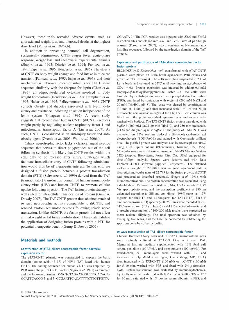
Ciliary neurotrophic factor and related cytokines inducethe activation of receptor-associated just another kinase(JAK)/Tyk tyrosine kinases and subsequent recruitment andphosphorylation of STAT proteins, in particular STAT3(Lelievre et al. 2001). Phosphorylated STAT proteinsdimerize and translocate to the nucleus where they activatetarget gene transcription (Heinrich et al. 1998; Levy andDarnell 2002). Incubation of human neuroblastomaSH-SH5Y cells, which express an intact CNTF receptorcomplex (Kaur et al. 2002), to rhCNTF or TAT-CNTFcaused a rapid and sustained increase in STAT3 phosphor-ylation (Fig. 2).
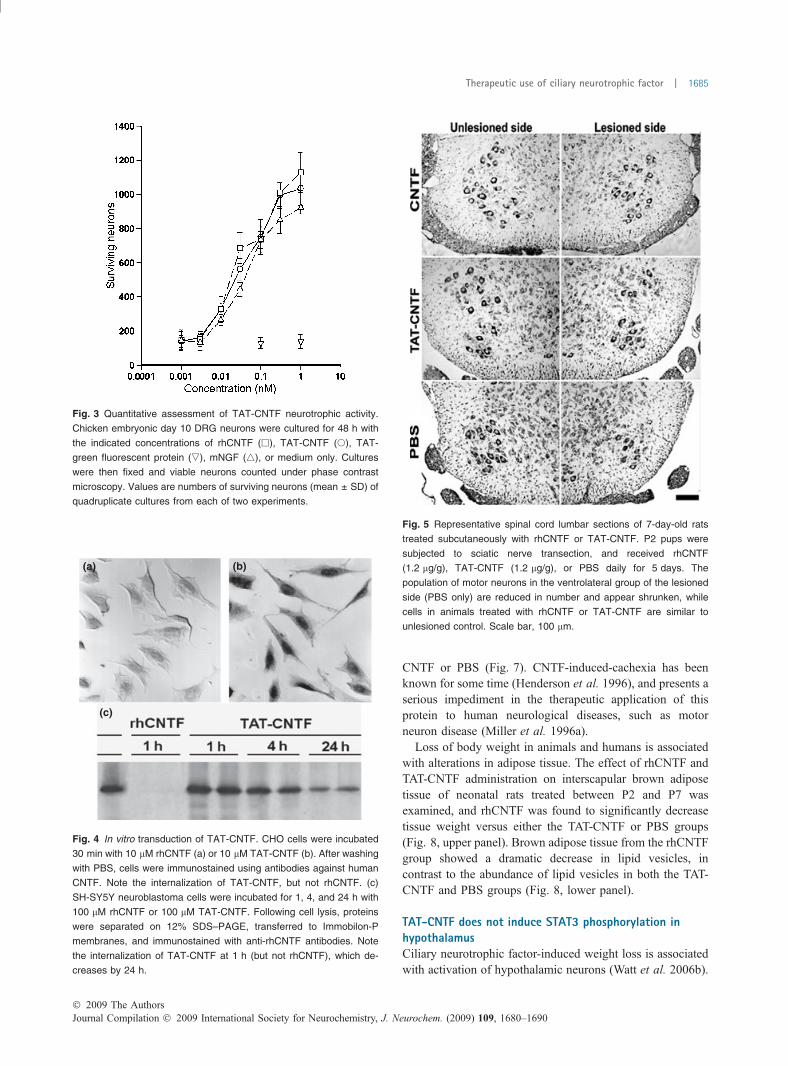
Ciliary neurotrophic factor is capable of supporting thesurvival of cultured chicken embryonic day 10 DRG neurons(Barbin et al. 1984), and this culture system was used toevaluate the biological activity of TAT-CNTF. Robustsurvival of DRG neurons was observed at the morphologicallevel in cultures supplemented with 1 nM rhCNTF, 1 nMTAT-CNTF, or 1 nM mNGF, but not TAT-green fluorescentprotein. These results are presented quantitatively in Fig. 3,with EC50 of 0.037 nM, 0.042 nM, and 0.076 nM forrhCNTF, TAT-CNTF, and mNGF, respectively. The neuro-trophic activity of TAT-CNTF is thus likely attributable to theCNTF domain, with no interference from either the TATdomain or histidine tag. This is not unexpected, as the 14amino acid N-terminus of CNTF is not required forneurotrophic activity (Negro et al. 1997).
TAT fusion proteins readily translocate into cells (Gumpand Dowdy 2007). Incubation of Chinese Hamster Ovarycells with TAT-CNTF resulted in internalization of the fusionprotein within minutes, while rhCNTF showed no such effect(Fig. 4a and b). TAT-CNTF was present mainly in the
� 2009 The AuthorsJournal Compilation � 2009 International Society for Neurochemistry, J. Neurochem. (2009) 109, 1680–1690
Therapeutic use of ciliary neurotrophic factor | 1683
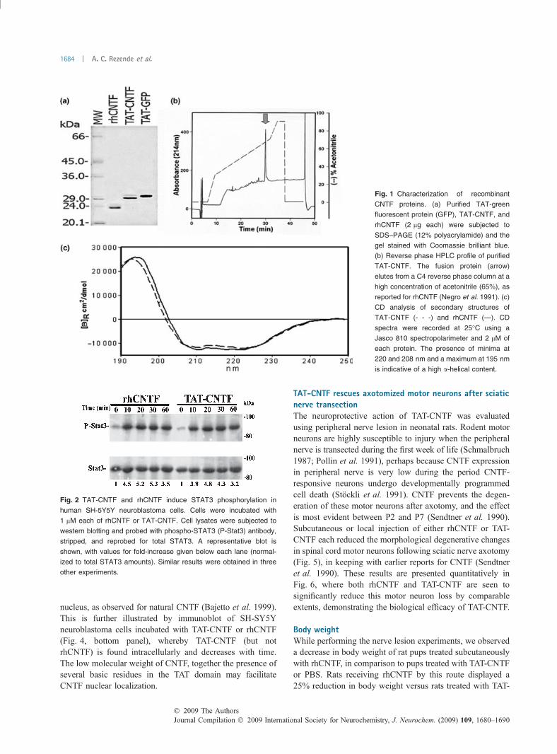
nucleus, as observed for natural CNTF (Bajetto et al. 1999).This is further illustrated by immunoblot of SH-SY5Yneuroblastoma cells incubated with TAT-CNTF or rhCNTF(Fig. 4, bottom panel), whereby TAT-CNTF (but notrhCNTF) is found intracellularly and decreases with time.The low molecular weight of CNTF, together the presence ofseveral basic residues in the TAT domain may facilitateCNTF nuclear localization.
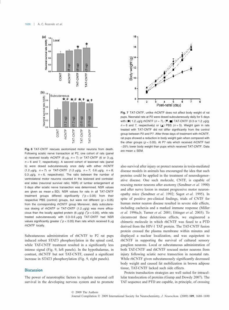
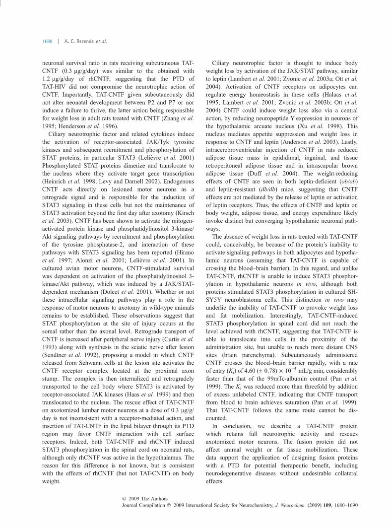
TAT-CNTF rescues axotomized motor neurons after sciaticnerve transectionThe neuroprotective action of TAT-CNTF was evaluatedusing peripheral nerve lesion in neonatal rats. Rodent motorneurons are highly susceptible to injury when the peripheralnerve is transected during the first week of life (Schmalbruch1987; Pollin et al. 1991), perhaps because CNTF expressionin peripheral nerve is very low during the period CNTF-responsive neurons undergo developmentally programmedcell death (Stockli et al. 1991). CNTF prevents the degen-eration of these motor neurons after axotomy, and the effectis most evident between P2 and P7 (Sendtner et al. 1990).Subcutaneous or local injection of either rhCNTF or TAT-CNTF each reduced the morphological degenerative changesin spinal cord motor neurons following sciatic nerve axotomy(Fig. 5), in keeping with earlier reports for CNTF (Sendtneret al. 1990). These results are presented quantitatively inFig. 6, where both rhCNTF and TAT-CNTF are seen tosignificantly reduce this motor neuron loss by comparableextents, demonstrating the biological efficacy of TAT-CNTF.
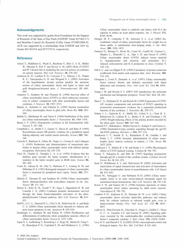
Body weightWhile performing the nerve lesion experiments, we observeda decrease in body weight of rat pups treated subcutaneouslywith rhCNTF, in comparison to pups treated with TAT-CNTFor PBS. Rats receiving rhCNTF by this route displayed a25% reduction in body weight versus rats treated with TAT-
Fig. 1 Characterization of recombinant
CNTF proteins. (a) Purified TAT-green
fluorescent protein (GFP), TAT-CNTF, and
rhCNTF (2 lg each) were subjected to
SDS–PAGE (12% polyacrylamide) and the
gel stained with Coomassie brilliant blue.
(b) Reverse phase HPLC profile of purified
TAT-CNTF. The fusion protein (arrow)
elutes from a C4 reverse phase column at a
high concentration of acetonitrile (65%), as
reported for rhCNTF (Negro et al. 1991). (c)
CD analysis of secondary structures of
TAT-CNTF (- - -) and rhCNTF (—). CD
spectra were recorded at 25�C using a
Jasco 810 spectropolarimeter and 2 lM of
each protein. The presence of minima at
220 and 208 nm and a maximum at 195 nm
is indicative of a high a-helical content.
Fig. 2 TAT-CNTF and rhCNTF induce STAT3 phosphorylation in
human SH-5Y5Y neuroblastoma cells. Cells were incubated with
1 lM each of rhCNTF or TAT-CNTF. Cell lysates were subjected to
western blotting and probed with phospho-STAT3 (P-Stat3) antibody,
stripped, and reprobed for total STAT3. A representative blot is
shown, with values for fold-increase given below each lane (normal-
ized to total STAT3 amounts). Similar results were obtained in three
other experiments.
Journal Compilation � 2009 International Society for Neurochemistry, J. Neurochem. (2009) 109, 1680–1690� 2009 The Authors
1684 | A. C. Rezende et al.
CNTF or PBS (Fig. 7). CNTF-induced-cachexia has beenknown for some time (Henderson et al. 1996), and presents aserious impediment in the therapeutic application of thisprotein to human neurological diseases, such as motorneuron disease (Miller et al. 1996a).
Loss of body weight in animals and humans is associatedwith alterations in adipose tissue. The effect of rhCNTF andTAT-CNTF administration on interscapular brown adiposetissue of neonatal rats treated between P2 and P7 wasexamined, and rhCNTF was found to significantly decreasetissue weight versus either the TAT-CNTF or PBS groups(Fig. 8, upper panel). Brown adipose tissue from the rhCNTFgroup showed a dramatic decrease in lipid vesicles, incontrast to the abundance of lipid vesicles in both the TAT-CNTF and PBS groups (Fig. 8, lower panel).
TAT-CNTF does not induce STAT3 phosphorylation inhypothalamusCiliary neurotrophic factor-induced weight loss is associatedwith activation of hypothalamic neurons (Watt et al. 2006b).
Fig. 3 Quantitative assessment of TAT-CNTF neurotrophic activity.
Chicken embryonic day 10 DRG neurons were cultured for 48 h with
the indicated concentrations of rhCNTF (h), TAT-CNTF (s), TAT-
green fluorescent protein (,), mNGF (4), or medium only. Cultures
were then fixed and viable neurons counted under phase contrast
microscopy. Values are numbers of surviving neurons (mean ± SD) of
quadruplicate cultures from each of two experiments.
(a)
(c)
(b)
Fig. 4 In vitro transduction of TAT-CNTF. CHO cells were incubated
30 min with 10 lM rhCNTF (a) or 10 lM TAT-CNTF (b). After washing
with PBS, cells were immunostained using antibodies against human
CNTF. Note the internalization of TAT-CNTF, but not rhCNTF. (c)
SH-SY5Y neuroblastoma cells were incubated for 1, 4, and 24 h with
100 lM rhCNTF or 100 lM TAT-CNTF. Following cell lysis, proteins
were separated on 12% SDS–PAGE, transferred to Immobilon-P
membranes, and immunostained with anti-rhCNTF antibodies. Note
the internalization of TAT-CNTF at 1 h (but not rhCNTF), which de-
creases by 24 h.
Fig. 5 Representative spinal cord lumbar sections of 7-day-old rats
treated subcutaneously with rhCNTF or TAT-CNTF. P2 pups were
subjected to sciatic nerve transection, and received rhCNTF
(1.2 lg/g), TAT-CNTF (1.2 lg/g), or PBS daily for 5 days. The
population of motor neurons in the ventrolateral group of the lesioned
side (PBS only) are reduced in number and appear shrunken, while
cells in animals treated with rhCNTF or TAT-CNTF are similar to
unlesioned control. Scale bar, 100 lm.
� 2009 The AuthorsJournal Compilation � 2009 International Society for Neurochemistry, J. Neurochem. (2009) 109, 1680–1690
Therapeutic use of ciliary neurotrophic factor | 1685
Subcutaneous administration of rhCNTF to P2 rat pupsinduced robust STAT3 phosphorylation in the spinal cord,while TAT-CNTF treatment resulted in a significantly lessintense signal (Fig. 9, left panels). In the hypothalamus, incontrast, rhCNTF but not TAT-CNTF, caused a significantincrease in STAT3 phosphorylation (Fig. 9, right panels).
Discussion
The power of neurotrophic factors to regulate neuronal cellsurvival in the developing nervous system and to promote
also survival after injury or protect neurons in toxin-mediateddisease models in animals has encouraged the idea that suchproteins could be applied in the treatment of neurodegener-ative disease. One such molecule, CNTF, is capable ofrescuing motor neurons after axotomy (Sendtner et al. 1990)and after nerve lesion in mutant progressive motor neuron-opathy mice (Sendtner et al. 1992; Sagot et al. 1995). Inspite of positive pre-clinical findings, trials of CNTF forhuman motor neuron disease resulted in severe side effects,including cachexia and a marked immune response (Milleret al. 1996a,b; Turner et al. 2001; Ettinger et al. 2003). Tocircumvent these deleterious effects, we engineered achimeric molecule in which rhCNTF was fused to a PTDderived from the HIV-1 TAT protein. The TAT-CNTF fusionprotein crossed the plasma membrane within minutes anddisplayed a nuclear localization, and was equipotent torhCNTF in supporting the survival of cultured sensoryganglion neurons. Local or subcutaneous administration ofboth TAT-CNTF and rhCNTF rescued motor neurons frominjury following sciatic nerve transection in neonatal rats.While rhCNTF given subcutaneously significantly decreasedbody weight and caused fat mobilization in brown adiposetissue, TAT-CNTF lacked such side effects.
Protein transduction strategies are well suited for intracel-lular translocation of proteins (Gump and Dowdy 2007). TheTAT sequence and PTD are capable, in principle, of crossing
Fig. 6 TAT-CNTF rescues axotomized motor neurons from death.
Following sciatic nerve transaction at P2, one cohort of rats (panel
a) received locally rhCNTF (6 lg, n = 7) or TAT-CNTF (6 or 3 lg,
n = 6 and 7, respectively). A second cohort of lesioned rats (panel
b) were dosed subcutaneously once daily with either rhCNTF
(1.2 lg/g, n = 7) or TAT-CNTF (1.2 lg/g, n = 7; 0.6 lg/g, n = 8;
0.3 lg/g, n = 6, respectively). The ratio between the number of
ventrolateral motor neurons counted in the lesioned and contralat-
eral sides (neuronal survival ratio; NSR) of lumbar enlargement at
5 days after sciatic nerve transection was determined. NSR values
are given as mean ± SD). NSR values for rats in all TAT-CNTF
treatment groups differed significantly (*p < 0.05) from their
respective PBS (control) groups, but were not different (p > 0.05)
from the corresponding rhCNTF group Moreover, daily subcutane-
ous dosing of rhCNTF or TAT-CNTF (1.2 lg/g) was more effica-
cious than the locally applied protein (6 lg/g) (#p < 0.05), while rats
treated subcutaneously with 0.3–0.6 lg/g TAT-CNTF had NSR
values significantly greater (�p < 0.05) than rats which received 6 lg
rhCNTF locally.
Fig. 7 TAT-CNTF, unlike rhCNTF does not affect body weight of rat
pups. Neonatal rats at P2 were dosed subcutaneously daily for 5 days
with (d) 1.2 lg/g rhCNTF (n = 7), (., ) TAT-CNTF (0.3 or 1.2 lg/g;
n = 6 and 7, respectively) or ( ) PBS (n = 5). Weight gain in rats
treated with TAT-CNTF did not differ significantly from the control
group between P2 and P7. After three days of treatment with rhCNTF,
rat pups showed a reduction in body weight gain when compared with
the other groups (p < 0.05). At P7 rats which received rhCNTF had
�25% lower body weight than pups which received TAT-CNTF. Data
are mean ± SEM.
Journal Compilation � 2009 International Society for Neurochemistry, J. Neurochem. (2009) 109, 1680–1690� 2009 The Authors
1686 | A. C. Rezende et al.

the plasma membrane in any cell type. For example, a TAT-b-galactosidase fusion protein injected in the rat tail vein wasfound widely distributed in brain (Schwarze et al. 1999). In
this study, subcutaneous treatment with rhCNTF (1.2 lg/g/day) or TAT-CNTF (0.3–1.2 lg/g/day) was more effectivethan local administration of rhCNTF alone. Moreover, the
(a) (b) (c)
Fig. 8 Recombinant human CNTF
(rhCNTF) decreases brown adipose tissue
weight in rat pups. Upper panel: rat pups
(P2) were dosed subcutaneously daily for
5 days with 1.2 lg/g of rhCNTF, 1.2 lg/g of
TAT-CNTF or PBS, after which time intra-
scapular brown adipose tissue was col-
lected. Values are mean ± SEM; *p < 0.05
and **p < 0.001 versus rhCNTF. Lower
panel: rhCNTF, but not TAT-CNTF de-
creases lipid vesicle content of brown adi-
pose tissue. Sections of intrascapular
brown adipose tissue were taken from P7
rat pups dosed subcutaneously daily for
5 days with PBS (a), 1.2 lg/g of rhCNTF
(b), or 1.2 lg/g of TAT-CNTF (c) beginning
on P2. Tissue from pups that received
rhCNTF displayed adipocytes with a few,
small fat droplets (b), in contrast to tissue
from pups treated with TAT-CNTF (c) or
PBS (a). Note the similarity in tissue mor-
phology between (a and c), with cells clo-
sely packed and showing a nucleus in an
eccentric position and fat droplets of varying
size (bar, 50 lm).
Fig. 9 Differential effects of rhCNTF and TAT-CNTF on STAT3
phosphorylation in neonatal rat spinal cord and hypothalamus. Rat
pups at P2 received subcutaneously either rhCNTF or TAT-CNTF
(1.2 lg/g body weight). An equal volume of PBS was given to control
rats. Phospho-specific immunoreactivity was determined as described
under Materials and methods, and quantified by densitometric analysis
and expressed relative to total STAT3 protein (mean ± SD, three
independent experiments, expressed as fold-increase over PBS-
treated group). A representative immunoblot is shown for spinal cord
(left panels) and hypothalamus (right panels); *p < 0.04 versus PBS
and **p < 0.05 versus rhCNTF.
� 2009 The AuthorsJournal Compilation � 2009 International Society for Neurochemistry, J. Neurochem. (2009) 109, 1680–1690
Therapeutic use of ciliary neurotrophic factor | 1687
neuronal survival ratio in rats receiving subcutaneous TAT-CNTF (0.3 lg/g/day) was similar to the obtained with1.2 lg/g/day of rhCNTF, suggesting that the PTD ofTAT-HIV did not compromise the neurotrophic action ofCNTF. Importantly, TAT-CNTF given subcutaneously didnot alter neonatal development between P2 and P7 or norinduce a failure to thrive, the latter action being responsiblefor weight loss in adult rats treated with CNTF (Zhang et al.1995; Henderson et al. 1996).
Ciliary neurotrophic factor and related cytokines inducethe activation of receptor-associated JAK/Tyk tyrosinekinases and subsequent recruitment and phosphorylation ofSTAT proteins, in particular STAT3 (Lelievre et al. 2001)Phosphorylated STAT proteins dimerize and translocate tothe nucleus where they activate target gene transcription(Heinrich et al. 1998; Levy and Darnell 2002). EndogenousCNTF acts directly on lesioned motor neurons as aretrograde signal and is responsible for the induction ofSTAT3 signaling in these cells but not the maintenance ofSTAT3 activation beyond the first day after axotomy (Kirschet al. 2003). CNTF has been shown to activate the mitogen-activated protein kinase and phosphatidylinositol 3-kinase/Akt signaling pathways by recruitment and phosphorylationof the tyrosine phosphatase-2, and interaction of thesepathways with STAT3 signaling has been reported (Hiranoet al. 1997; Alonzi et al. 2001; Lelievre et al. 2001). Incultured avian motor neurons, CNTF-stimulated survivalwas dependent on activation of the phosphatidylinositol 3-kinase/Akt pathway, which was induced by a JAK/STAT-dependent mechanism (Dolcet et al. 2001). Whether or notthese intracellular signaling pathways play a role in theresponse of motor neurons to axotomy in wild-type animalsremains to be established. These observations suggest thatSTAT phosphorylation at the site of injury occurs at thesomal rather than the axonal level. Retrograde transport ofCNTF is increased after peripheral nerve injury (Curtis et al.1993) along with synthesis in the sciatic nerve after lesion(Sendtner et al. 1992), proposing a model in which CNTFreleased from Schwann cells at the lesion site activates theCNTF receptor complex located at the proximal axonstump. The complex is then internalized and retrogradelytransported to the cell body where STAT3 is activated byreceptor-associated JAK kinases (Haas et al. 1999) and thentranslocated to the nucleus. The rescue effect of TAT-CNTFon axotomized lumbar motor neurons at a dose of 0.3 lg/g/day is not inconsistent with a receptor-mediated action, andinsertion of TAT-CNTF in the lipid bilayer through its PTDregion may favor CNTF interaction with cell surfacereceptors. Indeed, both TAT-CNTF and rhCNTF inducedSTAT3 phosphorylation in the spinal cord on neonatal rats,although only rhCNTF was active in the hypothalamus. Thereason for this difference is not known, but is consistentwith the effects of rhCNTF (but not TAT-CNTF) on bodyweight.
Ciliary neurotrophic factor is thought to induce bodyweight loss by activation of the JAK/STAT pathway, similarto leptin (Lambert et al. 2001; Zvonic et al. 2003a; Ott et al.2004). Activation of CNTF receptors on adipocytes canregulate energy homeostasis in these cells (Halaas et al.1995; Lambert et al. 2001; Zvonic et al. 2003b; Ott et al.2004) CNTF could induce weight loss also via a centralaction, by reducing neuropeptide Y expression in neurons ofthe hypothalamic arcuate nucleus (Xu et al. 1998). Thisnucleus mediates appetite suppression and weight loss inresponse to CNTF and leptin (Anderson et al. 2003). Lastly,intracerebroventricular injection of CNTF in rats reducedadipose tissue mass in epididimal, inguinal, and tissueretroperitoneal adipose tissue and in intrascapular brownadipose tissue (Duff et al. 2004). The weight-reducingeffects of CNTF are seen in both leptin-deficient (ob/ob)and leptin-resistant (db/db) mice, suggesting that CNTFeffects are not mediated by the release of leptin or activationof leptin receptors. Thus, the effects of CNTF and leptin onbody weight, adipose tissue, and energy expenditure likelyinvoke distinct but converging hypothalamic neuronal path-ways.
The absence of weight loss in rats treated with TAT-CNTFcould, conceivably, be because of the protein’s inability toactivate signaling pathways in both adipocytes and hypotha-lamic neurons (assuming that TAT-CNTF is capable ofcrossing the blood–brain barrier). In this regard, and unlikeTAT-CNTF, rhCNTF is unable to induce STAT3 phosphor-ylation in hypothalamic neurons in vivo, although bothproteins stimulated STAT3 phosphorylation in cultured SH-SY5Y neuroblastoma cells. This distinction in vivo mayunderlie the inability of TAT-CNTF to provoke weight lossand fat mobilization. Interestingly, TAT-CNTF-inducedSTAT3 phosphorylation in spinal cord did not reach thelevel achieved with rhCNTF, suggesting that TAT-CNTF isable to translocate into cells in the proximity of theadministration site, but unable to reach more distant CNSsites (brain parenchyma). Subcutaneously administeredCNTF crosses the blood–brain barrier rapidly, with a rateof entry (Ki) of 4.60 (± 0.78) · 10)4 mL/g min, considerablyfaster than that of the 99mTc-albumin control (Pan et al.1999). The Ki was reduced more than threefold by additionof excess unlabeled CNTF, indicating that CNTF transportfrom blood to brain achieves saturation (Pan et al. 1999).That TAT-CNTF follows the same route cannot be dis-counted.
In conclusion, we describe a TAT-CNTF proteinwhich retains full neurotrophic activity and rescuesaxotomized motor neurons. The fusion protein did notaffect animal weight or fat tissue mobilization. Thesedata support the application of designing fusion proteinswith a PTD for potential therapeutic benefit, includingneurodegenerative diseases without undesirable collateraleffects.
Journal Compilation � 2009 International Society for Neurochemistry, J. Neurochem. (2009) 109, 1680–1690� 2009 The Authors
1688 | A. C. Rezende et al.
Acknowledgements
This work was supported by grants from Foundation for the Support
of Research of the State of Sao Paolo (FAPESP: Grant 05/54613-7)
and Brazilian Council for Research (CnPq: Grant 473909/2004-9).
ACR was supported by a scholarships from FAPESP and ASV by
Grants 04/14210-8 and 05/57335-6, respectively.
References
Alonzi T., Middleton G., Wyatt S., Buchman V., Betz U. A. K., MullerW., Musiani P., Poli V. and Davies A. M. (2001) Role of STAT3and PI 3-kinase/Akt in mediating the survival actions of cytokineson sensory neurons. Mol. Cell. Neurosci. 18, 270–282.
Anderson K. D., Lambert P. D., Corcoran T. L., Murray J. D., ThabetK. E., Yancopoulos G. D. and Wiegand S. J. (2003) Activationof the hypothalamic arcuate nucleus predicts the anorecticactions of ciliary neurotrophic factor and leptin in intact andgold thioglucose-lesioned mice. J. Neuroendocrinol. 15, 649–660.
Arakawa Y., Sendtner M. and Thoenen H. (1990) Survival effect ofciliary neurotrophic factor (CNTF) on chick embryonic motoneu-rons in culture: comparison with other neurotrophic factors andcytokines. J. Neurosci. 10, 3507–3515.
Bajetto A., Schettini G. and Chimini G. (1999) Nuclear localizationof ciliary neurotrophic factor in glial cells. Brain Res. 818, 565–569.
Barbin G., Manthorpe M. and Varon S. (1984) Purification of the chickeye ciliary neuronotrophic factor. J. Neurochem. 43, 1468–1478.
Bazan J. F. (1991) Neuropoietic cytokines in the hematopoietic fold.Neuron 7, 197–208.
Campfield L. A., Smith F. J., Guisez Y., Devos R. and Burn P. (1995)Recombinant mouse OB protein: evidence for a peripheral signallinking adiposity and central neural networks. Science 269, 546–549.
Cazzola F., Battiston S., Fabris M., Negro A., Corona G. and CallegaroL. (1993) Production and characterization of monoclonal anti-bodies to human ciliary neurotrophic factor with defined epitoperecognition. Hybridoma 12, 259–270.
Chen H., Charlat O., Tartaglia L. A. et al. (1995) Evidence that thediabetes gene encodes the leptin receptor: identification of amutation in the leptin receptor gene in db/db mice. Science 84,491–495.
Curtis R., Adryan K. M., Zhu Y., Harkness P. J., Lindsay R. M. andDiStefano P. S. (1993) Retrograde transport of ciliary neurotrophicfactor is increased by peripheral nerve injury. Nature 365, 253–255.
Dittrich F., Thoenen H. and Sendtner M. (1994) Ciliary neurotrophicfactor: pharmacokinetics and acute-phase response in rat. Ann.Neurol. 35, 151–163.
Dolcet X., Soler R. M., Gould T. W., Egea J., Oppenheim R. W. andComella J. X. (2001) Cytokines promote motoneuron survivalthrough the Janus kinase-dependent activation of the phos-phatidylinositol 3-kinase pathway. Mol. Cell. Neurosci. 18, 619–631.
Duff E., Li C. L., Hartzell D. L., Choi Y.-H., Della-Fera M. A. and BaileC. A. (2004) Ciliary neurotrophic factor injected icv induces adi-pose tissue apoptosis in rats. Apoptosis 9, 629–634.
Ernsberger U., Sendtner M. and Rohrer H. (1989) Proliferation anddifferentiation of embryonic chick sympathetic neurons: effects ofciliary neurotrophic factor. Neuron 2, 1275–1284.
Espat N. J., Auffenberg T., Rosenberg J. J., Rogy M., Martin D., Fang C.H., Hasselgren P. O., Copeland E. M. and Moldawer L. L. (1996)
Ciliary neurotrophic factor is catabolic and shares with IL-6 thecapacity to induce an acute phase response. Am. J. Physiol. 271,R185–R190.
Ettinger M. P., Littlejohn T. W., Schwartz S. L. et al. (2003) Re-combinant variant of ciliary neurotrophic factor for weight loss inobese adults. A randomized, dose-ranging study. J. Am. Med.Assoc. 289, 1826–1832.
Fantuzzi G., Benigni F., Sironi M., Conni M., Carelli M., Cantoni L.,Shapiro L., Dinarello C. A., Sipe J. D. and Grezzi P. (1995)Ciliary neurotrophic factor (CNTF) induces serum amyloidA, hypoglycaemia and anorexia, and potentiates IL-1induced corticosterone and IL-6 production in mice. Cytokine 7,150–156.
Gill S. C. and von Hippel P. H. (1989) Calculation of protein extinctioncoefficients from amino acid sequence data. Anal. Biochem. 182,319–326.
Gloaguen I., Costa P., Demartis A. et al. (1997) Ciliary neurotrophicfactor corrects obesity and diabetes associated with leptindeficiency and resistance. Proc. Natl Acad. Sci. USA 94, 6456–6461.
Gump J. M. and Dowdy S. F. (2007) TAT transduction: the molecularmechanism and therapeutic prospects. Trends Mol. Med. 13, 443–448.
Haas C. A., Hofmann H. D. and Kirsch M. (1999) Expression of CNTF/LIF receptor components and activation of STAT3 signaling inaxotomized motoneurons: evidence for a sequential postlesionalfunction of the cytokines. J. Neurobiol. 41, 559–571.
Halaas J. L., Gajiwala K. S., Maffei M., Cohen S. L., Chait B. T.,Rabinowitz D., Lallone R. L., Burley S. K. and Friedman J. M.(1995) Weight-reducing effects of the plasma protein encoded bythe obese gene. Science 269, 543–546.
Heinrich P. C., Behrmann I., Muller-Newen G., Schaper F. and Graeve L.(1998) Interleukin-6-type cytokine signalling through the gp130/Jak/STAT pathway. Biochem. J. 334, 297–314.
Henderson J. T., Seniuk N. A., Richardson P. M., Gauldie J. andRoder J. C. (1994) Systemic administration of ciliary neuro-trophic factor induces cachexia in rodents. J. Clin. Invest. 93,2632–2638.
Henderson J. T., Mullen B. J. M. and Roder J. C. (1996) Physiologicaleffects of CNTF-induced wasting. Cytokine 8, 784–793.
Hirano T., Nakajima K. and Hibi M. (1997) Signaling mechanismsthrough gp130: a model of the cytokine system. Cytokine GrowthFactor Rev. 8, 241–252.
Kaur N., Wohlhueter A. L. and Halvorsens W. (2002) Activation andinactivation of signal transactivator and activators of transcriptionby ciliary neutrotrophic factor in neuroblastoma cells. Cell Signal14, 419–429.
Kirsch M., Terheggen U. and Hofmann H.-D. (2003) Ciliary neuro-trophic factor is an early lesion-induced retrograde signal foraxotomized facial motoneurons. Mol. Cell. Neurosci. 24, 130–138.
Kwon Y. W. and Gurney M. E. (1994) Systemic injections of ciliaryneurotrophic factor induce sprouting by adult motor neurons.NeuroReport 5, 789–792.
Lambert P. D., Anderson K. D., Sleeman M. W. et al. (2001) Ciliaryneurotrophic factor activates leptin-like pathways and reducesbody fat, without cachexia or rebound weight gain, even inleptin-resistant obesity. Proc. Natl Acad. Sci. USA 98, 4652–4657.
Lelievre E., Plun-Favreau H., Chevalier S., Froger J., Guillet C., ElsonG. C. A., Gauchat J.-F. and Gascan H. (2001) Signaling path-ways recruited by the cardiotrophin-like cytokine/cytokine-likefactor-1 composite cytokine. J. Biol. Chem. 276, 22476–22484.
Levy D. E. and Darnell J. E. Jr (2002) STATs: transcriptional control andbiological impact. Nat. Rev. Mol. Cell Biol. 3, 651–662.
� 2009 The AuthorsJournal Compilation � 2009 International Society for Neurochemistry, J. Neurochem. (2009) 109, 1680–1690
Therapeutic use of ciliary neurotrophic factor | 1689

Liu Q.-S., Wang Q.-J., Du G.-H., Zhu S.-Y., Gao M., Zhang L., ZhuJ.-M. and Cao J.-F. (2007) Recombinant human ciliaryneurotrophic factor reduces weight partly by regulating nuclearrespiratory factor 1 and mitochondrial transcription factor A. Eur.J. Pharmacol. 563, 77–82.
Louis J.-C., Magal E., Takayama S. and Varon S. (1993) CNTF pro-tection of oligodendrocytes again natural and tumor necrosis fac-tor-induced death. Science 259, 689–692.
McDonald N. Q., Panayotatos N. and Hendrickson W. A. (1995) Crystalstructure of dimeric human ciliary neurotrophic factor determinedby MAD phasing. EMBO J. 14, 2689–2699.
Miller R. G., Petajan J. H., Btyan W. W., Armon C., Barohn R. J.,Goodpasture J. C., Hoagland R. J., Parry G. J., Ross M. A. andStromatt S. C. (1996a) A placebo-controlled trial of recombinanthuman ciliary neurotrophic (rhCNTF) factor in amyotrophic lateralsclerosis. rhCNTF ALS Study Group. Ann. Neurol. 39, 256–260.
Miller R. G., Bryan W. W., Dietz M. A., Munsat T. L., Petajan J. H.,Smith S. A. and Goodpasture J. C. (1996b) Toxicity and tolera-bility of recombinant human ciliary neurotrophic factor in patientswith amyotrophic lateral sclerosis. Neurology 47, 1329–1331.
Negro A., Corona G., Bigon E., Martini I., Grandi C., Skaper S. D. andCallegaro L. (1991) Synthesis, purification and characterization ofhuman ciliary neurotrophic factor from E. coli. J. Neurosci. Res.29, 251–260.
Negro A., Grassato L., Polverino De Laureto P. and Skaper S. D. (1997)Genetic construction, properties and application of a green fluo-rescent protein tagged ciliary neurotrophic factor. Protein Eng. 10,1077–1083.
Oliviera A. L., Risling M., Negro A., Langone F. and Cullheim S.(2002) Apoptosis of spinal interneurons induced by sciatic nerveaxotomy in the neonatal rat is counteracted by nerve growthfactor and ciliary neurotrophic factor. J. Comp. Neurol. 447,381–393.
Oppenheim R. W., Prevette D., Yin Q.-W., Collins F. and MacDonald J.(1991) Control of embryonic motoneuron survival in vivo byciliary neurotrophic factor. Science 251, 1616–1618.
Ott V., Fasshauber M., Meier B., Dalski A., Kraus D., Gettys T. W.,Perwitz N. and Klein J. (2004) Cilary neurotrophic factorinfluences endocrine adipocyte function: inhibition of leptin via PI3-kinase. Mol. Cell. Endocrinol. 224, 21–27.
Pan W., Kastin A. J., Maness L. M. and Brennan J. M. (1999) Saturableentry of ciliary neurotrophic factor into brain. Neurosci. Lett. 263,69–71.
Pelleymounter M. A., Cullen M. J., Baker M. B., Hecht R., Winters D.,Boone T. and Collins F. (1995) Effects of the obese gene producton body weight regulation in ob/ob mice. Science 269, 540–543.
Peroni D., Negro A., Bahr M. and Dietz G. P. H. (2007) Intracellulardelivery of Neuroglobin using HIV-1 TAT protein transductiondomain fails to protect against oxygen and glucose deprivation.Neurosci. Lett. 421, 110–114.
Pollin M. M., McHanwell S. and Slater C. R. (1991) The effect of age onmotor neuron death following axotomy in the mouse. Development112, 83–89.
Rezende A. C., Vieira A. S., Rogerio F., Rezende L. F., Boschero A. C.,Negro A. and Langone F. (2008) Effects of systemic administrationof ciliary neurotrophic factor on Bax and Bcl-2 proteins in thelumbar spinal cord of neonatal rats after sciatic nerve transection.Braz. J. Med. Biol. Res. 41, 1024–1028.
Rogerio F., de Souza Queiroz L., Teixeira S. A., Oliveira A. L., de NucciG. and Langone F. (2002) Neuroprotective action of melatonin onneonatal rat motoneurons after sciatic nerve transection. Brain Res.926, 33–41.
Sagot Y., Tan S. A., Baetge E., Schmalbruch H., Kato A. C. andAebischer P. (1995) Polymer encapsulated cell lines geneticallyengineered to release ciliary neurotrophic factor can slow downprogressive motor neuronopathy in the mouse. Eur. J. Neurosci. 7,1313–1322.
Schmalbruch H. (1987) Loss of sensory neurons after sciatic nervesection in the rat. Anat. Rec. 219, 323–329.
Schwarze S. R., Ho A., Vocero-Akbani A. and Dowdy S. F. (1999) Invivo protein transduction: delivery of a biologically active proteininto the mouse. Science 285, 1569–1572.
Sendtner M., Kreutzberg G. W. and Thoenen H. (1990) Ciliary neuro-trophic factor prevents the degeneration of motor neurons afteraxotomy. Nature 345, 440–441.
Sendtner M., Schmalbruch H., Stockli K. A., Carroll P., KreutzbergG. W. and Thoenen H. (1992) Ciliary neurotrophic factor preventsdegeneration of motor neurons in mouse mutant progressive motorneuronopathy. Nature 358, 502–504.
Shapiro L., Zhang X.-X., Rupp R. G., Wolff S. M. and Dinarello C. A.(1993) Ciliary neurotrophic factor is an endogenous pyrogen. Proc.Natl Acad. Sci. USA 90, 8614–8618.
Skaper S. D., Facci L., Milani D., Leon A. and Toffano G. (1990)Culture and use of primary and clonal neural cells, in Methods inNeurosciences (Conn P. M., ed), Vol. 2, pp. 17–33. AcademicPress, San Diego, CA.
Stockli K. A., Lillien L. E., Naher-Noe M., Breitfeld G., HughesR. A., Raff M. C., Thoenen H. and Sendtner M. (1991)Regional distribution, developmental changes, and cellularlocalization of CNTF-mRNA and protein in the rat brain. J. CellBiol. 115, 447–459.
Taga T. and Kishimoto T. (1992) Cytokine receptors and signal trans-duction. FASEB J. 6, 3387–3396.
Turner M. R., Parton M. J. and Leigh P. N. (2001) Clinical trials in ALS:an overview. Sem. Neurol. 21, 167–175.
Watt M. J., DzamKo N., Thomas W. G., Rose-john S., Ernst M.,Carling D., Kemp B. E., Febbraio M. A. and SteinbergG. R. (2006a) CNTF reverses obesity-induced insulin resis-tance by activating skeletal muscle AMPK. Nat. Med. 12, 541–548.
Watt J. A., Bone S., Pressler M., Cranston H. J. and Paden C. M.(2006b) Ciliary neurotrophic factor is expressed in the magno-cellular neurosecretory system of the rat in vivo: evidence forinjury- and activity-induced upregulation. Exp. Neurol. 197,206–214.
Xu B., Dube M. G., Kalra P. S., Farmerie W. G., Kaibara A., MoldawerL. L., Martin D. and Kalra S. P. (1998) Anorectic effects of thecytokine, ciliary neurotropic factor, are mediated by hypothalamicneuropeptide Y: comparison with leptin. Endocrinology 139, 466–473.
ZhangF., Richardson P.M., HollandD. P., GuoQ. and TattonW.G. (1995)CNTF or ())-deprenyl in immature rats: survival of axotomizedfacial motoneurons and weight loss. J. Neurosci. Res. 40, 564–570.
Zvonic S., Cornelius P., Stewart W. C., Mynatt R. L. and Stephens J.M. (2003a) The regulation and activation of ciliary neurotrophicfactor signaling proteins in adipocytes. J. Biol. Chem. 278,2228–2235.
Zvonic S., Story D. J., Stephens J. M. and Mynatt R. L. (2003b)Growth hormone, but not insulin, activates STAT5 proteins inadipocytes in vitro and in vivo. Biochem. Biophys. Res. Comm.302, 359–362.
Zvonic S., Baugh J. E. Jr, Arbour-Reily P., Mynatt R. I. and Stephens J.M. (2005) Cross-talk among gp130 cytokines in adipocytes. J.Biol. Chem. 280, 33856–33863.
Journal Compilation � 2009 International Society for Neurochemistry, J. Neurochem. (2009) 109, 1680–1690� 2009 The Authors
1690 | A. C. Rezende et al.
Copyright © 2022 FDOKUMEN