MGSA/GRO-mediated melanocyte transformation involves induction of Ras expression
Upload
independentCategory
view
1download
0
Blockade of Smad4 in transformed keratinocytes containing a Rasoncogene leads to hyperactivation of the Ras-dependent Erk signallingpathway associated with progression to undi�erentiated carcinomas
Maite Iglesias1, Pilar Frontelo1, Carlos Gamallo2 and Miguel Quintanilla*,1
1Instituto de Investigaciones BiomeÂdicas Alberto Sols CSIC-UAM, Arturo Duperier 4, 28029-Madrid, Spain; 2Hospital de laPrincesa, Departamento de AnatomõÂa PatoloÂgica, Facultad de Medicina, UAM, 28029-Madrid, Spain
Smad4 functions as a transcription factor in TGF-bsignalling. We have investigated the role of Smad4 in theTGF-b1 cell responses of transformed PDV keratino-cytes, which contain a Ras oncogene, and of non-tumorigenic MCA3D keratinocytes, by transfecting bothcell lines with a dominant-negative Smad4 construct.Smad4 mediates TGF-b1-induced up-regulation of p21Cip1
and growth arrest in MCA3D cells. However, in PDVkeratinocytes, Smad4 is only partially involved in TGF-b1-induced growth inhibition, and does not mediateenhancement of p21Cip1 levels by the growth factor.TGF-b1 activates Ras/Erk signalling activity in both celllines. PD098059, a speci®c inhibitor of MEK, dis-minishes TGF-b1-induced p21Cip1 levels in PDV but not inMCA3D cells, suggesting an involvement of Erk in up-regulation of p21Cip1 by TGF-b1 in PDV cells. PDVdominant-negative Smad4 cell transfectants, but notMCA3D transfectants, showed constitutive hyperactiva-tion of the Ras/Erk signalling pathway, increasedsecretion of urokinase, higher motility properties, and achange to a ®broblastoid cell morphology associated invivo with the transition from a well di�erentiated to apoorly di�erentiated tumour phenotype. Infection ofMCA3D control and dominant negative Smad4 celltransfectants with retroviruses carrying a Ras oncogeneled to enhanced p21Cip1 and urokinase secreted levels,independently of TGF-b1 stimulation, that were reducedby PD098059. These results suggest that Smad4 actsinhibiting Ras-dependent Erk signalling activity in Ras-transformed keratinocytes. Loss of Smad4 function inthese cells results in hyperactivation of Erk signallingand progression to undi�erentiated carcinomas. Onco-gene (2000) 19, 4134 ± 4145.
Keywords: TGF-b1; Smad4; Ras; Erk; urokinase;carcinoma
Introduction
Transforming growth factor-b (TGF-b) regulates cellproliferation, di�erentiation, motility, adhesion, anddeath in most tissues, including epithelia (Massague ,1990; Lahio and Keski-Oja, 1992). During the lastyears the idea of a dual function of TGF-b in epithelialcarcinogenesis has gained considerable support. TGF-bacts as a suppressor of tumour formation by virtue of
its role in inhibiting proliferation of epithelial cells(Alexandrow and Moses, 1995; Massague , 1998), but italso stimulates malignant progression at later stages oftumorigenesis by inducing an epithelial-mesenchymaltransition associated with the development of a highlyaggressive undi�erentiated type of tumour (Caulõ n etal., 1995; Cui et al., 1996; Oft et al., 1996, 1998;Frontelo et al., 1998; Portella et al., 1998).
Studies on the mouse skin carcinogenesis model andon mammary epithelial cells suggest a synergistic co-operation between oncogenic Ras and TGF-b1 tostimulate malignant progression (Oft et al., 1996;Akhurst and Balmain, 1999). The e�ects of the growthfactor on carcinoma cell invasiveness and metastasisappear to be direct and require signalling through thetype II receptor (Oft et al., 1998; Portella et al., 1998).
TGF-b signals through formation of heteromericcomplexes between type I and type II serine/threoninekinase receptors. After activation, the signal transdu-cing type I receptor binds and phosphorylates Smad2/Smad3 proteins that associate with Smad4 in thecytoplasm, and these complexes, then, translocate intothe nucleus in which they activate transcription ofspeci®c genes (see Massague , 1998; Zhang andDerynck, 1999; for reviews). TGF-b signalling compo-nents, such as type II receptors and Smad proteins,have been shown to be inactivated by mutation ordeletion in human cancers (Massague , 1998). Particu-larly, Smad4 (DPC4) was originally identi®ed as atumour suppressor gene frequently inactivated inpancreatic and colon carcinomas. Reconstitution ofSmad4 in colon carcinoma defective cells leads tosuppression of tumorigenicity (Schwarte-Waldho� etal., 1999)
Several reports point to a rapid and direct activationof the Ras-mitogen-activated protein (MAP) kinasesignalling pathway associated with growth inhibitionby TGF-b in epithelial cells (Hartsough et al., 1996;reviewed in Hartsough and Mulder, 1997). The growthinhibitory response to TGF-b appears to be mediatedby induction of cyclin-dependent kinase (cdk) inhibi-tors, such as p21Cip1 and p15INK4B (Datto et al., 1995;Hannon and Beach, 1994; Reynisdo ttir et al., 1995).The signalling pathways involved in these TGF-b-induced gene responses remain an open question (seeMassague , 1998). Thus, p21Cip1 appears to be adownstream target gene regulated by Smad4 incarcinoma cells (Grau et al., 1997; Hunt et al., 1998),while it has been reported that Ras and the MAPkinase pathway are required for up-regulation ofp21Cip1 by TGF-b in intestinal epithelial cells andhuman keratinocytes, respectively (Yue et al., 1998; Hu
Oncogene (2000) 19, 4134 ± 4145ã 2000 Macmillan Publishers Ltd All rights reserved 0950 ± 9232/00 $15.00
www.nature.com/onc
*Correspondence: M QuintanillaReceived 2 March 2000; revised 16 June 2000; accepted 26 June2000
et al., 1999). The same appears to occur for TGF-b-mediated induction of other genes, such as the 3TP-luxreporter construct, which contains a known TGF-b-inducible plasminogen activator inhibitor-1 (PAI-1)promoter (Grau et al., 1997; Mucsi et al., 1996; deWinter et al., 1997). This scenario has been compli-cated as several reports demonstrate a cross-talkbetween the Smad and Ras-MAP kinase signallingpathways (reviewed in Zhang and Derynck, 1999).Recently, Kretzschmar et al. (1999) found thatoncogenic Ras, acting via MAP kinases, inhibitsTGF-b-induced nuclear accumulation of Smad2 andSmad3. However, another report has shown thatconstitutively active MEK, or inhibitors for MEK,have no e�ect on Smad activity in human keratinocytes(Hu et al., 1999).
In this work, we have analysed the role of Smad4 inthe TGF-b1 responses of immortalized (MCA3D) andtransformed (PDV) mouse epidermal keratinocytes bytransfecting both cell lines with a C-terminal truncated,dominant-negative, Smad4 construct (Lagna et al.,1996). MCA3D cells are non-tumorigenic and exhibitnormal Ras and p53 genes, while PDV are chemicallytransformed keratinocytes and produce low metastaticwell di�erentiated squamous cell carcinomas uponinjection in mice (Caulõ n et al., 1995; Frontelo et al.,1998). PDV cells contain a mutated H-Ras gene andexpress relatively low levels of activated Ras proteins(Quintanilla et al., 1991). They also have inactivatedp53 loci (Burns et al., 1991). Both cell lines are growth-inhibited by TGF-b1, but they respond di�erentially tochronic treatment with the growth factor. WhileMCA3D cells are induced to terminal di�erentiationand cell death in response to TGF-b1, the growthfactor stimulates cell motility and invasiveness in PDVcells, which undergo a reversible epithelial-mesenchy-mal conversion linked to progression toward ametastatic poorly di�erentiated tumour phenotype(Caulõ n et al., 1995; Frontelo et al., 1998). The invasiveresponse of PDV and other carcinoma cells to TGF-b1is associated with enhanced expression/secretion ofurokinase (uPA) and of its inhibitor PAI-1 (Farina etal., 1998; Santiba nÄ ez et al., 1999). uPA is a serineproteinase that converts the inactive plasminogen intothe broad-spectrum active proteinase plasmin, which inturn degrades many proteins of the extracellular matrixand activates other matrix metalloproteinases (Andrea-sen et al., 1997).
We show that inhibiting Smad4 in Ras-transformedkeratinocytes leads to hyperactivation of the Ras/Erksignalling pathway, increased secretion of uPA, andprogression to a poorly di�erentiated tumour pheno-type. On the other hand, a switch between the Smadand Erk signalling pathways, depending on thepresence of oncogenic Ras, appears to occur inkeratinocytes to mediate induction of p21Cipl by TGF-b1.
Results
Smad4 mediates growth inhibition in both MCA3D andPDV keratinocytes
MCA3D and PDV cell lines were transfected eitherwith a full-length Smad4 cDNA or with a C-terminal
truncated dominant-negative Smad4 (1 ± 514) construct,together with the pcDNA3 neomycine resistant vector.Previously, the presence of Smad4 transcripts in bothcell lines was demonstrated by RT ±PCR (data notshown). After transfection with the normal full-lengthcDNA, no viable clones were obtained from MCA3Dcells; while several clones could be selected up to thethird passage, in which they died, in the case of PDVcells. These results suggest that overexpression ofSmad4 induces cell death in keratinocytes, as reportedfor other epithelial cells (At® et al., 1997), althoughthis hypothesis was not further investigated. Stableclones transfected with the dominant-negative Smad4(1 ± 514) construct were isolated from both MCA3Dand PDV cell lines (designated as 3DdnS and PdnS,respectively). Clones transfected with the pcDNA3vector alone were also obtained (designated as 3DCand PC) to be used as controls.
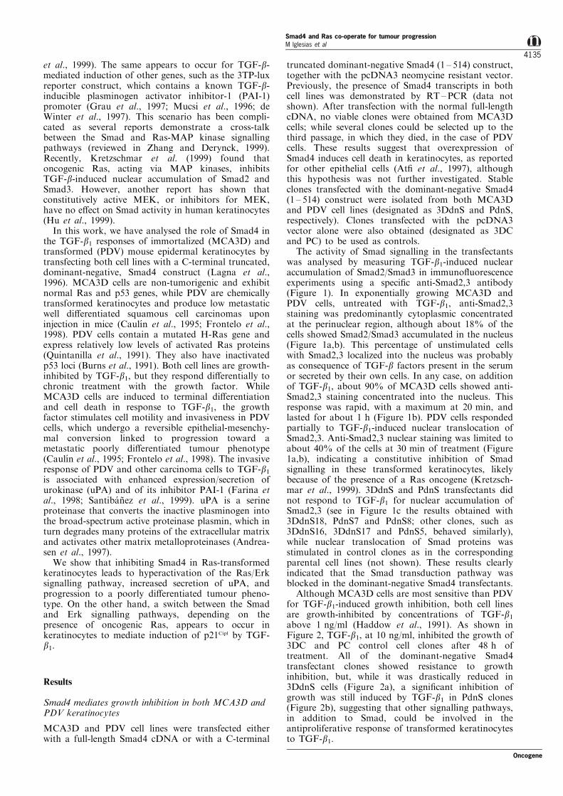
The activity of Smad signalling in the transfectantswas analysed by measuring TGF-b1-induced nuclearaccumulation of Smad2/Smad3 in immuno¯uorescenceexperiments using a speci®c anti-Smad2,3 antibody(Figure 1). In exponentially growing MCA3D andPDV cells, untreated with TGF-b1, anti-Smad2,3staining was predominantly cytoplasmic concentratedat the perinuclear region, although about 18% of thecells showed Smad2/Smad3 accumulated in the nucleus(Figure 1a,b). This percentage of unstimulated cellswith Smad2,3 localized into the nucleus was probablyas consequence of TGF-b factors present in the serumor secreted by their own cells. In any case, on additionof TGF-b1, about 90% of MCA3D cells showed anti-Smad2,3 staining concentrated into the nucleus. Thisresponse was rapid, with a maximum at 20 min, andlasted for about 1 h (Figure 1b). PDV cells respondedpartially to TGF-b1-induced nuclear translocation ofSmad2,3. Anti-Smad2,3 nuclear staining was limited toabout 40% of the cells at 30 min of treatment (Figure1a,b), indicating a constitutive inhibition of Smadsignalling in these transformed keratinocytes, likelybecause of the presence of a Ras oncogene (Kretzsch-mar et al., 1999). 3DdnS and PdnS transfectants didnot respond to TGF-b1 for nuclear accumulation ofSmad2,3 (see in Figure 1c the results obtained with3DdnS18, PdnS7 and PdnS8; other clones, such as3DdnS16, 3DdnS17 and PdnS5, behaved similarly),while nuclear translocation of Smad proteins wasstimulated in control clones as in the correspondingparental cell lines (not shown). These results clearlyindicated that the Smad transduction pathway wasblocked in the dominant-negative Smad4 transfectants.
Although MCA3D cells are most sensitive than PDVfor TGF-b1-induced growth inhibition, both cell linesare growth-inhibited by concentrations of TGF-b1above 1 ng/ml (Haddow et al., 1991). As shown inFigure 2, TGF-b1, at 10 ng/ml, inhibited the growth of3DC and PC control cell clones after 48 h oftreatment. All of the dominant-negative Smad4transfectant clones showed resistance to growthinhibition, but, while it was drastically reduced in3DdnS cells (Figure 2a), a signi®cant inhibition ofgrowth was still induced by TGF-b1 in PdnS clones(Figure 2b), suggesting that other signalling pathways,in addition to Smad, could be involved in theantiproliferative response of transformed keratinocytesto TGF-b1.
Oncogene
Smad4 and Ras co-operate for tumour progressionM lglesias et al
4135
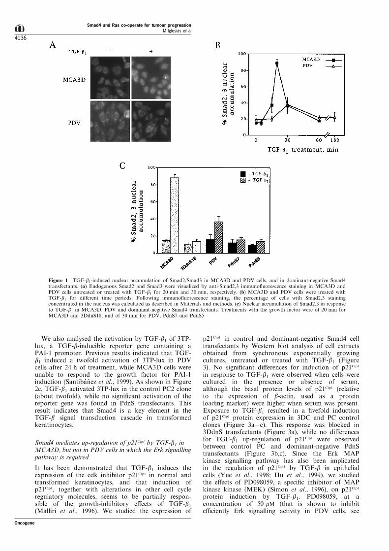
We also analysed the activation by TGF-b1 of 3TP-lux, a TGF-b-inducible reporter gene containing aPAI-1 promoter. Previous results indicated that TGF-b1 induced a twofold activation of 3TP-lux in PDVcells after 24 h of treatment, while MCA3D cells wereunable to respond to the growth factor for PAI-1induction (Santiba nÄ ez et al., 1999). As shown in Figure2c, TGF-b1 activated 3TP-lux in the control PC2 clone(about twofold), while no signi®cant activation of thereporter gene was found in PdnS transfectants. Thisresult indicates that Smad4 is a key element in theTGF-b signal transduction cascade in transformedkeratinocytes.
Smad4 mediates up-regulation of p21Clp1 by TGF-b1 inMCA3D, but not in PDV cells in which the Erk signallingpathway is required
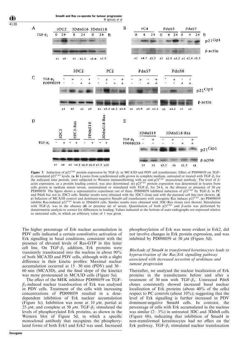
It has been demonstrated that TGF-b1 induces theexpression of the cdk inhibitor p21Cip1 in normal andtransformed keratinocytes, and that induction ofp21Cip1, together with alterations in other cell cycleregulatory molecules, seems to be partially respon-sible of the growth-inhibitory e�ects of TGF-b1(Malliri et al., 1996). We studied the expression of
p21Cip1 in control and dominant-negative Smad4 celltransfectants by Western blot analysis of cell extractsobtained from synchronous exponentially growingcultures, untreated or treated with TGF-b1 (Figure3). No signi®cant di�erences for induction of p21Cip1
in response to TGF-b1 were observed when cells werecultured in the presence or absence of serum,although the basal protein levels of p21Cip1 (relativeto the expression of b-actin, used as a proteinloading marker) were higher when serum was present.Exposure to TGF-b1 resulted in a ®vefold inductionof p21Cip1 protein expression in 3DC and PC controlclones (Figure 3a ± c). This response was blocked in3DdnS transfectants (Figure 3a), while no di�erencesfor TGF-b1 up-regulation of p21Cip1 were observedbetween control PC and dominant-negative PdnStransfectants (Figure 3b,c). Since the Erk MAPkinase signalling pathway has also been implicatedin the regulation of p21Cip1 by TGF-b in epithelialcells (Yue et al., 1998; Hu et al., 1999), we studiedthe e�ects of PD098059, a speci®c inhibitor of MAPkinase kinase (MEK) (Simon et al., 1996), on p21Cip1
protein induction by TGF-b1. PD098059, at aconcentration of 50 mM (that is shown to inhibite�ciently Erk signalling activity in PDV cells, see
Figure 1 TGF-b1-induced nuclear accumulation of Smad2/Smad3 in MCA3D and PDV cells, and in dominant-negative Smad4transfectants. (a) Endogenous Smad2 and Smad3 were visualized by anti-Smad2,3 immuno¯uorescence staining in MCA3D andPDV cells untreated or treated with TGF-b1 for 20 min and 30 min, respectively. (b) MCA3D and PDV cells were treated withTGF-b1 for di�erent time periods. Following immuno¯uorescence staining, the percentage of cells with Smad2,3 stainingconcentrated in the nucleus was calculated as described in Materials and methods. (c) Nuclear accumulation of Smad2,3 in responseto TGF-b1 in MCA3D, PDV and dominant-negative Smad4 transfectants. Treatments with the growth factor were of 20 min forMCA3D and 3DdnS18, and of 30 min for PDV, PdnS7 and PdnS5
Smad4 and Ras co-operate for tumour progressionM lglesias et al
4136
Oncogene
below), consistently reduced (by 35 ± 50%) TGF-b1-stimulated p21Cip1 expression in PC and PdnS clones,while it did not a�ect the enhancement of p21Cip1 bythe growth factor in 3DC cells (Figure 3c).Altogether, these data indicated that Smad4 doesnot mediate regulation of p21Cip1 protein levels byTGF-b1 in transformed PDV keratinocytes, and thatthis response appeared to require Erk MAP kinasesignalling activity.
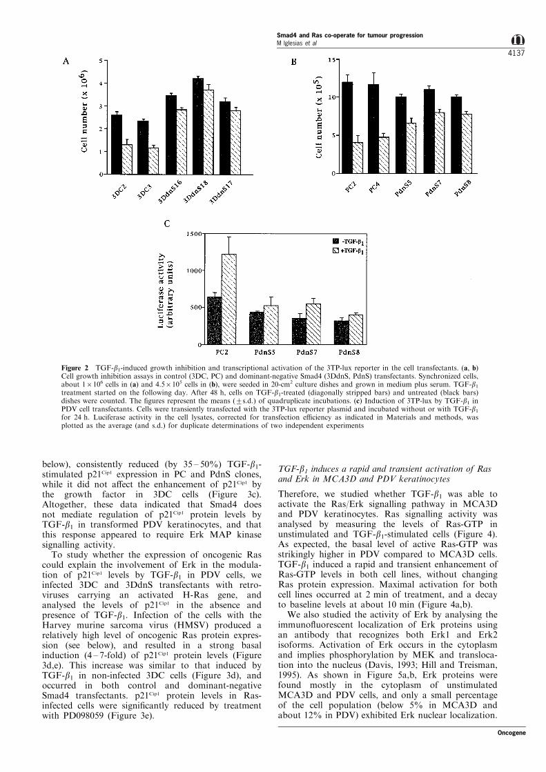
To study whether the expression of oncogenic Rascould explain the involvement of Erk in the modula-tion of p21Cip1 levels by TGF-b1 in PDV cells, weinfected 3DC and 3DdnS transfectants with retro-viruses carrying an activated H-Ras gene, andanalysed the levels of p21Cip1 in the absence andpresence of TGF-b1. Infection of the cells with theHarvey murine sarcoma virus (HMSV) produced arelatively high level of oncogenic Ras protein expres-sion (see below), and resulted in a strong basalinduction (4 ± 7-fold) of p21Cip1 protein levels (Figure3d,e). This increase was similar to that induced byTGF-b1 in non-infected 3DC cells (Figure 3d), andoccurred in both control and dominant-negativeSmad4 transfectants. p21Cip1 protein levels in Ras-infected cells were signi®cantly reduced by treatmentwith PD098059 (Figure 3e).
TGF-b1 induces a rapid and transient activation of Rasand Erk in MCA3D and PDV keratinocytes
Therefore, we studied whether TGF-b1 was able toactivate the Ras/Erk signalling pathway in MCA3Dand PDV keratinocytes. Ras signalling activity wasanalysed by measuring the levels of Ras-GTP inunstimulated and TGF-b1-stimulated cells (Figure 4).As expected, the basal level of active Ras-GTP wasstrikingly higher in PDV compared to MCA3D cells.TGF-b1 induced a rapid and transient enhancement ofRas-GTP levels in both cell lines, without changingRas protein expression. Maximal activation for bothcell lines occurred at 2 min of treatment, and a decayto baseline levels at about 10 min (Figure 4a,b).
We also studied the activity of Erk by analysing theimmuno¯uorescent localization of Erk proteins usingan antibody that recognizes both Erk1 and Erk2isoforms. Activation of Erk occurs in the cytoplasmand implies phosphorylation by MEK and transloca-tion into the nucleus (Davis, 1993; Hill and Treisman,1995). As shown in Figure 5a,b, Erk proteins werefound mostly in the cytoplasm of unstimulatedMCA3D and PDV cells, and only a small percentageof the cell population (below 5% in MCA3D andabout 12% in PDV) exhibited Erk nuclear localization.
Figure 2 TGF-b1-induced growth inhibition and transcriptional activation of the 3TP-lux reporter in the cell transfectants. (a, b)Cell growth inhibition assays in control (3DC, PC) and dominant-negative Smad4 (3DdnS, PdnS) transfectants. Synchronized cells,about 16106 cells in (a) and 4.56105 cells in (b), were seeded in 20-cm2 culture dishes and grown in medium plus serum. TGF-b1treatment started on the following day. After 48 h, cells on TGF-b1-treated (diagonally stripped bars) and untreated (black bars)dishes were counted. The ®gures represent the means (+s.d.) of quadruplicate incubations. (c) Induction of 3TP-lux by TGF-b1 inPDV cell transfectants. Cells were transiently transfected with the 3TP-lux reporter plasmid and incubated without or with TGF-b1for 24 h. Luciferase activity in the cell lysates, corrected for transfection e�ciency as indicated in Materials and methods, wasplotted as the average (and s.d.) for duplicate determinations of two independent experiments
Oncogene
Smad4 and Ras co-operate for tumour progressionM lglesias et al
4137
The higher percentage of Erk nuclear accumulation inPDV cells indicated a certain constitutive activation ofErk signalling in basal conditions, consistent with thepresence of elevated levels of Ras-GTP in this lattercell line. On TGF-b1 addition, Erk proteins weretransiently translocated into the nucleus in about 90%of both MCA3D and PDV cells, although with a slightdi�erence in their kinetic pro®les: Maximal nuclearaccumulation occurred at 15 ± 30 min (PDV) and 30 ±60 min (MCA3D), and the ®nal slope of the kineticswas more pronounced in MCA3D cells (Figure 5a).
The e�ect of the MEK inhibitor PD098059 on TGF-b1-induced nuclear translocation of Erk was analysedin PDV cells. Treatment of the cells with increasingconcentrations of PD098059 resulted in a dose-dependent inhibition of Erk nuclear accumulation(Figure 5c). Inhibition was none at 10 mM, partial at25 mM, and complete at 50 mM. TGF-b1 stimulated thelevels of phosphorylated Erk proteins, as shown in theWestern blot of Figure 5d, in which a speci®cmonoclonal antibody that recognizes the phosphory-lated forms of both Erk1 and Erk2 was used. Increased
phosphorylation of Erk was more evident in Erk2, didnot involve changes in Erk protein expression, and wasinhibited by PD098059 at 50 mM (Figure 5d).
Blockade of Smad4 in transformed keratinocytes leads tohyperactivation of the Ras/Erk signalling pathwayassociated with increased secretion of urokinase andtumour progression
Thereafter, we analysed the nuclear localization of Erkproteins in the transfectants before and after atreatment of 30 min with TGF-b1. Untreated PdnSclones consistently showed increased basal nuclearlocalization of Erk proteins (about 40% of the cells)respect to PC controls (about 10%); suggesting that thelevel of Erk signalling is further increased in PDVdominant-negative Smad4 cells. In contrast, thepercentage of cells with Erk accumulated in the nucleuswas similar (2 ± 3%) in untreated 3DC and 3DdnS cells(Figure 6b), indicating that inhibition of Smad4 innon-transformed keratinocytes had no e�ect on theErk pathway. TGF-b1 stimulated nuclear translocation
Figure 3 Induction of p21Cip1 protein expression by TGF-b1 in MCA3D and PDV cell transfectants. E�ect of PD098059 on TGF-b1-stimulated p21Cip1 levels. (a, b) Lysates from synchronized cells grown in complete medium, untreated or treated with TGF-b1 forthe indicated time periods, were subjected to Western immunoblotting with an anti-p21Cip1 monoclonal antibody. The level of b-actin expression, as a protein loading control, was also determined. (c) p21Cip1 protein expression was determined in lysates fromcells grown in medium minus serum, unstimulated or stimulated with TGF-b1 for 24 h, in the absence or presence of 50 mMPD098059. The ®gure shows a representative experiment out of three. PD098059 inhibited induction of p21Cip1 by TGF-b1 in PCand PdnS but not in 3DC2 cells. Similar results were obtained with the 3DC3 clone and with the parental cell line (not shown). (d,e) Infection of MCA3D control and dominant-negative Smad4 cell transfectants with oncogenic Ras induces p21Cip1. (e) PD098059inhibits Ras-induced p21Cip1 levels in 3DdnS18 cells. Similar results were obtained with 3DC-Ras clones (not shown). Stimulationwith TGF-b1 was in the absence (d) or presence (e) of serum. Quantitation of both p21Cip1 and b-actin was performed bydensitometric analysis to correct for di�erences in loading. Values indicated at the bottom of auto-radiographs are expressed relativeto untreated cells, in which an arbitrary value of 1 was given
Smad4 and Ras co-operate for tumour progressionM lglesias et al
4138
Oncogene
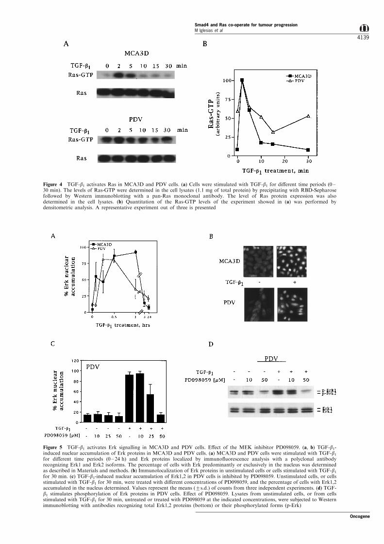
Figure 4 TGF-b1 activates Ras in MCA3D and PDV cells. (a) Cells were stimulated with TGF-b1 for di�erent time periods (0 ±30 min). The levels of Ras-GTP were determined in the cell lysates (1.1 mg of total protein) by precipitating with RBD-Sepharosefollowed by Western immunoblotting with a pan-Ras monoclonal antibody. The level of Ras protein expression was alsodetermined in the cell lysates. (b) Quantitation of the Ras-GTP levels of the experiment showed in (a) was performed bydensitometric analysis. A representative experiment out of three is presented
Figure 5 TGF-b1 activates Erk signalling in MCA3D and PDV cells. E�ect of the MEK inhibitor PD098059. (a, b) TGF-b1-induced nuclear accumulation of Erk proteins in MCA3D and PDV cells. (a) MCA3D and PDV cells were stimulated with TGF-b1for di�erent time periods (0 ± 24 h) and Erk proteins localized by immuno¯uorescence analysis with a polyclonal antibodyrecognizing Erk1 and Erk2 isoforms. The percentage of cells with Erk predominantly or exclusively in the nucleus was determinedas described in Materials and methods. (b) Immunolocalization of Erk proteins in unstimulated cells or cells stimulated with TGF-b1for 30 min. (c) TGF-b1-induced nuclear accumulation of Erk1,2 in PDV cells is inhibited by PD098059. Unstimulated cells, or cellsstimulated with TGF-b1 for 30 min, were treated with di�erent concentrations of PD098059, and the percentage of cells with Erk1,2accumulated in the nucleus determined. Values represent the means (+s.d.) of counts from three independent experiments. (d) TGF-b1 stimulates phosphorylation of Erk proteins in PDV cells. E�ect of PD098059. Lysates from unstimulated cells, or from cellsstimulated with TGF-b1 for 30 min, untreated or treated with PD098059 at the indicated concentrations, were subjected to Westernimmunoblotting with antibodies recognizing total Erk1,2 proteins (bottom) or their phosphorylated forms (p-Erk)
Oncogene
Smad4 and Ras co-operate for tumour progressionM lglesias et al
4139
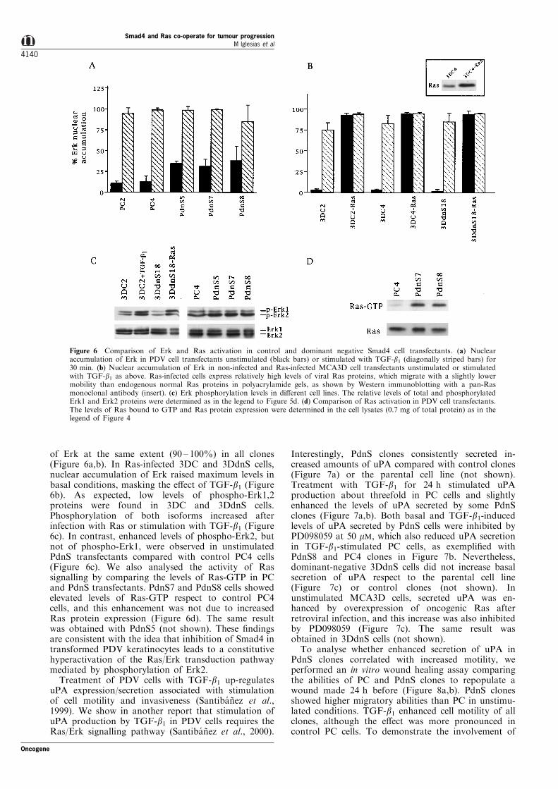
of Erk at the same extent (90 ± 100%) in all clones(Figure 6a,b). In Ras-infected 3DC and 3DdnS cells,nuclear accumulation of Erk raised maximum levels inbasal conditions, masking the e�ect of TGF-b1 (Figure6b). As expected, low levels of phospho-Erk1,2proteins were found in 3DC and 3DdnS cells.Phosphorylation of both isoforms increased afterinfection with Ras or stimulation with TGF-b1 (Figure6c). In contrast, enhanced levels of phospho-Erk2, butnot of phospho-Erk1, were observed in unstimulatedPdnS transfectants compared with control PC4 cells(Figure 6c). We also analysed the activity of Rassignalling by comparing the levels of Ras-GTP in PCand PdnS transfectants. PdnS7 and PdnS8 cells showedelevated levels of Ras-GTP respect to control PC4cells, and this enhancement was not due to increasedRas protein expression (Figure 6d). The same resultwas obtained with PdnS5 (not shown). These ®ndingsare consistent with the idea that inhibition of Smad4 intransformed PDV keratinocytes leads to a constitutivehyperactivation of the Ras/Erk transduction pathwaymediated by phosphorylation of Erk2.
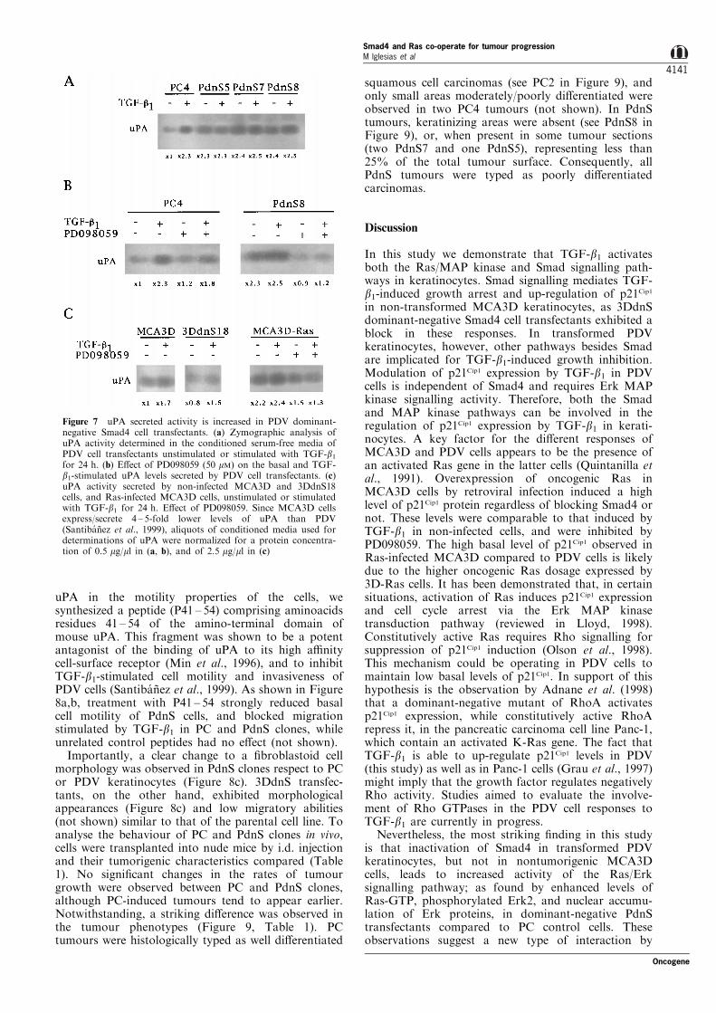
Treatment of PDV cells with TGF-b1 up-regulatesuPA expression/secretion associated with stimulationof cell motility and invasiveness (Santiba nÄ ez et al.,1999). We show in another report that stimulation ofuPA production by TGF-b1 in PDV cells requires theRas/Erk signalling pathway (Santiba nÄ ez et al., 2000).
Interestingly, PdnS clones consistently secreted in-creased amounts of uPA compared with control clones(Figure 7a) or the parental cell line (not shown).Treatment with TGF-b1 for 24 h stimulated uPAproduction about threefold in PC cells and slightlyenhanced the levels of uPA secreted by some PdnSclones (Figure 7a,b). Both basal and TGF-b1-inducedlevels of uPA secreted by PdnS cells were inhibited byPD098059 at 50 mM, which also reduced uPA secretionin TGF-b1-stimulated PC cells, as exempli®ed withPdnS8 and PC4 clones in Figure 7b. Nevertheless,dominant-negative 3DdnS cells did not increase basalsecretion of uPA respect to the parental cell line(Figure 7c) or control clones (not shown). Inunstimulated MCA3D cells, secreted uPA was en-hanced by overexpression of oncogenic Ras afterretroviral infection, and this increase was also inhibitedby PD098059 (Figure 7c). The same result wasobtained in 3DdnS cells (not shown).
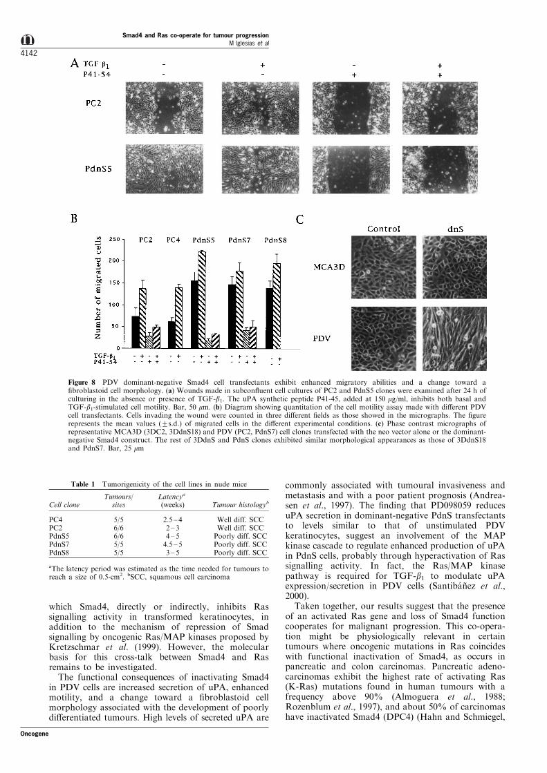
To analyse whether enhanced secretion of uPA inPdnS clones correlated with increased motility, weperformed an in vitro wound healing assay comparingthe abilities of PC and PdnS clones to repopulate awound made 24 h before (Figure 8a,b). PdnS clonesshowed higher migratory abilities than PC in unstimu-lated conditions. TGF-b1 enhanced cell motility of allclones, although the e�ect was more pronounced incontrol PC cells. To demonstrate the involvement of
Figure 6 Comparison of Erk and Ras activation in control and dominant negative Smad4 cell transfectants. (a) Nuclearaccumulation of Erk in PDV cell transfectants unstimulated (black bars) or stimulated with TGF-b1 (diagonally striped bars) for30 min. (b) Nuclear accumulation of Erk in non-infected and Ras-infected MCA3D cell transfectants unstimulated or stimulatedwith TGF-b1 as above. Ras-infected cells express relatively high levels of viral Ras proteins, which migrate with a slightly lowermobility than endogenous normal Ras proteins in polyacrylamide gels, as shown by Western immunoblotting with a pan-Rasmonoclonal antibody (insert). (c) Erk phosphorylation levels in di�erent cell lines. The relative levels of total and phosphorylatedErk1 and Erk2 proteins were determined as in the legend to Figure 5d. (d) Comparison of Ras activation in PDV cell transfectants.The levels of Ras bound to GTP and Ras protein expression were determined in the cell lysates (0.7 mg of total protein) as in thelegend of Figure 4
Smad4 and Ras co-operate for tumour progressionM lglesias et al
4140
Oncogene
uPA in the motility properties of the cells, wesynthesized a peptide (P41 ± 54) comprising aminoacidsresidues 41 ± 54 of the amino-terminal domain ofmouse uPA. This fragment was shown to be a potentantagonist of the binding of uPA to its high a�nitycell-surface receptor (Min et al., 1996), and to inhibitTGF-b1-stimulated cell motility and invasiveness ofPDV cells (Santiba nÄ ez et al., 1999). As shown in Figure8a,b, treatment with P41 ± 54 strongly reduced basalcell motility of PdnS cells, and blocked migrationstimulated by TGF-b1 in PC and PdnS clones, whileunrelated control peptides had no e�ect (not shown).
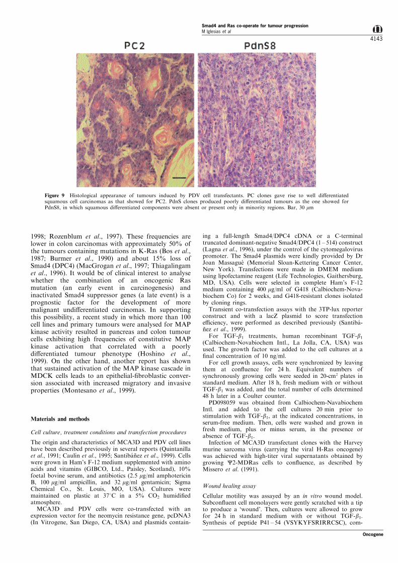
Importantly, a clear change to a ®broblastoid cellmorphology was observed in PdnS clones respect to PCor PDV keratinocytes (Figure 8c). 3DdnS transfec-tants, on the other hand, exhibited morphologicalappearances (Figure 8c) and low migratory abilities(not shown) similar to that of the parental cell line. Toanalyse the behaviour of PC and PdnS clones in vivo,cells were transplanted into nude mice by i.d. injectionand their tumorigenic characteristics compared (Table1). No signi®cant changes in the rates of tumourgrowth were observed between PC and PdnS clones,although PC-induced tumours tend to appear earlier.Notwithstanding, a striking di�erence was observed inthe tumour phenotypes (Figure 9, Table 1). PCtumours were histologically typed as well di�erentiated
squamous cell carcinomas (see PC2 in Figure 9), andonly small areas moderately/poorly di�erentiated wereobserved in two PC4 tumours (not shown). In PdnStumours, keratinizing areas were absent (see PdnS8 inFigure 9), or, when present in some tumour sections(two PdnS7 and one PdnS5), representing less than25% of the total tumour surface. Consequently, allPdnS tumours were typed as poorly di�erentiatedcarcinomas.
Discussion
In this study we demonstrate that TGF-b1 activatesboth the Ras/MAP kinase and Smad signalling path-ways in keratinocytes. Smad signalling mediates TGF-b1-induced growth arrest and up-regulation of p21Cip1
in non-transformed MCA3D keratinocytes, as 3DdnSdominant-negative Smad4 cell transfectants exhibited ablock in these responses. In transformed PDVkeratinocytes, however, other pathways besides Smadare implicated for TGF-b1-induced growth inhibition.Modulation of p21Cip1 expression by TGF-b1 in PDVcells is independent of Smad4 and requires Erk MAPkinase signalling activity. Therefore, both the Smadand MAP kinase pathways can be involved in theregulation of p21Cip1 expression by TGF-b1 in kerati-nocytes. A key factor for the di�erent responses ofMCA3D and PDV cells appears to be the presence ofan activated Ras gene in the latter cells (Quintanilla etal., 1991). Overexpression of oncogenic Ras inMCA3D cells by retroviral infection induced a highlevel of p21Cip1 protein regardless of blocking Smad4 ornot. These levels were comparable to that induced byTGF-b1 in non-infected cells, and were inhibited byPD098059. The high basal level of p21Cip1 observed inRas-infected MCA3D compared to PDV cells is likelydue to the higher oncogenic Ras dosage expressed by3D-Ras cells. It has been demonstrated that, in certainsituations, activation of Ras induces p21Cip1 expressionand cell cycle arrest via the Erk MAP kinasetransduction pathway (reviewed in Lloyd, 1998).Constitutively active Ras requires Rho signalling forsuppression of p21Cip1 induction (Olson et al., 1998).This mechanism could be operating in PDV cells tomaintain low basal levels of p21Cip1. In support of thishypothesis is the observation by Adnane et al. (1998)that a dominant-negative mutant of RhoA activatesp21Cip1 expression, while constitutively active RhoArepress it, in the pancreatic carcinoma cell line Panc-1,which contain an activated K-Ras gene. The fact thatTGF-b1 is able to up-regulate p21Cip1 levels in PDV(this study) as well as in Panc-1 cells (Grau et al., 1997)might imply that the growth factor regulates negativelyRho activity. Studies aimed to evaluate the involve-ment of Rho GTPases in the PDV cell responses toTGF-b1 are currently in progress.
Nevertheless, the most striking ®nding in this studyis that inactivation of Smad4 in transformed PDVkeratinocytes, but not in nontumorigenic MCA3Dcells, leads to increased activity of the Ras/Erksignalling pathway; as found by enhanced levels ofRas-GTP, phosphorylated Erk2, and nuclear accumu-lation of Erk proteins, in dominant-negative PdnStransfectants compared to PC control cells. Theseobservations suggest a new type of interaction by
Figure 7 uPA secreted activity is increased in PDV dominant-negative Smad4 cell transfectants. (a) Zymographic analysis ofuPA activity determined in the conditioned serum-free media ofPDV cell transfectants unstimulated or stimulated with TGF-b1for 24 h. (b) E�ect of PD098059 (50 mM) on the basal and TGF-b1-stimulated uPA levels secreted by PDV cell transfectants. (c)uPA activity secreted by non-infected MCA3D and 3DdnS18cells, and Ras-infected MCA3D cells, unstimulated or stimulatedwith TGF-b1 for 24 h. E�ect of PD098059. Since MCA3D cellsexpress/secrete 4 ± 5-fold lower levels of uPA than PDV(Santiba nÄ ez et al., 1999), aliquots of conditioned media used fordeterminations of uPA were normalized for a protein concentra-tion of 0.5 mg/ml in (a, b), and of 2.5 mg/ml in (c)
Oncogene
Smad4 and Ras co-operate for tumour progressionM lglesias et al
4141
which Smad4, directly or indirectly, inhibits Rassignalling activity in transformed keratinocytes, inaddition to the mechanism of repression of Smadsignalling by oncogenic Ras/MAP kinases proposed byKretzschmar et al. (1999). However, the molecularbasis for this cross-talk between Smad4 and Rasremains to be investigated.
The functional consequences of inactivating Smad4in PDV cells are increased secretion of uPA, enhancedmotility, and a change toward a ®broblastoid cellmorphology associated with the development of poorlydi�erentiated tumours. High levels of secreted uPA are
commonly associated with tumoural invasiveness andmetastasis and with a poor patient prognosis (Andrea-sen et al., 1997). The ®nding that PD098059 reducesuPA secretion in dominant-negative PdnS transfectantsto levels similar to that of unstimulated PDVkeratinocytes, suggest an involvement of the MAPkinase cascade to regulate enhanced production of uPAin PdnS cells, probably through hyperactivation of Rassignalling activity. In fact, the Ras/MAP kinasepathway is required for TGF-b1 to modulate uPAexpression/secretion in PDV cells (Santiba nÄ ez et al.,2000).
Taken together, our results suggest that the presenceof an activated Ras gene and loss of Smad4 functioncooperates for malignant progression. This co-opera-tion might be physiologically relevant in certaintumours where oncogenic mutations in Ras coincideswith functional inactivation of Smad4, as occurs inpancreatic and colon carcinomas. Pancreatic adeno-carcinomas exhibit the highest rate of activating Ras(K-Ras) mutations found in human tumours with afrequency above 90% (Almoguera et al., 1988;Rozenblum et al., 1997), and about 50% of carcinomashave inactivated Smad4 (DPC4) (Hahn and Schmiegel,
Figure 8 PDV dominant-negative Smad4 cell transfectants exhibit enhanced migratory abilities and a change toward a®broblastoid cell morphology. (a) Wounds made in subcon¯uent cell cultures of PC2 and PdnS5 clones were examined after 24 h ofculturing in the absence or presence of TGF-b1. The uPA synthetic peptide P41-45, added at 150 mg/ml, inhibits both basal andTGF-b1-stimulated cell motility. Bar, 50 mm. (b) Diagram showing quantitation of the cell motility assay made with di�erent PDVcell transfectants. Cells invading the wound were counted in three di�erent ®elds as those showed in the micrographs. The ®gurerepresents the mean values (+s.d.) of migrated cells in the di�erent experimental conditions. (c) Phase contrast micrographs ofrepresentative MCA3D (3DC2, 3DdnS18) and PDV (PC2, PdnS7) cell clones transfected with the neo vector alone or the dominant-negative Smad4 construct. The rest of 3DdnS and PdnS clones exhibited similar morphological appearances as those of 3DdnS18and PdnS7. Bar, 25 mm
Table 1 Tumorigenicity of the cell lines in nude mice
Tumours/ Latencya
Cell clone sites (weeks) Tumour histologyb
PC4 5/5 2.5 ± 4 Well diff. SCCPC2 6/6 2 ± 3 Well diff. SCCPdnS5 6/6 4 ± 5 Poorly diff. SCCPdnS7 5/5 4.5 ± 5 Poorly diff. SCCPdnS8 5/5 3 ± 5 Poorly diff. SCC
aThe latency period was estimated as the time needed for tumours toreach a size of 0.5-cm2. bSCC, squamous cell carcinoma
Smad4 and Ras co-operate for tumour progressionM lglesias et al
4142
Oncogene
1998; Rozenblum et al., 1997). These frequencies arelower in colon carcinomas with approximately 50% ofthe tumours containing mutations in K-Ras (Bos et al.,1987; Burmer et al., 1990) and about 15% loss ofSmad4 (DPC4) (MacGrogan et al., 1997; Thiagalingamet al., 1996). It would be of clinical interest to analysewhether the combination of an oncogenic Rasmutation (an early event in carcinogenesis) andinactivated Smad4 suppressor genes (a late event) is aprognostic factor for the development of moremalignant undi�erentiated carcinomas. In supportingthis possibility, a recent study in which more than 100cell lines and primary tumours were analysed for MAPkinase activity resulted in pancreas and colon tumourcells exhibiting high frequencies of constitutive MAPkinase activation that correlated with a poorlydi�erentiated tumour phenotype (Hoshino et al.,1999). On the other hand, another report has shownthat sustained activation of the MAP kinase cascade inMDCK cells leads to an epithelial-®broblastic conver-sion associated with increased migratory and invasiveproperties (Montesano et al., 1999).
Materials and methods
Cell culture, treatment conditions and transfection procedures
The origin and characteristics of MCA3D and PDV cell lineshave been described previously in several reports (Quintanillaet al., 1991; Caulõ n et al., 1995; Santiba nÄ ez et al., 1999). Cellswere grown in Ham's F-12 medium supplemented with aminoacids and vitamins (GIBCO, Ltd., Paisley, Scotland), 10%foetal bovine serum, and antibiotics (2.5 mg/ml amphotericinB, 100 mg/ml ampicillin, and 32 mg/ml gentamicin; SigmaChemical Co., St. Louis, MO, USA). Cultures weremaintained on plastic at 378C in a 5% CO2 humidi®edatmosphere.MCA3D and PDV cells were co-transfected with an
expression vector for the neomycin resistance gene, pcDNA3(In Vitrogene, San Diego, CA, USA) and plasmids contain-
ing a full-length Smad4/DPC4 cDNA or a C-terminaltruncated dominant-negative Smad4/DPC4 (1 ± 514) construct(Lagna et al., 1996), under the control of the cytomegaloviruspromoter. The Smad4 plasmids were kindly provided by DrJoan Massague (Memorial Sloan-Kettering Cancer Center,New York). Transfections were made in DMEM mediumusing lipofectamine reagent (Life Technologies, Gaithersburg,MD, USA). Cells were selected in complete Ham's F-12medium containing 400 mg/ml of G418 (Calbiochem-Nova-biochem Co) for 2 weeks, and G418-resistant clones isolatedby cloning rings.Transient co-transfection assays with the 3TP-lux reporter
construct and with a lacZ plasmid to score transfectione�ciency, were performed as described previously (Santiba -nÄ ez et al., 1999).For TGF-b1 treatments, human recombinant TGF-b1
(Calbiochem-Novabiochem Intl., La Jolla, CA, USA) wasused. The growth factor was added to the cell cultures at a®nal concentration of 10 ng/ml.For cell growth assays, cells were synchronized by leaving
them at con¯uence for 24 h. Equivalent numbers ofsynchronously growing cells were seeded in 20-cm2 plates instandard medium. After 18 h, fresh medium with or withoutTGF-b1 was added, and the total number of cells determined48 h later in a Coulter counter.PD098059 was obtained from Calbiochem-Navabiochem
Intl. and added to the cell cultures 20 min prior tostimulation with TGF-b1, at the indicated concentrations, inserum-free medium. Then, cells were washed and grown infresh medium, plus or minus serum, in the presence orabsence of TGF-b1.Infection of MCA3D transfectant clones with the Harvey
murine sarcoma virus (carrying the viral H-Ras oncogene)was achieved with high-titer viral supernatants obtained bygrowing C2-MDRas cells to con¯uence, as described byMissero et al. (1991).
Wound healing assay
Cellular motility was assayed by an in vitro wound model.Subcon¯uent cell monolayers were gently scratched with a tipto produce a `wound'. Then, cultures were allowed to growfor 24 h in standard medium with or without TGF-b1.Synthesis of peptide P41 ± 54 (VSYKYFSRIRRCSC), com-
Figure 9 Histological appearance of tumours induced by PDV cell transfectants. PC clones gave rise to well di�erentiatedsquamous cell carcinomas as that showed for PC2. PdnS clones produced poorly di�erentiated tumours as the one showed forPdnS8, in which squamous di�erentiated components were absent or present only in minority regions. Bar, 30 mm
Oncogene
Smad4 and Ras co-operate for tumour progressionM lglesias et al
4143
prising amino acid residues 41 ± 54 of the EGF-like domainof mouse uPA, and unrelated peptides derived from gelatinby limited trypsin degradation, used as a control, aredescribed elsewhere (Santiba nÄ ez et al., 1999). Peptides wereadded to the culture media, at 150 mg/ml, at the same time asTGF-b1.
Immunofluorescence analysis
Immuno¯uorescence stainings for Smad2,3 and Erk1,2proteins were performed by conventional procedures, in cells®xed and permeabilized in cold methanol, with polyclonalantibodies (Santa Cruz Biotechnology Inc., Santa Cruz, CA,USA): E-20, speci®c for Smad2 and Smad3, and K-23, whichrecognizes both Erk1 and Erk2 isoforms. Antibodies wereused at 1 : 50 dilution in PBS containing 2% BSA.Rhodamine-conjugated anti-goat and anti-rabbit IgG (Jack-son ImmunoResearch Lab., West Grove, PA, USA),respectively, were used as secondary antibodies. Di�erent®elds containing 30 ± 60 cells were photographed, and thepercentage of cells with Smad2,3 or Erk1,2 accumulated inthe nucleus determined counting 10 distinct ®elds fromtriplicates corresponding to each experimental situation.
Western blot analysis
Cells were lysed in RIPA bu�er (50 mM NaCl, 1% NP-40,0,5% sodium deoxycholate, 0,1% SDS, 50 mM Tris-HCl,pH 7.5) and a cocktail of proteinase inhibitors (2 mg/mlaprotinin, 2 mg/ml leupeptin and 1 mM phenylmethylsulpho-nyl ¯uoride). For phospho-Erk determinations, phosphataseinhibitors: 1 mM sodium orthovanadate, 50 mM sodium¯uoride, 10 mM sodium pyrophosphate and 2 mM hydrogenperoxide, were added to the lysis bu�er. For Ras proteindeterminations a lysis bu�er containing 100 mM NaCl, 5 mM
CaCl2, 5 mM MgCl2, 2% SDS, 50 mM Tris HCl, pH 7.4, andproteinase inhibitors was used. Proteins (20 or 30 mg/lane)were resolved by SDS±PAGE and transferred to ImmobilonP membranes (Millipore Corp., Bedford, MA, USA). Rasproteins were detected by a pan-Ras monoclonal antibody(Ab-3, Oncogene Science Inc., Uniondale, NY, USA). Forp21Cip1 and phospho-Erk1,2 protein determinations, a poly-clonal antibody (C-20) and a monoclonal antibody (E-4),respectively, both from Santa Cruz Biothechnology Inc., wereused as primary antibodies, followed by exposure toperoxidase-conjugated secondary antibodies (Amersham Intl.Plc. Amersham Bucks, UK), and developed using theenhanced chemoluminescent system (ECL, Amersham Corp.,Arlington Heights, FL, USA). The bands detected in the
Western blots were quanti®ed by scanning and digitalizationof the X-ray ®lms.
Measurement of Ras activation and zymographic assays
The capacity of Ras-GTP to bind the Ras-binding domain ofRaf-1 (RBD) was used to analyse the level of active Ras inthe cells (de Rooij and Bos, 1997). Brie¯y, cleared cell lysates(0.7 ± 1.1 mg of total protein) were incubated with 30 mg ofthe fusion protein GST-RBD bound to glutathione-Sephar-ose beads. Beads were washed and bound proteins solubilizedin Laemmli bu�er and resolved in SDS ±PAGE. Proteinswere then immunoblotted as described above, and precipi-tated Ras-GTP detected using a pan-Ras monoclonal anti-body. The level of Ras protein expression was determined inthe cell lysates by Western immunoblotting using the sameantibody.uPA-secreted activity of cell cultures was determined by
caseinolytic zymography, as previously reported (Santiba nÄ ezet al., 1999). Aliquots (20 ± 30 ml) of conditioned serum-freemedium, normalized for the same amount of protein (0.5 mg/ml in PDV experiments, and 2.5 mg/ml in MCA3D experi-ments), were ®rst subjected to SDS ±PAGE under nonredu-cing conditions, and then to zymography on agarose gelscontaining casein and plasminogen.
Tumorigenicity assays and histopathology
Suspensions of 1 ± 26106 cells were injected intradermaly intothe ¯anks of 8 ± 10-week-old Swiss athymic nude mice(CRIFFA, Barcelona, Spain). Animals were observed fortumour formation, and the size of tumours (in cm2)calculated from caliper measurements of two orthogonaldiameters at di�erent times. Histological typing of alldeveloped tumours was performed on formalin-®xed andpara�n-embedded sections by staining with hematoxylin andeosin.
AcknowledgmentsWe thank Dr Joan Massague for kindly providing Smad4plasmids, Dr Jaime Renart for the GST-RBD fusionconstruct, and Dr Carmelo Bernabeu for the 3TP-luxreporter. We also thank Drs Piero Crespo and AntonioVillalobo for critical reading of the manuscript and helpfulsuggestions. This work was supported by grants: SAF98-0085-CO3-02 from the Comisio n Interministerial de Cien-cia y Tecnologõ a (CICYT), and 8.1/22/97 from theComunidad Auto noma de Madrid (CAM).
References
Adnane J, Bizouarn FA, Qian Y, Hamilton AD and SebtiSM. (1998). Mol. Cell. Biol., 18, 6962 ± 6970.
Akhurst RJ and Balmain A. (1999). J. Pathol., 187, 82 ± 90.Alexandrow MG and Moses HL. (1995). Cancer Res., 55,
1452 ± 1457.Almoguera C, Shibata D, Forrester K, Martin J, Arnheim N
and Perucho M. (1988). Cell, 53, 549 ± 554.Andreasen PA, Kjoller L, Christensen L and Du�y MJ.
(1997). Int. J. Cancer, 72, 1 ± 22.At® A, Buisine M, Mazars A and Gespach C. (1997). J. Biol.
Chem., 272, 24731 ± 24734.Bos JL, Fearon ER, Hamilton SR, Verlaan-de Vries M, van
Boom JH, van der Eb AJ and Vogelstein B. (1987).Nature,327, 293 ± 297.
Burmer GC, Levine DS, Kulander BG, Haggitt RC, RubinCE and Rabinovitch PS. (1990). Gastroenterology, 99,416 ± 420.
Burns PA, Kemp CJ, Gannon JV, Lane DP, Bremner R andBalmain A. (1991). Oncogene, 6, 2363 ± 2369.
CaulõÂ n C, Scholl FG, Frontelo P, Gamallo C and QuintanillaM. (1995). Cell Growth Di�er., 6, 1027 ± 1035.
Cui W, Fowlis DJ, Bryson S, Du�e E, Ireland H, Balmain Aand Akhurst RJ. (1996). Cell, 86, 531 ± 542.
Datto MD, Li Y, Panus JF, Howe DJ, Xiong Y and Xiao-Fan W. (1995). Proc. Natl. Acad. Sci. USA, 92, 5545 ±5549.
Davis RJ. (1993). J. Biol. Chem., 268, 14553 ± 14556.de Rooij J and Bos JL. (1997). Oncogene, 14, 623 ± 625.de Winter JP, Roelen BAJ, ten Dijke P, van der Burg B and
van den Eijnden-van Raaij AJM. (1997). Oncogene, 14,1891 ± 1899.
Farina AR, Coppa A, Tiberio A, Tacconelli A, Turco A,Colletta G, Gulino A and Mackay AR. (1998). Int. J.Cancer, 75, 721 ± 730.
Frontelo P, Gonza lez-Garrigues M, Vilaro S, Gamallo C,Fabra A and Quintanilla M. (1998). Exp. Cell Res., 244,420 ± 432.
Smad4 and Ras co-operate for tumour progressionM lglesias et al
4144
Oncogene

Grau AM, Zhang L, Wang W, Ruan S, Evans DB,Abbruzzese JL, Zhang W and Chiao PJ. (1997). CancerRes., 57, 3929 ± 3934.
Haddow S, Fowlis DJ, Parkinson K, Akhurst RJ andBalmain A. (1991). Oncogene, 6, 1465 ± 1570.
Hahn SA and Schmiegel WH. (1998). Digestion, 59, 493 ±501.
Hannon GJ and Beach D. (1994). Nature, 371, 257 ± 261.Hartsough MT, Frey RS, Zipfel PA, Buard A, Cook SJ,
McCormick F and Mulder KM. (1996). J. Biol. Chem.,271, 22368 ± 22375.
Hartsough MT and Mulder KM. (1997). Pharmacol. Ther.,75, 21 ± 41.
Hill CS and Treisman R. (1995). Cell, 80, 199 ± 211.Hoshino R, Chatani Y, Yamori T, Tsuruo T, Oka H,
Yoshida O, Shimada Y, Ari-i S, Wada H, Fujimoto J andKohno M. (1999). Oncogene, 18, 813 ± 822.
Hu PP, Shen X, Huang D, Liu Y, Counter C and Wang X-F.(1999). J. Biol. Chem., 274, 35381 ± 35387.
Hunt KK, Fleming JB, Abramian A, Zhang L, Evans DBand Chiao PJ. (1998). Cancer Res., 58, 5656 ± 5661.
Kretzschmar M, Doody J, Timokhina I and Massague J.(1999). Genes Dev., 13, 804 ± 816.
Lagna G, Hata A, Hemmati-Brivaniou A and Massague J.(1996). Nature, 383, 832 ± 836.
Lahio M and Keski-Oja J. (1992). Crit. Rev. Oncog., 3, 1 ± 26.Lloyd AC. (1998). Curr. Opin. Gen. Dev., 8, 43 ± 48.MacGrogan DL, Pegram M, Slamon D and Bookstein R.
(1997). Oncogene, 15, 1111 ± 1114.Malliri A, Yeudall WA, Nikolic M, Crouch DH, Parkinson
EK and Ozanne B. (1996). Cell Growth Di�er., 7, 1291 ±1304.
Massague J. (1990). Annu. Rev. Cell Biol., 6, 597 ± 641.Massague J. (1998). Annu. Rev. Biochem., 67, 753 ± 791.Min HY, Doyle LV, Vitt CR, Zandonella CL, Stratton-
Thomas JR, Shuman MA and Rosenberg S. (1996).Cancer Res., 56, 2428 ± 2433.
Missero C, Filvaro� E and Dotto GP. (1991). Proc. Natl.Acad. Sci. USA, 88, 3489 ± 3493.
Montesano R, Soriano JV, Hosseini G, Pepper MS andSchramek H. (1999). Cell Growth Di�er., 10, 317 ± 332.
Mucsi I, Skorecki KL and Goldberg HJ. (1996). J. Biol.Chem., 271, 16567 ± 16572.
Oft M, Heider K-H and Beug H. (1998). Curr. Biol., 8, 1243 ±1252.
Oft M, Peli J, Rudaz C, Schwartz H, Beug H and ReichmannE. (1996). Genes Dev., 10, 2462 ± 2477.
Olson MF, Paterson HF and Marshall CJ. (1998). Nature,394, 295 ± 299.
Portella G, Cumming SA, Liddell J, Cui W, Ireland H,Akhurst RJ and Balmain A. (1998). Cell Growth Di�er., 9,393 ± 404.
Quintanilla M, Haddow S, Jonas D, Ja�e D, Bowden GTand Balmain A. (1991). Carcinogenesis, 12, 1875 ± 1881.
Reynisdo ttir I, Polyak K, Iavarone A and Massague J.(1995). Genes Dev., 9, 1831 ± 1845.
Rozenblum E, Schutte M, Goggins M, Hahn SA, Panzer S,Zahurak M, Goodman SN, Sohn TA, Hruban RH, YeoCJ and Kern SE. (1997). Cancer Res., 57, 1731 ± 1734.
Santiba nÄ ez JF, Frontelo P, Iglesias M, Martõ nez J andQuintanilla M. (1999). J. Cell. Biochem., 74, 61 ± 73.
Santiba nÄ ez JF, Iglesias M, Frontelo P, Martõ nez J andQuintanilla M. (2000). Biochem. Biophys. Res. Comm.,273, 521 ± 527.
Schwarte-Waldho� I, Klein S, Blass-Kampmann S, Hintel-mann A, Eilert C, Dreschers S, Kaltho� H, Hahn SA andSchmiegel W. (1999). Oncogene, 18, 3152 ± 3158.
Simon C, Juarez J, Nicolson GL and Boyd D. (1996). CancerRes., 56, 5369 ± 5374.
Thiagalingam S, Lengauer C, Leach FS, Schutte M, HahnSA, Overhauser J, Willson JKV, Markowitz S, HamiltonSR, Kern SE, Kinzler KW and Vogelstein B. (1996).Nature Genetics, 13, 343 ± 346.
Yue J, Buard A and Mulder K. (1998). Oncogene, 17, 47 ± 55.Zhang Y and Derynck R. (1999). Trends Cell Biol., 9, 274 ±
279.
Oncogene
Smad4 and Ras co-operate for tumour progressionM lglesias et al
4145
Copyright © 2022 FDOKUMEN