Partial protection against collagen antibody-induced arthritis in PARP1 deficient mice
2013;12:2110-2120. Published OnlineFirst July 24, 2013.Mol Cancer Ther Erik A. Bey, Kathryn E. Reinicke, Melissa C. Srougi, et al. Directed Programmed Necrosis in NQO1-Positive Breast Cancers
−Induced PARP1 Hyperactivation−-LapachoneβCatalase Abrogates
Updated version
10.1158/1535-7163.MCT-12-0962doi:
Access the most recent version of this article at:
Material
Supplementary
http://mct.aacrjournals.org/content/suppl/2013/07/24/1535-7163.MCT-12-0962.DC1.html
Access the most recent supplemental material at:
Cited Articles
http://mct.aacrjournals.org/content/12/10/2110.full.html#ref-list-1
This article cites by 41 articles, 20 of which you can access for free at:
E-mail alerts related to this article or journal.Sign up to receive free email-alerts
Subscriptions
Reprints and
To order reprints of this article or to subscribe to the journal, contact the AACR Publications Department at
Permissions
To request permission to re-use all or part of this article, contact the AACR Publications Department at
on May 5, 2014. © 2013 American Association for Cancer Research. mct.aacrjournals.org Downloaded from
Published OnlineFirst July 24, 2013; DOI: 10.1158/1535-7163.MCT-12-0962
on May 5, 2014. © 2013 American Association for Cancer Research. mct.aacrjournals.org Downloaded from
Published OnlineFirst July 24, 2013; DOI: 10.1158/1535-7163.MCT-12-0962

Cancer Therapeutics Insights
Catalase Abrogates b-Lapachone–Induced PARP1Hyperactivation–Directed Programmed Necrosis inNQO1-Positive Breast Cancers
Erik A. Bey1, Kathryn E. Reinicke2, Melissa C. Srougi3, Marie Varnes2, Vernon E. Anderson2, John J. Pink4,Long Shan Li5, Malina Patel5, Lifen Cao5, Zachary Moore5, Amy Rommel5, Michael Boatman1, Cheryl Lewis6,David M. Euhus6, William G. Bornmann7, Donald J. Buchsbaum8, Douglas R. Spitz9, Jinming Gao5, andDavid A. Boothman5
AbstractImproving patient outcome by personalized therapy involves a thorough understanding of an agent’s
mechanism of action. b-Lapachone (clinical forms, Arq501/Arq761) has been developed to exploit
dramatic cancer-specific elevations in the phase II detoxifying enzyme NAD(P)H:quinone oxidoreductase
(NQO1). NQO1 is dramatically elevated in solid cancers, including primary and metastatic [e.g., triple-
negative (ER�, PR�, Her2/Neu�)] breast cancers. To define cellular factors that influence the efficacy of
b-lapachone using knowledge of its mechanism of action, we confirmed that NQO1 was required for
lethality and mediated a futile redox cycle where �120 moles of superoxide were formed per mole
of b-lapachone in 2 minutes. b-Lapachone induced reactive oxygen species (ROS), stimulated DNA single-
strand break-dependent poly(ADP-ribose) polymerase-1 (PARP1) hyperactivation, caused dramatic loss of
essential nucleotides (NADþ/ATP), and elicited programmed necrosis in breast cancer cells. Although
PARP1 hyperactivation and NQO1 expression were major determinants of b-lapachone–induced lethality,
alterations in catalase expression, including treatment with exogenous enzyme, caused marked cyto-
protection. Thus, catalase is an important resistance factor and highlights H2O2 as an obligate ROS for cell
death from this agent. Exogenous superoxide dismutase enhanced catalase-induced cytoprotection.
b-Lapachone–induced cell death included apoptosis-inducing factor (AIF) translocation from mitochon-
dria to nuclei, TUNELþ staining, atypical PARP1 cleavage, and glyceraldehyde 3-phosphate dehydroge-
nase S-nitrosylation, which were abrogated by catalase. We predict that the ratio of NQO1:catalase
activities in breast cancer versus associated normal tissue are likely to be the major determinants affecting
the therapeutic window of b-lapachone and other NQO1 bioactivatable drugs. Mol Cancer Ther; 12(10);
2110–20. �2013 AACR.
IntroductionApproximately 1 in 8 women will be diagnosed with
breast cancer in their lifetimes in the United States (1).Most current therapeutic strategies designed to treatbreast cancer, as with other cancers, attack proliferativedifferences between tumor and associated normal breasttissue. These approaches frequently result in minimalefficacy, undesirable normal tissue toxicity, and oftenresult in selection of drug resistant, aggressive cancercells undergoing epithelial-to-mesenchymal transition,with increased metastatic potential (2–5). Because cas-pase pathways are commonly abrogated or altered inbreast or other cancers (6), strategies for treating cycling,as well as dormant cancer cells, that exploit caspase-independent cell death pathways (e.g., programmednecrosis) are desperately needed. Few drugs mechanis-tically act to induce poly(ADP-ribose) polymerase-1(PARP1)-mediated programmed necrosis in a tumor-specific manner and at clinically relevant doses (7). Most
Authors' Affiliations: 1Department of Basic Pharmaceutical Sciences,Mary Babb Randolph Cancer Center, West Virginia University, Morgan-town, West Virginia; Departments of 2Biochemistry, 3Pharmacology,and 4Oncology, Case Western Reserve University, Cleveland, Ohio;5Department of Pharmacology, Laboratory of Molecular Cell StressResponses, Program in Cell Stress and Cancer Nanomedicine, Sim-mons Cancer Center; 6Department of Surgery, UT Southwestern Med-ical Center at Dallas, Dallas; 7Department of Experimental Therapeutics,MD Anderson Cancer Center, Houston, Texas; 8Department of Radia-tion Oncology, University of Alabama-Birmingham, Birmingham, Ala-bama; and 9Free Radical and Radiation Biology Program, Department ofRadiation Oncology, Holden Comprehensive Cancer Center, Universityof Iowa, Iowa City, Iowa
Note: Supplementary data for this article are available at Molecular CancerTherapeutics Online (http://mct.aacrjournals.org/).
Corresponding Authors: Erik A. Bey, West Virginia University, 1 MedicalCenter Drive, Box 9300, Room1835,Morgantown,WV 26506. Phone: 214-645-6371; Fax: 214-645-6347; E-mail: [email protected]; and David A.Boothman, University of Texas South Western Medical Center at Dallas,6001 Forest Park Drive, ND2.210K, Mail stop 8807, Dallas, Texas 75390-8807; Email: [email protected]
doi: 10.1158/1535-7163.MCT-12-0962
�2013 American Association for Cancer Research.
MolecularCancer
Therapeutics
Mol Cancer Ther; 12(10) October 20132110
on May 5, 2014. © 2013 American Association for Cancer Research. mct.aacrjournals.org Downloaded from
Published OnlineFirst July 24, 2013; DOI: 10.1158/1535-7163.MCT-12-0962

agents known to stimulate PARP1-mediated progra-mmed necrosis (e.g., N-methyl, N’-nitro, N-nitrosogua-nidine (>5 mmol/L MNNG), or hydrogen peroxide(>200 mmol/L H2O2; refs. 8–10) do so at supra-lethal,nonachievable doses clinically. In contrast, b-lapachone(b-lap; see Fig. 1 for structure) stimulates PARP1 hyper-activation and causes dramatic intracellular nucleotideloss, particularly inNADþ andATPpools (10), at clinicallyachievable doses. PARP1 uses NADþ as a substrate toconduct poly(ADP-ribosylation) (PAR) posttranslationalmodification of proteins, including itself, where poly(ADP-ribosylated)-PARP1 (PAR-PARP1) is functionallyinactive.b-Lap is efficacious against solid tumors that have
endogenously overexpressed NAD(P)H:quinone oxido-reductase I (NQO1), including >80% non–small cell lungand pancreatic, as well as >60% prostate and >70%
breast cancers, where 5- to >100-fold increases in NQO1activities above adjacent normal tissue were reported(11–13). b-Lap undergoes an NQO1-dependent futileredox cycle (Fig. 1A), where >60 moles of NAD(P)Hare consumed per mole b-lap in 1 to 2 minutes(12, 14–18). This futile cycle results from the instabilityof its hydroquinone and semiquinone forms, presum-ably consuming oxygen and theoretically creating dra-matically elevated levels of superoxide (O2
.�). CombinedDNA damage and Ca2þ release from endoplasmic retic-ulum (Ca2þER) stores culminates in PARP1 hyper-activation, NADþ/ATP loss, DNA repair inhibition, andprogrammed necrosis by unknown protein effectors(12, 16–19). The majority of reported cellular affects inb-lap–treated cancer cells are actually the resultof dramatic NADþ/ATP losses (reviewed in ref. 20).Understanding the role(s) of reactive oxygen species
Figure 1. NQO1-dependent futile redox cycling of b-lap consumes O2 and generates robust ROS. A, proposed futile redox cycle of b-lap in thepresence of NQO1, where the hydroquinone form of b-lap is unstable. Through 2 one-electron oxidations, which presumably use O2, the hydroquinonespontaneously reverts back to b-lap. This "bioactivatable redox cycle" of b-lap uses�60moles NAD(P)H/mole b-lap in 2minutes (15), consuming 2moles O2
per cycle, creating�120moles superoxide in 2minutes. B (left), cell extracts fromNQO1-expressing (231-NQþ) or NQO1-deficient (231-NQ�) MDA-MB-231TNBC cells were placed in a closed system with 1, 2, or 15 mmol/L b-lap, � dicoumarol (DIC), an NQO1 inhibitor, for 5 minutes. OCR was measured withan Ocean Optics O2 sensor (left) as described in Materials and Methods. Each bar represents X-fold OCR means � SE, of 2 independent experimentsconducted in triplicate. �,P�0.05 (1 vs. 2mmol/Lb-lap); ��,P�0.007 (15mmol/Lb-lap�40mmol/LDIC). B (right),O2consumption rates andprotonproductionrates were measured in mock-treated (control) or b-Lap 4 mmol/L exposed 231-NQþ cells � 40 mmol/L DIC using the Seahorse XF24 Analyzer andAssayWizard software. Data represent means, percentage treated/control (T/C,%)�SE from quadruplicate samples. C and D, b-Lap–treated 231-NQþ and231-NQ� cells were analyzed for ROS formation at indicated times. Cells were stained with DHE (for O2
�) in C, or DCFDA (H2O2) in D, and micrographsquantified asdescribed (17) and inMaterials andMethods. DOX (5mmol/L, 30minutes), H2O2 (200mmol/L, 30minutes)were used aspositive controls. Studentt tests were conducted to determine significance between means of treatment conditions conducted in triplicate, repeated 3 individual times.
Catalase Confers Resistance to b-Lapachone Cell Death
www.aacrjournals.org Mol Cancer Ther; 12(10) October 2013 2111
on May 5, 2014. © 2013 American Association for Cancer Research. mct.aacrjournals.org Downloaded from
Published OnlineFirst July 24, 2013; DOI: 10.1158/1535-7163.MCT-12-0962

(ROS) and ROS-scavenging enzymes in deterring theefficacy of b-lap or deoxynyboquinone (DNQ), the onlyother NQO1 bioactivatable drug (7) against NQO1 over-expressed solid cancers remains unexplored.
Here, we report a potential mechanism of resistanceto b-lap–induced cell death by catalase, potentiated bysuperoxide dismutase (SOD). PARP1 hyperactivationwas the major determinant of downstream-programmednecrosis initiation when sublethal to lethal doses of b-lapwere compared. Exposure of NQO1þ cells to sublethalb-lap doses (�1 mmol/L) caused enhanced O2 consump-tion, ROS formation and single-strand breaks (SSBs);however, DNA lesions were below an apparent "thresh-old damage level" required for PARP1 hyperactivation.In contrast, lethal b-lap doses (�2 mmol/L) that killed�50% NQO1þ MDA-MB-231 triple-negative breast can-cer cells (TNBC, Supplemental Table S1) stimulatedPARP1 hyperactivation causing dramatic NADþ/ATPlosses. As a result, apoptosis inducing factor (AIF) acti-vation and glyceraldehyde 3-phosphate dehydrogenase(GAPDH) modification were noted that correspondedto programmed necrosis. Catalase spared b-lap–treatedcells from H2O2 (but not O2
.�) formation, PARP1 hyper-activation, AIF activation, atypical PARP1 and p53proteolysis, blocked TUNELþ staining, and enhancedclonogenic survival. The cytoprotective effects of catalasewere enhanced by SOD co-addition, consistent with anobligate role ofH2O2 in b-lap–induced lethality. Althoughinhibiting AIF expression slightly protected cells fromb-lap–induced lethality, altering downstream proteineffectors were much less effective than altering upstreamprocesses, such as scavenging ROS or chelating Ca2þER atpreventing lethality. The ratio of NQO1/catalase expres-sion in tumor versus normal tissue may be a majordeterminant in the efficacy of antitumor regimen involv-ing NQO1 bioactivatable drugs, such as b-lap.
Materials and MethodsChemicals, reagents, and antibodies
b-Lap (3,4-dihydro-2,2-dimethyl-2H-naphtho[1,2-b]pyran-5,6-dione; Fig. 1) was synthesized by us, con-firmed by NMR, dissolved in DMSO at 50 mmol/L, andconcentrations verified by spectrophotometry (15). Men-adione and doxorubicin (DOX) were obtained from Sig-ma-Aldrich and dissolved in DMSO or PBS (DOX),respectively. Dicoumarol (DIC; Sigma-Aldrich) was usedas described (15). 5-(and-6)Chloromethyl-2,7-dichlorodi-hydrofluorescein diacetate (DCFDA, 5 mmol/L) anddihy-droethidium (DHE, 5 mmol/L) used to assess ROS for-mation were purchased from Invitrogen and used asdescribed previously (18, 19). N-Acetyl-L-cysteine (NAC;Sigma-Aldrich) was used at 5 mmol/L for 24-hour pre-treatments and 2-hour cotreatments as described (21).Xanthine, nitroblue tetrazolium, diethylenetriaminepen-taacetic acid, catalase, CuZnSOD, bovine serum albumin(BSA), bathocuproine disulfonic acid disodium salthydrate, and xanthine oxidase (Sigma-Aldrich)were used
as described (22). Staurosporine (STS) and the pan-cas-pase inhibitor, zVAD-FMK (Z-VAD), were obtained andused as described (refs. 12, 14–18; see structures, Supple-mental Fig. S1). In transient AIF knockdown experiments,AIF (siAIF), NQO1 (si-NQ), and scrambled control vector(SCR) siRNAs were purchased from (Dharmacon).
Cell lines, cell culture, treatments, and survivalMDA-MB-231 TNBC cells lacking or expressing NQO1
and MCF-7:WS8 (MCF-7) cells were grown as described(15, 17). Additional breast cancer cell lines were pur-chased from ATTC, MAP-tested and were free of myco-plasma. Breast cancer cell lines were grown in RPMI 1640media at 37�C in a humidified 10% CO2-95% air atmo-sphere. Human mammary epithelial cells (HMEC 1585)were isolated from normal human mammary epithelialtissue of a patient without breast cancer and subjected toDNA finger printing. HMEC were grown in MEGM/DMEM-F12 complete media (Lonza and Mediatech) at37�C in a humidified 5% CO2-95% air atmosphere. Rela-tive survival assays were conducted as described (15).Experiments were repeated at least 3 times, and dataexpressed as relative survival (treated/control 100%)means � SE. Results using this assay directly correlatedwith clonogenic assays (15). A multitarget model wasused to describe the shape of relative survival curves,where the final slope (D0) and quasi-threshold (Dq) doseswere used to represent the size or width of the shoulder.Three parameters, n (extrapolation number), D0, and Dq
were related as: logen ¼ Dq/D0. In experiments withcatalase, bovine catalase (Sigma-Aldrich) was dissolvedin 5 mmol/L Hepes buffer (pH 7.4) or PBS. Dissolvedcatalase was filter sterilized and added to drug mediabefore exposure to drug treatment. O2 consumption rates(OCR) were measured using an Ocean Optics, Inc. Foxy-18G-AF oxygen sensor and S9 supernatants (15). S9extracts were added to a closed system in Tris-HClbuffer containing 10% BSA, pH 7.5, 0.5 mmol/L NADH,b-lap � 40 mmol/L DIC (23). The OOISensors program(Ocean Optics, Inc.) was used to quantify fluorescence at599.62 nm every 512 seconds, averaging every 4 readings.Data (means � SE) were graphed as X-fold OCR from3 independent experiments, each conducted in triplicate.In addition to Ocean Optics experiments, OCRs wereconfirmed in 231-NQþ and 231-NQ� cells using a Sea-horse XF24 extracellular flux analyzer (Seahorse Bio-sciences) as described previously (7). Briefly 25,000 231-NQþ or 231-NQ� cells were seeded in Seahorse 24-wellmicroplates. Before analysis, cells were left untreated(control) or treated with 4 mmol/L b-lap with or withoutDIC. OCR were measured as previously described (7).Data represent means, % treated/control (%T/C), �SEfrom quadruplicate assessments.
ROS, NADþ, and ATP measurementsCellular oxidative stresswas assessed by reduced/total
glutathione, %GSSG content (17), and normalized to pro-tein (24). Experiments were conducted 3 times and data
Bey et al.
Mol Cancer Ther; 12(10) October 2013 Molecular Cancer Therapeutics2112
on May 5, 2014. © 2013 American Association for Cancer Research. mct.aacrjournals.org Downloaded from
Published OnlineFirst July 24, 2013; DOI: 10.1158/1535-7163.MCT-12-0962

expressed as means � SE. ROS formation was assessedmicroscopically in 231 NQþ and NQ� cells stained withDCFDA or DHE. Quantitative data obtained from digitalimages of at least 100 cells were analyzed using NIHImageJ software. Changes in intracellular NADþ levelswere measured (17) and data expressed as %NADþ (%treated/control) means � SE for experiments conducted3 times in triplicate.
Comet assaysDNA damage was assessed by alkaline comet assays,
tail migration distance measured and analyzed usingNIH ImageJ software (17). Comet tail lengths were mea-sured in micrometers and mean lengths � SE reportedfrom 3 independent experiments conducted in triplicate.
ApoptosisTUNEL assayswere conducted (16) and data expressed
as means � SE from 3 separate experiments.
Western blotting and antibodiesWestern blot analyses were conducted as described
(15). a-PAR and a-g-H2AX were used as described (17).a-PARP1 (SC-8007, 1:2,000), a-AIF (SC-5586, 1:2,000),a-tubulin (1:10,000), and a-GAPDH (SC-25778 and32233, 1:50,000) were from Santa Cruz. Additionala-GAPDH antibodies (CB-1001, 1:50,000) and (ab-9485,1:50,000) were purchased from Calbiochem and fromAbcam, respectively. Relative PAR levels were calculatedby densitometric analyses using NIH ImageJ using
PARP1 loading controls. Measurements were normalizedto t ¼ 0 levels. Western blots shown were representativeof separate experiments conducted at least 3 times.
StatisticsAll experiments were repeated at least twice in tripli-
cate. Student t tests were conducted to compare condi-tions and data were reported as means � SE.
Resultsb-Lap-induced, NQO1-dependent O2 consumption
NQO1-driven futile redox cycling of b-lap (Fig. 1A;ref. 15) predicted a robust OCR, with concomitant rapidand dramatic O2
.� and H2O2 formation. UsingMDA-MB-231 TNBC cells expressing (231-NQþ) or lacking (231-NQ�) NQO1, we directly measured OCR in a closedsystem with an Ocean Optics O2 sensor (Fig. 1B, left).Appropriate controls using KCN and oligomycin, inhibi-tors of the electron transport chain, were conducted (Sup-plementary Fig. S2). NADH, b-lap, and NQO1 enzymaticactivity were necessary components for robust futileredox cycling and elevated OCR, because DIC preventeddose-dependent, b-lap–induced OCR increases (�40-foldin 5 minutes after 15 mmol/L) in 231-NQþ cells. Signif-icant increases in OCR were noted in NQO1þ cellsexposed to 2 mmol/L versus 1 mmol/L b-lap (Fig. 1B, left).In contrast, 231-NQ� cells showed noOCR increases afterb-lap, up to 15 mmol/L (Fig. 1B, left). OCRs in 231-NQþcells exposed to 4 mmol/L b-lap were then confirmed
Figure 2. NQO1-dependent, b-lap–induced oxidative stress correlateswith lethality. A and B, 231-NQþand 231-NQ� cells were exposedto b-lap (mmol/L, 2 hours) with orwithout 5 mmol/L NAC or40 mmol/L dicoumarol and survivalwas determined (17). ��, P � 0.01;���, P � 0.001. C and D, 231-NQþand 231-NQ� cells were exposedto indicated b-lap doses for varyingtimes and reduced glutathionelevels (% GSSG) assessed (17).Student t tests were conductedand differences between meansfrom 3 independent experimentswere performed in sextuplet (A andB) or triplicate (C and D).
Catalase Confers Resistance to b-Lapachone Cell Death
www.aacrjournals.org Mol Cancer Ther; 12(10) October 2013 2113
on May 5, 2014. © 2013 American Association for Cancer Research. mct.aacrjournals.org Downloaded from
Published OnlineFirst July 24, 2013; DOI: 10.1158/1535-7163.MCT-12-0962
using the Seahorse monitoring systems as previouslydescribed (7). b-Lap (4 mmol/L) exposure caused animmediate burst in OCRs that were inhibited by DIC,and dissipated over the next 100 minutes (Fig. 1B, right).NQO1-dependent OCRs were accompanied by dramaticincreases inO2
.� (Fig. 1C) andH2O2 (Fig. 1D). The absenceof significant ROS in NQO1- cells strongly suggestedthat within the 2 to 4 hours exposure of cells to b-lap(4 mmol/L), the drug was a relatively poor substratefor one-electron oxidoreductions, mediated by b5R andp450R oxidoreductases (ref. 17; Fig. 1A). Cells exposedto DOX (5 mmol/L, 30 minutes) or H2O2 (200 mmol/L,30 minutes) resulted in statistically equivalent O2
.
orH2O2 levels, respectively, in 231-NQ� or 231-NQþ cells(Fig. 1C and D), suggesting that scavenging enzymes/factors present were equivalent in these NQO1 isogeniccells.
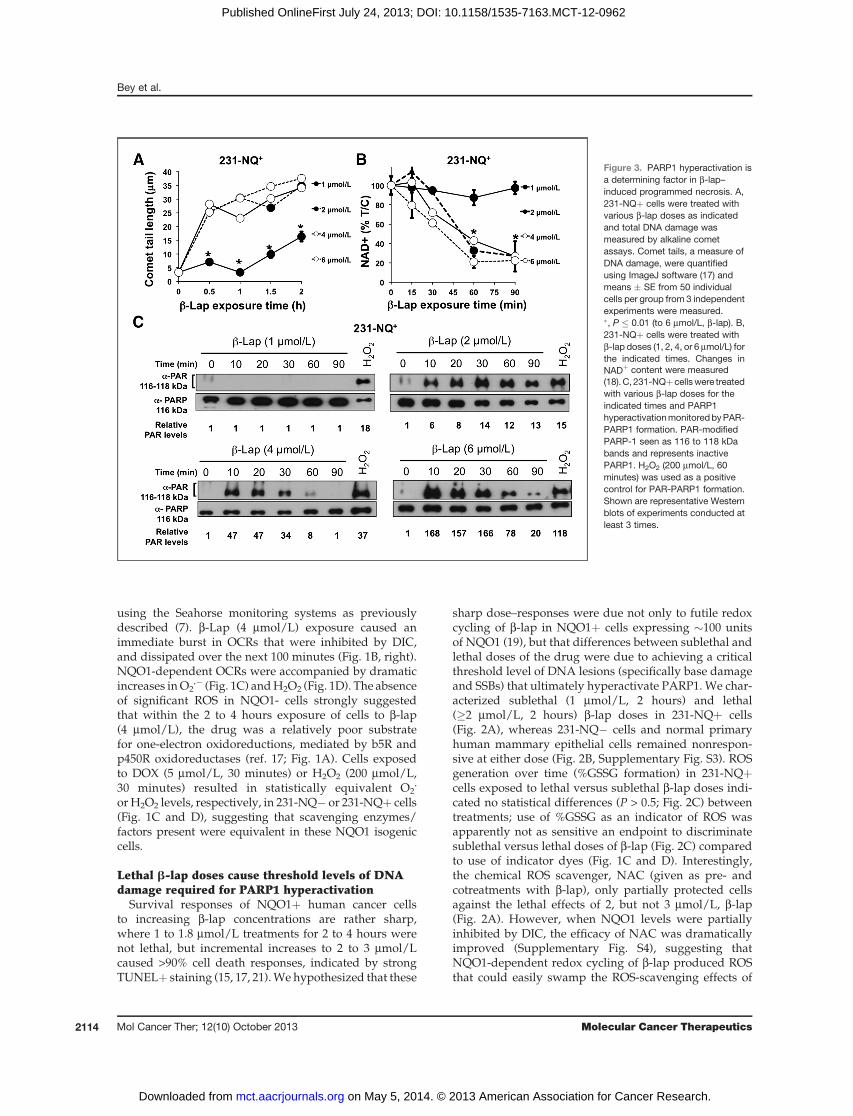
Lethal b-lap doses cause threshold levels of DNAdamage required for PARP1 hyperactivation
Survival responses of NQO1þ human cancer cellsto increasing b-lap concentrations are rather sharp,where 1 to 1.8 mmol/L treatments for 2 to 4 hours werenot lethal, but incremental increases to 2 to 3 mmol/Lcaused >90% cell death responses, indicated by strongTUNELþ staining (15, 17, 21).We hypothesized that these
sharp dose–responses were due not only to futile redoxcycling of b-lap in NQO1þ cells expressing �100 unitsof NQO1 (19), but that differences between sublethal andlethal doses of the drug were due to achieving a criticalthreshold level of DNA lesions (specifically base damageand SSBs) that ultimately hyperactivate PARP1. We char-acterized sublethal (1 mmol/L, 2 hours) and lethal(�2 mmol/L, 2 hours) b-lap doses in 231-NQþ cells(Fig. 2A), whereas 231-NQ� cells and normal primaryhuman mammary epithelial cells remained nonrespon-sive at either dose (Fig. 2B, Supplementary Fig. S3). ROSgeneration over time (%GSSG formation) in 231-NQþcells exposed to lethal versus sublethal b-lap doses indi-cated no statistical differences (P > 0.5; Fig. 2C) betweentreatments; use of %GSSG as an indicator of ROS wasapparently not as sensitive an endpoint to discriminatesublethal versus lethal doses of b-lap (Fig. 2C) comparedto use of indicator dyes (Fig. 1C and D). Interestingly,the chemical ROS scavenger, NAC (given as pre- andcotreatments with b-lap), only partially protected cellsagainst the lethal effects of 2, but not 3 mmol/L, b-lap(Fig. 2A). However, when NQO1 levels were partiallyinhibited by DIC, the efficacy of NAC was dramaticallyimproved (Supplementary Fig. S4), suggesting thatNQO1-dependent redox cycling of b-lap produced ROSthat could easily swamp the ROS-scavenging effects of
Figure 3. PARP1 hyperactivation isa determining factor in b-lap–induced programmed necrosis. A,231-NQþ cells were treated withvarious b-lap doses as indicatedand total DNA damage wasmeasured by alkaline cometassays. Comet tails, a measure ofDNA damage, were quantifiedusing ImageJ software (17) andmeans � SE from 50 individualcells per group from 3 independentexperiments were measured.�, P � 0.01 (to 6 mmol/L, b-lap). B,231-NQþ cells were treated withb-lap doses (1, 2, 4, or 6 mmol/L) forthe indicated times. Changes inNADþ content were measured(18). C, 231-NQþcellswere treatedwith various b-lap doses for theindicated times and PARP1hyperactivationmonitoredbyPAR-PARP1 formation. PAR-modifiedPARP-1 seen as 116 to 118 kDabands and represents inactivePARP1. H2O2 (200 mmol/L, 60minutes) was used as a positivecontrol for PAR-PARP1 formation.Shown are representative Westernblots of experiments conducted atleast 3 times.
Bey et al.
Mol Cancer Ther; 12(10) October 2013 Molecular Cancer Therapeutics2114
on May 5, 2014. © 2013 American Association for Cancer Research. mct.aacrjournals.org Downloaded from
Published OnlineFirst July 24, 2013; DOI: 10.1158/1535-7163.MCT-12-0962

NAC. In contrast, only minor levels of oxidative stresswere noted in 231-NQ� cells at any b-lap dose testedusing GSSG assays (Fig. 2D). These data suggested thatalthough ROS scavengers were easily overwhelmed atlow b-lap doses, factors such asNACmay play significantroles in protecting cells from sublethal or LD50 doses thatmight be used in combination therapies (14).Alkaline comet assays were then used to assess the
extent of total DNA lesions (base damage, SSBs, anddouble-strand breaks) created by various b-lap doses in231-NQþ versus 231-NQ� cells (Fig. 3A and B). Althougha sublethal 1 mmol/L b-lap dose produced significantGSSG formation (Fig. 2C), this exposure caused signifi-cantly less DNA damage versus a lethal dose (2 mmol/Lan �LD70; Fig. 3A). All lethal b-lap doses (2–6 mmol/L;Fig. 3A) caused similar saturating levels of DNA lesions,consistent with elevated and saturated oxidized GSSGlevels (Fig. 2C). In contrast, b-lap treatment of 231-NQ�cells did not result in significant DNA lesions, consistentwith the lack of OCR, ROS (H2O2) formation and lethalityin NQO1� cells.Exposure of 231-NQþ cells to a nonlethal b-lap
(1 mmol/L) dose did not result in measurable DNAlesions, loss of NADþ or PAR-PARP1 formation (Fig.3A and C). In contrast, treatment of NQO1þ cells withcytotoxic b-lap doses (�2 mmol/L) resulted in signifi-cant NADþ pool loss (Fig. 3B) and PAR-PARP1 forma-tion (Fig. 3C), consistent with PARP1 hyperactivation(17), and steady-state accumulation of posttranslationalPAR-modified and inactivated PARP1 (PAR; Fig. 3C).Peak PAR-PARP1 formation was noted 30 minutes after2 mmol/L b-lap, whose time to peak levels decreasedwith increasing doses (Fig. 3C; compare 2–6 mmol/Lb-lap exposures). Interestingly, loss of intracellularNADþ levels were similar with all lethal doses of b-lap,strongly suggesting saturated PARP1 hyperactivation atall doses (Fig. 3B).
Catalase detoxifies b-lap–induced H2O2 formationand is cytoprotectiveExogenous catalase (1,000 U) significantly lowered H2
O2 levels in b-lap–exposed 231-NQþ cells (Fig. 4A, left),whereas its addition had no affect on O2
.� formation(not shown). Similarly, exogenous CuZn2þ-dependentsuperoxide dismutase (CuZnSOD, 3,000 U) decreasedO2
.� formation (Fig. 4A, right), whereas slightly increas-ing H2 O2 levels (not shown). Forced overexpression ofcatalase in NQO1þ MCF-7 cells (Fig. 4B) using a CMV-driven expression vector (Open Biosystems) significantlyspared NQO1þ MCF-7 cells from lethal b-lap doses (Fig.4C), ranging from 2 to 4 mmol/L.Similarly, exogenous catalase (�500 U) significantly
protected 231-NQþ cells from b-lap lethality (Fig. 5A),whereas the survival of b-lap–treated 231-NQ� cells werenot affected by the drug, with or without catalase coaddi-tion (Fig. 5B). Exogenous coadministration of catalasewith CuZnSOD significantly decreased the effective cata-lase dose required to prevent b-lap–induced lethality
(Fig. 5C); for example, only 125U of catalase was requiredwith CuZnSOD to reach the same protection as 500 Ucatalase alone (Fig. 5A and C, P � 0.001, respectively).CuZnSOD enhanced the cytoprotective effects of all cata-lase doses used (Fig. 5A and C, P � 0.001, respectively).Exogenous catalase did not influence the survival of b--lap-resistant 231-NQ� cells with or without CuZnSOD(Fig. 5D), because OCR and ROS formation did not occurin the absence of NQO1 (Fig. 1).
Catalase also significantly suppressed DNA damagein b-lap–exposed 231-NQþ cells monitored by cometassays (Fig. 6A), whereas b-lap-resistant 231-NQ�cells formed no DNA lesions after b-lap treatment,with or without catalase (Fig. 6B). Consistently, catalaseblocked b-lap–induced PARP1 hyperactivation (Fig. 6C,top), formation of gH2AX (Fig. 6C, bottom), NADþ loss(Fig. 6D), downstream programmed necrosis-induced
Figure 4. Catalase prevents b-lap–induced H2O2 formation and enhancesclonogenic survival. A, 231-NQþ cells were treated with b-lap (4 mmol/L)in the presence or absence of 1,000 U catalase or 3,000 UCuZnSOD andstained with DHE or DCFDA for superoxide or H2O2 formation,respectively, as in Fig. 1. H2O2 (200 mmol/L) or DOX (5 mmol/L) exposureswere for 30 minutes as positive controls. Shown are means � SE ofintegrated density values from representative digital images of at least100 cells. Data are from experiments conducted 3 independent times intriplicate. ��, P � 0.01; ���, P � 0.001. B, MCF-7 cells were transientlytransfected with a CMV-driven catalase (CMV-CAT) expression vector orSCR, from Open Biosystems. Transfected MCF-7 cells were assayed forcatalase expression by Western blot analysis. GAPDH levels weremonitored as an internal loading control. C, MCF-7 cells were transientlytransfected with CMV-CAT or SCR expression vectors. After 48 hours,cells were trypsinized, counted, and plated in 48-well dishes. Afteradhering for 24 hours, cells were treated with b-lap (closed circle orclosed square represents SCR or CMV-CAT transfected MCF-7 cells,respectively) or b-lapþdicoumarol (open circle or square represents SCRor CMV-CAT transfected MCF-7 cells, respectively) at the indicateddoses and survival assessed.
Catalase Confers Resistance to b-Lapachone Cell Death
www.aacrjournals.org Mol Cancer Ther; 12(10) October 2013 2115
on May 5, 2014. © 2013 American Association for Cancer Research. mct.aacrjournals.org Downloaded from
Published OnlineFirst July 24, 2013; DOI: 10.1158/1535-7163.MCT-12-0962

atypical PARP1 and p53 proteolytic cleavage (Fig. 6Eand Supplementary Fig. S5) and TUNELþ responses(Fig. 6F) that are uniquely associated with NQO1 bio-activatable drug lethality.
b-Lap–inducedprogrammednecrosis is accompaniedby nuclear AIF translocation
b-Lap-treated MCF-7 cells (Supplementary Fig. S6)undergo atypical PARP1 and p53 proteolysis (15) due tom-calpain activation (16, 25), whereas treatment of thesame cells with staurosporine (STS) yielded classicapoptosis-related, �89 kDa PARP1 proteolysis (Supple-mentary Fig. S6). BAPTA-AM, a calcium chelator, orDIC prevented b-lap–induced programmed necrosis. Incontrast, the pan-caspase inhibitor, Z-VAD (see Supple-mentary Fig. S1 for structure), had no affect on b-lap–induced cell death or downstream PARP1/p53 prote-olysis (Supplementary Fig. S6), but blocked STS-relatedPARP1 cleavage (26).
Becausem-calpain activation can stimulate downstreamAIF translocation from mitochondria (9, 27), inducingprogrammed cell death (9), we examined a role for AIFin b-lap lethality. Indeed, activation and translocation ofAIF from mitochondria (DMSO; Fig. 7A) to nuclei wasnoted (4–24 hours posttreatment; Fig. 7A) in b-lap–treatedMCF-7 cells, consistent with m-calpain activation kinetics(16, 25). AIF activation was blocked by BAPTA-AM
(Fig. 7B) or catalase (1,000 U) cotreatments (Fig. 7C).However, siRNA-mediated AIF knockdown only part-ially decreased the lethal effects of b-lap in MCF-7 cells(Fig. 7D), most likely due to simultaneous activation ofother cell death mediators, including posttranslationalmodification/activation of GAPDH (Fig. 8) that mediatesapoptotic-like cell death (28). Stable AIF knockdown inMDA-MB-231 cells, like those recently reported (29),resulted in dramatic morphology and growth alterations,with similar partial blockage of lethality noted in b-lap–treated MCF-7 cells (Fig. 7D). Thus, b-lap stimulatesmultiple cell death factors, including m-calpain (16, 25),AIF (Fig. 7), and S-nitrosylated GAPDH (Fig. 8).
DiscussionWe used chemoresistant MDA-MB-231 TNBC cells to
link NQO1-dependent futile cycling of b-lap to ROS-induced DNA damage and PARP1 hyperactivation.Threshold-level responses in cells after b-lap treatmentswere noted by sharp dose-dependent trigger events,mediated by robust H2O2 formation that, in turn, causedbothDNAdamage andCa2þER release, ultimately leadingto PARP1 hyperactivation (Supplementary Fig. S7). H2O2
productionwas the obligate ROS inducing cell death afterb-lap, because catalase administration alone protectedcells. PARP1 hyperactivation, both in dose–response and
Figure 5. Exogenous catalaseenhances clonogenic survival inMDA-MB 231-NQþ cells. A and B,231-NQþ (A) or 231-NQ� (B) cellswere treated with varying b-lapdoses (mmol/L) with or withoutvarious catalase doses (U, units).Survival was assessed and resultsgraphed as means � SE of 3experiments conducted insextuplicate. C and D, 231-NQþ or231-NQ� cells were treated withvarious b-lap doses for 2 hours inthe presence or absence of varyingcatalase doses, with or without3,000 U CuZnSOD. Following 2-hour treatments, fresh media wasadded and survival assessed as inA and B. ���, P � 0.001.
Bey et al.
Mol Cancer Ther; 12(10) October 2013 Molecular Cancer Therapeutics2116
on May 5, 2014. © 2013 American Association for Cancer Research. mct.aacrjournals.org Downloaded from
Published OnlineFirst July 24, 2013; DOI: 10.1158/1535-7163.MCT-12-0962

temporal kinetics of triggering cell death, resulted incatastrophic loss of essential nucleotides (NADþ andATP) that correlated well with the "2 hours minimumtime to death" induced by b-lap in NQO1þ expressingcells (17). Downstream, AIF activation and nuclear trans-location (Fig. 7) were consistent with our prior findings ofm-calpain activation (25), and previously described role(s)ofm-calpain inAIF activation byothers (30).However,AIF
activation is one of a number of simultaneously acti-vated cell death responses stimulated during b-lap–induced programmed necrosis, and we were not ableto completely protect cells by AIF knockdown alone, asothers suggested (29). Differences in AIF knockdowncould explain the conflicting results, however, our iden-tification of various cell death factors (e.g., m-calpain,AIF, GAPDH modification, as well as loss of essential
Figure6. Exogenous catalase abrogates b-lap–induced cytotoxicity. A and B, 231-NQþ or 231-NQ� cells were treated with b-lap (4 mmol/L, 2 hours) withor without catalase (1,000 U). Alkaline comet assays were then conducted (17). Shown are comet tail means � SE of at least 50 comet tails percondition. Student t tests were conducted to determine significance between groups. ���, P � 0.001. C (top), 231-NQþ cells were treated with 4 mmol/Lb-lap as indicated with (right) or without (left) catalase (1,000 U). H2O2 (200 mmol/L, 60 minutes) was used a positive control. Whole-cell lysates wereassessed for PARP1 hyperactivation (PAR-PARP1). g-H2AX foci levels were assessed (bottom) in 231-NQþ cells exposed to 4 mmol/L b-lap (with orwithout 1,000 U of catalase) or 200 mmol/L H2O2. Foci formed in individual cells were assessed microscopically as described previously (17). D, 231-NQþ cells were treated with b-lap (4 mmol/L, 2 hours), with or without catalase (1,000 U), and with or without 40 mmol/L DIC. Cells were analyzed forNADþ levels as in Fig. 3 and data presented as means � SE from 3 experiments conducted in triplicate each. ���, P � 0.001. E, 231-NQþ cells weretreated with varying doses of b-lap for 2 hours with (right) or without (left) catalase (1,000 U). Whole-cell lysates were analyzed for PARP1 and p53proteolysis (15). F, 231-NQþ cells were treated with various b-lap doses (1, 2, 4, or 6 mmol/L) with or without catalase (1,000 U) for 2 hours andpopulations analyzed for TUNELþ staining 48 hours later (18). Data were graphed as X-fold TUNELþ staining changes of b-lap–exposed versus DMSO-treated cells �SE from 3 independent experiments repeated in triplicate. �, P � 0.05; ��, P � 0.01; and ���, P � 0.001.
Catalase Confers Resistance to b-Lapachone Cell Death
www.aacrjournals.org Mol Cancer Ther; 12(10) October 2013 2117
on May 5, 2014. © 2013 American Association for Cancer Research. mct.aacrjournals.org Downloaded from
Published OnlineFirst July 24, 2013; DOI: 10.1158/1535-7163.MCT-12-0962

nucleotides) simultaneously activated in b-lap–exposedNQO1þ cells, make it unlikely that AIF abrogationalone would spare most b-lap–exposed solid cancercells. Although not explored in detail, the posttransla-tional modification of GAPDH in b-lap–treated NQO1þcells has potentially important implications for under-standing b-lap–induced cell "clean-up." S-nitrosylationof GAPDH implies a potentially important role of nitricoxide species (NOS; ref. 28) in b-lap–induced lethality.GAPDH modification inhibits its functions in glycolysis(31, 32) and activates its DNA repair (33) and cell death(34) functions, suggesting dramatic metabolic changesin exposed cancer cells due to this agent. Exposure totherapeutic b-lap doses caused supra-lethal ROS levelsand DNA damage that was not repaired, presumablydue to loss of NADþ/ATP within �60 minutes ofexposure (Fig. 3B). We are currently exploring the rolesof NOS and posttranslational GAPDH in altered metab-olism and repair in NQO1þ cells exposed to b-lap.
We present the first evidence that H2O2 is the primaryobligate ROS species necessary for b-lap’s lethal effects. Aprimary role for H2O2 is also consistent with the specific
induction of SSBs caused by this promising tumor-selec-tive antitumor agent. CuZnSOD specifically enhanced theefficacy of catalase to block b-lap cytotoxicity in NQO1þcancer cells. These data predict that normal tissue, whichtypically has higher catalase levels than cancer cells(35–37), could be selectively spared from toxicity causedby this agent. Alternatively, cancer cells overexpressingcatalase and/or CuZnSODwould require higher doses ofb-lap to avoid "sublethal therapeutic treatments." Accord-ingly, we propose that the ratio of NQO1 to catalaseexpression in tumor versus normal tissue is a majordeterminant of tumor selectivity for b-lap and futuredrugs that work by "NQO1 bioactivation." On this point,it is interesting that an inverse correlation betweenNQO1expression and catalase is noted across breast cancer cells(Supplementary Fig. S8). We also noted that catalaseexpression in breast cancer cells was commonly low anddid not dramatically affect the lethality of b-lap–treatedbreast cancer cells that overexpress NQO1. We predictthat NQO1/catalase ratio calculations are importantwhen examining normal tissue versus tumor tissue, butthat b-lap–exposed NQO1 overexpressing cells will
Figure 7. b-Lap–induced cell death factor activation is abrogated by exogenous catalase. A to C, MCF-7 cells were treated with b-lap (4 mmol/L, 2 hours)(A), � BAPTA-AM (5 mmol/L) (B), or 1,000 U catalase (C). Cells were then fixed and stained for AIF expression and Hoescht as described (39) andrepresentative confocal micrographs taken at 100 magnification. Both BAPTA-AM and catalase prevented AIF translocation from mitochondriato nuclei, and movement of mitochondria to perinuclear space. Mitochondria-specific staining confirmed AIF localization in organelles. D, MCF-7 cellswere transiently transfected with 20 mmol/L siRNA specific for AIF or with scrambled nonsense siRNA as a control (40). Specific downregulationof AIF expression was confirmed by Western blotting (see inset) using a-tubulin as a loading control. AIF relative levels (RL) were assessed bycomparing densitometry scans (NIH ImageJ software) of the loading control and AIF protein levels. Stable shRNA–AIF–transfected MCF-7 or MDA-MB-231 cells had altered growth rates and morphology and were not used. After 48 hours, cells were treated with various b-lap doses (mmol/L, 2 hours), withor without 40 mmol/L DIC and survival assessed as in Fig. 2.
Bey et al.
Mol Cancer Ther; 12(10) October 2013 Molecular Cancer Therapeutics2118
on May 5, 2014. © 2013 American Association for Cancer Research. mct.aacrjournals.org Downloaded from
Published OnlineFirst July 24, 2013; DOI: 10.1158/1535-7163.MCT-12-0962

respond whether catalase is expressed or not, since ROSproduction canoverwhelmcatalase-mediated scavengingdue to the futile cycle (Fig. 1A).Catalase protects cells from H2O2-induced lipid perox-
idation that damages membranes and cellular organelles.Although exogenous catalase enhanced survival at lowdoses, excessive ROS and NOS formation by higher b-lapdoses (>6 mmol/L) were not inhibited by catalase alone.Although CuZnSOD alone did not spare b-lap–exposedNQO1þ cells, it enhanced the cytoprotective capacity ofcatalase. Combining CuZnSODwith catalase presumablyconverted superoxide to 2H2O2 that was easily detoxifiedby catalase to O2 þ 2H2O.b-Lap-treated NQO1þ breast cancer cells, including
TNBC cells, produce supra-lethal levels of ROS due tofutile cyclemetabolismof b-lap specifically byNQO1. Themechanism of action of this agent involves PARP1 hyper-activation that triggers simultaneous redundant down-stream lethal events, including essential nucleotide loss(NADþ andATP), AIF release, S-nitrosylation ofGAPDH,and eventually programmed necrosis. Identification ofROS-scavenging enzymes (i.e., SOD, catalase) that canpartially suppress b-lap–induced lethality is a crucial stepin understanding resistance factors for this agent. Inter-rogation of expression of these factors may allow us topredict efficaciousdoses required for improved efficacy ofNQO1bioactivatable drugs, such as b-lap,DNQ, and their
derivatives. Because NQO1 levels are overexpressed inbreast cancer versus adjacent normal tissue (11), andparticularly in ERþ breast cancer patients (38), these datastrongly indicate use of this agent against breast, aswell asmany other solid tumors that specifically overexpressedNQO1.MonitoringNQO1/catalase ratios in tumor versusnormal tissue will be essential for "personalized thera-pies" using b-lap or other NQO1 bioactivatable drugs.
Disclosure of Potential Conflicts of InterestJ. Gao is a consultant/advisory board member for StemPar Sciences,
Inc.. D.A. Boothman is a consultant/advisory boardmember for StemPARSciences, Inc. No potential conflicts of interest were disclosed by the otherauthors.
Authors' ContributionsConception and design: E.A. Bey, M.C. Srougi, L. Cao, Z. Moore, W.G.Bornmann, D.J. Buchsbaum, D.R. Spitz, J. Gao, D.A. BoothmanDevelopment of methodology: E.A. Bey, M.C. Srougi, M. Varnes,V.E. Anderson, J.J. Pink, L. Cao, Z. Moore, W.G. Bornmann, D.R. Spitz,D.A. BoothmanAcquisition of data (provided animals, acquired and managed patients,provided facilities, etc.): E.A. Bey, K.E. Reinicke, V.E. Anderson, L. Cao,Z. Moore, A. Rommel, C. Lewis, D.M. Euhus, W.G. Bornmann, D.J.Buchsbaum, D.R. Spitz, D.A. BoothmanAnalysis and interpretation of data (e.g., statistical analysis, biostatis-tics, computational analysis):E.A. Bey, K.E. Reinicke,M.C. Srougi, L.S. Li,M. Patel, L. Cao, Z. Moore, A. Rommel, D.J. Buchsbaum, D.R. Spitz, D.A.BoothmanWriting, review, and/or revision of the manuscript: E.A. Bey, K.E.Reinicke, L. Cao, Z. Moore, M. Boatman, D.J. Buchsbaum, D.R. Spitz,J. Gao, D.A. BoothmanAdministrative, technical, or material support (i.e., reporting or orga-nizing data, constructing databases): E.A. Bey, M. Varnes, L.S. Li, L. Cao,Z. Moore, M. Boatman, D.A. BoothmanStudy supervision: E.A. Bey, V.E. Anderson, L. Cao, Z. Moore, D.A.Boothman
AcknowledgmentsThis article is dedicated to Dr. M. Varnes, Emeritus Professor of
Radiation Oncology, CWRU, who began these studies. The authors alsothank the imaging, mouse metabolism, and flow cytometry cores, Sim-mons Comprehensive Cancer Center.
Grant SupportThis work was supported by NIH/NCI grant CA102792 to D.A. Booth-
man and DoD Breast Cancer Pre-Doctoral Fellowships to K.E. Reinicke(W81XWH-05-1-0248) and M.C. Srougi (W81XWH-04-1-0301). GrantCA13314 supported D.R. Spitz, as did CA086862, the Radiation and FreeRadicalResearchCore. Thisworkwasalso supportedbyaSusanG.Komengrant (KG090969) and NIH grant PS0 CA089019 to D.J. Buchsbaum.
The costs of publication of this article were defrayed in part by thepayment of page charges. This article must therefore be hereby markedadvertisement in accordance with 18 U.S.C. Section 1734 solely to indicatethis fact.
Received October 3, 2012; revised June 24, 2013; accepted July 18, 2013;published OnlineFirst July 24, 2013.
References1. Jemal A, Siegel R, Xu J, Ward E. Cancer statistics, 2010. CA Cancer J
Clin 2010;60:277–300.2. Pohlmann PR, Mayer IA, Mernaugh R. Resistance to trastuzumab in
breast cancer. Clin Cancer Res 2009;15:7479–91.3. Moreno-Aspitia A, Perez EA. Treatment options for breast cancer
resistant toanthracycline and taxane.MayoClinProc2009;84:533–45.4. May CD, Sphyris N, Evans KW, Werden SJ, Guo W, Mani SA.
Epithelial-mesenchymal transition and cancer stem cells: a danger-
ously dynamic duo in breast cancer progression. Breast Cancer Res2011;13:202.
5. O'Brien CS, Howell SJ, Farnie G, Clarke RB. Resistance to endocrinetherapy: are breast cancer stem cells the culprits? J Mammary GlandBiol Neoplasia 2009;14:45–54.
6. Kaufmann SH, Vaux DL. Alterations in the apoptotic machinery andtheir potential role in anticancer drug resistance. Oncogene 2003;22:7414–30.
Figure 8. GAPDH proteolysis is induced during b-Lap–induced celldeath. MCF-7 cells were treated with b-lap (4 mmol/L, 2 hours) andwhole-cell extracts monitored for PAR-PARP1, S-nitrosylatedGAPDH (M-GAPDH), or total GAPDH (R-GAPDH) using antibodiesthat detect a specific site known to be S-nitrosylated (41) or apolyclonal antibody to GAPDH, respectively. MSH2 levels weremonitored as an internal loading control.
Catalase Confers Resistance to b-Lapachone Cell Death
www.aacrjournals.org Mol Cancer Ther; 12(10) October 2013 2119
on May 5, 2014. © 2013 American Association for Cancer Research. mct.aacrjournals.org Downloaded from
Published OnlineFirst July 24, 2013; DOI: 10.1158/1535-7163.MCT-12-0962

7. Huang X, Dong Y, Bey EA, Kilgore JA, Bair JS, Li LS, et al. An NQO1substratewith potent antitumor activity that selectively kills byPARP1-induced programmed necrosis. Cancer Res 2012;72:3038–47.
8. ZongWX, Ditsworth D, Bauer DE,Wang ZQ, Thompson CB. AlkylatingDNA damage stimulates a regulated form of necrotic cell death. GenesDev 2004;18:1272–82.
9. YuSW,WangH,PoitrasMF,CoombsC,BowersWJ, Federoff HJ, et al.Mediation of poly(ADP-ribose) polymerase-1-dependent cell death byapoptosis-inducing factor. Science 2002;297:259–63.
10. Zong WX, Thompson CB. Necrotic death as a cell fate. Genes Dev2006;20:1–15.
11. Marin A, Lopez de Cerain A, Hamilton E, Lewis AD, Martinez-PenuelaJM, Idoate MA, et al. DT-diaphorase and cytochrome B5 reductase inhuman lung and breast tumours. Br J Cancer 1997;76:923–9.
12. Dong Y, Bey EA, Li LS, Kabbani W, Yan J, Xie XJ, et al. Prostate cancerradiosensitization through poly(ADP-Ribose) polymerase-1 hyperac-tivation. Cancer Res 2010;70:8088–96.
13. Lewis AM, Ough M, Hinkhouse MM, Tsao MS, Oberley LW, Cullen JJ.Targeting NAD(P)H:quinone oxidoreductase (NQO1) in pancreaticcancer. Mol Carcinog 2005;43:215–24.
14. Boothman DA, Trask DK, Pardee AB. Inhibition of potentially lethalDNA damage repair in human tumor cells by b-lapachone, an activatorof topoisomerase I. Cancer Res 1989;49:605–12.
15. Pink JJ, Planchon SM, Tagliarino C, Varnes ME, Siegel D, BoothmanDA. NAD(P)H:Quinone oxidoreductase activity is the principal deter-minant of b-lapachone cytotoxicity. J Biol Chem 2000;275:5416–24.
16. Tagliarino C, Pink JJ, Dubyak GR, Nieminen AL, Boothman DA.Calcium is a key signaling molecule in b-lapachone-mediated celldeath. J Biol Chem 2001;276:19150–9.
17. Bentle MS, Reinicke KE, Bey EA, Spitz DR, Boothman DA. Calcium-dependent modulation of poly(ADP-ribose) polymerase-1 alterscellular metabolism and DNA repair. J Biol Chem 2006;281:33684–96.
18. Bey EA, Bentle MS, Reinicke KE, Dong Y, Yang CR, Girard L, et al. AnNQO1- and PARP-1-mediated cell death pathway induced in non-small-cell lung cancer cells by b-lapachone. Proc Natl Acad Sci U S A2007;104:11832–7.
19. Li LS, Bey EA, Dong Y, Meng J, Patra B, Yan J, et al. Modulatingendogenous NQO1 levels identifies key regulatory mechanisms ofaction of b-lapachone for pancreatic cancer therapy. Clin Cancer Res2011;17:275–85.
20. BentleMS, Bey EA, DongY, Reinicke KE, BoothmanDA. New tricks forold drugs: the anticarcinogenic potential of DNA repair inhibitors. JMolHistol 2006;37:203–18.
21. Reinicke KE, Bey EA, Bentle MS, Pink JJ, Ingalls ST, Hoppel CL, et al.Development of b-lapachone prodrugs for therapy against humancancer cells with elevated NAD(P)H:quinone oxidoreductase 1 levels.Clin Cancer Res 2005;11:3055–64.
22. ZhaoW, Spitz DR, Oberley LW, Robbins ME. Redox modulation of thepro-fibrogenic mediator plasminogen activator inhibitor-1 followingionizing radiation. Cancer Res 2001;61:5537–43.
23. Goshe MB, Anderson VE. Hydroxyl radical-induced hydrogen/deute-rium exchange in amino acid carbon-hydrogen bonds. Radiat Res1999;151:50–8.
24. Lowry OH, Rosebrough NJ, Farr AL, Randall RJ. Protein measurementwith the Folin phenol reagent. J Biol Chem 1951;193:265–75.
25. Tagliarino C, Pink JJ, Reinicke KE, Simmers SM, Wuerzberger-DavisSM, Boothman DA. Mu-calpain activation in b-lapachone-mediatedapoptosis. Cancer Biol Ther 2003;2:141–52.
26. Casiano CA, Ochs RL, Tan EM. Distinct cleavage products of nuclearproteins in apoptosis and necrosis revealed by autoantibody probes.Cell Death Differ 1998;5:183–90.
27. Hong SJ, Dawson TM, Dawson VL. Nuclear and mitochondrial con-versations in cell death: PARP-1 and AIF signaling. Trends PharmacolSci 2004;25:259–64.
28. HaraMR, Agrawal N, KimSF, CascioMB, FujimuroM,Ozeki Y, et al. S-nitrosylated GAPDH initiates apoptotic cell death by nuclear translo-cation following Siah1 binding. Nat Cell Biol 2005;7:665–74.
29. Lee H, Park MT, Choi BH, Oh ET, Song MJ, Lee J, et al. Endoplasmicreticulum stress-induced JNK activation is a critical event leading tomitochondria-mediated cell death caused by b-lapachone treatment.PLoS One 2011;6:e21533.
30. Polster BM, Basanez G, Etxebarria A, Hardwick JM, Nicholls DG.Calpain I induces cleavage and release of apoptosis-inducing factorfrom isolated mitochondria. J Biol Chem 2005;280:6447–54.
31. Colell A, Green DR, Ricci JE. Novel roles for GAPDH in cell death andcarcinogenesis. Cell Death Differ 2009;16:1573–81.
32. Mohr S, Stamler JS, Brune B. Posttranslational modification of glyc-eraldehyde-3-phosphate dehydrogenase by S-nitrosylation and sub-sequent NADH attachment. J Biol Chem 1996;271:4209–14.
33. Sirover MA. Minireview. Emerging new functions of the glycolyticprotein, glyceraldehyde-3-phosphate dehydrogenase, in mammaliancells. Life Sci 1996;58:2271–7.
34. Dastoor Z, Dreyer JL. Potential role of nuclear translocation of glyc-eraldehyde-3-phosphate dehydrogenase in apoptosis and oxidativestress. J Cell Sci 2001;114:1643–53.
35. Alexander NM. Catalase inhibition by normal and neoplastic tissueextracts. J Biol Chem 1957;227:975–85.
36. Bannister WH, Bannister JV. Factor analysis of the activitiesof superoxide dismutase, catalase and glutathione peroxidase innormal tissues and neoplastic cell lines. Free Radic Res Commun1987;4:1–13.
37. Sihto H, Lundin J, Lundin M, Lehtimaki T, Ristimaki A, Holli K, et al.Breast cancer biological subtypes and protein expression predict forthe preferential distant metastasis sites: a nationwide cohort study.Breast Cancer Res 2011;13:R87.
38. JamshidiM,Bartkova J,GrecoD, Tommiska J, FagerholmR, AittomakiK, et al. NQO1 expression correlates inversely with NFkB activation inhuman breast cancer. Breast Cancer Res Treat 2012;132:955–68.
39. Chiu SM, Oleinick NL. Dissociation of mitochondrial depolarizationfrom cytochrome c release during apoptosis induced by photodynam-ic therapy. Br J Cancer 2001;84:1099–106.
40. Ough M, Lewis A, Bey EA, Gao J, Ritchie JM, Bornmann W, et al.Efficacy of b-lapachone in pancreatic cancer treatment: exploiting thenovel, therapeutic target NQO1. Cancer Biol Ther 2005;4:95–102.
41. Schonhoff CM, Matsuoka M, Tummala H, Johnson MA, Estevez AG,Wu R, et al. S-nitrosothiol depletion in amyotrophic lateral sclerosis.Proc Natl Acad Sci U S A 2006;103:2404–9.
Bey et al.
Mol Cancer Ther; 12(10) October 2013 Molecular Cancer Therapeutics2120
on May 5, 2014. © 2013 American Association for Cancer Research. mct.aacrjournals.org Downloaded from
Published OnlineFirst July 24, 2013; DOI: 10.1158/1535-7163.MCT-12-0962
Copyright © 2022 FDOKUMEN