Quercus infectoria galls possess antioxidant activity and abrogates oxidative stress-induced...
11
Chemico-Biological Interactions 171 (2008) 272–282 Available online at www.sciencedirect.com Quercus infectoria galls possess antioxidant activity and abrogates oxidative stress-induced functional alterations in murine macrophages Gurpreet Kaur a , Mohammad Athar a , M. Sarwar Alam b,∗ a Department of Medical Elementology & Toxicology, Faculty of Science, Jamia Hamdard, Hamdard Nagar, New Delhi 110062, India b Department of Chemistry, Faculty of Science, Jamia Hamdard, Hamdard Nagar, New Delhi 110062, India Received 8 April 2007; received in revised form 4 October 2007; accepted 15 October 2007 Available online 22 October 2007 Abstract The present study reports the antioxidant activity of ethanolic extract of Quercus infectoria galls. The antioxidant potency of galls was investigated employing several established in vitro model systems. Their protective efficacy on oxidative modulation of murine macrophages was also explored. Gall extract was found to contain a large amount of polyphenols and possess a potent reducing power. HPTLC analysis of the extract suggested it to contain 19.925% tannic acid (TA) and 8.75% gallic acid (GA). The extract potently scavenged free radicals including DPPH (IC 50 ∼0.5 g/ml), ABTS (IC 50 ∼1 g/ml), hydrogen peroxide (H 2 O 2 ) (IC 50 ∼2.6 g/ml) and hydroxyl ( • OH) radicals (IC 50 ∼6 g/ml). Gall extract also chelated metal ions and inhibited Fe 3+ –ascorbate- induced oxidation of protein and peroxidation of lipids. Exposure of rat peritoneal macrophages to tertiary butyl hydroperoxide (tBOOH) induced oxidative stress in them and altered their phagocytic functions. These macrophages showed elevated secretion of lysosomal hydrolases, and attenuated phagocytosis and respiratory burst. Activity of macrophage mannose receptor (MR) also diminished following oxidant exposure. Pretreatment of macrophages with gall extract preserved antioxidant armory near to control values and significantly protected against all the investigated functional mutilations. MTT assay revealed gall extract to enhance percent survival of tBOOH exposed macrophages. These results indicate that Q. infectoria galls possess potent antioxidant activity, when tested both in chemical as well as biological models. © 2007 Elsevier Ireland Ltd. All rights reserved. Keywords: Quercus infectoria; Galls; Antioxidant; Macrophages; Phagocytic functions; Oxidative stress; tBOOH 1. Introduction Oxidative damage to cells is believed to be a crucial factor implicated in the pathogenesis of several clinical disorders and in the normal process of ageing [1,2]. Potently reactive derivatives of oxygen, known as ∗ Corresponding author. Tel.: +91 11 26059688x5555; fax: +91 11 26059663. E-mail address: [email protected] (M.S. Alam). reactive oxygen species (ROS) such as superoxide, hydrogen peroxide (H 2 O 2 ) and hydroxyl radical ( • OH) are continuously generated by normal metabolic process or from exogenous chemicals in our ambient environment. Normally, ROS are efficiently neutralized by potent antioxidant armory of the cells without any untoward effect. However, hampering the balance between ROS generation and antioxidant defense leads to the onset of oxidative stress, which following a series of events, dysregulates cellular physiology leading to several chronic ailments such as cancer, atherosclerosis, 0009-2797/$ – see front matter © 2007 Elsevier Ireland Ltd. All rights reserved. doi:10.1016/j.cbi.2007.10.002
-
Upload
independent -
Category
Documents
-
view
0 -
download
0
Transcript of Quercus infectoria galls possess antioxidant activity and abrogates oxidative stress-induced...

Chemico-Biological Interactions 171 (2008) 272–282
Available online at www.sciencedirect.com
Quercus infectoria galls possess antioxidant activity andabrogates oxidative stress-induced functional
alterations in murine macrophages
Gurpreet Kaur a, Mohammad Athar a, M. Sarwar Alam b,∗a Department of Medical Elementology & Toxicology, Faculty of Science, Jamia Hamdard, Hamdard Nagar, New Delhi 110062, India
b Department of Chemistry, Faculty of Science, Jamia Hamdard, Hamdard Nagar, New Delhi 110062, India
Received 8 April 2007; received in revised form 4 October 2007; accepted 15 October 2007Available online 22 October 2007
Abstract
The present study reports the antioxidant activity of ethanolic extract of Quercus infectoria galls. The antioxidant potency ofgalls was investigated employing several established in vitro model systems. Their protective efficacy on oxidative modulation ofmurine macrophages was also explored. Gall extract was found to contain a large amount of polyphenols and possess a potentreducing power. HPTLC analysis of the extract suggested it to contain 19.925% tannic acid (TA) and 8.75% gallic acid (GA).The extract potently scavenged free radicals including DPPH (IC50∼0.5 �g/ml), ABTS (IC50∼1 �g/ml), hydrogen peroxide (H2O2)(IC50∼2.6 �g/ml) and hydroxyl (•OH) radicals (IC50∼6 �g/ml). Gall extract also chelated metal ions and inhibited Fe3+–ascorbate-induced oxidation of protein and peroxidation of lipids. Exposure of rat peritoneal macrophages to tertiary butyl hydroperoxide(tBOOH) induced oxidative stress in them and altered their phagocytic functions. These macrophages showed elevated secretionof lysosomal hydrolases, and attenuated phagocytosis and respiratory burst. Activity of macrophage mannose receptor (MR) also
diminished following oxidant exposure. Pretreatment of macrophages with gall extract preserved antioxidant armory near to controlvalues and significantly protected against all the investigated functional mutilations. MTT assay revealed gall extract to enhancepercent survival of tBOOH exposed macrophages. These results indicate that Q. infectoria galls possess potent antioxidant activity,when tested both in chemical as well as biological models.© 2007 Elsevier Ireland Ltd. All rights reserved.gocytic
Keywords: Quercus infectoria; Galls; Antioxidant; Macrophages; Pha1. Introduction
Oxidative damage to cells is believed to be a crucial
factor implicated in the pathogenesis of several clinicaldisorders and in the normal process of ageing [1,2].Potently reactive derivatives of oxygen, known as∗ Corresponding author. Tel.: +91 11 26059688x5555;fax: +91 11 26059663.
E-mail address: [email protected] (M.S. Alam).
0009-2797/$ – see front matter © 2007 Elsevier Ireland Ltd. All rights reservdoi:10.1016/j.cbi.2007.10.002
functions; Oxidative stress; tBOOH
reactive oxygen species (ROS) such as superoxide,hydrogen peroxide (H2O2) and hydroxyl radical (•OH)are continuously generated by normal metabolicprocess or from exogenous chemicals in our ambientenvironment. Normally, ROS are efficiently neutralizedby potent antioxidant armory of the cells without anyuntoward effect. However, hampering the balance
between ROS generation and antioxidant defense leadsto the onset of oxidative stress, which following a seriesof events, dysregulates cellular physiology leading toseveral chronic ailments such as cancer, atherosclerosis,ed.

ical In
aotprtaii
cssartmitttslioip
dgommttatpl
2
2
r2pgrfw
G. Kaur et al. / Chemico-Biolog
rthritis, diabetes, etc. Intake of antioxidants can avertr impede the oxidative damage and therefore, thwarthe disorders implicating oxidative stress in their patho-hysiology. Antioxidants mediate their effect by directlyeacting with ROS, quenching them and/or chelatinghe catalytic metal ion [3]. Synthetic antioxidants suchs BHA and BHT are available; however, their toxicitys a problem of concern [4]. This has led to a growingnterest in exploring natural antioxidants.
Macrophages, the major phagocytic cells, are thehief constituents of body’s cell mediated immuneystem. These cells possess a high proportion of polyun-aturated fatty acids in their plasmamembrane andre exposed to a huge amount of oxidants duringespiratory burst, a key step involved in the destruc-ion of engulfed microbes. These two factors together
ake macrophages remarkably perceptive to alterationsn oxidant–antioxidant equilibrium [5]. Macrophages,herefore, provide an excellent model system to explorehe efficacy of antioxidant compounds against oxida-ive injury induced to cellular systems. In the presenttudy, we investigated the antioxidant activity of ethano-ic extract of Quercus infectoria galls employing severaln vitro assays and also investigated the protective effectf extract against tertiary butyl hydroperoxide (tBOOH)nduced oxidative stress and physiological alterationsroduced by it in murine macrophages.
Q. infectoria Olivier (Fagaceae) is a small tree widelyistributed in Greece, Asia Minor and Iran. The tree bearsalls that emerge on its shoots as a consequence of attackf gall wasp, Cypnis gallae-tincotoriae. Galls are of greatedicinal value and have widely been used in folkloreedicines mainly as astringent and against inflamma-
ion. Pharmacological evaluation of galls has decipheredhem to be astringent, antiparkinsonian, antitremorinend antidiabetic [6–8]. We have previously revealed gallso possess potent anti-inflammatory activity [9]. Theresent study explores the antioxidant activity of ethano-ic extract of Q. infectoria galls.
. Materials and methods
.1. Chemicals
Thiobarbituric acid (TBA), RPMI-1640 media,educed nicotinamide adenine dinucleotide (NADH),-mercaptoethanol, ferricytochrome c, horseradisheroxidase, mannose-BSA, mannan, chloramine T, �-
lucuronidase, phenolphthalein glucuronic acid, phenoled, dithionitrobenzene (DTNB) and were purchasedrom Sigma Chemical Co. (St. Louis, MO, USA). Na125Ias purchased from Board of Radiation and Isotopeteractions 171 (2008) 272–282 273
Technology, Bhabha Atomic Research Center, Mumbai.All other chemicals and reagents used were of the highestpurity commercially available.
2.2. Preparation of gall extract
The air-dried galls of Q. infectoria were purchasedfrom Khari Baoli, New Delhi, India and authenticated byDr. M.P. Sharma, Reader in the Department of Botany inour University. One hundred and twenty-five grams ofpowdered galls/L of alcohol were extracted in soxhlet.The extract was filtered and then concentrated; it was18.3% of the starting material.
2.3. Total phenolics and reducing power
The total phenolics in gall extract were determinedusing Folin-Ciocalteu reagent [10] and its reducingpower according to Oyaizu [11] as described previouslyby us [12].
2.4. HPTLC quantification of tannic acid (TA) andgallic acid (GA) in gall extract
Methanolic solutions of the standard and the samplewere applied using the Camag linomat applicator ontothe precoated silica gel F254 plates (Merck) of 0.2 mmthickness. Toluene:ethyl acetate:formic acid (6:6:1) wasused as the mobile phase for TA and chloroform:ethylacetate:formic acid (5:4:1) was used as the mobile phasefor GA. After elution, the plate was dried and scanneddensitometrically using CAMAG TLC scanner 3. Thepercentage of TA and GA in the extract was calculatedby calibration using peak height ratio.
2.5. Scavenging of free radicals
To assay DPPH scavenging, 1 ml methanolic solutionof gall extract was incubated with 0.5 ml of 0.15 mMDPPH solution for 30 min and absorbance read there-after at 517 nm. The decrease in absorbance indicatedthe extent of DPPH radicals scavenged.
Scavenging of ABTS radical was determined accord-ing to the method of Song and Yen [13]. ABTS•+ cationradicals were generated by the interaction of ABTS(100 �M), H2O2 (50 �M) and peroxidase (4.4 units/ml).0.25 ml of gall extract was mixed with an equal volumeof ABTS, H O , peroxidase and water and absorbance
2 2was monitored at 734 nm for 10 min.H2O2 scavenging was determined by monitoringchange in absorbance of 4 mM H2O2 solution at 230 nmfollowing incubation with gall extract for 10 min [14].
ical In
274 G. Kaur et al. / Chemico-BiologScavenging of •OH radicals was determined by themethod of Aruoma and Halliwell [15].
2.6. Lipid peroxidation (LPO)
LPO was performed in hepatic microsomes by themethod of Wright et al. [16]. The reaction mixturecontaining 0.58 ml phosphate buffer (0.1 M, pH 7.4),0.2 ml of microsome (10%, w/v), 0.2 ml ascorbic acid(100 mM) and 0.02 ml ferric chloride (100 mM) wasincubated at 37 ◦C in a shaking water bath for 1 h. Thereaction was stopped by the addition of 1.0 ml 10%TCA. Following which, 1.0 ml 0.67% thiobarbituric acid(TBA) was added and all the tubes were placed in aboiling water bath for 20 min. Malonaldehyde (MDA)formed was determined by measuring the absorbance at535 nm.
2.7. Protein oxidation
Bovine serum albumin (BSA) (1 mg/mL) was incu-bated at 25 ◦C in solution with 2.5 mM H2O2, 1.0 mMFeCl3, 1.0 mM ascorbate and 3.0 mM EDTA in the pres-ence or absence of the extract. After incubation for45 min at 25 ◦C, protein was precipitated with 10% TCA(w/v, final concentration), centrifuged (3000 × g, 4 ◦C,2 min) and the supernatants was decanted. Protein pel-lets were dissolved by vortex mixing in 1 mL of 50 mMpotassium phosphate buffer, pH 7.5. Total sulphydrylgroup (TSH) determinations were performed accordingto the method of Sedlak and Lindsay [17] with Ellman’sreagent.
2.8. Metal chelating activity
The chelation of ferrous ions by gall extract was esti-mated by the method of Dinis et al. [18]. Briefly, 50 �l of2 mM FeCl2 was added to the extract (1 ml). The reactionwas initiated by the addition of 0.2 ml of 5 mM ferrozinesolution. The mixture was vigorously shaken and incu-bated at room temperature for 10 min. The absorbance ofthe solution was thereafter measured spectrophotomet-rically at 562 nm.
2.9. Protection against oxidative modulation inmacrophages
2.9.1. Experimental protocol
Thioglycollate elicited macrophages were preparedfrom male Wistar rats (procured from Central Ani-mal house, Jamia Hamdard) according to Li et al.[19] and cultured as reported previously by us [9].
teractions 171 (2008) 272–282
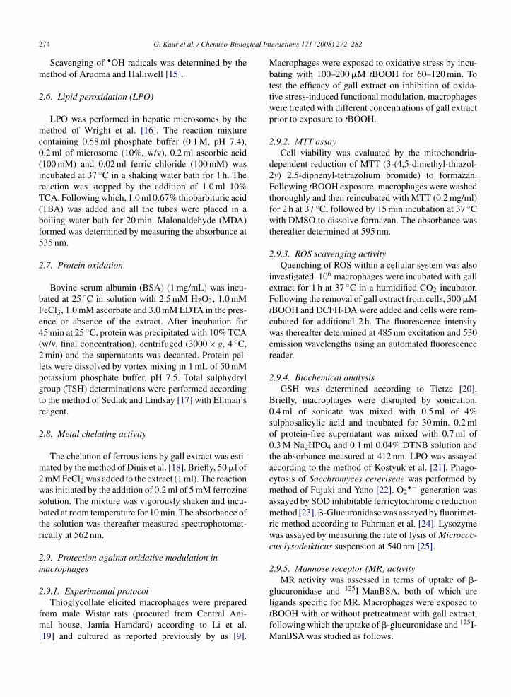
Macrophages were exposed to oxidative stress by incu-bating with 100–200 �M tBOOH for 60–120 min. Totest the efficacy of gall extract on inhibition of oxida-tive stress-induced functional modulation, macrophageswere treated with different concentrations of gall extractprior to exposure to tBOOH.
2.9.2. MTT assayCell viability was evaluated by the mitochondria-
dependent reduction of MTT (3-(4,5-dimethyl-thiazol-2y) 2,5-diphenyl-tetrazolium bromide) to formazan.Following tBOOH exposure, macrophages were washedthoroughly and then reincubated with MTT (0.2 mg/ml)for 2 h at 37 ◦C, followed by 15 min incubation at 37 ◦Cwith DMSO to dissolve formazan. The absorbance wasthereafter determined at 595 nm.
2.9.3. ROS scavenging activityQuenching of ROS within a cellular system was also
investigated. 106 macrophages were incubated with gallextract for 1 h at 37 ◦C in a humidified CO2 incubator.Following the removal of gall extract from cells, 300 �MtBOOH and DCFH-DA were added and cells were rein-cubated for additional 2 h. The fluorescence intensitywas thereafter determined at 485 nm excitation and 530emission wavelengths using an automated fluorescencereader.
2.9.4. Biochemical analysisGSH was determined according to Tietze [20].
Briefly, macrophages were disrupted by sonication.0.4 ml of sonicate was mixed with 0.5 ml of 4%sulphosalicylic acid and incubated for 30 min. 0.2 mlof protein-free supernatant was mixed with 0.7 ml of0.3 M Na2HPO4 and 0.1 ml 0.04% DTNB solution andthe absorbance measured at 412 nm. LPO was assayedaccording to the method of Kostyuk et al. [21]. Phago-cytosis of Sacchromyces cereviseae was performed bymethod of Fujuki and Yano [22]. O2
•− generation wasassayed by SOD inhibitable ferricytochrome c reductionmethod [23]. �-Glucuronidase was assayed by fluorimet-ric method according to Fuhrman et al. [24]. Lysozymewas assayed by measuring the rate of lysis of Micrococ-cus lysodeikticus suspension at 540 nm [25].
2.9.5. Mannose receptor (MR) activityMR activity was assessed in terms of uptake of �-
glucuronidase and 125I-ManBSA, both of which are
ligands specific for MR. Macrophages were exposed totBOOH with or without pretreatment with gall extract,following which the uptake of �-glucuronidase and 125I-ManBSA was studied as follows.
ical Interactions 171 (2008) 272–282 275
2fiaauwTmabaa
2b3omaMeaTb
2
aop
3
3
gflerosaf
3
rw
Fig. 1. Reducing power of alcoholic extract of Quercus infectoria
extract. Gall extract completely scavenged DPPH rad-icals at an extremely low concentration of 1.5 �g/ml,with the IC50 value of 0.5 �g/ml. Antioxidants reduceDPPH, a purple colored stable free radical, to a colorless
G. Kaur et al. / Chemico-Biolog
.9.5.1. β-Glucuronidase uptake. Two hundred andfty microlitres of �-glucuronidase (20 �g/ml) wasdded to macrophage monolayer. Mannan (1 mg/ml) wasdded to companion wells to determine non-specificptake. After incubation for 1 h at 37 ◦C, cells wereashed with HBSS and solublized in 150 �l of 0.1%riton-X-100. One hundred microlitres of lysate wasixed with 0.9 ml of 1 mM phenolphthalein glucuronic
cid and incubated for 10 min. The reaction was stoppedy adding 3 ml of carbonate buffer, pH 10 and the liber-ted phenolphthalein was quantitated by measuring thebsorbance at 550 nm.
.9.5.2. 125I-ManBSA uptake. ManBSA was iodinatedy chloramines-T method to specific activity of–5 × 10⁶ cpm/�g according to Stahl et al. [26]. Uptakef 125I-ManBSA was determined according to Boze-an et al. [27]. Three micrograms of 125I-ManBSA was
dded to culture medium over macrophage monolayer.annan (1 mg/ml) was added to companion wells to
stimate non-specific uptake. After 60 min incubationt 37 ◦C, cells were washed, solublized in 0.1% (v/v)riton-X-100 and cell associated counts were quantitatedy gamma counting.
.10. Statistical analysis
All values are expressed as mean ± S.E. Statisticalnalyses were performed by Student’s t-test. The valuesf P lower than 0.05 were considered significant (P isrobability).
. Results
.1. Total phenolics and reducing power
The yield of extract obtained from Q. infectoriaalls using ethanol was 23.5% (w/w). The extract wasound to contain 416 ± 10.6 mg/g of total polypheno-ics expressed as gallic acid equivalent (GAE, mg/g ofxtract). Since antioxidant activity may be directly cor-elated to the polyphenolic content, the obtained amountf total polyphenolics in gall extract indicated it to pos-ess a high antioxidant activity. The reducing capacity,nother vital indicator of antioxidant activity, was alsoound to be highly significant in gall extract (Fig. 1).
.2. HPTLC analysis of the extract
Since TA and GA, which are potent antioxidants, areeported to be present in Q. infectoria galls, there levelsas determined in gall extract [6,28]. HPTLC analy-
galls in comparison to that of gallic acid and tannic acid. Reducingpower was measured by spectrophotometric detection of Fe3+ to Fe2+
transformation as described in Section 2. Each value is mean ± S.E.(n = 5).
sis was done to quantiate their amount. The extract wasfound to contain 8.75% (w/w) GA (Fig. 2) and 19.925%(w/w) TA (Fig. 3).
3.3. Free radical scavenging activity of gall extract
Table 1 depicts the scavenging of free radicals by the
Fig. 2. HPTLC chromatogram of (a) gall extract in comparison with(b) standard gallic acid (GA).

276 G. Kaur et al. / Chemico-Biological Interactions 171 (2008) 272–282
Table 1Scavenging of ABTS, DPPH, hydrogen peroxide (H2O2) and hydroxyl radicals (•OH) by gall extract, tannic acid (TA) and gallic acid (GA)
Group (% of control)
ABTS DPPH H2O2•OH
Control 100 ± 2.7 100 ± 4 100 ± 5.6 100 ± 2
Gall extract (�g/ml)0.5 71.5 ± 4a 51.7 ± 4 90 ± 5.6 92 ± 6.71.0 50 ± 5a 27.5 ± 5a 82.7 ± 6.3b 88.7 ± 3.6c
2.5 18.7 ± 1.6a 0 53.7 ± 4.7a 74.2 ± 5a
5.0 3 ± 0.4 0 24.2 ± 3a 56.5 ± 5a
15.0 0 0 0 11.3 ± 1.6a
TA (5 �M) 25.4 ± 1.6a 19.6 ± 1.8a 78.1 ± 4.2b 74.7 ± 3.6b
GA (20 �M) 71.2 ± 1.5a 63.2 ± 4.7a 85.7 ± 1.4b 76.2 ± 1.9a
Each value is mean ± S.E. (n = 5).
a p < 0.001 vs. control group.b p < 0.01.c p < 0.05.�-�-diphenyl-�-picryl hydrazine. Scavenging of DPPHradicals is a potent tool to ascertain antioxidant capac-ity of a compound. The latter ability was evaluated interms of ABTS scavenging. Gall extract potently ABTSradicals (Table 1).
Ability of gall extract to scavenge ROS includingH2O2 and •OH was also assessed. Gall extract signif-icantly and dose dependently scavenged both (Table 1).The IC50 value of gall extract to scavenge H2O2 was2.6 �g/ml. The •OH radicals were generated by Fenton
type reaction and measured by their ability to degradedeoxyribose sugar into the fragments that react withthiobarbituric acid to form a pink chromogen. Fifteenmicrograms per millilitre of gall extract could scav-Fig. 3. HPTLC chromatogram of (a) gall extract in comparison with(b) standard tannic acid (TA).
enge 88.7% of •OH radicals (IC50 was 6 �g/ml). Controlexperiments showed that the extract did not interfere withdeoxyribose assay.
3.4. Effect of gall extract on lipid peroxidation andprotein oxidation
Gall extract dose dependently inhibited the amountof TBARS generated by Fe2+–ascorbate system in ratliver microsomes (Fig. 4a). The IC50 value was about46.5 �g/ml. The concentration of the extract used in thisstudy did not interfere with the TBA reaction.
Gall extract also effectively inhibited BSA oxida-tion induced by Fe2+–ascorbate system (Fig. 4b). BSAoxidation was determined in terms of –SH group lossby Ellman reagent [5,5′-dithiobis-(2-nitrobenzoic acid)],which forms a disulfide bond with the –SH group releas-ing a yellow-colored thiolate ion that absorbs at 412 nm.The oxidation of a protein is monitored by measuringthe decrease in absorbance. The incubation of BSA withFe–ascorbate system caused the oxidation of 63% of the–SH groups. Gall extract dose dependently inhibited this–SH oxidation (Fig. 4b).
3.5. Metal chelating activity of gall extract
The chelating of Fe2+ by gall extract was estimatedby ferrozine method (18). Ferrozine can quantitativelyform complexes with Fe2+. However, if a chelating agent
is present, the complex formation is disrupted with theresult that the red color of the complex is decreased.Quantitation of color intensity, therefore, allows the esti-mation of the chelating activity of the coexisting chelator.
G. Kaur et al. / Chemico-Biological Interactions 171 (2008) 272–282 277
Fig. 4. Inhibitory effect of gall extract on (a) lipid peroxidation (LPO) in rat liver microsomes and (b) BSA oxidation induced by Fe2+/ascorbates ed as sts detailec
Tftsf
3o
3o
dot36tflat
ystem in comparison with tannic acid (TA) and gallic acid (GA) usubstances (TBARS) and BSA oxidation in term of –SH group loss asp < 0.001 vs. control group.
he effect of gall extract on Fe2+ and ferrozine complexormation is shown in Fig. 5. Gall extract interfered withhe complex formation between ferrous and ferrozine,uggesting that it has chelating activity and captureserrous ion before ferrozine.
.6. Effect of gall extract on tBOOH-inducedxidative modulation of macrophages
.6.1. Effect of gall extract on tBOOH-inducedxidative stress and cytotoxicity in macrophages
Fluorimetric analysis using a fluorescent probe 2,7-ichlorofluoroscein diacetate revealed a large amount ofxidants in tBOOH-exposed macrophages (Fig. 6). Inhese cells, TBARS content was found to be elevated by.66-fold and GSH content was found to be depleted by5.2% of control (Fig. 7a and b). In cells that received
reatment with gall extract prior to tBOOH exposure, theuorescence intensity was much lesser indicating lessermount of intracellular oxidants (Fig. 6). In notion tohis, a significant and dose-dependent protection againstandards. LPO was measured in terms of thiobarbituric acid reactived in Section 2. Each value is the mean of five observations. ap < 0.05,
elevation of LPO and depletion of GSH (Fig. 7a andb) was observed following pretreatment of macrophageswith gall extract for 60 min.
MTT assay showed 24 h exposure to tBOOH killedas much as 67% of macrophages (Fig. 8). However, pre-treatment with gall extract significantly preserved theviability of cells.
3.6.2. Effect of gall extract on tBOOH-inducedmodulation of phagocytic functions of macrophages
Table 2 shows the effect of exposure of 200 �MtBOOH for 120 min on phagocytosis of S. cereviseae.Gall extract pretreatment significantly and dose depen-dently protected against tBOOH-induced inhibition(Table 2). PMA stimulated respiratory burst was alsoinhibited by tBOOH. Table 2 also shows protectionobtained by gall extract pretreatment against suppres-
sion in respiratory burst. An elevation was observedin the secretion of �-glucuronidase (66%) and that oflysozyme (94%) (Table 2) following tBOOH exposure.Prior treatment of macrophages with gall extract for
278 G. Kaur et al. / Chemico-Biological In
Fig. 5. Metal chelating effect of different concentrations of gall extractin comparison with tannic acid (TA) and gallic acid (GA) used asstandards. Metal chelating effect was determined by ferrozine methodas detailed in Section 2. Each point represents the mean ± S.E. (n = 5).
Fig. 6. Effect of gall extract on tBOOH-induced ROS generated withinthe macrophages. Cells, with or without pretreatment with gall extract,were exposed to tBOOH and DCFH-DA. Fluorescence intensity wasmeasured following 2 h incubation. Each value is mean ± S.E. (n = 5).bp < 0.01, cp < 0.001 vs. tBOOH exposed group.
teractions 171 (2008) 272–282
120 min afforded a significant protection against eleva-tion in secretion of both the enzymes (Table 2). Effectof gall extract on oxidative stress-induced modulationof macrophages MR activity was investigated. Both�-glucuronidase and 125I-ManBSA are known to beefficiently taken up by macrophage MR, so were usedas ligands for MR in the present study. Exposure ofmacrophages to 100 �M tBOOH for 60 min inhibited �-glucuronidase uptake by 53% and 125I-ManBSA uptakeby 62% (Fig. 9). The inhibition in uptake of both theseligands by MR was significantly prevented by prior treat-ment of macrophages with gall extract (Fig. 9).
4. Discussion
Various degenerative disorders implicate a defi-cient natural antioxidant defense as their etiological orpathological factor. The onset/progression of these dis-orders can be held or delayed by supplementing withantioxidant defense. Plant-based antioxidants such asresveratrol and quercetin potentiate body’s antioxidantdefense and are relatively safe. Thus, a huge body ofscientific research is focused on exploring plants withantioxidant potential. In the present study, we haverevealed Q. infectoria galls to possess potent free radicalquenching and antioxidant activity.
Antioxidant activity in plant materials generally orig-inates from their polyphenolics. Several authors haveobserved a direct correlation between polyphenolic con-tent and antioxidant activity [29]. We obtained a veryhigh content of polyphenolics in gall extract, to whichits antioxidant activity may be ascribed.
The phytochemical studies carried out so far haverevealed the presence of tannic acid, gallic acid,syringic acid, ellagic acid, �-sitosterol, amentoflavonehexamethyl ether, isocryptomerin, starch, essentialoils, anthocyanins, methyl-betulate, methyl-oleanate,hexagalloyl-glucose and polygalloyl-glucose [7,8,28] inQ. infectoria galls.
An assay for TA revealed TA to constitute a majorproportion of the extract. Owing to its high amount ingall extract, most properties of the extract can, at leastpartially, be attributed to TA. In notion to this, gall extractwas found to scavenge DPPH and •OH radicals, chelateFe2+ and inhibit lipid peroxidation as has been previouslybeen reported for TA [30–32]. Scavenging of DPPH freeradicals by antioxidants is due to their ability to donatehydrogen. Potent reducing power and DPPH scavenging
capacity of gall extract suggest that it contains reduc-tones, which exert their antioxidant activity by donatinga hydrogen atom to break free radical chain. Scavengingof •OH radicals is generally attributed to Fe2+ chelat-
G. Kaur et al. / Chemico-Biological Interactions 171 (2008) 272–282 279
F ation om Each vag
iupTilf
it
TPa
G
Ct
G
MpR
ig. 7. Inhibitory effect of gall extract on (a) tBOOH induced elevacrophages. Experiments were performed as detailed in Section 2.
roup.
ng activity since withdrawal of Fe2+ ion renders themnavailable for Fenton reaction [33]. Lopes et al. haveroposed a similar mechanism for •OH scavenging byA [31]. In the present study, ferrozine assay system alsondicated gall extract to scavenge Fe2+ ions. Thus, it isikely that chelating activity of gall extract is responsible
or inhibition of deoxyribose oxidation.Gall extract, with its potent free radical scaveng-ng capacity, was expected to inhibit oxidative damageo biomolecules. As envisaged, the extract potently
able 2rotection of tBOOH-induced inhibition in phagocytic capacity and respiratornd lysozyme by gall extract
roup Phagocytic index Respiratory bu
ontrol 100 ± 2.5 100 ± 4BOOH 47.5 ± 4.2a 41.6 ± 3.2a
all extract + tBOOH (�g/ml)5 54.6 ± 4.5 46.2 ± 2.310 59.5 ± 2.9c 58.5 ± 2d
30 72.3 ± 3.6c 71.5 ± 6.2c
acrophages were pretreated with gall extract for 60 min and then exposedhagocytosis, respiratory burst and secretion of �-glucuronidase and lysozymesults are expressed as mean ± S.E. of five determinations.a p < 0.001 compared to the control.b p < 0.05.c p < 0.01.d p < 0.001 compared to the tBOOH treated groups.
f lipid peroxidation and (b) tBOOH-induced depletion of GSH inlue is mean ± S.E. (n = 5). bp < 0.01, cp < 0.001 vs. tBOOH exposed
inhibited Fe–ascorbate-induced peroxidation of micro-somal lipids and oxidation of BSA. Fe2+ is known toincite oxidation of proteins and peroxidation of lipidsby causing •OH generation by the Fenton reaction.Fe2+ can also induce peroxidation by breakdown oflipid hydroperxides into alkoxyl and peroxyl radicals,
which are capable of abstracting hydrogen and per-petuating lipid peroxidation chain reaction [34]. Theinhibition of oxidative DNA damage by Fe–ascorbatesystem may either involve exclusion of the catalyst: Fe2+y burst and tBOOH-induced elevation in secretion of �-glucuronidase
rst �-Glucuronidase Lysozyme
100 ± 3.6 100 ± 6167.3 ± 12a 194.6 ± 9.5a
142 ± 5.7 155.7 ± 6b
127.4 ± 5c 137 ± 4.6c
107 ± 6c 106.5 ± 6c
to 200 �M tBOOH for 120 min. Cells were washed and assessed fore as detailed in Section 2.

280 G. Kaur et al. / Chemico-Biological In
Fig. 8. Protection of tBOOH induced cytotoxicity in macrophages bygall extract. Macrophages were pretreated with different concentra-tions of gall extract for 120 min and then exposed to 100 �M tBOOHfor 2, 6 and 24 h. Cells were washed and assessed for substrate adher-
ence capacity as described in Section 2. Results are expressed asmean ± S.E. of five determinations.Fig. 9. Protection afforded by gall extract against tBOOH-induced inhibition iMacrophages were preincubated with gall extract for 60 min and then treatedwith the ligand (�-glucuronidase or 125I-ManBSA), whose uptake was theremean ± S.E. of five determinations.
teractions 171 (2008) 272–282
or scavenging of the generated oxidants. Both the possi-bilities seem to be implicated; however, considering theextract to have rather week Fe2+ chelating activity, majorcontribution plausibly comes from scavenging of freeradicals.
Antioxidant activity of gall extract was also eval-uated in a cellular system. Exposure of rat peritonealmacrophages to tBOOH led to a huge amount of oxida-tive stress in them. tBOOH is a model peroxidant andhas been used by several workers to cause oxidativestress in macrophages [35–37]. Toxicity of tBOOHis thought to be mediated by alkoxyl radicals, intowhich it decomposes. Several workers have previouslyreported increase in lipid peroxidation and DNA dam-age due to elevated free radical formation followingtBOOH exposure [38]. In the present study, incubationof macrophages with tBOOH resulted in an increase infree radical production in macrophages as revealed byDCFH-DA. There was a concomitant elevation in LPO
and DNA damage by tBOOH. However, a prior treat-ment with gall extract significantly protected againstboth. tBOOH also depleted GSH content of macrophagesand induced several functional alterations. tBOOH isn ligand (�-glucuronidase and 125I-ManBSA) uptake by macrophages.with 100 �M tBOOH for 60 min. Cells were washed and incubated
after determined as explained in Section 2. Results are expressed as

ical In
dac(mteTeiaet
mSablLmovTsFbaisa
igcmehso
efeataMtfbd
[
[
[
[
[
[
[
[
[
[
G. Kaur et al. / Chemico-Biolog
etoxified by cellular glutathione peroxidase to t-butanolt the expense of GSH. GSH may also detoxify toxi-ants by reduction, conjugation or by direct quenchingin case of free radicals) [39,40]. Thus, exposure ofacrophages to tBOOH caused a significant attenua-
ion in GSH levels. In macrophages pretreated with gallxtract, tBOOH-induced attenuation in GSH was lesser.his could partially be attributed to free radical scav-nging activity of gall extract and thereby preservationn GSH levels. The amount of endogenous macrophagealntioxidant (GSH) and oxidative damage (MDA lev-ls) showed a clear correlation with tBOOH-inducedoxicity.
Phagocytosis, one of the most important functions ofacrophages, was found to be suppressed by tBOOH.ince phagocytosis is also a membrane linked function,ttenuation in it may be attributed to disruption of mem-rane structure due to peroxidative damage of membraneipids and/or oxidation of implicated receptors. Further,PO hampers the fluidity of membrane hindering theovement of receptors within the bilayer. Phagocytosis
f S. cereviseae is mediated by MR [41]. tBOOH ele-ated LPO as well as caused inhibition of MR activity.hus, any or all of the above reasons could be respon-ible for suppressing the phagocytosis by macrophages.urther, since extract supplementation prevented mem-rane LPO and also preserved MR activity, the protectionfforded by it against tBOOH-induced supplementationn phagocytosis seems to implicate its ability to pre-erve membrane structure by virtue of its antioxidantctivity.
LPO associated membrane damage could also bemplicated in tBOOH-induced suppression of O2
•−eneration since O2
•− is generated by membrane asso-iated NADPH oxidase enzyme. LPO also makes theembrane “leaky”, elevating the secretion of lytic
nzymes out of the membrane [42]. Fuhrman et al. [24]ave earlier reported an increase in �-glucuronidaseecretion from macrophages following exposure toxidants.
In conclusion, the present study demonstrates that thethanolic extract of Q. infectoria galls possess a potentree radical scavenging and antioxidant activities. Thextract is capable of protecting against oxidative dam-ge to lipids and proteins and also chelates metal ionshat catalyze the generation of oxidants. The extractlso protects a cellular system from oxidative damage.acrophages exposed to tBOOH were significantly pro-
ected by gall extract both from oxidative damage andunctional deterioration. This study implies the plausi-ility of therapeutic effect of gall extract on inflammatoryisorders.
[
teractions 171 (2008) 272–282 281
References
[1] B. Halliwell, J.M.C. Gutteridge, C.E. Cross, Free radicals, antiox-idants, and human disease: where are we now? J. Lab. Clin. Med.119 (1992) 598–620.
[2] N. Hogg, Free radicals in disease, Semin. Reprod. Endocrinol. 16(1998) 241–288.
[3] J. Robak, E. Marcinkiewicz, Scavenging of reactive oxygenspecies as the mechanism of drug action, Pol. J. Pharmacol. 47(1995) 89–98.
[4] D.L. Madhavi, D.K. Salunkhe:, Toxicological aspects of foodantioxidants, in: D.L. Madavi, S.S. Deshpande, D.K. Salunkhe(Eds.), Food Antioxidants, Dekker, New York, 1995, p. 267.
[5] S.N. Meydani, D. Wu, M.S. Santos, M.G. Hayek, Anti-oxidantsand immune response in aged persons: overview of present evi-dence, Am. J. Clin. Nutr. 62 (1995) 1462S–1476S.
[6] M.S. Dar, M. Ikram, T. Fakouhi, Pharmacology of Quercus infec-toria, J. Pharm. Sci. 65 (1976) 1791–1794.
[7] M.S. Dar, M. Ikram, Studies on Quercus infectoria; isolation ofsyringic acid and determination of its central depressive activity,Planta Med. 35 (1979) 156–161.
[8] J.K. Hwang, T.W. Kong, N.I. Baek, Y.R. Pyun, Alpha-glycosidaseinhibitory activity of hexagalloylglucose from the galls of Quer-cus infectoria, Planta Med. 66 (2000) 273–274.
[9] G. Kaur, H. Hamid, A. Ali, M.S. Alam, M. Athar, Antiinflamma-tory evaluation of alcoholic extract of galls of Quercus infectoria,J. Ethnopharmacol. 90 (2004) 285–292.
10] M.S. Taga, E.E. Miller, D.E. Pratt, Chia seeds as a source of nat-ural lipid antioxidant, J. Am. Oil Chem. Soc. 61 (1984) 928–931.
11] M. Oyaizu, Studies on products of browning reaction preparedfrom glucosamine, Jpn. J. Nutr. 44 (1986) 307–315.
12] G. Kaur, I.A. Lone, M. Athar, M.S. Alam, Protective effect ofDidymocarpus pedicellata on ferric nitrilotriacetate (Fe-NTA)induced renal oxidative stress and hyperproliferative response,Chem. Biol. Interact. 165 (2007) 33–44.
13] T.Y. Song, G.C. Yen, Antioxidant properties of Antrodia cam-phorata in submerged culture, J. Agric. Food Chem. 50 (2002)3322–3327.
14] R.J. Ruch, S.J. Cheng, J.E. Klaunig, Prevention of cytotoxicityand inhibition of intercellular communication by antioxidant cat-echins isolated from Chinese green tea, Carcinogenesis 10 (1989)1003–1008.
15] O.I. Aruoma, B. Halliwell, Action of hypochlorous acid on theantioxidant protective enzymes superoxide dismutase, catalaseand glutathione peroxidase, Biochem. J. 248 (1987) 973–976.
16] J.R. Wright, H.D. Colby, P.R. Miles, Cytosolic factors whichaffect microsomal lipid peroxidation in lung and liver, Arch.Biochem. Biophys. 206 (1981) 296–304.
17] J. Sedlak, R.H. Lindsay, Estimation of total, protein-bound, andnonprotein sulfhydryl groups in tissue with Ellman’s reagent,Anal. Biochem. 25 (1968) 192–205.
18] T.C.P. Dinis, V.M.C. Madeira, M.L.M. Almeida, Action of pheno-lic derivates (acetoaminophen, salycilate, and 5-aminosalycilate)as inhibitors of membrane lipid peroxidation and as per-oxyl radical scavengers, Arch. Biochem. Biophys. 315 (1994)161–169.
19] Y.M. Li, G. Baviello, H. Vlassara, T. Mitsuhashi, Glycation
products in aged thioglycollate medium enhance the elicitationof peritoneal macrophages, J. Immunol. Methods 201 (1997)183–188.20] F. Tietze, Enzymic method for quantitative determination ofnanogram amounts of total and oxidized glutathione: applications

ical In
[
[
[
[
[
[
[
[
[
[
[
[
[
[
[
[
[
[
[
[
[
282 G. Kaur et al. / Chemico-Biolog
to mammalian blood and other tissues, Anal. Biochem. 27 (1969)502–522.
21] V.A. Kostyuk, A.I. Potapovich, S.D. Speransky, G.T. Maslova,Protective effect of natural flavonoids on rat peritonealmacrophages injury caused by asbestos fibers, Free Radic. Biol.Med. 21 (1996) 487–493.
22] K. Fujuki, T. Yano, Effect of Sodium alginate on the non-specificdefense system of the common carp (Cyprinus carpio L.), FishShell Fish Immunol. 7 (1997) 417–427.
23] Y. Keisari, E. Pick, Macrophage-mediated cytolysis off erythro-cytes in the guinea pig, I. Activation by stimulators of the oxidativeburst, Cell Immunol. 62 (1981) 172–185.
24] B. Fuhrman, J. Oiknine, M. Aviram, Iron induces lipid peroxida-tion in cultured macrophages, increases their ability to oxidativelymodify LDL, and affects their secretary properties, Atheroscle-rosis 111 (1994) 65–78.
25] R.J. Smith, S.S. Iden, Phorbol myristate acetate-inducedrelease of granule enzymes from human neutrophils: inhibitionby the calcium antagonist, 8-(N,N-diethylamino)-octyl 3,4,5-trimethoxybenzoate hydrochloride, Biochem. Biophys. Res.Commun. 91 (1979) 263–271.
26] P. Stahl, P.H. Schlesinger, E. Sigardson, J.S. Rodman, Y.C. Lee,Receptor-mediated pinocytosis of mannose glycoconjugates bymacrophages: characterization and evidence for receptor recy-cling, Cell 19 (1980) 207–215.
27] P.M. Bozeman, J.R. Hoidal, V.L. Shepherd, Oxidant-mediatedinhibition of ligand uptake by the macrophage mannose receptor,J. Biol. Chem. 263 (1988) 1240–1247.
28] M. Ikram, F. Nowshad, Constituents of Quercus infectoria, PlantaMed. 31 (1977) 286–287.
29] C.A. Rice-Evans, J. Sampson, P.M. Bramley, D.E. Holloway, Whydo we expect carotenoids to be antioxidants in vivo? Free Radic.Res. 26 (1997) 381–398.
30] T. Yokozawa, C.P. Chen, E. Dong, T. Tanaka, G.I. Nonaka, I.Nishioka, Study on the inhibitory effect of tannins and flavonoidsagainst the 1,1-diphenyl-2 picrylhydrazyl radical, Biochem. Phar-
macol. 56 (1998) 213–222.31] G.K. Lopes, H.M. Schulman, M. Hermes-Lima, Polyphenol tan-nic acid inhibits hydroxyl radical formation from Fenton reactionby complexing ferrous ions, Biochim. Biophys. Acta 1472 (1999)142–152.
[
teractions 171 (2008) 272–282
32] M.A. Gyamfi, Y. Aniya, Antioxidant properties of ThonningianinA, isolated from the African medicinal herb, Thonningia san-guinea, Biochem. Pharmacol. 63 (2002) 1725–1737.
33] C. Smith, B. Halliwell, O.I. Aruoma, Protection by albuminagainst the pro-oxidant actions of phenolic dietary components,Food Chem. Toxicol. 30 (1992) 483–489.
34] M.E. Gulcin, M.E. Buyukokuroglu, M. Oktay, O.I. Kufre-vioglu, Antioxidant and analgesic activities of turpentine ofPinus nigra, Arn. subsp. pallsiana (Lamb.) Holmboe 86 (2003)51–58.
35] S.A. Altman, T.H. Zastawny, L. Randers, Z. Lin, J.A. Lumpkin,J. Remacle, M. Dizdaroglu, G. Rao, Tert-Butyl hydroperoxidemediated DNA base damage in cultured mammalian cells, Mutat.Res. 306 (1994) 35–44.
36] F.R. Livingston, E.M. Lui, G.A. Loeb, H.J. Forman, Sublethal oxi-dant stress induces a reversible increase in intracellular calciumdependent on NAD(P)H oxidation in rat alveolar macrophages,Arch. Biochem. Biophys. 299 (1992) 83–91.
37] T.W. Robison, H. Zhou, H.J. Forman, Modulation of ADP-stimulated inositol phosphate metabolism in rat alveolarmacrophages by oxidative stress, Arch. Biochem. Biophys. 318(1995) 215–220.
38] E.A. Lapshina, I.B. Zavodnik, M. Labieniec, K. Rekawiecka,M. Bryszewska, Cytotoxic and genotoxic effects of tert-butylhydroperoxide on Chinese hamster B14 cells, Mutat. Res. 583(2005) 189–197.
39] C.F. Lima, M. Fernandes-Ferreira, C. Pereira-Wilson, Pheno-lic compounds protect HepG2 cells from oxidative damage:relevance of glutathione levels, Life Sci. 79 (2006) 2056–2068.
40] S.A. Jewell, D. Di Monte, P. Richelmi, G. Bellomo, S. Orrenius,Tert-Butyl hydroperoxide-induced toxicity in isolated hepato-cytes: contribution of thiol oxidation and lipid peroxidation, J.Biochem. Toxicol. 1 (1986) 13–22.
41] J. Giaimis, Y. Lombard, P. Fonteneau, C.D. Muller, R. Levy, M.Makaya-Kumba, J. Lazdins, P. Poindron, Both mannose and beta-
glucan receptors are involved in phagocytosis of unopsonized,heat-killed Saccharomyces cerevisiae by murine macrophages, J.Leukoc. Biol. 54 (1993) 564–571.42] C. Mylonas, D. Kouretas, Lipid peroxidation and tissue damage,In Vivo 13 (1999) 295–309.