Glucocorticoid receptor mutants: man-made tools for functional research
Upload
universitasindonesiatimurCategory
view
1download
0
DEVELOPMENTAL BIOLOGY 65,476-482 (1978)
Larval Adipose Tissue of Homoeotic Bithorax Mutants of Drosophila
T. M. RIZKI AND ROSE M. RIZKI
Division of Biological Sciences, The University of Michigan, Ann Arbor, Michigan 48109
Received December 21, 1977; accepted in revised form April 3, 1978
In the homoeotic bithomx mutant combination bx3 pbx/lJbx*” of Drosophila melanogaster, the metathoracic segment is transformed to a mesothoracic segment and the adult flies have an extra pair of wings in place of the paired halteres [Lewis, E. B. (1983). Amer. 2001. 3,33-561. The morphology of the larval fat body, the number of cells in the fat body, and the distribution pattern of kynurenine autofluorescent materials (KAF+) in this tissue were compared in the homoeotic mutant and a wild-type strain. The mutant has an additional mass of adipose cells anterior to the posterior margin of the ventral commissure of the fat body. However, the total number of adipose cells in the two strains as well as the limits of the KAF+ cell population do not differ. Therefore, the bithorax transformation in the larval fat body involves rearrangement of the same cell population as that in the normal strain. This study suggests (1) that the bithorax mutant genes affect the pattern of cell segregation and/or migration of preblastoderm nuclei during embryogen-
esis and (2) that the larval fat body of Drosophila has a segmental origin.
INTRODUCTION
Five functionally related genes of the bi- thorax complex locus effect transformation of structures in the mesothorax, the meta- thorax, and the first abdominal segment of Drosophila melanogaster (Lewis, 1963, 1964). Among these genes, bithorax (bx) alters the anterior metathoracic segment such that it resembles the anterior part of the mesothorax and postbithorax (pbx) transforms the posterior part of the meta- thorax to the morphology of the posterior mesothorax. The regional differentiations of the segments affected by these two mu- tants are independent of each other since the double mutant strain (bx3 pbx) shows complete transformation of the metatho- racic region to a mesothoracic segment. In these flies, the halteres have been changed into a second pair of wings.
The cuticular structures of the adult fly arise from metamorphosis of the imaginal discs during the pupal period. In the larva, the various imaginal discs are morphologi- cally distinguishable and their positions are anatomically distinct. Wing discs are larger than haltere discs, and the increase in cell
number of the transformed haltere discs in bithorax mutants is evident in the larval stage. That bithorax gene action affects development considerably earlier than this time was first suggested by Gloor’s (1947) demonstration that bithorax phenocopies result from ether treatment of embryos at O-6 hr. Capdevila and Garcia-Bellido (1974) reexamined the kinetics of ether-induced phenocopies in wild-type individuals and various combinations of mutants in the bi- thorax complex and found that the pheno- critical period for bithorax phenocopies in- cludes the preblastoderm stage before the migrating syncytial nuclei have reached the cortex of the egg. Action of the bithorax complex at this early time in development suggests that larval structures should also show morphologic effects, provided their embryonic origin lies within the domain of the affected segments. Lewis (1964) noted that larvae of the Ultrabithorax (Ubx) strain as well as several double mutant combinations develop spiracles on the met- athoracic and first abdominal segments in addition to the normal anterior pair of spir- acles. These organs as well as the trans-
476 0012.X06/78/0652-0476$02.00/O Copyright 0 1978 by Academic Press, Inc. All rights of reproduction in any form reserved.

RIZKI AND RIZKI Fat Body of a Homoeotic Mutant 477
formed adult structures are modifications in the morphology of the exoskeleton, so the question remains whether internal lar- val structures are also influenced by bi- thorax mutant transformations. In the adult, the transformed metathorax does not contain flight muscles (El Shatoury, 1956), indicating that modifications of internal structures may not coincide with those of cuticular origin.
The differentiation of the internal larval tissues of Drosophila is completed by 18 hr of embryogenesis (Poulson, 1950), and cells of these tissues, such as the salivary glands, malpighian tubules, and fat body, do not continue to divide during the larval period, but rather grow by increase in cell size. It is relatively easy to determine the number of cells in these tissues due to their large size, so the effects of a bithorax transfor- mation on a larval tissue can be inferred by counting cell numbers in various regions of the transformed tissue and comparing this distribution pattern with that of the un- transformed pattern. For such a compari- son, we selected the larval fat body since it displays characteristic morphological and functional differentiation (Rizki, 1961; Rizki and Rizki, 1970) in the body segments that are affected by the bithorax genes.
MATERIALS AND METHODS
Ore-R wild-type and sbd2 bx3 pbx/T(2:3) Ubx’as larvae were grown at 24°C on stan- dard corn meal-molasses medium seeded with live yeast. Fat bodies were dissected from mid-third-instar female larvae in Dro- sophila Ringer solution, fixed in Carnoy, and reacted with Feulgen reagent. Only intact and complete fat body dissections were used. The images of the whole-mount dissections were projected through an in- verted microscope, and the tissue outlines along with the distribution of nuclei were traced for counting.
For examination of autofluorescence in fat body cells, the procedures used previ- ously were followed (Rizki, 1964). White and early tan puparial stages were used for
this aspect of the study to assure that the kynurenine autofluorescence pattern was fully developed and intense. Freshly dis- sected, whole-mount preparations were projected through a photographic enlarger onto a lantern slide plate, which was later developed to obtain an outline of the fat body tissue. The dissections were then ex- amined and photographed with the fluores- cence microscope. Since the field of the fluorescence microscope is limited to a small area excited by the 365~nm wave- length, several overlapping photographs of each specimen were required to record the pattern of kynurenine autofluorescence. A montage of these photographs was then compared with the photograph of the entire tissue outline to establish the limits of the autofluorescent cell distribution. In these preparations, total cell counts were not made.
RESULTS AND DISCUSSION
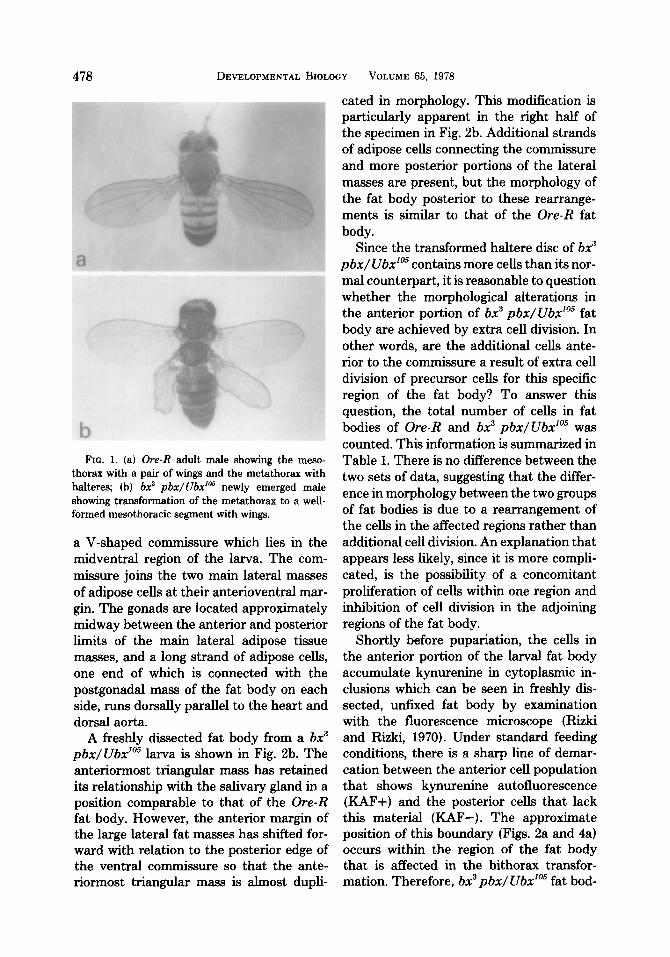
A photograph of a bx3 pbx/Ubx’” adult is included with a photograph of an Ore-R wild-type male to illustrate the marked dif- ference between the adults of the two strains used in this study (Fig. 1). The external morphology of the larvae of these strains is similar, but the pattern of regional differentiation of the larval fat bodies dif- fers. As described previously (Rizki, 1969), the fat body of the Ore-R larva consists of a single layer of cells forming a continuous sheet of tissue extending from the anterior region of the body to the caudal segment. Figure 2a is a freshly dissected, complete fat body of an Ore-R larva suspended in Ringer solution. The fat body is bilaterally symmetrical, and the salivary glands with their common duct have been retained dur- ing dissection so as not to disturb attach- ments with the fat body. The anteriormost adipose cell mass, which is roughly trian- gular in shape, adheres to the salivary gland and overlies the cerebral hemispheres in the body of the larva. A single strand of adipose cells which are in close apposition to the salivary glands connects this piece to
478 DEVELOPMENTAL BIOLOGY VOLUME 65, 1978
FIG. 1. (a) Ore-R adult male showing the meso-
thorax with a pair of wings and the metathorax with halteres; (b) bx3 pbx/lJbda5 newly emerged male
showing transformation of the metathorax to a well- formed mesothoracic segment with wings.
a V-shaped commissure which lies in the midventral region of the larva. The com- missure joins the two main lateral masses of adipose cells at their anterioventral mar- gin. The gonads are located approximately midway between the anterior and posterior limits of the main lateral adipose tissue masses, and a long strand of adipose cells, one end of which is connected with the postgonadal mass of the fat body on each side, runs dorsally parallel to the heart and dorsal aorta.
A freshly dissected fat body from a bx3 pbx/Ubx’” larva is shown in Fig. 2b. The anteriormost triangular mass has retained its relationship with the salivary gland in a position comparable to that of the Ore-R fat body. However, the anterior margin of the large lateral fat masses has shifted for- ward with relation to the posterior edge of the ventral commissure so that the ante- riormost triangular mass is almost dupli-
cated in morphology. This modification is particularly apparent in the right half of the specimen in Fig. 2b. Additional strands of adipose cells connecting the commissure and more posterior portions of the lateral masses are present, but the morphology of the fat body posterior to these rearrange- ments is similar to that of the Ore-R fat body.
Since the transformed haltere disc of bx3 pbx/ Ubx’05 contains more cells than its nor- mal counterpart, it is reasonable to question whether the morphological alterations in the anterior portion of bx3 pbx/Ubx’m fat body are achieved by extra cell division. In other words, are the additional cells ante- rior to the commissure a result of extra cell division of precursor cells for this specific region of the fat body? To answer this question, the total number of cells in fat bodies of Ore-R and bx3 pbx/Ubx’05 was counted. This information is summarized in Table 1. There is no difference between the two sets of data, suggesting that the differ- ence in morphology between the two groups of fat bodies is due to a rearrangement of the cells in the affected regions rather than additional cell division. An explanation that appears less likely, since it is more compli- cated, is the possibility of a concomitant proliferation of cells within one region and inhibition of cell division in the adjoining regions of the fat body.
Shortly before pupariation, the cells in the anterior portion of the larval fat body accumulate kynurenine in cytoplasmic in- clusions which can be seen in freshly dis- sected, unfixed fat body by examination with the fluorescence microscope (Rizki and Rixki, 1970). Under standard feeding conditions, there is a sharp line of demar- cation between the anterior cell population that shows kynurenine autofluorescence (KAF+) and the posterior cells that lack this material (KAF-). The approximate position of this boundary (Figs. 2a and 4a) occurs within the region of the fat body that is affected in the bithorax transfor- mation. Therefore, bx3 pbx/ Ubx’Os fat bod-

RIZKI AND RIZKI Fat Body of a Homoeotic Mutant 479
FIG. 2. Photographs of freshly dissected larval fat bodies from the Ore-R (a) and bx3 pbx/Ubx’” (b) strains suspended in Drosophila Ringer solution. For information on the disposition of the fat body masses within the body cavity, see the text. The bilaterally symmetrical fat body is held together in the anterior region by the common salivary duct (d) and the V-shaped ventral commissure (c) of adipose cells which joins the two lateral fat body masses. Compare the arrangement of the fat body masses in the two specimens by noting their disposition along the salivary glands and the position of the strands of adipose cells connected to the ventral commissure. The arrows indicate the approximate posterior extent of KAF+ cells in the Ore-R specimen. The continuity of the adipose cells in specimen b has been broken (star) during manipulation for photography. Note the sizes of the mesothoracic and metathoracic discs from this specimen (b) in the upper right corner. The bar represents 1 mm.
TABLE 1
NUMBER OF CELLS IN THE LARVAL FAT BODY~ Ore-R br,” p bx/ .!Jbx”s
2318 2254 2064 2264 2263 2121
2026 2134 2383 1903
2254 2211 zk 71 2155 + 57
n t = 0.44, df 9, P > 0.70.
ies were examined with the fluorescence microscope to determine the distribution of KAF+ cells when the morphology of the anterior fat body has been modified by a bithorax transformation.
A photographic montage showing the
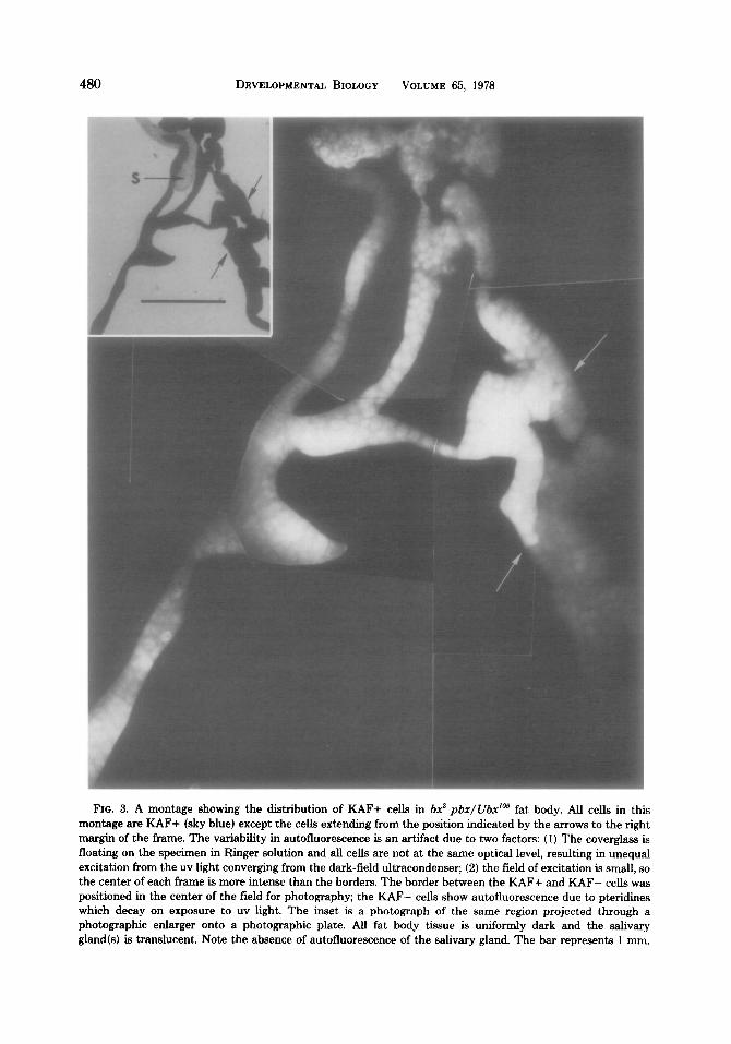
posterior limit of KAF+ cells in a bx3 pbx/Ubx’” fat body is shown in Fig. 3. This region of the same specimen projected through a photographic enlarger onto a photographic plate (inset) is included for purposes of comparison with Figs. 2b and 4b. The boundary between KAF+ and KAF- cells in bx3 pbx/Ubx105 fat bodies is sharp and occurs in the lateral region ad- joining the ventral commissure. This KAF+ cellular distribution pattern was consistent for all bx3 pbx/ Ubx’05 specimens (N = 11) that were examined. Since the total number of cells in the fat bodies of Ore-R and bx3 pbx/Ubx105 is the same but the boundary of the KAF+ cell population has moved forward with relation to the
480 DEVELOPMENTAL BIOLOGY VOLUME 65, 1978
FIG. 3. A montage showing the distribution of RAF+ cells in 62 pbr/Ubr’” fat body. All cells in this montage are RAF+ (sky blue) except the cells extending from the position indicated by the arrows to the right margin of the frame. The variability in autofluorescence is an artifact due to two factors: (1) The coverglass is
floating on the specimen in Ringer solution and all cells are not at the same optical level, resulting in unequal excitation from the uv light converging from the dark-field ultracondenser; (2) the field of excitation is small, so the center of each frame is more intense than the borders. The border between the RAF+ and RAF- cells was positioned in the center of the field for photography; the RAF- cells show autofluorescence due to pteridines which decay on exposure to uv light. The inset is a photograph of the same region projected through a photographic enlarger onto a photographic plate. All fat body tissue is uniformly dark and the salivary
gland(s) is translucent. Note the absence of autofluorescence of the salivary gland. The bar represents 1 mm.

RIZKI AND RIZKI Fat Body of a Homoeotic Mutant 481
a .:, :. . . . . .
c ::q;. :\;.,.. ‘, : .::: .( .~,$Z$ : ,: $“.
i‘
+
# 0
b
1 Y
C
FIG. 4. Semidiagrammatic representation of the left half of the larval fat body of Ore-R (a) and bx3 pbx/Ubxfas (b), showing the region of KAF+ cells (stippled). These diagrams are based on camera lucida drawings of fresh dissections and whole-mount prep- arations of dissections. For orientation, note the posi- tion of the salivary gland and common salivary duct; the ovary (solid circle) is located in the posterior half of the main lateral fat body mass. Diagram c repre- sents the metameric origin of the larval fat body of Drosophila deduced from comparison of the normal and bithorax transformation.
posterior margin of the ventral commissure, it is apparent that the KAF+ region in bx3 pbx/ UbxlOs fat bodies approximates the to- tal KAF+ region in Ore-R fat bodies. In the bithorax transformation, fat body cells have undergone anatomical rearrangement with- out functional modification of the rear- ranged cells.
When wild-type haltere disc cells are mixed with wild-type wing disc cells, the two cell types segregate, whereas the cells from haltere discs of bx3 will integrate with wild-type anterior-wing disc cells and the transformed haltere disc cells of pbx will integrate with wild-type posterior-wing disc cells (Garcia-Bellido and Lewis, 1976). The cellular affinities displayed by the bithorax discs in mixing experiments are thus those of their transformed states, and Garcia-Bel- lido and Lewis concluded that the effects of the bithorax mutants are cell autonomous,
reflecting their genotypic constitution rather than segmental origin. In the bx3 pbx/ Ubx’” larval fat body, altered patterns of cell adhesion are reflected in the altered organization of groups of fat body cells.
In addition to the developmental effect on cell surface properties associated with the transformation of the halter@ disc, the bithorax mutations increase the number of cells in this disc. The effect on cell number has recently been confirmed by Morata and Garcia-Bellido (1976), who analyzed the frequency and sizes of clones induced after irradiation of wild-type and bithorax mes- othoracic and metathoracic discs. Their study indicates that the initial as well as the final number of cells in the transformed haltere disc is the same as that of a normal wing disc. In evaluating the effects of the bithorax genes on adipose tissue, it should be recalled that the cells become nondivid- ing following embryogenesis and increase their genomic content by polyteny, whereas the imaginal disc cells continue to multiply by mitosis during the larval period. Our observations on the larval fat body indicate that the total number of cells in this tissue is not affected by the bithorax transforma- tion. Therefore, the tissue reorganization associated with the mutant genes was achieved by changes in the positional pat- terns of cells, involving either changes in the migration of the preblastodermal nuclei or changes in the distribution of cells at the early cellular blastoderm stage.
The pattern of regional differentiation of the larval fat body cells of D. melanogaster has been studied by utilizing mutant genes whose influence is limited to specific re- gions of this tissue (Rizki, 1969). Analysis of the distribution of fat body cells showing a KAF+ phenotype has been particularly useful, since this cell trait is autonomous as demonstrated by examination of gynandro- morphs containing u+ (normal) and u (mu- tant lacking kynurenine synthesis) cells (Rizki and Rizki, 1968). On the standard Drosophila diet, the pattern of KAF+ cells in the Ore-R fat body is specific and shows

GARCIA-BELLIDO, A., AND LEWIS, E. B. (1976). Auton- omous cellular differentiation of homoeotic bithorax mutants of Drosophila melanogaster. Develop. Biol. 48,400-410.
482 DEVELOPMENTAL BIOLOGY VOLUME 65, 1978
a discrete boundary between KAF+ and KAF- cells. These two cell types are side by side in the fat body and their junction occurs within the main lateral fat mass at a site not distinguishable by morphological landmarks or routine histological examina- tions. An explanation for this intercellular relationship within the fat body is proposed in Fig. 4. According to this model, the larval fat body is a metameric tissue in which cells of adjoining segments become morphologi- cally integrated as a continuum during de- velopment. This scheme requires that the functional differentiation of fat body re- gions related to the segmental origin of the cells is retained. The fat body is thus an ordered mosaic of cell groups, and segmen- tal reorganization in the bx3 pbx/Ubx’05 mutant combination results in new mor- phological relationships without affecting the KAF+ function of the cells.
GLOOR, H. (1947). Phanokopie-Versuche mit Aether an Drosophila. Rev. Sukse Zool. 54,637-712.
LEWIS, E. B. (1963). Genes and developmental path- ways. Amer. Zool. 3,33-56.
LEWIS, E. B. (1964). Genetic control and regulation of developmental pathways. In “Role of Chromosomes
in Development” (M. Locke, ed.), pp. 231-252. Ac-
ademic Press, New York. MORATA, G., AND GARCIA-BELLIDO, A. (1976). Devel-
opmental analysis of some mutants of the bithorax system of Drosophila. Wilhelm Roux’ Arch. 179,
125-143. POULSON, D. F. (1950). Hi&genesis, organogenesis,
and differentiation in the embryo of Drosophila melanogaster. In “Biology of Drosophila” (M. De-
merec, ed.), pp. 168-274. John Wiley & Sons, New York.
RIZKI, T. M. (1961). Intracellular localization of kyn- urenine in the fat body of Drosophila. J. Biophys. Biochem. Cytol. 9, 567-572.
This investigation was supported by Grant No. CA- 16619 awarded by the National Cancer Institute, DHEW.
RIZKI, T. M. (1964). Mutant genes regulating the
inducibility of kynurenine synthesis. J. Cell Biol. 21,203-211.
RI~KI, T. M. (1969). Genetics and evolution of a cell phenotype in Drosophila. Japan. J. Genet. (Suppl.) 44, 51-57.
REFERENCES
CAPDEVILA, M. P., AND GARCIA-BELLIDO, A. (1974).
Developmental and genetic analysis of bithorax phe- nocopies in Drosophila. Nature (London) 250, 500-502.
EL SHATOURY, H. H. (1956). Developmental interac- tions in the development of the imaginal muscles of
Drosophila. J. Embryol. Exp. Morpol. 4, 228-239.
RIZKI, T. M., AND RIZKI, R. M. (1968). Allele specific
patterns of suppression of the vermilion locus in Drosophila melanogaster. Genetics 59, 477-485.
RIZKI, T. M., AND RIZKI, R. M. (1970). The genetic basis of a cell-pattern homology in Drosophila spe- cies. In “Essays in Evolution and Genetics in Honor
of Theodosius Dolzhansky” (M. K. Hecht and W. C. Steere, eds.). Appleton-Century-Crofts, New
York.
Copyright © 2022 FDOKUMEN