A Qualitative Analysis of Larval Drosophila melanogaster ...
Upload
khangminh22Category
view
6download
0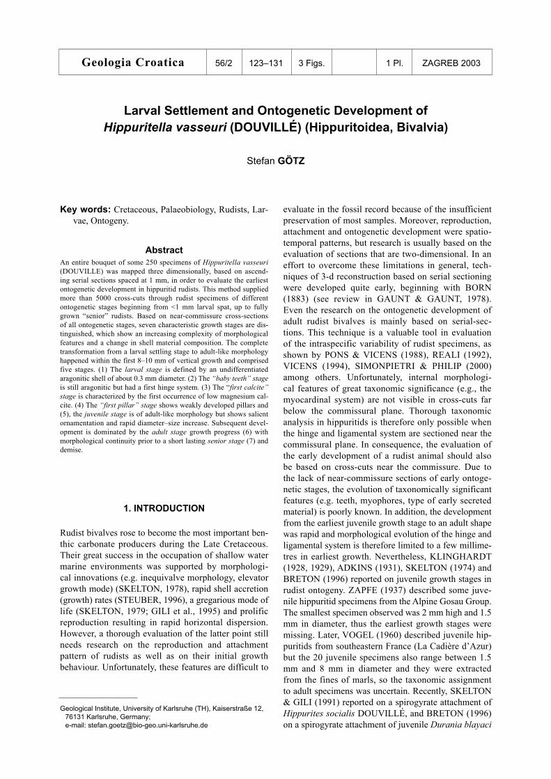
1. INTRODUCTION
Rudist bivalves rose to become the most important ben-thic carbonate producers during the Late Cretaceous. Their great success in the occupation of shallow water marine environments was supported by morphologi-cal innovations (e.g. inequivalve morphology, elevator growth mode) (SKELTON, 1978), rapid shell accretion (growth) rates (STEUBER, 1996), a gregarious mode of life (SKELTON, 1979; GILI et al., 1995) and prolific reproduction resulting in rapid horizontal dispersion. However, a thorough evaluation of the latter point still needs research on the reproduction and attachment pattern of rudists as well as on their initial growth behaviour. Unfortunately, these features are difficult to
Larval Settlement and Ontogenetic Development of Hippuritella vasseuri (DOUVILLÉ) (Hippuritoidea, Bivalvia)
Stefan GÖTZ
evaluate in the fossil record because of the insufficient preservation of most samples. Moreover, reproduction, attachment and ontogenetic development were spatio-temporal patterns, but research is usually based on the evaluation of sections that are two-dimensional. In an effort to overcome these limitations in general, tech-niques of 3-d reconstruction based on serial sectioning were developed quite early, beginning with BORN (1883) (see review in GAUNT & GAUNT, 1978). Even the research on the ontogenetic development of adult rudist bivalves is mainly based on serial-sec-tions. This technique is a valuable tool in evaluation of the intraspecific variability of rudist specimens, as shown by PONS & VICENS (1988), REALI (1992), VICENS (1994), SIMONPIETRI & PHILIP (2000) among others. Unfortunately, internal morphologi-cal features of great taxonomic significance (e.g., the myocardinal system) are not visible in cross-cuts far below the commissural plane. Thorough taxonomic analysis in hippuritids is therefore only possible when the hinge and ligamental system are sectioned near the commissural plane. In consequence, the evaluation of the early development of a rudist animal should also be based on cross-cuts near the commissure. Due to the lack of near-commissure sections of early ontoge-netic stages, the evolution of taxonomically significant features (e.g. teeth, myophores, type of early secreted material) is poorly known. In addition, the development from the earliest juvenile growth stage to an adult shape was rapid and morphological evolution of the hinge and ligamental system is therefore limited to a few millime-tres in earliest growth. Nevertheless, KLINGHARDT (1928, 1929), ADKINS (1931), SKELTON (1974) and BRETON (1996) reported on juvenile growth stages in rudist ontogeny. ZAPFE (1937) described some juve-nile hippuritid specimens from the Alpine Gosau Group. The smallest specimen observed was 2 mm high and 1.5 mm in diameter, thus the earliest growth stages were missing. Later, VOGEL (1960) described juvenile hip-puritids from southeastern France (La Cadière d’Azur) but the 20 juvenile specimens also range between 1.5 mm and 8 mm in diameter and they were extracted from the fines of marls, so the taxonomic assignment to adult specimens was uncertain. Recently, SKELTON & GILI (1991) reported on a spirogyrate attachment of Hippurites socialis DOUVILLÉ, and BRETON (1996) on a spirogyrate attachment of juvenile Durania blayaci
Geologia Croatica 56/2 123–131 3 Figs. 1 Pl. ZAGREB 2003
Key words: Cretaceous, Palaeobiology, Rudists, Lar-vae, Ontogeny.
Geological Institute, University of Karlsruhe (TH), Kaiserstraße 12, 76131 Karlsruhe, Germany; e-mail: [email protected]
AbstractAn entire bouquet of some 250 specimens of Hippuritella vasseuri (DOUVILLE) was mapped three dimensionally, based on ascend-ing serial sections spaced at 1 mm, in order to evaluate the earliest ontogenetic development in hippuritid rudists. This method supplied more than 5000 cross-cuts through rudist specimens of different ontogenetic stages beginning from <1 mm larval spat, up to fully grown “senior” rudists. Based on near-commissure cross-sections of all ontogenetic stages, seven characteristic growth stages are dis-tinguished, which show an increasing complexity of morphological features and a change in shell material composition. The complete transformation from a larval settling stage to adult-like morphology happened within the first 8–10 mm of vertical growth and comprised five stages. (1) The larval stage is defined by an undifferentiated aragonitic shell of about 0.3 mm diameter. (2) The “baby teeth” stage is still aragonitic but had a first hinge system. (3) The “first calcite” stage is characterized by the first occurrence of low magnesium cal-cite. (4) The “first pillar” stage shows weakly developed pillars and (5), the juvenile stage is of adult-like morphology but shows salient ornamentation and rapid diameter–size increase. Subsequent devel-opment is dominated by the adult stage growth progress (6) with morphological continuity prior to a short lasting senior stage (7) and demise.
124 Geologia Croatica 56/2 125Götz: Larval Settlement and Ontogenetic Development of Hippuritella vasseuri...
(TOUCAS) specimens. Unfortunately, this material was not suitable for an evaluation of the early myocardinal system.
Here, the ontogenetic development of the Late Cretaceous rudist Hippuritella vasseuri (DOUVILLÉ) is presented, based on numerous cross cuts through specimens from the earliest growth stage of <1 mm in height (0.3 mm diameter), up to adult specimens of >6 cm height (1.5 cm diameter). All growth stages were evaluated in situ, based on a sub-mm resolution 3-dimensional map of the entire bouquet, and the com-plete ontogenetic development of H. vasseuri could be described.
2. MATERIAL AND METHODS
The Hippuritella vasseuri (DOUVILLÉ) bouquet was collected as a boulder in the river Inn near the Northern Alpine Gosau group of Brandenberg, Austria. After cut-ting the rock slab is 90 by 90 mm in diameter, 60 mm high and contains an almost monospecific association of about 250 in situ specimens (Plate 1, Figs. 1 and 2) which are densely packed and cover approximately 85% of the surface (Fig. 1). Adult individuals are about 10 mm in diameter. The growth axes of rudist individu-als are parallel and the growth direction was considered to be vertical. In consequence, the original sedimenta-tion plane was easily identified and polished to a mm-step ascending series of parallel horizons that were digitally scanned (Plate 1, Fig. 2). The pile of a total of sixty image files provides the dataset for a 3-dimen-sional picture of the rudist community with a resolution of 236.22x236.22x10 dots per cm. In horizontal view, all objects >0.05 mm, and in vertical view, objects >0.9 mm were evaluated.
Our data-set reveals the presence of 145 rudist settlers throughout the bouquet. A majority of them died at very juvenile ages and provide numerous near-commissure sections of the early ontogenetic growth
stages. The rudist packing density reaches 85–88% of the total area and is thus very high. Dark brown homogenous wackestone fills interstitial areas and some rudist cavities throughout the sample. Hence, a strong colour contrast is present between interstitial sediment and the pale white rudist shells which makes identification easy, even of tiny rudist individuals and larvae. Dissolution seams and nests of neomorphic spar affected less than 3% of the sample volume. Most rudist cavities are filled by dog-tooth spar cement and a second generation of blocky calcite spar, or they keep their primary porosities that are lined with dog-tooth spar. Early marine calcitic cement may also be present covering the outer rims of rudist shells. The aragonitic (inner) hippuritid shell layer is diagenetically altered to blocky calcite spar, but the low magnesium calcite (LMC) outer shell layer still retains the original fibrous prismatic ultrastructure.
Fig. 1 Figure shows section 30 in different views. The original scan is shown in greyscale (left side), drawing of low magnesium calcite shells (centre) and drawing that accentuates the 85% packing density within the bouquet (right side). The boxes show the cross-sections of some 70 rudist specimens of Hippuritella vasseuri (DOUVILLÉ). Scale-bar is 1 cm.
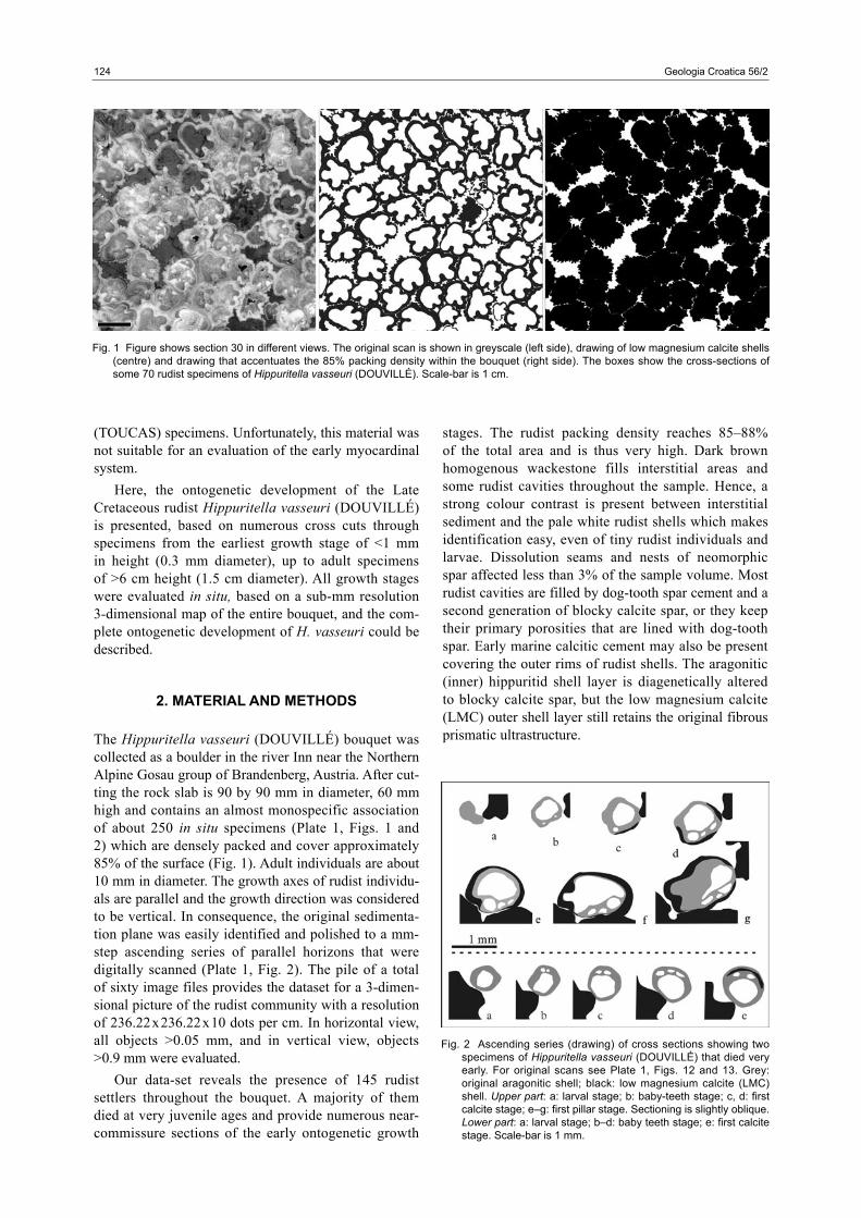
Fig. 2 Ascending series (drawing) of cross sections showing two specimens of Hippuritella vasseuri (DOUVILLÉ) that died very early. For original scans see Plate 1, Figs. 12 and 13. Grey: original aragonitic shell; black: low magnesium calcite (LMC) shell. Upper part: a: larval stage; b: baby-teeth stage; c, d: first calcite stage; e–g: first pillar stage. Sectioning is slightly oblique. Lower part: a: larval stage; b–d: baby teeth stage; e: first calcite stage. Scale-bar is 1 mm.
124 Geologia Croatica 56/2 125Götz: Larval Settlement and Ontogenetic Development of Hippuritella vasseuri...
3. ONTOGENETIC DEVELOPMENT
Based on our set of 60 high resolution scan images, we received more than 5000 cross-section pictures of rudist individuals. Naturally, most of them are 1 mm ascending serial-cuts through adult rudist specimens, from apex to free valve. But we also received numer-ous serial-cuts through individuals that died early, and near-commissure cross cuts were obtained of indi-viduals beginning from <1 mm in height and through subsequent specimens up to those achieving adult mor-phology. Beginning with the first appearence of rudist settlers in thin section or scan, seven representative growth stages are distinguished (summarized in Fig. 3), which are characterized by an increasing complex-ity of morphological features and a change in shell material composition. The complete transformation
from the larval-settling stage to adult-like morphology occurred within the first 8–10 mm of vertical growth and comprise stages 1–5. Subsequent development was dominated by the adult growth process (stage 6) and morphological continuity prior to a short lasting senior stage (stage 7) and demise.
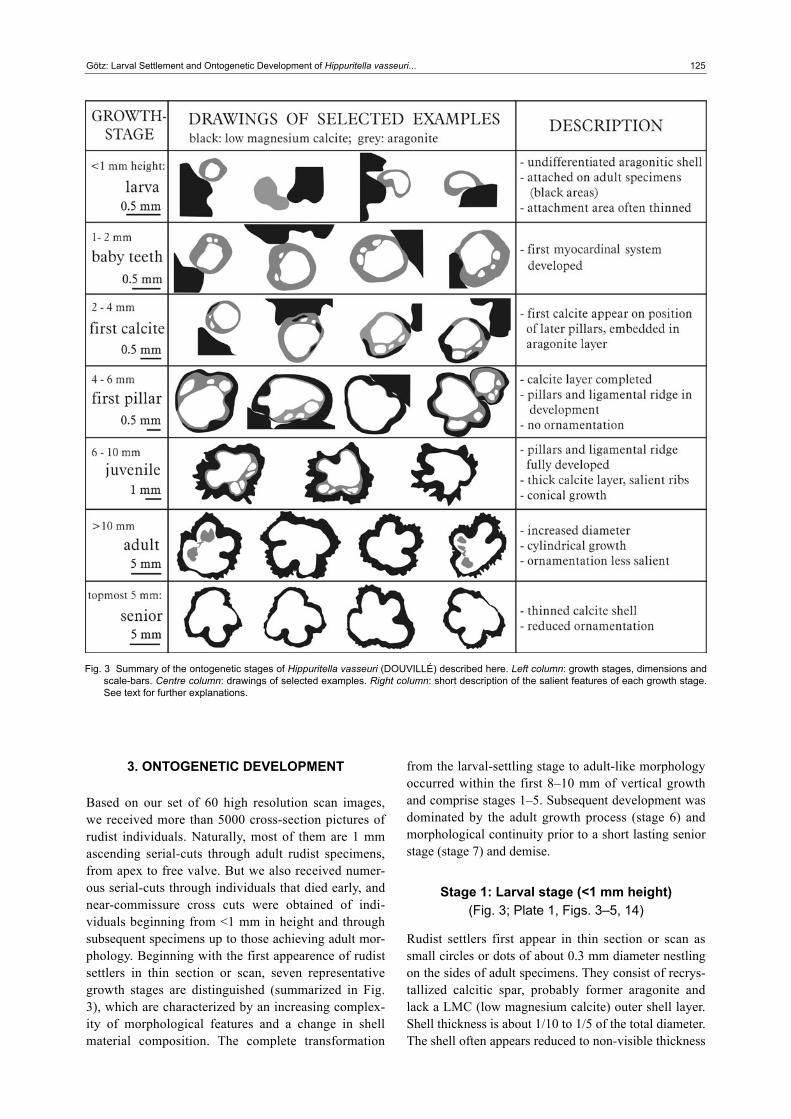
Stage 1: Larval stage (<1 mm height) (Fig. 3; Plate 1, Figs. 3–5, 14)
Rudist settlers first appear in thin section or scan as small circles or dots of about 0.3 mm diameter nestling on the sides of adult specimens. They consist of recrys-tallized calcitic spar, probably former aragonite and lack a LMC (low magnesium calcite) outer shell layer. Shell thickness is about 1/10 to 1/5 of the total diameter. The shell often appears reduced to non-visible thickness
Fig. 3 Summary of the ontogenetic stages of Hippuritella vasseuri (DOUVILLÉ) described here. Left column: growth stages, dimensions and scale-bars. Centre column: drawings of selected examples. Right column: short description of the salient features of each growth stage. See text for further explanations.
126 Geologia Croatica 56/2 127Götz: Larval Settlement and Ontogenetic Development of Hippuritella vasseuri...
at the attachment area, maybe due to an oblique cut through a spirogyrate shell shape. The shell is undiffer-entiated and shows no hinge or ligamental system. This initial aragonitic growth stage probably represents the prodissoconch of the rudist animal.
Stage 2: Baby teeth stage (1–3 mm height) (Fig. 3; Plate 1, Figs. 6–8, 15, 16)
This growth stage is characterized by a thicker, still exclusively aragonitic wall, whereas the attachment area is often thinned and first sights of a hinge system appear. The specimens are now 0.6– 0.9 mm in diam-eter. A simple developed myocardinal system is visible in thin section and scan. It is characterized by one, two or three slightly elliptical cavities that are filled by matrix. These cavities likely correspond to myocardinal elements of the left valve (i.e., posterior myophore, pos-terior tooth and anterior tooth). The earliest hinge sys-tem is already a pachydont one, and it is unlikely that a change in the organisation of myocardinal elements (e.g. from early “conservative” taxodont to pachydont) took place during the earliest ontogenetic development.
Stage 3: First calcite stage (2–4 mm height) (Fig. 3; Plate 1, Figs. 9–11)
This growth stage is characterized by the first occur-rences of calcitic shell-parts. The specimens are now 0.8–1.3 mm in diameter. The myocardinal elements of the “baby teeth” stage remain unchanged. The first signs of a secretion of low magnesium calcite (LMC) appear at the position of the later ligamental ridge and the pillars. These lense shaped LMC parts are embed-ded in the aragonitic shell and subsequently move to the external side. The transition to the next stage took place more or less during the formation of an almost closed LMC outer shell rim.
Stage 4: First pillar stage (3–7 mm height) (Fig. 3; Plate 1, Fig. 17)
This growth stage may be synonymous to the “worm stage” sensu ADKINS (1931). It is characterized by the development of the ligamental ridge and the two pillars. The diameters of the specimens now range between 1 mm and 2 mm. A closed outer LMC shell layer is now established and the ligamental ridge, as well as the pil-lars, are represented by thickenings of the outer shell layer. Later, indentations develop and mark the posi-tions of the ligamental ridge and the pillars at the exte-rior. These indentations appear slightly prior to the first elements of ornamentation.
Stage 5: Juvenile stage (6–10 mm height) (Fig. 3; Plate 1, Fig. 18)
In this stage, a strong ornamentation is apparent and the myocardinal system is fully developed. The specimens
are now between 2.5 and 5 mm in diameter. In this growth stage, the growth is conical and size increase is rapid. The former indentations are now furrows at the exterior, marking the ligamental ridge and pillars. The thickness of the LMC shell layer increased significantly and salient ribs cover the exterior of the specimens.
Stage 6: Adult stage (10–>60 mm height) (Fig. 3)
The adult stage is characterized by a change from conical to cylindrical growth. The specimens are now between 10 mm and 15 mm in diameter. The LMC shell is still thick, but the external ribs appear less salient due to the size increase of the specimens whilst the size of the ribs is still the same as in the juvenile growth stage. Most of the growth history of the rudists took place in the adult growth mode.
In this stage, many of the former settlers show a thinning of the outer shell layer at the area of contact with the substratum specimen. Interestingly, the substratum specimens also show these reductions in shell thickness at the area of contact with the settling specimen.
Stage 7: Senior stage (topmost 3–5 mm of final height)
(Fig. 3)
The senior growth stage is characterized by a reduction of the ornamentation and the thinning of the LMC outer shell layer. The specimens are between 10 mm and 15 mm in diameter. Nevertheless, the observed thinning of the outer shell layer could be a result of cross-cutting of the topmost right (attached) valve, where (in vertical section) the shell wedges out towards the commissure. This is a feature that is probably present in each growth stage of Hippuritella. Together with the observation of reduced ornamentation, however, a thinning of the outer shell layer defines fully grown rudist specimens shortly before decline.
4. DISCUSSION
The in situ evaluation of a H. vasseuri bouquet, based on serial sectioning supplied numerous sections through rudist individuals of different ontogenetic stages. Based on these cross-cuts the complete ontogenetic devel-opment of the H. vasseuri shell could be described. Although KLINGHARDT (1928, 1929); ADKINS (1931), ZAPFE (1937), VOGEL (1960), SKELTON (1974), and BRETON (1996) described juvenile rudists, their observations were limited to specimens greater than 1 mm diameter. Hence the ontogenetic develop-ment between the settlement of the larvae and the build-ing of the dissoconch still needed evaluation.
126 Geologia Croatica 56/2 127Götz: Larval Settlement and Ontogenetic Development of Hippuritella vasseuri...
Ontogenetic development and phylogenetical implications
The smallest rudist individuals evaluated here are 0.3 mm in diameter (see Plate 1, Figs. 3–5, 14) and appear in thin section and scan as small dots or circles, originally made of aragonite. They show no internal differentiation and likely represent a growth state of only days or weeks after larval settlement. Each of the evaluated 145 settlers was nestling on a neighbour´s right valve but none was found nestling on a bioclast. The observation of a preferred attachment to adult spec-imens is also reported by ZAPFE (1937) on hippuritids and by ADKINS (1931, p. 87) on radiolitids (Eoradio-lites): “...tightly attached to the substratum, usually an adult rudistid...”.
After settling, the exclusively aragonitic shell rap-idly developed the first hinge system within the first mm of vertical growth (the “baby teeth” stage). This hinge system is already comparable to the adult hinge except for the absence of ligamental or pillar structures (see Figs. 2, 3). Cross-cuts through the free valve were not obtained at this growth stage, but the presence of teeth sockets and a posterior myophore socket indicate the existence of a free valve. This agrees with obser-vations of ZAPFE (1937) who reported on a juvenile hippuritid of 2 mm in length with preserved free valve apparently devoid of a pore and canal system. Moreo-ver, the projection of the posterior myophore into a “pocket” implies a relationship with rudist groups that also show this caprotinid-type myophoral arrange-ment (SKELTON, pers. comm.). The cladistic analysis of rudist phylogeny by SKELTON & SMITH (2000) points to the Albian polyconitid Tepeyacia as a sister-group of hippuritids. The intriguing similarity of the H. vasseuri “baby teeth” myocardinal arrangement presented here with Tepeyacia described by SKELTON & SMITH (2000, p. 112) is additional evidence for this relationship.
Subsequent development of H. vasseuri is character-ized by the first appearance of LMC shell material, the “first calcite” growth stage. Lense shaped areas consist-ing of LMC appear embedded in the aragonitic shell and mark the position of the later ligamental ridge and the pillars (see Figs. 2, 3; Plate 1, Figs. 9–11). Because of this change in shell secretion and the presence of first position markers of the main adult shell features (e.g. ligamental ridge, pillars) this transition is interpreted to represent the prodissoconch/dissoconch transition in bivalve ontogeny. In the later development of H. vas-seuri the main adult morphological features evolve. The LMC shell layer is completed and covers the exterior of the shell. Pillars are still absent until a height of about 3 mm. Subsequently, first pillars evolve and appear as bulged LMC shell parts, similar to those in Tepeyacia (see SKELTON & SMITH, 2000, fig. 11b). This addi-tional morphological resemblance to the Albian polyco-nitid again implies a phylogenetic relationship between hippuritidae and Tepeyacia as supposed by SKELTON & SMITH (2000).
VOGEL (1960) recognized the presence of pillars in juvenile hippuritid specimens of more than 1.5 mm diameter and also described them as swelled LMC shell parts and not as indentations as in adult specimens. It seems that swelled LMC shell parts (“proto-pillars”) were a common morphological feature in the early growth stages of many hippuritids.
Here, H. vasseuri shows indentations (folds) later during the transition to the “juvenile stage” when the ornamentation evolves. During this stage, juveniles strongly increase in diameter, they acquire a thick LMC shell layer and develop prominent ribs (see Fig. 3; Plate 1, Fig. 18). This is presumed to reflect competition for horizontal space and oversized ribs may have helped them to claim additional horizontal space. After reach-ing the adult size of about 10 mm in diameter, further growth is cylindrical with no further increase in diam-eter. The ribs are still of the same size as in the juvenile stage, but appear now less salient because of a signifi-cantly increased diameter. Most of the former settlers now show a thinning of the outer shell layer at the area of contact with the substratum specimen. Interestingly, the substratum specimens also often show these reduc-tions in shell thickness at the area of contact with the settling specimen. This indicates a synecological in vivo interaction of both specimens. Many settlers must have caught up in growth with the substratum specimen, indicating an attachment place not too far below the commissural line. The rudist specimen is now tightly inset in the bouquet framework and cemented at least to one neighbour. After a vertical growth of several centimetres without further horizontal size-increase the specimens of H. vasseuri change morphology for the last time. ZAPFE (1937, p. 91) reported on senior hip-puritids which show reduction of ornamentation. This is confirmed here on H. vasseuri. The ribs become smooth and the outer LMC shell layer becomes thinner (Fig. 3). This is interpreted to represent the “senior stage”, a phase of reduced growth and shell secretion prior to death. Nevertheless, the observed thinning of the outer shell layer could also be a result of cross-cutting of the topmost right (attached) valve, where, in vertical section, the commissure rim shows a steep inclination towards the body cavity. This is a feature that is prob-ably present in each growth stage of Hippuritella.
Attachment and early growth
The juvenile Eoradiolites specimens described by ADKINS (1931), show a spirogyrate initial growth within the first 3.5 mm of shell diameter (the “coiled stage”). This feature was also described by SKELTON (1979) on Hippurites socialis. However, a spirogyrate initial growth mode is not clearly observable on the specimens described here. This is due to the limited vertical resolution of 1 mm, and the first 0.9 mm of (spirogyrate?) growth still needs evaluation. After the first mm, however, the prodissoconch changed the growth direction immediately to an upright position
128 Geologia Croatica 56/2 129Götz: Larval Settlement and Ontogenetic Development of Hippuritella vasseuri...
after attachment, yielding a tube-like growth mode. The second Eoradiolites growth stage of ADKINS (1931, p. 87), the “worm stage” described as “...small, smooth, worm-like, cylindrical tubes...” provides an anal-ogy to the early growth mode of H. vasseuri. Although ADKINS (1931) described his “worm stage” as the second growth stage of Eoradiolites, he considered that slender individuals or species may have only had a “worm stage” initial growth. However, this observation was probably also biased by insufficient resolution or preservation of the earliest growth stage.
5. CONCLUSIONS
The ontogenetic development of Hippuritella vasseuri (DOUVILLÉ) is described here, starting from a growth stage shortly after settling of the larva. Subsequently, from larva to decline, seven morphologically different growth stages are distinguished (Fig. 3). Within the first 6–10 mm of vertical growth all significant shell features of the adult animal are developed. The first growth stages in this 6–10 mm comprise the aragonitic larval stage and aragonitic “baby teeth” stage, the “first calcite” stage and the “first pillar” stage. The morpho-logical resemblance of H. vasseuri during these growth stages to the Albian polyconitid Tepeyacia (posterior myophore socket, “proto-pillars”) imply a phylogenetic relationship between the hippuritidae and Tepeyacia.
The prodissoconch/dissoconch transition in bivalve ontogeny is presumed to be analogous to the “baby teeth” stage/“first calcite” stage transition. Generally, H. vasseuri is highly variable in terms of the duration of each early growth stage or in the speed of growth of the early stages, respectively.
With the beginning of the development of orna-mental features (e.g., ribs) H. vasseuri bears all the adult-like shell features and stays morphologically quite stable for most of its life (e.g., the juvenile stage and the adult stage). A final morphological change is observable prior to the decline of the H. vasseuri specimens. Orna-mentation is reduced and the outer LMC shell layer is thinned, described here as the “senior growth” stage, and probably indicates a phase of reduced shell accre-tion on the brink of death.
Acknowledgements
I am grateful to Elke HERMANN, Sascha SCHMID and Stephan UNREIN for preparation, scanning and image analysis. Peter SKELTON and an anonymous reviewer provided valuable suggestions that greatly improved the final paper. Financial support was granted by the Deutsche Forschungsgemeinschaft (DFG) project GO1021/2–1.
6. REFERENCES
ADKINS, W.S. (1931): New rudistids from the Texas and Mexican Cretaceous.– The University of Texas Bulletin, 3001, 77–136.
BORN, G. (1883): Die Plattenmodellir Methode [The method of modelling with plates – in German].– Archiv mikrosko-pischer Anatomie (Breslau), 22, 584–599.
BRETON, G. (1996): Un groupe de juvéniles conservés dans la cavité palléale d´un rudiste Radiolitidae Durania blayaci (Toucas, 1909) du Cénomanien du Havre (Sei-ne–Maritime, France) [A group of juveniles, preserved in the pallial chamber of the radiolitid rudist Durania blayaci (Toucas, 1909) from the Cenomanian of Havre (Seine-Maritime, France) – in French].– C.R. Acad. Sci. Paris, 322, IIa, 493–500.
GAUNT, W.A. & GAUNT, P.N. (1978): Three dimensional reconstruction in biology.– Pitman Medical Press, Kent, 174 p.
GILI, E., MASSE, J.P. & SKELTON, P.W. (1995): Rudists as gregarious sediment-dwellers, not reef-builders, on Cre-taceous carbonate platforms.– Palaeogeography, Palaeocli-matology, Palaeoecology, 118/3–4, 245–267.
KLINGHARDT, F.I. (1928): Über sehr frühe Entwicklungs-stadien eines Rudisten [On very early ontogenetic stages of a rudist – in German].– Neues Jahrb. für Min., Geol, u. Pal., Beil. Bd. (B), 60, 173–178.
KLINGHARDT, F.I. (1929): Die stammesgeschichtliche Bedeutung, innere Organisation und Lebensweise von Eoradiolites liratus CONRAD sp. [The phylogenetical significance, internal organisation and way of life of Eoradiolites liratus Conrad sp. – in German].– Palaeonto-graphica, 72, 95–101.
PONS, J.M. & VICENS, E. (1988): Campanian and Maastrich-tian rudists from southern Valencia Province, South east Spain.– 1st Int. Conf. on Rudists, Serb. Geol. Soc. Spec. Publ., 2 [issued as reprint from volume in press].
REALI, S. (1992): Preliminary morphometric analysis for hippuritids taxonomy.– Geologica Romana, 28, 91–103.
SIMONPIETRI, G. & PHILIP, J. (2000): Relations onto-genese–phylogenese chez les rudistes; l’exemple des Hippuritidae Gray, 1848 [The ontogenic-phylogenic relationship in rudists; the Hippuritidae Gray, 1848 example – in French].– Comptes Rendus de l’Academie des Sciences, Serie II. Sciences de la Terre et des Planetes, 330/10, 717–724.
SKELTON, P.W. (1974): Aragonitic shell structures in the rudist Biradiolites, and some palaeobiological inferences.– Géologie Méditerranéenne, 1/2, 63–74.
SKELTON, P.W. (1978): The evolution of functional design in rudists (hippuritacea) and ist taxonomic implications.– Phil. Trans. R. Soc. Lond., B(284), 305–318.
SKELTON, P.W. (1979): Gregariousness and proto-coope-ration in rudists (bivalvia).– In: LARWOOD, G. & ROSEN, B.R. (eds.): Biology and Systematics of Colonial Organisms. Syst. Ass. Spec., London, New York, 257–279.
128 Geologia Croatica 56/2 129Götz: Larval Settlement and Ontogenetic Development of Hippuritella vasseuri...
SKELTON, P.W. & GILI, E. (1991): Palaeoecological classifi-cation of rudist morphotypes.– 1st Int. Conf. on Rudists, Serb. Geol. Soc. Spec. Publ., 2, 265–287.
SKELTON, P.W. & SMITH, A.B. (2000): A preliminary phylo-geny for rudist bivalves: sifting clades from grades.– In: HARPER, E.M., TAYLOR, J.D. & CRAME, J.A. (eds.): The Evolutionary Biology of the Bivalvia. Geol. Soc. Spec. Pub., 97–127.
STEUBER, T. (1996): Stable isotope sclerochronology of rudist bivalves: growth rates and Late Cretaceous seaso-nality.– Geology, 24, 315–318.
VICENS, E. (1994): Estudio de la fauna de rudistas (Hippu-ritidae y Radiolitidae) de los materiales cretácicos del Pirineo oriental: implicaciones bioestratigráficas [A study
on the rudist fauna (hippuritids and radiolitids) from Cretaceous materials of the Eastern Pyrenees: biostra-tigraphical implications – in Spanish].– Unpubl. PhD Thesis, Univ. Auton. Barcelona, Barcelona, 255 p.
VOGEL, K. (1960): Zur Struktur und Funktion der “Sipho-nalpfeiler” der Hippuriten (Lamellibranchiata) [On the texture and function of the sipho-pillars in hippuritids (Lamellibranchiata) – in German].– Paläontologische Zeit-schrift, 34/3-4, 257–294.
ZAPFE, H. (1937): Paläobiologische Untersuchungen an Hippuritenvorkommen der nordalpinen Gosauschichten [Palaeobiological investigations on hippuritid-occurren-ces of the north-Alpine Gosau-strata – in German].– Zoo-logisch–Botanische Gesellschaft Wien, 136/137, 73–121.
Manuscript received March 17, 2003.Revised manuscript accepted November 04, 2003.
130 Geologia Croatica 56/2 131
PLATE 1
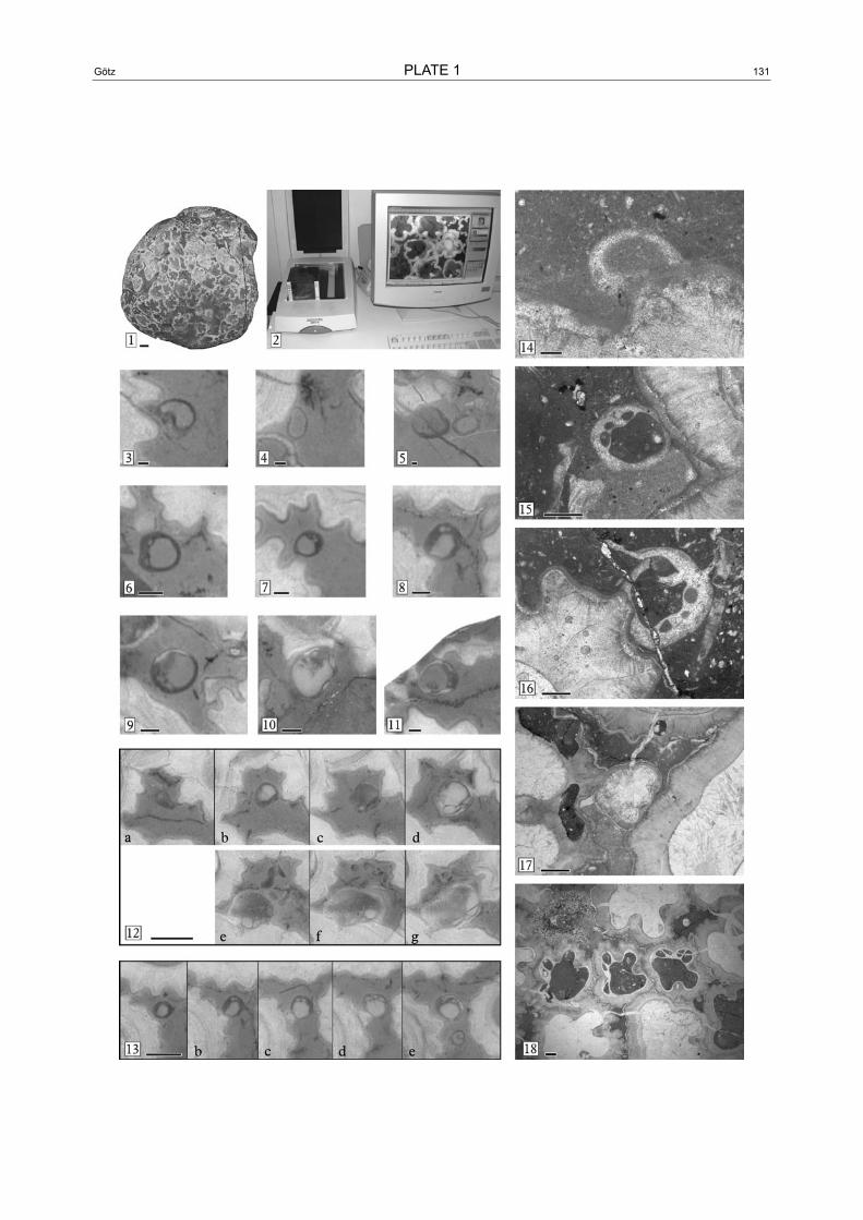
1 Original sample of the Hippuritella vasseuri (DOUVILLÉ) bouquet. Scale-bar is 1 cm.
2 Sample embedded in epoxy, resting on the scanning device. The computer monitor displays a part of the scanned horizon.
3–11 Original scans, highly magnified and therefore of low resolution. 3–5) settlers in “larval stage”, nestling on an adult shell. No internal differentiation, former aragonitic shell. Scale-bars are 0.1 mm. 6–8) specimens in “baby teeth” stage. A hinge system is developed as shown by small tooth-sockets. Scale-bars are 0.5 mm. 9–11) Specimens in “first calcite” stage. LMC shell parts appear on the position where pillars appear later. Scale-bars are 0.5 mm.
12–13 Ascending series (original scans) of cross sections showing two specimens of Hippuritella vasseuri (DOU-VILLÉ) that died very early. For fair drawings see Fig. 2. 12) a: larva stage; b: “baby-teeth” stage; c–d: “first calcite” stage; e–g: “first pillar” stage. 13) a: larval stage; b–d: “baby teeth” stage; e: “first calcite” stage. Scale-bars are 1 mm.
14–18 Thin section photomicrographs. 14) Larval stage. Scale-bar is 0.1 mm. 15–16) “baby teeth” stages. Scale-bar is 0.5 mm. 17) “first pillar” stage: pillars weakly developed, no ornamentation. Scale-bar is 1 mm. 18) Juvenile stage, thick LMC shell layer, prominent ribs. Scale-bar is 1 mm.
130 Geologia Croatica 56/2 131Götz PLATE 1

132 Geologia Croatica 56/2
Copyright © 2022 FDOKUMEN