Fascia Origin of Adipose Cells
13
Fascia Origin of Adipose Cells XUEYING SU, a YING L YU, a WEIYI WANG, a Y ANFEI ZHANG, a DANHUA LI, a SUNING WEI, a CONGKUO DU, a BIN GENG, a CAROLE SZTALRYD, b GUOHENG XU a Key Words. Superficial fascia • Fascia-derived stromal cell • Adipose precursor cell • Preadipocyte • Adipogenesis ABSTRACT Adipocytes might arise from vascular stromal cells, pericytes and endothelia within adipose tis- sue or from bone marrow cells resident in nonadipose tissue. Here, we identified adipose pre- cursor cells resident in fascia, an uninterrupted sheet of connective tissue that extends throughout the body. The cells and fragments of superficial fascia from the rat hindlimb were highly capable of spontaneous and induced adipogenic differentiation but not myogenic and osteogenic differentiation. Fascial preadipocytes expressed multiple markers of adipogenic pro- genitors, similar to subcutaneous adipose-derived stromal cells (ASCs) but discriminative from visceral ASCs. Such preadipocytes resided in fascial vasculature and were physiologically active in vivo. In growing rats, adipocytes dynamically arose from the adventitia to form a thin adi- pose layer in the fascia. Later, some adipocytes appeared to overlay on top of other adipocytes, an early sign for the formation of three-dimensional adipose tissue in fascia. The primitive adi- pose lobules extended invariably along blood vessels toward the distal fascia areas. At the lobule front, nascent capillaries wrapped and passed ahead of mature adipocytes to form the distal neovasculature niche, which might replenish the pool of preadipocytes and supply nutrients and hormones necessary for continuous adipogenesis. Our findings suggest a novel model for the origin of adipocytes from the fascia, which explains both neogenesis and expan- sion of adipose tissue. Fascial preadipocytes generate adipose cells to form primitive adipose lobules in superficial fascia, a subcutaneous nonadipose tissue. With continuous adipogenesis, these primitive adipose lobules newly formed in superficial fascia may be the rudiment of sub- cutaneous adipose tissue. STEM CELLS 2016;34:1407–1419 SIGNIFICANCE STATEMENT Adipocytes are thought to arise from vascular stromal cells, pericytes and endothelial cells within adipose tissue. Here, we identify preadipocytes in the rat superficial fascia, an uninter- rupted connective tissue that locates between skin and skeletal muscles. Fascial preadipocytes express stromal markers, are capable of spontaneous and inducible adipogenic differentiation in vitro, and are physiologically active in vivo. This finding suggests a novel model for the fascia origin of adipocytes to explain adipose neogenesis and expansion. Fascial preadipocytes gener- ate adipocytes to form primitive adipose lobules in superficial fascia, a subcutaneous nonadi- pose tissue, and with continuous expansion this lobule might naturally become a rudiment of subcutaneous adipose tissue. INTRODUCTION Postnatal development of adipose tissue com- prises two major processes: massive expansion of existing adipose tissue and de novo forma- tion of adipose tissue. Expansion occurs in cer- tain regions like the mesenteries, omenta, and between subcutaneous dermis and skeletal muscles, while de novo formation occurs at many other anatomical sites without preexist- ing adipose tissue, particularly during the onset of obesity. Because adipocytes do not divide, adipose precursor cells must exist, in adipose tissue and in certain nonadipose tis- sue, to generate new adipocytes for both expansion and neogenesis of adipose tissue. Until now, adipocytes have been thought to arise from four origins, including vascular stro- mal cells, pericytes, endothelial cells localized within adipose tissue, and hematopoietic stem cells resident in bone marrow, a nonadipose tis- sue (for reviews, see [1, 2]). Among them, adipose-derived vascular stromal cells (ASCs) have long been accepted as adipocyte progenitors inside adipose tissue [2]. A Lin 2 :CD24 1 :CD29 1 :CD34 1 :Sca-1 1 subpopulation of ASCs is capable of adipogenic differentiation a Department of Physiology and Pathophysiology, School of Basic Medical Sciences, Peking University, Beijing, China; b Department of Medicine, Division of Endocrinology, Baltimore Veterans Affairs Health Care Center School of Medicine, University of Maryland, Baltimore, Maryland Correspondence: Prof. Guoheng Xu, M.D., Ph.D., Department of Physiology and Pathophysiology, School of Basic Medical Sciences, Peking University, 38 Xueyuan Road, Beijing 100191, China. Telephone: 86-10-8280- 2916; Fax: 86-10-8280-2916; e- mail: [email protected] Received May 25, 2015; accepted for publication January 1, 2016; first published online in STEM CELLS EXPRESS February 11, 2016. V C AlphaMed Press 1066-5099/2016/$30.00/0 http://dx.doi.org/ 10.1002/stem.2338 STEM CELLS 2016;34:1407–1419 www.StemCells.com V C AlphaMed Press 2016 TISSUE-SPECIFIC STEM CELLS
-
Upload
khangminh22 -
Category
Documents
-
view
1 -
download
0
Transcript of Fascia Origin of Adipose Cells

Fascia Origin of Adipose Cells
XUEYING SU,a YING LYU,a WEIYI WANG,a YANFEI ZHANG,a DANHUA LI,a SUNING WEI,a
CONGKUO DU,a BIN GENG,a CAROLE SZTALRYD,b GUOHENG XUa
Key Words. Superficial fascia • Fascia-derived stromal cell • Adipose precursor cell • Preadipocyte •
Adipogenesis
ABSTRACT
Adipocytes might arise from vascular stromal cells, pericytes and endothelia within adipose tis-sue or from bone marrow cells resident in nonadipose tissue. Here, we identified adipose pre-cursor cells resident in fascia, an uninterrupted sheet of connective tissue that extendsthroughout the body. The cells and fragments of superficial fascia from the rat hindlimb werehighly capable of spontaneous and induced adipogenic differentiation but not myogenic andosteogenic differentiation. Fascial preadipocytes expressed multiple markers of adipogenic pro-genitors, similar to subcutaneous adipose-derived stromal cells (ASCs) but discriminative fromvisceral ASCs. Such preadipocytes resided in fascial vasculature and were physiologically activein vivo. In growing rats, adipocytes dynamically arose from the adventitia to form a thin adi-pose layer in the fascia. Later, some adipocytes appeared to overlay on top of other adipocytes,an early sign for the formation of three-dimensional adipose tissue in fascia. The primitive adi-pose lobules extended invariably along blood vessels toward the distal fascia areas. At thelobule front, nascent capillaries wrapped and passed ahead of mature adipocytes to form thedistal neovasculature niche, which might replenish the pool of preadipocytes and supplynutrients and hormones necessary for continuous adipogenesis. Our findings suggest a novelmodel for the origin of adipocytes from the fascia, which explains both neogenesis and expan-sion of adipose tissue. Fascial preadipocytes generate adipose cells to form primitive adiposelobules in superficial fascia, a subcutaneous nonadipose tissue. With continuous adipogenesis,these primitive adipose lobules newly formed in superficial fascia may be the rudiment of sub-cutaneous adipose tissue. STEM CELLS 2016;34:1407–1419
SIGNIFICANCE STATEMENT
Adipocytes are thought to arise from vascular stromal cells, pericytes and endothelial cellswithin adipose tissue. Here, we identify preadipocytes in the rat superficial fascia, an uninter-rupted connective tissue that locates between skin and skeletal muscles. Fascial preadipocytesexpress stromal markers, are capable of spontaneous and inducible adipogenic differentiationin vitro, and are physiologically active in vivo. This finding suggests a novel model for the fasciaorigin of adipocytes to explain adipose neogenesis and expansion. Fascial preadipocytes gener-ate adipocytes to form primitive adipose lobules in superficial fascia, a subcutaneous nonadi-pose tissue, and with continuous expansion this lobule might naturally become a rudiment ofsubcutaneous adipose tissue.
INTRODUCTION
Postnatal development of adipose tissue com-prises two major processes: massive expansionof existing adipose tissue and de novo forma-tion of adipose tissue. Expansion occurs in cer-tain regions like the mesenteries, omenta, andbetween subcutaneous dermis and skeletalmuscles, while de novo formation occurs atmany other anatomical sites without preexist-ing adipose tissue, particularly during theonset of obesity. Because adipocytes do notdivide, adipose precursor cells must exist, inadipose tissue and in certain nonadipose tis-
sue, to generate new adipocytes for bothexpansion and neogenesis of adipose tissue.
Until now, adipocytes have been thought toarise from four origins, including vascular stro-mal cells, pericytes, endothelial cells localizedwithin adipose tissue, and hematopoietic stemcells resident in bone marrow, a nonadipose tis-sue (for reviews, see [1, 2]). Among them,adipose-derived vascular stromal cells(ASCs) have long been accepted as adipocyteprogenitors inside adipose tissue [2]. ALin2:CD241:CD291:CD341:Sca-11 subpopulationof ASCs is capable of adipogenic differentiation
aDepartment of Physiologyand Pathophysiology, Schoolof Basic Medical Sciences,Peking University, Beijing,China; bDepartment ofMedicine, Division ofEndocrinology, BaltimoreVeterans Affairs Health CareCenter School of Medicine,University of Maryland,Baltimore, Maryland
Correspondence: Prof. GuohengXu, M.D., Ph.D., Department ofPhysiology and Pathophysiology,School of Basic MedicalSciences, Peking University, 38Xueyuan Road, Beijing 100191,China. Telephone: 86-10-8280-2916; Fax: 86-10-8280-2916; e-mail: [email protected]
Received May 25, 2015;accepted for publication January1, 2016; first published online inSTEM CELLS EXPRESS February 11,2016.
VC AlphaMed Press1066-5099/2016/$30.00/0
http://dx.doi.org/10.1002/stem.2338
STEM CELLS 2016;34:1407–1419 www.StemCells.com VC AlphaMed Press 2016
TISSUE-SPECIFIC STEM CELLS

in vitro, as well as in vivo in A-Zip lipodystrophic mice [3]. Fur-thermore, a subset of ASCs sharing pericyte markers has beenidentified to process adipogenic differentiation [4–6], such ASCsare localized within the vasculature of adipose tissue, but not innonadipose tissues like the skeletal muscles and kidneys [6]. Incontrast, the endothelial origin of preadipocytes remains contro-versial. It is reported that adipocytes might arise from vascularendothelium of adipose tissue [7], but another study arguesthat those endothelial cells do not contribute to the adipose lin-eage in vivo [4].
Given that adipogenic progenitors within vascular stromalcells, pericytes and endothelial cells are found only in existingadipose tissue, these mechanisms of adipocyte origination canonly explain expansion of preexisting adipose tissue, but can-not explain adipose neogenesis at locations where primitiveadipose tissue and its vasculature have not yet occurred.Therefore, the search for nonadipose-resident adipogenic pro-genitors has long been an area of interest since the last cen-tury [1]. One proposed candidate is bone marrow stem cells[1, 2]. Circulating hematopoietic progenitors could migrateinto adipose tissue and differentiate into multilocular adipo-cytes [8, 9]. However, other studies suggest that hematopoi-etic progenitors may only trans-differentiate into adipocyte-like cells with tiny lipid droplets in vitro, but not in vivo [10,11]. Also, it is unclear how hematopoietic progenitors couldhome selectively to adipose tissue or sites of adiposeneogenesis.
Considering anatomy and histology, we speculate thatadipogenic progenitors exist in certain nonadipose tissues,where they can generate adipose cells de novo forming aprimitive adipose tissue. Unlike circulating hematopoieticprogenitors, such adipogenic progenitors may be associatedwith a connective tissue distributed throughout the body,providing a capacity for continuous adipogenesis whenrequired. In this investigation, we identified a precursor ofadipose cells from superficial fascia, a nonadipose connectivetissue with a widespread distribution, similar to that of adi-pose tissue.
Fascia is a continuous framework of loose connective tis-sue that extends throughout the body to envelop and sepa-rate all organs, tissues and their inside cells [12]. Superficialfascia locates at the interface of subcutaneous dermis andskeletal muscles, anatomic locations where subcutaneous fatmay occur subsequently. The superficial fascia in juvenile ratshad almost no adipocytes and was easily separable, thus, wasan ideal material for investigating nonadipose-resident preadi-pocytes. We identified that superficial fascia-derived stromalcells (FSCs) comprised abundant lineage-committed preadipo-cytes capable of spontaneous and induced adipogenic differ-entiation. We found that fascial preadipocytes expressedmultiple markers of adipogenic progenitors, were localized inthe fascial vasculature, and were physiologically active in vivo.During growth of rats, early differentiated and fully differenti-ated adipocytes dynamically arose to form primitive adiposelobules in superficial fascia. Our findings provide the first evi-dence for a fascia origin of adipocytes. This new model foradipogenesis suggests that fascial preadipocytes generate adi-pose cells to form a primitive adipose lobule in subcutaneoussuperficial fascia, which may be a rudiment of subcutaneousadipose tissue.
MATERIALS AND METHODS
Animals
Sprague-Dawley male rats were fed standard chow diet anddecapitated at 1–7 weeks of age on demand. In each individ-ual experiment, samples were obtained from at least threerats but usually from more than five rats. Animal experimentswere conducted in accordance with the NIH guidelines for thecare and use of laboratory animals and approved by the ani-mal care and utilization committee of Peking University HealthScience Center.
Separation of the Fascia
The superficial fascia located between the dermis and hind-limb skeletal muscle groups, such as rectus femoris, adductorlongus, vastus medialis, and lateralis muscles, was lifted upusing surgical tweezer and then was cutoff by a scissor. Carewas taken to avoid contamination of superficial fascia samplesfrom the deep fascia, epimysial, and perimysial fasciae of theskeletal muscle. Superficial fasciae at other anatomic sitessuch as the forearm and abdomen were also separated. Forpurpose of comparison, the pericardium fascia of the heartand the fascial capsules of the kidney, liver, stomach, and tes-tis were separated.
Histology and Immunostaining of Whole-MountedFasciae
Fascial sheets were whole-mounted onto glass slides, air-dried, and fixed with 4% paraformaldehyde for 30 minutesand rinsed with PBS. For routine histology, fascial tissue wasstained with hematoxylin-eosin (H&E). Collagen-elastic fiberswere stained with 0.5% Victoria Blue in acid alcohol and 0.5%Ponceau S in 1% picric acid. For immunostaining, fascial tissuewas treated with 0.1% Triton X-100, blocked with 1% defattedalbumin, incubated at 48C overnight with antibodies toperilipin-1, CD24, CD29, or CD31, and then probed for 1 hourwith lgG (1:1000) conjugated to TRITC or FITC. Cell nucleiwere stained with Hoechst 33258 (Applygen Technologies,Beijing, China; URLs: http://www.applygen.com). Fluorescentsignals were captured under confocal microscope (Leica TCSSP8, Wetzlar, Germany; URLs: http://www.leica-microsystems.com) or Nikon Eclipse 50i microscope.
Oil Red and Nile Red Staining
Adipocytes in culture and in whole-mounted fascia werestained with 0.5% Oil Red for 15 minutes, or with 1 mg/mlNile Red for 20 minutes.
Isolation and Adipogenic Differentiation of FSCs
Fascia was minced to small pieces and digested for 1 hourwith 0.8mg/ml type I collagenase in DMEM at 378C, withshaking at 120 cycles/minutes. Digestive mixture was filtratedthrough a 100# steel mesh and centrifuged at 1,000 g for10min. FSCs pellets were collected and cultured in DMEMwith high glucose (4.5 g/l) and 10% FBS at 378C in 5% humidi-fied CO2. FSCs appeared confluent after cultured for 1 week,with changing fresh medium every 2 days. Then, FSCs wereinduced for 2 days with adipogenic differentiation cocktail(10 mg/ml insulin, 250mM isobutylmethylxanthine, and 0.1 mMdexamethasone, and 10% FBS in high glucose DMEM. At day
1408 Fascia Origin of Adipocytes
VC AlphaMed Press 2016 STEM CELLS

3, cells were maintained for 4 days in high glucose DMEMcontaining 5mg/ml insulin and 10% FBS.
Isolation and Adipogenic Differentiation of Adipose-Derived Stromal Cells
Adipose-derived stromal cells were isolated from epididymal oringuinal fat pads of male rats according to our laboratorymethod [13, 14]. Fat pads were minced and digested on ashaker at 378C in DMEM containing 0.8mg/ml collagenase typeI, 1% defatted albumin, 200nM adenosine, 25mM Hepes,pH7.2. Digestive mixture was filtered through an 80# and then400# steel mesh and then centrifuged at 800 g for 10min.ASCs pellets were collected and cultured in DMEM/F-12 (1:1)containing 10% FBS, at 378C in 5% humidified CO2. Adipogenicdifferentiation of confluent ASCs was induced for 2 days with5mg/ml insulin, 33mM biotin, and 200 pM triiodothyronine inserum-free DMEM/F-12. At day 3, cells were transferred toserum-free DMEM/F-12 and maintained for 4 days.
Flow Cytometry
According to our laboratory method [15], FSC and ASC cellswere cultured for 1 week and collected at 80%–90% conflu-ence at passage zero. Cells were stained with antibodiesagainst adipose progenitor markers [2, 3]: CD24-PE, CD29-Alexa Fluor 647, CD31-Alexa Fluor 488, CD44-FITC, CD45-PE-Cy7, CD90-V450, and CD106-PE. To set proper compensationand population gates, cells probed with single antibody alonewere designed as positive control and cells probed with fluo-rescent isotype-matched IgG were negative controls. Afterstaining for 30 minutes on ice, the cells were analyzed on aNavios 3-laser 10-color flow cytometer (Beckman). Three inde-pendent samples were analyzed for each measurement.
Localization of Proliferative FSCs and Preadipocytes inFasciae
For detection of proliferative FSCs, 10 rats (90–100 g) were i.p.injected with 5-ethynyl-2-deoxyuridine (EdU, 2mg/g bw) onceevery 2 days for 10 days. Control rats were injected with PBS.Rats were killed 12 hours after the last injection, and then fas-ciae were sampled and whole-mounted. EdU assay was per-formed according to the manufacturer instruction (RiboBio,Guangzhou, China). For detection of preadipocytes, whole-mounted fasciae were immunostained with CD24 or CD29.Early-differentiated adipocytes in whole-mounted fasciae wereimmunostained with perilipin-1 or recognized by H&E staining.
Osteogenic and Myogenic Differentiation
FSCs and bone marrow stromal cells were cultured for 2weeks, and medium was freshly changed every 2 days. Osteo-genic differentiation was induced by addition of 10 nM dexa-methasone, 2mM b-glycerophosphate, and 50mg/mlascorbate-2-phosphate. Positive osteocytes were stained with0.5% Alizarin Red. For myogenic differentiation, cells wereinduced for 2 weeks in DMEM with 10% FBS, 5% horseserum, and 50mM hydrocortisone. Myogenic cells were immu-nostained with primary antibody against desmin, an interme-diate filament protein of muscle.
Real-time Quantitative reverse transcription-polymerase chain reaction (RT-PCR) and Sources ofAntibodies
The methods for the assay of RT-PCR and primers, and sour-ces of antibodies, are described in Supplemental section.
Notes
All FSCs and ASCs cells were used at passage zero, without subcul-ture. Whole mounted fasciae were used as specimens for all mor-phological and immunohistochemistrical examinations of fascia.
RESULTS
Preparation and Morphological Observation of the RatSuperficial Fascia
The superficial fascia is a continuous layer of loose connectivetissue located at the interface between the subcutaneous dermisand skeletal muscles. The superficial fascia in vivo appearedsemitransparent and viscoelastic, and was easily separable fromthe deep fascia and muscle epimysium (Fig. 1A). After dissection,the fascia had a pale and jelly like appearance (Fig. 1B). Oil Redonly stained the boundary regions of fascia isolated from 1-week-old rats, a nonspecific stain because the edge was alwaysfolded back. The areas beyond the border were unstained, indi-cating scarce or no adipocytes resident in the fascia (Fig. 1C).For microscopic observation (see Fig. 1D, 1F), the fascia sheetswere whole-mounted on slides. Oil Red stain showed few earlydifferentiating adipocytes with small cytoplasmic lipid dropletslocalized near small blood vessels (Fig. 1D). H&E stain (Fig. 1E)and Victoria blue-Ponceau stain (Fig. 1F) showed that the fasciaconsisted of small blood vessels, abundant collagens, and elasticfibers, fibrocystes, and some mast cells in a thin substrate.
Spontaneous Adipogenic Differentiation of FSCs andFascial Tissue Fragments
We cultured fascial tissue pieces and isolated FSCs in standardDMEM media without adipogenic inducers. The cells grownfrom fascial tissue fragments exhibited some cytoplasmicvacuoles at day 3, but spontaneously accumulated large lipiddroplets in the cytoplasm after day 6 (Fig. 2A), indicating an exvivo spontaneous adipogenesis of the fascial tissue. IsolatedFSCs adhered to cell culture plates within 48 hours and slowlyreached full confluence, typically after 1 week. They exhibitedfibroblast-like morphology. Another cytological feature was thatFSCs harbored many large cytoplasmic vacuoles at day 3, butthese vacuoles were not lipid droplets because they were notstained by Oil Red (Fig. 2B). Interestingly, lipid droplets seemedto occur later in the space of the vacuoles, because somevacuoles became gradually stained by Oil Red from day 6 today 9 (Fig. 2B). At day 12, FSCs owned a classic phenotype ofadipocytes featuring multiple large lipid droplets in the cyto-plasm (Fig. 2B). Differentiated FSC-adipocytes versus total cellnuclei stained by Hoechst 33258, indicating that 25%–45% oftotal FSCs were able to spontaneously differentiate into adipo-cytes, although the differentiation rate varied slightly frombatch to batch of cell preparations (data not shown). ThemRNA expression of peroxisome proliferator-activated receptor-g (PPARg), a key adipogenic transcription factor, graduallyincreased in FSCs during spontaneous adipogenic differentiation
Su, Lyu, Wang, et al. 1409
www.StemCells.com VC AlphaMed Press 2016

from day 3 to day 12 (Fig. 2C). Fatty acid binding protein 4(also termed aP2) is an early marker of adipocyte differentia-tion and comprises �6% of total protein in mature adipocytes[16]. Perilipin-1 coats lipid droplets exclusively in adipocytes[17–19] and participates in the control of adipocyte differentia-tion [15]. The mRNA expression of FABP4 and perilipin-1 mod-erately increased during day 3 to day 9, but increased sharplyat day 12, dynamically corresponding to spontaneous adipo-genic differentiation of FSCs (Fig. 2C). Clearly, these resultsshow that fascia cells are highly capable of spontaneous adipo-genesis in vitro and ex vivo.
Fascia-Derived Stromal Cells are Lineage-CommittedPreadipocytes
After myogenic and osteogenic induction, FSCs failed to developphenotypes of myocytes (Fig. 3A) or osteocytes (Fig. 3B). In con-
trast, bone marrow-derived stromal cells, as a positive controlcell, were induced to differentiate into desmin-immunoreactivemyocytes (Fig. 3A) or into osteocytes stained by Alizarin red(Fig. 3B). Under adipogenic conditions (Fig. 3C), adipogenic dif-ferentiation of FSCs was faster and more efficient than sponta-neous differentiation (Fig. 3C). After a 1-week induction, 70%–90% FSCs fully differentiated into adipocytes, with many largelipid droplets positively stained by Oil Red and immunostainedfor perilipin-1 (Fig. 3C). Morphologically, the number and size oflipid droplets in FSC adipocytes seemed less uniform than therelatively homogeneous lipid droplet clusters in adipocytes dif-ferentiated from epididymal ASCs (Fig. 3D). The mRNA expres-sions of PPARg and its downstream adipogenic targets, such asfatty acid synthase, hormone-sensitive lipase, adipose triglycer-ide lipase, and perilipin-1, were increased on induction of adipo-genic differentiation (Fig. 3E). These observations suggest that
Figure 1. Preparation and morphology of the superficial fascia. Male rats at 1 week old were used. (A): The gross anatomy of thesuperficial fascia (asterisk) localized above the hindlimb vastus medialis muscle. Below the superficial fascia were the saphenous artery(a) and vein (v) and saphenous nerve (n) in the deep fascia and muscle epimysium. (B): Stereomicroscopic view of freshly dissected fas-cia. (C): Stereomicroscopic view of the fascia stained with Oil Red. Most regions of the fascia sheet had no adipocytes. Nonspecific stainappeared at border regions because the fascia sheet was folded. (D): Microscopic view of whole-mounted fascia stained with Oil Redand hematoxylin. A few early-differentiating adipocytes laden with small lipid droplets (arrows) were seen near a blood vessel (bv). (E):H&E stain showed vessels and fibrocystes in whole-mounted fascia. (F): Victoria Blue-Ponceau stain of whole-mounted fascia. The typicalcomponents of fascia were the blood vessels (bv), collagenous (red) and elastic fibers (f, blue threads), fibrocystes, mast cells (big bluecells) and ground substrate.
1410 Fascia Origin of Adipocytes
VC AlphaMed Press 2016 STEM CELLS

adipose precursor cells in fascia are lineage-committed preadi-pocytes capable of spontaneous and induced adipogenesis.
Fascial Cells from Different Species and Organs ExhibitDifferent Adipogenesis Capacity
In rats, FSCs isolated from superficial fasciae of skeletal musclesof the hindlimb (Supporting Information Fig. S1A) and forearmor abdomen (data not shown) can efficiently differentiate intoadipocytes after induction. In contrast, FSCs from the pericar-dial fascia (heart) only moderately differentiated into adipo-cytes. Only a few FSCs from fascial capsules of the kidney andtestis were found to differentiate, while FSCs from fascial cap-sules of the liver and stomach failed to differentiate. To exam-ine species dependence of differentiation, superficial fasciacells from porcine abdomen muscles were tested and foundcapable of both spontaneous (data not shown) and induced
differentiation into adipocytes (Supporting Information Fig.S1B). Surprisingly, FSCs derived from hindlimb superficial fas-ciae of mice failed to differentiate into adipocytes typicallyladen with large lipid droplets. Instead, mouse FSCs formedonly very tiny lipid droplets that did not further grow duringadipogenic induction (Supporting Information Fig. S1B). Quanti-tative RT-PCR assay showed that CD24 and CD29, surfacemarkers of adipose progenitor cells [2], were only highlyexpressed in FSCs in the superficial fascia close to the skeletalmuscle but not in capsular fasciae of the heart, kidney, testis,and stomach in rats (Supporting Information Fig. S1C).
Cytological Phenotypes of the FSCs
As assayed by quantitative RT-PCR, the surface markers ofmural cells (vascular pericytes and smooth muscle cells), suchas CD146, a-smooth muscle actin (a-SMA) and platelet
Figure 2. Spontaneous adipogenic differentiation of fascial cells. Isolated cells and tissue were cultured in standard DMEM containinghigh glucose and 10% FBS but without adipogenic inducers. (A): Small fragments of superficial fascia were cultured up to 12 days. Fas-cial cells outgrown from fascia fragments formed abundant cytoplasmic vacuoles (arrow) at day 3, and thereafter lipid droplets (arrow-heads) occurred and gradually increased in size and number. During day 9 to 12, the outgrown fascial cells spontaneously accumulatedmany large lipid droplets, demonstrating a typical phenotype of differentiated adipocytes under light microscope. (B): Fascia stromalcells (FSCs) were isolated, cultured and stained with Oil Red. FSCs formed many cytoplasmic vacuoles (arrow) at day 3 and thereafterlipid droplets gradually accumulated. At day 12, 25%–45% (data not shown) of total FSCs spontaneously differentiated into adipocytesladen with large lipid droplets. (C): Real-time RT-PCR assay of gene expression during spontaneous adipogenic differentiation from 3 to12 days. Abbreviations: PPARg, peroxisome proliferator-activated receptor g; FABP4, fatty acid binding protein 4; PLIN1, perilipin-1.
Su, Lyu, Wang, et al. 1411
www.StemCells.com VC AlphaMed Press 2016

derived growth factor receptor b (PDGFRb), were notdetected in primary FSCs. In contrast, these markers, espe-cially a-smooth muscle actin, were relatively high expressedin subcutaneous ASCs (Fig. 4A). In flow cytometry assays, FSCsrarely expressed hematopoietic marker CD45 (0.03%) or endo-thelial maker CD31 (0.44%; Fig. 4B). The FSC fraction containedonly a small subpopulation of cells that expressed CD24, but aconsiderable subpopulation of cells expressing CD29 (99.4%),CD44 (54.4%), CD90 (88%), or CD106 (34%) (Fig. 4B). These fivemarkers are known to label stromal adipogenic progenitors [2]. Incomparison, FSCs, subcutaneous ASCs and epididymal ASCs had alarge percentage of CD291 or CD441 cells. However, FSCs con-tained a high population of CD901 cells but a low percentage ofCD1061 cells, similar to subcutaneous ASCs but inverse with epi-didymal ASCs (Fig. 4B). Multiple-color flow cytometry (Fig. 4C-4E)indicated that considerable amounts of FSCs simultaneouslyexpressed multiple markers of adipose progenitor cells. TheCD452:CD312:CD291:CD901 subpopulation was 86.2%,
71.2%, and 25.3% in fractions of FSCs, subcutaneous ASCs andepididymal ASCs, respectively (Fig. 4D, 4E). TheCD452:CD291:CD441:CD901 subpopulation was 94.2%,77.6%, and 19.7%, and the CD452:CD291:CD441:CD901:CD1061 subpopulation was 28.3%, 27.6%, and 16.7% infractions of FSCs, subcutaneous ASCs and epididymal ASCs,respectively (Fig. 4D, 4E). These data indicate that FSCs containmany adipose progenitors, whose cytological characteristics aresimilar to subcutaneous ASCs but different from visceral ASCs.Because of the low yield of primary FSC preparations, and the sig-nificant loss of cells after flow cytometry, we failed to identify adi-pogenic subpopulations by sorting FSCs.
Localization of Preadipocytes in Fascia
Despite lacking definite markers of fascia stromal cells, weattempted to evaluate localization of preadipocytes within fascia.First, considering that progenitor cells, including preadipocytes,are a result of a proliferative status, the positions of proliferative
Figure 3. FSCs were lineage-committed preadipocytes. Isolated FSCs were cultured under different differentiation inductions. FSCswere not capable of myogenic (A) or osteogenic (B) differentiation. As controls, bone marrow mesenchymal stromal cells (BMSCs) weredifferentiated into myocytes immunolabeled with desmin (A) or osteocytes stained by Alizarin red (B). On adipogenic induction (C–E),FSCs were efficiently differentiated into adipocytes laden with many large lipid droplet labeled by Oil Red and perilipin-1 (C). Morpho-logically, lipid droplets in FSC-adipocytes appeared less uniform than relatively homogeneous lipid-droplet clusters of adipocytes differen-tiated from epididymal adipose-derived stromal cells (Epi ASCs) (D). The mRNA expressions (E) of PPARg, fatty acid synthase (FAS),hormone-sensitive lipase (HSL), adipose triglyceride lipase (ATGL), and perilipin-1 (PLIN1), were increased in FSCs, epididymal (Epi) andinguinal (Ing) ASCs on adipogenic induction. Nuclei were counterstained by Hoechst 33258 (blue).
1412 Fascia Origin of Adipocytes
VC AlphaMed Press 2016 STEM CELLS

cells should partially reflect preadipocyte locations in vivo. Wedetermined the positions of proliferative cells in whole-mountedfasciae from EdU-injected rats. Two types of proliferative cellswere integrated EdU in vivo in fascia, one with rod-like nucleilocalized along main blood vessels and another with oval-like
nuclei distributed sporadically near blood capillaries or aroundmature adipocytes (Fig. 5A, 5B). Second, whole-mounted fas-ciae were immunolabeled with CD24 and CD29, surfacemarkers of adipose precursors [2, 3]. CD24-positive cells wererelatively small and spindly, with rod-like nuclei, and
Figure 4. Cytological phenotypes of the FSCs. (A): Quantitative RT-PCR. Vascular pericyte markers CD146, a-smooth muscle actin (a-SMA) and platelet derived growth factor receptor b (PDGFRb) were undetectable in primary FSCs, but were highly expressed in inguinal(Ing) ASCs. (B–E): Flow cytometry. Rat FSCs, inguinal (Ing) and epididymal (Epi) ASCs at passage zero were analyzed by 10-color flowcytometry. Cells were immunostained with individual (B) or multiple (C-E) antibodies against indicated stromal markers. The adipose pro-genitor population was gated (in box) and further separated into different populations coexpressing multiple markers (C). In fractions ofFSCs, Ing ASCs and Epi ASCs, the percentage yields of adipocyte progenitor populations positive for single (B) or multiple (D,E) markerswere denoted and compared.
Su, Lyu, Wang, et al. 1413
www.StemCells.com VC AlphaMed Press 2016

distributed along inferior blood vessels as well as aroundmature adipocytes (Fig. 5C). CD29-positive precursorsappeared relatively larger and longer, with oval-like nuclei,and were distributed sporadically in fascia (Fig. 5D).
Third, considering that preadipocytes are nonmigratory atonset of differentiation, in reverse order the positions of early
differentiating adipocytes in situ in fascia may reflect the loca-tions of preadipocytes at onset of differentiation. Thus, weimmunolabeled early differentiated adipocytes with perilipin-1, which coats lipid droplets exclusively in adipocytes [17–19]and is an early marker of adipocyte differentiation [15]. Manyearly differentiating adipocytes laden with multiple small lipid
Figure 5. Localization of preadipocytes in the rat fascia. Images show whole-mounted superficial fasciae from the hindlimb of adultmale rats (bw, 902100 g). Boxed regions were amplified and showed aside. (A, B): Two types of proliferative cells existed in superficialfasciae from EdU-injected rats (n5 5), one with rod-like nuclei (solid box) localized along main blood vessels (bv) and another one withoval-like nuclei (dash box) distributed sporadically near blood capillaries and mature adipocytes (ad). (C): CD24-immunolabeled precur-sors were relatively small and thin, with rod-shaped nuclei, and localized along inferior blood vessels or around adipocytes. (D): CD29-immunolabeled precursors appeared relatively larger and longer, with oval-like nuclei, and were distributed sporadically in fascia. (E–G):The distribution pattern of early differentiating adipocytes might mirror the last locations of fascial preadipocytes before onset of differ-entiation. Early differentiating adipocytes with multiple small lipid droplets immunolabeled with perilipin-1 (E) were localized near smallblood vessels. Blood vessels were seen in the shadows of nonspecific stains (E) or labeled by vascular marker CD31 (F). H&E stain (G)showed a clearer map of early differentiating adipocytes and their adjacent structures in thick slices of whole-mounted fascia. Lipiddroplets appeared in white vacuoles after H&E staining. Many early differentiating adipocytes with multiple small lipid droplets andlately-differentiating adipocytes with relatively large lipid droplets were localized along small blood vessels or distributed sporadically inthe inferior vasculature network of fascia. Many early differentiating adipocytes had near round or oval-like nuclei, similar to EdU-labeled proliferative cells (A,B) and CD29-positive cells (D) with oval-like nuclei, distinguishable from vascular cells and fibrocytes withfusiform-shaped nuclei in fascia (G). Abbreviations: EdU, 5-ethynyl-2-deoxyuridine; bv, blood vessel; ad, mature adipocyte.
1414 Fascia Origin of Adipocytes
VC AlphaMed Press 2016 STEM CELLS
droplets occurred sporadically or near small blood vessels,shown by background stain (Fig. 5E) and CD31 stain (Fig. 5F).Furthermore, H&E staining allowed mapping of early differen-tiating adipocytes and relationship to adjacent structures inthick slices of whole-mounted fascia. Clearly, a fair number ofearly differentiating adipocytes with multiple small lipid drop-lets and fully differentiating adipocytes with relatively largelipid droplets were localized along small blood vessels, or dis-tributed sporadically in the inferior vasculature of fascia (Fig.5G). Interestingly, early differentiating adipocytes usuallyshowed a near round or oval-like nucleus (Fig. 5G), similar toEdU-labeled proliferative cells with oval-like nuclei but differ-ent from vascular cells and fibrocytes with fusiform-shapednuclei (Fig. 5A, 5G). We cannot identify whether early differ-entiating adipocytes with oval-like nuclei were derived frompreadipocytes with similar oval-like nuclei, but localization ofearly differentiating adipocytes might mirror the last locationof preadipocytes before differentiation.
Dynamic Development of Fascia AdiposeCells in Fascia
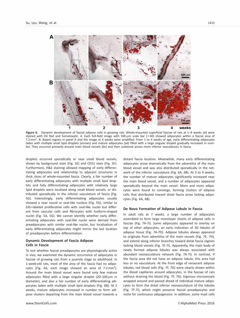
To test whether fascial preadipocytes are physiologically activein vivo, we examined the dynamic occurrence of adipocytes infasciae of growing rats from a juvenile stage to adulthood. In1-week-old rats, most of the area of the fascia had no adipo-cytes (Fig. 6A, each image showed an area of 7.2mm2).Around the main blood vessel were found only few matureadipocytes filled with a large singular droplet (20–100mm indiameter), and also a fair number of early differentiating adi-pocytes laden with multiple small lipid droplets (Fig. 6B). At 2weeks, mature adipocytes increased in number to form adi-pose clusters departing from the main blood vessel towards a
distant fascia location. Meanwhile, many early differentiatingadipocytes arose dramatically from the adventitia of the mainblood vessel and was also distributed sporadically in the net-work of the inferior vasculature (Fig. 6A, 6B). At 3 to 4 weeks,the number of mature adipocytes significantly increased nearthe main blood vessel, and a number of adipocytes appearedsporadically beyond the main vessel. More and more adipo-cytes were found to converge, forming clusters of adiposecells that distributed toward distal fascia areas lacking adipo-cytes (Fig. 6A, 6B).
De Novo Formation of Adipose Lobule in Fascia
In adult rats at 7 weeks, a large number of adipocytesassembled to form large monolayer sheets of adipose cells infasciae (Fig. 7A-7I). Some adipocytes appeared to overlay ontop of other adipocytes, an early indication of 3D lobules ofadipose tissue (Fig. 7A-7D). Adipose lobules always appearedto originate from adventitia of the main vessels (Fig. 7E, 7H),and extend along inferior branches toward distal fascia regionslacking blood vessels (Fig. 7E-7I). Apparently, the main body ofnewly formed adipose lobules was always associated withabundant neovasculature network (Fig. 7A-7I). In contrast, ifthe fascia area did not have an adipose lobule, this area hadless or no vasculature. At the front edge of renascent adiposelobules, red blood cells (Fig. 7F, 7G) were clearly shown withinthe blood capillaries around adipocytes, in the fasciae of ratswithout draining the blood (Fig. 7F, 7G). Vigorous microvesselswrapped around and passed ahead of individual mature adipo-cytes to form the distal inferior neovasculature of the lobules(Fig. 7F-7I), which might preserve fascial preadipocytes andniche for continuous adipogenesis. In addition, some mast cells
Figure 6. Dynamic development of fascial adipose cells in growing rats. Whole-mounted superficial fasciae of rats at 1–4 weeks old werestained with Oil Red and hematoxylin. A. Each full-field image with 500-mm scale bar (340) showed adipocytes within a fascial area of7.2mm2. B. Boxed regions in panel A and the image at 4 weeks were amplified. From 1 to 4 weeks of age, early differentiating adipocytesladen with multiple small lipid droplets (arrows) and mature adipocytes (ad) filled with a large singular droplet gradually increased in num-ber. They occurred primarily around main blood vessels (bv) and then scattered across more inferior vasculatures in fascia.
Su, Lyu, Wang, et al. 1415
www.StemCells.com VC AlphaMed Press 2016

appeared in the vasculature of adipose lobules (Fig. 7H, 7I),but this phenomenon was not investigated.
DISCUSSION
As opposed to human superficial fascia, which tightly interdi-gitates subcutaneous fat [20, 21], superficial fascia in juvenilerats had no or few adipocytes, and was easily separable fromdeep fascia and epimysium of the skeletal muscle, making itan ideal material for investigating nonadipose-resident preadi-pocytes. Rat superficial fascia had abundant committed-
preadipocytes capable of spontaneous and induced adipogenicdifferentiation but not myogenic and osteogenic differentia-tion. Fascial preadipocytes expressed multiple surface markersof stromal adipogenic precursors, localized in the fascial vas-culature, and were physiologically active in vivo. Duringgrowth, early differentiating and fully differentiating adipo-cytes dynamically arose from preadipocyte-resident vascula-tures, and appear to gradually assemble to form primitiveadipose lobules in fascia. At the lobule front, nascent micro-vessels wrapped around and passed ahead of mature adipo-cytes to form the distal neovasculature, which might replenisha pool of preadipocytes and preserve a niche for supplying
Figure 7. Formation of adipose lobule in fascia. Superficial fasciae from 7-week-old rats were whole-mounted. Adipose lobules newlyformed in fascia were stained by Nile Red (A, B) and Oil Red (C, D). Nuclei were stained with Hoechst 33258. Some adipocytes (ad)appeared to overlay on top of other adipocytes (B, D, boxes), indicating the formation of three-dimensional adipose tissue (A–D). H&Estain (E–G) and Victoria blue-Ponceau S stain (H, I) showed that adipose lobules originated from the adventitia of the main blood vessel(E, H) and extended along abundant inferior branches. At the front of adipose lobules, capillary vessels wrapped and passed ahead ofmature adipocytes to form a distal neovasculature niche (F, G, I). Red blood cells (RBC) were clearly seen within blood capillaries aroundmature adipocytes in fasciae of rats killed without draining the blood from the body (F, G). Histological relationships of microvessels(bv), elastic fibers (f, blue threads), mast cells (blue cells), and adipocytes (ad) in fascia were shown (E–I). In the final box (J), we pro-pose a schematic model of the fascia origin of adipose cells, explaining both adipose neogenesis and expansion (J). Adipose precursorcells reside in the vasculature of fascia and are physiologically active. During postnatal growth, adipose cells gradually arise from fascialpreadipocytes to form primitive adipose lobules. At the lobule front, capillaries wrap around and pass ahead of adipocytes to form thedistal neovasculature, which might replenish the pool of preadipocytes and supply nutrients and hormones to ensure continuous adipo-genesis in situ (J). When some adipocytes overlay on top of other adipocytes, a primitive 3D adipose tissue is formed in the fascia.Thereafter this fascia naturally becomes the connective tissue continuum of renascent adipose tissue, and still preserves fascial preadi-pocytes and niche to ensure regeneration of adipocytes inside adipose tissue (J). With continuous adipogenesis, primitive adiposelobules in superficial fascia, a subcutaneous nonadipose tissue, may gradually expand to from a rudiment of subcutaneous adipose tis-sue (J).
1416 Fascia Origin of Adipocytes
VC AlphaMed Press 2016 STEM CELLS

nutrients and hormonal factors necessary for continuous adi-pogenesis. These findings support a novel mechanism for afascia origin of adipocytes, which could explain the postnatalneogenesis and expansion of adipose tissue.
To date, the nomenclature, classification and even defini-tion of fasciae remain highly controversial [12, 22, 23].According to Paoletti’s The Fasciae, fascia is an uninterruptedsheet of loose connective tissue, which distributes ubiqui-tously and extends throughout all organs and tissues, andalso surrounds individual cells with fascial ground substance[12]. Gray’s Anatomy [23] and recent updates [22, 24] definefour common types of fasciae spanning from the dermis tothe inner body: (1) superficial fascia, (2) deep fascia, (3) fas-ciae of the skeletal muscle epimysium, perimysium, and endo-mysium, and (4) visceral fasciae of internal organs. Thehistology of all types of fascia is relatively simple; they consistof abundant collagenous and elastic fibers, fibroblasts, migrantblood cells, and ground substance. In addition, blood andlymph vessels may traverse through fascia [12], thus, leavingtheir component cells in the fascia. Recently, several studiesexamined the differentiation potential of progenitor cells in fas-cia. Li et al. [25] reported that cells isolated from epimysial andperimysial fasciae of the rat gluteus maximus have only chon-drogenic potential. Wong et al. observed that this epimysialtype of fascial cells possess high chondrogenic but low osteo-genic and adipogenic differentiation [26]. These two studies areinconsistent, and distinct from our observations, possiblybecause the fascial cells they tested were from muscle epimy-sium and perimysium, and were subject to serial subcultures.In contrast, we used only primary (nonpassaged) fascial cellsfrom superficial fascia, excluding deep fascia and muscle epimy-sium. Choi et al. described a 3D tissue culture system to cul-ture the cells outgrown from connective tissues of periadiposeand interskeletal muscles, periadipose and interadipose tissue,aorta adventitia, and peripheral nerve epineurium of humans[27]. The specimen processed by Choi et al [27], the connectivetissue of peri skeletal and interskeletal muscles, includes thesuperficial and deep fascia and the muscle epimysial fascia[27]. They observed that cells grown from these fasciae expressstromal markers CD29, CD44, and CD90, and are capable ofadipogenic differentiation in vitro [27], supporting our findingof the fascia origin of preadipocytes and adipocytes.
Stromal precursor cells of the rat superficial fascia rarelyexpressed surface markers of hematopoietic and vascular endo-thelia and pericytes, likely due to relatively poor vascularity inthe fascia. Instead, considerable amounts of fascial progenitorcells expressed CD29, CD44, and CD90 and relatively fewerexpressed CD106 and CD24. These five markers are known tolabel stromal adipogenic precursors derived from adult adiposetissue [2, 3]. Apparently, superficial fascia is a source of adipo-genic precursor cells. When comparison surface markers, fascialpreadipocytes appeared similar to ASCs of subcutaneous fat,but can be discriminated from ASCs of visceral fat, implicatinga similar origin of preadipocytes from subcutaneous superficialfascia and subcutaneous adipose tissue. In vivo, fascial preadi-pocytes showed two distribution patterns in superficial fascia,along the main blood vessels, and sporadically localized in theinferior vasculature that may surround mature adipocytes. Inagreement with this finding, early differentiated and fully differ-entiated adipocytes occurred always near the major vessel,only subsequently appearing around microvessels, from which
one can trace back to the last resident locations of fascial prea-dipocytes at the onset of differentiation.
Preadipocytes in superficial fascia were found to be physio-logically active in vivo. This was shown by the dynamic devel-opment of adipose cells and lobules in the superficial fascia ofrats from juvenile stage to adulthood. During growth, moreand more adipocytes occurred and converged to graduallyform a thin adipose sheet. Later, some adipocytes appeared tooverlay on top of other adipocytes to form primitive 3D adi-pose lobules. Likely, there was a self-organizing process of adi-pogenesis and angiogenesis simultaneously occurred in thefascia: primitive adipose lobules always originated from theadventitia and extended along vessel branches toward the dis-tal fascia, and vice versa, inferior blood vessels formed alongnascent adipose lobule and always branched toward the distalfascia area. At the front of adipose lobules, capillaries passedahead of adipocytes to form the distal neovasculature niche,which could replenish the pool of preadipocytes and also sup-ply nutrients and hormones necessary for continuous adipogen-esis. Taken together, we propose a model for a fascia origin ofadipocytes, as shown schematically in Figure 7J.
Subcutaneous adipose tissue occurs and extends at theintersection of multiple layers of superficial fascia andbetween superficial and deep fascia [20, 21, 24]. Who andwhere are the earliest adipogenic precursor cells responsiblefor adipogenesis, at an anatomical site where the primitivesubcutaneous fat and its inside preadipocytes have not yetemerged? Our model suggests that fascial preadipocytes gen-erate adipose cells to form primitive adipose lobules in super-ficial fascia, a nonadipose tissue. In view of anatomy andhistology, we speculate that the newly formed adipose lobulein subcutaneous superficial fascia could be the rudiment ofsubcutaneous adipose tissue, and with continuous adipogene-sis it could develop into a visible depot of subcutaneous fat(Fig. 7J). Fascia can universally and uninterruptedly extendinto adjacent soft tissues [12, 22, 23]. In fact, anatomicalstudies reveal that superficial fascia tightly interdigitates insubcutaneous fat in adult humans and large mammalians [20,21, 24]. Thus, once the 3D adipose tissue is formed in a givensuperficial fascia, this fascia will naturally become part of theconnective tissue continuum of the newly formed adipose tis-sue, but should still preserves fascial preadipocytes and nichemicroenvironment for continuous adipogenesis inside renas-cent adipose tissue (Fig. 7J). Therefore, this model might alsoexplain the generation of new adipocytes from fascial preadi-pocytes within already-existent adipose tissue, which leads toadipose massive expansion. In a broad sense, the so-calledadult adipose-derived stromal adipogenic precursors withinadipose tissue could be histologically considered to reside inthe adipose fascia or “adipofascia,” a term nominated recentlyon the demand of plastic and liposuction surgery [20]. Choiet al. observed that cells outgrown from loose connective tis-sue (adipofascia) of perilobular and interlobular adipose tissueof humans are capable of adipogenic differentiation [27], sup-porting our model. If a fascia-specific promoter was available,a lineage-tracing analysis could be performed to providedefinitive evidence for the fascia origin of adipocytes.
Depending on species, body region, gender, age, andnutrition status, the fascia widely varies in its components,vasculature, thickness, and arrangement [12, 21, 24, 28].Accordingly, fascial progenitors may also vary in cell yield
Su, Lyu, Wang, et al. 1417
www.StemCells.com VC AlphaMed Press 2016

and/or differentiation potential. Superficial fascial cells fromhumans [27], porcine and rats, but not mice, were capable ofadipocyte differentiation. The mouse fascial cells might con-tain less adipogenic progenitors, have low adipogenic poten-tial or require special induction conditions. In contrast withsmall rodents, humans and large mammals have considerableamounts of pericardial fat surrounding the heart and the adi-pose capsule of the kidneys [12, 29]. We observed that fascialcells of the rat pericardium were moderately differentiatedinto adipocytes. However, cells from fascial capsules of the ratkidney, stomach, and testis rarely expressed adipogenicmarkers and were very poorly differentiated, likely due tofewer adipogenic progenitors in such dense type of fascialcapsules that featured poor vasculatures but compact fibers.
With the advantage of this model for the fascia origin ofadipocytes, further investigation may help to understand widevariations for the development of adipose tissue in variousphysiological and pathological states such as bodily location,age, sex, or excess nutrition. Also, fascial preadipocytes mightbe particularly active at onset of obesity, and thereby imped-ing adipogenesis in the fascia could be a novel strategy forlimiting adipose neogenesis and expansion in obesity. Further-more, the bodily superficial fascia is widespread, self-regenerating, and removable without obviously affecting adja-cent tissues, therefore, it may be a promising alternativesource of adipogenic progenitors in clinical implications of tis-sue engineering therapies, such as breast reconstruction andcorrection of soft tissue defects after trauma or surgery [30].
CONCLUSION
We identified adipose precusor cells resident in the rat super-ficial fascia, which are capable of spontaneous and inducible
adipogenic differentiation in vitro, and are physiologicallyactive in vivo. Fascial preadipocytes generate adipocytes toform primitive adipose lobules in superficial fascia, a subcuta-neous nonadipose tissue, and with continuous expansion theadipose lobule might naturally become a rudiment of subcuta-neous adipose tissue. This finding suggests a novel model forthe fascia origin of adipose cells to explain adipose neogene-sis and expansion.
ACKNOWLEDGMENTS
This work was supported by the National Natural ScienceFoundation of China [81570791,91439119] and by the Bei-jing Natural Science Foundation [7152080] and by theNational Basic Research Program of China [2012CB517505,2009CB941603]. We thank Lingzi Xu at Peking UniversityHealth Science Center for her helps in editing themanuscript.
AUTHOR CONTRIBUTIONS
X.S.: Performed the experiments, data collection, analysis andinterpretation, manuscript writing; Y.L., W.W., Y.Z., D.L., S.W.,C.D., and B.G.: Performed the experiments, provision of studymaterial; G. X.: Conception and design, data analysis andinterpretation, manuscript writing, financial support and finalapproval of manuscript.
DISCLOSURE OF POTENTIAL CONFLICTS OF INTEREST
The authors indicate no potential conflicts of interest.
REFERENCES1 Hausman GJ, Campion DR, Martin RJ.Search for the adipocyte precursor cell andfactors that promote its differentiation.J Lipid Res 1980;21:657–670.2 Cawthorn WP, Scheller EL, MacDougaldOA. Adipose tissue stem cells meet preadipo-cyte commitment: going back to the future.J Lipid Res 2012;53:227–246.3 Rodeheffer MS, Birsoy K, Friedman JM.Identification of white adipocyte progenitorcells in vivo. Cell 2008;135:240–249.4 Gupta RK, Mepani RJ, Kleiner S et al.Zfp423 expression identifies committed prea-dipocytes and localizes to adipose endothelialand perivascular cells. Cell Metab 2012;15:230–239.5 Traktuev DO, Merfeld-Clauss S, Li J et al.A population of multipotent CD34-positiveadipose stromal cells share pericyte and mes-enchymal surface markers, reside in a perien-dothelial location, and stabilize endothelialnetworks. Circ Res 2008;102:77–85.6 Tang W, Zeve D, Suh JM et al. White fatprogenitor cells reside in the adipose vascu-lature. Science 2008;322:583–586.7 Tran KV, Gealekman O, Frontini A et al.The vascular endothelium of the adipose tis-sue gives rise to both white and brown fatcells. Cell Metab 2012;15:222–229.
8 Crossno JT, Jr., Majka SM, Grazia T et al.Rosiglitazone promotes development of anovel adipocyte population from bonemarrow-derived circulating progenitor cells.J Clin Invest 2006;116:3220–3228.9 Majka SM, Fox KE, Psilas JC et al. De
novo generation of white adipocytes fromthe myeloid lineage via mesenchymal inter-mediates is age, adipose depot, and genderspecific. Proc Natl Acad Sci USA 2010;107:14781–14786.10 Koh YJ, Kang S, Lee HJ et al. Bonemarrow-derived circulating progenitor cellsfail to transdifferentiate into adipocytes inadult adipose tissues in mice. J Clin Invest2007;117:3684–3695.11 Tomiyama K, Murase N, Stolz DB et al.Characterization of transplanted green flu-orescent protein1 bone marrow cells intoadipose tissue. STEM CELLS 2008;26:330–338.12 Paoletti S. The Fasciae: Anatomy, Dys-function & Treatment. Seattle, WA: EastlandPress; 2006:1–314.13 Jiang H, He J, Pu S et al. Heat shockprotein 70 is translocated to lipid droplets inrat adipocytes upon heat stimulation. Bio-chim Biophys Acta 2007;1771:66–74.14 Xu C, He J, Jiang H et al. Direct effect ofglucocorticoids on lipolysis in adipocytes.Mol Endocrinol 2009;23:1161–1170.
15 Lyu Y, Su X, Deng J et al. Defective dif-ferentiation of adipose precursor cells fromlipodystrophic mice lacking perilipin 1. PLoSOne 2015;10:e0117536.16 Spiegelman BM, Green H. Control ofspecific protein biosynthesis during the adi-pose conversion of 3T3 cells. J Biol Chem1980;255:8811–8818.17 Blanchette-Mackie EJ, Dwyer NK, BarberT et al. Perilipin is located on the surfacelayer of intracellular lipid droplets in adipo-cytes. J Lipid Res 1995;36:1211–1226.18 Sztalryd C, Xu G, Dorward H et al. Perili-pin A is essential for the translocation ofhormone-sensitive lipase during lipolytic acti-vation. J Cell Biol 2003;161:1093–1103.19 Xu G, Sztalryd C, Londos C. Degradationof perilipin is mediated throughubiquitination-proteasome pathway. BiochimBiophys Acta 2006;1761:83–90.20 Nakajima H, Imanishi N, Minabe T et al.Anatomical study of subcutaneous adipofas-cial tissue: A concept of the protective adipo-fascial system (PAFS) and lubricantadipofascial system (LAFS). Scand J PlastReconstr Surg Hand Surg 2004;38:261–266.21 Abu-Hijleh MF, Roshier AL, Al-Shboul Qet al. The membranous layer of superficialfascia: Evidence for its widespread distribu-tion in the body. Surg Radiol Anat 2006;28:606–619.
1418 Fascia Origin of Adipocytes
VC AlphaMed Press 2016 STEM CELLS

22 Schleip R, Jager H, Klingler W. What is‘fascia’?. A review of different nomencla-tures. J Bodyw Mov Ther 2012;16:496–502.23 Standring S. Gray’s Anatomy: The Ana-tomical Basis of Clinical Practice. ChurchillLivingstone: Elsevier; 2008:1–1576.24 Stecco C, Macchi V, Porzionato A et al.The fascia: The forgotten structure. Ital JAnat Embryol 2011;116:127–138.25 Li G, Zheng B, Meszaros LB et al. Identi-fication and characterization of chondrogenic
progenitor cells in the fascia of postnatalskeletal muscle. J Mol Cell Biol 2011;3:369–377.26 Wong HL, Siu WS, Fung CH et al. Char-acteristics of stem cells derived from rat fas-cia: In vitro proliferative and multilineagepotential assessment. Mol Med Rep 2015;11:1982–1990.27 Choi MY, Kim HI, Yang YI et al. The isola-tion and in situ identification of MSCs resid-ing in loose connective tissues using a niche-
preserving organ culture system. Biomaterials2012;33:4469–4479.28 Benjamin M. The fascia of the limbs andback–A review. J Anat 2009;214:1–18.29 Iozzo P. Myocardial, perivascular, andepicardial fat. Diabet Care 2011;34(suppl 2):S371–S379.30 Schaffler A, Buchler C. Concise review:Adipose tissue-derived stromal cells–basicand clinical implications for novel cell-basedtherapies. Stem Cells 2007;25:818–827.
See www.StemCells.com for supporting information available online.
Su, Lyu, Wang, et al. 1419
www.StemCells.com VC AlphaMed Press 2016