Immune Cells Gate White Adipose Tissue Expansion
14
MINI-REVIEW Immune Cells Gate White Adipose Tissue Expansion Aaron R. Cox, 1 Natasha Chernis, 1 Peter M. Masschelin, 1,2 and Sean M. Hartig 1,2 1 Division of Endocrinology, Diabetes, and Metabolism, Department of Medicine, Baylor College of Medicine, Houston, Texas 77030; and 2 Department of Molecular and Cellular Biology, Baylor College of Medicine, Houston, Texas 77030 ORCiD numbers: 0000-0002-3330-5746 (A. R. Cox); 0000-0002-2695-2072 (S. M. Hartig). The immune system plays a critical role in white adipose tissue (WAT) energy homeostasis and, by extension, whole-body metabolism. Substantial evidence from mouse and human studies firmly establishes that insulin sensitivity deteriorates as a result of subclinical inflammation in the adipose tissue of individuals with diabetes. However, the relationship between adipose tissue expandability and immune cell infiltration remains a complex problem important for understanding the pathogenesis of obesity. Notably, a large body of work challenges the idea that all immune re- sponses are deleterious to WAT function. This review highlights recent advances that describe how immune cells and adipocytes coordinately enable WAT expansion and regulation of energy ho- meostasis. (Endocrinology 160: 1645–1658, 2019) T he prevalence of obesity doubled in the last 25 years (1), increasing the burden of care on medical centers and government (2). The epidemiologic associations of obesity with type 2 diabetes mellitus (T2DM) and car- diovascular disease are unequivocal, but detailed mech- anisms accounting for these links are not well understood. Nonetheless, it is clear that obesity promotes chronic al- terations in energy storage and utilization resulting in lipid deposition in nonadipose tissues, insulin resistance, and T2DM. White adipose tissue (WAT) is an endocrine organ that dynamically expands and contracts to meet the metabolic demands of the organism. WAT also secretes peptides, hormones, and metabolites that contribute to insulin sensitivity in other peripheral tissues. WAT mass char- acterizes obesity and correlates with a strong predispo- sition for insulin resistance, T2DM, and cardiovascular disease. Adipocytes remain the singular cell type capable of sequestering lipids and protecting the periphery from lipotoxicity. Subcutaneous (peripheral) and visceral (central) WAT depots broadly constitute the bulk of adipose tissues in adults. In humans, the visceral (omental) fat resembles mouse epididymal fat based on gene expression profiling, inflammation, and expandability, despite anatomical differences; subcutaneous fat depots are anatomically and functionally similar in mice and humans (3–7). Excess calorie intake evokes WAT expansion through both increased adipocyte size (hypertrophy) and number (hyperplasia). Hyperplasia has been linked to increased gene expression of transcriptional regulators essen- tial for adipose tissue formation, such as peroxisome proliferator–activated receptor g (PPARg) (8). In chronic states of positive energy balance, such as consuming a Western diet, subcutaneous WAT differentiation becomes impaired (9) and visceral WAT expands (10–13), driving metabolic maladaptation. Increased hypertrophy is a hallmark of WAT en- largement in obesity and is typically associated with metabolic alterations, proinflammatory response, and increased risk of developing T2DM independent of total fat mass (14–16). Experimental observations indicate that larger, hypertrophic fat cells behave differently than do smaller, hyperplastic adipocytes, namely in responses to lipolytic stimuli, secretory functions, and the anabolic effects of insulin. Consequently, the failure of integral ISSN Online 1945-7170 Copyright © 2019 Endocrine Society Received 1 April 2019. Accepted 14 May 2019. First Published Online 20 May 2019 Abbreviations: eWAT, epididymal WAT; FFA, free fatty acid; Foxp3, forkhead box protein p3; HFD, high-fat diet; HIF1, hypoxia-inducible factor 1; ILC2, group 2 innate lymphoid cell; IFNg, interferon g; KO, knockout; PAHSA, palmitic acid–hydroxystearic acid; PPARg, peroxisome proliferator–activated receptor g; T2DM, type 2 diabetes mellitus; Treg, regulatory T cell; UCP1, uncoupling protein 1; WAT, white adipose tissue. doi: 10.1210/en.2019-00266 Endocrinology, July 2019, 160(7):1645–1658 https://academic.oup.com/endo 1645 Downloaded from https://academic.oup.com/endo/article/160/7/1645/5490758 by guest on 28 January 2022
-
Upload
khangminh22 -
Category
Documents
-
view
4 -
download
0
Transcript of Immune Cells Gate White Adipose Tissue Expansion

M I N I - R E V I E W
Immune Cells Gate White Adipose Tissue Expansion
Aaron R. Cox,1 Natasha Chernis,1 Peter M. Masschelin,1,2 and Sean M. Hartig1,2
1Division of Endocrinology, Diabetes, and Metabolism, Department of Medicine, Baylor College ofMedicine, Houston, Texas 77030; and 2Department of Molecular and Cellular Biology, Baylor College ofMedicine, Houston, Texas 77030
ORCiD numbers: 0000-0002-3330-5746 (A. R. Cox); 0000-0002-2695-2072 (S. M. Hartig).
The immune system plays a critical role in white adipose tissue (WAT) energy homeostasis and, byextension, whole-body metabolism. Substantial evidence from mouse and human studies firmlyestablishes that insulin sensitivity deteriorates as a result of subclinical inflammation in the adiposetissue of individuals with diabetes. However, the relationship between adipose tissue expandabilityand immune cell infiltration remains a complex problem important for understanding thepathogenesis of obesity. Notably, a large body of work challenges the idea that all immune re-sponses are deleterious to WAT function. This review highlights recent advances that describe howimmune cells and adipocytes coordinately enable WAT expansion and regulation of energy ho-meostasis. (Endocrinology 160: 1645–1658, 2019)
The prevalence of obesity doubled in the last 25 years(1), increasing the burden of care on medical centers
and government (2). The epidemiologic associations ofobesity with type 2 diabetes mellitus (T2DM) and car-diovascular disease are unequivocal, but detailed mech-anisms accounting for these links are not well understood.Nonetheless, it is clear that obesity promotes chronic al-terations in energy storage and utilization resulting in lipiddeposition in nonadipose tissues, insulin resistance, andT2DM.
White adipose tissue (WAT) is an endocrine organ thatdynamically expands and contracts to meet the metabolicdemands of the organism. WAT also secretes peptides,hormones, and metabolites that contribute to insulinsensitivity in other peripheral tissues. WAT mass char-acterizes obesity and correlates with a strong predispo-sition for insulin resistance, T2DM, and cardiovasculardisease. Adipocytes remain the singular cell type capableof sequestering lipids and protecting the periphery fromlipotoxicity.
Subcutaneous (peripheral) and visceral (central) WATdepots broadly constitute the bulk of adipose tissues inadults. In humans, the visceral (omental) fat resembles
mouse epididymal fat based on gene expression profiling,inflammation, and expandability, despite anatomicaldifferences; subcutaneous fat depots are anatomicallyand functionally similar in mice and humans (3–7).Excess calorie intake evokes WAT expansion throughboth increased adipocyte size (hypertrophy) and number(hyperplasia). Hyperplasia has been linked to increasedgene expression of transcriptional regulators essen-tial for adipose tissue formation, such as peroxisomeproliferator–activated receptor g (PPARg) (8). In chronicstates of positive energy balance, such as consuming aWestern diet, subcutaneous WAT differentiation becomesimpaired (9) and visceral WAT expands (10–13), drivingmetabolic maladaptation.
Increased hypertrophy is a hallmark of WAT en-largement in obesity and is typically associated withmetabolic alterations, proinflammatory response, andincreased risk of developing T2DM independent of totalfat mass (14–16). Experimental observations indicatethat larger, hypertrophic fat cells behave differently thando smaller, hyperplastic adipocytes, namely in responsesto lipolytic stimuli, secretory functions, and the anaboliceffects of insulin. Consequently, the failure of integral
ISSN Online 1945-7170Copyright © 2019 Endocrine SocietyReceived 1 April 2019. Accepted 14 May 2019.First Published Online 20 May 2019
Abbreviations: eWAT, epididymal WAT; FFA, free fatty acid; Foxp3, forkhead box proteinp3; HFD, high-fat diet; HIF1, hypoxia-inducible factor 1; ILC2, group 2 innate lymphoidcell; IFNg, interferon g; KO, knockout; PAHSA, palmitic acid–hydroxystearic acid; PPARg,peroxisome proliferator–activated receptor g; T2DM, type 2 diabetes mellitus; Treg,regulatory T cell; UCP1, uncoupling protein 1; WAT, white adipose tissue.
doi: 10.1210/en.2019-00266 Endocrinology, July 2019, 160(7):1645–1658 https://academic.oup.com/endo 1645
Dow
nloaded from https://academ
ic.oup.com/endo/article/160/7/1645/5490758 by guest on 28 January 2022

lipid metabolism and insulin sensitivity effectors reflectslow subcutaneous adipocyte differentiation and thuslimits WAT expandability. Subcutaneous WAT safelysequesters excess energy and promotes insulin sensitivityin the face of diet-induced obesity, though the molecularmechanisms underpinning these observations remainincompletely understood.
Distinct Precursor Cells andMicroenvironments Contributeto WAT Mass
WAT depots develop in a spatiotemporal manner,which likely engenders functional identity. Adipocyteprogenitor cells express overlapping progenitor andmesenchymal cell surface markers, including platelet-derived growth factor receptor-a (PDGFR-a), CD29,CD34, stem cell antigen-1 (SCA1), and CD24 (17–20).Mural and smooth muscle–related cells that expressActa2, Myh11, or Pdgfrb also contribute to adipocyteformation under certain conditions (21–25). Lineagetracing studies performed in mice argue that sub-cutaneous and intra-abdominal depots emanate fromdistinct lineages (26). Visceral adipocytes descend fromcells expressing the mesothelial cell marker Wilms tu-mor 1 (26, 27), whereas subcutaneous adipocytes can bemarked with paired related homeobox 1 (PRRX1)–Cre(28, 29) and myxovirus 1 (Mx1)–Cre transgenes (6).Adding to the complexity, anatomically distinct depotsmay contain a network of adipocytes and other stromalprecursor cells that express molecular and secretoryprograms to restrict or enable responses to dietarychallenges (7, 30–35). Such functional cell–cell in-teractions likely exert stop-and-go signals for WATdevelopment and metabolic plasticity.
Of note, murine and human adipose tissue depotsperform physiological and endocrine behaviors basedon distinct anatomical locations.Modern molecular andsingle-cell approaches have partly revealed the specificorigins of subcutaneous and visceral adipocytes in miceand humans (30, 34, 36, 37). A few studies indicate thatspecific anatomical niches provide adipose precursors invarious human tissues (38–40). Recent work discoveredthat DPP41 interstitial progenitors give rise to com-mitted populations of preadipocytes poised to undergoadipocyte differentiation (36). These cells reside in thefluid-filled collagen network of collagen and elastinfibers that surround adipose tissues and many otherorgans. Other efforts identified distinct populationsof adipocyte progenitor cells in human WAT thatvary in endocrine function and anatomical distributionby differential CD34 expression (37). These typesof studies ultimately elaborate the specification of
anatomical fat depots and how fat cells interpret mi-croenvironment cues.
Obesity as an Inflammatory Disease
Obesity-induced chronic inflammation in adipose tissuescontributes to the manifestation of insulin resistanceand T2DM. However, the precise triggers of obesity-associated inflammation remain poorly characterized.Numerous mechanisms have been investigated in rodentmodels of dietary and genetic obesity. It is likely thatthe trigger of inflammation in adipose tissue originatesfrom the anabolic pressure of positive energy balance.The catabolic inflammatory response alleviates anabolicpressure and supports the expansion of adipose tissues tomeet the need for increased lipid storage. However, overtime, the persistent stress of obesity permanently skewsthe reparative immune response and new thresholds foradipose tissue expansion cannot be met. This conceptsuggests that the insults that ultimately constrain fat cellexpandability must be buffered appropriately to counterthe energetic demands of dietary stress.
Many early observations in humans corroborate linksbetween inflammation and T2DM. The initial observa-tions noted that patients with meningitis also exhibitedtransient hyperglycemia (41). A large volume of studies inhumans continue to underscore the importance of im-mune cells in T2DM [reviewed in (42)]. Althoughmost ofthe direct evidence linking chronic inflammation to thecomorbidities of obesity stems from rodent studies,primary human cells ex vivo support the notion thatinflammation disrupts the metabolic flexibility in adi-pocytes (43–50).
The causal role for inflammation in the etiology ofobesity and T2DM remains unclear in humans. Genome-wide association studies mostly identify loci that predictT2DM risk near genes critical for insulin secretion andprocessing (51, 52). The lack of T2DM risk loci thatenrich in immune pathways may weaken the genetic linksbetween obesity and inflammation. However, humangenetic variability likely only partly contributes to theheritability of complex traits. Many questions remainunanswered, including how to restore the endocrine andanti-lipotoxic functions of WAT during excess nutrientintake.
WAT expandability and nutrient storage are closelylinked to hypoxia. As reviewed elsewhere, abundantevidence suggests that adipocytes experience hypoxicstress that triggers local inflammatory signatures (53).Hypoxia develops as adipose tissue grows in size due tolack of tissue perfusion, mechanical stress, or increasedoxygen consumption (54). The ensuing hypoxia activateshypoxia-inducible factor 1 (HIF1), which stimulates
1646 Cox et al Immune Cells Gate WAT Expansion Endocrinology, July 2019, 160(7):1645–1658
Dow
nloaded from https://academ
ic.oup.com/endo/article/160/7/1645/5490758 by guest on 28 January 2022
transcription of numerous inflammatory genes and che-mokine release. HIF1 knockout (KO) prevents obesity-induced inflammation and insulin resistance (55), whichfirmly demonstrates that HIF1 mediates maladaptiveresponses to hypoxia in adipose tissues. Likewise, im-munostaining of WAT from obese rodents and humanswith obesity reveals that regions of hypoxia correlatewith macrophage infiltration and fat cell necrosis (56,57). Despite the evidence that links hypoxia and in-flammation in WAT, it remains uncertain whether hyp-oxia is a consequence of adipose tissue expansion or adirect causative contributor to obesity-associated meta-bolic disease.
Hypoxia and inflammation likely foster fibrosis torestrict WAT expandability. During early WAT expan-sion, hypoxia induces HIF1-regulated profibrotic genesassociated with increased inflammation, insulin re-sistance, and adipocyte cell hypertrophy (58). Notably,subcutaneous WAT fibrosis is negatively correlated withthe effectiveness of weight loss following gastric bypasssurgery (53). Clinical studies show a strong correlationbetween increased subcutaneous WAT fibrosis with in-sulin resistance and obesity (59–61). Conversely, in-hibition of fibrosis through collagen VI or HIF1 KOreduces inflammation, glucose intolerance, and adipo-cyte cell size (62, 63). Hasegawa et al. (64) similarlydemonstrated that repression of WAT fibrosis improvesglucose homeostasis and insulin sensitivity. Fibrosis isoften viewed as a mere consequence of obesity, but thesestudies suggest that fibrosis may be a pathogenic factorthat restricts WAT expansion in obesity and degradesinsulin sensitivity.
Nutrient excess and activation of the immune re-sponse coincides with the pathogenesis of obesity and itscomorbidities, including fatty liver, insulin resistance,T2DM, and cardiovascular disease. Moreover, obesity-induced inflammation involves multiple metabolic andendocrine organs, including WAT, pancreas, skeletalmuscle, heart, and brain. Although obesity-inducedinflammation occurs in many tissues, WAT remains aprimary site for complex skewing of the immune system.The low level of persistent WAT inflammation inobesity should not be confused with acute infectionresponses. Successful immune responses require short-lived reactions necessary for survival of the organism. Incontrast, the nature of obesity-induced inflammationinvolves sustained low-level activation of the immuneresponse. Macronutrient overload ultimately leads tocompromised WAT function accompanied by in-filtration of proinflammatory cells, including fibrogeniccells (30), neutrophils (65), M1 macrophages (66),CD81 lymphocytes (67, 68), helper T cells (69), andadipogenesis regulatory cells (34). Supporting these
observations, proinflammatory mediators secreted byimmune cells and other fibroinflammatory progenitors,such as interferon g (IFNg) and TNFa, correlate withinsulin resistance and central WAT accumulation (45,46, 70, 71).
Macrophages comprise up to 40% of all stromalvascular cells (72) and supply inflammatory cytokinesthat disrupt homeostatic WAT function. Recruitmentof polarized M1 macrophages defines the adipose tis-sue inflammation that accompanies obesity. Althoughthe spectrum of polarization varies across adipose tis-sue volume (73–75), the term M1 depicts the proin-flammatory state of recruited adipose tissue macrophagesthat express CD11c. In obesity, free fatty acids (FFAs)promote polarization of adipose tissue M1 macrophages,which secrete factors and perform functions that can blockinsulin action. Indeed, numerous studies demonstrate thatgenetic alterations that deplete the function of M1 mac-rophages protect against obesity-induced insulin re-sistance and glucose intolerance (76).Many other immunecell types, including dendritic cells, mast cells, eosinophils,and lymphocytes, also contribute to the impact of in-flammation in WAT. Despite the numerous discoverieslinking these cells to the metabolic profile of obesity, thetissue-dependent and pleiotropic functions of the immunesystem foster inherent challenges that slow development oftherapies to minimize the dysfunction caused by diet-induced inflammation.
Immune Signals Mediate AdiposeTissue Expansion
WAT expansion in response to excess nutrients dependsgreatly on a microenvironment composed of immunecells, blood vessels, and stromal cells. In particular,crosstalk between adipocytes and immune cells involvesan intricate signaling network to modulate inflamma-tion and consequently whole-body energy homeostasis.Broadly, insulin sensitivity is preserved when WAT ex-pansion maintains an anti-inflammatory state, primarilythrough the actions of M2 macrophages (77), innatelymphoid type 2 cells, and regulatory T cells (Tregs) (78).Tregs play a prominent role in WAT to promote an anti-inflammatory environment (79). In contrast, several mousemodels of obesity [ob/ob, high-fat diet (HFD), Ay/a,New Zealand obese mice] exhibit dramatically reducednumbers of Tregs in epididymal WAT (eWAT) (79–81).Treg depletion in obesity correlates with infiltration ofWAT by Th1 CD41 T cells, cytotoxic CD81 T cells, andclassically activated macrophages (M1), leading to aproinflammatory milieu that is highly associated withobesity and insulin resistance (79, 81, 82). These ob-servations highlight the complex relationship between
doi: 10.1210/en.2019-00266 https://academic.oup.com/endo 1647
Dow
nloaded from https://academ
ic.oup.com/endo/article/160/7/1645/5490758 by guest on 28 January 2022

Treg suppressive functions, cytokines, and insulin sen-sitivity in adipocytes.
The type 1 immune phenotype in obesity derives fromM1 macrophages, cytotoxic CD8 T cells, CD4 type 1helper T cells, natural killer cells, and B cells (Fig. 1). Type2 immunity in WAT reflects the behavior of M2-likemacrophages, eosinophils, group 2 innate lymphoid cells(ILC2s), CD4 type 1 helper T cells, and Tregs. Type 1inflammation in WAT can be counteracted by the im-munosuppressive cytokine IL-10, secreted from Tregsand other type 2 immune cells. eWAT Tregs producehigher levels of IL-10 compared with Tregs from lym-phoid tissues and exhibit enhanced IL-10 signaling (79).IL-10 suppresses M1 macrophage polarization andprevents macrophage recruitment by decreasing theadipocyte-derived chemokine MCP-1 (83, 84). Notably,systemic overexpression of IL-10 in mice reduces weightgain and improves insulin sensitivity with fewer eWATmacrophages (85). IL-10 also protects against TNFa-induced expression of inflammatory genes (MCP-1, IL-6,MMP-3, SAA-3, RANTES) in cultured mouse and hu-man adipocytes (72, 79).
IL-10 and forkhead box protein p3 (Foxp3) showreduced expression in both rodent and human eWATduring obesity (79, 80, 86, 87). Decreased Foxp3 ex-pression and Treg function can be attributed, at least inpart, to proinflammatory cytokine secretion by adipo-cytes and infiltrating CD41 Th1 T cells, causing an al-tered Treg phenotype marked by upregulation of IFNg
(79, 80, 86, 87). This shift in Treg phenotype mightpromote Treg fragility (88) andmay underlie some loss ofeWAT Tregs in obesity. IL-2 treatment of HFD-fed mice
increased eWAT Tregs and IL-10 expression, althoughadoptive transfers of eWAT Tregs into obese mice havebeen limited by isolation of sufficient Tregs, among othertechnical challenges (79). Induction of Tregs by anti-CD3and b-glucosylceramide administration reduced WATinflammation and restored insulin sensitivity in ob/obmice (89). Although these studies suggest that expansionof IL-10–producing Foxp31 Tregs in eWAT might be aneffective therapeutic strategy, further studies are neededto understand how Tregs and adipocytes communicateand mediate metabolic effects.
Eosinophils and ILC2s support Treg suppression oftype 1 immunity. ILC2 secretion of IL-5 stimulates thematuration and infiltration of eosinophils, and both celltypes promote M2 macrophage polarization throughtype 2 cytokines IL-4 and IL-13 (90–92). However, HFD-induced obesity reduces ILC2s and eosinophils in mice,whereas ILC2s are decreased in WAT of humans withobesity (90, 92–94). Activation of the ILC2–eosinophilaxis induces metabolic effects related with subcutaneousWAT expandability and “browning,”whichmay involveimmune cell secretion of peptides and other hormones(93–96).
IL-33 secretion from adipocytes and stromal cellsactivates ILC2s (90, 94, 95, 97, 98). Interestingly, IL-33also acts directly on eWAT Tregs through the ST2 re-ceptor (Il1rl1) (81). IL-33–deficient mice show reducednumbers of Tregs specifically in eWAT, indicating thesignificance of IL-33 to maintain eWAT Tregs. IL-33administration in HFD and New Zealand obese micerestored eWAT Tregs with increased Foxp3 and PPARgexpression, resulting in improved glucose tolerance. One
Figure 1. White adipose immune cell composition in obesity. (A) WAT from lean individuals is primarily composed of Tregs, alternativelyactivated M2 macrophages, ILC2s, and eosinophils that suppress proinflammatory responses and support insulin-sensitive adipocytes. In contrast,adipocytes from individuals with obesity are hypertrophic and insulin resistant due, in part, to infiltrating B cells and various T cells (CD41 Th1,CD81, and natural killer), as well as differentiation and polarization of M1 macrophages with reductions in Tregs and ILC2s. (B) The inset tablesummarizes skewing of immune cells in obesity. This proinflammatory environment impinges on adipocyte insulin signaling, adipocytehyperplasia, and function.
1648 Cox et al Immune Cells Gate WAT Expansion Endocrinology, July 2019, 160(7):1645–1658
Dow
nloaded from https://academ
ic.oup.com/endo/article/160/7/1645/5490758 by guest on 28 January 2022

study suggested that ILC2s are not required for IL-33–induced Treg effects (99); in contrast, Brestoff et al.(93) demonstrate IL-33–elicited ILC2 induction ofuncoupling protein 1 (UCP1)1 beige adipocytes in sub-cutaneous WAT from mice lacking adaptive immunecells. Although Tregs have not been shown to promoterecruitment of UCP11 adipocytes in subcutaneousWAT,Tregs and ILC2s interact through ICOS and ICOSLcostimulatory molecules, which are inhibited by IFNg
(100). Perhaps the ILC2–eosinophil axis and Tregs act asintegrated or partly redundant systems to suppress type 1inflammation and enableWAT expandability and insulinsensitivity.
Treg-Derived Signals Promote AdipocyteInsulin Sensitivity and Metabolism
In addition to Treg-mediated suppression of proin-flammatory immune cells, Tregs might also directly affectadipocyte function and metabolism. Depletion of Tregsin Foxp3–diphtheria toxin receptor mice worsens theinflammatory profile and degrades insulin action ineWAT (79). Most studies suggest that Tregs likely pro-mote insulin signaling in adipocytes via IL-10–directedrepression of inflammatory cytokine synthesis (72, 79).Seminal work by Hotamisligil and many others dem-onstrated that inflammatory cytokines (TNFa, IL1b, andIFNg) interfere with insulin receptor signaling in adi-pocytes (43, 44, 72, 101, 102). Accordingly, TNFa andIFNg lowerGlut4 expression in adipocytes, contributingto reduced insulin-stimulated glucose uptake (72, 79).However, treatment of adipocytes with IL-10 enhancesinsulin-stimulated glucose uptake and antagonizes TNFablockade of insulin receptor signaling and glucose uptake(72). These studies suggest an important role of Tregs tomodulate adipocyte function through IL-10 signaling.
Several studies, however, established divergent effectsof IL-10 on metabolism and insulin resistance in IL-10KO mice (103–106). A recent study demonstrated thatinjection of IL-10 receptor (IL-10Ra)–targeted antisenseoligonucleotides decreased body weight and fat mass inchow-fed mice and increased UCP1 expression in sub-cutaneous WAT (107). Accordingly, IL-10 treatment ofsubcutaneous WAT ex vivo suppressed UCP1 and otherthermogenic genes and oxygen consumption. As a directtarget of PPARg, IL-10Ra expression likely coincideswith adipogenesis and other critical insulin sensitivitygenes in adipocytes (107). It is also unclear whether othertype 2 cytokines (such as IL-4, IL-13) and associatedsignaling pathways might be altered in the absence of IL-10. Treg depletion studies in vivo and IL-10–stimulatedadipocytes in vitro present (72, 79, 108) a strong argu-ment for their role in adipocyte metabolism and insulin
sensitivity; however, the adipose microenvironment is adiverse network of cells and signals that requires furtherstudy given recent findings.
Inter-Cell Crosstalk Performs MetabolicFunctions in WAT
While cytokines comprise a substantial portion of in-tercellular communication within the WAT microenvi-ronment, adipokines and extracellular vesicles also play arole. In particular, Tregs express receptors for leptin,adiponectin, and FFAs (109–113). The suppressive ac-tions of leptin on Treg function and proliferation havebeen well described (110, 111, 114). De Rosa et al. (110)demonstrated that in vitro stimulation of activated hu-man Tregs reduced proliferation and the ability to sup-press CD41 T effector cells, which was partially restoredwith addition of a leptin monoclonal antibody. In vivo,Treg proliferation and Foxp3 expression increased withleptin monoclonal antibody administration in wild-typemice, although transfer of wild-type Tregs into ob/obleptin-deficient mice also exhibited a higher degree ofproliferation. The nutrient-sensing mTOR signalingcascade mediates leptin effects in Tregs while leptin re-ceptor (db/db)–deficient Tregs exhibited reduced mTORsignaling and higher proliferative capacity (111). Incontrast, leptin stimulates proliferation of CD41 Th1T cells coupled with increased IFNg expression (110,114, 115). Thus, elevated leptin production duringobesity might contribute to Treg depletion and expansionof CD41 Th1 T cells, thereby altering the inflammatoryenvironment in WAT.
Adiponectin production by adipocytes exerts anti-inflammatory, antidiabetic, and cardioprotective effects(116–118). Adiponectin KO mice have fewer Tregs ineWAT (119) and, although not directly tested to date,might induce IL-10 production by Tregs, as observed inmacrophages (79, 120). Ramos-Ramırez et al. (112)showed that eWAT-resident Tregs expressed higherlevels of adiponectin receptor 1 than did Tregs in thespleen, and its expression on adipose tissue Helios1Tregscorrelated negatively with eWAT mass. Thus, it is pos-sible that the inverse relationship between adiponectinand leptin reflects the dynamic regulation of Tregs byadipocytes during changes in the nutritional status of leanindividuals and those with obesity.
FFAs derived from WAT may also contribute to pe-ripheral insulin sensitivity (121). For example, palmito-leate treatment of eWAT-derived adipocytes reducesproinflammatory cytokine expression (MCP-1, TNFa).Interestingly, anti-inflammatory IL-4 may also inducelipolysis, in addition to dampening of WAT inflam-mation and mitigating effects of diet-induced obesity
doi: 10.1210/en.2019-00266 https://academic.oup.com/endo 1649
Dow
nloaded from https://academ
ic.oup.com/endo/article/160/7/1645/5490758 by guest on 28 January 2022
(122, 123). FFAs are the preferred substrate for func-tional Tregs (114, 124, 125) and therefore might secretelipolytic cytokines to increase the local concentration ofFFAs for use as fuel (122). Kahn and colleagues (126)identified a new class of lipids called fatty acid estersof hydroxy fatty acids, which include palmitic acid–hydroxystearic acids (PAHSAs) abundantly found inmouse serum, WAT, brown adipose tissue, and, to a lesserextent, liver. PAHSAs correlate with insulin sensitivityin serum and subcutaneous WAT in humans. PAHSAsalso regulate insulin-stimulated glucose uptake throughGLUT4 translocation in adipocytes in vitro and decreaseinflammatory cytokine production by macrophages inHFD-fed mice. Future studies should help illustrate howspecific classes of lipids act on Treg metabolism andfunction, and how theymight impact Tregs during obesity.
Aside from adipokines, local insulin levels are abundantin WAT. Whereas insulin acts on adipocytes to suppresslipolysis and promote energy storage, insulin stimulation ofTregs decreases IL-10 production and reduces the ability tosuppress CD41 Th1 T cells in vitro (86). In obese,hyperinsulinemic mice, elevated serum insulin was associ-ated with decreased Foxp31Tregs in the eWAT, expressinglower levels of IL-10 with a switch toward increased ex-pression of the type 1 cytokine, IFNg (86). A recent re-port showed a moderate reduction in insulin production(Ins12/2;Ins2fl/1;Pdx1-CreER mice) induced weight loss onan HFD, mainly affecting eWAT mass, without alteringglucose tolerance (127). It will be interesting to considerhow eWATTregs and other immune cell populationsmightvary in the setting of reduced insulin. However, anecdotalevidence suggests that Treg-specific insulin receptor de-letion might not impact glucose tolerance (128). Thus,although reducing insulin appears important for adipocytemetabolism, it might not be a key signal during the obesity-related shift to a proinflammatory environment and thesuppression of Treg function.
Another potential signaling mechanism between im-mune cells and adipocytes may occur through extracel-lular vesicles, such as exosomes. Local and circulatingexosomes carry cargo-containing proteins, RNA, andparticularly miRNAs that can affect function of acceptorcells [reviewed in detail in (129)]. Multiple studies haveshown that adipocyte-derived circulating exosomesregulate gene expression and insulin sensitivity of pe-ripheral tissues (130–133). Mainly, miRNAs withincirculating exosomes from obese mice confer glucoseintolerance and insulin resistance when transferred tolean mice (130, 134). Obese adipose tissue secretes twiceas many exosomes as do lean mice, which induce dif-ferentiation of bone marrow–derived cells into macro-phages (135). Similar findings were observed with humanadipocyte exosomes, which induced differentiation of
monocytes into macrophages that, in turn, could reduceadipocyte insulin signaling (136). Adipose-derived exo-somes from ob/ob mice also activate circulating macro-phages marked by increased serum IL-6 and TNFa levels(131). In contrast, adipose-derived stem cell exosomesfrom lean mice induced M2 macrophage polarization inobese mice associated with improved insulin resistance andhepatic steatosis (137). Additionally, macrophages withinadipose tissue also secrete exosomes that influence glucosetolerance and insulin sensitivity. Exosomes derived frommacrophages in lean mice improve insulin sensitivity,whereas macrophages collected from obese mice secreteexosomes that promote insulin resistance (134). Of note,miR-155withinmacrophages targets PPARg, whichmightlead to restricted WAT expansion (134).
We know less about how exosomes affect Tregfunction in obesity and metabolic diseases. In a mousemodel of type 1 diabetes, treatment with exosomes de-rived from adipose tissue mesenchymal stem cells in-creased the percentage of splenic Tregs, along withincreased type 2 cytokines IL-4, IL-10, and TGF-b (138).In support of these observations, human mesenchymalstem cell exosomes induced differentiation of Tregs withenhanced suppressive capacity (139, 140). Studies havealso shown in various contexts that Tregs generateexosomes (141–144). Okoye et al. (142) demonstratedthat Tregs produce miRNA-containing exosomes de-pendent on Dicer (required for biogenesis of mostmiRNAs) expression and canonical exosomal extrusionpathways. The authors subsequently showed that Tregexosomes suppress Th1 T cell proliferation and IFNg
production through let-7d. Treg-derived exosomes alsoskew dendritic cells toward a type 2 phenotype, markedby increased IL-10 and decreased IL-6 production, whichinvolved transfer of miR-150-5p and miR-142-3p (144).Whether resident adipose Tregs exhibit similar exosomalsuppressive activity upon type 1 immune cells duringobesity will be an important question in the emergingarea of the adipose tissue exosomes. Additionally, Tregexosomes might also influence adipocyte-specific in-flammatory pathways, such as IFNg and TNFa, tomodulate insulin sensitivity. Meanwhile, adipocyte-derived exosomes carry lipids (135) that could influ-ence Treg metabolism and function.
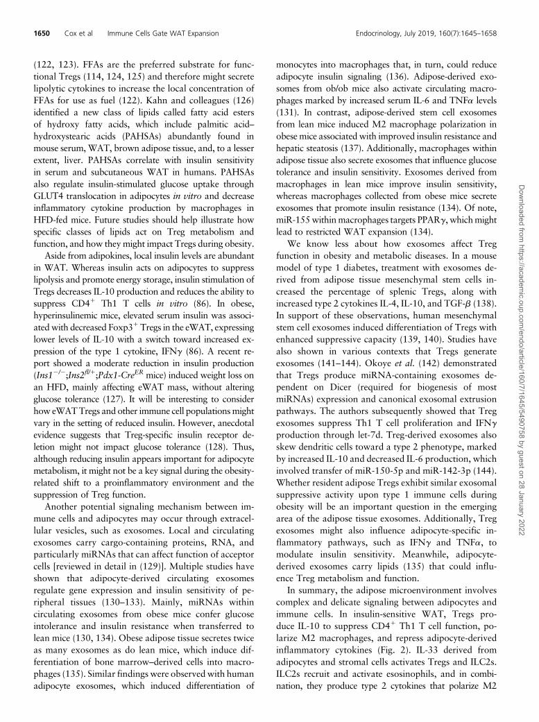
In summary, the adipose microenvironment involvescomplex and delicate signaling between adipocytes andimmune cells. In insulin-sensitive WAT, Tregs pro-duce IL-10 to suppress CD41 Th1 T cell function, po-larize M2 macrophages, and repress adipocyte-derivedinflammatory cytokines (Fig. 2). IL-33 derived fromadipocytes and stromal cells activates Tregs and ILC2s.ILC2s recruit and activate esosinophils, and in combi-nation, they produce type 2 cytokines that polarize M2
1650 Cox et al Immune Cells Gate WAT Expansion Endocrinology, July 2019, 160(7):1645–1658
Dow
nloaded from https://academ
ic.oup.com/endo/article/160/7/1645/5490758 by guest on 28 January 2022

macrophages. Adipocytes secrete adiponectin and FFAs,which also suppress macrophage cytokine production andmight influence Treg function, whereas the relative ab-sence of leptin allows Tregs to proliferate and maintainsuppressive activity. Both adipocytes and Tregs produceexosomes, with adipocyte-derived exosomes influencingmacrophage differentiation and function, although Tregexosomes suppress T cells in some contexts. Lastly, adi-pocytes secrete fatty acid esters of hydroxy fatty acids, suchas PAHSAs, that regulate macrophages and peripheral in-sulin sensitivity. In contrast, obese insulin-resistant adipo-cytes secrete inflammatory cytokines (IFNg, IL-6, RANTES,SAA) and recruit M1 macrophages (via MCP-1). Tregs,ILC2s, and eosinophils are reduced, with Tregs shiftingtoward IFNg production. High levels of leptin and insulinalso reduce Treg proliferation, function, and fatty acidmetabolism. Accumulation of CD41 Th1 T cells and M1macrophages contributes to the increasing inflammatory
milieu that restricts adipocyte insulin signaling, function,and expansion. Obesity-derived adipocyte or macrophageexosomes also reduce insulin sensitivity. Notably, theseobservations largely focused on immune cell–adipocytesignaling in eWAT given its propensity for greater immunecell infiltration in obese mice; however, some studies arebeginning to uncover roles of immune cells in subcutaneousWAT and the signals that might preserve insulin sensitivityand WAT expansion (43, 145). Finally, the complexity ofthe adipose tissue microenvironment suggests that thera-peutic intervention will likely require targeting of multiplesignals and cell types to restore adipocyte expansion andinsulin sensitivity in individuals with obesity.
Future Perspectives
More than 20 years of detailed mechanistic and physio-logical studies firmly establish that chronic inflammation
Figure 2. Inter-cell crosstalk regulates metabolic and inflammatory functions in WAT. In lean WAT, insulin-sensitive adipocytes expandappropriately to anabolic pressure and secrete adipokines (adiponectin), FFAs and PAHSAs, and exosomes. Tregs produce IL-10 to suppress CD41
Th1 T cell function, polarize M2 macrophages, and repress adipocyte-derived inflammatory cytokines. IL-33 derived from adipocytes and stromalcells activates Tregs and ILC2s. ILC2s recruit and activate esosinophils and, in combination, they produce type 2 cytokines that polarize M2macrophages. Adipocytes secrete adiponectin and FFAs, which also suppress macrophage cytokine production and might influence Tregfunction, whereas the relative absence of leptin allows Tregs to proliferate and maintain suppressive activity. Both adipocytes and Tregs produceexosomes, with adipocyte-derived exosomes influencing macrophage differentiation and function, whereas Treg exosomes suppress T cells insome contexts. Lastly, lean adipocytes secrete fatty acid esters of hydroxy fatty acids, such as PAHSAs, that regulate macrophages and peripheralinsulin sensitivity. In summary, type 2 immune cells promote adipocyte insulin sensitivity, differentiation, and function through cytokines IL-4, IL-10, and M2 macrophage-derived exosomes. In obese adipose tissue, insulin resistance develops, and adipocyte differentiation is restricted, whichcontributes to ectopic lipid deposition. Adipocytes secrete inflammatory cytokines (IFNg, TNFa, IL-6, RANTES, SAA) and recruit M1 macrophages(via MCP-1). Tregs, ILC2s, and eosinophils are reduced, with Tregs shifting toward IFNg production. High levels of leptin and insulin also reduceTreg proliferation, function, and fatty acid metabolism. Obese adipocyte- or macrophage-derived exosomes also reduce insulin sensitivity.Collectively, accumulation of CD41 Th1 T cells and M1 macrophages contributes to the increasing inflammatory milieu (IFNg, TNFa, IL-6, andexosomes) that restricts adipocyte insulin sensitivity, expansion, and function.
doi: 10.1210/en.2019-00266 https://academic.oup.com/endo 1651
Dow
nloaded from https://academ
ic.oup.com/endo/article/160/7/1645/5490758 by guest on 28 January 2022

in WAT depots leads to systemic glucose and lipid dys-regulation. Although WAT inflammation is one con-served pathway that links obesity to insulin resistanceand T2DM, numerous questions remain unanswered,including how to sustain healthy noninflamed sub-cutaneous WAT expansion during excess nutrient in-take. For people with T2DM, we are hopeful that theimmune system will be leveraged as a tool to maintaininsulin sensitivity. However, targeting the immunecomponent of obesity has proved elusive. For example,antibodies that neutralize TNF improve insulin sensi-tivity in obese mice, but humans showed no metabolicimprovements (146). In clinical trials, anti-inflammatorysalicylates improved glycemia and adipose inflamma-tion profiles in individuals with obesity and T2D, butinsulin sensitivity was unaffected (147–149). New strat-egies that target higher baseline inflammation in adiposetissues appear promising (150) in small clinical trials.Note that several well-established antidiabetic drugsinfluence anti-inflammatory endpoints. Metformin in-hibits reactive oxygen species production and the pro-duction of numerous inflammatory cytokines (151, 152).Thiazolidinediones also exert anti-inflammatory effectsinmacrophages and adipocytes (153). In both cases, thesedrugs block nuclear factor kB (151, 154) and restore theM2 phenotype of macrophages (134, 155). Further ex-ploration into how biologics targeting inflammation inrheumatoid arthritis and Crohn disease (156) may revealtherapeutic vulnerabilities for obesity. A thoughtful sur-vey of how manipulating the immune response impactsphysiology is warranted. Importantly, note that generaldisruption of inflammatory pathways may compromiseimmune responses, but also result in tissue damage,dysbiosis, and amplification of other autoimmune con-ditions (157).
The anatomical and functional differences of WATdepots portend distinct immune cell niches that con-tribute to adipocyte function and expansion duringobesity. The subcutaneous WAT has a tremendous ca-pacity to expand and store excess nutrients, as demon-strated by MitoNEET transgenic mice (158, 159). Inaddition to varied WAT depots, a recent study suggestedthat beige fat may also exist in multiple distinct forms,conventional and glycolytic beige fat (160). The immunecell composition also greatly varies, with the eWATcontaining a greater abundance of immune cells, evenduring metabolic homeostasis, in contrast to the lessinfiltrated brown adipose tissue. The tissue- and depot-specific roles for immune cells is unknown, but there isclearly an extensive communication network betweenadipocytes and immunity through multiple signalingmechanisms (e.g., hormones, cytokines, exosomes, poly-unsaturated fatty acids) within anatomical niches. The
characterization of immune cells within WAT marchesforward, as highlighted by the identification of multiplemacrophage subtypes in WAT beyond the traditionalM1/M2 subtypes (161). Ultimately, treatment of obesityand T2DM will require a multipronged approach to ad-dress adipocyte insulin sensitivity and nutrient storagewhile maintaining an anti-inflammatory environment. Ad-vances in immunotherapy and gene editing tools mightprovide unique opportunities to skew macrophage andT cell populations toward an anti-inflammatory state.Collaborative efforts to couple cell biology with immu-nology and genetics will be pivotal to address the currentknowledge gaps and identify new therapeutic strategies toimprove insulin sensitivity and nutrient storage for patientswith obesity and T2DM.
Acknowledgments
We apologize to our colleagues in the field for not being able todiscuss all of the outstanding studies that detail how immunecells regulate WAT function. We thank RobbMoses for criticalreading of the manuscript and useful discussions.
Financial Support: This work was supported by AmericanDiabetes Association Grant 1-18-IBS-105 and National In-stitutes of Health/National Institute of Diabetes and Digestiveand Kidney Diseases Grant R01 DK114356.
Correspondence: Sean M. Hartig, PhD, Baylor College ofMedicine, One Baylor Plaza, BCM185, Houston, Texas 77030.E-mail: [email protected].
Disclosure Summary: The authors have nothing todisclose.
References and Notes
1. Afshin A, Forouzanfar MH, Reitsma MB, Sur P, Estep K, Lee A,Marczak L, Mokdad AH, Moradi-Lakeh M, Naghavi M, SalamaJS, Vos T, Abate KH, Abbafati C, Ahmed MB, Al-Aly Z, AlkerwiA, Al-Raddadi R, Amare AT, Amberbir A, Amegah AK, Amini E,Amrock SM, Anjana RM, Arnlov J, Asayesh H, Banerjee A, BaracA, Baye E, Bennett DA, Beyene AS, Biadgilign S, Biryukov S,Bjertness E, Boneya DJ, Campos-Nonato I, Carrero JJ, Cecilio P,Cercy K, Ciobanu LG, Cornaby L, Damtew SA, Dandona L,DandonaR, Dharmaratne SD, Duncan BB, Eshrati B, EsteghamatiA, Feigin VL, Fernandes JC, Furst T, Gebrehiwot TT, Gold A,Gona PN, Goto A, Habtewold TD, Hadush KT, Hafezi-Nejad N,Hay SI, Horino M, Islami F, Kamal R, Kasaeian A, KatikireddiSV, Kengne AP, Kesavachandran CN, Khader YS, Khang YH,Khubchandani J, Kim D, Kim YJ, Kinfu Y, Kosen S, Ku T, DefoBK, Kumar GA, Larson HJ, Leinsalu M, Liang X, Lim SS, Liu P,Lopez AD, Lozano R, Majeed A, Malekzadeh R, Malta DC,Mazidi M, McAlinden C, McGarvey ST, Mengistu DT, MensahGA, Mensink GB, Mezgebe HB, Mirrakhimov EM, Mueller UO,Noubiap JJ, Obermeyer CM,Ogbo FA,OwolabiMO, PattonGC,Pourmalek F, QorbaniM, Rafay A, Rai RK, Ranabhat CL, ReinigN, Safiri S, Salomon JA, Sanabria JR, Santos IS, Sartorius B,Sawhney M, Schmidhuber J, Schutte AE, Schmidt MI, SepanlouSG, Shamsizadeh M, Sheikhbahaei S, Shin MJ, Shiri R, Shiue I,Roba HS, Silva DAS, Silverberg JI, Singh JA, Stranges S,Swaminathan S, Tabares-Seisdedos R, Tadese F, Tedla BA,Tegegne BS, Terkawi AS, Thakur JS, Tonelli M, Topor-Madry R,
1652 Cox et al Immune Cells Gate WAT Expansion Endocrinology, July 2019, 160(7):1645–1658
Dow
nloaded from https://academ
ic.oup.com/endo/article/160/7/1645/5490758 by guest on 28 January 2022

Tyrovolas S, Ukwaja KN, Uthman OA, Vaezghasemi M,Vasankari T, Vlassov VV, Vollset SE,Weiderpass E,Werdecker A,Wesana J, Westerman R, Yano Y, Yonemoto N, Yonga G, ZaidiZ, Zenebe ZM, Zipkin B, Murray CJL; GBD 2015 ObesityCollaborators. Health effects of overweight and obesity in 195countries over 25 years. N Engl J Med. 2017;377(1):13–27.
2. Boyle JP, Thompson TJ, Gregg EW, Barker LE, Williamson DF.Projection of the year 2050 burden of diabetes in the US adultpopulation: dynamic modeling of incidence, mortality, and pre-diabetes prevalence. Popul Health Metr. 2010;8(1):29.
3. Gesta S, BluherM, Yamamoto Y,Norris AW, Berndt J, Kralisch S,Boucher J, Lewis C, Kahn CR. Evidence for a role of de-velopmental genes in the origin of obesity and body fat distri-bution. Proc Natl Acad Sci USA. 2006;103(17):6676–6681.
4. Harman-Boehm I, Bluher M, Redel H, Sion-Vardy N, Ovadia S,Avinoach E, Shai I, Kloting N, Stumvoll M, Bashan N, Rudich A.Macrophage infiltration into omental versus subcutaneous fatacross different populations: effect of regional adiposity and thecomorbidities of obesity. J Clin Endocrinol Metab. 2007;92(6):2240–2247.
5. Kralova Lesna I, Kralova A, Cejkova S, Fronek J, Petras M,Sekerkova A, Thieme F, Janousek L, Poledne R. Characterisationand comparison of adipose tissue macrophages from humansubcutaneous, visceral and perivascular adipose tissue. J TranslMed. 2016;14(1):208.
6. Lee KY, Luong Q, Sharma R, Dreyfuss JM, Ussar S, Kahn CR.Developmental and functional heterogeneity of white adipocyteswithin a single fat depot. EMBO J. 2019;38(3):e99291.
7. Macotela Y, Emanuelli B, Mori MA, Gesta S, Schulz TJ, TsengYH, Kahn CR. Intrinsic differences in adipocyte precursor cellsfrom different white fat depots.Diabetes. 2012;61(7):1691–1699.
8. Tchoukalova Y, Koutsari C, Jensen M. Committed subcutaneouspreadipocytes are reduced in human obesity. Diabetologia. 2007;50(1):151–157.
9. van Tienen FH, van der Kallen CJ, Lindsey PJ, Wanders RJ, vanGreevenbroek MM, Smeets HJ. Preadipocytes of type 2 diabetessubjects display an intrinsic gene expression profile of decreaseddifferentiation capacity. Int J Obes. 2011;35(9):1154–1164.
10. Jeffery E, Church CD, Holtrup B, Colman L, Rodeheffer MS.Rapid depot-specific activation of adipocyte precursor cells at theonset of obesity. Nat Cell Biol. 2015;17(4):376–385.
11. Jeffery E, Wing A, Holtrup B, Sebo Z, Kaplan JL, Saavedra-Pe~naR, Church CD, Colman L, Berry R, Rodeheffer MS. The adiposetissue microenvironment regulates depot-specific adipogenesis inobesity. Cell Metab. 2016;24(1):142–150.
12. Kim SM, Lun M, Wang M, Senyo SE, Guillermier C, Patwari P,Steinhauser ML. Loss of white adipose hyperplastic potential isassociated with enhanced susceptibility to insulin resistance. CellMetab. 2014;20(6):1049–1058.
13. Wang QA, Tao C, Gupta RK, Scherer PE. Tracking adipogenesisduring white adipose tissue development, expansion and re-generation. Nat Med. 2013;19(10):1338–1344.
14. Acosta JR, Douagi I, Andersson DP, Backdahl J, Ryden M, ArnerP, Laurencikiene J. Increased fat cell size: a major phenotype ofsubcutaneous white adipose tissue in non-obese individuals withtype 2 diabetes. Diabetologia. 2016;59(3):560–570.
15. Veilleux A, Caron-Jobin M, Noel S, Laberge PY, Tchernof A.Visceral adipocyte hypertrophy is associated with dyslipidemiaindependent of body composition and fat distribution in women.Diabetes. 2011;60(5):1504–1511.
16. Weyer C, Foley JE, Bogardus C, Tataranni PA, Pratley RE. En-larged subcutaneous abdominal adipocyte size, but not obesityitself, predicts type II diabetes independent of insulin resistance.Diabetologia. 2000;43(12):1498–1506.
17. Berry R, Rodeheffer MS. Characterization of the adipocyte cel-lular lineage in vivo. Nat Cell Biol. 2013;15(3):302–308.
18. Lee YH, Petkova AP, Mottillo EP, Granneman JG. In vivoidentification of bipotential adipocyte progenitors recruited by
b3-adrenoceptor activation and high-fat feeding. Cell Metab.2012;15(4):480–491.
19. Rodeheffer MS, Birsoy K, Friedman JM. Identification of whiteadipocyte progenitor cells in vivo. Cell. 2008;135(2):240–249.
20. TangW, Zeve D, Suh JM, Bosnakovski D, KybaM, Hammer RE,Tallquist MD, Graff JM. White fat progenitor cells reside in theadipose vasculature. Science. 2008;322(5901):583–586.
21. Berry DC, Jiang Y, Graff JM. Mouse strains to study cold-inducible beige progenitors and beige adipocyte formation andfunction. Nat Commun. 2016;7(1):10184.
22. Jiang Y, Berry DC, Tang W, Graff JM. Independent stem celllineages regulate adipose organogenesis and adipose homeostasis.Cell Reports. 2014;9(3):1007–1022.
23. Long JZ, SvenssonKJ, Tsai L, ZengX, RohHC,KongX, RaoRR,Lou J, Lokurkar I, Baur W, Castellot JJ Jr, Rosen ED, SpiegelmanBM. A smooth muscle-like origin for beige adipocytes. CellMetab. 2014;19(5):810–820.
24. Shao M, Vishvanath L, Busbuso NC, Hepler C, Shan B, SharmaAX, Chen S, Yu X, An YA, Zhu Y, Holland WL, Gupta RK. Denovo adipocyte differentiation from Pdgfrß1 preadipocytes pro-tects against pathologic visceral adipose expansion in obesity.NatCommun. 2018;9(1):890.
25. Vishvanath L, MacPherson KA, Hepler C, Wang QA, Shao M,Spurgin SB, Wang MY, Kusminski CM, Morley TS, Gupta RK.Pdgfrb1 mural preadipocytes contribute to adipocyte hyperplasiainduced by high-fat-diet feeding and prolonged cold exposure inadult mice. Cell Metab. 2016;23(2):350–359.
26. Chau YY, Bandiera R, Serrels A, Martınez-Estrada OM, QingW,Lee M, Slight J, Thornburn A, Berry R, McHaffie S, Stimson RH,Walker BR, Chapuli RM, Schedl A, Hastie N. Visceral andsubcutaneous fat have different origins and evidence supports amesothelial source. Nat Cell Biol. 2014;16(4):367–375.
27. Cohen P, Levy JD, Zhang Y, Frontini A, Kolodin DP, SvenssonKJ, Lo JC, ZengX, Ye L, KhandekarMJ,Wu J, Gunawardana SC,Banks AS, Camporez JP, Jurczak MJ, Kajimura S, Piston DW,Mathis D, Cinti S, Shulman GI, Seale P, Spiegelman BM. Ablationof PRDM16 and beige adipose causes metabolic dysfunctionand a subcutaneous to visceral fat switch. Cell. 2014;156(1–2):304–316.
28. Krueger KC, Costa MJ, Du H, Feldman BJ. Characterization ofCre recombinase activity for in vivo targeting of adipocyte pre-cursor cells. Stem Cell Reports. 2014;3(6):1147–1158.
29. Sanchez-Gurmaches J, Hsiao WY, Guertin DA. Highly selectivein vivo labeling of subcutaneous white adipocyte precursors withPrx1-Cre. Stem Cell Reports. 2015;4(4):541–550.
30. Hepler C, Shan B, Zhang Q, Henry GH, Shao M, Vishvanath L,Ghaben AL, Mobley AB, Strand D, Hon GC, Gupta RK. Iden-tification of functionally distinct fibro-inflammatory and adipo-genic stromal subpopulations in visceral adipose tissue of adultmice. eLife. 2018;7:e39636.
31. Lee KY, Russell SJ, Ussar S, Boucher J, Vernochet C, Mori MA,Smyth G, RourkM, Cederquist C, Rosen ED, Kahn BB, KahnCR.Lessons on conditional gene targeting in mouse adipose tissue.Diabetes. 2013;62(3):864–874.
32. Morgan-Bathke M, Harteneck D, Jaeger P, Sondergaard E,Karwoski R, Espinosa De Ycaza A, Carranza-Leon BG, FaubionWA Jr, Oliveira AM, Jensen MD. Comparison of methods foranalyzing human adipose tissue macrophage content. Obesity(Silver Spring). 2017;25(12):2100–2107.
33. Sanchez-Gurmaches J, Guertin DA. Adipocytes arise from mul-tiple lineages that are heterogeneously and dynamically distrib-uted. Nat Commun. 2014;5(1):4099.
34. Schwalie PC, Dong H, Zachara M, Russeil J, Alpern D, AkchicheN, Caprara C, Sun W, Schlaudraff KU, Soldati G, Wolfrum C,Deplancke B. A stromal cell population that inhibits adipogenesisin mammalian fat depots. Nature. 2018;559(7712):103–108.
35. Wu J, Bostrom P, Sparks LM, Ye L, Choi JH, Giang AH,Khandekar M, Virtanen KA, Nuutila P, Schaart G, Huang K, Tu
doi: 10.1210/en.2019-00266 https://academic.oup.com/endo 1653
Dow
nloaded from https://academ
ic.oup.com/endo/article/160/7/1645/5490758 by guest on 28 January 2022

H, vanMarken Lichtenbelt WD, Hoeks J, Enerback S, SchrauwenP, Spiegelman BM. Beige adipocytes are a distinct type of ther-mogenic fat cell in mouse and human. Cell. 2012;150(2):366–376.
36. Merrick D, Sakers A, Irgebay Z, Okada C, Calvert C,MorleyMP,Percec I, Seale P. Identification of a mesenchymal progenitor cellhierarchy in adipose tissue. Science. 2019;364(6438):eaav2501.
37. Raajendiran A, Ooi G, Bayliss J, O’Brien PE, Schittenhelm RB,Clark AK, Taylor RA, Rodeheffer MS, Burton PR, Watt MJ.Identification of metabolically distinct adipocyte progenitor cellsin human adipose tissues. Cell Rep. 2019;27(5):1528–1540.e7.
38. Crisan M, Yap S, Casteilla L, Chen CW, Corselli M, Park TS,Andriolo G, Sun B, Zheng B, Zhang L, Norotte C, Teng PN, TraasJ, Schugar R, Deasy BM, Badylak S, Buhring HJ, Giacobino JP,Lazzari L, Huard J, Peault B. A perivascular origin for mesen-chymal stem cells in multiple human organs.Cell Stem Cell. 2008;3(3):301–313.
39. Yuan SM, Guo Y, Zhou XJ, Shen WM, Chen HN. PDGFR-b (1)perivascular cells from infantile hemangioma display the featuresof mesenchymal stem cells and show stronger adipogenic potentialin vitro and in vivo. Int J Clin Exp Pathol. 2014;7(6):2861–2870.
40. Zimmerlin L, Donnenberg VS, Pfeifer ME, Meyer EM, Peault B,Rubin JP, Donnenberg AD. Stromal vascular progenitors in adulthuman adipose tissue. Cytometry A. 2010;77(1):22–30.
41. Fox MJ, Kuzma JF, Washam WT. Transitory diabetic syndromeassociated with meningococcic meningitis. Arch Intern Med(Chic). 1947;79(6):614–621.
42. Hotamisligil GS. Inflammation, metaflammation and immuno-metabolic disorders. Nature. 2017;542(7640):177–185.
43. Koh EH, Chernis N, Saha PK, Xiao L, Bader DA, Zhu B,Rajapakshe K, Hamilton MP, Liu X, Perera D, Chen X, York B,TraunerM, Coarfa C, Bajaj M,Moore DD, Deng T,McGuire SE,Hartig SM. miR-30a remodels subcutaneous adipose tissue in-flammation to improve insulin sensitivity in obesity. Diabetes.2018;67(12):2541–2553.
44. McGillicuddy FC, Chiquoine EH, Hinkle CC, Kim RJ, Shah R,Roche HM, Smyth EM, Reilly MP. Interferon g attenuates insulinsignaling, lipid storage, and differentiation in human adipocytesvia activation of the JAK/STAT pathway. J Biol Chem. 2009;284(46):31936–31944.
45. Hotamisligil GS, Arner P, Caro JF, Atkinson RL, Spiegelman BM.Increased adipose tissue expression of tumor necrosis factor-alphain human obesity and insulin resistance. J Clin Invest. 1995;95(5):2409–2415.
46. Kintscher U, Hartge M, Hess K, Foryst-Ludwig A, Clemenz M,Wabitsch M, Fischer-Posovszky P, Barth TF, Dragun D, Skurk T,Hauner H, Bluher M, Unger T, Wolf AM, Knippschild U,Hombach V, Marx N. T-lymphocyte infiltration in visceral adi-pose tissue: a primary event in adipose tissue inflammation and thedevelopment of obesity-mediated insulin resistance. ArteriosclerThromb Vasc Biol. 2008;28(7):1304–1310.
47. Laurencikiene J, van Harmelen V, Arvidsson Nordstrom E,Dicker A, Blomqvist L, Naslund E, Langin D, Arner P, Ryden M.NF-kB is important for TNF-a-induced lipolysis in human adi-pocytes. J Lipid Res. 2007;48(5):1069–1077.
48. Creely SJ, McTernan PG, Kusminski CM, Fisher M, Da Silva NF,Khanolkar M, Evans M, Harte AL, Kumar S. Lipopolysaccharideactivates an innate immune system response in human adiposetissue in obesity and type 2 diabetes. Am J Physiol EndocrinolMetab. 2007;292(3):E740–E747.
49. Xu H, Uysal KT, Becherer JD, Arner P, Hotamisligil GS. Alteredtumor necrosis factor-alpha (TNF-a) processing in adipocytes andincreased expression of transmembrane TNF-a in obesity. Di-abetes. 2002;51(6):1876–1883.
50. Liu LS, Spelleken M, Rohrig K, Hauner H, Eckel J. Tumor ne-crosis factor-alpha acutely inhibits insulin signaling in humanadipocytes: implication of the p80 tumor necrosis factor receptor.Diabetes. 1998;47(4):515–522.
51. Dimas AS, Lagou V, Barker A, Knowles JW, Magi R, Hivert MF,Benazzo A, Rybin D, JacksonAU, StringhamHM, Song C, Fischer-Rosinsky A, Boesgaard TW, Grarup N, Abbasi FA, Assimes TL,Hao K, Yang X, Lecoeur C, Barroso I, Bonnycastle LL, Bottcher Y,Bumpstead S, Chines PS, ErdosMR, Graessler J, Kovacs P,MorkenMA, Narisu N, Payne F, Stancakova A, Swift AJ, Tonjes A,Bornstein SR, Cauchi S, Froguel P, Meyre D, Schwarz PE, HaringHU, Smith U, Boehnke M, Bergman RN, Collins FS, Mohlke KL,Tuomilehto J, Quertemous T, Lind L, Hansen T, Pedersen O,Walker M, Pfeiffer AF, Spranger J, Stumvoll M, Meigs JB,Wareham NJ, Kuusisto J, Laakso M, Langenberg C, Dupuis J,Watanabe RM, Florez JC, Ingelsson E,McCarthyMI, ProkopenkoI; MAGIC Investigators. Impact of type 2 diabetes susceptibilityvariants on quantitative glycemic traits reveals mechanistic het-erogeneity. Diabetes. 2014;63(6):2158–2171.
52. Strawbridge RJ, Dupuis J, Prokopenko I, Barker A, Ahlqvist E,Rybin D, Petrie JR, Travers ME, Bouatia-Naji N, Dimas AS, NicaA, Wheeler E, Chen H, Voight BF, Taneera J, Kanoni S, Peden JF,Turrini F, Gustafsson S, Zabena C, Almgren P, Barker DJ, BarnesD, Dennison EM, Eriksson JG, Eriksson P, Eury E, Folkersen L,Fox CS, Frayling TM, Goel A, Gu HF, Horikoshi M, Isomaa B,Jackson AU, Jameson KA, Kajantie E, Kerr-Conte J, KuulasmaaT, Kuusisto J, Loos RJ, Luan J, Makrilakis K, Manning AK,Martınez-Larrad MT, Narisu N, Nastase Mannila M, Ohrvik J,Osmond C, Pascoe L, Payne F, Sayer AA, Sennblad B, Silveira A,Stancakova A, Stirrups K, Swift AJ, Syvanen AC, Tuomi T, van ’tHooft FM, Walker M, Weedon MN, Xie W, Zethelius B, OngenH, Malarstig A, Hopewell JC, Saleheen D, Chambers J, Parish S,Danesh J, Kooner J, Ostenson CG, Lind L, Cooper CC, Serrano-Rıos M, Ferrannini E, Forsen TJ, Clarke R, Franzosi MG, SeedorfU, Watkins H, Froguel P, Johnson P, Deloukas P, Collins FS,Laakso M, Dermitzakis ET, Boehnke M, McCarthy MI,Wareham NJ, Groop L, Pattou F, Gloyn AL, Dedoussis GV,Lyssenko V, Meigs JB, Barroso I, Watanabe RM, Ingelsson E,Langenberg C, Hamsten A, Florez JC; DIAGRAM Consortium;GIANT Consortium; MuTHER Consortium; CARDIoGRAMConsortium; C4D Consortium. Genome-wide association iden-tifies nine common variants associated with fasting proinsulinlevels and provides new insights into the pathophysiology of type2 diabetes. Diabetes. 2011;60(10):2624–2634.
53. Sun K, Tordjman J, Clement K, Scherer PE. Fibrosis and adiposetissue dysfunction. Cell Metab. 2013;18(4):470–477.
54. Lee YS, Kim JW, Osborne O, Oh DY, Sasik R, Schenk S, Chen A,Chung H, Murphy A, Watkins SM, Quehenberger O, JohnsonRS, Olefsky JM. Increased adipocyte O2 consumption triggersHIF-1a, causing inflammation and insulin resistance in obesity.Cell. 2014;157(6):1339–1352.
55. Jiang C, Qu A, Matsubara T, Chanturiya T, Jou W, Gavrilova O,Shah YM, Gonzalez FJ. Disruption of hypoxia-inducible factor 1in adipocytes improves insulin sensitivity and decreases adiposityin high-fat diet-fed mice. Diabetes. 2011;60(10):2484–2495.
56. Cancello R, Henegar C, Viguerie N, Taleb S, Poitou C, Rouault C,Coupaye M, Pelloux V, Hugol D, Bouillot JL, Bouloumie A,Barbatelli G, Cinti S, Svensson PA, Barsh GS, Zucker JD,Basdevant A, Langin D, Clement K. Reduction of macrophageinfiltration and chemoattractant gene expression changes in whiteadipose tissue of morbidly obese subjects after surgery-inducedweight loss. Diabetes. 2005;54(8):2277–2286.
57. Rausch ME, Weisberg S, Vardhana P, Tortoriello DV. Obesity inC57BL/6J mice is characterized by adipose tissue hypoxia andcytotoxic T-cell infiltration. Int J Obes. 2008;32(3):451–463.
58. Halberg N, Khan T, Trujillo ME, Wernstedt-Asterholm I, AttieAD, Sherwani S, Wang ZV, Landskroner-Eiger S, Dineen S,Magalang UJ, Brekken RA, Scherer PE. Hypoxia-inducible factor1a induces fibrosis and insulin resistance in white adipose tissue.Mol Cell Biol. 2009;29(16):4467–4483.
59. Henegar C, Tordjman J, Achard V, Lacasa D, Cremer I, Guerre-Millo M, Poitou C, Basdevant A, Stich V, Viguerie N, Langin D,
1654 Cox et al Immune Cells Gate WAT Expansion Endocrinology, July 2019, 160(7):1645–1658
Dow
nloaded from https://academ
ic.oup.com/endo/article/160/7/1645/5490758 by guest on 28 January 2022

Bedossa P, Zucker JD, Clement K. Adipose tissue transcriptomicsignature highlights the pathological relevance of extracellularmatrix in human obesity. Genome Biol. 2008;9(1):R14.
60. Lackey DE, Burk DH, Ali MR, Mostaedi R, Smith WH, Park J,Scherer PE, Seay SA, McCoin CS, Bonaldo P, Adams SH. Con-tributions of adipose tissue architectural and tensile propertiestoward defining healthy and unhealthy obesity. Am J PhysiolEndocrinol Metab. 2014;306(3):E233–E246.
61. Reggio S, Rouault C, Poitou C, Bichet J-C, Prifti E, Bouillot J-L,Rizkalla S, Lacasa D, Tordjman J, Clement K. Increased basementmembrane components in adipose tissue during obesity: links withTGFb and metabolic phenotypes. J Clin Endocrinol Metab. 2016;101(6):2578–2587.
62. Khan T, Muise ES, Iyengar P, Wang ZV, Chandalia M, Abate N,Zhang BB, Bonaldo P, Chua S, Scherer PE. Metabolic dysregu-lation and adipose tissue fibrosis: role of collagen VI. Mol CellBiol. 2009;29(6):1575–1591.
63. Sun K, Halberg N, Khan M, Magalang UJ, Scherer PE. Selectiveinhibition of hypoxia-inducible factor 1a ameliorates adiposetissue dysfunction. Mol Cell Biol. 2013;33(5):904–917.
64. Hasegawa Y, Ikeda K, Chen Y, Alba DL, Stifler D, Shinoda K,Hosono T, Maretich P, Yang Y, Ishigaki Y, Chi J, Cohen P,Koliwad SK, Kajimura S. Repression of adipose tissue fibrosisthrough a PRDM16-GTF2IRD1 complex improves systemicglucose homeostasis. Cell Metab. 2018;27(1):180–194.e6.
65. Nagareddy PR, KraakmanM,Masters SL, Stirzaker RA, GormanDJ, Grant RW, Dragoljevic D, Hong ES, Abdel-Latif A, Smyth SS,Choi SH, Korner J, Bornfeldt KE, Fisher EA, Dixit VD, Tall AR,Goldberg IJ, Murphy AJ. Adipose tissue macrophages promotemyelopoiesis andmonocytosis in obesity.Cell Metab. 2014;19(5):821–835.
66. Weisberg SP, McCann D, Desai M, Rosenbaum M, Leibel RL,Ferrante AW Jr. Obesity is associated with macrophage accu-mulation in adipose tissue. J Clin Invest. 2003;112(12):1796–1808.
67. Anderson EK, Gutierrez DA, Kennedy A, Hasty AH. Weightcycling increases T-cell accumulation in adipose tissue and impairssystemic glucose tolerance. Diabetes. 2013;62(9):3180–3188.
68. Nishimura S, Manabe I, Nagasaki M, Eto K, Yamashita H,Ohsugi M, Otsu M, Hara K, Ueki K, Sugiura S, Yoshimura K,Kadowaki T, Nagai R. CD81 effector T cells contribute tomacrophage recruitment and adipose tissue inflammation inobesity. Nat Med. 2009;15(8):914–920.
69. Bertola A, Ciucci T, Rousseau D, Bourlier V, Duffaut C,Bonnafous S, Blin-Wakkach C, Anty R, Iannelli A, Gugenheim J,Tran A, Bouloumie A, Gual P, Wakkach A. Identification ofadipose tissue dendritic cells correlated with obesity-associatedinsulin-resistance and inducing Th17 responses in mice and pa-tients. Diabetes. 2012;61(9):2238–2247.
70. Dahlman I, Forsgren M, Sjogren A, Nordstrom EA, Kaaman M,Naslund E, Attersand A, Arner P. Downregulation of electrontransport chain genes in visceral adipose tissue in type 2 diabetesindependent of obesity and possibly involving tumor necrosisfactor-a. Diabetes. 2006;55(6):1792–1799.
71. Schmidt FM, Weschenfelder J, Sander C, Minkwitz J, ThormannJ, Chittka T, Mergl R, Kirkby KC, Faßhauer M, Stumvoll M,Holdt LM, Teupser D, Hegerl U, Himmerich H. Inflammatorycytokines in general and central obesity and modulating effects ofphysical activity. PLoS One. 2015;10(3):e0121971.
72. Lumeng CN, Bodzin JL, Saltiel AR. Obesity induces a phenotypicswitch in adipose tissue macrophage polarization. J Clin Invest.2007;117(1):175–184.
73. Kratz M, Coats BR, Hisert KB, Hagman D, Mutskov V, Peris E,Schoenfelt KQ, Kuzma JN, Larson I, Billing PS, Landerholm RW,Crouthamel M, Gozal D, Hwang S, Singh PK, Becker L. Meta-bolic dysfunction drives a mechanistically distinct proinflammatoryphenotype in adipose tissue macrophages. Cell Metab. 2014;20(4):614–625.
74. Lumeng CN,DelProposto JB,Westcott DJ, Saltiel AR. Phenotypicswitching of adipose tissue macrophages with obesity is generatedby spatiotemporal differences in macrophage subtypes. Diabetes.2008;57(12):3239–3246.
75. Nguyen MT, Favelyukis S, Nguyen AK, Reichart D, Scott PA,Jenn A, Liu-Bryan R, Glass CK, Neels JG, Olefsky JM. A sub-population of macrophages infiltrates hypertrophic adipose tissueand is activated by free fatty acids via Toll-like receptors 2 and 4and JNK-dependent pathways. J Biol Chem. 2007;282(48):35279–35292.
76. Lackey DE, Olefsky JM. Regulation of metabolism by the innateimmune system. Nat Rev Endocrinol. 2016;12(1):15–28.
77. McNelis JC, Olefsky JM.Macrophages, immunity, and metabolicdisease. Immunity. 2014;41(1):36–48.
78. Burzyn D, Benoist C, Mathis D. Regulatory T cells in non-lymphoid tissues. Nat Immunol. 2013;14(10):1007–1013.
79. Feuerer M, Herrero L, Cipolletta D, Naaz A, Wong J, Nayer A,Lee J, Goldfine AB, Benoist C, Shoelson S,Mathis D. Lean, but notobese, fat is enriched for a unique population of regulatory T cellsthat affect metabolic parameters. Nat Med. 2009;15(8):930–939.
80. Cipolletta D, Cohen P, Spiegelman BM, Benoist C, Mathis D.Appearance and disappearance of the mRNA signature charac-teristic of Treg cells in visceral adipose tissue: age, diet, and PPARgeffects. Proc Natl Acad Sci USA. 2015;112(2):482–487.
81. Vasanthakumar A,Moro K, Xin A, Liao Y, Gloury R, KawamotoS, Fagarasan S, Mielke LA, Afshar-Sterle S, Masters SL, Nakae S,Saito H, Wentworth JM, Li P, Liao W, Leonard WJ, Smyth GK,Shi W, Nutt SL, Koyasu S, Kallies A. The transcriptional regu-lators IRF4, BATF and IL-33 orchestrate development andmaintenance of adipose tissue-resident regulatory T cells [pub-lished correction appears in Nat Immunol. 2015;16(5):544]. NatImmunol. 2015;16(3):276–285.
82. Winer S, Chan Y, Paltser G, Truong D, Tsui H, Bahrami J,Dorfman R, Wang Y, Zielenski J, Mastronardi F, Maezawa Y,Drucker DJ, Engleman E, Winer D, Dosch HM. Normalization ofobesity-associated insulin resistance through immunotherapy.Nat Med. 2009;15(8):921–929.
83. Kanda H, Tateya S, Tamori Y, Kotani K, Hiasa K, Kitazawa R,Kitazawa S, Miyachi H, Maeda S, Egashira K, KasugaM.MCP-1contributes to macrophage infiltration into adipose tissue, insulinresistance, and hepatic steatosis in obesity. J Clin Invest. 2006;116(6):1494–1505.
84. Park-Min KH, Antoniv TT, Ivashkiv LB. Regulation of macro-phage phenotype by long-term exposure to IL-10. Immunobiol-ogy. 2005;210(2–4):77–86.
85. Gao M, Zhang C, Ma Y, Bu L, Yan L, Liu D. Hydrodynamicdelivery of mIL10 gene protects mice from high-fat diet-inducedobesity and glucose intolerance. Mol Ther. 2013;21(10):1852–1861.
86. Han JM, Patterson SJ, Speck M, Ehses JA, Levings MK. Insulininhibits IL-10–mediated regulatory T cell function: implicationsfor obesity. J Immunol. 2014;192(2):623–629.
87. Pettersson US, Walden TB, Carlsson P-O, Jansson L, PhillipsonM. Female mice are protected against high-fat diet inducedmetabolic syndrome and increase the regulatory T cell populationin adipose tissue. PLoS One. 2012;7(9):e46057.
88. Overacre-Delgoffe AE, Chikina M, Dadey RE, Yano H, BrunazziEA, Shayan G, Horne W, Moskovitz JM, Kolls JK, Sander C,Shuai Y, Normolle DP, Kirkwood JM, Ferris RL, Delgoffe GM,Bruno TC, Workman CJ, Vignali DAA. Interferon-g drives Tregfragility to promote anti-tumor immunity. Cell. 2017;169(6):1130–1141.e11.
89. Ilan Y, Maron R, Tukpah AM, Maioli TU, Murugaiyan G, YangK, Wu HY, Weiner HL. Induction of regulatory T cells decreasesadipose inflammation and alleviates insulin resistance in ob/obmice. Proc Natl Acad Sci USA. 2010;107(21):9765–9770.
90. Molofsky AB,Nussbaum JC, LiangHE, VanDyken SJ, Cheng LE,Mohapatra A, Chawla A, Locksley RM. Innate lymphoid type 2
doi: 10.1210/en.2019-00266 https://academic.oup.com/endo 1655
Dow
nloaded from https://academ
ic.oup.com/endo/article/160/7/1645/5490758 by guest on 28 January 2022

cells sustain visceral adipose tissue eosinophils and alternativelyactivated macrophages. J Exp Med. 2013;210(3):535–549.
91. Nussbaum JC, VanDyken SJ, vonMoltke J, Cheng LE,MohapatraA, Molofsky AB, Thornton EE, Krummel MF, Chawla A, LiangH-E, Locksley RM. Type 2 innate lymphoid cells control eosinophilhomeostasis. Nature. 2013;502(7470):245–248.
92. Wu D, Molofsky AB, Liang H-E, Ricardo-Gonzalez RR, JouihanHA, Bando JK, Chawla A, Locksley RM. Eosinophils sustainadipose alternatively activated macrophages associated withglucose homeostasis. Science. 2011;332(6026):243–247.
93. Brestoff JR, Kim BS, Saenz SA, Stine RR, Monticelli LA,Sonnenberg GF, Thome JJ, Farber DL, Lutfy K, Seale P, Artis D.Group 2 innate lymphoid cells promote beiging of white adiposetissue and limit obesity. Nature. 2015;519(7542):242–246.
94. Ding X, Luo Y, Zhang X, Zheng H, Yang X, Yang X, Liu M.IL-33-driven ILC2/eosinophil axis in fat is induced by sympathetictone and suppressed by obesity. J Endocrinol. 2016;231(1):35–48.
95. Lee MW, Odegaard JI, Mukundan L, Qiu Y, Molofsky AB,Nussbaum JC, Yun K, Locksley RM, Chawla A. Activated type 2innate lymphoid cells regulate beige fat biogenesis. Cell. 2015;160(1–2):74–87.
96. Qiu Y, Nguyen KD, Odegaard JI, Cui X, Tian X, Locksley RM,Palmiter RD, Chawla A. Eosinophils and type 2 cytokine signalingin macrophages orchestrate development of functional beige fat.Cell. 2014;157(6):1292–1308.
97. Wood IS, Wang B, Trayhurn P. IL-33, a recently identifiedinterleukin-1 gene family member, is expressed in human adi-pocytes. Biochem Biophys Res Commun. 2009;384(1):105–109.
98. Zeyda M, Wernly B, Demyanets S, Kaun C, Hammerle M,Hantusch B, Schranz M, Neuhofer A, Itariu BK, Keck M, PragerG, Wojta J, Stulnig TM. Severe obesity increases adipose tissueexpression of interleukin-33 and its receptor ST2, both pre-dominantly detectable in endothelial cells of human adipose tis-sue. Int J Obes. 2013;37(5):658–665.
99. Zeng Q, Sun X, Xiao L, Xie Z, Bettini M, Deng T. A uniquepopulation: Adipose-resident regulatory T cells. Front Immunol.2018;9:2075.
100. Molofsky AB, Van Gool F, Liang H-E, Van Dyken SJ, NussbaumJC, Lee J, Bluestone JA, Locksley RM. Interleukin-33 and in-terferon-g counter-regulate group 2 innate lymphoid cell acti-vation during immune perturbation. Immunity. 2015;43(1):161–174.
101. Uysal KT, Wiesbrock SM, Marino MW, Hotamisligil GS. Pro-tection from obesity-induced insulin resistance in mice lackingTNF-a function. Nature. 1997;389(6651):610–614.
102. Xu H, Hirosumi J, Uysal KT, Guler AD, Hotamisligil GS. Ex-clusive action of transmembrane TNFa in adipose tissue leads toreduced adipose mass and local but not systemic insulin re-sistance. Endocrinology. 2002;143(4):1502–1511.
103. Clementi AH, Gaudy AM, van Rooijen N, Pierce RH, MooneyRA. Loss of Kupffer cells in diet-induced obesity is associated withincreased hepatic steatosis, STAT3 signaling, and further de-creases in insulin signaling. Biochim Biophys Acta. 2009;1792(11):1062–1072.
104. den BoerMA,Voshol PJ, Schroder-van der Elst JP, KorsheninnikovaE, Ouwens DM, Kuipers F, Havekes LM, Romijn JA. Endogenousinterleukin-10 protects against hepatic steatosis but does not im-prove insulin sensitivity during high-fat feeding in mice. Endocri-nology. 2006;147(10):4553–4558.
105. Faulkner JL, Gomolak JR, Didion SP. Interleukin-10 deficiencylimits the development of obesity and insulin resistance producedby a high fat diet. FASEB J. 2013;27(Suppl 1):1183.6.
106. Miller AM,Wang H, Bertola A, Park O, Horiguchi N, Ki SH, YinS, Lafdil F, Gao B. Inflammation-associated interleukin-6/signaltransducer and activator of transcription 3 activation amelioratesalcoholic and nonalcoholic fatty liver diseases in interleukin-10–deficient mice. Hepatology. 2011;54(3):846–856.
107. Rajbhandari P, Thomas BJ, FengAC,HongC,Wang J, Vergnes L,Sallam T, Wang B, Sandhu J, Seldin MM, Lusis AJ, Fong LG,Katz M, Lee R, Young SG, Reue K, Smale ST, Tontonoz P. IL-10signaling remodels adipose chromatin architecture to limit ther-mogenesis and energy expenditure. Cell. 2018;172(1–2):218–233.e17.
108. Kalin S, BeckerM,Ott VB, Serr I, Hosp F,MollahMM,Keipert S,Lamp D, Rohner-Jeanrenaud F, Flynn VK, Scherm MG,Nascimento LF, Gerlach K, Popp V, Dietzen S, Bopp T,Krishnamurthy P, Kaplan MH, Serrano M, Woods SC, Tripal P,Palmisano R, Jastroch M, Bluher M, Wolfrum C, Weigmann B,Ziegler AG, Mann M, Tschop MH, Daniel C. A Stat6/Pten axislinks regulatory T cells with adipose tissue function. Cell Metab.2017;26(3):475–492.e7.
109. Cipolletta D, Feuerer M, Li A, Kamei N, Lee J, Shoelson SE,Benoist C, Mathis D. PPAR-g is a major driver of the accumu-lation and phenotype of adipose tissue Treg cells. Nature. 2012;486(7404):549–553.
110. De Rosa V, Procaccini C, CalıG, Pirozzi G, Fontana S, ZappacostaS, La Cava A, Matarese G. A key role of leptin in the control ofregulatory T cell proliferation. Immunity. 2007;26(2):241–255.
111. Procaccini C, De Rosa V, GalganiM, Abanni L, CalıG, PorcelliniA, Carbone F, Fontana S, Horvath TL, La Cava A, Matarese G.An oscillatory switch in mTOR kinase activity sets regulatoryT cell responsiveness. Immunity. 2010;33(6):929–941.
112. Ramos-Ramırez P, Malmhall C, Johansson K, Lotvall J, BossiosA. Weight gain alters adiponectin receptor 1 expression on adi-pose tissue-resident Helios1 regulatory T cells. Scand J Immunol.2016;83(4):244–254.
113. Smith PM, Howitt MR, Panikov N, Michaud M, Gallini CA,Bohlooly-Y M, Glickman JN, Garrett WS. The microbial me-tabolites, short-chain fatty acids, regulate colonic Treg cell ho-meostasis. Science. 2013;341(6145):569–573.
114. Gerriets VA, Danzaki K, Kishton RJ, Eisner W, Nichols AG,Saucillo DC, Shinohara ML, MacIver NJ. Leptin directly pro-motes T-cell glycolytic metabolism to drive effector T-cell dif-ferentiation in a mouse model of autoimmunity. Eur J Immunol.2016;46(8):1970–1983.
115. Lord GM,Matarese G, Howard JK, Baker RJ, Bloom SR, LechlerRI. Leptin modulates the T-cell immune response and reversesstarvation-induced immunosuppression.Nature. 1998;394(6696):897–901.
116. Arita Y, Kihara S, Ouchi N, TakahashiM,MaedaK,Miyagawa J,Hotta K, Shimomura I, Nakamura T, Miyaoka K, Kuriyama H,NishidaM, Yamashita S, Okubo K,Matsubara K,Muraguchi M,Ohmoto Y, Funahashi T, Matsuzawa Y. Paradoxical decrease ofan adipose-specific protein, adiponectin, in obesity. BiochemBiophys Res Commun. 1999;257(1):79–83.
117. . Maeda N, Shimomura I, Kishida K, Nishizawa H, Matsuda M,Nagaretani H, Furuyama N, Kondo H, Takahashi M, Arita Y,Komuro R, Ouchi N, Kihara S, Tochino Y, Okutomi K, Horie M,Takeda S, Aoyama T, Funahashi T, Matsuzawa Y. Diet-inducedinsulin resistance in mice lacking adiponectin/ACRP30.Nat Med.2002;8(7):731–737.
118. Ohashi K, Parker JL, Ouchi N, Higuchi A, Vita JA, Gokce N,Pedersen AA, Kalthoff C, Tullin S, Sams A, Summer R, Walsh K.Adiponectin promotes macrophage polarization toward an anti-inflammatory phenotype. J Biol Chem. 2010;285(9):6153–6160.
119. Onodera T, Fukuhara A, Jang MH, Shin J, Aoi K, Kikuta J,Otsuki M, Ishii M, Shimomura I. Adipose tissue macrophagesinduce PPARg-high FOXP31 regulatory T cells. Sci Rep. 2015;5(1):16801.
120. Wolf AM, Wolf D, Rumpold H, Enrich B, Tilg H. Adiponectininduces the anti-inflammatory cytokines IL-10 and IL-1RA inhuman leukocytes. Biochem Biophys Res Commun. 2004;323(2):630–635.
121. Cao H, Gerhold K, Mayers JR, Wiest MM, Watkins SM,Hotamisligil GS. Identification of a lipokine, a lipid hormone
1656 Cox et al Immune Cells Gate WAT Expansion Endocrinology, July 2019, 160(7):1645–1658
Dow
nloaded from https://academ
ic.oup.com/endo/article/160/7/1645/5490758 by guest on 28 January 2022

linking adipose tissue to systemic metabolism. Cell. 2008;134(6):933–944.
122. DiSpirito JR, Mathis D. Immunological contributions to adiposetissue homeostasis. Semin Immunol. 2015;27(5):315–321.
123. Tsao CH, Shiau MY, Chuang PH, Chang YH, Hwang J.Interleukin-4 regulates lipid metabolism by inhibiting adipo-genesis and promoting lipolysis. J Lipid Res. 2014;55(3):385–397.
124. Gerriets VA, Kishton RJ, Nichols AG, Macintyre AN, Inoue M,Ilkayeva O, Winter PS, Liu X, Priyadharshini B, Slawinska ME,Haeberli L, Huck C, Turka LA, Wood KC, Hale LP, Smith PA,Schneider MA, MacIver NJ, Locasale JW, Newgard CB,Shinohara ML, Rathmell JC. Metabolic programming andPDHK1 control CD41 T cell subsets and inflammation. J ClinInvest. 2015;125(1):194–207.
125. Michalek RD, Gerriets VA, Jacobs SR, Macintyre AN, MacIverNJ, Mason EF, Sullivan SA, Nichols AG, Rathmell JC. Cuttingedge: distinct glycolytic and lipid oxidative metabolic programsare essential for effector and regulatory CD41 T cell subsets.J Immunol. 2011;186(6):3299–3303.
126. Yore MM, Syed I, Moraes-Vieira PM, Zhang T, Herman MA,Homan EA, Patel RT, Lee J, Chen S, Peroni OD, Dhaneshwar AS,Hammarstedt A, Smith U, McGraw TE, Saghatelian A, Kahn BB.Discovery of a class of endogenous mammalian lipids with anti-diabetic and anti-inflammatory effects. Cell. 2014;159(2):318–332.
127. Page MM, Skovsø S, Cen H, Chiu AP, Dionne DA, HutchinsonDF, Lim GE, Szabat M, Flibotte S, Sinha S, Nislow C, RodriguesB, Johnson JD. Reducing insulin via conditional partial geneablation in adults reverses diet-induced weight gain. FASEB J.2018;32(3):1196–1206.
128. Bettini M, personal communication.129. Huang-Doran I, Zhang CY, Vidal-Puig A. Extracellular vesicles:
novel mediators of cell communication in metabolic disease.Trends Endocrinol Metab. 2017;28(1):3–18.
130. Casta~no C, Kalko S, Novials A, Parrizas M. Obesity-associatedexosomal miRNAs modulate glucose and lipid metabolism inmice. Proc Natl Acad Sci USA. 2018;115(48):12158–12163.
131. Deng ZB, Poliakov A, Hardy RW, Clements R, Liu C, Liu Y,Wang J, Xiang X, Zhang S, Zhuang X, Shah SV, Sun D,MichalekS, Grizzle WE, Garvey T, Mobley J, Zhang HG. Adipose tissueexosome-like vesicles mediate activation of macrophage-inducedinsulin resistance. Diabetes. 2009;58(11):2498–2505.
132. HubalMJ, Nadler EP, Ferrante SC, BarberioMD, Suh J-H,WangJ, Dohm GL, Pories WJ, Mietus-Snyder M, Freishtat RJ. Circu-lating adipocyte-derived exosomal microRNAs associated withdecreased insulin resistance after gastric bypass. Obesity (SilverSpring). 2017;25(1):102–110.
133. Thomou T, Mori MA, Dreyfuss JM, Konishi M, Sakaguchi M,Wolfrum C, Rao TN, Winnay JN, Garcia-Martin R, GrinspoonSK, Gorden P, Kahn CR. Adipose-derived circulating miRNAsregulate gene expression in other tissues [published correctionappears inNature. 2017;545(7653):252].Nature. 2017;542(7642):450–455.
134. Ying W, Riopel M, Bandyopadhyay G, Dong Y, Birmingham A,Seo JB, Ofrecio JM, Wollam J, Hernandez-Carretero A, Fu W, LiP, Olefsky JM. Adipose tissue macrophage-derived exosomalmiRNAs canmodulate in vivo and in vitro insulin sensitivity.Cell.2017;171(2):372–384.e12.
135. Flaherty SE III, Grijalva A, Xu X, Ables E, Nomani A, FerranteAW Jr. A lipase-independent pathway of lipid release and immunemodulation by adipocytes. Science. 2019;363(6430):989–993.
136. Kranendonk ME, Visseren FL, van Balkom BW, Nolte-’t HoenEN, vanHerwaarden JA, de JagerW, SchipperHS, BrenkmanAB,Verhaar MC, Wauben MH, Kalkhoven E. Human adipocyteextracellular vesicles in reciprocal signaling between adipo-cytes and macrophages. Obesity (Silver Spring). 2014;22(5):1296–1308.
137. ZhaoH, ShangQ, Pan Z, Bai Y, Li Z, ZhangH, ZhangQ, GuoC,Zhang L, Wang Q. Exosomes from adipose-derived stem cellsattenuate adipose inflammation and obesity through polarizingM2 macrophages and beiging in white adipose tissue. Diabetes.2018;67(2):235–247.
138. Nojehdehi S, Soudi S, Hesampour A, Rasouli S, Soleimani M,Hashemi SM. Immunomodulatory effects of mesenchymal stemcell–derived exosomes on experimental type-1 autoimmune di-abetes. J Cell Biochem. 2018;119(11):9433–9443.
139. Du YM, Zhuansun YX, Chen R, Lin L, Lin Y, Li JG. Mesen-chymal stem cell exosomes promote immunosuppression ofregulatory T cells in asthma.ExpCell Res. 2018;363(1):114–120.
140. ZhangQ, FuL, LiangY,GuoZ,WangL,MaC,WangH. Exosomesoriginating from MSCs stimulated with TGF-b and IFN-g promoteTreg differentiation. J Cell Physiol. 2018;233(9):6832–6840.
141. Azimi M, Ghabaee M, Moghadasi AN, Noorbakhsh F, Izad M.Immunomodulatory function of Treg-derived exosomes is im-paired in patients with relapsing-remitting multiple sclerosis.Immunol Res. 2018;66(4):513–520.
142. Okoye IS, Coomes SM, Pelly VS, Czieso S, Papayannopoulos V,Tolmachova T, Seabra MC, Wilson MS. MicroRNA-containingT-regulatory-cell-derived exosomes suppress pathogenic T helper1 cells. Immunity. 2014;41(1):89–103.
143. Smyth LA, Ratnasothy K, Tsang JYS, Boardman D, Warley A,Lechler R, Lombardi G. CD73 expression on extracellular vesiclesderived from CD41CD251Foxp31 T cells contributes to theirregulatory function. Eur J Immunol. 2013;43(9):2430–2440.
144. Tung SL, Boardman DA, Sen M, Letizia M, Peng Q, Cianci N,Dioni L, Carlin LM, Lechler R, Bollati V, Lombardi G, Smyth LA.Regulatory T cell-derived extracellular vesicles modify dendriticcell function. Sci Rep. 2018;8(1):6065.
145. Chung KJ, Chatzigeorgiou A, EconomopoulouM, Garcia-MartinR, Alexaki VI, Mitroulis I, Nati M, Gebler J, Ziemssen T, GoelzSE, Phieler J, Lim JH, Karalis KP, Papayannopoulou T, BluherM,Hajishengallis G, Chavakis T. A self-sustained loop of inflammation-driven inhibition of beige adipogenesis in obesity. Nat Immunol.2017;18(6):654–664.
146. Moller DE. Potential role of TNF-a in the pathogenesis of insulinresistance and type 2 diabetes. Trends Endocrinol Metab. 2000;11(6):212–217.
147. Goldfine AB, Conlin PR, Halperin F, Koska J, Permana P,Schwenke D, Shoelson SE, Reaven PD. A randomised trial ofsalsalate for insulin resistance and cardiovascular risk factors inpersons with abnormal glucose tolerance. Diabetologia. 2013;56(4):714–723.
148. Goldfine AB, Fonseca V, Jablonski KA, Pyle L, Staten MA,Shoelson SE; TINSAL-T2D (Targeting Inflammation Using Sal-salate in Type 2 Diabetes) Study Team. The effects of salsalate onglycemic control in patients with type 2 diabetes: a randomizedtrial. Ann Intern Med. 2010;152(6):346–357.
149. Salastekar N, Desai T, Hauser T, Schaefer EJ, Fowler K, Joseph S,Shoelson SE, Goldfine AB; TINSAL-CVD study team. Salsalateimproves glycaemia in overweight persons with diabetes riskfactors of stable statin-treated cardiovascular disease: a 30-monthrandomized placebo-controlled trial. Diabetes Obes Metab.2017;19(10):1458–1462.
150. Oral EA, Reilly SM, Gomez AV, Meral R, Butz L, Ajluni N,Chenevert TL, Korytnaya E, Neidert AH, Hench R, Rus D,Horowitz JF, Poirier B, Zhao P, Lehmann K, JainM, Yu R, LiddleC, AhmadianM, DownesM, Evans RM, Saltiel AR. Inhibition ofIKKe and TBK1 improves glucose control in a subset of patientswith type 2 diabetes. Cell Metab. 2017;26(1):157–170.e7.
151. Isoda K, Young JL, Zirlik A, MacFarlane LA, Tsuboi N, GerdesN, Schonbeck U, Libby P. Metformin inhibits proinflammatoryresponses and nuclear factor-kB in human vascular wall cells.Arterioscler Thromb Vasc Biol. 2006;26(3):611–617.
152. Wheaton WW, Weinberg SE, Hamanaka RB, Soberanes S,Sullivan LB, Anso E, Glasauer A, Dufour E, Mutlu GM, Budigner
doi: 10.1210/en.2019-00266 https://academic.oup.com/endo 1657
Dow
nloaded from https://academ
ic.oup.com/endo/article/160/7/1645/5490758 by guest on 28 January 2022

GS, Chandel NS. Metformin inhibits mitochondrial complex I ofcancer cells to reduce tumorigenesis. eLife. 2014;3:e02242.
153. Soccio RE, Chen ER, Lazar MA. Thiazolidinediones and thepromise of insulin sensitization in type 2 diabetes. Cell Metab.2014;20(4):573–591.
154. Peraldi P, Xu M, Spiegelman BM. Thiazolidinediones block tu-mor necrosis factor-alpha-induced inhibition of insulin signaling.J Clin Invest. 1997;100(7):1863–1869.
155. Prieur X, Mok CY, Velagapudi VR, Nu~nez V, Fuentes L,Montaner D, IshikawaK, CamachoA, BarbarrojaN,O’Rahilly S,Sethi JK, Dopazo J, OresicM, RicoteM,Vidal-Puig A. Differentiallipid partitioning between adipocytes and tissue macrophagesmodulates macrophage lipotoxicity and M2/M1 polarization inobese mice. Diabetes. 2011;60(3):797–809.
156. Donath MY. Targeting inflammation in the treatment of type 2diabetes: time to start.Nat Rev Drug Discov. 2014;13(6):465–476.
157. Stamatouli AM, Quandt Z, Perdigoto AL, Clark PL, Kluger H,Weiss SA, Gettinger S, Sznol M, Young A, Rushakoff R, Lee J,
Bluestone JA, Anderson M, Herold KC. Collateral damage:insulin-dependent diabetes induced with checkpoint inhibitors.Diabetes. 2018;67(8):1471–1480.
158. Kusminski CM, Holland WL, Sun K, Park J, Spurgin SB, Lin Y,Askew GR, Simcox JA, McClain DA, Li C, Scherer PE.MitoNEET-driven alterations in adipocyte mitochondrial activityreveal a crucial adaptive process that preserves insulin sensitivityin obesity. Nat Med. 2012;18(10):1539–1549.
159. Kusminski CM, Park J, Scherer PE. MitoNEET-mediated effectson browning of white adipose tissue. Nat Commun. 2014;5(1):3962.
160. Chen Y, Ikeda K, Yoneshiro T, Scaramozza A, Tajima K, WangQ, Kim K, Shinoda K, Sponton CH, Brown Z, Brack A, KajimuraS. Thermal stress induces glycolytic beige fat formation via amyogenic state. Nature. 2019;565(7738):180–185.
161. Russo L, Lumeng CN. Properties and functions of adiposetissue macrophages in obesity. Immunology. 2018;155(4):407–417.
1658 Cox et al Immune Cells Gate WAT Expansion Endocrinology, July 2019, 160(7):1645–1658
Dow
nloaded from https://academ
ic.oup.com/endo/article/160/7/1645/5490758 by guest on 28 January 2022