Randomized Trial of Therapeutic Group by Teleconference African American Women with Breast Cancer
Upload
khangminh22Category
view
0download
0
M E D I C I N E
Continuing Medical Education
Benign Breast Disease in WomenAngrit Stachs, Johannes Stubert, Toralf Reimer, Steffi Hartmann
SummaryBackground: Most clinical breast changes in women are benign; in only 3% to 6% of cases are they due to breast cancer. How-ever, there is a lack of up-to-date, evidence-based treatment recommendations for the various benign differential diagnoses.
Methods: Selective literature search of PubMed from 1985 to May 2019, including current national (AWMF, Arbeitsgemeinschaft der Wissenschaftlichen Medizinischen Fachgesellschaften [Association of Scientific Medical Societies in Germany]) and inter-national guidelines.
Results: Mastalgia and fibrocystic changes are common (around 50% of all women over the age of 30). Fibroadenomas occur in 25% of women; they are the most common benign tumors of the breast and do not require treatment. With most benign breast changes the risk of dedifferentiation is very low. However, it is important in the differential diagnosis to distinguish between such benign changes and breast cancer or changes that carry a risk of malignancy. Complex cysts, for example, carry a risk of malig-nancy of 23% to 31%, papillary lesions 16% , and radial scars 7%. Where there is doubt, histological confirmation should be sought by means of percutaneous biopsy.
Conclusion: Benign breast changes can be definitively distinguished from malignant lesions through the selective use of avail-able diagnostic investigations and interdisciplinary collaboration. When lesions of uncertain malignant potential are found (B3 in the biopsy classification), complete excision is indicated. Prospective studies on the early diagnosis of breast cancer in lesions carrying a risk of malignancy are desirable.
Cite this as: Stachs A, Stubert J, Reimer T, Hartmann S: Benign breast disease in women. Dtsch Arztebl Int 2019; 116: 565–74. DOI: 10.3238/arztebl.2019.0565
A ccording to data from the Netherlands and the USA, around 3% of women’s consultations with their general practitioners (GPs) are about breast symp-
toms (1, e1). At around 70 000 new cases a year, breast cancer is the most common form of cancer in women in Germany, occurring in approximately one in eight women at some time during their lives. For this reason, breast changes are a cause of anxiety in patients and require a carefully targeted diagnostic process (2). Although breast cancer is detected in only 3% to 6 % of women with clini-cal symptoms, and in most cases the cause of the symp-toms is benign, no evidence-based recommendations for the management of benign disease have been produced because the focus has been on the diagnosis and treatment of breast cancer (1, 3) (e1). Benign breast changes are more common in women of child-bearing age, peaking between the ages of 30 and 50, whereas the incidence of
Department of Obstetrics and Gynecology, Univer-sity of Rostock: PD Dr. med. Angrit Stachs, Johannes Stubert, Ph.D., Prof. Dr. med. Toralf Reimer, Dr. med. Steffi Hartmann
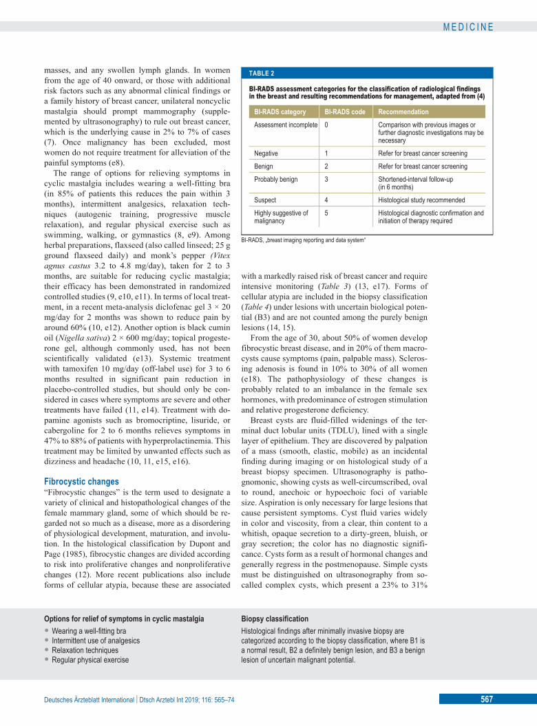
breast cancer peaks during the postmenopause (e2, e3). The management of benign breast changes includes clini-cal, radiological, and if necessary histological diagnostic investigations to rule out malignancy; palliation of symptoms; and counseling and monitoring of patients at increased risk of breast cancer. Typical presenting symp-toms such as pain, a palpable mass, and nipple discharge can be caused by a wide array of benign differential diag-noses (Table 1) and require targeted diagnostic imaging in addition to a comprehensive history and clinical examina -tion (e4). The BI-RADS (“breast imaging reporting and data system”) classification—the standardized descrip-tion of radiological findings—offers the clinician recom-mendations for action (Table 2) (4). Today’s minimally invasive techniques for achieving a confirmed histologi-cal diagnosis mean that surgical excision, previously common, can now be avoided in most cases.
PrevalenceBenign breast changes are more common in women of child-bearing age, peaking between the ages of 30 and 50, whereas the incidence of breast cancer peaks during the postmenopause.
DiagnosisIn addition to a comprehensive history and clinical examina -tion, diagnostic imaging (ultrasonography, mammography as indicated) is important for ruling out breast cancer.
Deutsches Ärzteblatt International | Dtsch Arztebl Int 2019; 116: 565–74 565

M E D I C I N E
MethodThe literature review performed for this article was based on a selective literature search of PubMed from 1985 to May 2019 including current national (AWMF, Association of Scientific Medical Societies in Ger-many) and international guidelines (EUSOMA, WHO, American College of Obstetricians and Gynecologists, American College of Radiology). This article discusses selected benign breast changes according to their inci-dence and significance in everyday clinical routine, grouping them by their main presenting symptoms.
Learning goalsAfter reading this article, readers should:
● Be familiar with the symptoms, diagnosis, and management of the most common benign breast diseases of women (mastalgia, fibrocystic changes, benign tumors, mammary duct ectasia, mastitis);
● Be able to distinguish benign breast changes from malignant findings;
● Know the breast cancer risk associated with cer-tain benign breast conditions.
Mastalgia Mastalgia (also called mastodynia) is the name given to pain related to the mammary gland occurring either spontaneously or in response to touch. Mastalgia is
classified as cyclic and noncyclic. Differential diag-noses to rule out are chest pain of extramammary origin such as intercostal neuralgia and pain from cardiac or vertebrogenic causes.
Surveys have shown that more than half of all women report significant breast pain, which in 30% to 40% of cases impairs their everyday and sexual life (e5). In two-thirds of cases, the pain is cyclic and is worst a week premenstrually and perimenstrually. Cyclic mastalgia manifests at around 30 years of age; the onset of noncyclic mastalgia is notably later, at a mean age of 41. What causes mastalgia is unknown. The fact that cyclic mastalgia improves in association with hormonal changes such as when the menopause is reached, and during pregnancy and lactation, suggest a hormonal cause (e5). In a woman with non-cyclic mastalgia, inflammatory, neoplastic, and vascular breast disease needs to be ruled out (e6). In cases with no underlying pathology, rates of sponta-neous remission within a few months to up to 3 years are high (5). After breast cancer treatment, 30% of women have persistent noncyclic mastalgia, especially those who are premenopausal, have a high body mass index, or have a concomitant psychologi-cal condition (6, e7).
Diagnostic investigations include obtaining a com-prehensive history and clinical exam. Palpation will reveal the localization of the pain, any mass or
MastalgiaIn two-thirds of cases, mastaliga is cyclic. Differential diag-noses for noncyclic mastalgia include inflammatory, neoplastic, and vascular breast disease.
Fibrocystic changes and risk of breast cancerOf the common fibrocystic breast changes, only proliferating lesions with atypia are associated with a clearly increased risk of breast cancer. These include flat epithelial atypia, atypical ductal hyperplasia, and atypical lobular hyperplasia.
TABLE 1
Differential diagnoses of benign breast changes, classified by main symptom
Symptom
Pain (unilateral)
Palpable mass
Nipple discharge
Benign causes
– Cysts– Fibrocystic breast disease– Hyperplasia of the breast– Mastitis– Postoperative changes
– Cysts– Fibrocystic breast disease– Fibroadenoma– Lipoma– Hamartoma– Pseudoangiomatous stromal hyperplasia (PASH)– Intramammary lymph nodes
– Hypothyreoidism– Galactorrhea– Intraductal papilloma– Periductal mastitis– Ductal ectasia
Incidence of breast cancer
2% to 7% (7)
8% (1, e1)
5% to 21% (29, e26)
566 Deutsches Ärzteblatt International | Dtsch Arztebl Int 2019; 116: 565–74
M E D I C I N E
masses, and any swollen lymph glands. In women from the age of 40 onward, or those with additional risk factors such as any abnormal clinical findings or a family history of breast cancer, unilateral noncyclic mastalgia should prompt mammography (supple-mented by ultrasonography) to rule out breast cancer, which is the underlying cause in 2% to 7% of cases (7). Once malignancy has been excluded, most women do not require treatment for alleviation of the painful symptoms (e8).
The range of options for relieving symptoms in cyclic mastalgia includes wearing a well-fitting bra (in 85% of patients this reduces the pain within 3 months), intermittent analgesics, relaxation tech-niques (autogenic training, progressive muscle relaxation), and regular physical exercise such as swimming, walking, or gymnastics (8, e9). Among herbal preparations, flaxseed (also called linseed; 25 g ground flaxseed daily) and monk’s pepper (Vitex agnus castus 3.2 to 4.8 mg/day), taken for 2 to 3 months, are suitable for reducing cyclic mastalgia; their efficacy has been demonstrated in randomized controlled studies (9, e10, e11). In terms of local treat-ment, in a recent meta-analysis diclofenac gel 3 × 20 mg/day for 2 months was shown to reduce pain by around 60% (10, e12). Another option is black cumin oil (Nigella sativa) 2 × 600 mg/day; topical progeste-rone gel, although commonly used, has not been scientifically validated (e13). Systemic treatment with tamoxifen 10 mg/day (off-label use) for 3 to 6 months resulted in significant pain reduction in placebo-controlled studies, but should only be con-sidered in cases where symptoms are severe and other treatments have failed (11, e14). Treatment with do-pamine agonists such as bromocriptine, lisuride, or cabergoline for 2 to 6 months relieves symptoms in 47% to 88% of patients with hyperprolactinemia. This treatment may be limited by unwanted effects such as dizziness and headache (10, 11, e15, e16).
Fibrocystic changes“Fibrocystic changes” is the term used to designate a variety of clinical and histopathological changes of the female mammary gland, some of which should be re-garded not so much as a disease, more as a disordering of physiological development, maturation, and involu-tion. In the histological classification by Dupont and Page (1985), fibrocystic changes are divided according to risk into proliferative changes and nonproliferative changes (12). More recent publications also include forms of cellular atypia, because these are associated
with a markedly raised risk of breast cancer and require intensive monitoring (Table 3) (13, e17). Forms of cellular atypia are included in the biopsy classification (Table 4) under lesions with uncertain biological poten-tial (B3) and are not counted among the purely benign lesions (14, 15).
From the age of 30, about 50% of women develop fibrocystic breast disease, and in 20% of them macro-cysts cause symptoms (pain, palpable mass). Scleros-ing adenosis is found in 10% to 30% of all women (e18). The pathophysiology of these changes is probably related to an imbalance in the female sex hormones, with predominance of estrogen stimulation and relative progesterone deficiency.
Breast cysts are fluid-filled widenings of the ter-minal duct lobular units (TDLU), lined with a single layer of epithelium. They are discovered by palpation of a mass (smooth, elastic, mobile) as an incidental finding during imaging or on histological study of a breast biopsy specimen. Ultrasonography is patho -gnomonic, showing cysts as well-circumscribed, oval to round, anechoic or hypoechoic foci of variable size. Aspiration is only necessary for large lesions that cause persistent symptoms. Cyst fluid varies widely in color and viscosity, from a clear, thin content to a whitish, opaque secretion to a dirty-green, bluish, or gray secretion; the color has no diagnostic signifi-cance. Cysts form as a result of hormonal changes and generally regress in the postmenopause. Simple cysts must be distinguished on ultrasonography from so-called complex cysts, which present a 23% to 31%
Options for relief of symptoms in cyclic mastalgia• Wearing a well-fitting bra• Intermittent use of analgesics• Relaxation techniques• Regular physical exercise
Biopsy classificationHistological findings after minimally invasive biopsy are categorized according to the biopsy classification, where B1 is a normal result, B2 a definitely benign lesion, and B3 a benign lesion of uncertain malignant potential.
TABLE 2
BI-RADS assessment categories for the classification of radiological findings in the breast and resulting recommendations for management, adapted from (4)
BI-RADS, „breast imaging reporting and data system“
BI-RADS category
Assessment incomplete
Negative
Benign
Probably benign
Suspect
Highly suggestive of malignancy
BI-RADS code
0
1
2
3
4
5
Recommendation
Comparison with previous images or further diagnostic investigations may be necessary
Refer for breast cancer screening
Refer for breast cancer screening
Shortened-interval follow-up (in 6 months)
Histological study recommended
Histological diagnostic confirmation and initiation of therapy required
Deutsches Ärzteblatt International | Dtsch Arztebl Int 2019; 116: 565–74 567

M E D I C I N E
risk of malignancy and must be histologically investi-gated (16).
Usual ductal hyperplasia (UDH) is characterized by cell proliferation within the ducts without cellular atypia. Blunt duct adenosis is characterized by an increase in the size and number of epithelial cells. Sclerosing adenosis features increased number and size of the acini within the TDLU accompanied by stromal hyperplasia. Because of their intraluminal (UDH) or subepithelial calcifications (blunt duct ade-nosis), these two forms of adenosis are often detected in the context of minimally invasive biopsies of sus-pect microcalcifications identified on mammography and according to the biopsy classification are classed as benign (B2) (17, e19). Radial scars are benign changes which on imaging show as suggestive of malignancy; histologically they show a stellate fibroelastic core with entrapped ducts and radial epithelial structures. Frequently, benign changes are found within this, as in usual ductal hyperplasia, ade-nosis, or ductal ectasia, and occasionally also in atypi-cal epithelial hyperplasia. Lesions >1 cm are termed complex sclerosing lesions (CSL) (18). Radial scars discovered incidentally and removed completely dur-ing minimally invasive diagnostic procedures are classified as B2 lesions; otherwise they are consid -ered as B3 (Table 4). After surgical excision of
minimally invasively diagnosed radial scars, the overall malignancy rate according to a recent meta-analysis is 7%, and after vacuum-assisted biopsy and without atypia it is only 1% (19, 20).
After histological evidence of usual ductal hyperplasia, sclerosing or blunt duct adenosis, and radial scar after percutaneous biopsy, it is important to correlate clinical, imaging, and pathology findings at a postinterventional case conference, in order to avoid a false-negative biopsy result. It is equally important to distinguish these conditions by histological study from atypical and malignant breast lesions (21, 22).
Proliferative changes without atypia are associated with only a slightly increased risk of breast cancer (e17). Chemoprevention, e.g., with raloxifen or tam-oxifen, is not indicated because of the unfavorable risk–benefit ratio (e20).
Similarly, if there is no family history and no clini-cal symptoms, no particular investigations are needed outside the officially recommended breast cancer screening program (screening every 2 years from age 50 to age 69).
Benign tumors (neoplastic changes)With an incidence of 25%, fibroadenoma is the most common benign tumor of the breast; peak onset is
Ductal hyperplasiaUsual ductal hyperplasia is characterized by cell proliferation within the ducts without cellular atypia. Blunt duct adenosis is characterized by an increase in the size and number of epithelial cells.
Multidisciplinary case conferenceTo avoid a false-negative biopsy finding, clinical, imaging, and pathology results all need to be discussed at a postinterven-tional case conference.
TABLE 3
Fibrocystic breast changes and associated risk of developing breast cancer, adapted from (12, 13)
UDH („usual ductal hyperplasia“): Cell proliferation in the ducts without cellular atypia; simple hyperplasia (3 to 4 cell layers), florid hyperplasia (>4 cell layers)
Blunt duct adenosis: Increase in size and number of epithelial cells, typical cylindrical differentiation with monomorphic cell nuclei and without atypia (columnar cell hyperplasia)
Sclerosing adenosis: Increase in size and number of acini in the TDLU (terminal ductal lobular unit) accompanied by proliferation of the surrounding stroma
Lesion type
Nonproliferating
Proliferating without atypia
Proliferating with atypia
Histopathological diagnosis
– Simple cysts – Papillary apocrine metaplasia
– Usual hyperplasia (UDH) – Columnar cell hyperplasia (blunt duct adenosis) – Sclerosing adenosis – Radial scar
– Flat epithelial atypia (FEA) – Atypical ductal hyperplasia (ADH) – Atypical lobular hyperplasia (ALH)
Relative risk of developing breast cancer in the future compared to the risk in the normal population[95% confidence interval]
1.17 [0.94; 1.47]
1.76 [1.58; 1.95]
3.93 [3.24; 4.76]
568 Deutsches Ärzteblatt International | Dtsch Arztebl Int 2019; 116: 565–74

M E D I C I N E
between 15 and 35 years of age (23). In terms of etiology and pathogenesis, a hormonally triggered mechanism is likely, as suggested by early onset during the premeno-pause, growth during pregnancy or estrogen therapy, and regression during the menopause (24, e21). A strik-ing clinical characteristic is a palpable mass measuring up to 3 cm. Sonographically, fibroadenomas appears as oval, well-circumscribed, hypoechoic focal masses that displace the surrounding parenchyma. Fast-growing breast carcinomas in younger women can look like fibroadenomas on ultrasonography. Asymptomatic fibroadenomas are often discovered as incidental find-ings during screening mammography. Fibroadenoma typically appears on a mammogram as a well- circumscribed mass, with or without popcorn-like calcifications, is pathognomonic and needs no further investigation (25). In the presence of the following
findings, histological diagnostic confirmation by means of percutaneous biopsy is advisable:
● Inconclusive ultrasound findings (BI-RADS 4)● Evidence of tendency to grow (clinical or sono-
graphic)● New palpable mass in a menopausal patient● Firm mass found in a patient with risk factors in
her history (positive family history, BRCA mu-tation)
● Mass with suspect microcalcifications on mam-mography.
Histologically there is a typical pattern of stromal proliferation with slit-like compressed epithelial com-ponents. It is important to distinguish this pattern from phyllodes tumors (incidence: 0.3% to 1% of all breast tumors), which are characterized by stromal hypercellularity, increased mitoses, and in some cases
FibroadenomasWith an prevalence of 25%, fibroadenomas are the most common benign tumors of the breast; peak onset is between 15 and 35 years of age.
Detection of fibroadenomasAsymptomatic fibroadenomas are often discovered as incidental findings during screening mammography. The typical appearance on mammography is that of a well-circumscribed mass with or without popcorn-like calcifications.
TABLE 4
Biopsy classification, adapted from (15)
ALH, atypical lobular neoplasia; DCIS, ductal carcinoma in situ; LCIS, lobular carcinoma in situ (classical); LN, lobular neoplasia
Category
B1
B2
B3
B4
B5
Definition
Tissue normal or cannot be assessed
Benign lesion
Benign lesion of uncertain malignant potential
Suspect lesion
Malignancy
Diagnoses
– Adipose tissue only (exception: lipoma, categorize as B2) – Regressive changes/involution�– Lactational changes
Mass: – Fibroadenoma, adenoma – Fibrocystic lesions, adenosis – Hamartoma – Small papilloma retrieved in toto – Pseudoangiomatous stromal hyperplasia (PASH) – Mastitis, abscess – Fat necrosis
Radiologically significant microcalcification(s): – Fibrocystic breast disease/(papillary) apocrine metaplasia – Blunt duct adenosis, sclerosing adenosis – Calcified fat necrosis
Lesions with increased risk of associated DCIS or invasive carcinoma:�– Atypical ductal hyperplasia (ADH) – Flat epithelial atypia (FEA)�– Classical lobular neoplasia (LN; ALH and LCIS)
Potentially heterogeneous lesions with the risk of incomplete sampling:�– Cellular fibroepithelial lesion or phyllodes tumor�– Intraductal papilloma with/without atypia, incompletely sampled – Radial scar or complex sclerosing lesion
B5a: In situ carcinoma�B5b: Invasive carcinoma B5c: Cannot be subcategorized as invasive or in situ carcinoma B5d: Malignancy of other histogenesis or metastasis
Deutsches Ärzteblatt International | Dtsch Arztebl Int 2019; 116: 565–74 569
M E D I C I N E
stromal cell atypia (24). Whereas asymptomatic fibroadenoma does not require any treatment, phyl-lodes tumors should if possible be excised with clear margins of 10 mm, as they can only be classified his-tologically as benign, malignant, or borderline on the basis of the surgical specimen. Phyllodes tumors have a high tendency to recur and high metastatic potential: 0.1% in benign phyllodes tumors, 1.6% in borderline tumors, and 16.7% in malignant tumors (26, e22). Symptomatic fibroadenomas can be removed by sur-gical excision (a good option if the mass measures >2 cm) or by ultrasound-guided vacuum-assisted biopsy. Cryoablation and treatment with high-intensity fo-cused ultrasound (HIFU) are experimental techniques (e23, e24).
In patients with a palpable breast tumor, the rare differential diagnosis of pseudoangiomatous stromal hyperplasia (PASH) is also a possibility. The radio-logical appearance is similar to that of fibroadenoma and the diagnosis is made by percutaneous core biopsy (e25). If there is evidence of a tendency to grow, or a discrepancy between clinical findings, im-aging findings, and histology, extirpation with wide margins should be carried out; if the tumor is pro-gressive, this could mean complete mastectomy (27). For PASH discovered incidentally on biopsy without any corresponding clinical or imaging findings, ultra-sound follow-up will suffice, as the risk of breast cancer is not increased (28).
Pathologic nipple discharge is the name given to spontaneous, often unilateral release of fluid from the nipple. In 50% of cases the cause is intraductal papilloma, in 25% to 35% of cases ductal ectasia, and in 5% to 15% of cases breast carcinoma (29, e26). Pa-pillomas are relatively common, making up 5% to 10% of benign breast tumors. They can occur as cen-trally located, solitary masses or as multiple, usually peripherally located lesions (“intraductal papilloma-tosis”) (30). In 80% of cases papillomas are clinically identified because of a spontaneous blood-stained or serous discharge from the nipple; less often they will be picked up by palpation or as an incidental finding on mammography. In a patient with low breast den-sity, solitary papillomas appear on the mammogram as rounded masses, in some cases with internal calcifications. Galactography shows papillomas as a filling defect or complete obstruction. On ultra -sonography, papillomas show as rounded, well- circumscribed, hypoechoic masses, but intraductal or intracystic proliferations may also be seen (e27). Histological study shows ragged, branching epithelial
protrusions intruding into a dilated duct, with a fibrovascular stromal core. As a result of the stromal sclerosis, microcalcifications are often present. The epithelial components can show a wide spectrum of morphological changes including atypical ductal hy-perplasia and ductal carcinoma in situ (DCIS) (e28). If the diagnosis is made histologically by minimally invasive biopsy, the papilloma is classified as a lesion of uncertain malignant potential (B3). Because of the lesion’s heterogeneity, it is possible that even if the core or vacuum-assisted biopsy was performed correctly, an area of higher-grade malignancy was missed (“sampling error”). The upgrade risk of papil-lary lesions confirmed by core biopsy—i.e., the risk that they will be identified at subsequent surgery as DCIS or invasive cancer—was reported in a recent meta-analysis as 16% (31). For this reason, it is recommended that a biopsy-confirmed, incompletely removed papilloma without atypia should be com-pletely excised. Papillary lesions showing atypia should always be surgically excised. No treatment is required for papillomas without atypia that are re-moved in toto, which are classified as B2 (Table 4) (20, e29). Because papillomas are so heterogeneous, postinterventional management should be determined in a multidisciplinary case conference (radiology, gynecology, pathology).
Galactorrhea is the name used to describe a milky secretion, which is often bilateral. The important differential diagnoses are listed in Table 5. Galactor-rhea is often caused by dopamine antagonists such as tricyclic antidepressants or selective serotonin reuptake inhibitors (SSRI). A serum prolactin concentration >200 ng/mL indicates the presence of a prolactinoma in the pituitary gland, and the patient should be referred for cerebral MRI and consultation with an endocrinologist (e30). If the prolactin concentration is normal and the secretion only occurs on pressure, no further diagnostic investigations are needed.
Inflammatory breast diseaseInflammatory breast conditions that occur in associ-ation with lactation are referred to as puerperal mastitis. Nonpuerperal mastitis is a collective term applied to all forms of mastitis occurring outside the lactation period, the most common of which are bacterial mastitis (59%), nonbacterial mastitis (25%), and special forms of nonpuerperal mastitis (14%) (e31).
Puerperal mastitis generally occurs during the first 3 months after delivery. Depending on study protocol,
Phyllodes tumorsWhereas asymptomatic fibroadenomas do not require treat-ment, phyllodes tumors should if possible be excised with clear margins of 10 mm, as they can only be histologically classified as benign, malignant, or borderline on the basis of the surgical specimen.
GalactorrheaGalactorrhea is the name used to describe a milky secretion, which is often bilateral. It is often caused by dopamine antago -nists such as tricyclic antidepressants or selective serotonin reuptake inhibitors.
570 Deutsches Ärzteblatt International | Dtsch Arztebl Int 2019; 116: 565–74

M E D I C I N E
its incidence worldwide has been reported at between 2% and 50%, and in selective cohorts a higher inci-dence has been observed (32, e32). A prospective study of 420 breastfeeding women in Glasgow found a cumulative incidence within 6 months of 18% (e33). According to the WHO definition, the symptoms of puerperal mastitis are:
● Pain● Local redness, warmth, and swelling of the breast,
usually unilateral● Fever (>38.4 °) and aching limbs● General feeling of illness (32).Diagnosis is based on the typical clinical symp-
toms. Risk factors include incorrect breastfeeding technique, stress, and lack of sleep (33). In many cases, epithelial lesions in the nipple area form an entry portal for pathogens from the infant’s naso -pharynx, which in the presence of blocked ducts can ascend and cause an infection (e34). In >90% of cases, the cause is Staphylococcus aureus; more rarely, coagulase-negative staphyloccoci, strepto -cocci, Pseudomonas aeruginosa, or Escherichia coli are the cause. Bacterial culture of the milk for diagnostic purposes is reserved for the following situ-ations:
● Symptoms remain uncontrolled after 48 h anti-biotic treatment
● Recurrent puerperal mastitis● Patient needs hospitalization.Histologically, puerperal mastitis is a phlegmon
that can lead to the formation of abscess and fistula (e35). The risk of abscess increases with the duration of symptoms before treatment starts, so early diag-
nosis and initiation of treatment are important (e36). Puerperal mastitis needs to be distinguished from milk stasis, a common condition the clinical symp-toms of which are much less severe and systemic symptoms are often absent altogether (e37). The most important steps for the treatment of puerperal mastitis are regular emptying of the breast (evidence level 2b) and early antibiotic therapy (33, 34, e38). Paraceta-mol (max. 4 × 1 g/day) or ibuprofen (3 × 500–800 mg/day) are suitable for relief of the general symp-toms (e39). Local antiphlogistic measures include the application of heat immediately before feeding and cooling the inflamed area during the breaks in feeding (33, e39, e40). If symptomatic treatment fails, after no more than 48 h calculated antibiotic therapy with an oral β-lactamase-resistant penicillin, e.g., flucloxa -cillin 3 × 1 g/day or dicloxacillin 4 × 1 g/day, or a first- or second-generation cephalosporin should be started (evidence level 2b). In patients allergic to penicillin, macrolide antibiotics (e.g., clarithromycin 4 × 500 mg/day or clindamycin 3 × 600 mg/day) are indicated. Treatment should be continued for at least 10 (to 14) days (33). If no improvement is seen within 48 to 72 h, ultrasonography should be carried out to check for abscess formation. Should an abscess be shown, it must be aspirated under ultrasonographic guidance (33, 35, e41). Surgical incision and drainage of an abscess should be done only if the abscess persists despite repeated aspiration, or is extensive, or is in an unfavorable location (36). Continued breast-feeding or expression of milk may be possible. Sec-ondary weaning is reserved for treatment-resistant cases and is carried out using orally administered
Symptoms of puerperal mastitisPuerperal mastitis is accompanied by fever, a general sense of illness, pain, and usually unilateral local redness and swelling of the breast.
Treatment of puerperal mastitisIn puerperal mastitis, which in most cases is caused by Staphylococcus aureus, early initiation of treatment with an oral β-lactamase-resistant penicillin is the therapy of choice. As an alternative in patients allergic to penicillin, macrolide antibiotics or clindamycin may be given.
TABLE 5
Differential diagnoses of galactorrhea
SSRI, selective serotonin reuptake inhibitors
Physiological
– Pregnancy and lactation – (Occasional) persistence after
weaning – Puberty – Stress
Medication-related
– Neuroleptics (phenothiazine, risperidone) – Metoclopramide – SSRI – Tricyclic antidepressants – Opiates – Methyldopamine – Clonidine – Verapamil – Cimetidine, ranitidine – Estrogens, oral contraceptives
Neoplastic
– Prolactinoma – Prolactin-secreting tumors (e.g., of
the lung)
Endocrine
– Hypo- and hyperthyroidism – Renal failure
Deutsches Ärzteblatt International | Dtsch Arztebl Int 2019; 116: 565–74 571
M E D I C I N E
bromocriptine (2 × 1.25 mg day 1, 2 × 2.5 mg day 2 to day 14). Prevention is about education in the right technique for breastfeeding:
● A calm environment, switching sides when breast-feeding
● Avoiding milk stasis, e.g., by stroking the breast towards the nipple during feeding
● Optimal technique for attaching the baby (e42)● Treating nipples with lanolin ointment to prevent
fissuring (33).Nonpuerperal mastitis includes all forms of peri-
ductal mastitis, the rarer granulomatous mastitis, and iatrogenic inflammation after surgery or radiotherapy. In patients over the age of 35 with nonpuerperal mas-titis, breast carcinoma should be excluded by a careful history and clinical examination followed by the selective use of mammography and/or ultra -sonography. Periductal mastitis is an inflammatory condition of the subareolar ducts and has a prevalence in nonbreastfeeding women of 5% to 9% (e43). It often occurs in women who are overweight with macromastia and nicotine abuse. The etiology is pre-sumed to be nicotine-induced damage to the ducts with tissue necrosis and consequent infection (e44). The clinical manifestation of periductal mastitis is of periareolar signs of inflammation (redness, swelling, warmth). Secondary bacterial infection can lead to ab-scess and fistula formation. Treatment involves relief of symptoms, antibiotic therapy, and, if there is ab-scess formation, aspiration and drainage (36, e43, e45). A prospective study of 151 patients showed complete resolution of symptoms in 81% after a single aspiration and antibiotic treatment (e46). For this reason, in cases of abscess-forming nonpuerperal mastitis, ultrasound-guided abscess aspiration and antibiotic therapy are the treatment of choice. The pathogen spectrum corresponds to the normal flora of the breast and nipple area. If S. aureus is shown to be present, it should be assumed that in more than 50% of cases it will be MRSA (e47). The drug of choice is clindamycin 3 × 600 mg/day, or alternatively amoxicillin/clavulanic acid 2 × 875/125 mg (e43). Treatment should be continued for at least 7 days (e47). Recurrent nonpuerperal mastitis is often caused by a ductal fistula exiting close to the areola, and this requires surgical revision with extirpation of the fistula (37, e48).
Necrotizing nonpuerperal mastitis is an extremely rare and life-threatening disease in a category of its own; it occurs in multimorbid patients and those under immune suppression or with diabetes mellitus.
Typical pathogens are group A β-hemolytic strepto-cocci, or combined infections with Bacteroides species or Escherichia coli. Clinical features in the early stage are edema, pain, skin pallor, and formation of blisters; later in the course, deep necrosis takes place. Since the inflammation will follow a fulminant course if untreated, spreading along the fascia and resulting in sepsis and multi -organ failure, immediate therapy with penicillin G 0.5–1 million IU i.v. (or alternatively broad-spectrum antibiotics) is required, as is meticulous surgical removal of necrotic tissue, even mastectomy if necessary (38, e49, e50).
Granulomatous mastitis is a rare inflammatory breast disease of women of childbearing age; its eti-ology is unknown (e51). Associations with lactation, hyperprolactinemia, and the presence of Corynebac-terium kroppenstedtii have been described (e52). The main symptom is a painful palpable mass, often with redness and swelling, and sometimes also skin retrac-tion. The symptoms and appearance on imaging are similar to those of diffuse breast cancer (39). In some cases abscess formation can be seen on ultra -sonography. Diagnosis is via percutaneous core biopsy. On histological study, the presence of granu-lomas with multinucleated giant cells indicates the di-agnosis.
There are two treatment strategies: ● Surgical excision (for small masses)● Systemic high-dose glucocorticoid therapy with
prednisolone 30 mg/day for at least 2 to 6 months followed by slow tapering with monitoring of the lesion (40).
According to a recent meta-analysis, the rate of complete remission after surgical intervention is 90.6% (95% confidence interval [83.8; 95.7]); after oral steroid therapy it is 71.8% [67.1; 76.3], and after combined treatment it is 94.5% [88.9; 98.3] (e53). In cases where there is extensive inflammation, gluco-corticoid therapy, despite its associated unwanted effects such as weight gain, hyperglycemia, gastro-duodenal ulcer, and the risk of Cushing syndrome, is regarded as the treatment of choice. Within a year after the cessation of treatment, a 20% recurrence rate has been reported (e53, e54). If symptoms are not se-vere and the patient rejects systemic glucocorticoid therapy, topical treatment with hydrocortisone acetate 0.5% 1 × daily may be attempted (e55). If glucocorti-coid therapy fails, low-dose methotrexate (7.5 to 25 mg/week + folic acid supplementation) is an option for treatment (e56).
Nonpuerperal mastitisNicotine abuse, overweight, and macromastia are regarded as risk factors for nonpuerperal mastitis. The differential diagnosis should include consideration of inflammatory breast cancer. Failure of antibiotic therapy may be due to the presence of an abscess, which can be diagnosed by ultrasound followed by aspiration.
Granulomatous mastitisBecause the symptoms and appearance on imaging of granulomatous mastitis are similar to those of breast cancer, the diagnosis is made on the histology after minimally invasive biopsy. Treatment is with prednisolone 30 mg/day given orally for at least 2 months.
572 Deutsches Ärzteblatt International | Dtsch Arztebl Int 2019; 116: 565–74

M E D I C I N E
Conflict of interest statement The authors declare that no conflict of interest exists.
Translated from the original German by Kersti Wagstaff, MA.
Manuscript received on 2 April 2019, revised version accepted on 2 July 2019.
References1. Eberl MM, Phillips RL Jr., Lamberts H, Okkes I, Mahoney MC: Characterizing
breast symptoms in family practice. Ann Fam Med 2008; 6: 528–33.2. Institut ZfKiRK: Bericht zum Krebsgeschehen in Deutschland 2016. Berlin: Robert
Koch-Institut; 2016. 73ff.3. Leitlinienprogramm Onkologie (Deutsche Krebsgesellschaft DK, AWMF): Interdis-
ziplinäre S3-Leitlinie für die Früherkennung, Diagnostik, Therapie und Nachsorge des Mammakarzinoms. Berlin 2018. www.leitlinienprogramm-onkologie.de/leitlinien/mammakarzinom/ (last accessed on 30 April 2019).
4. Walthers EM: ACR BI-RADS Atlas der Mammadiagnostik. Berlin, Heidelberg: Springer 2016.
5. Talimi-Schnabel J, Fink D: Mastodynie – wie soll man «Brustschmerz» abklären und behandeln? Praxis 2017; 106: 1101–6.
6. Wang K, Yee C, Tam S, et al.: Prevalence of pain in patients with breast cancer post-treatment: A systematic review. Breast 2018; 42: 113–27.
7. Iddon J, Dixon JM: Mastalgia. BMJ (Clinical research ed) 2013; 347: f3288.8. Hafiz SP, Barnes NLP, Kirwan CC: Clinical management of idiopathic mastalgia:
a systematic review. J Prim Health Care 2018; 10: 312–23.9. Verkaik S, Kamperman AM, van Westrhenen R, Schulte PFJ: The treatment of
premenstrual syndrome with preparations of Vitex agnus castus: a systematic review and meta-analysis. Am J Obstet Gynecol 2017; 217: 150–66.
10. Groen JW, Grosfeld S, Wilschut JA, Bramer WM, Ernst MF, Mullender MM: Cyclic and non-cyclic breast-pain: A systematic review on pain reduction, side effects, and quality of life for various treatments. Eur J Obstet Gynecol Reprod Biol 2017; 219: 74–93.
11. Srivastava A, Mansel RE, Arvind N, Prasad K, Dhar A, Chabra A: Evidence-based management of Mastalgia: a meta-analysis of randomised trials. Breast (Edinburgh, Scotland) 2007; 16: 503–12.
12. Dupont WD, Page DL: Risk factors for breast cancer in women with proliferative breast disease. N Engl J Med 1985; 312: 146–51.
13. Dyrstad SW, Yan Y, Fowler AM, Colditz GA: Breast cancer risk associated with benign breast disease: systematic review and meta-analysis. Breast Cancer Res Treat 2015; 149: 569–75.
14. Office for Official Publications of the European Communities: European guide -lines for quality assurance in breast cancer screening and diagnosis. Luxemburg: 2006, 246. https://publications.europa.eu/en/publication-detail/-/publication/4e74ee9b-df80–4c91-a5fb-85efb0fdda2b (last accessed on 31 March 2019).
15. Ellis IO, Humphreys S, Michell M, Pinder SE, Wells CA, Zakhour HD: Best Practice No 179. Guidelines for breast needle core biopsy handling and reporting in breast screening assessment. J Clin Pathol 2004; 57: 897–902.
16. Athanasiou A, Aubert E, Vincent Salomon A, Tardivon A: Complex cystic breast masses in ultrasound examination. Diagn Interv Imaging 2014; 95: 169–79.
17. Sinn HP, Elsawaf Z, Helmchen B, Aulmann S: Early Breast Cancer Precursor Lesions: Lessons learned from molecular and clinical studies. Breast care 2010; 5: 218–26.
18. Cohen MA, Newell MS: Radial scars of the breast encountered at core biopsy: re-view of histologic, imaging, and management considerations. AJR Am J Roentge-nol 2017; 209: 1168–77.
19. Farshid G, Buckley E: Meta-analysis of upgrade rates in 3163 radial scars ex-cised after needle core biopsy diagnosis. Breast Cancer Res Treat 2019; 174: 165–77.
20. Calhoun BC, Collins LC: Recommendations for excision following core needle biopsy of the breast: a contemporary evaluation of the literature. Histopathology 2016; 68: 138–51.
21. Tot T, Tabar L: The role of radiological-pathological correlation in diagnosing early breast cancer: the pathologist‘s perspective. Virchows Arch: an international jour-nal of pathology 2011; 458: 125–31.
22. Racz JM, Carter JM, Degnim AC: Challenging atypical breast lesions including flat epithelial atypia, radial scar, and intraductal papilloma. Ann Surg Oncol 2017; 24: 2842–7.
23. El-Wakeel H, Umpleby HC: Systematic review of fibroadenoma as a risk factor for breast cancer. Breast 2003; 12: 302–7.
24. Krings G, Bean GR, Chen YY: Fibroepithelial lesions; The WHO spectrum. Semin Diagn Pathol 2017; 34: 438–52.
25. Heywang-Köbrunner Sylvia H. SI: Bildgebende Mammadiagnostik. Stuttgart: Thieme Verlag 2015; 292–308.
26. Tan BY, Acs G, Apple SK, et al.: Phyllodes tumours of the breast: a consensus review. Histopathology 2016; 68: 5–21.
27. Layon DR, Wang C, Roth S, Brooks AD: Is surgical excision necessary in pseudo angiomatous stromal hyperplasia? Breast J 2016; 22: 595–6.
28. Virk RK, Khan A: Pseudoangiomatous stromal hyperplasia: an overview. Arch Pathol Lab Med 2010; 134: 1070–4.
29. Lee SJ, Trikha S, Moy L, et al.: ACR Appropriateness Criteria evaluation of nipple discharge. J Am Coll Radiol: JACR 2017; 14: 138–53.
30. Langer F, Hille-Betz U, Kreipe HH: [Papillary lesions of the breast]. Der Pathologe 2014; 35: 36–44.
31. Wen X, Cheng W: Nonmalignant breast papillary lesions at core-needle biopsy: a meta-analysis of underestimation and influencing factors. Ann Surg Oncol 2013; 20: 94–101.
32. World Health Organisation: Mastitis: causes and management. In: WHO, ed.: Geneva 2000.
33. Jacobs A, Abou-Dakn M, Becker K, et al.: S3-Leitlinie „Therapie entzündlicher Brusterkrankungen in der Stillzeit“. Senologie – Zeitschrift für Mammadiagnostik und -therapie 2014; 11: 50–6.
34. Peters J: [Mastitis puerperalis – causes and therapy]. Zentralbl Gynakol 2004; 126: 73–6.
35. Irusen H, Rohwer AC, Steyn DW, Young T: Treatments for breast abscesses in breastfeeding women. Cochrane Database Syst Rev 2015; 8: CD010490.
36. Lam E, Chan T, Wiseman SM: Breast abscess: evidence based management recommendations. Expert Rev Anti Infect Ther 2014; 12: 753–62.
37. Taffurelli M, Pellegrini A, Santini D, Zanotti S, Di Simone D, Serra M: Recurrent periductal mastitis: Surgical treatment. Surgery 2016; 160: 1689–92.
38. Ward ND, Harris JW, Sloan DA: Necrotizing fasciitis of the breast requiring emergent radical mastectomy. Breast J 2017; 23: 95–9.
39. Barreto DS, Sedgwick EL, Nagi CS, Benveniste AP: Granulomatous mastitis: etiology, imaging, pathology, treatment, and clinical findings. Breast Cancer Res Treat 2018; 171: 527–34.
40. Wolfrum A, Kummel S, Theuerkauf I, Pelz E, Reinisch M: Granulomatous mastitis: a therapeutic and diagnostic challenge. Breast care 2018; 13: 413–8.
Corresponding author: PD Dr. med. Angrit Stachs UFK Rostock am Klinikum Südstadt Südring 81 18059 Rostock, Germany [email protected]
Cite this as: Stachs A, Stubert J, Reimer T, Hartmann S: Benign breast disease in women. Dtsch Arztebl Int 2019; 116: 565–74. DOI: 10.3238/arztebl.2019.0565
►Supplementary materialeReferences: www.aerzteblatt-international.de/ref3319Case Study: www.aerzteblatt-international.de/19m0565
Further information on CME● Participation in the CME certification program is possible only over the
Internet: cme.aerzteblatt.de. This unit can be accessed until 10 November 2019. Submissions by letter, e-mail or fax cannot be con-sidered.
● The following CME units can still be accessed for credit: – „Psychopharmacological Treatment in Older People“
(issue 29-30/2019) until 21 October 2019– „Toxoplasmosis in Germany“
(issue 25/2019) until 15 September 2019● This article has been certified by the North Rhine Academy for Continu-
ing Medical Education. Participants in the CME program can manage their CME points with their 15-digit “uniform CME number” (einheitliche Fortbildungsnummer, EFN), which is found on the CME card (8027XXXXXXXXXXX). The EFN must be stated during registration on www.aerzteblatt.de (“Mein DÄ”) or else entered in “Meine Daten,” and the participant must agree to communication of the results.
Deutsches Ärzteblatt International | Dtsch Arztebl Int 2019; 116: 565–74 573
M E D I C I N E
CME credit for this unit can be obtained via cme.aerzteblatt.de until 10 November 2019.Only one answer is possible per question. Please select the answer that is most appropriate.
Question 1A patient is suffering from cyclic mastalgia. It mainly troubles her during her weekly session at the gym. You advise her to wear a well-fitting bra. What topical medication would you recommend?a) Diclofenac gelb) Rosemary oilc) Flaxseed oild) Prednisolone creame) Progesterone gel
Question 2Which of the diagnoses included under fibrocystic breast changes repre-sents a four-fold increased risk of breast cancer compared to the normal population, and therefore requires more intensive screening for breast cancer?a) Sclerosing adenosisb) Atypical ductal hyperplasiac) Radial scard) Blunt duct adenosise) Papillary apocrine metaplasia
Question 3A nonproliferating form of benign breast disease is diagnosed by screening mammography in a 52-year-old patient with no relevant personal or family history. What further investigations do you recommend in terms of breast cancer screening? a) Mammography every 12 monthsb) Breast ultrasound every 12 monthsc) Breast MRI every 12 monthsd) Clinical examination of the breast every 6 monthse) Attending screening mammography every 2 years
Question 4What are the most important therapeutic measures in a patient with incipient puerperal mastitis?a) Apply vaseline to the nipples and use yogurt compressesb) Start calculated oral antibiotic treatment and regular milk expressionc) Cool and compress the breastd) Secondary weaning; discard the remaining milke) Use nipple shields and oral nonsteroidal analgesics
Question 5A 22-year-old patient with puerperal mastitis in the upper lateral quadrant of the left breast shows no clear improvement of symptoms after 3 days of treatment. What diagnostic investigation should you now carry out with-out further delay?a) Take blood for blood count and CRP determination to check the diagnosisb) Galactography to rule out blocked ductsc) Breast ultrasound to rule out an abscessd) Mammography to rule out inflammatory breast cancere) Core biopsy of the area involved to rule out breast cancer
Question 6When should a bacterial culture of the breast milk be perfor-med in the case of a patient with puerperal mastitis?a) When symptom control has not been achieved after 48 h anti-biotic treatmentb) When the analgesics administered make breastfeeding
impossiblec) When cooling the affected breast fails to relieve symptoms
within 24 hd) When the patient has had puerperal mastitis after previous
deliveriese) When there is a family history of puerperal mastitis
Question 7What is the treatment of choice for abscess-forming nonpuerperal mastitis?a) Ultrasound-guided aspiration of the abscess and antibiotic therapyb) Expression of milk and cooling of the affected breastc) Mastectomyd) Galactography to rule out blocked ductse) Low-dose glucocorticoid therapy
Question 8In a 35-year-old patient with the BRCA1 mutation, breast ultrasonography is carried out for a newly discovered palpable mass and fibroadenoma is suspected. What should be done next?a) Mammographyb) Follow-up breast ultrasonography in 6 to 12 monthsc) Ultrasound-guided core biopsy of the massd) Surgical excision of the masse) Prophylactic bilateral mastectomy and reconstruction with
implants
Question 9For what diagnosis is spontaneous blood-streaked or serous nipple discharge the main symptom?a) Breast cancerb) Phyllodes tumorc) Blunt duct adenosisd) Lactating adenomae) Papilloma
Question 10With which class of drugs can galactorrhea occur as an unwanted effect?a) Nonsteroidal antirheumaticsb) Selective serotonin reuptake inhibitorsc) Thyroid hormoned) Proton pump inhibitorse) ACE inhibitors
►Participation in the CME certification program is possible only over the Internet: cme.aerzteblatt.de
574 Deutsches Ärzteblatt International | Dtsch Arztebl Int 2019; 116: 565–74
M E D I C I N E
Deutsches Ärzteblatt International | Dtsch Arztebl Int 2019; 116: 565–72 | Supplementary material I
e22. Tan PH, Ellis IO: Myoepithelial and epithelial-myoepithelial, me -senchymal and fibroepithelial breast lesions: updates from the WHO Classification of Tumours of the Breast 2012. J Clin Pathol 2013; 66: 465–70.
e23. Tomkovich KR: Interventional radiology in the diagnosis and treatment of diseases of the breast: a historical review and future perspective based on currently available techniques. Am J Roentgenol 2014; 203: 725–33.
e24. Golatta M, Harcos A, Pavlista D, et al.: Ultrasound-guided cryoab-lation of breast fibroadenoma: a pilot trial. Arch Gynecol Obstet 2015; 291: 1355–60.
e25. Raj SD, Sahani VG, Adrada BE, et al.: Pseudoangiomatous stromal hyperplasia of the breast: multimodality review with pathologic correlation. Curr Probl Diagn Radiol 2017; 46: 130–5.
e26. Dupont SC, Boughey JC, Jimenez RE, et al.: Frequency of dia -gnosis of cancer or high-risk lesion at operation for pathologic nipple discharge. Surgery 2015; 158: 988–94.
e27. Eiada R, Chong J, Kulkarni S, Goldberg F, Muradali D: Papillary lesions of the breast: MRI, ultrasound, and mammographic appear-ances. Am J Roentgenol 2012; 198: 264–71.
e28. Mulligan AM, O‘Malley FP: Papillary lesions of the breast: a review. Adv Anat Pathol 2007; 14: 108–19.
e29. Rageth CJ, O‘Flynn EAM, Pinker K, et al.: Second International Consensus Conference on lesions of uncertain malignant potential in the breast (B3 lesions). Breast Cancer Res Treat 2019; 174: 279–96.
e30. Verhelst J, Abs R: Hyperprolactinemia: pathophysiology and man -agement. Treat Endocrinol 2003; 2: 23–32.
e31. Peters F: Die nonpuerperale Mastitis. Gynäkologe 2001; 34: 930–9.e32. Angelopoulou A, Field D, Ryan CA, Stanton C, Hill C, Ross RP: The
microbiology and treatment of human mastitis. Med Microbiol Immunol 2018; 207: 83–94.
e33. Scott JA, Robertson M, Fitzpatrick J, Knight C, Mulholland S: Occurrence of lactational mastitis and medical management: a prospective cohort study in Glasgow. Int Breastfeed J 2008; 3: 21.
e34. Cullinane M, Amir LH, Donath SM, et al.: Determinants of mastitis in women in the CASTLE study: a cohort study. BMC Fam Pract 2015; 16: 181.
e35. Günther Klöppel HK, Wolfgang Remmele: Pathologie. In: Manfred Dietel GK, (ed.): Mamma, Weibliches Genitale, Schwangerschaft und Kindererkrankungen. Berlin, Heidelberg: Springer 2013.
e36. Dener C, Inan A: Breast abscesses in lactating women. World J Surg 2003; 27: 130–3.
e37. Betzold CM: An update on the recognition and management of lac-tational breast inflammation. J Midwifery Womens Health 2007; 52: 595–605.
e38. Thomsen AC, Espersen T, Maigaard S: Course and treatment of milk stasis, noninfectious inflammation of the breast, and infectious mastitis in nursing women. Am J Obstet Gynecol 1984; 149: 492–5.
e39. ABM clinical protocol #4: Mastitis. Breastfeed Med 2008; 3: 177–80.e40. Hamburger Still-Empfehlungen. Hamburg: HAG Gesundheit; 2009.
www.hag-gesundheit.de/uploads/docs/45.pdf (Last accessed on 17 July 2019).
e41. Lawrence R LR: Breastfeeding. A guide for the medical profession. 7 ed. Mosby, maryland Heights: Elsevier; 2011.
e42. Mohrbacher N SJ: Handbuch für die Stillberatung. In: Mohrbacher N SJ, ed.: München: La Leche Liga Deutschland e.V. 2000.
e43. Boakes E, Woods A, Johnson N, Kadoglou N: Breast Infection: a review of diagnosis and management practices. Eur J Breast Health 2018; 14: 136–43.
e44. Dixon JM, Ravisekar O, Chetty U, Anderson TJ: Periductal mastitis and duct ectasia: different conditions with different aetiologies. Br J Surg 1996; 83: 820–2.
eReferencese1. Barton MB, Elmore JG, Fletcher SW: Breast symptoms among
women enrolled in a health maintenance organization: frequency, evaluation, and outcome. Ann Intern Med 1999; 130: 651–7.
e2. Orr B, Kelley JL, 3rd: Benign breast diseases: evaluation and man -agement. Clin Obstet Gynecol 2016; 59: 710–26.
e3. Verdial FC, Etzioni R, Duggan C, Anderson BO: Demographic changes in breast cancer incidence, stage at diagnosis and age associated with population-based mammographic screening. J Surg Oncol 2017; 115: 517–22.
e4. Klassen CL, Hines SL, Ghosh K: Common benign breast concerns for the primary care physician. Cleve Clin J Med 2019; 86: 57–65.
e5. Scurr J, Hedger W, Morris P, Brown N: The prevalence, severity, and impact of breast pain in the general population. Breast J 2014; 20: 508–13.
e6. Wisbey JR, Kumar S, Mansel RE, Peece PE, Pye JK, Hughes LE: Natural history of breast pain. Lancet (London, England) 1983; 2: 672–4.
e7. Spivey TL, Gutowski ED, Zinboonyahgoon N, et al.: Chronic pain after breast surgery: a prospective, observational study. Ann Surg Oncol 2018; 25: 2917–24.
e8. Holland PA, Gateley CA: Drug therapy of mastalgia. What are the options? Drugs 1994; 48: 709–16.
e9. Hadi MS: Sports Brassiere: Is it a solution for Mastalgia? Breast J 2000; 6: 407–9.
e10. Schellenberg R: Treatment for the premenstrual syndrome with agnus castus fruit extract: prospective, randomised, placebo controlled study. BMJ (Clinical research ed) 2001; 322: 134–7.
e11. Mirghafourvand M, Mohammad-Alizadeh-Charandabi S, Ahmadpour P, Javadzadeh Y: Effects of Vitex agnus and Flaxseed on cyclic mastalgia: a randomized controlled trial. Complement Ther Med 2016; 24: 90–5.
e12. Colak T, Ipek T, Kanik A, Ogetman Z, Aydin S: Efficacy of topical nonsteroidal antiinflammatory drugs in mastalgia treatment. J Am Coll Surg 2003; 196: 525–30.
e13. Huseini HF, Kianbakht S, Mirshamsi MH, Zarch AB: Effectiveness of topical nigella sativa seed oil in the treatment of cyclic mastalgia: a randomized, triple-blind, active, and placebo-controlled clinical trial. Planta Med 2016; 82: 285–8.
e14. Fentiman IS, Caleffi M, Hamed H, Chaudary MA: Dosage and duration of tamoxifen treatment for mastalgia: a controlled trial. Br J Surg 1988; 75: 845–6.
e15. Kaleli S, Aydin Y, Erel CT, Colgar U: Symptomatic treatment of premenstrual mastalgia in premenopausal women with lisuride maleate: a double-blind placebo-controlled randomized study. Fertil Steril 2001; 75: 718–23.
e16. Aydin Y, Atis A, Kaleli S, Uludaq S, Goker N: Cabergolin versus bromocriptine for symptomatic treatment of premenstrual mastalgia: a randomised, open-label study. Eur J Obstet Gynecol Reprod Biol 2010; 150: 203–6.
e17. Hartmann LC, Sellers TA, Frost MH, et al.: Benign breast disease and the risk of breast cancer. N Engl J Med 2005; 353: 229–37.
e18. Rungruang B, Kelley JL, 3rd: Benign breast diseases: epidemiology, evaluation, and management. Clin Obstet Gynecol 2011; 54: 110–24.
e19. Pinder SE, Reis-Filho JS: Non-operative breast pathology: columnar cell lesions. J Clin Pathol 2007; 60: 1307–12.
e20. Cuzick J, Sestak I, Thorat MA: Impact of preventive therapy on the risk of breast cancer among women with benign breast disease. Breast 2015; 24 (Suppl 2): 51–5.
e21. Hughes LE, Mansel RE, Webster DJ: Aberrations of normal development and involution (ANDI): a new perspective on pathogenesis and nomenclature of benign breast disorders. Lancet 1987; 2: 1316–9.
Supplementary material to:
Benign Breast Disease in Womenby Angrit Stachs, Johannes Stubert, Toralf Reimer, and Steffi HartmannDtsch Arztebl Int 2019; 116: 565–74. DOI: 10.3238/arztebl.2019.0565
M E D I C I N E
II Deutsches Ärzteblatt International | Dtsch Arztebl Int 2019; 116: 565–72 | Supplementary material
e45. Fahrni M, Schwarz EI, Stadlmann S, Singer G, Hauser N, Kubik-Huch RA: Breast abscesses: diagnosis, treatment and outcome. Breast care (Basel, Switzerland) 2012; 7: 32–8.
e46. Christensen AF, Al-Suliman N, Nielsen KR, et al.: Ultrasound-guided drainage of breast abscesses: results in 151 patients. Br J Radiol 2005; 78: 186–8.
e47. Moazzez A, Kelso RL, Towfigh S, Sohn H, Berne TV, Mason RJ: Breast abscess bacteriologic features in the era of community-ac-quired methicillin-resistant Staphlyloccus aureus epidemics. Arch Surg (Chicago, III:1960) 2007; 142: 881–4.
e48. Hanavadi S, Pereira G, Mansel RE: How mammillary fistulas should be managed. Breast J 2005; 11: 254–6.
e49. Fayman K, Wang K, Curran R: A case report of primary necrotising fasciitis of the breast: A rare but deadly entity requiring rapid surgical management. Int J Surg Case Rep 2017; 31: 221–4.
e50. Al-Shareef B, Al-Shareef N: Necrotizing fasciitis of the breast: case report with literature review. Case Rep Surg 2018; 2018: 1370680.
e51. Freeman CM, Xia BT, Wilson GC, et al.: Idiopathic granulomatous mastitis: A diagnostic and therapeutic challenge. Am J Surg 2017; 214: 701–6.
e52. Johnstone KJ, Robson J, Cherian SG, Wan Sai Cheong J, Kerr K, Bligh JF: Cystic neutrophilic granulomatous mastitis associated with Corynebacterium including Corynebacterium kroppenstedtii. Pathol-ogy 2017; 49: 405–12.
e53. Lei X, Chen K, Zhu L, Song E, Su F, Li S: Treatments for idiopathic granulomatous mastitis: systematic review and meta-analysis. Breastfeed Med: 2017; 12: 415–21.
e54. Co M, Cheng VCC, Wei J, et al.: Idiopathic granulomatous mastitis: a 10-year study from a multicentre clinical database. Pathology 2018; 50: 742–7.
e55. Altintoprak F, Kivilcim T, Yalkin O, Uzunoglu Y, Kahyaoglu Z, Dilek ON: Topical steroids are effective in the treatment of idiopathic granulomatous mastitis. World J Surg 2015; 39: 2718–23.
e56. Akbulut S, Arikanoglu Z, Senol A, et al.: Is methotrexate an acceptable treatment in the management of idiopathic granulo -matous mastitis? Arch Gynecol Obstet 2011; 284: 1189–95.
Copyright © 2022 FDOKUMEN