Radioiodine Breast Uptake in Nonbreastfeeding Women: Clinical and Scintigraphic Characteristics
6
Radioiodine Breast Uptake in Nonbreastfeeding Women: Clinical and Scintigraphic Characteristics Muhammad M. Hammami and Siema Bakheet Departments of Medicine and Radiology, King Faisal Specialist Hospital and Research Centre, Riyadh, Saudi Arabia We studied the scintigraphic and associated clinical characteristics of radioiodine breast uptake in nonbreastfeeding thyroid cancer patients undergoing routine whole-body radioiodine scanning. Methods: We performed a retrospective review of the radioiodine scans and medical records of 30 prospectively collected cases. Results: Twenty-three nonpregnant patients had discontinued breastfeeding for a mean of 11.4 mo. Three postmenopausal and four single nulliparous patients had radioiodine breast uptake on one or more occasions. This represented about 6% of all female patients who had radioiodine scans over a 3-yr period. Four patterns of uptake, full, focal, crescentic and irregular, were observed. Breast uptake mimicked lung metastasis in nine patients. Expressible galactorrhea and moderately elevated prolactin levels were present in 48% and 24%, respectively, of patients examined. In 14 patients followed for an average of 11.4 mo, there were no consistent changes in the pattern or intensity of breast uptake. In 18 patients who had both 123Idiagnostic and 131Ipostablation scans within a few days, breast uptake was present on both scans in 75%. In four patients, breast uptake was present, despite the 4%-9% radioio dine uptake by the thyroid; inone patient, ¡odinatedcontrast material blocked the uptake of the thyroid gland but not of the breast. Conclusion: Althoughthe mechanisms of radioiodinebreast uptake remain unclear, breast uptake should be suspected in all female patients with radioiodine uptake in the chest area, even in the absence of a history of breastfeeding. Key Words: radioiodinescan; breast uptake; breastfeeding;prolac tin J NucÃ- Med 1996; 37:26-31 XVadioiodine, used routinely in the diagnosis and treatment of differentiated thyroid cancer, is known to be taken up by the lactating breast (1-4). Radioiodine uptake by the nonlactating breast, however, is not well recognized (5). Breast uptake of radioiodine can be readily distinguished from thyroid cancer métastases to the lung by its characteristic appearance, by obtaining posterior and/or lateral views of the chest or by observing a shift in the site of maximum concentration of radioiodine when the position of the breast is altered manually (5). Breast uptake, however, may be misinterpreted as lung métastasesfrom differentiated thyroid cancer if it presents with an atypical pattern and/or is clinically unexpected. We studied the prevalence and characteristics of radioiodine breast uptake in nonbreastfeeding patients with differentiated thyroid cancer. Since radioiodine is excreted in breast milk (6-8), the production of which is under the positive influence of prolactin (9), we also examined the prevalence of galactor rhea and elevated prolactin levels in these patients. Further, possible changes in the pattern and relative intensity of breast uptake over time, and possible differences in breast uptake between 123I diagnostic and 131Ipostablation scans, were also studied. Received Dec. 8, 1994; revision accepted May 15, 1995. For correspondence or reprints contact: Muhammad M. Hammami, MD, Department of Medicine (MBC 46), King Faisal Specialist Hospital and Research Centre, P.O. Box 3354, Riyadh 11211, Saudi Arabia. METHODS Patients All female patients with differentiated thyroid cancer who had breast uptake on radioiodine scans during a 3-yr period (January 1990 to December 1992) were prospectively identified. Their radioiodine scans and medical records were retrospectively re viewed. In our center, a questionnaire about breastfeeding, preg nancy and the date of the last menstrual period as well as measurement of TSH, free T4, thyroglobulin levels and plasma ß-hCGscreening test are routinely obtained prior to the adminis tration of radioiodine in all female patients. All patients had a TSH 2:30 mU/liter, free T4 < 6 pmole/liter and a negative plasma ß-hCGpregnancy test. Patients who had radioiodine breast uptake on scans obtained within 2 mo of terminating breastfeeding were excluded from this study. Whole-body Imaging Whole-body radioiodine scans were obtained after withdrawal of thyroxine therapy for 4 wk, using a large field of view gamma camera and appropriate collimation (medium-energy for 123I,high energy for 131I)for 300,000 counts or 10 min. Six spot images, including a posterior chest view, were obtained 24 hr after administration of 185 MBq (5 mCi) 123Ifor diagnostic scans and about 3 days after oral administration of a 131Itherapeutic dose for postablation scans (when the exposure rate is <1.8 mR/hr at 3 ft). Semiquantitative Analysis Because breast uptake of radioiodine was not quantitated, we related the intensity of breast uptake to that of residual thyroid tissue (or salivary glands if all thyroid tissue was previously ablated) on the same scan to semiquantite it for follow-up com parison (I23I diagnostic to I23Idiagnostic scans without intervening treatment) and for 123Idiagnostic scan to 131Ipostablation scan comparison. The following scale was used: + : minimal thyroid uptake; + + : moderate thyroid uptake; and + + + : marked thyroid uptake. TSH (normal range: 0.2-5.0 mU/liter), thyroglobulin (normal range: 2-70 /xg/liter), prolactin (normal range: 4.6-38 /u,g/ml) and free T4 (normal range: 10-25 pmole/liter) assays were performed according to the manufacturer's recommendations. RESULTS During a 3-yr period, a total of 2000 scans (diagnostic and postablation) were performed in our center. Since 72% of our patients with differentiated thyroid cancer are women and an average patient is scanned at least once a year (including diagnostic and postablation scans), it is estimated that about 480 (or less) female patients were scanned during this time frame. Of these, 23 nonpregnant patients had radioiodine breast uptake despite not having breastfed, when they first presented, for an average of 11.4 mo (Table 1). In addition, radioiodine breast uptake was seen in four single nulliparous patients and in three postmenopausal patients who did not breastfeed for more than 13 yr (Table 2). Thus, radioiodine breast uptake in the absence 26 THE JOURNALOFNUCLEARMEDICINE• Vol. 37 • No. 1 • January 1996
Transcript of Radioiodine Breast Uptake in Nonbreastfeeding Women: Clinical and Scintigraphic Characteristics

Radioiodine Breast Uptake in NonbreastfeedingWomen: Clinical and Scintigraphic CharacteristicsMuhammad M. Hammami and Siema BakheetDepartments of Medicine and Radiology, King Faisal Specialist Hospital and Research Centre, Riyadh, Saudi Arabia
We studied the scintigraphic and associated clinical characteristicsof radioiodine breast uptake in nonbreastfeeding thyroid cancerpatients undergoing routine whole-body radioiodine scanning.Methods: We performed a retrospective review of the radioiodinescans and medical records of 30 prospectively collected cases.Results: Twenty-three nonpregnant patients had discontinuedbreastfeeding for a mean of 11.4 mo. Three postmenopausal andfour single nulliparous patients had radioiodine breast uptake on oneor more occasions. This represented about 6% of all female patientswho had radioiodine scans over a 3-yr period. Four patterns ofuptake, full, focal, crescentic and irregular, were observed. Breastuptake mimicked lung metastasis in nine patients. Expressiblegalactorrhea and moderately elevated prolactin levels were presentin 48% and 24%, respectively, of patients examined. In 14 patientsfollowed for an average of 11.4 mo, there were no consistentchanges in the pattern or intensity of breast uptake. In 18 patientswho had both 123Idiagnostic and 131Ipostablation scans within a
few days, breast uptake was present on both scans in 75%. In fourpatients, breast uptake was present, despite the 4%-9% radioiodine uptake by the thyroid; in one patient, ¡odinatedcontrast materialblocked the uptake of the thyroid gland but not of the breast.Conclusion: Althoughthe mechanisms of radioiodinebreast uptakeremain unclear, breast uptake should be suspected in all femalepatients with radioiodine uptake in the chest area, even in theabsence of a history of breastfeeding.
Key Words: radioiodinescan; breast uptake; breastfeeding;prolactinJ NucÃMed 1996; 37:26-31
XVadioiodine, used routinely in the diagnosis and treatment ofdifferentiated thyroid cancer, is known to be taken up by thelactating breast (1-4). Radioiodine uptake by the nonlactatingbreast, however, is not well recognized (5). Breast uptake ofradioiodine can be readily distinguished from thyroid cancermétastases to the lung by its characteristic appearance, byobtaining posterior and/or lateral views of the chest or byobserving a shift in the site of maximum concentration ofradioiodine when the position of the breast is altered manually(5). Breast uptake, however, may be misinterpreted as lungmétastasesfrom differentiated thyroid cancer if it presents withan atypical pattern and/or is clinically unexpected.
We studied the prevalence and characteristics of radioiodinebreast uptake in nonbreastfeeding patients with differentiatedthyroid cancer. Since radioiodine is excreted in breast milk(6-8), the production of which is under the positive influence
of prolactin (9), we also examined the prevalence of galactorrhea and elevated prolactin levels in these patients. Further,possible changes in the pattern and relative intensity of breastuptake over time, and possible differences in breast uptakebetween 123Idiagnostic and 131Ipostablation scans, were also
studied.
Received Dec. 8, 1994; revision accepted May 15, 1995.For correspondence or reprints contact: Muhammad M. Hammami, MD, Department
of Medicine (MBC 46), King Faisal Specialist Hospital and Research Centre, P.O. Box3354, Riyadh 11211, Saudi Arabia.
METHODS
PatientsAll female patients with differentiated thyroid cancer who had
breast uptake on radioiodine scans during a 3-yr period (January1990 to December 1992) were prospectively identified. Theirradioiodine scans and medical records were retrospectively reviewed. In our center, a questionnaire about breastfeeding, pregnancy and the date of the last menstrual period as well asmeasurement of TSH, free T4, thyroglobulin levels and plasmaß-hCGscreening test are routinely obtained prior to the administration of radioiodine in all female patients. All patients had a TSH2:30 mU/liter, free T4 < 6 pmole/liter and a negative plasmaß-hCGpregnancy test. Patients who had radioiodine breast uptakeon scans obtained within 2 mo of terminating breastfeeding wereexcluded from this study.
Whole-body ImagingWhole-body radioiodine scans were obtained after withdrawal of
thyroxine therapy for 4 wk, using a large field of view gammacamera and appropriate collimation (medium-energy for 123I,highenergy for 131I)for 300,000 counts or 10 min. Six spot images,
including a posterior chest view, were obtained 24 hr afteradministration of 185 MBq (5 mCi) 123Ifor diagnostic scans andabout 3 days after oral administration of a 131Itherapeutic dose for
postablation scans (when the exposure rate is <1.8 mR/hr at 3 ft).
Semiquantitative AnalysisBecause breast uptake of radioiodine was not quantitated, we
related the intensity of breast uptake to that of residual thyroidtissue (or salivary glands if all thyroid tissue was previouslyablated) on the same scan to semiquantite it for follow-up comparison (I23I diagnostic to I23Idiagnostic scans without interveningtreatment) and for 123Idiagnostic scan to 131Ipostablation scan
comparison. The following scale was used: + : minimal thyroiduptake; + + : moderate thyroid uptake; and + + + : marked thyroiduptake.
TSH (normal range: 0.2-5.0 mU/liter), thyroglobulin (normalrange: 2-70 /xg/liter), prolactin (normal range: 4.6-38 /u,g/ml) andfree T4 (normal range: 10-25 pmole/liter) assays were performedaccording to the manufacturer's recommendations.
RESULTSDuring a 3-yr period, a total of 2000 scans (diagnostic and
postablation) were performed in our center. Since 72% of ourpatients with differentiated thyroid cancer are women and anaverage patient is scanned at least once a year (includingdiagnostic and postablation scans), it is estimated that about 480(or less) female patients were scanned during this time frame.Of these, 23 nonpregnant patients had radioiodine breast uptakedespite not having breastfed, when they first presented, for anaverage of 11.4 mo (Table 1). In addition, radioiodine breastuptake was seen in four single nulliparous patients and in threepostmenopausal patients who did not breastfeed for more than13 yr (Table 2). Thus, radioiodine breast uptake in the absence
26 THE JOURNALOF NUCLEARMEDICINE•Vol. 37 •No. 1 •January 1996

TABLE 1Clinical and Scintigraphic Findings in 23 Nonbreastfeeding Women with Radioiodine Breast Uptake
ProlactinlevelNormal
ElevatedUnavailableNo.
of patients11
48Age(yr)35
(24-44)31 (29-35)
35(19^6)No
breastfeeding(mo)*13.1
(2-72)7 (5-11)
11.1 (3-26)Galactorrhea
(%)igt
7533fScintigraphic
pattern of breast uptake(%)FU50
62.519FO18
2562.5IR27
06CR0
12.512.5NE5*
00
"On first presentation (average 11.4 mo).
^Two patients were not examined for expressible galactorrhea.*One patient had a unilateral breast uptake.Data are presented as the mean (range). Mean prolactin level was 20.3 (6-34) and 69 (41-95) ng/ml in the normal and elevated groups, respectively.
FU = full, FO = focal, IR = irregular, CR = crescentic, NE = negative.
of breastfeeding could be expected in at least 6% of hypothy-roid female patients with differentiated thyroid cancer.
Patterns of Radioiodine Breast UptakeAs shown in Table 1, four patterns of breast uptake, full,
focal, crescentic and irregular, similar to those reported in thelactating breast (10), were observed (Figs. 1-5). Two patients,a 32-yr-old who stopped breastfeeding for 4 mo and a post-menopausal patient (Patient 5, Table 2), had unilateral (right)breast uptake.
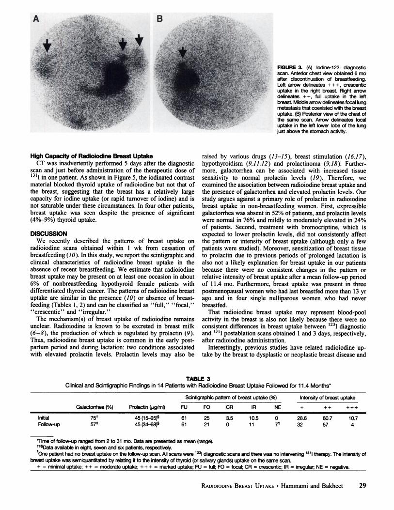
In most patients, the pattern and location (e.g., being lateral tothe torso) of radioiodine uptake by the breast were characteristic. In eight patients, however, breast uptake was irregular andmimicked lung métastases(Fig. 1A). Lung métastaseswereruled out by examining the posterior chest views that showed noactivity and normal chest radiographs. In addition, in a 35-yr-old woman, the combined picture of bilateral breast uptake andfocal residual stomach activity was difficult to distinguish fromlung métastases(Fig. 2C). That the uptake is in the stomach wassuggested from comparing the scan (Fig. 2C) to a previous scan(Fig. 2B). Furthermore, a posterior view of the chest showed noactivity, chest radiograph was normal and the thyroglobulinlevel was undetectable. In another 29-yr-old patient, breastuptake coexisted with a focus of lung metastasis (Fig. 3). In thispatient, the thyroglobulin level was 30 /itg/liter (with the onlyother uptake being in the thyroid bed and measuring <1%) anda lateral chest view (not shown) revealed clear separation of thefocus from stomach activity (being posterior to the stomach). In
four patients with insignificant residual thyroid uptake (^1thyroglobulin levels were 8.8, 18, 86, and 95 ¿ig/liter,respectively, initially suggesting that the uptake in the chest arearepresented metastatic thyroid foci. The negative posterior chestviews and chest radiographs and the typical appearance ofbreast uptake confirmed that the uptake was in fact related tothe breast and that thyroglobulin was most likely secreted byscan-negative microscopic thyroid foci.
Associated Expressible Galactorrhea and Prolactin LevelTwenty-one patients were examined for expressible galactor
rhea, whereas 17 patients had their prolactin level measured byscan findings. Galactorrhea was present in 10/21 (48%) patients. As shown in Table 1, the elevation of the prolactin level,when present, was mild to moderate (up to 2.5 times uppernormal level). This elevation is most likely secondary to thehypothyroid state (9,11,12). In three patients with elevatedprolactin levels, pituitary images were obtained which showedpartially empty sella in one patient and normal pituitary in theothers. Furthermore, prolactin levels were subsequently measured in two patients while on L-thyroxine treatment; theselevels became normal.
There were no consistent differences in the patterns of breastuptake in patients with normal prolactin levels compared tothose with elevated prolactin levels (Table 1). Furthermore, inthree patients, treatment with bromocriptine (up to 7.5 mg twicea day for 2 mo) did not result in a consistent change in thepattern or relative intensity of breast uptake, suggesting that
TABLE 2Radioiodine Breast Uptake in Single Nulliparous and Postmenopausal Patients
Patientno.Single1234Postmenopausal567Agefyr)25242533525955Lastbreastfeeding(yr)NeverNeverNeverNever132627Pattern/IntensityRightFUII/+
+FUII/++FUII/+FUII/+FUII/+FUII/+
+Crescentic/++LeftFUII/+
+FUII/++FUII/+FUII/+NegativeFUII/+
+FUII/++
Patients 1 and 5 had a negative examination for galactorrhea and normal prolactin levels of 32 and 25 jug/ml, respectively. The intensity of breast uptakewas semiquantitated by relating it to the intensity of thyroid (or salivary glands) uptake on the same scan.
+ = minimal uptake; + + = moderate uptake.
RADIOIODINEBREASTUPTAKE•Hammami and Bakheet 27

FIGURE 1. (A) lodine-123 diagnosticscan. Anterior chest view obtained 3 moafter discontinuation of breastfeeding.Curved arrows delineate +, irregularbreast uptake, bilaterally, mimicking lungmetastasis. (B) Anterior view of the chestof 131I(5550 MBq) postablation scan in
the same patient obtained 6 days later.Arrows delineate + ++, full uptake in theleft breast and +, focal uptake in the rightbreast. Breast uptake is more prominentin (B) than in (A) and the uptake is nowlateral to the torso, confirming its presence in the breasts rather than in thelungs.
»
m
prolactin may not play a primary role in radioiodine breastuptake in this group of patients. Moreover, patterns of breastuptake were similar among the 10 patients with expressiblegalactorrhea (50% full, 25% focal, 10% crescentic, 15%irregular), 11 patients without expressible galactorrhea (32%full, 50% focal, 4.5% crescentic, 9% irregular, 4.5% negative) and 9 patients not examined for expressible galactorrhea (67% full, 5.5% focal, 5.5% crescentic, 17% irregular,5.5% negative).
Breast Uptake on Follow-up ScansIn 14 patients, follow-up 123I diagnostic scans were
available 2-31 mo (mean 11.4) after the initial 123Idiagnos
tic scan without intervening radioiodine therapy (Table 3).One patient's scan was negative for breast uptake; this
patient had been followed for 5 mo. The remaining scans didnot show consistent changes in uptake pattern or relativeintensity. On some scans, there was a relative decrease inbreast uptake over time (Fig. 2), whereas on others theopposite was found, suggesting that breast uptake of radio-iodine may not be directly related to the time elapsed sinceprevious breastfeeding.
Comparison of lodine-123 Diagnostic Scans toIodine-131 Postablation Scans
Since the resolution of 123Iscans may be different than thatof 131Iscans and breast uptake on the relatively early 123Iimages (obtained at 24 hr as opposed to about 72 hr for 131Iimages) may represent blood-pool activity, we compared theresults of the two scans in 18 patients who had both scanswithin a few days of each other. The therapeutic I3II doseranged from 4403 to 7141 MBq (119-193 mCi). Each patient's
scans were studied by an examiner blinded to their relationship.As shown in Table 4, seven breasts were 123Iscan-negative and131Iscan-positive, whereas two breasts were 123Iscan-positiveand 131Iscan-negative. Figures 1 and 4 depict examples of thediscrepancy in the patterns of breast uptake between 123Idiagnostic and 131Ipostablation scans. There was, however,
total agreement on the pattern and relative intensity of theuptake on the two scans in 44% and 42% of breasts, respectively. Thus, there was no consistent difference in radioiodinebreast uptake between the two types of scans. The dose nor theradioiodine isotope nor the scanning time appeared to beimportant factors in radioiodine breast uptake.
. T3Õ
è
FIGURE 2. (A) lodine-123 diagnostic scan. Anteriorchest view obtained 6 mo after discontinuationof breastfeeding. Arrows delineate + ++, full, in (A)breastuptake bilaterally. (B)Anterior view of the chest of 123Idiagnostic scan in the same patient obtained 9 mo after the scan. Arrowhead delineates +, focal uptakein the left breast and the arrow delineates ++, full uptake in the right breast. (C) lodine-123 diagnostic scan. Anterior chest view obtained 3 mo after the scanin (B). The curved arrow delineates faint stomach activity that together with bilateral +, focal breast uptake, which persisted for 1 yr, mimicking lungmétastases.
28 THEJOURNALOFNUCLEARMEDICINE•Vol. 37 •No. 1 •January1996
B
FIGURE 3. (A) lodine-123 diagnostic
scan. Anterior chest view obtained 6 moafter discontinuation of breastfeeding.Left arrow delineates + + +, crescenticuptake in the right breast. Right arrowdelineates ++, full uptake in the leftbreast. Middle arrow delineates focal lungmetastasis that coexisted with the breastuptake. (B) Posterior view of the chest ofthe same scan. Arrow delineates focaluptake in the left lower lobe of the lungjust above the stomach activity.
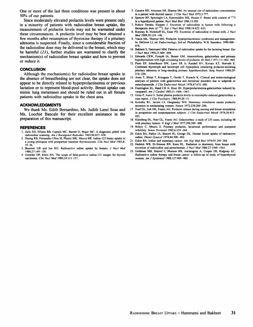
High Capacity of Radioiodine Breast UptakeCT was inadvertently performed 5 days after the diagnostic
scan and just before administration of the therapeutic dose of13II in one patient. As shown in Figure 5, the iodinated contrast
material blocked thyroid uptake of radioiodine but not that ofthe breast, suggesting that the breast has a relatively largecapacity for iodine uptake (or rapid turnover of iodine) and isnot saturable under these circumstances. In four other patients,breast uptake was seen despite the presence of significant(4%-9%) thyroid uptake.
DISCUSSIONWe recently described the patterns of breast uptake on
radioiodine scans obtained within 1 wk from cessation ofbreastfeeding (10). In this study, we report the scintigraphic andclinical characteristics of radioiodine breast uptake in theabsence of recent breastfeeding. We estimate that radioiodinebreast uptake may be present on at least one occasion in about6% of nonbreastfeeding hypothyroid female patients withdifferentiated thyroid cancer. The patterns of radioiodine breastuptake are similar in the presence (10) or absence of breastfeeding (Tables 1, 2) and can be classified as "full," "focal,""crescentic" and "irregular."
The mechanism(s) of breast uptake of radioiodine remainsunclear. Radioiodine is known to be excreted in breast milk(6-8), the production of which is regulated by prolactin (9).Thus, radioiodine breast uptake is common in the early post-partum period and during lactation: two conditions associatedwith elevated prolactin levels. Prolactin levels may also be
raised by various drugs (¡3-15), breast stimulation (16,17),hypothyroidism (9,11,12) and prolactinoma (9,18). Furthermore, galactorrhea can be associated with increased tissuesensitivity to normal prolactin levels (79). Therefore, weexamined the association between radioiodine breast uptake andthe presence of galactorrhea and elevated prolactin levels. Ourstudy argues against a primary role of prolactin in radioiodinebreast uptake in non-breastfeeding women. First, expressiblegalactorrhea was absent in 52% of patients, and prolactin levelswere normal in 76% and mildly to moderately elevated in 24%of patients. Second, treatment with bromocriptine, which isexpected to lower prolactin levels, did not consistently affectthe pattern or intensity of breast uptake (although only a fewpatients were studied). Moreover, sensitization of breast tissueto prolactin due to previous periods of prolonged lactation isalso not a likely explanation for breast uptake in our patientsbecause there were no consistent changes in the pattern orrelative intensity of breast uptake after a mean follow-up periodof 11.4 mo. Furthermore, breast uptake was present in threepostmenopausal women who had last breastfed more than 13 yrago and in four single nulliparous women who had neverbreastfed.
That radioiodine breast uptake may represent blood-poolactivity in the breast is also not likely because there were noconsistent differences in breast uptake between 123Idiagnosticand I3II postablation scans obtained 1 and 3 days, respectively,
after radioiodine administration.Interestingly, previous studies have related radioiodine up
take by the breast to dysplastic or neoplastic breast disease and
TABLE 3Clinical and Scintigraphic Findings in 14 Patients with Radioiodine Breast Uptake Followed for 11.4 Months*
Scintigraphic pattern of breast uptake (%) Intensity of breast uptake
Galactorrhea (%) Prolactin (/xg/ml) FU FO CR IR NE
InitialFollow-up75f 57*45(15-95)§ 45 (34-68)§61 6125 213.5 010.5 110 7*28.6 3260.7 5710.7 4
Time of follow-up ranged from 2 to 31 mo. Data are presented as mean (range).n§Dataavailable in eight, seven and six patients, respectively.One patient had no breast uptake on the follow-up scan. All scans were 123Idiagnostic scans and there was no intervening 131Itherapy. The intensity of
breast uptake was semiquantitated by relating it to the intensity of thyroid (or salivary glands) uptake on the same scan.+ = minimal uptake; ++ = moderate uptake; + + + = marked uptake; FU = full; FO = focal; CR = crescentic; IR = irregular; NE = negative.
RADIOIODINEBREASTUPTAKE•Hammami and Bakheet 29

TABLE 4Comparison of Radioiodine Breast Uptake on Radioiodine
Diagnostic and Postablation Scans
123Idiagnosticscan
Full Focal Crescentic Irregular Negative
1311PostablationscanFullFocalIrregularNegative101122
361—
—21———7——
123IDiagnostic scan
131IPostablationscan-
—+
3++4++ + ——13362—21——2—
The scintigraphic patterns and intensity of radioiodine breast uptake werecompared on the two scans in 18 patients (36 breasts). The number ofbreasts in each category is presented. The postablation scan was obtainedwithin few days of, and interpreted independently from, the diagnostic scanin each patient. The intensity of breast uptake was semiquantitated byrelating it to the intensity of thyroid (or salivary glands) uptake on the samescan. + = minimal uptake; + + = moderate uptake; + + + = marked uptake.
to iodine deficiency (20,21). Whether iodine deficiency contributed to the prevalence of positive breast uptake, or whethersome of our patients had dysplastic/neoplastic breast diseases, isnot known. Finally, the role of elevated TSH levels or of thehypothyroid state, routinely induced before image acquisition,remains to be studied.
The presence of breast uptake, despite 4%-9% thyroid uptake
of radioiodine in four patients, suggests that the breast has arelatively high affinity and/or capacity for radioiodine. Thelatter is supported by the observation that iodinated contrastmaterial suppressed thyroid, but not breast, radioiodine uptake.The relatively high capacity of the breast for radioiodine uptakecould be related to the larger mass of the breast or to the factthat turnover of iodine in the breast is faster, since it remainslargely unorganified (22).
Radioiodine breast uptake can potentially be misinterpretedas lung métastaseswhen:
1. A history of breastfeeding is not obtained or the occurrence of breast uptake without breastfeeding is not acknowledged.
2. The uptake is irregular or unilateral.3. There is a coexisting lung (or other) metastasis.4. There is a coexisting elevated thyroglobulin level but an
otherwise unremarkable scan.
FIGURE 4. (A) lodine-123 diagnosticscan. Anterior chest view of Patient 1.Arrows delineate + +, full breast uptakebilaterally, a picture typical of a lactatingbreast. (B) lodine-131 postablation scan.
Anterior view of the chest in the samepatient obtained 5 days after the scan in(A). Arrows delineate minimal full breastuptake bilaterally. Breast uptake is moreprominent in (A) than in (B).
B
B
FIGURE 5. (A) lodine-123 diagnosticscan. Anterior chest view obtained 18 moafter discontinuation of breastfeeding. Arrows delineate + + , focal and +, focaluptake, in the right and left breasts, respectively. Black triangles delineate twofoci of remnant thyroid tissue. (B) Anteriorchest view of a 5550-MBq 131Ipostabla-
tion scan obtained 5 days after the scanin (A) and after inadvertent administrationof CT contrast material for an unrelatedstudy. There is a disappearance of thyroid uptake and persistent breast (curvedarrows) as well as submandibular glanduptake.
30 THE JOURNALOFNUCLEARMEDICINE•Vol. 37 •No. 1 •January 1996
One or more of the last three conditions was present in about50% of our patients.
Since moderately elevated prolactin levels were present onlyin a minority of patients with radioiodine breast uptake, themeasurement of prolactin levels may not be warranted underthese circumstances. A prolactin level may be best obtained afew months after resumption of thyroxine therapy if a pituitaryadenoma is suspected. Finally, since a considerable fraction ofthe radioiodine dose may be delivered to the breast, which maybe harmful (23), further studies are warranted to clarify themechanism(s) of radioiodine breast uptake and how to preventor reduce it.
CONCLUSIONAlthough the mechanism(s) for radioiodine breast uptake in
the absence of breastfeeding are not clear, the uptake does notappear to be directly related to hyperprolactinemia or previouslactation or to represent blood-pool activity. Breast uptake canmimic lung métastasesand should be ruled out in all femalepatients with radioiodine uptake in the chest area.
ACKNOWLEDGMENTSWe thank Ms. Edith Bernardino, Ms. Judith Lansi Sosa and
Ms. Loochie Bancale for their excellent assistance in thepreparation of this manuscript.
REFERENCES1. Zalis ED, Ellison RB. Captain MC. Barrett O, Major MC. A diagnostic pitfall with
radioiodine scanning. Am J Roentgvnol Rutlitither 1965;94:837-838.2. Duong RB. Fernandez-Ulloa M. Planitz MK. Maxon HR. Iodine-123 breast uptake in
a young primipara with postpartum transient thyrotoxicosis. Clin NucÃMei/ 1983:8:35-36.
3. Baumier GR and Joo KG. Radioactive iodine uptake by breasts. J NucÃMed1986:27:149-150.
4. Greenler DP. Klein HA. The scope of false-positive iodine-131 images for thyroidcarcinoma. Clin NucÃMed 1989:14:111-117.
5. Ganatra RD. Atmaram SH. Sharma SM. An unusual site of radioiodine concentrationin a patient with thyroid cancer. J Clin NucÃMed 1972:3:777.
6. Spencer RP, Spitznagle LA. Karimeddini MK, Hosain F. Breast milk content of ml
in a hypothyroid patient. NucÃMed Biol 1986:13:585.7. Rubow Sietske. Klopper J. Excretion of radioiodine in human milk following a
therapeutic dose of I3II. Eur J NucÃMed 1988:14:632-633.
8. Romney B. Nicholoff EL. Esser PD. Excretion of radioiodine in breast milk. J NucÃMed 1989;30:124-146.
9. Vance ML. Thomer MO. Prolactin: hyperprolactinemic syndromes and management.In: DeGroot LJ, ed. Endocrinology, 2nd ed. Philadelphia: W.B. Saunders; 1990:408-
418.10. Bakheet S, Hammami MM. Patterns of radioiodine uptake by the lactating breast. Eur
JNucI Med 1994:21:604-608.
11. Edwards CRW, Forsyth 1A. Besser GM. Amenorrhoea. galactorrhea and primaryhypothyroidism with high circulating levels of prolactin. BrMedJ 1971:111:462-464.
12. Pioro EP, Scheithauer BW, Laws ER Jr, Randall RV, Kovacs KT, Horvath E.Combined thyrotroph and lactotroph cell hyperplasia simulating prolactin-secretingpituitary adenoma in long-standing primary hypothyroidism. Surg Neural I988;29:218-226.
13. Aono T, Shioji T, Kinugasa T. Onishi T. Kurachi K. Clinical and endocrinologicalanalyses of patients with galactorrhea and menstrual disorders due to sulpiride ormetoclopramide. J Clin Endocrino! Melah 1978:47:675-680.
14. Fearrington EL, Rand CH Jr, Rose JD. Hyperprolactinemia-galactorrhea induced byverapamil. Am J Cardiol 1983;5I:1466-1467.
15. Gioia P, Asnis G. Serial plasma prolactin levels in neuroleptic-induccd galactorrhea: acase report. J Clin Psychiatry 1988:49:28-31.
16. Kolodny RC. Jacobs LS, Daughaday WH. Mammary stimulation causes prolactinsecretion in nonlactating women. Nature 1972:238:284-286.
17. Noel GL. Suh HK, Frantz AG. Prolactin release during nursing and breast stimulationin postpartum and nonpostpartum subjects. J Clin Endocrinol Metah 1974:38:413-
423.18. Kleinberg DL, Noel GL, Frantz AG. Galactorrhea: a study of 235 cases, including 48
with pituitary tumors. .V EnglJ Med 1977:296:589-600.
19. Robyn C. Meuris S. Pituitary prolactin. lactational performance and puerperalinfertility. Semin Perinatal 1982:6:254-264.
20. Eskin BA, Parker JA, Bassett JG, George DL. Human breast uptake of radioactiveiodine. Obstet Gynecol 1974:44:398-402.
21. Eskin BA. Iodine and mammary cancer. Adv Exp Med Biol 1974;91:293-304.22. Hedrick WR. Di-Simone RN, Keen RL. Radiation in dosimetry from breast milk
excretion of radioiodine and pertechnetate. J NucÃMed 1986:27:1569-1561.
23. Goldman MB, Maloof F. Monson RR. Aschengrau A. Cooper DS. Ridgway EC.Radioactive iodine therapy and breast cancer: a follow-up of study of hyperthyroidwomen. Am J Epidemial 1988:127:969-980.
RADIOIODINEBREASTUPTAKE•Hammami and Bakheet 31