Macrophage Inhibitory Cytokine-1 (MIC-1/GDF15) Gene Deletion Promotes Cancer Growth in TRAMP...
12
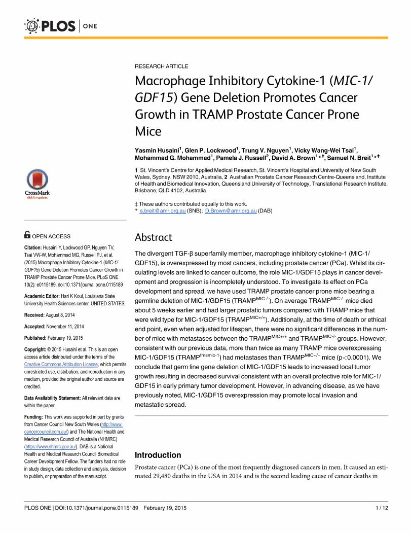
RESEARCH ARTICLE Macrophage Inhibitory Cytokine-1 (MIC-1/ GDF15) Gene Deletion Promotes Cancer Growth in TRAMP Prostate Cancer Prone Mice Yasmin Husaini 1 , Glen P. Lockwood 1 , Trung V. Nguyen 1 , Vicky Wang-Wei Tsai 1 , Mohammad G. Mohammad 1 , Pamela J. Russell 2 , David A. Brown 1 * ‡ , Samuel N. Breit 1 * ‡ 1 St. Vincent’s Centre for Applied Medical Research, St. Vincent’s Hospital and University of New South Wales, Sydney, NSW 2010, Australia, 2 Australian Prostate Cancer Research Centre-Queensland, Institute of Health and Biomedical Innovation, Queensland University of Technology, Translational Research Institute, Brisbane, QLD 4102, Australia ‡ These authors contributed equally to this work. * [email protected] (SNB); [email protected] (DAB) Abstract The divergent TGF-β superfamily member, macrophage inhibitory cytokine-1 (MIC-1/ GDF15), is overexpressed by most cancers, including prostate cancer (PCa). Whilst its cir- culating levels are linked to cancer outcome, the role MIC-1/GDF15 plays in cancer devel- opment and progression is incompletely understood. To investigate its effect on PCa development and spread, we have used TRAMP prostate cancer prone mice bearing a germline deletion of MIC-1/GDF15 (TRAMP MIC-/- ). On average TRAMP MIC-/- mice died about 5 weeks earlier and had larger prostatic tumors compared with TRAMP mice that were wild type for MIC-1/GDF15 (TRAMP MIC+/+ ). Additionally, at the time of death or ethical end point, even when adjusted for lifespan, there were no significant differences in the num- ber of mice with metastases between the TRAMP MIC+/+ and TRAMP MIC-/- groups. However, consistent with our previous data, more than twice as many TRAMP mice overexpressing MIC-1/GDF15 (TRAMP fmsmic-1 ) had metastases than TRAMP MIC+/+ mice (p<0.0001). We conclude that germ line gene deletion of MIC-1/GDF15 leads to increased local tumor growth resulting in decreased survival consistent with an overall protective role for MIC-1/ GDF15 in early primary tumor development. However, in advancing disease, as we have previously noted, MIC-1/GDF15 overexpression may promote local invasion and metastatic spread. Introduction Prostate cancer (PCa) is one of the most frequently diagnosed cancers in men. It caused an esti- mated 29,480 deaths in the USA in 2014 and is the second leading cause of cancer deaths in PLOS ONE | DOI:10.1371/journal.pone.0115189 February 19, 2015 1 / 12 OPEN ACCESS Citation: Husaini Y, Lockwood GP, Nguyen TV, Tsai VW-W, Mohammad MG, Russell PJ, et al. (2015) Macrophage Inhibitory Cytokine-1 (MIC-1/ GDF15) Gene Deletion Promotes Cancer Growth in TRAMP Prostate Cancer Prone Mice. PLoS ONE 10(2): e0115189. doi:10.1371/journal.pone.0115189 Academic Editor: Hari K Koul, Louisiana State University Health Sciences center, UNITED STATES Received: August 8, 2014 Accepted: November 11, 2014 Published: February 19, 2015 Copyright: © 2015 Husaini et al. This is an open access article distributed under the terms of the Creative Commons Attribution License, which permits unrestricted use, distribution, and reproduction in any medium, provided the original author and source are credited. Data Availability Statement: All relevant data are within the paper. Funding: This work was supported in part by grants from Cancer Council New South Wales (http://www. cancercouncil.com.au/) and The National Health and Medical Research Council of Australia (NHMRC) (https://www.nhmrc.gov.au/). DAB is a National Health and Medical Research Council Biomedical Career Development Fellow. The funders had no role in study design, data collection and analysis, decision to publish, or preparation of the manuscript.
-
Upload
independent -
Category
Documents
-
view
0 -
download
0
Transcript of Macrophage Inhibitory Cytokine-1 (MIC-1/GDF15) Gene Deletion Promotes Cancer Growth in TRAMP...
RESEARCH ARTICLE
Macrophage Inhibitory Cytokine-1 (MIC-1/GDF15) Gene Deletion Promotes CancerGrowth in TRAMP Prostate Cancer ProneMiceYasmin Husaini1, Glen P. Lockwood1, Trung V. Nguyen1, VickyWang-Wei Tsai1,Mohammad G. Mohammad1, Pamela J. Russell2, David A. Brown1*‡, Samuel N. Breit1*‡
1 St. Vincent’s Centre for Applied Medical Research, St. Vincent’s Hospital and University of New SouthWales, Sydney, NSW 2010, Australia, 2 Australian Prostate Cancer Research Centre-Queensland, Instituteof Health and Biomedical Innovation, Queensland University of Technology, Translational Research Institute,Brisbane, QLD 4102, Australia
‡ These authors contributed equally to this work.* [email protected] (SNB); [email protected] (DAB)
AbstractThe divergent TGF-β superfamily member, macrophage inhibitory cytokine-1 (MIC-1/
GDF15), is overexpressed by most cancers, including prostate cancer (PCa). Whilst its cir-
culating levels are linked to cancer outcome, the role MIC-1/GDF15 plays in cancer devel-
opment and progression is incompletely understood. To investigate its effect on PCa
development and spread, we have used TRAMP prostate cancer prone mice bearing a
germline deletion of MIC-1/GDF15 (TRAMPMIC-/-). On average TRAMPMIC-/- mice died
about 5 weeks earlier and had larger prostatic tumors compared with TRAMP mice that
were wild type for MIC-1/GDF15 (TRAMPMIC+/+). Additionally, at the time of death or ethical
end point, even when adjusted for lifespan, there were no significant differences in the num-
ber of mice with metastases between the TRAMPMIC+/+ and TRAMPMIC-/- groups. However,
consistent with our previous data, more than twice as many TRAMP mice overexpressing
MIC-1/GDF15 (TRAMPfmsmic-1) had metastases than TRAMPMIC+/+ mice (p<0.0001). We
conclude that germ line gene deletion of MIC-1/GDF15 leads to increased local tumor
growth resulting in decreased survival consistent with an overall protective role for MIC-1/
GDF15 in early primary tumor development. However, in advancing disease, as we have
previously noted, MIC-1/GDF15 overexpression may promote local invasion and
metastatic spread.
IntroductionProstate cancer (PCa) is one of the most frequently diagnosed cancers in men. It caused an esti-mated 29,480 deaths in the USA in 2014 and is the second leading cause of cancer deaths in
PLOSONE | DOI:10.1371/journal.pone.0115189 February 19, 2015 1 / 12
OPEN ACCESS
Citation: Husaini Y, Lockwood GP, Nguyen TV,Tsai VW-W, Mohammad MG, Russell PJ, et al.(2015) Macrophage Inhibitory Cytokine-1 (MIC-1/GDF15) Gene Deletion Promotes Cancer Growth inTRAMP Prostate Cancer Prone Mice. PLoS ONE10(2): e0115189. doi:10.1371/journal.pone.0115189
Academic Editor: Hari K Koul, Louisiana StateUniversity Health Sciences center, UNITED STATES
Received: August 8, 2014
Accepted: November 11, 2014
Published: February 19, 2015
Copyright: © 2015 Husaini et al. This is an openaccess article distributed under the terms of theCreative Commons Attribution License, which permitsunrestricted use, distribution, and reproduction in anymedium, provided the original author and source arecredited.
Data Availability Statement: All relevant data arewithin the paper.
Funding: This work was supported in part by grantsfrom Cancer Council New South Wales (http://www.cancercouncil.com.au/) and The National Health andMedical Research Council of Australia (NHMRC)(https://www.nhmrc.gov.au/). DAB is a NationalHealth and Medical Research Council BiomedicalCareer Development Fellow. The funders had no rolein study design, data collection and analysis, decisionto publish, or preparation of the manuscript.
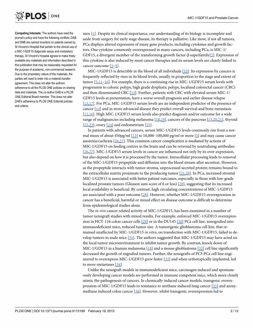
men [1]. Despite its clinical importance, our understanding of its biology is incomplete andaside from surgery for early stage disease, its therapy is palliative. Like most, if not all tumors,PCa displays altered expression of many gene products, including cytokines and growth fac-tors. One cytokine commonly overexpressed in many cancers, including PCa, is MIC-1/GDF15, a divergent member of the transforming growth factor-β superfamily[2]. Expression ofthis cytokine is also induced by most cancer therapies and its serum levels are clearly linked tocancer outcome [3–9].
MIC-1/GDF15 is detectible in the blood of all individuals [10]. Its expression by cancers isfrequently reflected by rises in its blood levels, usually in proportion to the stage and extent oftumor [5,11–16]. For example, there is a continuing rise in MIC-1/GDF15 serum levels withprogression to colonic polyps, high grade dysplastic polyps, localized colorectal cancer (CRC)and then disseminated CRC [13]. Further, patients with CRC with elevated serumMIC-1/GDF15 levels at presentation, have a worse overall prognosis and earlier disease relapse[13,17]. For PCa, MIC-1/GDF15 serum levels are an independent predictor of the presence ofcancer [14] and in more advanced disease they predict overall survival and bone metastasis[12,16]. High MIC-1/GDF15 serum levels also predict diagnosis and/or outcome for a widerange of malignancies including melanoma [18,19], cancers of the pancreas [15,20,21], thyroid[22,23], ovary [24] and endometrium [25].
In patients with advanced cancers, serum MIC-1/GDF15 levels commonly rise from a nor-mal mean of about 450pg/ml [13] to 10,000–100,000 pg/ml or more [5] and may cause canceranorexia/cachexia [26,27]. This common cancer complication is mediated by actions ofMIC-1/GDF15 on feeding centres in the brain and can be reversed by neutralising antibodies[26,27]. MIC-1/GDF15 serum levels in cancer are influenced not only by its over-expression,but also depend on how it is processed by the tumor. Intracellular processing leads to removalof the MIC-1/GDF15 propeptide and diffusion into the blood stream after secretion. However,as the propeptide interacts with tumor stroma, unprocessed secreted protein remains bound tothe extracellular matrix proximate to the producing tumor [21,28]. In PCa, increased stromalMIC-1/GDF15 is associated with better patient outcomes, especially in those with low-gradelocalized prostate tumors (Gleason sum score of 6 or less) [28], suggesting that its increasedlocal availability is beneficial. By contrast, high circulating concentrations of MIC-1/GDF15are associated with a poor outcome [28]. However, whether MIC-1/GDF15 overexpression incancer has a beneficial, harmful or mixed effect on disease outcome is difficult to determinefrom epidemiological studies alone.
The in vivo cancer related activity of MIC-1/GDF15, has been examined in a number oftumor xenograft studies with mixed results. For example, enforced MIC-1/GDF15 overexpres-sion in HCT-116 colon cancer cells [29] or in the DU145 [30] PCa cell line, xenografted intoimmunodeficient mice, reduced tumor size. A tumorigenic glioblastoma cell line, that re-mained unaffected by MIC-1/GDF15 in vitro, on transfection with MIC-1/GDF15, failed to de-velop tumors in nude mice [31]. The authors suggested that MIC-1/GDF15 may have acted onthe local tumor microenvironment to inhibit tumor growth. By contrast, knock down ofMIC-1/GDF15 in a human melanoma [18] and a mouse glioblastoma [32] cell line significantlydecreased the growth of engrafted tumors. Further, the xenografts of PC3 PCa cell line engi-neered to overexpress MIC-1/GDF15 grew faster [33] and when orthotopically implanted, ledto more metastases [34].
Unlike the xenograft models in immunodeficient mice, carcinogen induced and spontane-ously developing cancer models are performed in immune competent mice, which more closelymimic the pathogenesis of cancers. In chemically induced cancer models, transgenic overex-pression of MIC-1/GDF15 leads to resistance to urethane induced lung cancer [35] and azoxy-methane induced colon cancer [36]. However, whilst transgenic overexpression led to
MIC-1/GDF15 and Prostate Cancer
PLOS ONE | DOI:10.1371/journal.pone.0115189 February 19, 2015 2 / 12
Competing Interests: The authors have read thejournal’s policy and have the following conflicts: DABand SNB are named inventors on patents owned bySt Vincent’s Hospital that pertain to the clinical use ofa MIC-1/GDF15 diagnostic assay and modulatorytherapy. St Vincent’s Hospital agrees to make freelyavailable any materials and information described inthis publication that may be reasonably requested forthe purpose of academic, non-commercial research.Due to the proprietary nature of the materials, theparties will need to enter into a material transferagreement. This does not alter the authors’adherence to all the PLOS ONE policies on sharingdata and materials. The co-author DAB is a PLOSONE Editorial Board member. This does not alterDAB’s adherence to PLOS ONE Editorial policiesand criteria.
protection in these two instances, gene deletion did not modify the development of diethylni-trosamine induced hepatocellular carcinoma [37].
Spontaneously developing cancers in transgenic mice often most closely conform to humancancers and all studies based on their use suggest that MIC-1/GDF15 is largely protective inearly disease. Development of large bowel polyps and cancer in Apcmin mice is reduced bytransgenic overexpression of MIC-1/GDF15 [36]. Further, germline deletion of MIC-1/GDF15in Apcmin mice abolished the protection afforded from the COX inhibitor sulindac [38]. Asthis class of drug is known to induce expression of MIC-1/GDF15 in both mice and men [38],this data suggests that tumor suppression may be dependent on the expression of MIC-1/GDF15 [38]. Further supporting this view is a study utilising samples from the Polyp Preven-tion Trial [39]. This demonstrated that non-steroidal anti inflammatory drug (NSAID) usershad a higher serumMIC-1/GDF15 level than non-users and only NSAID users with an elevat-ed serum MIC-1/GDF15 level were protected from colonic adenoma recurrence [39,40].
More recently we have assessed the effect of MIC-1/GDF15 overexpression on the course ofcancer in Transgenic Adenocarcinoma ofMouse Prostate (TRAMP) prostate cancer pronetransgenic mice. TRAMP mice express SV40 early genes (T and t; Tag) under the control of ratprobasin (rPB) promoter [41], which targets its expression to prostatic epithelium. Heterozy-gous TRAMP male mice develop progressive prostate cancer exhibiting the same spectrum ofdisease as found in men. Over the course of 6–12 months these mice progressively develop lo-calized then invasive cancer that exhibits metastatic spread to distant sites, primarily the pelviclymph nodes, liver, kidney and lungs [42]. Our data indicates that TRAMPmice, overexpres-sing MIC-1/GDF15, have substantially increased survival due to decreased growth and histo-logical grade of the primary tumor [43], further supporting a beneficial role for MIC-1/GDF15in early cancer. However, as the tumor advanced, these mice also developed more metastases[43], suggesting that MIC-1/GDF15 overexpression may have deleterious actions late in thecourse of cancer. There are no other data from transgenic cancer models where the effect ofMIC-1/GDF15 on advanced cancers has been investigated.
It is important to understand the effect that MIC-1/GDF15 has on the biology of cancers asit is highly overexpressed by many cancers and its expression is induced by cancer therapies.Thus any effect it has on the biology of cancer is likely to be of clinical significance. To furtheradvance our understanding of this cytokine in cancer, we have determined how MIC-1/GDF15deficiency influenced the evolution of PCa. We have utilised TRAMP prostate cancer pronemice that also bear a germline deletion of the MIC-1/GDF15 gene (TRAMPMIC-/-) or wild typeMIC-1/GDF15 (TRAMPMIC+/+), to compare survival rate, pattern of PCa growth and metastat-ic spread. TRAMPMIC-/- mice had significantly larger prostate tumors and shorter survivalthan TRAMPMIC+/+ mice, but there was no significant difference in the incidence and rate ofmetastasis in the two mouse lines suggesting that different mechanisms mediate the effects ofMIC-1/GDF-15 on local and metastatic PCa development. These data are consistent with earli-er studies, identifying a largely protective role for MIC-1/GDF15 in the local growth ofearly cancers.
Materials and Methods
Ethics StatementAll research and animal care procedures were approved by the Garvan Institute/St Vincent’sHospital Animal Experimentation Ethics Committee (Ethics No: 07/05, 10/05 and 13/08) andwere in agreement with the Australian Code of Practice for the Care and Use of Animals forScientific Purpose.
MIC-1/GDF15 and Prostate Cancer
PLOS ONE | DOI:10.1371/journal.pone.0115189 February 19, 2015 3 / 12
Transgenic miceHeterozygous male TRAMPmice (TRAMP+/-) [41] were generated by mating TRAMP+/- fe-males (C57BL/6 background) with non-transgenic C57BL/6 males. Mice with a germline dele-tion of theMIC-1/GDF15 gene (MIC-1-/-) [44], also on a C57BL/6 background were bred withTRAMPmice to generate MIC-1-/- mice also bearing the TRAMP transgene (TRAMPMIC-/-).The PB-SV40 T transgene was identified using DNA extracted from tail samples and PCRprimers directed at the PB-SV40 T-antigen sequence: Pb-forward: 5’-CCGGTCGACCG-GAAGCTTCCACAAGTGCATTTA-3’ and SV40Tag-reverse: 5’-CTCCTTTCAAGACCTA-GAAGGTCCA-3’. MIC-1/GDF15 gene deletion was identified using primers MIC1Exon2for:5’-GGCGGCGCACAGCTGGAACTGC-3’ with MIC1Exon2Rev: 5’-CAGCCCCGGGCCAC-CAGGTCAT-3’ (Wild type pair) and MIC-1/GDF15KOfor: 5’-GAGAGGACTCGAACTCA-GAACCA-3’ with MIC-1/GDF15KORev: 5’-GAAGTTATATTAAGGGTTCCGCAAGC-3’(Knock-out pair). Syngeneic mice overexpressing MIC-1/GDF15 under control of the myeloidcell specific c-fms promoter (MIC-1fms) were used to breed TRAMPmice that also overexpressMIC-1/GDF15 (TRAMPfmsmic-1). The double transgenic TRAMPfmsmic-1 mice were generatedby crossing TRAMP+/- females with homozygous MIC-1fms males. The MIC-1/GDF15 transgenein TRAMPfmsmic-1 mice was identified by PCR using primers, Flag-forward: 5’-GACTACAAG-GACGACGATGACAAG-3’ and MS8-reverse: 5’-CGAAGCCTACCGCGTGCACCGAG-3’.The reaction conditions used were: denaturation at 95°C for 10 s, annealing at 60°C for 20 s, andextension at 72°C for 30 s.
Survival studyBased on a statistical power analysis for sample size, (Alpha error = 0.05, statisticalpower = 0.95), 35 TRAMPMIC+/+ and 35 TRAMPMIC-/- mice were allocated at 4–6 weeks ofage, for a survival study. From that time, mice were weighed once a week and monitored twicea week for tumor size and extent by palpating the abdomen. Mice either died or were culledwhen they reached ethical end points of tumor size larger than 11mm X 11mm X 11mm, morethan 20% weight loss or meeting any other ethical end point criteria for euthanasia. The overallsurvival of individual mice was calculated from birth to ethical end point or death from thetumor. Survival distribution was estimated using the method of Kaplan-Meier. At necropsy thegenitourinary complex (GU) consisting of prostate (including dorsal, lateral, ventral, and ante-rior lobes), urethra, ampullary gland, seminal vesicle (SV) and urinary bladder was taken outand weighed. Prostate was excised from GU and weighed separately. Weight of the GU andprostate of each mouse was normalized by its body weight (organ wt/body wt).
Primary tumor sizeIn a separate cohort to that above, prostate tumor growth was compared in TRAMPMIC+/+ andTRAMPMIC-/- mice. At the start of the study 88 TRAMP and 88 TRAMPMIC-/- mice, 22 of eachfor each stage, were pre-allocated to be sacrificed at different time points from early to ad-vanced tumor stages (8, 17, 25 and 33 weeks of age). For each of the 88 mice necropsied, theGU was excised and prostate was separated from GU. Total GU and prostate weight were re-corded and normalized for the donor mouse total body weight (organ wt/body wt).
Identification of tumor metastasesTo estimate the occurrence of metastasis at the time of death or culling in TRAMPMIC+/+ andTRAMPMIC-/- mice, examined a different cohort of TRAMPMIC+/+ (n = 63) and TRAMPMIC-/-
(n = 63). For comparison, we also examined a similar number of MIC-1/GDF15 overexpressing
MIC-1/GDF15 and Prostate Cancer
PLOS ONE | DOI:10.1371/journal.pone.0115189 February 19, 2015 4 / 12
TRAMPfmsmic-1 mice (n = 63), whose PCa was known to be associated with increased metasta-ses [43]. Mice were looked after and euthanized using the same criteria as mentioned above inthe survival study. At the necropsy pelvic lymph nodes, kidney, and liver tumors (if present)were harvested and fixed in 10% neutral buffered formalin. Lungs were excised, weighed andfixed in Bouin’s fixative (Sigma-Aldrich) to visualize and count lung tumor colonies. Metastaticlesions on all the organs were counted under a dissecting microscope. Some of the lesions wereconfirmed by H&E staining and further by immunostaining of frozen tissue sections with antiTag antibody (Santa Cruz) to confirm the prostatic origin of the tumor. The number of micehaving distant organ metastasis was compared in all the three mouse lines.
Statistical analysisAll graphs and statistical evaluation of all the experiments were performed with GraphPadPrism software version 6 for Mac OS X, (GraphPad Software, San Diego, CA, USA). Kaplan–Meier analysis and log-rank statistic were used to compare survival curves. The data for GUand prostate tumor sizes between groups were compared using 2-way ANOVA or t-test. TheChi-square test was used for categorical analysis. Statistical power analysis for sample size wasdone with the online power and sample size analysis tool (http://www.stat.ubc.ca/) using anAlpha Error of 0.05 and statistical power level of 0.95. Multivariate logistic regression analysiswas used to examine the relationship of metastasis with survival time. A p value less than 0.05considered statistically significant.
Results
MIC-1/GDF15 gene deleted TRAMPmice die earlier of PCaIn order to assess the effects of MIC-1/GDF15 gene deletion on the overall survival of TRAMPmice, we monitored a cohort (n = 35) of TRAMPMIC+/+ and TRAMPMIC-/- mice (SurvivalGroup) till death or ethical end point. Kaplan-Meier survival analysis showed that TRAMP-MIC-/- mice had significantly shorter survival than TRAMPMIC+/+ mice (Fig. 1A, p = 0.0416,log-rank test). The mean survival of 39.5 weeks in TRAMPMIC+/+ mice was reduced by about5 weeks (34.4 weeks) in the TRAMPMIC-/- group. Further, while only 20% of TRAMPMIC-/-
mice survived at week 40, 42.85% of TRAMPMIC+/+ mice were still alive (Fig. 1A). These dataindicate that germline gene deletion of MIC-1/GDF15 reduced PCa related survival inTRAMP mice.
TRAMPMIC-/- mice have larger prostate tumors at necropsyAt the necropsy of the above-mentioned survival group of TRAMPMIC+/+ and TRAMPMIC-/-
mice, GU and prostate were isolated and their weights, corrected for body weight, were recorded.Despite dying earlier than TRAMPMIC+/+ mice, TRAMPMIC-/- mice on average had significantlyheavier prostate tumors at the time of death (Fig. 1C, p = 0.0240). Further, the TRAMPMIC-/-
group had far more mice with very large prostate tumors (normalized prostate weight>10mg/g)than the TRAMPMIC+/+ group (Fig. 1D, p = 0.0027, Chi-square test). There was no significantdifference in total GU wt (Fig. 1B) between two mouse lines because TRAMPMIC+/+ had signifi-cantly larger SV tumors than TRAMPMIC-/- mice (data not shown). This data suggests that dele-tion ofMIC-1/GDF15 gene was associated with increased local prostate tumor growth inTRAMPmice, perhaps with reduced seminal vesicle invasion.
MIC-1/GDF15 and Prostate Cancer
PLOS ONE | DOI:10.1371/journal.pone.0115189 February 19, 2015 5 / 12
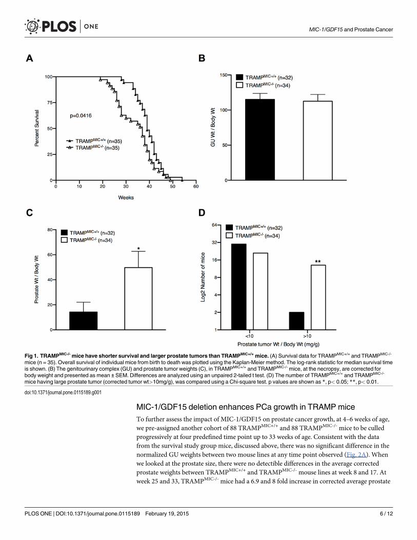
MIC-1/GDF15 deletion enhances PCa growth in TRAMPmiceTo further assess the impact of MIC-1/GDF15 on prostate cancer growth, at 4–6 weeks of age,we pre-assigned another cohort of 88 TRAMPMIC+/+ and 88 TRAMPMIC-/- mice to be culledprogressively at four predefined time point up to 33 weeks of age. Consistent with the datafrom the survival study group mice, discussed above, there was no significant difference in thenormalized GU weights between two mouse lines at any time point observed (Fig. 2A). Whenwe looked at the prostate size, there were no detectible differences in the average correctedprostate weights between TRAMPMIC+/+ and TRAMPMIC-/- mouse lines at week 8 and 17. Atweek 25 and 33, TRAMPMIC-/- mice had a 6.9 and 8 fold increase in corrected average prostate
Fig 1. TRAMPMIC-/- mice have shorter survival and larger prostate tumors than TRAMPMIC+/+ mice. (A) Survival data for TRAMPMIC+/+ and TRAMPMIC-/-
mice (n = 35). Overall survival of individual mice from birth to death was plotted using the Kaplan-Meier method. The log-rank statistic for median survival timeis shown. (B) The genitourinary complex (GU) and prostate tumor weights (C), in TRAMPMIC+/+ and TRAMPMIC-/- mice, at the necropsy, are corrected forbody weight and presented as mean ± SEM. Differences are analyzed using an unpaired 2-tailed t test. (D) The number of TRAMPMIC+/+ and TRAMPMIC-/-
mice having large prostate tumor (corrected tumor wt>10mg/g), was compared using a Chi-square test. p values are shown as *, p< 0.05; **, p< 0.01.
doi:10.1371/journal.pone.0115189.g001
MIC-1/GDF15 and Prostate Cancer
PLOS ONE | DOI:10.1371/journal.pone.0115189 February 19, 2015 6 / 12
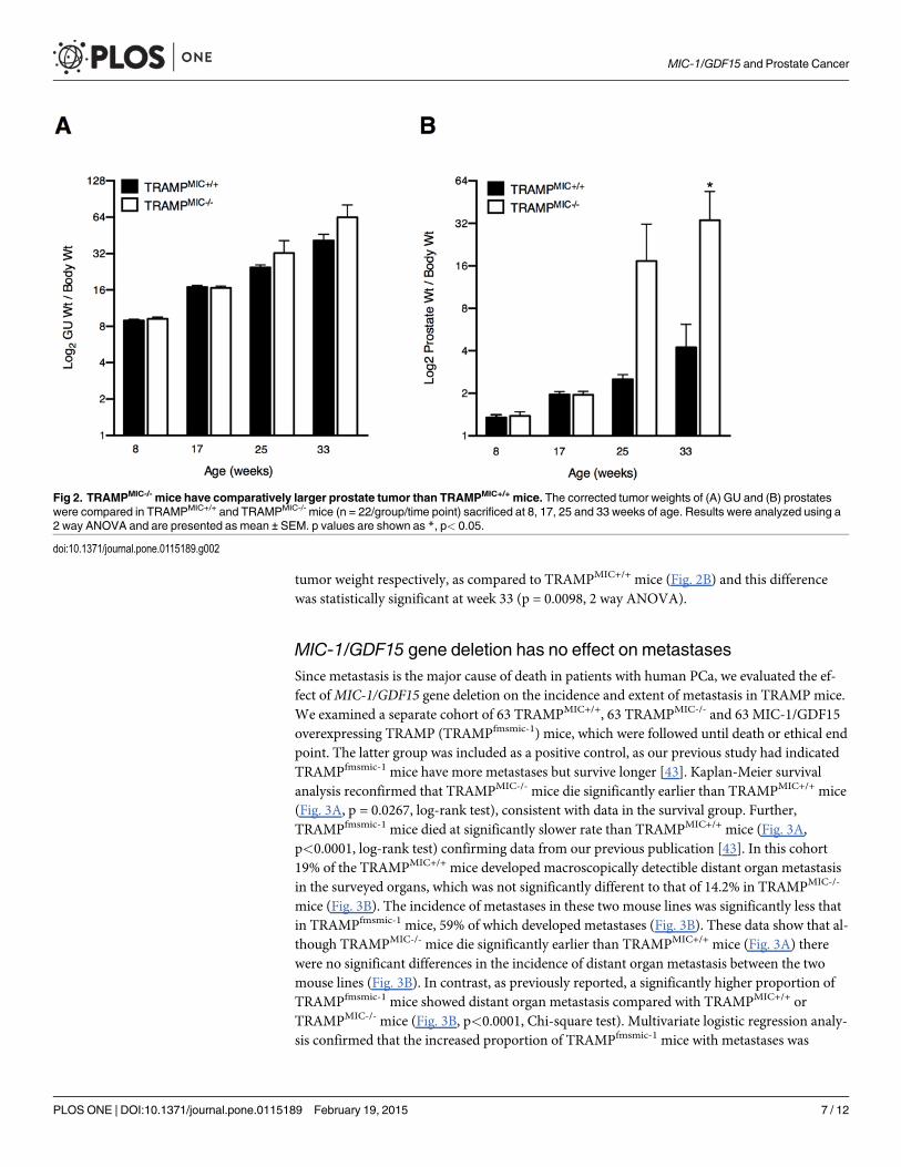
tumor weight respectively, as compared to TRAMPMIC+/+ mice (Fig. 2B) and this differencewas statistically significant at week 33 (p = 0.0098, 2 way ANOVA).
MIC-1/GDF15 gene deletion has no effect on metastasesSince metastasis is the major cause of death in patients with human PCa, we evaluated the ef-fect ofMIC-1/GDF15 gene deletion on the incidence and extent of metastasis in TRAMP mice.We examined a separate cohort of 63 TRAMPMIC+/+, 63 TRAMPMIC-/- and 63 MIC-1/GDF15overexpressing TRAMP (TRAMPfmsmic-1) mice, which were followed until death or ethical endpoint. The latter group was included as a positive control, as our previous study had indicatedTRAMPfmsmic-1 mice have more metastases but survive longer [43]. Kaplan-Meier survivalanalysis reconfirmed that TRAMPMIC-/- mice die significantly earlier than TRAMPMIC+/+ mice(Fig. 3A, p = 0.0267, log-rank test), consistent with data in the survival group. Further,TRAMPfmsmic-1 mice died at significantly slower rate than TRAMPMIC+/+ mice (Fig. 3A,p<0.0001, log-rank test) confirming data from our previous publication [43]. In this cohort19% of the TRAMPMIC+/+ mice developed macroscopically detectible distant organ metastasisin the surveyed organs, which was not significantly different to that of 14.2% in TRAMPMIC-/-
mice (Fig. 3B). The incidence of metastases in these two mouse lines was significantly less thatin TRAMPfmsmic-1 mice, 59% of which developed metastases (Fig. 3B). These data show that al-though TRAMPMIC-/- mice die significantly earlier than TRAMPMIC+/+ mice (Fig. 3A) therewere no significant differences in the incidence of distant organ metastasis between the twomouse lines (Fig. 3B). In contrast, as previously reported, a significantly higher proportion ofTRAMPfmsmic-1 mice showed distant organ metastasis compared with TRAMPMIC+/+ orTRAMPMIC-/- mice (Fig. 3B, p<0.0001, Chi-square test). Multivariate logistic regression analy-sis confirmed that the increased proportion of TRAMPfmsmic-1 mice with metastases was
Fig 2. TRAMPMIC-/- mice have comparatively larger prostate tumor than TRAMPMIC+/+ mice. The corrected tumor weights of (A) GU and (B) prostateswere compared in TRAMPMIC+/+ and TRAMPMIC-/- mice (n = 22/group/time point) sacrificed at 8, 17, 25 and 33 weeks of age. Results were analyzed using a2 way ANOVA and are presented as mean ± SEM. p values are shown as *, p< 0.05.
doi:10.1371/journal.pone.0115189.g002
MIC-1/GDF15 and Prostate Cancer
PLOS ONE | DOI:10.1371/journal.pone.0115189 February 19, 2015 7 / 12
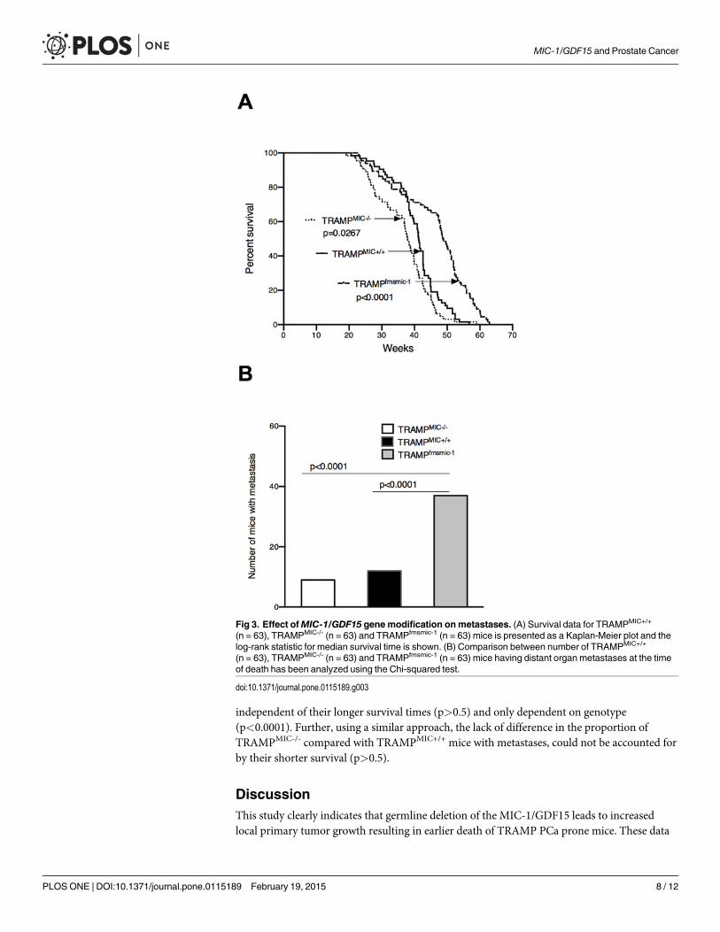
independent of their longer survival times (p>0.5) and only dependent on genotype(p<0.0001). Further, using a similar approach, the lack of difference in the proportion ofTRAMPMIC-/- compared with TRAMPMIC+/+ mice with metastases, could not be accounted forby their shorter survival (p>0.5).
DiscussionThis study clearly indicates that germline deletion of the MIC-1/GDF15 leads to increasedlocal primary tumor growth resulting in earlier death of TRAMP PCa prone mice. These data
Fig 3. Effect ofMIC-1/GDF15 genemodification onmetastases. (A) Survival data for TRAMPMIC+/+
(n = 63), TRAMPMIC-/- (n = 63) and TRAMPfmsmic-1 (n = 63) mice is presented as a Kaplan-Meier plot and thelog-rank statistic for median survival time is shown. (B) Comparison between number of TRAMPMIC+/+
(n = 63), TRAMPMIC-/- (n = 63) and TRAMPfmsmic-1 (n = 63) mice having distant organ metastases at the timeof death has been analyzed using the Chi-squared test.
doi:10.1371/journal.pone.0115189.g003
MIC-1/GDF15 and Prostate Cancer
PLOS ONE | DOI:10.1371/journal.pone.0115189 February 19, 2015 8 / 12
are consistent with our previous publication indicating that transgenic overexpression ofMIC-1/GDF15 decreases local PCa tumor growth and substantially increases survival ofTRAMP mice. This result is consistent with results in another transgenic model of early cancerin Apcmin colonic polyp prone mice also overexpressing MIC-1/GDF15 [38]. This reinforcesthe argument that MIC-1/GDF15 plays a protective role in early local tumor development andgrowth. Several epidemiological studies suggest that this finding may be translated to at leastsome human cancers. For example, increased local concentrations of extracellular matrix asso-ciated MIC-1/GDF15 in prostate cancer biopsies were associated with reduced risk of diseaseprogression, especially in the subgroup with early cancer and with Gleason grade of 6 or less.In this group MIC-1/GDF15 localised to the tumor matrix was the single best predictor oftumor recurrence [28]. In patients from the polyp prevention trial, a higher polyp free serumMIC-1/GDF15 level afforded protection from polyp recurrence and NSAID mediated protec-tion was lost if MIC-1/GDF15 serum levels were not elevated with NSAID treatment [39].
The apparently discordant effect of MIC-1/GDF15 on local tumor growth and the metastat-ic process, that we described previously, is again demonstrated here [43]. Transgenic overex-pression leads to smaller local tumors and longer survival, as TRAMPcfmsmic-1 mice no longerdie early of local disease. Independently of increased survival time, these same TRAMPcfmsmic-1
mice have more metastases. However, we could demonstrate no difference in the proportion ofmice with metastases between TRAMPMIC+/+ and TRAMPMIC-/- mice. As few mice of eitherline developed metastases by the experimental endpoint we may not have studied enough miceto be certain that no differences exist. Additionally, as prostate tumors in TRAMPMIC+/+ miceexpress little MIC-1/GDF15 [43], it may be that this model is more sensitive to the effects of in-creased expression compared to gene deletion.
Whilst there was no significant difference in metastases between TRAMPMIC+/+ andTRAMPMIC-/- mice, there may be differences in local invasion. TRAMPMIC-/- mice with largerprostate tumors had smaller SVs (data not shown), which suggests that there may be lessSV invasion.
HowMIC-1/GDF15 might exert effects on tumor growth and spread is uncertain. Elucidat-ing its mechanisms of action is greatly hampered because the identity of its receptor is un-known. It is presumed that the receptor is a member of the highly conserved hetrotetramericTGF-b receptor (TBR) superfamily, but there is limited direct evidence for this. There is someindirect evidence, based on antibody blockade, that it may utilise TBRII [26,45] the class 2 re-ceptor used by TGF-b itself, but there is no direct biochemical or genetic evidence supportingthis data. The signalling cascade of the receptor complex or class I receptor utilised by MIC-1/GDF15 is to date, completely unknown.
There is now a plethora of published data on the role of MIC-1/GDF15 on tumor growthand spread with a confusing range of results. Data from experiments using human cell lines, inwhich MIC-1/GDF15 expression has either been induced or knocked down, then xenograftedinto immunodeficient mice, have provided sometimes, contradictory results [18,29–34]. Stud-ies using transgenic models of spontaneously developing cancer that also bear genetically mod-ified MIC-1/GDF15 expression and an intact immune system all point to a protective role forMIC-1/GDF15 on local tumor development. A similar effect can also be observed in two mod-els of carcinogen induced cancer in transgenic mice overexpressing MIC-1/GDF15, which alsohave an intact immune system [35,36]. The most parsimonious explanation that may explainthese contradictions is that MIC-1/GDF15 regulates anti-cancer immunity, which in turn regu-lates cancer growth.
Overall, our results support an important protective role for MIC-1/GDF15 in the develop-ment and early growth of PCa and probably cancer in general. Unravelling the biological effectof MIC-1/GDF15 on tumor evolution and biology is of practical importance for several
MIC-1/GDF15 and Prostate Cancer
PLOS ONE | DOI:10.1371/journal.pone.0115189 February 19, 2015 9 / 12
reasons. A high proportion of cancers express it, to the extent that serum level can rise up to10–100 fold and cause cancer anorexia/cachexia. Further, its expression is increased by all can-cer treatment modalities including surgery, radiotherapy and chemotherapy. Thus any effectthat MIC-1/GDF15 has on local tumor biology, particularly tumor spread is likely to impactmost cancer patients, raising the prospect that modulation of MIC-1/GDF15 actions duringtherapy might reduce the risk of metastatic disease and other complications of cancer.
Author ContributionsConceived and designed the experiments: YH SNB DAB. Performed the experiments: YH GPLTVN. Analyzed the data: YH DAB VWWTMGM. Contributed reagents/materials/analysistools: SNB PJR. Wrote the paper: YH SNB DAB.
References1. American CS (2014) Cancer Facts & Figures. Atlanta: American Cancer Society. Available: http://
www.cancer.org/research/cancerfactsstatistics/cancerfactsfigures2014/.
2. Bootcov MR, Bauskin AR, Valenzuela SM, Moore AG, Bansal M, et al. (1997) MIC-1, a novel macro-phage inhibitory cytokine, is a divergent member of the TGF-beta superfamily. Proc Natl Acad SciU S A 94: 11514–11519. PMID: 9326641
3. Bauskin AR, Brown DA, Kuffner T, Johnen H, Luo XW, et al. (2006) Role of macrophage inhibitorycytokine-1 in tumorigenesis and diagnosis of cancer. Cancer Res 66: 4983–4986. PMID: 16707416
4. Breit SN, Johnen H, Cook AD, Tsai VW, MohammadMG, et al. (2011) The TGF-beta superfamily cyto-kine, MIC-1/GDF15: a pleotrophic cytokine with roles in inflammation, cancer and metabolism. GrowthFactors 29: 187–195. doi: 10.3109/08977194.2011.607137 PMID: 21831009
5. Welsh JB, Sapinoso LM, Kern SG, Brown DA, Liu T, et al. (2003) Large-scale delineation of secretedprotein biomarkers overexpressed in cancer tissue and serum. Proc Natl Acad Sci U S A 100:3410–3415. PMID: 12624183
6. Joiner MC, Thomas RA, Grever WE, Smolinski JM, Divine GW, et al. (2011) Developing point of careand high-throughput biological assays for determining absorbed radiation dose. Radiother Oncol 101:233–236. doi: 10.1016/j.radonc.2011.05.068 PMID: 21724286
7. Boyer J, AllenWL, McLean EG, Wilson PM, McCulla A, et al. (2006) Pharmacogenomic identification ofnovel determinants of response to chemotherapy in colon cancer. Cancer Res 66: 2765–2777. PMID:16510598
8. Modlich O, Prisack HB, Munnes M, AudretschW, Bojar H (2004) Immediate gene expression changesafter the first course of neoadjuvant chemotherapy in patients with primary breast cancer disease. ClinCancer Res 10: 6418–6431. PMID: 15475428
9. Huang CY, Beer TM, Higano CS, True LD, Vessella R, et al. (2007) Molecular alterations in prostatecarcinomas that associate with in vivo exposure to chemotherapy: identification of a cytoprotectivemechanism involving growth differentiation factor 15. Clin Cancer Res 13: 5825–5833. PMID:17908975
10. Brown DA, Bauskin AR, Fairlie WD, Smith MD, Liu T, et al. (2002) Antibody-based approach to high-volume genotyping for MIC-1 polymorphism. Biotechniques 33: 118–120, 122, 124 passim. PMID:12139236
11. Baek KE, Yoon SR, Kim JT, Kim KS, Kang SH, et al. (2009) Upregulation and secretion of macrophageinhibitory cytokine-1 (MIC-1) in gastric cancers. Clin Chim Acta 401: 128–133. doi: 10.1016/j.cca.2008.12.008 PMID: 19133249
12. Brown DA, Lindmark F, Stattin P, Balter K, Adami HO, et al. (2009) Macrophage inhibitory cytokine 1: anew prognostic marker in prostate cancer. Clin Cancer Res 15: 6658–6664. doi: 10.1158/1078-0432.CCR-08-3126 PMID: 19843661
13. Brown DA, Ward RL, Buckhaults P, Liu T, Romans KE, et al. (2003) MIC-1 serum level and genotype:associations with progress and prognosis of colorectal carcinoma. Clin Cancer Res 9: 2642–2650.PMID: 12855642
14. Brown DA, Stephan C, Ward RL, LawM, Hunter M, et al. (2006) Measurement of serum levels of mac-rophage inhibitory cytokine 1 combined with prostate-specific antigen improves prostate cancer diagno-sis. Clin Cancer Res 12: 89–96. PMID: 16397029
MIC-1/GDF15 and Prostate Cancer
PLOS ONE | DOI:10.1371/journal.pone.0115189 February 19, 2015 10 / 12
15. Koopmann J, Buckhaults P, Brown DA, Zahurak ML, Sato N, et al. (2004) Serummacrophage inhibitorycytokine 1 as a marker of pancreatic and other periampullary cancers. Clin Cancer Res 10:2386–2392. PMID: 15073115
16. Selander KS, Brown DA, Sequeiros GB, Hunter M, Desmond R, et al. (2007) Serummacrophage inhibi-tory cytokine-1 concentrations correlate with the presence of prostate cancer bone metastases. CancerEpidemiol Biomarkers Prev 16: 532–537. PMID: 17372249
17. Wallin U, Glimelius B, Jirstrom K, Darmanis S, Nong RY, et al. (2011) Growth differentiation factor 15: aprognostic marker for recurrence in colorectal cancer. Br J Cancer 104: 1619–1627. doi: 10.1038/bjc.2011.112 PMID: 21468045
18. Boyle GM, Pedley J, Martyn AC, Banducci KJ, Strutton GM, et al. (2009) Macrophage inhibitorycytokine-1 is overexpressed in malignant melanoma and is associated with tumorigenicity. J Invest Der-matol 129: 383–391. doi: 10.1038/jid.2008.270 PMID: 18754039
19. Suesskind D, Schatz A, Schnichels S, Coupland SE, Lake SL, et al. (2011) GDF-15: a novel serummarker for metastases in uveal melanoma patients. Graefes Arch Clin Exp Ophthalmol.
20. Koopmann J, Rosenzweig CN, Zhang Z, Canto MI, Brown DA, et al. (2006) Serummarkers in patientswith resectable pancreatic adenocarcinoma: macrophage inhibitory cytokine 1 versus CA19–9. ClinCancer Res 12: 442–446. PMID: 16428484
21. Bauskin AR, Jiang L, Luo XW,Wu L, Brown DA, et al. (2010) The TGF-beta superfamily cytokineMIC-1/GDF15: secretory mechanisms facilitate creation of latent stromal stores. J Interferon CytokineRes 30: 389–397. doi: 10.1089/jir.2009.0052 PMID: 20187768
22. Weber F, Shen L, Aldred MA, Morrison CD, Frilling A, et al. (2005) Genetic classification of benign andmalignant thyroid follicular neoplasia based on a three-gene combination. J Clin Endocrinol Metab 90:2512–2521. PMID: 15713710
23. Fluge O, Bruland O, Akslen LA, Lillehaug JR, Varhaug JE (2006) Gene expression in poorly differentiat-ed papillary thyroid carcinomas. Thyroid 16: 161–175. PMID: 16676402
24. Staff AC, Bock AJ, Becker C, Kempf T, Wollert KC, et al. (2010) Growth differentiation factor-15 as aprognostic biomarker in ovarian cancer. Gynecol Oncol 118: 237–243. doi: 10.1016/j.ygyno.2010.05.032 PMID: 20576287
25. Staff AC, Trovik J, Eriksson AG, Wik E, Wollert KC, et al. (2011) Elevated plasma growth differentiationfactor-15 correlates with lymph node metastases and poor survival in endometrial cancer. Clin CancerRes 17: 4825–4833. doi: 10.1158/1078-0432.CCR-11-0715 PMID: 21616994
26. Johnen H, Lin S, Kuffner T, Brown DA, Tsai VW, et al. (2007) Tumor-induced anorexia and weight lossare mediated by the TGF-beta superfamily cytokine MIC-1. Nat Med 13: 1333–1340. PMID: 17982462
27. Tsai VW, Husaini Y, Manandhar R, Lee-Ng KK, Zhang HP, et al. (2012) Anorexia/cachexia of chronicdiseases: a role for the TGF-beta family cytokine MIC-1/GDF15. J Cachexia Sarcopenia Muscle 3:239–243. doi: 10.1007/s13539-012-0082-6 PMID: 22936174
28. Bauskin AR, Brown DA, Junankar S, Rasiah KK, Eggleton S, et al. (2005) The propeptide mediates for-mation of stromal stores of PROMIC-1: role in determining prostate cancer outcome. Cancer Res 65:2330–2336. PMID: 15781647
29. Baek SJ, Kim KS, Nixon JB, Wilson LC, Eling TE (2001) Cyclooxygenase inhibitors regulate the ex-pression of a TGF-beta superfamily member that has proapoptotic and antitumorigenic activities. MolPharmacol 59: 901–908. PMID: 11259636
30. Wang X, Chrysovergis K, Bienstock RJ, ShimM, Eling TE (2012) The H6D variant of NAG-1/GDF15 in-hibits prostate xenograft growth in vivo. Prostate 72: 677–689. doi: 10.1002/pros.21471 PMID: 21809352
31. Albertoni M, Shaw PH, Nozaki M, Godard S, Tenan M, et al. (2002) Anoxia induces macrophage inhibi-tory cytokine-1 (MIC-1) in glioblastoma cells independently of p53 and HIF-1. Oncogene 21:4212–4219. PMID: 12082608
32. Roth P, Junker M, Tritschler I, Mittelbronn M, Dombrowski Y, et al. (2010) GDF-15 contributes to prolif-eration and immune escape of malignant gliomas. Clin Cancer Res 16: 3851–3859. doi: 10.1158/1078-0432.CCR-10-0705 PMID: 20534737
33. Tsui KH, Chang YL, Feng TH, Chung LC, Lee TY, et al. (2012) Growth differentiation factor-15 upregu-lates interleukin-6 to promote tumorigenesis of prostate carcinoma PC-3 cells. J Mol Endocrinol 49:153–163. doi: 10.1530/JME-11-0149 PMID: 22872134
34. Senapati S, Rachagani S, Chaudhary K, Johansson SL, Singh RK, et al. (2010) Overexpression ofmacrophage inhibitory cytokine-1 induces metastasis of human prostate cancer cells through the FAK-RhoA signaling pathway. Oncogene 29: 1293–1302. doi: 10.1038/onc.2009.420 PMID: 19946339
35. CekanovaM, Lee SH, Donnell RL, Sukhthankar M, Eling TE, et al. (2009) Nonsteroidal anti-inflammatorydrug-activated gene-1 expression inhibits urethane-induced pulmonary tumorigenesis in transgenic mice.Cancer Prev Res (Phila) 2: 450–458. doi: 10.1158/1940-6207.CAPR-09-0057 PMID: 19401523
MIC-1/GDF15 and Prostate Cancer
PLOS ONE | DOI:10.1371/journal.pone.0115189 February 19, 2015 11 / 12
36. Baek SJ, Okazaki R, Lee SH, Martinez J, Kim JS, et al. (2006) Nonsteroidal anti-inflammatory drug-activated gene-1 over expression in transgenic mice suppresses intestinal neoplasia. Gastroenterology131: 1553–1560. PMID: 17101328
37. Zimmers TA, Jin X, Gutierrez JC, Acosta C, McKillop IH, et al. (2008) Effect of in vivo loss of GDF-15 onhepatocellular carcinogenesis. J Cancer Res Clin Oncol 134: 753–759. doi: 10.1007/s00432-007-0336-4 PMID: 18210153
38. Zimmers TA, Gutierrez JC, Koniaris LG (2010) Loss of GDF-15 abolishes sulindac chemoprevention inthe ApcMin/+ mouse model of intestinal cancer. J Cancer Res Clin Oncol 136: 571–576. doi: 10.1007/s00432-009-0691-4 PMID: 19784846
39. Brown DA, Hance KW, Rogers CJ, Sansbury LB, Albert PS, et al. (2012) Serummacrophage inhibitorycytokine-1 (MIC-1/GDF15): a potential screening tool for the prevention of colon cancer? Cancer Epide-miol Biomarkers Prev 21: 337–346. doi: 10.1158/1055-9965.EPI-11-0786 PMID: 22144502
40. Mehta RS, Song M, Bezawada N, Wu K, Garcia-Albeniz X, et al. (2014) A prospective study of macro-phage inhibitory cytokine-1 (MIC-1/GDF15) and risk of colorectal cancer. J Natl Cancer Inst 106:dju016. doi: 10.1093/jnci/dju016 PMID: 24565956
41. Greenberg NM, DeMayo F, Finegold MJ, Medina D, Tilley WD, et al. (1995) Prostate cancer in a trans-genic mouse. Proc Natl Acad Sci U S A 92: 3439–3443. PMID: 7724580
42. Gingrich JR, Barrios RJ, Morton RA, Boyce BF, DeMayo FJ, et al. (1996) Metastatic prostate cancer ina transgenic mouse. Cancer Res 56: 4096–4102. PMID: 8797572
43. Husaini Y, Qiu MR, Lockwood GP, Luo XW, Shang P, et al. (2012) Macrophage inhibitory cytokine-1(MIC-1/GDF15) slows cancer development but increases metastases in TRAMP prostate cancer pronemice. PLoS ONE 7: e43833. doi: 10.1371/journal.pone.0043833 PMID: 22952779
44. Tsai VW, Macia L, Johnen H, Kuffner T, Manadhar R, et al. (2013) TGF-b superfamily cytokine MIC-1/GDF15 is a physiological appetite and body weight regulator. PLoS One 8: e55174. doi: 10.1371/journal.pone.0055174 PMID: 23468844
45. de Jager SC, Bermudez B, Bot I, Koenen RR, Bot M, et al. (2011) Growth differentiation factor 15 defi-ciency protects against atherosclerosis by attenuating CCR2-mediated macrophage chemotaxis. J ExpMed 208: 217–225. doi: 10.1084/jem.20100370 PMID: 21242297
MIC-1/GDF15 and Prostate Cancer
PLOS ONE | DOI:10.1371/journal.pone.0115189 February 19, 2015 12 / 12