Multi-site tumor sampling highlights molecular intra-tumor ...
Upload
independentCategory
view
4download
0
Caveolin-1 Promotes Tumor Progression in an AutochthonousMouse Model of Prostate CancerGENETIC ABLATION OF Cav-1 DELAYS ADVANCED PROSTATE TUMOR DEVELOPMENT IN TRAMP MICE*
Received for publication, February 1, 2005, and in revised form, March 22, 2005Published, JBC Papers in Press, March 30, 2005, DOI 10.1074/jbc.M501186200
Terence M. Williams,a,b Ghada S. Hassan,a,c Jiangwei Li,a Alex W. Cohen,a,b Freddy Medina,a,d
Philippe G. Frank,a,e,f Richard G. Pestell,g Dolores Di Vizio,h Massimo Loda,h
and Michael P. Lisantia,i
From the aDepartment of Molecular Pharmacology and The Albert Einstein Cancer Center, eDepartment of Urology, AlbertEinstein College of Medicine, Bronx, New York 10461, gDepartment of Oncology, Lombardi Comprehensive Cancer Center,Georgtown University, Washington, D. C. 20007, and hDepartment of Medical Oncology, Dana-Farber Cancer Institute,Boston, Massachusetts 02115
Caveolin-1 (Cav-1) is the primary structural compo-nent of caveolae and is implicated in the processes ofvesicular transport, cholesterol balance, transforma-tion, and tumorigenesis. Despite an abundance of datasuggesting that Cav-1 has transformation suppressorproperties both in vitro and in vivo, Cav-1 is expressed atincreased levels in human prostate cancer. To investi-gate the role of Cav-1 in prostate cancer onset and pro-gression, we interbred Cav-1(�/�) null mice with aTRAMP (transgenic adenocarcinoma of mouse prostate)model that spontaneously develops advanced prostatecancer and metastatic disease. We found that, althoughthe loss of Cav-1 did not affect the appearance of mini-mally invasive prostate cancer, its absence significantlyimpeded progression to highly invasive and metastaticdisease. Inactivation of one (�/�) or both (�/�) alleles ofCav-1 resulted in significant reductions in prostate tu-mor burden, as well as decreases in regional lymph nodemetastases. Moreover, further examination revealed de-creased metastasis to distant organs, such as the lungs,in TRAMP/Cav-1(�/�) mice. Utilizing prostate carci-noma cell lines (C1, C2, and C3) derived from TRAMPtumors, we also showed a positive correlation betweenCav-1 expression and the ability of these cells to formtumors in vivo. Furthermore, down-regulation of Cav-1expression in these cells, using a small interfering RNAapproach, significantly reduced their tumorigenic andmetastatic potential. Mechanistically, we showed thatloss or down-regulation of Cav-1 expression results inincreased apoptosis, with increased prostate apoptosisresponse factor-4 and PTEN levels in Cav-1(�/�) nullprostate tumors. Our current findings provide the first
in vivo molecular genetic evidence that Cav-1 does in-deed function as a tumor promoter during prostate car-cinogenesis, rather than as a tumor suppressor.
Caveolin-1 (Cav-1)1 is the principal structural componentand marker protein of caveolae, 50–100-nm flask-shaped in-vaginations of the plasma membrane. Cav-1 was first identifiedas a tyrosine-phosphorylated substrate when avian fibroblastswere transformed with the v-Src oncogene, suggesting thatCav-1 may have a critical role in modulating cellular transfor-mation (1). To date, three caveolin family members have beenidentified, with diverse tissue expression patterns in variousorganisms ranging from mammals and amphibians to the nem-atode. Such ubiquity in higher organisms insinuates the im-portance of the caveolin gene family for normal physiology (2).The majority of caveolin-related research has focused on caveo-lin-1 as the prototypical member, and the past decade hasbrought considerable advances in our understanding of thefunctions of Cav-1. Thus far, Cav-1 has been implicated inmany cellular processes, from signal transduction and vesicu-lar transport to cholesterol homeostasis and lipid transport(reviewed in Ref. 3).
Concurrently, a growing field of research is beginning todefine the role of the caveolins in the development of humancancers. Identification of a novel function for Cav-1 in oncogen-esis began with the demonstration that Cav-1 levels inverselycorrelated with soft agar growth in transformed NIH 3T3 fi-broblasts (4). Subsequently, several groups have shown thatrecombinant expression of Cav-1 in certain human or mousetumor-derived cell lines significantly inhibits their soft agargrowth (5–8). More recently, we and others have reported thatCav-1 has in vivo tumor suppressor properties in certain tis-sues, such as the mammary gland and skin, utilizing variousmurine animal models (9–12). For example, using a mousemodel of mammary tumorigenesis (MMTV-PyMT), we haveshown that Cav-1 expression dramatically inhibits the progres-sion of early mammary dysplastic lesions, as well as the devel-opment of primary mammary tumors and spontaneous metas-
* This work was supported in part by grants from the NationalInstitutes of Health as well as a Hirschl/Weil-Caulier Career ScientistAward (to M. P. L.). The costs of publication of this article were de-frayed in part by the payment of page charges. This article musttherefore be hereby marked “advertisement” in accordance with 18U.S.C. Section 1734 solely to indicate this fact.
b Supported by a National Institutes of Health Medical ScientistTraining Program Grant (T32-GM07288).
c Recipient of a post-doctoral fellowship from the Foundation ofHealth Research, Quebec, Canada.
d Supported by a National Institutes of Health Graduate TrainingProgram Grant (T32-DK07513).
f Recipient of a Scientist Development grant from the AmericanHeart Association. To whom correspondence may be addressed. Tel.:718-430-8829; Fax: 718-430-8830; E-mail: [email protected].
i To whom correspondence may be addressed. Tel.: 718-430-8828;Fax: 718-430-8830; E-mail: [email protected].
1 The abbreviations used are: Cav-1, caveolin-1; TRAMP, transgenicadenocarcinoma of mouse prostate; siRNA, small interfering RNA;PCNA, proliferating cell nuclear antigen; WT, wild-type; PBS, phos-phate-buffered saline; TUNEL, terminal deoxynucleotidyltransferase-mediated dUTP nick-end label; KO, knock-out; HET, heterozygous;PIN, prostatic intraepithelial neoplasia; ERK, extracellular signal-reg-ulated kinase; Par4, prostate apoptosis response factor-4.
THE JOURNAL OF BIOLOGICAL CHEMISTRY Vol. 280, No. 26, Issue of July 1, pp. 25134–25145, 2005© 2005 by The American Society for Biochemistry and Molecular Biology, Inc. Printed in U.S.A.
This paper is available on line at http://www.jbc.org25134
by guest on August 20, 2016
http://ww
w.jbc.org/
Dow
nloaded from

tases. In terms of human cancers, mutations in the CAV-1 genehave been identified in breast carcinomas, as well as oralsquamous cell carcinomas (13, 14), and CAV-1 gene localizes toa suspected tumor suppressor locus on chromosome 7q31.1,which is commonly deleted in a variety of human cancers(reviewed in Ref. 15). In accordance with these findings, Cav-1protein levels are consistently down-regulated in a wide rangeof human cancers, including ovarian carcinomas, sarcomas,and mammary carcinomas (15).
Despite this association between Cav-1 and tumor suppres-sion, there is accumulating evidence that Cav-1 does not func-tion similarly in all types of human cancers. In fact, severalgroups have now documented that Cav-1 protein expressionlevels are up-regulated in other human neoplasias, includingbladder, esophageal, and prostate carcinomas (16–22). Thesefindings suggest that Cav-1 does not have transformation sup-pressor effects in these tissues. In fact, further clinicopatho-logic analyses of human bladder, esophageal, and prostate car-cinomas reveal that Cav-1 up-regulation correlates positivelywith increased tumor grade, invasive potential, regional me-tastasis, and decreased overall survival (16, 17, 19, 21, 22).Therefore, these results argue that Cav-1 has tumor-promotingfunctions in certain tumor cell types, such as prostate epithelialcells. This notion has been substantiated by cell culture studieswith human prostate tumor-derived cell lines. In these prostatecarcinoma cell lines, Cav-1 up-regulation appears to be corre-lated with increased cell survival, androgen independence, andincreased metastatic potential (23, 24). Taken together, thesefindings suggest that Cav-1 has distinct cell type-specific tu-mor-modulating functions, either promoting or inhibiting tu-mor development, depending on its cellular context.
However, it remains unknown whether Cav-1 expression canpromote the development of prostate cancers in vivo. Previousstudies have focused only on the oncogenic behavior of culturedprostate cancer-derived cell lines. Here, we employed a wellestablished transgenic mouse model of prostate cancer, termedTRAMP (transgenic adenocarcinoma of mouse prostate), tostudy the potential role of Cav-1 in modulating prostate carci-nogenesis in vivo. For this purpose, we interbred Cav-1(�/�)null mice, completely lacking Cav-1 expression, with the wellcharacterized TRAMP mouse model (25–27). This approachallowed us to directly assess whether genetic loss of one (�/�)or both alleles (�/�) of Cav-1 alters prostate tumor develop-ment in an in vivo setting. Use of this autochthonous TRAMPmodel has many advantages, because it allows prostate tumorsto develop in their native environment (i.e. with exposure to theproper growth factors, hormonal, immunomodulatory, and an-giogenic milieu). (See the end of the “Discussion” for furtherelaboration on the advantages and the validity of the TRAMPmodel).
Here, we show that prostate tumors derived from TRAMPmice demonstrate an up-regulation of Cav-1 expression, anal-ogous to human prostate cancers. More importantly, ablation ofone or both Cav-1 alleles significantly attenuates the progres-sion of prostate carcinoma with regard to primary tumor bur-den and more advanced metastatic disease. These findings arefurther corroborated with cell lines derived from TRAMP pros-tate tumors (C1, C2, and C3). Importantly, all three TRAMPcell lines express cytokeratins, E-cadherin, and the androgenreceptor, confirming their epithelial and prostatic origin (28).In these cell lines, Cav-1 expression directly correlates withtumor growth. Additionally, siRNA-mediated silencing ofCav-1 gene expression in the Cav-1-expressing TRAMP C1 cellline inhibits the processes of primary tumor growth and exper-imental metastasis. Analysis of both (i) transgenic prostatetumors and (ii) cell line-derived tumors indicates that loss of
Cav-1 results in decreased cell survival, as well as the up-regulation of pro-apoptotic markers including PTEN and Par4.
Therefore, our findings provide the first in vivo moleculargenetic evidence that Cav-1 does indeed function as a tumorpromoter during mouse prostate carcinogenesis, rather than asa tumor suppressor. As we previously reported that Cav-1 hasa tumor suppressor role in the mouse mammary gland (12), ourcurrent results conclusively demonstrated that Cav-1 has cleartissue-specific functions with regard to both tumorigenesis andmetastasis.
EXPERIMENTAL PROCEDURES
Materials and Expression Vectors—Mouse monoclonal antibodies tocaveolin-1 (clones 2297 and 2234) and caveolin-2 were the generousgifts of Dr. Roberto Campos-Gonzalez (BD Biosciences). Antibodies andtheir sources were as follows: anti-Cav-1 (N-20) rabbit polyclonal anti-body (from Santa Cruz Biotechnology), anti-�-actin monoclonalantibody AC-15 (Sigma), anti-�-tubulin monoclonal antibody (Sigma),anti-phospho (p)-Akt (Ser-473) polyclonal antibody (Cell Signaling,Inc.), anti-Par4 polyclonal antibody (Santa Cruz), anti-PTEN polyclonalantibody (Cell Signaling), anti-PCNA monoclonal antibody (BD Bio-sciences), and anti-SV-40 large T-antigen (BD Biosciences). pBABE(empty vector) and pBABE-Cav-1 constructs were as we previouslydescribed (12).
Transgenic Animal Studies—All animals were housed and main-tained in a barrier facility at the Institute for Animal Studies, AlbertEinstein College of Medicine (AECOM). Mice were kept on a 12-hlight/dark cycle with ad libitum access to chow (Picolab 20, PMI Nutri-tion International) and water. Animal protocols used for this study wereapproved by the AECOM Institute for Animal Studies. Cav-1 null micewere generated as previously described (9). TRAMP mice expressingthe SV40 large T-antigen, under control of the minimal rat probasinpromoter, were obtained from The Jackson Laboratory (JAX) andscreened for the presence of the SV40 large T-antigen by PCR, asdetailed on The Jackson Laboratory website. All the mice used in thisstudy were in the C57Bl/6 background. Cav-1(�/�) null mice wereinterbred with TRAMP mice to generate three male cohorts: (i) TRAMP/Cav-1(�/�) mice (WT), (ii) TRAMP/Cav-1(�/�) (heterozygous (HET))mice, and (iii) TRAMP/Cav-1(�/�) (knock-out (KO)) mice. All TRAMPmice from this study were hemizygous for the TRAMP transgene.TRAMP transgenic mice were sacrificed at 28 weeks of age, and a fullnecropsy was performed on all animals, including microscopic exami-nation of the thoracic, pelvic, and abdominal organs. Primary tumorswere excised and weighed, cut into smaller portions, fixed in 10%neutral buffered formalin for 24–48 h for paraffin embedding, and5-�m sections were cut and stained with hematoxylin and eosin forhistological analysis. In cases where there was no prostate tumor evi-dent, the prostate tissue was isolated and likewise processed for micro-scopic analysis. Lymph nodes and other organs were similarly pro-cessed for histological analysis. For metastasis analysis, lymph nodesand lungs were sectioned at 50-�m intervals and either stained withhematoxylin and eosin or left unstained for immunohistochemistry.Lymph nodes were photographed with a Nikon Coolscope camera andareas with metastases scored with NIH Image J software. Lung metas-tases were scored as the total number of metastatic foci (defined as acluster of 10 or more cells) per lung.
Cell Culture—TRAMP cell lines (C1, C2, and C3) were obtained fromthe American Type Culture Collection (ATCC, Manassas, VA). Thesecell lines were previously characterized and are derived from prostateadenocarcinomas isolated and cultured from TRAMP/Cav-1(�/�) mice(28). In brief, these cell lines demonstrate androgen receptor, E-cad-herin, and cytokeratin expression, confirming their prostate epithelialorigin. In addition, all three cell lines no longer express detectable SV40large T-antigen. C1 and C2 cell lines also form tumors in syngeneic(C57BL/6) or nude mice, but C1 is the only cell line that forms coloniesin soft agar. C1 cells are the fastest growing in vitro and in vivo,whereas C3 cells are the slowest growing and do not form tumors in vivo(28).
All three cell lines were cultured in Dulbecco’s modified Eagle’s me-dium (high glucose), without sodium pyruvate, with L-glutamine, 5%fetal bovine serum, 5% Nu-Serum IV (BD Biosciences), 0.005 mg/mlbovine insulin (Sigma), 10 nM dehydroisoandrosterone (Sigma), 100units/ml penicillin, and 100 �g/ml streptomycin. The target region usedfor siRNA-mediated caveolin-1 down-regulation was designed with theaid of the Oligoengine sequence design software (Oligoengine, Inc.,Seattle, WA). A 19-nt region of caveolin-1 (nucleotide sequence GCAAGTG-
Caveolin-1 and Prostate Tumorigenesis 25135
by guest on August 20, 2016
http://ww
w.jbc.org/
Dow
nloaded from

TATGACGCGCAC) was chosen as a potential target and used to produce a64-nt pair of oligos containing restriction enzyme cleavage sites and a shortintervening hairpin sequence (sense: 5�-GATCCCCGCAAGTGTATGACG-CGCACTTCAAGAGAGTGCGCGTCATACACTTGCTTTTTGGAAA-3�; an-tisense: 5�-AGCTTTTCCAAAAAGCAAGTGTATGACGCGCACTCTCTTG-AAGTGCGCGTCATACACTTGCGGG-3�). These oligos were annealed andinserted into the pSUPER retroviral vector, according to the manufacturer’sinstructions. Production of stable cell lines by retroviral infection andpuromycin selection was performed as we previously described (12).
Immunoblotting and Immunohistochemistry—Western blot analysisand immunostaining were performed essentially as we previously de-scribed in detail (9, 12).
Cell Injections—TRAMP cells were grown to confluency in 15-cmdishes, trypsinized, and counted with a hematocytometer. 2 � 106 cellsin 0.1 ml of PBS were injected subcutaneously into the flanks of2-month-old male C57Bl/6 mice, and tumors were isolated after 8weeks. For the nude mouse experiments with stable cell lines, 5 � 105
cells in 0.1 ml of PBS were injected into the flanks of 2-month-oldathymic nude male mice (Taconic Farms), and tumors were isolatedafter 6 weeks. Tumors were weighed and sized with calipers. Volumeswere estimated using the formula (x2y)/2, where x is the length and y iswidth. For experimental metastasis, 106 cells in 0.1 ml of PBS of eithercell line were injected via the tail vein. Lungs were isolated 3 weekslater, fixed in formalin, and analyzed by hematoxylin and eosin stainingexactly as for the transgenic study.
Terminal Deoxynucleotidyltransferase-mediated dUTP Nick-end La-bel (TUNEL) Staining—Briefly, 5-�m sections of WT TRAMP and KOTRAMP prostate tumors or C1/pSUPER and C1/siRNA tumors wereTUNEL-stained with a kit according to the manufacturer’s instructions(TACs 2 TdT in situ apoptosis detection kit; Trevigen, Inc., Gaithers-burg, MD). TUNEL-positive nuclei were scored under a microscopeusing a 40� objective.
RESULTS
To determine whether Cav-1 plays an important role in pros-tate cancer progression, we interbred Cav-1(�/�) null mice withTRAMP mice (a mouse model of spontaneous prostate cancer) to
produce three cohorts of TRAMP transgenic male mice: (i)TRAMP/Cav-1(�/�) wild-type, (ii) TRAMP/Cav-1(�/�) heterozy-gous, and (iii) TRAMP/Cav-1(�/�) null mice. All mice were het-erozygous for the TRAMP transgene, which directs specific ex-pression of the SV40 large T-antigen to the prostate via the ratprobasin promoter. As previously reported by Greenberg andcolleagues (26), TRAMP mice develop metastases to the regionallymph nodes after 28 weeks of age, with 100% penetrance and tomore distant sites, such as the lung, with less frequency (26).Therefore, to study both prostate tumorigenesis and metastasis,we sacrificed mice from the three cohorts at 28 weeks of age andperformed a full necropsy, including microscopic examination ofthe prostate. A total of 38 TRAMP animals were included in thisstudy, including 18 Cav-1(�/�) WT, 8 Cav-1(�/�) HET, and 12Cav-1(�/�) KO mice. TRAMP-expressing mice that died prior tothe time point of sacrifice were excluded from the study. Of these,7 of 25 (28%) WT mice died, 3 of 11 (27%) HET mice died, and 2of 14 (14%) KO mice died. Necropsy of the WT and HET micerevealed that 100% of these mice possessed substantial tumorburden, indicating that advanced prostate cancer was the pri-mary cause of mortality. However, the two KO mice that died didnot demonstrate any significant prostate cancer development,suggesting that these mice died from other complications.
Caveolin-1 Is Up-regulated in Advanced Prostate TumorsDerived from TRAMP Mice—To determine whether Cav-1 isnormally up-regulated during the process of mouse prostatecarcinogenesis, we first performed immunostaining on normal(non-TRAMP) wild-type prostate tissue and prostate tumorsderived from TRAMP/Cav-1(�/�) mice at 28 weeks of age.These prostate tumors were harvested from mice with a largetumor burden and metastatic disease, indicating the advanced
FIG. 1. Cav-1 is up-regulated in prostate tumors derived from TRAMP mice. Portions of normal prostate epithelial tissue from wild-typemice and advanced prostate tumors excised from TRAMP/Cav-1(�/�) or TRAMP/Cav-1(�/�) mice at 28 weeks of age were fixed in formalin andparaffin-embedded. Thereafter, 5-�m sections were cut and immunostained with a rabbit polyclonal antibody to Cav-1. Sections were counter-stained with hematoxylin dye to visualize the nuclei. The anterior (A), lateral (B), dorsal (C), and ventral (not shown) prostate lobes in normalnon-TRAMP tissue all demonstrated virtually no Cav-1 immunoreactivity in the epithelium, despite strong staining (note the brown color) inendothelia and smooth muscle cells (arrows in panels A–C). However, the majority of poorly differentiated prostate tumors (�80%) derived fromTRAMP/Cav-1(�/�) mice display significantly more immunoreactivity directly within the cytoplasm of tumor cells (panels D and E), indicatingthat Cav-1 is up-regulated in advanced prostate carcinogenesis. As an important negative control, note the complete lack of staining for Cav-1 inprostate tumors derived from TRAMP/Cav-1(�/�) mice (panel F). All images were photographed with a Nikon Coolscope using a 20� objective andare at the same magnification. Scale bar � 50 �m.
Caveolin-1 and Prostate Tumorigenesis25136
by guest on August 20, 2016
http://ww
w.jbc.org/
Dow
nloaded from

nature of these tumors. Histological examination of these tu-mors also revealed that they were poorly differentiated highgrade prostate carcinomas. We first examined Cav-1 expres-sion in normal prostate epithelium of the anterior, lateral,dorsal, and ventral lobes. Virtually no immunoreactivity wasobserved in the epithelium of all prostate lobes in wild-typemice, despite abundant staining for Cav-1 in the endothelium,a cell-type that normally expresses high levels of Cav-1 (Fig. 1,A–C). On the other hand, the majority of tumors isolated fromTRAMP/Cav-1(�/�) mice showed strong Cav-1 immuno-staining directly within the prostate tumor cells, in addition tothe endothelium (Fig. 1, D and E). As controls, immunostainingwas performed with prostate tissue from Cav-1(�/�) mice (notshown) and prostate tumors from TRAMP/Cav-1(�/�) mice(Fig. 1F). Importantly, no Cav-1 immunostaining was detected
in these Cav-1(�/�) null control tissues. These results indicatethat Cav-1 is expressed at very low levels in untransformedprostate epithelium but is significantly up-regulated duringthe acquisition of transformed properties, ultimately leading tothe development of high grade prostate tumors. These findingsparallel observations made for Cav-1 in human prostate andprovide further validation for using this animal model to studythe role of Cav-1 in prostate tumorigenesis.
Loss of Cav-1 Significantly Impedes the Development of Pri-mary Tumor Growth—Upon necropsy at 28 weeks of age, 56%of TRAMP/Cav-1(�/�) mice demonstrated a grossly evidentprostate tumor. In comparison, the incidence of a grossly iden-tifiable tumor in TRAMP/Cav-1(�/�) mice was only 33% (Fig.2A). Next, to assess total tumor burden, prostate tumors werecarefully excised and weighed. The mean tumor wet weight permouse was 4.72 � 1.53 g for the WT cohort and 1.96 � 1.1 g forthe KO cohort, indicating that the complete loss of Cav-1 mark-edly impacts prostate tumor burden in TRAMP mice. Interest-ingly, inactivation of one allele of CAV-1 in HET mice was alsosufficient to result in substantial reductions in total tumorburden (1.28 � 0.47 g). As a consequence, results obtained withHET and KO cohorts were combined. Combining the HET andKO cohorts resulted in a statistically significant 2.8-fold reduc-tion in mean primary tumor burden/mouse (1.69 � 0.38 g;p � 0.05, Student’s t test; Fig. 2B). Furthermore, only 10% ofHET and KO mice developed large tumors (defined as 4.0 g)as compared with 44% of the WT TRAMP mice (Fig. 2C). Takentogether, these results clearly indicate that partial or completeloss of Cav-1 is sufficient to impede prostate carcinogenesis.
Histological examination of the prostate tumors derived fromWT, HET, or KO TRAMP mice revealed that all tumors werehigh grade, poorly differentiated carcinomas (not shown). Ofthose mice that did not develop visibly identifiable (gross) tu-mors, we isolated their prostates and evaluated them micro-scopically. Previous microscopic analysis of TRAMP mice at 6months of age revealed numerous types of microscopically iden-tifiable prostate cancer, including areas of prostatic intraepi-thelial neoplasia (PIN), phylloides-like lesions, and small areasof well differentiated or poorly differentiated neoplastic tissuein the dorsal, lateral, ventral, and anterior prostate (27). His-tologically, we found no significant differences among the pros-tates of WT (n � 8), HET (n � 3), or KO (n � 8) TRAMP miceat 28 weeks of age. All mice developed microscopic evidence ofdisease, ranging from early (low grade PIN) to late (poorlydifferentiated) transformed tissue in all lobes to approximatelythe same degree (not shown). This suggests that the partial orcomplete loss of Cav-1 does not affect the early transformativesteps in the establishment of prostate cancer, but rather delaysprogression to more advanced invasive disease.
Loss of Cav-1 Attenuates the Development of Regional andDistant Metastatic Disease—Because Cav-1 up-regulation hasbeen detected in human metastatic prostate cancer and corre-lates positively with the Gleason score and extraprostatic ex-tension (21, 22), we also sought to determine whether the lossof Cav-1 affected the development of metastatic disease inTRAMP mice. By 28 weeks, we detected the presence of metas-tases in regional pelvic lymph nodes in 100% of the TRAMPmice, regardless of CAV-1 gene status, through a combinationof hematoxylin and eosin staining and immunohistochemistryfor the SV40 large T-antigen. This is consistent with previousstudies in WT TRAMP mice that have reported 100% incidenceof metastases to lymph nodes at this age (26). However,we found that partial or complete loss of Cav-1 resultedin substantial decreases in the mean total area occupied bythese metastases in the regional lymph nodes (Fig. 3A). Perpelvic lymph node, TRAMP/Cav-1(�/�) mice demonstrated
FIG. 2. Genetic ablation of one or both Cav-1 alleles impedesprostate tumorigenesis. A, incidence of prostate tumors at 28 weeksof age in male TRAMP mice. Note that 56% of TRAMP/Cav-1(�/�) (WT)mice (n � 18) developed tumors, as compared with only 33% of TRAMP/Cav-1(�/�) (KO) mice (n � 12). B, mean tumor wet weight/mouse (pri-mary tumor burden) at 28 weeks of age. WT mice (n � 18) have prostatetumors weighing 4.72 � 1.53 g on average, as compared with1.28 � 0.47 g for HET mice (n � 8) and 1.96 � 1.1 g for KO mice(n � 12). Note that combining HET and KO cohorts (n � 20) results ina statistically significant �2.8-fold decrease in mean tumor burden,with tumors weighing on average 1.69 � 0.38 g (Student’s t test, pvalue � 0.05). C, size distribution of tumors comparing WT and com-bined HET and KO cohorts. The percentage of mice that developed notumor, a tumor �4 g, or a tumor 4 g is shown. Note that the majorityof the WT prostate tumors are 4 g (44%, n � 18). In contrast, themajority of HET and KO tumors are �4 g (35%, n � 20).
Caveolin-1 and Prostate Tumorigenesis 25137
by guest on August 20, 2016
http://ww
w.jbc.org/
Dow
nloaded from
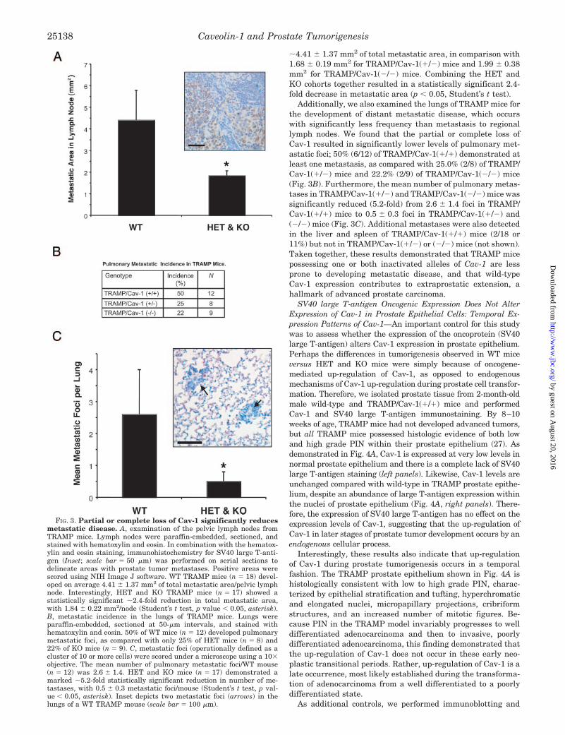
�4.41 � 1.37 mm2 of total metastatic area, in comparison with1.68 � 0.19 mm2 for TRAMP/Cav-1(�/�) mice and 1.99 � 0.38mm2 for TRAMP/Cav-1(�/�) mice. Combining the HET andKO cohorts together resulted in a statistically significant 2.4-fold decrease in metastatic area (p � 0.05, Student’s t test).
Additionally, we also examined the lungs of TRAMP mice forthe development of distant metastatic disease, which occurswith significantly less frequency than metastasis to regionallymph nodes. We found that the partial or complete loss ofCav-1 resulted in significantly lower levels of pulmonary met-astatic foci; 50% (6/12) of TRAMP/Cav-1(�/�) demonstrated atleast one metastasis, as compared with 25.0% (2/8) of TRAMP/Cav-1(�/�) mice and 22.2% (2/9) of TRAMP/Cav-1(�/�) mice(Fig. 3B). Furthermore, the mean number of pulmonary metas-tases in TRAMP/Cav-1(�/�) and TRAMP/Cav-1(�/�) mice wassignificantly reduced (5.2-fold) from 2.6 � 1.4 foci in TRAMP/Cav-1(�/�) mice to 0.5 � 0.3 foci in TRAMP/Cav-1(�/�) and(�/�) mice (Fig. 3C). Additional metastases were also detectedin the liver and spleen of TRAMP/Cav-1(�/�) mice (2/18 or11%) but not in TRAMP/Cav-1(�/�) or (�/�) mice (not shown).Taken together, these results demonstrated that TRAMP micepossessing one or both inactivated alleles of Cav-1 are lessprone to developing metastatic disease, and that wild-typeCav-1 expression contributes to extraprostatic extension, ahallmark of advanced prostate carcinoma.
SV40 large T-antigen Oncogenic Expression Does Not AlterExpression of Cav-1 in Prostate Epithelial Cells: Temporal Ex-pression Patterns of Cav-1—An important control for this studywas to assess whether the expression of the oncoprotein (SV40large T-antigen) alters Cav-1 expression in prostate epithelium.Perhaps the differences in tumorigenesis observed in WT miceversus HET and KO mice were simply because of oncogene-mediated up-regulation of Cav-1, as opposed to endogenousmechanisms of Cav-1 up-regulation during prostate cell transfor-mation. Therefore, we isolated prostate tissue from 2-month-oldmale wild-type and TRAMP/Cav-1(�/�) mice and performedCav-1 and SV40 large T-antigen immunostaining. By 8–10weeks of age, TRAMP mice had not developed advanced tumors,but all TRAMP mice possessed histologic evidence of both lowand high grade PIN within their prostate epithelium (27). Asdemonstrated in Fig. 4A, Cav-1 is expressed at very low levels innormal prostate epithelium and there is a complete lack of SV40large T-antigen staining (left panels). Likewise, Cav-1 levels areunchanged compared with wild-type in TRAMP prostate epithe-lium, despite an abundance of large T-antigen expression withinthe nuclei of prostate epithelium (Fig. 4A, right panels). There-fore, the expression of SV40 large T-antigen has no effect on theexpression levels of Cav-1, suggesting that the up-regulation ofCav-1 in later stages of prostate tumor development occurs by anendogenous cellular process.
Interestingly, these results also indicate that up-regulationof Cav-1 during prostate tumorigenesis occurs in a temporalfashion. The TRAMP prostate epithelium shown in Fig. 4A ishistologically consistent with low to high grade PIN, charac-terized by epithelial stratification and tufting, hyperchromaticand elongated nuclei, micropapillary projections, cribriformstructures, and an increased number of mitotic figures. Be-cause PIN in the TRAMP model invariably progresses to welldifferentiated adenocarcinoma and then to invasive, poorlydifferentiated adenocarcinoma, this finding demonstrated thatthe up-regulation of Cav-1 does not occur in these early neo-plastic transitional periods. Rather, up-regulation of Cav-1 is alate occurrence, most likely established during the transforma-tion of adenocarcinoma from a well differentiated to a poorlydifferentiated state.
As additional controls, we performed immunoblotting and
FIG. 3. Partial or complete loss of Cav-1 significantly reducesmetastatic disease. A, examination of the pelvic lymph nodes fromTRAMP mice. Lymph nodes were paraffin-embedded, sectioned, andstained with hematoxylin and eosin. In combination with the hematox-ylin and eosin staining, immunohistochemistry for SV40 large T-anti-gen (Inset; scale bar � 50 �m) was performed on serial sections todelineate areas with prostate tumor metastases. Positive areas werescored using NIH Image J software. WT TRAMP mice (n � 18) devel-oped on average 4.41 � 1.37 mm2 of total metastatic area/pelvic lymphnode. Interestingly, HET and KO TRAMP mice (n � 17) showed astatistically significant �2.4-fold reduction in total metastatic area,with 1.84 � 0.22 mm2/node (Student’s t test, p value � 0.05, asterisk).B, metastatic incidence in the lungs of TRAMP mice. Lungs wereparaffin-embedded, sectioned at 50-�m intervals, and stained withhematoxylin and eosin. 50% of WT mice (n � 12) developed pulmonarymetastatic foci, as compared with only 25% of HET mice (n � 8) and22% of KO mice (n � 9). C, metastatic foci (operationally defined as acluster of 10 or more cells) were scored under a microscope using a 10�objective. The mean number of pulmonary metastatic foci/WT mouse(n � 12) was 2.6 � 1.4. HET and KO mice (n � 17) demonstrated amarked �5.2-fold statistically significant reduction in number of me-tastases, with 0.5 � 0.3 metastatic foci/mouse (Student’s t test, p val-ue � 0.05, asterisk). Inset depicts two metastatic foci (arrows) in thelungs of a WT TRAMP mouse (scale bar � 100 �m).
Caveolin-1 and Prostate Tumorigenesis25138
by guest on August 20, 2016
http://ww
w.jbc.org/
Dow
nloaded from

immunohistochemistry on poorly differentiated prostate tu-mors isolated from 28-week-old TRAMP mice. Importantly,complete loss of Cav-1 did not alter the expression of the SV40large T-antigen, as assessed by immunoblotting (Fig. 4B) andimmunohistochemistry (Fig. 4C). It has been previously dem-onstrated that single allelic inactivation of Cav-1 results in an�50% reduction in expression in various tissues (29). There-fore, we also performed SV40 large T-antigen and Cav-1 stain-ing on prostate tumors derived from TRAMP/Cav-1(�/�) mice.Interestingly, although SV40 oncogene expression is un-changed compared with TRAMP/Cav-1(�/�) tumors, there isvirtually no Cav-1 immunoreactivity detected in the prostatecarcinoma cells of TRAMP/Cav-1(�/�) tumors (Fig. 4C). Like-wise, Cav-1 immunoblotting demonstrated that these TRAMP/Cav-1(�/�) tumors show dramatic reductions in Cav-1 levels(by �80–90%), more than the 50% reduction predicted frominactivating one Cav-1 allele (Fig. 4D). These findings indi-cated that TRAMP/Cav-1(�/�) tumors have a Cav-1 expressionprofile more comparable to TRAMP/Cav-1(�/�) tumors andoffer an explanation as to why prostate tumorigenesis inTRAMP/Cav-1(�/�) and TRAMP/Cav-1(�/�) mice behavessimilarly.
Expression of Cav-1 in TRAMP Tumor-derived Cell LinesCorrelates with Primary Tumor Growth—To directly assess therole of Cav-1 in mouse prostate cancer cells with regard totumorigenesis, we obtained three prostate carcinoma cell linesderived from WT TRAMP prostate tumors-TRAMP-C1, -C2,and -C3. Importantly, all three TRAMP cell lines express cy-tokeratins, E-cadherin, and the androgen receptor, confirmingtheir epithelial and prostate origin (28).
We examined these cell lines for Cav-1 expression by immu-noblotting and found that C1 demonstrated relatively highlevels of Cav-1, C2 possessed moderate Cav-1 levels, whereasC3 expressed virtually no Cav-1 (Fig. 5A). However, Cav-2levels were unchanged among the various cell lines. Immuno-cytochemistry revealed that Cav-1 localizes properly to theplasma membrane in a typical punctate staining pattern ob-served for Cav-1 in C1 and C2 cells (not shown). Next, weinjected these cell lines into the flanks of syngeneic host malemice and observed the development of tumors. As previouslyreported (28), C1 and C2 cell lines form tumors, with C1 cellsresulting in the largest tumors, whereas C3 cells do not formtumors, even after extended observation (Fig. 5, B and C). It isinteresting to note that tumor size correlates positively with
FIG. 4. SV40 large T-antigen and Cav-1 expression levels at various stages of prostate tumor development in TRAMP mice. A,immunohistochemical analysis of SV40 large T-antigen (LT-Ag) and Cav-1 expression in prostate tissue from 2-month-old male wild-type (WT) andTRAMP/Cav-1(�/�) mice. Note that the presence of LT-Ag expression in TRAMP prostate tissue (brown nuclear staining) does not result inup-regulation of Cav-1 expression. In both wild-type and TRAMP prostate tissue, Cav-1 is expressed at virtually undetectable levels compared withexpression in poorly differentiated TRAMP tumors (see Fig. 1, panels D and E). Despite evidence of high grade PIN at this age, the lack of Cav-1expression in TRAMP/Cav-1(�/�) prostate suggests that Cav-1 is up-regulated at a more advanced stage of prostate carcinoma development. Note thepresence of positive Cav-1 staining in adipocytes (arrows) and capillary endothelium (arrowheads). All images are the same magnification (originalmagnification 40�). Scale bar � 50 �m. B, immunoblotting of poorly differentiated TRAMP prostate tumors at 28 weeks of age. TRAMP/Cav-1(�/�)(WT) and TRAMP/Cav-1(�/�) (KO) tumors were isolated, homogenized, separated by SDS-PAGE, and immunoblotted for SV40 large T-antigen. Tworepresentative samples are shown. Note that levels of the SV40 transgenic oncogene are not altered because of loss of Cav-1. �-actin levels are shownas an equal loading control. C, immunohistochemistry of poorly differentiated TRAMP prostate tumors at 28 weeks of age. Staining for SV40 largeT-antigen (LT-Ag) reveals that the loss of one or both Cav-1 alleles does not significantly alter the expression of this oncogene (top panels; brown nuclearstaining). Interestingly, Cav-1 staining in TRAMP/Cav-1(�/�) tumors is virtually undetectable, despite positive staining in the endothelium (arrows).Note the positive cytoplasmic staining pattern for Cav-1 in prostate tumor cells of TRAMP/Cav-1(�/�) adenocarcinomas. All images are the samemagnification (original magnification 40�). Scale bar � 50 �m. D, Cav-1 immunoblotting of poorly differentiated TRAMP prostate tumors at 28 weeksof age. Note that Cav-1 expression in TRAMP/Cav-1(�/�) tumors is reduced to a greater extent (�80–90%) than the predicted 50% reduction frominactivation of one Cav-1 allele. As expected, Cav-1 levels are absent in TRAMP/Cav-1(�/�) tumors. �-actin levels are shown as an equal loadingcontrol.
Caveolin-1 and Prostate Tumorigenesis 25139
by guest on August 20, 2016
http://ww
w.jbc.org/
Dow
nloaded from

Cav-1 expression in these cells, suggesting that the levels ofCav-1 impact the capacity of these murine prostate carcinoma-derived cells to form tumors.
Targeted Down-regulation of Cav-1 in the TRAMP-C1 CellLine Diminishes Primary Tumor Growth and ExperimentalMetastasis—To directly address the role of Cav-1 in tumorigen-esis in these cells, we down-regulated Cav-1 in C1 cells usingan siRNA approach. We also chose to overexpress Cav-1 in C3cells by a recombinant approach to see whether the forcedexpression of Cav-1 would be sufficient to induce tumor growth.We created stable cell lines through a retroviral approach usingthe pSUPER retroviral vector for the siRNA-mediated down-regulation of Cav-1 and the pBABE retroviral vector for recom-binant Cav-1 overexpression. Initial characterization of thestable cell lines was performed by immunoblotting (Fig. 6A).Note that Cav-1 levels are markedly down-regulated in C1/siRNA cells, as compared with C1 parental or C1/pSUPER(empty vector) cells. When these cell lines were injected intothe flanks of athymic nude male mice, we found a dramaticdecrease in the weight (3.8-fold) and volume (4.6-fold) of tu-mors derived from C1/siRNA cells (Fig. 6B). However, overex-pression of Cav-1 in C3 cells was not sufficient to drive tumorformation. These findings indicated that, although Cav-1 is notsufficient to induce tumors, high Cav-1 expression levels canpromote the development of larger tumors by providing a se-lective advantage for neoplastic growth. Conversely, down-reg-ulation of Cav-1 in TRAMP-C1 cells attenuates the ability ofprostate carcinoma cells to form tumors.
We also assessed whether the targeted down-regulation ofCav-1 in C1 cells affected the ability of these cells to metasta-size. Equal numbers of C1/pSUPER and C1/siRNA cells wereinjected directly into the bloodstream of athymic male micethrough the tail vein (termed “experimental metastasis”). Thistechnique allows the direct assessment of whether the loss ofCav-1 affects the ability of these prostate tumor cells to exit thebloodstream and form metastatic tumors in an ectopic site.After 3 weeks, the lungs were excised, fixed in formalin, andprocessed for histological analysis. Lungs derived from miceinjected with C1/siRNA cells demonstrated an average of7.6 � 1.6 metastatic foci/lung, a dramatic reduction (�5.1-fold)in the number of pulmonary metastatic foci compared withlungs isolated from mice injected with C1/pSUPER cells, whichdeveloped an average of 38.8 � 10.3 foci/lung (Fig. 6C). Addi-tionally, pulmonary metastatic foci arising from C1/pSUPERcells were significantly enlarged compared with C1/siRNA foci(Fig. 6D). These results corroborate the findings from thetransgenic mouse study and indicate that down-regulation ofCav-1 in prostate carcinoma-derived cells attenuates both theprocesses of tumorigenesis and metastasis. Moreover, the cel-lular studies provide direct evidence that the decreases inprostate tumorigenesis and metastasis in HET and KOTRAMP mice are cell-autonomous effects because of the intrin-sic loss of Cav-1 expression in prostate carcinoma cells.
Loss of Cav-1 Results in Increased Levels of Apoptosis inTRAMP Tumors—To begin to identify how the loss of Cav-1results in diminished prostate tumorigenesis and metastasis,we examined the processes of proliferation and apoptosis in theTRAMP-C1 cell lines, C1-derived tumors, and TRAMP prostatetumors. We have previously established that Cav-1 has a neg-ative regulatory role in cellular proliferation in certain cellFIG. 5. Cav-1 expression correlates with tumor growth in
TRAMP cell lines. A, TRAMP cell lines were grown to confluency,collected into lysis buffer, and subjected to immunoblot analysis forCav-1 and Cav-2 expression. Note that the levels of Cav-1 are highest inC1 cells, moderate in C2 cells, and virtually absent in C3 cells. How-ever, Cav-2 levels are equivalent in all three cell lines. �-actin levels areshown as an equal loading control. B and C, C57Bl/6 male mice weresubcutaneously injected with 2 � 106 cells in 0.1 ml of PBS for eachTRAMP cell line (n � 5 each for C1, C2, and C3). After 8 weeks, tumors
were excised, weighed, and sized with calipers. Note that C1 cellsformed larger tumors than C2 cells, whereas C3 cells do not form anytumors. C1 cells formed tumors weighing 1.56 � 0.05 g and were2330 � 206 mm3 in size, whereas C2 cells formed tumors weighing1.13 � 0.06 g and were 1550 � 133 mm3 in size. Differences in size andweight were statistically significant (Student’s t test, p value � 0.05).
Caveolin-1 and Prostate Tumorigenesis25140
by guest on August 20, 2016
http://ww
w.jbc.org/
Dow
nloaded from

types, including transformed fibroblasts and mammary epi-thelial cells (12, 30). Therefore, we performed cell prolifer-ation assays and thymidine-incorporation experiments on theTRAMP-C1 cell line in culture. We found that the down-regu-lation of Cav-1 in C1/siRNA cells resulted in a slight prolifer-ative advantage for growth in culture, in accordance with pre-
vious results showing that loss of Cav-1 exerts a proliferativeadvantage (not shown). Despite this, immunohistochemistryfor proliferating cell nuclear antigen (PCNA), a well character-ized marker for cellular proliferation, revealed no significantdifferences in the number of PCNA-positive cells in WT or KOTRAMP prostate tumors (Fig. 7A). PCNA staining performed
FIG. 6. siRNA-mediated down-regulation of Cav-1 in TRAMP-C1 cells attenuates both tumorigenesis and experimental metastasis.A, creation of stable TRAMP cell lines lacking or overexpressing Cav-1. TRAMP C1 cells were retrovirally infected with pSUPER (empty vector)and pSUPER-Cav-1-siRNA to down-regulate Cav-1 levels. In contrast, TRAMP C3 cells were retrovirally infected with pBABE (empty vector) andpBABE-Cav-1 to overexpress Cav-1. Thereafter, cells were selected with puromycin for 1 week to generate stable “pools” of transduced cells.Finally, cells were grown to confluency, collected into lysis buffer, and subjected to immunoblotting for Cav-1 levels. Note that Cav-1 levels aremarkedly down-regulated using an siRNA approach in C1 cells, as compared with parental (C1) or empty vector (C1/pSUPER) controls. Conversely,C3/Cav-1 cells overexpress recombinant murine Cav-1, as compared with parental (C3) or empty vector (C3/pBABE) control cells. �-actin levels areshown as an equal loading control. B, male nude mice were subcutaneously injected with 5 � 105 cells in 0.1 ml of PBS of either C1/pSUPER,C1/siRNA, C3/pBABE, or C3/Cav-1 cells. After 6 weeks, tumors were excised, weighed, and sized with calipers. Note that both C3 cell lines did notform tumors. Interestingly, C1/siRNA cells formed dramatically smaller tumors (0.19 � 0.04 g; 203 � 42 mm3; n � 14) with a 3.8-fold reductionin weight and 4.6-fold reduction in volume as compared with C1/pSUPER cells (0.72 � 0.12 g; 927 � 137 mm3; n � 14). Differences in size andweight were highly statistically significant (Student’s t test, p value � 0.0002, asterisk). Data represent the average of several independentexperiments, all with virtually identical results. C, male nude mice were injected through the tail vein with 106 cells of either the C1/pSUPER orC1/siRNA cell lines in 0.1 ml of PBS. Lungs were isolated after 3 weeks, processed for hematoxylin and eosin analysis, and examined under amicroscope for the presence of metastatic foci (defined as a cluster of 10 or more cells). Mice (n � 5) injected with C1/pSUPER cells developed anaverage of 38.8 � 10.3 metastatic foci/lung. In contrast, mice injected (n � 5) with C1/siRNA cells demonstrated a 5.1-fold reduction in metastaticfoci (7.6 � 1.6 foci; Student’s t test, p � 0.05, asterisk). D, pulmonary metastatic foci (arrows) derived from C1/pSUPER cells (left panels) aresignificantly larger than C1/siRNA foci (right panels). Photographs were taken from 5-�m sections of the lungs stained with hematoxylin and eosin(original magnifications 10 and 20�; scale bars � 100 �m). Arrows in the top panels (10�) delineate the boundaries of metastatic foci. Arrows inthe bottom panels (20�) indicate the position of metastatic foci.
Caveolin-1 and Prostate Tumorigenesis 25141
by guest on August 20, 2016
http://ww
w.jbc.org/
Dow
nloaded from

FIG. 7. Loss of Cav-1 expression renders prostate tumor cells more susceptible to apoptosis. A, PCNA immunohistochemistry ofprostate tumors from WT and KO TRAMP mice reveals no significant differences in the proliferative index of these tumors. A total of 30 fields (40�)were counted/genotype. Representative PCNA photomicrographs of prostate tumors from WT and KO TRAMP mice are shown. Scale bar � 25 �m.
Caveolin-1 and Prostate Tumorigenesis25142
by guest on August 20, 2016
http://ww
w.jbc.org/
Dow
nloaded from
on tumors derived from C1/pSUPER and C1/siRNA cells alsorevealed no significant differences (not shown). We also exam-ined the expression of cyclin D1 and phospho-ERK-1/2 inTRAMP WT and KO tumors and found no differences in thelevels of these pro-proliferative molecules (not shown).
It has been established that, in prostate cancers, resistanceto apoptotic stimuli plays an especially critical role during theprocess of tumorigenesis (31). Furthermore, a role for Cav-1 inpromoting prostate cancer cell survival has been previouslyestablished in cell culture systems (24, 32). Therefore, we per-formed TUNEL staining to measure DNA strand breakage, ahallmark of apoptosis, in TRAMP prostate tumors and tumorsderived from TRAMP-C1 cells. We found that loss of Cav-1resulted in substantial increases in the apoptotic index of KOTRAMP tumors, as compared with WT TRAMP tumors (Fig.7B). TRAMP/Cav-1(�/�) prostate tumors demonstrated an av-erage of 15.6 � 0.9 TUNEL-positive cells/field, as comparedwith 31.1 � 1.4 TUNEL-positive cells/field in TRAMP/Cav-1(�/�) tumors. This �2.0-fold increase was highly significantand suggests that the presence of Cav-1 in prostate carcinomacells has an anti-apoptotic role. To examine whether the anti-apoptotic function of Cav-1 is indeed a cell-autonomous effect,we also performed TUNEL staining on tumors derived fromTRAMP-C1 prostate tumor cells. Once again, we found that theloss of Cav-1 resulted in highly significant increases (9.0-fold)in the degree of apoptosis in tumors derived from C1/siRNAcells (Fig. 7C). Taken together, these findings clearly indicatean anti-apoptotic role for Cav-1 in prostate carcinoma cellsduring prostate tumorigenesis.
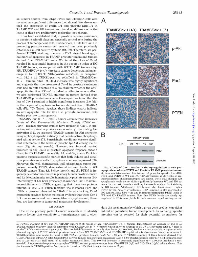
TRAMP/Cav-1(�/�) Null Tumors Demonstrate IncreasedLevels of Two Pro-apoptotic Markers, Namely PTEN andPar4—Because previous studies have implicated Cav-1 in pro-moting cell survival in prostate cancer cells by potentiating Aktactivation (32), we assessed TRAMP tumors for Akt-activationusing a phosphospecific antibody that detects active phosphoryl-ated Akt at serine 473. Surprisingly, we did not observe signifi-cant differences in the levels of phospho (p)-Akt among the tu-mors (Fig. 8A, top panels). However, we observed markedincreases in the levels of prostate apoptosis response factor-4(Par4) in KO TRAMP tumors (Fig. 8A, middle panels). Par4 is aprostate apoptosis-specific marker that both induces and sensi-tizes prostate cancer cells to apoptosis when overexpressed (33).Moreover, the well characterized lipid phosphatase tumor sup-pressor, namely PTEN, demonstrated reduced levels in WTTRAMP tumors (Figs. 8A, bottom panels, and B). PTEN is fre-quently deleted or inactivated in primary human prostate cancer,and its deletion in mice results in metastatic prostate cancer (34).Interestingly, it has been previously shown that Cav-1 co-immu-noprecipitates with PTEN, suggesting that these two proteinsinteract in vivo (21). Taken together, the increased Par4 andPTEN expression observed in TRAMP tumors lacking Cav-1expression provides further molecular evidence that these Cav-1KO tumors are indeed more susceptible to apoptosis and, there-fore, are less prone to tumor and metastasis development.
DISCUSSION
One of the primary goals of cancer research is to identifygenetic factors that contribute to tumorigenesis and to eluci-
date the mechanisms by which a given gene product can eitherinhibit or potentiate tumor development. In so doing, specificproteins can be selected for their potential as markers for
FIG. 8. Loss of Cav-1 results in the up-regulation of two pro-apoptotic markers (PTEN and Par4) in TRAMP prostate tumors.A, immunohistochemical localization of phospho (p)-Akt (Ser-473),Par4, and PTEN in WT and KO TRAMP tumors at 28 weeks of age.Representative photomicrographs are shown. Note that phospho-Aktcytoplasmic levels do not differ significantly between WT and KO tu-mors. In contrast, there is a striking increase in nuclear Par4 stainingin KO tumors. Additionally, KO tumors also demonstrated higherPTEN levels. Finally, cytoplasmic PTEN staining is also increased inKO tumors. Scale bars � 25 �m. B, immunoblotting for PTEN levels inWT and KO TRAMP tumors. Note that PTEN levels are clearly up-regulated in KO tumors. �-tubulin is shown as an equal loading control.
B, TUNEL staining of WT and KO TRAMP tumors at 28 weeks of age. TRAMP/Cav-1(�/�) tumors demonstrated an average of 15.6 � 0.9TUNEL-positive cells/40� field as compared with TRAMP/Cav-1(�/�) tumors, which show an average of 31.1 � 1.4 apoptotic cells/40� field (atotal of 75 fields were counted/genotype). This 2.0-fold difference is extremely significant (p � 0.00001, Student’s t test, asterisk). A representativephotomicrograph of TUNEL-stained prostate tumors from WT TRAMP (left) and KO TRAMP (right) mice is also shown. Note the increase inTUNEL-positive blue nuclei (arrows) in KO TRAMP prostate tumors. Scale bar � 25 �m. C, TUNEL staining of flank tumors derived fromC1/pSUPER and C1/siRNA cells. C1/pSUPER-derived tumors demonstrated on average 0.23 � 0.08 TUNEL-positive cells/40� field compared with2.07 � 0.25 cells/40� field (total of 30 fields counted/cell line). This 9.0-fold decrease is extremely significant (p � 0.00001), Student’s t test,asterisk). A representative photomicrograph of TUNEL stained prostate tumors from C1/pSUPER (left) and C1/siRNA (right) cells is shown. Notethe increase in TUNEL-positive blue nuclei (arrows) in C1/siRNA tumors. Scale bar � 25 �m.
Caveolin-1 and Prostate Tumorigenesis 25143
by guest on August 20, 2016
http://ww
w.jbc.org/
Dow
nloaded from

cancer progression or as targets for therapeutic intervention.The Cav-1 gene has garnered attention because of its alteredexpression patterns in human cancers, e.g. (i) its down-regula-tion in ovarian carcinomas, sarcomas, and mammary carcino-mas and (ii) its up-regulation in bladder, esophageal, and pros-tate carcinomas.
Prostate cancer is the second leading cause of cancer-relateddeaths in males in the United States. As such, identifyinggenes that serve as markers, predictors of outcome, or targetsfor pharmacological therapy is critical in establishing diag-noses and alleviating morbidity and mortality. Interestingly,there is an established relationship between Cav-1 proteinlevels and prostate cancer progression. Increased Cav-1 expres-sion appears to correlate with higher prostate carcinoma gradeand a negative outcome in clinical studies (21, 22). These stud-ies have demonstrated important clinical correlations, but mo-lecular genetic proof of a direct causal link between Cav-1expression and prostate tumorigenesis is lacking. Full recapit-ulation of prostate tumor development using a transgenicmouse model enables investigators to directly test whethersuch relationships are indeed causal in nature.
Here, our current results provided the first molecular geneticevidence that Cav-1 promotes prostate tumorigenesis in vivo.Initial characterization of prostates derived from wild-type andTRAMP mice revealed that Cav-1 is expressed at virtuallyundetectable levels in wild-type and early transformed (PIN)prostate epithelium. However, Cav-1 was up-regulated in thelater stages of prostate adenocarcinoma development, becausepoorly differentiated TRAMP tumors demonstrated significantCav-1 expression. More importantly, we showed that loss ofCav-1 attenuates prostate cancer development by significantlyreducing both primary tumor burden and metastatic disease ina transgenic prostate cancer model. Either partial or completeloss of Cav-1 is sufficient to reduce both of these indices ofadvanced neoplastic development. The observation thatTRAMP/Cav-1(�/�) prostate tumors demonstrate markedlyreduced Cav-1 levels (by �80–90%) offers at least a partialexplanation for why prostate tumorigenesis in TRAMP HETmice proceeds similarly to TRAMP KO mice.
Furthermore, we have established that these results arebecause of the intrinsic loss of Cav-1 within prostate carcinomacells, as targeted down-regulation of Cav-1 in a mouse prostatecarcinoma epithelial cell line (TRAMP-C1 cells) dramaticallyreduces tumor burden and experimental metastasis. We havealso provided direct evidence to support previous cell culturestudies that Cav-1 has pro-survival roles in prostate cancer, asboth TRAMP prostate tumors and TRAMP-C1 cell line tumorslacking Cav-1 expression demonstrated increased rates of apo-ptosis. Taken together, these results delineate a direct causalrelationship between Cav-1 expression and the development ofmore advanced prostate carcinomas, with regard to total pri-mary tumor burden and metastatic disease. Our findings alsovalidated the use of Cav-1 as a marker for prostate cancerprogression and identified Cav-1 as a potential target for che-motherapeutic or gene therapy-based interventions.
Interestingly, these results directly contrast with our previ-ous studies in mammary tissue, demonstrating that loss ofCav-1 accelerates mammary tumorigenesis and metastasisthrough a different transgenic model approach (9, 12). Re-cently, another group independently demonstrated that Cav-1expression inhibits mammary tumor growth and metastasis(11). In these mammary carcinoma studies, Cav-1 appears tohave a growth-inhibitory role, because genetic ablation ofCav-1 results in cyclin D1 overexpression and ERK-1/2 hyper-activation. In contrast, no differences were detected in ERK-1/2activation or cyclin D1 expression between WT and Cav-1 KO
TRAMP prostate tumors (not shown). Taken together, thesedata directly indicate that Cav-1 has tissue or cell type-specificroles with regard to tumorigenesis (i.e. growth-inhibitory intransformed mammary epithelium versus anti-apoptotic intransformed prostate epithelium). These results, taken in com-bination with other studies, provide clear evidence that Cav-1up-regulation serves to provide a selective advantage to trans-formed cells in certain human cancers (i.e. prostate), whereasCav-1 down-regulation may also promote cancer developmentin a completely different group of cancers (i.e. mammarygland). Further analysis of the molecular pathways responsiblefor this observed dichotomy should yield interesting insightsinto how Cav-1 normally functions as a “tumor suppressor” or“tumor promoter” in different cell- and tissue-specific contexts.
Previously, there was some concern regarding the validity ofthe TRAMP animal model for studying prostate carcinogenesis.However, this has now been resolved in favor of the TRAMPmodel. It was argued that a large proportion of prostate tumorsarising in TRAMP mice are derived from neuroendocrine pre-cursor cells and not from the prostate epithelium. This hypoth-esis was based on the presence of several neuroendocrine mark-ers, such as synaptophysin, in advanced TRAMP prostatetumors. However, Greenberg and colleagues (27) have nowprovided direct evidence that this is not the case. They havedemonstrated that the neuroendocrine markers present inpoorly differentiated TRAMP tumors arise stochastically, asneuroendocrine markers are almost completely absent in PINand low grade prostatic carcinoma TRAMP lesions. Addition-ally, the emergence of neuroendocrine markers does not di-rectly indicate that transformed epithelial cells have “trans-differentiated” into neuroendocrine cells, as Greenberg andcolleagues also show that the majority of these cells also co-express the androgen receptor, a protein not normally ex-pressed by cells of neuroendocrine origin. Therefore, the pres-ence of neuroendocrine markers more likely indicates theemergence of a distinct “neuroendocrine-like” phenotype.
Finally, because neuroendocrine markers are present in focalareas of nearly all clinical human prostatic adenocarcinomas,this acquired neuroendocrine phenotype further parallels theprogression of the human prostatic disease and actually vali-dates the use of the TRAMP model for studying prostate car-cinoma progression (27). Similarly, we showed here that Cav-1protein expression levels are indeed elevated in TRAMP pros-tate tumors, consistent with the up-regulation of Cav-1 inhuman prostate cancers (21, 22). Thus, our current results withCav-1 further highlight the utility of the TRAMP model instudying the evolution of prostate cancers in vivo.
Acknowledgment—We thank Dr. R. Mahmood for her technical ex-pertise regarding tissue processing, sectioning, and image acquisition.
REFERENCES
1. Glenney, J. R., Jr. (1989) J. Biol. Chem. 264, 20163–201662. Williams, T. M., and Lisanti, M. P. (2004) Genome Biol. 5, 214.1–214.83. Cohen, A. W., Hnasko, R., Schubert, W., and Lisanti, M. P. (2004) Physiol. Rev.
84, 1341–13794. Koleske, A. J., Baltimore, D., and Lisanti, M. P. (1995) Proc. Natl. Acad. Sci.
U. S. A. 92, 1381–13855. Lee, S. W., Reimer, C. L., Oh, P., Campbell, D. B., and Schnitzer, J. E. (1998)
Oncogene 16, 1391–13976. Engelman, J. A., Wykoff, C. C., Yasuhara, S., Song, K. S., Okamoto, T., and
Lisanti, M. P. (1997) J. Biol. Chem. 272, 16374–163817. Zhang, W., Razani, B., Altschuler, Y., Bouzahzah, B., Mostov, K. E., Pestell,
R. G., and Lisanti, M. P. (2000) J. Biol. Chem. 275, 20717–207258. Fiucci, G., Ravid, D., Reich, R., and Liscovitch, M. (2002) Oncogene 21,
2365–23759. Williams, T. M., Cheung, M. W., Park, D. S., Razani, B., Cohen, A. W., Muller,
W. J., Di Vizio, D., Chopra, N. G., Pestell, R. G., and Lisanti, M. P. (2003)Mol. Biol. Cell 14, 1027–1042
10. Capozza, F., Williams, T. M., Schubert, W., McClain, S., Bouzahzah, B., Sotgia,F., and Lisanti, M. P. (2003) Am. J. Pathol. 162, 2029–2039
11. Sloan, E. K., Stanley, K. L., and Anderson, R. L. (2004) Oncogene 23,7893–7897
12. Williams, T. M., Medina, F., Badano, I., Hazan, R. B., Hutchinson, J., Muller,
Caveolin-1 and Prostate Tumorigenesis25144
by guest on August 20, 2016
http://ww
w.jbc.org/
Dow
nloaded from

W. J., Chopra, N. G., Scherer, P. E., Pestell, R. G., and Lisanti, M. P. (2004)J. Biol. Chem. 279, 51630–51646
13. Hayashi, K., Matsuda, S., Machida, K., Yamamoto, T., Fukuda, Y., Nimura, Y.,Hayakawa, T., and Hamaguchi, M. (2001) Cancer Res. 61, 2361–2364
14. Han, S. E., Park, K. H., Lee, G., Huh, Y. J., and Min, B. M. (2004) Int. J. Oncol.24, 435–440
15. Razani, B., and Lisanti, M. P. (2001) J. Clin. Investig. 108, 1553–156116. Rajjayabun, P. H., Garg, S., Durkan, G. C., Charlton, R., Robinson, M. C., and
Mellon, J. K. (2001) Urology 58, 811–81417. Fong, A., Garcia, E., Gwynn, L., Lisanti, M. P., Fazzari, M. J., and Li, M. (2003)
Am. J. Clin. Pathol. 120, 93–10018. Hu, Y. C., Lam, K. Y., Law, S., Wong, J., and Srivastava, G. (2001) Clin. Cancer
Res. 7, 3519–352519. Kato, K., Hida, Y., Miyamoto, M., Hashida, H., Shinohara, T., Itoh, T.,
Okushiba, S., Kondo, S., and Katoh, H. (2002) Cancer 94, 929–93320. Yang, G., Truong, L. D., Timme, T. L., Ren, C., Wheeler, T. M., Park, S. H.,
Nasu, Y., Bangma, C. H., Kattan, M. W., Scardino, P. T., and Thompson,T. C. (1998) Clin. Cancer Res. 4, 1873–1880
21. Yang, G., Truong, L. D., Wheeler, T. M., and Thompson, T. C. (1999) CancerRes. 59, 5719–5723
22. Satoh, T., Yang, G., Egawa, S., Addai, J., Frolov, A., Kuwao, S., Timme, T. L.,Baba, S., and Thompson, T. C. (2003) Cancer 97, 1225–1233
23. Nasu, Y., Timme, T. L., Yang, G., Bangma, C. H., Li, L., Ren, C., Park, S. H.,DeLeon, M., Wang, J., and Thompson, T. C. (1998) Nat. Med. 4, 1062–1064
24. Li, L., Yang, G., Ebara, S., Satoh, T., Nasu, Y., Timme, T. L., Ren, C., Wang,J., Tahir, S. A., and Thompson, T. C. (2001) Cancer Res. 61, 4386–4392
25. Greenberg, N. M., DeMayo, F., Finegold, M. J., Medina, D., Tilley, W. D.,
Aspinall, J. O., Cunha, G. R., Donjacour, A. A., Matusik, R. J., and Rosen,J. M. (1995) Proc. Natl. Acad. Sci. U. S. A. 92, 3439–3443
26. Gingrich, J. R., Barrios, R. J., Morton, R. A., Boyce, B. F., DeMayo, F. J.,Finegold, M. J., Angelopoulou, R., Rosen, J. M., and Greenberg, N. M. (1996)Cancer Res. 56, 4096–4102
27. Kaplan-Lefko, P. J., Chen, T. M., Ittmann, M. M., Barrios, R. J., Ayala, G. E.,Huss, W. J., Maddison, L. A., Foster, B. A., and Greenberg, N. M. (2003)Prostate 55, 219–237
28. Foster, B. A., Gingrich, J. R., Kwon, E. D., Madias, C., and Greenberg, N. M.(1997) Cancer Res. 57, 3325–3330
29. Razani, B., Engelman, J. A., Wang, X. B., Schubert, W., Zhang, X. L., Marks,C. B., Macaluso, F., Russell, R. G., Li, M., Pestell, R. G., Di Vizio, D., Hou,H., Jr., Kneitz, B., Lagaud, G., Christ, G. J., Edelmann, W., and Lisanti,M. P. (2001) J. Biol. Chem. 276, 38121–38138
30. Williams, T. M., Lee, H., Cheung, M. W., Cohen, A. W., Razani, B., Iyengar, P.,Scherer, P. E., Pestell, R. G., and Lisanti, M. P. (2004) J. Biol. Chem. 279,24745–24756
31. Gurumurthy, S., Vasudevan, K. M., and Rangnekar, V. M. (2001) CancerMetastasis Rev. 20, 225–243
32. Li, L., Ren, C. H., Tahir, S. A., Ren, C., and Thompson, T. C. (2003) Mol. Cell.Biol. 23, 9389–9404
33. Chakraborty, M., Qiu, S. G., Vasudevan, K. M., and Rangnekar, V. M. (2001)Cancer Res. 61, 7255–7263
34. Wang, S., Gao, J., Lei, Q., Rozengurt, N., Pritchard, C., Jiao, J., Thomas, G. V.,Li, G., Roy-Burman, P., Nelson, P. S., Liu, X., and Wu, H. (2003) CancerCells 4, 209–221
Caveolin-1 and Prostate Tumorigenesis 25145
by guest on August 20, 2016
http://ww
w.jbc.org/
Dow
nloaded from

LisantiPhilippe G. Frank, Richard G. Pestell, Dolores Di Vizio, Massimo Loda and Michael P. Terence M. Williams, Ghada S. Hassan, Jiangwei Li, Alex W. Cohen, Freddy Medina,
PROSTATE TUMOR DEVELOPMENT IN TRAMP MICEProstate Cancer: GENETIC ABLATION OF Cav-1 DELAYS ADVANCED
Caveolin-1 Promotes Tumor Progression in an Autochthonous Mouse Model of
doi: 10.1074/jbc.M501186200 originally published online March 30, 20052005, 280:25134-25145.J. Biol. Chem.
10.1074/jbc.M501186200Access the most updated version of this article at doi:
Alerts:
When a correction for this article is posted•
When this article is cited•
to choose from all of JBC's e-mail alertsClick here
http://www.jbc.org/content/280/26/25134.full.html#ref-list-1
This article cites 34 references, 19 of which can be accessed free at
by guest on August 20, 2016
http://ww
w.jbc.org/
Dow
nloaded from
Copyright © 2022 FDOKUMEN