Functional Characterization of Circulating Tumor Cells with a Prostate-Cancer-Specific Microfluidic...
10
Functional Characterization of Circulating Tumor Cells with a Prostate-Cancer-Specific Microfluidic Device Brian J. Kirby 1,2 *, Mona Jodari 3 , Matthew S. Loftus 3 , Gunjan Gakhar 3 , Erica D. Pratt 1,2 , Chantal Chanel- Vos 3 , Jason P. Gleghorn 1,2 , Steven M. Santana 1,2 , He Liu 4 , James P. Smith 1,2 , Vicente N. Navarro 4 , Scott T. Tagawa 3 , Neil H. Bander 4 , David M. Nanus 3,4 , Paraskevi Giannakakou 3 * 1 Sibley School of Mechanical and Aerospace Engineering, Cornell University, Ithaca, New York, United States of America, 2 Biomedical Engineering, Cornell University, Ithaca, New York, United States of America, 3 Division of Hematology and Medical Oncology, Department of Medicine, Weill Cornell Medical College and New York Presbyterian Hospital, New York, New York, United States of America, 4 Department of Urology, Weill Cornell Medical College and New York Presbyterian Hospital, New York, New York, United States of America Abstract Cancer metastasis accounts for the majority of cancer-related deaths owing to poor response to anticancer therapies. Molecular understanding of metastasis-associated drug resistance remains elusive due to the scarcity of available tumor tissue. Isolation of circulating tumor cells (CTCs) from the peripheral blood of patients has emerged as a valid alternative source of tumor tissue that can be subjected to molecular characterization. However, issues with low purity and sensitivity have impeded adoption to clinical practice. Here we report a novel method to capture and molecularly characterize CTCs isolated from castrate-resistant prostate cancer patients (CRPC) receiving taxane chemotherapy. We have developed a geometrically enhanced differential immunocapture (GEDI) microfluidic device that combines an anti-prostate specific membrane antigen (PSMA) antibody with a 3D geometry that captures CTCs while minimizing nonspecific leukocyte adhesion. Enumeration of GEDI-captured CTCs (defined as intact, nucleated PSMA+/CD452 cells) revealed a median of 54 cells per ml identified in CRPC patients versus 3 in healthy donors. Direct comparison with the commercially available CellSearchH revealed a 2–400 fold higher sensitivity achieved with the GEDI device. Confocal microscopy of patient-derived GEDI-captured CTCs identified the TMPRSS2:ERG fusion protein, while sequencing identified specific androgen receptor point mutation (T868A) in blood samples spiked with only 50 PC C4-2 cells. On-chip treatment of patient-derived CTCs with docetaxel and paclitaxel allowed monitoring of drug-target engagement by means of microtubule bundling. CTCs isolated from docetaxel-resistant CRPC patients did not show any evidence of drug activity. These measurements constitute the first functional assays of drug-target engagement in living circulating tumor cells and therefore have the potential to enable longitudinal monitoring of target response and inform the development of new anticancer agents. Citation: Kirby BJ, Jodari M, Loftus MS, Gakhar G, Pratt ED, et al. (2012) Functional Characterization of Circulating Tumor Cells with a Prostate-Cancer-Specific Microfluidic Device. PLoS ONE 7(4): e35976. doi:10.1371/journal.pone.0035976 Editor: Natasha Kyprianou, University of Kentucky College of Medicine, United States of America Received December 20, 2011; Accepted March 23, 2012; Published April 27, 2012 Copyright: ß 2012 Kirby et al. This is an open-access article distributed under the terms of the Creative Commons Attribution License, which permits unrestricted use, distribution, and reproduction in any medium, provided the original author and source are credited. Funding: This work was supported by grants from the US National Institutes of Health (R01 CA137020-01 and U54 CA143876) (PG), the NIH Physical Sciences Oncology Center at Cornell (BK, PG DN), New York Office of Science, Technology, and Research (BK), the Weill Cornell Clinical and Translational Science Center (DN, PG BK) a Creativity Award from the Prostate Cancer Foundation (PG and DN) and support from the Genitourinary Oncology Research Fund (DN). CCV is a recipient of an NRSA postdoctoral fellowship. EP and SS were supported by National Science Foundation Graduate Research Fellowships. The funders had no role in study design, data collection and analysis, decision to publish, or preparation of the manuscript. Competing Interests: BK, PG, ST and DN disclose consulting income from Sanofi US. This does not alter our adherence to all the Plos One’s policies on sharing data and materials. * E-mail: [email protected] (BJK); or [email protected] (PG) Introduction The development of metastases in patients with solid tumor malignancies is believed to result from tumor cells entering the circulatory system and migrating to distant organs, where they extravasate and multiply [1–3]. Although circulating tumor cells (CTCs) are rare—as few as one cell per 100 million blood cells [3,4], molecular and functional analyses of CTCs may provide a greater understanding of the biology of cancer metastases, help identify novel therapeutic targets, and enable clinical correlations to monitor patients undergoing treatment [5]. A variety of technologies has been developed to improve the detection and capture of CTCs from the peripheral blood. These include density gradient centrifugation, immunomagnetic bead separation using monoclonal antibodies targeting epithelial cell-surface antigens, cell sorting using flow cytometry, filtration based size separation [6] and microfluidic devices. Although advances in CTC capture have been made, the low frequency of CTCs in cancer patients, their heterogeneity, the lack of organ-specific capture approaches, and the plasticity of the CTC population has limited the ability to capture and track all CTCs [2,6]. Currently, the epithelial cell- adhesion molecule (EpCAM), represents the antigen of choice for the majority of microfluidic devices that have been developed to capture circulating tumor cells [7–10]. However, accumulating evidence suggests that the expression of EpCAM during cancer progression and in particular during epithelial-to-mesenchymal transition has not been well character- ized, raising concerns about the universality of this antigen for immunocapture systems [11,12]. EpCAM has been reported to have oncogenic potential [13] and correlate with proliferation in PLoS ONE | www.plosone.org 1 April 2012 | Volume 7 | Issue 4 | e35976
-
Upload
independent -
Category
Documents
-
view
2 -
download
0
Transcript of Functional Characterization of Circulating Tumor Cells with a Prostate-Cancer-Specific Microfluidic...
Functional Characterization of Circulating Tumor Cellswith a Prostate-Cancer-Specific Microfluidic DeviceBrian J. Kirby1,2*, Mona Jodari3, Matthew S. Loftus3, Gunjan Gakhar3, Erica D. Pratt1,2, Chantal Chanel-
Vos3, Jason P. Gleghorn1,2, Steven M. Santana1,2, He Liu4, James P. Smith1,2, Vicente N. Navarro4,
Scott T. Tagawa3, Neil H. Bander4, David M. Nanus3,4, Paraskevi Giannakakou3*
1 Sibley School of Mechanical and Aerospace Engineering, Cornell University, Ithaca, New York, United States of America, 2 Biomedical Engineering, Cornell University,
Ithaca, New York, United States of America, 3 Division of Hematology and Medical Oncology, Department of Medicine, Weill Cornell Medical College and New York
Presbyterian Hospital, New York, New York, United States of America, 4 Department of Urology, Weill Cornell Medical College and New York Presbyterian Hospital, New
York, New York, United States of America
Abstract
Cancer metastasis accounts for the majority of cancer-related deaths owing to poor response to anticancer therapies.Molecular understanding of metastasis-associated drug resistance remains elusive due to the scarcity of available tumortissue. Isolation of circulating tumor cells (CTCs) from the peripheral blood of patients has emerged as a valid alternativesource of tumor tissue that can be subjected to molecular characterization. However, issues with low purity and sensitivityhave impeded adoption to clinical practice. Here we report a novel method to capture and molecularly characterize CTCsisolated from castrate-resistant prostate cancer patients (CRPC) receiving taxane chemotherapy. We have developed ageometrically enhanced differential immunocapture (GEDI) microfluidic device that combines an anti-prostate specificmembrane antigen (PSMA) antibody with a 3D geometry that captures CTCs while minimizing nonspecific leukocyteadhesion. Enumeration of GEDI-captured CTCs (defined as intact, nucleated PSMA+/CD452 cells) revealed a median of 54cells per ml identified in CRPC patients versus 3 in healthy donors. Direct comparison with the commercially availableCellSearchH revealed a 2–400 fold higher sensitivity achieved with the GEDI device. Confocal microscopy of patient-derivedGEDI-captured CTCs identified the TMPRSS2:ERG fusion protein, while sequencing identified specific androgen receptorpoint mutation (T868A) in blood samples spiked with only 50 PC C4-2 cells. On-chip treatment of patient-derived CTCs withdocetaxel and paclitaxel allowed monitoring of drug-target engagement by means of microtubule bundling. CTCs isolatedfrom docetaxel-resistant CRPC patients did not show any evidence of drug activity. These measurements constitute the firstfunctional assays of drug-target engagement in living circulating tumor cells and therefore have the potential to enablelongitudinal monitoring of target response and inform the development of new anticancer agents.
Citation: Kirby BJ, Jodari M, Loftus MS, Gakhar G, Pratt ED, et al. (2012) Functional Characterization of Circulating Tumor Cells with a Prostate-Cancer-SpecificMicrofluidic Device. PLoS ONE 7(4): e35976. doi:10.1371/journal.pone.0035976
Editor: Natasha Kyprianou, University of Kentucky College of Medicine, United States of America
Received December 20, 2011; Accepted March 23, 2012; Published April 27, 2012
Copyright: � 2012 Kirby et al. This is an open-access article distributed under the terms of the Creative Commons Attribution License, which permitsunrestricted use, distribution, and reproduction in any medium, provided the original author and source are credited.
Funding: This work was supported by grants from the US National Institutes of Health (R01 CA137020-01 and U54 CA143876) (PG), the NIH Physical SciencesOncology Center at Cornell (BK, PG DN), New York Office of Science, Technology, and Research (BK), the Weill Cornell Clinical and Translational Science Center (DN,PG BK) a Creativity Award from the Prostate Cancer Foundation (PG and DN) and support from the Genitourinary Oncology Research Fund (DN). CCV is a recipientof an NRSA postdoctoral fellowship. EP and SS were supported by National Science Foundation Graduate Research Fellowships. The funders had no role in studydesign, data collection and analysis, decision to publish, or preparation of the manuscript.
Competing Interests: BK, PG, ST and DN disclose consulting income from Sanofi US. This does not alter our adherence to all the Plos One’s policies on sharingdata and materials.
* E-mail: [email protected] (BJK); or [email protected] (PG)
Introduction
The development of metastases in patients with solid tumor
malignancies is believed to result from tumor cells entering the
circulatory system and migrating to distant organs, where they
extravasate and multiply [1–3]. Although circulating tumor cells
(CTCs) are rare—as few as one cell per 100 million blood cells
[3,4], molecular and functional analyses of CTCs may provide a
greater understanding of the biology of cancer metastases, help
identify novel therapeutic targets, and enable clinical correlations
to monitor patients undergoing treatment [5]. A variety of
technologies has been developed to improve the detection and
capture of CTCs from the peripheral blood. These include density
gradient centrifugation, immunomagnetic bead separation using
monoclonal antibodies targeting epithelial cell-surface antigens,
cell sorting using flow cytometry, filtration based size separation
[6] and microfluidic devices. Although advances in CTC capture
have been made, the low frequency of CTCs in cancer patients,
their heterogeneity, the lack of organ-specific capture approaches,
and the plasticity of the CTC population has limited the ability to
capture and track all CTCs [2,6]. Currently, the epithelial cell-
adhesion molecule (EpCAM), represents the antigen of choice for
the majority of microfluidic devices that have been developed to
capture circulating tumor cells [7–10].
However, accumulating evidence suggests that the expression of
EpCAM during cancer progression and in particular during
epithelial-to-mesenchymal transition has not been well character-
ized, raising concerns about the universality of this antigen for
immunocapture systems [11,12]. EpCAM has been reported to
have oncogenic potential [13] and correlate with proliferation in
PLoS ONE | www.plosone.org 1 April 2012 | Volume 7 | Issue 4 | e35976
cell lines [14]; however, it is downregulated during EMT [1], and
EMT markers have been shown to be more important than
epithelial markers e.g., cytokeratin in predicting cancer progres-
sion [15]. Thus, while EpCAM is clearly useful in identifying CTC
populations in many cancers, the biases associated with EpCAM
enrichment are currently unknown.
In addition to the uncertainties regarding surface antigens, the
specificity of immunocapture from the blood is confounded by the
non-specific adhesive properties of leukocytes on most antibody
surfaces. Because of the presence of numerous leukocytes in blood
at an approximately 104–105:1 ratio with respect to the CTCs,
immunospecific surfaces enrich CTCs but cannot isolate them
from contaminating leukocytes entirely. Identifying CTCs requires
additional steps and often involves staining with DAPI to ensure
the presence of an intact nucleus and immunostaining to identify
the presence of epithelial markers (i.e., cytokeratin) and the lack of
the leukocytic marker CD45. Such immunostaining has identified
a family of criteria that correlate CTC number with patient
prognosis [16], but it requires that fixation and staining be central
to CTC identification, as in the commercial CellSearchH system
[17,18]. Although enumeration of CTCs from patients with
advanced prostate cancer receiving chemotherapy using the
commercially available CTC capture system by CellSearchH has
demonstrated utility as a prognostic indicator of patient survival
[17,18] and enumeration is currently being studied in a number of
clinical trials, the presence of contaminating leukocytes impede the
downstream utility of CTC capture devices, in that assays based
on RNA or protein quantification are obfuscated by the need for
fixation and material of leukocytic origin.
To facilitate high-efficiency capture of prostate CTCs, we have
developed a prototype microfluidic device that employs an
approach that we term geometrically enhanced differential
immunocapture (GEDI). This device combines a geometry that
reduces capture of contaminating leukocytes by generating size-
dependent cell-wall collisions. We combine this geometric
approach with a prostate-specific immunocapture surface using
the J591 monoclonal antibody that recognizes the extracellular
domain of prostate-specific membrane antigen (PSMA) [10].
PSMA is a cell surface peptidase highly expressed by malignant
prostate epithelial cells. PSMA is an attractive target for prostate
cancer CTC capture, as it is expressed on virtually all prostate
cancer cells and expression increases following castration. We
report here a detailed demonstration of the multiple utilities of the
GEDI device, including a comparison of CTC enumeration with
CellSearchH, the detection of a specific AR mutation from blood
samples spiked with only 50 cells; the identification of the
TMPRSS2-ERG fusion by immunostaining, and the ex-vivo
assessment of CTC sensitivity to taxane-treatment using microtu-
bule bundling as a marker of drug-target engagement.
Materials and Methods
Device FabricationAll device fabrication was carried out at the Cornell NanoScale
Science and Technology Facility (Ithaca, NY). Standard photoli-
thography techniques were used to define array geometries on
silicon wafers. The wafers were etched with an oxygen plasma
deep reactive ion etcher (Uniaxis SLR770) to a depth of 100 mm,
and cleaned using sulfuric acid and hydrogen peroxide prior to
antibody surface functionalization. The J591 monoclonal antibody
(manufactured by Lonza plc (Slough, England) for BZL Biologics,
inc.) was immobilized on the device surfaces using MPTMS-
GMBS-NeutrAvidin-biotin chemistry [10]. Polydimethylsiloxane
(PDMS) sheets (5:1 base:curing agent), approximately 3 mm thick,
were polymerized for 18 hrs at 60uC and trimmed to form covers
for the GEDI device. A PDMS sheet was clamped to the top of the
device with a custom jig to create closed channels populated with
post arrays. Inlet and outlet holes were created with a biopsy
punch, and 23-gauge metal tubes were inserted into the PDMS to
connect inlet and outlets to external tubing. Devices were primed
with a 50/50 isopropanol/water mixture, and then flushed with
DI water and PBS before experiments.
Sample Collection and Microfluidic CapturePeripheral blood samples were collected in tubes containing
sodium citrate anticoagulant (Becton-Dickinson) from healthy
volunteers and patients with metastatic castrate-resistant prostate
cancer under a clinical protocol entitled ‘‘Analysis of circulating
tumor cells in prostate cancer. Predicting response to taxanes: a
pilot study’’ which was approved by the Institutional Review
Board (IRB) of Weill Cornell Medical College of Cornell
University. Blood was obtained from patients or healthy donors
following written informed consent, which was also approved by
the IRB committee of Weill Cornell Medical College of Cornell
University. As previously described [10], 1 ml of blood from each
specimen was processed through the GEDI chip within 24 hr of
blood draw by pushing the blood through the device at a
volumetric flow rate of 1 ml/hr (Chemyx syringe pump).
Cell staining and analysisThe cell lines used in these experiments as controls for staining
or in spiked experiments are: the human leukemia cell line U937,
and the human prostate cancer cell lines PC-3, LNCaP and C4-2.
All cell lines were purchased from ATCC. Post-capture, cells were
fixed on-chip with PHEMO fixative at 37uC (PHEMO buffer:
PIPES acid, HEPES acid, EGTA disodium salt, Mg-Cl2-6H20,
10%DMSO), gluteraldehyde, and 3.7%formaldehyde. Cells were
then blocked (10% Normal Goat Serum – Jackson Immuno
Research) and immunostained with FITC-conjugated humanized
mAb J591 to detect PSMA expression. Monoclonal mouse anti-
CD-45 (BD Biosciences) followed by AlexaFluor568 labeled goat
anti-mouse secondary (Invitrogen) and mouse anti-EpCAM
directly conjugated to AlexaFluor647 (Biolegend). For the
detection of intracellular antigens, cells were permeabilized with
0.1% Triton X-100 (Sigma-Aldrich) in PBS and stained by use of
rat anti-alpha tubulin (YL1/2, Millipore) and rabbit anti-ERG
monoclonal antibody (clone EPR 3864; Epitomics, Burlingame,
CA). The anti-ERG antibody was a generous gift from Dr. Mark
Rubin (Weill Cornell Medical College, New York, NY). All
primary antibodies were incubated for 1 hour at room tempera-
ture; secondary antibodies were stained at room temperature for
30 minutes. DAPI was used for DNA counterstaining. GEDI
devices were mounted to coverslips with Mowiol and stored at
220uC before analysis.
CTC enumerationBlinded CTC enumeration following antibody labeling was
performed by use of a Zeiss LSM-700 point scanning confocal
microscope, equipped with 405-, 488-, 555-, and 632-nm laser
lines. All PSMA+/CD452 nucleated cells were identified as
CTCs. Initial validation of CTC enumeration was accomplished
by two independent, blinded testers. Positive and negative controls
for antibody performance and staining were included in each
experiment: U937 human leukemia cells (CD45+/PSMA2/
EpCAM2), and the human prostate cancer cell lines (C4-2 and
LNCaP: PSMA+/CD452/EpCAM+ and PC-3 (PSMA2/
Cd452/EpCAM dim). Individual z-stacks were acquired using
100X/NA 1.46 and 63X/NA 1.3 Plan-Apo Zeiss objectives
CTC Analysis with a PSMA-based Microfluidic Device
PLoS ONE | www.plosone.org 2 April 2012 | Volume 7 | Issue 4 | e35976
controlled by Zen software (Zeiss) and presented as maximum
intensity projections.
RNA extractionFollowing cell capture, the GEDI device was rinsed by flowing
PBS for 30 min at a rate of 1 ml/hr. Cells were lysed with 700 ml
of RLT Lysis buffer supplemented with 1% b-mercaptoethanol at
a flow rate of 15 ml/hr. The lysate was collected, and RNA was
extracted using the QiagenRNEasy Micro Plus Kit (Qiagen Inc,
Valencia, CA) according to the manufacturer’s instructions.
Ex-vivo Drug treatments and AnalysisFor ex vivo drug treatment experiments, the blood sample from
each patient was divided and 1 ml was flown to each of three
GEDI devices simultaneously. After CTC capture and subsequent
PBS wash, each GEDI microdevice was gently placed in a culture
dish with RPMI-1640 media containing 2% serum and supple-
mented with either 0.1% DMSO control or paclitaxel at
concentrations of 100 nM or 1 mM and incubated at 37uC for
24 hr. At the end of treatment, the GEDI-captured cells were fixed
with PHEMO buffer and processed for multiplex confocal
microscopy following immunostaining with different cell surface
and cytoplasmic antibodies as indicated. All CTCs (PSMA+/
CD452/DAPI+) were assessed for the presence of microtubule
bundles as evidence of effective drug-target engagement. The
percent of CTCs with evidence of microtubule bundling was
calculated. In all samples analyzed, bundling was clearly apparent
by the distinct shape, width, orientation and increased fluores-
cence intensity of microtubule bundles as compared with
microtubules from untreated cells. In addition, we used DAPI
counterstain to assess the presence of mitotic or apoptotic nuclei
following drug treatment.
Statistical AnalysisStatistical analysis was performed to compare the mean CTC
counts obtained from CRPC patients and healthy donors. We
used a non-parametric (Wilcoxon signed-rank) analysis, as CTC
counts did not exhibit normal distribution. Statistical significance
was defined with a= 0.05.
Results
To characterize the performance of the device (Figure 1A), we
determined cell capture rates with PSMA-positive cancer cells. We
determined cell capture as a function of varying mAb J591
concentrations (1.5–20 mg/ml) using shear stress magnitudes
representative of those experienced by the functionalized surfaces
of the device (0.08–0.24 Pa). These experiments revealed a dose-
dependent increase in cell capture up to mAb concentration of
10 mg/ml, which was used for all subsequent experiments
(Figure 1B).
Although the J591 antibody is specific for PSMA-expressing
cells, non-specific leukocyte adhesion has been a major problem
for all blood-based immunocapture techniques. To minimize
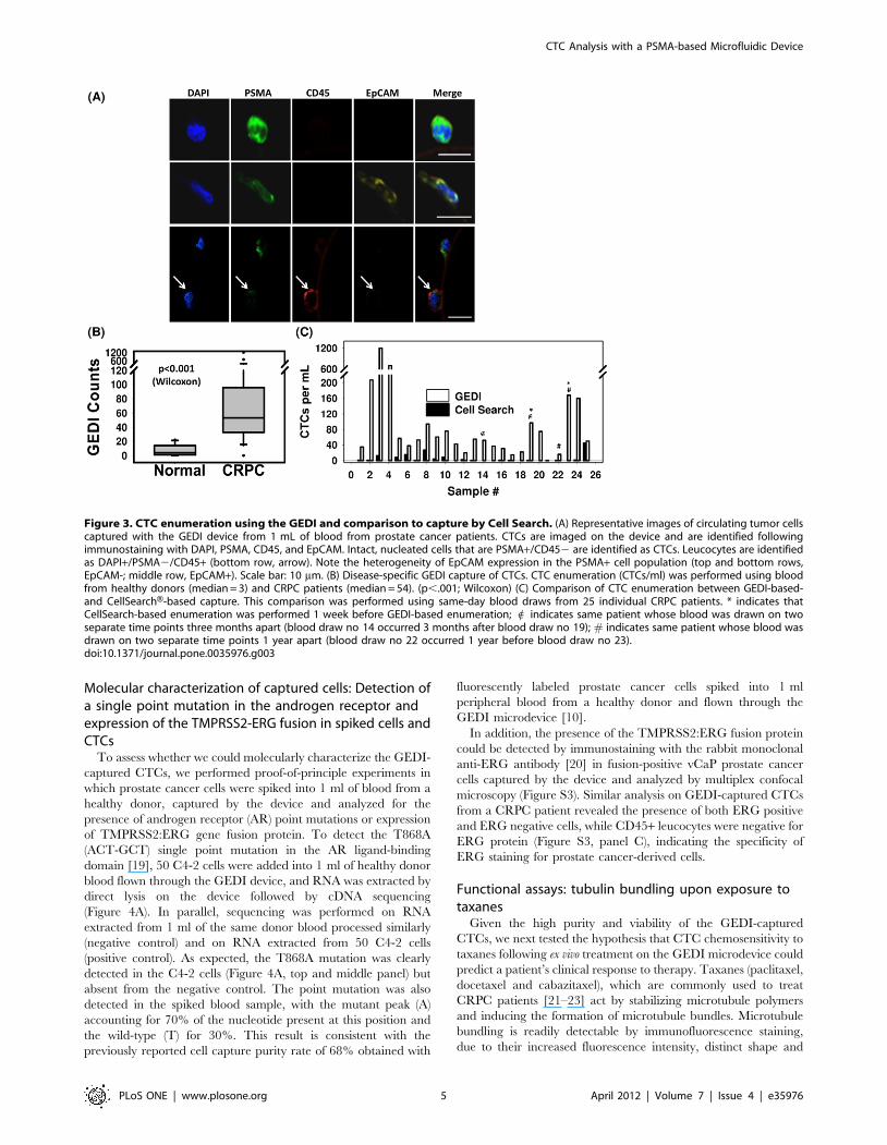
leukocyte adhesion, we conducted a parametric study to
characterize collision rate (CpR; collision per row) as a function
of cell size and obstacle offset. Collision rates from a subset of
these offsets exhibit a sharp cutoff according to cell size, as
shown in Figure 2A; hence we selected an obstacle offset (7 mm)
that generates a sharp cutoff at the cell diameter of 14 mm. The
physics describing this cutoff is best illustrated by the size-
dependent cell pathlines (Figure 2A), which show how large cells
experience repeated collision whereas small cells separate from
the obstacles and escape capture. We then tested this hypothesis
by measuring capture of LNCaP prostate cancer cells
(Figure 2B). In this experiment, spiked LNCaP cells were flown
into J591-functionalized devices that had a 7 mm offset (GEDI)
versus those that had no offset (straight). Although these two
devices have the same surface-area-to-volume ratio, the GEDI
geometry greatly increased cell capture efficiency, as measured
by captured and enumerated cells normalized by input cell
counts.
Because of the dependence of cell trajectory on cell diameter,
collision rates are a complicated function of both cell diameter and
array parameters such as row offsets. The collision rate per row
(CpR) is a strong function of the row offset, exhibiting
discontinuities, size dependence, and startup effects related to
the finite array size (Figure 2C). The dramatic difference between
performance of different designs is caused by the deflection of
particles—in poorly-chosen geometries, the deflection causes cells
to deflect onto streamlines that do not come into proximity with
later obstacles, whereas in well-chosen geometries, the deflection
causes cells to deflect onto streamlines that do come into proximity
with later obstacles. Thus the collision rate increases as the cells
proceed through the device for the GEDI design, and decreases for
poorly chosen designs such as straight arrays (Figure 2C). This
cutoff allows the user to identify a cutoff between hematocytes
(,14 mm) and the cell population that will experience maximum
collisions (.15 mm).
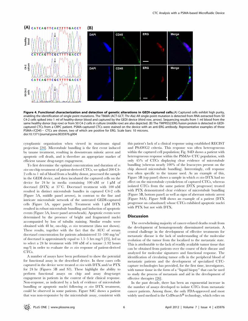
Cell capture, imaging, and enumeration of CTCs frommetastatic prostate cancer patients
Next, we used the GEDI device to capture and characterize
circulating tumor cells (CTCs) from the blood of patients with
metastatic CRPC. One ml of peripheral blood was flown through
the device, captured cells were fixed and immunostained for
PSMA, CD45, EpCAM and DAPI, and the captured cells were
analyzed by confocal microscopy. CTCs were defined as intact,
nucleated, PSMA+/CD452 cells. Representative examples of
CTCs and leucocytes are shown in Figure 3A. Interestingly,
PSMA+ cells had variable EpCAM staining, ranging from highly-
positive to weak to negative in terms of EpCAM fluorescent
intensity. In a subset of patients, we quantitated the percent of
PSMA-captured CTCs that were EpCAM positive. We observed
that 40–70% of GEDI-captured CTCs were positive for both
markers, with the median being 60% (data not shown). Controls
for antibody performance were included with every experiment
using two prostate cancer cell lines expressing different levels of
PSMA and EpCAM (C4-2:PSMA+/EpCAM+/CD452 and PC3:
PSMA2/EpCAM2/CD452) and the CD45+ leukemia cell line
U937 (Figure S1A).
We processed blood obtained from 10 healthy donors (controls)
and 30 patients with metastatic CRPC using the GEDI device.
The median number of CTCs/ml detected was 3 (range 0 to 22)
and 54 (range 0 to 1200), respectively (p,0.001; Figure 3B). Next,
we performed a direct comparison of CTC capture and
enumeration by comparing the GEDI microdevice with the
FDA-approved EpCAM-based CellSearchH CTC Test on same-
day blood draws from 25 CRPC patients (Figure 3C). We detected
a 2 to 400-fold increase in the number of CTCs/ml reported with
the GEDI microdevice relative to the CellSearch H CTC Test
(Figure 3C; p,.0001, calculated with Wilcoxon test), and a weak
correlation (r = 0.44; outliers removed with Cook’s distance
restriction) between GEDI CTC counts and CellSearchH (Figure
S2).
CTC Analysis with a PSMA-based Microfluidic Device
PLoS ONE | www.plosone.org 3 April 2012 | Volume 7 | Issue 4 | e35976
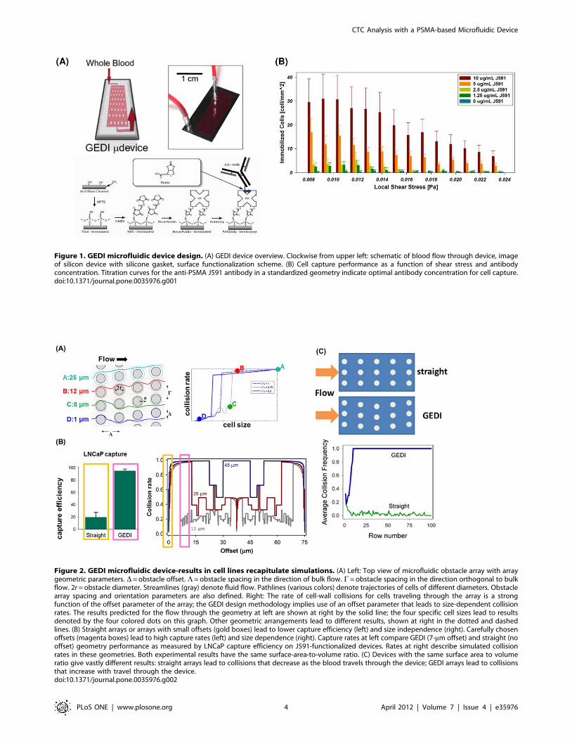
Figure 1. GEDI microfluidic device design. (A) GEDI device overview. Clockwise from upper left: schematic of blood flow through device, imageof silicon device with silicone gasket, surface functionalization scheme. (B) Cell capture performance as a function of shear stress and antibodyconcentration. Titration curves for the anti-PSMA J591 antibody in a standardized geometry indicate optimal antibody concentration for cell capture.doi:10.1371/journal.pone.0035976.g001
Figure 2. GEDI microfluidic device-results in cell lines recapitulate simulations. (A) Left: Top view of microfluidic obstacle array with arraygeometric parameters. D= obstacle offset. L= obstacle spacing in the direction of bulk flow. C= obstacle spacing in the direction orthogonal to bulkflow. 2r = obstacle diameter. Streamlines (gray) denote fluid flow. Pathlines (various colors) denote trajectories of cells of different diameters. Obstaclearray spacing and orientation parameters are also defined. Right: The rate of cell-wall collisions for cells traveling through the array is a strongfunction of the offset parameter of the array; the GEDI design methodology implies use of an offset parameter that leads to size-dependent collisionrates. The results predicted for the flow through the geometry at left are shown at right by the solid line; the four specific cell sizes lead to resultsdenoted by the four colored dots on this graph. Other geometric arrangements lead to different results, shown at right in the dotted and dashedlines. (B) Straight arrays or arrays with small offsets (gold boxes) lead to lower capture efficiency (left) and size independence (right). Carefully chosenoffsets (magenta boxes) lead to high capture rates (left) and size dependence (right). Capture rates at left compare GEDI (7-mm offset) and straight (nooffset) geometry performance as measured by LNCaP capture efficiency on J591-functionalized devices. Rates at right describe simulated collisionrates in these geometries. Both experimental results have the same surface-area-to-volume ratio. (C) Devices with the same surface area to volumeratio give vastly different results: straight arrays lead to collisions that decrease as the blood travels through the device; GEDI arrays lead to collisionsthat increase with travel through the device.doi:10.1371/journal.pone.0035976.g002
CTC Analysis with a PSMA-based Microfluidic Device
PLoS ONE | www.plosone.org 4 April 2012 | Volume 7 | Issue 4 | e35976
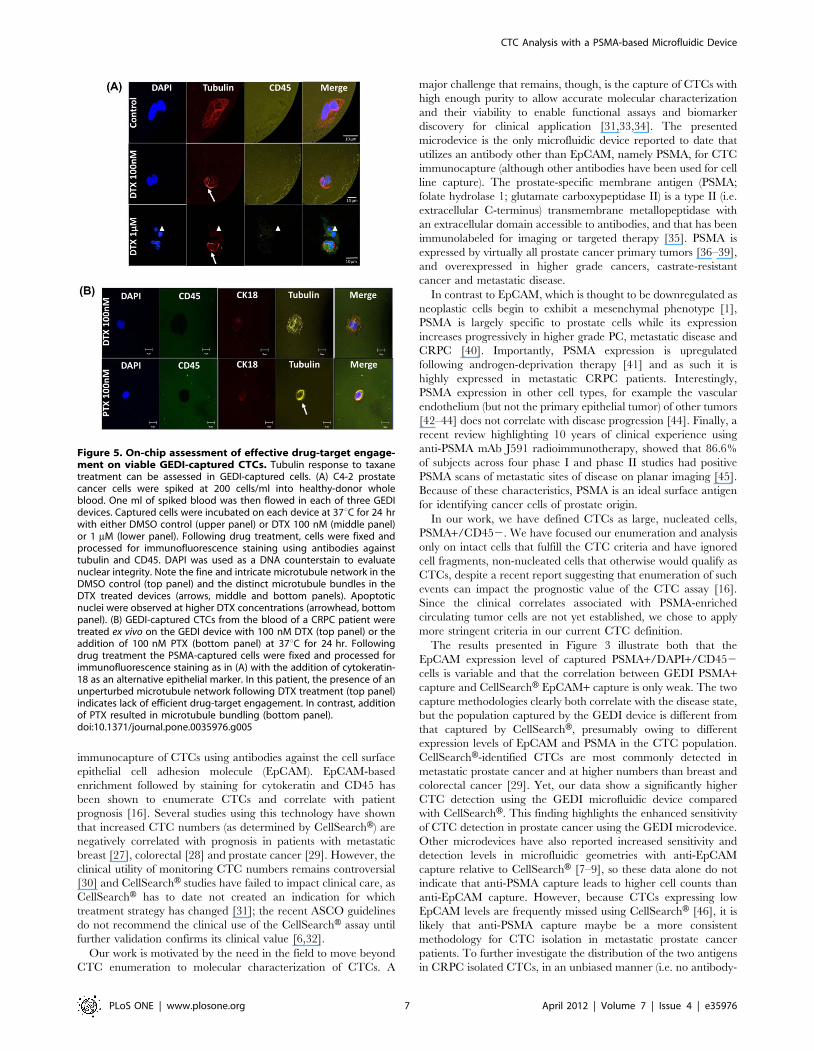
Molecular characterization of captured cells: Detection ofa single point mutation in the androgen receptor andexpression of the TMPRSS2-ERG fusion in spiked cells andCTCs
To assess whether we could molecularly characterize the GEDI-
captured CTCs, we performed proof-of-principle experiments in
which prostate cancer cells were spiked into 1 ml of blood from a
healthy donor, captured by the device and analyzed for the
presence of androgen receptor (AR) point mutations or expression
of TMPRSS2:ERG gene fusion protein. To detect the T868A
(ACT-GCT) single point mutation in the AR ligand-binding
domain [19], 50 C4-2 cells were added into 1 ml of healthy donor
blood flown through the GEDI device, and RNA was extracted by
direct lysis on the device followed by cDNA sequencing
(Figure 4A). In parallel, sequencing was performed on RNA
extracted from 1 ml of the same donor blood processed similarly
(negative control) and on RNA extracted from 50 C4-2 cells
(positive control). As expected, the T868A mutation was clearly
detected in the C4-2 cells (Figure 4A, top and middle panel) but
absent from the negative control. The point mutation was also
detected in the spiked blood sample, with the mutant peak (A)
accounting for 70% of the nucleotide present at this position and
the wild-type (T) for 30%. This result is consistent with the
previously reported cell capture purity rate of 68% obtained with
fluorescently labeled prostate cancer cells spiked into 1 ml
peripheral blood from a healthy donor and flown through the
GEDI microdevice [10].
In addition, the presence of the TMPRSS2:ERG fusion protein
could be detected by immunostaining with the rabbit monoclonal
anti-ERG antibody [20] in fusion-positive vCaP prostate cancer
cells captured by the device and analyzed by multiplex confocal
microscopy (Figure S3). Similar analysis on GEDI-captured CTCs
from a CRPC patient revealed the presence of both ERG positive
and ERG negative cells, while CD45+ leucocytes were negative for
ERG protein (Figure S3, panel C), indicating the specificity of
ERG staining for prostate cancer-derived cells.
Functional assays: tubulin bundling upon exposure totaxanes
Given the high purity and viability of the GEDI-captured
CTCs, we next tested the hypothesis that CTC chemosensitivity to
taxanes following ex vivo treatment on the GEDI microdevice could
predict a patient’s clinical response to therapy. Taxanes (paclitaxel,
docetaxel and cabazitaxel), which are commonly used to treat
CRPC patients [21–23] act by stabilizing microtubule polymers
and inducing the formation of microtubule bundles. Microtubule
bundling is readily detectable by immunofluorescence staining,
due to their increased fluorescence intensity, distinct shape and
Figure 3. CTC enumeration using the GEDI and comparison to capture by Cell Search. (A) Representative images of circulating tumor cellscaptured with the GEDI device from 1 mL of blood from prostate cancer patients. CTCs are imaged on the device and are identified followingimmunostaining with DAPI, PSMA, CD45, and EpCAM. Intact, nucleated cells that are PSMA+/CD452 are identified as CTCs. Leucocytes are identifiedas DAPI+/PSMA2/CD45+ (bottom row, arrow). Note the heterogeneity of EpCAM expression in the PSMA+ cell population (top and bottom rows,EpCAM-; middle row, EpCAM+). Scale bar: 10 mm. (B) Disease-specific GEDI capture of CTCs. CTC enumeration (CTCs/ml) was performed using bloodfrom healthy donors (median = 3) and CRPC patients (median = 54). (p,.001; Wilcoxon) (C) Comparison of CTC enumeration between GEDI-based-and CellSearchH-based capture. This comparison was performed using same-day blood draws from 25 individual CRPC patients. * indicates thatCellSearch-based enumeration was performed 1 week before GEDI-based enumeration; 1 indicates same patient whose blood was drawn on twoseparate time points three months apart (blood draw no 14 occurred 3 months after blood draw no 19); # indicates same patient whose blood wasdrawn on two separate time points 1 year apart (blood draw no 22 occurred 1 year before blood draw no 23).doi:10.1371/journal.pone.0035976.g003
CTC Analysis with a PSMA-based Microfluidic Device
PLoS ONE | www.plosone.org 5 April 2012 | Volume 7 | Issue 4 | e35976
cytoplasmic organization when viewed in maximum signal
projection [24]. Microtubule bundling is the first event induced
by taxane treatment, resulting in downstream mitotic arrest and
apoptotic cell death, and is therefore an appropriate marker of
efficient taxane drug-target engagement.
To first determine the optimal concentration and duration of ex
vivo on-chip treatment of patient-derived CTCs, we spiked 200 C4-
2 cells to 1 ml of blood from a healthy donor, processed the sample
in the GEDI device, and then incubated the captured cells on the
device for 24 hr in media containing 100 nM or 1 mM of
docetaxel (DTX) at 37uC. Docetaxel treatment with 100 nM
resulted in distinct microtubule bundles in captured C4-2 cells
(Figure 5A, middle panel arrows), in contrast to the fine and
intricate microtubule network of the untreated GEDI-captured
cells (Figure 5A, upper panel). Treatment with 1 mM DTX
resulted in robust microtubule bundling and induction of apoptotic
events (Figure 5A, lower panel arrowheads). Apoptotic events were
determined by the presence of bright and fragmented nuclei
accompanied by loss of tubulin staining. Similar results were
obtained with 48 hr, on-chip, ex vivo treatment (data not shown).
These results, together with the fact that the AUC of serum
docetaxel concentration for patients administered 55–100 mg/m2
of docetaxel is approximately equal to 1.5–5 hrs mg/l [25], led us
to select a 24 hr treatment with 100 nM of a taxane (1.92 hours
mg/l) in order to evaluate the ex vivo response of patient-derived
CTCs.
A number of assays have been performed to show the potential
for functional assay in the described device. In these cases cells
captured in the device were treated ex vivo with DTX and/or PTX
for 24 hr (Figures 5B and S3). These highlight the ability to
perform functional assays on chip and assay drug-target
engagement in patients in the context of their clinical response.
Non-response, as indicated by a lack of evidence of microtubule
bundling or apoptotic nuclei following ex vivo DTX treatment,
could be observed in some patients. Figure S4C shows a patient
that was non-responsive by the microtubule assay, consistent with
this patient’s lack of a clinical response using established RECIST
and PSAWG2 criteria. This response was often heterogeneous
within the captured cell population; Fig. S4D shows a patient with
heterogeneous response within the PSMA+ CTC population, with
only 45% of CTCs displaying clear evidence of microtubule
bundling (whereas nearly 100% of the leucocytes present on the
chip showed microtubule bundling). Interestingly, cell response
was often specific to the taxane used. As an example of this,
Figure 5B (top panel) shows a sample in which ex vivo DTX had no
effect on the microtubule cytoskeleton of captured CTCs, whereas
isolated CTCs from the same patient (DTX progressor) treated
with PTX demonstrated clear evidence of microtubule bundling
(Figure 5B, bottom panel) as well as signs of aberrant mitotic arrest
(Figure S4A). Figure S4B shows an example of a patient (DTX
progressor on cabazitaxel) whose CTCs exhibited apoptotic nuclei
with PTX but not with DTX.
Discussion
The overwhelming majority of cancer-related deaths result from
the development of hematogenously disseminated metastasis. A
central challenge in the development of effective treatments for
metastatic disease is the lack of understanding of the molecular
evolution of the tumor from the localized to the metastatic state.
This is attributable to the lack of readily available tumor tissue that
can be obtained from patients over the course of their disease and
analyzed for molecular signatures and functional response. The
identification of circulating tumor cells in the peripheral blood of
metastatic patients and the development of specialized CTC-
capture technologies has provided, for the first time, investigators,
with tumor tissue in the form of a ‘‘liquid biopsy’’ that can be used
to study the process of metastasis and aid in the development of
effective therapies [26].
In the past decade, there has been an exponential increase in
the number of assays developed to isolate CTCs from metastatic
cancer patients. Among those, the only FDA-approved and most
widely used method is the CellSearchH technology, which relies on
Figure 4. Functional characterization and detection of genetic alterations in GEDI-captured cells.(A) Captured cells exhibit high purity,enabling the identification of single point mutations. The T868A (ACT-GCT; Thr-Ala) AR single-point mutation is detected from RNA extracted from 50C4-2 cells spiked into 1 ml of healthy-donor blood and captured by the GEDI device (third row, arrow). Sequencing results from 1 ml blood from thesame healthy donor (top row) or from 50 C4-2 cells in culture (middle row) are also depicted. (B) The TMPRSS2:ERG fusion protein is detected in GEDI-captured CTCs from a CRPC patient. PSMA-captured CTCs were stained on the device with an anti-ERG antibody. Representative examples of threePSMA+/CD452 CTCs are shown, two of which are positive for ERG. Scale bars: 10 microns.doi:10.1371/journal.pone.0035976.g004
CTC Analysis with a PSMA-based Microfluidic Device
PLoS ONE | www.plosone.org 6 April 2012 | Volume 7 | Issue 4 | e35976
immunocapture of CTCs using antibodies against the cell surface
epithelial cell adhesion molecule (EpCAM). EpCAM-based
enrichment followed by staining for cytokeratin and CD45 has
been shown to enumerate CTCs and correlate with patient
prognosis [16]. Several studies using this technology have shown
that increased CTC numbers (as determined by CellSearchH) are
negatively correlated with prognosis in patients with metastatic
breast [27], colorectal [28] and prostate cancer [29]. However, the
clinical utility of monitoring CTC numbers remains controversial
[30] and CellSearchH studies have failed to impact clinical care, as
CellSearchH has to date not created an indication for which
treatment strategy has changed [31]; the recent ASCO guidelines
do not recommend the clinical use of the CellSearchH assay until
further validation confirms its clinical value [6,32].
Our work is motivated by the need in the field to move beyond
CTC enumeration to molecular characterization of CTCs. A
major challenge that remains, though, is the capture of CTCs with
high enough purity to allow accurate molecular characterization
and their viability to enable functional assays and biomarker
discovery for clinical application [31,33,34]. The presented
microdevice is the only microfluidic device reported to date that
utilizes an antibody other than EpCAM, namely PSMA, for CTC
immunocapture (although other antibodies have been used for cell
line capture). The prostate-specific membrane antigen (PSMA;
folate hydrolase 1; glutamate carboxypeptidase II) is a type II (i.e.
extracellular C-terminus) transmembrane metallopeptidase with
an extracellular domain accessible to antibodies, and that has been
immunolabeled for imaging or targeted therapy [35]. PSMA is
expressed by virtually all prostate cancer primary tumors [36–39],
and overexpressed in higher grade cancers, castrate-resistant
cancer and metastatic disease.
In contrast to EpCAM, which is thought to be downregulated as
neoplastic cells begin to exhibit a mesenchymal phenotype [1],
PSMA is largely specific to prostate cells while its expression
increases progressively in higher grade PC, metastatic disease and
CRPC [40]. Importantly, PSMA expression is upregulated
following androgen-deprivation therapy [41] and as such it is
highly expressed in metastatic CRPC patients. Interestingly,
PSMA expression in other cell types, for example the vascular
endothelium (but not the primary epithelial tumor) of other tumors
[42–44] does not correlate with disease progression [44]. Finally, a
recent review highlighting 10 years of clinical experience using
anti-PSMA mAb J591 radioimmunotherapy, showed that 86.6%
of subjects across four phase I and phase II studies had positive
PSMA scans of metastatic sites of disease on planar imaging [45].
Because of these characteristics, PSMA is an ideal surface antigen
for identifying cancer cells of prostate origin.
In our work, we have defined CTCs as large, nucleated cells,
PSMA+/CD452. We have focused our enumeration and analysis
only on intact cells that fulfill the CTC criteria and have ignored
cell fragments, non-nucleated cells that otherwise would qualify as
CTCs, despite a recent report suggesting that enumeration of such
events can impact the prognostic value of the CTC assay [16].
Since the clinical correlates associated with PSMA-enriched
circulating tumor cells are not yet established, we chose to apply
more stringent criteria in our current CTC definition.
The results presented in Figure 3 illustrate both that the
EpCAM expression level of captured PSMA+/DAPI+/CD452
cells is variable and that the correlation between GEDI PSMA+capture and CellSearchH EpCAM+ capture is only weak. The two
capture methodologies clearly both correlate with the disease state,
but the population captured by the GEDI device is different from
that captured by CellSearchH, presumably owing to different
expression levels of EpCAM and PSMA in the CTC population.
CellSearchH-identified CTCs are most commonly detected in
metastatic prostate cancer and at higher numbers than breast and
colorectal cancer [29]. Yet, our data show a significantly higher
CTC detection using the GEDI microfluidic device compared
with CellSearchH. This finding highlights the enhanced sensitivity
of CTC detection in prostate cancer using the GEDI microdevice.
Other microdevices have also reported increased sensitivity and
detection levels in microfluidic geometries with anti-EpCAM
capture relative to CellSearchH [7–9], so these data alone do not
indicate that anti-PSMA capture leads to higher cell counts than
anti-EpCAM capture. However, because CTCs expressing low
EpCAM levels are frequently missed using CellSearchH [46], it is
likely that anti-PSMA capture maybe be a more consistent
methodology for CTC isolation in metastatic prostate cancer
patients. To further investigate the distribution of the two antigens
in CRPC isolated CTCs, in an unbiased manner (i.e. no antibody-
Figure 5. On-chip assessment of effective drug-target engage-ment on viable GEDI-captured CTCs. Tubulin response to taxanetreatment can be assessed in GEDI-captured cells. (A) C4-2 prostatecancer cells were spiked at 200 cells/ml into healthy-donor wholeblood. One ml of spiked blood was then flowed in each of three GEDIdevices. Captured cells were incubated on each device at 37uC for 24 hrwith either DMSO control (upper panel) or DTX 100 nM (middle panel)or 1 mM (lower panel). Following drug treatment, cells were fixed andprocessed for immunofluorescence staining using antibodies againsttubulin and CD45. DAPI was used as a DNA counterstain to evaluatenuclear integrity. Note the fine and intricate microtubule network in theDMSO control (top panel) and the distinct microtubule bundles in theDTX treated devices (arrows, middle and bottom panels). Apoptoticnuclei were observed at higher DTX concentrations (arrowhead, bottompanel). (B) GEDI-captured CTCs from the blood of a CRPC patient weretreated ex vivo on the GEDI device with 100 nM DTX (top panel) or theaddition of 100 nM PTX (bottom panel) at 37uC for 24 hr. Followingdrug treatment the PSMA-captured cells were fixed and processed forimmunofluorescence staining as in (A) with the addition of cytokeratin-18 as an alternative epithelial marker. In this patient, the presence of anunperturbed microtubule network following DTX treatment (top panel)indicates lack of efficient drug-target engagement. In contrast, additionof PTX resulted in microtubule bundling (bottom panel).doi:10.1371/journal.pone.0035976.g005
CTC Analysis with a PSMA-based Microfluidic Device
PLoS ONE | www.plosone.org 7 April 2012 | Volume 7 | Issue 4 | e35976
specific CTC capture) we isolated CRPC-patient CTCs by
performing immuno-magnetic CD45 depletion of Ficoll-isolated
peripheral blood mononuclear cells. These immunodepleted cells
were subsequently labeled with antibodies against PSMA (J591),
EpCAM and CD45. In this assay, CTCs were classified as
nucleated CD452 cells that were positive for either PSMA,
EpCAM or both. We analyzed a total of eleven patient samples
and have identified CTCs in seven patients. Our results revealed
over 80% of dual PSMA+/EpCAM+ CTCs in six of the seven
patients (Figure S5A and B). Interestingly, using this technique, we
also found that EpCAM staining intensity was variable which is
consistent with the data obtained using the GEDI-microdevice
(Figure 3).
Taken together these data indicate that an important contrib-
uting factor to the enhanced sensitivity of the GEDI is that our
microdevice is designed to optimize CTC capture and minimize
leukocyte capture; it does this by inducing a fluid flow that
generates size-dependent trajectories of cells that lead to size-
dependent collision rates. Spatial separation of cells based on size
alone is of limited use in CTC capture from blood—although
CTCs tend to be larger on average that hematological cells, the
sizes of cells and cell fragments of epithelial origin has a broad
distribution, and size is much less specific to the CTC phenotype
than surface markers such as EpCAM, PSMA, or EGFR.
However, when a surface antibody is present, size-dependent
particle trajectories enable the captured cell population to be
biased to reject nonspecific leukocyte adhesion. The surface
collision rate and capture rate (Figure 2) can be made size-specific,
enhancing the receiver-operator characteristic of the rare cell
capture. By capturing CTCs at high efficiency and purity,
functional and molecular assays, exemplified by the ERG and
SNP measurements in Figure 4 and the tubulin measurements in
Figure 5 can be made applicable in a clinical setting.
ERG gene rearrangements are reported to occur only in
neoplastic and preneoplastic tissue and not in normal tissue
[47,48]. Our findings suggest that the CTCs we detect are
malignant in origin and heterogeneous in nature, as observed in
previous studies of the presence of ERG gene rearrangement in
CTCs using FISH [9,49]. The advantage of the use of an
antibody-based detection method for ERG is that is allows
multiplexing for different cell markers that can further characterize
tumor cell heterogeneity. Although it is well established that in
primary PC there is heterogeneity in terms of ERG rearrange-
ment, data is limited on ERG heterogeneity within a metastatic
patient or potential changes in the ETS rearrangement event over
the course of therapy, similar to HER-2 gene amplification that is
enhanced as breast cancer progresses [50]. In addition, the impact
of ERG on taxane sensitivity is not known, or potential
enrichment of ERG-positive CTCs as a patient becomes refractory
to taxane treatment. These are important clinical questions that
can be answered prospectively in clinical trials that incorporate the
GEDI-based biomarker studies.
A major challenge in the clinical management of CRPC is that
currently there is no biomarker that predicts clinical efficacy of
chemotherapy. The taxanes represent the only class of cancer
chemotherapeutics demonstrated to prolong survival in CRPC
[22,23]. Although clinically defined patient groups inform the
statistical efficacy of microtubule-targeting agents, no predictive
markers are in use to direct microtubule-targeted therapy on an
individual-patient basis. Despite their clinical success, treatment
efficacy can be transient and the development of clinical taxane
resistance is the major cause of cancer-related death. As there are
two FDA-approved taxanes for CRPC, the important clinical
question is to understand why patients who fail treatment with one
taxane respond to another, and how clinicians can anticipate
progression and proactively switch treatment.
At the cellular level, taxanes bind and stabilize microtubules,
compromising their function during interphase [51] as well as
mitosis [52], ultimately inducing cell death. However, these effects
are variable between patients and may be variable within the
metastases and circulating tumor cell population of an individual
patient. Historically, investigators have searched for surrogate
markers predictive of taxane clinical activity, albeit without
success. Attempts to correlate drug-induced microtubule stabili-
zation by quantitating the percent of patient derived PBMCs with
microtubule bundles and correlating these results with clinical
response to therapy bore no correlation [53]. This result may not
be surprising given that PBMC biology is entirely distinct from
that of tumor tissue. In fact, our results do not indicate any relation
between tubulin bundling in leucocytes and clinical response to
taxane chemotherapy (Figure S4D). On the other hand, absence of
taxane-induced microtubule bundling following ex vivo treatment
of GEDI-captured CTCs trended with clinical progression on the
same taxane (Figure 5). Interestingly, our results show differential
CTC response to docetaxel versus paclitaxel, which recapitulates
the known clinical observation for lack of cross-resistance between
the three taxanes used for CRPC treatment. Furthermore, these
observations generate the hypothesis that ex vivo drug-target
engagement of the GEDI-captured CTCs combined with the
use of other relevant biomarkers, such as inhibition of androgen
receptor nuclear accumulation downstream of taxane-induced
microtubule stabilization, [54] may predict clinical response and
help identify a subset of patients more likely to benefit from
treatment with a specific taxane.
The presented work demonstrates the first functional assay of a
microtubule-targeting agent on living circulating tumor cells
microfluidically extracted from patient blood. This work highlights
the potential for tailoring of chemotherapy by real-time monitor-
ing of drug-target engagement in CTCs. Further, the demonstrat-
ed ability to identify genetic mutations and fusions in rare cell
populations points to the potential for identifying mechanisms
underlying clinical response and resistance.
Supporting Information
Figure S1 Multiplex immunostaining for specific cellsurface markers. Different cell lines were used as controls for
antibody staining for PSMA, CD45, and EpCAM, as follows: the
C4-2 prostate cancer cells are PSMA+/EpCAM+/CD452 while
the PC3 prostate cancer cells are PSMA2/EpCAM2/CD452.
The U937 leukemic cell line was used as a positive control for the
leukocyte marker CD45. DAPI was used to stain the DNA.
(TIFF)
Figure S2 GEDI-CellSearch correlation. Correlation be-
tween the number of CTCs detected by the CellSearchH system vs.
the GEDI system from same day blood draws. A correlation
coefficient of r = 0.44 (outliers were removed with Cook’s distance
restriction) was determined. Hashtag and asterisk denote two pairs
of data each taken on the same patient at two longitudinal time
points. r is not changed significantly by inclusion or rejection of
these points.
(TIFF)
Figure S3 TMPRSS2:ERG detection by immunofluores-cence on GEDI-captured cells. (A) The performance of the
ERG antibody staining was tested in TMPRSS2:ERG fusion-
positive (vCaP) and fusion-negative (C4-2) prostate cancer cell
lines. Representative images acquired by confocal microscopy are
CTC Analysis with a PSMA-based Microfluidic Device
PLoS ONE | www.plosone.org 8 April 2012 | Volume 7 | Issue 4 | e35976
displayed. Note the nuclear ERG staining in fusion-positive vCaP
cells. (B) Two hundred vCaP cells were spiked in 1 ml of healthy-
donor blood, flown through the GEDI device and processed for
ERG immunofluorescence labeling. Nuclear ERG staining was
detected in the GEDI-captured vCaP cells, identified as PSMA+/
DAPI+/CD452 cells. (C) Representative example of ERG-
negative/CD45+ leucocytes identified in the blood from a CRPC
patient processed by the GEDI device as in Figure 4B.
(TIFF)
Figure S4 Additional examples of on-chip assessment ofeffective drug-target engagement from different CRPCpatients: taxane-induced microtubule bundling andmitotic defects as evidence of drug-target engagementin GEDI-captured CTCs. (A) GEDI-captured CTCs from the
same patient as in Figure 5B. PTX-induced prometaphase arrest
of GEDI-captured CTCs provides additional evidence of effective
drug-target engagement. (B) GEDI-captured CTCs from patient 3
following ex vivo on-chip treatment with 100 nM DTX do not
show any evidence of microtubule response (bundling) to drug
treatment. (C) GEDI-captured CTCs from patient 2 display
microtubule bundling (arrow) following ex vivo on-chip treatment
with 100 nM or 1 mM PTX. (D) GEDI-captured CTCs from
patient 4 following ex vivo on-chip treatment with 50 nM PTX
show heterogeneous response to drug treatment. Note, distinct
microtubule bundling in a PSMA+ CTC (middle panel, barbed
arrow) and no detectable microtubule network in another PSMA+
CTC from the same patient (bottom panel, standard arrow). The
adjacent leucocyte (PSMA2) shows clear microtubule bundling in
response to PTX treatment.
(TIFF)
Figure S5 PSMA and EpCAM expression in CRPCpatient CTCs isolated using CD452 immunodepletion.(A) Table showing the percentage of both PSMA and EpCAM
positive CTCs from CRPC patients obtained using CD45-
immunodepletion. (B) Representative images of CTCs isolated
from 2 prostate cancer patients, stained for PSMA (Green) and
EpCAM (Red) and analyzed by point scanning confocal
microscopy. Scale Bar = 10 mm. The yellow arrows point to
PSMA+/EpCAM dim staining. Notice the variability in EpCAM
fluorescence intensity within each sample.
(TIF)
Acknowledgments
We acknowledge useful discussions with M.A. Rubin, as well as his
donation of the antibody used to target the ETS protein.
Author Contributions
Conceived and designed the experiments: BK DN ST PG JGEP SS JS ML
MJ GG. Performed the experiments: VN CCV. Analyzed the data: BK
DN ST PG JGEP SS JS ML MJ GG. Contributed reagents/materials/
analysis tools: SS EP NB HL. Wrote the paper: BK DN PG.
References
1. Maheswaran S, Haber DA (2010) Circulating tumor cells: a window into cancerbiology and metastasis. Current opinion in genetics & development 20: 96–99.
2. Zieglschmid V, Hollmann C, Bocher O (2005) Detection of disseminated tumor
cells in peripheral blood. Critical reviews in clinical laboratory sciences 42:155–196.
3. Racila E, Euhus D, Weiss AJ, Rao C, McConnell J, et al. (1998) Detection andcharacterization of carcinoma cells in the blood. Proceedings of the National
Academy of Sciences of the United States of America 95: 4589–4594.
4. Krivacic RT, Ladanyi A, Curry DN, Hsieh HB, Kuhn P, et al. (2004) A rare-celldetector for cancer. Proceedings of the National Academy of Sciences of the
United States of America 101: 10501–10504.
5. Pantel K, Brakenhoff RH, Brandt B (2008) Detection, clinical relevance andspecific biological properties of disseminating tumour cells. Nature reviews
Cancer 8: 329–340.
6. Riethdorf S, Pantel K (2010) Advancing personalized cancer therapy bydetection and characterization of circulating carcinoma cells. Annals of the New
York Academy of Sciences 1210: 66–77.
7. Nagrath S, Sequist LV, Maheswaran S, Bell DW, Irimia D, et al. (2007)Isolation of rare circulating tumour cells in cancer patients by microchip
technology. Nature 450: 1235–1239.
8. Stott SL, Hsu CH, Tsukrov DI, Yu M, Miyamoto DT, et al. (2010) Isolation ofcirculating tumor cells using a microvortex-generating herringbone-chip.
Proceedings of the National Academy of Sciences of the United States ofAmerica 107: 18392–18397.
9. Stott SL, Lee RJ, Nagrath S, Yu M, Miyamoto DT, et al. (2010) Isolation and
characterization of circulating tumor cells from patients with localized andmetastatic prostate cancer. Science translational medicine 2: 25ra23.
10. Gleghorn JP, Pratt ED, Denning D, Liu H, Bander NH, et al. (2010) Capture of
circulating tumor cells from whole blood of prostate cancer patients usinggeometrically enhanced differential immunocapture (GEDI) and a prostate-
specific antibody. Lab on a chip 10: 27–29.
11. Mani SA, Guo W, Liao MJ, Eaton EN, Ayyanan A, et al. (2008) The epithelial-
mesenchymal transition generates cells with properties of stem cells. Cell 133:
704–715.
12. Polyak K, Weinberg RA (2009) Transitions between epithelial and mesenchymal
states: acquisition of malignant and stem cell traits. Nature reviews Cancer 9:
265–273.
13. Munz M, Baeuerle PA, Gires O (2009) The emerging role of EpCAM in cancer
and stem cell signaling. Cancer research 69: 5627–5629.
14. Gostner JM, Fong D, Wrulich OA, Lehne F, Zitt M, et al. (2011) Effects ofEpCAM overexpression on human breast cancer cell lines. BMC cancer 11: 45.
15. Gradilone A, Raimondi C, Nicolazzo C, Petracca A, Gandini O, et al. (2011)
Circulating tumour cells lacking cytokeratin in breast cancer: the importance ofbeing mesenchymal. Journal of cellular and molecular medicine 15: 1066–1070.
16. Coumans FA, Doggen CJ, Attard G, de Bono JS, Terstappen LW (2010) All
circulating EpCAM+CK+CD452 objects predict overall survival in castration-
resistant prostate cancer. Annals of oncology : official journal of the European
Society for Medical Oncology/ESMO 21: 1851–1857.
17. Danila DC, Heller G, Gignac GA, Gonzalez-Espinoza R, Anand A, et al. (2007)
Circulating tumor cell number and prognosis in progressive castration-resistant
prostate cancer. Clinical cancer research : an official journal of the American
Association for Cancer Research 13: 7053–7058.
18. Scher HI, Jia X, de Bono JS, Fleisher M, Pienta KJ, et al. (2009) Circulating
tumour cells as prognostic markers in progressive, castration-resistant prostate
cancer: a reanalysis of IMMC38 trial data. The lancet oncology 10: 233–239.
19. Veldscholte J, Ris-Stalpers C, Kuiper GG, Jenster G, Berrevoets C, et al. (1990)
A mutation in the ligand binding domain of the androgen receptor of human
LNCaP cells affects steroid binding characteristics and response to anti-
androgens. Biochemical and biophysical research communications 173:
534–540.
20. Park K, Tomlins SA, Mudaliar KM, Chiu YL, Esgueva R, et al. (2010)
Antibody-based detection of ERG rearrangement-positive prostate cancer.
Neoplasia 12: 590–598.
21. de Bono JS, Oudard S, Ozguroglu M, Hansen S, Machiels JP, et al. (2010)
Prednisone plus cabazitaxel or mitoxantrone for metastatic castration-resistant
prostate cancer progressing after docetaxel treatment: a randomised open-label
trial. Lancet 376: 1147–1154.
22. Petrylak DP, Tangen CM, Hussain MH, Lara PN, Jr., Jones JA, et al. (2004)
Docetaxel and estramustine compared with mitoxantrone and prednisone for
advanced refractory prostate cancer. The New England journal of medicine 351:
1513–1520.
23. Tannock IF, de Wit R, Berry WR, Horti J, Pluzanska A, et al. (2004) Docetaxel
plus prednisone or mitoxantrone plus prednisone for advanced prostate cancer.
The New England journal of medicine 351: 1502–1512.
24. Marcus AI, Peters U, Thomas SL, Garrett S, Zelnak A, et al. (2005) Mitotic
kinesin inhibitors induce mitotic arrest and cell death in Taxol-resistant and -
sensitive cancer cells. The Journal of biological chemistry 280: 11569–11577.
25. Extra JM, Rousseau F, Bruno R, Clavel M, Le Bail N, et al. (1993) Phase I and
pharmacokinetic study of Taxotere (RP 56976; NSC 628503) given as a short
intravenous infusion. Cancer research 53: 1037–1042.
26. Yu M, Stott S, Toner M, Maheswaran S, Haber DA (2011) Circulating tumor
cells: approaches to isolation and characterization. The Journal of cell biology
192: 373–382.
27. Cristofanilli M, Budd GT, Ellis MJ, Stopeck A, Matera J, et al. (2004)
Circulating tumor cells, disease progression, and survival in metastatic breast
cancer. The New England journal of medicine 351: 781–791.
28. Cohen SJ, Punt CJ, Iannotti N, Saidman BH, Sabbath KD, et al. (2008)
Relationship of circulating tumor cells to tumor response, progression-free
survival, and overall survival in patients with metastatic colorectal cancer.
Journal of clinical oncology : official journal of the American Society of Clinical
Oncology 26: 3213–3221.
CTC Analysis with a PSMA-based Microfluidic Device
PLoS ONE | www.plosone.org 9 April 2012 | Volume 7 | Issue 4 | e35976
29. de Bono JS, Scher HI, Montgomery RB, Parker C, Miller MC, et al. (2008)
Circulating tumor cells predict survival benefit from treatment in metastaticcastration-resistant prostate cancer. Clinical cancer research : an official journal
of the American Association for Cancer Research 14: 6302–6309.
30. Wicha MS, Hayes DF (2011) Circulating tumor cells: not all detected cells arebad and not all bad cells are detected. Journal of clinical oncology : official
journal of the American Society of Clinical Oncology 29: 1508–1511.31. Attard G, de Bono JS (2011) Utilizing circulating tumor cells: challenges and
pitfalls. Current opinion in genetics & development 21: 50–58.
32. Harris L, Fritsche H, Mennel R, Norton L, Ravdin P, et al. (2007) AmericanSociety of Clinical Oncology 2007 update of recommendations for the use of
tumor markers in breast cancer. Journal of clinical oncology : official journal ofthe American Society of Clinical Oncology 25: 5287–5312.
33. Danila DC, Fleisher M, Scher HI (2011) Circulating tumor cells as biomarkersin prostate cancer. Clinical cancer research : an official journal of the American
Association for Cancer Research 17: 3903–3912.
34. Haber DA, Gray NS, Baselga J (2011) The evolving war on cancer. Cell 145:19–24.
35. Bander NH, Nanus DM, Milowsky MI, Kostakoglu L, Vallabahajosula S, et al.(2003) Targeted systemic therapy of prostate cancer with a monoclonal antibody
to prostate-specific membrane antigen. Seminars in oncology 30: 667–676.
36. Horoszewicz JS, Kawinski E, Murphy GP (1987) Monoclonal antibodies to anew antigenic marker in epithelial prostatic cells and serum of prostatic cancer
patients. Anticancer research 7: 927–935.37. Israeli RS, Powell CT, Corr JG, Fair WR, Heston WD (1994) Expression of the
prostate-specific membrane antigen. Cancer research 54: 1807–1811.38. Israeli RS, Miller WH, Jr., Su SL, Powell CT, Fair WR, et al. (1994) Sensitive
nested reverse transcription polymerase chain reaction detection of circulating
prostatic tumor cells: comparison of prostate-specific membrane antigen andprostate-specific antigen-based assays. Cancer research 54: 6306–6310.
39. Ananias HJ, van den Heuvel MC, Helfrich W, de Jong IJ (2009) Expression ofthe gastrin-releasing peptide receptor, the prostate stem cell antigen and the
prostate-specific membrane antigen in lymph node and bone metastases of
prostate cancer. The Prostate 69: 1101–1108.40. Sweat SD, Pacelli A, Murphy GP, Bostwick DG (1998) Prostate-specific
membrane antigen expression is greatest in prostate adenocarcinoma and lymphnode metastases. Urology 52: 637–640.
41. Wright GL, Jr., Grob BM, Haley C, Grossman K, Newhall K, et al. (1996)Upregulation of prostate-specific membrane antigen after androgen-deprivation
therapy. Urology 48: 326–334.
42. Chang SS, Reuter VE, Heston WD, Bander NH, Grauer LS, et al. (1999) Fivedifferent anti-prostate-specific membrane antigen (PSMA) antibodies confirm
PSMA expression in tumor-associated neovasculature. Cancer research 59:
3192–3198.
43. Silver DA, Pellicer I, Fair WR, Heston WD, Cordon-Cardo C (1997) Prostate-
specific membrane antigen expression in normal and malignant human tissues.
Clinical cancer research : an official journal of the American Association for
Cancer Research 3: 81–85.
44. Haffner MC, Kronberger IE, Ross JS, Sheehan CE, Zitt M, et al. (2009)
Prostate-specific membrane antigen expression in the neovasculature of gastric
and colorectal cancers. Human pathology 40: 1754–1761.
45. Akhtar NH, Pail O, Saran A, Tyrell L, Tagawa ST (2012) Prostate-specific
membrane antigen-based therapeutics. Adv Urol 2012: 973820.
46. Scher HI, Jia X, Chi K, de Wit R, Berry WR, et al. (2011) Randomized, open-
label phase III trial of docetaxel plus high-dose calcitriol versus docetaxel plus
prednisone for patients with castration-resistant prostate cancer. Journal of
clinical oncology : official journal of the American Society of Clinical Oncology
29: 2191–2198.
47. Tomlins SA, Rhodes DR, Perner S, Dhanasekaran SM, Mehra R, et al. (2005)
Recurrent fusion of TMPRSS2 and ETS transcription factor genes in prostate
cancer. Science 310: 644–648.
48. Clark J, Attard G, Jhavar S, Flohr P, Reid A, et al. (2008) Complex patterns of
ETS gene alteration arise during cancer development in the human prostate.
Oncogene 27: 1993–2003.
49. Attard G, Jameson C, Moreira J, Flohr P, Parker C, et al. (2009) Hormone-
sensitive prostate cancer: a case of ETS gene fusion heterogeneity. Journal of
clinical pathology 62: 373–376.
50. Meng S, Tripathy D, Frenkel EP, Shete S, Naftalis EZ, et al. (2004) Circulating
tumor cells in patients with breast cancer dormancy. Clinical cancer research :
an official journal of the American Association for Cancer Research 10:
8152–8162.
51. Komlodi-Pasztor E, Sackett D, Wilkerson J, Fojo T (2011) Mitosis is not a key
target of microtubule agents in patient tumors. Nature reviews Clinical oncology
8: 244–250.
52. Jordan MA, Wilson L (2004) Microtubules as a target for anticancer drugs.
Nature reviews Cancer 4: 253–265.
53. McDaid HM, Mani S, Shen HJ, Muggia F, Sonnichsen D, et al. (2002)
Validation of the pharmacodynamics of BMS-247550, an analogue of
epothilone B, during a phase I clinical study. Clinical cancer research : an
official journal of the American Association for Cancer Research 8: 2035–2043.
54. Darshan MS, Loftus MS, Thadani-Mulero M, Levy BP, Escuin D, et al. (2011)
Taxane-Induced Blockade to Nuclear Accumulation of the Androgen Receptor
Predicts Clinical Responses in Metastatic Prostate Cancer. Cancer research.
CTC Analysis with a PSMA-based Microfluidic Device
PLoS ONE | www.plosone.org 10 April 2012 | Volume 7 | Issue 4 | e35976