GRK3 is essential for metastatic cells and promotes prostate tumor progression
6
GRK3 is essential for metastatic cells and promotes prostate tumor progression Wenliang Li a,b,1 , Nanping Ai a,2 , Suming Wang c,d,2 , Nandita Bhattacharya e,2 , Vladimir Vrbanac f , Michael Collins g , Sabina Signoretti g , Yanhui Hu h , Frederick M. Boyce i , Karsten Gravdal j,k , Ole J. Halvorsen j,k , Hawa Nalwoga j,k , Lars A. Akslen j,k , Ed Harlow h,1 , and Randolph S. Watnick c,d a Brown Foundation Institute of Molecular Medicine, University of Texas Health Science Center at Houston, Houston, TX 77030; b Division of Oncology, Department of Internal Medicine, University of Texas Health Science Center at Houston, Houston, TX 77030; c Vascular Biology Program, Department of Surgery, Children’s Hospital Boston, Boston, MA 02115; d Department of Surgery, Harvard Medical School, Boston, MA 02115; e Center of Cancer Systems Biology, Tufts University School of Medicine, Boston, MA 02135; f Center for Immunology and Inflammatory Diseases, Massachusetts General Hospital, Charlestown, MA 02129; g Department of Pathology, Brigham and Women’s Hospital, Boston, MA 02115; h Department of Biological Chemistry and Molecular Pharmacology, Harvard Medical School, Boston, MA 02115; i Department of Neurology, Massachusetts General Hospital, Cambridge, MA 02139; j Centre for Cancer Biomarkers, Department of Clinical Medicine, University of Bergen, N-5020 Bergen, Norway; and k Department of Pathology, Haukeland University Hospital, N-5021 Bergen, Norway Contributed by Ed Harlow, December 13, 2013 (sent for review October 10, 2012) The biochemical mechanisms that regulate the process of cancer metastasis are still poorly understood. Because kinases, and the signaling pathways they comprise, play key roles in regulation of many cellular processes, we used an unbiased RNAi in vitro screen and a focused cDNA in vivo screen against human kinases to identify those with previously undocumented roles in metastasis. We discovered that G-protein–coupled receptor kinase 3 (GRK3; or β-adrenergic receptor kinase 2) was not only necessary for survival and proliferation of metastatic cells, but also sufficient to promote primary prostate tumor growth and metastasis upon exogenous expression in poorly metastatic cells in mouse xenograft models. Mechanistically, we found that GRK3 stimulated angiogenesis, at least in part through down-regulation of thrombospondin-1 and plas- minogen activator inhibitor type 2. Furthermore, GRK3 was found to be overexpressed in human prostate cancers, especially in metastatic tumors. Taken together, these data suggest that GRK3 plays an im- portant role in prostate cancer progression and metastasis. functional screens | essential kinases | ADRBK2 | TSP-1 | PAI-2 M etastasis, and resultant organ failure, is responsible for more than 90% of cancer deaths (1). However, the bio- chemical and molecular mechanisms that regulate tumor pro- gression to the metastatic phenotype are still poorly understood, especially the control of survival and proliferation of metastatic cells (2, 3). To form metastases, tumor cells need to complete a multistep process, including escape from primary tumor site, intravasation into circulation, and extravasation into secondary sites, where they must be able to survive and proliferate. Cells at these sites then progress from micrometastases to macro- metastases through enhancement of angiogenesis and other processes (4, 5). Recent studies have suggested that cell migra- tion and invasion can occur effectively during early phases of primary tumor development (6–8), and survival and proliferation of metastatic cells may be the rate-limiting step for what we often describe as metastasis (2, 3). As such, the ability to initiate an- giogenesis at the metastatic site could serve as a distinguishing functional feature between metastatic and nonmetastatic cells. Our previous work has shown the value of comparing func- tional differences between closely related cell lines through RNAi screens (9–12). In this study, we performed RNAi screens to compare essential kinase profiles between paired highly metastatic and weakly metastatic human cancer cell lines, and identified kinases that are essential preferentially for highly metastatic cells. To prioritize these kinases for further studies, an orthotopic prostate metastasis model was then used to test the corresponding cDNAs in poorly metastatic cells. From these studies, we discovered that G-protein–coupled receptor kinase 3 (GRK3) is not only essential for survival and proliferation of metastatic cells in vitro and in vivo, but also is capable of pro- moting primary tumor growth and metastasis in a prostate tumor model. Identifying and studying essential kinases in this manner expands our understanding of pathways controlling survival and proliferation in metastatic cells and may ultimately lead to dis- covery of novel drug targets for metastasis. Results GRK3 Promotes Orthotopic Prostate Tumor Growth and Metastasis. We first sought to identify human kinases that were essential for highly metastatic cells compared with matched poorly metastatic cells. As such, we performed primary arrayed RNAi screens by using a lentiviral library expressing ∼2,100 shRNAs against ∼440 human kinases on four pairs of human melanoma and prostate and colon cancer cell lines. After applying appropriate criteria to the results from primary and secondary screens as detailed in the SI Materials and Methods (Figs. S1 and S2), we identified 30 essential kinases whose roles in metastasis have heretofore gone largely unstudied. Significance Although the majority of cancer-related deaths are con- sequences of metastatic dissemination, the molecular and cel- lular forces that drive tumor cell dispersion are still poorly understood. To help identify new regulators that play critical roles in these processes, we screened for human kinases that are important for continued survival of metastatic cells. One kinase identified from these screens, the G-protein–coupled receptor kinase 3 (GRK3; or β-adrenergic receptor kinase 2), was found to have a key role in promoting prostate tumor growth and metastasis in mouse models through enhancing angiogenesis. Notably, GRK3 is overexpressed in human pros- tate metastatic tumors. Further studies on GRK3 and its path- ways promise to expand our knowledge of cancer metastasis and also yield new cancer therapeutic targets. Author contributions: W.L., E.H., and R.S.W. designed research; W.L., N.A., S.W., N.B., V.V., M.C., S.S., K.G., O.J.H., H.N., and L.A.A. performed research; W.L., Y.H., and F.M.B. con- tributed new reagents/analytic tools; W.L., E.H., and R.S.W. analyzed data; and W.L., E.H., and R.S.W. wrote the paper. The authors declare no conflict of interest. Data deposition: The data reported in this paper have been deposited in the Gene Ex- pression Omnibus (GEO) database, www.ncbi.nlm.nih.gov/geo (accession no. GSE36022). 1 To whom correspondence may be addressed. E-mail: [email protected] or [email protected]. 2 N.A., S.W., and N.B. contributed equally to this work. This article contains supporting information online at www.pnas.org/lookup/suppl/doi:10. 1073/pnas.1320638111/-/DCSupplemental. www.pnas.org/cgi/doi/10.1073/pnas.1320638111 PNAS | January 28, 2014 | vol. 111 | no. 4 | 1521–1526 MEDICAL SCIENCES
-
Upload
independent -
Category
Documents
-
view
1 -
download
0
Transcript of GRK3 is essential for metastatic cells and promotes prostate tumor progression

GRK3 is essential for metastatic cells and promotesprostate tumor progressionWenliang Lia,b,1, Nanping Aia,2, Suming Wangc,d,2, Nandita Bhattacharyae,2, Vladimir Vrbanacf, Michael Collinsg,Sabina Signorettig, Yanhui Huh, Frederick M. Boycei, Karsten Gravdalj,k, Ole J. Halvorsenj,k, Hawa Nalwogaj,k,Lars A. Akslenj,k, Ed Harlowh,1, and Randolph S. Watnickc,d
aBrown Foundation Institute of Molecular Medicine, University of Texas Health Science Center at Houston, Houston, TX 77030; bDivision of Oncology,Department of Internal Medicine, University of Texas Health Science Center at Houston, Houston, TX 77030; cVascular Biology Program, Department ofSurgery, Children’s Hospital Boston, Boston, MA 02115; dDepartment of Surgery, Harvard Medical School, Boston, MA 02115; eCenter of Cancer SystemsBiology, Tufts University School of Medicine, Boston, MA 02135; fCenter for Immunology and Inflammatory Diseases, Massachusetts General Hospital,Charlestown, MA 02129; gDepartment of Pathology, Brigham and Women’s Hospital, Boston, MA 02115; hDepartment of Biological Chemistry and MolecularPharmacology, Harvard Medical School, Boston, MA 02115; iDepartment of Neurology, Massachusetts General Hospital, Cambridge, MA 02139; jCentrefor Cancer Biomarkers, Department of Clinical Medicine, University of Bergen, N-5020 Bergen, Norway; and kDepartment of Pathology, Haukeland UniversityHospital, N-5021 Bergen, Norway
Contributed by Ed Harlow, December 13, 2013 (sent for review October 10, 2012)
The biochemical mechanisms that regulate the process of cancermetastasis are still poorly understood. Because kinases, and thesignaling pathways they comprise, play key roles in regulation ofmany cellular processes, we used an unbiased RNAi in vitro screenand a focused cDNA in vivo screen against human kinases toidentify those with previously undocumented roles in metastasis.We discovered that G-protein–coupled receptor kinase 3 (GRK3; orβ-adrenergic receptor kinase 2) was not only necessary for survivaland proliferation of metastatic cells, but also sufficient to promoteprimary prostate tumor growth and metastasis upon exogenousexpression in poorly metastatic cells in mouse xenograft models.Mechanistically, we found that GRK3 stimulated angiogenesis, atleast in part through down-regulation of thrombospondin-1 and plas-minogen activator inhibitor type 2. Furthermore, GRK3 was found tobe overexpressed in human prostate cancers, especially in metastatictumors. Taken together, these data suggest that GRK3 plays an im-portant role in prostate cancer progression and metastasis.
functional screens | essential kinases | ADRBK2 | TSP-1 | PAI-2
Metastasis, and resultant organ failure, is responsible formore than 90% of cancer deaths (1). However, the bio-
chemical and molecular mechanisms that regulate tumor pro-gression to the metastatic phenotype are still poorly understood,especially the control of survival and proliferation of metastaticcells (2, 3). To form metastases, tumor cells need to completea multistep process, including escape from primary tumor site,intravasation into circulation, and extravasation into secondarysites, where they must be able to survive and proliferate. Cellsat these sites then progress from micrometastases to macro-metastases through enhancement of angiogenesis and otherprocesses (4, 5). Recent studies have suggested that cell migra-tion and invasion can occur effectively during early phases ofprimary tumor development (6–8), and survival and proliferationof metastatic cells may be the rate-limiting step for what we oftendescribe as metastasis (2, 3). As such, the ability to initiate an-giogenesis at the metastatic site could serve as a distinguishingfunctional feature between metastatic and nonmetastatic cells.Our previous work has shown the value of comparing func-
tional differences between closely related cell lines throughRNAi screens (9–12). In this study, we performed RNAi screensto compare essential kinase profiles between paired highlymetastatic and weakly metastatic human cancer cell lines, andidentified kinases that are essential preferentially for highlymetastatic cells. To prioritize these kinases for further studies, anorthotopic prostate metastasis model was then used to test thecorresponding cDNAs in poorly metastatic cells. From thesestudies, we discovered that G-protein–coupled receptor kinase 3(GRK3) is not only essential for survival and proliferation of
metastatic cells in vitro and in vivo, but also is capable of pro-moting primary tumor growth and metastasis in a prostate tumormodel. Identifying and studying essential kinases in this mannerexpands our understanding of pathways controlling survival andproliferation in metastatic cells and may ultimately lead to dis-covery of novel drug targets for metastasis.
ResultsGRK3 Promotes Orthotopic Prostate Tumor Growth and Metastasis.We first sought to identify human kinases that were essential forhighly metastatic cells compared with matched poorly metastaticcells. As such, we performed primary arrayed RNAi screens byusing a lentiviral library expressing ∼2,100 shRNAs against ∼440human kinases on four pairs of human melanoma and prostateand colon cancer cell lines. After applying appropriate criteria tothe results from primary and secondary screens as detailed in theSI Materials and Methods (Figs. S1 and S2), we identified 30essential kinases whose roles in metastasis have heretofore gonelargely unstudied.
Significance
Although the majority of cancer-related deaths are con-sequences of metastatic dissemination, the molecular and cel-lular forces that drive tumor cell dispersion are still poorlyunderstood. To help identify new regulators that play criticalroles in these processes, we screened for human kinases thatare important for continued survival of metastatic cells. Onekinase identified from these screens, the G-protein–coupledreceptor kinase 3 (GRK3; or β-adrenergic receptor kinase 2),was found to have a key role in promoting prostate tumorgrowth and metastasis in mouse models through enhancingangiogenesis. Notably, GRK3 is overexpressed in human pros-tate metastatic tumors. Further studies on GRK3 and its path-ways promise to expand our knowledge of cancer metastasisand also yield new cancer therapeutic targets.
Author contributions: W.L., E.H., and R.S.W. designed research; W.L., N.A., S.W., N.B., V.V.,M.C., S.S., K.G., O.J.H., H.N., and L.A.A. performed research; W.L., Y.H., and F.M.B. con-tributed new reagents/analytic tools; W.L., E.H., and R.S.W. analyzed data; and W.L., E.H.,and R.S.W. wrote the paper.
The authors declare no conflict of interest.
Data deposition: The data reported in this paper have been deposited in the Gene Ex-pression Omnibus (GEO) database, www.ncbi.nlm.nih.gov/geo (accession no. GSE36022).1To whom correspondence may be addressed. E-mail: [email protected] [email protected].
2N.A., S.W., and N.B. contributed equally to this work.
This article contains supporting information online at www.pnas.org/lookup/suppl/doi:10.1073/pnas.1320638111/-/DCSupplemental.
www.pnas.org/cgi/doi/10.1073/pnas.1320638111 PNAS | January 28, 2014 | vol. 111 | no. 4 | 1521–1526
MED
ICALSC
IENCE
S

To determine whether any of the 30 kinases had a role intumorigenesis or metastasis, we performed an in vivo cDNAscreen by using a poorly metastatic variant of the American TypeCulture Collection PC3 cell line, the PC3M cells (confirmed byDNA fingerprinting; referred as PC3 cells hereafter) (13). Wegrouped retroviruses for the cDNAs of the 30 kinase candidatesrandomly into two pools (15 kinases per pool), which were thenused to infect PC3 cells. Naïve PC3 and PC3-GFP cells wereused as the controls. Four populations of PC3 cells (naïve PC3,
PC3-GFP; pool 1 and pool 2) were injected orthotopically intothe prostate glands of four cohorts of SCID mice. Pool 1 con-sistently generated prostate tumors that were >30-fold largerthan the naïve PC3 and PC3-GFP controls (Fig. 1A and Fig. S3).Therefore, we chose to focus on pool 1 and divided the 15kinases in this pool into four subpools, one of which producedlarger tumors than the other three subpools in next round. Ge-nomic PCR on the tumors from this subpool identified GRK3 asthe dominant gene (Fig. S4). We confirmed the result by testingthe four kinases in this subpool individually. Only tumors formedby GRK3 overexpressing cells grew consistently larger than PC3-GFP cells (n = 6–7; P = −3 × 10⁶). Thus, we concluded thatGRK3 was the major driver in promoting primary tumor growthin PC3 cells in these experiments.To extend our observation in PC3 cells to other prostate cancer
cell lines, we overexpressed GRK3 in DU145 and LNCaP prostatecancer cells and tested its effect in vivo (n = 3–5 mice). On average,tumors ectopically expressing GRK3 were between 2.4 and 8 timeslarger than control tumors (Fig. 1 B and C). Notably, when weextended the PC3 in vivo experiment to 12 wk postimplantation,five of seven mice bearing PC3-GRK3 cells developed lung me-tastases, and three mice also had lymph node metastases. Mean-while none of the 20 mice bearing PC3 cells expressing GFP orthree other genes tested concurrently developed lung or lymphnode metastases (P = 0.0003 for lung metastasis and P = 0.01 forlymph node metastasis, Fisher exact test; Fig. 1 D and E).
GRK3 Is Essential for Proliferation of Human Metastatic Cells. Ourprevious experience suggested that an shRNA lentiviral titrationexperiment with several doses of virus is more informative fordetermining the quantitative differences in sensitivities to shRNAexpression between cell lines (12). Therefore, we performed GRK3shRNA viral titration experiment on five pairs of tumor cell linesand observed that two GRK3 shRNAs (shRNA-1 in Fig. 2A andshRNA-2 in Fig. S5) targeting different regions of GRK3 mRNAclearly had preferential inhibitory effects on metastatic lines(WM266-4, SW620, and LN4 cells in particular). Importantly, al-though both shRNAs partially inhibited some poorly metastaticlines, these effects were mainly seen at higher viral titers. Thesefindings served as confirmation for the initial shRNA screen andfor the cDNA in vivo results described earlier.To further confirm that GRK3 played an essential role in vivo,
we transduced the highly metastatic LN4 prostate cancer cellswith a tetracycline-inducible shRNA lentiviral vector. Inductionof shRNA expression by doxycycline treatment in vitro inhibitedproliferation in LN4 cells carrying GRK3 shRNA-1, but not inLN4 cells carrying scrambled control shRNA or an ineffectiveGRK3 shRNA-3 (Fig. 2B). Importantly, inhibitory GRK3 shRNA-1led to efficient knockdown of GRK3 protein, whereas noninhi-bitory GRK3 shRNA-3 and scrambled control shRNA did not(Fig. 2B). These results underscore the specificity of the GRK3shRNA effects observed and mitigate against “off-target” effectsof the shRNAs.We then injected LN4 cells carrying scrambled control shRNA
or GRK3 shRNA-1 into the prostate glands of SCID mice. Twoweeks later, doxycycline was administrated via i.p. injection dailyfor 4 wk to induce shRNA expression. Tumor weights in miceinjected with LN4 cells expressing doxycycline-induced GRK3shRNA-1 were significantly smaller than those from mice injectedwith LN4 cells with doxycycline-induced scrambled shRNA (n =7–8; P = 0.00024; Fig. 2C). Histological examination revealed thatprostate tumors from LN4 cells expressing GRK3 shRNA-1 werelargely necrotic (Fig. 2D). This confirmed the essential role ofGRK3 for metastatic LN4 cells in vivo.
GRK3 Stimulates Angiogenesis in Vitro and in Vivo. We next soughtto determine the mechanism by which GRK3 promotes primarytumor growth and metastasis. Strikingly, we observed no obvious
Fig. 1. GRK3 promoted prostate tumor growth and metastasis. (A) Repre-sentative prostates from mice injected with PC3-GFP or PC3 pool 1 cells. PC3pool 1 cells were generated from PC3 cells infected with pooled viruses ofcDNAs for 15 essential kinases identified from shRNA screens. (B) GRK3promoted orthotopic tumor growth of three prostate cancer cell lines. They axis is prostate weight of ICR/SCID mice 8 wk after injection with GFP- orGRK3-expressing cells. P values were calculated by using Student t test. Errorbars refer to SEM. (C) GRK3 stimulates prostate tumor growth. Shown areimages of representative tumors formed by PC3-GFP or PC3-GRK3 cells. (D)GRK3 stimulates lung metastasis. Representative pan-cytokeratin staining oflungs of mice bearing PC3-GFP or PC3-GRK3 cells are shown (magnificationof 25×). Five of seven PC3-GRK3 mice had lung metastases by 12 wk afterinjection. (E) GRK3 induced lymph node metastasis of PC3 cells. Shown are twoexamples of enlarged lymph nodes from mice injected with PC3-GRK3 cells.Three of seven mice in the GRK3 group had obviously enlarged lymph nodes.None of 20 mice injected with PC3 cells expressing GFP or three other genestested simultaneously had obviously enlarged lymph nodes. (Scale bar: 100 μm.)
1522 | www.pnas.org/cgi/doi/10.1073/pnas.1320638111 Li et al.

differences in proliferation rate, migration, or invasion betweenPC3-GFP or PC3-GRK3 cells (Fig. 3 A and B). To investigatepotential mechanisms of GRK3, we performed microarray anal-ysis to examine global changes in gene expression induced by GRK3and identified 86 genes that were up-regulated or down-regulatedby GRK3 by more than 1.5-fold. Interestingly, Gene Ontologyanalysis on the microarray data using the DAVID tool (The Da-tabase for Annotation, Visualization and Integrated Discovery)(14, 15) indicated that wound healing (13 genes; Table S1) wasthe only biological process enriched among these differentiallyexpressed genes with (P < 0.05 after Bonferroni correction;false discovery rate of 0.01).Because wound healing is closely linked to angiogenesis,
a critical process for tumor growth and progression, we hy-pothesized that GRK3 promoted tumor growth and metastasisthrough enhancing angiogenesis (16). We first sought to de-termine whether GRK3 directly affected endothelial cell mi-gration, an essential step for angiogenesis. We observed thatPC3-GRK3 cells stimulated endothelial cell migration fivefoldgreater than control PC3-GFP cells in vitro (Fig. 3C). Thissuggests that GRK3 stimulates primary tumor growth and me-tastasis, at least in part, by stimulating angiogenesis.We then analyzed the primary tumors generated by PC3-
GRK3 and PC3-GFP cells for differences in angiogenesis. Tothis end, we performed microvessel density (MVD) analysis andexamined endothelial cell proliferation. Consistent with the invitro endothelial migration assays, we observed a significant dif-ference in MVD between GRK3-expressing tumors and GFPcontrol tumors (Fig. 3D). Specifically, PC3-GRK3 tumors had anaverage MVD of 71.8 per square millimeter (range, 60.2–80.8 persquare millimeter; SD, 7.8) compared with 42.8 per square
millimeter (range, 28.1–58.9 per square millimeter; SD, 15.4) forPC3-GFP tumors (P = 0.011, Mann–Whitney test; Fig. 3D). Wethen used another metric for tumor angiogenesis, vascular pro-liferation by costaining for CD31 and Ki-67 (17). Consistent withthe MVD analysis, we observed an increase in proliferatingmicrovessels in the primary tumor and significantly also in themetastases formed by GRK3-expressing tumors (Fig. 3D). Con-sistently, tumors from LN4 cells expressing GRK3 shRNA hadstrikingly fewer vessels than those tumors from LN4 cells ex-pressing scramble control shRNA, as shown in Fig. 3E. Thesefindings strongly suggest that GRK3 enhances tumor growth andmetastasis by stimulating angiogenesis.
GRK3 Suppresses Expression of Antiangiogenesis Proteins Throm-bospondin-1 and Plasminogen Activator Inhibitor 2. Among the 13wound healing genes regulated by GRK3, we focused on twogenes, thrombospondin-1 (TSP-1; THBS1) and plasminogenactivator inhibitor 2 (PAI-2; SERPINB2). Each has been shownto profoundly inhibit angiogenesis in human tumors (18–22). Real-time (RT) QPCR and Western analysis showed that GRK3 is ex-pressed at a higher level in metastatic cell lines than their poorlymetastatic parental cell lines, and expression of TSP-1 and PAI-2were down-regulated by GRK3 (Fig. 4 A and B). We then useda kinase dead mutant of GRK3 (K220R) (23, 24) and observedthat GRK3 kinase activity was required for its repression of TSP-1and PAI-2 (Fig. 4C). Furthermore, shRNA-mediated silencing ofGRK3 in PC3-GRK3 cells relieved the repression of TSP-1 andPAI-2 by GRK3 (Fig. 4D). Strikingly, knocking down endogenouslevels of GRK3 in PC3-GFP control cells also resulted in anincrease in TSP-1 and PAI-2 (Fig. 4D). Furthermore, whenGRK3 was knocked down in LN4 cells, PAI-2 expression was
Fig. 2. GRK3 was essential for survival and proliferation of human metastatic cells. (A) GRK3 shRNA-1 preferentially inhibited metastatic cells in culture.Shown on the x axis is three doses of GRK3 shRNA-1 viruses on five pairs of poorly metastatic cell lines (blue) and metastatic lines (pink). Shown on the y axis isthe percentage of cell survival and growth normalized to scrambled control shRNA at each titration for each cell line. The three virus titrations were withtwofold decrement from left to right. In the absence of puromycin selection, the effects were caused merely by expression of shRNA. (B) GRK3 down-regulation by its shRNAs correlated with inhibition of LN4 cells in vitro. Three LN4 cell lines with Tet-On inducible shRNAs were treated with doxycycline (0.8μg/mL) for 3 d before being lysed for Western blotting. In a parallel experiment, effects of induced shRNAs on cell survival and proliferation were assessed byAlamar blue at day 5. Shown on the y axis is the average percentage of cell survival and growth from triplicate experiments. Error bars refer to SD. (C) GRK3 isessential for metastatic LN4 cell in vivo. Two LN4 derived lines with doxycycline-inducible shRNAs were injected into prostates of two cohorts of ICR/SCID mice.Two weeks later, shRNAs were induced daily with doxycycline (dox) for an additional 4 wk, at which time the mice were killed. Shown on the y axis are finalweights of prostates from LN4 cells expressing doxycycline-induced scrambled control shRNA (Left) or GRK3-shRNA-1 (Right). P values were calculated byusing Student t test, and error bars refer to SEM. (D) Representative H&E staining for prostate tumors from LN4 cells expressing doxycycline-inducible shRNAs.
Li et al. PNAS | January 28, 2014 | vol. 111 | no. 4 | 1523
MED
ICALSC
IENCE
S

also increased, further supporting the suppression of PAI-2 byGRK3 (Fig. S6). These results indicate that GRK3 induces re-pression of antiangiogenic proteins TSP-1 and PAI-2, which isconsistent with our observations that GRK3 stimulates angio-genesis in vitro and in vivo.
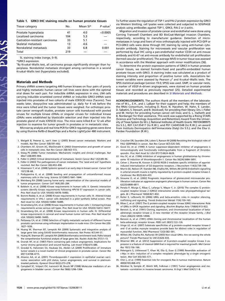
GRK3 Is Overexpressed in Human Malignant Tumors. The preferen-tial inhibitory effects of GRK3 shRNAs in human metastaticcells, taken together with the ability of GRK3 cDNA to promotexenograft tumor growth and metastasis, suggest that GRK3 mayplay a physiologically relevant role in promoting human cancerprogression. To assess the role of GRK3 in human cancer, weperformed immunohistochemistry (IHC) staining of GRK3 pro-tein on a human prostate tumor tissue microarray. GRK3 wassignificantly stronger in carcinomas compared with benign pro-static hyperplasias (P < 0.0005, Kruskal–Wallis test). Amongcarcinomas, the strongest expression was observed in nonskeletalmetastases (P = 0.001, Kruskal–Wallis test; Fig. 5 A–E and Table 1).The staining results using two different antibodies were equiva-lent, with a Spearman ρ of 0.65 (rank correlation). These resultsindicate that GRK3 expression correlates with visceral metastasesand supports the experimental xenograft results presented earlier.Furthermore, in the human prostate cancer patient series,
there was a strong trend between GRK3 expression and glo-meruloid microvascular proliferation (GMP; i.e., vascular nests,a marker of VEGF-A–driven angiogenesis) (25) with a P value of0.08 by Pearson χ2 test. Specifically, only 6% of patients with lowGRK3 expression were positive for GMP, whereas 18% of
patients with high GRK3 expression were positive for GMP (Fig.5F shows a representative GMP+ tumor). These findings furtherconfirm the in vitro and in vivo experimental results and theconclusion that GRK3 stimulates angiogenesis.
DiscussionWe report here a previously undescribed role of GRK3 in prostatecancer metastasis. Our findings were the result of a combinationof an unbiased shRNA library screen and subsequent validation invitro and in vivo. Our results indicate that GRK3 is necessary forsurvival and proliferation of metastatic cells in culture. We con-firmed that GRK3 remained essential for metastatic cells in vivothrough inducible shRNA expression. Additionally, we demon-strated that GRK3 is also sufficient to promote primary prostatetumor growth and metastasis upon exogenous expression in poorlymetastatic cells. Moreover, we have demonstrated that GRK3 isoverexpressed in prostate cancers, especially in metastasis, sug-gesting that it may play physiologic roles in the progression ofhuman prostate cancer. Of note, these findings represent a here-tofore undocumented role for GRK3 in human prostate cancerprogression.GRK3 (or β-adrenergic receptor kinase 2) belongs to a sub-
family of kinases called GRKs (26–28). GRK3 is best known tophosphorylate the agonist-occupied form of β-adrenergic re-ceptors, leading to desensitization of the receptors to their ag-onists (29, 30). It has also been shown to regulate signaling andinternalization of several other G-protein–coupled receptors(GPCR) (31, 32). Although GRK3 has not been implicated in
Fig. 3. GRK3 induced angiogenesis in vitro and in vivo. (A) GFP- and GRK3-expressing PC3 cells proliferated at similar rates in vitro. These two cell lines werecultured in media with various concentrations of serum for 3 d. Shown on the y axis are growth rates (expressed in fold change) at different concentrations ofserum as normalized to 0% serum (x axis). (B) GFP and GRK3-expressing PC3 cells migrated or invaded at similar rates in vitro. Migration or invasion capa-bilities of these two cell lines were measured by using Transwell migration or Matrigel invasion assays. Error bars refer to SEM. (C) GRK3-expressing PC3 cellswere able to induce migration of human endothelial cell seeded in the upper chamber in Transwell assay. Shown on the y axis are numbers of migratedendothelial cells per high-power field. Error bars refer to SEM. (D) MVD is lower in PC3-GFP (Left) than in PC3-GRK3 primary tumor tissues (Middle). Vessels arestained in red with mouse CD34 (green arrows), and dark blue nuclei stains with mouse Ki-67 indicates proliferating endothelial cells (black arrows). Lungmicrometastasis (Right) shows ingrowth of proliferating microvessels by coexpression of the endothelial marker CD34 (red) and endothelial cell proliferationby Ki-67 (blue) (magnification of 400×). (E) Representative CD31 staining in red and DAPI staining in blue for prostate tumors from LN4 cells expressingscrambled shRNA or GRK3-shRNA-1.
1524 | www.pnas.org/cgi/doi/10.1073/pnas.1320638111 Li et al.

cancer metastasis, it has been shown to be down-regulated spe-cifically in a subtype of glioblastoma (33), suggesting a subtype-and tissue-specific role of GRK3, which may result from differencesin GPCR profiles, tumorigenic pathways, or tumor microenvi-ronment in different organs and cancer types.We further found that GRK3 enhances angiogenesis through
regulation of several genes involved in angiogenesis and microen-vironment modulation, including TSP-1 and PAI-2 (or SERPINB2),both of which are down-regulated by GRK3. TSP-1 was the firstidentified endogenous inhibitor of angiogenesis (21). It is a largeglycoprotein (150 kDa) that binds to cell surface receptors CD36and CD47 and inhibits endothelial proliferation and migration.Reduced expression of TSP-1 is an important component of theangiogenic switch, and decrease in its expression facilitates growthof several tumor types, including bladder, breast, and ovarian can-cer, fibrosarcoma, and glioblastoma (18, 19, 22). PAI-2 is one of thetwo major physiological inhibitors of urokinase plasminogen acti-vator, which has been shown to promote invasion and metastasis invarious cancer types (20). Strikingly, a number of studies indicatedthat higher PAI-2 expression in tumors is linked with prolonged
survival, decreased metastasis or decreased tumor size (20). Be-cause access to vasculature is a critical component to metastasis,kinases that increase the angiogenic potential of tumor cells wouldbe expected to increase their metastatic potential as well. It is notclear which signaling pathways underlying GRK3 suppression ofTSP-1 and PAI-2 expression, which we are actively investigating.It is intriguing that GRK3 not only controlled survival and
proliferation of metastatic cells, but also promoted primaryprostate tumor growth and metastasis by enhancing angiogene-sis. Such activity is not without precedent, as several oncogenes,such as Ras and Myc, have been shown to stimulate cellularproliferation as well as angiogenesis (34, 35). Similarly, silencinga key regulator in angiogenesis such as GRK3 could lead to in-hibition of cellular survival and proliferation in vitro and in vivo.Finally, our experimental findings of the role of GRK3 in tu-
mor progression were corroborated by examining human tumorsvia IHC staining of prostate cancer patient samples. These anal-yses confirmed that regulation of GRK3 expression was physio-logically relevant to human prostate cancer progression. Theseresults may lead to improved disease prognosis using GRK3 asa novel biomarker or part of a panel of biomarkers for prostatecancer progression. Ultimately, as kinases have proven to besuccessful drug targets, it is our hope that a selective GRK3inhibitor may be developed as an effective therapy for patientswith metastatic cancer.
0.0
0.2
0.4
0.6
0.8
1.0
1.2
1.4
PAI-2 QPCR
PAI-2 Illumina
0.0
0.2
0.4
0.6
0.8
1.0
1.2
1.4
PC3-GFP PC3-LN4 PC3-GRK3
TSP-1 QPCRTSP-1 Illumina
FoldvsPC3-GFP
FoldvsPC3-GFP
B
C
TSP-1Actin
GRK3
1 2 3WBLysate
1 2 3WBLysate
Actin
PAI-2
TSP-1
GRK3
PAI-2TSP-1GRK3
Actin
D
A
Fig. 4. GRK3 represses TSP-1 and PAI-2 expression. (A) TSP-1 and PAI-2mRNAs were down-regulated by GRK3. Shown on the x axis are the threecell lines tested. Shown on the y axis are fold changes of mRNA for TSP-1(Upper) and PAI-2 (Lower) measured by RT-QPCR (blue) or Illumina micro-array (red). Error bars refer to SD from replicates. (B–D) TSP-1 and PAI-2proteins were down-regulated by GRK3, for which its kinase activity is re-quired. (B) Up-regulation of GRK3 in metastatic lines (LN4 and 1205Lu) fromparental poorly metastatic lines (PC3-GFP and WM793B), and down-regula-tion of TSP-1 and PAI-2 by GRK3. (C) Down-regulation of TSP-1 and PAI-2 byGRK3 WT was reversed by expression of GRK3-K220R, a kinase-dead version(KD). (D) Down-regulation of TSP-1 and PAI-2 by GRK3 WT was reversed byGRK3 shRNA silencing in PC3-GFP and PC3-GRK3 cells.
Fig. 5. GRK3 was up-regulated in human metastatic prostate tumors andassociated with elevated angiogenesis. IHC staining of GRK3 in benign andmalignant prostatic tissues (A–E). Weak and focal staining in benign pros-tatic hyperplasia and negative luminal cells (A), comparable weak staining inlocalized prostate cancer (B), stronger staining in advanced castration re-sistant prostate cancer (C), skeletal metastasis around bone trabeculaedemonstrating stronger staining (D), and soft tissue metastasis with thestrongest staining observed in these tissue categories (E). This subgroup wassignificantly stronger than other carcinomas categories (P = 0.001). Magni-fication of 400×. (Scale bar: 50 μm.) (F) GMP, or vascular nest, by factor VIIIstaining in a prostate carcinoma (considered to represent VEGF-A–drivenangiogenesis; magnification of 400×).
Li et al. PNAS | January 28, 2014 | vol. 111 | no. 4 | 1525
MED
ICALSC
IENCE
S
Materials and MethodsPrimary shRNA screens targeting 440 human kinases on four pairs of poorlyand highly metastatic human cancer cell lines were done with the optimalviral doses for each pair. For inducible shRNA expression in vivo, LN4 cellscarrying inducible scrambled control shRNA or inducible GRK3 shRNA wereinjected into prostates of two cohorts of male ICR/SCID mice (Taconic). Twoweeks later, doxycycline was administrated i.p. daily for 4 wk before themice were killed and the tumor tissues were weighed. For orthotopic pros-tate cancer xenograft studies, prostate cancer cells transduced with pooledviruses for multiple kinase cDNAs or singular viruses for individual kinasecDNAs were established by blasticidin selection and then injected into theprostate gland of male ICR/SCID mice. The mice were killed 8 or 12 wk afterinjection to examine for tumor growth in prostates or in metastatic sites.
Microarray analysis and real time PCR forGRK3-regulating geneswere doneby using Illumina Ref8-v3 BeadChips and a Roche LightCycler 480 instrument.
To further assess the regulation of TSP-1 and PAI-2 protein expression by GRK3via Western blotting, cell lysates were collected and subjected to SDS/PAGEanalysis using antibodies against TSP-1, GRK3, PAI-2 or β-actin.
Migration and invasion of prostate cancer and endothelial were done usingCorning Transwell Chambers and BD BioCoat-Matrigel Invasion Chambers,respectively, according to manufacturer guidance. Detection of micro-metastases in lungs and livers of mice orthotopically injected with PC3-GFP orPC3-GRK3 cells were done through IHC staining by using anti-human cyto-keratin antibody. Staining for microvessels and vascular proliferation wasperformed by dual IHC using a pan-endothelial marker CD34 rat anti-mouseantibody and Ki-67 rat anti-mouse antibody for endothelial cell proliferation(termed vascular proliferation). The averageMVD in tumor tissuewas assessedin accordance with the Weidner approach with minor modifications (36).
To determine the protein expression patterns of GRK3 in human prostatecancer tissues, we stained formalin-fixed and paraffin-embedded humanprostate tissues with GRK3. A staining index was calculated as a product ofstaining intensity and proportion of positive tumor cells. Associations be-tween variables were assessed by Pearson χ2 and Kruskal–Wallis tests. TheSPSS statistical package (version 19.0; SPSS) was used. GMP, or vascular nests,a marker of VEGF-A–driven angiogenesis, were stained on human prostatetissues and recorded as previously reported (25). Detailed experimentalmaterials and procedures are described in SI Materials and Methods.
ACKNOWLEDGMENTS. The authors thank all the members of the laborato-ries of W.L., E.H., and J. LaBaer for their support and help; the members ofthe RNAi Consortium, including D. Root, N. Hacohen, W. Hahn, E. Lander,D. Sabatini, S. Stewart, and B. Stockwell, for providing their library; Drs. I. Fidlerand M. Herlyn for providing human cancer cell lines; and G. L. Hallseth andB. Nordanger for their assistance. This work was supported by a Rising STARS(Science and Technology Acquisition and Retention) Award from the Univer-sity of Texas System (toW.L.); National Institutes of Health Grants 1R01DK089975(to S.S.), R01 CA135417 (to R.W.), and P01 CA045548 (to R.W.); an awardfrom Instituto Dermopatico dell’Immacolata (Italy) (to S.S.); and the Elsa U.Pardee Foundation (R.W.).
1. Weigelt B, Peterse JL, van’t Veer LJ (2005) Breast cancer metastasis: Markers andmodels. Nat Rev Cancer 5(8):591–602.
2. Chambers AF, Groom AC, MacDonald IC (2002) Dissemination and growth of cancercells in metastatic sites. Nat Rev Cancer 2(8):563–572.
3. Steeg PS (2006) Tumor metastasis: Mechanistic insights and clinical challenges. NatMed 12(8):895–904.
4. Fidler IJ (2002) Critical determinants of metastasis. Semin Cancer Biol 12(2):89–96.5. Fidler IJ (2003) The pathogenesis of cancer metastasis: The ‘seed and soil’ hypothesis
revisited. Nat Rev Cancer 3(6):453–458.6. Hüsemann Y, et al. (2008) Systemic spread is an early step in breast cancer. Cancer Cell
13(1):58–68.7. Podsypanina K, et al. (2008) Seeding and propagation of untransformed mouse
mammary cells in the lung. Science 321(5897):1841–1844.8. Weinberg RA (2008) Leaving home early: reexamination of the canonical models of
tumor progression. Cancer Cell 14(4):283–284.9. Baldwin A, et al. (2008) Kinase requirements in human cells: II. Genetic interaction
screens identify kinase requirements following HPV16 E7 expression in cancer cells.Proc Natl Acad Sci USA 105(43):16478–16483.
10. Bommi-Reddy A, et al. (2008) Kinase requirements in human cells: III. Altered kinaserequirements in VHL-/- cancer cells detected in a pilot synthetic lethal screen. ProcNatl Acad Sci USA 105(43):16484–16489.
11. Grueneberg DA, et al. (2008) Kinase requirements in human cells: I. Comparing kinaserequirements across various cell types. Proc Natl Acad Sci USA 105(43):16472–16477.
12. Grueneberg DA, et al. (2008) Kinase requirements in human cells: IV. Differentialkinase requirements in cervical and renal human tumor cell lines. Proc Natl Acad SciUSA 105(43):16490–16495.
13. Pettaway CA, et al. (1996) Selection of highly metastatic variants of different humanprostatic carcinomas using orthotopic implantation in nude mice. Clin Cancer Res 2(9):1627–1636.
14. Huang W, Sherman BT, Lempicki RA (2009) Systematic and integrative analysis oflarge gene lists using DAVID bioinformatics resources. Nat Protoc 4(1):44–57.
15. HuangW, Sherman BT, Lempicki RA (2009) Bioinformatics enrichment tools: Paths towardthe comprehensive functional analysis of large gene lists. Nucleic Acids Res 37(1):1–13.
16. Dvorak HF, et al. (1987) Fibrin containing gels induce angiogenesis. Implications fortumor stroma generation and wound healing. Lab Invest 57(6):673–686.
17. Gravdal K, Halvorsen OJ, Haukaas SA, Akslen LA (2009) Proliferation of immaturetumor vessels is a novel marker of clinical progression in prostate cancer. Cancer Res69(11):4708–4715.
18. Alvarez AA, et al. (2001) Thrombospondin-1 expression in epithelial ovarian carci-noma: association with p53 status, tumor angiogenesis, and survival in platinum-treated patients. Gynecol Oncol 82(2):273–278.
19. Campbell SC, Volpert OV, Ivanovich M, Bouck NP (1998) Molecular mediators of an-giogenesis in bladder cancer. Cancer Res 58(6):1298–1304.
20. Croucher DR, Saunders DN, Lobov S, Ranson M (2008) Revisiting the biological roles ofPAI2 (SERPINB2) in cancer. Nat Rev Cancer 8(7):535–545.
21. Good DJ, et al. (1990) A tumor suppressor-dependent inhibitor of angiogenesis isimmunologically and functionally indistinguishable from a fragment of thrombo-spondin. Proc Natl Acad Sci USA 87(17):6624–6628.
22. Hsu SC, et al. (1996) Inhibition of angiogenesis in human glioblastomas by chromo-some 10 induction of thrombospondin-1. Cancer Res 56(24):5684–5691.
23. Celver J, Sharma M, Kovoor A (2010) RGS9-2 mediates specific inhibition of agonist-induced internalization of D2-dopamine receptors. J Neurochem 114(3):739–749.
24. Morris GE, Nelson CP, Standen NB, Challiss RA, Willets JM (2010) Endothelin signallingin arterial smooth muscle is tightly regulated by G protein-coupled receptor kinase 2.Cardiovasc Res 85(3):424–433.
25. Straume O, et al. (2002) Prognostic importance of glomeruloid microvascular pro-liferation indicates an aggressive angiogenic phenotype in human cancers. Cancer Res62(23):6808–6811.
26. Penela P, Murga C, Ribas C, Lafarga V, Mayor F, Jr. (2010) The complex G protein-coupled receptor kinase 2 (GRK2) interactome unveils new physiopathological tar-gets. Br J Pharmacol 160(4):821–832.
27. Reiter E, Lefkowitz RJ (2006) GRKs and beta-arrestins: roles in receptor silencing,trafficking and signaling. Trends Endocrinol Metab 17(4):159–165.
28. Ribas C, et al. (2007) The G protein-coupled receptor kinase (GRK) interactome: Roleof GRKs in GPCR regulation and signaling. Biochim Biophys Acta 1768(4):913–922.
29. Benovic JL, et al. (1991) Cloning, expression, and chromosomal localization of beta-adrenergic receptor kinase 2. A new member of the receptor kinase family. J BiolChem 266(23):14939–14946.
30. Benovic JL, et al. (1991) cDNA cloning and chromosomal localization of the humanbeta-adrenergic receptor kinase. FEBS Lett 283(1):122–126.
31. Vinge LE, et al. (2007) Substrate specificities of g protein-coupled receptor kinase-2and -3 at cardiac myocyte receptors provide basis for distinct roles in regulation ofmyocardial function. Mol Pharmacol 72(3):582–591.
32. Willets JM, Challiss RA, Nahorski SR (2003) Non-visual GRKs: Are we seeing the wholepicture? Trends Pharmacol Sci 24(12):626–633.
33. Woerner BM, et al. (2012) Suppression of G-protein-coupled receptor kinase 3 ex-pression is a feature of classical GBM that is required for maximal growth. Mol CancerRes 10(1):156–166.
34. Pelengaris S, Littlewood T, Khan M, Elia G, Evan G (1999) Reversible activation ofc-Myc in skin: induction of a complex neoplastic phenotype by a single oncogeniclesion. Mol Cell 3(5):565–577.
35. Chin L, et al. (1999) Essential role for oncogenic Ras in tumour maintenance. Nature400(6743):468–472.
36. Weidner N, Semple JP, Welch WR, Folkman J (1991) Tumor angiogenesis and me-tastasis—correlation in invasive breast carcinoma. N Engl J Med 324(1):1–8.
Table 1. GRK3 IHC staining results on human prostate tissues
Tissue category No. Mean SI* P value†
Prostate hyperplasia 41 4.0 <0.0005Localized carcinoma 104 5.3 —
Castration-resistant carcinoma 33 5.1 —
Skeletal metastasis 13 4.6 —
Nonskeletal metastasis 33 6.8 0.001Total 219 — —
SI, staining index (range, 0–9).*GRK3 expression.†By Kruskal–Wallis test, all carcinoma groups significantly stronger than hy-perplasia. Nonskeletal metastasis strongest among carcinomas in a secondKruskal–Wallis test (hyperplasia excluded).
1526 | www.pnas.org/cgi/doi/10.1073/pnas.1320638111 Li et al.