Ryanodine receptor-operated activation of TRP-like channels can trigger critical Ca2+ signaling...
23
©2004 FASEB The FASEB Journal express article 10.1096/fj.04-2621fje. Published online November 30, 2004. Ryanodine receptor-operated activation of TRP-like channels can trigger critical Ca 2+ signaling events in pancreatic β-cells Amanda Jabin Gustafsson, Hanna Ingelman-Sundberg,* Mensur Dzabic, Justina Awasum, Nguyen Khanh Hoa,* Claes-Göran Östenson,* Cristina Pierro, Patrizia Tedeschi, Orison Woolcott, Shiue Chiounan, Per-Eric Lund, † Olof Larsson, † and Md. Shahidul Islam* *Karolinska Institutet, Department of Medicine, Stockholm Söder Hospital, Research Center, 118 83 Stockholm, Sweden; Department of Molecular Medicine, Karolinska University Hospital; and † AstraZeneca R&D, S-151 85, Södertälje, Sweden Corresponding author: Amanda Jabin Gustafsson, Karolinska Institutet, Department of Medicine, Stockholm Söder Hospital S-118 83 Stockholm, Sweden. E-mail: [email protected] ABSTRACT There is little information available concerning the link between the ryanodine (RY) receptors and the downstream Ca 2+ signaling events in β-cells. In fura-2 loaded INS-1E cells, activation of RY receptors by 9-methyl 5,7-dibromoeudistomin D (MBED) caused a rapid rise of [Ca 2+ ] i followed by a plateau and repetitive [Ca 2+ ] i spikes on the plateau. The [Ca 2+ ] i plateau was abolished by omission of extracellular Ca 2+ and by SKF 96365. In the presence of SKF 96365, MBED produced a transient increase of [Ca 2+ ] i , which was abolished by thapsigargin. Activation of RY receptors caused Ca 2+ entry even when the ER Ca 2+ pool was depleted by thapsigargin. The [Ca 2+ ] i plateau was not inhibited by nimodipine or ruthenium red, but was inhibited by membrane depolarization, La 3+ , Gd 3+ , niflumic acid, and 2-aminoethoxydiphenyl borate, agents that inhibit the transient receptor potential channels. The [Ca 2+ ] i spikes were inhibited by nimodipine and ryanodine, indicating that they were due to Ca 2+ influx through the voltage-gated Ca 2+ channels and Ca 2+ -induced Ca 2+ release (CICR). Activation of RY receptors depolarized membrane potential as measured by patch clamp. Thus, activation of RY receptors leads to coherent changes in Ca 2+ signaling, which includes activation of TRP-like channels, membrane depolarization, activation of the voltage-gated Ca 2+ channels and CICR. Key words: islets of Langerhans • Ca 2+ -induced Ca 2+ release • transient receptor potential channels n the pancreatic β-cells, a cascade of signaling events participates in transducing the effects of glucose and incretin hormones into the exocytosis of insulin. Such events include progressive membrane depolarization, Ca 2+ entry through the voltage-gated Ca 2+ channels and Ca 2+ -induced Ca 2+ release (CICR) (1). Among the mechanisms that mediate initial depolarization to the threshold potential for the activation of the voltage-gated Ca 2+ channels, closure of the ATP-sensitive potassium (K ATP ) channel is most well known. It is, however, not I Page 1 of 23 (page number not for citation purposes)
-
Upload
independent -
Category
Documents
-
view
1 -
download
0
Transcript of Ryanodine receptor-operated activation of TRP-like channels can trigger critical Ca2+ signaling...

©2004 FASEB The FASEB Journal express article 10.1096/fj.04-2621fje. Published online November 30, 2004.
Ryanodine receptor-operated activation of TRP-like channels can trigger critical Ca2+ signaling events in pancreatic β-cells Amanda Jabin Gustafsson, Hanna Ingelman-Sundberg,* Mensur Dzabic, Justina Awasum, Nguyen Khanh Hoa,* Claes-Göran Östenson,* Cristina Pierro, Patrizia Tedeschi, Orison Woolcott, Shiue Chiounan, Per-Eric Lund,† Olof Larsson,† and Md. Shahidul Islam* *Karolinska Institutet, Department of Medicine, Stockholm Söder Hospital, Research Center, 118 83 Stockholm, Sweden; Department of Molecular Medicine, Karolinska University Hospital; and †AstraZeneca R&D, S-151 85, Södertälje, Sweden
Corresponding author: Amanda Jabin Gustafsson, Karolinska Institutet, Department of Medicine, Stockholm Söder Hospital S-118 83 Stockholm, Sweden. E-mail: [email protected]
ABSTRACT
There is little information available concerning the link between the ryanodine (RY) receptors and the downstream Ca2+ signaling events in β-cells. In fura-2 loaded INS-1E cells, activation of RY receptors by 9-methyl 5,7-dibromoeudistomin D (MBED) caused a rapid rise of [Ca2+]i followed by a plateau and repetitive [Ca2+]i spikes on the plateau. The [Ca2+]i plateau was abolished by omission of extracellular Ca2+ and by SKF 96365. In the presence of SKF 96365, MBED produced a transient increase of [Ca2+]i, which was abolished by thapsigargin. Activation of RY receptors caused Ca2+ entry even when the ER Ca2+ pool was depleted by thapsigargin. The [Ca2+]i plateau was not inhibited by nimodipine or ruthenium red, but was inhibited by membrane depolarization, La3+, Gd3+, niflumic acid, and 2-aminoethoxydiphenyl borate, agents that inhibit the transient receptor potential channels. The [Ca2+]i spikes were inhibited by nimodipine and ryanodine, indicating that they were due to Ca2+ influx through the voltage-gated Ca2+ channels and Ca2+-induced Ca2+ release (CICR). Activation of RY receptors depolarized membrane potential as measured by patch clamp. Thus, activation of RY receptors leads to coherent changes in Ca2+ signaling, which includes activation of TRP-like channels, membrane depolarization, activation of the voltage-gated Ca2+ channels and CICR.
Key words: islets of Langerhans • Ca2+-induced Ca2+ release • transient receptor potential channels
n the pancreatic β-cells, a cascade of signaling events participates in transducing the effects of glucose and incretin hormones into the exocytosis of insulin. Such events include progressive membrane depolarization, Ca2+ entry through the voltage-gated Ca2+ channels and Ca2+ -induced Ca2+ release (CICR) (1). Among the mechanisms that mediate initial
depolarization to the threshold potential for the activation of the voltage-gated Ca2+ channels, closure of the ATP-sensitive potassium (KATP) channel is most well known. It is, however, not
I Page 1 of 23
(page number not for citation purposes)

always appreciated that depolarization by closure of the KATP channels requires a second event, namely the existence of a depolarizing inward current primarily carried by Na+ or Ca2+. The identity of the channels that mediate such depolarizing currents and their regulation remain unclear.
Several members of the transient receptor potential (TRP) superfamily of cation channels are known to mediate membrane depolarization in different cells (2, 3). TRP channels are conserved from worms to humans and are present in both electrically nonexcitable and excitable cells including the β-cells (4–7). The biophysical properties, regulation mechanisms, and functions of these channels remain enigmatic. Much evidence has been presented that different TRP channels can be activated by depletion of the endoplasmic reticulum (ER) Ca2+ stores, conformational coupling to the inositol 1,4,5-trisphosphate receptor (IP3 receptor), and ryanodine receptor (RY receptor) and by diacylglycerol (8–10). β-cells possess IP3 receptors, RY receptors and probably other intracellular Ca2+ release channels. Depletion of the IP3-sensitive ER Ca2+ store induces a small capacitative or store-operated Ca2+ influx in these cells (11, 12). Candidate subunits of the store-operated Ca2+ channels belong to the TRPC (TRP-canonical) or TRPV (TRP-vanilloid) family (13, 14). Recent studies demonstrate that RY receptors of β-cells are activated as a consequence of glucose metabolism and in response to stimulation by some incretin hormones (15–17). However, it is unknown whether activation of RY receptors could lead to the activation of ion channels in the plasma membrane of β-cells. In previous studies, investigators have used caffeine to activate RY receptors of β-cells (18). The consequences of activation of RY receptors on the downstream Ca2+ signaling events were difficult to elucidate in those studies because of the numerous side effects of the xanthine compound. 9-methyl 5,7-dibromoeudistomin D (MBED) is a compound derived from the natural product eudistomin D. MBED is a more specific and more potent activator of RY receptors and is thus suitable for mechanistic studies of these channels (19). We tested the hypothesis that activation of RY receptors may lead to the activation of the TRP channels and that such activation may contribute to the membrane depolarization, Ca2+ entry through the voltage-gated Ca2+ channels and CICR in the β-cells.
MATERIALS AND METHODS
Chemicals
Fura-2 acetoxymethyl ester was from Molecular Probes Europe. Ryanodine (98% pure), SKF 96365 and thapsigargin were from Calbiochem. 2-aminoethoxydiphenyl borate (2-APB, also called diphenylboric acid 2-aminoethyl ester), N-propargylnitrendipene (MRS 1845) and niflumic acid were from Sigma. Gadolinium (III) chloride hexahydrate was from Aldrich. 9-methyl 5,7-dibromo eudistomin D (MBED) was synthesized. INS-1E cells were a gift from C. B. Wollheim and P. Maechler, Geneva. Cell culture materials were from Life Technologies.
Cell culture
We used a highly differentiated rat insulinoma cell line (S5-cells). This clone of cells was derived from INS-1E cells (20). The cells were cultured in RPMI-1640 medium supplemented with fetal bovine serum (2.5%, v/v), penicillin (50 i.u./ml), streptomycin (50 µg/ml), 2-mercaptoethanol (500 µM), HEPES (10 mM), and sodium pyruvate (1 mM). Cells were
Page 2 of 23(page number not for citation purposes)

incubated at 37°C in humidified incubator in 5% CO2. The medium was changed every other day and cells were passaged every other week.
Preparation of rat β-cells
Male Wister rats were killed by decapitation after anesthesia with CO2. The islets were isolated by injecting collagenase A in Hanks’ solution (9 mg/10 ml) into pancreas through the pancreatic duct. The gland was removed, incubated for 24 min at 37°C, washed with Hanks’ solution and islets were picked up after separation on Histopaque gradient. Islets were dispersed by trypsin digestion and the cells were plated on glass cover slips. For measurement of [Ca2+]I, only large cells were used to exclude as much as possible non-β-cells.
Measurement of cytosolic free [Ca2+] ([Ca2+]i) by microfluorometry
Cells (~20,000/ml) plated on glass cover slips were incubated in RPMI-1640 medium supplemented with 0.1% bovine serum albumin and 1 µM fura-2 acetoxymethyl ester for 35 min at 37°C. Cells were then incubated for an additional 10 min in the basal medium containing 140 NaCl, 3.6 KCl, 0.5 NaH2PO4, 0.5 MgSO4, 1.5 CaCl2, 10 HEPES, 3 glucose (in mM), and 0.1% bovine serum albumin (pH 7.4). Cover slips were mounted as the exchangeable bottom of an open perifusion chamber on the stage of an inverted epifluorescence microscope (Olympus CK 40). The superfusion chamber was designed to allow rapid exchange of fluids. The chamber was thermostatically controlled to maintain a temperature of 37°C in the perifusate. The microscope was connected to a fluorescence system (M-39/2000 RatioMaster, PhotoMed) for dual wavelength excitation fluorometry. The excitation wavelengths generated by a monochromator (DeltaRam, PhotoMed) were directed to the cell by a dichroic mirror. The emitted light selected by a 510-nm filter was monitored by a photomultiplier. The excitation wavelengths were alternated at a frequency of 1 Hz, and the duration of data collection at each wavelength was 0.33 s. The emission at the excitation wavelength of 340 nm (F340) and that of 380 nm (F380) were used to calculate the fluorescence ratio (R340/380). Single cells isolated optically by means of a diaphragm, were studied by using a 40× 1.3 NA oil immersion objective (40× UV APO). The background fluorescence was measured and subtracted from the traces before calculation of [Ca2+]i. [Ca2+]i was calculated from R340/380 according to Grynkiewicz et al. (21). Rmax and Rmin were determined by using thin films of external standards containing fura-2 and 2 M sucrose (22). The Kd for Ca2+-fura-2 was taken as 225 nM.
Patch clamp experiments
The cells were detached and seeded on 60-mm dishes and used within two days. Membrane potential was recorded at room temperature (21–23°C) using the perforated-patch whole cell approach with a computer based EPC-10/2 patch amplifier together with PULSE 8.63 (HEKA Elektronik, Lambrecht, Germany). Cells were continuously perifused with a saline solution containing 137 NaCl, 5 KCl, 1.2 MgCl2, 1 CaCl2, 10 HEPES-NaOH (in mM), pH 7.4, and 10 glucose. Patch pipettes were pulled from borosilicate glass, fire-polished, and had resistances between 3 and 4 MΩ after filling the pipettes with the standard intracellular solution containing 140 KCl, 3 NaCl, 1.2 MgCl2, 1 EGTA, 10 HEPES-NaOH (in mM), pH 7.2, and 200 µg/ml amphotericin B. Membrane potential recordings were started when RS < 30 MΩ, at which time
Page 3 of 23(page number not for citation purposes)

the amplifier was switched from voltage-clamp to current clamp. MBED was applied by a local pressure application device from a wide-tipped micropipette placed within 100 µm from the cell.
RESULTS
Application of glucose (10 mM) to fura-2 loaded single INS-1E cells for two minutes increased [Ca2+]i in ~50% of cells by ~50 nM. In the rest of the cells there was no [Ca2+]i increase. Activation of RY receptors by MBED (50 µM) in the presence of glucose (10 mM), increased [Ca2+]i immediately and in a characteristic pattern (Fig. 1A). There was an initial rapid increase of [Ca2+]i followed by a prolonged [Ca2+]i signal which remained elevated during the period of application of MBED. A third and more conspicuous feature of MBED-induced [Ca2+]i increase was large regenerative [Ca2+]i spikes superimposed on the [Ca2+]i plateau. [Ca2+]i changes returned to the baseline when MBED was washed away in the continued presence of 10 mM glucose. Similar [Ca2+]i changes were observed when we used primary β-cells obtained from Wister rats (Fig. 1B). When extracellular Ca2+ was chelated by addition of EGTA, MBED still caused a transient increase of [Ca2+]i but the [Ca2+]i plateau was absent (Fig. 1C). Under this condition, the transient increase in [Ca2+]i continuously declined and reached the baseline in less than 2 min. MBED was dissolved in DMSO (final concentration 1.6%). At this concentration DMSO alone caused only a small increase in the basal [Ca2+]i (~50 nM). 25-µM MBED also increased [Ca2+]i, the magnitude of which was smaller compared with that obtained with 50 µM MBED. Six and twelve µM MBED did not increase [Ca2+]i. The effect of MBED reversed on washout and repeated application of MBED caused repeated [Ca2+]i increase showing similar pattern. During continued stimulation by MBED, the [Ca2+]i plateau returned to the baseline over a period of four to 10 min (Fig. 10C). Taken together, these results suggest that activation of RY receptors by MBED releases Ca2+ from the intracellular stores as well as activation of Ca2+ influx across the plasma membrane.
Next we investigated whether the voltage-gated Ca2+ channels were involved in mediating Ca2+ entry following the activation of RY receptors. In these cells, L-type Ca2+ channels constitute the main type of voltage-gated Ca2+ channels. We inhibited the L-type Ca2+ channels by treating the cells with nimodipine (5 µM). The effectiveness of the inhibition of the voltage-gated Ca2+ channels was verified in separate experiments by demonstrating that nimodipine completely blocked the [Ca2+]i-increase induced by 30 mM KCl. As shown in Fig. 2, nimodipine did not inhibit the initial rapid rise of [Ca2+]i and the [Ca2+]i plateau that followed activation of RY receptors. Nimodipine inhibited the regenerative [Ca2+]i spikes that were superimposed on the [Ca2+]i plateau (Fig. 1A), indicating that Ca2+ entry through the L-type Ca2+ channels is required for the generation of these spikes. However, it should be noted that the large transient Ca2+ spikes were generated mainly by CICR rather than by Ca2+ entry through the L-type voltage gated Ca2+ channels (Fig. 10B). We tested the effect of MBED on plasma membrane potential using perforated patch current-clamp technique and found that MBED depolarized plasma membrane potential from approximately –80 mV to approximately –40 mM in a reversible manner (Fig. 3).
The plateau phase of [Ca2+]i increase was completely blocked by SKF 96365 that blocks several TRP channels as well as the voltage-gated Ca2+ channels (23, 24). When applied in the presence of SKF 96365 (10 µM), MBED induced only a transient [Ca2+]i increase (Fig. 4A). This transient [Ca2+]i increase by MBED, observed in the SKF 96365-treated cells, was due to Ca2+ release
Page 4 of 23(page number not for citation purposes)

from the ER. This is evident from the observation that this [Ca2+]i increase was abolished when the ER Ca2+ pool was depleted by treatment with thapsigargin (Fig. 4B). Thus, the results obtained from experiments with nimodipine and SKF 96365 suggest that the [Ca2+]i plateau that follows activation of RY receptors is due to Ca2+ influx through the TRP family of Ca2+ permeable channels.
In β-cells carbachol and thapsigargin releases Ca2+ from ER Ca2+ stores (Fig. 5A). To test whether activation of the TRP channels was due to the depletion of the ER or due to an increase of [Ca2+]i, we first depleted the ER Ca2+ pools by treatment with thapsigargin. Under such condition, MBED would activate RY receptors but such activation would not deplete the ER Ca2+ pool further. There would be no release of Ca2+ from the ER and thus no increase of [Ca2+]i attributable to the release of Ca2+. Consistent with our previous reports (18), treatment of cells with thapsigargin (500 nM- 1 µM) for 45 min completely emptied the ER Ca2+ pools as evidenced from the fact that carbachol did not increase [Ca2+]i (Fig. 5B). The basal [Ca2+]i in the thapsigargin-treated cells was similar to that in the control cells. The plasma membrane potential of the thapsigargin-treated cells is similar to that of the untreated cells (25). As shown in Fig. 5B, activation of RY receptors in the thapsigargin-treated cells also resulted in a plateau increase in [Ca2+]i. This [Ca2+]i plateau was due to Ca2+ entry through the plasma membrane since it was abolished by SKF 96365 (Fig. 4B). The initial rapid increase of [Ca2+]i and the large regenerative [Ca2+]i spikes were absent in the thapsigargin-treated cells. These results suggest that activation of RY receptors can lead to the activation of TRP-channels by a mechanism that can operate without involving depletion of the ER Ca2+ pools.
We investigated whether activation of the phosphatidylinositol-specific phospholipase C (PI-PLC) and IP3 system could be involved in the MBED-induced [Ca2+]i changes. Carbachol (100 µM), a muscarinic agonist that activates PI-PLC, caused a biphasic increase in [Ca2+]i with an initial peak reflecting Ca2+ release from the ER and a small plateau phase representing the capacitative Ca2+ influx (Fig. 6). Application of MBED in the continued presence of carbachol resulted in a pattern of [Ca2+]i increase that was similar to that observed when the eudistomin was used without prior application of carbachol (Fig. 1A). The [Ca2+]i plateau induced by MBED was larger than that induced by carbachol. These results suggest that activation of RY receptors triggers [Ca2+]i-entry mechanisms which are different from those triggered by agonists that engage the PI-PLC and IP3 pathway.
Ca2+ entry through the TRP channels should be reduced when plasma membrane is depolarized because of the reduced driving force for Ca2+. In Fig. 7A, the membrane potential was clamped at a depolarized level by 30 mM KCl. This increased [Ca2+]i to a plateau of ~265 nM. Activation of RY receptors by MBED in the depolarized cells caused a transient [Ca2+]i increase, but it did not affect the plateau level. The repetitive [Ca2+]i spikes that are normally elicited by MBED (Fig. 1A) were absent when MBED was added to the depolarized cells. Diazoxide, an agent that hyperpolarizes membrane potential by opening the KATP channels, did not alter the [Ca2+]i response to MBED (Fig. 7B). The large [Ca2+]i spikes that are indicative of membrane depolarization and Ca2+ influx through the L-type Ca2+ channels were observed even when MBED was added in the presence of diazoxide (Fig. 7B). Taken together, these data indicate that the MBED-induced membrane depolarization was not due to the inhibition of KATP channels by the eudistomin compound and that Ca2+ influx through the TRP channels can depolarize membrane potential even when the KATP channels are open (Fig. 7B).
Page 5 of 23(page number not for citation purposes)
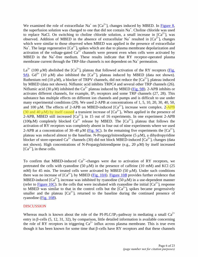
We examined the role of extracellular Na+ on [Ca2+]i changes induced by MBED. In Figure 8, the superfusion solution was changed to one that did not contain Na+. Choline chloride was used to replace NaCl. On switching to choline chloride solution, a small increase in [Ca2+]i was observed. Addition of MBED in the absence of extracellular Na+ resulted in [Ca2+]i changes which were similar to those observed when MBED was applied in the presence of extracellular Na+. The large regenerative [Ca2+]i spikes which are due to plasma membrane depolarization and activation of the voltage-gated Ca2+ channels were present even when cells were activated by MBED in the Na+-free medium. These results indicate that RY receptor-operated plasma membrane current through the TRP-like channels is not dependent on Na+ permeation.
La3+ (100 µM) abolished the [Ca2+]i plateau that followed activation of the RY receptors (Fig. 9A). Gd3+ (10 µM) also inhibited the [Ca2+]i plateau induced by MBED (data not shown). Ruthenium red (10 µM), a blocker of TRPV channels, did not reduce the [Ca2+]i plateau induced by MBED (data not shown). Niflumic acid inhibits TRPC4 and several other TRP channels (26). Niflumic acid (30 µM) inhibited the Ca2+ plateau induced by MBED (Fig. 9B). 2-APB inhibits or activates different channels, for example, IP3 receptors and some TRP channels (27, 28). This substance has multiple effects on different ion channels and pumps and is difficult to use under many experimental conditions (29). We used 2-APB at concentrations of 1, 5, 10, 20, 30, 40, 50, and 100 µM. The effects of 2-APB on MBED-induced [Ca2+]i increase were complex. 2-APB (30 and 40 µM) by itself caused a transient increase of [Ca2+]i. When applied in the presence of 2-APB, MBED still increased [Ca2+]i in 15 out of 16 experiments. In one experiment 2-APB (100µM) completely blocked Ca2+ release by MBED. The [Ca2+]i plateau that follows the activation of RY receptors was completely absent in four out of nine experiments where we used 2-APB at a concentration of 30–40 µM (Fig. 9C). In the remaining five experiments the [Ca2+]i plateau was reduced almost to the baseline. N-Propargylnitrendipene (5 µM), a dihydropyridine blocker of store-operated Ca2+ channels (30) did not block MBED-induced [Ca2+]i changes (data not shown). High concentrations of N-Propargylnitrendipene (e.g., 20 µM) by itself increased [Ca2+]i in these cells.
To confirm that MBED-induced Ca2+-changes were due to activation of RY receptors, we pretreated the cells with ryanodine (50 µM) in the presence of caffeine (10 mM) and KCl (25 mM) for 45 min. The treated cells were activated by MBED (50 µM). Under such conditions there was no increase of [Ca2+]i by MBED (Fig. 10A). Figure 10B provides further evidence that MBED-induced [Ca2+]i increase was inhibited by ryanodine (50 µM) in a use-dependent manner (refer to Figure 10C). In the cells that were incubated with ryanodine the initial [Ca2+]i response to MBED was similar to that in the control cells but the [Ca2+]i spikes became progressively smaller and the plateau [Ca2+]i returned to the baseline during the continued presence of ryanodine (Fig. 10B).
DISCUSSION
Whereas much is known about the role of the PI-PLC/IP3-pathway in mediating a small Ca2+ entry in β-cells (5, 12, 31, 32), by comparison, little detailed information is available concerning the role of RY receptors in triggering Ca2+ influx across plasma membrane. This is true even though it has been known for some time that β-cells have RY receptors and that these channels
Page 6 of 23(page number not for citation purposes)
kishis
Markering
kishis
Markering

amplify Ca2+ signals and exocytosis (25, 33–35). Part of the difficulty in addressing the issue arises from lack of suitable pharmacological agonists of RY receptors. For activation of RY receptors, previous studies have used caffeine, which inhibits many ion channels, enzymes and receptors. In fact, caffeine inhibits plasma membrane Ca2+ entry pathways, including both the voltage-gated Ca2+ channels and the store-operated channels (18, 36). MBED is more potent than caffeine in its action on the RY receptors. The compound has been used as an activator of RY receptors for nearly 20 years, and so far no major nonspecific effects have been reported. Unlike caffeine, MBED does not inhibit cAMP-phosphodiesterases of β-cells and does not inhibit the IP3 receptors or the KATP channels (15). These properties make MBED a suitable probe for mechanistic studies of RY receptors. We found that activation of RY receptors of β-cells by MBED caused a characteristic pattern of changes in [Ca2+]i that consisted of three distinct components: (a) an initial rapid rise of [Ca2+]i (b) a prolonged plateau of [Ca2+]i and (c) a series of high amplitude [Ca2+]i spikes superimposed on the plateau. These effects of MBED were reversible, and a similar pattern of [Ca2+]i changes was observed on repeated application of MBED.
The initial rapid increase of [Ca2+]i observed after application of MBED was due to the release of Ca2+ from the ER, since it was present even when the extracellular Ca2+ was omitted or when Ca2+ influx was blocked by SKF 96365. This initial increase of [Ca2+]i was also abolished when the ER Ca2+ pool was depleted by thapsigargin. Consistent with previous reports, these results confirm that MBED causes a transient Ca2+ release from the ER by activation of RY receptors of β-cells (15, 37).
The most important finding of this study was that activation of RY receptors resulted in a prolonged [Ca2+]i increase after the initial rapid rise of [Ca2+]i. This [Ca2+]i plateau was dependent on extracellular Ca2+. These results demonstrate that activation of RY receptors not only releases Ca2+ from the ER but also leads to the activation of Ca2+ permeable cation channels in the plasma membrane. In thapsigargin-treated cells, MBED did not increase [Ca2+]i by releasing Ca2+ from the ER but there was still activation of Ca2+ entry across the plasma membrane. This finding argues against the possibility that Ca2+ influx was triggered by the increase in [Ca2+]i itself. It is also unlikely that Ca2+ entry was due to activation of any RY receptors located on the plasma membrane since Ca2+ influx was blocked by SKF 96365 (Fig. 4A), which does not block RY receptors. The Ca2+ entry responsible for the [Ca2+]i plateau was not mediated by the voltage-gated Ca2+ channels since it was not blocked by nimodipine. N-propargylnitrendipene, a dihydropyridine that blocks store-operated Ca2+ influx did not reduce the [Ca2+]i plateau. La3+ (100 µM), Gd3+ (10 µM), SKF 96365, niflumic acid and 2-APB (30 µM) which block store-operated Ca2+ channels and many TRP channels, blocked Ca2+ entry (26, 29). Taken together, these pharmacological properties suggest that the Ca2+ channels that are activated as a consequence of activation of RY receptors belong to the TRP family of cation channels.
Another dramatic consequence of activation of RY receptors was the appearance of large regenerative [Ca2+]i spikes that were superimposed on the [Ca2+]i plateau. Multiple mechanisms contributed to the generation of these spikes. First, Ca2+ entry through the L- type voltage-gated Ca2+ channels was essential for generation of these spikes. This is evidenced from the fact that the spikes were abolished by nimodipin. Our results demonstrate that following activation of RY receptor, plasma membrane is depolarized to about –40 mV as a result of Ca2+ current through
Page 7 of 23(page number not for citation purposes)

the TRP channels (Fig. 3). Such depolarization in turn activates L-type Ca2+ channels. Second, the generation of these spikes required CICR through RY receptors. This is evident from the fact that the spikes were inhibited in a use-dependent manner by high concentration of ryanodine or when the ER Ca2+ pool was emptied by thapsigargin.
Previous studies have shown that glucose and glucagon-like peptide-1 can induce a cationic inward current in β-cells (31, 38). The pharmacological property or molecular identity of the channels that mediate such inward current has not been elucidated in these studies. Other studies have demonstrated that depletion of the IP3-sensitive ER Ca2+ pools induces a small capacitative Ca2+ influx in β-cells (11, 12). In the present study, we have demonstrated a mechanism of Ca2+ entry that involves the RY receptors. The RY receptor-operated Ca2+ influx described in the present study is much larger compared with the small capacitative Ca2+ entry that is observed in β-cells after application of high concentrations of carbachol (Fig. 6). Consistent with this, the RY receptor-operated Ca2+ influx readily depolarizes membrane potential to the threshold potential for activation of the L-type Ca2+ channels. In this study we have depended primarily on fluorescence methods and characterization of the inward current by patch-clamp technique is planned for future studies. The molecular identity of the Ca2+ channel(s) that are activated as a result of the activation of RY receptors in β-cells is not fully known from our study. In this respect, members of the TRP family appear to be the best candidates. In β-cells, transcripts for TRPC1-6, TRPM2, TRPM5, TRPV1, and TRPV5 have been found (4–7, 31). TRPV5 is abundant in β-cells where it is located on the secretory granules (39). However, TRPV channels are unlikely candidates because the Ca2+ influx was not blocked by ruthenium red, a potent inhibitor of TRPV channels (40). It should be noted that assembly of different types of TRP channels into homo- and hetero-multimers is likely to yield a wide variety of channels with diverse regulation mechanisms (41).
The Ca2+ influx described in the present study is different from the store-operated Ca2+ entry described earlier in these cells (11, 12). This is evident from the fact that prior depletion of ER stores by thapsigargin or carbachol did not eliminate the Ca2+ plateau elicited by RY receptor activation. This suggests that activation of RY receptors likely stimulates TRP channels through a mechanism that is not essentially dependent on the filling state of the ER. Rather, it is likely that the putative TRP channels are activated by their linkage to the RY receptors as has been proposed in other cells (10, 42). At present, it is unclear how the activation of RY receptors couples to and gates the TRP channels in β-cells. This may involve protein–protein interactions and conformational coupling. Such gating of TRP channels by RY receptors has been demonstrated in other systems (10).
Thus, we describe a novel route of Ca2+ influx, which is coupled to the activation of RY receptors. The channels that mediate this Ca2+ influx are presumably members of the TRP family. Molecules generated from glucose metabolism for example, fructose 1,6-diphosphate (43) and long-chain Acyl CoA (44) can activate RY receptors. Thus, RY receptors are potential links between nutrient metabolism and membrane excitability. We demonstrate that activation of RY receptor by pharmacological tools leads to a series of distinct signaling events, which include not only the release of Ca2+ from the ER but also activation of TRP-like channels, membrane depolarization, Ca2+ entry through the voltage-gated Ca2+ channels, and CICR. This sequence of events has been illustrated in a model shown in Fig. 11. CICR mediated by RY receptors has
Page 8 of 23(page number not for citation purposes)

been implicated in mediating amplification of exocytosis in β-cells (34, 45). Thus, activation of RY receptors in β-cells may be an important signaling event that is transduced into a coherent cellular response by participation of the TRP-like channels and the voltage-gated Ca2+ channels.
ACKNOWLEDGMENTS
This work was supported in part by Swedish Medical Research Council Grant K2003-32X-14648-01A, funds of Karolinska Institutet, Swedish Medical Society, Novo Nordisk Foundation, Stiftelsen Irma och Arvid Larsson-Rösts minne. Svenska Diabetesstiftelsen and Swedish Diabetes Society has supported A. J. G. M. S. I. is a recipient of career development award from the Juvenile Diabetes Research Foundation, International. O. W. is a Swedish Institute guest researcher.
REFERENCES
1. Islam, M. S. (2000) Calcium and diabetes. In Calcium: the molecular basis of calcium action in biology and medicine (Pochet, R., Donato, R., Haiech, J., Heizmann, C., and Gerke, V., eds) Amersterdam: Kluwer Academic Publishers; pp. 402–413
2. Minke, B., Wu, C., and Pak, W. L. (1975) Induction of photoreceptor voltage noise in the dark in Drosophila mutant. Nature 258, 84–87
3. Launay, P., Fleig, A., Perraud, A. L., Scharenberg, A. M., Penner, R., and Kinet, J. P. (2002) TRPM4 is a Ca2+-activated nonselective cation channel mediating cell membrane depolarization. Cell 109, 397–407
4. Qian, F., Huang, P., Ma, L., Kuznetsov, A., Tamarina, N., and Philipson, L. H. (2002) TRP genes: candidates for nonselective cation channels and store-operated channels in insulin-secreting cells. Diabetes 51, Suppl 1, S183–S189
5. Sakura, H., and Ashcroft, F. M. (1997) Identification of four trp1 gene variants murine pancreatic beta-cells. Diabetologia 40, 528–532
6. Inamura, K., Sano, Y., Mochizuki, S., Yokoi, H., Miyake, A., Nozawa, K., Kitada, C., Matsushime, H., and Furuichi, K. (2003) Response to ADP-ribose by activation of TRPM2 in the CRI-G1 insulinoma cell line. J. Membr. Biol. 191, 201–207
7. Akiba, Y., Kato, S., Katsube, K., Nakamura, M., Takeuchi, K., Ishii, H., and Hibi, T. (2004) Transient receptor potential vanilloid subfamily 1 expressed in pancreatic islet beta cells modulates insulin secretion in rats. Biochem. Biophys. Res. Commun. 321, 219–225
8. Putney, J. W., Jr., Broad, L. M., Braun, F. J., Lievremont, J. P., and Bird, G. S. (2001) Mechanisms of capacitative calcium entry. J. Cell Sci. 114, 2223–2229
9. Kiselyov, K., Xu, X., Mozhayeva, G., Kuo, T., Pessah, I. N., Mignery, G., Zhu, X., Birnbaumer, L., and Muallem, S. (1998) Functional interaction between InsP3 receptors and store-operated Htrp3 channels. Nature 396, 478–482
Page 9 of 23(page number not for citation purposes)

10. Kiselyov, K. I., Shin, D. M., Wang, Y., Pessah, I. N., Allen, P. D., and Muallem, S. (2000) Gating of store-operated channels by conformational coupling to ryanodine receptors. Mol. Cell 6, 421–431
11. Miura, Y., Henquin, J. C., and Gilon, P. (1997) Emptying of intracellular Ca2+ stores stimulates Ca2+ entry in mouse pancreatic beta-cells by both direct and indirect mechanisms. J. Physiol. 503, 387–398
12. Dyachok, O., and Gylfe, E. (2001) Store-operated influx of Ca2+ in pancreatic beta-cells exhibits graded dependence on the filling of the endoplasmic reticulum. J. Cell Sci. 114, 2179–2186
13. Minke, B., and Cook, B. (2002) TRP channel proteins and signal transduction. Physiol. Rev. 82, 429–472
14. Putney, J. W. (2003) Capacitative calcium entry in the nervous system. Cell Calcium 34, 339–344
15. Bruton, J. D., Lemmens, R., Shi, C. L., Persson-Sjogren, S., Westerblad, H., Ahmed, M., Pyne, N. J., Frame, M., Furman, B. L., Islam, M. S. (2002) Ryanodine receptors of pancreatic beta-cells mediate a distinct context-dependent signal for insulin secretion. FASEB J 10.1096/fj.02-0481fje
16. Kang, G., and Holz, G. G. (2003) Amplification of exocytosis by Ca2+-induced Ca2+ release in INS-1 pancreatic beta cells. J. Physiol. 546, 175–189
17. Roe, M. W., Lancaster, M. E., Mertz, R. J., Worley, J. F., III, and Dukes, I. D. (1993) Voltage-dependent intracellular calcium release from mouse islets stimulated by glucose. J. Biol. Chem. 268, 9953–9956
18. Islam, M. S., Larsson, O., Nilsson, T., and Berggren, P. O. (1995) Effects of caffeine on cytoplasmic free Ca2+ concentration in pancreatic β-cells are mediated by interaction with ATP-sensitive K+ channels and L-type voltage-gated Ca2+ channels but not the ryanodine receptor. Biochem. J. 306, 679–686
19. Seino-Umeda, A., Fang, Y. I., Ishibashi, M., Kobayashi, J., and Ohizumi, Y. (1998) 9-Methyl-7-bromoeudistomin D induces Ca2+ release from cardiac sarcoplasmic reticulum. Eur. J. Pharmacol. 357, 261–265
20. Maechler, P., Antinozzi, P. A., and Wollheim, C. B. (2000) Modulation of glutamate generation in mitochondria affects hormone secretion in INS-1E beta cells. IUBMB Life 50, 27–31
21. Grynkiewicz, G., Poenie, M., and Tsien, R. Y. (1985) A new generation of Ca2+ indicators with greatly improved fluorescence properties. J. Biol. Chem. 260, 3440–3450
22. Poenie, M. (1990) Alteration of intracellular Fura-2 fluorescence by viscosity: a simple correction. Cell Calcium 11, 85–91
Page 10 of 23(page number not for citation purposes)

23. Merritt, J. E., Armstrong, W. P., Benham, C. D., Hallam, T. J., Jacob, R., Jaxa-Chamiec, A., Leigh, B. K., McCarthy, S. A., Moores, K. E., and Rink, T. J. (1990) SK&F 96365, a novel inhibitor of receptor-mediated calcium entry. Biochem. J. 271, 515–522
24. Zhu, X., Jiang, M., and Birnbaumer, L. (1998) Receptor-activated Ca2+ influx via human Trp3 stably expressed in human embryonic kidney (HEK)293 cells. Evidence for a non-capacitative Ca2+ entry. J. Biol. Chem. 273, 133–142
25. Lemmens, R., Larsson, O., Berggren, P. O., and Islam, M. S. (2001) Ca2+-induced Ca2+ release from the endoplasmic reticulum amplifies the Ca2+ signal mediated by activation of voltage-gated L-type Ca2+ channels in pancreatic β-cells. J. Biol. Chem. 276, 9971–9977
26. Walker, R. L., Koh, S. D., Sergeant, G. P., Sanders, K. M., and Horowitz, B. (2002) TRPC4 currents have properties similar to the pacemaker current in interstitial cells of Cajal. Am. J. Physiol. Cell Physiol. 283, C1637–C1645
27. Ma, H. T., Patterson, R. L., van Rossum, D. B., Birnbaumer, L., Mikoshiba, K., and Gill, D. L. (2000) Requirement of the inositol trisphosphate receptor for activation of store-operated Ca2+ channels. Science 287, 1647–1651
28. Hermosura, M. C., Monteilh-Zoller, M. K., Scharenberg, A. M., Penner, R., and Fleig, A. (2002) Dissociation of the store-operated calcium current I(CRAC) and the Mg-nucleotide-regulated metal ion current MagNuM. J. Physiol. 539, 445–458
29. Peppiatt, C. M., Collins, T. J., MacKenzie, L., Conway, S. J., Holmes, A. B., Bootman, M. D., Berridge, M. J., Seo, J. T., and Roderick, H. L. (2003) 2-Aminoethoxydiphenyl borate (2-APB) antagonises inositol 1,4,5-trisphosphate-induced calcium release, inhibits calcium pumps and has a use-dependent and slowly reversible action on store-operated calcium entry channels. Cell Calcium 34, 97–108
30. Harper, J. L., Camerini-Otero, C. S., Li, A. H., Kim, S. A., Jacobson, K. A., and Daly, J. W. (2003) Dihydropyridines as inhibitors of capacitative calcium entry in leukemic HL-60 cells. Biochem. Pharmacol. 65, 329–338
31. Roe, M. W., Worley, J. F., III, Qian, F., Tamarina, N., Mittal, A. A., Dralyuk, F., Blair, N. T., Mertz, R. J., Philipson, L. H., and Dukes, I. D. (1998) Characterization of a Ca2+ release-activated nonselective cation current regulating membrane potential and [Ca2+]i oscillations in transgenically derived beta-cells. J. Biol. Chem. 273, 10,402–10,410
32. Worley, J. F., III, McIntyre, M. S., Spencer, B., and Dukes, I. D. (1994) Depletion of intracellular Ca2+ stores activates a maitotoxin-sensitive nonselective cationic current in beta-cells. J. Biol. Chem. 269, 32,055–32,058
33. Islam, M. S. (2002) The ryanodine receptor calcium channel of β-cells: molecular regulation and physiological significance. Diabetes 51, 1299–1309
34. Kang, G., and Holz, G. G. (2003) Amplification of exocytosis by Ca2+-induced Ca2+ release in INS-1 pancreatic beta cells. J. Physiol. 546, 175–189
Page 11 of 23(page number not for citation purposes)

35. Mitchell, K. J., Lai, F. A., and Rutter, G. A. (2003) Ryanodine receptor type I and nicotinic acid adenine dinucleotide phosphate receptors mediate Ca2+ release from insulin-containing vesicles in living pancreatic beta-cells (MIN6). J. Biol. Chem. 278, 11,057–11,064
36. Bennett, D. L., Bootman, M. D., Berridge, M. J., and Cheek, T. R. (1998) Ca2+ entry into PC12 cells initiated by ryanodine receptors or inositol 1,4,5-trisphosphate receptors. Biochem. J. 329, 349–357
37. Ohi, Y., Atsuki, K., Tori, Y., Ohizumi, Y., Watanabe, M., and Imaizumi, Y. (2001) Imaging of Ca2+ release by caffeine and 9-methyl-7-bromoeudistomin D and the associated activation of large conductance Ca2+-dependent K+ channels in urinary bladder smooth muscle cells of the guinea pig. Jpn. J. Pharmacol. 85, 382–390
38. Leech, C. A., and Habener, J. F. (1997) Insulinotropic glucagon-like peptide-1-mediated activation of non-selective cation currents in insulinoma cells is mimicked by maitotoxin. J. Biol. Chem. 272, 17,987–17,993
39. Janssen, S. W., Hoenderop, J. G., Hermus, A. R., Sweep, F. C., Martens, G. J., and Bindels, R. J. (2002) Expression of the novel epithelial Ca2+ channel ECaC1 in rat pancreatic islets. J. Histochem. Cytochem. 50, 789–798
40. Guler, A. D., Lee, H., Iida, T., Shimizu, I., Tominaga, M., and Caterina, M. (2002) Heat-evoked activation of the ion channel, TRPV4. J. Neurosci. 22, 6408–6414
41. Strubing, C., Krapivinsky, G., Krapivinsky, L., Clapham, D. E. (2003) Formation of novel TRPC channels by complex subunit interactions in embryonic brain. J Biol.Chem. 278, 39,014-39,019
42. Guerrero, A., Singer, J. J., and Fay, F. S. (1994) Simultaneous measurement of Ca2+ release and influx into smooth muscle cells in response to caffeine. A novel approach for calculating the fraction of current carried by calcium. J. Gen. Physiol. 104, 395–422
43. Kermode, H., Chan, W. M., Williams, A. J., and Sitsapesan, R. (1998) Glycolytic pathway intermediates activate cardiac ryanodine receptors. FEBS Lett. 431, 59–62
44. Mitchell, K. J., Pinton, P., Varadi, A., Tacchetti, C., Ainscow, E. K., Pozzan, T., Rizzuto, R., and Rutter, G. A. (2001) Dense core secretory vesicles revealed as a dynamic Ca2+ store in neuroendocrine cells with a vesicle-associated membrane protein aequorin chimaera. J. Cell Biol. 155, 41–51
45. Graves, T. K., and Hinkle, P. M. (2003) Ca2+-Induced Ca2+ Release in the Pancreatic beta-cell: Direct evidence of endoplasmic reticulum Ca2+ release. Endocrinology 144, 3565–3574
Received June 24, 2004; accepted October 20, 2004.
Page 12 of 23(page number not for citation purposes)

Fig. 1
Figure 1. Activation of RY receptors causes a characteristic pattern of [Ca2+]i changes. [Ca2+]i was measured from fura-2-loaded single rat insulinoma cells as described in the materials and methods section. A) Activation of RY receptor by 9-methyl 5,7-dibromoeudistomin D (MBED) resulted in a characteristic pattern of changes in [Ca2+]i. After addition of MBED (50 µM) in the presence 10 mM glucose, there was an initial rapid rise of [Ca2+]i, which was then followed by a plateau. Superimposed on the [Ca2+]i plateau were a series of large [Ca2+]i spikes. B) Similar [Ca2+]i changes were observed when MBED was applied to primary β-cells obtained from Wister rats. C). MBED increased [Ca2+]i only transiently when extracellular Ca2+ was chelated. Observe that the scales are different in the graphs. Traces are representative of experiments repeated at least 10 times.
Page 13 of 23(page number not for citation purposes)

Fig. 2
Figure 2. The [Ca2+]i plateau that followed activation of RY receptors was not due to Ca2+ influx through the voltage-gated Ca2+ channels. Experimental protocols were as described in the legends to the Fig. 1. Nimodipine (5 µM) did not block the initial rapid rise of [Ca2+]i and the [Ca2+]i plateau that followed the activation of RY receptor by MBED (50 µM) but inhibited the repetitive [Ca2+]i spikes that were normally seen in the absence of nimodipine (see Fig. 1A). Similar results were obtained in 3 independent experiments.
Page 14 of 23(page number not for citation purposes)

Fig. 3
Figure 3. Activation of RY receptors depolarized membrane potential. Membrane potential was recorded at room temperature (21–23°C) using the perforated-patch whole cell approach. The concentration of glucose in the medium was 10 mM. Application of MBED (50 µM) depolarized membrane potential. The trace is representative of experiments repeated 3 times.
Page 15 of 23(page number not for citation purposes)
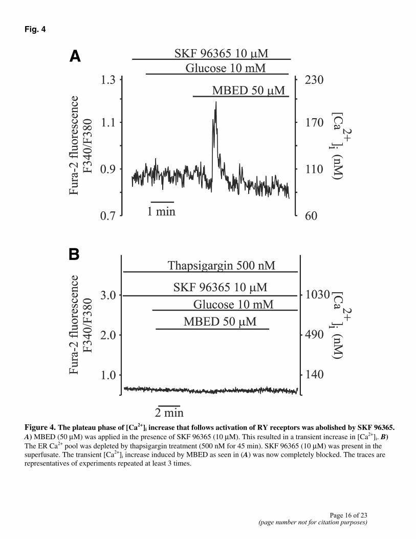
Fig. 4
Figure 4. The plateau phase of [Ca2+]i increase that follows activation of RY receptors was abolished by SKF 96365. A) MBED (50 µM) was applied in the presence of SKF 96365 (10 µM). This resulted in a transient increase in [Ca2+]i. B) The ER Ca2+ pool was depleted by thapsigargin treatment (500 nM for 45 min). SKF 96365 (10 µM) was present in the superfusate. The transient [Ca2+]i increase induced by MBED as seen in (A) was now completely blocked. The traces are representatives of experiments repeated at least 3 times.
Page 16 of 23(page number not for citation purposes)

Fig. 5
Figure 5. Activation of RY receptors caused Ca2+ influx even when the ER Ca2+ pools were depleted by inhibition of the SERCA. A) Application of carbachol (100 µM) or thapsigargin (500 nM) released Ca2+ from ER Ca2+ stores. B) Cells were treated by thapsigargin (500 nM) for 45 min. Such treatment depleted the ER Ca2+ pools as evidenced from the fact that carbachol (100 µM) failed to increase [Ca2+]i. Activation of RY receptors by MBED increased [Ca2+]i to a plateau in the thapsigargin-treated cells. Such [Ca2+]i increase was due to Ca2+ influx since it was blocked by SKF 96365 (see Fig. 4B). Similar results were obtained in at least 3 independent experiments.
Page 17 of 23(page number not for citation purposes)

Fig. 6
Figure 6. Effect of prior application of carbachol on [Ca2+]i-increase induced by RY receptor activation. A) The cells were first stimulated by a high concentration of carbachol (100 µM), which resulted in two [Ca2+]i transients. Carbachol caused rapid release of Ca2+ from the ER and a small [Ca2+]i plateau representing capacitative Ca2+ entry shown on expanded scale in (B). When RY receptors were activated by MBED, in the continued presence of carbachol, there was a large increase of [Ca2+]I, followed by a plateau and [Ca2+]i spikes superimposed on the plateau. The trace is representative of at least 3 different experiments.
Page 18 of 23(page number not for citation purposes)
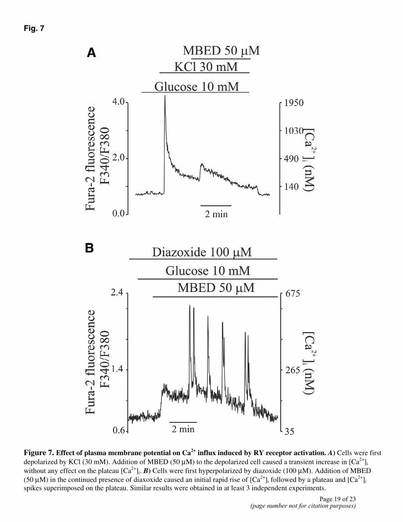
Fig. 7
Figure 7. Effect of plasma membrane potential on Ca2+ influx induced by RY receptor activation. A) Cells were first depolarized by KCl (30 mM). Addition of MBED (50 µM) to the depolarized cell caused a transient increase in [Ca2+]i without any effect on the plateau [Ca2+]i. B) Cells were first hyperpolarized by diazoxide (100 µM). Addition of MBED (50 µM) in the continued presence of diaxoxide caused an initial rapid rise of [Ca2+]i followed by a plateau and [Ca2+]i spikes superimposed on the plateau. Similar results were obtained in at least 3 independent experiments.
Page 19 of 23(page number not for citation purposes)

Fig. 8
Figure 8. Effect of activation of RY receptors on [Ca2+]i in Na+-free extracellular medium. The cell was first perifused with the basal medium. At times indicated by the horizontal bar the solution was changed to one that contained choline chloride instead of NaCl. Choline chloride caused a transient increase of [Ca2+]i. Activation of RY receptor by MBED (50 µM) in the absence of extracellular Na+ caused [Ca2+]i changes that were similar to those observed when MBED was used in the presence of Na+. The trace is representative of at least 3 different experiments.
Page 20 of 23(page number not for citation purposes)

Fig. 9
Figure 9. La3+, niflumic acid and 2-APB inhibited the [Ca2+]i plateau that followed activation of RY receptors. Activation of RY receptor by MBED (50 µM) in the continued presence of LaCl3 (100 µM) (A) or niflumic acid (50 µM) (B) caused the initial rise of [Ca2+]i but the plateau phase of [Ca2+]i increase was inhibited. The traces are representative of experiments that has been repeated at least 3 times. Complete inhibition of the plateau phase of [Ca2+]i by 2-APB (30 µM) (C) was seen in four out of nine experiments. In the remaining five experiments the plateau phase of [Ca2+]i was partially inhibited.
Page 21 of 23(page number not for citation purposes)

Fig. 10
Figure 10. Effect of ryanodine on MBED-induced [Ca2+]i changes. A) Cells were pretreated with caffeine (10 mM), KCl (25 mM), and ryanodine (50 µM) for 45 min. Application of MBED (50 µM) to the treated cells failed to increase [Ca2+]i. B) Acute application of ryanodine (50 µM) did not affect the initial rise of [Ca2+]i that was triggered by MBED, but it inhibited the [Ca2+]i spikes progressively in a use-dependent manner. The trace is representative of experiments repeated at least 3 times. C) The control experiment, where ryanodine was not included.
Page 22 of 23(page number not for citation purposes)

Fig. 11
Figure 11. Schematic diagram of hypothesized involvement of RY receptor and TRP-like channels in Ca2+ entry andmembrane depolarization in β-cells. The cartoon illustrates a sequence of events, whereby activation of RY receptors (A) leads to the activation of TRP-like channels (B), an initial membrane depolarization to approximately –40 mV (C), activation of the L-type voltage-gated Ca2+ channels (D), CICR (E) and exocytosis (F).
Page 23 of 23(page number not for citation purposes)