A second-generation anchored genetic linkage map of the tammar wallaby (Macropus eugenii)
Upload
independentCategory
view
2download
0
Molecular Biology of the CellVol. 19, 2402–2412, June 2008
The Novel Tail-anchored Membrane Protein MffControls Mitochondrial and Peroxisomal Fission inMammalian CellsShilpa Gandre-Babbe and Alexander M. van der Bliek
Department of Biological Chemistry, David Geffen School of Medicine at UCLA, Los Angeles, CA 90095
Submitted December 26, 2007; Revised February 19, 2008; Accepted March 12, 2008Monitoring Editor: Janet Shaw
Few components of the mitochondrial fission machinery are known, even though mitochondrial fission is a complexprocess of vital importance for cell growth and survival. Here, we describe a novel protein that controls mitochondrialfission. This protein was identified in a small interfering RNA (siRNA) screen using Drosophila cells. The humanhomologue of this protein was named Mitochondrial fission factor (Mff). Mitochondria of cells transfected with MffsiRNA form a closed network similar to the mitochondrial networks formed when cells are transfected with siRNA fortwo established fission proteins, Drp1 and Fis1. Like Drp1 and Fis1 siRNA, Mff siRNA also inhibits fission induced byloss of mitochondrial membrane potential, it delays cytochrome c release from mitochondria and further progression ofapoptosis, and it inhibits peroxisomal fission. Mff and Fis1 are both tail anchored in the mitochondrial outer membrane,but other parts of these proteins are very different and they exist in separate 200-kDa complexes, suggesting that they playdifferent roles in the fission process. We conclude that Mff is a novel component of a conserved membrane fission pathwayused for constitutive and induced fission of mitochondria and peroxisomes.
INTRODUCTION
Mitochondria are dynamic organelles that continually di-vide and fuse. Mitochondrial fission facilitates the redistri-bution of mitochondria in response to local changes in thedemand for ATP, whereas mitochondrial fusion is needed toexchange mitochondrial DNA and other components thatmay become damaged over time (Okamoto and Shaw, 2005;Chan, 2006). The rates of fission and fusion are usuallybalanced, but they do vary between different cell types, andthey may respond to environmental changes. There are cir-cumstances in which the rates of mitochondrial fission aregreatly accelerated. This occurs when cytochrome c is re-leased from mitochondria during apoptosis (Desagher andMartinou, 2000) and also when mitochondria are depolar-ized, for example, with the protonophore carbonylcyanide3-chloro phenyl hydrazone (CCCP) (Ishihara et al., 2003).Induced fission can lead to fragmented mitochondria,whereas mutations in fission proteins lead to a closed net-work of mitochondria as a result of continuing fusion with-out fission.
Mitochondrial fission and fusion are controlled by the op-posing actions of different dynamin family members on or inmitochondria. The dynamins are large proteins with an ami-no-terminal guanosine triphosphatase (GTPase) domain fol-lowed by a middle domain and a GTPase effector domain(GED) (Praefcke and McMahon, 2004). The GTPase domain
is thought to provide mechanical force, but it may also act asa molecular switch, similar to roles of other GTP-bindingproteins (Song and Schmid, 2003). The middle domain andthe GED of dynamin family members mediate assembly ofthese proteins into multimeric complexes. The archetypalfamily member, dynamin, which is required for endocytosis(van der Bliek and Meyerowitz, 1991; van der Bliek et al.,1993), wraps around constrictions at the necks of vesiclesthat are budding from the plasma membrane (Hinshaw,2000; Praefcke and McMahon, 2004).
Mitochondrial dynamin proteins are called Drp1 in mam-mals and Dnm1 in yeast (van der Bliek, 1999). These proteinsare largely cytosolic, but cycle on and off of mitochondria asneeded for fission (Bleazard et al., 1999; Labrousse et al.,1999; Smirnova et al., 2001). After translocating to mitochon-dria, they wrap around constricted parts of the mitochon-dria where they induce a late stage of mitochondrial outermembrane fission (Bleazard et al., 1999; Labrousse et al.,1999). Consistent with this role, yeast Dnm1 was shown toform rings or spirals with a diameter that matches the min-imal diameter of a double membrane constriction (Ingermanet al., 2005). Mutations in Dnm1 and Drp1 give rise to ahighly interconnected mesh of mitochondrial tubules. Thismesh forms because fusion still occurs while fission isblocked (Bleazard et al., 1999; Labrousse et al., 1999; Smir-nova et al., 2001). The rates of mitochondrial fission aredramatically increased during apoptosis (Desagher andMartinou, 2000). Fission occurs just before or at the sametime as cytochrome c release (Munoz-Pinedo et al., 2006).Mutations in Drp1 delay cytochrome c release, suggestingthat Drp1 is proapoptotic (Frank et al., 2001).
Other members of the dynamin family are involved infusion between mitochondria. Mitochondrial outer mem-branes are fused by proteins called Fzo1 in yeast (Hermannet al., 1998) and Mitofusins in mammals (Mfn1 and Mfn2)(Chen et al., 2003). These proteins have two transmembrane
This article was published online ahead of print in MBC in Press(http://www.molbiolcell.org/cgi/doi/10.1091/mbc.E07–12–1287)on March 19, 2008.
Address correspondence to: Alexander M. van der Bliek ([email protected]).
Abbreviations used: BNGE, Blue Native Gel electrophoresis; CCCP,carbonylcyanide 3-chloro phenyl hydrazone; TPR, tetratricopeptide.
2402 © 2008 by The American Society for Cell Biology http://www.molbiolcell.org/content/suppl/2008/03/18/E07-12-1287.DC1.htmlSupplemental Material can be found at:
segments that anchor them in the mitochondrial outer mem-brane. Mutations in Fzo1 and the Mitofusins give rise tofragmented mitochondria, but this can be reversed by mu-tations in yeast Dnm1 and mammalian Drp1 (Hales andFuller, 1997; Hermann et al., 1998; Santel and Fuller, 2001).Mitochondrial inner membranes are fused by dynamin fam-ily members called Opa1 in mammals (Alexander et al., 2000;Delettre et al., 2000), EAT-3 in Caenorhabditis elegans (Ka-nazawa et al., 2008), and Mgm1 in yeast (Shepard and Yaffe,1999; Wong et al., 2000). Mutations in these proteins alsofragment mitochondria and this fragmentation is reversedby mutations in mitochondrial fission proteins, similar to thereversal of outer membrane fusion defects by mutations inmitochondrial fission proteins (Fekkes et al., 2000; Mozdy etal., 2000; Tieu and Nunnari, 2000; Sesaki et al., 2003; Ka-nazawa et al., 2008).
Two more components of the mitochondrial fission ma-chinery were identified with genetic screens aimed at rever-sal of the mitochondrial fusion defects in yeast Mgm1 andFzo1 mutants (Fekkes et al., 2000; Mozdy et al., 2000; Tieuand Nunnari, 2000; Cerveny et al., 2001). The proteins iden-tified in these screens were called Fis1 and Mdv1. Mdv1 andits close relative Caf4 are cytosolic WD proteins that bind toFis1 and Dnm1 in yeast (Tieu et al., 2002; Griffin et al., 2005;Bhar et al., 2006; Naylor et al., 2006; Wells et al., 2007; Zhangand Chan, 2007). There are no obvious homologues of Mdv1in metazoans, but there are metazoan homologues of Fis1.Transfection of mammalian cells with Fis1 small interferingRNA (siRNA) increases the numbers of connections betweenmitochondria, similar to the connections observed withDrp1 siRNA, whereas overexpression of Fis1 causes mito-chondria to fragment (James et al., 2003; Yoon et al., 2003;Stojanovski et al., 2004). Fis1 proteins have two tetratricopep-tide (TPR) repeats, which are common protein–protein in-teraction domains, and a carboxy-terminal membrane-span-ning segment that anchors Fis1 in the mitochondrial outermembrane. Mammalian Fis1 and Drp1 bind to each other invitro, suggesting transient interactions during the fissionprocess (Yoon et al., 2003). A new twist to this story camewith the discovery that Fis1 and Drp1 proteins affect perox-isomal fission (Koch et al., 2003; Li and Gould, 2003; Koch etal., 2005; Kobayashi et al., 2007). Peroxisomes are semiauton-omous single-membrane organelles that can bud from theendoplasmic reticulum, but they also undergo furthergrowth and fission after their release into the cytosol. Utili-zation of Fis1 and Drp1 suggests that the mechanisms ofperoxisome and mitochondrial fission are very similar if notidentical (Schrader and Yoon, 2007).
Mitochondrial fission and fusion are controlled by severalregulatory mechanisms. Drp1 is activated by cyclin-depen-dent kinase 1/cyclin B-mediated phosphorylation duringmitosis (Taguchi et al., 2007) and inactivated by cAMP-dependent protein kinase (PKA) in quiescent cells (Changand Blackstone, 2007; Cribbs and Strack, 2007). Reversal ofPKA phosphorylation by the calcium-dependent phospha-tase calcineurin triggers mitochondrial fission (Cribbs andStrack, 2007). These newly discovered connections amongcAMP, calcium, and the actions of Drp1 are gratifying, be-cause it has long been known that these molecules influencethe rates of mitochondrial fission (Bereiter-Hahn and Voth,1994). Mitochondrial fission and fusion are also regulated byubiquination as shown with F-box proteins that determinethe amounts of Fzo1 in yeast (Fritz et al., 2003; Kondo-Okamoto et al., 2006) and an E3 ubiquitin ligase that controlsmitochondrial fission in mammalian cells (Karbowski et al.,2007). In addition, mitochondrial fission might also be reg-ulated by sumoylation in mammalian cells (Zunino et al.,
2007), but the targets of sumoylation and ubiquitinationhave not yet been unequivocally determined.
It is clear from the above-mentioned description that mi-tochondrial fission is a complex process that follows a se-quence of well-defined stages. These stages include the ac-tivation of cytosolic Drp1 by calcineurin or other cytosolicsignal transduction proteins and recruitment of Drp1 tospots on mitochondria, most likely through the actions ofFis1. However, the typical diameters of mitochondria (500–1000 nm) are too large to accommodate Drp1 rings (100 nm,as determined with yeast Dnm1) (Ingerman et al., 2005),suggesting that a separate constriction process is needed toreduce the mitochondrial diameter before Drp1 can formrings. Once this constriction occurs and Drp1 rings are as-sembled, those rings will sever the mitochondrial outermembrane through GTP hydrolysis. Drp1 then remains at-tached to one of the newly formed mitochondrial tips andslowly disassembles before returning to the cytoplasm (La-brousse et al., 1999). Considering that other membrane-traf-ficking processes, such as endo- and exocytosis, make use ofmany more proteins (Pfeffer, 2007), it seems likely that all ofthese steps require additional, as yet unknown proteins. Wesought new mitochondrial fission and fusion proteins usinga high-throughput screen of Drosophila siRNAs. With thisapproach, we identified a novel tail-anchored protein thatcontrols mitochondrial fission. This protein is conserved inmetazoans. It affects apoptosis and peroxisome fission, but itis in a complex that is separate from other known fissionproteins, suggesting that it acts independently.
MATERIALS AND METHODS
Nucleic AcidsHuman Mitochondrial fission factor (Mff) cDNA was cloned with reversetranscription-polymerase chain reaction (RT-PCR) by using RNA isolatedfrom HeLa cells. The PCR product was cloned into pCRII-TOPO (Invitrogen,Carlsbad, CA) and sequenced to rule out mutations introduced by PCR. OurcDNA is derived from isoform 8, which lacks exons 5, 6, and 8 (Figure 1). Theamino-terminal green fluorescent protein (GFP) fusion was made by reclon-ing this Mff cDNA behind the GFP sequences of pEGFP-C1 (Clontech, Moun-tain View, CA) with PCR. A fusion construct lacking the carboxy-terminaltransmembrane domain was made with a 3� PCR primer that removes the last20 amino acids of Mff. A 5XMYC epitope-tagged construct was made bycloning the Mff PCR fragment after the first five myc tags of pCS2�MT byusing NcoI and XhoI sites. All constructs were resequenced. pcDNA3.1myc::Mfn2(K109T) and pEGFP-N1::Mfn1(K88T) were kindly provided byAnsgar Santel (Atugen, Berlin, Germany).
Oligonucleotides for siRNA were made by Sigma-Proligo (The Woodlands,TX). Two pairs of Mff siRNA oligonucleotides were tested: 5�-CGCUGAC-CUGGAACAAGGAdTdT-3� for exon 2 and 5�-CUAAAUAGACGUCUA-CAACdTdT-3� for exon 8. The results that are shown were obtained with thefirst pair, but the second pair was equally effective. A scrambled sequence wasused as negative control (5�-AGGAACAAGGUCCAGUCGC-3�). Pairs of Drp1and Fis1 siRNA oligonucleotides were based on the sequences 5�-GCAGAA-GAAUGGGGUAAAU-3� and 5�-AGGCCUUAAAGUACGUCCG-3�, respectively.
For expression analysis, a blot with RNA from a range of human tissues(OriGene, Rockville, MD) was hybridized with 32P labeled Mff cDNA pre-pared with a Random Primed StripAble DNA Probe Synthesis kit (Ambion,Austin, TX). Hybridization conditions were as specified in the manufacturer’sprotocol. Hybridizing bands were detected with a Typhoon phosphorimager(GE Healthcare, Chalfont St. Giles, United Kingdom).
Antibodies and Other ReagentsRabbit polyclonal antibodies against Mff were produced by cloning humanMff cDNA into pET21d, which introduces a carboxy-terminal 6xHis tag.Bacterially expressed protein was purified with nickel-nitrilotriacetic acidagarose using 8 M urea. The protein was further purified with preparativeSDS-polyacrylamide gel electrophoresis (PAGE) gels and then used to immu-nize rabbits. Anti-Fis1 antibodies were purchased from Alexis Laboratories(San Diego, CA), �-tubulin antibodies were from Sigma-Aldrich (St. Louis,MO), Tom20 and c-myc/9E10 were from Santa Cruz Biotechnology (SantaCruz, CA), cytochrome c antibodies were from BD Biosciences PharMingen,prohibitin antibodies were from RDI (Flanders, NJ), and DLP1 antibodieswere from BD Biosciences Transduction Laboratories (Lexington, KY). Sec-
A Novel Mitochondrial Fission Protein
Vol. 19, June 2008 2403
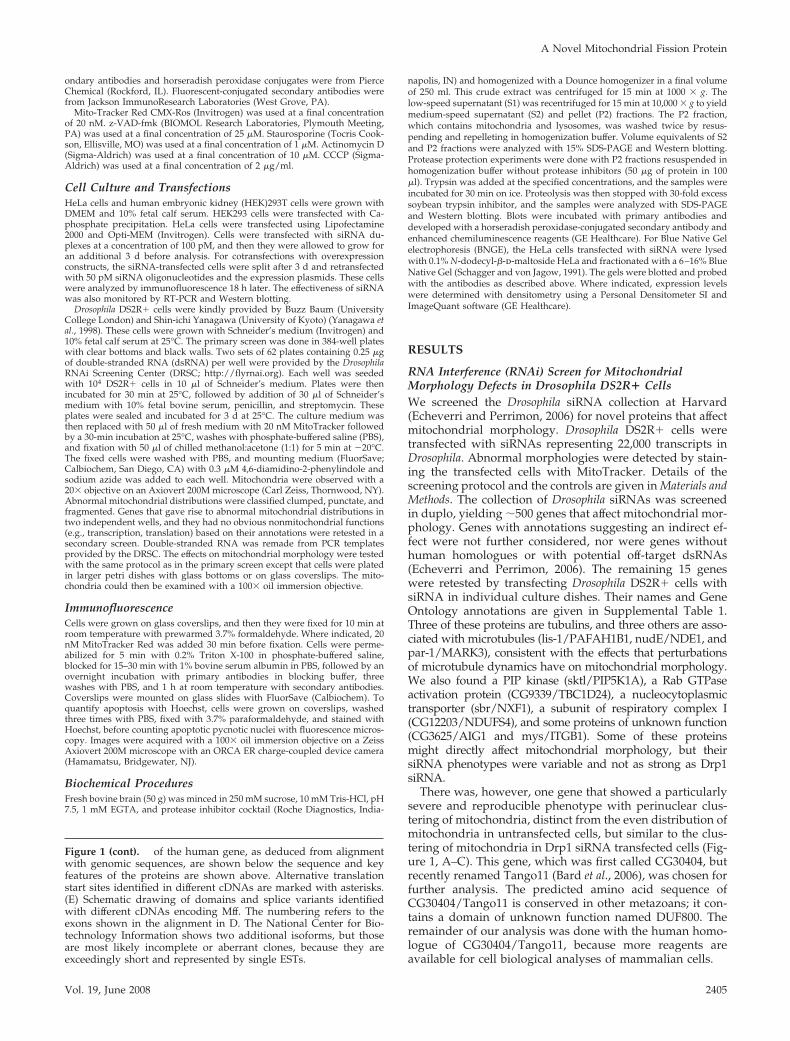
Figure 1. Identification of an Mff homologue in a Drosophila siRNA screen. (A and A�) Untransfected Drosophila S2 cells stained withMitoTracker. (B and B�) Cells transfected with Drosophila Drp1 siRNA. (C and C�) Cells transfected with CG30404 siRNA. A, B, and C areimages taken with a 20� lens in the primary screen (bar, 100 �m), and A�, B�, and C� are images taken with a 100� lens in the secondaryscreen (bar, 10 �m). (D) Alignment of the Drosophila CG30404 protein with Mff, which is encoded by C2ORF33. Positions of exon boundaries
S. Gandre-Babbe and A. M. van der Bliek
Molecular Biology of the Cell2404
ondary antibodies and horseradish peroxidase conjugates were from PierceChemical (Rockford, IL). Fluorescent-conjugated secondary antibodies werefrom Jackson ImmunoResearch Laboratories (West Grove, PA).
Mito-Tracker Red CMX-Ros (Invitrogen) was used at a final concentrationof 20 nM. z-VAD-fmk (BIOMOL Research Laboratories, Plymouth Meeting,PA) was used at a final concentration of 25 �M. Staurosporine (Tocris Cook-son, Ellisville, MO) was used at a final concentration of 1 �M. Actinomycin D(Sigma-Aldrich) was used at a final concentration of 10 �M. CCCP (Sigma-Aldrich) was used at a final concentration of 2 �g/ml.
Cell Culture and TransfectionsHeLa cells and human embryonic kidney (HEK)293T cells were grown withDMEM and 10% fetal calf serum. HEK293 cells were transfected with Ca-phosphate precipitation. HeLa cells were transfected using Lipofectamine2000 and Opti-MEM (Invitrogen). Cells were transfected with siRNA du-plexes at a concentration of 100 pM, and then they were allowed to grow foran additional 3 d before analysis. For cotransfections with overexpressionconstructs, the siRNA-transfected cells were split after 3 d and retransfectedwith 50 pM siRNA oligonucleotides and the expression plasmids. These cellswere analyzed by immunofluorescence 18 h later. The effectiveness of siRNAwas also monitored by RT-PCR and Western blotting.
Drosophila DS2R� cells were kindly provided by Buzz Baum (UniversityCollege London) and Shin-ichi Yanagawa (University of Kyoto) (Yanagawa etal., 1998). These cells were grown with Schneider’s medium (Invitrogen) and10% fetal calf serum at 25°C. The primary screen was done in 384-well plateswith clear bottoms and black walls. Two sets of 62 plates containing 0.25 �gof double-stranded RNA (dsRNA) per well were provided by the DrosophilaRNAi Screening Center (DRSC; http://flyrnai.org). Each well was seededwith 104 DS2R� cells in 10 �l of Schneider’s medium. Plates were thenincubated for 30 min at 25°C, followed by addition of 30 �l of Schneider’smedium with 10% fetal bovine serum, penicillin, and streptomycin. Theseplates were sealed and incubated for 3 d at 25°C. The culture medium wasthen replaced with 50 �l of fresh medium with 20 nM MitoTracker followedby a 30-min incubation at 25°C, washes with phosphate-buffered saline (PBS),and fixation with 50 �l of chilled methanol:acetone (1:1) for 5 min at �20°C.The fixed cells were washed with PBS, and mounting medium (FluorSave;Calbiochem, San Diego, CA) with 0.3 �M 4,6-diamidino-2-phenylindole andsodium azide was added to each well. Mitochondria were observed with a20� objective on an Axiovert 200M microscope (Carl Zeiss, Thornwood, NY).Abnormal mitochondrial distributions were classified clumped, punctate, andfragmented. Genes that gave rise to abnormal mitochondrial distributions intwo independent wells, and they had no obvious nonmitochondrial functions(e.g., transcription, translation) based on their annotations were retested in asecondary screen. Double-stranded RNA was remade from PCR templatesprovided by the DRSC. The effects on mitochondrial morphology were testedwith the same protocol as in the primary screen except that cells were platedin larger petri dishes with glass bottoms or on glass coverslips. The mito-chondria could then be examined with a 100� oil immersion objective.
ImmunofluorescenceCells were grown on glass coverslips, and then they were fixed for 10 min atroom temperature with prewarmed 3.7% formaldehyde. Where indicated, 20nM MitoTracker Red was added 30 min before fixation. Cells were perme-abilized for 5 min with 0.2% Triton X-100 in phosphate-buffered saline,blocked for 15–30 min with 1% bovine serum albumin in PBS, followed by anovernight incubation with primary antibodies in blocking buffer, threewashes with PBS, and 1 h at room temperature with secondary antibodies.Coverslips were mounted on glass slides with FluorSave (Calbiochem). Toquantify apoptosis with Hoechst, cells were grown on coverslips, washedthree times with PBS, fixed with 3.7% paraformaldehyde, and stained withHoechst, before counting apoptotic pycnotic nuclei with fluorescence micros-copy. Images were acquired with a 100� oil immersion objective on a ZeissAxiovert 200M microscope with an ORCA ER charge-coupled device camera(Hamamatsu, Bridgewater, NJ).
Biochemical ProceduresFresh bovine brain (50 g) was minced in 250 mM sucrose, 10 mM Tris-HCl, pH7.5, 1 mM EGTA, and protease inhibitor cocktail (Roche Diagnostics, India-
napolis, IN) and homogenized with a Dounce homogenizer in a final volumeof 250 ml. This crude extract was centrifuged for 15 min at 1000 � g. Thelow-speed supernatant (S1) was recentrifuged for 15 min at 10,000 � g to yieldmedium-speed supernatant (S2) and pellet (P2) fractions. The P2 fraction,which contains mitochondria and lysosomes, was washed twice by resus-pending and repelleting in homogenization buffer. Volume equivalents of S2and P2 fractions were analyzed with 15% SDS-PAGE and Western blotting.Protease protection experiments were done with P2 fractions resuspended inhomogenization buffer without protease inhibitors (50 �g of protein in 100�l). Trypsin was added at the specified concentrations, and the samples wereincubated for 30 min on ice. Proteolysis was then stopped with 30-fold excesssoybean trypsin inhibitor, and the samples were analyzed with SDS-PAGEand Western blotting. Blots were incubated with primary antibodies anddeveloped with a horseradish peroxidase-conjugated secondary antibody andenhanced chemiluminescence reagents (GE Healthcare). For Blue Native Gelelectrophoresis (BNGE), the HeLa cells transfected with siRNA were lysedwith 0.1% N-dodecyl-�-d-maltoside HeLa and fractionated with a 6–16% BlueNative Gel (Schagger and von Jagow, 1991). The gels were blotted and probedwith the antibodies as described above. Where indicated, expression levelswere determined with densitometry using a Personal Densitometer SI andImageQuant software (GE Healthcare).
RESULTS
RNA Interference (RNAi) Screen for MitochondrialMorphology Defects in Drosophila DS2R� CellsWe screened the Drosophila siRNA collection at Harvard(Echeverri and Perrimon, 2006) for novel proteins that affectmitochondrial morphology. Drosophila DS2R� cells weretransfected with siRNAs representing 22,000 transcripts inDrosophila. Abnormal morphologies were detected by stain-ing the transfected cells with MitoTracker. Details of thescreening protocol and the controls are given in Materials andMethods. The collection of Drosophila siRNAs was screenedin duplo, yielding �500 genes that affect mitochondrial mor-phology. Genes with annotations suggesting an indirect ef-fect were not further considered, nor were genes withouthuman homologues or with potential off-target dsRNAs(Echeverri and Perrimon, 2006). The remaining 15 geneswere retested by transfecting Drosophila DS2R� cells withsiRNA in individual culture dishes. Their names and GeneOntology annotations are given in Supplemental Table 1.Three of these proteins are tubulins, and three others are asso-ciated with microtubules (lis-1/PAFAH1B1, nudE/NDE1, andpar-1/MARK3), consistent with the effects that perturbationsof microtubule dynamics have on mitochondrial morphology.We also found a PIP kinase (sktl/PIP5K1A), a Rab GTPaseactivation protein (CG9339/TBC1D24), a nucleocytoplasmictransporter (sbr/NXF1), a subunit of respiratory complex I(CG12203/NDUFS4), and some proteins of unknown function(CG3625/AIG1 and mys/ITGB1). Some of these proteinsmight directly affect mitochondrial morphology, but theirsiRNA phenotypes were variable and not as strong as Drp1siRNA.
There was, however, one gene that showed a particularlysevere and reproducible phenotype with perinuclear clus-tering of mitochondria, distinct from the even distribution ofmitochondria in untransfected cells, but similar to the clus-tering of mitochondria in Drp1 siRNA transfected cells (Fig-ure 1, A–C). This gene, which was first called CG30404, butrecently renamed Tango11 (Bard et al., 2006), was chosen forfurther analysis. The predicted amino acid sequence ofCG30404/Tango11 is conserved in other metazoans; it con-tains a domain of unknown function named DUF800. Theremainder of our analysis was done with the human homo-logue of CG30404/Tango11, because more reagents areavailable for cell biological analyses of mammalian cells.
Figure 1 (cont). of the human gene, as deduced from alignmentwith genomic sequences, are shown below the sequence and keyfeatures of the proteins are shown above. Alternative translationstart sites identified in different cDNAs are marked with asterisks.(E) Schematic drawing of domains and splice variants identifiedwith different cDNAs encoding Mff. The numbering refers to theexons shown in the alignment in D. The National Center for Bio-technology Information shows two additional isoforms, but thoseare most likely incomplete or aberrant clones, because they areexceedingly short and represented by single ESTs.
A Novel Mitochondrial Fission Protein
Vol. 19, June 2008 2405
Mff, Encoded by C2orf33, Is the Human Homologue ofDrosophila CG30404/Tango11A BLAST search shows that humans have two proteins withsubstantial homology to CG30404/Tango11. The first pro-tein was named FATE, because it is specifically expressed infetal and adult testis, but the homology to this protein islimited to its carboxy-terminal half. Moreover, pilot experi-ments with FATE siRNA showed no effect on mitochondriain HeLa cells or other human cell lines commonly used inour laboratory (data not shown), as was expected becauseFATE is testis specific (Olesen et al., 2001). The second pro-tein, encoded by C2ORF33 and renamed Mff, shows homol-ogy over the entire length of the protein (Figure 1D). Theexpression pattern of Mff was determined with an RNA blotrepresenting a range of human tissues (Supplemental Figure1). This blot shows that Mff is highly expressed in heart,kidney, liver, brain, muscle, and stomach and at low levelsin other tissues. Because this pattern is compatible with ageneral mitochondrial function, we focused our analysis onthis protein.
The main features of human Mff and Drosophila CG30404/Tango11 sequences are a short amino-terminal repeat, amiddle segment with strong coiled coil forming propensity,and a carboxy-terminal hydrophobic segment that mostlikely serves as membrane anchor. The Mff gene encodes atleast nine different isoforms represented by multiple ex-pressed sequence tags (ESTs) in GenBank (Figure 1E). Theseisoforms are generated by the presence or absence of exon 1and various combinations of exons 5, 6, and 7, which encodethe middle part of the protein. The presence or absence ofexon 1 most likely results from the use of alternative pro-moters. Isoforms lacking exon 1 have an alternative transla-tion initiation codon embedded in exon 2. Alternativelyspliced exons 5, 6, and 7 encode parts of the protein that arenot conserved in Drosophila. However, other mammalianspecies have largely identical patterns of alternative splicing,suggesting that at least for mammalian cells sequence vari-ations caused by the presence or absence of exons 5, 6, and7 are functionally important.
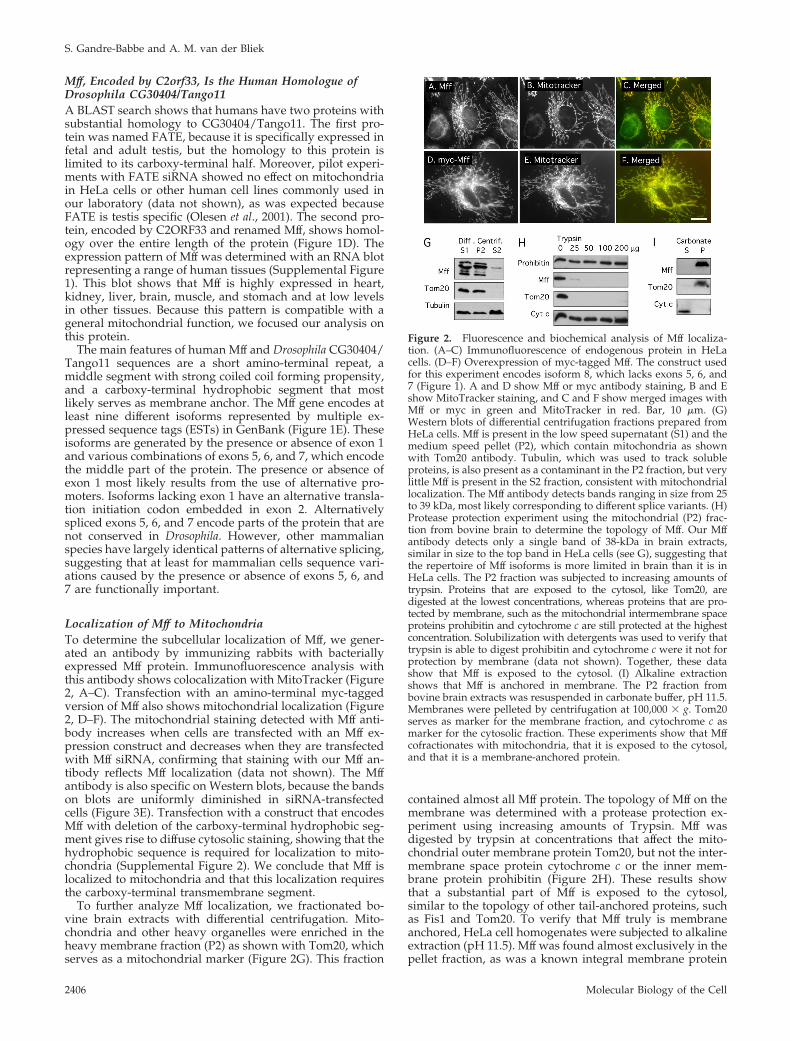
Localization of Mff to MitochondriaTo determine the subcellular localization of Mff, we gener-ated an antibody by immunizing rabbits with bacteriallyexpressed Mff protein. Immunofluorescence analysis withthis antibody shows colocalization with MitoTracker (Figure2, A–C). Transfection with an amino-terminal myc-taggedversion of Mff also shows mitochondrial localization (Figure2, D–F). The mitochondrial staining detected with Mff anti-body increases when cells are transfected with an Mff ex-pression construct and decreases when they are transfectedwith Mff siRNA, confirming that staining with our Mff an-tibody reflects Mff localization (data not shown). The Mffantibody is also specific on Western blots, because the bandson blots are uniformly diminished in siRNA-transfectedcells (Figure 3E). Transfection with a construct that encodesMff with deletion of the carboxy-terminal hydrophobic seg-ment gives rise to diffuse cytosolic staining, showing that thehydrophobic sequence is required for localization to mito-chondria (Supplemental Figure 2). We conclude that Mff islocalized to mitochondria and that this localization requiresthe carboxy-terminal transmembrane segment.
To further analyze Mff localization, we fractionated bo-vine brain extracts with differential centrifugation. Mito-chondria and other heavy organelles were enriched in theheavy membrane fraction (P2) as shown with Tom20, whichserves as a mitochondrial marker (Figure 2G). This fraction
contained almost all Mff protein. The topology of Mff on themembrane was determined with a protease protection ex-periment using increasing amounts of Trypsin. Mff wasdigested by trypsin at concentrations that affect the mito-chondrial outer membrane protein Tom20, but not the inter-membrane space protein cytochrome c or the inner mem-brane protein prohibitin (Figure 2H). These results showthat a substantial part of Mff is exposed to the cytosol,similar to the topology of other tail-anchored proteins, suchas Fis1 and Tom20. To verify that Mff truly is membraneanchored, HeLa cell homogenates were subjected to alkalineextraction (pH 11.5). Mff was found almost exclusively in thepellet fraction, as was a known integral membrane protein
Figure 2. Fluorescence and biochemical analysis of Mff localiza-tion. (A–C) Immunofluorescence of endogenous protein in HeLacells. (D–F) Overexpression of myc-tagged Mff. The construct usedfor this experiment encodes isoform 8, which lacks exons 5, 6, and7 (Figure 1). A and D show Mff or myc antibody staining, B and Eshow MitoTracker staining, and C and F show merged images withMff or myc in green and MitoTracker in red. Bar, 10 �m. (G)Western blots of differential centrifugation fractions prepared fromHeLa cells. Mff is present in the low speed supernatant (S1) and themedium speed pellet (P2), which contain mitochondria as shownwith Tom20 antibody. Tubulin, which was used to track solubleproteins, is also present as a contaminant in the P2 fraction, but verylittle Mff is present in the S2 fraction, consistent with mitochondriallocalization. The Mff antibody detects bands ranging in size from 25to 39 kDa, most likely corresponding to different splice variants. (H)Protease protection experiment using the mitochondrial (P2) frac-tion from bovine brain to determine the topology of Mff. Our Mffantibody detects only a single band of 38-kDa in brain extracts,similar in size to the top band in HeLa cells (see G), suggesting thatthe repertoire of Mff isoforms is more limited in brain than it is inHeLa cells. The P2 fraction was subjected to increasing amounts oftrypsin. Proteins that are exposed to the cytosol, like Tom20, aredigested at the lowest concentrations, whereas proteins that are pro-tected by membrane, such as the mitochondrial intermembrane spaceproteins prohibitin and cytochrome c are still protected at the highestconcentration. Solubilization with detergents was used to verify thattrypsin is able to digest prohibitin and cytochrome c were it not forprotection by membrane (data not shown). Together, these datashow that Mff is exposed to the cytosol. (I) Alkaline extractionshows that Mff is anchored in membrane. The P2 fraction frombovine brain extracts was resuspended in carbonate buffer, pH 11.5.Membranes were pelleted by centrifugation at 100,000 � g. Tom20serves as marker for the membrane fraction, and cytochrome c asmarker for the cytosolic fraction. These experiments show that Mffcofractionates with mitochondria, that it is exposed to the cytosol,and that it is a membrane-anchored protein.
S. Gandre-Babbe and A. M. van der Bliek
Molecular Biology of the Cell2406
(Tom20), but not cytochrome c (Figure 2I), from which weconclude that Mff is firmly anchored in membrane. Mff thusbelongs to the growing number of tail-anchored membraneproteins.
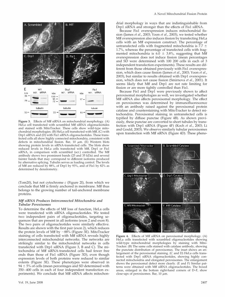
Mff siRNA Produces Interconnected Mitochondria andTubular PeroxisomesTo determine the effects of Mff loss of function, HeLa cellswere transfected with siRNA oligonucleotides. We testedtwo independent pairs of oligonucleotides, targeting se-quences that are present in all isoforms (exon 2 and exon 8).The two pairs of oligonucleotides were similarly effective.Results are shown with the first pair (exon 2), which reducesthe protein levels of Mff by �88% (Figure 3E). MitoTrackerstaining of cells transfected with Mff siRNA reveals highlyinterconnected mitochondrial networks. The networks arestrikingly similar to the mitochondrial networks in cellstransfected with Drp1 siRNA (Figure 3, B and C). The mi-tochondria of Mff siRNA-transfected cells had fewer freeends than those of Fis1 siRNA (Figure 3D), even thoughexpression levels of both proteins were reduced to similarextents (Figure 3E). These phenotypes were observed in94 � 5% of cells (mean percentages and SD determined with350–400 cells in each of four independent transfection ex-periments). We conclude that Mff siRNA affects mitochon-
drial morphology in ways that are indistinguishable fromDrp1 siRNA and stronger than the effects of Fis1 siRNA.
Because Fis1 overexpression induces mitochondrial fis-sion (James et al., 2003; Yoon et al., 2003), we tested whetherMff overexpression also induces fission by transfecting HeLacells with an Mff expression construct. The percentage ofuntransfected cells with fragmented mitochondria is 7.7 �1.7%, whereas the percentage of transfected cells with frag-mented mitochondria is 6.0 � 3.8%, suggesting that Mffoverexpression does not induce fission (mean percentagesand SD were determined with 100 200 cells in each of 3independent transfection experiments). These results are dif-ferent from those obtained previously with Fis1 overexpres-sion, which does cause fission (James et al., 2003; Yoon et al.,2003), but similar to results obtained with Drp1 overexpres-sion, which does not cause fission (Smirnova et al., 2001). Itseems likely that Mff and Drp1 are not rate limiting forfission or are more tightly controlled than Fis1.
Because Fis1 and Drp1 were previously shown to affectperoxisomal morphologies as well, we investigated whetherMff siRNA also affects peroxisomal morphology. The effecton peroxisomes was determined by immunofluorescencewith an antibody raised against the peroxisomal proteincatalase and counterstaining with MitoTracker to detect mi-tochondria. Peroxisomal staining in untransfected cells istypified by diffuse punctae (Figure 4B). As shown previ-ously, these punctae are converted to short tubules by trans-fection with Drp1 siRNA (Figure 4F) (Koch et al., 2003; Liand Gould, 2003). We observe similarly tubular peroxisomesupon transfection with Mff siRNA (Figure 4D). These pheno-
Figure 4. Effects of Mff siRNA on peroxisomal morphology. (A)HeLa cells transfected with scrambled oligonucleotides showingwild-type mitochondrial morphologies by staining with Mito-Tracker. (B) The same cells stained with catalase antibody, showingthe punctate distribution of peroxisomes. The inset shows an en-largement of the peroxisomal staining. (C and D) HeLa cells trans-fected with Drp1 siRNA oligonucleotides, showing highly con-nected mitochondria and elongated peroxisomes. The enlargementshows the peroxisomal defect more clearly. (E and F) Similar pat-terns were obtained with Mff siRNA oligonucleotides. The boxedareas, enlarged in the bottom right-hand corners of D–F, showclose-ups of peroxisomes. Bar, 10 �m.
Figure 3. Effects of Mff siRNA on mitochondrial morphology. (A)HeLa cell transfected with scrambled Mff siRNA oligonucleotidesand stained with MitoTracker. These cells show wild-type mito-chondrial morphologies. (B) HeLa cell transfected with Mff, (C) withDrp1 siRNA and (D) with Fis1 siRNA oligonucleotides. These trans-fected cells all show highly connected mitochondria, consistent withdefects in mitochondrial fission. Bar, 10 �m. (E) Western blotsshowing protein levels in siRNA-transfected cells. The blots showreduced levels in HeLa cells transfected with Mff, Drp1 or Fis1siRNA, in comparison with scrambled (scr.) controlled. The Mffantibody shows two prominent bands (25 and 35 kDa) and severalfainter bands that may correspond to different isoforms producedby alternative splicing. Tubulin serves as loading control. The levelsof Mff are reduced by 88%, of Drp1 by 93%, and of Fis1 by 82% asdetermined by densitometry.
A Novel Mitochondrial Fission Protein
Vol. 19, June 2008 2407
types were observed in 84 � 6% of cells (mean percentages andSD were determined with 350 cells in 3 independent transfec-tion experiments). Counterstaining with MitoTracker Redshows that cells with affected peroxisomes invariably also haveaffected mitochondria (Figure 4, C and E). Because smallamounts of peroxisomal staining might have been obscured bymitochondrial staining, we changed the mitochondrial distri-bution with Drp1 siRNA to make parts of the cell devoid ofmitochondria. These areas show colocalization of myc-taggedMff with a peroxisomal marker (Supplemental Figure 3). Weconclude that there is a small but significant amount of Mff onperoxisomes, similar to Fis1, which is also localized to peroxi-somes and mitochondria (Koch et al., 2005; Kobayashi et al.,2007). The dual roles and localizations of Drp1, Fis1, and Mff onperoxisomes and mitochondria suggest that these proteins arepart of the same fission apparatus, acting on two differentorganelles.
The Drosophila homologue of Mff (CG30404/Tango11) wasalso identified in a screen for defects in constitutive secre-tion, suggesting that Mff might affect the secretory pathway(Bard et al., 2006). We tested whether Mff siRNA also affectssecretion in mammalian cells, by using a luciferase assay.We were unable to detect significant differences in the rate ofsecretion or in Golgi morphology, even when there werelarge differences in mitochondrial and peroxisomal mor-phologies (data not shown). The secretion defects that wereobserved in Drosophila cells may have been due to respira-tory defects, because these defects can result from mitochon-drial fission defects (Estaquier and Arnoult, 2007). We con-clude that Mff affects peroxisomal and mitochondrialmorphologies in ways that are indistinguishable from theeffects of Drp1 siRNA. It seems likely that Mff, Fis1, andDrp1 all affect different aspects of the pathways that mediatemitochondrial and peroxisomal fission.
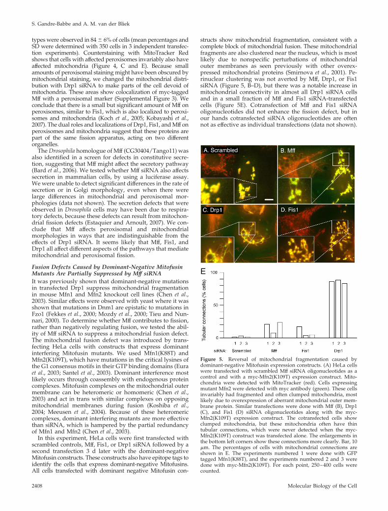
Fusion Defects Caused by Dominant-Negative MitofusinMutants Are Partially Suppressed by Mff siRNAIt was previously shown that dominant-negative mutationsin transfected Drp1 suppress mitochondrial fragmentationin mouse Mfn1 and Mfn2 knockout cell lines (Chen et al.,2003). Similar effects were observed with yeast where it wasshown that mutations in Dnm1 are epistatic to mutations inFzo1 (Fekkes et al., 2000; Mozdy et al., 2000; Tieu and Nun-nari, 2000). To determine whether Mff contributes to fission,rather than negatively regulating fusion, we tested the abil-ity of Mff siRNA to suppress a mitochondrial fusion defect.The mitochondrial fusion defect was introduced by trans-fecting HeLa cells with constructs that express dominantinterfering Mitofusin mutants. We used Mfn1(K88T) andMfn2(K109T), which have mutations in the critical lysines ofthe G1 consensus motifs in their GTP binding domains (Euraet al., 2003; Santel et al., 2003). Dominant interference mostlikely occurs through coassembly with endogenous proteincomplexes. Mitofusin complexes on the mitochondrial outermembrane can be heteromeric or homomeric (Chen et al.,2003) and act in trans with similar complexes on opposingmitochondrial membranes during fusion (Koshiba et al.,2004; Meeusen et al., 2004). Because of these heteromericcomplexes, dominant interfering mutants are more effectivethan siRNA, which is hampered by the partial redundancyof Mfn1 and Mfn2 (Chen et al., 2003).
In this experiment, HeLa cells were first transfected withscrambled controls, Mff, Fis1, or Drp1 siRNA followed by asecond transfection 3 d later with the dominant-negativeMitofusin constructs. These constructs also have epitope tags toidentify the cells that express dominant-negative Mitofusins.All cells transfected with dominant negative Mitofusin con-
structs show mitochondrial fragmentation, consistent with acomplete block of mitochondrial fusion. These mitochondrialfragments are also clustered near the nucleus, which is mostlikely due to nonspecific perturbations of mitochondrialouter membranes as seen previously with other overex-pressed mitochondrial proteins (Smirnova et al., 2001). Pe-rinuclear clustering was not averted by Mff, Drp1, or Fis1siRNA (Figure 5, B–D), but there was a notable increase inmitochondrial connectivity in almost all Drp1 siRNA cellsand in a small fraction of Mff and Fis1 siRNA-transfectedcells (Figure 5E). Cotransfection of Mff and Fis1 siRNAoligonucleotides did not enhance the fission defect, but inour hands cotransfected siRNA oligonucleotides are oftennot as effective as individual transfections (data not shown).
Figure 5. Reversal of mitochondrial fragmentation caused bydominant-negative Mitofusin expression constructs. (A) HeLa cellswere transfected with scrambled Mff siRNA oligonucleotides as acontrol and with a myc-Mfn2(K109T) expression construct. Mito-chondria were detected with MitoTracker (red). Cells expressingmutant Mfn2 were detected with myc antibody (green). These cellsinvariably had fragmented and often clumped mitochondria, mostlikely due to overexpression of aberrant mitochondrial outer mem-brane protein. Similar transfections were done with Mff (B), Drp1(C), and Fis1 (D) siRNA oligonucleotides along with the myc-Mfn2(K109T) expression construct. The cotransfected cells showclumped mitochondria, but these mitochondria often have thintubular connections, which were never detected when the myc-Mfn2(K109T) construct was transfected alone. The enlargements inthe bottom left corners show these connections more clearly. Bar, 10�m. The percentages of cells with mitochondrial connections areshown in E. The experiments numbered 1 were done with GFPtagged Mfn1(K88T), and the experiments numbered 2 and 3 weredone with myc-Mfn2(K109T). For each point, 250–400 cells werecounted.
S. Gandre-Babbe and A. M. van der Bliek
Molecular Biology of the Cell2408
It therefore remains possible that Mff and Fis1 are partiallyredundant. Alternatively, we may not have achieved lowenough levels of Mff and Fis1 to fully block their function orMff and Fis1 are not absolutely required for fission. We can,nevertheless, conclude that Mff and Fis1 siRNA reversesome of the fragmentation caused by dominant-negativeMitofusins, whereas Drp1 siRNA fully reverses their effects.
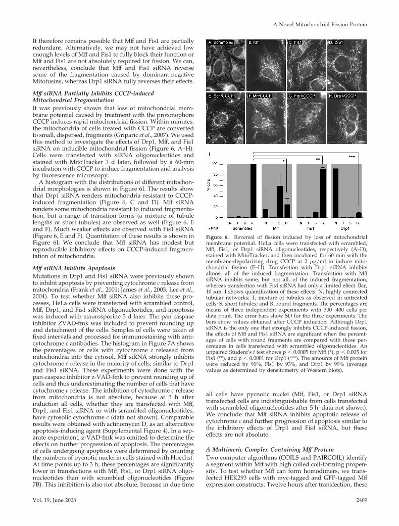
Mff siRNA Partially Inhibits CCCP-inducedMitochondrial FragmentationIt was previously shown that loss of mitochondrial mem-brane potential caused by treatment with the protonophoreCCCP induces rapid mitochondrial fission. Within minutes,the mitochondria of cells treated with CCCP are convertedto small, dispersed, fragments (Griparic et al., 2007). We usedthis method to investigate the effects of Drp1, Mff, and Fis1siRNA on inducible mitochondrial fission (Figure 6, A–H).Cells were transfected with siRNA oligonucleotides andstained with MitoTracker 3 d later, followed by a 60-minincubation with CCCP to induce fragmentation and analysisby fluorescence microscopy.
A histogram with the distributions of different mitochon-drial morphologies is shown in Figure 6I. The results showthat Drp1 siRNA renders mitochondria resistant to CCCP-induced fragmentation (Figure 6, C and D). Mff siRNArenders some mitochondria resistant to induced fragmenta-tion, but a range of transition forms (a mixture of tubulelengths or short tubules) are observed as well (Figure 6, Eand F). Much weaker effects are observed with Fis1 siRNA(Figure 6, E and F). Quantitation of these results is shown inFigure 6I. We conclude that Mff siRNA has modest butreproducible inhibitory effects on CCCP-induced fragmen-tation of mitochondria.
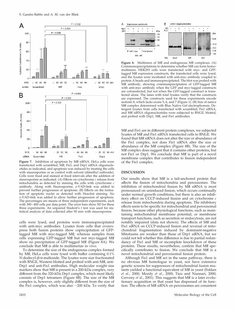
Mff siRNA Inhibits ApoptosisMutations in Drp1 and Fis1 siRNA were previously shownto inhibit apoptosis by preventing cytochrome c release frommitochondria (Frank et al., 2001; James et al., 2003; Lee et al.,2004). To test whether Mff siRNA also inhibits these pro-cesses, HeLa cells were transfected with scrambled control,Mff, Drp1, and Fis1 siRNA oligonucleotides, and apoptosiswas induced with staurosporine 3 d later. The pan caspaseinhibitor ZVAD-fmk was included to prevent rounding upand detachment of the cells. Samples of cells were taken atfixed intervals and processed for immunostaining with anti-cytochrome c antibodies. The histogram in Figure 7A showsthe percentages of cells with cytochrome c released frommitochondria into the cytosol. Mff siRNA strongly inhibitscytochrome c release in the majority of cells, similar to Drp1and Fis1 siRNA. These experiments were done with thepan-caspase inhibitor z-VAD-fmk to prevent rounding up ofcells and thus underestimating the number of cells that havecytochrome c release. The inhibition of cytochrome c releasefrom mitochondria is not absolute, because at 5 h afterinduction all cells, whether they are transfected with Mff,Drp1, and Fis1 siRNA or with scrambled oligonucleotides,have cytosolic cytochrome c (data not shown). Comparableresults were obtained with actinomycin D, as an alternativeapoptosis-inducing agent (Supplemental Figure 4). In a sep-arate experiment, z-VAD-fmk was omitted to determine theeffects on further progression of apoptosis. The percentagesof cells undergoing apoptosis were determined by countingthe numbers of pycnotic nuclei in cells stained with Hoechst.At time points up to 3 h, these percentages are significantlylower in transfections with Mff, Fis1, or Drp1 siRNA oligo-nucleotides than with scrambled oligonucleotides (Figure7B). This inhibition is also not absolute, because in due time
all cells have pycnotic nuclei (Mff, Fis1, or Drp1 siRNAtransfected cells are indistinguishable from cells transfectedwith scrambled oligonucleotides after 5 h; data not shown).We conclude that Mff siRNA inhibits apoptotic release ofcytochrome c and further progression of apoptosis similar tothe inhibitory effects of Drp1 and Fis1 siRNA, but theseeffects are not absolute.
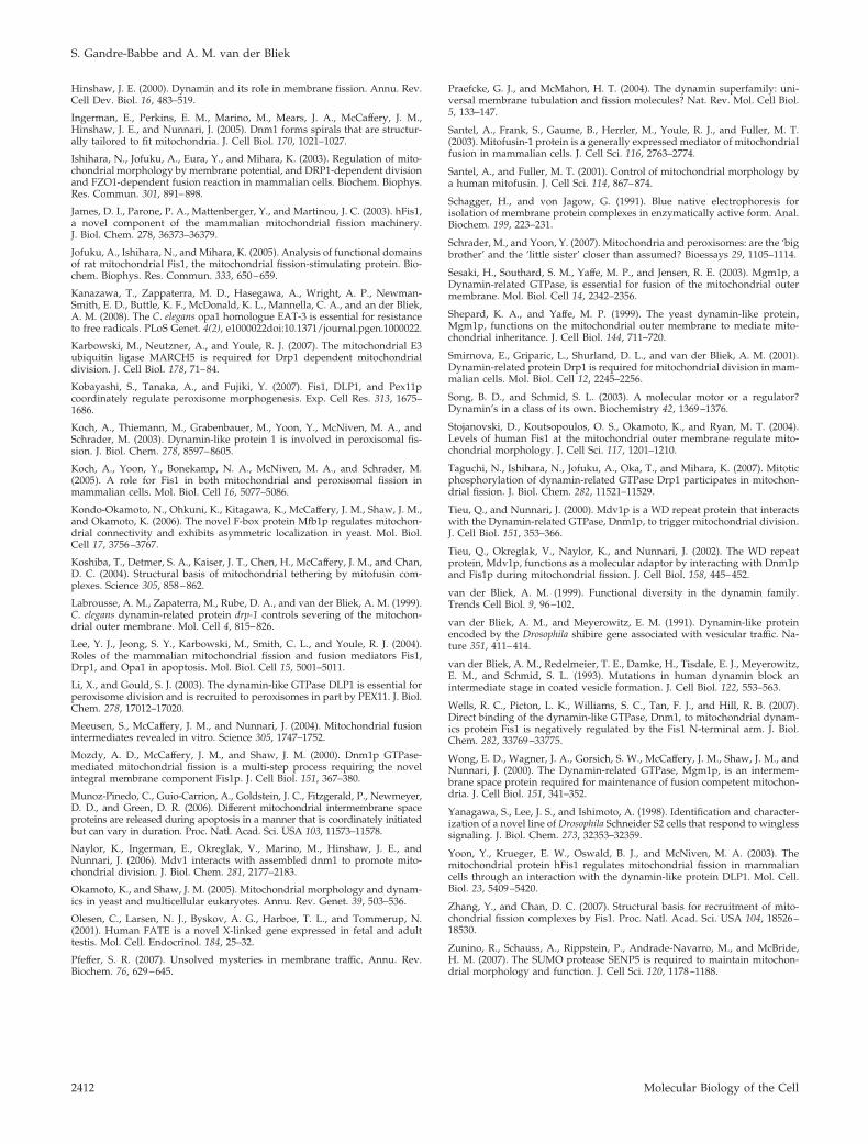
A Multimeric Complex Containing Mff ProteinTwo computer algorithms (COILS and PAIRCOIL) identifya segment within Mff with high coiled coil-forming propen-sity. To test whether Mff can form homodimers, we trans-fected HEK293 cells with myc-tagged and GFP-tagged Mffexpression constructs. Twelve hours after transfection, these
Figure 6. Reversal of fission induced by loss of mitochondrialmembrane potential. HeLa cells were transfected with scrambled,Mff, Fis1, or Drp1 siRNA oligonucleotides, respectively (A–D),stained with MitoTracker, and then incubated for 60 min with themembrane-depolarizing drug CCCP at 2 �g/ml to induce mito-chondrial fission (E–H). Transfection with Drp1 siRNA inhibitsalmost all of the induced fragmentation. Transfection with MffsiRNA inhibits some, but not all, of the induced fragmentation,whereas transfection with Fis1 siRNA had only a limited effect. Bar,10 �m. I shows quantification of these effects. N, highly connectedtubular networks; T, mixture of tubules as observed in untreatedcells; S, short tubules; and R, round fragments. The percentages aremeans of three independent experiments with 300–400 cells perdata point. The error bars show SD for the three experiments. Thebars show values obtained after CCCP induction. Although Drp1siRNA is the only one that strongly inhibits CCCP-induced fission,the effects of Mff and Fis1 siRNA are significant when the percent-ages of cells with round fragments are compared with those per-centages in cells transfected with scrambled oligonucleotides. Anunpaired Student’s t test shows p � 0.0005 for Mff (*), p � 0.005 forFis1 (**), and p � 0.0001 for Drp1 (***). The amounts of Mff proteinwere reduced by 91%, Fis1 by 93%, and Drp1 by 99% (averagevalues as determined by densitometry of Western blots).
A Novel Mitochondrial Fission Protein
Vol. 19, June 2008 2409
cells were lysed, and proteins were immunoprecipitatedwith anti-myc antibodies. Lysates from cells that overex-press both fusion proteins show coprecipitation of GFP-tagged Mff with myc-tagged Mff, whereas samples fromcells expressing GFP-tagged Mff but not myc-tagged Mffshow no precipitation of GFP-tagged Mff (Figure 8A). Weconclude that Mff is able to multimerize in vivo.
To determine the size of the endogenous complex formedby Mff, HeLa cells were lysed with buffer containing 0.1%N-dodecyl-�-d-maltoside. The lysates were size fractionatedwith BNGE, Western blotted and probed with anti-Mff, anti-Drp1 and anti-Fis1 antibodies. High molecular weight sizemarkers show that Mff is present in a 200-kDa complex, verydifferent from the 320-kDa Drp1 complex, which most likelyconsists of Drp1 tetramers (Figure 8B). The size of the Mffcomplex is, however, only slightly different from the size ofthe Fis1 complex, which was also �200 kDa. To verify that
Mff and Fis1 are in different protein complexes, we subjectedlysates of Mff and Fis1 siRNA transfected cells to BNGE. Wefound that Mff siRNA does not alter the size or abundance ofthe Fis1 complex, nor does Fis1 siRNA alter the size orabundance of the Mff complex (Figure 8B). The size of theMff complex does suggest that it contains other proteins, butnot Fis1 or Drp1. We conclude that Mff is part of a novelmembrane complex that contributes to fission independentof the Fis1 complex.
DISCUSSION
Our results show that Mff is a tail-anchored protein thataffects the fission of mitochondria and peroxisomes. Theinhibition of mitochondrial fission by Mff siRNA is mostpronounced on uninduced fission, which occurs continuallyunder normal growth conditions, but there is also an inhib-itory effect on CCCP-induced fission and on cytochrome crelease from mitochondria during apoptosis. The inhibitoryeffects seem to be specific for mitochondrial and peroxisomalfission, because other physiological functions, such as main-taining mitochondrial membrane potential, or membranetransport functions, such as secretion or endocytosis, are notnotably impaired (data not shown). The effects of Mff andFis1 siRNA on CCCP-induced fission and reversal of mito-chondrial fragmentation induced by dominant-negativeMitofusins are weaker than those of Drp1 siRNA, but wecould not tell whether this difference is due to partial redun-dancy of Fis1 and Mff or incomplete knockdown of theseproteins. These results, nevertheless, confirm that Mff spe-cifically contributes to fission. We conclude that Mff is anovel mitochondrial and peroxisomal fission protein.
Although Fis1 and Mff act in the same pathway, there isno obvious Mff homologue in yeast, nor have extensivegenetic screens for suppressors of mitochondrial fusion mu-tants yielded a functional equivalent of Mff in yeast (Fekkeset al., 2000; Mozdy et al., 2000; Tieu and Nunnari, 2000;Cerveny et al., 2001). This suggests that Mff is a later evolu-tionary acquisition or that yeast has dispensed of its func-tion. The effects of Mff siRNA on peroxisomes are consistent
Figure 7. Inhibition of apoptosis by Mff siRNA. HeLa cells weretransfected with scrambled, Mff, Fis1, and Drp1 siRNA oligonucle-otides as indicated, and apoptosis was induced by treating the cellswith staurosporine or as control with solvent (dimethyl sulfoxide).Cells were fixed and stained at fixed intervals after the addition ofstaurosporine as indicated. (A) Effects on cytochrome c release frommitochondria as detected by staining the cells with cytochrome cantibody. Along with Staurosporine, z-VAD-fmk was added toprevent further progression of apoptosis. (B) Effects on the forma-tion of apoptotic nuclei as detected with Hoechst staining. Noz-VAD-fmk was added to allow further progression of apoptosis.The percentages are means of three independent experiments, eachwith 300–400 cells per data point. The error bars show SD for thesethree experiments. An unpaired Student’s t test was used for sta-tistical analysis of data collected after 90 min with staurosporine.
Figure 8. Multimers of Mff and endogenous Mff complexes. (A)Coimmunoprecipitations to determine whether Mff can form homo-multimers. HEK293 cells were transfected with myc- and GFP-tagged Mff expression constructs, the transfected cells were lysed,and the lysates were incubated with anti-myc antibody coupled toprotein A beads and immunoprecipitated. The blot was probed withMff antibody, showing coimmunpreciptation of GFP-tagged Mffwith anti-myc antibody when the GFP and myc-tagged constructsare cotransfected, but not when the GFP-tagged construct is trans-fected alone. The lanes with total lysates verify that the constructsare expressed. The constructs used for these experiments encodeisoform 8, which lacks exons 5, 6, and 7 (Figure 1). (B) Size of nativeMff complex determined with Blue Native Gel electrophoresis. De-tergent lysates from cells transfected with scrambled, Fis1 siRNA,and Mff siRNA oligonucleotides were subjected to BNGE, blotted,and probed with Drp1, Mff, and Fis1 antibodies.
S. Gandre-Babbe and A. M. van der Bliek
Molecular Biology of the Cell2410

with the discovery of a small but important fraction of Mffon peroxisomes, similar to the dual localization of Fis1 (Kochet al., 2005; Kobayashi et al., 2007). Both proteins are localizedthrough their carboxy-terminal transmembrane segments, asis commonly observed with other tail-anchored proteins(Borgese et al., 2007). We conclude that Mff and Fis1 aresimilarly localized, have similar topologies and act in thesame pathway. Because Fis and Mff are equally importantfor mitochondrial and peroxisomal fission, but they are notin the same complex, it seems likely that they fulfill differentfunctions in these processes.
The sequence of Mff gives some clues as to its function.Mff has two short repeats in the amino terminal half. Theserepeats are conserved in metazoans, suggesting that theymight function as binding motifs, for example, recruitingother molecules to the mitochondrial outer membrane, sim-ilar to the functions of the TPR motifs of Fis1 (Jofuku et al.,2005; Wells et al., 2007; Zhang and Chan, 2007). The coiledcoil domain and the transmembrane segment of Mff are alsoconserved, and the level of conservation is more thanneeded to maintain the alpha helical periodicity normallyfound in coiled-coil proteins, suggesting that these domainsmight also bind to other proteins. Moreover, Mff is in aprotein complex that is much larger than expected for ahomodimer. It therefore seems likely that Mff interacts withother proteins. Dimerization, as observed by us, may simplybe a first step toward forming a larger complex on thesurface of mitochondria.
Mitochondrial fission can be divided into several stages,including the initial constriction of mitochondrial tubules,the mobilization of cytosolic Drp1, its recruitment to mito-chondria, the assembly of a scission complex, the actualscission event and disassembly of the scission complex.Some of these stages have been observed by light micros-copy (Labrousse et al., 1999; Frank et al., 2001), others havebeen inferred from the localization or proposed functions ofscission proteins. It is not yet known what causes the initialconstriction of mitochondria, but this most likely entailsother as yet unidentified proteins, because the diameter ofundivided mitochondria is in the range of 500-1000 nm,whereas the largest diameter of Drp1 rings, as measuredwith the yeast Drp1 homologue Dnm1, is �100 nm (Inger-man et al., 2005). The final constriction and scission processare most likely driven by Drp1 GTP binding and hydrolysis,but other parts of the cycle might be controlled by ubiquiti-nation (Karbowski et al., 2007). There are several plausiblefunctions for Mff within this framework. Mff could, forexample, recruit proteins that mediate the initial constrictionprocess or it could act together with Fis1 to form the scissioncomplex.
In conclusion, Mff siRNA affects mitochondrial and per-oxisomal fission similar to the effects of Drp1 and Fis1siRNA in mammalian cells. Mff is localized to the mitochon-drial outer membrane, but it is not in a complex with Fis1,suggesting that Mff and Fis1 act at different stages of thefission process. We therefore propose that Mff is a novelmolecular adaptor that helps to recruit or organize compo-nents of the mitochondrial outer membrane fission machin-ery alongside Fis1.
ACKNOWLEDGMENTS
We thank other members of the lab for helpful discussions and criticalreading of the manuscript. We also thank Dr. Bernard Mathey-Prevot, Dr.Norbert Perrimon, and other members of the Drosophila RNA ScreeningCenter at Harvard Medical School for the wonderful opportunities providedby the center, for help with the screen, and for help evaluating screening
results. This work was supported by American Cancer Society grant RSG-01-147-01-CSM and National Institutes of Health grant GM-051866.
REFERENCES
Alexander, C. et al. (2000). OPA1, encoding a dynamin–related GTPase, ismutated in autosomal dominant optic atrophy linked to chromosome 3q28.Nat. Genet. 26, 211–215.
Bard, F., et al. (2006). Functional genomics reveals genes involved in proteinsecretion and Golgi organization. Nature 439, 604–607.
Bereiter-Hahn, J., and Voth, M. (1994). Dynamics of mitochondria in livingcells: shape changes, dislocations, fusion, and fission of mitochondria. Mi-crosc. Res. Tech. 27, 198–219.
Bhar, D., Karren, M. A., Babst, M., and Shaw, J. M. (2006). Dimeric Dnm1–G385D interacts with Mdv1 on mitochondria and can be stimulated to assem-ble into fission complexes containing Mdv1 and Fis1. J. Biol. Chem. 281,17312–17320.
Bleazard, W., McCaffery, J. M., King, E. J., Bale, S., Mozdy, A., Tieu, Q.,Nunnari, J., and Shaw, J. M. (1999). The dynamin-related GTPase Dnm1regulates mitochondrial fission in yeast. Nat. Cell Biol. 1, 298–304.
Borgese, N., Brambillasca, S., and Colombo, S. (2007). How tails guide tail-anchored proteins to their destinations. Curr. Opin. Cell Biol. 19, 368–375.
Cerveny, K. L., McCaffery, J. M., and Jensen, R. E. (2001). Division of mito-chondria requires a novel DMN1-interacting protein, Net2p. Mol. Biol. Cell12, 309–321.
Chan, D. C. (2006). Mitochondrial fusion and fission in mammals. Annu. Rev.Cell Dev. Biol. 22, 79–99.
Chang, C. R., and Blackstone, C. (2007). Cyclic AMP-dependent protein kinasephosphorylation of Drp1 regulates its GTPase activity and mitochondrialmorphology. J. Biol. Chem. 282, 21583–21587.
Chen, H., Detmer, S. A., Ewald, A. J., Griffin, E. E., Fraser, S. E., and Chan,D. C. (2003). Mitofusins Mfn1 and Mfn2 coordinately regulate mitochondrialfusion and are essential for embryonic development. J. Cell Biol. 160, 189–200.
Cribbs, J. T., and Strack, S. (2007). Reversible phosphorylation of Drp1 bycyclic AMP-dependent protein kinase and calcineurin regulates mitochon-drial fission and cell death. EMBO Rep. 8, 939–944.
Delettre, C. et al. (2000). Nuclear gene OPA1, encoding a mitochondrialdynamin-related protein, is mutated in dominant optic atrophy. Nat. Genet.26, 207–210.
Desagher, S., and Martinou, J. C. (2000). Mitochondria as the central controlpoint of apoptosis. Trends Cell Biol. 10, 369–377.
Echeverri, C. J., and Perrimon, N. (2006). High-throughput RNAi screening incultured cells: a user’s guide. Nat. Rev. Genet. 7, 373–384.
Estaquier, J., and Arnoult, D. (2007). Inhibiting Drp1-mediated mitochondrialfission selectively prevents the release of cytochrome c during apoptosis. CellDeath Differ. 14, 1086–1094.
Eura, Y., Ishihara, N., Yokota, S., and Mihara, K. (2003). Two mitofusinproteins, mammalian homologues of FZO, with distinct functions are bothrequired for mitochondrial fusion. J. Biochem. 134, 333–344.
Fekkes, P., Shepard, K. A., and Yaffe, M. P. (2000). Gag3p, an outer membraneprotein required for fission of mitochondrial tubules. J. Cell Biol. 151, 333–340.
Frank, S., Gaume, B., Bergmann-Leitner, E. S., Leitner, W. W., Robert, E. G.,Catez, F., Smith, C. L., and Youle, R. J. (2001). The role of dynamin-relatedprotein 1, a mediator of mitochondrial fission, in apoptosis. Dev. Cell 1,515–525.
Fritz, S., Weinbach, N., and Westermann, B. (2003). Mdm30 is an F-boxprotein required for maintenance of fusion-competent mitochondria in yeast.Mol. Biol. Cell 14, 2303–2313.
Griffin, E. E., Graumann, J., and Chan, D. C. (2005). The WD40 protein Caf4pis a component of the mitochondrial fission machinery and recruits Dnm1p tomitochondria. J. Cell Biol. 170, 237–248.
Griparic, L., Kanazawa, T., and van der Bliek, A. M. (2007). Regulation of themitochondrial dynamin-like protein Opa1 by proteolytic cleavage. J. Cell Biol.178, 757–764.
Hales, K. G., and Fuller, M. T. (1997). Developmentally regulated mitochon-drial fusion mediated by a conserved, novel, predicted GTPase. Cell 90,121–129.
Hermann, G. J., Thatcher, J. W., Mills, J. P., Hales, K. G., Fuller, M. T.,Nunnari, J., and Shaw, J. M. (1998). Mitochondrial fusion in yeast requires thetransmembrane GTPase Fzo1p. J. Cell Biol. 143, 359–373.
A Novel Mitochondrial Fission Protein
Vol. 19, June 2008 2411
Hinshaw, J. E. (2000). Dynamin and its role in membrane fission. Annu. Rev.Cell Dev. Biol. 16, 483–519.
Ingerman, E., Perkins, E. M., Marino, M., Mears, J. A., McCaffery, J. M.,Hinshaw, J. E., and Nunnari, J. (2005). Dnm1 forms spirals that are structur-ally tailored to fit mitochondria. J. Cell Biol. 170, 1021–1027.
Ishihara, N., Jofuku, A., Eura, Y., and Mihara, K. (2003). Regulation of mito-chondrial morphology by membrane potential, and DRP1-dependent divisionand FZO1-dependent fusion reaction in mammalian cells. Biochem. Biophys.Res. Commun. 301, 891–898.
James, D. I., Parone, P. A., Mattenberger, Y., and Martinou, J. C. (2003). hFis1,a novel component of the mammalian mitochondrial fission machinery.J. Biol. Chem. 278, 36373–36379.
Jofuku, A., Ishihara, N., and Mihara, K. (2005). Analysis of functional domainsof rat mitochondrial Fis1, the mitochondrial fission-stimulating protein. Bio-chem. Biophys. Res. Commun. 333, 650–659.
Kanazawa, T., Zappaterra, M. D., Hasegawa, A., Wright, A. P., Newman-Smith, E. D., Buttle, K. F., McDonald, K. L., Mannella, C. A., and an der Bliek,A. M. (2008). The C. elegans opa1 homologue EAT-3 is essential for resistanceto free radicals. PLoS Genet. 4(2), e1000022doi:10.1371/journal.pgen.1000022.
Karbowski, M., Neutzner, A., and Youle, R. J. (2007). The mitochondrial E3ubiquitin ligase MARCH5 is required for Drp1 dependent mitochondrialdivision. J. Cell Biol. 178, 71–84.
Kobayashi, S., Tanaka, A., and Fujiki, Y. (2007). Fis1, DLP1, and Pex11pcoordinately regulate peroxisome morphogenesis. Exp. Cell Res. 313, 1675–1686.
Koch, A., Thiemann, M., Grabenbauer, M., Yoon, Y., McNiven, M. A., andSchrader, M. (2003). Dynamin-like protein 1 is involved in peroxisomal fis-sion. J. Biol. Chem. 278, 8597–8605.
Koch, A., Yoon, Y., Bonekamp, N. A., McNiven, M. A., and Schrader, M.(2005). A role for Fis1 in both mitochondrial and peroxisomal fission inmammalian cells. Mol. Biol. Cell 16, 5077–5086.
Kondo-Okamoto, N., Ohkuni, K., Kitagawa, K., McCaffery, J. M., Shaw, J. M.,and Okamoto, K. (2006). The novel F-box protein Mfb1p regulates mitochon-drial connectivity and exhibits asymmetric localization in yeast. Mol. Biol.Cell 17, 3756–3767.
Koshiba, T., Detmer, S. A., Kaiser, J. T., Chen, H., McCaffery, J. M., and Chan,D. C. (2004). Structural basis of mitochondrial tethering by mitofusin com-plexes. Science 305, 858–862.
Labrousse, A. M., Zapaterra, M., Rube, D. A., and van der Bliek, A. M. (1999).C. elegans dynamin-related protein drp-1 controls severing of the mitochon-drial outer membrane. Mol. Cell 4, 815–826.
Lee, Y. J., Jeong, S. Y., Karbowski, M., Smith, C. L., and Youle, R. J. (2004).Roles of the mammalian mitochondrial fission and fusion mediators Fis1,Drp1, and Opa1 in apoptosis. Mol. Biol. Cell 15, 5001–5011.
Li, X., and Gould, S. J. (2003). The dynamin-like GTPase DLP1 is essential forperoxisome division and is recruited to peroxisomes in part by PEX11. J. Biol.Chem. 278, 17012–17020.
Meeusen, S., McCaffery, J. M., and Nunnari, J. (2004). Mitochondrial fusionintermediates revealed in vitro. Science 305, 1747–1752.
Mozdy, A. D., McCaffery, J. M., and Shaw, J. M. (2000). Dnm1p GTPase-mediated mitochondrial fission is a multi-step process requiring the novelintegral membrane component Fis1p. J. Cell Biol. 151, 367–380.
Munoz-Pinedo, C., Guio-Carrion, A., Goldstein, J. C., Fitzgerald, P., Newmeyer,D. D., and Green, D. R. (2006). Different mitochondrial intermembrane spaceproteins are released during apoptosis in a manner that is coordinately initiatedbut can vary in duration. Proc. Natl. Acad. Sci. USA 103, 11573–11578.
Naylor, K., Ingerman, E., Okreglak, V., Marino, M., Hinshaw, J. E., andNunnari, J. (2006). Mdv1 interacts with assembled dnm1 to promote mito-chondrial division. J. Biol. Chem. 281, 2177–2183.
Okamoto, K., and Shaw, J. M. (2005). Mitochondrial morphology and dynam-ics in yeast and multicellular eukaryotes. Annu. Rev. Genet. 39, 503–536.
Olesen, C., Larsen, N. J., Byskov, A. G., Harboe, T. L., and Tommerup, N.(2001). Human FATE is a novel X-linked gene expressed in fetal and adulttestis. Mol. Cell. Endocrinol. 184, 25–32.
Pfeffer, S. R. (2007). Unsolved mysteries in membrane traffic. Annu. Rev.Biochem. 76, 629–645.
Praefcke, G. J., and McMahon, H. T. (2004). The dynamin superfamily: uni-versal membrane tubulation and fission molecules? Nat. Rev. Mol. Cell Biol.5, 133–147.
Santel, A., Frank, S., Gaume, B., Herrler, M., Youle, R. J., and Fuller, M. T.(2003). Mitofusin-1 protein is a generally expressed mediator of mitochondrialfusion in mammalian cells. J. Cell Sci. 116, 2763–2774.
Santel, A., and Fuller, M. T. (2001). Control of mitochondrial morphology bya human mitofusin. J. Cell Sci. 114, 867–874.
Schagger, H., and von Jagow, G. (1991). Blue native electrophoresis forisolation of membrane protein complexes in enzymatically active form. Anal.Biochem. 199, 223–231.
Schrader, M., and Yoon, Y. (2007). Mitochondria and peroxisomes: are the ‘bigbrother’ and the ‘little sister’ closer than assumed? Bioessays 29, 1105–1114.
Sesaki, H., Southard, S. M., Yaffe, M. P., and Jensen, R. E. (2003). Mgm1p, aDynamin-related GTPase, is essential for fusion of the mitochondrial outermembrane. Mol. Biol. Cell 14, 2342–2356.
Shepard, K. A., and Yaffe, M. P. (1999). The yeast dynamin-like protein,Mgm1p, functions on the mitochondrial outer membrane to mediate mito-chondrial inheritance. J. Cell Biol. 144, 711–720.
Smirnova, E., Griparic, L., Shurland, D. L., and van der Bliek, A. M. (2001).Dynamin-related protein Drp1 is required for mitochondrial division in mam-malian cells. Mol. Biol. Cell 12, 2245–2256.
Song, B. D., and Schmid, S. L. (2003). A molecular motor or a regulator?Dynamin’s in a class of its own. Biochemistry 42, 1369–1376.
Stojanovski, D., Koutsopoulos, O. S., Okamoto, K., and Ryan, M. T. (2004).Levels of human Fis1 at the mitochondrial outer membrane regulate mito-chondrial morphology. J. Cell Sci. 117, 1201–1210.
Taguchi, N., Ishihara, N., Jofuku, A., Oka, T., and Mihara, K. (2007). Mitoticphosphorylation of dynamin-related GTPase Drp1 participates in mitochon-drial fission. J. Biol. Chem. 282, 11521–11529.
Tieu, Q., and Nunnari, J. (2000). Mdv1p is a WD repeat protein that interactswith the Dynamin-related GTPase, Dnm1p, to trigger mitochondrial division.J. Cell Biol. 151, 353–366.
Tieu, Q., Okreglak, V., Naylor, K., and Nunnari, J. (2002). The WD repeatprotein, Mdv1p, functions as a molecular adaptor by interacting with Dnm1pand Fis1p during mitochondrial fission. J. Cell Biol. 158, 445–452.
van der Bliek, A. M. (1999). Functional diversity in the dynamin family.Trends Cell Biol. 9, 96–102.
van der Bliek, A. M., and Meyerowitz, E. M. (1991). Dynamin-like proteinencoded by the Drosophila shibire gene associated with vesicular traffic. Na-ture 351, 411–414.
van der Bliek, A. M., Redelmeier, T. E., Damke, H., Tisdale, E. J., Meyerowitz,E. M., and Schmid, S. L. (1993). Mutations in human dynamin block anintermediate stage in coated vesicle formation. J. Cell Biol. 122, 553–563.
Wells, R. C., Picton, L. K., Williams, S. C., Tan, F. J., and Hill, R. B. (2007).Direct binding of the dynamin-like GTPase, Dnm1, to mitochondrial dynam-ics protein Fis1 is negatively regulated by the Fis1 N-terminal arm. J. Biol.Chem. 282, 33769–33775.
Wong, E. D., Wagner, J. A., Gorsich, S. W., McCaffery, J. M., Shaw, J. M., andNunnari, J. (2000). The Dynamin-related GTPase, Mgm1p, is an intermem-brane space protein required for maintenance of fusion competent mitochon-dria. J. Cell Biol. 151, 341–352.
Yanagawa, S., Lee, J. S., and Ishimoto, A. (1998). Identification and character-ization of a novel line of Drosophila Schneider S2 cells that respond to winglesssignaling. J. Biol. Chem. 273, 32353–32359.
Yoon, Y., Krueger, E. W., Oswald, B. J., and McNiven, M. A. (2003). Themitochondrial protein hFis1 regulates mitochondrial fission in mammaliancells through an interaction with the dynamin-like protein DLP1. Mol. Cell.Biol. 23, 5409–5420.
Zhang, Y., and Chan, D. C. (2007). Structural basis for recruitment of mito-chondrial fission complexes by Fis1. Proc. Natl. Acad. Sci. USA 104, 18526–18530.
Zunino, R., Schauss, A., Rippstein, P., Andrade-Navarro, M., and McBride,H. M. (2007). The SUMO protease SENP5 is required to maintain mitochon-drial morphology and function. J. Cell Sci. 120, 1178–1188.
S. Gandre-Babbe and A. M. van der Bliek
Molecular Biology of the Cell2412
Copyright © 2022 FDOKUMEN