Mammalian Reoviruses: Propagation, Quantification, and Storage
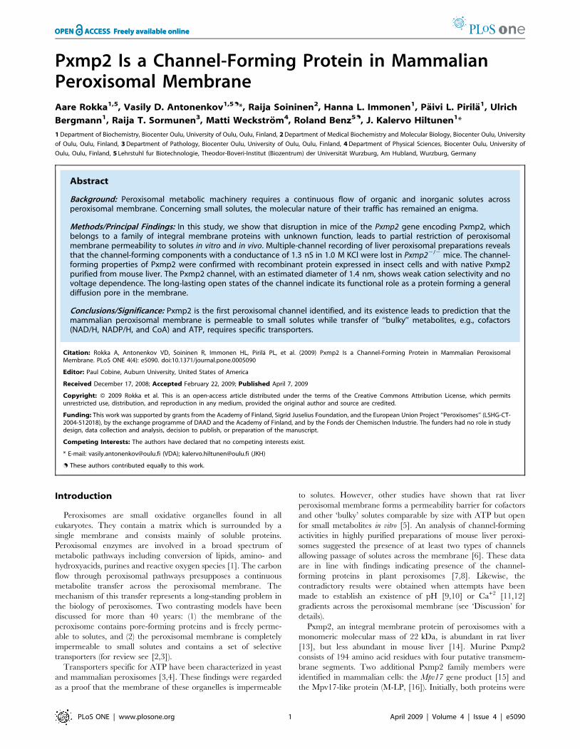
Pxmp2 Is a Channel-Forming Protein in MammalianPeroxisomal MembraneAare Rokka1,5, Vasily D. Antonenkov1,5.*, Raija Soininen2, Hanna L. Immonen1, Paivi L. Pirila1, Ulrich
Bergmann1, Raija T. Sormunen3, Matti Weckstrom4, Roland Benz5., J. Kalervo Hiltunen1*
1 Department of Biochemistry, Biocenter Oulu, University of Oulu, Oulu, Finland, 2 Department of Medical Biochemistry and Molecular Biology, Biocenter Oulu, University
of Oulu, Oulu, Finland, 3 Department of Pathology, Biocenter Oulu, University of Oulu, Oulu, Finland, 4 Department of Physical Sciences, Biocenter Oulu, University of
Oulu, Oulu, Finland, 5 Lehrstuhl fur Biotechnologie, Theodor-Boveri-Institut (Biozentrum) der Universitat Wurzburg, Am Hubland, Wurzburg, Germany
Abstract
Background: Peroxisomal metabolic machinery requires a continuous flow of organic and inorganic solutes acrossperoxisomal membrane. Concerning small solutes, the molecular nature of their traffic has remained an enigma.
Methods/Principal Findings: In this study, we show that disruption in mice of the Pxmp2 gene encoding Pxmp2, whichbelongs to a family of integral membrane proteins with unknown function, leads to partial restriction of peroxisomalmembrane permeability to solutes in vitro and in vivo. Multiple-channel recording of liver peroxisomal preparations revealsthat the channel-forming components with a conductance of 1.3 nS in 1.0 M KCl were lost in Pxmp22/2 mice. The channel-forming properties of Pxmp2 were confirmed with recombinant protein expressed in insect cells and with native Pxmp2purified from mouse liver. The Pxmp2 channel, with an estimated diameter of 1.4 nm, shows weak cation selectivity and novoltage dependence. The long-lasting open states of the channel indicate its functional role as a protein forming a generaldiffusion pore in the membrane.
Conclusions/Significance: Pxmp2 is the first peroxisomal channel identified, and its existence leads to prediction that themammalian peroxisomal membrane is permeable to small solutes while transfer of ‘‘bulky’’ metabolites, e.g., cofactors(NAD/H, NADP/H, and CoA) and ATP, requires specific transporters.
Citation: Rokka A, Antonenkov VD, Soininen R, Immonen HL, Pirila PL, et al. (2009) Pxmp2 Is a Channel-Forming Protein in Mammalian PeroxisomalMembrane. PLoS ONE 4(4): e5090. doi:10.1371/journal.pone.0005090
Editor: Paul Cobine, Auburn University, United States of America
Received December 17, 2008; Accepted February 22, 2009; Published April 7, 2009
Copyright: � 2009 Rokka et al. This is an open-access article distributed under the terms of the Creative Commons Attribution License, which permitsunrestricted use, distribution, and reproduction in any medium, provided the original author and source are credited.
Funding: This work was supported by grants from the Academy of Finland, Sigrid Juselius Foundation, and the European Union Project ‘‘Peroxisomes’’ (LSHG-CT-2004-512018), by the exchange programme of DAAD and the Academy of Finland, and by the Fonds der Chemischen Industrie. The funders had no role in studydesign, data collection and analysis, decision to publish, or preparation of the manuscript.
Competing Interests: The authors have declared that no competing interests exist.
* E-mail: [email protected] (VDA); [email protected] (JKH)
. These authors contributed equally to this work.
Introduction
Peroxisomes are small oxidative organelles found in all
eukaryotes. They contain a matrix which is surrounded by a
single membrane and consists mainly of soluble proteins.
Peroxisomal enzymes are involved in a broad spectrum of
metabolic pathways including conversion of lipids, amino- and
hydroxyacids, purines and reactive oxygen species [1]. The carbon
flow through peroxisomal pathways presupposes a continuous
metabolite transfer across the peroxisomal membrane. The
mechanism of this transfer represents a long-standing problem in
the biology of peroxisomes. Two contrasting models have been
discussed for more than 40 years: (1) the membrane of the
peroxisome contains pore-forming proteins and is freely perme-
able to solutes, and (2) the peroxisomal membrane is completely
impermeable to small solutes and contains a set of selective
transporters (for review see [2,3]).
Transporters specific for ATP have been characterized in yeast
and mammalian peroxisomes [3,4]. These findings were regarded
as a proof that the membrane of these organelles is impermeable
to solutes. However, other studies have shown that rat liver
peroxisomal membrane forms a permeability barrier for cofactors
and other ‘bulky’ solutes comparable by size with ATP but open
for small metabolites in vitro [5]. An analysis of channel-forming
activities in highly purified preparations of mouse liver peroxi-
somes suggested the presence of at least two types of channels
allowing passage of solutes across the membrane [6]. These data
are in line with findings indicating presence of the channel-
forming proteins in plant peroxisomes [7,8]. Likewise, the
contradictory results were obtained when attempts have been
made to establish an existence of pH [9,10] or Ca+2 [11,12]
gradients across the peroxisomal membrane (see ‘Discussion’ for
details).
Pxmp2, an integral membrane protein of peroxisomes with a
monomeric molecular mass of 22 kDa, is abundant in rat liver
[13], but less abundant in mouse liver [14]. Murine Pxmp2
consists of 194 amino acid residues with four putative transmem-
brane segments. Two additional Pxmp2 family members were
identified in mammalian cells: the Mpv17 gene product [15] and
the Mpv17-like protein (M-LP, [16]). Initially, both proteins were
PLoS ONE | www.plosone.org 1 April 2009 | Volume 4 | Issue 4 | e5090
localized to peroxisomal membrane [15,16]. However, the
localization of Mpv17 to peroxisomes has recently been challenged
since the mammalian protein [17] and its yeast homolog Sym1p
[18] were detectable in the inner mitochondrial membrane.
Pxmp2 was speculated previously as to having a role in the
transmembrane transport of solutes by acting as a nonselective
pore-forming protein [19]. This assumption was based on
experiments showing that a protein fraction containing Pxmp2,
PMP28, and some other peroxisomal membrane proteins from rat
liver was able to promote leakage of small molecules such as
sucrose from liposomes preloaded with these solutes. However,
protein data-based analysis revealed that the Pxmp2 family
members share no sequence or structural similarities with known
porin proteins or other channels. Moreover, the presence of any
type of pore-forming proteins in mammalian and yeast peroxi-
somes and their participation in the transfer of solutes across the
membrane is widely challenged (for review see [2,3]).
The present work addresses the molecular mechanism of
transferring solutes across the peroxisomal membrane and the
physiological role of the peroxisomal membrane protein Pxmp2.
The data revealed that Pxmp2 is a channel-forming protein that
functions as a size-selective filter with an exclusion limit of
approximately 0.6 kDa for hydrophilic solutes.
Results
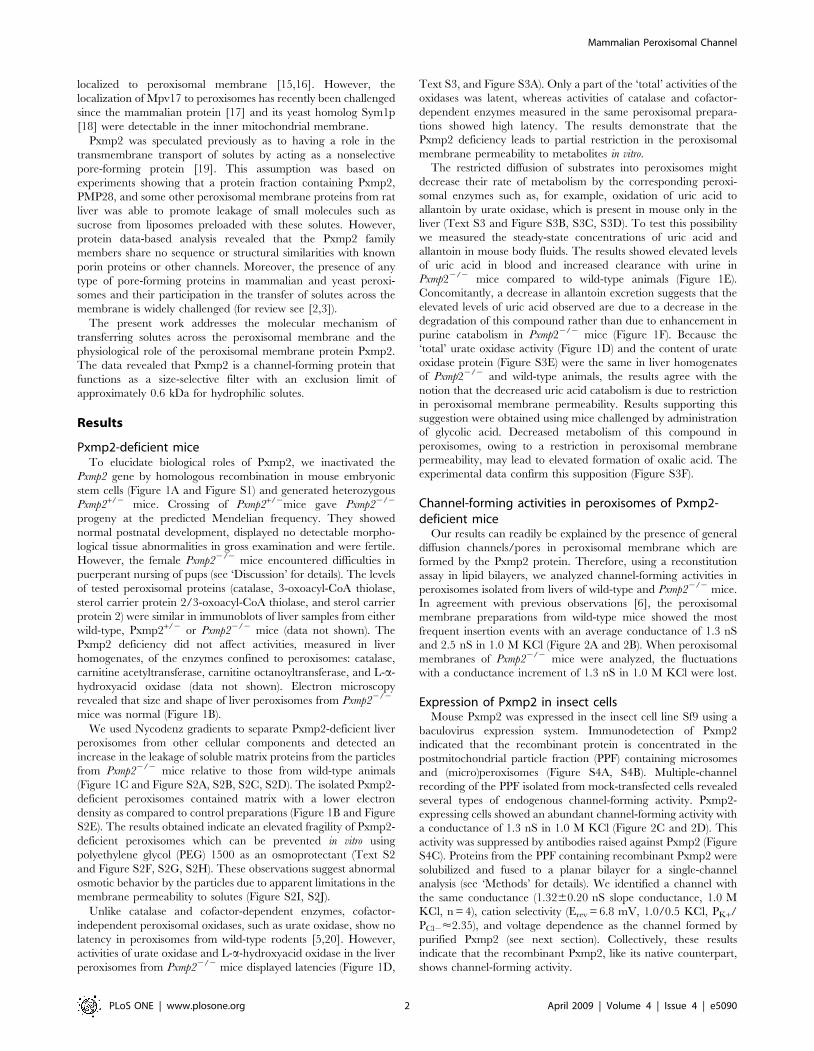
Pxmp2-deficient miceTo elucidate biological roles of Pxmp2, we inactivated the
Pxmp2 gene by homologous recombination in mouse embryonic
stem cells (Figure 1A and Figure S1) and generated heterozygous
Pxmp2+/2 mice. Crossing of Pxmp2+/2mice gave Pxmp22/2
progeny at the predicted Mendelian frequency. They showed
normal postnatal development, displayed no detectable morpho-
logical tissue abnormalities in gross examination and were fertile.
However, the female Pxmp22/2 mice encountered difficulties in
puerperant nursing of pups (see ‘Discussion’ for details). The levels
of tested peroxisomal proteins (catalase, 3-oxoacyl-CoA thiolase,
sterol carrier protein 2/3-oxoacyl-CoA thiolase, and sterol carrier
protein 2) were similar in immunoblots of liver samples from either
wild-type, Pxmp2+/2 or Pxmp22/2 mice (data not shown). The
Pxmp2 deficiency did not affect activities, measured in liver
homogenates, of the enzymes confined to peroxisomes: catalase,
carnitine acetyltransferase, carnitine octanoyltransferase, and L-a-
hydroxyacid oxidase (data not shown). Electron microscopy
revealed that size and shape of liver peroxisomes from Pxmp22/2
mice was normal (Figure 1B).
We used Nycodenz gradients to separate Pxmp2-deficient liver
peroxisomes from other cellular components and detected an
increase in the leakage of soluble matrix proteins from the particles
from Pxmp22/2 mice relative to those from wild-type animals
(Figure 1C and Figure S2A, S2B, S2C, S2D). The isolated Pxmp2-
deficient peroxisomes contained matrix with a lower electron
density as compared to control preparations (Figure 1B and Figure
S2E). The results obtained indicate an elevated fragility of Pxmp2-
deficient peroxisomes which can be prevented in vitro using
polyethylene glycol (PEG) 1500 as an osmoprotectant (Text S2
and Figure S2F, S2G, S2H). These observations suggest abnormal
osmotic behavior by the particles due to apparent limitations in the
membrane permeability to solutes (Figure S2I, S2J).
Unlike catalase and cofactor-dependent enzymes, cofactor-
independent peroxisomal oxidases, such as urate oxidase, show no
latency in peroxisomes from wild-type rodents [5,20]. However,
activities of urate oxidase and L-a-hydroxyacid oxidase in the liver
peroxisomes from Pxmp22/2 mice displayed latencies (Figure 1D,
Text S3, and Figure S3A). Only a part of the ‘total’ activities of the
oxidases was latent, whereas activities of catalase and cofactor-
dependent enzymes measured in the same peroxisomal prepara-
tions showed high latency. The results demonstrate that the
Pxmp2 deficiency leads to partial restriction in the peroxisomal
membrane permeability to metabolites in vitro.
The restricted diffusion of substrates into peroxisomes might
decrease their rate of metabolism by the corresponding peroxi-
somal enzymes such as, for example, oxidation of uric acid to
allantoin by urate oxidase, which is present in mouse only in the
liver (Text S3 and Figure S3B, S3C, S3D). To test this possibility
we measured the steady-state concentrations of uric acid and
allantoin in mouse body fluids. The results showed elevated levels
of uric acid in blood and increased clearance with urine in
Pxmp22/2 mice compared to wild-type animals (Figure 1E).
Concomitantly, a decrease in allantoin excretion suggests that the
elevated levels of uric acid observed are due to a decrease in the
degradation of this compound rather than due to enhancement in
purine catabolism in Pxmp22/2 mice (Figure 1F). Because the
‘total’ urate oxidase activity (Figure 1D) and the content of urate
oxidase protein (Figure S3E) were the same in liver homogenates
of Pxmp22/2 and wild-type animals, the results agree with the
notion that the decreased uric acid catabolism is due to restriction
in peroxisomal membrane permeability. Results supporting this
suggestion were obtained using mice challenged by administration
of glycolic acid. Decreased metabolism of this compound in
peroxisomes, owing to a restriction in peroxisomal membrane
permeability, may lead to elevated formation of oxalic acid. The
experimental data confirm this supposition (Figure S3F).
Channel-forming activities in peroxisomes of Pxmp2-deficient mice
Our results can readily be explained by the presence of general
diffusion channels/pores in peroxisomal membrane which are
formed by the Pxmp2 protein. Therefore, using a reconstitution
assay in lipid bilayers, we analyzed channel-forming activities in
peroxisomes isolated from livers of wild-type and Pxmp22/2 mice.
In agreement with previous observations [6], the peroxisomal
membrane preparations from wild-type mice showed the most
frequent insertion events with an average conductance of 1.3 nS
and 2.5 nS in 1.0 M KCl (Figure 2A and 2B). When peroxisomal
membranes of Pxmp22/2 mice were analyzed, the fluctuations
with a conductance increment of 1.3 nS in 1.0 M KCl were lost.
Expression of Pxmp2 in insect cellsMouse Pxmp2 was expressed in the insect cell line Sf9 using a
baculovirus expression system. Immunodetection of Pxmp2
indicated that the recombinant protein is concentrated in the
postmitochondrial particle fraction (PPF) containing microsomes
and (micro)peroxisomes (Figure S4A, S4B). Multiple-channel
recording of the PPF isolated from mock-transfected cells revealed
several types of endogenous channel-forming activity. Pxmp2-
expressing cells showed an abundant channel-forming activity with
a conductance of 1.3 nS in 1.0 M KCl (Figure 2C and 2D). This
activity was suppressed by antibodies raised against Pxmp2 (Figure
S4C). Proteins from the PPF containing recombinant Pxmp2 were
solubilized and fused to a planar bilayer for a single-channel
analysis (see ‘Methods’ for details). We identified a channel with
the same conductance (1.3260.20 nS slope conductance, 1.0 M
KCl, n = 4), cation selectivity (Erev = 6.8 mV, 1.0/0.5 KCl, PK+/
PCl2<2.35), and voltage dependence as the channel formed by
purified Pxmp2 (see next section). Collectively, these results
indicate that the recombinant Pxmp2, like its native counterpart,
shows channel-forming activity.
Mammalian Peroxisomal Channel
PLoS ONE | www.plosone.org 2 April 2009 | Volume 4 | Issue 4 | e5090
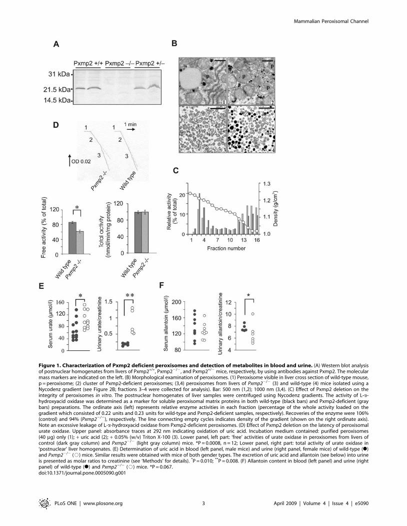
Figure 1. Characterization of Pxmp2 deficient peroxisomes and detection of metabolites in blood and urine. (A) Western blot analysisof postnuclear homogenates from livers of Pxmp2+/+, Pxmp22/2, and Pxmp2+/2 mice, respectively, by using antibodies against Pxmp2. The molecularmass markers are indicated on the left. (B) Morphological examination of peroxisomes. (1) Peroxisome visible in liver cross section of wild-type mouse,p = peroxisome; (2) cluster of Pxmp2-deficient peroxisomes; (3,4) peroxisomes from livers of Pxmp22/2 (3) and wild-type (4) mice isolated using aNycodenz gradient (see Figure 2B; fractions 3–4 were collected for analysis). Bar: 500 nm (1,2); 1000 nm (3,4). (C) Effect of Pxmp2 deletion on theintegrity of peroxisomes in vitro. The postnuclear homogenates of liver samples were centrifuged using Nycodenz gradients. The activity of L-a-hydroxyacid oxidase was determined as a marker for soluble peroxisomal matrix proteins in both wild-type (black bars) and Pxmp2-deficient (graybars) preparations. The ordinate axis (left) represents relative enzyme activities in each fraction (percentage of the whole activity loaded on thegradient which consisted of 0.22 units and 0.23 units for wild-type and Pxmp2-deficient samples, respectively). Recoveries of the enzyme were 106%(control) and 94% (Pxmp22/2), respectively. The line connecting empty cycles indicates density of the gradient (shown on the right ordinate axis).Note an excessive leakage of L-a-hydroxyacid oxidase from Pxmp2-deficient peroxisomes. (D) Effect of Pxmp2 deletion on the latency of peroxisomalurate oxidase. Upper panel: absorbance traces at 292 nm indicating oxidation of uric acid. Incubation medium contained: purified peroxisomes(40 mg) only (1); + uric acid (2); + 0.05% (w/v) Triton X-100 (3). Lower panel, left part: ‘free’ activities of urate oxidase in peroxisomes from livers ofcontrol (dark gray column) and Pxmp22/2 (light gray column) mice. *P = 0.0008, n = 12; Lower panel, right part: total activity of urate oxidase in‘postnuclear’ liver homogenates. (E) Determination of uric acid in blood (left panel, male mice) and urine (right panel, female mice) of wild-type (N)and Pxmp22/2 (#) mice. Similar results were obtained with mice of both gender types. The excretion of uric acid and allantoin (see below) into urineis presented as molar ratios to creatinine (see ‘Methods’ for details). *P = 0.010; **P = 0.008. (F) Allantoin content in blood (left panel) and urine (rightpanel) of wild-type (N) and Pxmp22/2 (#) mice. *P = 0.067.doi:10.1371/journal.pone.0005090.g001
Mammalian Peroxisomal Channel
PLoS ONE | www.plosone.org 3 April 2009 | Volume 4 | Issue 4 | e5090
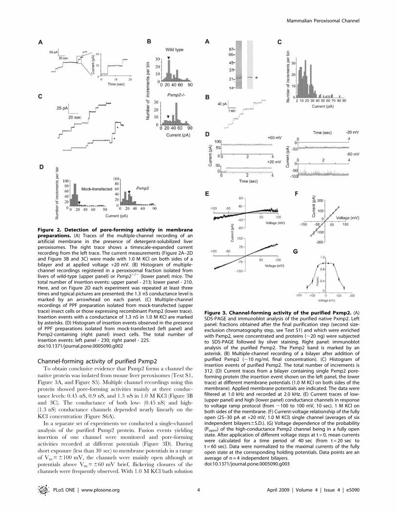
Channel-forming activity of purified Pxmp2To obtain conclusive evidence that Pxmp2 forms a channel the
native protein was isolated from mouse liver peroxisomes (Text S1,
Figure 3A, and Figure S5). Multiple channel recordings using this
protein showed pore-forming activities mainly at three conduc-
tance levels: 0.45 nS, 0.9 nS, and 1.3 nS in 1.0 M KCl (Figure 3B
and 3C). The conductance of both low- (0.45 nS) and high-
(1.3 nS) conductance channels depended nearly linearly on the
KCl concentration (Figure S6A).
In a separate set of experiments we conducted a single-channel
analysis of the purified Pxmp2 protein. Fusion events yielding
insertion of one channel were monitored and pore-forming
activities recorded at different potentials (Figure 3D). During
short exposure (less than 30 sec) to membrane potentials in a range
of Vm = 6100 mV, the channels were mainly open although at
potentials above Vm = 660 mV brief, flickering closures of the
channels were frequently observed. With 1.0 M KCl bath solution
Figure 2. Detection of pore-forming activity in membranepreparations. (A) Traces of the multiple-channel recording of anartificial membrane in the presence of detergent-solubilized liverperoxisomes. The right trace shows a timescale-expanded currentrecording from the left trace. The current measurements (Figure 2A–2Dand Figure 3B and 3C) were made with 1.0 M KCl on both sides of abilayer and at applied voltage +20 mV. (B) Histogram of multiple-channel recordings registered in a peroxisomal fraction isolated fromlivers of wild-type (upper panel) or Pxmp22/2 (lower panel) mice. Thetotal number of insertion events: upper panel - 213; lower panel - 210.Here, and on Figure 2D each experiment was repeated at least threetimes and typical pictures are presented; the 1.3 nS conductance level ismarked by an arrowhead on each panel. (C) Multiple-channelrecordings of PPF preparation isolated from mock-transfected (uppertrace) insect cells or those expressing recombinant Pxmp2 (lower trace).Insertion events with a conductance of 1.3 nS in 1.0 M KCl are markedby asterisks. (D) Histogram of insertion events observed in the presenceof PPF preparations isolated from mock-transfected (left panel) andPxmp2-containing (right panel) insect cells. The total number ofinsertion events: left panel - 230; right panel - 225.doi:10.1371/journal.pone.0005090.g002
Figure 3. Channel-forming activity of the purified Pxmp2. (A)SDS-PAGE and immunoblot analysis of the purified native Pxmp2. Leftpanel: fractions obtained after the final purification step (second size-exclusion chromatography step, see Text S1) and which were enrichedwith Pxmp2, were concentrated and proteins (,20 ng) were subjectedto SDS-PAGE followed by silver staining. Right panel: immunoblotanalysis of the purified Pxmp2. The Pxmp2 band is marked by anasterisk. (B) Multiple-channel recording of a bilayer after addition ofpurified Pxmp2 (,10 ng/ml, final concentration). (C) Histogram ofinsertion events of purified Pxmp2. The total number of increments is312. (D) Current traces from a bilayer containing single Pxmp2 pore-forming protein (the insertion event shown on the left panel, the lowertrace) at different membrane potentials (1.0 M KCl on both sides of themembrane). Applied membrane potentials are indicated. The data werefiltered at 1.0 kHz and recorded at 2.0 kHz. (E) Current traces of low-(upper panel) and high (lower panel) conductance channels in responseto voltage ramp protocol (from 2100 to 100 mV, 10 sec). 1 M KCl onboth sides of the membrane. (F) Current-voltage relationship of the fullyopen (25–30 pA at +20 mV, 1.0 M KCl) single channel (averages of sixindependent bilayers6S.D.). (G) Voltage dependence of the probability(Popen) of the high-conductance Pxmp2 channel being in a fully openstate. After application of different voltage steps at t = 0, mean currentswere calculated for a time period of 40 sec (from t = 20 sec tot = 60 sec). Data were normalized to the maximal currents of the fullyopen state at the corresponding holding potentials. Data points are anaverage of n = 4 independent bilayers.doi:10.1371/journal.pone.0005090.g003
Mammalian Peroxisomal Channel
PLoS ONE | www.plosone.org 4 April 2009 | Volume 4 | Issue 4 | e5090
on both sides of the membrane, the low- and high-conductance
channels showed a near linear current-voltage relationship in
response to a rapidly increasing voltage ramp (Figure 3E). The
calculated slope conductance of the fully open channel was
1.3460.16 nS (Figure 3F). Figure 3G shows the voltage depen-
dence of the high-conductance channel open probability during
prolonged application of a constant voltage. In a range of
membrane potentials Vm = 660 mM the channel was near
completely open, whereas it stepwise closed at more positive or
negative membrane potentials with transition to intermediate- and
low-conductance states (see below). Thus, the Pxmp2 channel
closes slowly during prolonged exposure to elevated membrane
potentials. In asymmetric bath solutions (1.0 M KCl/0.5 M KCl)
the reversal potential was Erev = +6.5 mV for the high-conduc-
tance single channel (Figure S6B, S6C). Accordingly, the channel
is moderately cation-selective (PK+/PCl2 = 2.3). A very similar
value (PK+/PCl2 = 2.4) was detected when the measurements were
made on a low-conductance channel (0.45 nS in 1.0 M KCl, data
not shown). Addition of antibodies against Pxmp2 into the bath
solutions during current recordings of a single fully open channel
led to its closure (Figure 4A).
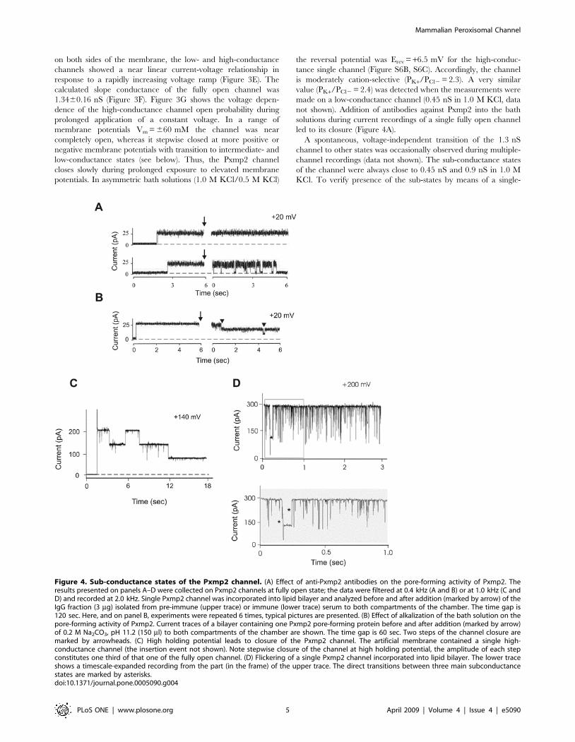
A spontaneous, voltage-independent transition of the 1.3 nS
channel to other states was occasionally observed during multiple-
channel recordings (data not shown). The sub-conductance states
of the channel were always close to 0.45 nS and 0.9 nS in 1.0 M
KCl. To verify presence of the sub-states by means of a single-
Figure 4. Sub-conductance states of the Pxmp2 channel. (A) Effect of anti-Pxmp2 antibodies on the pore-forming activity of Pxmp2. Theresults presented on panels A–D were collected on Pxmp2 channels at fully open state; the data were filtered at 0.4 kHz (A and B) or at 1.0 kHz (C andD) and recorded at 2.0 kHz. Single Pxmp2 channel was incorporated into lipid bilayer and analyzed before and after addition (marked by arrow) of theIgG fraction (3 mg) isolated from pre-immune (upper trace) or immune (lower trace) serum to both compartments of the chamber. The time gap is120 sec. Here, and on panel B, experiments were repeated 6 times, typical pictures are presented. (B) Effect of alkalization of the bath solution on thepore-forming activity of Pxmp2. Current traces of a bilayer containing one Pxmp2 pore-forming protein before and after addition (marked by arrow)of 0.2 M Na2CO3, pH 11.2 (150 ml) to both compartments of the chamber are shown. The time gap is 60 sec. Two steps of the channel closure aremarked by arrowheads. (C) High holding potential leads to closure of the Pxmp2 channel. The artificial membrane contained a single high-conductance channel (the insertion event not shown). Note stepwise closure of the channel at high holding potential, the amplitude of each stepconstitutes one third of that one of the fully open channel. (D) Flickering of a single Pxmp2 channel incorporated into lipid bilayer. The lower traceshows a timescale-expanded recording from the part (in the frame) of the upper trace. The direct transitions between three main subconductancestates are marked by asterisks.doi:10.1371/journal.pone.0005090.g004
Mammalian Peroxisomal Channel
PLoS ONE | www.plosone.org 5 April 2009 | Volume 4 | Issue 4 | e5090

channel analysis we treated the high conductance channel inserted
in the bilayer at alkaline pH and observed a stepwise closure of the
channel with each step showing a conductance of 0.45 nS in
1.0 M KCl (Figure 4B). Likewise, a stepwise closure of the high-
conductance channel at holding potentials Vm = 6100 mV or
higher with two sub-conductance levels was observed (Figure 4C).
Similarly, short exposure to high potentials (Vm$150 mV) led to a
frequent appearance of the high-conductance channels (1.3 nS in
1.0 M KCl) that showed flickering closure sometimes with evident
sub-conductance levels of 0.9 nS and 0.45 nS in 1.0 M KCl,
respectively (Figure 4D). The transition of the high-conductance
channel into sub-states can be interpreted in terms of a cluster of
three small channels, each of them with a conductance around
0.45 nS in 1.0 M KCl (see ‘Discussion’ for more details).
Permeability properties of the Pxmp2 channelWe performed additional reconstitution assays in lipid bilayers
in order to obtain information on the size of the channels formed
by Pxmp2 (Text S1 and Figure S7A, S7B, S7C). From these
experiments we concluded that the radius of the narrowest space
of the channel (channel friction) is about 0.7 nm. Such a channel
allows nearly free diffusion of ions and non-electrolytes with
molecular masses of up to 200–300 Da across the membrane. The
movement of larger molecules, from 300 Da to 600 Da in size, is
limited, while more bulky solutes are unable to permeate the
membrane through the channel.
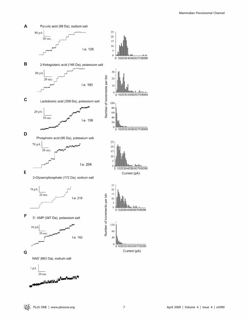
To further analyze the function of the Pxmp2 channel we made
direct measurements of the pore-forming activity of purified
protein using various organic anions as electrolytes (Figure 5A–
5G). A variety of small mono- and divalent anions known to be
peroxisomal metabolites, such as glycolate, pyruvate, 2-ketogluta-
rate, and others, can be transferred through the Pxmp2 channel.
As expected, if the size of the anion is over 300 Da, e.g, lactobionic
acid (358 Da) or AMP (347 Da), the single-channel conductance
of Pxmp2 is significantly decreased, indicating that the movement
of these compounds inside the channel is partially restricted. The
only traces of channel-forming activity with a conductance well
below 20 pS were detected in the presence of NAD (663 Da). No
channel-forming activities were observed with ATP (507 Da),
probably due to the high net negative charge of this molecule,
which prevents diffusion of ATP through the cation-selective
Pxmp2 channel.
Discussion
In our study we tried to resolve two important interrelated
problems in the physiology of mammalian peroxisomes: (i) the
molecular foundation for the permeability of the peroxisomal
membrane to solutes, and (ii) the functional role of Pxmp2, which
belongs to a membrane protein family with a previously unknown
function.
Pxmp2 forms a channel in peroxisomal membraneAccording to our recent observation [6], the putative peroxi-
somal channels show single channel conductance of 1.3 nS and
2.5 nS in 1.0 M KCl, respectively, when crude peroxisomal
membrane preparations from mouse liver were assayed in
reconstitution experiments using lipid bilayers. The present work
demonstrates that Pxmp2 is responsible for one of the described
channel-forming activities, namely for the activity with a
conductance of 1.3 nS in 1.0 M KCl. The evidence that Pxmp2
forms a channel is at least five-fold: (i) knocking out of Pxmp2 leads
to partial restriction of peroxisomal membrane permeability to
small solutes in vitro and in vivo; (ii) the pore-forming activity with a
characteristic conductance of 1.3 nS in 1.0 M KCl was not
observed in the peroxisomal membrane preparations isolated from
the livers of Pxmp2- deficient mice; (iii) expression of recombinant
Pxmp2 in insect cells resulted in the appearance of a pore-forming
activity with a conductance of 1.3 nS in 1.0 M KCl; (iv) this
activity was inhibited following treatment of solubilized membrane
proteins with antibodies generated against Pxmp2; and (v) isolated
Pxmp2 showed pore-forming activity with three conductance
levels, the highest one being 1.3–1.4 nS in 1.0 M KCl.
Does peroxisomal membrane open to small solutes?The controversy of the area (see Introduction) is exemplified by
two recent studies of the apparent role of mammalian peroxisomes
in Ca homeostasis published ‘back-to-back’ in the same journal
[11,12]. The data concerning pH gradients across peroxisomal
membrane are even more confusing. For instance, in one report
the authors made a conclusion that the matrix of mammalian
peroxisomes is basic [9]. However, another study revealed that
mammalian peroxisomes have no cross-membrane pH gradient at
all [10]. The results obtained on yeast peroxisomes are also
contradicting: some reports claim basic pH in the particles [21]
while other publications indicate acid pH in the same organelles
[22].
One explanation for difference in the results described above is
the possible existence of Donnan equilibrium between peroxisomal
matrix and cytoplasm surrounding the particles. In this case the
formation of pH or Ca gradients does not require membrane
impermeable to small ions. Instead, the gradients are formed
across the membrane permeable to solutes by difference in overall
charges of molecules unable penetrate this membrane (e.g.,
proteins) which are localized inside and outside the particles
[23]. For example, if the overall charge of proteins inside
peroxisomes is more positive than outside the particles, these
proteins should attract small negatively charged solutes, including
hydroxyl ions, to preserve electroneutrality. As a result, the pH
gradient is formed, where pH inside peroxisomes is more basic
than outside the particles. The mechanism of Donnan equilibrium
depends on free permeation of small charged solutes, including
protons and hydroxyl ions, across the membrane [23]. The
Donnan-type equilibrium may be responsible for creation of a pH
gradient across outer mitochondrial membrane [24] and involved
in the maintenance of an acid pH in lysosomes [25].
An apparent role of Donnan equilibrium in creation of ion
gradients across peroxisomal membrane open to small solutes is
consistent with numerous observations collected within last 40
years by different groups showing that mammalian peroxisomal
membrane does not form a barrier to these solutes in vitro (see, e.g.,
ref. [5,19,20,26]). Our data obtained on peroxisomes isolated from
livers of Pxmp2-deficient mice are in line with this conclusion.
Moreover, these data revealed direct involvement of the Pxmp2
channel in the transfer of solutes across the membrane. Likewise,
the results of our study of peroxisomal metabolites (urate, oxalate)
in body fluids of Pxmp2-deficient mice corroborate in vitro findings.
Apparent functions of the Pxmp2 channelOur results predict that the Pxmp2 protein forms a relatively
wide, water-filled channel in which the mobility of small solutes is
determined by their diffusion coefficients. This implies that the
Pxmp2 channel is non-selective with respect to the chemical
nature of solutes, whereas it is highly selective relative to the size of
solutes. In addition, because of its very long open states the Pxmp2
behaves like a pore similar to porins of outer mitochondrial
membrane or outer membrane of gram-negative bacteria. These
features of the channel apparently determine the unusual
Mammalian Peroxisomal Channel
PLoS ONE | www.plosone.org 6 April 2009 | Volume 4 | Issue 4 | e5090
Mammalian Peroxisomal Channel
PLoS ONE | www.plosone.org 7 April 2009 | Volume 4 | Issue 4 | e5090
permeability properties of peroxisomal membrane, resulting in a
novel type of biomembrane exploiting both: pore-forming proteins
as well as solute transporters (e.g., ATP transporter [4]) to transfer
metabolites in and out of peroxisomes. The present results
combined with our previous observations [5] point to the ability
of peroxisomal membrane to discriminate between small metab-
olites with sizes typically below 200 Da and ‘bulky’ solutes
including ATP and cofactors (NAD/H, NADP/H, CoA and its
acylated derivatives) (Figure S7D). It appears that the large
molecular size of cofactors and some other solutes is an important
factor determining their subcellular localization.
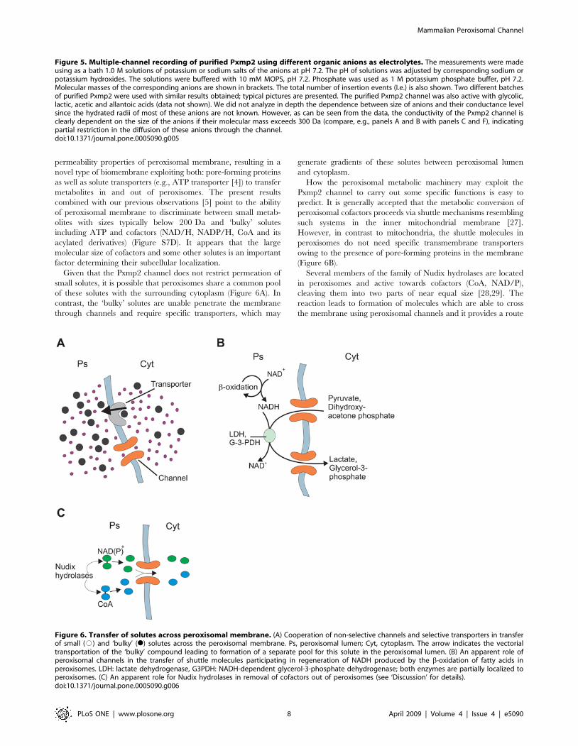
Given that the Pxmp2 channel does not restrict permeation of
small solutes, it is possible that peroxisomes share a common pool
of these solutes with the surrounding cytoplasm (Figure 6A). In
contrast, the ‘bulky’ solutes are unable penetrate the membrane
through channels and require specific transporters, which may
generate gradients of these solutes between peroxisomal lumen
and cytoplasm.
How the peroxisomal metabolic machinery may exploit the
Pxmp2 channel to carry out some specific functions is easy to
predict. It is generally accepted that the metabolic conversion of
peroxisomal cofactors proceeds via shuttle mechanisms resembling
such systems in the inner mitochondrial membrane [27].
However, in contrast to mitochondria, the shuttle molecules in
peroxisomes do not need specific transmembrane transporters
owing to the presence of pore-forming proteins in the membrane
(Figure 6B).
Several members of the family of Nudix hydrolases are located
in peroxisomes and active towards cofactors (CoA, NAD/P),
cleaving them into two parts of near equal size [28,29]. The
reaction leads to formation of molecules which are able to cross
the membrane using peroxisomal channels and it provides a route
Figure 6. Transfer of solutes across peroxisomal membrane. (A) Cooperation of non-selective channels and selective transporters in transferof small (#) and ‘bulky’ (N) solutes across the peroxisomal membrane. Ps, peroxisomal lumen; Cyt, cytoplasm. The arrow indicates the vectorialtransportation of the ‘bulky’ compound leading to formation of a separate pool for this solute in the peroxisomal lumen. (B) An apparent role ofperoxisomal channels in the transfer of shuttle molecules participating in regeneration of NADH produced by the b-oxidation of fatty acids inperoxisomes. LDH: lactate dehydrogenase, G3PDH: NADH-dependent glycerol-3-phosphate dehydrogenase; both enzymes are partially localized toperoxisomes. (C) An apparent role for Nudix hydrolases in removal of cofactors out of peroxisomes (see ‘Discussion’ for details).doi:10.1371/journal.pone.0005090.g006
Figure 5. Multiple-channel recording of purified Pxmp2 using different organic anions as electrolytes. The measurements were madeusing as a bath 1.0 M solutions of potassium or sodium salts of the anions at pH 7.2. The pH of solutions was adjusted by corresponding sodium orpotassium hydroxides. The solutions were buffered with 10 mM MOPS, pH 7.2. Phosphate was used as 1 M potassium phosphate buffer, pH 7.2.Molecular masses of the corresponding anions are shown in brackets. The total number of insertion events (I.e.) is also shown. Two different batchesof purified Pxmp2 were used with similar results obtained; typical pictures are presented. The purified Pxmp2 channel was also active with glycolic,lactic, acetic and allantoic acids (data not shown). We did not analyze in depth the dependence between size of anions and their conductance levelsince the hydrated radii of most of these anions are not known. However, as can be seen from the data, the conductivity of the Pxmp2 channel isclearly dependent on the size of the anions if their molecular mass exceeds 300 Da (compare, e.g., panels A and B with panels C and F), indicatingpartial restriction in the diffusion of these anions through the channel.doi:10.1371/journal.pone.0005090.g005
Mammalian Peroxisomal Channel
PLoS ONE | www.plosone.org 8 April 2009 | Volume 4 | Issue 4 | e5090

for the removal of cofactors from peroxisomes (Figure 6C). Thus,
cleavage of NAD+ (663 Da) by the corresponding peroxisomal
Nudix hydrolase NUDT12 produces NMNH (334 Da) and AMP
(347 Da). Permeation of NAD+ through the Pxmp2 channel is
negligible (see Figure 5G). However, the channel is permeable to
AMP and apparently also to NMNH based on the similar size of
these molecules (NMNH has no net charge, preventing use of this
compound for multiple-channel recording).
Pxmp2-deficient miceThe Pxmp2 knock-out mouse model allowed us to collect
additional evidence for the presence of at least two types of
channels in mammalian peroxisomes: (i) the Pxmp2 deficiency led
to only partial restriction in peroxisomal membrane permeability
to solutes in vitro; (ii) the increase in the content of uric acid in the
serum and urine of Pxmp22/2 mice relative to wild-type control,
being 1.4-fold and 3.1-fold, respectively (see Figure 1E), was much
lower than that seen for urate oxidase-deficient mice, where the
corresponding levels of uric acid were 10-fold and 9-fold higher
than in the wild-type animals [30]. The last observation indicates
that in spite of the absence of Pxmp2, a significant amount of uric
acid is still degraded in peroxisomes (as also indicated by excretion
of allantoic acid into urine) and suggests the existence of a second
peroxisomal transmembrane route for this metabolite; (iii)
solubilized membrane preparations from Pxmp2-deficient perox-
isomes still displayed the pore-forming activity with a wide range
of peroxisomal metabolites as electrolytes (data not shown).
The mild phenotype of Pxmp2-deficient mice may reflect
redundancy of the peroxisomal channels in their ability to transfer
solutes across the membrane. The redundancy of the function of
mammalian peroxisomal proteins, at least at normal physiological
conditions, is a well known phenomenon. The examples of poor
phenotype of the mouse models deficient in certain peroxisomal
protein are numerous and include multifunctional enzyme type 1
[31], racemase [32], liver isoform of fatty acid binding protein
[33], and peroxisomal membrane ABC transporters (reviewed in
[34]). The functional redundancy might be limited or even
abolished by harsh environmental conditions. To examine this
possibility we challenged Pxmp22/2 mice with diets containing
clofibrate (a known proliferator of mouse liver peroxisomes) or
phytol (methyl-branched fatty alcohol metabolized via a-oxidation
only in peroxisomes). These treatments did not trigger develop-
ment of phenotypes in Pxmp2-deficient mice different from those
of wild-type animals (data not shown). However, our in depth
analysis of the effect of Pxmp2 deletion on female mice reveals
some unexpected results. In addition to disturbances in uric and
oxalic acids metabolism (see above), these mice were unable to
nurse their pups due to low production of milk. Pxmp22/2 female
mice showed retarded growth of mammary glands and limited
abnormalities in reproductive organs during pregnancy (Rokka A.
and Vapola M, unpublished results). It’s not yet clear how Pxmp2
deficiency affects mechanisms responsible for normal development
of mammary glands. This specific problem is under of our current
investigation.
Cluster organization of the Pxmp2 channelTo describe the nature of the sub-conductance levels of the
isolated Pxmp2 channel, one would have to discriminate between
two possibilities: (i) whether the large channel represents a cluster
of small channels as in the case of some antibiotics forming
membrane pores [35] or bacterial porins [36] or (ii) whether the
sub-conductance states are the result of channel gating, as was
shown for the mitochondrial voltage-dependent anion channel
(VDAC) [37]. The transition of the high conductance level (1.3 nS
in 1.0 M KCl) to lower conductance levels (0.9 nS and 0.45 nS in
1.0 M KCl, respectively) can be observed (see Figure 4B–4D). If
this last observation were to reflect the closure of the channel, it
would then be reasonable to expect: (i) the pore radii of the large
and small channels should be different, and (ii) cation-anion
selectivity of the channel would be affected by the closure event, as
was shown for mitochondrial VDAC [37]. If large conductance
channels represent clusters of smaller ones, then both of them
should demonstrate very similar size and ion selectivity. Our data
showing high similarity in ion selectivity and predicted size
between Pxmp2 channels with different conductance rates favor
the cluster organization model for the channel.
In view of the homotrimeric composition of Pxmp2 (see Text S1
and Figure S5), it would be reasonable to predict that each subunit
of the protein cluster forms a channel on its own. This
architecture, a cluster of three identical monomers, each forming
a discrete transmembrane pore, is not unique, since most bacterial
porins [36] and the preprotein translocation channel (TOM
complex channel) of the outer membrane of mitochondria [38]
show similar multimeric assemblies. Interestingly, the freshly isolated
TOM complex shows three conductance levels. However, after
sonication or multiple freeze-thaw cycles, channels having only two
main conductance levels were observed, prompting the authors to
suggest that physical treatments may lead to inactivation of one of
the pores in the complex [38]. This observation resembles our
finding indicating three conductance levels for the isolated Pxmp2
channel instead of one conductance level of around 1.3 nS in 1.0 M
KCl as would have been expected from the data obtained using
peroxisomal membranes from Pxmp2 deficient mice and mem-
branes from transfected insect cells.
The results presented here demonstrate a function for Pxmp2
and predicts a mechanism by which water-soluble metabolites
penetrate the peroxisomal membrane. The overall transport
function of this membrane is derived from exploitation of pore-
forming proteins and transporters specific for certain metabolites.
This arrangement of two different transport systems in one
membrane may not be limited only to the mammalian
peroxisomes, but it could be a property of the other members of
the microbody organelle family. For instance, glycosomes of
trypanosomatids, unicellular parasites that cause sleeping sickness
in humans, contain almost the whole set of glycolytic enzymes and
conduct several functions attributable to mammalian and yeast
peroxisomes [39]. The low diversity of glycosomal membrane
proteins [40] suggests that these organelles may rely on a
transmembrane transfer mechanism similar to that of mammalian
peroxisomes, avoiding the requirement for specific transporters
while preserving the tight regulation of glycolysis that, as has been
shown [41], is vitally important for survival of the parasite in
erythrocytes of host mammals.
Materials and Methods
Targeted disruption of Pxmp2 in miceThe mouse Pxmp2 gene is located head to head with the PoleI
gene encoding the catalytic subunit of DNA polymerase e. The
translation initiation codons of these two genes are separated by
only 393 bp. Pxmp2 and PoleI have been shown to have
independently regulated expression [42]. To produce Pxmp2
deficiency, a targeting vector was constructed such that after
homologous recombination of the disruption cassette at exon 2 of
Pxmp2, a 2.7 kb segment flanking the region 59 of the translation
initiation code in PoleI remained intact (Figure S1A). This strategy
was chosen so as not to alter any regulatory elements or impact the
expression of PoleI.
Mammalian Peroxisomal Channel
PLoS ONE | www.plosone.org 9 April 2009 | Volume 4 | Issue 4 | e5090
To disrupt Pxmp2 a LacZ-PGKneo cassette (see below) was
inserted into exon 2 and the disruption was verified by partial
sequencing of the exon/intron junctions. The restriction map of
the Pxmp2/PoleI structure was a gift of Prof. J. Syvaoja (University
of Oulu, Finland). The BAC ES-clone containing Pxmp2 was
obtained from GenomeSystems. A 1.2-kb fragment spanning the
region from the EcoRI site downstream of exon 1 to the XbaI site in
exon 2 was generated from the Pxmp2 genomic BAC ES-clone for
use as the 59-homology arm of the targeting construct, and
subcloned into a pBluescript II SK vector (Stratagene). A LacZ-
PGKneo cassette containing the lacZ reporter gene without an
ATG start codon and the neomycine-resistance gene for positive
selection (PGKneo) in head to head transcriptional orientation,
was ligated downstream of the 59-homology arm. As the 39
homology arm, a 4.7 kb XbaI-BamHI fragment containing the
second half of exon 2, exon 3 and intron 3 was inserted
downstream of the LacZ-PGKneo cassette.
The targeting vector was linearized with NotI and electroporated
into 129/SvJ RW4 embryonic stem (ES) cells. After 24 h cell growth,
200 mg/ml G418 (GIBCO/BRL) was added to the medium and the
neomycine selection was carried out for 4–5 days. The surviving ES-
cell colonies were screened for the correct insertion of the LacZ-
PGKneo cassette by PCR using 3 primers (Figure S1A): (1) the
forward primer for wild-type and mutated alleles (GGTCAGAAG-
CACAGAGAAGAGAAGC) corresponding to the sequence from
intron 1, upstream of the 59-flanking region; (2) the reverse primer for
the wild-type allele (CGCCCAGCTTCTCTGATGCTTCTTA)
from intron 2, and (3) the reverse primer for the mutated allele
(GCGGGCCTCTTCGCTATTACG) from the lacZ-reporter gene.
The sizes of the PCR products generated were 1.7 kb and 1.5 kb,
corresponding to the wild-type and targeted alleles, respectively.
Positive ES-cell clones were verified by Southern blotting (see below).
Recombinant (Pxmp2+/2) ES-cell clones were used for aggregation
with C57BL6/J morulas. The resulting chimeras were mated with
C57BL/6J mice. The Pxmp2+/2 germline offsprings (F1 generation)
identified by PCR analysis of tail-tip genomic DNA, were
backcrossed with C57BL/6J mice for 7 generations.
The disruption of Pxmp2 was verified by Southern blot analysis
(Figure S1B). Northern blotting (Figure S1C) and quantitative real-
time PCR (data not shown) confirmed the absence of Pxmp2
transcripts. The inactivation of Pxmp2 was further demonstrated
by immunodetection of the corresponding protein in liver
homogenates from wild-type, Pxmp2+/2 and Pxmp22/2 mice
(Figure 1A). The proximity of PoleI to Pxmp2 prompted us to
investigate the expression of this gene in spleen, a tissue with a
high rate of cell proliferation characterized by a substantial level of
PoleI expression [42]. The results of quantitative real time PCR
showed no difference in the expression levels between wild-type
and Pxmp2-deficient mice, indicating that the expression of PoleI
was not affected by the disruption of Pxmp2.
Expression of recombinant Pxmp2 in insect cellsMouse kidney total cDNA was used to amplify Pxmp2 cDNA by
PCR with the forward and reverse primers (CCGGAATTCAC-
CATGGCAACCTGCGGG and CCGGAATTCT-
CACTTCCCCAGAGACC, respectively) containing the EcoRI
restriction sites (shown in bold). The blunt-ended PCR products
were cloned into the SmaI site of pUC18 vector (Amersham). The
BAC-TO-BACTM Baculovirus Expression System (Invitrogen)
was used for generating of recombinant baculovirus and
transfection of Sf9 insect cells. Mock-transfection was performed
with recombinant baculovirus containing the gene coding for
human lysyl hydroxylase (gift of Prof. R. Myllyla, University of
Oulu, Finland).
The infected cells were homogenized in 20 mM MOPS, pH 7.2
containing 0.25 M sucrose and 1 mM EDTA. The homogenate
was centrifuged at 800 gmax for 10 min to remove nuclei and cell
debris. The resulting postnuclear supernatant was centrifuged at
100,000 gmax for 45 min to obtain the total membrane fraction
and the cytosol. The postmitochondrial particles fraction (PPF)
was isolated by centrifugation of the post-nuclear homogenate at
6000 gmax for 20 min and the resulting supernatant was
centrifuged at 100,000 gmax for 45 min. The compositions of the
isolated fractions were determined using marker enzymes for
different subcellular organelles.
Southern and northern analysisFor Southern analysis the genomic DNA was extracted from
mouse liver using a Blood and Cell Culture Midi Kit (Qiagen).
12 mg of DNA was digested with SacI restriction enzyme and
hybridization of the blot was carried out at 65uC using a 32P-
labeled (Random Primed Labeling Kit, Amersham) 670 bp
external probe which corresponds to the sequence of intron 1
upstream of the 59 flanking region (Figure S1A). For Northern
analysis, total RNA was isolated from mouse liver using a Quick
Prep Total RNA Extraction kit (Amersham). 45–50 mg of RNA
was separated using agarose gel electrophoresis and blotted for
hybridization with full-length mouse Pxmp2 cDNA.
Subcellular fractionation and isolation of mouse liverperoxisomes
The use of experimental animals was approved by the
committee on animal experimentation at the University of Oulu.
Male or female C57BL/6J mice were used. In some experiments
mice were maintained 12 weeks on a standard diet containing
0.5% (w/w) phytol (Aldrich) or two weeks on a diet containing
0.5% (v/w) clofibrate (Aldrich). Peroxisomes were isolated using
density gradient centrifugation technique as described in details in
Text S1.
Measurement of enzyme activities and latencydetermination
Enzyme activities and latency were detected by spectrophoto-
metric assay as described in Text S1.
Isolation of native Pxmp2 and characterization ofoligomeric structure
The Pxmp2 protein was isolated from mouse liver peroxisomes
using conventional chromatography technique. The oligomeric
structure of the Pxmp2 protein was analyzed by means of size-
exclusion chromatography and cross-linking experiments. See
Text S1 for details.
Estimation of the pore size of the Pxmp2 channelThe pore diameter of isolated Pxmp2 channel was estimated by
means of electrophysiological technique using monovalent cations
as electrolytes or concentrated solutions of non-electrolytes (mainly
polyethylene glycols with different hydrated radii). See Text S1
section for details.
Electrophysiological measurementsMultiple-channel recordings and single-channel analysis of
Pxmp2 protein were performed using Planar Lipid Bilayer
Workstation equipped with a BC-535 amplifier and a 8 pole
low-pass Bessel filter (Warner Instruments). Acquisition and
analysis were performed using the pCLAMP software (Axon
Instruments).
Mammalian Peroxisomal Channel
PLoS ONE | www.plosone.org 10 April 2009 | Volume 4 | Issue 4 | e5090
Multiple-channel recordings were performed as described
previously [38] with some modifications. The artificial membrane
was formed by means of painting techniques using 1% (w/v)
diphytanoyl phosphatidylcholine (Avanti Polar Lipids), dissolved in
n-decane/butanol (9:1, v/v). Membrane formation occurred
across a circular hole (0.2 mm2) in the thin wall separating two
compartments (5 ml each) in a Teflon chamber. The resulting
bilayers had a typical capacitance of 300–700 pF. The aqueous
salt solutions (analytical grade) were unbuffered (unless otherwise
stated) and had a pH of around 6. Membrane proteins were
solubilized in 0.5% (w/v) Genapol X-080 (Fluka) by rotating for
1 h at +4uC. An insoluble material was sedimented by centrifu-
gation at 100,000 gmax for 45 min and the resulting supernatant
was immediately used for detection of the pore-forming activity.
Purified Pxmp2 protein was dissolved in 0.5% (w/v) n-dodecyl-b-
D-maltoside (Sigma). Peroxisomal channels were inserted into the
lipid bilayer at high frequency in 1.0 M KCl bathing solution [6]
and this salt concentration was used in all experiments unless
stated otherwise. Solubilized membrane proteins or purified
Pxmp2 protein preparations (4 ml) were added to both compart-
ments of the chamber for incorporation into the bilayer, which
occurred spontaneously within 5–10 min. The temperature was
maintained at 20uC. Control experiments did not reveal any
spontaneous channel-like activity in the presence of the detergent
only. Membrane currents were measured at a membrane potential
of +20 mV (unless otherwise stated) with a pair of Ag/AgCl
electrodes connected to the compartments via 2 M KCl-agar
briges. The data were filtered at 30 Hz and recorded at 2.0 kHz.
Current amplitudes were determined by cursor measurements at
current increments that indicated insertion of a new channel in the
artificial membrane. Single-channel conductance was calculated
by dividing the current amplitudes by the applied transmembrane
voltage. The histograms of frequency of the insertion events
relative to their current amplitudes were constructed. For each
histogram, the absolute number of insertion events with certain
current amplitude (bin size 2.0 or 5.0 pA) is presented.
For single-channel analysis we used commercial chambers
(Warner Instruments) with two compartments (4 ml each)
separated by wall with a circular hole (0.05 mm2). Both
compartments were equipped with magnetic stirrers. As in the
case of multiple-channel recordings, the electrode of the trans
compartment was directly connected to the headstage of a current
amplifier. Reported membrane potentials are referred to the trans
compartment. The capacitance of the bilayer was in the range of
70–110 pF. The data were filtered at 0.4 kHz or 1.0 kHz and
recorded at 2.0 kHz. Measurements of reversal potentials were
performed by establishing a two-fold (1.0 M KCl cis/0.5 M KCl
trans compartment) salt gradients after formation a stable lipid
bilayer. After insertion of a single channel the current was initially
measured at 0 mV and than at different membrane potentials.
Determination of metabolites in blood and urineUric Acid. Blood was harvested by orbital puncture of
anaesthetized mice. Urine was collected in metabolic cages
(model 3700M021, Tecniplast) over a time period of 24 h. The
samples from male or female mice were analyzed by enzymatic
colorimetric tests for uric acid (blood and urine) and creatinine
(urine) using the COBASH Integra diagnostic system (Roche) at
the Oulu University Hospital. According to standard clinical
practice, the data on urate and allantoin (see below) measurements
in urine are given as molar ratios to creatinine. This is a more
valuable parameter than molar concentration per volume of urine
since urine volume is quite variable between animals.
Allantoin. Allantoin was measured in mouse (male) serum
and urine as described previously [43] with some modifications.
Sera were supplemented with a stable isotope of allantoin ([1-15N,
5-13C] DL-allantoin, Isotec) as an internal standard and treated
with acetone to precipitate proteins. The samples for allantoin
detection were prepared by a single step solid-phase extraction of
urine and serum using Supelco Discovery DSC-18 column
(Supelco). The extracted samples were analyzed on a
PolarityTMdC18 HPLC column (Waters) interfaced with a
Micromass Quattro II mass spectrometer (Micromass). Control
urine and serum pools were used for the preparation of calibration
standards.
Oxalic acid. Urine was collected for 24 h and mice were then
administered i.p. 400 mg/kg body mass glycolic acid (Sigma).
After injection, urine was collected for two consecutive 24 h time
intervals and oxalic acid was determined with an Oxalate kit
(Trinity Biotech). The both male and female mice were tested
separately.
Histology and electron microscopyLiver samples for light microscopy were fixed in 4% (w/v)
paraformaldehyde and embedded in paraffin using standard
procedures. Sections (5 mm thick) were stained with hematoxylin
and eosin. For transmission electron microscopy, samples of liver
were fixed in 2.5% (w/v) glutaraldehyde, postfixed in 1% (w/v)
osmium-tetroxide, dehydrated in acetone and embedded in Epon
Embed 812 (Electron Microscopy Science). Isolated peroxisomes
were fixed in 1% (w/v) glutaraldehyde and processed further as
described previously [44]. The samples were examined in a Philips
EM410 transmission electron microscope.
Other methodsComposition of subcellular fractions was examined by SDS/
PAGE using 15% (w/v) Criterion Precast Gels (Bio-Rad) or home-
made 10% (w/v) polyacrylamide gels. Protein bands were
visualized by silver or Coomassie blue staining. Immunoblotting
was performed using a semi-dry blotter and the blots were
incubated with the primary antibodies, followed by detection with
alkaline phosphatase-labeled anti-rabbit or anti-goat IgG. Poly-
clonal antibodies were generated in rabbits against: catalase from
bovine liver (Chemicon), rat peroxisomal 3-oxoacyl-CoA thiolase
(thiolase), rat sterol carrier protein 2 (SCP-2) (a gift of Dr. K.
Wirtz, University of Utrecht, The Netherlands), a synthetic
peptide corresponding to a predicted cytosolic domain (amino
acids 403–417) of the rat 70 kDa peroxisomal membrane protein
(PMP70) sequence (a gift of Dr. S. Alexson, Karolinska Institutet,
Stockholm, Sweden), and against murine recombinant Mpv17
protein (ProteinTech). Antibodies against a synthetic peptide
corresponding to the N-terminus of mouse PMP22 (NH2-
APAASRLRVESELG) were prepared by standard procedures.
Protein concentration was determined according to Bradford.
Statistical analysisData are presented as means6SD. Significance was determined
using a two-tailed Student’s t test. When data from the
measurements of blood or urine components with frequent
deviations from the normal distribution were analyzed, we used
a non-parametric U-test.
Supporting Information
Figure S1 Pxmp2 disruption strategy and verification of gene
inactivation. (A) Schematic representation of the mouse Pxmp2
targeting vector and structure of the locus following gene targeting.
Mammalian Peroxisomal Channel
PLoS ONE | www.plosone.org 11 April 2009 | Volume 4 | Issue 4 | e5090
The directions of the transcription of Pxmp2 and PoleI are shown
by angled arrows. The first exon of PoleI is denoted as a black box
and the exons of Pxmp2 are presented as numbered boxes.
Relevant restriction sites are shown: S = SacI, E = EcoRI,
X = XbaI and B = BamHI. The arrows for lacZ (b-galactosidase
gene) and neo (neomycin phosphotransferase gene) indicate the
direction of transcription of the corresponding genes. The
locations of a 59 external probe (Probe) for Southern analysis
and primers used for PCR genotyping (small arrows) are shown.
(B) Southern blot analysis of genomic DNA isolated from livers of
wild-type (+/+), heterozygous (+/2) and homozygous (2/2)
mice. DNA was digested with SacI and hybridized with the 59
probe (shown in A). The 5.1 kb and 4.2 kb fragments represent
wild-type and targeted alleles, respectively. (C) Northern blot
analysis using total RNA isolated from liver and probed with
Pxmp2 cDNA.
Found at: doi:10.1371/journal.pone.0005090.s001 (1.33 MB TIF)
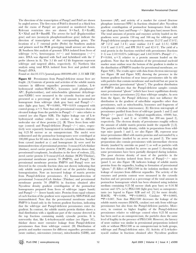
Figure S2 Peroxisomes from Pxmp2-deficient mouse liver are
fragile. (A) Contents of protein and activities of the soluble matrix
enzymes in different organelles: peroxisomes (catalase, L-a-
hydroxyacid oxidase/HAOX/), lysosomes (acid phosphatase/
AP/, b-galactosidase), and mitochondria (glutamate dehydroge-
nase/GDH/) were measured in the cytosolic fraction and are
presented as a percentage of the total amount in postnuclear
homogenate from wild-type (dark gray bars) and Pxmp22/2
mice (light gray bars). *P = 0.0001, **P = 0.019 compared with
control group, n = 3. Note that only peroxisomal enzymes show an
elevated leakage from the particles in Pxmp22/2 mice relative to
control (see also Figure S2B). The higher leakage rate of L-a-
hydroxyacid oxidase relative to catalase is due to different
molecular size of these proteins [1]. (B) Three livers from wild-
type (control) and Pxmp2-deficient (Pxmp22/2) mice, respec-
tively were separately homogenized in isolation medium contain-
ing 0.25 M sucrose as an osmoprotectant. The nuclei were
sedimented and the postnuclear homogenates were centrifuged at
100,000 gmax for 60 min to obtain the cytosolic fraction. Samples
from cytosol (marked as c) and homogenate (h) were used for
immunodetection of peroxisomal proteins: 3-oxoacyl-CoA thiolase
(thiolase), sterol carrier protein 2 (SCP2, this protein shows dual,
peroxisomal/cytoplasmic, localization in the liver of rodents, [2]),
sterol carrier protein 2/3-oxoacyl-CoA thiolase (SCP2/Thiolase),
peroxisomal membrane protein 70 (PMP70), and Pxmp2. The
peroxisomal membrane proteins PMP70 and Pxmp2 were not
detected in the cytosolic fraction (data not shown) indicating that
only soluble matrix proteins leaked out of the particles during
homogenization. Note an increased leakage of matrix proteins
from Pxmp2-deficient peroxisomes. (C) Immunodetection of
peroxisomal 3-oxoacyl-CoA thiolase (Thiolase) and peroxisomal
membrane protein 70 (PMP70) in fractions obtained after
Nycodenz density gradient centrifugation of the postnuclear
homogenates prepared from livers of wild-type (upper bands)
and Pxmp22/2 (lower bands) mice. Proteins from equal volumes
of each fraction of the gradient were separated by SDS-PAGE and
immunoblotted. Note that the peroxisomal membrane marker
PMP70 is found only in the bottom gradient fractions, indicating
that the wild-type and Pxmp2-deficient peroxisomes enter the
gradient. In contrast, the soluble matrix protein thiolase shows a
dual distribution with a significant part of the enzyme detected in
the top fractions containing mainly cytosolic proteins. It is
noteworthy that, like L-a-hydroxyacid oxidase (see Figure 1C),
thiolase shows more extensive leakage from Pxmp2-deficient
peroxisomes than from wild-type control. (D) Distribution of
protein and marker enzymes for different organelles: peroxisomes
(urate oxidase), microsomes (esterase), mitochondria (GDH), and
lysosomes (AP), and activity of a marker for cytosol (fructose
phosphate isomerase/FPI/) in fractions obtained after Nycodenz
gradient centrifugation of postnuclear homogenates from wild-
type (dark gray bars) and Pxmp2/2 (light gray bars) mouse livers.
The total amounts of protein and enzymes activity loaded on the
gradients were: protein 150 mg and 166 mg for wild-type and
Pxmp2-deficient samples respectively; esterase 55.2 U and 57.0 U;
AP 7.2 U and 7.4 U; urate oxidase 2.4 U and 2.5 U; GDH
11.6 U and 11.0 U, and FPI 39.0 U and 42.4 U. The yield of a
total protein in the fractions enriched with peroxisomes (fractions
2–5) was 2.1060.05% (wild-type) and 1.6460.04% (Pxmp22/2,
P = 0.044, n = 3) relative to a protein amount loaded on the
gradients. Note that the localization of the peroxisomal nucleoid
marker urate oxidase near the bottom of the gradient is similar to
the distribution of PMP70 (marker for peroxisomal membrane, see
Figure S2C). The results agree with data from electron microscopy
(see Figure 1B and Figure S2E) showing the presence in the
bottom gradient fractions of near intact peroxisomes side by side
with particles that contain membrane and nucleoid but are poor in
matrix proteins (peroxisomal ‘‘ghosts’’). The gradient distribution
of PMP70 indicates that the Pxmp2-deficient samples contain
more peroxisomal ‘‘ghosts’’ (which have lower equilibrium density
relative to intact peroxisomes [1]) than wild-type samples. As seen
from the figure, the Pxmp2 deficiency does not influence the
distribution in the gradient of subcellular organelles other than
peroxisomes, such as mitochondria, lysosomes and fragments of
endoplasmic reticulum (microsomes). (E) Electron micrograph of
peroxisomes purified from livers of wild-type (panels 1 and 2) and
Pxmp22/2 (panel 3) mice. Original magnification, 68400; bar,
500 nm (panels 1 and 3) or 615000; bar 200 nm (panel 2),
respectively. The electron density of peroxisomes roughly reflects
the amount of soluble matrix proteins inside the organelle [1,3].
The particles with high density, visible in preparations from wild-
type mice (panels 1 and 2, see also Figure 1B), represent near
intact peroxisomes filled with matrix proteins and surrounded by a
single membrane (marked by an arrow on panel 2). The same
preparations also contain peroxisomes with intermediate electron
density (marked by asterisks on panel 1) as well as particles with
low electron density (marked by arrow on panel 1) showing that
some peroxisomes have been partially disrupted during isolation.
The poor electron density of most of the particles in the
peroxisomal fraction isolated from livers of Pxmp22/2 mice
(panel 3, see also Figure 1B) indicates leakage of soluble matrix
proteins from the organelles, leading to formation of peroxisomal
‘‘ghosts.’’ (F) Effect of PEG1500 in the isolation medium on the
leakage of enzymes from different organelles. The activity of the
enzymes and protein content were measured in the cytosolic
fraction and are presented as a percentage of the total amount in
postnuclear homogenate which has been obtained using isolation
medium containing 0.25 M sucrose (dark gray bars) or 0.16 M
sucrose and 12% (w/v) PEG1500 (light gray bars) as osmoprotec-
tants (see legend to Figure S2B and ref. [1] for more details).
HAOX: *P = 0.006, **P = 0.00012; Catalase: *P = 0.0005,
**P = 0.007. Note that PEG1500 decreases the leakage of the
soluble matrix enzymes (HAOX, catalase) not only from wild-type
peroxisomes but also from the Pxmp2-deficient particles. More-
over, in contrast to higher fragility of the Pxmp2-deficient
peroxisomes relative to wild-type control when 0.25 M sucrose
has been used as an osmoprotectant, the particles show the same
level of fragility in the presence of PEG1500. However, the latency
of urate oxidase was similar in peroxisomal preparations isolated
with or without PEG1500 as an osmoprotectant in both groups,
wild-type and Pxmp2-deficient mice. (G) Activity of L-a-hydro-
xyacid oxidase in fractions obtained after Nycodenz gradient
Mammalian Peroxisomal Channel
PLoS ONE | www.plosone.org 12 April 2009 | Volume 4 | Issue 4 | e5090

centrifugation of the postnuclear homogenates prepared from livers
of Pxmp2-deficient mice. The homogenization medium contained
0.25 M sucrose (light gray bars) or 0.16 M sucrose and 12% (w/v)
PEG1500 (dark gray bars). Recoveries of the enzyme were 110%
(sucrose) and 108% (PEG1500), respectively. See legend to Figure 1C
for more details. Note decrease in the leakage of HAOX from
peroxisomes when homogenates were prepared using PEG1500 as
an osmoprotectant. (H) Immunodetection of peroxisomal thiolase in
fractions obtained after Nycodenz gradient centrifugation of liver
homogenates from Pxmp22/2 mice. The homogenates were
prepared in the presence of 0.25 M sucrose (upper band) or 0.16 M
sucrose and 12% (w/v) PEG1500 (lower band) as osmoprotectants.
See legend to Figure S2C for more details. (I,J) Illustration explaining
an osmotic behavior of peroxisomes during isolation. I, Homoge-
nization using an isolation medium containing an inappropriate
osmoprotectant (e.g., sucrose is a very poor osmoprotectant for
mammalian peroxisomes because it easily penetrates the membrane
[1,4]) leads to an abrupt dropin the concentration of solutes outside
peroxisomes, provoking a flow of water into the particles and an
outflow of solutes, the movement of water occurs by spontaneous
diffusion across the phospholipids bilayer as well as through the
channels; J-1, movement of water (small black circles) and solutes
(large gray circles) in wild-type peroxisomes containing at least two
types of channels, marked on the picture with single and double
walls, respectively; J-2, The same picture as before, but one type of
channels (with double walls) is missing. Note that outflow of solutes is
restricted; J-3 Presence of PEG1500 prevents water to flow into the
particles decreasing an osmotic pressure in peroxisomes (see Text
S2).
Found at: doi:10.1371/journal.pone.0005090.s002 (0.92 MB TIF)
Figure S3 Permeability of Pxmp2-deficient peroxisomal mem-
brane to substrates of peroxisomal enzymes in vitro and in vivo. (A)
Effect of Pxmp2 deletion on the latency of peroxisomal enzymes:
L-a-hydroxyacid oxidase (HAOX), catalase, lactate dehydroge-
nase (LDH), and glycerol-3-phosphate dehydrogenase (GPDH).
The last two enzymes are NADH-dependent. ‘‘Free’’ activity of
the enzymes in peroxisomes isolated from wild-type (dark gray
columns) and Pxmp22/2 (light gray columns) mouse livers is
presented as a percentage of the ‘‘total’’ activity, *P = 0.0001,
n = 7–8. Note that the ‘‘free’’ activity of HAOX is significantly
lower in Pxmp2-deficient peroxisomes than in wild-type control
(see Text S3 for details). (B) Illustration explaining the mechanism
of peroxisomal enzyme latency. Substrate (dark square) penetrat-
ing the membrane through the channel with velocity V1 is
converted by the peroxisomal enzyme into the corresponding
product (dark circle) with velocity V2. (C) Dependence of urate
oxidase activity on substrate concentration. Activity of urate
oxidase was measured in a purified peroxisomal fraction treated
with 0.05% (v/v) Triton X-100 to recover ‘‘total’’ activity of the
enzyme. The concentration of uric acid was detected spectropho-
tometrically using the molar extinction coefficient
e292 nm = 7.66103 M-1 cm-1. Arrows 2 and 3 indicate ‘‘free’’
activity of urate oxidase in wild-type and Pxmp2-deficient
peroxisomes, respectively relative to the ‘‘total’’ activity that was
detected at the final substrate concentration of 150 mM (see Text
S3 for details). (D) A Lineweaver-Burk plot was calculated using
data points from Figure S3C. The results indicate that the
dependence of urate oxidase activity on substrate concentration
follows Michaelis-Menten kinetics. (E) Samples of postnuclear
homogenates were subjected to SDS-PAGE, electroblotted and
stained with anti-urate oxidase antibodies (Santa-Cruz biotech-
nology). Note that the content of urate oxidase protein is similar in
wild-type and Pxmp2-deficient mice. (F) Detection of oxalic acid in
urine. Oxalic acid is the final product of glycolic acid oxidation.
The metabolism of glycolic acid in mammals proceeds in
cytoplasm with formation of oxalic acid and in peroxisomes with
production of glycine [5]. One can expect that restriction in access
of glycolic acid to peroxisomes might cause elevated oxidation of
this compound in cytoplasm, resulting in an increase in the
production of oxalic acid. Wild-type (dark circle) and Pxmp22/2
(open circle) mice were injected with glycolic acid and excretion of
oxalic acid in urine was measured. The absolute quantity of
excreted oxalic acid was calculated as mmoles in the total urine
collected within 24 h per 100 g body mass (left panel). The urine
was collected for 24 h before glycolate injection (1), for 24 h after
injection (2) and from 24 h to 48 h after injection (3). The
difference between the content of oxalic acid excreted in urine
collected 24 h before and 24 h after the injection of glycolic acid is
shown in the right panel. The difference was calculated for each
mouse. *P = 0.035. The data for female mice are shown. Note that
the levels of oxalic acid excreted before injection of glycolic acid
were similar in both groups of mice. However, after injection of
glycolic acid, a higher rate of oxalic acid excretion in Pmpx2-
deficient mice, as compare to wild-type control, was detected.
Measurements of the ‘‘total’’ activity of enzymes participating in
oxidation of glycolic acid in peroxisomes (HAOX, alanine-
glyoxylate aminotransferase) or in cytoplasm (LDH) in liver
homogenates did not reveal any difference between Pxmp22/2
and wild-type mice, respectively (data not shown).
Found at: doi:10.1371/journal.pone.0005090.s003 (1.86 MB TIF)
Figure S4 Figure S4. Detection of channel-forming activity in
insect cells. (A) Isolation of a Pxmp2-enriched membrane fraction
from insect cells (see Methods for details). According to marker
enzyme activity detection (see Figure S4B), the postmitochondrial
particle fraction (PPF) was enriched with fragments of endoplasmic
reticulum (microsomes) and (micro)peroxisomes. Samples from
each fraction containing an equal amount of protein (40 mg) were
subjected to SDS/PAGE and immunoblotted with antibodies
against mouse Pxmp2. Control: mock transfected cells. (B)
Characterization of subcellular fractions isolated from insect cells.
Catalase, GDH, esterase, AP, and LDH were used as markers for
(micro)peroxisomes, mitochondria, endoplasmic reticulum, lyso-
somes, and cytosol, respectively. Specific activities of the enzymes
were measured in the corresponding fractions isolated from mock-
transfected (dark gray columns) and Pxmp2-transfected (light gray
columns) insect cells and are presented as percentage of activity in
post-nuclear homogenates (100%). Note that in addition to the
marker for endoplasmic reticulum (esterase), the postmitochon-
drial particle fraction (PPF) shows a high catalase activity, implying
that this fraction contains a significant amount of (micro)peroxi-
somes. The PPF also contains the highest concentration of
recombinant Pxmp2 (see Figure S4A) and was used for detection
of the pore-forming activity (see Figure 2C and 2D). (C) Effect of
anti-Pxmp2 antibody on the channel-forming activity. The PPF
was isolated from insect cells expressing Pxmp2, and an aliquot of
solubilized protein (400 mg) was incubated (30 min at +4uC) with
IgG (5 mg) purified from pre-immune (left panel) or immune (right
panel) serum. The preparations were then used for multiple-
channel recording. Note the decrease in the amount of insertion
events with current amplitudes 20–35 pA (1.0 M KCl, +20 mV)
after treatment of the samples with anti-Pxmp2 antibodies. The
total number of insertion events: left panel 251, right panel 295.
Found at: doi:10.1371/journal.pone.0005090.s004 (1.40 MB TIF)
Figure S5 Figure S5. Purification and characterization of the
quaternary structure of Pxmp2. (A) Partial purification of Pxmp2
(see: Text S1). Proteins were separated by SDS-PAGE and stained
with silver nitrate. Lane 1, isolated peroxisomes (10 mg); lane 2,
Mammalian Peroxisomal Channel
PLoS ONE | www.plosone.org 13 April 2009 | Volume 4 | Issue 4 | e5090

peroxisomal membrane preparation after treatment at pH 11.3
(8 mg); lane 3, peroxisomal membranes solubilized by Genapol
(5 mg); lane 4, fraction enriched with Pxmp2 obtained after ion-
exchange chromatography and first size-exclusion chromatogra-
phy steps (0.6 mg). The protein identified by MALDI-TOF mass
spectrometry as Pxmp2 is marked with an asterisk. The migration
of molecular mass (in kDa) markers (LMW Calibration kit, Bio-
Rad) is indicated at the left side. (B) Size-exclusion chromatogra-
phy of partially purified Pxmp2 before (upper panel) and after
(lower panel) treatment of the protein with n-dodecyl b-D-
maltoside (DDM, see: Text S1). Proteins in 200 ml column
fractions were separated by SDS-PAGE, transferred to nitrocel-
lulose, and incubated with anti-Pxmp2 antibodies. (C) Mobility of
Pxmp2 on a SuperdexTM200 column. The calibration curve was
generated by plotting the log of the molecular mass of the
standards versus mobility on the column. The molecular mass
markers are: (1) catalase, 240 kDa; (2) lactate dehydrogenase,
140 kDa; (3) bovine serum albumin, 66 kDa; (4) carbonic
anhydrase, 29 kDa; and (5) cytochrome c, 12.4 kDa. The void
volume was determined using Blue Dextran (2000 kDa), which
was detected in fractions 1–3. The mobility of the different forms
of Pxmp2 are marked by arrows: (I) Pxmp2 partially purified in the
presence of Genapol (see Figure S5B, upper panel); (II) partially
purified Pxmp2 treated with DDM (see Figure S5B, lower panel);
(III) Pxmp2 after thermal treatment (see Figure 5D, lower panel)
or after solubilization from the membrane using 2.0% (w/v) SDS
(blot not shown). (D) Mobility of purified Pxmp2 on a Super-
dexTM200 column before (upper panel) and after (lower panel)
treatment of the protein at 75uC for 6 min. Traces of Pxmp2
visible in fraction 36 are apparently caused by retention of some
molecules of denaturized protein on the column. (E) Cross-linking
and immunoblot analysis of purified Pxmp2. The protein eluted
from a SuperdexTM200 column was treated with ethylene
glycolbis(sulfosuccinimidylsuccinate) (Sulfo-EGS, right lane). Con-
trol sample did not have cross-linker (left lane). The immunore-
active product of the cross-linking is marked by an asterisk. (F)
Cross-linking of the His-tagged Pxmp2 protein. The expression of
the His-tagged Pxmp2 was carried out using the BAC-TO-
BACTM Baculovirus Expression System (Invitrogen) and Sf9
insect cell line (A. Isomursu, unpublished data). The postmito-
chondrial particles fraction (PPF) was isolated and the proteins
were solubilized with 0.5% (v/v) Genapol X-080. The aliquots of
the solubilized proteins (about 50 mg) were treated with disucci-
nimidyl suberate (DSS, Pierce) for 30 min at room temperature.
DSS concentrations were: 0.1, 0.2, 0.5, 1.0 mM (lanes 2,3,4,5).
Control sample was without cross-linker (lane 1). After cross-
linking, the proteins were separated by SDS-PAGE followed by
immunoblot analysis using anti-His-Tag antibodies (Novagen). It
appears that application of these antibodies led to more valid
results if compare with the anti-Pxmp2 antibodies which produced
the only low signal with the cross-linked protein. Note that, similar
to purified Pxmp2 (see Figure S5E), the cross-linking of the
recombinant protein leads to appearance of molecules with the
size (about 80 kDa) three times larger than the size of the His-
tagged Pxmp2 protein (about 27 kDa). It is highly unlikely that the
mouse recombinant Pxmp2 produces heteromers with the insect
cell proteins. Therefore, the data support the notion that the
Pxmp2 protein forms a homotrimer in the membrane.
Found at: doi:10.1371/journal.pone.0005090.s005 (1.32 MB TIF)
Figure S6 Electrophysiological analysis of purified Pxmp2. (A)
Channel conductance as a function of KCl concentration. The
figure presents measurements (multiple channel recording) of the
increments in current which show peaks in channel activity at an
average conductance of 1.3 nS (upper line) or 0.45 nS (lower line)
in 1.0 M KCl (see Figure 3B and 3C). The mean conductance
6SD of 30–40 single events is shown. (B) Current traces of a
bilayer containing one Pxmp2 pore-forming protein (the insertion
event is visible on the upper trace) under asymmetric salt
conditions: 1.0 M KCl cis/0.5 M KCl trans compartment. The
holding potentials are marked. The data were filtered at 1.0 kHz
and recorded at 2.0 kHz. (C) Current-voltage relationship of the
Pxmp2 pore-forming protein under asymmetric salt concentra-
tions; data points are averages of at least 6 independent
measurements6S.D.
Found at: doi:10.1371/journal.pone.0005090.s006 (1.37 MB TIF)
Figure S7 Determination of the size of the Pxmp2 channel. (A)
Average conductance of high (1.3 nS in 1.0 M KCl, light gray
bars) and low (0.45 nS in 1.0 M KCl, open bars) conductance
Pxmp2 channels in different salt solutions (1.0 M, final concen-
tration). The average conductance6SD (G, right ordinate axis)
was calculated from at least 40 single events. Bulk electrolyte
conductivity (sigma, left ordinate axis) of the corresponding salt
solutions is shown as dark-gray bars. (B) Estimation of the size of
the Pxmp2 channel using cations of different hydrated radii. The
relative conductance rates (G/sigma) for high- (dark circle) and
low- (open circle) conductance channels (see Figure S7A) were
normalized to those of Rb+ (100 arbitrary units) and logarithms of
the resulting values were plotted against the hydrated radii of the
corresponding cations: Rb, 0.105 nm; KCl, 0.111 nm; NaCl,
0.163 nm; LiCl, 0.216 nm; Tris-Cl, 0.321 nm; TEA-Cl,
0.426 nm. The hydrated radii of cations [6] and non-electrolytes
[7] (see below) including polyethylene glycols (PEGs, Sigma) were
taken from the corresponding publications. (C) Estimation of the
size of the Pxmp2 channel (high-conductance state) using polymer
exclusion method. Left panel, the average conductance was
calculated from 40–60 single events. Ratios of the channel
conductance without (Go) and with (G) non-electrolyte was plotted
against hydrated radii of non-electrolytes: ethylene glycol,
0.26 nm; glycerol, 0.31 nm; arabinose, 0.34 nm; PEG200,
0.43 nm; PEG300, 0.60 nm; PEG400, 0.70 nm; PEG600,
0.78 nm; PEG1000, 0.94 nm; PEG2000, 1.22 nm; PEG3400,
1.63 nm. The 20% (w/v) solutions of non-electrolytes containing
1.0 M KCl (final concentration) were added to both halves of the
chamber. Right panel: second derivative of the data from left
panel showing two turning points. The upper one indicates the size
of non-electrolytes which partition into the pore become restricted.
The lower one indicates the maximal size of non-electrolytes
which still able to penetrate the channel (see Text S1 for details).
(D) Structure of ‘‘bulky’’ (ATP, NADH) and small (uric acid,
glycine) solutes.
Found at: doi:10.1371/journal.pone.0005090.s007 (0.31 MB TIF)
Text S1 Supporting methods.
Found at: doi:10.1371/journal.pone.0005090.s008 (0.04 MB
DOC)
Text S2 Fragility of Pxmp2-deficient peroxisomes in vitro.
Found at: doi:10.1371/journal.pone.0005090.s009 (0.03 MB
DOC)
Text S3 Latency of peroxisomal enzymes.
Found at: doi:10.1371/journal.pone.0005090.s010 (0.03 MB
DOC)
Author Contributions
Conceived and designed the experiments: AR VA RS RB JKH. Performed
the experiments: AR VA HI PP UB RS. Analyzed the data: AR VA RS PP
UB RS MW RB JKH. Contributed reagents/materials/analysis tools: RS
RS RB. Wrote the paper: VA JKH.
Mammalian Peroxisomal Channel
PLoS ONE | www.plosone.org 14 April 2009 | Volume 4 | Issue 4 | e5090

References
1. Wanders RJ, Waterham HR (2006) Biochemistry of mammalian peroxisomes
revisited. Annu Rev Biochem 75: 295–332.
2. Antonenkov VD, Hiltunen JK (2006) Peroxisomal membrane permeability and
solute transfer. Biochim Biophys Acta 1763: 1697–1706.
3. Visser WF, van Roermund CW, Ijlst L, Waterham HR, Wanders RJ (2007)Metabolite transport across the peroxisomal membrane. Biochem J 401:
365–375.
4. Palmieri L, Rottensteiner H, Girzalsky W, Scarcia P, Palmieri F, et al. (2001)
Identification and functional reconstitution of the yeast peroxisomal adeninenucleotide transporter. EMBO J 20: 5049–5059.
5. Antonenkov VD, Sormunen RT, Hiltunen JK (2004) The rat liver peroxisomal
membrane forms a permeability barrier for cofactors but not for smallmetabolites in vitro. J Cell Sci 117: 5633–5642.
6. Antonenkov VD, Rokka A, Sormunen RT, Benz R, Hiltunen JK (2005) Solutetraffic across mammalian peroxisomal membrane–single channel conductance
monitoring reveals pore-forming activities in peroxisomes. Cell Mol Life Sci 62:2886–2895.
7. Reumann S, Maier E, Benz R, Heldt HW (1995) The membrane of leaf
peroxisomes contains a porin-like channel. J Biol Chem 270: 17559–17565.
8. Reumann S, Maier E, Heldt HW, Benz R (1998) Permeability properties of the
porin of spinach leaf peroxisomes. Eur J Biochem 251: 359–366.
9. Dansen TB, Wirtz KW, Wanders RJ, Pap EH (2000) Peroxisomes in humanfibroblasts have a basic pH. Nat Cell Biol 2: 51–53.
10. Jankowski A, Kim JH, Collins RF, Daneman R, Walton P, et al. (2001) In situmeasurements of the pH of mammalian peroxisomes using the fluorescent
protein pHluorin. J Biol Chem 276: 48748–48753.
11. Lasorsa FM, Pinton P, Palmieri L, Scarcia P, Rottensteiner H, et al. (2008)Peroxisomes as novel players in cell calcium homeostasis. J Biol Chem 283:
15300–15308.
12. Drago I, Giacomello M, Pizzo P, Pozzan T (2008) Calcium dynamics in the
peroxisomal lumen of living cells. J Biol Chem 283: 14384–14390.
13. Gouveia AM, Reguenga C, Oliveira ME, Eckerskorn C, Sa-Miranda C, et al.(1999) Alkaline density gradient floatation of membranes: polypeptide
composition of the mammalian peroxisomal membrane. Anal Biochem 274:
270–277.
14. Chen NH, Crane DI, Masters CJ (1988) Analysis of the major integralmembrane proteins of peroxisomes from mouse liver. Biochim Biophys Acta
945: 135–144.
15. Zwacka RM, Reuter A, Pfaff E, Moll J, Gorgas K, et al. (1994) The
glomerulosclerosis gene Mpv17 encodes a peroxisomal protein producingreactive oxygen species. EMBO J 13: 5129–5134.
16. Iida R, Yasuda T, Tsubota E, Takatsuka H, Masuyama M, et al. (2003) M-LP,
Mpv17-like protein, has a peroxisomal membrane targeting signal comprising atransmembrane domain and a positively charged loop and up-regulates
expression of the manganese superoxide dismutase gene. J Biol Chem 278:
6301–6306.
17. Spinazzola A, Viscomi C, Fernandez-Vizarra E, Carrara F, D’Adamo P, et al.(2006) MPV17 encodes an inner mitochondrial membrane protein and is
mutated in infantile hepatic mitochondrial DNA depletion. Nat Genet 38:570–575.
18. Trott A, Morano KA (2004) SYM1 is the stress-induced Saccharomycescerevisiae ortholog of the mammalian kidney disease gene Mpv17 and is
required for ethanol metabolism and tolerance during heat shock. Eukaryot Cell3: 620–631.
19. van Veldhoven PP, Just WW, Mannaerts GP (1987) Permeability of the
peroxisomal membrane to cofactors of beta-oxidation. Evidence for the presence
of a pore-forming protein. J Biol Chem 262: 4310–4318.
20. Baudhuin P (1969) Liver peroxisomes, cytology and function. Ann N Y Acad Sci168: 214–228.
21. van Roermund CW, de Jong M, van Marle IJ, Dansen TB, Wanders RJ, et al.
(2004) The peroxisomal lumen in Saccharomyces cerevisiae is alkaline. J Cell Sci
117: 4231–4237.
22. Lasorsa FM, Scarcia P, Erdmann R, Palmieri F, Rottensteiner H, et al. (2004)The yeast peroxisomal adenine nucleotide transporter: characterization of two
transport modes and involvement in DeltapH formation across peroxisomalmembranes. Biochem J 381: 581–585.
23. Price NC, Dwek RA, Ratcliffe RG, Wormald MR (2001) Principles and
problems in physical chemistry for biochemists. Oxford: Oxford UniversityPress. 401 p.
24. Porcelli AM, Ghelli A, Zanna C, Pinton P, et al. (2005) pH difference across theouter mitochondrial membrane measured with a green fluorescent protein
mutant. Biochem Biophys Res Commun 326: 799–804.
25. Moriyama Y, Maeda M, Futai M (1992) Involvement of a non-proton pumpfactor (possibly Donnan-type equilibrium) in maintenance of an acidic pH in
lysosomes. FEBS Lett 302: 18–20.26. Verleur N, Wanders RJ (1993) Permeability properties of peroxisomes in
digitonin-permeabilized rat hepatocytes. Evidence for free permeability towards
a variety of substrates. Eur J Biochem 218: 75–82.27. van Roermund CW, Elgersma Y, Singh N, Wanders RJ, Tabak HF (1995) The
membrane of peroxisomes in Saccharomyces cerevisiae is impermeable toNAD(H) and acetyl-CoA under in vivo conditions. EMBO J 14: 3480–3486.
28. Gasmi L, McLennan AG (2001) The mouse Nudt7 gene encodes a peroxisomalnudix hydrolase specific for coenzyme A and its derivatives. Biochem J 357:
33–38.
29. Abdelraheim SR, Spiller DG, McLennan AG (2003) Mammalian NADHdiphosphatases of the Nudix family: cloning and characterization of the human
peroxisomal NUDT12 protein. Biochem J 374: 329–335.30. Wu X, Wakamiya M, Vaishnav S, Geske R, Montgomery C, et al. (1994)
Hyperuricemia and urate nephropathy in urate oxidase-deficient mice. Proc
Natl Acad Sci USA 91: 742–746.31. Qi C, Zhu Y, Pan N, Usuda N, Maeda N, et al. (1999) Absence of spontaneous
peroxisome proliferations in enoyl-CoA hydratase/L-3-hydroxyacyl-CoA dehy-drogenase-deficient mouse liver. J Biol Chem 274: 15775–15780.
32. Savolainen K, Kotti TJ, Schmitz W, Savolainen TI, Sormunen RT, et al. (2004)A mouse model for alpha-methylacyl-CoA racemase deficiency: adjustment of
bile acid synthesis and intolerance to dietary methyl-branched lipids. Hum Mol
Genet 13: 955–965.33. Newberry EP, Xie Y, Kennedy S, Han X, Buhman KK, et al. (2003) Decreased
hepatic fatty acid binding capacity and altered liver lipid distribution in micelacking the liver fatty-acid protein gene. J Biol Chem 278: 51664–51672.
34. Wanders RJ, Visser WF, van Roermund CW, Kemp S, Waterham HR (2007)
The peroxisomal ABC transporter family. Pflugers Arch – Eur J Physiol 453:719–734.
35. Kaulin YA, Schagina LV, Bezrukov SM, Malev VV, Feigin AM, et al. (1998)Cluster organization of ion channels formed by the antibiotic syringomycin E in
bilayer lipid membranes. Biophys J 74: 2918–2925.36. Benz R, Bauer K (1988) Permeation of hydrophilic molecules through the outer
membrane of gram-negative bacteria. Review on bacterial porins. Eur J Biochem
176: 1–19.37. Ujwal R, Cascio D, Colletier J-P, Faham S, Toro L, et al. (2008) The crystal
structure of mouse VDAC1 at 2.3 A resolution reveals mechanistic insights intometabolite gating. Proc Natl Acad Sci USA 105: 17742–17747.
38. Kunkele KP, Heins S, Dembowski M, Nargang FE, Benz R, et al. (1998) The
preprotein translocation channel of the outer membrane of mitochondria. Cell93: 1009–1019.
39. Michels PA, Bringaud F, Herman M, Hannaert V (2006) Metabolic functions ofglycosomes in trypanosomatids. Biochim Biophys Acta 1763: 1463–1477.
40. Colasante C, Ellis M, Ruppert T, Voncken F (2006) Comparative proteomics ofglycosomes from bloodstream form and procyclic culture form Trypanosoma
brucei brucei. Proteomics 6: 3275–3293.
41. Bakker BM, Mensonides FI, Teusink B, van Hoek P, Michels PA, et al. (2000)Compartmentation protects trypanosomes from the dangerous design of
glycolysis. Proc Natl Acad Sci USA 97: 2087–2092.42. Otte DM, Schwaab U, Luers GH (2003) The Pxmp2 and PoleI genes are linked
by a bidirectional promoter in an evolutionary conserved fashion. Gene 313:
119–126.43. Berthemy A, Newton J, Wu D, Buhrman D (1999) Quantitative determination
of an extremely polar compound allantoin in human urine by LC-MS/MSbased on the separation on a polymeric amino column. J Pharm Biomed Anal
19: 429–434.
44. Antonenkov VD, Sormunen RT, Hiltunen JK (2004) The behavior ofperoxisomes in vitro: mammalian peroxisomes are osmotically sensitive particles.
Am J Physiol Cell Physiol 287: C1623–C1635.
Mammalian Peroxisomal Channel
PLoS ONE | www.plosone.org 15 April 2009 | Volume 4 | Issue 4 | e5090
Copyright © 2022 FDOKUMEN