Role for Hsp70 Chaperone in Saccharomyces cerevisiae Prion Seed Replication
Upload
independentCategory
view
3download
0
Veenhuis and Ida J. van der KleiT. van Roermund, Rinse de Boer, MartenAlexey Kikhney, Lukasz Opalinski, Carlo W. Magdalena Bartoszewska, Chris Williams, and ChaperoneFamily Protein That Functions as Protease Peroxisomal Proteostasis Involves a LonCell Biology:
doi: 10.1074/jbc.M112.381566 originally published online June 25, 20122012, 287:27380-27395.J. Biol. Chem.
10.1074/jbc.M112.381566Access the most updated version of this article at doi:
.JBC Affinity SitesFind articles, minireviews, Reflections and Classics on similar topics on the
Alerts:
When a correction for this article is posted•
When this article is cited•
to choose from all of JBC's e-mail alertsClick here
Supplemental material:
http://www.jbc.org/content/suppl/2012/06/25/M112.381566.DC1.html
http://www.jbc.org/content/287/33/27380.full.html#ref-list-1
This article cites 57 references, 11 of which can be accessed free at
at University of Groningen on September 4, 2013http://www.jbc.org/Downloaded from

Peroxisomal Proteostasis Involves a Lon Family Protein ThatFunctions as Protease and Chaperone*□S
Received for publication, May 14, 2012, and in revised form, June 12, 2012 Published, JBC Papers in Press, June 25, 2012, 2012, DOI 10.1074/jbc.M112.381566
Magdalena Bartoszewska‡1, Chris Williams§, Alexey Kikhney§, Łukasz Opalinski‡1, Carlo W. T. van Roermund¶,Rinse de Boer‡2, Marten Veenhuis‡, and Ida J. van der Klei‡3
From the ‡Molecular Cell Biology, Groningen Biomolecular Sciences and Biotechnology Institute (GBB), University of Groningen,Kluyver Centre for Genomics of Industrial Fermentation, P. O. Box 11103, 9700CC Groningen, The Netherlands, §EMBL HamburgOutstation, c/o DESY, Notkestrasse 85 22 603, Hamburg, Germany, and ¶Laboratory Genetic Metabolic Diseases, AcademicMedical Center, University of Amsterdam, Meibergdreef 9, 1105 AZ Amsterdam, The Netherlands
Background: A putative Lon protease has been identified in peroxisomes of various species (Pln).Results: Pln is an ATP-dependent protease that digests unfolded substrates e.g. oxidatively damaged catalase-peroxidase, anddisplays chaperone-like activity, circumventing accumulation of protein aggregates in peroxisomes that compromise organellefunction.Conclusion: Pln is a bifunctional protein with chaperone and protease activities.Significance: Pln is crucial for peroxisome proteostasis.
Proteins are subject to continuous quality control for optimalproteostasis. The knowledge of peroxisome quality control sys-tems is still in its infancy. Here we show that peroxisomes con-tain amember of theLon family of proteases (Pln).We show thatPln is a heptameric protein and acts as an ATP-fueled proteaseand chaperone. Hence, Pln is the first chaperone identified infungal peroxisomes. In cells of a PLN deletion strain peroxi-somes contain protein aggregates, a major component of whichis catalase-peroxidase. We show that this enzyme is sensitive tooxidative damage. The oxidatively damaged, but not the nativeprotein, is a substrate of the Pln protease. Cells of the pln straincontain enhanced levels of catalase-peroxidase protein butreduced catalase-peroxidase enzyme activities. Together withthe observation that Pln has chaperone activity in vitro, our datasuggest that catalase-peroxidase aggregates accumulate in per-oxisomes of pln cells due to the combined absence of Pln prote-ase and chaperone activities.
Functional proteins generally adapt a defined three-dimen-sional structure, designated their native fold.Nevertheless, theyneed to retain conformational flexibility to allow functioning,and as a consequence, they are only marginally thermodynam-ically stable macromolecules. Hence, preexisting folded pro-teins are at constant risk of being unfolded, especially at
environmental stress conditions (1–3). Aberrantly folded poly-peptides as well as folding intermediates expose hydrophobicresidues that are normally buried in the native fold and areprone to trigger protein aggregation (4). Aggregation of mis-folded protein species is linkedwith the perturbation of cellularfunctions, aging, and various human disorders (5). To circum-vent this, cells evolved sophisticated protein quality control sys-tems, among which are molecular chaperones that facilitatefolding and refolding of polypeptides and proteolytic machinesthat remove misfolded conformers from the cellular interior(6). These systems are indispensable for protein homeostasis(proteostasis) in cells (7).Yet little is known of protein quality control in peroxisomes.
Despite their simple architecture, peroxisomes are importantorganelles that display an unprecedented diversity of functionsdepending on metabolic needs and external stimuli (8). Theyare crucial inman,manifested by the presence various inheriteddisorders (i.e. neonatal adrenoleukodystrophy, Zellweger syn-drome, infantile Refsum’s disease, rhizomelic chondrodysplasiapunctata) caused by impairment of one or more peroxisomalfunctions (9, 10). The hallmark of peroxisomes is the metabo-lism of hydrogen peroxide that is generated as a byproduct ofthe organelle oxidative metabolism (11). Hydrogen peroxidecan be the source of the most highly reactive and toxic form ofreactive oxygen species, the hydroxyl radical (�OH), which isgenerated in the Fenton reaction (12). To cope with reactiveoxygen species formation, peroxisomes contain a set of perox-ide decomposing enzymes among which are catalase, peroxire-doxins, or peroxidases (11). Peroxisomes may import foldedproteins and even oligomeric protein complexes (13) renderingthese organelles attractive models to analyze the mechanismsinvolved in housekeeping of preexisting, folded proteins.Recently, a conserved ATP-dependent Lon protease has
been identified in peroxisomes (14, 15). Themitochondrial iso-form of this protease was shown to represent the main qualitycontrol protease of the organellematrix (16, 17).MitochondrialLon selectively degradesmisfolded, unassembled, and damaged
* This work was supported in part by a Rubicon Fellowship (825.08.023) fromthe Netherlands Organization for Scientific Research (to C. W.).
□S This article contains supplemental Figs. S1–S5.1 Supported by the Research Program of the Kluyver Centre for Genomics of
Industrial Fermentation, which is part of the Netherlands Genomics Initia-tive/Netherlands Organization for Scientific Research.
2 Supported by the Netherlands Organization for Scientific Research via theIBOS (Integration of Biosynthesis and Organic Synthesis) Programme ofAdvanced Chemical Technologies for Sustainability (ACTS) (project IBOS053.63.011).
3 To whom correspondence should be addressed: Molecular Cell Biology,Groningen Biomolecular Sciences and Biotechnology Institute (GBB), P. O.Box 11103, 9700CC Groningen, The Netherlands. Tel.: 31-50-363-2179; Fax:31-50-363-8280; E-mail: [email protected].
THE JOURNAL OF BIOLOGICAL CHEMISTRY VOL. 287, NO. 33, pp. 27380 –27395, August 10, 2012© 2012 by The American Society for Biochemistry and Molecular Biology, Inc. Published in the U.S.A.
27380 JOURNAL OF BIOLOGICAL CHEMISTRY VOLUME 287 • NUMBER 33 • AUGUST 10, 2012 at University of Groningen on September 4, 2013http://www.jbc.org/Downloaded from

proteins (18, 19). However, the molecular function of the per-oxisomal Lon protease is not yet clear.In this work we demonstrate that peroxisomal Lon is a mul-
tifunctional protein that acts as a protease, selectively degrad-ing aberrant peroxisomal matrix proteins, but also assists inrefolding of these components. As such, Lon is the first perox-isomal chaperone identified in fungi so far.
EXPERIMENTAL PROCEDURES
Strains and Growth Conditions—Penicillium chrysogenumstrains used in this study are listed in Table 1. Cells were grownat 25 °C on mineral medium plates (20) supplemented with 1%glucose, 1% ethanol, or 1% oleic acid or in batch cultures onYGGmedium (21), penicillin (PEN)4 production medium sup-plemented with 0.05% phenylacetic acid (22), or in mineralmedium containing 0.1% oleic acid and 0.05% Tween 80. Toinduce conidiospore formation, R-agar was used (21). Acet-amide-resistant (AmdS�) transformants were selected onplates containing 0.1% acetamide (23).To determine fungal autolysis, P. chrysogenum strains were
cultivated for 120 h in PEN media. Subsequently, each culturewas divided into two halves, and 0.1% oleic acid and 0.05%Tween 80 were added to one-half. After incubation for 24 h,fungal biomass was determined as described previously (21).Escherichia coli DH5� was used for cloning purposes. E. coliRosetta 2 cells (Novagen) were used for production of heterol-ogous protein. E. coli cells were grown at 37 °C on LB mediumsupplemented with the appropriate antibiotics.Molecular Techniques—Plasmids and oligonucleotides used
in this study are listed in Tables 2 and 3. Standard DNAmanip-ulations were performed according to established procedures(24). Transformation of P. chrysogenum protoplasts was per-formed as described previously (23). All PCR fragments weresequenced (Service XS). Restriction enzymes (Fermentas), T4DNA ligase (Fermentas), and other DNA-modifying enzymeswere used as recommended by the suppliers. PCR was carriedout using high fidelity Phusion (Fermentas) polymerase. South-ern blotting was performed according to the DIG High Primelabeling and detection kit (Roche Applied Science). In silicoanalysis of DNA sequences and construction of vector mapswas carried out using CloneManager 5 software (Scientific andEducational Software, Durham, NC).Construction of the P. chrysogenum Pln Deletion Strain—The
pln deletion cassette was constructed using theMultisite Gate-
way cloning system. The upstream (1.759 bp) and downstream(1.474 bp) flanking regions of pln gene were amplified by PCRwith the primers AKR0007.PlnB4 and AKR0008.PlnB1 andwith AKR0009.PlnB2 and AKR0010.PlnB3, respectively, usingDS54465 genomic DNA as template. The resulting PCR prod-ucts were introduced into pDONRP2R-P3 (3�-flanking region)and pDONRP4-P1R (5�-flanking region of pln) by using theGateway BP clonase reaction, resulting in plasmids pENTR2.3-PLNdownstream and pENTR4.1-PLNupstream, respectively.Next, a Gateway LR reaction was performed using pENTR4.1-PLNupstream, pENTR221/PniadAMDS, pENTR2.3-PLN-downstream, and the destination vector pDEST R4-R3,resulting in the formation of the pln deletion constructpDest43_�PcPLN. This plasmid was linearized with AatII andtransformed into P. chrysogenum DS54465 GFP-SKL proto-plasts. Transformants were selected based on their ability togrow on acetamide as a sole nitrogen source. Disruption of thepln gene was confirmed by Southern blotting.Construction of P. chrysogenum Strains Producing Various
GFP Fusion Proteins—A plasmid enabling production of thePln protein fused N-terminally with GFP was constructedusing Multisite Gateway technology. The Pln coding sequencewith a stop codon was amplified by PCR using primersAKR0005.PlnB1 and AKR0006.PlnB2 and a cDNA library astemplate (25). The obtained PCR product was cloned into theentry vector pDONR221, resulting in pENTR221_PcPLN.Subse-quently, plasmids pENTR41/PpcbCGFP, pENTR221_PcPLN,pENTR23-His8.TpenDE, and pDEST R4-R3/AMDS wererecombined, resulting in a plasmid designated pDest43.pIPNS.eGFP.pcPLN.Tat. This plasmid was linearized with Acc65I andtransformed to a P. chrysogenum strain producing DsRed-SKL.
A plasmid enabling production of a catalase-peroxidase pro-tein fused N-terminally with GFP was constructed using theMultisite Gateway technology. The Pgpda region was cut outfrom vector pBBK-007 and cloned into pGBRH2-mGFP in theplace of PpcbC using Asp718I and BamHI, resulting in vectorLMOP-Pgpda-mGFP. Next, the Pgpda-mGFP region wasamplified by PCR using primers LMOp080 and LMOp081and LMOP-Pgpda-mGFP as a template and recombined topDONR4-1 in a BP recombination reaction, resulting in vec-tor LMOP41-Pgpda-mGFP. The catalase-peroxidase codingsequence with a stop codon was amplified by PCR usingprimers KatGfor and KatGrev and cDNA library as template(25). The obtained PCR product was cloned into the entry vec-tor pDONR221, resulting in pDONR221KatG. Subsequently,plasmids LMOP41pgpdAmGFP, pDONR221KatG, pENTR23-His8.TpenDE, and pDEST R4-R3/AMDS were recombined,resulting in a plasmid pGFPKatG. This plasmid was linearized
4 The abbreviations used are: PEN, penicillin production; MBP, maltose-bind-ing protein; TEV, tobacco etch virus; TBARS, thiobarbituric acid-reacting sub-stance; SLS, static light scattering; DLS, dynamic light scattering; SAXS, small-angle x-ray scattering; MM, molecular mass; DHE, dihydroethidium; PTS1,peroxisomal targeting signal 1; CS, citrate synthase; s, scattering vector.
TABLE 1P. chrysogenum strains used in this study
Strain Genotype or characteristic Reference
DS54465 GFP-SKL DS54465 with integrated PgpdA-GFP.SKL-TpenDE cassette at niaD locus, AmdS�, chlorate-resistant (50)
pln::GFP-SKL DS54465 GFP-SKL with deletion of pln gene, chlorate-resistant, AmdS� This studyDsRed-SKL DS17690 with integrated PpcbC-DsRed.SKL-TpenDE cassette, AmdS� (26)DsRed-SKL GFP.Pln DsRed-SKL with integrated PgpdA-mGFP.pln-TpenDE, AmdS� This studyDsRed-SKL GFP-catalase-peroxidase DsRed-SKL with integrated PgpdA-mGFP.catalase-peroxidase-TpenDE, AmdS� This studyDsRed-SKL GFP-chDEH DsRed-SKL with integrated PgpdA-mGFP.chDEH-TpenDE, AmdS� This study
Lon Functions in Peroxisome Proteostasis
AUGUST 10, 2012 • VOLUME 287 • NUMBER 33 JOURNAL OF BIOLOGICAL CHEMISTRY 27381 at University of Groningen on September 4, 2013http://www.jbc.org/Downloaded from

with NotI and transformed to a P. chrysogenum strain produc-ing DsRed-SKL.A plasmid enabling the production of the putative oxi-
doreductase protein fused N-terminally with GFP was con-structed usingMultisite Gateway technology. The putative oxi-doreductase encoding sequence with a stop codon wasamplified by PCR using primers chDEHfor and chDEHrev and
a cDNA library as template (25). The obtained PCR productwas cloned into the entry vector pDONR221, resulting inpDONR221chDEH. Subsequently, plasmids LMOP41pgpdAmGFP,pDONR221chDEH, pENTR23-His8.TpenDE, and pDEST R4-R3/AMDS were recombined, resulting in a plasmid pGFP-chDEH.This plasmid was linearized with AatII and transformed toP. chrysogenum producing DsRed-SKL. All transformants were
TABLE 2Plasmids used in this study
Name Description Source/Reference
pDONR P4-P1R Multisite Gateway vector, KanR CmR InvitrogenpDEST R4-R3 Multisite Gateway vector, AmpR CmR InvitrogenpDONR 221 Multisite Gateway vector, KanR CmR InvitrogenpDONR P2R-P3 Multisite Gateway vector, KanR CmR InvitrogenpENTR221/PniadAMDS pDONR 221 vector with PgpdA-amdS cassette flanked by niaD sequences, KanR Gift from J. G. NijlandpENTR2.3-PLNdownstream pDONR P2R-P3 with 3�-flanking region of pln, KanR This studypENTR4.1-PLNupstream pDONR P4-P1R with 5�-flanking region of pln, KanR This studypDest43_�PcPLN pDEST R4-R3 with pln deletion cassette, AmpR This studypENTR221_PcPLN pDONR 221 with pln CDS with stop codon, KanR This studypENTR41/PpcbCGFP pDONR P4-P1R with pcbC promoter and eGFP gene lacking a stop codon, KanR (26)pENTR23-His8.TpenDE His-tag with C-terminal terminator of penDE gene, KanR (59)pDEST R4-R3/AMDS pDESTR4-R3 with PgpdA-amdS cassette, AmpR (26)pDest43.pIPNS.eGFP.pcPLN.Tat Expression vector with PpcbC-eGFP.pln-TpenDE cassette, AmpR This studyLMOP41pgpdAmGFP pDONR P4-P1R with gpdA promoter andmGFP gene lacking a stop codon, KanR This studypDONR221KatG pDONR 221 with catalase-peroxidase CDS with stop codon, KanR This studypGFP.KatG Expression vector with PgpdA-mGFP-catalase-peroxidase-TpenDE cassette, AmpR This studypBBK-007 Plasmid with PgpdA-DsRed.SKL-TpenDE expression cassette, AmpR (50)pGBRH2-mGFP pBluescript vector with pcbC promoter,mGFP gene, and penDE terminator, AmpR (26)LMOP-Pgpda-mGFP pBluescript vector with gpdA promoter,mGFP gene, and penDE terminator, AmpR This studypDONR221chDEH pDONR 221 with oxidoreductase CDS with stop codon, KanR This studypGFP.chDEH Expression vector with PgpdA-mGFP.oxidoreductase-TpenDE cassette, AmpR This studypMAL-c2 Expression vector for N-terminal MBP-tag fusions, AmpR New England BiolabspMDBPlnMBP Expression vector containing P. chrysogenum Pln fused N-terminally to maltose-
binding protein tag, AmpRThis study
pMDBPlnHis Expression vector containing P. chrysogenum Pln fused N-terminally to maltose-binding protein tag and C-terminally to the His tag, AmpR
This study
pMDBLONTEV Expression vector containing P. chrysogenum Pln fused N-terminally to maltose-binding protein tag and C-terminally to the His tag, with TEV protease cleavagesite between MBP and Pln, AmpR
This study
pMDBPlninMBP Expression vector containing proteolytically inactive mutant of P. chrysogenumPln, Plnin, fused N-terminally to maltose-binding protein tag, AmpR
This study
pMDBPlninHis Expression vector containing proteolytically inactive mutant of P. chrysogenumPln, Plnin, fused N-terminally to maltose-binding protein tag and C-terminallyto the His tag, AmpR
This study
pMDBLONinTEV Expression vector containing proteolytically inactive mutant of P. chrysogenumPln, Plnin, fused N-terminally to maltose-binding protein tag and C-terminallyto the His tag, with TEV protease cleavage site between MBP and Plnin, AmpR
This study
pGEX-4T-2 Expression vector for N-terminal GST-tag fusions, AmpR GE HealthcarepGSTKatG Expression vector containing P. chrysogenum catalase-peroxidase fused N-
terminally to glutathione S-transferase tag, AmpRThis study
TABLE 3Oligonucleotides used in this study
Name Sequence (5�-3�)
AKR0007.PlnB4 GGGGACAACTTTGTATAGAAAAGTTGTGATATCTTGAACGGGGACGAKR0008.PlnB1 GGGGACTGCTTTTTTGTACAAACTTGCAGGCCCTAGTGATGATGGAATGGAKR0009.PlnB2 GGGGACAGCTTTCTTGTACAAAGTGGTCAGAGACGAGGCTGAAATCCAKR0010.PlnB3 GGGGACAACTTTGTATAATAAAGTTGTATTGACTGGCATGAACGCATCACAKR0005.PlnB1 GGGGACAAGTTTGTACAAAAAAGCAGGCTTGATGGGAACAAACGGTAGAAGTACCAKR0006.PlnB2 GGGGACCACTTTGTACAAGAAAGCTGGGTTCACAGTCGGCTCTCAACGAAGKatGfor GGGGACAAGTTTGTACAAAAAAGCAGGCTTTATGAGCGAATGTCCTGTTGCGCKatGrev GGGGACCACTTTGTACAAGAAAGCTGGGTTTCACAGGCGAGGACGACCLMOp080 GGGGACAACTTTGTATAGAAAAGTTGTTACCGCGGCCGCTCTGTACAGTGACLMOp081 GGGGACTGCTTTTTTGTACAAACTTGCCTTGTACAGCTCGTCCATGCCGAGchDEHfor GGGGACAAGTTTGTACAAAAAAGCAGGCTTTATG GCCACCACGAACGAGTTCchDEHrev GGGGACCACTTTGTACAAGAAAGCTGGGTTTTACAGACGGGGCTGGTTGCMDBPlnfor ATAGGATCCATGGGAACAAACGGTAGAAGTACCMDBPlnrev AAACTGCAGTCACAGTCGGCTCTCAACMDBhisfor CGTTGAGAGCCGACTGCATCACCATCACCATCACTGACTGCAGGCAAGCMDBhisrev GCTTGCCTGCAGTCAGTGATGGTGATGGTGATGCAGTCGGCTCTCAACGMDB.TEV.for GGATCGAGGGAAGGGAAAATCTTTACTTTCAAGGTATTTCAGAATTCGGMDB.TEV.rev CCGAATTCTGAAATACCTTGAAAGTAAAGATTTTCCCTTCCCTCGATCClonmutfor CCCAAGGATGGTCCTGCTGCTGGTCTTGCCCACGCClonmutrev GGCGTGGGCAAGACCAGCAGCAGGACCATCCTTGGGGST.KatGfor AAAGGATCCGAAAATCTTTACTTTCAAGGTATGAGCGAATGTCCTGTTGCGCGST.KatGrev AAAGAATTCTCACAGATCTTCTTCAGAAATAAGTTTTTGTTCCAGGCGAGGACGACCGGT
Lon Functions in Peroxisome Proteostasis
27382 JOURNAL OF BIOLOGICAL CHEMISTRY VOLUME 287 • NUMBER 33 • AUGUST 10, 2012 at University of Groningen on September 4, 2013http://www.jbc.org/Downloaded from

selected based on their ability to utilize acetamide as a solenitrogen source.Production and Purification of Pln—The coding sequence of
Pln was amplified using a P. chrysogenum library as a template(25) and primers MDBPlnfor and MDBPlnrev containingBamHI and PstI sites, respectively. The obtained PCR productand expression vector pMAL-c2 (New England Biolabs) weredigested with BamHI and PstI and ligated, resulting in plasmidpMDBPlnMBP, encoding P. chrysogenum Pln protein fused tomaltose-binding protein (MBP) at the N terminus. Subse-quently, a His6 tag was introduced at the C terminus of theMBP-Pln fusion protein using the Lightening site-directedmutagenesis kit (Stratagene) and primers MDBhisfor andMDBhisrev and plasmid pMDBPlnMBP as a template, result-ing in plasmid pMDBPlnHis. To introduce a tobacco etch virus(TEV) cleavage site between MBP and Pln, pMDBPlnHis plas-mid, primersMDB.TEV.for, andMDB.TEV.rev and Lighteningsite-directed mutagenesis kit (Stratagene) were used, resultingin the final construct pMDBLONTEV. Correctness of the plas-mid was confirmed by sequencing.E. coli Rosetta 2 cells containing expression plasmid
pMDBLONTEVwere grown on LBmedium at 37 °C to an opti-caldensityat600nmof1.1mMisopropyl1-thio-�-D-galactopyran-osidewas added followed by 18 h incubation at 16 °C.Cellswereharvested by centrifugation, and pellets were suspended inbuffer A (50 mM Tris-HCl, 100 mM KCl, 10 mM MgCl2, 5%glycerol, pH 8.0) and disrupted using a French press. The solu-ble fraction, which was obtained by centrifugation for 1 h at16,000 � g at 4 °C was incubated with nickel-nitrilotriaceticacid resin (Qiagen) for 45 min at 4 °C. The resin was washedwith buffer A containing 10 mM imidazole, and bound proteinswere eluted with buffer A containing 300 mM imidazole. Frac-tions were analyzed by SDS-PAGE andWestern blotting using�-His6-tag antibodies (Santa Cruz Biotechnology). Fractionscontaining the MBP-Pln-His6 fusion protein were pooled andloaded on amylose resin (New England Biolabs) equilibratedwith buffer B (50 mM Tris-HCl, 100 mM KCl, 10 mMMgCl2, 5%glycerol, pH8.0) and incubated for 45min at 4 °C. The resinwaswashed with buffer B, and bound Pln fusion protein was elutedwith buffer B containing 10 mM maltose.Generation and Purification of a Proteolytically Inactive Pln
Mutant (Plnin)—The Pln mutant, S815A (Plnin), encoded byplasmid pMDBPlninMBP, was generated using the pMDBPl-nMBP plasmid, primers lonmutfor and lonmutrev, and theLightening site-directedmutagenesis kit (Stratagene). To intro-duce a His6 tag at the C terminus of the MBP-Plnin fusion pro-tein, plasmid pMDBPlninMBP used as the template, primersMDBhisfor and MDBhisrev and Lightening site-directedmutagenesis kit (Stratagene) were applied, resulting in plasmidpMDBPlninHis. To introduce a TEV cleavage site betweenMBP and Plnin, the pMDBPlninHis plasmid was used as a tem-plate together with primers MDB.TEV.for and MDB.TEV.revand the Lightening site-directed mutagenesis kit (Stratagene),resulting in the final construct pMDBLONinTEV. Correctnessof the plasmid was confirmed by sequencing. pMDBLONin-TEV plasmid was transformed into the Rosetta 2 E. coli cellstrain. The Plnin fusion protein was expressed and purified tohomogeneity using the protocol for the Pln fusion protein.
Production and Purification of Catalase-Peroxidase—Thecoding sequence of catalase-peroxidase was amplified usingP. chrysogenum cDNA library (25) as a template and primersGST.KatGfor and GST.KatGrev containing BamHI and EcoRIsites, respectively. The obtained PCR product and expressionvector pGEX-4T-2 (GE Healthcare) were digested with BamHIand EcoRI and ligated, resulting in plasmid pGSTKatG, encod-ing catalase-peroxidase P. chrysogenum protein fused to gluta-thione S-transferase (GST) at the N terminus. Correctness ofthe plasmid was confirmed by sequencing.E. coli Rosetta 2 cells containing expression plasmid
pGSTKatG were grown on LB medium to an optical density at600 nm of 1 and induced by the addition of 1 mM isopropyl1-thio-�-D-galactopyranoside with simultaneous addition of2.5�Mhemin (Sigma) followedwith 18 h of incubation at 16 °C.Cells were harvested by centrifugation at 5200� g for 10min at4 °C. The cell pellet was suspended in column buffer (50 mM
NaH2PO4, 150 mMNaCl, 1 mMDTT, and 1 mM EDTA, pH 7.2)and disrupted using a French press. The soluble fraction, whichwas obtained by centrifugation for 1 h 16,000 � g at 4 °C, wasincubatedwith glutathione Superflow resin (Qiagen) for 45minat 4 °C. The resin was washed with column buffer, and boundproteins were elutedwith elution buffer (50mMTris-HCl, 0.1 M
NaCl, 50 mM reduced L-glutathione, 1 mM DTT, pH 8.0).Obtained fractions were analyzed by SDS-PAGE and Westernblotting using �-c-Myc antibodies (Santa Cruz Biotechnology).Fractions containing GST-catalase-peroxidase were pooledand cleaved using AcTEV protease (Invitrogen). Separation ofcatalase-peroxidase from GST and TEV protease was accom-plished using Superose 6 10/300 GL (GE Healthcare) columnequilibrated with 50mMNaH2PO4, 150mMNaCl, pH 7.2. Puri-fied GST-catalase-peroxidase was used for immunization ofrabbits (Eurogentec, Seraing, Belgium; supplemental Fig. S5).BiochemicalMethods—Crude extract ofP. chrysogenum cells
were prepared as described previously (26). Protein concentra-tions were determined using the RC-DC assay system or Bio-Rad assay kit (Bio-Rad) using bovine serum albumin (BSA) as astandard. SDS-PAGE andWestern blotting were carried out inaccordance with established procedures. P. chrysogenum cellfractionation and peroxisome isolation were performed asdescribed previously (26). Catalase activity was measured spec-trophotometrically as described before (14). Peroxidase activitywas determined spectrophotometrically as described by Fraaijeet al. (27). Catalytic and peroxidatic activities weremeasured inP. chrysogenum extracts at the optimal pH values as determinedfor purified catalase-peroxidase (supplemental Fig. S4). TheATPase activity of Pln and Plnin was measured colorimetricallyusingmalachite green according to the instructions of theman-ufacturer (Innova Biosciences).Isolation of Triton X-100-insoluble Peroxisomal Proteins—
Triton X-100 insoluble proteins were isolated from fractionsenriched in peroxisomes essentially as described previously(28). For identification of the proteins, bands of interest werecut from the SDS-PAGEgel and analyzed bymass spectrometry(MALDI-TOF peptide mass fingerprint and MALDI-TOF/TOF peptide sequencing, Alphalyse A/S, Denmark).Analysis of Penicillin Production—�-Lactam antibiotic bio-
assays were performed as described previously (21). Quantita-
Lon Functions in Peroxisome Proteostasis
AUGUST 10, 2012 • VOLUME 287 • NUMBER 33 JOURNAL OF BIOLOGICAL CHEMISTRY 27383 at University of Groningen on September 4, 2013http://www.jbc.org/Downloaded from

tive analysis of extracellular penicillin G was determined byHPLC using a Shim-Pack XR-ODS column (3.0-mm internaldiameter � 75 mm) and an isocratic flow of 310 g�liter�1 ace-tonitrile, 640 mg�liter�1 KH2PO4, and 340 mg�liter�1 H3PO4.Peaks of penicillin G were detected at a wavelength of 254 nm.Measurements of �-Oxidation of Fatty Acids in Intact Cells—
Spores of P. chrysogenum strains were inoculated in YGGmedium and incubated for 48 h. Next, cells were shifted tomineral medium supplemented with 0.1% oleic acid, 0.05%Tween 20 for 10 h. Subsequently, cells were washed and resus-pended in phosphate-buffered saline (PBS). �-Oxidation activ-ities were measured in intact cells as described previously (29).Cell suspensions were incubated in 200�l of PBS containing 10�M [1-14C]stearate (C18:0). Reactions were allowed to proceedfor 1 h at 28 °C followed by termination of the reactions by theaddition of 50�l of 2.6 M perchloric acid. RadiolabeledCO2wastrapped overnight in 500 �l of 2 M NaOH. The 14C-labeled�-oxidation products were subsequently collected afterextracting the acidified material with chloroform/methanol/heptane and quantified in a liquid scintillation counter.Lipid Peroxidation Analysis—Lipids peroxidation was quan-
tified by determination of thiobarbituric acid-reacting sub-stances (TBARSs) according to the method described by Para-szkiewicz et al. (30) using lysed protoplasts. The TBARSconcentration was expressed as nmol of malondialdehyde/mgor protein.In Vitro Protein Degradation Assays—Purified Pln and the
proteolytically inactive mutant Plnin were used for in vitro pro-tease assays. Proteolytic activity against unfolded model pro-teins was assayed with �-casein (Sigma) or �-casein labeledwith resorufin (Universal Protease Substrate, Roche AppliedScience). For hydrolysis of �-casein, the assay buffer C consist-ing of 50 mM Tris-HCl, pH 8.0, 10 mM MgCl2, 1 mM DTT withorwithout 4mMATP and theATP regeneration system (50mM
creatine phosphate, 80 �g/ml creatine kinase) was used. Plnand the proteolytically inactivemutant (Plnin) (final concentra-tion of 0.26 �M) were incubated at 25 °C with 5.8 �M �-caseinovernight. Proteolytic degradation of �-casein was detectedusing SDS-PAGE and Coomassie Brilliant Blue staining. Forhydrolysis of �-casein labeled with resorufin Pln and Plnin, 1.2�M, were incubated at 25 °C with 0.1% of �-casein labeled withresorufin in buffer C according to the instructions of the man-ufacturer. Proteolysis was monitored spectrophotometricallyat 574 nm.The proteolytic activity against catalase-peroxidase was
assayed in buffer C with or without 4 mM ATP and an ATPregeneration system (50 mM creatine phosphate, 80 �g/ml cre-atine kinase). Pln (0.46 �M) was incubated at 25 °C with native,thermally denatured orH2O2-treated catalase-peroxidase (0.52�M). To prepare H2O2-treated catalase-peroxidase, catalase-peroxidase (final concentration 5.2 �M) was incubated with 5mM H2O2 for 30 min at room temperature. Next H2O2 wasremoved by buffer exchange using an Amicon Ultra-4 andMicrocon YM-3 centrifugation device (Millipore). To preparethermally denatured catalase-peroxidase, catalase-peroxidase(at a concentration of 5.2�M)was incubated for 10min at 80 °C.Proteolytic activity was analyzed using SDS-PAGE electropho-resis and Coomassie Brilliant Blue staining. Quantification of
catalase-peroxidase bands was performed with ImageJ soft-ware. The results from two independent experiments wereaveraged.Catalase-Peroxidase Unfolding and Aggregation—Changes
in intrinsic (tryptophan) fluorescence (excitation � � 295 nm,emission � � 345 nm) were measured at room temperatureusing 15 �M purified catalase-peroxidase in 50 mM Tris-HCl,pH 8.0, and 150 mM NaCl upon incubation for 10 min withdifferent concentrations of hydrogen peroxide. Fluorescencemeasurements were performed using Fluoromax-3 spectro-fluorometer (Jobin Yvon-Spex).Aggregation of catalase-peroxidase (1.3 �M in 50 mM Tris-
HCl, pH 8.0, and 150 mM NaCl) upon treatment with 20 mM
hydrogen peroxide was monitored over a period of 60 min atroom temperature by measuring light scattering at 360 nm in aPerkinElmer Life Sciences Lambda 35 spectrophotometer.Citrate Synthase Aggregation and Refolding Assays—The
capacity of Pln to prevent protein aggregation was determinedusing chemically denatured citrate synthase as a substrate asdescribed previously (31). Citrate synthase (final concentration30 �M of native enzyme; Sigma) was chemically denatured byincubation at room temperature in 6 M guanidinium/HCl and20 mM DTT for at least 2 h. Aggregation was initiated by a200-fold dilution of the denatured enzyme under vigorous stir-ring in 50mMTris-HCl, pH 8.0, with 5mMMgCl2 at 25 °C withor without Pln and with or without 1 mM ATP. The kinetic ofcitrate synthase aggregation was monitored by measuring theintensity of light scattering at 360 nm in a PerkinElmer LifeSciences Lambda 35 spectrophotometer.To measure refolding activity of Pln, chemically denatured
citrate synthase was prepared as described above at a concen-tration of 15 �M (31). Reactivation of citrate synthase was initi-ated by a 100-fold dilution of the denatured enzyme under vig-orous stirring in 50 mM Tris-HCl, pH 8.0, with 5 mM MgCl2 at25 °C with or without Pln or Plnin and with or without 1 mM
ATP. The recovery of citrate synthase activity was tested asdescribed (32) using oxaloacetic acid, acetyl-CoA, and dithio-bis-[2-nitrobenzoic] acid (all from Sigma).Static andDynamic Light Scattering—Before static light scat-
tering (SLS), dynamic light scattering (DLS), and small-anglex-ray scattering (SAXS) analysis, Plnin was subjected to gel fil-tration using a Superdex S200 16/10 column equilibrated with50 mM Tris, 150 mM NaCl, 1 mM DTT, 4 mM ATP, pH 8.0. Theprocedure used for SLS coupled to the AKTA system isdescribed in Nettleship et al. (33). DLS measurements wereperformed on aDynaProTMDLS (Protein solutionsTM) at 10 °Cwith the protein at a concentration of 1.1 �M.SAXS Analysis—Synchrotron radiation x-ray scattering data
from solutions of Plnin were collected on the X33 camera of theEMBL on the storage ring DORIS III (DESY, Hamburg, Ger-many) (34). Using a photon counting Pilatus 1 M detector at asample-detector distance of 2.7m and a wavelength of � � 0.15nm, the range of momentum transfer 0. 1 � s � 6 nm�1 wascovered (s � 4�sin�/�, where 2� is the scattering angle). Foursolute concentrations in the range 0.5–3.7 mg/ml were mea-sured. To monitor radiation damage, 8 successive 15-s expo-sures were compared, and no significant changes wereobserved. The data were normalized to the intensity of the
Lon Functions in Peroxisome Proteostasis
27384 JOURNAL OF BIOLOGICAL CHEMISTRY VOLUME 287 • NUMBER 33 • AUGUST 10, 2012 at University of Groningen on September 4, 2013http://www.jbc.org/Downloaded from

transmitted beam and radially averaged; the scattering of thebuffer was subtracted, and the difference curves were scaled forprotein concentration. The low angle data collected from Plninwere extrapolated to infinite dilution and merged with thehigher concentration high angle data to yield the final compos-ite scattering curve.The forward scattering I(0) and the radius of gyrationRgwere
evaluated using the Guinier approximation (35) assuming thatat very small angles (s � 1.3/Rg) the intensity is represented asI(s) � I(0) exp(�(sRg)2/3). These parameters were computedusing the programs AUTORG and AUTOGNOM (36); the lat-ter program also provided the pair distribution function of theparticle p(r) and themaximumdimensionDmax. Themolecularmass (MM) of Plnin was evaluated by comparison of the for-ward scattering with that from a reference solution of bovineserum albumin (MM � 66 kDa).Ab Initio Shape Determination—The “shape scattering”
curve was further used to generate low resolution ab initioshapes of the Plnin with the program DAMMIF (37). This pro-gram represents the particle shape with an assembly of denselypacked beads and employs simulated annealing to construct acompact interconnected model fitting the experimental dataIexp(s) to minimize discrepancy,
�2 �1
N 1�j
� Iexp�sj cIcalc�sj
sj�2
(Eq. 1)
where N is the number of experimental points, c is a scalingfactor, and Icalc(s) and (sj) are the calculated intensity and theexperimental error at the momentum transfer sj, respectively.Ten DAMMIF runs were performed to check the stability ofsolution, and the results were well superimposable with eachother. These models were averaged to determine commonstructural features using the programs DAMAVER (38) andSUPCOMB (39). The latter program aligns two arbitrary low orhigh resolution models represented by ensembles of points byminimizing a dissimilarity measure called normalized spatialdiscrepancy. For every point (bead or atom) in the first model,the minimum value among the distances between this pointand all points in the second model is found, and the same isdone for the points in the second model. These distances areadded and normalized against the average distances betweenthe neighboring points for the two models. Generally, normal-ized spatial discrepancy values close to unity indicate that thetwomodels are similar. The programDAMAVER generates theaverage model of the set of superimposed structures and alsospecifies the most typical model (i.e. that having the lowestaverage normalized spatial discrepancy with all the other mod-els of the set).Molecular Modeling—The program BUNCH (40) combines
rigid body and ab initio modeling of proteins consisting ofdomains (forwhich high resolutionmodels are available) linkedby flexible loops of unknown structure. A simulated annealingprotocol is employed to find the optimal positions and orienta-tions of the domains moved as rigid bodies and probable con-formations of the flexible linkers attached to the appropriateterminal residues of domains. These linkers are represented asinterconnected chains of dummy residues (41), where protein
fragments are substituted by a flexible chain of residues sepa-rated by 0.38 nm. The x-ray scattering intensity from the entiremodel is expressed as,
I�s � 2� �l � 0
�m � �l
l � �k
A�klm�s � �
i
D�ilm�s�2
(Eq. 2)
Here, A(k)lm(s)H are the partial scattering amplitudes of the
rigid domains calculated from their atomic coordinates usingCRYSOL (42). The partial scattering amplitudes of the linkersD(i)
lm(s) are computed from the positions of the dummy resi-dues using the form factor equal to that of an average residue inwater (41). Starting from an arbitrary configuration of domainsand linkers, the programperforms randommodifications of themodel maintaining the structures of the domains and connec-tivity of the linkers.Microscopy—Confocal laser scanning microscopy images
were obtained using a Zeiss LSM510 confocal laser scanningmicroscope equipped with a Zeiss Plan-Neofluar�100/1.3 andZeiss Plan-Apochromatic �63/1.4 oil objectives. Images wereacquired usingAIM4.2 software (Carl Zeiss). GFP fluorescencewas analyzed by excitation of the cells with a 488-nm argon-krypton laser, and fluorescence was detected with BP 500–530Photo Multiplier Tube. DsRed fluorescence was analyzed byexcitation of the cells with a 543-nm argon/krypton laser, andfluorescence was detected with a BP 565–615 PhotoMultiplierTube.Wide-field fluorescence microscopy was performed with a
Zeiss Axio Observer Z1 fluorescence microscope. Images wereobtained with an EC-Plan-Neofluar �100/1.3 objective andcoolsnap HQ2 Camera (Roper Scientific Inc.). The GFP signalwas analyzed with a 470/40-nm bandpass excitation filter, a495-nm dichromatic mirror, and a 525/50-nm bandpass emis-sion filter. DsRed as well as dihydroethidium (DHE, Invitrogen)fluorescence was visualized with a 545/25-nm bandpass excita-tion filter, a 570-nm dichromatic mirror, and a 605/70-nmbandpass emission filter. Z-stack images were made with aninterval of 1 �m. Images were acquired using AxioVision 4.7software (Carl Zeiss).P. chrysogenummyceliawere stainedwithfluorescent probes before microscopy investigation in accord-ancewithmanuals provided by the suppliers (Invitrogen).Max-imum fluorescence intensities of DHE-stained nuclei weremeasured using ImageJ software.For electron microscopy P. chrysogenum cells were prepared
as described previously (43). Image analysis was performedusing ImageJ (rsbweb.nih.gov), and figures were prepared usingAdobe Photoshop CS3.
RESULTS
P. chrysogenum Peroxisomes Contain a Protease of the Lon AProtein Family—Previously, we identified a putative peroxi-somal Lon protease (designated Pln) in the fungus P. chrysoge-num by in silico identification of proteins that contain a puta-tive peroxisomal targeting signal 1 (PTS1 (26). We confirmedthe peroxisomal localization of this protein by fluorescencemicroscopy using cells producing a GFP-Pln fusion protein inconjunction with DsRed-SKL as a peroxisomal matrix marker(Fig. 1A).
Lon Functions in Peroxisome Proteostasis
AUGUST 10, 2012 • VOLUME 287 • NUMBER 33 JOURNAL OF BIOLOGICAL CHEMISTRY 27385 at University of Groningen on September 4, 2013http://www.jbc.org/Downloaded from

Inspection of the protein sequence revealed that Plnbelongs to the Lon A protein family. These proteins havethree functional domains: an N-terminal (Lon N domain), anATPase (AAA� module with Walker A and B motifs), and aprotease domain (P domain with conserved catalytic dyad)(Fig. 1B).Pln, purified to homogeneity as MBP fusion protein upon
production in E. coli (supplemental Fig. S1), proved to be ratherunstable in contrast to its proteolytically inactive variant (Plnin;S815A, see below). However, both MBP fusion proteins (Plnand Plnin) precipitated upon cleavage ofMBP by TEV protease.We, therefore, performed all further in vitro studies usingMBPfusion proteins. In vitro assays revealed that Pln displayedATP-
driven proteolytic activity toward �-casein and �-casein, bothunfolded protein substrates (Fig. 1, C and D). As expected, thisactivity was fully abolished by the single amino acid substitu-tion, S815A, in the conserved catalytic dyad.Also no proteolyticactivity was detected when the assays were performed in theabsence of ATP (Fig. 1, C and D). Purified Pln showed ATPaseactivity, which was not abolished by the mutation in the cata-lytic dyad (Fig. 1E).P. chrysogenum Pln Is a Heptameric Protein—Previous
reports indicate that Lon proteases form large oligomeric com-plexes, ranging from tetramers to octamers (44–46). To gaininsight into the oligomeric state of P. chrysogenum Pln, we sub-jected the MBP-Plnin fusion protein to SLS, DLS, and SAXS in
FIGURE 1. Peroxisomes contain an ATP-dependent Lon protease. A, shown is confocal laser scanning microscopy analysis of the subcellular localization ofperoxisomal Lon protease using cells producing GFP-Pln and DsRed-SKL as marker of peroxisomes. GFP fluorescence co-localized with DsRed. Confocal laserscanning microscopy pictures were taken after 40 h of growth in PEN medium. The scale bar represents 5 �m. B, shown is a schematic representation of thedomain architecture of P. chrysogenum Pln showing the three conserved domains as well as the C-terminal PTS1 tripeptide, SRL. aa, amino acids. C, P. chryso-genum Pln degrades �-casein in an ATP-dependent manner. Purified Pln or the inactive variant Plnin was incubated with �-casein in the presence (ATP�) orabsence (ATP�) of ATP and an ATP regeneration system. After overnight incubation, the samples were subjected to SDS-PAGE, and the gels were stained withCoomassie Brilliant Blue. Upon incubation in the presence of ATP and Pln, the bulk of the �-casein protein was degraded, as evident from the absence of the35-kDa �-casein band. The bands marked with asterisks represent proteins of the ATP regeneration system. D, Pln degrades �-casein in an ATP-dependentmanner. Quantitative analysis of the proteolytic degradation of resorufin-labeled �-casein by Pln (green) and Plnin (blue) in the presence (ATP�) or absence(ATP�) of ATP and an ATP regeneration system is shown. Controls lacking Pln or Plnin (�) are shown in gray. E, ATPase activities of purified Pln and Plnin.n.s., statistically not significant (Student’s t test). Error bars in D and E represent S.D.
Lon Functions in Peroxisome Proteostasis
27386 JOURNAL OF BIOLOGICAL CHEMISTRY VOLUME 287 • NUMBER 33 • AUGUST 10, 2012 at University of Groningen on September 4, 2013http://www.jbc.org/Downloaded from

the presence of ATP (Table 4, supplemental Fig. S2). SLS andDLS analysis suggested that Plnin consists of 6.7 and 7.2 mono-mers, respectively, indicative of a heptamer (Table 4). Such anoligomeric state has also been reported for mitochondrial Lonfrom Saccharomyces cerevisiae (47).Next, we performed SAXS analysis of Plnin to obtain a low
resolution model of the protein (supplemental Fig. S2). Themolecularmass of the protein estimated from the forward scat-tering amounted to 1100� 100 kDa comparable to the SLS andDLS estimates of 980 � 110 and 1060 � 330 kDa respectively.The experimental radius of gyrationRg andmaximumsizeDmax(10.3 � 9 and 34 � 3 nm, respectively) point to an oblate struc-ture. The smaller angles (up to s � 0.5 nm�1) can be fitted (� �2.5) with an oblate ellipsoid of revolution with a diameter of 29nm and a height 9.4 nm. Assuming that one Plnin monomer is147 kDa, the oligomeric state of Plnin in solution could be hexa-meric, heptameric, or octameric. The low resolution shape ofPlnin was first reconstructed ab initio using the bead modelingprogram DAMMIF (37) employing the range of scattering vec-tors up to s� 0.8 nm�1. Ten ab initiomodels were constructedusing the symmetries P6, P7, and P8 (hexameric, heptameric,and octameric, respectively) and compared; the reconstruc-
tions in P6 and P7 were more reliable (normalized spatial dis-crepancy� 0.82� 0.08 and 0.75� 0.09, respectively) than inP8(normalized spatial discrepancy� 2.13� 0.25). Using the avail-able crystal structure of MBP (PBD code 1ANF; Ref. 48) as wellas two models built with the program SWISS-Model (49) cor-responding to amino acids 11–201 and 378–902 in Plnin (nostructure of full-length Lon is available), hexamers, heptamers,and octamers were constructed using the program BUNCH(40). P8 models exhibited many domain clashes, effectively rul-ing out an octameric state for Plnin. The P6 models had a sys-tematically worse fit (� � 1.4) compared with the P7 models(� � 1.2), although the difference is not large. However thiscoupled with the SLS and DLS data inclines us to favor a hepta-meric state for Plnin in solution. This assumption is supportedby our fit to the ellipsoid, as the obtained height also corre-sponds to a P7 model. A putative P6 model would require a1.8�bigger height. Themost probableab initioP7model out of10 reconstructions (Fig. 2) displays an oblate, ring-shaped, andseven-armed structure. The BUNCH rigid body model, whichis comparable with the DAMMIF model in overall appearance,is similar to the transmission electron microscopy model ofmitochondrial Lon (47).Lon Deficiency Is Associated with a Defect in Growth on Oleic
Acid and Enhanced Oxidative Stress—To gain insight into thein vivo function of Pln, a P. chrysogenum knock-out strain wasconstructed (designated pln). The P. chrysogenum pln straindisplayed normal growth on complex media (R-agar) as well ason mineral media containing glucose or ethanol as sole carbonsource (Fig. 3A). The cells also grew normally on PEN media(data not shown), which was accompanied by the production ofpenicillinG levels that were similar to the parental strain (Fig. 3,D and E).Remarkably, pln cells displayed a severe defect in growth on
oleic acid as a sole carbon source (Fig. 3A). Further analysis
FIGURE 2. Ab initio models of heptameric MBP-Plnin. The DAMMIF bead model is shown in gray. The BUNCH rigid body model, consisting of the crystalstructure of MBP (green) and the homology models of residues 11–201 (cyan) and 378 –902 (magenta), is shown in stick representation. A, top view. B, side view.
TABLE 4SLS, DLS, and SAXS data
Static light scattering Molecular weight (Mr) 984 kDa (� 11%)Number weighted mean (Mn) 982 kDa (� 11%)Poly-disparity (Mr/Mn) 1.0 (� 16%)
Dynamic lightscattering
Hydrodynamic radius 11.5 nm (� 31%)Molecular weight 1060 kDa (� 31%)
SAXS analysis Radius of gyration 10.3 nm (� 9%)Molecular weight 1100 kDa (� 10%)
Theoretical molecularweight of Plnin
147 kDa (monomer)882 kDa (hexamer)1029 kDa (heptamer)1176 kDa (octamer)
Lon Functions in Peroxisome Proteostasis
AUGUST 10, 2012 • VOLUME 287 • NUMBER 33 JOURNAL OF BIOLOGICAL CHEMISTRY 27387 at University of Groningen on September 4, 2013http://www.jbc.org/Downloaded from

suggested that oleic acid was toxic for pln cells. This was indi-cated by the observation that the addition of oleic acid to sta-tionary phase cultures of pln cells resulted in a decrease in bio-mass (Fig. 3B). The oleic acid growth defect was not due to ablock in peroxisomal fatty acid metabolism as C18:0 �-oxida-tion was not decreased in these cells (Fig. 3C).Deletion of PLN in Hansenula polymorpha resulted in
enhanced oxidative stress (14). We, therefore, also analyzedwhether the lack of Pln influences the homeostasis of reactiveoxygen species inP. chrysogenum. Cells were stainedwithDHE,
a fluorescent dye that specifically reacts with intracellular 0 2�and is converted to the red fluorescent compound ethidium,which irreversibly binds to double-stranded DNA andappears as punctate nuclear staining (Fig. 4A). Enhancednuclear staining with DHE was observed in pln cells grownon PEN medium relative to the wild-type control (supple-mental Fig S3). This difference was much more pronouncedwhen peroxisome proliferation and the peroxisomal �-oxi-dation pathway were induced by the addition of oleic acid(50); Fig. 4, A and B).
FIGURE 3. Growth properties and penicillin production. A, growth of pln and wild-type (WT) cells on agar plates containing complex medium (R-agar) ormineral medium supplemented with 1% glucose, 1% ethanol, or 1% oleic acid as sole carbon sources is shown. Plates were incubated at 25 °C for 5–10 days.B, shown is quantification of biomass 24 h after the addition of 0.1% oleic acid to WT or pln cultures grown for 120 h on PEN medium. Error bars represent S.D.C, shown is fatty acid (C18:0) �-oxidation in intact cells of WT (blue) and pln (red) incubated for 10 h in medium containing 0.1% oleate. �-Oxidation is expressedas the sum of [14C]CO2 and water-soluble �-oxidation products produced/�g of protein used in the assay. D, spent media of pln and WT strains cultivated onpenicillin G production media for 5 days were diluted and loaded into wells of a bioassay plate overgrown with cells of a Micrococcus luteus strain. Cell densitiesof the wild-type and pln cultures (in terms of dry weight/liter) were comparable. After 24 h of incubation at 30 °C, the sizes of the growth inhibition zones weresimilar, indicating that similar concentrations of penicillin G were produced. E, spent medium of pln and wild-type cultures grown for 5 days on PEN mediumwere used to quantitatively determine penicillin G levels by HPLC. Average values from three independent cultures are presented for each strain. The barsrepresent S.D. n.s. , statistically not significant (Student’s t test).
FIGURE 4. Lack of peroxisomal Lon protease results in enhanced oxidative stress. A, shown are fluorescence microscopy images of DHE stained WT and plncells grown for 120 h in PEN medium after the addition of 0.1% oleic acid and further incubation for 12 h. pln cells show enhanced DHE fluorescence relative tothe wild-type control. The scale bars represent 5 �m. DIC, differential interference contrast. B, shown is quantification of DHE fluorescence intensities from cellsshown in A. Error bars represent S.E. C, lipid peroxidation was quantified by determination of TBARSs. Measurements were performed using cell homogenatesof wild-type and pln cells grown for 120 h on PEN medium. The TBARS concentration is expressed as nmol of malondialdehyde (MDA)/mg of protein. Error barsrepresent S.D. Statistical analysis was performed with Student’s t test.
Lon Functions in Peroxisome Proteostasis
27388 JOURNAL OF BIOLOGICAL CHEMISTRY VOLUME 287 • NUMBER 33 • AUGUST 10, 2012 at University of Groningen on September 4, 2013http://www.jbc.org/Downloaded from

We alsomeasured the difference in lipid oxidative damage inpln and wild-type cells, which is determined by the measure-ment of TBARSs. Malondialdehyde is an important byproductof lipid peroxidation and reacts with thiobarbituric acid. Asshown in Fig. 4C, significantly enhanced lipid peroxidation wasobserved in pln cells grown on PEN media relative to thatobserved in the wild-type control.The Absence of Pln Results in the Formation of Protein Aggre-
gates in the PeroxisomalMatrix—Cells of the pln strain showednormal import of peroxisomal matrix proteins, as was evidentfrom the peroxisomal localization of GFP-SKL in pln cells,which was similar to that in the wild-type control (Fig. 5). Theabsence of Pln was associated with the accumulation of proteinaggregates in the peroxisomal matrix that were visualized aselectron-dense inclusions in the organelle lumen by electronmicroscopy (Fig. 6B). These inclusionswere confined to peroxi-somes andnever observed in other cell compartments of cells ofthe pln strain. Also, peroxisomal protein aggregates were neverobserved in wild-type control cells (Fig. 6A). The protein con-tent of the aggregates that are present in P. chrysogenum plncells likely represents substrates of Pln. To identify these pro-teins, the aggregates (defined as proteins that are not solubi-lized byTritonX-100) were isolated fromTritonX-100-treatedperoxisomal fractions by high velocity centrifugation. SDS-PAGE revealed two distinct protein bands that were enhancedin the insoluble fractions obtained from organelles isolatedfrom pln cells relative to identical fractions obtained fromwild-type controls (Fig. 6C). Mass spectrometry revealed that thesebands consisted of a putative catalase-peroxidase and a putativeoxidoreductase from the glucose-methanol-choline (GMC)oxidoreductase family (Fig. 6C). Hex1, which forms a crystal-
line inclusion of Woronin bodies, is not solubilized by TritonX-100 (51), and was observed in both the insoluble fractionsisolated from pln and wild-type control organelles. The perox-isomal localization of catalase-peroxidase and glucose-metha-nol-choline oxidoreductase was confirmed using P. chrysoge-num cells producing N-terminal GFP fusions of these proteinsand DsRed-SKL as peroxisomal marker (Fig. 6, D and E).Unfolded Catalase-Peroxidase Is a Substrate of Pln in Vitro—
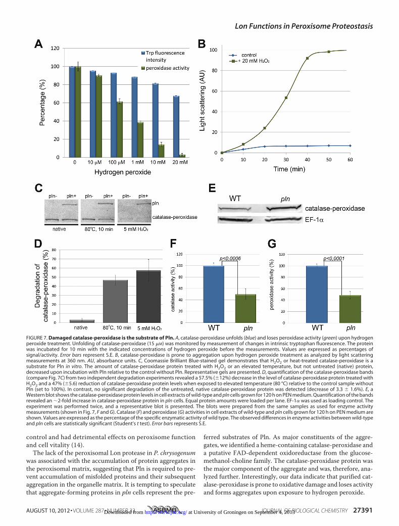
Catalase-peroxidase, one of the major Triton X-100 insolubleproteins in peroxisome fractions of pln cells (Fig. 6C), wasselected for further analysis. As peroxisomes are oxidativeorganelles, we studied whether oxidative stress may cause cat-alase-peroxidase enzyme inactivation or protein aggregation.Catalase-peroxidase was produced in E. coli and purified to
homogeneity (supplemental Fig. S1). Enzyme assays revealedthat the catalase-peroxidase enzyme displayed catalase andperoxidase activities with pH optima of 6–7 and 5, respectively(supplemental Fig. S4). Treatment of purified catalase-peroxi-dase proteinwith increasing concentrations ofH2O2 resulted ina decrease in intrinsic Trp fluorescence and peroxidase enzymeactivity, indicative for protein unfolding (Fig. 7A). Relativelylow concentrations of H2O2 already caused significant changesin Trp fluorescence and peroxidase activity (enzyme activitydecreased by 40% after treatmentwith 100�MH2O2). Catalase-peroxidase was also prone to aggregation upon treatment withH2O2 in vitro, as evident from light scattering measurements(Fig. 7B). Finally, we tested whether catalase-peroxidase was asubstrate for Pln using an in vitro degradation assay. Thesestudies revealed that the native protein was not degraded byPln. However, upon pretreatment with H2O2 or preincubationat high temperature, the protein was degraded by Pln (Fig. 7, Cand D). These data indicate that unfolded catalase-peroxidase,but not the native protein, is a substrate of the peroxisomal Lon.Pln Displays Chaperone-like Activity—The presence of
aggregates in peroxisomes of pln cells could be explained byaggregation of unfolded catalase-peroxidase protein. Westernblot analysis revealed that pln cells contained enhanced cata-lase-peroxidase protein levels (Fig. 7E), whereas enzyme activ-ity measurements revealed that the catalytic (Fig. 7F) and per-oxidatic activities (Fig. 7G) were reduced. These observationssuggest that Pln may display, additional to its proteolytic activ-ity, also chaperone activity in vivo. To test the putative chaper-one activity of Pln, citrate synthase (CS) was used as a substratein in vitro protein refolding experiments. To test if purified Plncan suppress aggregation of this substrate, guanidine hydro-chloride-denatured CS was diluted into buffer containing puri-fied Pln. Relative to the control without Pln, a significantdecrease inCS aggregation (�40%)was observed (Fig. 8A). Thisdecrease was independent of the presence of ATP (Fig. 8,A andB). The addition of BSA instead of Pln did not preventCS aggre-gation (Fig. 8C). Aggregation of denatured CS was inhibited byPln in a dose-dependent manner (Fig. 8D) and was the mostefficient at a ratio of one or more Pln monomers to two CSmonomers. Purified Pln also stimulated reactivation of chemi-cally denatured CS (Fig. 8F), whichwas also optimal at a ratio ofat least 1:2 Plnmonomers to CSmonomers (Fig. 8E). This stim-ulatory effect of Pln was not observed in the absence of ATP(Fig. 8G). Interestingly, the proteolytically inactive mutant of
FIGURE 5. Localization of GFP containing a PTS1 in WT and pln cells. Fluo-rescence microscopy images showing GFP-SKL localization in WT and plncells grown for 120 h in PEN media. Like in the wild-type control, no cytosolicGFP was observed in pln cells. The scale bars represent 5 �m. DIC, differentialinterference contrast.
Lon Functions in Peroxisome Proteostasis
AUGUST 10, 2012 • VOLUME 287 • NUMBER 33 JOURNAL OF BIOLOGICAL CHEMISTRY 27389 at University of Groningen on September 4, 2013http://www.jbc.org/Downloaded from

Pln (Plnin) facilitated refolding of CSwith the same efficiency asthe active protein (Fig. 8F). These data suggest that Pln is amultifunctional enzyme that, besides proteolytic activity, dis-plays chaperone-like activity that is independent of the proteo-lytic catalytic dyad.
DISCUSSION
In this work we provide evidence that peroxisomes of thefungus P. chrysogenum contain an isoform of the Lon protease,termed Pln, a member of the AAA protein family, that displaysa dual function asATP-stimulated protease in conjunctionwith
a role as molecular chaperone. As such, Pln is the first chaper-one that is identified in fungal peroxisomes.Because proteins are marginally stable macromolecules
with a limited lifespan, we imagined that also peroxisomalmatrix proteins require constant housekeeping processes.As yet, the fate of luminal proteins within the oxidativemilieu of peroxisomes was, however, still an enigma.Recently, a Lon protease was identified in the peroxisomalmatrix of rat (15), yeast (14), and plant (52). Studies per-formed in H. polymorpha indeed suggest that the deletion ofperoxisomal Lon protease (pln) affected organelle quality
FIGURE 6. The absence of Pln results in the formation of protein aggregates in the peroxisomal matrix. A and B, electron microscopy images of a detailof a wild-type (A) and pln (B) cell show the presence of a protein aggregate (arrow, Fig. 6B) in the peroxisomal matrix of the pln cell. The cells were fixed withKMnO4. P, peroxisome; M, mitochondrion; N, nucleus. The scale bar represents 1 �m. C, shown is a Coomassie Brilliant Blue (CBB)-stained SDS-PAGE gel ofTriton-X100-insoluble proteins isolated from peroxisomal fractions obtained from wild-type and pln cells. The indicated protein bands were identified by massspectrometry. GMC, glucose-methanol-choline. D and E, shown are subcellular localizations of N-terminal GFP fusion proteins of catalase-peroxidase (D) andglucose-methanol-choline oxidoreductase (E), analyzed by fluorescence microscopy in cells cultivated for 40 h in PEN media. DsRed-SKL was used as a markerof peroxisomes. The scale bar represents 5 �m.
Lon Functions in Peroxisome Proteostasis
27390 JOURNAL OF BIOLOGICAL CHEMISTRY VOLUME 287 • NUMBER 33 • AUGUST 10, 2012 at University of Groningen on September 4, 2013http://www.jbc.org/Downloaded from
control and had detrimental effects on peroxisome functionand cell vitality (14).The lack of the peroxisomal Lon protease in P. chrysogenum
was associated with the accumulation of protein aggregates inthe peroxisomal matrix, suggesting that Pln is required to pre-vent accumulation of misfolded proteins and their subsequentaggregation in the organelle matrix. It is tempting to speculatethat aggregate-forming proteins in pln cells represent the pre-
ferred substrates of Pln. As major constituents of the aggre-gates, we identified a heme-containing catalase-peroxidase anda putative FAD-dependent oxidoreductase from the glucose-methanol-choline family. The catalase-peroxidase protein wasthe major component of the aggregate and was, therefore, ana-lyzed further. Interestingly, our data indicate that purified cat-alase-peroxidase is prone to oxidative damage and loses activityand forms aggregates upon exposure to hydrogen peroxide.
FIGURE 7. Damaged catalase-peroxidase is the substrate of Pln. A, catalase-peroxidase unfolds (blue) and loses peroxidase activity (green) upon hydrogenperoxide treatment. Unfolding of catalase-peroxidase (15 �M) was monitored by measurement of changes in intrinsic tryptophan fluorescence. The proteinwas incubated for 10 min with the indicated concentrations of hydrogen peroxide before the measurements. Values are expressed as percentages ofsignal/activity. Error bars represent S.E. B, catalase-peroxidase is prone to aggregation upon hydrogen peroxide treatment as analyzed by light scatteringmeasurements at 360 nm. AU, absorbance units. C, Coomassie Brilliant Blue-stained gel demonstrates that H2O2 or heat-treated catalase-peroxidase is asubstrate for Pln in vitro. The amount of catalase-peroxidase protein treated with H2O2 or an elevated temperature, but not untreated (native) protein,decreased upon incubation with Pln relative to the control without Pln. Representative gels are presented. D, quantification of the catalase-peroxidase bands(compare Fig. 7C) from two independent degradation experiments revealed a 57.5% (�12%) decrease in the level of catalase-peroxidase protein treated withH2O2 and a 47% (�5.6) reduction of catalase-peroxidase protein levels when exposed to elevated temperature (80 °C) relative to the control sample withoutPln (set to 100%). In contrast, no significant degradation of the untreated, native catalase-peroxidase protein was detected (decrease of 3.3 � 1.6%). E, aWestern blot shows the catalase-peroxidase protein levels in cell extracts of wild-type and pln cells grown for 120 h on PEN medium. Quantification of the bandsrevealed an �2-fold increase in catalase-peroxidase protein in pln cells. Equal protein amounts were loaded per lane. EF-1� was used as loading control. Theexperiment was performed twice, and a representative blot is presented. The blots were prepared from the same samples as used for enzyme activitymeasurements (shown in Fig. 7, F and G). Catalase (F) and peroxidase (G) activities in cell extracts of wild-type and pln cells grown for 120 h on PEN medium areshown. Values are expressed as the percentage of the specific enzymatic activity of wild type. The observed differences in enzyme activities between wild-typeand pln cells are statistically significant (Student’s t test). Error bars represents S.E.
Lon Functions in Peroxisome Proteostasis
AUGUST 10, 2012 • VOLUME 287 • NUMBER 33 JOURNAL OF BIOLOGICAL CHEMISTRY 27391 at University of Groningen on September 4, 2013http://www.jbc.org/Downloaded from

Western blot analysis revealed that catalase-peroxidase pro-tein levels were elevated in crude extracts of pln cells relative towild type. This indicates that catalase-peroxidase most likelyconstitutes a substrate of the Pln protease in vivo. Indeed, dam-aged catalase-peroxidase (heat denaturated or H2O2 treated),but not the native enzyme, was degraded in vitro by Pln in vitro.pln cells also showed reduced catalase-peroxidase enzyme
activities. Together with our in vitro experiments, which indi-cate that Pln has chaperone activity, this observation suggeststhat Pln also shows chaperone activity toward catalase-peroxi-dase. Hence, the increased catalase-peroxidase protein levelstogether with the reduced enzyme activities most likely is theresult of the absence of both the proteolytic and chaperoneactivities of the Lon protease in P. chrysogenum pln cells.
FIGURE 8. Peroxisomal Lon protease displays chaperone-like activity. A and B, the aggregation kinetics of 150 nM denatured CS were monitored in theabsence of Pln or presence of 80 nM Pln in the presence (A) or absence (B) of 1 mM ATP. AU, absorbance units. C, a reduction in aggregation was not observedwhen the reaction was performed in the presence of 80 nM BSA and ATP (red). D, denatured CS was diluted to a final concentration 150 nM in a solutionsupplemented with different concentrations of Pln (monomer). Aggregation kinetics of CS were measured. Normalized light scattering values obtained after360 s are shown. The highest measured value was set to 100. E, reactivation of 150 nM denatured CS was determined in the presence of 1 mM ATP and differentconcentrations of Pln (monomer). CS reactivation was determined after 15 min of incubation. The highest refolding activity of Pln was set to 100%. F, reacti-vation of 150 nM denatured CS was determined in the presence of 1 mM ATP. Without the addition of Pln, only minor reactivation was observed. However, inthe presence of 80 nM Pln or Plnin, �25% of the CS was reactivated. G, reactivation of denatured CS (150 nM) was similar in reactions containing Pln or lackingPln when the reactions were performed in the absence of ATP.
Lon Functions in Peroxisome Proteostasis
27392 JOURNAL OF BIOLOGICAL CHEMISTRY VOLUME 287 • NUMBER 33 • AUGUST 10, 2012 at University of Groningen on September 4, 2013http://www.jbc.org/Downloaded from

As yet the bulk of the research on Lon substrate specificityhas been carried out using bacterial Pln homologues and artifi-cial substrates. Although some possible substrates have beenproposed for mitochondrial Lon in man and bakers’ yeast,direct evidence for Lon substrates has not been presented so far(19). Here, we show for the first time a bona fide substrate forLon protease, namely catalase-peroxidase for Pln fromP. chrysogenum.Our data are consistent with the view that cat-alase peroxidase, which is a crucial component of peroxisomalantioxidant defense system (26), may exemplify a particularlyvulnerable peroxisomal protein and, therefore, requires sub-stantial turnover by peroxisomal Lon protease.In Arabidopsis thaliana, peroxisomal Lon protease was pro-
posed to be involved in sustaining matrix protein import intoperoxisomes of older cotyledon (52). For mammalian systems,contradictory results were reported; in cells of a HeLa cell line,peroxisomal matrix import was not affected by knockdown ofperoxisomal Lon protease (53), whereas overproduction of adominant negative variant of this protein in HEK293 cellsresulted inmislocalization of the PTS1-containing enzyme cat-alase (54). In the filamentous fungus P. chrysogenum as well asin the methylotropic yeast H. polymorpha, peroxisomal Lonprotease is not involved in sustaining matrix protein import, asmislocalization of GFP-SKL was not observed in these organ-isms (this study and Ref. 14).Surprisingly, in P. chrysogenum, various peroxisomal meta-
bolic pathways function normally in the absence of Pln exceptfor the utilization of oleate, a process that requires peroxisomal�-oxidation. Moreover, oleic acid also was shown to be toxic tothe cells. Activity measurements of the �-oxidation pathway
revealed no major change in pln cells relative to wild-type con-trols. Hence, oleic acid oxidation can proceed normally inthese cells. However, strongly enhanced oxidative stress wasobserved upon the addition of oleic acid to pln cells. A possibleexplanation for the failure of the cells to grow on oleate is thatenhanced levels of H2O2, produced in the first oxidation step ofoleate, inactivates the catalase-peroxidase protein, therebyreleasing its co-factor heme, resulting in the generation of oxy-gen radicals via iron fromheme in the Fenton reaction and thusleading to cell death.Our biophysical data suggest that the P. chrysogenum perox-
isomal Lon protease forms heptameric ring-shaped assemblies.Almost all members of the AAA protein family assemble intoring-like higher order structures (55). A stoichiometry of sevensubunits was previously reported for mitochondrial Lon fromS. cerevisiae (47), whereas bacterial Lon proteases are hexa-meric (19). Stahlberg et al. (47) demonstrated that significantconformational changes occur in yeast mitochondrial Lon pro-tease depending on the presence of ATP. Because we analyzedPln only in the presence of ATP, we cannot exclude that perox-isomal Lon protease undergoes similar ATP-dependent struc-tural changes.The hallmark of the AAA family of proteins is the ATP bind-
ing domain (AAA module), which contains conserved WalkerA and B motifs, that function as an unfoldase (56). Cycles ofATP binding and hydrolysis drive conformational changes, cre-ating pulses of pulling force that denature the substrate andtranslocate the unfolded polypeptide through the pore into theproteolytic chamber. Several cycles of protein binding andrelease are required to accomplish protein degradation by
FIGURE 9. Model proposing the role of Pln in protein quality control in the peroxisome matrix. Peroxisomal matrix proteins most likely are delivered to theorganelle in a folded conformation (1). Peroxisomal matrix proteins encounter stress (e.g. oxidative stress generated by peroxisomal metabolism) that mayaccelerate protein damage (2) that can be followed by protein aggregation (3). To cope with this, peroxisomes possess a bifunctional ATP-dependent Pln thatcontrols the quality of peroxisomal proteome in two ways, as a chaperone protein (4) that facilitates refolding of destabilized peroxisomal proteins and as aprotease that digests misfolded macromolecules (5) to prevent their aggregation. The lack of Pln may lead to accumulation of damaged proteins that maycompromise peroxisomal function and cell viability.
Lon Functions in Peroxisome Proteostasis
AUGUST 10, 2012 • VOLUME 287 • NUMBER 33 JOURNAL OF BIOLOGICAL CHEMISTRY 27393 at University of Groningen on September 4, 2013http://www.jbc.org/Downloaded from

ATP-fueled proteolytic machines (57). Our in vitro experi-ments indicate that Pln displays ATP-driven refolding activi-ties. Our data suggest that a destabilized protein that iscaptured by the peroxisomal Lon is able to escape from trans-location into the proteolytic chamber and, instead of beingdegraded, is refolded. This is noteworthy as the observed chap-erone activitywas independent of the proteolytic activity of Pln.In agreement with these findings, a chaperone-like activity hasbeen suggested before for the mitochondrial isoform of Lonprotease (58) based on in vivo experiments.
Taken together, our data suggest that the P. chrysogenumperoxisomal Lon is a multifunctional protein (Fig. 9) and rep-resents the first fungal peroxisomal chaperone identified. Ourdata are consistent with the view that Pln represents the firstexample of a conserved peroxisomal protein that functions inperoxisomematrix quality control and actively assists in refold-ing of peroxisomal matrix proteins.
Acknowledgments—We gratefully acknowledgeDr. F. UlrichHartl forvaluable discussions. We thank Stefan Weber, Katarzyna Łukaszuk,and Annemarie Kralt for assistance in various parts of this study,EMBL Hamburg for providing resources, and the SPC facility (EMBLHamburg) for technical support.
REFERENCES1. Dobson, C. M. (2003) Protein folding and misfolding. Nature 426,
884–8902. Jaenicke, R. (1998) Protein self-organization in vitro and in vivo: partitioning
between physical biochemistry and cell biology. Biol. Chem. 379, 237–2433. Jahn, T. R., and Radford, S. E. (2005) The Yin and Yang of protein folding.
FEBS J. 272, 5962–59704. Ellis, R. J., and Minton, A. P. (2006) Protein aggregation in crowded envi-
ronments. Biol. Chem. 387, 485–4975. Hartl, F. U., Bracher, A., and Hayer-Hartl, M. (2011) Molecular chaper-
ones in protein folding and proteostasis. Nature 475, 324–3326. Tyedmers, J., Mogk, A., and Bukau, B. (2010) Cellular strategies for con-
trolling protein aggregation. Nat. Rev. Mol. Cell Biol. 11, 777–7887. Powers, E. T., Morimoto, R. I., Dillin, A., Kelly, J. W., and Balch, W. E.
(2009) Biological and chemical approaches to diseases of proteostasis de-ficiency. Annu. Rev. Biochem. 78, 959–991
8. Islinger, M., Grille, S., Fahimi, H. D., and Schrader, M. (2012) The perox-isome. An update on mysteries. Histochem. Cell Biol. 137, 547–574
9. Wanders, R. J., and Waterham, H. R. (2006) Peroxisomal disorders. Thesingle peroxisomal enzyme deficiencies. Biochim. Biophys. Acta 1763,1707–1720
10. Steinberg, S. J., Raymond, G. V., Braverman, N. E., andMoser, A. B. (1993)Peroxisome Biogenesis Disorders, Zellweger Syndrome Spectrum, Univer-sity of Washington, Seattle, WA
11. Antonenkov, V. D., Grunau, S., Ohlmeier, S., and Hiltunen, J. K. (2010)Peroxisomes are oxidative organelles.Antioxid. Redox Signal. 13, 525–537
12. Reeder, B. J., Svistunenko, D. A., Cooper, C. E., and Wilson, M. T. (2004)The radical and redox chemistry of myoglobin and hemoglobin. From invitro studies to human pathology. Antioxid. Redox Signal 6, 954–966
13. Walton, P. A., Hill, P. E., and Subramani, S. (1995) Import of stably foldedproteins into peroxisomes.Mol. Biol. Cell 6, 675–683
14. Aksam, E. B., Koek, A., Kiel, J. A., Jourdan, S., Veenhuis, M., and van derKlei, I. J. (2007) A peroxisomal lon protease and peroxisome degradationby autophagy play key roles in vitality of Hansenula polymorpha cells.Autophagy 3, 96–105
15. Kikuchi, M., Hatano, N., Yokota, S., Shimozawa, N., Imanaka, T., andTaniguchi, H. (2004) Proteomic analysis of rat liver peroxisome. Presenceof peroxisome-specific isozyme of Lon protease. J. Biol. Chem. 279,421–428
16. Wagner, I., Arlt, H., van Dyck, L., Langer, T., and Neupert, W. (1994)Molecular chaperones cooperate with PIM1 protease in the degradationof misfolded proteins in mitochondria. EMBO J. 13, 5135–5145
17. von Janowsky, B., Knapp, K.,Major, T., Krayl,M., Guiard, B., andVoos,W.(2005) Structural properties of substrate proteins determine their prote-olysis by the mitochondrial AAA� protease Pim1. Biol. Chem. 386,1307–1317
18. Bota, D. A., and Davies, K. J. (2002) Lon protease preferentially degradesoxidizedmitochondrial aconitase by anATP-stimulatedmechanism.Nat.Cell Biol. 4, 674–680
19. Venkatesh, S., Lee, J., Singh, K., Lee, I., and Suzuki, C. K. (2012)Multitask-ing in the mitochondrion by the ATP-dependent Lon protease. Biochim.Biophys. Acta 1823, 56–66
20. van Dijken, J. P., Otto, R., and Harder, W. (1976) Growth of Hansenulapolymorpha in a methanol-limited chemostat. Physiological responsesdue to the involvement of methanol oxidase as a key enzyme in methanolmetabolism. Arch. Microbiol. 111, 137–144
21. Bartoszewska, M., Kiel, J. A., Bovenberg, R. A., Veenhuis, M., and van derKlei, I. J. (2011) Autophagy deficiency promotes �-lactam production in.Penicillium chrysogenum. Appl. Environ. Microbiol. 77, 1413–1422
22. Hillenga, D. J., Versantvoort, H. J., Driessen, A. J., and Konings, W. N.(1994) Structural and functional properties of plasma membranes fromthe filamentous fungus. Penicillium chrysogenum. Eur. J. Biochem. 224,581–587
23. Opalinski, L., Kiel, J. A., Homan, T. G., Veenhuis, M., and van der Klei, I. J.(2010) Penicillium chrysogenum Pex14/17p. A novel component of theperoxisomal membrane that is important for penicillin production. FEBSJ. 277, 3203–3218
24. Sambrook, J., Fritsch, E. F., Maniatis, T., and Ford, N. (1989) MolecularCloning: A Laboratory Manual, 2nd Ed., Cold Spring Harbor LaboratoryPress, Cold Spring Harbor, NY
25. Kiel, J. A., Hilbrands, R. E., Bovenberg, R. A., and Veenhuis, M. (2000)Isolation of Penicillium chrysogenum PEX1 and PEX6 encoding AAA pro-teins involved in peroxisome biogenesis. Appl. Microbiol. Biotechnol. 54,238–242
26. Kiel, J. A., van den Berg, M. A., Fusetti, F., Poolman, B., Bovenberg, R. A.,Veenhuis, M., and van der Klei, I. J. (2009) Matching the proteome to thegenome. Themicrobody of penicillin-producingPenicillium chrysogenumcells. Funct. Integr. Genomics 9, 167–184
27. Fraaije, M. W., Roubroeks, H. P., Hagen, W. R., and Van Berkel, W. J.(1996) Purification and characterization of an intracellular catalase-per-oxidase from. Penicillium simplicissimum. Eur. J. Biochem. 235, 192–198
28. Bender, T., Lewrenz, I., Franken, S., Baitzel, C., and Voos, W. (2011) Mi-tochondrial enzymes are protected from stress-induced aggregation bymitochondrial chaperones and the Pim1/LONprotease.Mol. Biol. Cell22,541–554
29. van Roermund, C. W., Elgersma, Y., Singh, N., Wanders, R. J., and Tabak,H. F. (1995) Themembrane of peroxisomes in Saccharomyces cerevisiae isimpermeable toNAD(H) and acetyl-CoAunder in vivo conditions.EMBOJ. 14, 3480–3486
30. Paraszkiewicz, K., Bernat, P., Naliwajski,M., andDługonski, J. (2010) Lipidperoxidation in the fungus Curvularia lunata exposed to nickel. Arch.Microbiol. 192, 135–141
31. Buchner, J., Grallert, H., and Jakob, U. (1998) Analysis of chaperone func-tion using citrate synthase as nonnative substrate protein.Methods Enzy-mol. 290, 323–338
32. West, S. M., Kelly, S. M., and Price, N. C. (1990) The unfolding and at-tempted refolding of citrate synthase from pig heart. Biochim. Biophys.Acta 1037, 332–336
33. Nettleship, J. E., Brown, J., Groves, M. R., and Geerlof, A. (2008) Methodsfor protein characterization by mass spectrometry, thermal shift (Ther-moFluor) assay, and multiangle or static light scattering. Methods Mol.Biol. 426, 299–318
34. Roessle, M., Klaering, R., Ristau, U., Robrahn, B., Jahn, D., Gehrmann, T.,Konarev, P., Round, A., Fiedler, S., Hermes, C., and Svergun, D. (2007)Upgrade of the small-angle X-ray scattering beamline X33 at the Euro-pean Molecular Biology Laboratory, Hamburg. J. Appl. Crystallogr. 40,190–194
Lon Functions in Peroxisome Proteostasis
27394 JOURNAL OF BIOLOGICAL CHEMISTRY VOLUME 287 • NUMBER 33 • AUGUST 10, 2012 at University of Groningen on September 4, 2013http://www.jbc.org/Downloaded from

35. Guinier, A. (1939) La diffraction des rayons X aux tres petits angles; ap-plication a l’etude de phenomenes ultramicroscopiques.Ann. Phys. (Paris)12, 161–237
36. Petoukhov, M. V., Konarev, P., Kikhney, A. G., and Svergun, D. I. (2007)ATSAS 2.1. Toward automated andweb-supported small-angle scatteringdata analysis. J. Appl. Crystallogr. 40, 223–228
37. Franke, D., and Svergun, D. I. (2009) DAMMIF, a program for rapid abinitio shape determination in small-angle scattering. J. Appl. Crystallogr.42, 342–346
38. Volkov, V. V., and Svergun, D. I. (2003) Uniqueness of ab initio shapedetermination in small-angle scattering. J. Appl. Crystallogr. 36, 860–864
39. Kozin, M., and Svergun, D. I. (2001) Automatedmatching of high and lowresolution structural models. J. Appl. Crystallogr. 34, 33–41
40. Petoukhov, M. V., and Svergun, D. I. (2005) Global rigid bodymodeling ofmacromolecular complexes against small-angle scattering data.Biophys. J.89, 1237–1250
41. Petoukhov, M. V., Eady, N. A., Brown, K. A., and Svergun, D. I. (2002)Addition of missing loops and domains to protein models by x-ray solu-tion scattering. Biophys. J. 83, 3113–3125
42. Svergun, D. I., Barberato, C., and Koch, M. H. J. (1995) CRYSOL. A pro-gram to evaluate x-ray solution scattering of biological macromoleculesfrom atomic coordinates. J. Appl. Crystallogr. 28, 768–773
43. Waterham,H. R., Titorenko, V. I., Haima, P., Cregg, J.M., Harder,W., andVeenhuis, M. (1994) The Hansenula polymorpha PER1 gene is essentialfor peroxisome biogenesis and encodes a peroxisomalmatrix protein withboth carboxyl- and amino-terminal targeting signals. J. Cell Biol. 127,737–749
44. Goldberg, A. L., Moerschell, R. P., Chung, C. H., andMaurizi, M. R. (1994)ATP-dependent protease La (lon) from Escherichia coli. Methods Enzy-mol. 244, 350–375
45. Roudiak, S. G., Seth, A., Knipfer, N., and Shrader, T. E. (1998) The lonprotease from Mycobacterium smegmatis. Molecular cloning, sequenceanalysis, functional expression, and enzymatic characterization. Biochem-istry 37, 377–386
46. Lee, A. Y., Hsu, C.H., andWu, S.H. (2004) Functional domains ofBreviba-cillus thermoruber lon protease for oligomerization and DNA binding.Role of N-terminal and sensor and substrate discrimination domains.J. Biol. Chem. 279, 34903–34912
47. Stahlberg, H., Kutejova, E., Suda, K., Wolpensinger, B., Lustig, A., Schatz,G., Engel, A., and Suzuki, C. K. (1999) Mitochondrial Lon of Saccharomy-ces cerevisiae is a ring-shaped protease with seven flexible subunits. Proc.Natl. Acad. Sci. U.S.A. 96, 6787–6790
48. Quiocho, F. A., Spurlino, J. C., and Rodseth, L. E. (1997) Extensive featuresof tight oligosaccharide binding revealed in high resolution structures ofthe maltodextrin transport/chemosensory receptor. Structure 5,997–1015
49. Arnold, K., Bordoli, L., Kopp, J., and Schwede, T. (2006) The SWISS-MODEL workspace. A web-based environment for protein structure ho-mology modeling. Bioinformatics 22, 195–201
50. Meijer, W. H., Gidijala, L., Fekken, S., Kiel, J. A., van den Berg, M. A.,Lascaris, R., Bovenberg, R. A., and van der Klei, I. J. (2010) Peroxisomes arerequired for efficient penicillin biosynthesis in. Penicillium chrysogenum.Appl. Environ. Microbiol. 76, 5702–5709
51. Liu, F., Lu, Y., Pieuchot, L., Dhavale, T., and Jedd, G. (2011) Import oligo-mers induce positive feedback to promote peroxisome differentiation andcontrol organelle abundance. Dev. Cell 21, 457–468
52. Lingard, M. J., and Bartel, B. (2009) Arabidopsis LON2 is necessary forperoxisomal function and sustained matrix protein import. Plant Physiol.151, 1354–1365
53. Okumoto, K., Kametani, Y., and Fujiki, Y. (2011) Two proteases, trypsindomain-containing 1 (Tysnd1) and peroxisomal lon protease (PsLon), co-operatively regulate fatty acid �-oxidation in peroxisomal matrix. J. Biol.Chem. 286, 44367–44379
54. Omi, S., Nakata, R., Okamura-Ikeda, K., Konishi, H., and Taniguchi, H.(2008) Contribution of peroxisome-specific isoform of Lon protease insorting PTS1 proteins to peroxisomes. J. Biochem. 143, 649–660
55. Iyer, L.M., Leipe, D. D., Koonin, E. V., andAravind, L. (2004) Evolutionaryhistory and higher order classification of AAA� ATPases. J. Struct. Biol.146, 11–31
56. Hanson, P. I., andWhiteheart, S.W. (2005) AAA� proteins. Have engine,will work. Nat. Rev. Mol. Cell Biol. 6, 519–529
57. Sauer, R. T., Bolon, D. N., Burton, B. M., Burton, R. E., Flynn, J. M., Grant,R. A.,Hersch,G. L., Joshi, S. A., Kenniston, J. A., Levchenko, I., Neher, S. B.,Oakes, E. S., Siddiqui, S. M., Wah, D. A., and Baker, T. A. (2004) Sculptingthe proteome with AAA(�) proteases and disassembly machines. Cell119, 9–18
58. Rep,M., vanDijl, J.M., Suda, K., Schatz, G., Grivell, L. A., and Suzuki, C. K.(1996) Promotion of mitochondrial membrane complex assembly by aproteolytically inactive yeast Lon. Science 274, 103–106
59. Nijland, J. G., Kovalchuk, A., van den Berg, M. A., Bovenberg, R. A., andDriessen, A. J. (2008) Expression of the transporter encoded by the cefTgene of Acremonium chrysogenum increases cephalosporin production inPenicillium chrysogenum. Fungal Genet. Biol. 45, 1415–1421
Lon Functions in Peroxisome Proteostasis
AUGUST 10, 2012 • VOLUME 287 • NUMBER 33 JOURNAL OF BIOLOGICAL CHEMISTRY 27395 at University of Groningen on September 4, 2013http://www.jbc.org/Downloaded from
Copyright © 2022 FDOKUMEN


![The Drowsy Chaperone Program [2012] - USM Digital ...](https://static.fdokumen.com/doc/165x107/6322912f887d24588e044a66/the-drowsy-chaperone-program-2012-usm-digital-.jpg)