
Carboxypeptidase E promotes cancer cell survival, but inhibits migration and invasion
10
Carboxypeptidase E promotes cancer cell survival, but inhibits migration and invasion Saravana R.K. Murthy 1 , Evan Dupart 1 , Najla Al-Sweel, Alexander Chen, Niamh X. Cawley, Y. Peng Loh ⇑ Section on Cellular Neurobiology, Program on Developmental Biology, Eunice Kennedy Shriver National Institute of Child Health and Human Development, Bethesda, MD 20892, USA article info Article history: Received 21 May 2013 Received in revised form 1 August 2013 Accepted 6 August 2013 Available online xxxx Keywords: Pheochromocytoma Hepatocellular carcinoma Fibrosarcoma Cell survival Cell invasion abstract Carboxypeptidase E (CPE), a prohormone processing enzyme is highly expressed and secreted from (neuro)endocrine tumors and gliomas, and has been implicated in cancer progression by promoting tumor growth. Our study demonstrates that secreted or exogenously applied CPE promotes survival of pheochromocytoma (PC12) and hepatocellular carcinoma (MHCC97H) cells under nutrient starvation and hypoxic conditions, but had no effect on their proliferation. CPE also reduced migration and invasion of fibrosarcoma (HT1080) cells. We show that CPE treatment mediates survival of MHCC97H cells during metabolic stress by up-regulating the expression of anti-apoptotic protein BCL-2, and other pro-survival genes, via activation of the ERK1/2 pathway. Published by Elsevier Ireland Ltd. 1. Introduction Cancer is the second leading cause of death worldwide, yet de- spite extensive research, the mechanisms involved in tumor growth, survival and metastatic potential are still not fully under- stood [1]. Neuroendocrine cancers such as pheochromocytoma, are some of the least understood forms of the disease [2]. It is well established that various types of (neuro)endocrine tumors, includ- ing pheochromocytomas, lung small cell carcinomas [3], insulino- mas [4], breast adenocarcinoma [5] and glioblastomas [6] secrete large numbers of hormones and growth factors, as well as their processing enzymes, such as carboxypeptidase E (CPE) and pept- idylglycine alpha-amidating mono-oxygenase [7]. Among the pro- hormone processing enzymes, CPE has been the most studied with respect to tumorigenesis. CPE is a multifunctional protein that in addition to its enzymatic function, sub-serves many essential non-enzymatic roles in the endocrine and nervous system, besides a role in cancer (for a review, see Ref. [8]) and it has been recently shown to act as a trophic factor [9]. High throughput microarray studies have correlated elevated CPE mRNA levels with metastasis in a number of non-endocrine cancers [10], although the form of CPE protein translated from these mRNAs was not analyzed. Recently, an N-terminally trun- cated splice isoform of CPE, known as CPE-DN was identified and shown to be a molecule that induces tumor metastasis [11]. It is elevated in highly metastatic hepatocellular carcinoma (HCC), breast, colon and head and neck cancer cell lines compared to their isogenic low metastatic counterparts. CPE-DN is also a powerful biomarker in diagnosing and predicting future metastasis in sev- eral cancers including HCC, pheochromocytoma/paraganglioma (PHEO/PGL) and colorectal cancer [11,12]. CPE-DN, up-regulates the expression of Nedd9, a metastatic gene in a HDAC1/2–depen- dent manner [11]. In addition to the CPE-DN splice variant just de- scribed, the primary gene product of the CPE gene, full length wild- type CPE (referred to as CPE) is normally localized in regulated secretory granules of (neuro)endocrine cells. Within these granules it processes peptide hormone and neuropeptide intermediates to their mature bioactive peptides. CPE is secreted from (neuro)endo- crine tumor and glioblastoma (GBM) cell lines. It was found that CPE acts as a pro-growth, but anti-metastatic factor [6]. Another recent study has shown that CPE acts extracellularly as a negative regulator of the canonical Wnt signaling pathway [13] and as a neurotrophic factor to protect neurons against oxidative stress in- duced cell death [9] or during chronic stress [14]. However, the mechanism of action of CPE in these functions is not fully understood. Neuroendocrine tumors such as PHEO/PGL express both CPE and CPE-DN. However, we found that only high levels of the splice isoform, but not CPE, was correlated with enhanced PHEO/PGL tu- mor metastasis and poor prognosis [11]. In this paper, we investi- gated whether secreted CPE from these tumors could serve a different role, perhaps as a survival/proliferative factor, similar to that reported for glioblastoma [6]. To determine this possibility, 0304-3835/$ - see front matter Published by Elsevier Ireland Ltd. http://dx.doi.org/10.1016/j.canlet.2013.08.011 ⇑ Corresponding author. Address: 49, Convent Drive, Bldg. 49, Rm. 5A22, Bethesda, MD 20892, USA. Tel.: +1 301 4963239; fax: +1 301 4969938. E-mail address: [email protected] (Y.P. Loh). 1 These authors contributed equally to the work. Cancer Letters xxx (2013) xxx–xxx Contents lists available at ScienceDirect Cancer Letters journal homepage: www.elsevier.com/locate/canlet Please cite this article in press as: S.R.K. Murthy et al., Carboxypeptidase E promotes cancer cell survival, but inhibits migration and invasion, Cancer Lett. (2013), http://dx.doi.org/10.1016/j.canlet.2013.08.011
Transcript of Carboxypeptidase E promotes cancer cell survival, but inhibits migration and invasion

Carboxypeptidase E promotes cancer cell survival, but inhibits migration
and invasion
Saravana R.K. Murthy 1, Evan Dupart 1, Najla Al-Sweel, Alexander Chen, Niamh X. Cawley, Y. Peng Loh ⇑
Section on Cellular Neurobiology, Program on Developmental Biology, Eunice Kennedy Shriver National Institute of Child Health and Human Development, Bethesda, MD 20892, USA
a r t i c l e i n f o
Article history:
Received 21 May 2013
Received in revised form 1 August 2013
Accepted 6 August 2013
Available online xxxx
Keywords:
Pheochromocytoma
Hepatocellular carcinoma
Fibrosarcoma
Cell survival
Cell invasion
a b s t r a c t
Carboxypeptidase E (CPE), a prohormone processing enzyme is highly expressed and secreted from
(neuro)endocrine tumors and gliomas, and has been implicated in cancer progression by promoting
tumor growth. Our study demonstrates that secreted or exogenously applied CPE promotes survival of
pheochromocytoma (PC12) and hepatocellular carcinoma (MHCC97H) cells under nutrient starvation
and hypoxic conditions, but had no effect on their proliferation. CPE also reduced migration and invasion
of fibrosarcoma (HT1080) cells. We show that CPE treatment mediates survival of MHCC97H cells during
metabolic stress by up-regulating the expression of anti-apoptotic protein BCL-2, and other pro-survival
genes, via activation of the ERK1/2 pathway.
Published by Elsevier Ireland Ltd.
1. Introduction
Cancer is the second leading cause of death worldwide, yet de-
spite extensive research, the mechanisms involved in tumor
growth, survival and metastatic potential are still not fully under-
stood [1]. Neuroendocrine cancers such as pheochromocytoma, are
some of the least understood forms of the disease [2]. It is well
established that various types of (neuro)endocrine tumors, includ-
ing pheochromocytomas, lung small cell carcinomas [3], insulino-
mas [4], breast adenocarcinoma [5] and glioblastomas [6] secrete
large numbers of hormones and growth factors, as well as their
processing enzymes, such as carboxypeptidase E (CPE) and pept-
idylglycine alpha-amidating mono-oxygenase [7]. Among the pro-
hormone processing enzymes, CPE has been the most studied with
respect to tumorigenesis. CPE is a multifunctional protein that in
addition to its enzymatic function, sub-serves many essential
non-enzymatic roles in the endocrine and nervous system, besides
a role in cancer (for a review, see Ref. [8]) and it has been recently
shown to act as a trophic factor [9].
High throughput microarray studies have correlated elevated
CPE mRNA levels with metastasis in a number of non-endocrine
cancers [10], although the form of CPE protein translated from
these mRNAs was not analyzed. Recently, an N-terminally trun-
cated splice isoform of CPE, known as CPE-DN was identified and
shown to be a molecule that induces tumor metastasis [11]. It is
elevated in highly metastatic hepatocellular carcinoma (HCC),
breast, colon and head and neck cancer cell lines compared to their
isogenic low metastatic counterparts. CPE-DN is also a powerful
biomarker in diagnosing and predicting future metastasis in sev-
eral cancers including HCC, pheochromocytoma/paraganglioma
(PHEO/PGL) and colorectal cancer [11,12]. CPE-DN, up-regulates
the expression of Nedd9, a metastatic gene in a HDAC1/2–depen-
dent manner [11]. In addition to the CPE-DN splice variant just de-
scribed, the primary gene product of the CPE gene, full length wild-
type CPE (referred to as CPE) is normally localized in regulated
secretory granules of (neuro)endocrine cells. Within these granules
it processes peptide hormone and neuropeptide intermediates to
their mature bioactive peptides. CPE is secreted from (neuro)endo-
crine tumor and glioblastoma (GBM) cell lines. It was found that
CPE acts as a pro-growth, but anti-metastatic factor [6]. Another
recent study has shown that CPE acts extracellularly as a negative
regulator of the canonical Wnt signaling pathway [13] and as a
neurotrophic factor to protect neurons against oxidative stress in-
duced cell death [9] or during chronic stress [14]. However, the
mechanism of action of CPE in these functions is not fully
understood.
Neuroendocrine tumors such as PHEO/PGL express both CPE
and CPE-DN. However, we found that only high levels of the splice
isoform, but not CPE, was correlated with enhanced PHEO/PGL tu-
mor metastasis and poor prognosis [11]. In this paper, we investi-
gated whether secreted CPE from these tumors could serve a
different role, perhaps as a survival/proliferative factor, similar to
that reported for glioblastoma [6]. To determine this possibility,
0304-3835/$ - see front matter Published by Elsevier Ireland Ltd.
http://dx.doi.org/10.1016/j.canlet.2013.08.011
⇑ Corresponding author. Address: 49, Convent Drive, Bldg. 49, Rm. 5A22,
Bethesda, MD 20892, USA. Tel.: +1 301 4963239; fax: +1 301 4969938.
E-mail address: [email protected] (Y.P. Loh).1 These authors contributed equally to the work.
Cancer Letters xxx (2013) xxx–xxx
Contents lists available at ScienceDirect
Cancer Letters
journal homepage: www.elsevier .com/locate /canlet
Please cite this article in press as: S.R.K. Murthy et al., Carboxypeptidase E promotes cancer cell survival, but inhibits migration and invasion, Cancer Lett.
(2013), http://dx.doi.org/10.1016/j.canlet.2013.08.011
we added neutralizing CPE antibodies to the culture medium of rat
pheochromocytoma-derived PC12 cells [15] and found that the
cells showed significantly decreased survival in serum free media.
We then investigated the mechanism of action of CPE as a survival
factor, using a highly metastatic HCC cell line (MHCC97H) lacking
background endogenous CPE [11], as a model. We examined the
survival effect of exogenously added CPE during metabolic stress
on these cells, as well as the signaling pathway and expression of
downstream genes. We also used the highly invasive and aggres-
sive fibrosarcoma cell line HT1080, which does not express wild
type CPE (see Suppl. Fig. S1), but only CPE-DN, to assay the effects
of CPE on invasion and migration.
2. Materials and methods
2.1. Cell lines
Rat pheochromocytoma PC12 cells were obtained from ATCC
(Manassas, VA). The human HCC cell line, MHCC97H, was obtained
from the Liver Cancer Institute, Fudan University (Shanghai, Chi-
na). The HT1080 fibrosarcoma cells were obtained from TREVIGEN
Inc. (Gaithersburg, MD).
2.2. Secretion studies for PC12 cells
PC12 cells were seeded on 10 cm dishes and allowed to grow to
70–85% confluency in DMEM medium (ATCC, Manassas, VA) sup-
plemented with 10% FBS and 5% horse serum. Cells were later
washed with 1� PBS and 1.5 mL of serum free DMEM medium
was added to the cells. After 24 h the medium was collected and
briefly centrifuged to remove debris. The supernatant containing
secreted proteins was collected and stored at �80 �C for later use.
2.3. Treatment of PC12 cells with CPE antibodies and cell survival
assay
PC12 cells were seeded on a 96 well plate and allowed to grow
to 50–75% confluency in DMEM supplemented with 10% FBS and
5% horse serum. Cells were washed with 1X PBS and then low glu-
cose (1 g/L D-glucose) serum free (LGSF) DMEMmediumwas added
to induce stress. To neutralize the action of any CPE secreted from
the cells, 0.25 lg of rabbit polyclonal anti-CPE IgG (Anti-CPE
#6135, generated in our Lab, was custom made against an 18 aa
synthetic peptide corresponding to aa #362-379 of mouse CPE,
NCBI accession# NP_038522 and coupled to KLH at the N-termi-
nus), was added to each of 6 experimental wells. Another six wells
had 0.25 lg of IgGs from a non-specific rabbit antibody added to
the medium to serve as an antibody control. Six wells were left
completely untreated in serum free DMEM medium. This plate
containing the three groups of were immediately placed inside
the Modular Incubator Chamber (MIC-101) (Billups-Rothenburg),
sealed, and flushed with a low oxygen mixture comprising of 1%
oxygen, 5% carbon dioxide, and 94% nitrogen for 60 s at a rate of
40 L/min after which it was placed into the 37 �C incubator. The
chamber was subsequently flushed every 12 h as described above,
to ensure that a uniform hypoxic environment was maintained. Six
wells in a separate plate were untreated in DMEM complete media
and placed in the incubator for 24 h to serve as a normal cell con-
trol. After the incubation for 24 h under the various conditions all
cells were harvested to evaluate the cytotoxic effects of nutrient
deprivation and hypoxia on the cells using the a LDH assay (Prome-
ga, Madison, WI) according to manufacturer’s instructions.
2.4. Cell viability and proliferation analysis
Cell viability or cytotoxicity was evaluated by determining the
levels of LDH released into the culture media after various treat-
ments [16]. LDH was assayed with a CytoTox 96 Non-Radioactive
Cytotoxicity Assay kit according to the manufacturer’s instructions
(Promega, Madison, WI). Proliferation was evaluated by quantifica-
tion of the DNA binding dye, DRAQ5 that fluoresces at 680 nm
upon binding to DNA and is a direct measure of cell density. HCC
cells were plated at a density of 40–50% and allowed to grow to
�75% confluency overnight at 37 �C, 5% CO2 in DMEM medium
supplemented with 10 % fetal calf serum, sodium pyruvate
(0.11 mg/mL), penicillin (100 U/mL) and streptomycin (100 mg/
mL). The medium was then removed and after rinsing with 1�
PBS, the cells were incubated with medium with or without (con-
trol) purified recombinant CPE protein (200 nM). The cells were
stained with DRAQ5 (BioStatus Ltd., UK) according to the manufac-
turer’s instructions at the following time points: 0, 8, 24, 34, 48, 56,
and 72 h. Cells stained with DRAQ5 were analyzed on the Odyssey
infrared imager at 680 nm. Integrated intensity values were ob-
tained and used to generate a growth curve.
2.5. Metabolic stress paradigm for HCC cells
HCC cells were plated at a density of 40–50% and allowed to
grow to �75% confluency overnight in as mentioned above. The
following day the medium was changed to LGSF medium and the
plates were immediately placed inside the Modular Incubator
Chamber (MIC-101) (Billups-Rothenburg), sealed, and flushed with
a low oxygen mixture comprising of 1% oxygen (as described above
for PC12 cells). The chamber was placed into the 37 �C incubator
and subsequently flushed every 12 h as described above to ensure
that a uniform hypoxic environment was maintained.
2.6. Treatment of HCC cells with exogenous CPE
HCC cells were treated with different concentrations (50–
400 nM) of recombinant mouse full length CPE (custom expressed
in HEK293 cells and purified by Creative Biolabs, Shirley, NY) by
adding it to the cell medium at zero time point of each experiment.
A concentration of 200 nM was established as the most effective
dose for eliciting an increase in the phosphorylation of ERK 1/2.
Since the CPE is very stable in the media, no additional CPE was
added throughout the experiment. It was also found that human
CPE lacking the C-terminal cytoplasmic tail (purchased from Sino
Biological Inc., Beijing, China) gave the same results as full length
mCPE. For inhibition of CPE activity, the HCC cells were treated
with 5 lM guanidino-ethyl-mercapto-succinic acid (GEMSA), a
competitive inhibitor of CPE with a Ki �8 nM [17] along with
200 nM CPE. For inhibition of ERK 1/2, cells were treated with
10 lM U0126 for 30 mins with/without 200 nM CPE. After
30 min the cells were harvested and proteins were analyzed by
Western blotting.
2.7. Western blot for CPE, BCL-2, ERK 1/2, GSK3b/a, AKT, b-catenin and
PKC
Proteins from cell lines were prepared using cell lysis buffer
(Cell Signaling Technology) supplemented with Complete Inhibitor
Cocktail (Roche) to prevent protein degradation. The cell lysate
was collected and centrifuged at 15,000g for 10 min at 4 �C and
the protein concentration in the supernatant determined using
the Bio-Rad Protein Assay. Twenty lg of protein was denatured
at 95 �C for 3 min, ran on 4–12% SDS–PAGE gels, and then trans-
ferred onto nitrocellulose membrane (Invitrogen), according to
standard protocols. After blocking with 5% nonfat milk at 4 �C for
2 S.R.K. Murthy et al. / Cancer Letters xxx (2013) xxx–xxx
Please cite this article in press as: S.R.K. Murthy et al., Carboxypeptidase E promotes cancer cell survival, but inhibits migration and invasion, Cancer Lett.
(2013), http://dx.doi.org/10.1016/j.canlet.2013.08.011

3 h, BCL-2 was detected using a monoclonal antibody (Cell Signal-
ing Technology, Inc., Danvers, MA) at a 1:1000 dilution. Phospho-
ERK was detected using a monoclonal antibody which crossreacts
with p-ERK 1/2 at 1:1000 dilution. CPE was detected using a mouse
anti-CPE monoclonal antibody against the 49-200 amino acid se-
quence (BD Biosciences, San Jose, CA) at 1:4000 dilution. Anti-
phospho-AKT monoclonal antibody, anti-AKT polyclonal antibody,
anti-phospho-PKC (pan) monoclonal antibody, anti-b-catenin poly-
clonal antibody, phospho-GSK3b(Ser9), phospho-GSK3b(Tyr216),
total-GSK3bpolyclonal antibodies (Cell Signaling), anti-active-b-
catenin monoclonal antibody (Santa Cruz) at 1:3000, 1:3000, 1:
2000, 1:3000, 1:3000, 1:1000 dilutions respectively. After incubat-
ing with fluorophore-conjugated anti-mouse secondary antibodies
(Rockland Immunochemicals, Inc, Gilbertsville, PA), the bands
were visualized by the Odyssey infrared imaging system version
2.1 (LI-COR Biotechnology, Lincoln, NE) and the direct integrated
pixel intensity of acquired light from the bands were measured
and analyzed by Odyssey Image Studio Ver 2.0 software (LI-COR
Biotechnology, Lincoln, NE).
2.8. Quantitative real-time RT-PCR
RNA was extracted from MHCC97H cells using the RNeasy Mini
Kit (Qiagen), and first-strand cDNA was synthesized with 1 lg of
total RNA from these cells using Transcriptor First Strand cDNA
Synthesis Kit (Roche Applied Science). Quantitative PCR was per-
formed using 1 lL first strand cDNA under the conditions of
95 �C for 15 s, annealing at 60 �C for 60 s, extension at 72 �C for
30 s for 40 cycles, and a final extension at 72 �C for 10 min using
SYBR Green Master Mix (Applied Biosciences). Primer sequences
for analyzing 18S RNA were used for normalization. Primer
sequences were: for amplifying 18S RNA, fwd: 50-CTCTTAGCT-
GAGTGTCCCGC-30, rev: 50 CTGATCGTCTTCGAACCTCC-30; TNF fwd
50-CAGAGGGCCTGTACCTCATC-30 rev 50-GGAAGACCCCTCCCAGA-
TAG-30; IL-8 fwd: 50-GTGCAGTTTTGCCAAGGAGT-30 rev: 50-CTCTG
CACCAGTTTTCCTT-30; FOXC2 wd: 50-AGTTCATCATGGACCGCTTC-
30 rev: 50-TCTCCTTGGACACGTCCTTC-30; NF-kB fwd: 50-CCTGGATG
ACTCTTGGGAAA-30 rev: TCAGCCAG CTGTTTCATG TC-30; IKBafwd: 50-GCAAAATCCTGACCTGGTGT-30 rev: GCTCGTCCTCTGT
GAACTCC-30. BCL-2 fwd: 50 TTCTTTGAGTTCGG TGGGGTC-30
rev_50-TGCATATTTGTTTGGGGCAGG-30.
2.9. PCR microarray
The Human Cancer Pathway Finder Superarray (PAHS-033Z;
Qiagen, Valencia, CA) was used to analyze mRNA levels of 84 genes
related to cell proliferation, apoptosis, cell cycle, angiogenesis,
invasion, and metastasis. Quality control of RNA samples, synthesis
of cDNA, and real-time RT-PCR arrays were performed as described
previously (4). All genes represented by the array showed a single
peak on the melting curve characteristic of the specific products.
Data analysis of gene expression was performed using Excel-based
PCR Array Data Analysis Software provided by the manufacturer
(Qiagen). Fold-changes in gene expression were calculated using
the DDCt method, and five stably expressed housekeeping genes
(b2-Microglobulin (b2M), hypoxanthine phosphoribosyltransfer-
ase 1, ribosomal protein L13a, GAPDH, and b-actin) were used for
normalization of the results.
2.10. Wound healing assays
Approximately 5 � 104 HCC cells were plated on a 30l-Dish35 mm high culture-chamber (Ibidi, Martinsried, Germany) and
were allowed to form a monolayer. A wound of �500 lm was cre-
ated using the manufacturer’s protocol. The cells were then treated
with 200 nM CPE in complete DMEM (or left untreated in complete
DMEM to serve as a control) and placed on a live cell imaging incu-
bated microscope. Using Metamorph image processing software, a
picture was taken of the wound every minute for 12 h. The pictures
were then evaluated to determine the time it took for the wound to
completely heal with and without CPE treatment. The wound heal-
ing data was analyzed by ImageJ software (NIH, Bethesda, MD,
USA) (http://imagej.nih.gov/ij/).
2.11. Invasion assays
Cell invasion assays were carried out using Trevigen Cultrex 24
well cell invasion assay kits (Trevigen Inc, Gaithersburg, MD). Inva-
sion chambers were coated with a 1� solution of Basement Mem-
brane Extract (BME) per the manufacturer’s protocol and allowed
to gel overnight. The following day, 5 � 104 serum starved
HT1080 cells were added to the top of each chamber in 100 lL of
serum free media. Five hundred lL of complete DMEM, which
serves as the chemo-attractant, was added to the bottom of each
well. Three chambers were treated with 200 nM CPE and three
wells were left as untreated controls. After 19 h of incubation at
37 �C, the chambers were removed and the assay was developed
using a calcein marker included with the kit, and read on a fluores-
cent plate reader.
2.12. SiRNA treatment
HCC cells were plated at a density of 40–50% and allowed to
grow to �75% confluency overnight as mentioned above. The fol-
lowing day cells were treat with/out 20 pmole BCL-2 siRNA
(Ambion, Carlsbad, CA) with anti-sense seq 50
AAUGGAUGUACUUCAUCACTA 30 using Lipofectamine RNAiMAX
transfection reagent (Invitrogen, Carlsbad, CA) according to manu-
facturers’ protocol and incubated under metabolic stress paradigm
as mentioned above. After 12 h, subsets of HCC cells were treated
with/out BCL-2 siRNA and with/out 200 nM recombinant CPE,
Fig. 1. Treatment of PC12 cells with CPE antibodies inhibited cell survival. (A)
Immunoblot of CPE secreted into the media from cells with/without metabolic
stress. PC12 cells were subjected to metabolic stress by replacing DMEM supple-
mented with 10% FBS (complete media) with low glucose (1 g/L D-glucose) serum
free (LGSF) DMEM media under hypoxic condition. B) Bar graphs show viability of
PC12 cells maintained either in normal complete media indicated as NC (Normal
cells), or under metabolic stress conditions (control) with addition of non-specific
IgG (nIgG), or with rabbit polyclonal anti-CPE IgGs (CPE-Ab). Cell viability was
evaluated by the LDH assay (�p < 0.05, ��p < 0.01, ���p < 0.001, Student’s t-test).
S.R.K. Murthy et al. / Cancer Letters xxx (2013) xxx–xxx 3
Please cite this article in press as: S.R.K. Murthy et al., Carboxypeptidase E promotes cancer cell survival, but inhibits migration and invasion, Cancer Lett.
(2013), http://dx.doi.org/10.1016/j.canlet.2013.08.011
and control cells were treated with mock transfection. Forty eight
hours after CPE treatment, cell viability or cytotoxicity was evalu-
ated by LDH assay (Promega, Madison, WI). Cells were harvested
and total RNA and protein lysates were prepared. Expression of
BCL-2mRNA and protein were determined by qRTPCR andWestern
blotting respectively as mentioned above.
2.13. Statistical analysis
Data presented represent the means of a minimum of 3–6 sep-
arate cultures. Data were analyzed by Student’s t-test or one-way
analysis of variance (ANOVA). Error bars indicate standard error
of the mean and P values represent the results of Student’s t-test
(�p < 0.05, ��p < 0.01, ���p < 0.001) unless mentioned otherwise.
3. Results
3.1. Secreted CPE promotes PC12 cell survival under metabolic stress
To test our hypothesis that CPE acts extracellularly as a pro-sur-
vival factor for neuroendocrine tumor cells, we used rat PC12 cells,
an immortalized cell line of pheochromocytoma origin, to study
the effects of endogenous secreted CPE on cell survival under met-
abolic stress. First we analyzed the 24 h secretion medium from
PC12 cells with and without metabolic stress to determine if CPE
is secreted. Fig. 1A shows that CPE is secreted from PC12 cells into
the media with and without metabolic stress and therefore could
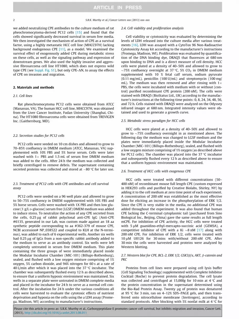
Fig. 2. CPE increased survival of MHCC97H cells in hypoxia conditions. MHCC97H
cells were induced with metabolic stress by method described above. Cells were
maintained either in normal condition (Normoxia) or under metabolic stress
conditions (Hypoxia) with addition of mouse recombinant CPE (mCPE), and with or
without CPE inhibitor, GEMSA. Cell viability was evaluated by the LDH assay
(�p < 0.05, ��p < 0.01, ���p < 0.001, Student’s t-test).
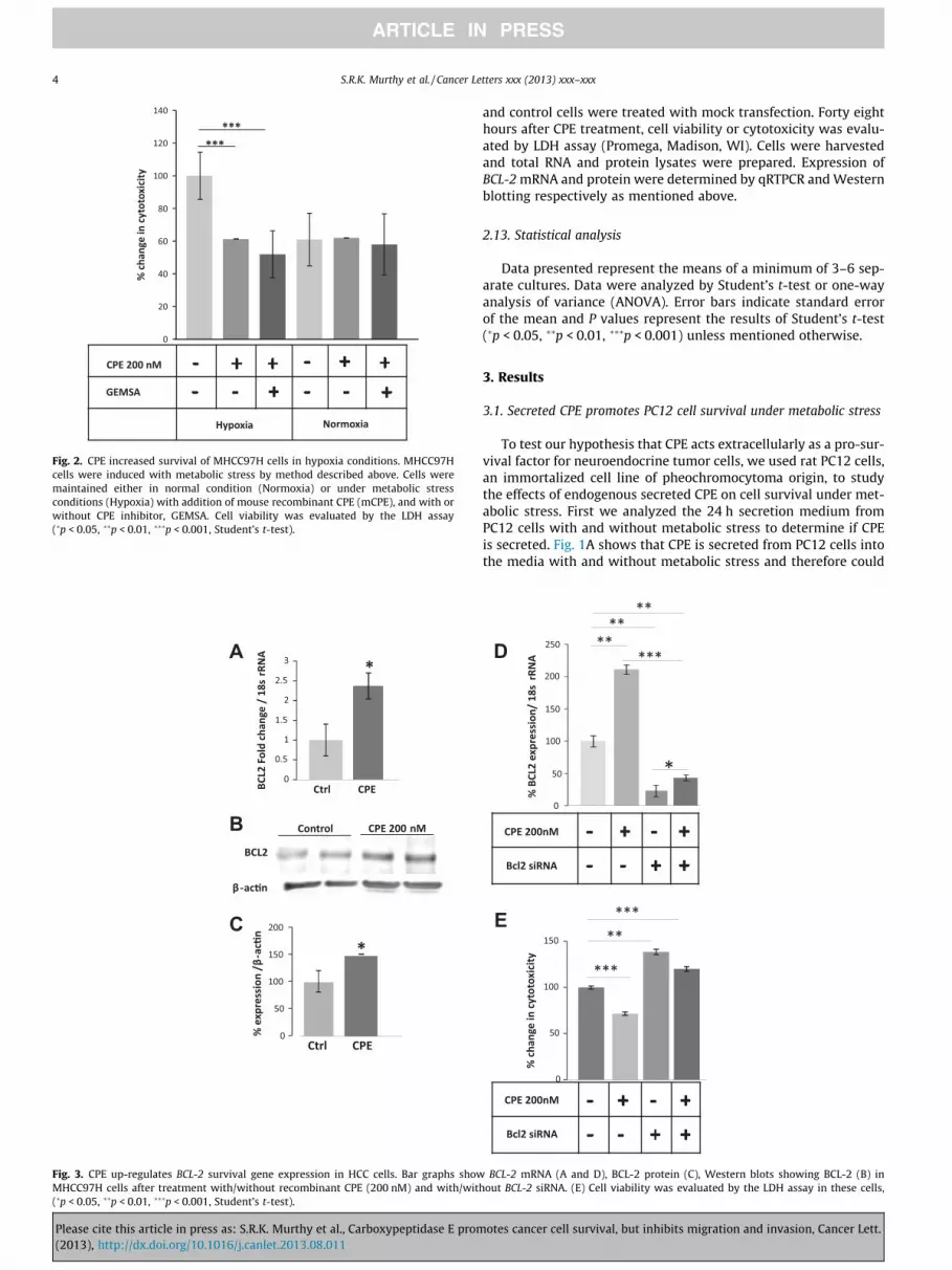
Fig. 3. CPE up-regulates BCL-2 survival gene expression in HCC cells. Bar graphs show BCL-2 mRNA (A and D), BCL-2 protein (C), Western blots showing BCL-2 (B) in
MHCC97H cells after treatment with/without recombinant CPE (200 nM) and with/without BCL-2 siRNA. (E) Cell viability was evaluated by the LDH assay in these cells,
(�p < 0.05, ��p < 0.01, ���p < 0.001, Student’s t-test).
4 S.R.K. Murthy et al. / Cancer Letters xxx (2013) xxx–xxx
Please cite this article in press as: S.R.K. Murthy et al., Carboxypeptidase E promotes cancer cell survival, but inhibits migration and invasion, Cancer Lett.
(2013), http://dx.doi.org/10.1016/j.canlet.2013.08.011

act on the cells in an autocrine/paracrine manner. To study the ef-
fect of CPE on PC12 cell survival, cells were grown to 50–75% con-
fluency and then treated under different conditions (see Methods)
with and without anti-CPE neutralizing antibodies. All cells were
incubated for 24 h and then harvested to evaluate the cytotoxic ef-
fects of nutrient deprivation and hypoxia (metabolic stress) on the
cells [16]. Fig. 1B shows that cells that had been treated with the
polyclonal anti-CPE IgGs exhibited 2-fold more cytotoxicity than
cells that were treated with non-specific IgGs. The cells not treated
with IgGs clearly showed resistance to metabolic stress in the pres-
ence of secreted CPE (as shown in Fig. 1B) similar to cells treated
with non-specific IgGs. Control cells that were not metabolically
stressed were healthy and showed no cytotoxicity (Fig. 1B, NC).
Thus, the survival of neuroendocrine PC12 cells during metabolic
stress depends on the presence of CPE.
3.2. Exogenous CPE protects HCC cells from cytotoxicity under
metabolic stress
To confirm that CPE is a survival factor indicated in the study on
PC12 cells above, where CPE was effectively removed (Fig. 1B), we
used MHCC97H (referred to as HCC) cells to test the effect of add-
ing exogenous CPE to a system that does not synthesize or secrete
endogenous CPE. HCC cells were maintained in low glucose serum
free (LGSF) DMEM or high glucose (4.5 g/L D-glucose) supple-
mented with 10% FBS under hypoxic or normoxic conditions. These
cells were treated with or without 200 nM recombinant mouse CPE
for 24 h and the cytotoxicity was measured for the treated and
control cells using the LDH assay. Fig. 2 shows that cells treated
with recombinant mCPE exhibited significantly less cytotoxicity
under metabolic stress than the untreated controls, further sup-
porting our hypothesis that CPE serves as a survival factor.
To determine if the survival effect of CPE is dependent on its
enzymatic activity, HCC cells were subjected to metabolic stress
as described above, in the presence of CPE pretreated with its
inhibitor, GEMSA. Fig. 2 shows that under metabolic stress, cells
treated with CPE and GEMSA showed decreased cytotoxicity com-
pared to untreated cells (ANOVA between Control, CPE treated and
CPE-GEMSA treated groups; during Hypoxia, F(2,27) = 93.48,
p < 0.001; Normoxia, F(2,27) = 0.43, p = 0.65). This result indicates
that the survival effect of CPE under metabolic stress does not de-
pend on its enzymatic activity.
3.3. CPE up-regulates BCL-2 survival gene expression in HCC cells
To investigate the mechanism by which CPE increases survival
of tumor cells, we again used the CPE-deficient HCC cells. Under
the same stressed conditions as were used for cytotoxicity studies,
Fig. 4. CPE up-regulates ERK 1/2 phosphorylation in HCC cells. (A), Western blots and (B) bar graphs showing BCL-2 and phosho-ERK 1/2 and total-ERK 1/2 (E) expression in
MHCC97H cells after treatment with/without recombinant CPE (200 nM) and 10 lM ERK inhibitor for 30 mins under metabolic stress conditions. b-actin served as a loading
control. (�p < 0.05, ��p < 0.01, ���p < 0.001, Student’s t-test).
S.R.K. Murthy et al. / Cancer Letters xxx (2013) xxx–xxx 5
Please cite this article in press as: S.R.K. Murthy et al., Carboxypeptidase E promotes cancer cell survival, but inhibits migration and invasion, Cancer Lett.
(2013), http://dx.doi.org/10.1016/j.canlet.2013.08.011
HCC cells were either treated or not treated with 200 nM CPE in
LGSF media and then incubated for 24 h in the hypoxia chamber.
The cells were then collected and RNA and protein were extracted
for evaluation of the expression of the survival gene, BCL-2 [18].
Using qRT-PCR to assay for BCL-2 mRNA, it was shown that HCC
cells treated with CPE exhibited a �2.4-fold increase in BCL-2
mRNA expression (t-test p < 0.05) (Fig. 3A). Western blot analysis
also revealed an increase in expression of the pro-survival/anti-
apoptotic protein BCL-2 in cells that were treated with CPE (t-test
p < 0.05) (Fig. 3B and C). To demonstrate that CPE is directly regu-
lating BCL-2 we silenced BCL-2 by siRNA (Fig. 3D). Suppression of
BCL-2 increased cytotoxicity of HCC cells and treatment of CPE
did not rescue the cytotoxicity (ANOVA between mock control,
CPE treated, BCL-2 siRNA treated and siRNA-CPE treated groups;
during Hypoxia, F(3,20) = 45.44, p < 0.0001). Thus, under metabolic
stress, CPE promotes survival of HCC cells by directly increasing
the expression of both BCL-2 mRNA and protein.
3.4. CPE up-regulates BCL-2 through ERK signaling in HCC cells
To investigate the signaling pathway by which CPE mediates
cell survival, and the increase in BCL-2 expression, we assayed
for phosphorylation of ERK, a protein kinase that is known to acti-
vate various cell survival cascades and ERK pathway is one of the
main pathways that regulate BCL-2 both transcriptionally and
post-transcriptionally [19,20]. Immunoblotting showed that
ERK1/2 were phosphorylated as early as 15 min after treatment
of HCC cells with CPE (Fig. 4A and B). An increase in BCL-2 protein
expression was observed after 30 min treatment with CPE. Further-
more this phosphorylation was nullified and BCL-2 expression was
reduced when the cells were treated with CPE in the presence of
ERK inhibitor U0126 (10 lM) (Fig. 4A and B) (ANOVA between
Control, CPE treated, ERK inhibitor treated and CPE-ERK inhibitor
treated groups; for p-ERK 1/2, F(3,20) = 71.60, p < 0.0001; for
BCL-2, F(3,20) = 18.98, p < 0.001). Thus, the signaling cascade for
up-regulation of BCL-2 expression by CPE involves the phosphory-
lation of ERK1/2.
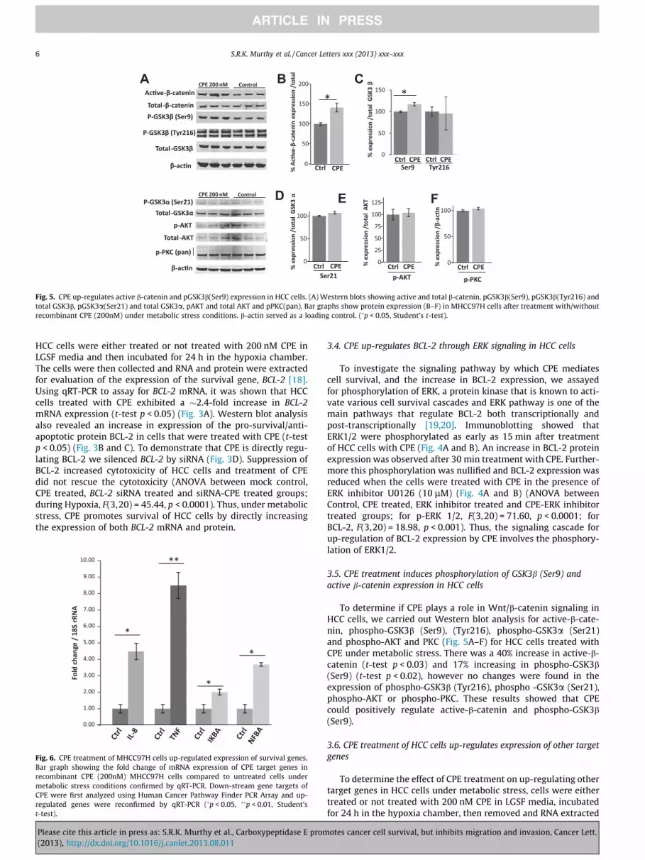
3.5. CPE treatment induces phosphorylation of GSK3b (Ser9) and
active b-catenin expression in HCC cells
To determine if CPE plays a role in Wnt/b-catenin signaling in
HCC cells, we carried out Western blot analysis for active-b-cate-
nin, phospho-GSK3b (Ser9), (Tyr216), phospho-GSK3a (Ser21)
and phospho-AKT and PKC (Fig. 5A–F) for HCC cells treated with
CPE under metabolic stress. There was a 40% increase in active-b-
catenin (t-test p < 0.03) and 17% increasing in phospho-GSK3b
(Ser9) (t-test p < 0.02), however no changes were found in the
expression of phospho-GSK3b (Tyr216), phospho -GSK3a (Ser21),
phospho-AKT or phospho-PKC. These results showed that CPE
could positively regulate active-b-catenin and phospho-GSK3b
(Ser9).
3.6. CPE treatment of HCC cells up-regulates expression of other target
genes
To determine the effect of CPE treatment on up-regulating other
target genes in HCC cells under metabolic stress, cells were either
treated or not treated with 200 nM CPE in LGSF media, incubated
for 24 h in the hypoxia chamber, then removed and RNA extracted
Fig. 5. CPE up-regulates active b-catenin and pGSK3b(Ser9) expression in HCC cells. (A) Western blots showing active and total b-catenin, pGSK3b(Ser9), pGSK3b(Tyr216) and
total GSK3b, pGSK3a(Ser21) and total GSK3a, pAKT and total AKT and pPKC(pan). Bar graphs show protein expression (B–F) in MHCC97H cells after treatment with/without
recombinant CPE (200nM) under metabolic stress conditions. b-actin served as a loading control. (�p < 0.05, Student’s t-test).
Fig. 6. CPE treatment of MHCC97H cells up-regulated expression of survival genes.
Bar graph showing the fold change of mRNA expression of CPE target genes in
recombinant CPE (200nM) MHCC97H cells compared to untreated cells under
metabolic stress conditions confirmed by qRT-PCR. Down-stream gene targets of
CPE were first analyzed using Human Cancer Pathway Finder PCR Array and up-
regulated genes were reconfirmed by qRT-PCR (�p < 0.05, ��p < 0.01, Student’s
t-test).
6 S.R.K. Murthy et al. / Cancer Letters xxx (2013) xxx–xxx
Please cite this article in press as: S.R.K. Murthy et al., Carboxypeptidase E promotes cancer cell survival, but inhibits migration and invasion, Cancer Lett.
(2013), http://dx.doi.org/10.1016/j.canlet.2013.08.011
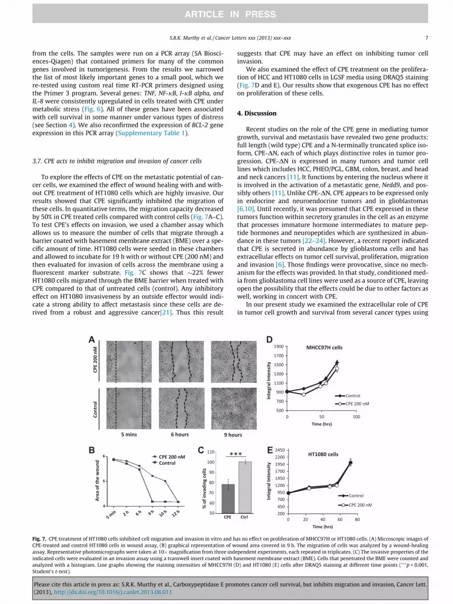
from the cells. The samples were run on a PCR array (SA Biosci-
ences-Qiagen) that contained primers for many of the common
genes involved in tumorigenesis. From the results we narrowed
the list of most likely important genes to a small pool, which we
re-tested using custom real time RT-PCR primers designed using
the Primer 3 program. Several genes: TNF, NF-jB, I-jB alpha, and
IL-8 were consistently upregulated in cells treated with CPE under
metabolic stress (Fig. 6). All of these genes have been associated
with cell survival in some manner under various types of distress
(see Section 4). We also reconfirmed the expression of BCL-2 gene
expression in this PCR array (Supplementary Table 1).
3.7. CPE acts to inhibit migration and invasion of cancer cells
To explore the effects of CPE on the metastatic potential of can-
cer cells, we examined the effect of wound healing with and with-
out CPE treatment of HT1080 cells which are highly invasive. Our
results showed that CPE significantly inhibited the migration of
these cells. In quantitative terms, the migration capacity decreased
by 50% in CPE treated cells compared with control cells (Fig. 7A–C).
To test CPE’s effects on invasion, we used a chamber assay which
allows us to measure the number of cells that migrate through a
barrier coated with basement membrane extract (BME) over a spe-
cific amount of time. HT1080 cells were seeded in these chambers
and allowed to incubate for 19 h with or without CPE (200 nM) and
then evaluated for invasion of cells across the membrane using a
fluorescent marker substrate. Fig. 7C shows that �22% fewer
HT1080 cells migrated through the BME barrier when treated with
CPE compared to that of untreated cells (control). Any inhibitory
effect on HT1080 invasiveness by an outside effector would indi-
cate a strong ability to affect metastasis since these cells are de-
rived from a robust and aggressive cancer[21]. Thus this result
suggests that CPE may have an effect on inhibiting tumor cell
invasion.
We also examined the effect of CPE treatment on the prolifera-
tion of HCC and HT1080 cells in LGSF media using DRAQ5 staining
(Fig. 7D and E). Our results show that exogenous CPE has no effect
on proliferation of these cells.
4. Discussion
Recent studies on the role of the CPE gene in mediating tumor
growth, survival and metastasis have revealed two gene products:
full length (wild type) CPE and a N-terminally truncated splice iso-
form, CPE-DN, each of which plays distinctive roles in tumor pro-
gression. CPE-DN is expressed in many tumors and tumor cell
lines which includes HCC, PHEO/PGL, GBM, colon, breast, and head
and neck cancers [11]. It functions by entering the nucleus where it
is involved in the activation of a metastatic gene, Nedd9, and pos-
sibly others [11]. Unlike CPE-DN, CPE appears to be expressed only
in endocrine and neuroendocrine tumors and in glioblastomas
[6,10]. Until recently, it was presumed that CPE expressed in these
tumors function within secretory granules in the cell as an enzyme
that processes immature hormone intermediates to mature pep-
tide hormones and neuropeptides which are synthesized in abun-
dance in these tumors [22–24]. However, a recent report indicated
that CPE is secreted in abundance by glioblastoma cells and has
extracellular effects on tumor cell survival, proliferation, migration
and invasion [6]. Those findings were provocative, since no mech-
anism for the effects was provided. In that study, conditioned med-
ia from glioblastoma cell lines were used as a source of CPE, leaving
open the possibility that the effects could be due to other factors as
well, working in concert with CPE.
In our present study we examined the extracellular role of CPE
in tumor cell growth and survival from several cancer types using
Fig. 7. CPE treatment of HT1080 cells inhibited cell migration and invasion in vitro and has no effect on proliferation of MHCC97H or HT1080 cells. (A) Microscopic images of
CPE-treated and control HT1080 cells in wound assay, (B) graphical representation of wound area covered in 9 h. The migration of cells was analyzed by a wound-healing
assay. Representative photomicrographs were taken at 10�magnification from three independent experiments, each repeated in triplicates. (C) The invasive properties of the
indicated cells were evaluated in an invasion assay using a transwell insert coated with basement membrane extract (BME). Cells that penetrated the BME were counted and
analyzed with a histogram. Line graphs showing the staining intensities of MHCC97H (D) and HT1080 (E) cells after DRAQ5 staining at different time points (���p < 0.001,
Student’s t-test).
S.R.K. Murthy et al. / Cancer Letters xxx (2013) xxx–xxx 7
Please cite this article in press as: S.R.K. Murthy et al., Carboxypeptidase E promotes cancer cell survival, but inhibits migration and invasion, Cancer Lett.
(2013), http://dx.doi.org/10.1016/j.canlet.2013.08.011
purified recombinant CPE. First we showed that rat pheochromo-
cytoma cells, a neuroendocrine tumor cell line (PC12) secretes
CPE (Fig. 1A) and addition of an anti-CPE neutralizing antibody in
the cell medium resulted in increased cytotoxic effects and poor
survival of the cells under metabolic stress (nutrient starvation
and hypoxia) (Fig. 1B). To confirm that CPE is a survival factor indi-
cated in the study on PC12 cells above, where CPE was effectively
removed (Fig. 1B), we used MHCC97H (referred to as HCC) cells to
test the effect of adding exogenous CPE to a system that does not
synthesize or secrete endogenous CPE. In our gain of function
experiments (Fig. 2), we found that HCC cells, that do not synthe-
size CPE, showed significantly less cytotoxicity under these meta-
bolic stress conditions when purified recombinant CPE protein
was added to the culture medium. This effect was also observed
when CPE was treated with 5 lM GEMSA, a specific and potent
inhibitor of CPE, indicating that the extracellular role of CPE in
imparting resistance to the metabolic stress is independent of its
enzymatic activity. It is not so surprising that this would be the
case as CPE has a pH optimum at �5–5.5 with no activity above
pH 7.0 [25], hence it is not expected to be enzymatically active
in the culture media.
Chronic metabolic stress is obviously detrimental to cells;
therefore treatment with recombinant CPE, that helps protect the
cells, suggests that signaling pathways involved in cell survival
are activated by CPE. We found that treatment of HCC cells under
metabolic stress, with CPE, resulted in increased phosphorylation
of ERK1/2 and an increase in the expression of the survival gene
BCL-2 [18], at the mRNA and protein levels (Fig. 3). In addition,
CPE treatment caused an increase of phospho-GSK3b (Ser9) and ac-
tive-b-catenin (Fig. 4), suggesting the involvement of the canonical
Wnt signaling pathway (see below). Several other genes (TNF, NF-
jB, I-jB alpha, and IL-8) (Fig. 5), which could support tumor cell
survival were also up-regulated in the CPE-treated HCC cells under
metabolic stress. The ability of tumor cells to migrate and invade is
a key property of a metastatic phenotype, hence we assessed
whether CPE could affect these properties, similar to that reported
for glioblastomas [6], in a highly metastatic cell line, HT1080 cells.
We showed that addition of CPE to the media of these cells inhib-
ited their metastatic phenotype as measured by wound healing
and invasion assays (Fig. 7), however, contrary to that reported
for glioblastoma cells [6], CPE had no effect on proliferation of
these cells (Fig. 7).
Our findings indicate that CPE is a tumor pro-survival factor
that functions extracellularly through ERK signaling to up-regulate
expression of BCL-2, the pro-survival (anti-apoptotic) protein
which acts to inhibit mitochondrial leakage during cell stress
[26]. ERK1/2 regulate cellular activity by acting on more than
100 substrates in the cytoplasm and nucleus, including indirect
inducers of gene expression, transcription factors and cell cycle-re-
lated kinases, and is a key signaling pathway involved in pituitary
tumorigenesis [27–30]. ERK signaling could be activated by GPCRs
through PKC [31], however we did not observe any change in pho-
sho-PKC after CPE treatment (Fig. 4), suggesting that CPE signaling
may be mediated by other signaling cascades. Interestingly, we
also did not see any increase in AKT (protein kinase B), a protein
kinase family of genes involved in regulating cell survival and is
a key regulator of BCL-2 [32]. Hence, induction of BCL-2 by CPE is
primarily through ERK signaling.
A key component of the Wnt signaling pathway, b-catenin plays
a major role during initiation of carcinogenesis [33]. b-catenin and
pGSK3b(Ser9) (inactive form of GSK3b and is a key regulator of nu-
clear factor (NF)jB nuclear activity [34], discussed below) were
significantly increased after CPE treatment. Even though b-catenin
acts as a co-activator during cell proliferation, it also plays an
important role in cell survival [35]. Hence in our studies, increased
expression of b-catenin and activation of the Wnt pathway with
the treatment of CPE may contribute to the survival of the HCC
cells under these metabolic stress conditions. At the resting state
of the cell no change in pGSK3b(Tyr216), the active form of GSK3b
was found. We also found no change in GSK3a in CPE mediated cell
survival.
The role of the other genes that are up-regulated in promoting
survival of the HCC cells is more complex. TNF mRNA was highly
expressed with CPE treatment of HCC cells under stress. However,
TNF has both pro-apoptotic effects by triggering NFKB activity, as
well as anti-apoptotic effects. NF-jB is a transcription factor that
plays a crucial role in many physiological and patho-physiological
processes, including survival and differentiation [34,36]. NF-jB has
been shown to be activated by several signaling pathways [37,38].
GSK3b regulates NF-jB activity either positively or negatively in a
cell type-dependent manner [39]. Since the NF-jB inhibitor, I-jBmRNA was also increased, the NF-jB pro-apoptotic effects may
be nullified. Thus, the likely role of TNF is in supporting pro-sur-
vival in this case, consistent with another study showing the in-
crease of NF-jB after CPE induction [40]. TNF drives expression
of IL-8 which in vivo would promote angiogenesis [41]. Hence
the increase in IL-8 expression with CPE treatment would also sup-
port survival of the tumor cells through increased angiogenesis.
Interestingly, unlike for glioblastoma cells [6], there was no effect
of CPE on proliferation of HCC or HT1080 cells (Fig. 7). This is con-
sistent with studies showing a negative effect of CPE on prolifera-
tion of adult neural stem cells [42], as well as negatively regulating
the canonical Wnt signaling pathway [13], which normally pro-
motes cancer cell proliferation [43].
The ability of CPE to inhibit migration and invasion of a very
aggressive fibrosarcoma cell line, HT1080, suggests that CPE has
powerful anti-metastatic effects as was also demonstrated in glio-
blastomas. The mechanism underlying the inhibition of migration
or invasion by CPE is less clear. Since the Wnt pathway compo-
nents mediate cancer cell invasion [44], one can speculate that
the negative regulation of the Wnt pathway by CPE [13] could be
responsible for the inhibition of migration and invasion observed
with CPE treatment. It has generally been thought that the same
pathways or molecules lead to proliferation, survival and metasta-
sis in cancer progression. However, our studies on CPE have led to
the recognition that there are ‘‘ying and yang’’ pathways and mol-
ecules; ones that specifically drive tumor cell survival and others
that promote proliferation, migration and invasion. These path-
ways are in competition with each other depending on the tumor
environment.
With respect to the CPE gene, epithelial-derived tumors have
thus far been shown to express only the splice variant, CPE-DN,
and its main role seems to be to induce metastasis. On the other
hand endocrine, neuroendocrine tumors, glioblastomas and neu-
roblastomas synthesize both CPE-DN and CPE which have compet-
ing opposing effects on metastasis. Hence, depending on their
relative levels of expression, each will dictate whether the tumor
metastasizes or not.
Identifying the role of CPE as an inhibitor of metastasis has re-
solved an on-going enigma where a clinical study showed that high
CPE levels in small cell carcinoma of the lung, a neuroendocrine tu-
mor, indicated good prognosis [23], presumably because of its anti-
metastatic effects. On the other hand, our studies on CPE-DN in
PHEOs showed that high copy numbers of CPE-DNmRNA indicated
a metastatic tumor, or future recurrence or metastasis with poor
prognosis [11]. Given the current knowledge, to use CPE as a prog-
nostic biomarker for (neuro)endocrine tumors such as PHEOs, spe-
cific primers for measuring CPE-DN and CPE transcripts must be
used to avoid confounding results.
Our findings also suggest that CPE could be a good target for
various inhibitory therapeutic agents, with the potential benefits
of decreased tumor growth and increased susceptibility to hypoxic
8 S.R.K. Murthy et al. / Cancer Letters xxx (2013) xxx–xxx
Please cite this article in press as: S.R.K. Murthy et al., Carboxypeptidase E promotes cancer cell survival, but inhibits migration and invasion, Cancer Lett.
(2013), http://dx.doi.org/10.1016/j.canlet.2013.08.011

and nutrient deprived conditions that arise as a result of anti
angiogenic therapies. However, to accomplish this, more work to
determine whether CPE is a ligand with its own receptor, or signals
through binding to other receptor complexes, such as Wnt3a/friz-
zled [13], will be necessary. Since invasion is often considered the
marker for a particular molecule’s metastatic potential, we can
conclude that although CPE may serve to protect a tumor from
external stress, it does not promote the spread of the cancer and
therefore may actually be a positive attribute in some cases. This
fragile dynamic between the pro-survival and anti-metastatic ef-
fects of CPE is one that must be considered carefully and further
investigated, but certainly offers the potential to be an extraordi-
narily important aspect of the way we approach treatment of neu-
roendocrine, endocrine tumors and glioblastomas that secrete CPE.
Conflict of interest
The authors declare that no conflict of interests exists.
Acknowledgements
This research was supported by the Intramural Research Pro-
gram of the Eunice Kennedy Shriver National Institute of Child
Health and Human Development, National Institutes of Health,
USA,
Appendix A. Supplementary material
Supplementary data associated with this article can be found, in
the online version, at http://dx.doi.org/10.1016/j.canlet.2013.
08.011.
References
[1] A.F. Chambers, A.C. Groom, I.C. MacDonald, Dissemination and growth ofcancer cells in metastatic sites. Nature reviews, Cancer 2 (2002) 563–572.
[2] H.P. Neumann, B. Bausch, S.R. McWhinney, B.U. Bender, O. Gimm, G. Franke, J.Schipper, J. Klisch, C. Altehoefer, K. Zerres, A. Januszewicz, C. Eng, W.M. Smith,R. Munk, T. Manz, S. Glaesker, T.W. Apel, M. Treier, M. Reineke, M.K. Walz, C.Hoang-Vu, M. Brauckhoff, A. Klein-Franke, P. Klose, H. Schmidt, M. Maier-Woelfle, M. Peczkowska, C. Szmigielski, Germ-line mutations in nonsyndromicpheochromocytoma, The New England Journal of Medicine 346 (2002) 1459–1466.
[3] W.G. North, J. Du, Key peptide processing enzymes are expressed by a variantform of small-cell carcinoma of the lung, Peptides 19 (1998) 1743–1747.
[4] C. Azzoni, T. D’Adda, G. Tamburrano, C. Coscelli, O.D. Madsen, L. Scopsi, C.Bordi, Functioning human insulinomas. An immunohistochemical analysis ofintracellular insulin processing, Virchows Archiv: An International Journal ofPathology 433 (1998) 495–504.
[5] J.H. Zhang, D. Zhou, J. You, B.S. Tang, P.Y. Li, S.S. Tang, Differential processing ofneuropeptide proprotein in human breast adenocarcinoma, Journal ofEndocrinological Investigation (2013).
[6] E. Horing, P.N. Harter, J. Seznec, J. Schittenhelm, H.J. Buhring, S. Bhattacharyya,E. von Hattingen, C. Zachskorn, M. Mittelbronn, U. Naumann, The ‘‘go or grow’’potential of gliomas is linked to the neuropeptide processing enzymecarboxypeptidase E and mediated by metabolic stress, ActaNeuropathologica 124 (2012) 83–97.
[7] P. He, L. Varticovski, E.D. Bowman, J. Fukuoka, J.A. Welsh, K. Miura, J. Jen, E.Gabrielson, E. Brambilla, W.D. Travis, C.C. Harris, Identification ofcarboxypeptidase E and gamma-glutamyl hydrolase as biomarkers forpulmonary neuroendocrine tumors by cDNA microarray, Human Pathology35 (2004) 1196–1209.
[8] N.X. Cawley, W.C. Wetsel, S.R. Murthy, J.J. Park, K. Pacak, Y.P. Loh, New roles ofcarboxypeptidase E in endocrine and neural function and cancer, EndocrineReviews 33 (2012) 216–253.
[9] Y. Cheng, N.X. Cawley, Y.P. Loh, Carboxypeptidase E/NFa1: A new neurotrophicfactor against oxidative stress-induced apoptotic cell death mediated by ERKand PI3-K/AKT pathways, PloS one (2013).
[10] S.R. Murthy, K. Pacak, Y.P. Loh, Carboxypeptidase E: elevated expressioncorrelated with tumor growth and metastasis in pheochromocytomas andother cancers, Cellular and Molecular Neurobiology 30 (2010) 1377–1381.
[11] T.K. Lee, S.R. Murthy, N.X. Cawley, S. Dhanvantari, S.M. Hewitt, H. Lou, T. Lau, S.Ma, T. Huynh, R.A. Wesley, I.O. Ng, K. Pacak, R.T. Poon, Y.P. Loh, An N-terminaltruncated carboxypeptidase E splice isoform induces tumor growth and is a
biomarker for predicting future metastasis in human cancers, The Journal ofClinical Investigation 121 (2011) 880–892.
[12] K. Zhou, H. Liang, Y. Liu, C. Yang, P. Liu, X. Jiang, Overexpression of CPE-DeltaNpredicts poor prognosis in colorectal cancer patients. Tumour Biology: theJournal of the International Society for Oncodevelopmental Biology andMedicine (2013).
[13] N. Skalka, M. Caspi, E. Caspi, Y.P. Loh, R. Rosin-Arbesfeld, Carboxypeptidase E: anegative regulator of the canonical Wnt signaling pathway, Oncogene (2012).
[14] S.R. Murthy, E. Thouennon, W.S. Li, Y. Cheng, J. Bhupatkar, N.X. Cawley, M.Lane, I. Merchenthaler, Y.P. Loh, Carboxypeptidase E protects hippocampalneurons during Stress in male mice by up-regulating pro-survival BCL2 proteinexpression, Endocrinology (2013) 10.
[15] L.A. Greene, A.S. Tischler, Establishment of a noradrenergic clonal line of ratadrenal pheochromocytoma cells which respond to nerve growth factor,Proceedings of the National Academy of Sciences of the United States ofAmerica 73 (1976) 2424–2428.
[16] C. Korzeniewski, D.M. Callewaert, An enzyme-release assay for naturalcytotoxicity, Journal of Immunological Methods 64 (1983) 313–320.
[17] L.D. Fricker, T.H. Plummer Jr., S.H. Snyder, Enkephalin convertase: potent,selective, and irreversible inhibitors, Biochemical and Biophysical ResearchCommunications 111 (1983) 994–1000.
[18] M.D. Jacobson, M.C. Raff, Programmed cell death and Bcl-2 protection in verylow oxygen, Nature 374 (1995) 814–816.
[19] K. Balmanno, S.J. Cook, Tumour cell survival signalling by the ERK1/2 pathway,Cell Death and Differentiation 16 (2009) 368–377.
[20] B.A. Ballif, J. Blenis, Molecular mechanisms mediating mammalian mitogen-activated protein kinase (MAPK) kinase (MEK)-MAPK cell survival signals, CellGrowth & Differentiation: the Molecular Biology Journal of the AmericanAssociation for Cancer Research 12 (2001) 397–408.
[21] C.J. Marshall, A. Hall, R.A. Weiss, A transforming gene present in humansarcoma cell lines, Nature 299 (1982) 171–173.
[22] X. Fan, S.J. Olson, L.S. Blevins, G.S. Allen, M.D. Johnson, Immunohistochemicallocalization of carboxypeptidases D, E, and Z in pituitary adenomas andnormal human pituitary, Journal of Histochemistry and Cytochemistry 50(2002) 1509–1516.
[23] P. He, L. Varticovski, E.D. Bowman, J. Fukuoka, J.A. Welsh, K. Miura, J. Jen, E.Gabrielson, E. Brambilla, W.D. Travis, C.C. Harris, Identification ofcarboxypeptidase E and gamma-glutamyl hydrolase as biomarkers forpulmonary neuroendocrine tumors by cDNA microarray, Human Pathology35 (2004) 1196–1209.
[24] S.S. Tang, J.H. Zhang, H.X. Liu, H.Z. Li, PC2/CPE-mediated pro-proteinprocessing in tumor cells and its differentiated cells or tissues, Molecularand Cellular Endocrinology 303 (2009) 43–49.
[25] D. Greene, B. Das, L.D. Fricker, Regulation of carboxypeptidase E. Effect of pH,temperature and Co2+ on kinetic parameters of substrate hydrolysis, TheBiochemical Journal 285 (Pt 2) (1992) 613–618.
[26] M.J. Boucher, J. Morisset, P.H. Vachon, J.C. Reed, J. Laine, N. Rivard, MEK/ERKsignaling pathway regulates the expression of Bcl-2, Bcl-X(L), and Mcl-1 andpromotes survival of human pancreatic cancer cells, Journal of CellularBiochemistry 79 (2000) 355–369.
[27] W. Kolch, Meaningful relationships: the regulation of the Ras/Raf/MEK/ERKpathway by protein interactions, The Biochemical Journal 351 (Pt 2) (2000)289–305.
[28] A. Schulze, K. Lehmann, H.B. Jefferies, M. McMahon, J. Downward, Analysis ofthe transcriptional program induced by Raf in epithelial cells, Genes &Development 15 (2001) 981–994.
[29] J. Xaus, M. Comalada, A.F. Valledor, M. Cardo, C. Herrero, C. Soler, J. Lloberas, A.Celada, Molecular mechanisms involved in macrophage survival, proliferation,activation or apoptosis, Immunobiology 204 (2001) 543–550.
[30] M. Cakir, A.B. Grossman, Targeting MAPK (Ras/ERK) and PI3K/Akt pathways inpituitary tumorigenesis, Expert Opinion on Therapeutic Targets 13 (2009)1121–1134.
[31] Z. Naor, Signaling by G-protein-coupled receptor (GPCR): studies on the GnRHreceptor, Frontiers in Neuroendocrinology 30 (2009) 10–29.
[32] S. Pugazhenthi, A. Nesterova, C. Sable, K.A. Heidenreich, L.M. Boxer, L.E.Heasley, J.E. Reusch, Akt/protein kinase B up-regulates Bcl-2 expressionthrough cAMP-response element-binding protein, The Journal of BiologicalChemistry 275 (2000) 10761–10766.
[33] A. de La Coste, B. Romagnolo, P. Billuart, C.A. Renard, M.A. Buendia, O.Soubrane, M. Fabre, J. Chelly, C. Beldjord, A. Kahn, C. Perret, Somatic mutationsof the beta-catenin gene are frequent in mouse and human hepatocellularcarcinomas, Proceedings of the National Academy of Sciences of the UnitedStates of America 95 (1998) 8847–8851.
[34] D.D. Billadeau, Primers on molecular pathways. The glycogen synthase kinase-3beta, Pancreatology 7 (2007) 398–402.
[35] E.J. Chung, S.G. Hwang, P. Nguyen, S. Lee, J.S. Kim, J.W. Kim, P.A. Henkart, D.P.Bottaro, L. Soon, P. Bonvini, S.J. Lee, J.E. Karp, H.J. Oh, J.S. Rubin, J.B. Trepel,Regulation of leukemic cell adhesion, proliferation, and survival by beta-catenin, Blood 100 (2002) 982–990.
[36] N.D. Perkins, NF-kappaB: tumor promoter or suppressor?, Trends in CellBiology 14 (2004) 64–69
[37] S. Ghosh, M. Karin, Missing pieces in the NF-kappaB puzzle, Cell 109 (Suppl.)(2002) S81–96.
[38] N. Silverman, T. Maniatis, NF-kappaB signaling pathways in mammalian andinsect innate immunity, Genes & Development 15 (2001) 2321–2342.
S.R.K. Murthy et al. / Cancer Letters xxx (2013) xxx–xxx 9
Please cite this article in press as: S.R.K. Murthy et al., Carboxypeptidase E promotes cancer cell survival, but inhibits migration and invasion, Cancer Lett.
(2013), http://dx.doi.org/10.1016/j.canlet.2013.08.011

[39] C.A. Grimes, R.S. Jope, The multifaceted roles of glycogen synthase kinase3beta in cellular signaling, Progress in Neurobiology 65 (2001) 391–426.
[40] V.A. Meliopoulos, L.E. Andersen, P. Brooks, X. Yan, A. Bakre, J.K. Coleman, S.M.Tompkins, R.A. Tripp, MicroRNA regulation of human protease genes essentialfor influenza virus replication, PLoS ONE 7 (2012) e37169.
[41] R. Persad, H.Q. Huynh, L. Hao, J.R. Ha, C. Sergi, R. Srivastava, S. Persad,Angiogenic remodeling in pediatric EoE is associated with increased levels ofVEGF-A, angiogenin, IL-8, and activation of the TNF-alpha-NFkappaB pathway,Journal of Pediatric Gastroenterology and Nutrition 55 (2012) 251–260.
[42] C. Lee, J. Hu, S. Ralls, T. Kitamura, Y.P. Loh, Y. Yang, Y.S. Mukouyama, S. Ahn,The molecular profiles of neural stem cell niche in the adult subventricularzone, PLoS ONE 7 (2012) e50501.
[43] H. Clevers, Wnt/beta-catenin signaling in development and disease, Cell 127(2006) 469–480.
[44] A. Nguyen, A. Rosner, T. Milovanovic, C. Hope, K. Planutis, B. Saha, B. Chaiwun,F. Lin, S.A. Imam, J.L. Marsh, R.F. Holcombe, Wnt pathway component LEF1mediates tumor cell invasion and is expressed in human and murine breastcancers lacking ErbB2 (her-2/neu) overexpression, International Journal ofOncology 27 (2005) 949–956.
10 S.R.K. Murthy et al. / Cancer Letters xxx (2013) xxx–xxx
Please cite this article in press as: S.R.K. Murthy et al., Carboxypeptidase E promotes cancer cell survival, but inhibits migration and invasion, Cancer Lett.
(2013), http://dx.doi.org/10.1016/j.canlet.2013.08.011