Enzymatic rotating biosensor for ciprofloxacin determination
Upload
independentCategory
view
1download
0
RESEARCH PAPER
Fibroblast activation protein-a promotes tumor growthand invasion of breast cancer cells through non-enzymaticfunctions
Yan Huang • Avis E. Simms • Anna Mazur •
Sophie Wang • Noel R. Leon • Barry Jones •
Nazneen Aziz • Thomas Kelly
Received: 1 October 2010 / Accepted: 23 April 2011 / Published online: 22 May 2011
� Springer Science+Business Media B.V. 2011
Abstract Fibroblast activation protein-a (FAP) is a cell
surface, serine protease of the post-prolyl peptidase family
that is expressed in human breast cancer but not in normal
tissues. Previously, we showed that FAP expression
increased tumor growth rates in a mouse model of human
breast cancer. Here the role of the proteolytic activities of
FAP in promoting tumor growth, matrix degradation and
invasion was investigated. Mammary fat pads of female
SCID mice were inoculated with breast cancer cells that
express FAP and the mice treated with normal saline
or Val-boroPro (talabostat); Glu-boroPro (PT-630); or
1-[[(3-hydroxy-1-adamantyl)amino]acetyl]-2-cyano-(S)-pyr-
rolidine (LAF-237) that inhibit prolyl peptidases. Other
mice were injected with breast cancer cells expressing a
catalytically inactive mutant of FAP and did not receive
inhibitor treatment. PT-630 and LAF-237 did not slow
growth of tumors produced by any of the three cell lines
expressing FAP. Talabostat slightly decreased the growth
rates of the FAP-expressing tumors but because PT-630
and LAF-237 did not, the growth retardation was likely not
related to the inhibition of FAP or the related post-prolyl
peptidase dipeptidyl peptidase IV. Breast cancer cells
expressing a catalytically inactive mutant of FAP
(FAPS624A) also produced tumors that grew rapidly.
In vitro studies revealed that cells expressing wild type
FAP or FAPS624A degrade extracellular matrix (ECM)
more extensively, accumulate higher levels of matrix
metalloproteinase-9 (MMP-9) in conditioned medium, are
more invasive in type I collagen gels, and have altered
signaling compared to control transfectants that do not
express FAP and form slow growing tumors. We conclude
that the proteolytic activity of FAP participates in matrix
degradation, but other functions of the protein stimulate
increased tumor growth.
Keywords Serine protease � Seprase � Dipeptidyl
peptidase IV � Invadopodia � Metastasis
Abbreviations
ANOVA Analysis of variance
GIP Glucose dependent and insulinotropic
polypeptide
GLP-1 Glucagon-like peptide-1
ECM Extracellular matrix
FACS Fluorescence activated cell sorting
FAP Fibroblast activation protein-aFAPS624A MDA MB-231 cells expressing a
catalytically inactive mutant of FAP
FITC Fluorescien isothiocyanate
F19 Mouse monoclonal antibody directed
against human FAP
LAF-237 (vildagliptin), 1-[[(3-hydroxy-1-
adamantyl)amino]acetyl]-2-cyano-(S)-
pyrrolidine, an inhibitor of DPPIV and
related proteases
MDA MB-231 A human breast adenocarcinoma cell line
MMPs Matrix metalloproteinases
MMP-9 Matrix metalloproteinase-9
pcDNA3.1 A plasmid mammalian expression vector
Y. Huang � A. E. Simms � A. Mazur � S. Wang �N. R. Leon � T. Kelly (&)
Department of Pathology, Winthrop P. Rockefeller Cancer
Institute, Arkansas Cancer Research Center, University of
Arkansas for Medical Sciences, 4301 W. Markham St., Slot 753,
Little Rock, AR 72205-7199, USA
e-mail: [email protected]
B. Jones � N. Aziz
Point Therapeutics, Inc., Boston, MA, USA
123
Clin Exp Metastasis (2011) 28:567–579
DOI 10.1007/s10585-011-9392-x
PT-100 Val-boroPro, an inhibitor of FAP and
related proteases
PT-630 Glu-boroPro, an inhibitor of FAP and
related proteases
RT-PCR Reverse transcription-polymerase chain
reaction
SCID Severe combined immune deficient
WTY-1 Stable transfectant of MDA MB-231
engineered to express high levels of FAP
WTY-6 Stable transfectant of MDA MB-231
engineered to express high levels of FAP
4G10 Monoclonal antibody to phosphotyrosine
6-6B Monoclonal antibody to MMP-9
Introduction
Fibroblast activation protein-a (FAP), also called seprase,
is an integral membrane serine protease and member of the
post-prolyl peptidase family [1, 2]. FAP is closely related
to dipeptidyl peptidase IV (DPPIV) [3, 4] and exhibits a
DPPIV-like fold, featuring alpha/beta-hydrolase and eight-
bladed beta-propeller domains [5]. FAP is synthesized as a
97-kDa protein that is proteolytically inactive and requires
assembly into a dimer of 170-kDa to become an active
protease [1, 6]. Like DPPIV, it cleaves NH2-X-Pro pep-
tides [3]; but in addition, FAP possesses a unique endo-
peptidase activity and is able to degrade gelatin [3, 4].
Recently a2-antiplasmin was identified as the first natu-
ral substrate for FAP, and FAP-mediated cleavage of
a2-antiplasmin results in more efficient cross linking of
truncated a2-antiplasmin to fibrin while retaining the
inhibitory action to plasmin [7]. Thus, FAP may have a role
in regulating the dissolution of fibrin by plasmin.
Recent work has suggested many pathological functions
for FAP [8]. FAP may have roles in osteoarthritis [9],
pulmonary fibrosis [10], fibrosis in liver diseases such as
hepatitis [11] and cirrhosis [12], and roles in numerous
cancers, for example: multiple myeloma [13], pancreatic
cancer [14], colon cancer [15], melanoma [16], ovarian
carcinoma [17], and breast cancer [18]. FAP is expressed to
high levels in human breast cancer but is not expressed in
normal breast tissue [2, 18]. FAP protease activity is
abnormally high in extracts of patient tumors indicating
that increased expression leads to increased FAP protease
activity in breast cancer [6]. The observation of increased
expression of FAP generally in cancers of epithelial origin
has led to the hypothesis that its proteolytic activity might
be involved in tissue remodeling required for tumor pro-
gression and vascularization [19, 20].
Because of the conspicuous absence of FAP in normal
adult tissues and its marked up-regulation in a variety of
diseases, there is interest in the potential of small molecule
inhibitors of the FAP protease to reduce pathogenesis.
Moreover, DPPIV is a target for drug discovery in type 2
diabetes and hematopoietic stem cell engraftment [21].
Sitagliptin is a DPPIV inhibitor already approved for type 2
diabetes because it has insulinotropic effects caused by
prolonging the half-life of glucagon-like peptide-1 (GLP-1)
and glucose-dependent insulinotropic polypeptide (GIP),
both of which are substrates for DPPIV [21]. In this study
we inhibited FAP using Val-boroPro (PT-100/talabostat),
which inhibits FAP with a Ki of 6.2 nM and DPPIV with a
Ki of 0.18 nM [22] and Glu-boroPro (PT-630), which
inhibits DPPIV with a Ki of 3 nM and FAP with a Ki of
5 nM [22, 23]. Because DPPIV is expressed ubiquitously
and constitutively in body organs [24], it was necessary to
distinguish between the biological consequences of its
inhibition from that of FAP. To do this, we also used
1-[[(3-hydroxy-1-adamantyl)amino]acetyl]-2-cyano-(S)-
pyrrolidine (LAF-237) which inhibits DPPIV with a Ki
within a range of 17–51 nM [25, 26], but is a much less
potent inhibitor of FAP with a Ki greater than 20 lM [27].
In this study, the role of the proteolytic activity of FAP
in promoting tumorigenesis, tumor growth, extracellular
matrix (ECM) degradation and invasiveness was investi-
gated using small-molecule inhibitors of FAP and breast
cancer cells engineered to express either active FAP or an
inactive catalytic mutant of FAP. Our findings indicate that
the protease activity of FAP is not critical for its growth
promoting and invasive functions in tumors of human
breast cancer cells. The results also suggest that cell sur-
face expression of FAP stimulates elevated production of
factors such as MMP-9 that are required for ECM degra-
dation and tissue invasion during tumor progression.
Materials and methods
MDA MB-231 human breast adenocarcinoma cells trans-
fected with pcDNA 3.1 (Neo) or this vector containing the
insert for wild type FAP (WTY-1 and WTY-6) were pro-
duced and maintained as described earlier [19].
Production of cDNA encoding S624A mutant
A cDNA for human FAP [19] in the pcDNA3.1 vector was
subjected to oligonucleotide-directed mutagenesis (Trans-
formerTM site-directed mutagenesis kit; Clontech, Palo
Alto, CA) to yield constructs having a single point mutation
(T ? G) that changed the codon for serine 624 (TCC) to a
codon for alanine (GCC). The mutagenic primer was 50-CATATGGGGCTGGGCCTATGGAGGATAC-30 (mutant
base in bold) and the selection primer eliminated a
BSTZ174 site and was 50-TATCTTATCATGTCTGTAT
568 Clin Exp Metastasis (2011) 28:567–579
123
ACCGTCGACCTCTAGCT-30. Sequence analysis of the
entire insert was performed to confirm that the cDNA was
identical to wild-type FAP, except for the point mutation.
The analysis was performed by Alan Gies in the DNA
sequencing core facility in the Department of Microbiology
and Immunology at the University of Arkansas for Medical
Sciences (UAMS).
Transfection of cells
The cDNA encoding S624A mutant was transfected into
the human breast cancer cell line MDA MB-231 using
lipofectamine 2000 (Invitrogen, Carlsbad, CA) as directed.
The transfectants were selected with G418 (800 lg/ml) and
sorted by fluorescence activated cell sorting based on
staining with F19 antibody to FAP as described previously
[19]. Once, sorted the cells were maintained in growth
medium with G418 (400 lg/ml). The transfectants were
used to form tumors in animals when the population of
FAP-positive cells comprised at least 40% of the cells.
Gelatin zymography
To analyze FAP expression, extracts were prepared and
subjected to gelatin zymography and western blotting as
described previously [16, 19, 28]. Several steps were
taken to enhance FAP-specific gelatinolytic activity while
diminishing gelatinase activities of MMPs. First, FAP was
partially purified from the extracts by pulling it down
with WGA Agarose as described earlier [16, 19, 28].
Secondly, FAP gelatinolytic activity was preserved while
that of MMPs was inhibited by including EDTA in the
extraction and zymogram incubation buffers [16, 19, 28].
Finally, the FAP was eluted from the WGA beads in gel
sample buffer containing N-acetyl glucosamine and
dithiothreitol reducing reagent that inhibits MMPs but
preserves FAP activity [16, 19, 28] and zymography was
performed.
Tumor biology
2 9 106 tumor human mammary carcinoma cells were
implanted into each of four mammary fat pads per female
SCID mouse, and, starting on day 2 after tumor implan-
tation, the mice were administered normal saline (placebo
control) or protease inhibitors by gavage. Tumor growth
was measured with calipers as previously described [19],
and wet tumor weights were determined for tumors excised
at the end of experiments. In the first animal experiment
with S624A-5 cells there were seven animals per group,
and in the second experiment, there were five animals per
treatment group. For the inhibitor studies there were five
animals per group.
Preparation of talabostat, PT-630 and LAF-237
A 0.1 M stock solution prepared by dissolving talabostat at
a concentration of 30.6 mg/ml in 0.1 N HCl and PT-630 at
a concentration of 28.1 mg/ml was supplied by Point
Therapeutics (Boston, MA). Stock solutions were stored at
-20�C in small aliquots (50–200 ll). Working solutions
were prepared by diluting the 0.1 M acidified stock with
sterile normal saline. Working solutions were made at
concentrations that delivered 10-lg/mouse talabostat;
200-lg/mouse PT-630, and 100-lg/mouse LAF-237 in
0.2 ml. To avoid excessive production of the cyclic form of
the compounds, talabostat was not held at neutral pH for
longer than 10 min before administration. Consequently,
the working saline solutions were prepared with ice cold
reagents in the animal facility, immediately prior to
administration to mice. The inhibitor solutions (0.2 ml)
were administered orally, once daily, via a blunt gavage
needle fitted to a 1-ml syringe.
High titer F19 acites fluid
Five female Balb/c mice (6–8 weeks old) were housed for
1 week and injected IP 0.5 ml of pristane (Sigma) per
mouse. One week later 5 9 106 F19 hybridoma cells were
injected IP into each mouse. These hybridomas produce the
F19 monoclonal antibody to FAP. When ascites fluid
formed (approximately 3 weeks later) the fluid was har-
vested with a syringe. Ascites was collected at least twice
per animal.
FAP protease activity assay
FAP was extracted from cells by using Triton X-100
using a procedure modified from that reported previously
[16, 28]. Each cell type was grown to 90% confluence in
three 75 cm2 flasks. Media was removed; the cells washed
three times with PBS, and then suspended using 1 mM
EDTA in PBS. Cells were pelleted by centrifugation at
10,0009g for 5 min at 25�C and the supernatant was
removed. Cells were resuspended in 1 ml of extraction
buffer (2.5%Triton X-100, 150 mM NaCl, 5 mM EDTA,
10 mM Tris–HCl, pH 7.5) and then the lysate was gently
homogenized on ice in an 1.5 ml Eppendorf tube with 30
strokes of the pestle. Care was taken to avoid excessive
foaming. The homogenate was cleared by centrifugation
for 5 min at 5249g at 4�C. The supernatant was trans-
ferred to a fresh tube and incubated with 100 ll of protein
G magnetic Dynabeads (Invitrogen) that had been coated
with approximately 5 lg F19 mAb to FAP for 30 min at
25�C and washed according to the manufacturer’s
instructions. FAP collected on the protein G Dynabeads,
Clin Exp Metastasis (2011) 28:567–579 569
123
was exposed to 100 lM of z-Gly-Pro-AMC (Bachem
Bioscience Inc, King of Prussia, PA, USA) for 2 h at
37�C in 400 ll of 50 mM Tris, 100 mM NaCl, 1 mM
EDTA, pH 7.6 with shaking. To inhibit FAP, the FAP-
coated protein G beads were first exposed to 10 lM of
PT-100 or PT-630 in 390 ll of the above buffer for
30 min 25�C prior to addition of the 10 ll of 4 mM
z-Gly-Pro-AMC to achieve a final concentration of
100 lM z-Gly-Pro-AMC. The beads were collected with
the magnet and the fluorescence at 460 nm of the
supernatants determined with excitation of 360 nM with
the Tecan Safire microtiter plate reader. All samples were
tested in duplicate. 7-Amino 4-Methyl Coumarin (AMC)
(ACROS, New Jersey, USA) was used to develop a
standard curve where the fluorescence emitted by free
AMC at concentrations of 0.00125, 0.0125, 0.125, 1.25,
2.5, 5, 10 and 20 lM was determined by applying an
excitation wavelength of 360 nm and measuring the
fluorescence intensity at 460 nm (Tecan Safire fluorescent
microplate reader using the Magellan software). This
curve was used to relate fluorescence obtained from the
unknown samples to lM free AMC.
To determine FAP activity in tumor tissues an extract
was prepared by homogenizing 100 mg of tumor tissue in
500 ll 2.5%Triton X-100, 150 mM NaCl, 5 mM EDTA,
10 mM Tris–HCl, pH 7.5. The extract was cleared by
centrifugation and the total protein determined using
bicinchoninic acid assay (Pierce) where BSA was used to
produce a standard curve. The F19-coated protein G dyn-
abeads (50 ll) prepared as described above, were exposed
to 5 mg of tumor extract protein to capture FAP. The FAP
coated beads were then tested for FAP-activity using
100 lM z-Gly-Pro-AMC substrate as described above.
Samples were tested in duplicate.
Matrix degradation assay
Glutaraldehyde-crosslinked gelatin films with immobilized
fluorescein isothiocyanate (FITC)-fibronectin were pre-
pared on cover slips (18 mm circular glass) as described
previously [29]. Cells were seeded onto the FITC-fibro-
nectin matrices and grown in growth medium for 48 h at
37�C, 5% CO2. Adherent cells were washed three times
with sterile PBS and then fixed and prepared for fluores-
cence microscopy as described previously [29]. Coverslips
were mounted in 80% glycerol, 10 mM Tris–HCl, pH 7.6,
150 mM NaCl, and a trace amount of p-phenylenediamine
(Sigma, St. Louis, MO). Cells were observed and images
captured using a Zeiss Axioskop 2 mot plus microscope
and the Zeiss AxioCam MRc digital camera in the digital
microscopy core laboratory at UAMS. The digital images
were analyzed using the NIH Image J program.
MMP-9
To investigate MMP-9 accumulation in conditioned med-
ium from the cells, gelatin zymography and western blots
using the 6-6B monoclonal antibody to MMP-9 (Oncogene
Sciences; [30]) were performed as described [31]).
YPPs
To investigate proteins phosphorylated on tyrosines, wes-
tern blots of cell extracts were probed with monoclo-
nal antibody 4G10 to phosphotyrosine (Upstate Cell
Signaling, Lake Placid, NY). Protein was determined by
the bicinchoninic acid assay (Pierce).
Invasion assay
Type I collagen gels (0.3 mg/ml) were prepared and used
for invasion assays as described previously [29], except
that invading cells were directly counted using a Coulter
Z1 counter (Coulter Corporation, Miami, FL).
Results
Elevated expression of FAP is associated with human
breast cancer [18, 32] and FAP promotes tumor growth and
increased microvessel densities in a mouse model of breast
cancer [19]. This study investigated the role of the prote-
olytic activity of FAP in promoting tumorigenesis, rapid
tumor growth and invasive behavior of FAP-expressing
tumor cells. Expression of a catalytically inactive FAP and
inhibitors of post-prolyl peptidases were used to investigate
the role of the FAP protease in determining tumor biology.
To produce a proteolytically-inactive form of FAP, the
catalytic serine at position 624 was mutated by site-direc-
ted mutagenesis to alanine. MDA MB-231 cells, which do
not normally express FAP, were transfected with the
mutant cDNA in the pcDNA3.1 expression vector, selected
with G418, enriched by fluorescence activated cell sorting
for cells positive for FAP, and termed S624A-5. The
S624A-5 cells are not a clonal population, but a mixed cell
population that stably expresses FAPS624A. There were
42% cells positive for surface expression of FAPS624A as
judged by immunohistochemistry (Fig. 1a) and FACS
analysis of living cells with FAP-specific F19 monoclonal
antibody (Fig. 1b). The protease activity of 170-kDa FAP
was not detected in zymograms of extracts of S624A-5
cells that were enriched in FAP by wheat germ agglutinin
chromatography (Fig. 1c, S624A-5). As expected, the
protease activity was detected in extracts of transfectants
expressing wild type FAP (Fig. 1c, WTY-1 and WTY-6,
570 Clin Exp Metastasis (2011) 28:567–579
123
see also [19]). Note that MMPs were not detected by
zymography in these cell extracts due to the enrichment of
FAP by binding to wheat germ agglutinin, the presence of
EDTA in the extraction buffer, and the reducing agents in
gel-sample buffer used for optimal detection of FAP
activity. MMPs are, however, released into the medium by
these cells as described below. Importantly, western blot
analysis using mAb F19 was performed on samples that
were suspended in SDS sample buffer but not boiled to
preserve the 170 kDa FAP dimer. This analysis revealed
that the 170-kDa dimer of FAP was formed by the mutant
FAP (Fig. 1d, S624A-5). Thus, the failure to detect pro-
tease activity in the S624A-5 extracts is due to the loss of
the catalytic serine and not to a defect in folding of the
97-kDa monomer and subsequent degradation. Comparable
levels of the 97-kDa monomer were also detected in
extracts of cells expressing active and mutant FAP
(Fig. 1d, WTY-1 and WTY-6).
FAP expression promotes growth of tumors
independent of its protease activity
In comparison to MDA MB-231 human mammary aden-
ocacinoma cells that do not express FAP, MDA MB-231
cells engineered to express proteolytically active FAP
(hereafter called WTY-1 and WTY-6) have greater
tumorigenicity and form tumors that grow more rapidly in
SCID mice [19]. In order to determine the role of the
proteolytic activity of FAP in tumor growth in vivo, the
effect of inhibitors of post prolyl peptidases on tumor
growth was investigated in SCID mice implanted in
mammary fat pads with human mammary tumor cell lines
as previously described [19]. Starting on day 2 after tumor
implantation in the mammary fat pads, mice were gavaged
once daily with 1.3 mg/kg PT-100/talabostat, or 13.3 mg/
kg PT-630; or 6.7 mg/kg LAF-237/vildagliptin. Tumors
developed from inoculae of FAP-expressing human breast
cancer cell lines WTY-1 (Fig. 2a) and WTY-6 (Fig. 2b),
and from MDA MB-435 cells, which express FAP
endogenously [33] (Fig. 2c). Of the post prolyl protease
inhibitors tested, only PT-100/talabostat appeared to slow
tumor growth (Fig. 2a–c), and this was especially pro-
nounced in the case of the MDA MB-435 cells where
production of measurable tumors was delayed by nearly 12
days relative to control (Fig. 2c). But even this reduction in
tumor growth by PT-100/talabostat did not achieve statis-
tical significance as compared to the rapidly growing MDA
MB-435 tumors in saline-treated control mice. Also,
weights of tumors excised on post-inoculation day 30–39
(depending on how fast the largest tumor in each treatment
group reached 2 mm3) indicated no statistically significant
size differences between tumors grown in mice treated with
S62
4A-5
WT
Y-6
WT
Y-1
170
231
Neo
WT
Y-1
WT
Y-6
S62
4A-5
97
170
A B
C D
231
S624A-5
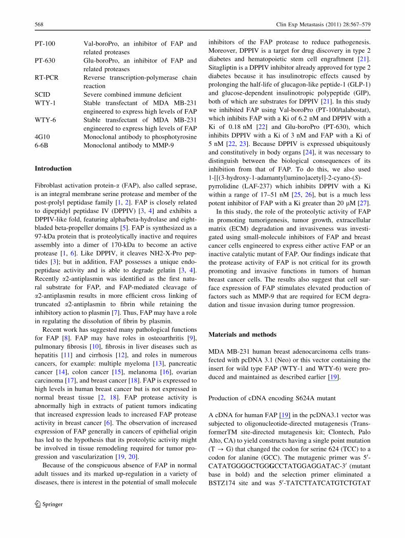
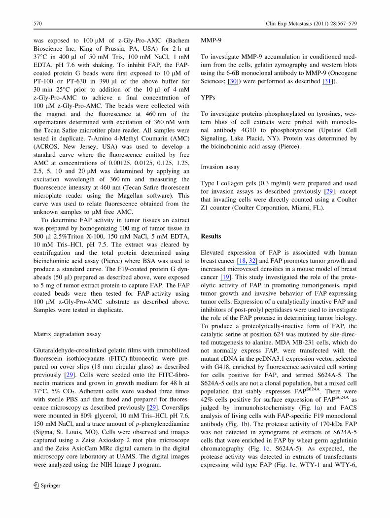
Fig. 1 Characterization of MDA MB 231 cells engineered to express
FAPS624A. a Photomicrograph of a cytospin of cells transfected with
FAPS624A catalytic mutant (called S624A-5) prior to flow sorting.
Cells stained with F19 mAb to FAP appear brown due to the DAB
reaction product while the blue hematoxylin detects all cells. b FACS
showing density plots of cell surface FAP when cells expressing
FAPS624A were stained with non-immune IgG (left panel) or F19 mAb
to FAP (right panel). The regions R1 and R2 cover identical areas in
both left and right panels and serve to indicate the FAP negative cells
(R1) and the FAP positive cells identified by F19 (R2). c Gelatin
zymogram showing a lack of gelatinolytic activity in catalytic mutant
lane (S624A-5) but there is gelatinase activity in cells expressing wild
type FAP (lanes WTY-1 and WTY-6). d The catalytic mutant can
assemble into the 170-kDa dimer as detected by western blot in the
cells expressing mutant FAP (lane S624A-5) and wild type FAP
(lanes WTY-1 and WTY-6) but not in the parental MDA MB-231
cells (231) or these cells transfected with the empty vector (Neo)
Clin Exp Metastasis (2011) 28:567–579 571
123
inhibitors versus saline (P [ 0.05). However, the lowest
average weights were consistently obtained for tumors
treated with PT-100/talabostat.
The protease inhibitor results suggest that the tumorigenic
effect of FAP is independent of its proteolytic activities.
MDA MB-231 cells transfected with proteolytically inactive
FAPS624A were used to further investigate tumor growth
stimulation by FAP in the absence of proteolytic activity,
MDA MB-231 cells expressing FAPS624A (42.6% positive
cells) (S624A-5, Fig. 1b) and FAP-negative cells transfected
with empty vector (Neo, Fig. 1) were injected subcutane-
ously into the mammary fat pads of female SCID mice as
described above. The S624A-5 cells formed tumors that
grew considerably faster than tumors of Neo transfectants
that do not express FAP (Fig. 2d). The yield of tumors from
S624A-5 cells expressing the proteolytically inactive FAP
(26 tumors/28 injection sites; 93%) was greater than that
observed for Neo control cells (19 tumors/28 sites; 68%) and
comparable to that observed with WTY-1 and WTY-6 cells
expressing active FAP (23 tumors/28 sites; 82% and 28
tumors/28 sites respectively; 100% [19]). Tumors of S624A-
5 cells expressing FAPS624A were considerably larger than
those of cells lacking FAP (Neo) as judged by the average
wet weight of the tumors excised on day 30 (Neo,
0.066 g ± 0.038; S624A, 0.8 g ± 0.41). The S624A-5
tumors were similar in size to those of cells expressing wild
type FAP [19]. A second in vivo experiment was performed
using a population of S624A-5 cells that was sorted by FACS
to contain 41% FAP624A-expressing cells. Implantation of
these cells into the mammary fat pads of female SCID mice
again resulted in rapidly growing tumors (not shown) with a
high tumor yield (17 tumors/20 injection sites; 85%). The
promotion of tumorigenesis and rapid tumor growth by the
expression of proteolytically inactive FAP in mammary
tumors in mice and the observation that small-molecule
inhibitors of FAP protease activity did not significantly
inhibit growth of tumors expressing proteolytically active
FAP suggest that non-enzymatic functions of FAP may be
responsible for increased tumor growth in vivo.
A FAP-specific protease activity assay was developed to
confirm that FAP was inhibited in the tumors of the ani-
mals that were treated with talabostat and PT-630 but not in
those treated with LAF-237. For the assay, FAP is immune
precipitated from extracts of cells or tumor tissues and then
exposed to the substrate z-Gly-Pro-AMC. FAP cleaves the
peptide bond linking Pro to AMC and consequently the
fluorescence emitted by AMC is increased. Extracts of
MDA MB-231 cells that do not express FAP, revealed low
activity in this assay (Fig. 3a, 231, black bars) and this
activity was unchanged or showed a decrease when treated
with PT-630 or talabostat (Fig. 3a, 231, white and gray
bars). However, WTY-6 and WTY-1 transfectants of MDA
0
0.1
0.2
0.3
0.4
0.5
0.6
3 6 9 12 15 18 21 24 27 30 33 36
WTY-6 Saline
WTY-6 Talabostat
WTY-6 PT-630
0
0.02
0.04
0.06
0.08
0.1
0.12
0.14
0.16
0.18
3 6 9 12 15 18 21 24 27 30 33 36
435 saline435 Talabostat435 PT-630
0
0.2
0.4
0.6
0.8
1
1.2
12 15 18 21 24 27 30 33 36 39
WTY-1 SalineWTY-1 LAF237WTY-1 TalabostatWTY-1 PT630
A B
C D
Days post inoculation
Days post inoculation
Days post inoculation
Days post inoculation
Tum
mor
siz
e (c
m3 )
Tum
mor
siz
e (c
m3 )
Tum
mor
siz
e (c
m3 )
0
0.5
1
1.5
2
2.5
3
3.5
4
4.5
11 13 15 17 19 21 23 25 27 29 30
S624A-5Neo
Tum
mor
siz
e (c
m3 )
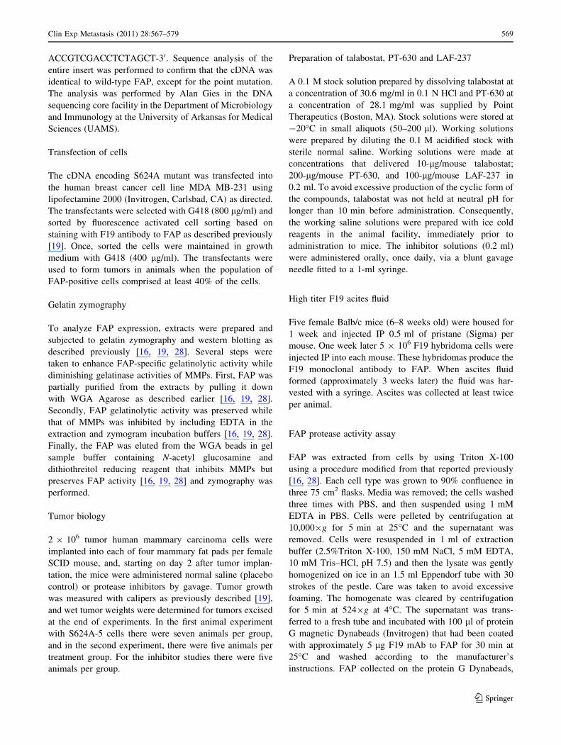
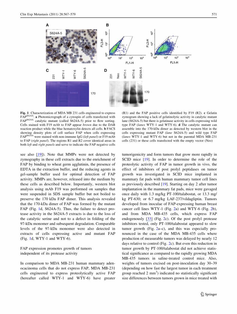
Fig. 2 Breast tumors expressing FAP grow rapidly even when FAP
catalytic activity is inhibited. a–c Talabostat slows growth of breast
tumors in the mammary fat pads of SCID mice. Tumor volumes
plotted over time of FAP-expressing human breast cancer cells in
SCID mice treated with saline (filled diamond), LAF-237 (filled
circle), PT-630 (filled triangle), or talabostat (filled square). The
tumors are formed from the following cells: a WTY-1; b WTY-6; and
c MDA MB-435. d Growth of tumors from S624A-5 cells (filleddiamond) and Neo cells (filled triangle). In all cases tumor growth is
recorded as volume (cm3)
572 Clin Exp Metastasis (2011) 28:567–579
123
MB-231 cells that express wild type and active FAP to high
levels reveal high FAP activity in this assay (Fig. 3a WTY-
6 and WTY-1, black bars) that is inhibited by both PT-630
and talabostat (Fig. 3a WTY-6 and WTY-1, white and gray
bars). In contrast, the mutant FAP reveals low levels of
FAP activity similar to the FAP negative MDA MB-231
cells (Fig. 3a, S624A-5, black bars) that remains low when
exposed to PT-630 and talabostat (Fig. 3a, S624A-5, white
and gray bars). For comparison, intact z-Gly-Pro-AMC was
used to determine back ground fluorescence (Fig. 3a, G-P-
AMC, black, white, and gray bars).
Extracts of tumor tissues were prepared and subjected to
the FAP-activity assay. In the case of WTY-6 tumors,
Fresh frozen tissues were available from normal saline
treated animals and PT-630 treated animals. The extracts
from WTY-6 tumors of animals treated with normal saline
had high FAP activity (Fig. 3b, saline) whereas the extracts
of WTY-6 tumors from PT-630 animals had low FAP
activity (Fig. 3b, PT-630). Similarly, extracts of WTY-1
tumors obtained from animals treated with LAF-237 that
preferentially inhibits DPPIV and not FAP, had high FAP
activity (Fig. 3c, LAF-237) but extracts of WTY-1 tumors
treated with PT-630 had low FAP activity (Fig. 3c,
PT-630). Overall the results are consistent with the
inhibitors acting as expected with LAF-237 producing
relatively little inhibition of FAP proteolytic activity in the
tumors but PT-630 causing significant inhibition of FAP
activity.
FAP promotes matrix degradation and invasion
by tumor cells independent of its protease activity
The proteolytic degradation of fluorescent fibronectin
matrices by cells expressing proteolytically active FAP was
investigated in the presence or absence of FAP inhibitors.
In this assay, the cells adhere to the matrix surface and
fluorescence-negative black spots are observed where the
matrix has been degraded [16, 29]. These studies focused
on the WTY-1 cells that over express proteolytically active
FAP. Cells were administered 1 lM of the inhibitors and
allowed to interact with the matrices for 48-h at 37�C. The
inhibitors were replenished after 24 h. The reduction of
matrix degradation was compared in treated and untreated
cells. Matrix degradation remained high, even in the
presence of FAP/DPPIV inhibitors (Fig. 4a). Two different
analyses were used to quantify the matrix degradation. In
the first, the fluorescent matrices were examined by ran-
domly choosing five fields of the matrices and scoring
0
500
1000
1500
2000
2500
Saline PT-630
Flu
ore
scen
ce U
nit
s
FAP Activity in WTY-6 Tumors
0
200
400
600
800
1000
1200
1400
LAF-237 PT-630
Flu
ore
scen
ce U
nit
s
FAP Activity in WTY-1 Tumors
A
B
C
0
0.1
0.2
0.3
0.4
0.5
0.6
0.7
0.8
0.9
231 WTY-6 WTY-1 S624A-5 G-P-AMC
Co
nce
ntr
atio
n (µ
M)
FAP Activity Assay
Untreated
PT-630 10 µM
Talabostat 10 µM
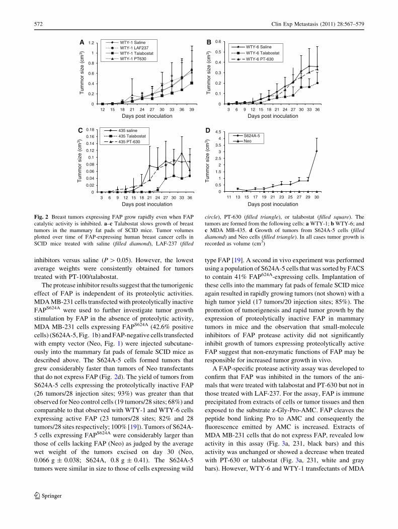
Fig. 3 Effect of inhibitors on FAP activity. a. FAP activity was
assessed by binding FAP in extracts of parental MDA-MB-231 (231)
cells that do not express FAP; WTY-6 (WTY-6), WTY-1(WTY-1)
cells that express wild type and active FAP, or S624A-5 cells that
express a mutant FAP that is catalytically inactive (S624A-5) to F19
mAb immobilized on magnetic protein G beads. In addition, results
are shown for substrate only (G-P-AMC). The FAP-bound beads were
exposed to the FAP substrate z-Gly-Pro-AMC, the supernatant
collected and the fluorescence emitted at 460 nm when excited by
360 nm was determined. A standard curve using free AMC allows
plotting the concentration of free AMC on the y axis. Results are
shown for FAP that is uninhibited (black bars) or for FAP inhibition
by PT-630 (white bars) or PT-100 (gray bars). b, c FAP activity was
determined in extracts of tumor tissues derived from b WTY-6 cells
(WTY-6) exposed to normal saline (Saline), or PT-630 (PT-630), and
c extracts of tumor tissues derived from WTY-1 cells (WTY-1) and
exposed to LAF-237 (LAF-237) or PT-630 as described in the animal
experiments shown in Fig. 2. Results are given in fluorescence
intensity determined at 460 nm wavelength with excitation wave-
length of 360 nm
Clin Exp Metastasis (2011) 28:567–579 573
123
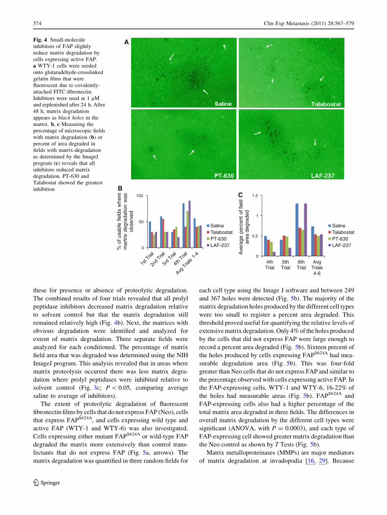
these for presence or absence of proteolytic degradation.
The combined results of four trials revealed that all prolyl
peptidase inhibitors decreased matrix degradation relative
to solvent control but that the matrix degradation still
remained relatively high (Fig. 4b). Next, the matrices with
obvious degradation were identified and analyzed for
extent of matrix degradation. Three separate fields were
analyzed for each conditioned. The percentage of matrix
field area that was degraded was determined using the NIH
ImageJ program. This analysis revealed that in areas where
matrix proteolysis occurred there was less matrix degra-
dation where prolyl peptidases were inhibited relative to
solvent control (Fig. 3c; P \ 0.05, comparing average
saline to average of inhibitors).
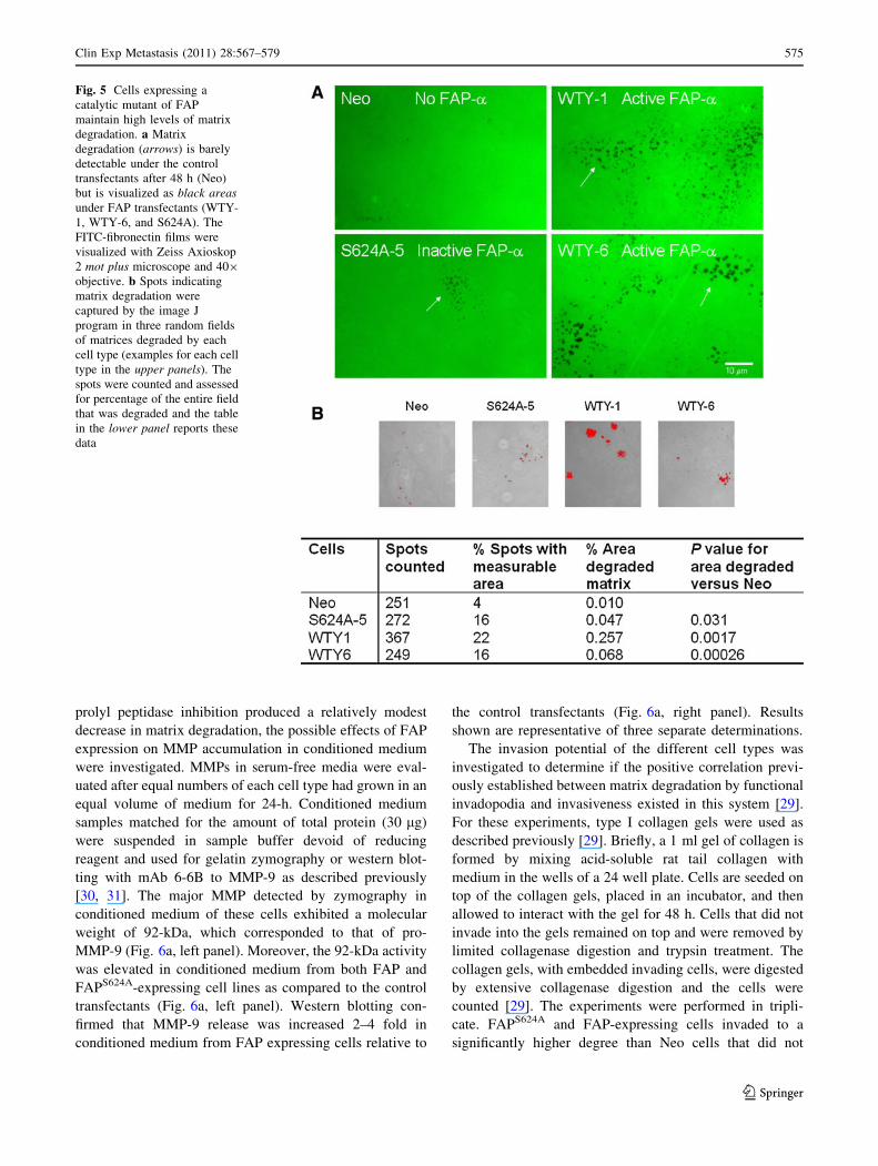
The extent of proteolytic degradation of fluorescent
fibronectin films by cells that do not express FAP (Neo), cells
that express FAPS624A, and cells expressing wild type and
active FAP (WTY-1 and WTY-6) was also investigated.
Cells expressing either mutant FAPS624A or wild-type FAP
degraded the matrix more extensively than control trans-
fectants that do not express FAP (Fig. 5a, arrows). The
matrix degradation was quantified in three random fields for
each cell type using the Image J software and between 249
and 367 holes were detected (Fig. 5b). The majority of the
matrix degradation holes produced by the different cell types
were too small to register a percent area degraded. This
threshold proved useful for quantifying the relative levels of
extensive matrix degradation. Only 4% of the holes produced
by the cells that did not express FAP were large enough to
record a percent area degraded (Fig. 5b). Sixteen percent of
the holes produced by cells expressing FAPS624A had mea-
surable degradation area (Fig. 5b). This was four-fold
greater than Neo cells that do not express FAP and similar to
the percentage observed with cells expressing active FAP. In
the FAP-expressing cells, WTY-1 and WTY-6, 16-22% of
the holes had measurable areas (Fig. 5b). FAPS624A and
FAP-expressing cells also had a higher percentage of the
total matrix area degraded in three fields. The differences in
overall matrix degradation by the different cell types were
significant (ANOVA, with P = 0.0003), and each type of
FAP-expressing cell showed greater matrix degradation than
the Neo control as shown by T Tests (Fig. 5b).
Matrix metalloproteinases (MMPs) are major mediators
of matrix degradation at invadopodia [16, 29]. Because
A
BC
0
50
100
% o
f usa
ble
field
s w
here
m
artr
ix d
egra
datio
n w
as
obse
rved
SalineTalabostatPT-630LAF-237
0
0.5
1
1.5
4th Trial
5th Trial
6th Trial
Avg Trials 4-6
Ave
rage
per
cent
of f
ield
ar
ea d
egra
ded
SalineTalabostatPT-630LAF-237
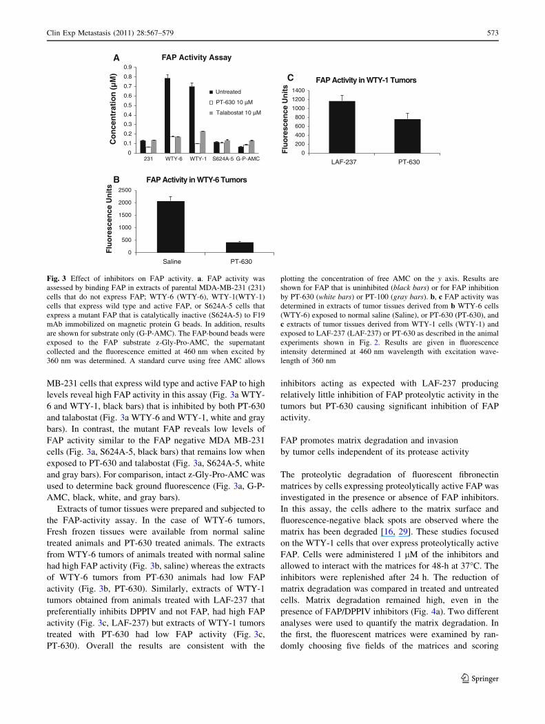
Fig. 4 Small-molecule
inhibitors of FAP slightly
reduce matrix degradation by
cells expressing active FAP.
a WTY-1 cells were seeded
onto glutaradehyde-crosslinked
gelatin films that were
fluorescent due to covalently-
attached FITC-fibronectin.
Inhibitors were used at 1 lM
and replenished after 24 h. After
48 h, matrix degradation
appears as black holes in the
matrix. b, c Measuring the
percentage of microscopic fields
with matrix degradation (b) or
percent of area degraded in
fields with matrix-degradation
as determined by the ImageJ
program (c) reveals that all
inhibitors reduced matrix
degradation. PT-630 and
Talabostat showed the greatest
inhibition
574 Clin Exp Metastasis (2011) 28:567–579
123
prolyl peptidase inhibition produced a relatively modest
decrease in matrix degradation, the possible effects of FAP
expression on MMP accumulation in conditioned medium
were investigated. MMPs in serum-free media were eval-
uated after equal numbers of each cell type had grown in an
equal volume of medium for 24-h. Conditioned medium
samples matched for the amount of total protein (30 lg)
were suspended in sample buffer devoid of reducing
reagent and used for gelatin zymography or western blot-
ting with mAb 6-6B to MMP-9 as described previously
[30, 31]. The major MMP detected by zymography in
conditioned medium of these cells exhibited a molecular
weight of 92-kDa, which corresponded to that of pro-
MMP-9 (Fig. 6a, left panel). Moreover, the 92-kDa activity
was elevated in conditioned medium from both FAP and
FAPS624A-expressing cell lines as compared to the control
transfectants (Fig. 6a, left panel). Western blotting con-
firmed that MMP-9 release was increased 2–4 fold in
conditioned medium from FAP expressing cells relative to
the control transfectants (Fig. 6a, right panel). Results
shown are representative of three separate determinations.
The invasion potential of the different cell types was
investigated to determine if the positive correlation previ-
ously established between matrix degradation by functional
invadopodia and invasiveness existed in this system [29].
For these experiments, type I collagen gels were used as
described previously [29]. Briefly, a 1 ml gel of collagen is
formed by mixing acid-soluble rat tail collagen with
medium in the wells of a 24 well plate. Cells are seeded on
top of the collagen gels, placed in an incubator, and then
allowed to interact with the gel for 48 h. Cells that did not
invade into the gels remained on top and were removed by
limited collagenase digestion and trypsin treatment. The
collagen gels, with embedded invading cells, were digested
by extensive collagenase digestion and the cells were
counted [29]. The experiments were performed in tripli-
cate. FAPS624A and FAP-expressing cells invaded to a
significantly higher degree than Neo cells that did not
Fig. 5 Cells expressing a
catalytic mutant of FAP
maintain high levels of matrix
degradation. a Matrix
degradation (arrows) is barely
detectable under the control
transfectants after 48 h (Neo)
but is visualized as black areasunder FAP transfectants (WTY-
1, WTY-6, and S624A). The
FITC-fibronectin films were
visualized with Zeiss Axioskop
2 mot plus microscope and 409
objective. b Spots indicating
matrix degradation were
captured by the image J
program in three random fields
of matrices degraded by each
cell type (examples for each cell
type in the upper panels). The
spots were counted and assessed
for percentage of the entire field
that was degraded and the table
in the lower panel reports these
data
Clin Exp Metastasis (2011) 28:567–579 575
123
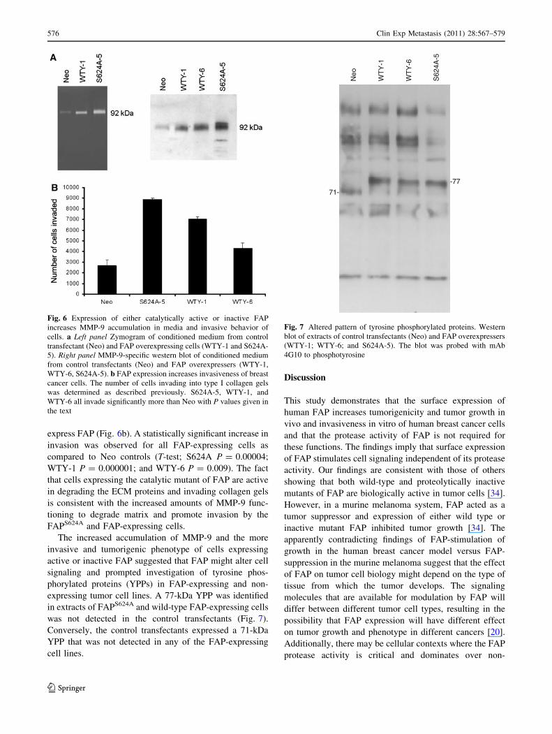
express FAP (Fig. 6b). A statistically significant increase in
invasion was observed for all FAP-expressing cells as
compared to Neo controls (T-test; S624A P = 0.00004;
WTY-1 P = 0.000001; and WTY-6 P = 0.009). The fact
that cells expressing the catalytic mutant of FAP are active
in degrading the ECM proteins and invading collagen gels
is consistent with the increased amounts of MMP-9 func-
tioning to degrade matrix and promote invasion by the
FAPS624A and FAP-expressing cells.
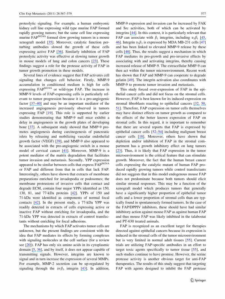
The increased accumulation of MMP-9 and the more
invasive and tumorigenic phenotype of cells expressing
active or inactive FAP suggested that FAP might alter cell
signaling and prompted investigation of tyrosine phos-
phorylated proteins (YPPs) in FAP-expressing and non-
expressing tumor cell lines. A 77-kDa YPP was identified
in extracts of FAPS624A and wild-type FAP-expressing cells
was not detected in the control transfectants (Fig. 7).
Conversely, the control transfectants expressed a 71-kDa
YPP that was not detected in any of the FAP-expressing
cell lines.
Discussion
This study demonstrates that the surface expression of
human FAP increases tumorigenicity and tumor growth in
vivo and invasiveness in vitro of human breast cancer cells
and that the protease activity of FAP is not required for
these functions. The findings imply that surface expression
of FAP stimulates cell signaling independent of its protease
activity. Our findings are consistent with those of others
showing that both wild-type and proteolytically inactive
mutants of FAP are biologically active in tumor cells [34].
However, in a murine melanoma system, FAP acted as a
tumor suppressor and expression of either wild type or
inactive mutant FAP inhibited tumor growth [34]. The
apparently contradicting findings of FAP-stimulation of
growth in the human breast cancer model versus FAP-
suppression in the murine melanoma suggest that the effect
of FAP on tumor cell biology might depend on the type of
tissue from which the tumor develops. The signaling
molecules that are available for modulation by FAP will
differ between different tumor cell types, resulting in the
possibility that FAP expression will have different effect
on tumor growth and phenotype in different cancers [20].
Additionally, there may be cellular contexts where the FAP
protease activity is critical and dominates over non-
Fig. 6 Expression of either catalytically active or inactive FAP
increases MMP-9 accumulation in media and invasive behavior of
cells. a Left panel Zymogram of conditioned medium from control
transfectant (Neo) and FAP overexpressing cells (WTY-1 and S624A-
5). Right panel MMP-9-specific western blot of conditioned medium
from control transfectants (Neo) and FAP overexpressers (WTY-1,
WTY-6, S624A-5). b FAP expression increases invasiveness of breast
cancer cells. The number of cells invading into type I collagen gels
was determined as described previously. S624A-5, WTY-1, and
WTY-6 all invade significantly more than Neo with P values given in
the text
-77
71-
Neo
WT
Y-1
WT
Y-6
S62
4A-5
Fig. 7 Altered pattern of tyrosine phosphorylated proteins. Western
blot of extracts of control transfectants (Neo) and FAP overexpressers
(WTY-1; WTY-6; and S624A-5). The blot was probed with mAb
4G10 to phosphotyrosine
576 Clin Exp Metastasis (2011) 28:567–579
123
proteolytic signaling. For example, a human embryonic
kidney cell line expressing wild type murine FAP formed
rapidly growing tumors; but the same cell line expressing
murine FAPS624A formed slow growing tumors in a mouse
xenograft model [35]. Moreover, catalytic function per-
turbing antibodies slowed the growth of these cells
expressing active FAP [36]. Similarly inhibition of FAP
proteolytic activity was effective at slowing tumor growth
in mouse models of lung and colon cancers [23]. These
findings suggest a role for the protease activity of FAP in
tumor growth promotion in these models.
Several lines of evidence suggest that FAP activates cell
signaling that changes cell behavior. Firstly, MMP-9
accumulation in conditioned medium is high for cells
expressing FAPS624A or wild-type FAP. The increase in
MMP-9 levels of FAP-expressing cells is particularly rel-
evant to tumor progression because it is a pro-angiogenic
factor [37–40] and may be an important mediator of the
increased angiogenesis previously observed in tumors
expressing FAP [19]. This role is supported by earlier
studies demonstrating that MMP-9 null mice exhibit a
delay in angiogenesis in the growth plates of developing
bone [37]. A subsequent study showed that MMP-9 pro-
motes angiogenesis during carcinogenesis of pancreatic
islets by releasing and mobilizing vascular endothelial
growth factor (VEGF) [39], and MMP-9 also appeared to
be associated with the pro-angiogenic switch in a mouse
model of cervical cancer [41]. Moreover, MMP-9 is a
potent mediator of the matrix degradation that facilitates
tumor invasion and metastasis. Secondly, YPP expression
appeared to be similar between cells that express FAPS624A
or FAP and different from that in cells that lack FAP.
Interestingly, others have shown that extracts of membrane
preparations enriched for invadopodia or podosomes; the
membrane protrusions of invasive cells that contact and
degrade ECM, contain four major YPPs identified as 150,
130, 81, and 77-kDa proteins [42]. YPPs of 150 and
71-kDa were identified as components of normal focal
contacts [42]. In the present study, a 77-kDa YPP was
readily detected in extracts of cells expressing active or
inactive FAP without enriching for invadopodia, and the
71-kDa YPP was detected in extracts of control transfec-
tants without enriching for focal adhesions.
The mechanisms by which FAP activates tumor cells are
unknown, but the present findings are consistent with the
idea that FAP mediates its effects by forming complexes
with signaling molecules at the cell surface (for a review
see [20]). FAP has only six amino acids in its cytoplasmic
domain [5, 36], and by itself, it does not appear capable of
transmitting signals. However, integrins are known to
signal and in turn increase the expression of several MMPs.
For example, MMP-2 secretion can be up-regulated by
signaling through the avb3 integrin [43]. In addition,
MMP-9 expression and invasion can be increased by FAK
and Src activities, both of which can be activated by
integrins [44]. In this context, it is particularly relevant that
FAP can associate with b1 integrins, including a3b1 [45,
46]. Integrin a3b1 is expressed by MDA MB-231 cells [47]
and has been linked to elevated MMP-9 release by these
cells [48]. Thus, the results suggest a mechanism in which
FAP mediates its pro-growth and pro-invasive effects by
associating with and activating integrins, thereby causing
increased release of MMP-9. The extracellular MMP-9 can
then act within the tumor microenvironment. Recent work
has shown that FAP and MMP-9 can cooperate to degrade
gelatin [49]. The integrin activation also coordinates with
MMP-9 to promote tumor invasion and metastasis.
This study forced over-expression of FAP in the epi-
thelial cancer cells and did not focus on the stromal cells.
However, FAP is best known for its induced expression on
stromal fibroblasts reacting to epithelial cancers [32, 50,
51]. Therefore, FAP expression on tumor cells themselves
may have distinct effects on tumor growth as compared to
the effects of the better known expression of FAP on
stromal cells. In this regard, it is important to remember
that there are several reports that FAP is expressed by
epithelial cancer cells [52–54] including malignant breast
cancer cells [18]. Moreover, others have shown that
depletion and/or inhibition of FAP in the stromal com-
partment has a growth inhibitory effect on lung tumors
[23]. Thus, it is likely that FAP expression in the tumor
microenvironment is the critical feature that can stimulate
growth. Moreover, the fact that the human breast cancer
cells expressing the catalytic mutant of human FAP pro-
duced rapidly growing tumors while control transfectants
did not suggests that in this model endogenous mouse FAP
does not predominate because both tumors should elicit
similar stromal responses. This may be a function of the
xenograft model which produces tumors that generally
have a significantly higher proportion of epithelial tumor
cells and a lower proportion of stromal cells than are typ-
ically found in spontaneously formed tumors. In the case of
the FAP/DPPIV inhibitors, these should have had similar
inhibitory action against mouse FAP as against human FAP
and thus mouse FAP was likely inhibited in the talabostat
and PT-630 treated animals.
FAP is recognized as an excellent target for therapies
directed against epithelial cancers because its expression is
induced in the stromal cells of the tumor microenvironment
but is very limited in normal adult tissues [55]. Current
trials are utilizing FAP-specific antibodies in an effort to
target toxic agents specifically to tumor tissue [55], and
such studies continue to have promise. However, the serine
protease activity is another obvious target for anti-FAP
therapeutics. The results of this study suggest that targeting
FAP with agents designed to inhibit the FAP protease
Clin Exp Metastasis (2011) 28:567–579 577
123
activity may not be effective in breast cancer; but there is
insufficient data to predict the efficacy of such agents in
other types of cancer. Inhibition of FAP protease activity
may contribute to the antitumor effects of talabostat in
mouse tumor models [23, 56]. More studies are required to
clarify the roles of the protease activities and the inter-
molecular interactions of FAP in its biological functions.
Conclusions
We conclude that the proteolytic activity of FAP partici-
pates in matrix degradation, but other domains within the
protein stimulate the production of biologically active
factors, such as MMP-9, which enhance matrix remodeling
and facilitate tumor growth.
Acknowledgments The authors thank Amir Khan for help pro-
ducing the cDNA encoding the S624A mutant FAP. The authors
thank Steven Post for critical review of the manuscript. Supported by
grants from the DoD CDMRP-BCRP-BC074331, Arkansas Breast
Cancer Research Program and funds from Point Therapeutics, Inc.
Conflict of interest The study was funded in part by Point Thera-
peutics who produced the talabostat and PT-630 compounds used in
this research.
References
1. Pineiro-Sanchez ML, Goldstein LA, Dodt J, Howard L, Yeh Y, Tran
H, Argraves WS, Chen WT (1997) Identification of the 170-kDa
melanoma membrane-bound gelatinase (seprase) as a serine inte-
gral membrane protease. J Biol Chem 272(12):7595–7601
2. Scanlan MJ, Raj BK, Calvo B, Garin-Chesa P, Sanz-Moncasi
MP, Healey JH, Old LJ, Rettig WJ (1994) Molecular cloning of
fibroblast activation protein alpha, a member of the serine pro-
tease family selectively expressed in stromal fibroblasts of epi-
thelial cancers. Proc Natl Acad Sci USA 91(12):5657–5661
3. O’Brien P, O’Connor BF (2008) Seprase: an overview of an
important matrix serine protease. Biochim Biophys Acta
1784(9):1130–1145
4. Wolf BB, Quan C, Tran T, Wiesmann C, Sutherlin D (2008) On
the edge of validation–cancer protease fibroblast activation pro-
tein. Mini Rev Med Chem 8(7):719–727
5. Aertgeerts K, Levin I, Shi L, Snell GP, Jennings A, Prasad GS,
Zhang Y, Kraus ML, Salakian S, Sridhar V et al (2005) Structural
and kinetic analysis of the substrate specificity of human fibro-
blast activation protein alpha. J Biol Chem 280(20):19441–19444
6. Kelly T (1999) Evaluation of seprase activity. Clin Exp Metas-
tasis 17:57–62
7. Lee KN, Jackson KW, Christiansen VJ, Lee CS, Chun JG,
McKee PA (2006) Antiplasmin-cleaving enzyme is a soluble
form of fibroblast activation protein. Blood 107(4):1397–1404
8. Chen WT, Kelly T, Ghersi G (2003) DPPIV, seprase, and related
serine peptidases in multiple cellular functions. Curr Top Dev
Biol 54:207–232
9. Milner JM, Kevorkian L, Young DA, Jones D, Wait R, Donell
ST, Barksby E, Patterson AM, Middleton J, Cravatt BF et al
(2006) Fibroblast activation protein alpha is expressed by
chondrocytes following a pro-inflammatory stimulus and is ele-
vated in osteoarthritis. Arthritis Res Ther 8(1):R23
10. Acharya PS, Zukas A, Chandan V, Katzenstein AL, Pure E
(2006) Fibroblast activation protein: a serine protease expressed
at the remodeling interface in idiopathic pulmonary fibrosis. Hum
Pathol 37(3):352–360
11. Gorrell MD, Wang XM, Levy MT, Kable E, Marinos G, Cox G,
McCaughan GW (2003) Intrahepatic expression of collagen and
fibroblast activation protein (FAP) in hepatitis C virus infection.
Adv Exp Med Biol 524:235–243
12. Levy MT, McCaughan GW, Abbott CA, Park JE, Cunningham
AM, Muller E, Rettig WJ, Gorrell MD (1999) Fibroblast acti-
vation protein: a cell surface dipeptidyl peptidase and gelatinase
expressed by stellate cells at the tissue remodelling interface in
human cirrhosis. Hepatology 29(6):1768–1778
13. Pennisi A, Li X, Ling W, Khan S, Gaddy D, Suva LJ, Barlogie B,
Shaughnessy JD, Aziz N, Yaccoby S (2009) Inhibitor of DASH
proteases affects expression of adhesion molecules in osteoclasts
and reduces myeloma growth and bone disease. Br J Haematol
145(6):775–787
14. Cohen SJ, Alpaugh RK, Palazzo I, Meropol NJ, Rogatko A, Xu
Z, Hoffman JP, Weiner LM, Cheng JD (2008) Fibroblast acti-
vation protein and its relationship to clinical outcome in pan-
creatic adenocarcinoma. Pancreas 37(2):154–158
15. Henry LR, Lee HO, Lee JS, Klein-Szanto A, Watts P, Ross EA,
Chen WT, Cheng JD (2007) Clinical implications of fibroblast
activation protein in patients with colon cancer. Clin Cancer Res
13(6):1736–1741
16. Monsky WL, Lin CY, Aoyama A, Kelly T, Akiyama SK, Mueller
SC, Chen WT (1994) A potential marker protease of invasive-
ness, seprase, is localized on invadopodia of human malignant
melanoma cells. Cancer Res 54(21):5702–5710
17. Kennedy A, Dong H, Chen D, Chen WT (2009) Elevation of
seprase expression and promotion of an invasive phenotype by
collagenous matrices in ovarian tumor cells. Int J Cancer
124(1):27–35
18. Kelly T, Kechelava S, Rozypal TL, West KW, Korourian S
(1998) Seprase, a membrane-bound protease, is overexpressed by
invasive ductal carcinoma cells of human breast cancers. Mod
Pathol 11(9):855–863
19. Huang Y, Wang S, Kelly T (2004) Seprase promotes rapid tumor
growth and increased microvessel density in a mouse model of
human breast cancer. Cancer Res 64(8):2712–2716
20. Kelly T (2005) Fibroblast activation protein-alpha and dipeptidyl
peptidase IV (CD26): cell-surface proteases that activate cell
signaling and are potential targets for cancer therapy. Drug Resist
Updat 8(1–2):51–58
21. Thornberry NA, Gallwitz B (2009) Mechanism of action of
inhibitors of dipeptidyl-peptidase-4 (DPP-4). Best Pract Res Clin
Endocrinol Metab 23(4):479–486
22. Connolly BA, Sanford DG, Chiluwal AK, Healey SE, Peters DE,
Dimare MT, Wu W, Liu Y, Maw H, Zhou Y et al (2008)
Dipeptide boronic acid inhibitors of dipeptidyl peptidase IV:
determinants of potency and in vivo efficacy and safety. J Med
Chem 51(19):6005–6013
23. Santos AM, Jung J, Aziz N, Kissil JL, Pure E (2009) Targeting
fibroblast activation protein inhibits tumor stromagenesis and
growth in mice. J Clin Invest 119(12):3613–3625
24. Rosenblum JS, Kozarich JW (2003) Prolyl peptidases: a serine
protease subfamily with high potential for drug discovery. Curr
Opin Chem Biol 7(4):496–504
25. Brandt I, Joossens J, Chen X, Maes MB, Scharpe S, De Meester I,
Lambeir AM (2005) Inhibition of dipeptidyl-peptidase IV cata-
lyzed peptide truncation by Vildagliptin ((2S)-{[(3-hydroxyada-
mantan-1-yl)amino]acetyl}-pyrrolidine-2-carbonitrile). Biochem
Pharmacol 70(1):134–143
578 Clin Exp Metastasis (2011) 28:567–579
123
26. Van der Veken P, Haemers A, Augustyns K (2007) Prolyl pep-
tidases related to dipeptidyl peptidase IV: potential of specific
inhibitors in drug discovery. Curr Top Med Chem 7(6):621–635
27. Tsai TY, Hsu T, Chen CT, Cheng JH, Chiou MC, Huang CH,
Tseng YJ, Yeh TK, Huang CY, Yeh KC et al (2009) Rational
design and synthesis of potent and long-lasting glutamic acid-
based dipeptidyl peptidase IV inhibitors. Bioorg Med Chem Lett
19(7):1908–1912
28. Kelly T (1999) Evaluation of seprase activity. Clin Exp Metas-
tasis 17(1):57–62
29. Kelly T, Yan Y, Osborne RL, Athota AB, Rozypal TL, Colcla-
sure JC, Chu WS (1998) Proteolysis of extracellular matrix by
invadopodia facilitates human breast cancer cell invasion and is
mediated by matrix metalloproteinases. Clin Exp Metastasis
16:501–512
30. Ramos-DeSimone N, Hahn-Dantona E, Sipley J, Nagase H,
French DL, Quigley JP (1999) Activation of matrix metallopro-
teinase-9 (MMP-9) via a converging plasmin/stromelysin-1 cas-
cade enhances tumor cell invasion. J Biol Chem 274(19):
13066–13076
31. Kaushal GP, Xiong X, Athota AB, Rozypal TL, Sanderson RD,
Kelly T (1999) Syndecan-1 expression suppresses the level of
myeloma matrix metalloproteinase-9. Br J Haematol 104:365–
373
32. Garin-Chesa P, Old LJ, Rettig WJ (1990) Cell surface glyco-
protein of reactive stromal fibroblasts as a potential antibody
target in human epithelial cancers. Proc Natl Acad Sci USA
87:7235–7239
33. Goodman JD, Rozypal TL, Kelly T (2003) Seprase, a membrane-
bound protease, alleviates the serum growth requirement of
human breast cancer cells. Clin Exp Metastasis 20(5):459–470
34. Ramirez-Montagut T, Blachere NE, Sviderskaya EV, Bennett
DC, Rettig WJ, Garin-Chesa P, Houghton AN (2004) FAPalpha,
a surface peptidase expressed during wound healing, is a tumor
suppressor. Oncogene 23(32):5435–5446
35. Cheng JD, Valianou M, Canutescu AA, Jaffe EK, Lee HO, Wang
H, Lai JH, Bachovchin WW, Weiner LM (2005) Abrogation of
fibroblast activation protein enzymatic activity attenuates tumor
growth. Mol Cancer Ther 4(3):351–360
36. Cheng JD, Dunbrack RL Jr, Valianou M, Rogatko A, Alpaugh
RK, Weiner LM (2002) Promotion of tumor growth by murine
fibroblast activation protein, a serine protease, in an animal
model. Cancer Res 62(16):4767–4772
37. Vu TH, Shipley JM, Bergers G, Berger JE, Helms JA, Hanahan
D, Shapiro SD, Senior RM, Werb Z (1998) MMP-9/gelatinase B
is a key regulator of growth plate angiogenesis and apoptosis of
hypertrophic chondrocytes. Cell 93(3):411–422
38. Hangai M, Kitaya N, Xu J, Chan CK, Kim JJ, Werb Z, Ryan SJ,
Brooks PC (2002) Matrix metalloproteinase-9-dependent expo-
sure of a cryptic migratory control site in collagen is required
before retinal angiogenesis. Am J Pathol 161(4):1429–1437
39. Bergers G, Brekken R, McMahon G, Vu TH, Itoh T, Tamaki K,
Tanzawa K, Thorpe P, Itohara S, Werb Z et al (2000) Matrix
metalloproteinase-9 triggers the angiogenic switch during carci-
nogenesis. Nat Cell Biol 2(10):737–744
40. Chantrain CF, Shimada H, Jodele S, Groshen S, Ye W, Shalinsky
DR, Werb Z, Coussens LM, DeClerck YA (2004) Stromal matrix
metalloproteinase-9 regulates the vascular architecture in neuro-
blastoma by promoting pericyte recruitment. Cancer Res
64(5):1675–1686
41. Giraudo E, Inoue M, Hanahan D (2004) An amino-bisphospho-
nate targets MMP-9-expressing macrophages and angiogenesis to
impair cervical carcinogenesis. J Clin Invest 114(5):623–633
42. Mueller SC, Yeh Y, Chen WT (1992) Department of A, Cell
Biology GUSoMWDC: tyrosine phosphorylation of membrane
proteins mediates cellular invasion by transformed cells. J Cell
Biol 119(5):1309–1325
43. Brooks PC, Streomblad S, Sanders LC, von Schalscha TL, Aimes
RT, Stetler-Stevenson WG, Quigley JP, Cheresh DA (1996)
Localization of matrix metalloproteinase MMP-2 to the surface
of invasive cells by interaction with integrin alpha v beta 3. Cell
85(5):683–693
44. Hsia DA, Mitra SK, Hauck CR, Streblow DN, Nelson JA, Ilic D,
Huang S, Li E, Nemerow GR, Leng J et al (2003) Differential
regulation of cell motility and invasion by FAK. J Cell Biol
160(5):753–767
45. Mueller SC, Ghersi G, Akiyama SK, Sang Q-XA, Howard L,
Pineiro-Sanchez M, Nakahara H, Yeh Y, Chen W-T (1999) A
novel protease-docking function of integrin at invadopodia. J Biol
Chem 274:24947–24952
46. Artym VV, Kindzelskii AL, Chen WT, Petty HR (2002)
Molecular proximity of seprase and the urokinase-type plasmin-
ogen activator receptor on malignant melanoma cell membranes:
dependence on beta1 integrins and the cytoskeleton. Carcino-
genesis 23(10):1593–1601
47. Lichtner RB, Howlett AR, Lerch M, Xuan JA, Brink J, Langton-
Webster B, Schneider MR (1998) Negative cooperativity between
alpha 3 beta 1 and alpha 2 beta 1 integrins in human mammary
carcinoma MDA MB 231 cells. Exp Cell Res 240(2):368–376
48. Morini M, Mottolese M, Ferrari N, Ghiorzo F, Buglioni S,
Mortarini R, Noonan DM, Natali PG, Albini A (2000) The alpha
3 beta 1 integrin is associated with mammary carcinoma cell
metastasis, invasion, and gelatinase B (MMP-9) activity. Int J
Cancer 87(3):336–342
49. Christiansen VJ, Jackson KW, Lee KN, McKee PA (2007) Effect
of fibroblast activation protein and alpha2-antiplasmin cleaving
enzyme on collagen types I, III, and IV. Arch Biochem Biophys
457(2):177–186
50. Rettig WJ, Garin-Chesa P, Healey JH, Su SL, Ozer HL, Schwab
M, Albino AP, Old LJ (1993) Regulation and heteromeric
structure of the fibroblast activation protein in normal and
transformed cells of mesenchymal and neuroectodermal origin.
Cancer Res 53(14):3327–3335
51. Ariga N, Sato E, Ohuchi N, Nagura H, Ohtani H (2001) Stromal
expression of fibroblast activation protein/seprase, a cell mem-
brane serine proteinase and gelatinase, is associated with longer
survival in patients with invasive ductal carcinoma of breast. Int J
Cancer 95(1):67–72
52. Iwasa S, Jin X, Okada K, Mitsumata M, Ooi A (2003) Increased
expression of seprase, a membrane-type serine protease, is
associated with lymph node metastasis in human colorectal
cancer. Cancer Lett 199(1):91–98
53. Iwasa S, Okada K, Chen WT, Jin X, Yamane T, Ooi A, Mitsu-
mata M (2005) ‘Increased expression of seprase, a membrane-
type serine protease, is associated with lymph node metastasis in
human colorectal cancer. Cancer Lett 227(2):229–236
54. Jin X, Iwasa S, Okada K, Mitsumata M, Ooi A (2003) Expression
patterns of seprase, a membrane serine protease, in cervical
carcinoma and cervical intraepithelial neoplasm. Anticancer Res
23(4):3195–3198
55. Scott AM, Wiseman G, Welt S, Adjei A, Lee FT, Hopkins W,
Divgi CR, Hanson LH, Mitchell P, Gansen DN et al (2003) A
phase I dose-escalation study of sibrotuzumab in patients with
advanced or metastatic fibroblast activation protein-positive
cancer. Clin Cancer Res 9(5):1639–1647
56. Adams S, Miller GT, Jesson MI, Watanabe T, Jones B, Wallner
BP (2004) PT-100, a small molecule dipeptidyl peptidase inhib-
itor, has potent antitumor effects and augments antibody-medi-
ated cytotoxicity via a novel immune mechanism. Cancer Res
64(15):5471–5480
Clin Exp Metastasis (2011) 28:567–579 579
123
Copyright © 2022 FDOKUMEN