Pregnancy Hypertension - GLOWM
456
The FIGO Textbook of Pregnancy Hypertension An evidence-based guide to monitoring, prevention and management
-
Upload
khangminh22 -
Category
Documents
-
view
0 -
download
0
Transcript of Pregnancy Hypertension - GLOWM

The FIGO Textbook of
Pregnancy HypertensionAn evidence-based guide to monitoring,
prevention and management


The FIGO Textbook of
Pregnancy HypertensionAn evidence-based guide to monitoring,
prevention and management
Edited by
Laura A. Magee MD, FRCPC, FACP, Peter von Dadelszen MBChB, DPhil, FRANZCOG,
William Stones MD, FRCOG
and
Matthews Mathai MD, PhD, FRCOG
Incorporating the key findings of the PRE-EMPT global studies
With Forewords by the President of FIGO
and by the Immediate Past President of FIGO
Published byThe Global Library of Women’s Medicine

Published byThe Global Library of Women’s Medicine
9 Provost Court
London NW3 4SR
Copyright © 2016 The Foundation for the Global Library of Women’s Medicine
ISBN: 978-0-9927545-5-6
This book is published on a not-for-profit basis and is also available at www.glowm.com
All rights reserved. No part of this publication may be reproduced, stored in or introduced into a retrieval system, or transmitted, in any form, or by any means (electronic, mechanical, photocopying, recording or otherwise) without the prior permission of the publisher.
Typeset by AMA DataSet Ltd, Preston, UK
Printed in the UK by CPI Group (UK), Croydon CR0 4YY, UK
Dedicated to the memory of Sabrina Dwan – and with gratitude to the Sabrina’s Foundation for the financial support
generously provided to assist in the publication of this book

v
Contributors vii
Abbreviations x
Forewords xii
Introduction xiv
Section 1: Clinical Review
1. Hypertension 1
2. Measurement of proteinuria 19
3. Classification of the hypertensive disorders of pregnancy 33
4. Epidemiology of the hypertensive disorders of pregnancy 63
5. Risk factors and predictors of pre-eclampsia 75
6. Preventing pre-eclampsia and its complications 101
7. Diet, lifestyle and place of care 123
8. Fluids, drugs and transfusion 133
9. Timing and mode of delivery 167
10. Anaesthesia 185
11. Treatment postpartum – immediate and long term 215
Section 2: Appendices 233
Index 421
Contents


vii
Contributors
EDITORS
Laura A. Magee MD, MSc, FRCPC, FACP
St George’s, University of London
London, UK
Peter von Dadelszen BmedSc, MBChB, DipObst,
DPhil, FRANZCOG, FRCSC, FRCOG
St George’s, University of London
London, UK
William Stones MD, FRCOG
University of St Andrews School of Medicine
St Andrews, UK
and
Malawi College of Medicine
Blantyre, Malawi
Matthews Mathai MBBS, MD, DGO, MObstet,
PhD, FRCOG
Liverpool School of Tropical Medicine
Liverpool, UK
AUTHORS
Alice B.R. Aarvold
University of British Columbia
Vancouver, Canada
and
Wessex School of Anaesthesia
Winchester, UK
Olalekan O. Adetoro
Centre for Research in Reproductive Health
Olabisi Onabanjo University
Sagamu, Nigeria
Diogo Ayres de Campos
University of Porto
Portugal
Shashidhar Bannale
S Nijalingappa Medical College
Bagalkote, India
Wame Barivalala
Maternal & Reproductive Health
UNFPA APRO
Thailand
José M. Belizán
Institute for Clinical Effectiveness and Health
Policy (IECS)
Buenos Aires, Argentina
Mauricio Belizán
Sanatorio de la Mujer
Rosario, Argentina
Mrutunjaya Bellad
KLE University
Belgaum, India
Emmanuel Bujold
Université Laval
Quebec City, Canada
Anne-Marie Côté
Université de Sherbrooke
Sherbooke, Canada
M. Joanne Douglas
University of British Columbia
Vancouver, Canada
Susheela Engelbrecht
Jhpiego
John Hopkins University
USA
Tabassum Firoz
University of British Columbia
Vancouver, Canada

THE FIGO TEXTBOOK OF PREGNANCY HYPERTENSION
viii
David Hall
Stellenbosch University
Stellenbosch, RSA
Alice Han
Brigham and Women’s Hospital
Boston, MA, USA
and
Women’s Health District Clinical Advisor for
Partners in Health (PIH)
Inshuti Mu Buzima (IMB), Rwanda
Claudia Hanson
Karolinska Institute
Stockholm, Sweden
and
London School of Hygiene and Tropical
Medicine
London, UK
Michael Helewa
University of Manitoba
Winnipeg, Canada
Zahra Hoodbhoy
Aga Khan University
Karachi, Pakistan
Jenn Hutcheon
University of British Columbia
Vancouver, Canada
Joaquin Jaime
Sanatorio de la Mujer
Rosario, Argentina
Geetanjali Katageri
S Nijalingappa Medical College
Bagalkote, India
Andrew Kintu
Makerere University College of Health Sciences
Kampala, Uganda
Oladapo A. Ladipo
Association for Reproductive and Family Health
(ARFH)
Nigeria
André Lalonde
University of Ottawa, McGill University
Canada
Laura A. Magee
St George’s, University of London
London, UK
Ashalata Mallapur
S Nijalingappa Medical College
Bagalkote, India
Matthews Mathai
Liverpool School of Tropical Medicine
Liverpool, UK
Suellen Miller
University of California San Francisco
San Francisco, USA
Jean-Marie Moutquin
Universite de Sherbrooke
Sherbrooke
Quebec, Canada
Shuchita Mundle
Department of Obstetrics and Gynecology
Government Medical College
Nagpur, India
Vitalis Mung’ayi
Aga Khan University
Nairobi, Kenya
Hannah Nathan
King’s College London
London, UK
Beth Payne
University of British Columbia
Vancouver, Canada
Anoul Pels
Academic Medical Center
The Netherlands
Rahat Qureshi
Aga Khan University
Karachi, Pakistan

CONTRIBUTORS
ix
Umesh Ramdurg
S Nijalingappa Medical College
Bagalkote, India
Evelyne Rey
University of Montreal
Montreal, Canada
Helen Ryan
University of British Columbia
Vancouver, Canada
Ilana Sebbag
Schulich School of Medicine and Dentistry
Western University
Ontario, Canada
Sumedha Sharma
University of British Columbia
Vancouver, Canada
John Sotunsa
Babcock University Teaching Hospital
Nigeria
William Stones
University of St Andrews School of Medicine
St Andrews, UK
and
Malawi College of Medicine
Blantyre, Malawi
Ugochi V. Ukah
University of British Columbia
Vancouver, Canada
Hannes van der Mer
Stellenbosch University
Stellenbosch, RSA
Maria F. Escobar Vidarte
Fundación Valle del Lili
Cali, Colombia
Peter von Dadelszen
St George’s, University of London
London, UK
Li Wang
BC Children’s Hospital
Vancouver, Canada
Joanne Wiggins
Royal Bournemouth Hospital
Bournemouth, UK

x
ABPM ambulatory blood pressure monitoring
ACE inhibitor angiotensin converting enzyme inhibitor
ACOG American College of Obstetricians and Gynecologists
ACR albumin : creatinine ratio
AFI amniotic fluid index
AFLP acute fatty liver of pregnancy
ANC antenatal care
AOM Association of Ontario Midwives
APTT activated partial thromboplastin time
ARB angiotensin receptor blocker
AST aspartate transaminase
BEmONC basic emergency obstetric and neonatal care
BID twice daily dosing
BMI body mass index
BP blood pressure
BPP biophysical profile
CBC complete blood count
CEmONC comprehensive emergency obstetric and neonatal care
cGMP cyclic guanosine monophosphate
CO carbon monoxide
CPG clinical practice guideline
CSE combined spinal–epidural analgesia
CVP central venous pressure
DASH dietary approaches to stop hypertension
dBP diastolic blood pressure
DIC disseminated intravascular coagulation
EmONC emergency obstetric and neonatal care
eNOS endothelial nitric oxide synthase
GDM gestational diabetes mellitus
GH gestational hypertension
GMP guanosine monophosphate
GRADE grades of recommendation, assessment, development and evaluation
GSNO S-nitrosoglutathione
GTP guanosine triphosphate
HDL high-density lipoprotein
HDP hypertensive disorder of pregnancy
HELLP syndrome haemolysis, elevated liver enzymes and low platelets syndrome
HIC high-income country
HR heart rate
IM intramuscular
INR international normalised ratio
IOL induction of labour
Abbreviations

ABBREVIATIONS
xi
ISSHP International Society for the Study of Hypertension in Pregnancy
IUGR intrauterine fetal growth restriction
IV intravenous
LDH lactate dehydrogenase
LDL low-density liporotein
LMICs low- and middle-income countries
LMP last menstrual period
MEOWS modified early obstetric warning systems
MRI magnetic resonance imaging
NICE National Institute for Health and Clinical Excellence
NICU neonatal intensive care unit
NO nitric oxide
NSAIDs non-steroidal anti-inflammatory drugs
NST non-stress test
NVOG National Obstetrics and Gynaecology Society, The Netherlands
PCA patient-controlled labour analgesia
PDE5 phosphodiesterase-5
PET pre-eclampsia
PLGF placental growth factor
PO per os – by mouth
pRBCs packed red blood cells
PrCr protein : creatinine ratio
PRECOG pre-eclampsia community guideline
PRES posterior reversible leukoencephalopathy syndrome
QID four times daily dosing
QLD Queensland Maternity and Neonatal Clinical Guidelines Program
RCT randomised controlled trial
RDA recommended daily allowance
sFlt-1 soluble fms-like tyrosine kinase-1
sBP systolic blood pressure
sEng soluble endoglin
SFH symphysis–fundal height
SGA small for gestational age
sGC soluble guanylate cyclase
SOGC Society of Obstetricians and Gynaecologists of Canada
TID three times daily dosing
TTE transthoracic echocardiography
UNFPA United Nations Population Fund
US United States
VEGF vascular endothelial growth factor
WHO World Health Organization

xii
Hypertension in pregnancy is a major contributor
to maternal and perinatal mortality and morbidity.
Every year 70,000 women die and there are half a
million stillbirths or neonatal deaths owing to
hypertensive disorders of pregnancy – the vast
majority being in the developing world. Those
who survive, especially those who had preterm
pre-eclampsia, face the issues of hypertensive,
cerebro- and cardiovascular events in the future
resulting in premature deaths. The International
Federation of Gynecology and Obstetrics (FIGO)
have responded to this important issue by
commissioning The FIGO Textbook of Pregnancy Hypertension. It provides an evidence-based guide
to monitoring, prevention and management of this
common disease that affects 5–10% of pregnant
women.
Our sincere gratitude to the editors Laura
Magee, Peter von Dadelszen, William Stones and
Matthews Mathai – as well as to the international
team of authors all of whom have first-hand clinical
experience of this condition; together they have
produced a book that should be immensely useful
to health care personnel whatever the setting they
work in. The main section of the book consists
of a clinical review that covers the knowledge
needed to provide the best care for women. It
deals with hypertension; measurement of
proteinuria; classification of hypertensive disorders;
epidemiology; risk factors; diet, lifestyle and care;
Foreword by the Immediate Past President of FIGO
fluids, drugs and transfusion; timing and mode of
delivery; anaesthesia; and immediate postpartum
and long-term management. This is a complete
review of the subject and it incorporates the
important findings from the global PRE-EMPT
studies. Section 2 is devoted to the appendices and
provides extensive, additional information.
This monograph on pregnancy hypertension
endorsed by FIGO should be available to all health
care personnel caring for pregnant mothers globally.
We are grateful for the kind generosity of Paula and
David Bloomer, for making this useful resource
available free of charge to everyone including
women, health care personnel, advocates and
administrators through the free web resource – The Global Library of Women’s Medicine (www.glowm.
com), which acts as the Official Educational
Platform for FIGO. I am sure this book will help to
reduce the maternal and perinatal mortality and
morbidity.
Yours truly,
Sir Sabaratnam Arulkumaran
Professor Emeritus of Obstetrics & Gynaecology
Former President of BMA, RCOG & FIGO
May 2016
Medicine, Biomedical Sciences, Health and Social
Care Science
Cranmer Terrace
London SW17 0RE
Sir Sabaratnam Arulkumaran PhD DSc FRCS FRCOG
Professor Emeritus of Obstetrics & Gynaecology

xiii
Foreword by the President of FIGO
The International Federation of Gynecology and
Obstetrics (FIGO) has a longstanding commitment
to initiatives devoted to the improvement of
maternal morbidity and mortality – fortunately in
recent years there has been some improvement in
their incidence but much more urgently needs to
be done. The quality of care provided to pregnant
women in different locations still varies markedly
and far too many women’s lives are still lost that
might have been saved if their carers had been
better informed and better trained, which explains
why effective knowledge transfer of current best
practice is so important.
FIGO has placed a very high priority on
improving education and training in maternal
medicine and that is why I am particularly pleased
to welcome this new FIGO Textbook of Pregnancy Hypertension – an evidence-based guide to monitoring, prevention and management. It is a landmark volume
that provides a definitive clinical guide to the
diagnosis and management of pre-eclampsia, one of
the principal, worldwide, causes of maternal
mortality. Pre-eclampsia is a condition that often
seems symptomless in its earliest stages but which
can develop in a surprisingly rapid, complex and
life-threatening manner if not diagnosed promptly
and treated appropriately.
What makes this volume particularly important
is that it incorporates many of the key findings of
the PRE-EMPT global studies – a major, 7-year,
multicountry programme led by Professor Peter
von Dadelszen to investigate pre-eclampsia and the
most effective methods of managing it both in the
community and in tertiary care settings. This book
draws on the studies as well as on wider research
plus the best practice protocols produced by a
number of leading authorities to produce a guide
that is clear, specific and immediately practical. I
would like to thank all the editors and authors for
the work that they have undertaken to produce
such a valuable aid to clinical practice and I
welcome its timely and well-presented publication.
C. N. Purandare
President of FIGO
May 2016

xiv
Introduction
LA Magee, P von Dadelszen, W Stones, M Mathai
Hypertensive disorders complicate 5–10% of
pregnancies worldwide, with limited data
suggesting an upward trend in incidence most
likely related to increasing maternal weight and
sedentary lifestyle (Chapter 4). With few differences,
all international societies define the hypertensive
disorders of pregnancy as chronic hypertension,
gestational hypertension and pre-eclampsia
(Chapter 3). Although women with pre-eclampsia
have the greatest risk of maternal and perinatal
complications, what constitutes pre-eclampsia is
controversial, and diagnostic distinctions are often
blurred. As such, it is important to view all women
with a hypertensive disorder of pregnancy and their
babies as being at increased risk of mortality and
morbidity, and act accordingly.
Pre-eclampsia remains one of the top five causes
of maternal and perinatal mortality worldwide.
Our best estimate is that pre-eclampsia claims the
lives of more than 70,000 women per year and
more than 500,000 of their fetuses and newborns;
this is equivalent to the loss of 1600 lives per day1.
More than 99% of these losses occur in low- and
middle-income countries (LMICs), particularly
those on the Indian subcontinent and sub-Saharan
Africa2. For every woman who dies, it is estimated
that another 20 suffer a life-altering morbidity 3,4.
Given that maternal (and perinatal) deaths and
sequelae result primarily from delays in triage,
transport and treatment, it would seem important
for the global community to turn its attention
to community-based care1. A community-focused
approach could include community engagement
and use of innovative technologies, like
smartphone applications could be used to support
community-based health workers. In addition,
however, care at facility must be of high quality in
order for outcomes to be improved, a point that has
been highlighted by the move towards encouraging
more facility births and concerns about the quality
of care received there. In the World Health
Organization Multicountry Survey on Maternal
and Newborn Health (WHOMCS) that covered
357 health facilities in 29 countries, high coverage
of essential interventions was not associated with
reduced maternal mortalit y5. As such, attention
must also be focused on strengthening provision
of evidence-based comprehensive emergency
obstetric care (CEmO C)6, conducting maternal
death and near-miss morbidity surveillance and
response (www.who.int/mdsr), and performing
large-scale effectiveness evaluations, with the district
as the unit of design and analysis and the clear
message that there is local ownership, by women,
communities, care providers and government7.
K nowledge is power, and the impact that
evidence-based knowledge can have on practice
and policy is highlighted by the WHO IMPAC
(Integrated Management of Pregnancy and
Childbirth) guidance documents (2000) (www.
who.int/preadolescence/topics/maternal/impac/
en/). These were among the first WHO documents
to recommend MgSO4 for eclampsia prevention
and treatment. The information was adopted in
national guidelines in many African and Asian
countries, and formed the core of EmOC training
packages, as well as led to policy changes in
countries on use of MgSO4 as reflected in national
medicines lists.
In the 1980s, it was noted that the dramatic
decline in maternal mortality over the prior 50
years in Britain was related to the standard of
maternity care, even in the face of ongoing social
deprivation:
“In obstetrics the difference between a careful
doctor (or midwife) and a careless one can be
very large indeed. The introduction, therefore,
of an ordinary standard of good obstetric
practice, not necessarily at the level of the
hospital specialist, can be expected to have a
profoundly beneficial effect in societies that still
suffer high maternal mortality.”
Irvine Loudon, British Med J 19868

INTRODUCTION
xv
The purpose of this book is to promote
evidence-based maternity care for all women,
regardless of where they live. This text covers all
clinical aspects of hypertensive disorder of
pregnancy diagnosis and management of women in
both well- and under-resourced settings. Each
chapter begins with a synopsis of the material,
followed by a summary of the evidence. Best
practice points are designed to provide practical
advice; the evidence on which the recommendations
are based, and the strength of each recommendation,
is presented in appendix tables for readers interested
in more detail. There is specific discussion of
priorities for under-resourced settings, what
international guidelines say, and logical future
directions. Each chapter includes material in the
appendices, ranging from the evidence grading for
recommendations (mentioned above) to internal
guideline recommendations and policy brief
templates (e.g., Chapters 1 and 2) and practice drills
(Chapter 8).
Chapters 1 and 2 address the diagnosis of
hypertension and proteinuria, the two most
common diagnostic criteria for pre-eclampsia and
the only ones for which there is international
agreement.
The diagnosis of hypertension is based on
systolic and diastolic blood pressure values, taken in
any setting by auscultatory or oscillometric
(automated) devices. In LMICs, the assessment of
service gaps and programmatic responses to ensure
access to blood pressure measurement are a priority,
supported where appropriate by implementation
research.
Increasingly, it is recognised that proteinuria is
not essential for the diagnosis of pre-eclampsia,
which can be based on other end-organ
complications (such as elevated liver enzymes).
Although heavy proteinuria has been linked with
an increased risk of stillbirth in a ‘signs and
symptoms only’ model of maternal risk (i.e.,
miniPIERS), we lack the ability to identify a level
of proteinuria above which maternal and/or
perinatal risk is heightened. Therefore, at present,
we rely on the detection of proteinuria that exceeds
what is normally excreted by healthy pregnant
women. Proteinuria detection methods are also a
matter of keen debate, with all available methods
having advantages and disadvantages.
Chapter 3 presents the classification of the
hypertensive disorders of pregnancy, relating
categories directly to maternal and perinatal
complications and recommendations for
surveillance. In addition to the universal categories
of pre-existing (chronic) hypertension, gestational
hypertension and pre-eclampsia, other categories of
white coat and masked hypertension are also
discussed. Of note, there is tremendous controversy
over whether the term ‘severe’ pre-eclampsia
should be used and, if so, how it should be defined.
We endorse the 2014 Canadian approach of
defining ‘severe’ pre-eclampsia according to the
presence of severe complications that mandate
delivery so timing of delivery is clear to those with
less experience with the disease9.
The distinction between identification of
women at increased risk of pre-eclampsia (Chapter
5) and the identification of women at increased risk
of complications once a hypertensive disorder of
pregnancy has been diagnosed (Chapter 3) is an
important one. The potential for accurate prediction
of pre-eclampsia lies in multivariable models, with
the most promising predictors being the angiogenic
factors and uterine artery Doppler velocimetry
combined with other biochemical factors. There is
an urgent need to evaluate how new diagnostic and
risk-stratifying biomarkers can be incorporated into
existing protocols and to improve both prediction
of pre-eclampsia itself among women who are
well, as well as the prediction of complications
among women who already have pre-eclampsia.
Having these biomarkers available as point-of-care
tests in all clinical settings would be the ultimate
goal.
Preventative strategies for pre-eclampsia and
its complications are based on risk (Chapter 6).
Women are classified as being at ‘low’ or ‘increased’
risk of pre-eclampsia most commonly by the
presence or absence of one or more of the risk
markers discussed in Chapter 5. There is strong
evidence that low risk women who have low
dietary intake of calcium (<600 mg/d) may benefit
from calcium supplementation (of at least 1 g/d,
orally) to prevent pre-eclampsia. High risk women
are recommended to take calcium supplementation
(of at least 1 g/d) if calcium intake is low, and are
also recommended to initiate low-dose aspirin
(75–100 mg/d) at bedtime before 16 weeks’
gestation, when most of the physiologic
transformation of uterine spiral arteries occurs, or
even before pregnancy; such early intervention has
the greatest potential to decrease the early forms of
THE FIGO TEXTBOOK OF PREGNANCY HYPERTENSION
xvi
pre-eclampsia that are associated with incomplete
transformation of uterine spiral arteries. Widespread
implementation of these interventions is
recommended to help prevent pre-eclampsia and
its complications.
The management of hypertensive disorders of
pregnancy involves non-pharmacological (Chapter
7) as well as drug, blood product and fluid
administration (Chapter 8).
Although widespread, use of lifestyle (e.g., stress
reduction, increased rest at home, or bed rest) to
manage women with pre-eclampsia is based on a
lack of high quality evidence, as are dietary
interventions (e.g., salt reduction). There is also
little information about the relative benefits and
risks of place of care if delivery is deferred. In
under-resourced settings, addressing a lack of safe
and available transport from community to facility
has enormous potential to address maternal
and perinatal mortality and morbidity. Also,
communities have a critical role to play in ensuring
that women and their families are prepared for
birth and any complications that may arise, from
the hypertensive disorders of pregnancy or other
conditions that may arise.
Women with pre-existing or gestational
hypertension are at risk of any of the hypertensive
disorders of pregnancy evolving into pre-eclampsia,
a multisystem disorder of endothelial dysfunction.
As such, attention must be paid to judicious fluid
management, antihypertensive therapy of severe
and non-severe hypertension with oral or parenteral
agents, magnesium sulphate (MgSO4) for eclampsia
prevention and treatment as well as fetal
neuroprotection with birth at <34 weeks, antenatal
corticosteroids for acceleration of fetal pulmonary
maturity, and various therapies for HELLP
syndrome (haemolysis, elevated liver enzyme, low
platelet), including transfusion of blood products
and, possibly, corticosteroids. The WHO Model
List of Essential Medicines includes all of the
aforementioned interventions other than fluid
therapy for pregnant women. We must advocate
for use of effective interventions whether we
practice in well- or under-resourced settings.
The phrase, “planned childbirth on the best day
in the best way,” alludes to the fact that there is a
myriad of considerations regarding timing (and
mode of ) childbirth in women with a hypertensive
disorder of pregnancy, particularly pre-eclampsia
(Chapter 9). Complicating this decision-making is
inaccurate determination of gestational age,
difficulty identifying those women who are at
particular risk of an adverse outcome if pregnancy
is prolonged, and the fact that ‘severe’ pre-eclampsia
has been variably defined by international
organisations and, yet, all list ‘severe’ pre-eclampsia
as an indication for delivery. Regardless, the past
decade has seen publication of a significant body of
work that informs our decisions about timing of
delivery in women with a hypertensive disorder of
pregnancy, particularly pre-eclampsia. Childbirth is
recommended for women with pre-eclampsia or
gestational hypertension at term for maternal
benefit, although expectant care is recommended
for women with any hypertensive disorder of
pregnancy at late preterm gestational ages to reduce
neonatal respiratory morbidity (associated with
labour induction and Caesarean delivery). Small
trials suggest that expectant care of women with
pre-eclampsia from fetal viability to 33+6 weeks
reduces neonatal morbidity, but the magnitude
of maternal risk has not been fully quantified.
There are no trials to inform timing of
delivery determination of women with chronic
hypertension, but observational literature suggests
that the optimal period is between 38+0 and 39+6
weeks.
Mode of delivery is usually determined by usual
obstetric indications (Chapter 9). However, if there
is evidence of fetal compromise at a gestational age
remote from term, women with a hypertensive
disorder of pregnancy may benefit from delivery by
Caesarean. It is particularly important for women
with a hypertensive disorder of pregnancy to have
the third stage of labour actively managed,
particularly in the presence of thrombocytopoenia
or coagulopathy. Ergometrine maleate should not
be administered to women with any hypertensive
disorder of pregnancy given its potential to
precipitate severe hypertension.
No text on a common and dangerous
pregnancy-related complication would be complete
without discussion of the anaesthetic issues. Chapter
10 presents a focused discussion of anaesthetic issues
specifically related to parturients with a hypertensive
disorder of pregnancy. Early consultation and
involvement of anaesthesia will result in the best
possible outcome for these women and their babies.
Provision of effective analagesia for labour will not
only decrease pain, but also attenuate its effects on
blood pressure and cardiac output. In addition,

INTRODUCTION
xvii
epidural analgesia benefits the fetus by decreasing
maternal respiratory alkalosis, compensatory
metabolic acidosis and release of catecholamines.
An effective labour epidural can be used should a
Caesarean delivery be required, avoiding the need
for general anaesthesia. Both neuraxial (epidural,
spinal, continuous spinal and combined spinal
epidural) and general anaesthesia are appropriate for
Caesarean delivery. The choice of technique will
depend on the overall condition of the parturient,
the urgency of the situation, and whether there are
contraindications to any particular technique.
Challenges associated with anaesthesia include
maintaining haemodynamic stability during
laryngoscopy and intubation with general
anaesthesia, or after sympathetic block secondary to
neuraxial anaesthesia. Although neuraxial anaesthesia
is preferred to general anaesthesia, owing to
potential problems with the airway in the woman
with pre-eclampsia, neuraxial anaesthesia may not
be possible in the presence of a low platelet count or
other coagulation abnormality. The interaction of
non-depolarising muscle relaxants (as part of general
anaesthesia) and MgSO4 will limit their use in the
woman with pre-eclampsia. Adequate analgesia and
ongoing monitoring are important components of
overall postpartum management.
Chapter 11 emphasises the importance of
postpartum care, to prevent short-term
complications, as well as initiating thoughts about
the implications for future pregnancy and long-term
health in an evolving circle of life (below). In the
immediate postpartum period, hypertension may
worsen transiently, especially between days 3 and 6
when blood pressure peaks. Hypertension and
pre-eclampsia may even develop for the first time
postpartum. Hypertension, proteinuria and the
biochemical changes of pre-eclampsia begin to
resolve by 6 weeks postpartum but may persist for
longer, especially when those changes have been
extreme (e.g., nephrotic-range proteinuria). Care
in the 6 weeks postpartum includes management of
hypertension, ensuring resolution of biochemical
changes, and screening for secondary causes of
hypertension in women with resistant hypertension,
impaired renal function, or abnormal urinalysis.
Care providers must be aware of the mental health
implications of the hypertensive disorders of
pregnancy, such as anxiety, depression and
post-traumatic stress disorder. The hypertensive
disorders of pregnancy are also associated with a
number of long-term complications and the
postpartum period provides an ideal window of
opportunity to address these risks, such as premature
cardiovascular disease and chronic kidney disease.
Women with a history of a hypertensive disorder of
pregnancy should adopt a heart-healthy lifestyle
and should be screened and treated for traditional
cardiovascular risk factors according to locally
accepted guidelines.
It is hoped that this text will play a role in
promoting high-quality, evidence-based care of
women with the hypertensive disorders of
pregnancy, because none should die or become
seriously ill owing to their own ignorance or that of
their care providers.
REFERENCE LIST
1. Firoz T, Sanghvi H, Merialdi M, von Dadelszen P. Pre-eclampsia in low and middle income countries. Best Pract Res Clin Obstet Gynaecol 2011 Aug;25(4):537–48
2. Khan KS, Wojdyla D, Say L, Gulmezoglu AM, Van Look PF. WHO analysis of causes of maternal death: a systematic review. Lancet 2006 Apr 1;367(9516):1066–74
3. Ghulmiyyah L, Sibai B. Maternal mortality from preeclampsia/eclampsia. Semin Perinatol 2012 Feb;36(1):56–9
4. Pattinson RC, Hall M. Near misses: a useful adjunct to maternal death enquiries. Br Med Bull 2003;67:231–43
5. Souza JP, Gulmezoglu AM, Vogel J, Carroli G, Lumbiganon P, Qureshi Z, et al. Moving beyond essential interventions for reduction of maternal mortality (the WHO Multicountry Survey on Maternal and Newborn Health): a cross-sectional study. Lancet 2013 May 18;381(9879):1747–55

THE FIGO TEXTBOOK OF PREGNANCY HYPERTENSION
xviii
6. von Dadelszen P, Ansermino JM, Dumont G, Hofmeyr GJ, Magee LA, Mathai M, et al. Improving maternal and perinatal outcomes in the hypertensive disorders of pregnancy: a vision of a community-focused approach. Int J Gynaecol Obstet 2012 Oct;119 Suppl 1:S30–S34
7. Victora CG, Black RE, Boerma JT, Bryce J. Measuring impact in the Millennium Development Goal era and beyond: a new approach to large-scale effectiveness evaluations. Lancet 2011 Jan 1;377(9759):85–95
8. Loudon I. Obstetric care, social class, and maternal mortality. Br Med J (Clin Res Ed) 1986 Sep 6;293(6547):606–8
9. Magee LA, Pels A, Helewa M, Rey E, von Dadelszen P. Diagnosis, evaluation, and management of the hypertensive disorders of pregnancy. Pregnancy Hypertens 2014;4(2):105–145

1
1Hypertension
A Han, M Helewa, W Stones, H Nathan, S Miller, LA Magee
DEFINING HYPERTENSION
Defining what represents hypertension in pregnancy
is complicated by the fact that blood pressure levels
in pregnancy are even more dynamic than they are
in non-pregnant women. Blood pressure levels in
pregnancy vary according to gestational age, and
the circadian rhythm in women with a hypertensive
disorder of pregnancy may differ by more than in
normotensive pregnant women and non-pregnant
women.
SYNOPSIS
Defining hypertension in pregnancy is challenging because blood pressure levels in pregnancy are dynamic, having a circadian rhythm and also changing with advancing gestational age. The accepted definition is a sustained systolic (sBP) of 140 mmHg or a sustained diastolic blood pressure (dBP) 90 mmHg, by office (or in-hospital) measurement.
Measurement of blood pressure in pregnancy should follow standardised methods, as outside pregnancy. Blood pressure measurement may occur in three types of settings, which will dictate in part, which measurement device(s) will be used. The settings are (1) health care facility; and two types of settings outside the facility: (2) ‘ambulatory’ blood pressure measurement (ABPM); and (3) home blood pressure measurement (HBPM). Furthermore, blood pressure can be measured using auscultatory (mercury or aneroid devices) or automated methods.
Factors to consider when selecting a blood pressure measurement device include validation, disease specificity, observer error and the need for regular recalibration. The accuracy of a device is repeatedly compared to two calibrated mercury sphygmomanometers (the gold standard), by trained observers, over a range of blood pressures and for women with different hypertensive disorders of pregnancy; only a few devices have been validated among women with pre-eclampsia.
This chapter discusses the advantages and/or disadvantages of the various settings and devices.
Low- and middle-income countries (LMICs) bear a disproportionate burden of maternal morbidity and mortality from the hypertensive disorders of pregnancy. While regular blood pressure monitoring can cost-effectively reduce this disparity, LMIC-health systems face unique challenges that reduce this capacity. Assessment of service gaps and programmatic responses to ensure access to blood pressure measurement are a priority, supported where appropriate by implementation research.

THE FIGO TEXTBOOK OF PREGNANCY HYPERTENSION
2
Outside pregnancy, both sBP and dBP peak in
the afternoon and drop in the evening and during
the night. However, this pattern tends to be blunted
in women with gestational hypertension and
pre-eclampsia among whom it tends to peak in the
evening and overnight1,2. Proposed theories to
explain this include a compensatory mechanism to
maintain organ blood flow during sleep in response
to ischaemia, or a disturbance in hypothalamic
pituitary adrenal periodicity and in sympathetic
nervous system activity3.
Blood pressure tends to reach its nadir during
pregnancy just before or at 20 weeks’ gestation,
with some variation by parity. In nulliparous
women, sBP reaches its nadir at 17 weeks, and dBP
at 19 weeks. These troughs in blood pressure are
slightly later in multiparous women – 18 weeks for
sBP and 20 weeks for dBP4.
Hypertension is defined according to systolic
and diastolic criteria, with either needing to be
sustained (i.e., present on repeat measurement):
sBP 140 mmHg or a dBP 90 mmHg. A dBP of
90 mmHg represents a level that is both: (1) two
standard deviations above values at any point
in normal pregnancy, and (2) associated with
increased perinatal morbidity in non-proteinuric
hypertension. Systolic blood pressure is included in
the definition, recognising that it is more susceptible
to environmental influences and an inferior
predictor of adverse pregnancy outcomes than is
dBP5–7. Furthermore, a focus on sBP is appropriate
given that inadequate treatment of severe systolic
hypertension has been recognised as a major failing
in the care of women who died with pre-eclampsia8.
A conservative diagnostic approach is particularly
important where ANC follow-up may be less
reliable, as illustrated by the following quote:
“If they feel there is any fluctuations or rise in
blood pressure, immediately they should refer
to the primary health center or directly refer to
the gynecologist . . . then the initial proper
treatment can be started to the hypertension
with the help of the gynecologist then they can
continue treatment until delivery.”
Health Administrator, Bagalkot, India
On average, obese women have higher blood
pressure in each trimester compared with those
who are not obese, even when an appropriately
sized cuff is used. The difference is about 10 mmHg
for sBP and 8 mmHg for dBP4.
The importance of repeat measurement
It is important to remember that blood pressure,
whether systolic or diastolic, must be confirmed to be
elevated on repeat measurement before the woman
can be considered to be hypertensive to reduce the
potential for misdiagnosis based on a spurious reading
or the patient’s anxiety during the consultation. The
first auscultatory measurement should be discarded
(as the first is in lieu of taking blood pressure by
palpation), and two additional measurements should
be taken and averaged to get the blood pressure for
that visit. Ideally, repeat measurement should be at
least 15 minutes apart at that visit.
Up to 30–70% of women with an office blood
pressure of 140/90 mmHg are found to have
normal blood pressure on subsequent measurements
on the same visit, or after serial measurement by
ABPM (i.e., serial measurements by a portable
recording device over 24 hours) or HBPM (i.e.,
measuring the blood pressure at home)5,9–12.
Whether the woman is reassessed in hours, days, or
weeks will depend on the level of the blood
pressure and the underlying hypertensive disorder
of pregnancy diagnosed or suspected, as the elevated
office blood pressure may be owing to a situational
rise, the ‘white coat’ effect, or early manifestations
of pre-eclampsia13,14.
Severe hypertension
Severe pregnancy hypertension is defined as sBP
160 mmHg or a dBP 110 mmHg. The systolic
value was reduced from 170 mmHg by most
international societies after recognition that a sBP
160 mmHg is associated with an increased risk of
stroke in pregnancy15,16.
KEY POINT
Hypertension in pregnancy is a sustained sBP
140 mmHg or dBP 90 mmHg by office (or in
hospital) measurement
KEY POINT
Severe hypertension in pregnancy is a sustained
sBP 160 mmHg or dBP 110 mmHg

HYPERTENSION
3
What is not included in the definition of pregnancy hypertension
A relative rise in blood pressure of 30 mmHg in
sBP or 15 mmHg in dBP is not part of the definition
of a hypertensive disorder of pregnancy, given that
it is within the variation seen in all trimesters of
pregnancy, and there is a high false positive rate if it
is taken as a diagnostic criterion for pre-eclampsia17.
Mean arterial pressure (MAP) is not part of the
definition of hypertension in pregnancy as there are
no clinical studies that relate MAP levels to risk and
outcomes.
Blood pressure measurements taken in the community
Outside pregnancy, a widely accepted threshold
for normal (daytime) ABPM or HBPM is
<135/85 mmHg18. As such, a diagnosis of
hypertension in pregnancy is consistent with a
daytime ABPM or average HBPM of sBP
135 mmHg and/or dBP 85 mmHg19,20.
It is recommended that given issues with
automated blood pressure machines in pregnancy
and/or self-monitoring techniques, that elevated
values outside the office be confirmed in the office/
clinic setting. (These issues are discussed in detail
under blood pressure measurement devices and
HBPM sections, below.)
There can be discordance between blood
pressure values taken in the office/clinic compared
with those taken in the community. When the
discordance cannot be attributed to the blood
pressure machine and/or the measurement
technique, two patterns of discordance are widely
recognised. ‘White coat’ effect is defined as an
elevated blood pressure in the health facility (i.e.,
140/90 mmHg), but a normal measurement in
the community (i.e., average daytime ABPM or
average HBPM values <135/85 mmHg). ‘Masked’
hypertension is defined as a normal blood pressure
in the health facility (i.e., <140/90 mmHg), but an
elevated measurement in the community (i.e.,
average daytime ABPM or average HBPM values
135/85 mmHg). Outside pregnancy, it is widely
recognised that patients with ‘white coat’ effect are
at lower, but not baseline, risk of adverse
cardiovascular outcomes related to hypertension
(such as cardiac or renal disease)21–28. Also, patients
with ‘masked’ hypertension (i.e., normal office
blood pressure but elevated ABPM) are at similar
cardiovascular risk to patients who are hypertensive
in both the facility and community settings29,30.
Both ‘white coat’ effect and ‘masked’ hypertension
are discussed in detail, along with the implications
for pregnancy outcome, in Chapter 3.
BLOOD PRESSURE MEASUREMENT TECHNIQUE
Blood pressure measurement in pregnancy should
follow the same standardised technique as outside
pregnancy18,31,32 and the ‘Best Practice Points’
below for recommendations specific to pregnant
women. In brief, the following steps should be
taken:
1. The woman must be positioned appropriately:
seated, still, and with her legs uncrossed, feet
flat on the floor, and her back resting on the
back of the chair. Women should be in the
sitting position that gives a blood pressure
reading that reflects the true value; supine
positioning has the potential to cause
hypotension, and left lateral positioning has the
potential to give a spuriously low reading,
because the right arm is frequently elevated
above the level of the heart during blood
pressure measurement33.
2. The woman should not talk, read, look at her phone/computer, or watch television.
3. The woman’s arm should be resting at the level of her heart. This may require use of a
pillow.
4. The woman should rest for 5 minutes before
her blood pressure is taken.
5. The blood pressure cuff should be placed on the woman’s bare upper arm, and not
over clothing.
6. The blood pressure cuff must be the right size. It must be long enough and wide enough.
The length should cover two-thirds of the
distance between her shoulder and elbow; the
bottom should end up about 1–2 cm above
the elbow. The width must be such that the
KEY POINT
A diagnosis of hypertension in pregnancy in a
community setting is consistent with a daytime
ABPM or average HBPM of sBP 135 or dBP
85 mmHg

THE FIGO TEXTBOOK OF PREGNANCY HYPERTENSION
4
inflatable part of the blood pressure cuff should
go around about 80% of the woman’s upper
arm where the blood pressure is being
measured. If the cuff is too small (e.g., a 22–
32 cm cuff used on a 35 cm circumference
arm), it will overestimate sBP by 7–13 mmHg
and dBP by 5–10 mmHg.
7. The blood pressure should be measured using
appropriate technique for the machine in use.
a. Use of auscultatory techniques requires
a stethoscope and special training. Blood
pressure is taken at least three times, with
the first measurement discarded as it is the
range-finding measurement; the second
and third measurements are taken one
minute apart and the average is the
measurement for that visit. Korotkoff
phase V (marked by the disappearance of
Korotkoff sounds) should be used for
designation of dBP; compared to phase IV
(marked by muffling of Korotkoff sounds);
identification of phase V is more reliable34
than that of phase IV and pregnancy
outcomes are similar when either is used35.
Korotkoff phase IV should be used for
dBP only if Korotkoff sounds are audible
as the dBP level approach 0 mmHg.
b. Use of automated devices requires the
operator to follow the manufacturer’s
instructions carefully. Two measurements
are taken 1 minute apart and the average is
the measurement for that visit.
Blood pressure measurement devices
Blood pressure can be measured using auscultatory
devices (mercury, aneroid, or liquid-crystal
sphygmomanometer) or automated methods.
Mercury devices have largely been removed from
clinical areas owing to safety concerns. Table 1.1
outlines the advantages and disadvantages of
auscultatory and automated methods36.
Auscultatory methods
Auscultatory methods are used primarily in the
health facility (i.e., office/clinic or hospital) setting
(with health care personnel trained to use
stethoscopes), as discussed below.
Aneroid devices appear to give more variable
blood pressure readings; one study found that 50%
of aneroid devices had at least one reading that
was more than 10 mmHg different from others,
compared with only 10% of mercury devices37.
The liquid-crystal device is a hybrid
sphygmomanometer developed as an alternative
to mercury; in an initial study in pregnancy,
this hybrid device appears to be accurate and
may be a reasonable alternative to mercury
sphygmomanometry (or an automated device)38.
Table 1.1 Blood pressure measurement methods36
Auscultatory methods Automated*
Method Observer uses a stethoscope and a mercury,
aneroid, or crystal device to directly identify
Korotkoff sounds reflecting sBP and dBP
Oscillometric: proprietary algorithms use maximal
oscillations during cuff inflation or deflation to estimate sBP and dBP
Ultrasonographic: ultrasound transducer uses Doppler
principles to estimate sBP and dBP
Advantages Uniformly available in all clinical settings Widely available for purchase at reasonable prices
Avoids observer bias
Disadvantages Observer bias and observer error related to
external noise or auditory acuity
Sensitive to physical movement
Comments Mercury devices have been removed from most
clinical settings
Aneroid devices require recalibration every 2 years
Require validation in pregnancy and pre-eclampsia
specifically
Most devices used in ABPM or HBPM are oscillometric
ABPM, ambulatory blood pressure monitoring; dBP, diastolic blood pressure; HBPM, home blood pressure monitoring;
sBP, systolic blood pressure
* List of validated automated blood pressure devices is available at http://www.bhsoc.org/default.stm

HYPERTENSION
5
Automated devices
Automated machines may be used in the office/
clinic, community, or home settings, as discussed
below. A comprehensive list of automated devices
approved for HBPM can be found at http://www.
dableducational.org and http://www.bhsoc.org/
default.stm.
When choosing an automated blood pressure
measurement device, considerations include
validation, disease specificity, observer error (largely
eliminated with automated devices), and the need
for regular recalibration. A key issue is that ideally,
women who are pregnant or postpartum should
use devices that are accurate for use in both
pregnancy and pre-eclampsia. First, detection of
pre-eclampsia is a major objective of all antenatal
care as maternal and perinatal complications are
focused in this group of women. Second, women
with chronic or gestational hypertension are at
increased risk of pre-eclampsia39–49; women with
pre-existing hypertension have an approximately
20% risk of pre-eclampsia39–43, and women with
gestational hypertension have a risk as high as 35%
if they present with gestational hypertension prior
to 34 weeks44–49. Unfortunately in practice, there
may be no pregnancy and pre-eclampsia approved
device available locally in well- or under-resourced
settings, making calibration a particularly important
concept to understand (see below).
The accuracy of a device is repeatedly compared
with two calibrated mercury sphygmomanometers
(the gold standard), by trained observers, over a
range of blood pressures and for women with
different hypertensive disorders of pregnancy. This
must be done for pregnant women compared with
non-pregnant subjects, as well as specifically for
women with pre-eclampsia. Pre-eclampsia is
associated with decreased vessel wall compliance
and increased interstitial oedema that can lead to
under-reading of blood pressure by the algorithm
used by any given automated device; on average,
the under-reading is by 5 mmHg in systolic and
diastolic, although there is wide variation50. A
device that is accurate for measurement of blood
pressure in a healthy pregnant woman may be
inaccurate in a woman with pre-eclampsia.
Although automated blood pressure
measurement devices will eliminate some observer
error, only some devices have been validated in
pregnancy51–53 and in pre-eclampsia, specifically54,55.
It should be noted that in a randomised controlled
trial of 220 hypertensive pregnant women,
approximately 20% of whom had pre-eclampsia,
management using a mercury sphygmomanometer
or a validated automated electronic blood pressure
device (Omron HEM-705CP) was associated with
similar maternal and fetal outcomes1. If anything,
severe hypertension was more common in the
group that had blood pressure measured by the
automated device, possibly related to a reduction in
observer error associated with use of an automated
device.
Recalibration involves comparing readings from
an aneroid or automated blood pressure machine
with those taken with a mercury manometer. As
most mercury manometers have been removed
from clinical settings around the world, most clinics
will have available to them only aneroid devices.
Aneroid devices require the most frequent
calibration in comparison with automated devices56.
As the devices that women use will be compared
with the clinic aneroid device in many settings, it is
critical to understand that aneroid devices must be
recalibrated every 2 years against mercury devices,
usually by the hospital biomedical department;
this must be arranged separately by practitioners
with private offices. In under-resourced settings,
procurement processes will need to be strengthened
to specify devices that are amenable to calibration
and adjustment, together with a means of tracking
device maintenance within health facilities over
months and years of use.
Blood pressure measurement settings
The settings will drive (in part) the choice of blood
pressure measurement devices, as discussed above19.
Table 1.2 outlines which devices are used in which
settings.
Health facility blood pressure measurement
Health facility blood pressure measurement is
usually undertaken by a physician, nurse, or other
trained health care provider in an office, clinic, or
hospital setting. It involves use of any of the
aforementioned blood pressure measurement
devices, although most clinics and hospitals use
aneroid or automated devices. The potential for
‘white coat’ effect is reduced when multiple
readings are taken, using proper technique (see

THE FIGO TEXTBOOK OF PREGNANCY HYPERTENSION
6
‘Blood pressure measurements taken in the
community’, above), and by either trained
non-physician health care providers or using a fully
automated machine that takes multiple readings57–59.
The fact that health facility blood pressure
measurements may also be falsely normal in the
approximately 10% of patients with ‘masked’
hypertension60 underscores the need for
community-measurement, by either ABPM or
HBPM.
Ambulatory blood pressure measurement
ABPM is a process by which blood pressure
readings are obtained either in a community setting
(serially over a 24 hour period using an automated
measuring device) or by serial blood pressure
measurements in an obstetric or maternal health
ambulatory care setting. This could be in a
specialised day unit where women can be monitored
over several hours without facility admission, or a
formal programme in which health care workers
visit women in their homes.
ABPM has the advantage of reducing errors
associated with clinic measurements61. Also, ABPM
in the community provides a more comprehensive,
actual blood pressure profile of a patient’s blood
pressure during daily activities and at night during
sleep during which women with pre-eclampsia
may have a diminished decrease in their blood
pressure or an actual rise36. The addition of ABPM
to health facility measurements may be of particular
value when women have non-severe hypertension
in the office or other facility setting and
pre-eclampsia is not suspected, particularly if office
blood pressure values are fluctuating.
Pregnant women with elevated office blood
pressure measurements but normal ABPM (i.e.,
‘white coat’ effect) are at lower risk of adverse
maternal and perinatal complications such as severe
hypertension, preterm delivery and admission to
neonatal intensive care9,49,54,62,63. However, studies
have demonstrated that ABPM has only modest
predictive value for adverse outcomes such as severe
hypertension, preterm delivery and admission to
the NICU9,19,49,63. Therefore, the service priority is
to assure comprehensive conventional measurement
of blood pressure in pregnancy during clinical
encounters.
Home blood pressure measurement
HBPM is undertaken by the woman in a home
environment using an automated blood pressure
device. Several proposed monitoring schedules
have been recommended. All involve duplicate
measurements taken at least twice daily over several
monitoring days18,64. When HBPM values are
normal but health facility blood pressure is elevated,
repeated HBPM (or ABPM) are recommended
outside pregnancy18.
Regardless of the brand of automated device
used by the woman, or the chosen system of
measurement (ABPM or HBPM), the woman
should be educated about the appropriate blood
pressure monitoring procedures and interpretation
Table 1.2 Blood pressure measurement devices used in various settings
Mercury or liquid-crystal sphygmomanometer Aneroid device Automated device
Office/clinic/hospital
Out-of-office
Community
Obstetric day unit
24 hour ABPM – –
Home – –
ABPM, ambulatory blood pressure monitoring

HYPERTENSION
7
of the values recorded, including when and whom
to call about blood pressure values of concern.
Which is best – ambulatory blood pressure
measurement or home blood pressure measurement?
In the past two decades, both ABPM and HBPM
have gained popularity in confirming diagnosis and
improving blood pressure monitoring, compliance
with antihypertensive medication, and achievement
of blood pressure targets27. Evidence from
cross-sectional studies shows that HBPM and
ABPM have modest diagnostic agreement65 and
they are similar in identifying patients with ‘white
coat’ effect and ‘masked’ hypertension. However,
HBPM offers some advantages. HBPM is
economical, comfortable, engages the patient and is
easy to repeat when disease evolution is suspected,
a particularly important issue in pregnancy66. Also,
pregnant women and practitioners prefer HBPM
to ABPM67; a Canadian survey on the practices
surrounding the use of ABPM by maternity care
providers to diagnose hypertension and to rule out
the ‘white coat’ effect indicated that the majority
preferred to use HBPM, while only a minority
used ABPM68. ABPM is less comfortable; up to
15% of patients enrolled in ABPM may discontinue
the process because of discomfort69. There is an
important cautionary note about HBPM, however;
HBPM values have not been validated against
adverse pregnancy outcomes, and, to date, no
randomised trial has assessed the impact of either
HBPM or ABPM on maternal or perinatal
outcomes17.
Literature from outside pregnancy suggests that
addition of ABPM or HBPM to office/clinic
measurements is cost-effective19,70. However,
further implementation research will be needed in
pregnant women before we can be confident that
the favourable outcomes seen outside pregnancy
can be generalised to pregnancy.
UNDER-RESOURCED SETTINGS
Regular blood pressure monitoring is an essential,
cost-effective intervention for early identification
and management of the hypertensive disorders of
pregnancy71. Regular blood pressure monitoring
may reduce the burden of maternal morbidity and
mortality from the hypertensive disorders of
pregnancy that disproportionately affect women in
LMICs72–75. The obvious priority is the availability
of functioning equipment to measure blood
pressure. Additional challenges to address include a
lack of good quality antenatal care, inadequate
staffing of health facilities, and gaps in health care
worker competency.
Availability of equipment in good repair
A service challenge in many LMIC health facilities,
including maternity wards, is poorly functioning or
absent equipment that prevents health care workers
from taking blood pressure measurements (or those
that are accurate) and acting on the results71,76,77.
For example, the Malawi Demographic Health
Survey (DHS) reports that only 64% of health
centres offering ANC services were equipped with
blood pressure measurement apparatus78. The
following quotes serve to further highlight this
from the perspectives of both health care workers
and women:
“You must make equipment available, like the
sphygmomanometer, just ordinary sphyg . . . is
not available until a patient just throws a fit that
you know that the problem is high. So, making
sure simple, simple, things that can save life
are available, like I said sometimes, the
sphygmomanometer to monitor blood pressure
. . .”
Focus Group Discussion participant from
SOGON (Society of Gynaecology &
Obstetrics of Nigeria (SOGON)
“Even sometimes you find out that in a health
center that there is no appropriate instrument
to take blood pressure. You get to a primary
health centre and find out that there is
nothing.”
Focus Group Discussion participant from
SOGON (Society of Gynaecology &
Obstetrics of Nigeria (SOGON)
There are several novel technologies that may
improve access to accurate blood pressure
measurement at community and health facility
levels80,81,83–87:
1. A semi-automated blood pressure device and vital signs early warning tool 83–85 This device is
unique for many reasons, most importantly
because it is one of a few to be accurate in

THE FIGO TEXTBOOK OF PREGNANCY HYPERTENSION
8
pregnancy and pre-eclampsia, and it is the only
device known to be accurate at the low blood
pressure values seen commonly in pregnancy.
The ‘traffic light’ early warning system alerts
untrained health care workers to the need for
urgent intervention and referral of women
with hypertension or shock (secondary to
obstetric haemorrhage or sepsis), even if the
vital sign thresholds are not well understood by
that health care worker. In addition, the device
achieves the criteria stipulated by WHO for
use of automated devices in low-resource
settings. These features include the following:
(a) reliance on manual inflation (deflating
automatically), limiting the power
requirements; (b) use of sealed lithium batteries
that are charged through a micro-USB port, a
method that is ubiquitous even in low-resource
settings; and (c) the low cost of only $19 USD.
The device is being evaluated at both
community- and institutional-levels in a
number of LMIC sites; qualitative evaluation
to date of both trained and untrained health
care users has been overwhelmingly positive. A
randomised controlled trial is underway to
assess the ability of the device to reduce
maternal mortality and morbidity in
under-resourced settings.
2. An interface connecting blood pressure devices to mobile smartphone and tablet technology86 This
technology is currently under development.
An audio-based interface allows for blood
pressure readings (amongst other vital signs) to
be automatically recorded for tracking and
trending. Furthermore, there is potential for
further transmission of advice from a central
facility to minimally trained health care
workers based on the blood pressure values.
3. A solar panel-powdered blood pressure device87 A
semi-automated blood pressure device designed
for under-resourced settings charges using a
solar panel and fulfills other WHO criteria for
use of devices in LMICs. Furthermore,
qualitative evaluation has demonstrated
acceptability by non-physician health care
workers. Although the device has been
validated as accurate for use in a non-pregnant
population, it has not been validated for use in
pregnancy, and so cannot be used in a pregnant
population at the current time.
In summary, the current priority is the procurement,
distribution and maintenance of standard blood
pressure apparatus of robust manufacture that can
withstand heavy use. Innovative blood pressure
measurement devices for low-resource settings
have great potential to reduce maternal mortality
from pre-eclampsia and eclampsia in LMICs. With
an emphasis on task-sharing, blood pressure
measurement devices must not rely on knowledge
of proper auscultation with a stethoscope in order
that more workers can use the devices correctly
(Figure 1.1). Investments will be needed to realise
the potential of these technologies88, particularly
if a focus is placed on implementation in the
community89.
Quality antenatal care
The provision of good quality ANC is an
evidence-based intervention that reduces maternal
and neonatal mortality and morbidity, particularly
in LMICs90,91. The quality of ANC is measured by
three dimensions: number of visits, timing of
initiation of care, and inclusion of all recommended
components of care90.
Number of antenatal care visits
Compared to a country’s defined standard care,
attending a reduced number of antenatal visits is
associated with an increase in perinatal mortality92.
Globally, only 64% of pregnant women receive the
Figure 1.1 Taking blood pressure in the primary health
centre with an automated device

HYPERTENSION
9
recommended minimum of four ANC visits in
pregnancy93. A disproportionate number of these
women reside in LMICs, such as rural Nigeria
where only 39% of pregnant women were found to
attend four or more ANC visits94. However, this
pattern of fewer than recommended ANC visits has
also been reported in inner city women in
high-income countries95.
Timing of initiation of care
Despite WHO recommendations to start ANC
within the first 4 months of pregnancy, on a global
scale, many women start ANC in the second or
third trimester96. This is a particular issue in
sub-Saharan Africa96, such as in Tanzania where the
median month of first visit for ANC was 5.5
months97. However, unsatisfactory patterns of care
are also reported by other developing countries,
such as Cambodia where the Cambodian
Demographic Health Survey found that 30% of
women who received ANC started that care in the
second trimester98.
Inclusion of all recommended components of care
The critical importance of inclusion of blood
pressure in ANC is illustrated by the following
quote:
“Eclampsia doesn’t happen frequently without
pre-eclampsia and the way to know that, first,
is the blood pressure”
Focus Group Discussion participant from
Society of Gynaecology & Obstetrics of
Nigeria (SOGON)
Blood pressure measurement (and urine testing for
proteinuria) is a key component of ANC that has as
a primary aim, the detection of pre-eclampsia90.
Although blood pressure measurement is one of
the more commonly received components of ANC
in LMICs90,99,100, many women still do not have
their blood pressure measured91,100,101 and there is
variability in rates of measurement from country to
country. According to Demographic Health Survey
publications, the proportion of women receiving
ANC who have their blood pressure measured is
>90% in Cambodia and Ghana, just over 85% in
Nepal, Pakistan and Rwanda90,98,102–104, but only
53% in Laos105 and variable in many African
countries (i.e., 75% in Malawi78, 52.5% in Uganda96
and 40% in Kenya106).
Continued efforts are required to improve
access to quality ANC. Predictors of women’s
attendance at four or more ANC visits and receipt
of good quality ANC have been identified and are
listed in Table 1.390,107. Included among these
characeristics are higher maternal education and
higher household economic status. It follows from
this information that interventions that aim to
reduce maternal and perinatal morbidity and
mortality from pre-eclampsia may focus in the
short-term on targeting women at higher risk, such
as those with lower levels of education and lower
socioeconomic status. A sustainable longer-term
intervention will require a multi-sectoral approach
involving entire communities, including
governments and policy-makers with the aim of
improving access to education by girls and women
and reducing economic inequalities90. However, to
generate confidence in the health system and
appropriate demand for services, women must be
assured that each and every antenatal attendance
will lead to provision of the essential components
of care, such as blood pressure measurement
using a correct technique and with functional
equipment.
Health care worker staffing
The challenges of measuring blood pressure may
be compounded by an inadequate number of
health care workers and/or a lack of their training
to measure blood pressure using appropriate
technique. Inadequate staffing numbers can strain
the ability of a facility to diagnose pre-eclampsia,
KEY POINT
Blood pressure measurement is one of the more
commonly received components of ANC in
LMICs, but estimates vary from country to
country
KEY POINT
WHO recommends that the first ANC visit be
within the first 4 months of pregnancy

THE FIGO TEXTBOOK OF PREGNANCY HYPERTENSION
10
whether during ANC visits in an overcrowded
health centre, or monitoring women in labour on
a maternity ward. Although task-shifting to the
community level and use of automated devices
may address some service access gaps, the emphasis
needs to be on functionality across the levels of the
health system whether under government authority
or other initiatives77. Interventions to improve
health worker training and maintenance of
competency for good maternity care are
needed99,101. Appendix 1.1108,109 contains an
example of material used to train community
health care workers to take blood pressure using
the Microlife 3AS1-2 semi-automated blood
pressure device (Figure 1.2).
Table 1.3 Factors associated with better access to antenatal care (ANC)
Attendance at 4 ANC visits Receipt of quality ANC
Maternal characteristics
Older age
Higher parity
Higher maternal education
Higher household economic status
Non-smokers
Women have a say in decision-making
Higher paternal education
Maternal occupation other than agriculture
Urban residence
Exposure to general media
Characteristics of ANC
Receiving ANC from a skilled provider
Receiving ANC in a hospital
F igure 1.2 Taking blood pressure in the community
with the Microlife 3AS1-2 hand-held device

HYPERTENSION
11
BEST PRACTICE POINTS
(Please see Appendix 1.2 for the evaluation of the strength of the recommendation and the quality of the
evidence on which they are based.)
Diagnosis of hypertension
1. The diagnosis of hypertension should be confirmed by health facility blood pressure measurements.
2. Hypertension in pregnancy should be defined as a sBP 140 mmHg and/or dBP 90 mmHg, based
on the average of at least two measurements, taken at least 15 minutes apart, using the same arm.
3. For the purposes of defining superimposed pre-eclampsia in women with pre-existing hypertension,
‘resistant hypertension’ should be defined as the need for three antihypertensive medications for
blood pressure control at 20 weeks’ gestation.
4. A ‘transient’ hypertensive effect should be defined a sBP 140 mmHg or a dBP 90 mmHg which
is not confirmed on the same visit after the woman rests, or on subsequent visits.
5. A ‘white coat’ hypertensive effect refers to blood pressure that is elevated in a health facility (i.e.,
sBP 140 mmHg or dBP 90 mmHg) but by ABPM or HBPM, sBP is <135 mmHg and dBP is
<85 mmHg.
6. ‘Masked’ hypertension refers to blood pressure that is normal in a health facility (i.e., sBP <140 mmHg
and dBP <90 mmHg) but elevated by ABPM or HBPM (i.e., sBP of 135 mmHg or dBP
85 mmHg).
7. Severe hypertension should be defined as a sBP of 160 mmHg or a dBP of 110 mmHg based on
the average of at least two measurements, taken at least 15 minutes apart, using the same arm. This
finding should prompt urgent intervention to control the blood pressure.
Blood pressure measurement
1. Blood pressure should be measured using standardised technique, particularly with the woman
seated and her arm at the level of the heart.
2. An appropriately sized cuff (i.e., length of 1.5 times the circumference of the arm) should be used.
3. Korotkoff phase V (marked as disappearance of Korotkoff sounds) should be used to designate dBP.
4. If blood pressure is consistently higher in one arm, the arm with the higher values should be used
for all blood pressure measurements.
5. Blood pressure can be measured using a mercury sphygmomanometer, calibrated aneroid device, or
an automated device that has been validated for use in pre-eclampsia.
6. Automated blood pressure machines that have not been validated for use in pre-eclampsia may
under- or over-estimate blood pressure in those women, so those readings should be compared with
those using mercury sphygmomanometry or a calibrated aneroid device.
7. In the health facility setting, when blood pressure elevation is non-severe and pre-eclampsia is not
suspected, ABPM or HBPM is useful to confirm persistently elevated blood pressure.
8. When HBPM is used, maternity care providers should ensure that women have adequate training
in measuring their blood pressure and interpreting the readings taken.
9. The accuracy of all blood pressure measurement devices used in health facilities should be checked
regularly (e.g., annually) against a calibrated device.
10. The accuracy of all automated devices used for HBPM should be checked regularly against a
calibrated device (e.g., at multiple ANC for an individual woman).

THE FIGO TEXTBOOK OF PREGNANCY HYPERTENSION
12
PRIORITIES FOR UNDER-RESOURCED SETTINGS
Table 1.4 outlines priorities for under-resourced
settings. Unlike most diagnostic or therapeutic
interventions in the area of hypertensive disorders
of pregnancy, measurement of blood pressure and
diagnosis of hypertension have more priorities at
the community rather than the facility level. A
sample policy brief that focuses on blood pressure
measurement is contained in Appendix 1.3.
WHAT INTERNATIONAL GUIDELINES SAY (APPENDIX 1.4)
Abbreviations for Clinical Practice Guidelines are
ACOG (American College of Obstetricians and
Gynecologists)110, AOM (Association of Ontario
Midwives), NICE (National Institutes of Clinical
Excellence)111, NVOG (National Obstetrics and
Gynaecology Society, Netherlands)112, PRECOG
(Pre-eclampsia Community Guideline)119,
PRECOG II (Pre-eclampsia Community
Guideline II)120, QLD (Queensland, Australia)113,114,
SOGC (Society of Obstetricians and Gynaecologists
of Canada)115, SOMANZ (Society of Obstetric
Medicine of Australia and New Zealand)116, WHO
(World Health Organization)117.
In a review of international clinical practice
guidelines on the hypertensive disorders of
pregnancy118, nine guidelines stated that pregnancy
hypertension was defined by both sBP and dBP
together (140/90 mmHg) (QLD, NICE, NVOG,
ACOG, SOGC, SOMANZ 2014), or dBP alone
(90 mmHg) (PRECOG, PRECOG II, AOM);
no definition is offered by the WHO guidelines.
Eight of 10 guidelines define severe hypertension,
seven as blood pressure 160/110 mmHg (NICE,
QLD, NVOG, AOM, ACOG, SOGC, SOMANZ)
and one as 170/110 mmHg (PRECOG II); one
document specifies that severe hypertension
requiring urgent treatment is 170/110 mmHg
(SOMANZ 2014).
Table 1.4 Priorities for under-resourced settings
Antepartum & postpartum
Initial priority Ultimate goal
Community
Primary health care centre
(detect, stabilise and refer)
Availability of BP measurement devices Availability of BP measurement devices
Measurement of BP
at each ANC and PNC visit
Measurement of BP
at each ANC and PNC visit
Training and re-training of health workers with
regards to appropriate BP measurement technique
Training of community health care workers to
take BP at home visits
Facility
Secondary-level facility
(detect, manage and refer if
necessary)
Availability of BP measurement devices Availability of BP measurement devices
Measurement of BP
at each ANC and PNC visit
Measurement of BP
at each ANC and PNC visit
Training and re-training of health workers with
regards to appropriate BP measurement technique
Tertiary-level (referral)
facility (detect and manage
definitively)
Availability of BP measurement devices Availability of BP measurement devices
Measurement of BP
at each ANC and PNC visit
Measurement of BP
at each ANC and PNC visit
Training and re-training of health workers with
regards to appropriate BP measurement technique
ANC, antenatal care; BP, blood pressure; PNC, postnatal care

HYPERTENSION
13
PRIORITIES FOR FUTURE RESEARCH
These cover care in well- and under-resourced settings,
particularly as mercury sphygmomanometers have
been removed from the vast majority of health
facilities internationally, and their most common
replacement, aneroid devices, are not as accurate
and require regular calibration. An alternative
to mercury manometry is needed. Low-cost,
energy-efficient and robust automated blood
pressure machines are needed for use in LMICs, in
order that women have blood pressure measured
(and accurately) as part of high-quality ANC. Also,
further research is needed into the usefulness of
HBPM in the ANC of all women, to detect and
manage the hypertensive disorders of pregnancy.
Implementation research on which cadres of health
care workers, including community health workers,
can accurately use the automated devices will
enhance task sharing at facilities and in the
community.
REFERENCES
1. Brown MA, Roberts LM, Mackenzie C, Mangos G, Davis GK. A Prospective Randomized Study of Automated versus Mercury Blood Pressure Recordings in Hypertensive Pregnancy (PRAM Study). Hypertens Pregnancy 2012;31(1):107–119
2. Gupta HP, Singh RK, Singh U, Mehrotra S, Verma NS, Baranwal N. Circadian Pattern of Blood Pressure in Normal Pregnancy and Preeclampsia. J Obstet Gynaecol India 2011;61(4):413–417
3. Benedetto C, Zonca M, Marozio L, et al. Blood pressure pattern in normal pregnancy and in pregnancy induced hypertension. Preeclampsia and chronic hypertension. Obstet Gynaecol 1996;88(4, Part I): 503–10
4. Macdonald-Wallis C, Silverwood RJ, Fraser A, Nelson SM, Tilling K, Lawlor DA, et al. Gestational-age-specific reference ranges for blood pressure in pregnancy: findings from a prospective cohort. J Hypertens 2015;33(1):96–105
5. Peek M, Shennan A, Halligan A, Lambert PC, Taylor DJ, De Swiet M. Hypertension in pregnancy: Which method of blood pressure measurement is most predictive of outcome? Obstet Gynecol 1996;88(6): 1030–1033
6. Bergel E, Carroli G, Althabe F. Ambulatory versus conventional methods for monitoring blood pressure during pregnancy. Cochrane Database Syst Rev 2002(2):CD001231
7. Retzke U, Graf H. Incidence of hypertension in pregnancy in relation to the definition of hypertension. Zentralbl Gynakol 1994;116(2):73–75
8. Wilkinson H. Saving Mothers’ Lives: Reviewing maternal deaths to make motherhood safer: 2006–2008. BJOG 2011;118(S1):1–203
9. Brown MA, Mangos G, Davis G, Homer C. The natural history of white coat hypertension during pregnancy. BJOG 2005;112(5):601–606
10. Penny JA, Halligan AWF, Shennan AH, Lambert PC, Jones DR, de Swiet M, et al. Automated, ambulatory, or conventional blood pressure measurement in pregnancy: Which is the better predictor of severe hypertension? Am J Obstet Gynecol 1998;178(3): 521–526
11. Denolle T, Weber JL, Calvez C, Daniel JC, Cheve MT, Marechaud M, et al. Home blood pressure measured telemetrically in hypertensive pregnant women. Am J Hypertens 2001;14(4):A42-A43
12. Wilton A, De Greef A, Shennan A. Rapid Assessment of Blood Pressure in the Obstetric Day Unit Using Microlife MaM Technology. Hypertens Pregnancy 2007;26(1):31–37
13. Rey E, Morin F, Boudreault J, Pilon F, Vincent D, Ouellet D. Blood pressure assessments in different subtypes of hypertensive pregnant women: office versus home patient- or nurse-measured blood pressure. Hypertens Pregnancy 2009 05;28(2): 168–177
14. Rey E, Pilon F, Boudreault J. Home Blood Pressure Levels in Pregnant Women with Chronic Hypertension. Hypertens Pregnancy 2007;26(4): 403–414
15. CEMACH Why mothers die 1997–1999. The confidential enquiries into maternal deaths in the UK. London: CEMACH; 2001
16. Martin J,James N., Thigpen BD, Moore RC, Rose CH, Cushman J, May W. Stroke and Severe Preeclampsia and Eclampsia: A Paradigm Shift Focusing on Systolic Blood Pressure. Obstet Gynecol 2005;105(2): 246–254
17. Helewa M, Heaman M, Robinson MA, Thompson L. Community-based home-care program for the management of pre-eclampsia: an alternative. Can Med Assoc J 1993;149(6):829–834
18. Daskalopoulou S, Khan N, Quinn R, Ruzicka M, Mckay D, Hackam D, et al. The 2012 Canadian Hypertension Education Program Recommendations for the Management of Hypertension: Blood Pressure Measurement,

THE FIGO TEXTBOOK OF PREGNANCY HYPERTENSION
14
Diagnosis, Assessment of Risk, and Therapy. Can J Cardiol 2012;28(3):270–287
19. Head GA, McGrath BP, Mihailidou AS, Nelson MR, Schlaich MP, Stowasser M, et al. Ambulatory blood pressure monitoring in Australia: 2011 consensus position statement. J Hypertens 2012;30(2):253–266
20. Brown MA, Robinson A, Bowyer L, Buddle ML, Martin A, Hargood JL, et al. Ambulatory blood pressure monitoring in pregnancy: What is normal? Am J Obstet Gynecol 1998;178(4):836–842
21. Hansen TW, Kikuya M, Thijs L, Björklund-Bodegård K, Kuznetsova T, Ohkubo T, et al. Prognostic superiority of daytime ambulatory over conventional blood pressure in four populations: a meta-analysis of 7030 individuals. J Hypertens 2007;25(8):1554–1564
22. Fagard RH, Thijs L, Staessen JA, Clement DL, De Buyzere ML, De Bacquer DA. Prognostic significance of ambulatory blood pressure in hypertensive patients with history of cardiovascular disease. Blood Press Monit 2008;13(6):325–332
23. Hansen TW, Jeppesen J, Rasmussen S, Ibsen H, Torp-Pedersen C. Ambulatory Blood Pressure and Mortality: A Population-Based Study. Hypertension 2005; 45(4):499–504
24. Imai Y. Prognostic significance of ambulatory blood pressure. Blood Press Monit 1999;4(5):249–256
25. Dolan E, Stanton A, Thijs L, Hinedi K, Atkins N, McClory S, et al. Superiority of ambulatory over clinic blood pressure measurement in predicting mortality: the Dublin outcome study. Hypertension 2005; 46(1):156–161
26. Verdecchia P, Porcellati C, Schillaci G, Borgioni C, Ciucci A, Battistelli M, et al. Ambulatory Blood Pressure: An Independent Predictor of Prognosis in Essential Hypertension. Hypertension 1994;24(6): 793–801
27. Stergiou GS, Kollias A, Zeniodi M, Karpettas N, Ntineri A. Home Blood Pressure Monitoring: Primary Role in Hypertension Management. Curr Hypertens Rep 2014;16(8):1–7
28. Parati G, Stergiou G, O’Brien E, Asmar R, Beilin L, Bilo G, et al. European Society of Hypertension practice guidelines for ambulatory blood pressure monitoring. J Hypertens 2014;32(7):1359
29. Hermida RC, Ayala DE. Prognostic value of ambulatory blood pressure monitoring in pregnancy. J Hypertens 2010;28(5):1110–1111
30. Hermida RC, Ayala DE. Prognostic Value of Office and Ambulatory Blood Pressure Measurements in Pregnancy. Hypertension 2002;40(3):298–303
31. Pickering TG, Hall JE, Appel LJ, Falkner BE, Graves J, Hill MN, et al. Recommendations for Blood Pressure Measurement in Humans and Experimental Animals: Part 1: Blood Pressure Measurement in Humans: A Statement for Professionals From the Subcommittee of Professional and Public Education of the American Heart Association Council on High Blood Pressure Research. Hypertension 2005;45(1): 142–161
32. Hypertension Canada. CHEP Guidelines. 2015; Available at: https://www.hypertension.ca/en/chep. Accessed Oct/16, 2015
33. Wichman K, Rydén G, Wichman M. The influence of different positions and Korotkoff sounds on the blood pressure measurements in pregnancy. Acta Obstet Gynecol Scand Suppl 1984;118:25–28
34. Shennan A, Gupta M, de Swiet M, Halligan A, Taylor DJ. Lack of reproducibility in pregnancy of Korotkoff phase IV as measured by mercury sphygmomanometry. Lancet 1996;347(8995):139–142
35. Brown MA, Buddle ML, Farrell T, Davis G, Jones M. Randomised trial of management of hypertensive pregnancies by Korotkoff phase IV or phase V. Lancet 1998;352(9130):777–781
36. Ogedegbe G, Pickering T. Principles and Techniques of Blood Pressure Measurement. Cardiol Clin 2010; 28(4):571–586
37. Waugh JJS, Gupta M, Rushbrook J, Halligan A, Shennan AH. Hidden errors of aneroid sphygmomanometers. Blood Press Monit 2002;7(6): 309–312
38. Davis GK, Roberts LM, Mangos GJ, Brown MA. Comparisons of auscultatory hybrid and automated sphygmomanometers with mercury sphygmomanometry in hypertensive and normotensive pregnant women: parallel validation studies. J Hypertens 2015; 33(3):499–506
39. Sibai BM, Lindheimer M, Hauth J, Caritis S, VanDorsten P, Klebanoff M, et al. Risk factors for preeclampsia, abruptio placentae, and adverse neonatal outcomes among women with chronic hypertension. National Institute of Child Health and Human Development Network of Maternal-Fetal Medicine Units. N Engl J Med 1998;339(10):667
40. Ferrazzani S, Caruso A, De Carolis S, Martino IV, Mancuso S. Proteinuria and outcome of 444 pregnancies complicated by hypertension. Am J Obstet Gynecol 1990;162(2):366–371

HYPERTENSION
15
41. Mabie WC, Pernoll ML, Biswas MK. Chronic Hypertension in Pregnancy. Obstet Gynecol 1986; 67(2):197–205
42. Rey E, Couturier A. The prognosis of pregnancy in women with chronic hypertension. Am J Obstet Gynecol 1994;171(2):410–416
43. Sibai BM, Abdella TN, Anderson GD. Pregnancy Outcome in 211 Patients With Mild Chronic Hypertension. Obstet Gynecol 1983;61(5):571–576
44. Barton JR, O’Brien JM, Bergauer NK, Jacques DL, Sibai BM. Mild gestational hypertension remote from term: Progression and outcome. Am J Obstet Gynecol 2001;184(5):979–983
45. Brown MA, Buddie ML. The Importance of Nonproteinuric Hypertension in Pregnancy. Hypertens Pregnancy 1995;14(1):57–65
46. Horsager R, Adams M, Richey S, Leveno KJ, Cunningham FG. Outpatient management of mild pregnancy induced hypertension. Am J Obstet Gynecol 1995;172(1):383–383
47. Magee LA, von Dadelszen P, Bohun CM, Rey E, El-Zibdeh M, Stalker S, et al. Serious perinatal complications of non-proteinuric hypertension: an international, multicentre, retrospective cohort study. J Obstet Gynaecol Can 2003;25(5):372–382
48. Magee L, Dadelszen Pv, Chan S, Gafni A, Gruslin A, Helewa M, et al. Protocol: The Control of Hypertension In Pregnancy Study pilot trial. BJOG 2007;114(6):770
49. Saudan P, Brown MA, Buddle ML, Jones M. Does gestational hypertension become pre-eclampsia? Br J Obstet Gynaecol 1998;105(11):1177–1184
50. Why mothers die 2000–2002. The sixth report of the confidential enquiries into maternal deaths in the United Kingdom. London: CEMACH; 2004. 2004;6: 79–85
51. Ramsay LE, Williams B, Johnston GD, MacGregor GA, Poston L, Potter JF, et al. British Hypertension Society Guidelines for Hypertension Management 1999: Summary. BMJ 1999;319(7210):630–635
52. Nouwen E, Snijder M, van Montfrans G, Wolf H. Validation of the Omron M7 and Microlife 3BTO-A Blood Pressure Measuring Devices in Preeclampsia. Hypertens Pregnancy 2012;31(1):131–139
53. Chung Y, de Greeff A, Shennan A. Validation and Compliance of a Home Monitoring Device in Pregnancy: Microlife WatchBP Home. Hypertens Pregnancy 2009;28(3):348–359
54. Reinders A, Cuckson AC, Lee JTM, Shennan AH. An accurate automated blood pressure device for use in pregnancy and pre-eclampsia: the Microlife 3BTO-A. BJOG 2005;112(7):915–920
55. Nathan HL, de Greeff A, Hezelgrave NL, Chappell LC, Shennan AH. An accurate semiautomated oscillometric blood pressure device for use in pregnancy (including pre-eclampsia) in a low-income and middle-income country population: the Microlife 3AS1-2. Blood Press Monit 2015;20(1):52–5
56. O’Brien E, Asmar R, Beilin L, Imai Y, Mallion J-M, Mancia G, Mengden T, Myers M, Padfield P, Palatini P, Parati G, Pickering T, Redon J, Staessen J, Stergiou G, Verdecchia P on behalf of the European Society of Hypertension Working Group on Blood Pressure Monitoring. European Society of Hypertension recommendations for conventional, ambulatory and home blood pressure measurement. J Hypertens 2003; 21:821–848
57. Head GA, Mihailidou AS, Duggan KA, Beilin LJ, Berry N, Brown MA, et al. Definition of ambulatory blood pressure targets for diagnosis and treatment of hypertension in relation to clinic blood pressure: a prospective cohort study. BMJ 2010;340(7751):c1104
58. Espinosa R, Spruill TM, Zawadzki MJ, Vandekar L, Garcia-Vera MP, Sanz J, et al. Can blood pressure measurements taken in the physician’s office avoid the ‘white coat’ bias? Blood Press Monit 2011;16(5):231
59. Myers MG. The great myth of office blood pressure measurement. J Hypertens 2012;30(10):1894–1898
60. Pickering TG, Eguchi K, Kario K. Masked Hypertension: A Review. Hypertens Res 2007;30(6): 479–488
61. Hermida RC, Ayala DE. Prognostic Value of Office and Ambulatory Blood Pressure Measurements in Pregnancy. Hypertension 2002;40(3):298–303
62. Villar J, Say L, Shennan A, Lindheimer M, Duley L, Conde-Agudelo A, et al. Methodological and technical issues related to the diagnosis, screening, prevention, and treatment of pre-eclampsia and eclampsia. Int J Gynaecol Obstet 2004;85 Suppl 1:S28-S41
63. Bellomo G, Narducci PL, Rondoni F. Prognostic value of 24-hour blood pressure in pregnancy. JAMA 1999;282(15):1447–1452
64. Parati G, Stergiou GS, Asmar R, Bilo G, de Leeuw P, Imai Y, et al. European Society of Hypertension Practice Guidelines for home blood pressure monitoring. J Hum Hypertens 2010;24(12):779–785
65. Stergiou GS, Bliziotis IA. Home Blood Pressure Monitoring in the Diagnosis and Treatment of

THE FIGO TEXTBOOK OF PREGNANCY HYPERTENSION
16
Hypertension: A Systematic Review. Am J Hypertens 2011;24(2):123–134
66. Carney S, Gillies A, Garvey L, Smith A. Direct comparison of repeated same-day self and ambulatory blood pressure monitoring. Nephrology 2005;10(2): 151–156
67. Taylor RS, Freeman L, North RA. Evaluation of Ambulatory and Self-Initiated Blood Pressure Monitors by Pregnant and Postpartum Women. Hypertens Pregnancy 2001;20(1):25–33
68. Dehaeck U, Thurston J, Gibson P, Stephanson K, Ross S. Blood pressure measurement for hypertension in pregnancy. J Obstet Gynaecol Can 2010;32(4): 328–334
69. Walker SP, Permezel MJ, Brennecke SP, Tuttle LK, Higgins JR. Patient Satisfaction with the SpaceLabs 90207 Ambulatory Blood Pressure Monitor in Pregnancy. Hypertens Pregnancy 2004;23(3):295–301
70. Wang YC, Koval AM, Nakamura M, Newman JD, Schwartz JE, Stone PW. Cost-effectiveness of secondary screening modalities for hypertension. Blood Press Monit 2013;18(1):1
71. Baker EC, Hezelgrave N, Magesa SM, Edmonds S, de Greeff A, Shennan A. Introduction of automated blood pressure devices intended for a low resource setting in rural Tanzania. Trop Doct 2012;42(2):101
72. Steegers EA, von Dadelszen P, Duvekot JJ, Pijnenborg R. Pre-eclampsia. Lancet 2010 Aug 21;376(9741): 631–644
73. Hutcheon J, Lisonkova S, Joseph K. Epidemiology of pre-eclampsia and the other hypertensive disorders of pregnancy. Best Pract Res Clin Obstet Gynaecol 2011;25(4):391–403
74. Firoz T, Sanghvi H, Merialdi M, von Dadelszen P. Pre-eclampsia in low and middle income countries. Best Pract Res Clin Obstet Gynaecol 2011;25(4): 537–548
75. Nour NM. Global women’s health – A global perspective. Scand J Clin Lab Invest Suppl 2014; 74(S244):8–12
76. Tetui M, Ekirapa EK, Bua J, Mutebi A, Tweheyo R, Waiswa P. Quality of Antenatal care services in eastern Uganda: implications for interventions. Pan Afr Med J 2012;13:27
77. Oiyemhonlan B, Udofia E, Punguyire D. Identifying Obstetrical Emergencies at Kintampo Municipal Hospital: a perspective from Pregnant Women and Nursing Midwives. Afr J Reprod Health 2013;17(2): 129–140
78. Malawi Ministry of Health, ICF International. Malawi Service Provision Assessment Survey 2013–14: Key Findings. 2014
79. Saving Lives, Improving Mothers’ Care – Lessons learned to inform future maternity care from the UK and Ireland Confidential Enquiries into Maternal Deaths and Morbidity 2009–12. London: RCOG; 2014
80. Imholz BP, Wieling W, van Montfrans GA, Wesseling KH. Fifteen years experience with finger arterial pressure monitoring: assessment of the technology. Cardiovasc Res 1998;38(3):605–616
81. Talke P, Nichols J,R J., Traber DL. Does measurement of systolic blood pressure with a pulse oximeter correlate with conventional methods? J Clin Monit 1990;6(1):5–9
82. Buhimschi I, Buhimschi C, Funai E, Zhao G, Dulay A, Lee S, et al. 20: Assessment of global protein misfolding load by urine “Congo Red Dot” test for diagnosis and prediction of outcome in women with preeclampsia (PE). Am J Obstet Gynecol 2009;201(6): S12-S13
83. de Greeff A, Nathan H, Stafford N, Liu B, Shennan AH. Development of an accurate oscillometric blood pressure device for low resource settings. Blood pressure monitoring 2008;13(6):342–8
84. Nathan H, de Greeff A, Hezelgrave N, Chappell L, Shennan A. Accuracy Validation of the Microlife 3AS1-2 Blood Pressure Device in a Pregnant Population with Low Blood Pressure. Blood Pressure Monitoring 2015;20(5):299–302
85. Nathan HL, El Ayadi AM, Hezelgrave NL, Seed P, Butrick E, Miller S, et al. Shock index: an effective predictor of outcome in postpartum haemorrhage? BJOG 2015;122(2):268–75
86. Golparvar M, Naddafnia H, Saghaei M. Evaluating the Relationship Between Arterial Blood Pressure Changes and Indices of Pulse Oximetric Plethysmography. Anesth Analg 2002;95(6): 1686–1690
87. Parati G, Kilama MO, Faini A, Facelli E, Ochen K, Opira C, et al. A new solar-powered blood pressure measuring device for low-resource settings. Hypertension 2010;56(6):1047–53
88. Herrick TM, Harner-Jay CM, Levisay AM, Coffey PS, Free MJ, LaBarre PD. Prioritizing investments in innovations to protect women from the leading causes of maternal death. BMC Pregnancy Childbirth 2014; 14(1):10
89. von Dadelszen P, Ansermino JM, Dumont G, Hofmeyr GJ, Magee LA, Mathai M, et al. Improving

HYPERTENSION
17
maternal and perinatal outcomes in the hypertensive disorders of pregnancy: a vision of a community-focused approach. Int J Gynaecol Obstet 2012;119 Suppl 1(1):S30-S34
90. Joshi C, Torvaldsen S, Hodgson R, Hayen A. Factors associated with the use and quality of antenatal care in Nepal: a population-based study using the demographic and health survey data. BMC Pregnancy Childbirth 2014;14(1):94
91. Ejigu T, Woldie M, Kifle Y. Quality of antenatal care services at public health facilities of Bahir-Dar special zone, Northwest Ethiopia. BMC Health Serv Res 2013;13(1):443
92. Dowswell T, Carroli G, Duley L, Gates S, Gulmezoglu AM, Khan-Neelofur D, et al. Alternative versus standard packages of antenatal care for low-risk pregnancy. Cochrane Database Syst Rev 2010(10): CD000934
93. World Health Organization. Global health observatory (GHO) data. Available at: http://www.who.int/gho/maternal_health/reproductive_health/antenatal_care_text/en/. Accessed June/2, 2015
94. Okoli U, Abdullahi MJ, Pate MA, Abubakar IS, Aniebue N, West C. Prenatal care and basic emergency obstetric care services provided at primary healthcare facilities in rural Nigeria. Int J Gynaecol Obstet 2012;117(1):61–65
95. Heaman MI, Moffatt M, Elliott L, Sword W, Helewa ME, Morris H, et al. Barriers, motivators and facilitators related to prenatal care utilization among inner-city women in Winnipeg, Canada: a case-control study. BMC Pregnancy Childbirth 2014; 14(1):227
96. Wang W, Alva S, Wang S, Fort A. Levels and Trends in the use of Maternal Health Services in Developing Countries. DHS Comparative Reports 2011;26
97. National Bureau of Statistics [Tanzania], Macro International Inc. Tanzania Reproductive and Child Health Survey 1999. 2000
98. National Institute of Statistics, Directorate General for Health, ICF Macro. Cambodia Demographic and Health Survey. 2010
99. Wilson A, Tabrizi JS, Gholipour K, Farahbakhsh M. Technical Quality of Maternity Care: the Pregnant Women’s Perspective. Health Promot Perspect 2013; 3(1):23–30
100. Worku AG, Yalew AW, Afework MF. Availability and components of maternity services according to providers and users perspectives in North Gondar, Northwest Ethiopia. Reprod Health 2013;10(1):43
101. Bazant E, Rakotovao JP, Rasolofomanana JR, Tripathi V, Gomez P, Favero R, et al. Quality of care to prevent and treat postpartum hemorrhage and pre-eclampsia/eclampsia : an observational assessment in Madagascar’s hospitals. Med Sante Trop 2013; 23(2):168
102. Ghana Statistical Service (GSS), Ghana Health Service (GHS), Macro International. Ghana Maternal Health Survey 2007. 2009
103. National Institute of Population Studies (NIPS) [Pakistan], ICF International. Pakistan Demographic and Health Survey 2012–13. 2013
104. National Institute of Statistics of Rwanda (NISR) [Rwanda], Ministry of Health (MOH) [Rwanda], ICF International. Rwanda Demographic and Health Survey 2010. 2012
105. Ministry of Health, Lao Statistics Bureau, ICF Macro. Lao People’s Democratic Republic: Lao Social Indicator Survey (LSIS) 2011–12. 2012
106. Kenya National Bureau of Statistics (KNBS), ICF Macro. Kenya Demographic and Health Survey 2008–09. 2010
107. Faye A, Diouf M, Niang K, Leye MM, Ndiaye S, Ayad M, et al. Social inequality and antenatal care: impact of economic welfare on pregnancy monitoring in Senegal. Rev Epidemiol Sante Publique 2013;61(2): 180–185
108. de Greeff A, Nathan H, Stafford N, Liu B, Shennan AH. Development of an accurate oscillometric blood pressure device for low resource settings. Blood Press Monit 2008 12;13(6):342–348
109. Nathan HL, de Greeff A, Hezelgrave NL, Chappell LC, Shennan AH. An accurate semiautomated oscillometric blood pressure device for use in pregnancy (including pre-eclampsia) in a low-income and middle-income country population: the Microlife 3AS1-2. Blood Press Monit 2015 02;20(1):52–55
110. American College of Obstetricians and Gynecologists, Task Force on Hypertension in Pregnancy. Hypertension in pregnancy. Report of the American College of Obstetricians and Gynecologists’ Task Force on Hypertension in Pregnancy. Obstet Gynecol 2013 Nov; 122(5):1122–1131
111. National Collaborating Centre for Women’s and Children’s Health (UK). CG107: Hypertension in pregnancy: The management of hypertensive disorders during pregnancy. NICE: Guidance 2010 Aug
112. Nederlandse Vereniging voor Obstetrie en Gynaecologie. Hypertensieve aandoeningen in de zwangerschap. 2011

THE FIGO TEXTBOOK OF PREGNANCY HYPERTENSION
18
113. Queensland Maternity and Neonatal Clinical, Guidelines Program. Hypertensive disorders of pregnancy. 2013;MN10.13-V4-R15
114. Queensland Maternity and Neonatal Clinical, Guidelines Program. Supplement: hypertensive disorders of pregnancy. 2013;MN10.15.V4-R15
115. Magee LA, Pels A, Helewa M, Rey E, von Dadelszen P. Diagnosis, evaluation, and management of the hypertensive disorders of pregnancy. Pregnancy Hypertens 2014;4(2):105–145
116. Lowe SA, Bowyer L, Lust K, McMahon LP, Morton MR, North RA, et al. The SOMANZ guideline for the management of hypertensive disorders of pregnancy. Sydney: SOMANZ; 2014
117. World Health Organization. WHO recommendations for prevention and treatment of pre-eclampsia and eclampsia. 2011
118. Gillon TE, Pels A, von Dadelszen P, MacDonell K, Magee LA. Hypertensive disorders of pregnancy: a systematic review of international clinical practice guidelines. PLoS One 2014;9(12):e113715
119. PRECOG: Milne F, Redman C, Walker J, Baker P, Bradley J, Cooper C, et al. The pre-eclampsia community guideline (PRECOG): how to screen for and detect onset of pre-eclampsia in the community. BMJ 2005 Mar 12;330(7491):576–80
120. PRECOG II: Milne F, Redman C, Walker J, Baker P, Black R, Blincowe J, et al. Assessing the onset of pre-eclampsia in the hospital day unit: summary of the pre-eclampsia guideline (PRECOG II). BMJ 2009;339:b3129

19
“I had every [indication] of pre-eclampsia except for proteinuria until 38 weeks. When I finally presented
with +4 protein, my BP was 198/130 and I had gained 50 lbs of water in 6 weeks.”
Jenn P
2Measurement of proteinuria
AM Côté, A Mallapur, G Katageri, U Ramadurg, S Bannale, L Wang, LA Magee, S Miller,
W Stones
PHYSIOLOGICAL CHANGES OF PROTEINURIA IN PREGNANCY
During normal pregnancy, proteinuria increases
through the trimesters, from 0.15 g/d outside
pregnancy to 0.3 g/d during pregnancy. This is
attributable to the increase in renal plasma flow and
glomerular filtration rate, as well as changes in
protein handling in the nephron; these changes
resolve after pregnancy1.
The proteinuria of pregnancy consists of both
glomerular and tubular proteins, although the
proportion of each is still a matter of debate2.The
most abundant individual protein is from the renal
tubules, Tamm-Horsfall protein. Other proteins
include albumin, thyroxine-binding prealbumin,
immunoglobulins, 1-antitrypsin, transferrin,
-lipoprotein and low-molecular weight proteins1.
CAUSES OF PROTEINURIA
Proteinuria screening in pregnancy is focused on
the detection of pre-eclampsia, the most common
cause of proteinuria in pregnancy. Pre-eclampsia
affects the glomeruli, and the lesion has been termed
‘glomerular endotheliosis’. This terms describes
glomerular endothelial swelling and loss of the
integrity of the fenestrae (i.e., sieving apparatus),
leading to leakage of protein into the renal tubules
and associated occlusion of the capillary lumens3.
Proteinuria may be transient in pregnancy,
although when identified, repeat testing must be
SYNOPSIS
In pregnancy, there is a focus on measurement of proteinuria as it has been regarded as critical to the diagnosis of pre-eclampsia, the most dangerous of the hypertensive disorders of pregnancy. However, it is increasingly recognised that proteinuria is not essential for the diagnosis of pre-eclampsia, which can be based on other end-organ complications (such as elevated liver enzymes). Although heavy proteinuria has been linked with an increased risk of stillbirth in a ‘signs and symptoms only’ model of maternal risk (i.e., miniPIERS), we lack the ability to identify a level of proteinuria above which maternal and/or perinatal risk is heightened. Therefore, at present, we rely on the detection of proteinuria that exceeds what is normally excreted by healthy pregnant women. Proteinuria detection methods are also a matter of keen debate, with all available methods having advantages and disadvantages.

THE FIGO TEXTBOOK OF PREGNANCY HYPERTENSION
20
done within days to ensure that pre-eclampsia is not
missed and allowed to evolve unobserved. Transient
causes are associated with normal renal function
and no abnormalities of urinary sediment. Causes
include orthostasis (i.e., upright posture), exercise,
fever or sepsis, congestive cardiac disease, or central
nervous system causes such as subarachnoid or
intracerebral haemorrhage, or seizures. It should be
noted that orthostatic proteinuria occurs in no
more than 5% of adolescents and decreases in
frequency with age, being less common in those 30
years of age or older4.
When considering the causes of persistent
proteinuria in pregnancy, a full differential diagnosis
should be considered. How often new proteinuria
is due to causes other than pre-eclampsia is unclear,
especially in under-resourced settings. In the face
of this uncertainty about the cause of the proteinuria,
pre-eclampsia should be regarded as the working
diagnosis given the maternal and fetal risks
associated with this condition. Persistent proteinuria
in pregnancy may be also caused by
non-pre-eclampsia glomerular disease, tubular
disease, or even non-renal disease (Table 2.1).
Nephrotic-range proteinuria (3 g/d) is suggestive
of glomerular renal disease. Abnormalities of the
urinary sediment (e.g., micro- or macroscopic
haematuria with IgA nephropathy) may or may not
be seen with renal causes of proteinuria.
SCREENING FOR PROTEINURIA IN ANTENATAL CARE
At minimum, all pregnant women should be assessed
for proteinuria in early pregnancy, to detect
pre-existing renal disease and to obtain a baseline
measurement in women at increased risk of
pre-eclampsia7. Thereafter, most assessment for
proteinuria occurs in women suspected of having
pre-eclampsia, such as when women present with
hypertension or suggestive symptoms (such as
headache). The frequency of such screening is
uncertain. Ideally, countries should move toward
universal screening at every visit as pre-eclampsia/
eclampsia may first present with isolated proteinuria8.
In the meantime, it would seem reasonable to retest
for proteinuria in response to a rising blood pressure
and/or maternal symptoms or maternal/fetal signs of
Table 2.1 Causes of proteinuria (modified from Côté
and Sauve67)
Transient causes
Orthostatic (i.e., related to upright posture)
Systemic (e.g., exercise, fever or sepsis, congestive cardiac
disease)
Central nervous system (e.g., subarachnoic or intracerebral
haemorrhage, seizures)
Contamination (e.g., from vaginal bleeding)
Persistent
Glomerular diseases
Pre-eclampsia
Pre-gestational diabetes type 1 or type 2
Immunoglobulin A (IgA) GN
Focal and segmental glomerulosclerosis (FSGS)
Lupus nephritis
Infection-related GN (e.g., HIV, hepatitis B and C,
post-streptococcal, visceral abcess, endocarditis, other)
Drug-related GN
Other glomerular disease in young women: minimal
change, membranous GN, membranoproliferative GN,
other rare glomerular disease (e.g., amyloidosis), Fabry,
Alport)
Non-glomerular (tubulointerstitial) disease
Structural (e.g., congenital anomalies, reflux nephropathy)
Polycystic kidney disease
Interstitial nephritis
Urinary tract infection
GN, glomerulonephritis
POLICY IMPLICATION
Detecting proteinuria
• Proteinuria screening must be available
wherever antenatal or postnatal care is
provided
• At minimum, proteinuria testing must be
performed at the first of the four
WHO-recommended antenatal visits and
whenever hypertension is detected5
• Proteinuria testing must be performed at the
6-week postpartum visit in women who
developed proteinuria in pregnancy6
(See Appendix 2.1)
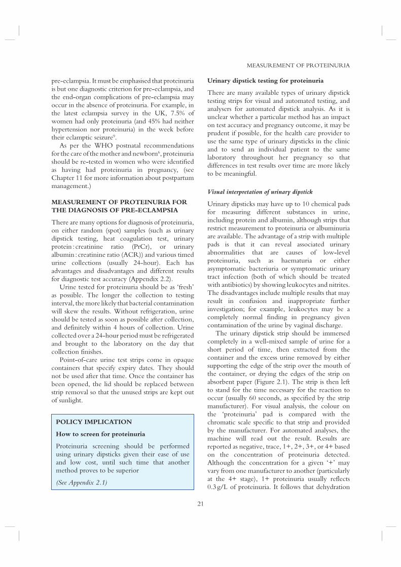
MEASUREMENT OF PROTEINURIA
21
pre-eclampsia. It must be emphasised that proteinuria
is but one diagnostic criterion for pre-eclampsia, and
the end-organ complications of pre-eclampsia may
occur in the absence of proteinuria. For example, in
the latest eclampsia survey in the UK, 7.5% of
women had only proteinuria (and 45% had neither
hypertension nor proteinuria) in the week before
their eclamptic seizure9.
As per the WHO postnatal recommendations
for the care of the mother and newborn6, proteinuria
should be re-tested in women who were identified
as having had proteinuria in pregnancy, (see
Chapter 11 for more information about postpartum
management.)
MEASUREMENT OF PROTEINURIA FOR THE DIAGNOSIS OF PRE-ECLAMPSIA
There are many options for diagnosis of proteinuria,
on either random (spot) samples (such as urinary
dipstick testing, heat coagulation test, urinary
protein : creatinine ratio (PrCr), or urinary
albumin : creatinine ratio (ACR)) and various timed
urine collections (usually 24-hour). Each has
advantages and disadvantages and different results
for diagnostic test accuracy (Appendix 2.2).
Urine tested for proteinuria should be as ‘fresh’
as possible. The longer the collection to testing
interval, the more likely that bacterial contamination
will skew the results. Without refrigeration, urine
should be tested as soon as possible after collection,
and definitely within 4 hours of collection. Urine
collected over a 24-hour period must be refrigerated
and brought to the laboratory on the day that
collection finishes.
Point-of-care urine test strips come in opaque
containers that specify expiry dates. They should
not be used after that time. Once the container has
been opened, the lid should be replaced between
strip removal so that the unused strips are kept out
of sunlight.
Urinary dipstick testing for proteinuria
There are many available types of urinary dipstick
testing strips for visual and automated testing, and
analysers for automated dipstick analysis. As it is
unclear whether a particular method has an impact
on test accuracy and pregnancy outcome, it may be
prudent if possible, for the health care provider to
use the same type of urinary dipsticks in the clinic
and to send an individual patient to the same
laboratory throughout her pregnancy so that
differences in test results over time are more likely
to be meaningful.
Visual interpretation of urinary dipstick
Urinary dipsticks may have up to 10 chemical pads
for measuring different substances in urine,
including protein and albumin, although strips that
restrict measurement to proteinuria or albuminuria
are available. The advantage of a strip with multiple
pads is that it can reveal associated urinary
abnormalities that are causes of low-level
proteinuria, such as haematuria or either
asymptomatic bacteriuria or symptomatic urinary
tract infection (both of which should be treated
with antibiotics) by showing leukocytes and nitrites.
The disadvantages include multiple results that may
result in confusion and inappropriate further
investigation; for example, leukocytes may be a
completely normal finding in pregnancy given
contamination of the urine by vaginal discharge.
The urinary dipstick strip should be immersed
completely in a well-mixed sample of urine for a
short period of time, then extracted from the
container and the excess urine removed by either
supporting the edge of the strip over the mouth of
the container, or drying the edges of the strip on
absorbent paper (Figure 2.1). The strip is then left
to stand for the time necessary for the reaction to
occur (usually 60 seconds, as specified by the strip
manufacturer). For visual analysis, the colour on
the ‘proteinuria’ pad is compared with the
chromatic scale specific to that strip and provided
by the manufacturer. For automated analyses, the
machine will read out the result. Results are
reported as negative, trace, 1+, 2+, 3+, or 4+ based
on the concentration of proteinuria detected.
Although the concentration for a given ‘+’ may
vary from one manufacturer to another (particularly
at the 4+ stage), 1+ proteinuria usually reflects
0.3 g/L of proteinuria. It follows that dehydration
POLICY IMPLICATION
How to screen for proteinuria
Proteinuria screening should be performed
using urinary dipsticks given their ease of use
and low cost, until such time that another
method proves to be superior
(See Appendix 2.1)

THE FIGO TEXTBOOK OF PREGNANCY HYPERTENSION
22
can increase proteinuria concentration and result in
a ‘positive’ proteinuria dipstick result.
Urinary dipstick testing for proteinuria is
inexpensive, easy and widely used. In a systematic
review, 1+ proteinuria by visual dipstick testing
showed low sensitivity (55%, 95% CI 37–72) and
reasonable specificity (84%, 95% CI 57–95) for
detection of 0.3 g/d of proteinuria10. A threshold of
2+ proteinuria by visual dipstick testing has
reported sensitivity and specificity that varies from
values of 58% 11 to values of 80%12–14. How should
these results be interpreted for clinical practice?
Given the <90% sensitivity of dipsticks using a
threshold of 1+, a negative or trace value should
not be ignored in a woman with new hypertension
or symptoms or signs suggestive of pre-eclampsia.
Given the reasonable specificity of dipsticks (at 1+
or 2+ levels), a result 1+ should prompt additional
investigations even when the suspicion of
pre-eclampsia is low. A urinary dipstick result of
2+ is suggestive of 0.3 g/d or more of proteinuria
by 24-hour urine collection.
Automated testing of urinary dipstick
In theory, automation has the potential to reduce
errors arising from subjective interpretation of
dipstick readings.
Comparisons of automated with visual-read
dipsticks have used thresholds of either 1+ or 2+.
Two studies have compared the diagnostic test
properties of automated dipsticks for proteinuria
with visual read urinary test strips for proteinur ia13,15,
using a threshold of 1+. Although one study
compared test strips with 24-hour urinary protein
excretion (g/d)15 and the other study used 24-hour
urinary protein concentration (g/L) as the
comparator13, both studies demonstrated superior
diagnostic test properties of automated (versus
visual) testing, using a threshold of 1+ for
proteinuria. In contrast, a more recent study failed
to show superiority of automated over visual
testing16. When a threshold of 2+ proteinuria was
used, automated testing also appeared to be superior
to visual testing13, with absolute values for sensitivity
by automated testing as high as > 80%13,14 but as low
as 23% in another study17.
For detection of proteinuria by 24-hour urine
collection or PrCr, published sensitivities for an
automated dipstick threshold of 1+ or more (41%17,
82%15, 90%13 and 100%18) and corresponding
specificities (100%, 81%, 86% and 37%) have varied
widely, even when the prevalence of proteinuria in
the study populations was similar (i.e., 45%15 and
48%17).
The diagnostic accuracy of automated testing
may depend on the choice of test strip and/or
analyser. It may be premature to recommend
widespread adoption of automated urine proteinuria
test strip readers, although one international
guideline makes such a recommendation19.
Urinary dipstick test strips are also available for
detection of albuminuria (i.e., albumin
concentration) specifically. However, we are not
aware of studies that have compared albuminuria
dipstick testing with proteinuria dipstick testing or
other methods of proteinuria testing for detection
of significant proteinuria in pregnancy. Of note,
albuminuria dipsticks are more expensive than are
proteinuria dipsticks.
HEAT COAGULATION TEST
The heat coagulation test may be used in
under-resourced settings as an alternative to dipstick
testing or other methods (discussed below) that are
unavailable or too costly. A test tube is filled to
two-thirds with urine. A few drops of dilute acetic
acid are added to make the urine sample acidic.
The upper part of the test tube containing urine is
heated (but not boiled) over a burner.
The presence of protein is signified by the
turbidity of the urine when the tube is placed in
front of a typed sheet of paper according to a
pre-specified chart (Figur e 2.2)20,21. The lower part
of the tube of urine acts as a control as that urine
should remain clear (Figure 2.3).
The heat coagulation test may be less sensitive
than visually interpreted urinary dipsticks (at 1+
level) for detecting 0.3 g/d or more of urinary
Figure 2.1 Accredited social health associate (ASHA)
worker, India, performing urinary dipstick testing

MEASUREMENT OF PROTEINURIA
23
protein, however, it has reported specificity that is
more tha n 90%20,21.
Sulfosalicylic acid testing
The sulfosalicylic acid (SSA) test is an alternative
method of proteinuria testing for under-resourced
settings. Ideally, the pH of urine is tested, and if >6,
urine is acidified by adding one or two drops of
10% acetic acid. Then, 2 mL of 3% sulfosalicylic
acid is added. After shaking the test tube, the
turbidity is observed (Figure 2.4) and the tube is
placed in front of a black line or bold printed fonts.
The turbidity of the urine (as inferred by the ability
Figure 2.2 Performing the heat coagulation test and interpreting its results
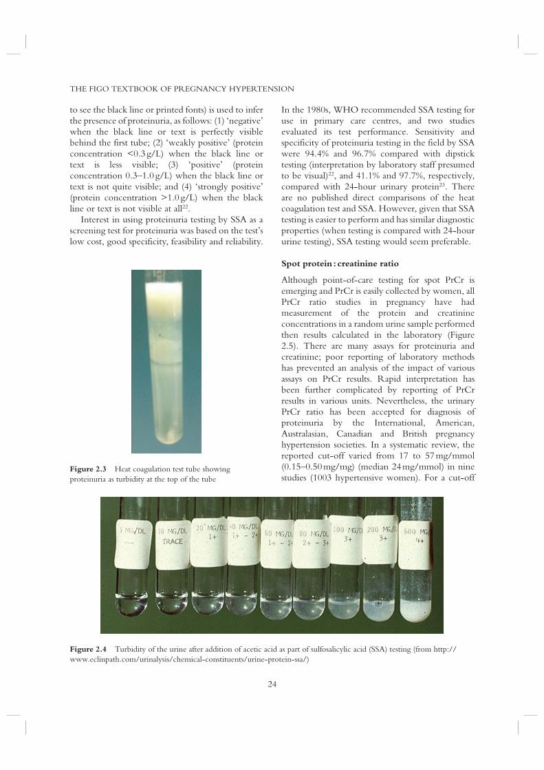
THE FIGO TEXTBOOK OF PREGNANCY HYPERTENSION
24
to see the black line or printed fonts) is used to infer
the presence of proteinuria, as follows: (1) ‘negative’
when the black line or text is perfectly visible
behind the first tube; (2) ‘weakly positive’ (protein
concentration <0.3 g/L) when the black line or
text is less visible; (3) ‘positive’ (protein
concentration 0.3–1.0 g/L) when the black line or
text is not quite visible; and (4) ‘strongly positive’
(protein concentration >1.0 g/L) when the black
line or text is not visible at all22.
Interest in using proteinuria testing by SSA as a
screening test for proteinuria was based on the test’s
low cost, good specificity, feasibility and reliability.
In the 1980s, WHO recommended SSA testing for
use in primary care centres, and two studies
evaluated its test performance. Sensitivity and
specificity of proteinuria testing in the field by SSA
were 94.4% and 96.7% compared with dipstick
testing (interpretation by laboratory staff presumed
to be visual)22, and 41.1% and 97.7%, respectively,
compared with 24-hour urinary protein23. There
are no published direct comparisons of the heat
coagulation test and SSA. However, given that SSA
testing is easier to perform and has similar diagnostic
properties (when testing is compared with 24-hour
urine testing), SSA testing would seem preferable.
Spot protein : creatinine ratio
Although point-of-care testing for spot PrCr is
emerging and PrCr is easily collected by women, all
PrCr ratio studies in pregnancy have had
measurement of the protein and creatinine
concentrations in a random urine sample performed
then results calculated in the laboratory (Figure
2.5). There are many assays for proteinuria and
creatinine; poor reporting of laboratory methods
has prevented an analysis of the impact of various
assays on PrCr results. Rapid interpretation has
been further complicated by reporting of PrCr
results in various units. Nevertheless, the urinary
PrCr ratio has been accepted for diagnosis of
proteinuria by the International, American,
Australasian, Canadian and British pregnancy
hypertension societies. In a systematic review, the
reported cut-off varied from 17 to 57 mg/mmol
(0.15–0.50 mg/mg) (median 24 mg/mmol) in nine
studies (1003 hypertensive women). For a cut-off Figure 2.3 Heat coagulation test tube showing
proteinuria as turbidity at the top of the tube
Figure 2.4 Turbidity of the urine after addition of acetic acid as part of sulfosalicylic acid (SSA) testing (from http://
www.eclinpath.com/urinalysis/chemical-constituents/urine-protein-ssa/)

MEASUREMENT OF PROTEINURIA
25
of 30 mg of protein/mmol urinary creatinine, and
among women with a hypertensive disorder of
pregnancy specifically, the sensitivities and
specificities were 83.6% (95% CI 77.5–89.7) and
76.3% (95% CI 72.6–80.0), respectively24. A more
recent systematic review suggests that the optimum
threshold for PrCr ratio to detect significant
proteinuria may actually be slightly higher, at
34–40 mg/mmol (0.30–0.35 mg/mg) (summary
sensitivity and specificity both >75% for 15 studies,
2790 women), although no threshold gave a
sensitivity and specificit y >80%25. A further
meta-analysis of 24 studies (3186 women)
endorsed a cut-off of 34 mg/mmol (0.30 mg/mg),
with sensitivity and specificity >80%26. Four
additional studies individually found sensitivity and
specificities of at least 80% with optimal cut-offs of
27 mg/mmol (0.24 mg/mg)27, 30 mg/mmol28,
51 mg/mmol (0.45 mg/mg)11, and 53 mg/mmol
(0.47 mg/mg)29, consistent with the previously
reported range of 17–57 mg/mmol. One additional
report was just outside this range (71 mg/mmol,
0.63 mg/mg)30, and three others found that optimal
cut-offs did not have both sensitivity and specific ity
80%31–33. Taken together, we feel that continued
use of the threshold of 30 mg/mmol is reasonable,
but do recommend that proteinuria testing be
viewed as only one aspect of the investigation of
women with a hypertensive disorder of pregnancy
and interpreted in the context of clinical symptoms,
signs and other laboratory testing. A higher
threshold may be more appropriate in twin
pregnancy34,35.
The best timing of spot urine sampling is
debated. However, timing may not be critical in
pregnancy36–38 which is ideal for women with
suspected pre-eclampsia who can be tested for
proteinuria at the time of clinical presentation.
Spot albumin : creatinine ratio
Most clinical laboratories use immunoassays to
measure urinary albumin, so there is less theoretical
inter-laboratory variability for albuminuria than
for proteinuria. (The remainder of labs use
colourimetric methods that are less precise for
low-level albuminuria.) However, there is no
standardisation of method, and there are also
multiple methods for measuring urinary creatinine,
as stated for the PrCr. The impact of laboratory
assays on albumin : creatinine ratio (ACR) results is
not known.
Urinary ACR testing is available by a variety of
point-of-care dipsticks. Three studies have
evaluated performance in pregnancy. Two studies
found the automated-read ACR dipstick to be
insensitive: one used the ACR performed on a spot
sample sent to the laboratory as the reference test
using a cut-off of 3.4 mg/mmol (65 low risk and 43
high risk pregnancy cases)39. The second used
24-hour urinary protein as the reference test;
reported sensitivity and specificity were 63% and
81%, respectively (163 hypertensive women)40.
The third evaluated both visual and automated
ACR dipstick performed at the bedside compared
with 24-hour urinary protein (171 hypertensive
women); automated ACR dipstick fared only
slightly better than visual ACR dipstick with
regards to sensitivity (i.e., 58% vs. 49%, respectively)
and specificities were 83% for both approaches;
neither ACR dipstick (visual or automated-read) in
that study was better than visual proteinuria dipstick
testing (which had a sensitivity of 51% and a
specificity of 78%) for detection of 0.3 g/d or more
of urinary protein in 24-hour collection15.
Urinary ACR testing on spot urine samples is
widely available in clinical laboratories in
well-resourced settings. Most, but not all, studies
have reported good test performance. The urinary
ACR has performed well in: (1) detection of
24-hour urinary protein excretion in four
prospecti ve studies18,41–43 (410 pregnant women),
and (2) detection of 24-hour urinary albumin
excretion in two ot her studies44,45 (119 pregnant
Figure 2.5 Woman in Nigeria preparing to collect her
spot urine sample for protein : creatinine ration (PrCr)
testing

THE FIGO TEXTBOOK OF PREGNANCY HYPERTENSION
26
women). An additional study reported that ACR
correlated well with 24-hour albuminuria but not
with 24-hour proteinuria46 (31 women diagnosed
with pre-eclampsia). Moreover, three different
diagnostic cut-offs (of 2, 8 and 22.8 mg/mmol,
equivalent to 18, 71 and 205 mg/g) have been
reported for significan t proteinuria15,18.
In summary, there is insufficient information
about use of ACR testing (by dipstick or through
the laboratory) in pregnancy to recommend their
use at the present time.
Timed urine collection
Quantification of urinary protein by 24-hour
urine collection is considered to be the gold
standard. However, 24-hour urine collection is
time-consuming, inconvenient and often inaccurate
due to inadequate 24-hour urine collection (as
assessed by urinary creatinine collection of 13–18%
of pre-pregnancy body weight as urinary creatinine
(mmol/d))47. For diagnosis of proteinuria in
non-pregnant populations, these logistical
considerations have prompted the National Kidney
Foundation and the International Society of
Nephrology to abandon timed collections in
favour of the spot urine samples48,49. However, if
quantification of proteinuria is sought, then
24-hour urine collection for protein and creatinine
should be used at high levels of proteinuria (i.e.,
spot PrCr >125 mg/mmol which is roughly
equivalent to more than 1 g/d of proteinuria by
24-hour urine collection) as the spot PrCr is less
reliable at high levels of proteinuria.
WHAT CONSTITUTES ‘SIGNIFICANT’ PROTEINURIA IN PREGNANCY?
Although 0.3 g/d of proteinuria represents the
upper 95% confidence interval for proteinuria
excretion in pregnancy, this threshold does not
necessarily identify women at increased risk of
adverse maternal and/or fetal outcomes. That
threshold is not known.
A recent study reported that women who had
0.5 g/d were at higher risk of adverse outcomes
than those wi th 0.3–0.5 g/d50. (This is discussed
further in Chapter 3.)
In well-resourced settings where full maternal
and fetal assessment is available, the magnitude of
proteinuria once identified is not related to either
short-term adverse maternal or perinatal outcomes,
or long-term maternal renal prognosis. In the
fullPIERS cohort, a prospective study of women
admitted to hospital with pre-eclampsia, the
magnitude of proteinuria (by 24-hour urine
collection, visual dipstick testing, or spot PrCr) was
not associated with adverse maternal or perinatal
outcomes independent of routinely collected
information on maternal symptoms, signs and b asic
blood work51 (see Chapter 3). At least one
observational study of women with pre-eclampsia
failed to identify a definition of heavy proteinuria
that was associated with adverse renal prognosis52.
In contrast, in resource-poor settings where
maternal symptoms and signs alone are used to
guide treatment, proteinuria of 4+ is associated
with an increased r isk of stillbirth53.
COST CONSIDERATIONS
Although visual dipstick proteinuria testing is the
most widely used of the screening methods, there is
no cost-effectiveness analysis of its use followed by
confirmatory testing (with PrCr or 24-hour urine
collection) for values 1+ or 2+.
The only health economic analyses identified
were those conducted by the NICE Clinical
Guideline Committee, for women with gestational
hypertension who live in settings where all tests are
available54. The Committee considered both the
convenience of testing for health care providers
and women, and the trade-off between the costs of
a false positive test for proteinuria and the costs of
missed adverse pregnancy outcomes. The analyses
were highly influenced by the sensitivity of
proteinuria testing methods. Assuming that
sensitivity is high for both the automated dipstick
and spot PrCr methods, spot PrCr may be more
cost-effective than a strategy of automated dipstick
testing followed by confirmation of 1+ proteinuria
by either spot PrCr or 24-hour urine collection.
In low-resource referral hospital settings,
limitations in central laboratory facilities will affect
cost-benefit considerations.
MEASUREMENT OF PROTEINURIA
27
PRIORITIES FOR UNDER-RESOURCED SETTINGS
Proteinuria testing is recognised by WHO to be as
a marker of high quality antenatal care55. In fact,
proteinuria testing was recommended along with
blood pressure monitoring as the original rationale
for antenatal care. As such, implementation of
proteinuria screening in low- and middle-income
countries (LMICs) is a priority.
Demographic health survey data (2002–2008)
indicate that few LMICs exceed a standard of urine
testing in more than 80% of women attending
antenatal care. The rate of urine testing at routine
antenatal care visits is highly variable56, particularly
in sub-Saharan Africa and South/Southeast Asia
where urine testing rates vary from testing in only
25% of women to testing in close to 100%. Urine
testing occurs in at least 50% of women in North
Africa/West Asia/Europe and at least 67% of
women in Latin American/Caribbean countries.
These data indicate a major failure of basic health
system provision that inevitably results in avoidable
large scale morbidity and mortality from
hypertensive disease in pregnancy.
Table 2.2 outlines the priorities for
implementation of proteinuria testing in LMICs,
depending on the timing of testing (in pregnancy
and postpartum) and the level of the health care
system. In brief, the first priority is detection
of women with pre-eclampsia (by testing for
proteinuria at 20 weeks of pregnancy and beyond),
followed by detection of women with underlying
renal disease (by testing in the first or early second
trimester, and at 6 weeks postpartum among
women with proteinuria in pregnancy) who are at
increased risk of pre-eclampsia.
Innovative proteinuria measurement devices are
on the horizon for use in under-resourced settings
and it is hoped that they will facilitate
implementation of the priorities for testing outlined
in Table 2.2. While the priority in high-income
settings is towards laboratory-based analyses, the
focus in LMICs is on point-of-care testing,
particularly by community health care providers.
Three active research tracks are as follows:
• The proteinuria self-test for early detection of pre-eclampsia (the ‘proteinuria pen’) was designed
by graduate students at John Hopkins University,
BEST PRACTICE POINTS
(Please see Appendix 2.3 for the evaluation of the strength of the recommendation and the quality of the
evidence on which they are based.)
1. All pregnant women should be assessed for proteinuria, at minimum, at their first antenatal visit.
2. Urinary dipstick testing (or SSA or heat coagulation testing if dipsticks are not available) may be used
for screening for proteinuria when the suspicion of pre-eclampsia is low.
3. Significant proteinuria should be strongly suspected when urinary dipstick proteinuria is 2+.
4. Definitive testing for proteinuria (by urinary protein : creatinine ratio or 24-hour urine collection) is
encouraged when there is a suspicion of pre-eclampsia.
5. Significant proteinuria is 0.3 g/d in a complete 24-hour urine collection or 30 mg/mmol (0.3 mg/
mg) urinary creatinine in a random urine sample.
6. There is insufficient information to make a recommendation about the accuracy of the urinary
albumin : creatinine ratio, although values <2 mg/mmol (<18 mg/g) are normal and all values
8 mg/mmol (71 mg/g) are elevated.
7. In well-resourced settings with sophisticated fetal monitoring, proteinuria testing does not need to
be repeated once the significant proteinuria of pre-eclampsia has been confirmed.
8. In under-resourced settings, proteinuria testing should be repeated to detect 4+ dipstick proteinuria
that is associated with an increased risk of stillbirth.

THE FIGO TEXTBOOK OF PREGNANCY HYPERTENSION
28
USA (http://www.appropedia.org/Proteinuria_
Self-Test_Pen). Field testing is currently under
the management of Jhpeigo. This felt-tip or
ballpoint pen is filled with reagent that is used to
mark a strip of paper. When a drop of urine is
placed on the paper, if there is proteinuria, the
reagent changes colour. The test is anticipated to
cost <US$0.10 per use.
• Point-of-care paper-based microfluidic diagnostic ‘stamps’ have been developed by Diagnostics for
All. Paper and an office printer are the equipment
required to generate the postage stamp-sized
paper testing tool, onto which a reagent and
drop of urine are applied to indicate proteinuria
( h t t p : / / w w w. s a v i n g l i v e s a t b i r t h . n e t /
summaries/60). The test is anticipated to cost
<US$0.10 per use).
• The urinary Congo red dot test uses a textile
dye to detect elevated concentrations of
misfolded urinary protein associated
with pre-eclampsia57 (http://www.usaid.gov/
n e w s - i n f o r m a t i o n / f r o n t l i n e s /
open-development-development-defense/
pinpointing-preeclampsia-simple-red).Testing
requires the user to mix dye and urine together
and put a drop on a piece of paper, where dye
and any misfolded proteins in the urine
combined to form a ‘red dot’57. The test is
anticipated to cost pennies per use.
WHAT INTERNATIONAL GUIDELINES SAY (APPENDIX 2.4)
Abbreviations for Clinical Practice Guidelines:
ACOG (American College of Obstetricians and
Gynecologists)58, AOM (Association of Ontario
Midwives), NICE (National Institutes of Clinical
Excellence)59, NVOG (National Obstetrics and
Gynaecology Society, Netherlands)60, PRECOG
(Pre-eclampsia Community Guideline), PRECOG
II (Pre-eclampsia Community Guideline II), QLD
(Queensland, Australia)61, SOGC (Society of
Obstetricians and Gynaecologists of Canada)62,
SOMANZ (Society of Obstetric Medicine of
Australia and New Zealand)63, WHO (World
Health Organization)64.
Screening for proteinuria is advocated by five
clinical practice guidelines for women with a
hypertensive disorder of pregnancy (AOM65, NICE,
PRECOG66, SOGC, SOMANZ); when performed,
Table 2.2 Prioritisation of urine testing for proteinuria by timing and level of health care system at which testing occurs
Antepartum Postpartum
Initial priority Ultimate goal Initial priority Ultimate goal
Community
Primary health
care centre
(detect and
refer)
Urinary (clean-catch)
dipstick testing at
each visit after 20
weeks to detect
pre-eclampsia
Urinary (clean-catch)
dipstick testing at
booking and at each
visit after 20 weeks to
detect both chronic
renal disease and
pre-eclampsia
Urinary (clean-catch) dipstick
testing within 24 hours of
delivery in hypertensive women
to detect postpartum
pre-eclampsia
Urinary (clean-catch)
dipstick testing at 6
weeks after delivery for
women with antenatal
proteinuria to detect
underlying renal disease
and prompt referral
Facility
Secondary-level
facility
(detect and
manage/refer)
Urinary (clean-catch)
dipstick testing at
booking and at each
visit after 20 weeks
to detect both
chronic renal disease
and pre-eclampsia
Availability of
confirmatory test for
proteinuria in women
with 1+ by urinary
dipstick testing
Postpartum urinary (clean-catch)
dipstick testing within 24 hours
of delivery in hypertensive
women to detect postpartum
pre-eclampsia
Postpartum urinary (clean-catch)
dipstick testing at 6 weeks after
delivery for women with
antenatal proteinuria to detect
underlying renal disease and
prompt referral
Tertiary-level
(referral) facility
(detect and
manage)

MEASUREMENT OF PROTEINURIA
29
testing methods should be by dipstick (visual)
(PRECOG, AOM), automated (NICE), or either
(SOGC), but NICE advocates using a random urine
protein:creatinine ratio (PrCr) in a secondary care
setting. Significant thresholds for proteinuria are:
1+ (PRECOG, SOGC) or 2+ (PRECOG II68,
QLD), with two guidelines specifying that a
threshold of 1+ should be used only when there is
associated hypertension (PRECOG II) or other
manifestations of pre-eclampsia (AOM).
For quantification of proteinuria, criteria are:
‘dipstick’ 1+ (AOM), random urine PrCr
30 mg/mmol (PRECOG, PRECOG II, NICE,
SOGC), and/or 24-hour urinary protein 0.3 g/d
(PRECOG, PRECOG II, NICE, NVOG, ACOG
SOGC) (with completeness of the urine collection
emphasised by two CPGs (NICE, SOGC)).
PRIORITIES FOR FUTURE RESEARCH
• In low-resource country service settings, health
systems research is needed on how to ensure
consistent proteinuria screening in antenatal
care, to the levels that are now being achieved
for HIV testing.
• By current testing methods, what is the level of
proteinuria that identifies a woman and/or fetus
at increased risk of an adverse outcome?
• Are there better ways of measuring
proteinuria? These should be cheaper and related
to the risk of adverse pregnancy outcome. Three
simple approaches, all point of care, show
promise.
REFERENCES
1. Conrad K, Lindheimer MD. Renal and cardiovascular alterations. In: Lindheimer MD, Roberts JM, Cunningham FG, editors. Chesley’s hypertensive disorders in pregnancy. 2nd edition. Stamford: Appleton and Lange; 1999
2. Holt JL, Mangos GJ, Brown MA. Measuring protein excretion in pregnancy. Nephrology (Carlton ) 2007 Oct;12(5):425–30
3. Stillman IE, Karumanchi SA. The glomerular injury of preeclampsia. J Am Soc Nephrol 2007 Aug;18(8): 2281–4
4. Springberg PD, Garrett LE, Jr., Thompson AL, Jr., Collins NF, Lordon RE, Robinson RR. Fixed and reproducible orthostatic proteinuria: results of a 20-year follow-up study. Ann Intern Med 1982 Oct;97(4): 516–9
5. World Health Organization. Pregnancy, childbirth, postpartum and newborn care: a guide for essential practice. 2006
6. World Health Organization. WHO recommendations on postnatal care of the mother and newborn. 2013
7. Murphy DJ, Redman CW. The clinical utility of routine urinalysis in pregnancy. Med J Aust 2003 May 19;178(10):524–5
8. Douglas KA, Redman CW. Eclampsia in the United Kingdom. BMJ 1994 Nov 26;309(6966):1395–400
9. Knight M. Eclampsia in the United Kingdom 2005. BJOG 2007 Sep;114(9):1072–8
10. Waugh JJ, Clark TJ, Divakaran TG, Khan KS, Kilby MD. Accuracy of urinalysis dipstick techniques in predicting significant proteinuria in pregnancy. Obstet Gynecol 2004 Apr;103(4):769–77
11. Amin SV, Illipilla S, Hebbar S, Rai L, Kumar P, Pai MV. Quantifying proteinuria in hypertensive disorders of pregnancy. Int J Hypertens 2014;2014:941408
12. Khashia KM, Willett MJ, Elgawly RM. A 24-hour urine collection for proteinuria in pregnancy: is it worthwhile doing the test? J Obstet Gynaecol 2007 May;27(4):388–9
13. Saudan PJ, Brown MA, Farrell T, Shaw L. Improved methods of assessing proteinuria in hypertensive pregnancy. Br J Obstet Gynaecol 1997 Oct;104(10): 1159–64
14. Phelan LK, Brown MA, Davis GK, Mangos G. A prospective study of the impact of automated dipstick urinalysis on the diagnosis of preeclampsia. Hypertens Pregnancy 2004;23(2):135–42
15. Waugh JJ, Bell SC, Kilby MD, Blackwell CN, Seed P, Shennan AH, et al. Optimal bedside urinalysis for the detection of proteinuria in hypertensive pregnancy: a study of diagnostic accuracy. BJOG 2005 Apr;112(4): 412–7
16. De Silva DA, Halstead CA, Cote AM, von Dadelszen P, Sabr Y, Magee LA. Urinary dipstick proteinuria testing – does automated strip analysis offer an advantage over visual testing? JOGC 2014;#:#
17. Dwyer BK, Gorman M, Carroll IR, Druzin M. Urinalysis vs urine protein-creatinine ratio to predict significant proteinuria in pregnancy. J Perinatol 2008 Jul;28(7):461–7
18. Kyle PM, Fielder JN, Pullar B, Horwood LJ, Moore MP. Comparison of methods to identify significant proteinuria in pregnancy in the outpatient setting. BJOG 2008 Mar;115(4):523–7

THE FIGO TEXTBOOK OF PREGNANCY HYPERTENSION
30
19. National Institute for Health and Clinical Excellence. Hypertension in pregnancy: The management of hypertensive disorders during pregnancy. guidance nice org uk/cg107 2013
20. Dissanayake VH, Morgan L, Broughton PF, Vathanan V, Premaratne S, Jayasekara RW, et al. The urine protein heat coagulation test--a useful screening test for proteinuria in pregnancy in developing countries: a method validation study. BJOG 2004 May;111(5): 491–4
21. Saxena I, Kapoor S, Gupta RC. Detection of proteinuria in pregnancy: comparison of qualitative tests for proteins and dipsticks with urinary protein creatinine index. J Clin Diagn Res 2013 Sep;7(9):1846–8
22. Robert CF, Mauris A, Bouvier P, Rougemont A. Proteinuria screening using sulfosalicylic acid: advantages of the method for the monitoring of prenatal consultations in West Africa. Soz Praventivmed 1995; 40(1):44–9
23. Penagos JAV, Tobon JJZ, Jaramillo JDL, Marulanda NLG, Gallego JG. Use of sulfosalicylic acid in the detection of proteinuria and its application to hypertensive problems in pregnancy. IATREIA 2011; 24(3):259–66
24. Cote AM, Brown MA, Lam E, von Dadelszen P, Firoz T, Liston RM, et al. Diagnostic accuracy of urinary spot protein : creatinine ratio for proteinuria in hypertensive pregnant women: systematic review. BMJ 2008 May 3;336(7651):1003–6
25. Morris RK, Riley RD, Doug M, Deeks JJ, Kilby MD. Diagnostic accuracy of spot urinary protein and albumin to creatinine ratios for detection of significant proteinuria or adverse pregnancy outcome in patients with suspected pre-eclampsia: systematic review and meta-analysis. BMJ 2012;345:e4342
26. Sanchez-Ramos L, Gillen G, Zamora J, Stenyakina A, Kaunitz AM. The protein-to-creatinine ratio for the prediction of significant proteinuria in patients at risk for preeclampsia: a meta-analysis. Ann Clin Lab Sci 2013;43(2):211–20
27. Rodrigue CZ, Weyer KL, Dornelles A, Longo SA. Comparison of timed urine collection to protein-creatinine ratio for hte diagnosis of preeclampsia. Obstet Gynecol 2014;123(Suppl 1):76s-7s
28. Sethuram R, Kiran TS, Weerakkody AN. Is the urine spot protein/creatinine ratio a valid diagnostic test for pre-eclampsia? J Obstet Gynaecol 2011;31(2):128–30
29. Mohseni SM, Moez N, Naghizadeh MM, Abbasi M, Khodashenas Z. Correlation of random urinary protein to creatinine ratio in 24-hour urine samples of pregnant women with preeclampsia. J Family Reprod Health 2013 Jun;7(2):95–101
30. Park JH, Chung D, Cho HY, Kim YH, Son GH, Park YW, et al. Random urine protein/creatinine ratio readily predicts proteinuria in preeclampsia. Obstet Gynecol Sci 2013 Jan;56(1):8–14
31. Kayatas S, Erdogdu E, Cakar E, Yilmazer V, Arinkan SA, Dayicioglu VE. Comparison of 24-hour urinary protein and protein-to-creatinine ratio in women with preeclampsia. Eur J Obstet Gynecol Reprod Biol 2013 Oct;170(2):368–71
32. Stout MJ, Scifres CM, Stamilio DM. Diagnostic utility of urine protein-to-creatinine ratio for identifying proteinuria in pregnancy. J Matern Fetal Neonatal Med 2013 Jan;26(1):66–70
33. Tun C, Quinones JN, Kurt A, Smulian JC, Rochon M. Comparison of 12-hour urine protein and protein: creatinine ratio with 24-hour urine protein for the diagnosis of preeclampsia. Am J Obstet Gynecol 2012 Sep;207(3):233–8
34. Smith NA, Lyons JG, McElrath TF. Protein:creatinine ratio in uncomplicated twin pregnancy. Am J Obstet Gynecol 2010 Oct;203(4):381–4
35. Osmundson SS, Lafayette RA, Bowen RA, Roque VC, Garabedian MJ, Aziz N. Maternal proteinuria in twin compared with singleton pregnancies. Obstet Gynecol 2014 Aug;124(2 Pt 1):332–7
36. Leanos-Miranda A, Marquez-Acosta J, Romero-Arauz F, Cardenas-Mondragon GM, Rivera-Leanos R, Isordia-Salas I, et al. Protein:creatinine ratio in random urine samples is a reliable marker of increased 24-hour protein excretion in hospitalized women with hypertensive disorders of pregnancy. Clin Chem 2007 Sep;53(9):1623–8
37. Valerio EG, Ramos JG, Martins-Costa SH, Muller AL. Variation in the urinary protein/creatinine ratio at four different periods of the day in hypertensive pregnant women. Hypertens Pregnancy 2005;24(3):213–21
38. Verdonk K, Niemeijer I, Hop W, de RY, Steegers E, van den Meiracker A, et al. Variation of urinary protein to creatinine ratio during the day in women with suspected pre-eclampsia. BJOG 2014 Apr 25
39. Wilde HM, Banks D, Larsen CL, Connors G, Wallace D, Lyon ME. Evaluation of the Bayer microalbumin/creatinine urinalysis dipstick. Clin Chim Acta 2008 Jul 17;393(2):110–3
40. Gangaram R, Naicker M, Moodley J. Accuracy of the spot urinary microalbumin:creatine ratio and visual dipsticks in hypertensive pregnant women. Eur J Obstet Gynecol Repro Biol 2009;144:146–8
41. Huang Q, Gao Y, Yu Y, Wang W, Wang S, Zhong M. Urinary spot albumin:creatinine ratio for documenting

MEASUREMENT OF PROTEINURIA
31
proteinuria in women with preeclampsia. Rev Obstet Gynecol 2012;5(1):9–15
42. Waugh J, Kilby M, Lambert P, Bell SC, Blackwell CN, Shennan A, et al. Validation of the DCA 2000 microalbumin:creatinine ratio urinanalyzer for its use in pregnancy and preeclampsia. Hypertens Pregnancy 2003;22(1):77–92
43. Wilkinson C, Lappin D, Vellinga A, Heneghan HM, O’Hara R, Monaghan J. Spot urinary protein analysis for excluding significant proteinuria in pregnancy. J Obstet Gynaecol 2013 Jan;33(1):24–7
44. Nisell H, Trygg M, Back R. Urine albumin/creatinine ratio for the assessment of albuminuria in pregnancy hypertension. Acta Obstet Gynecol Scand 2006;85(11): 1327–30
45. Risberg A, Larsson A, Olsson K, Lyrenas S, Sjoquist M. Relationship between urinary albumin and albumin/creatinine ratio during normal pregnancy and pre-eclampsia. Scand J Clin Lab Invest 2004;64(1):17–23
46. Wikstrom AK, Wikstrom J, Larsson A, Olovsson M. Random albumin/creatinine ratio for quantification of proteinuria in manifest pre-eclampsia. BJOG 2006 Aug;113(8):930–4
47. Cote AM, Firoz T, Mattman A, Lam EM, von Dadelszen P, Magee LA. The 24-hour urine collection: gold standard or historical practice? Am J Obstet Gynecol 2008 Dec;199(6):625–6
48. National Kidney Foundation. K/DOQI Clinical Practice Guidelines for Chronic Kidney Disease: Evaluation, Classification and Stratification. Am J Kidney Dis 2002;39(Suppl 1):S1-S266
49. National Kidney Foundation. KDIGO 2012 Clinical Practice Guideline for the Evaluation and Management of Chronic Kidney Disease. Kidney Int Supplements 2013;3(1):136–50
50. Bramham K, Poli-de-Figueiredo CE, Seed PT, Briley AL, Poston L, Shennan AH, et al. Association of proteinuria threshold in pre-eclampsia with maternal and perinatal outcomes: a nested case control cohort of high risk women. PLoS One 2013;8(10): e76083
51. Payne B, Magee LA, Cote AM, Hutcheon JA, Li J, Kyle PM, et al. PIERS proteinuria: relationship with adverse maternal and perinatal outcome. J Obstet Gynaecol Can 2011 Jun;33(6):588–97
52. Lampinen KH, Ronnback M, Groop PH, Kaaja RJ. Renal and vascular function in women with previous preeclampsia: a comparison of low- and high-degree proteinuria. Kidney Int 2006 Nov;70(10):1818–22
53. Payne BA, Hutcheon JA, Ansermino JM, Hall DR, Bhutta ZA, Bhutta SZ, et al. A risk prediction model for the assessment and triage of women with hypertensive disorders of pregnancy in low-resourced settings: the miniPIERS (Pre-eclampsia Integrated Estimate of RiSk) multi-country prospective cohort study. PLoS Med 2014 Jan;11(1):e1001589
54. National Institute for Health and Clinical Excellence. Hypertension in pregnancy: The management of hypertensive disorders during pregnancy. guidance nice org uk/cg107 2013
55. WHO. Antenatal care randomized trial: manual for the implementation of the new model. Geneva; 2001
56. Wang W, Alva S, Wang S, Fort A. Levels and Trends in the Use of Maternal Health Services in Developing Countries. DHS Comparative Reports No. 26. USA; 2011
57. Buhimschi IA, Nayeri UA, Zhao G, Shook LL, Pensalfini A, Funai EF, et al. Protein misfolding, congophilia, oligomerization, and defective amyloid processing in preeclampsia. Sci Transl Med 2014 Jul 16;6(245):245ra92
58. Hypertension in pregnancy. Report of the American College of Obstetricians and Gynecologists’ Task Force on Hypertension in Pregnancy. Obstet Gynecol 2013; 122(5):1122–1131
59. National Institute for Health and Clinical Excellence. Hypertension in pregnancy: The management of hypertensive disorders during pregnancy. guidance nice org uk/cg107 2013
60. Nederlandse Vereniging voor Obstetrie Gynaecologie (The Dutch Society of Obstetrics and Gynaecology). Hypertensieve aandoeningen in de zwangerschap. (www nvog nl) 2011
61. Queensland Maternity and Neonatal Clinical Guideline: Hypertensive disorders of pregnancy. 2013. Queensland Maternity and Neonatal Clinical Guidelines Program, Queensland Health
62. Magee LA, Pels A, Helewa M, Rey E, von Dadelszen P. Diagnosis, evaluation, and management of the hypertensive disorders of pregnancy. Pregnancy Hypertens 2014;4(2):105–145
63. Gillon TE, Pels A, von Dadelszen P, MacDonell K, Magee LA. Hypertensive disorders of pregnancy: a systematic review of international clinical practice guidelines. PLoS One 2014;9(12):e113715World Health Organization. WHO recommendations for prevention and treatment of pre-eclampsia and eclampsia. 2011

THE FIGO TEXTBOOK OF PREGNANCY HYPERTENSION
32
64. World Health Organization. WHO recommendations for prevention and treatment of pre-eclampsia and eclampsia. Geneva: WHO; 2011
65. HDP CPG Working Group.Association of Ontario Midwives. Hypertensive Disorders of Pregnancy. 2012. Association of Ontario Midwives
66. Milne F, Redman C, Walker J, Baker P, Bradley J, Cooper C, et al. The pre-eclampsia community guideline (PRECOG): how to screen for and detect
onset of pre-eclampsia in the community. BMJ 2005 Mar 12;330(7491):576–80
67. Côté AM, Sauve N. The management challenges of non-preeclampsia-related nephrotic syndrome in pregnancy. Obstet Med 2011;4:133–139
68. Milne F, Redman C, Walker J, Baker P, Black R, Blincowe J, et al. Assessing the onset of pre-eclampsia in the hospital day unit: summary of the pre-eclampsia guideline (PRECOG II). BMJ 2009;339:b3129

33
3Classification of the hypertensive disorders of
pregnancy
P von Dadelszen, D Ayres de Campos, W Barivalala
INTRODUCTION
The purpose of classifying diseases is to facilitate
communication among caregivers, and to create
meaningful groups with different prognoses,
considerations for surveillance, and/or outcomes1.
As such, the hypertensive disorders of pregnancy
are classified in Canada as pre-existing hypertension,
gestational hypertension, pre-eclampsia, or ‘other’
(Table 3.1). A final diagnosis of the type of
hypertensive disorder of pregnancy is retrospective,
following the postpartum period. Pre-existing
hypertension is often called chronic hypertension
in other clinical practice guidelines (see “What
international guidelines say” below).
The provision of antenatal care using the Scottish
paradigm of accelerating frequency of visits towards
term was developed, in large part, to facilitate the
diagnosis of pre-eclampsia2. The full implementation
SYNOPSIS
During pregnancy, it is important to detect hypertension of any sort, as pregnancy hypertension is associated with increased maternal and perinatal risks. However, not all hypertensive disorders of pregnancy carry the same level of risk for women and their babies. Therefore, the classification of the hypertensive disorders of pregnancy into pre-existing (chronic) hypertension, gestational hypertension, pre-eclampsia, white coat hypertension and masked hypertension matters. Reducing the rates of false-positive and false-negative classification relative to current standard of care should help to better target health care spending and lower overall costs associated with the care of women with pre-eclampsia. Although classification of the hypertensive disorders of pregnancy is usually straightforward in higher income countries, this may not be the case in settings where late gestational age at booking is prevalent, and the final diagnosis may only be possible at 6 weeks postpartum. Also, as it is critical to identify women who require delivery, the only way to initiate the cure for pre-eclampsia, we endorse the Canadian approach of defining ‘severe’ pre-eclampsia according to the presence of severe complications that mandate delivery.
As pre-eclampsia is the most dangerous of the hypertensive disorders of pregnancy, tools have been developed in all settings to facilitate identification of women at highest risk of adverse outcomes: miniPIERS for under-resourced settings and fullPIERS for well-resourced settings; both models are optimised by pulse oximetry. There is a need to evaluate how new diagnostic and risk-stratifying biomarkers can be incorporated into existing protocols and to make these biomarkers available as point-of-care tests in all clinical settings.

THE FIGO TEXTBOOK OF PREGNANCY HYPERTENSION
34
of this paradigm was associated with reduced
maternal mortality in the United Kingdom since the
early 1900s and in Sri Lanka half a century later2,3.
For pre-existing and gestational hypertension,
there are two subgroups2: with comorbid
conditions, because they constitute indications for
tighter blood pressure control outside pregnancy,
and evolution of disease can be more difficult to
determine; and with pre-eclampsia, because it is
associated with the greatest maternal and perinatal
risks. Of women under 30 years of age, 1% are
hypertensive, and approximately 1% of pregnancies
Table 3.1 Classification of the hypertensive disorders of pregnancy (reproduced with permission by the SOGC)60
Comments
Pre-existing (chronic) hypertension
This is defined as hypertension that was present either pre-pregnancy or that develops at
<20+0 weeks gestation
With comorbid conditions(s) Comorbid conditions (e.g., pre-gestational type I or II diabetes mellitus or kidney
disease) warrant tighter BP control outside of pregnancy because of their association with
heightened cardiovascular risk
With evidence of pre-eclampsia This is also know as ‘superimposed pre-eclampsia’ and is defined by the development of
one or more of the following at 20 weeks:
• Resistant hypertension, or• New or worsening proteinuria, or• One/more adverse condition(s)¥ or• One/more severe complication(s)¥
Severe pre-eclampsia is defined as pre-eclampsia with one or more severe complication(s)
Gestational hypertension
This is defined as hypertension that develops for the first time at 20+0 weeks’ gestation
With comorbid conditions(s) Comorbid conditions (e.g., pregestational type I or II diabetes mellitus or kidney disease)
warrant tighter BP control outside of pregnancy because of their association with
heightened cardiovascular risk
With evidence of pre-eclampsia Evidence of pre-eclampsia may appear weeks after the onset of gestational hypertension
Pre-eclampsia
Pre-eclampsia may arise de novo. It is defined by gestational hypertnesion and one or
more of the following:
• New proteinuria, or• One/more adverse condition(s)¥ or• One/more severe complication(s)¥
Severe pre-eclampsia is defined as pre-eclampsia with one or more severe complication(s)
‘Other hypertensive effects’*
Transient hypertensive effect Elevated BP may be due to environmental stimuli or the pain of labour, for example
White coat hypertensive effect BP that is elevated in the office (sBP 140 mmHg or dBP 90 mmHg) but is consistently
normal outside of the office (<135/85 mmHg) by ABPM or HBPM
Masked hypertensive effect BP that is consistently normal in the office (sBP <140 mmHg or dBP <90 mmHg) but is
elevated outside of the office (135/85 mmHg) by ABPM or repeated HBPM
ABPM, ambulatory BP monitoring; BP, blood pressure; dBP, diastolic BP; HBPM, home BP monitoring; sBP, systolic
blood pressure after monitoring
* These may occur in women whose BP is elevated at <20+0 or 20+0 weeks who are suspected at having pre-existing or
gestational hypertension/pre-eclampsia, respectively¥ Please see Table 3.2 for definitions of adverse conditions and severe complications of pre-eclampsia

CLASSIFICATION OF THE HYPERTENSIVE DISORDERS OF PREGNANCY
35
are complicated by pre-existing hypertension,
5–6% by gestational hypertension without
proteinuria, and 1–2% by pre-eclampsia 4,5. The
incidence of the hypertensive disorders of
pregnancy can be expected to increase in settings
where there is a trend towards an older and more
obese maternity population.
PRE-EXISTING (OR CHRONIC) HYPERTENSION
Pre-existing hypertension is defined as that which
either pre-dates pregnancy or appears before 20+0
weeks of pregnancy. Pre-existing hypertension is
associated with adverse outcomes for both mother
and baby. For the mother, the following risks
are heightened: superimposed pre-eclampsia
(approximately 20% )6–19, half of which develops
at te rm8,14,15,19,20, preterm delivery (about
3 3%)6–8,10,12–19, and placental abruption (1.8%).
Babies born to women with pre-existing
hypertension are also at increased risk of acute or
chronic hypoxia/acidosis. Approximately 15% of
these babies are born small for gestational age
( SGA)8,10,11,13,14,16–19,21–27. In a secondary analysis of
women with singleton pregnancies and chronic
hypertension diagnosed before 20 weeks in the
National Institutes of Child Health and
Development aspirin trial28, the risks of adverse
pregnancy outcomes increased with increasing
blood pr essure29.
It is important to recognise that stillbirth risk
reaches 0.1% by 36 weeks in pregnancies
complicated by hypertension, similar to that
reached at 41 weeks in low-risk pregnancies to
justify labour induction30. Up to 50% of these
newborns are admitted to high-level NICU care
because of short-term complications, such as
hypothermia, respiratory failure and feeding
problems15.
Women with comorbid conditions are
highlighted because they may warrant special blood
pressure treatment thresholds, particularly if the
comorbid condition is type I or II (but not
gestational) diabetes. Other comorbid conditions
include major cardiovascular risk factors other than
diabetes, renal parenchymal disease, vascular disease,
or cerebrovascular disease. (For further discussion
of antihypertensive therapy see Chapter 8.)
GESTATIONAL HYPERTENSION
Gestational hypertension is defined as hypertension
that appears at 20+0 weeks, without the occurrence
of proteinuria. However, using ambulatory blood
pressure monitoring (ABPM), a ‘white coat’ effect
is seen among about 30% of women diagnosed
with hypertension at 20 weeks, and this rises to
approximately 70% by the third trimester31. (For
discussion of ‘white coat’ effect, see Chapter 1.)
Women with gestational hypertension have
maternal and perinatal risks that are highly
dependent on the gestational age at presentation
and the progression to pre-eclampsia. When
gestational hypertension appears before 34+0 weeks,
approximately 35% of women develop
pre-eclampsia with the associated heightened risks
of maternal and perinatal compli cations26,32–36.
Development of that pre-eclampsia takes an average
of about 5 weeks35,36.
For a discussion of the impact of
comorbid conditions on recommendations for
antihypertensive therapy see Chapter 8.
PRE-ECLAMPSIA
The term pre-eclampsia continues to be widely
used internationally. It is widely recognised to be
the hypertensive disorder of pregnancy associated
with the greatest maternal and perinatal risks,
particularly when it is severe in nature and/or
presents before 34 weeks. In the latter case, a
stillbirth rate of about 10% and a perinatal mortality
rate of at least 5% have been reported37. The risk of
small-for-gestational age (SGA) is also primarily
concentrated in cases presenting at <34 weeks,
while there is an increased number of
large-for-gestational age (LGA) fetuse s at term37–39.
The origins of pre-eclampsia
As long ago as 1996, Ness and Roberts stated:
KEY POINT
In our opinion, the term ‘pregnancy-induced
hypertension,’ or PIH, should no longer be
used. In North America, PIH is used as a
synonym for pre-eclampsia, whereas in the UK
it means gestational hypertension without proteinuria. As such, the term has become
debased, and may lead to confusion between
clinicians

THE FIGO TEXTBOOK OF PREGNANCY HYPERTENSION
36
“The cause of preeclampsia remains elusive in
spite of many attempts to understand its
biologic characteristics and to characterize its
predictors. We suggest that there are distinct
origins of preeclampsia, each with its own
pathologic characteristics and natural history.
One genesis is the result of reduced placental
perfusion, which we will call placental, and
another results from maternal disorders
pre-existing (but sometimes not evident before)
pregnancy. These pre-existing maternal
disorders comprise predisposing factors for
cardiovascular disease such as hypertension,
renal disease, overweight, and diabetes.”40
Over the past two decades, the amount of evidence
to support this hypothesis has grown, leading
more to pre-eclampsia being a pregnancy-specific
inflammatory disorder of variable pathogenesis. We
will share two examples of probable pathways to
disease, summarised in Figure 3.141.
Angiogenic factor imbalance, with an excess
of circulating anti-angiogenic factors (e.g., soluble
fms-like tyrosine kinase (sFlt)-1 and soluble
Figure 3.1 A model of pre-eclampsia (reproduced with permission from Staff AC et al. Hypertension 201341). FGR, fetal
growth restriction; PIGF, placental growth factor
Abnormal pregestational
maternal endothelial or inflammatory function
Maternal endothelial/
inflammatory dysfunction
Maternal endothelial/
inflammatory dysfunctionNormal maternal endothelium
Poor placentation
Maternal Pre-eclampsia
FGR absent
Placental Pre-eclampsiaMixed Pre-eclampsia
FGR presentFGR without Pre-eclampsia
Normal placentation Poor placentation
Normal pregestational
maternal endothelial or inflammatory function
Normal circulating
placental factors: PIGF
Hypertension, proteinuria Normotensive
Abnormal circulating
placental factors: PIGF
Oxidatively
stressed placentaNormal placenta
Oxidatively
stressed placenta
endoglin) and a reduction in pro-angiogenic factors
(e.g., placental growth factor (PlGF)), has a clear
role in identifying pregnancies complicated by
placental underperfusion, be that manifested as
pre-eclampsia or normotensive intrauterine growth
r estriction42–45. This angiogenic imbalance appears
to be predictive of early-onset (at or before 34
weeks of pregnancy), primarily placental
underperfusion-related, pre-eclampsia that is more
dangerous to the individual woman with the
condition, as demonstrated in both well- and
under-resourc ed settings41,46–48. It may be of
particular importance in identifying women with
pre-existing medical conditions, especially renal
disease, who have developed superimposed
p re-eclampsia49–51. As yet, it is unclear why some
women with angiogenic factor imbalance develop
pre-eclampsia, while others remain normotensive,
but the concentration of circulating placental
debris may be an example of an important
co-factor in stimulating the clinical syndrome of
pre-eclampsia52.
Data from the SCOPE (Screening for Pregnancy
Endpoints) Consortium show that late-onset

CLASSIFICATION OF THE HYPERTENSIVE DISORDERS OF PREGNANCY
37
pre-eclampsia is more closely related to factors that
predict later cardiovascular disease through the
meta bolic syndrome53,54, the so-called “maternal
pre-eclampsia.” Reflecting these findings,
point-of-care assessment with glycosylated
fibronectin, a strong marker of the risk of gestational
diabetes and management, may provide a readily
available method of confirming the diagnosis of
“maternal ” pre-eclampsia55.
Clearly, in some women there is an overlap in
precipitating factors. The combined aetiology may
become increasingly important in lower-income
countries in many of which pre-eclampsia of
placental (versus maternal) origin currently
predominates. This may be explained by the shorter
time between first intercourse, coitarche and first
pregnancy, thereby reducing the opportunity for
exposure to paternal antigen through exposure to
seminal fluid; this reduces the maternal immune
adaptiveness that facilitates normal place ntal
development41,46. However, pre-eclampsia of
maternal origin may increase in prevalence as the
obesity epidemic spreads across the globe. In
addition, the maternal factors associated with the
metabolic syndrome are associated with a
pro-inflammatory state53 that may be amplified by
the burden of infectious diseases and chronic
inflammation borne by women in less developed
countries56.
Defining pre-eclampsia
All hypertension societies consider pre-eclampsia
to be a hypertensive disorder commonly defined by
new-onset proteinuria, and possibly other adverse
conditions (Table 3.2). A restrictive definition of
pre-eclampsia is gestational hypertension with
proteinuria, and this is often used by the research
community and was endorsed for this purpose by
the International Society for the Study of
Hypertension in Pregnancy (ISSHP)57. The
definition of pre-eclampsia as gestational
hypertension with proteinuria or typical end-organ
dysfunction is generally supported by other clinical
practice guidelines (see “What international
guidelines say” below)58, and is likely to reduce
maternal and perinatal risks59.
‘Resistant hypertension’ is defined as
hypertension that requires three concurrent
antihypertensive medications for blood pressure
control after 20 weeks’ gestation. The ‘adverse
conditions’ associated with pre-eclampsia consist of
maternal symptoms and signs, abnormal maternal
laboratory results, and abnormal fetal monitoring
results that may herald the development of more
severe complications. They are conditions to which
we respond (e.g., low oxygen saturation) in order
to avoid end-organ complications of pre-eclampsia
(e.g., pulmonary oedema). The adverse conditions
are discussed in detail below. This somewhat liberal
definition of pre-eclampsia is intended to signal a
need for heightened maternal and fetal surveillance,
recognising that none of the adverse conditions are
specific to pre-eclampsia.
Oedema and weight gain remain excluded from
the definition of pre-eclampsia, as neither are
significantly associated with perinatal morta lity and
morbidity57,61. Oedema, even facial oedema, is
neither sensitive nor specifi c for pre-eclampsia46,60,62.
Angiogenic imbalance is yet to be included in
the definition. The diagnosis of hypertension and
proteinuria are discussed in Chapters 1 and 2.
It should be remembered that pre-eclampsia can
arise de novo postpartum, a condition that carries
similar risks to antenatally detected pre-eclampsia
that persists postpartum63. Women with de novo postpartum pre-eclampsia were included in
the miniPIERS and fullPIERS (Pre-eclampsia
Integrated Estimate of RISK) studies described i n
more detail, below47,48. (See ‘Treatment postpartum’
for further detail.)
‘Severe’ pre-eclampsia
What constitutes ‘severe’ pre-eclampsia is a matter
of international controversy, although multi-organ
involvement is the basis for the definition in
guidelines from the UK (www.nice.org.uk/
guidance)64, Australasia (http://www.somanz.
org/)65, the United States62, and the ISSHP66 (see
“What international guidelines say” below for
more details)58.
In Canada, the definition of ‘severe’ pre-eclampsia
was modified to describe pre-eclampsia associated
with one or more severe complications (including
stillbirth). As such, women with severe
pre-eclampsia as defined in Canada require delivery
regardless of gestational age. Noticeable differences
with other published definitions include the
removal of heavy proteinuria as a criterion and the
absence of the gestational age criterion present in
the American and ISSHP guidelines.

THE FIGO TEXTBOOK OF PREGNANCY HYPERTENSION
38
HELLP syndrome
While most guidelines identify HELLP (haemolysis,
elevated liver enzymes, low platelets) syndrome as a
severity criterion, the 2014 Canadian guidelines do
not. Why? As practitioners, when we are looking
after women remote from term (e.g., <30 weeks’
gestation), we offer expectant management to those
women who accept this approach after counselling.
Rather than wishing to avoid HELLP syndrome,
or any of its component criteria, we aim to identify
its development as early as possible and to respond
to it to avoid more dangerous complications. We
recognise that the HELLP syndrome add s risk to a
pregnancy67,68, and, by multivariable regression
analysis, both platelet count and AST (aspartate
aminotransferase) are independently informative
components of the fullPIERS (Pre-eclampsia
Integrated Estimate of R iSk) model (see below)47.
The overlap between, and contrasting features of,
HELLP syndrome and acute fatty liver of pregnancy
should be considered69. Our practice is to include a
random glucose in our laboratory assessment of
women with suspected or confirmed pre-eclampsia
to ensure that acute fatty liver of pregnancy d oes
not go unrecognised70,71.
Table 3.2 The adverse conditions that define pre-eclampsia and ‘severe’ pre-eclampsia according to the SOGC
(reproduced with permission from SOGC)60
Organ system affected
Adverse conditionals (that increase the risk of severe complications) Severe complications (that warrant delivery)
CNS Headache/visual symptoms Eclampsia
PRES
Cortical blindness or retinal detachment
Glasgow coma scale <13
Stroke, TIA, or RIND
Cardiorespiratory Chest pain/dyspnoea
Oxygen saturation <97%76
Uncontrolled severe hypertension (over a period of 12 h despite use
of three antihypertensive agents)
Oxygen saturation <90%, need for 50% oxygen for >1 h, intubation
(other than for Caesarean section), pulmonary oedema
Positive inotopic support
Myocardial ischaemia or infarction
Haematological Elevated WBC count
Elevated INR or aPTT74
Low platelet count
Platelet count <50 109/L
Transfusion of any blood product
Renal Elevated serum creatinine47
Elevated serum uric acid
Acute kidney injury (creatinine >150 M with no prior renal disease)
New indication for dialysis
Hepatic Nausea or vomiting
RUQ or epigastric pain
Elevated serum AST, ALT,
LDH, or bilirubin
Low plasma albumin73
Hepatic dysfunction (INR >2 in absence of DIC or warfarin/
coumarin)
Hepatic haematoma or rupture
Feto-placental Non-reassurring FHR
IUGR37
Oligohydramnios
Absent or reversed end-diastolic
flow by Doppler velocimetry
Abruption with evidence of maternal or fetal compromise
Reverse ductus venosus A wave37
Stillbirth
AST, aspartate aminotransferase; ALT, alanine aminotransferase; aPTT, activated partial thromboplastin time; DIC,
disseminated intravascular coagulation; FHR, fetal heart rate; INR, international normalised ratio; LDH, lactate
dehydrogenase; PRES, posterior reversible leukoencephalopathy syndrome; RIND, reversible neurological deficit <48 h;
RUQ, right upper quadrant; TIA, transient ischaemic attack

CLASSIFICATION OF THE HYPERTENSIVE DISORDERS OF PREGNANCY
39
The adverse conditions of pre-eclampsia
(Table 3.2)
In this classification system, we do not include
those conditions that are serious end-organ
complications of pre-eclampsia (e.g., eclampsia,
abruption, pulmonary oedema, and stillbirth).
These are complications that can have permanent
sequelae and are life-threatening. Clinicians should
seek to avoid these complications altogether, rather
than await their development and then react
with timely delivery. For this reason, reversed
end-diastolic flow remains an adverse condition.
We have modified the remaining adverse
conditions based on their associations with severe
complications. Most of the informative data came
from the univariable analyses in the fullPIERS
model developed from a cohort of 2024 women
admitted to hos pital with pre-eclampsia47,72–82. As
such, we have included in the adverse conditions
maternal oxygen saturation, serum uric acid and
serum albumin. Although headache and visual
symptoms were not associated with severe
complications in fullPIERS (p values of 0.30 and
0.96, respectively), we have retained them for face
validity until the fullPIERS model can be externally
validated.
We have noted but not added as adverse
conditions other risk factors for severe complications
among women with pre-eclampsia: young maternal
age, maternal age 35 years in nullipara, immigrant
status, nulliparity, extremes of maternal weight and,
in the index pregnancy, multiple pregnancy and
lower gestational age at presentation (so-call ed
‘early’ pre-eclampsia)47,48,80–82.
In development of the fullPIERS model,
oliguria was not examined as a predictor of adverse
maternal (or perinatal) outcome. Oliguria is
measurable in women who are hospitalised with
pre-eclampsia (most accurately with an indwelling
catheter) and is defined as <15 mL/h for 6
consecutive hours83. Oliguria is commonly
observed in the hours following either vaginal or
Caesarean delivery. Prolonged oliguria (for more
than 12–24 hours) is more indicative of renal injury
outside pregnancy83, particularly when associated
with a rising serum creatinine.
Each adverse condition is not associated with
the same risk of severe complications. In the
fullPIERS model, the following were independently
associated with adverse maternal outcomes: preterm
pre-eclampsia, chest pain or dyspnoea, or an
abnormality of any of: oxygen saturation by pulse
oximetry, platelet count , serum creatinine, or
AST47. In fact, only pulse oximetry reaches an
international standard of independent ability to
personalise risk in women with pre-eclampsia as a
solo test76, and is retained in the fullPIERS model.
Although other factors such as symptoms of
headache and laboratory abnormalities may be
predictive of adverse maternal eve nts in univariable
analyses47,48,73–77,79,84–89, they were not independently
predictive in the f ullPIERS multivariable model47.
Although an online calculator (www.cfri.ca/
piers) is available for entry of continuous variables
(like gestational age) into the fullPIERS model, the
fullPIERS model must be externally validated
before it can be recommended for routine clinical
use, whether on admission or over the f irst 48
hours after admission78. Preliminary external
validation suggests that the fullPIERS model has
clinical utility, especially in women with more
severe forms of pre-eclampsia72.
It may be that factors such as uric acid would
become important if the fullPIERS model were
recalibrated to include women with the full
spectrum of hype rtensive disorders of pregnancy84,
another task currently underway. Evolving tests of
platelet consumption, such as platelet distribution
width may be informative of risk in women with
pregnancy hypertension shou ld initial findings be
confirmed90. How more recently derived markers
of platelet consumption (e.g., platelet distribution
width) may interact with fullPIERS or have
independen t predictive ability is uncertain90.
Definitive temporal and external validation studies
of fullPIERS, and testing the interaction between
fullPIERS and biomarkers being introduced into
clinical practice (see below), are underway.
In a single site study of 46 women with either
pre-eclampsia or eclampsia undergoing MRI,
predictors specifically of posterior reversible
leukoencephalopathy syndrome were younger age,
higher systolic and diastolic blood pressures,
eclampsia and lower platelets9 1.
The associated miniPIERS model48, which is
solely based on demographics, symptoms and signs,
is discussed in the “Priorities for under-resourced
settings” section below, but may be informative for
practitioners who are in well-resourced settings but
who do not have immediate recourse to laboratory
tests.

THE FIGO TEXTBOOK OF PREGNANCY HYPERTENSION
40
As all forms of pregnancy hypertension,
especially pre-eclampsia, are risk factors for the
development of peripartum cardiomyopathy, the
routine use of pulse oximetry and judicious use of
cardiac imaging will improve the detection of this
life-threatening complication. Women of African
and East Asian descent seem particularly prone to
peripartum cardiomyopathy92.
How maternal adverse conditions may predict
adverse outcomes for the fetus or neonate among
women with pre-eclampsia is unclear. The general
perinatal literature identifies that abnormal fetal
monitoring of various types may identify increased
fetal risk. Table 3.2 reflects this literature as well as
univariable analyses of the PIERS dataset for
non-stress testing and maternal predictors of
perinatal death or admission to NICU for >48
hours; other tests of fetal well-being were collected
too infrequently to be considered. The biophysical
profile is not listed because this test has not been
demonstrated to be useful in women with
hypertensive disorders of pr egnancy or other high
risk patients37,93; and, indeed may falsely reassure
both practitioners and women when pregnancies
are complicated by either early-onset IUGR94 or
pre-eclampsia95. Of fetal assessment modalities,
umbilical artery Do ppler studies are the best
supported37,96.
In 1153 women who participated in the
Dutch Obstetric Consortium’s HYPITAT trial at
gestational ages 36+0 weeks97, nulliparity,
increasing body mass index (BMI), heavy dipstick
proteinuria (3+), increasing serum uric acid and
increasing serum creatinine were independent
antenatal predictors of adverse neonatal outcomes
of: 5-minute Apgar scor e <7, cord pH <7.05, or
NICU admission98.
The independent value, within a multivariable
model, of various additional Doppler studies in
assessing maternal and perinatal risks associated
with a diagnosis of pre-eclampsia (not predicting
that diagnosis) has yet to be assessed. Preliminary
data suggest that uterine and ophthalmic artery
Do ppler may assist in risk stratification99,100.
While conserved in the miniPIERS model (see
below), proteinuria was not retained in the
fullPIERS model developed in women with
pre-eclampsia. However, like uric acid, proteinuria
may be important to identify risk in women with
the full spectrum of the hypertensive disorders of
pregnancy, as women with 0.3 g/d of proteinuria
had complication rates above those of women
managed as outpatients (gestational hypertension
and pre-existing hypertension), meriting closer
surveillance and endorsing 0.3 g/d as an appropriate
threshold for determining in-patient management101,
confirming previous studies102. Adverse perinatal
outcomes were higher still in women with 0.5 g/d
prot einuria, as observed in miniPIERS, below47,101.
OTHER
In Canada, in 2014, a new category of ‘other’ was
added to the classification system, to raise awareness
that blood pressure that is not consistently elevated
in the office setting and at home is associated with
maternal and perinatal risks that appear to be
intermediate between those of women with normal
blood pressure and those with hypertension in the
office and ambulatory or home settings.
‘White coat’ effect
‘White coat’ hypertension is seen when blood
pressure is elevated in the office, but normal by
ambulatory blood pressure monitoring (ABPM) or
at home. (See Chapter 1 for numerical values.)
White coat effect in early pregnancy is common
(approximately 30%), similar to estimates outside of
pregnancy103. The limited literature suggests that
there is a heightened risk of adverse maternal
outcomes compared with normotensive pregnancy,
but the risks are probably smaller than with
pre-existing hypertension104. Of these women,
40% progress to gestational hypertension and 8% to
pre-eclampsia.
ABPM has identified that approximately 30% of
women with gestational hypertension demonstrate
a white coat effect on their blood pressure, although
estimates have been as high as 70% in the third
trimester31. There is wide variability in the rates of
associated maternal and perinatal complications,
but many studies have identified risk that is
intermediate between that of normotensive women
and that of women with gestational hypertension31.
Masked hypertension
‘Masked’ hypertension refers to blood pressure that
is normal in the office but elevated by ABPM or at
home. (See Chapter 1 for numerical values.)
Masked hypertension may be present in about
30% of women with pre-existing hypertension103.

CLASSIFICATION OF THE HYPERTENSIVE DISORDERS OF PREGNANCY
41
However, the associated perinatal risks are
unknown. Outside pregnancy, cardiovascular risk
associated with masked hypertension is similar to
that associated with sustained hypertension.
Masked gestational hypertension was seen in
4–15% of women in prospective cohort studies;
pregnancy outcomes were similar to those of
wome n with sustained gestational hypertension105,106.
This diagnosis could be considered (and ABPM or
home blood pressure monitoring performed) if
there are unexplained maternal or perinatal
complications that are associated with the
hypertensive disorder of pregnancy, but the
usefulness of this approach has not been studied.
INVESTIGATIONS TO CLASSIFY THE HYPERTENSIVE DISORDERS OF PREGNANCY
Pre-existing hypertension
Women with pre-existing hypertension are most
likely (>95%) to have essential hypertension, but
secondary causes should be considered. A basic
work-up has been suggested for women for whom
suspicion of a secondary cause is low (see the
annually updated Canadian Hypertension
Education Program document for a more extensive
discussion (https://www.hypertension.ca/en/
chep)).
Conditions such as obesity, associated
non-alcoholic steatohepatitis, or immune
thrombocytopoenia may make interpretation of
blood work for pre-eclampsia end-organ
dysfunction difficult later in pregnancy.
Consequently, it may be appropriate to conduct
additional baseline testing in women with these
conditions early in pregnancy.
Women with a strong clinical risk marker for
pre-eclampsia should be considered for baseline
proteinuria quantification (by spot protein :
creatinine ratio or 24 h urine collection) given the
insensitivity of dipstick proteinuria testing. A fasting
blood glucose 7 mmol/l prior to pregnancy or
5.3 mmol/l in pregnancy should prompt
appropriate investigation and subspecialty referral107.
An abnormal P wave in lead V1 by
electrocardiogram may increase the risk for
gestational hypertension or pre-eclampsia108.
In terms of imaging, echocardiography may be
useful in selected women, such as those with
known or suspected left ventricular dysfunction or
heart failure (https://www.hypertension.ca/en/
chep). Plasma lipids should not be measured
routinely because both cholesterol and triglycerides
increase physiologically during pregnancy and
are not considered when making treatment
decisions.
When pre-eclampsia is suspected
Pre-eclampsia may be a disease in evolution, with
clinical manifestations unfolding in a serial fashion.
When there is ongoing suspicion of pre-eclampsia,
the nature and frequency of serial surveillance are
unclear, but a change in clinical status for mother
or fetus would be a reasonable indication for repeat
testing. Pre-eclampsia imitators share manifestations
with pre-eclampsia, but require different treatments
(Table 3.3).
Maternal investigations
In addition to measurement of blood pressure,
women with suspected pre-eclampsia should
undergo bloo d and urine testing as outlined in
Table 3.370. This testing is designed to (1) detect
end-organ involvement that increases the risk of
adverse maternal and/or perinatal outcomes (e.g.,
elevated serum uric acid), (2) detect one of those
adverse outcomes (e.g., acute renal failure), (3)
evaluate the seriousness of the adverse outcome
(e.g., haemoglobin in setting of placental abruption)
(Table 3.2), or (4) explore important differential
diagnoses (e.g., acute fatty liver of pregnancy or
primary renal disease).
The maternal testing in Table 3.3 (alone or
in combination) is of prognostic value once
pre-eclampsia has been diagnosed, but its value for
the purposes of diagnosis is based on expert opinion.
Most abnormalities are not specific to pre-eclampsia,
so the usefulness of the testing relies more on
multiple (rather than single) abnormalities. In
addition, as differentiating pre-eclampsia from
gestational hypertension can be difficult, it is
possible that maternal venous Doppler studies,
particularly renal interlobar vein impedance index
(RIVI), will assist in this regard, but initial findings
require further validation109,110. Innovative methods
such as brain mapping with electroencephalography
and advanced retinal imaging have not been fully
evaluated, but may assist in targeting magnesium
sulphate therapy toward s those women who would
most benefit from it111,112.

THE FIGO TEXTBOOK OF PREGNANCY HYPERTENSION
42
Table 3.3 Investigations to diagnose and monitor women with a hypertensive disorder of pregnancy (reproduced with
permission from the SOGC)60
Investigations for diagnosis Description in women with pre-eclampsia Description in women with other conditions
Maternal testing
Uterine testing
Urinalysis (routine
and microscopy
with/without
additional tests for
proteinuria)
Proteinuria (as discussed under
Proteinuria) without RBCs or casts
Haemoglobinuria (dipstick ‘haematuria’ without RBCs):
haemolytic anaemia
RBCs alone: renal stones, renal cortical necrosis (also
associated with back pain and oliguria/anuria)
RBCs and/or casts are associated with other glomerular disease
and scleroderma renal crisis and (about half of ) TTP-HUS
Bacteria: UTI or asymptomatic bacteriuria
Proteinuria is usually absent in secondary causes of
hypertension such as phenochromocytoma,
hyperaldosteronism, thyrotoxicosis, coarctation of the aorta,
and withdrawal syndromes
Oxygen saturation
Pulse oximetry SpO2 <97% associated with a
heightened risk of severe
complications (including
non-respiratory)
May be decreased in any cardiorespiratory complication (e.g.,
pulmonary embolism)
CBC and blood film
Haemoglobin due to intravascular volume
depletion
if microangiopathic haemolysis
(with HELLP)
due to volume depletion from any cause (e.g., vomiting
if microangiopathic haemolysis from other cause
with any chronic anaemia (nutritional or myelodysplasia)
with acute bleeding of any cause
WBC and differential due to neutrophilia of normal pregnancy
with inflammation/infection
with corticosteroids
Platelet count associated with adverse maternal
outcome
with gestational, immune (ITP), or thrombotic
thrombocytopoenia (TTP), APS, AFLP, myelodysplasia
Blood film RBC fragmentation Microangiopathy due to mechanical causes (e.g., cardiac
valvopathy, cavernous haemangioma), DIC or other disorders
of endothelial function (e.g., APS, TTP-HUS, vasculitis,
malignant hypertension)
Tests of coagulation
INR and aPTT with DIC which is usually
associated with placental abruption
is associated with adverse maternal
outcome
May be in APS, DIC from other causes including sepsis,
amniotic fluid embolism, stillbirth, massive haemorrhage,
haemangiomas, shock
is prominent in AFLP
Fibrinogen with all causes of DIC including massive haemorrhage,
genetic disorders
more profound with AFLP than with HELLP
Usually normal in TTP-HUS (ADAMTS13 vWF cleaving
protein may be moderately decreased in HELLP106 but
ADAMTS13 antibody should be absent
continued

CLASSIFICATION OF THE HYPERTENSIVE DISORDERS OF PREGNANCY
43
The pre-eclampsia imitators are conditions with
numerous man ifestations shared by pre-eclampsia
(Table 3.3)46,113. To greater or lesser degrees, all
of these conditions share the clinical features of
hypertension, central nervous system symptoms
and abdominal pain, and the laboratory features
of proteinuria, anaemia, thrombocytopoenia,
micro-angiopathic haemolysis and elevated lactate
dehydrogenase (LDH). However, acute fatty liver
of pregnancy tends to have prominent vomiting,
liver dysfunction (with jaundice and diabetes
insipidus) and renal failure114.
Thromboses and skin involvement suggest
catastrophic antiphospholipid antibody syndrome115.
Thrombotic thrombocytopaenic purpura-haemolytic
uraemic syndrome (TTP-HUS) has prominent
neurological manifestations (termed TTP),
vomiting and renal manifestations (termed HUS);
laboratory evidence of disseminated intravascular
coagulation and elevated liver enzymes would
be unusual116. Scleroderma renal crisis and
malignant hype rtension show predominant renal
manifestations117,118.
Phaeochromocytoma may mimic pre-eclampsia
in bo th well-resourced and resource-limited
settings119,120, presenting as episodic headaches,
anxiety (resembling a panic attack), altered skin
sensation, seizures, flank pain, pallor, diaphoresis,
tachycardia, paroxysmal hypertension with
paradoxical orthostatic/postural hypotension (a fall
in systolic blood pressure >20 mmHg or in diastolic
blood pressure >10 mmHg upon standing), and
Table 3.3 continued
Investigations for diagnosis Description in women with pre-eclampsia Description in women with other conditions
Serum chemistry
Serum creatinine due to haemoconcentration and/
or renal failure
associated with adverse maternal
outcome
with other acute or chronic kidney disease
Renal failure prominent in malignant hypertension,
TTP-HUS (along with thrombocytopoenia), AFLP (along
with liver dysfunction)
Serum uric acid associated wtih adverse maternal
and perinatal outcomes
with dehydration, medication (e.g., HCTZ), genetic causes
Glucose with AFLP, insulin therapy
AST or ALT associated with adverse maternal
outcome
with AFLP and other ‘PET imitators’† but to a lesser
degree, and usually normal in TTP-HUS
May be increased in other pregnancy-related conditions (e.g.,
intrahpatic cholestasis of pregnancy) or conditions not associated
with pregnancy (e.g., viral hepatitis or cholecystitis)
LDH which may be prominent
the is associated with adverse
maternal outcome
with AFLP, intravascular haemolysis
LDH/AST ratio (>22) with TTP-HUS107
Bilirubin unconjugated from haemolysis or
conjugated from liver dysfunction
(early) in AFLP, with haemolytic anaemia, other liver
disease with dysfunction, genetic diseases
Albumin associated with adverse maternal
and perinatal outcomes
as negative actue phase reactant with acute severe illness,
malnutrition, nephrotic syndrome, crystalloid infusion
Fetal testing Abnormalities are not specific to the cause of poor placentation and/or placental dysfunction
Uterine artery
Doppler velocimetry
Unilateral/bilateral notching, or elevated pulsatility index or resistance index may support a
diagnosis of placental insufficiency including pre-eclampsia
AFLP, acute fatty liver of pregnancy; APS, antiphospholipid syndrome; CBC, complete blood count; DIC, disseminated
intravascular coagulation; HCTZ, hydrochlorthiazide; HUS, haemolytic-uraemic syndrome; ITP, immune
thrombocytopoenic purpura; PET, pre-eclampsia; SpO2, oxygen saturation by pule oximetry; RBC, red blood cell;
TTP, thrombotic thrombocytopoenic purpura

THE FIGO TEXTBOOK OF PREGNANCY HYPERTENSION
44
hyperglycaemia. Elevated plasma metanephrins,
urinary catecholamines and imaging will secure the
diagnosis.
The importance of determining whether or not
pre-eclampsia (particularly in those exceptional
cases with onset before 20 weeks or more than
3 days postpartum) may actually be another
disorder is that subspecialty referral is warranted for
specific therapy (beyond supportive care). Such
specific therapies include immunosuppression and
heparin for catastrophic antiphospholipid syndrome,
plasma exchange and steroids for TTP-HUS,
angiotensin converting enzyme (ACE) inhibitors
for scleroderma renal crisis and alpha-agonists for
phaeochromocytoma. Of course, women with
acute fatty liver of pregnancy (AFLP) should be
delivered immediately regardless of gestational age.
Biomarkers
Taking into account the points made above, in a
minority of women with pre-eclampsia, clinical
uncertainty around the diagnosis arises. It is in this
context that translational biomarkers may improve
diagnostic accuracy and clinical performance. Such
women could be those who appear to have
pre-eclampsia superimposed o n pre-existing
hypertension and/or renal disease49–51, are highly
symptomatic but normotensive and/or
non-proteinuric, or present with either gestational
hypertension or gestational proteinuria in isolation.
These women with “atypical” pre-eclampsia bear
the same risks as others with classically clinically
defined disease121. Indeed, such women may bear
increased risks as responses to their symptoms and
signs may be delayed as they do not fulfil diagnostic
criteria that can be strictly applied by clinicians
unaware of the atypical presentation of
pre-eclampsia in a significant subset of women.
As we approach the definition of varying
phenotypes of pre-eclampsia (e.g., severe versus
non-severe, early- versus late-onset) by clinical and
biochemical criteria, adherence to standardised
biomedical research protocols will hasten our
understanding of the causes of pre-eclampsia and
development of targeted treatment strategies. To
assist in that process, the PRE-EMPT Global
Pregnancy Collaboration (http://pre-empt.cfri.ca/
colaboratory) has presented what they consider to
be the minimum requirements for a data set in a
study of pre-eclampsia that will facilitate
comparisons122. In addition, they have presented a
comprehensive or “optimal” data set for in-depth
investigation of pathophysiology122.
As intimated above, within the past decade, an
imbalance between pro- and anti-angiogenic
factors has been proposed to underlie many features
of the maternal syndrome of pre-eclampsia42,123–127.
While such an imbalance appears to be observed
quite con sistently in women with early-onset
pre-eclampsia41,43, it is shared with pregnancies
complicated by placentally mediated fetal
growth restriction44. Therefore, while angiogenic
imbalance may reflect the presence of the
underlying placental dysfunction particularly
important with early-onset pre-eclampsia, it is
unlikely to be a singular aetiological pathway but
will be shared with risks of intrauterine growth
retardation and fetal death128,129.
In the near future, those biomarkers with the
greatest potential to be introduced into day-to-day
clinical care to individualise maternal and perinatal
risk are PlGF and sFlt-1, either a single analyte
(i.e., PlGF) or as a ratio between anti- a nd
pro-angiogenic factors (e.g., sFlt-1/PlGF
ratio)41–43,45,123–127,130,131. We are aware of two such
platforms that are being licensed and brought to the
international market.
The recently published PELICAN prospective
multicentre study evaluated the diagnostic accuracy
of low plasma PlGF concentration (<5th centile for
gestation) in women presenting with suspected
pre-eclampsia between 20+0 and 34+6 weeks’
gestation (an d up to 41 weeks’ gestation as a
secondary analysis)45. The outcome was delivery
for confirmed pre-eclampsia within 14 days. Of
625 women, 346 (55%) developed confirmed
pre-eclampsia. In 287 women enrolled <35+0
weeks’ gestation, PlGF <5th centile had high
sensitivity (0.96; 95% confidence interval,
0.89–0.99) and negative predictive value (0.98;
0.93–0.995) for delivery for pre-eclampsia within
14 days; specificity was lower (0.55; 0.48–0.61).
Area under the receiver-operating characteristic
curve for low PlGF (0.87, standard error 0.03) for
predicting delivery for pre-eclampsia within 14
days among women presenting with suspected
pre-eclampsia was greater than all other commonly
used tests, singly or in combination (range,
0.58–0.76; p < 0.001 for all comparisons). The
authors concluded that PlGF is better than other
currently used tests and presents an innovative

CLASSIFICATION OF THE HYPERTENSIVE DISORDERS OF PREGNANCY
45
adjunct to management of such women. This i s
consistent with studies using the sFlt-1/PlGF
ratio125,126.
Adding Doppler studies may further improve
the maternal and fetal risk stratification capacity of
angiog enic imbalance in women with established
pre-eclampsia126,127.
Other time-of-disease potential biomarkers of
both the presence of pre-eclampsia and its severity
in clude neutrophil gelatinase-associated lipocalin
(NGAL)132 –134, the marker of central nervous system
injury S100B135, lepti n136, interferon-136 and
glycosylated fibronectin55.
Urinary tests of interest for the differentiation of
pre-eclampsia from other hypertensive disorders of
pregnancy are th e Congo red test, podocyturia and
kidney injury molecule-1137–139. (See Chapter 2 for
further detail about the Congo red test.)
Fetal monitoring
Fetal testing is also listed in Table 3.3. Uterine
artery Doppler velocimetry may be useful in
hypertensive pregnant women to support a
placental origin f or the hypertension, proteinuria,
and/or adverse conditions140; obstetric consultation
would then be warranted.
Oligohydramnios, absent or reversed
end-diastolic flow in the umbilical artery, or a deep,
absent or reversed A wave in the ductus venosus
would be more consistent with placental
dysfunction than with decreased biological growth
potential, uncertain dates, or aneuploidy as a cause
of IUGR and may also be useful to inform timing
of delivery37,141–144. Reduced maternal plasma PlGF
implies IUGR of placental origin, rather than
constitutionally small fetal size44.
It is very important to note that the addition of
biophysical profile to a schedule of fetal surveillance
has not been shown to improve outcomes in
high risk pregnancies93. Indeed, it appears that
the biophysical profile tends to falsely reassure
practitioners and lead to worse outcomes whe n
pregnancies are complicated by either pre-eclampsia
or IUGR94,95.
THE PATIENT PERSPECTIVE
We support incorporating the patient perspective
into care. Engaged patient advocacy organisations
are the Preeclampsia Foundation (www.
preeclampsia.org/), Action on Pre-eclampsia
(APEC) (www.apec.org.uk/), Australian Action on
Pre-eclampsia (AAPEC; www.aapec.org.au), New
Zealand Action on Pre-eclampsia (NZ APEC)
(www.nzapec.com/) and Association de Prevention
et d’Actions contre la Pre-Eclampsie (APAPE)
(www.eclampsie.moonfruit.fr/)145.
The Preeclampsia Foundation advocates for
better patient (and health care provider) education
about the antenatal, early postnatal and long-term
maternal implications of pre-eclampsia; an emphasis
on early maternal signs and symptoms
of pre-eclampsia; better doctor–patient
communication about pre-eclampsia; and
evidence-based guidelines for pre-eclampsia
screening, detection; and management145. This is an
approach that would seem to have global appeal, as
illustrated by the following quote.
“. . . they also have pre-eclampsia in the
developed countries, but they don’t die the
way our own patients are dying, not because
we do not know how to manage them but
[because] they don’t come early and by the
time they come, it is so late”.
Focus Group Discussant, Society of
Obstetricians and Gynaecologists of Nigeria,
03 Nov 2012
Post-traumatic stress
There is growing evidence that women may
expe rience post-traumatic stress disorder up to 7
years postpartum146–156, the prevalence of symptoms
being highly variable, ranging from the minority to
the majority of women, and higher after maternal
hospitalisation for more than 1 week, preterm
hypertensive disorder of pregnancy onset or
delivery, NICU admission, adverse neona tal
outcomes, or uncertainty about the child’s
long-term health151. Symptoms are not specific to
the hypertensive disorders of pregnancy, and follow
preterm delivery for other indications155. Although
post-traumatic stress symptoms do not have an
impact on infant cognitive or psychomotor
development at 1 year of age, maternal symptoms
are amenable to clinical psy chological therapy, and
earlier referral may abbreviate treatment152.
Women and their maternity care providers seem
to view experiences of pre-eclampsia differently.
For health care professionals, pre-eclampsia
represented the care that must be delivered,
primarily responding to the biology of

THE FIGO TEXTBOOK OF PREGNANCY HYPERTENSION
46
pre-eclampsia. For women, generally lacking
knowledge and understanding about pre-eclampsia,
pre-eclampsia represents fear and risk157.
Patient education and engagement
In a survey of women who had experienced
pre-eclampsia, eclampsia and/or HELLP,
pre-eclampsia was viewed as very important to all,
and traumatic to many, respondents including
women, their partners, close relatives, or friends.
The provision of information and support was
valued prior to, and at the time of, diagnosis as well
as being revisited during ongoing care157.
Even in well-resourced settings, women are not
knowledgeable about the hypertensive disorders
of pregnancy, even those with pre-existing
hypertension. They have a poor understanding of
pre-eclampsia158,159, and hypertensive disorders of
pregnancy, even those with pre-existing
hypertension, and are not satisfied with the medical
information they receive. This suggests that
clinicians should both place more value on
informing women about either their condition or
its pote ntial course, and check that women have
understood the information160,161. Although limited
health literacy may complic ate risk, communication
tools have been developed for such purposes160,161.
Current ANC practice guidelines offer little
information on educating patients about
pre-eclampsia. However, when women receive
and understand education about pre-eclampsia,
they are more likely to promptly report symptoms159.
Formal study is required to see whether this will
indeed lead to early diagnosis and management,
and improved maternal and perinatal outcomes, as
hoped158,159.
Women enjoy participating in aspe cts of their
care, be it receiving information as study
parti cipants162, or participating in management of
their blood pressure163. Women have expressed a
preference for home or day care164 and self (rather
than 24-h ambulatory) blood pressure monitoring165.
They do not object to being randomised166.
BEST PRACTICE POINTS
(Please see Appendix 3.1 for the evaluation of the strength of the recommendation and the quality of the
evidence on which they are based.)
1. Hypertensive disorders of pregnancy should be classified as pre-existing hypertension or gestational
hypertension with or without pre-eclampsia, or ‘other’ hypertension on the basis of different
diagnostic and therapeutic considerations.
2. The presence or absence of pre-eclampsia must be ascertained, given its clear association with more
adverse maternal and perinatal outcomes.
3. In women with pre-existing hypertension, pre-eclampsia should be defined as resistant hypertension,
new or worsening proteinuria, one or more adverse conditions, or one or more severe complications.
4. In women with gestational hypertension, pre-eclampsia should be defined as new-onset proteinuria,
one or more adverse conditions, or one or more severe complications.
5. The assessment of maternal angiogenic factor balance appears to inform the diagnosis of pre-eclampsia,
and other placental complications of pregnancy, where uncertainty exists, especially when
‘superimposed pre-eclampsia’ is suspected.
6. Severe pre-eclampsia should be defined as pre-eclampsia complicated by one or more severe
complications.
7. For women with pre-existing hypertension, serum creatinine, fasting blood glucose, serum potassium
and urinalysis should be performed in early pregnancy if not previously documented.
8. Among women with pre-existing hypertension or those with a strong clinical risk marker for
pre-eclampsia, additional baseline laboratory testing may be based on other considerations deemed
important by health care providers.
9. Women with suspected pre-eclampsia should undergo laboratory maternal testing and a schedule of
pertinent fetal testing described in Table 3.3.

CLASSIFICATION OF THE HYPERTENSIVE DISORDERS OF PREGNANCY
47
PRIORITIES FOR UNDER-RESOURCED SETTINGS
Hypertensive disorders of pregnancy diagnosis and severity
Identifying women with pregnancy hypertension
before it becomes life-threatening is particularly
important for colleagues in resource-constrained
settings. For example, in a single-site retrospective
analysis of demographic and clinical data of 1027
patients with eclampsia over a 10-year period,
Adamu et al. observed a maternal case fatality rate
of 17.9%, which was particularly high among
women who had received no antenatal care
(18.7%), compared with those who had received
such care (5.9%)167. In this series, the perinatal
mortality rate was 38%, of which 81% were
stillbirths.
For many colleagues in the global maternal care
community, access to laboratory facilities and
modalities for outpatient blood pressure monitoring
is limited or even absent. Therefore, it has been
imperative to determine how best to classify
pregnancy hypertension in resource-constrained
settings. Clarifying the diagnosis of pre-eclampsia is
a clinical priority as the WHO Multicountry
Survey on Maternal and Newborn Health has
determined that maternal near-miss cases were
eight times more frequent in women with
pre-eclampsia, and up to 60 times more frequent in
women with eclampsia, when compared with
women with other hypertensive disorders of
pregnancy168.
It is imperative that all women everywhere have
access to accurate blood pressure measurements and
dipstick proteinuria assessment as a global priority.
In the UK, which had maternal mortality data a
century ago that were similar to those of many
less-developed countries today, over 90% of the
observed reduction in maternal mortality occurred
prior to the provision of either effective
antihypertensives or magnesium sulphate (Figure
3.2). As stated above, the provision of antenatal
care including blood pressure and proteinuria
assessment, with appropriate referral pathways, has
been associated with markedly reduced maternal
mortality in Sri Lanka, even during the period
when care was complicated by the presence of civil
war3 (Figure 3.3). (See Chapters 1 and 2 for priority
recommendations for further detail about accurate
and cost-effective semi-automated blood pressure
devices and costs of proteinuria detection, as well as
advocacy tools for colleagues to use to elicit
appropriat e funding for these key resources for
pregnant women wherever they reside169,170).
In many less-developed countries, women do
not present for maternity care until at least 20
weeks’ gestation171–182. As a result, the firm
classification of a hypertensive disorder of pregnancy
may not be achievable until after the woman has
delivered. In response to this reality, we have
developed and validated the miniPIERS model to
personalise the risk for severe complications
experienced by women with any hypertensive
disorder of pregnancy who present in
under-resourced settings48. Data were collected
10. Doppler velocimetry-based assessment of the fetal circulation may be useful to support a placental
origin for hypertension, proteinuria, and/or adverse conditions (including IUGR), and for timing of
delivery.
11. The biophysical profile is not recommended as part of a schedule of fetal testing in women with a
hypertensive disorder of pregnancy.
12. If initial testing is reassuring, maternal and fetal testing should be repeated if there is ongoing concern
about pre-eclampsia (e.g., change in maternal and/or fetal condition).
13. In resource-constrained settings, the miniPIERS model can provide personalised risk estimation for
women with any hypertensive disorder of pregnancy. In many of these women, the ultimate
diagnosis cannot be confirmed until at least 3 months after delivery.
14. Health care providers should be alert to symptoms of post-traumatic stress following a hypertensive
disorder of pregnancy; and refer women for appropriate evaluation and treatment.
15. Health care providers should inform their patients, antepartum and postpartum, about pre-eclampsia,
its signs and symptoms, and the importance of timely reporting of symptoms to health care providers.
16. Information should be re-emphasised at subsequent visits.

THE FIGO TEXTBOOK OF PREGNANCY HYPERTENSION
48
prospectively on 2081 women with any
hypertensive disorder of pregnancy admitted to a
participating centre in Brazil, Fiji, Pakistan, South
Africa, or Uganda. The final miniPIERS model
includes parity (nulliparous versus multiparous);
gestational age on admission (as best ascertained at
the time of the encounter), headache/visual
disturbances, chest pain/dyspnoea, vaginal bleeding
with abdominal pain, systolic blood pressure, and
dipstick proteinuria. In the miniPIERS cohort, up
to 40% of the women were unbooked at the time
that their hypertension was first diagnosed.
However, best estimation of gestational age at that
encounter led to gestational age being a powerful
and independent identifier of maternal risk in the
miniPIERS cohort.
An online calculator (cfri.ca/piers) is available
for entry of continuous variables (such as gestational
age) into the miniPIERS model to provide real-time
personalised risks to all women whose caregivers
have access to the internet. An mHealth app is in
development and will be made available through
the PRE-EMPT website (pre-empt.cfri.ca).
Once women in less-resourced settings are
diagnosed with pre-eclampsia every effort should
be made t o give them access to oximetry and
appropriately targeted laboratory testing47.
Over 90% of women who suffer hepatic
haematoma and/or rupture will have preceding
HELLP syndrome183.
Defining a woman with ‘severe’ pre-eclampsia
as one with a miniPIERS predicted probability
>25% classifies women with 85. 5% accuracy; this
accuracy is greater if pulse oximetry is added to the
model184. We believe that miniPIERS could be
used in resource-constrained settings to identify
women who would benefit most from interventions
such as magnesium sulphate, antihypertensives, or
transportation to a higher level of care, especially if
supported by a usability-tested mobile health
(mHealth) application such as PIERS on the
Move185,186.
Facility versus community
In our view, maternity care providers should be
able to screen women for pre-eclampsia and the
other hypertensive disorders of pregnancy
irrespective of where that woman is encountered.
In addition, should hypertension be detected, then
personalised risk assessment should be universal
through the strengths and flexibility of mHealth. In
the community, the determination of an individual
woman’s hypertensive disorder of pregnancy will
Figure 3.2 The rate of maternal deaths in the UK compared with sentinel events in maternity care and public health.
The dotted line represents the projected rate of fall without the introduction of universal pregnancy and postpartum care
through the National Health Service
8
7
6
5
4
3
2
1
0
Deat
hs
per
1000 t
ota
l bir
ths
1840 1860 1880 1900 1920 1940 1960 1980 2000

CLASSIFICATION OF THE HYPERTENSIVE DISORDERS OF PREGNANCY
49
be driven largely by the presence or absence of
proteinuria and knowledge of any prior diagnosis
of hypertension.
With the miniPIERS model and app, especially
supplemented by oximetry, maternity care
providers can assess and triage risk for hypertensive
women wherever they are, whether t hat is in a hut
in sub-Saharan Africa or in a private practice on
Harley Street48,184–186. The outpatient use of a
point-of-care assessment of angiogenic factor
imbalance and Gl yFn may aid in decision-making
about the necessity for, and timing of,
admission45,55,126.
Where resources are limited, miniPIERS-based
maternal assessment may remain the cornerstone of
care either out-of-hospital/heath centre or in
facilities where laboratory support is not readily
accessible. However, the certainty of discriminating
between pre-eclampsia and other hypertensive
disorders of pregnancy is increased by access to
laboratory results and ancillary clinical investigations,
so practitioners who have the advantage of working
in well-resourced settings should use evidence-based
assessment of risk that takes into account maternal
demographics, symptoms, signs, fetal assessment,
and laboratory tests. Using the fullPIE RS model
limits the scope, and cost, of that testing for
maternal risk assessment47.
Finally, there is a need to strengthen
pre-eclampsia knowledge among women and their
communities, as illustrated by the following quote
and discussed above.
“We arrange community based meetings to
educate the women, their family members and
traditional birth attendants. We try to share
knowledge with them about pregnancy, and
complications during pregnancy, so much so,
that we can prevent women from dying.”
Lady Health Supervisor, Matiari, Pakistan,
19 Mar 2012
Figure 3.3 The trend of the maternal mortality ratio in Sri Lanka, 1931–1996 (modified from Fernando et al.3)
28
26
24
22
20
18
16
14
12
10
8
6
4
2
0
Mat
ern
al m
ort
ality r
atio
per
1000 liv
e b
irth
s
1931
1933
1935
1937
1939
1941
1943
1945
1947
1948
1950
1952
1954
1956
1958
1960
1962
1964
1966
1968
1970
1972
1974
1976
1979
1981
1983
1985
1987
1988
1989
1990
1991
1992
1993
1994
1995
1996
Year
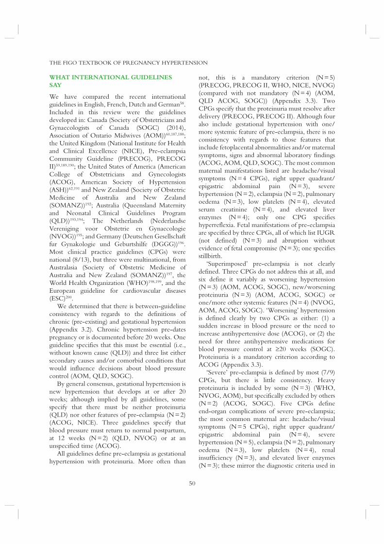
THE FIGO TEXTBOOK OF PREGNANCY HYPERTENSION
50
WHAT INTERNATIONAL GUIDELINES SAY
We have compared the recent international
guidelines in English, French, Dutch and German58.
Included in this review were the guidelines
developed in: Canada (Society of Obstetricians and
Gynaecologists of Canada (SOGC) (2014),
Association of Ontario Midwives (AOM))60,187,188;
the United Kingdom (National Institute for Health
and Clinical Excellence (NICE), Pre-eclampsia
Community Guideline (PRECOG), PRECOG
II)59,189,190; the United States of America (American
Colleg e of Obstetricians and Gynecologists
(ACOG), American Society of Hypertension
(ASH))62,191 and New Zealand (Society of Obstetric
Medicine of Australia and New Zealand
(SOMANZ))192; Australia (Queensland Maternity
and Neonatal Clinical Guidelines Program
(QLD))193,194; The Netherlands (Nederlandse
Vereniging voor Obstetrie en Gynaecologie
(NVOG))195; and Germany (Deutschen Gesellschaft
fur Gynakologie und Geburtshilfe (DGGG))196.
Most clinical practice guidelines (CPGs) were
national (8/13), but three were multinational, f rom
Australasia (Society of Obstetric Medicine of
Australia and New Zealand (SOMANZ))197, the
World Health Orga nization (WHO)198,199, and the
European guideline for cardiovascular diseases
(ESC)200.
We determined that there is between-guideline
consistency with regards to the definitions of
chronic (pre-existing) and gestational hypertension
(Appendix 3.2). Chronic hypertension pre-dates
pregnancy or is documented before 20 weeks. One
guideline specifies that this must be essential (i.e.,
without known cause (QLD)) and three list either
secondary causes and/or comorbid conditions that
would influence decisions about blood pressure
control (AOM, QLD, SOGC).
By general consensus, gestational hypertension is
new hypertension that develops at or after 20
weeks; although implied by all guidelines, some
specify that there must be neither proteinuria
(QLD) nor other features of pre-eclampsia (N = 2)
(ACOG, NICE). Three guidelines specify that
blood pressure must return to normal postpartum,
at 12 weeks (N = 2) (QLD, NVOG) or at an
unspecified time (ACOG).
All guidelines define pre-eclampsia as gestational
hypertension with proteinuria. More often than
not, this is a mandatory criterion (N = 5)
(PRECOG, PRECOG II, WHO, NICE, NVOG)
(compared with not mandatory (N = 4) (AOM,
QLD ACOG, SOGC)) (Appendix 3.3). Two
CPGs specify that the proteinuria must resolve after
delivery (PRECOG, PRECOG II). Although four
also include gestational hypertension with one/
more systemic feature of pre-eclampsia, there is no
consistency with regards to those features that
include fetoplacental abnormalities and/or maternal
symptoms, signs and abnormal laboratory findings
(ACOG, AOM, QLD, SOGC). The most common
maternal manifestations listed are headache/visual
symptoms (N = 4 CPGs), right upper quadrant/
epigastric abdominal pain (N = 3), severe
hypertension (N = 2), eclampsia (N = 2), pulmonary
oedema (N = 3), low platelets (N = 4), elevated
serum creatinine (N = 4), and elevated liver
enzymes (N = 4); only one CPG specifies
hyperreflexia. Fetal manifestations of pre-eclampsia
are specified by three CPGs, all of which list IUGR
(not defined) (N = 3) and abruption without
evidence of fetal compromise (N = 3); one specifies
stillbirth.
‘Superimposed’ pre-eclampsia is not clearly
defined. Three CPGs do not address this at all, and
six define it variably as worsening hypertension
(N = 3) (AOM, ACOG, SOGC), new/worsening
proteinuria (N = 3) (AOM, ACOG, SOGC) or
one/more other systemic features (N = 4) (NVOG,
AOM, ACOG, SOGC). ‘Worsening’ hypertension
is defined clearly by two CPGs as either: (1) a
sudden increase in blood pressure or the need to
increase antihypertensive dose (ACOG), or (2) the
need for three antihypertensive medications for
blood pressure control at 20 weeks (SOGC).
Proteinuria is a mandatory criterion according to
ACOG (Appendix 3.3).
‘Severe’ pre-eclampsia is defined by most (7/9)
CPGs, but there is little consistency. Heavy
proteinuria is included by some (N = 3) (WHO,
NVOG, AOM), but specifically excluded by others
(N = 2) (ACOG, SOGC). Five CPGs define
end-organ complications of severe pre-eclampsia;
the most common maternal are: headache/visual
symptoms (N = 5 CPGs), right upper quadrant/
epigastric abdominal pain (N = 4), severe
hypertension (N = 5), eclampsia (N = 2), pulmonary
oedema (N = 3), low platelets (N = 4), renal
insufficiency (N = 3), and elevated liver enzymes
(N = 3); these mirror the diagnostic criteria used in

CLASSIFICATION OF THE HYPERTENSIVE DISORDERS OF PREGNANCY
51
some guidelines. Fetal manifestations of
pre-eclampsia are specified by three CPGs, all of
which list stillbirth and none of which specify
abruption without evidence of fetal compromise;
IUGR is included by WHO and SOGC, but
specifically excluded by ACOG. The SOGC
‘severity’ criteria are indications for delivery, and
include some features that are in other CPGs: (1)
define pre-eclampsia but not severe pre-eclampsia
(e.g., stroke), (2) define both pre-eclampsia and
severe pre-eclampsia (e.g., eclampsia, pulmonary
oedema, platelet count <100109/L, and acute
kidney injury), or (3) define neither pre-eclampsia
nor severe pre-eclampsia but are widely regarded as
indications for delivery (e.g., uncontrolled severe
hypertension).
In the three CPGs that specify that proteinuria is
mandatory to define pre-eclampsia (WHO, NICE,
NVOG), severe pre-eclampsia is the development
of: (1) pre-eclampsia at <34 weeks (WHO), or (2)
one/more features of end-organ dysfunction that is
either not defined (WHO, NICE) or listed as
‘‘symptoms’’ (NVOG), heavy proteinuria (NVOG,
WHO), or severe hypertension (NVOG, WHO)
(Appendix 3.3).
In the four CPGs that do not include proteinuria
as mandatory to define pre-eclampsia (AOM, QLD
ACOG, SOGC), severe pre-eclampsia is the
development of: (1) pre-eclampsia at 34 weeks
(AOM), (2) proteinuria plus one/more features
that alone would signify pre-eclampsia (cerebral/
visual disturbances, pulmonary oedema, platelet
count, 100109/L, renal insufficiency, or elevated
liver enzymes) (ACOG), or (3) one/more features
of end-organ dysfunction described as: heavy
proteinuria (AOM), one/more features of HELLP
(QLD), new persistent and otherwise unexplained
right upper quadrant/epigastric abdominal pain
(ACOG), severe hypertension (AOM, ACOG), or
those dysfunctions requiring delivery (SOGC)
(Appendix 3.3).
Eclampsia is consistently defined by new onset
and otherwise unexplained seizures in the setting of
pre-eclampsia (N = 5 CPGs) (NICE, QLD, WHO,
ACOG, SOGC). No guideline defines the widely
used term, ‘imminent eclampsia.’
PRIORITIES FOR FUTURE RESEARCH
All future research activities should be compliant
with new international, consensus-derived minimal
standards for pre-eclampsia research122. Through
the PRE-EMPT Global Pregnancy Collaboration,
investigators will be able to gain access to data
management platforms by the end of 2016
(https://pre-empt.cfri.ca/colaboratory). It is a
global imperative that representative biobanks are
developed that have whole blood, plasma, serum,
placental tissue to inform our understanding of
pathways to healthy and complicated pregnancies,
and the design of tailored interventions, for the
most vulnerable women in less developed countries.
Biomarkers and biology
In our opinion, a singular priority is to better
determine the biomarkers that are either specific to
pre-eclampsia or more general to placental disease
and are relevant to women in specific global
regions. How these variations in the pathways
towards, and responses to, disease modify the
performance of current, translational and future
diagnostic and classification tests is largely unknown.
Through better understanding of the biology of
pre-eclampsia and the other hypertensive disorders
of pregnancy, we will be enabled to better define
and sub-classify the forms of hypertensive disorders
of pregnancy that complicate pregnancies globally41.
It is almost certain that the pathways to gestational
hypertension and pre-eclampsia vary between
women in well-resourced, more socially liberal
countries, and those from less-resourced and more
socially conservative countries. While the clinical
manifestations of the disease appear to be common
between communities of women, we need to
determine whether the pathways to disease are
shared. Differential origins of disease may arise
due to variability in the social, environmental,
infectious, and inflammatory determinants of
maternal health and vulnerability.
The interaction between genes, the epigenome,
commensal and pathological organisms, and the
wider environment must vary between and within
clusters of women. Indeed, biomarkers passed
over in more-developed countries may become
important time-of-disease risk identifiers
in less-developed countries where the burden
of severe disease with multiple end-organ
complications is far greater. Such a biomarker is
S100B135.
Obtaining robust socio-demographic, clinical
and biomarker data from before pregnancy, during

THE FIGO TEXTBOOK OF PREGNANCY HYPERTENSION
52
pregnancy (normal and complicated), and at time
of disease is an urgent priority, especially for
women in less-developed countries who bear a
disproportionate burden of risk in terms of the
development of pre-eclampsia, from dying from it,
or losing their baby to it.
Precision medicine
For better assessment of personalised risks borne by
women with a hypertens ive disorder of pregnancy,
we need to strengthen the miniPIERS model with
pulse oximetry48,184 and/or point-of-care assessment
of angiogenic factor balance and GlyFn42,43,45,55,
should they be shown to improve the performance
of the miniPIERS model. This may place advanced
diagnostic capability in the hands of minimally
trained, mHealth app-supported health workers in
women’s homes.
The incremental value of supplementing and/or
recalibrating the fullPIERS model with angiogenic
factors (e.g., PlGF) and/or GlyFn needs to be
assessed. In addition, expanding the scope of
fullPIERS to include women with all hypertensive
disorders of pregnancy would improve its clinical
utility. Recalibration of the model may well be
required.
Similar parallel models to miniPIERS and
fullPIERS are required to assess fetal risks and to
optimise the timing of delivery and long-term
outcomes for the fetuses of pregnancies complicated
by pregnancy hypertension. Initially, this research is
likely to be focused on well-resourced settings and,
subsequently, on less-resourced settings. However,
it must be remembered that there are many highly
resourced centres providing care in less-developed
countries. Partnering with such institutions will
accelerate discovery that is pertinent to the global
maternal population.
Markers of maternal cardiorespiratory health
A priority for research is the better assessment of
the cardiorespiratory status of pregnant women,
especially those with pre-eclampsia.
A prospective population-based study with
nested case–control analysis used the UK Obstetric
Surveillance System to identify all 25 women in the
UK over a 6 year period with myocardial infarction
(MI) in pregnancy, compared with a control group
of 1360 women. Following mutivariable logistic
regression, hypertension and pre-eclampsia were
independ ently associated with MI in pregnancy
as well as maternal age, smoking and twin
pregnancy201. This may stem from global diastolic
dysfunction, left ventricular remodelling, interstitial
pulmonary fluid and increased brain natriuretic
peptide (BNP) that may represent an adaptive
response to maintain myocard ial contractility with
pre-eclampsia, at least at term in less- and
more-developed settings202–205. These preliminary
findings are consistent with what has been observed
in the miniPIERS and fullPIERS studies with
respect to pulse oximetry47,76,184.
These findings need to be confirmed and
expanded across the clinical spectrum of disease
(early- and late-onset pre-eclampsia) as well as the
geographical and socio-economic spectra in which
pregnant women find themselves.
The impact of classification
Finally, implementation research observing the
impact on maternal and perinatal outcomes and
health services costs (direct and indirect) of
introducing new classification paradigms is
important, so that health decision-makers can make
evidence-informed choices about defining national
classification systems. Such implementation
research might usefully include a stepped wedge
design through a series of jurisdictions.
REFERENCES
1. Hadker N, Garg S, Costanzo C, van der Helm W, Creeden J. Are there financial savings associated with supplementing current diagnostic practice for preeclampsia with a novel test? Learnings from a modeling analysis from a German payer perspective. Hypertens Pregnancy 2013 May;32(2):105–19
2. Why mothers die 2000–2002. The sixth report of the confidential enquiries into maternal deaths in the United Kingdom. London: CEMACH; 2004. London: CEMACH; 2004. Report No.: 6
3. Fernando D, Jayatilleka A, Karunaratna V. Pregnancy-reducing maternal deaths and disability in Sri Lanka: national strategies. Br Med Bull 2003;67:85–98
4. Allen VM. The effect of hypertensive disorders in pregnancy on perinatal outcomes: a population-based cohort study. Ottawa: National Library of Canada; 2002
5. Hutcheon JA, Lisonkova S, Joseph KS. Epidemiology of pre-eclampsia and the other hypertensive disorders

CLASSIFICATION OF THE HYPERTENSIVE DISORDERS OF PREGNANCY
53
of pregnancy. Best Pract Res Clin Obstet Gynaecol 2011 Aug;25(4):391–403
6. Ananth CV, Savitz DA, Bowes WA, Jr. Hypertensive disorders of pregnancy and stillbirth in North Carolina, 1988 to 1991. Acta Obstet Gynecol Scand 1995 Nov;74(10):788–93
7. Bagga R, Aggarwal N, Chopra V, Saha SC, Prasad GR, Dhaliwal LK. Pregnancy complicated by severe chronic hypertension: a 10-year analysis from a developing country. Hypertens Pregnancy 2007;26(2):139–49
8. Chappell LC, Enye S, Seed P, Briley AL, Poston L, Shennan AH. Adverse perinatal outcomes and risk factors for preeclampsia in women with chronic hypertension: a prospective study. Hypertension 2008 Apr;51(4):1002–9
9. Clausson B, Cnattingius S, Axelsson O. Preterm and term births of small for gestational age infants: a population-based study of risk factors among nulliparous women. Br J Obstet Gynaecol 1998 Sep; 105(9):1011–7
10. Ferrazzani S, Caruso A, De Carolis S, Martino IV, Mancuso S. Proteinuria and outcome of 444 pregnancies complicated by hypertension. Am J Obstet Gynecol 1990 Feb;162(2):366–71
11. Haelterman E, Breart G, Paris-Llado J, Dramaix M, Tchobroutsky C. Effect of uncomplicated chronic hypertension on the risk of small-for-gestational age birth. Am J Epidemiol 1997 Apr 15;145(8):689–95
12. Lydakis C, Beevers DG, Beevers M, Lip GY. Obstetric and neonatal outcome following chronic hypertension in pregnancy among different ethnic groups. QJM 1998 Dec;91(12):837–44
13. Mabie WC, Pernoll ML, Biswas MK. Chronic hypertension in pregnancy. Obstet Gynecol 1986 Feb;67(2):197–205
14. McCowan LM, Buist RG, North RA, Gamble G. Perinatal morbidity in chronic hypertension. Br J Obstet Gynaecol 1996 Feb;103(2):123–9
15. Ray JG, Burrows RF, Burrows EA, Vermeulen MJ. MOS HIP: McMaster outcome study of hypertension in pregnancy. Early Hum Dev 2001 Sep;64(2):129–43
16. Rey E, Couturier A. The prognosis of pregnancy in women with chronic hypertension. Am J Obstet Gynecol 1994 Aug;171(2):410–6
17. Sibai BM, Abdella TN, Anderson GD. Pregnancy outcome in 211 patients with mild chronic hypertension. Obstet Gynecol 1983 May;61(5):571–6
18. Sibai BM, Anderson GD. Pregnancy outcome of intensive therapy in severe hypertension in first trimester. Obstet Gynecol 1986 Apr;67(4):517–22
19. Sibai BM, Lindheimer M, Hauth J, Caritis S, Van Dorsten P, Klebanoff M, et al. Risk factors for preeclampsia, abruptio placentae, and adverse neonatal outcomes among women with chronic hypertension. National Institute of Child Health and Human Development Network of Maternal-Fetal Medicine Units. N Engl J Med 1998 Sep 3;339(10):667–71
20. Samuel A, Lin C, Parviainen K, Jeyabalan A. Expectant management of preeclampsia superimposed on chronic hypertension. J Matern Fetal Neonatal Med 2011 Jul;24(7):907–11
21. Barton JR, O’Brien JM, Bergauer NK, Jacques DL, Sibai BM. Mild gestational hypertension remote from term: progression and outcome. Am J Obstet Gynecol 2001 Apr;184(5):979–83
22. Brown MA, Buddle ML. The importance of nonproteinuric hypertension in pregnancy. Hypertens Pregn 2002;14:57–65
23. Ferrazzani S, Caruso A, De Carolis S, Martino IV, Mancuso S. Proteinuria and outcome of 444 pregnancies complicated by hypertension. Am J Obstet Gynecol 1990 Feb;162(2):366–71
24. Horsager R, Adams M, Richey S, Leveno KJ, Cunningham FG. Outpatient management of mild pregnancy induced hypertension. 15th Annual Meeting of The Society of Perinatal Obstetricians, Atlanta, Georgia 1995
25. Mabie WC, Pernoll ML, Biswas MK. Chronic hypertension in pregnancy. Obstet Gynecol 1986 Feb;67(2):197–205
26. Saudan P, Brown MA, Buddle ML, Jones M. Does gestational hypertension become pre-eclampsia? Br J Obstet Gynaecol 1998 Nov;105(11):1177–84
27. Scott A, Moar V, Ounsted M. The relative contributions of different maternal factors in small-for-gestational-age pregnancies. Eur J Obstet Gynecol Reprod Biol 1981 Sep;12(3):157–65
28. Caritis S, Sibai B, Hauth J, Lindheimer MD, Klebanoff M, Thom E, et al. Low-dose aspirin to prevent preeclampsia in women at high risk. National Institute of Child Health and Human Development Network of Maternal-Fetal Medicine Units. N Engl J Med 1998 Mar 12;338(11):701–5
29. Ankumah NA, Cantu J, Jauk V, Biggio J, Hauth J, Andrews W, et al. Risk of adverse pregnancy outcomes in women with mild chronic hypertension before 20

THE FIGO TEXTBOOK OF PREGNANCY HYPERTENSION
54
weeks of gestation. Obstet Gynecol 2014 May;123(5): 966–72
30. Hutcheon JA, Lisonkova S, Magee LA, von Dadelszen P, Woo HL, Liu S, et al. Optimal timing of delivery in pregnancies with pre-existing hypertension. BJOG 2011 Jan;118(1):49–54
31. Magee LA, Ramsay G, von Dadelszen P. What is the role of out-of-office BP measurement in hypertensive pregnancy? Hypertens Pregnancy 2008;27(2):95–101
32. Barton JR, O’Brien JM, Bergauer NK, Jacques DL, Sibai BM. Mild gestational hypertension remote from term: progression and outcome. Am J Obstet Gynecol 2001 Apr;184(5):979–83
33. Brown MA, Buddle ML. The importance of nonproteinuric hypertension in pregnancy. Hypertens Pregnancy 2002;14:57–65
34. Magee LA, von Dadelszen P, Bohun CM, Rey E, El-Zibdeh M, Stalker S, et al. Serious perinatal complications of non-proteinuric hypertension: an international, multicentre, retrospective cohort study. J Obstet Gynaecol Can 2003 May;25(5):372–82
35. Magee LA, von Dadelszen P, Chan S, Gafni A, Gruslin A, Helewa M, et al. The Control of Hypertension In Pregnancy Study pilot trial. BJOG 2007 Jun;114(6): 770, e13-770, e20
36. Magee LA, von Dadelszen P, Rey E, Ross S, Asztalos E, Murphy KE, et al. Less-tight versus tight control of hypertension in pregnancy. N Engl J Med 2015 Jan 29;372(5):407–17
37. Gruslin A, Lemyre B. Pre-eclampsia: fetal assessment and neonatal outcomes. Best Pract Res Clin Obstet Gynaecol 2011 Aug;25(4):491–507
38. Xiong X, Mayes D, Demianczuk N, Olson DM, Davidge ST, Newburn-Cook C, et al. Impact of pregnancy-induced hypertension on fetal growth. Am J Obstet Gynecol 1999 Jan;180(1 Pt 1):207–13
39. Xiong X, Demianczuk NN, Buekens P, Saunders LD. Association of preeclampsia with high birth weight for age. Am J Obstet Gynecol 2000 Jul;183(1):148–55
40. Ness RB, Roberts JM. Heterogeneous causes constituting the single syndrome of preeclampsia: a hypothesis and its implications. Am J Obstet Gynecol 1996 Nov;175(5):1365–70
41. Staff AC, Benton SJ, von Dadelszen P, Roberts JM, Taylor RN, Powers RW, et al. Redefining preeclampsia using placenta-derived biomarkers. Hypertension 2013 May;61(5):932–42
42. Maynard SE, Karumanchi SA. Angiogenic factors and preeclampsia. Semin Nephrol 2011 Jan;31(1):33–46
43. Benton SJ, Hu Y, Xie F, Kupfer K, Lee SW, Magee LA, et al. Angiogenic factors as diagnostic tests for preeclampsia: a performance comparison between two commercial immunoassays. Am J Obstet Gynecol 2011 Nov;205(5):469–8
44. Benton SJ, Hu Y, Xie F, Kupfer K, Lee SW, Magee LA, et al. Can placental growth factor in maternal circulation identify fetuses with placental intrauterine growth restriction? Am J Obstet Gynecol 2012 Feb;206(2):163–7
45. Chappell LC, Duckworth S, Seed PT, Griffin M, Myers J, Mackillop L, et al. Diagnostic accuracy of placental growth factor in women with suspected preeclampsia: a prospective multicenter study. Circulation 2013 Nov 5;128(19):2121–31
46. Steegers EA, von Dadelszen P, Duvekot JJ, Pijnenborg R. Pre-eclampsia. Lancet 2010 Aug 21;376(9741): 631–44
47. von Dadelszen P, Payne B, Li J, Ansermino JM, Broughton PF, Cote AM, et al. Prediction of adverse maternal outcomes in pre-eclampsia: development and validation of the fullPIERS model. Lancet 2011 Jan 15;377(9761):219–27
48. Payne BA, Hutcheon JA, Ansermino JM, Hall DR, Bhutta ZA, Bhutta SZ, et al. A risk prediction model for the assessment and triage of women with hypertensive disorders of pregnancy in low-resourced settings: the miniPIERS (Pre-eclampsia Integrated Estimate of RiSk) multi-country prospective cohort study. PLoS Med 2014 Jan;11(1):e1001589
49. Bramham K, Seed P, Nelson-Piercy C, Lightstone L, Ashford L, Butler J, et al. [42-OR]: Diagnostic accuracy of placental growth factor in women with chronic kidney disease or hypertension and suspected preeclampsia: A prospective cohort study. Pregnancy Hypertens 2015 Jan;5(1):21
50. Perni U, Sison C, Sharma V, Helseth G, Hawfield A, Suthanthiran M, et al. Angiogenic factors in superimposed preeclampsia: a longitudinal study of women with chronic hypertension during pregnancy. Hypertension 2012 Mar;59(3):740–6
51. Rolfo A, Attini R, Nuzzo AM, Piazzese A, Parisi S, Ferraresi M, et al. Chronic kidney disease may be differentially diagnosed from preeclampsia by serum biomarkers. Kidney Int 2013 Jan;83(1):177–81
52. Goswami D, Tannetta DS, Magee LA, Fuchisawa A, Redman CW, Sargent IL, et al. Excess syncytiotrophoblast microparticle shedding is a feature of early-onset pre-eclampsia, but not normotensive

CLASSIFICATION OF THE HYPERTENSIVE DISORDERS OF PREGNANCY
55
intrauterine growth restriction. Placenta 2006 Jan;27(1):56–61
53. Kenny LC, Black MA, Poston L, Taylor R, Myers JE, Baker PN, et al. Early pregnancy prediction of preeclampsia in nulliparous women, combining clinical risk and biomarkers: the Screening for Pregnancy Endpoints (SCOPE) international cohort study. Hypertension 2014 Sep;64(3):644–52
54. Myers JE, Tuytten R, Thomas G, Laroy W, Kas K, Vanpoucke G, et al. Integrated proteomics pipeline yields novel biomarkers for predicting preeclampsia. Hypertension 2013 Jun;61(6):1281–8
55. Rasanen J, Quinn MJ, Laurie A, Bean E, Roberts CT, Jr., Nagalla SR, et al. Maternal serum glycosylated fibronectin as a point-of-care biomarker for assessment of preeclampsia. Am J Obstet Gynecol 2015 Jan;212(1): 82–9
56. Yach D, Hawkes C, Gould CL, Hofman KJ. The global burden of chronic diseases: overcoming impediments to prevention and control. JAMA 2004 Jun 2;291(21):2616–22
57. Brown MA, Lindheimer MD, de Swiet M, Van Asche A, Moutquin JM. The classification and diagnosis of the hypertensive disorders of pregnancy: statement from the International Society for the Study of Hypertension in Pregnancy (ISSHP). Hypertens Pregnancy 2001;20(1):IX-XIV
58. Gillon TE, Pels A, von Dadelszen P, MacDonell K, Magee LA. Hypertensive disorders of pregnancy: a systematic review of international clinical practice guidelines. PLoS One 2014;9(12):e113715
59. National Collaborating Centre for Women’s and Children’s Health (UK). CG107: Hypertension in pregnancy: The management of hypertensive disorders during pregnancy. NICE: Guidance 2010 Aug
60. Magee LA, Pels A, Helewa M, Rey E, von Dadelszen P. Diagnosis, evaluation, and management of the hypertensive disorders of pregnancy. Pregnancy Hypertens 2014;4(2):105–45
61. Sibai BM. Pitfalls in diagnosis and management of preeclampsia. Am J Obstet Gynecol 1988 Jul;159(1):1–5
62. Hypertension in pregnancy. Report of the American College of Obstetricians and Gynecologists’ Task Force on Hypertension in Pregnancy. Obstet Gynecol 2013 Nov;122(5):1122–31
63. Filetti LC, Imudia AN, Al-Safi Z, Hobson DT, Awonuga AO, Bahado-Singh RO. New onset delayed postpartum preeclampsia: different disorders? J Matern Fetal Neonatal Med 2012 Jul;25(7):957–60
64. Hypertension in pregnancy: the management of hypertensive disorders during pregnancy. Manchester: National Institute for Health and Clinical Excellence; 2011. Report No.: 107
65. Lowe SA, Bowyer L, Lust K, McMahon LP, Morton MR, North RA, et al. The SOMANZ guideline for the management of hypertensive disorders of pregnancy. Sydney: SOMANZ; 2014
66. Tranquilli AL, Brown MA, Zeeman GG, Dekker G, Sibai BM. The definition of severe and early onset preeclampsia. Statements from the International Society for the Study of Hypertension in Pregnancy. Pregnancy Hypertens 2013;3(1):44–7
67. Aydin S, Ersan F, Ark C, Arioglu AC. Partial HELLP syndrome: maternal, perinatal, subsequent pregnancy and long-term maternal outcomes. J Obstet Gynaecol Res 2014 Apr;40(4):932–40
68. Martin JN, Jr., Brewer JM, Wallace K, Sunesara I, Canizaro A, Blake PG, et al. HELLP syndrome and composite major maternal morbidity: importance of Mississippi classification system. J Matern Fetal Neonatal Med 2013 Aug;26(12):1201–6
69. Minakami H, Morikawa M, Yamada T, Yamada T, Akaishi R, Nishida R. Differentiation of acute fatty liver of pregnancy from syndrome of hemolysis, elevated liver enzymes and low platelet counts. J Obstet Gynaecol Res 2014 Mar;40(3):641–9
70. Menzies J, Magee LA, Li J, Macnab YC, Yin R, Stuart H, et al. Instituting surveillance guidelines and adverse outcomes in preeclampsia. Obstet Gynecol 2007 Jul;110(1):121–7
71. von Dadelszen P, Sawchuck D, McMaster R, Douglas MJ, Lee SK, Saunders S, et al. The active implementation of pregnancy hypertension guidelines in British Columbia. Obstet Gynecol 2010 Sep;116(3): 659–66
72. Akkermans J, Payne B, von Dadelszen P, Groen H, de Vries J, Magee LA, et al. Predicting complications in pre-eclampsia: external validation of the fullPIERS model using the PETRA trial dataset. Eur J Obstet Gynecol Reprod Biol 2014 Aug;179:58–62
73. Kozic JR, Benton SJ, Hutcheon JA, Payne BA, Magee LA, von Dadelszen P. Abnormal liver function tests as predictors of adverse maternal outcomes in women with preeclampsia. J Obstet Gynaecol Can 2011 Oct; 33(10):995–1004
74. Laskin S, Payne B, Hutcheon JA, Qu Z, Douglas MJ, Ford J, et al. The role of platelet counts in the assessment of inpatient women with preeclampsia. J Obstet Gynaecol Can 2011 Sep;33(9):900–8

THE FIGO TEXTBOOK OF PREGNANCY HYPERTENSION
56
75. Livingston JR, Payne B, Brown M, Roberts JM, Cote AM, Magee LA, et al. Uric acid as a predictor of adverse maternal and perinatal outcomes in women hospitalized with preeclampsia. J Obstet Gynaecol Can 2014 Oct;36(10):870–7
76. Millman AL, Payne B, Qu Z, Douglas MJ, Hutcheon JA, Lee T, et al. Oxygen saturation as a predictor of adverse maternal outcomes in women with preeclampsia. J Obstet Gynaecol Can 2011 Jul;33(7): 705–14
77. Payne B, Magee LA, Cote AM, Hutcheon JA, Li J, Kyle PM, et al. PIERS proteinuria: relationship with adverse maternal and perinatal outcome. J Obstet Gynaecol Can 2011 Jun;33(6):588–97
78. Payne B, Hodgson S, Hutcheon JA, Joseph KS, Li J, Lee T, et al. Performance of the fullPIERS model in predicting adverse maternal outcomes in pre-eclampsia using patient data from the PIERS (Pre-eclampsia Integrated Estimate of RiSk) cohort, collected on admission. BJOG 2013 Jan;120(1):113–8
79. Yen TW, Payne B, Qu Z, Hutcheon JA, Lee T, Magee LA, et al. Using clinical symptoms to predict adverse maternal and perinatal outcomes in women with preeclampsia: data from the PIERS (Pre-eclampsia Integrated Estimate of RiSk) study. J Obstet Gynaecol Can 2011 Aug;33(8):803–9
80. Lamminpaa R, Vehvilainen-Julkunen K, Gissler M, Heinonen S. Preeclampsia complicated by advanced maternal age: a registry-based study on primiparous women in Finland 1997–2008. BMC Pregnancy Childbirth 2012;12:47
81. Sebastian Manzanares G, Angel Santalla H, Irene Vico Z, Lopez Criado MS, Alicia Pineda L, Jose Luis Gallo V. Abnormal maternal body mass index and obstetric and neonatal outcome. J Matern Fetal Neonatal Med 2012 Mar;25(3):308–12
82. Urquia ML, Ying I, Glazier RH, Berger H, De Souza LR, Ray JG. Serious preeclampsia among different immigrant groups. J Obstet Gynaecol Can 2012 Apr;34(4):348–52
83. KDIGO clinical practice guideline for acute kidney injury. Kidney Int Suppl 2012;2(1):1–138
84. Hawkins TL, Roberts JM, Mangos GJ, Davis GK, Roberts LM, Brown MA. Plasma uric acid remains a marker of poor outcome in hypertensive pregnancy: a retrospective cohort study. BJOG 2012 Mar;119(4): 484–92
85. Marozio L, Facchinetti F, Allais G, Nappi RE, Enrietti M, Neri I, et al. Headache and adverse pregnancy outcomes: a prospective study. Eur J Obstet Gynecol Reprod Biol 2012 Apr;161(2):140–3
86. Thangaratinam S, Ismail KM, Sharp S, Coomarasamy A, Khan KS. Accuracy of serum uric acid in predicting complications of pre-eclampsia: a systematic review. BJOG 2006 Apr;113(4):369–78
87. Thangaratinam S, Coomarasamy A, O’Mahony F, Sharp S, Zamora J, Khan KS, et al. Estimation of proteinuria as a predictor of complications of pre-eclampsia: a systematic review. BMC Med 2009;7: 10
88. Thangaratinam S, Koopmans CM, Iyengar S, Zamora J, Ismail KM, Mol BW, et al. Accuracy of liver function tests for predicting adverse maternal and fetal outcomes in women with preeclampsia: a systematic review. Acta Obstet Gynecol Scand 2011 Jun;90(6):574–85
89. Thangaratinam S, Gallos ID, Meah N, Usman S, Ismail KM, Khan KS. How accurate are maternal symptoms in predicting impending complications in women with preeclampsia? A systematic review and meta-analysis. Acta Obstet Gynecol Scand 2011 Jun; 90(6):564–73
90. Yang SW, Cho SH, Kwon HS, Sohn IS, Hwang HS. Significance of the platelet distribution width as a severity marker for the development of preeclampsia. Eur J Obstet Gynecol Reprod Biol 2014 Apr;175: 107–11
91. Saraf S, Egbert NM, Mittal G, Homel P, Minkoff H, Fisher N. Predictors of posterior reversible encephalopathy syndrome in preeclampsia and eclampsia [abstract]. Obstet Gynecol 2014;123(5 (Suppl 1)):169S
92. Kao DP, Hsich E, Lindenfeld J. Characteristics, adverse events, and racial differences among delivering mothers with peripartum cardiomyopathy. JACC Heart Fail 2013 Oct;1(5):409–16
93. Lalor JG, Fawole B, Alfirevic Z, Devane D. Biophysical profile for fetal assessment in high risk pregnancies. Cochrane Database Syst Rev 2008;(1):CD000038
94. Kaur S, Picconi JL, Chadha R, Kruger M, Mari G. Biophysical profile in the treatment of intrauterine growth-restricted fetuses who weigh <1000 g. Am J Obstet Gynecol 2008 Sep;199(3):264
95. Payne BA, Kyle PM, Lim K, Lisonkova S, Magee LA, Pullar B, et al. An assessment of predictive value of the biophysical profile in women with preeclampsia using data from the fullPIERS database. Pregnancy Hypertens 2013;3(3):166–71
96. Simsek Y, Celen S, Simsek A, Danisman N, Mollamahmutoglu L. Predictive value of umbilical artery Doppler for adverse perinatal outcome in patients with HELLP syndrome. Eur Rev Med Pharmacol Sci 2013 Jun;17(12):1599–603

CLASSIFICATION OF THE HYPERTENSIVE DISORDERS OF PREGNANCY
57
97. Koopmans CM, Bijlenga D, Groen H, Vijgen SM, Aarnoudse JG, Bekedam DJ, et al. Induction of labour versus expectant monitoring for gestational hypertension or mild pre-eclampsia after 36 weeks’ gestation (HYPITAT): a multicentre, open-label randomised controlled trial. Lancet 2009 Sep 19;374(9694):979–88
98. van der Tuuk K, Holswilder-Olde Scholtenhuis MA, Koopmans CM, van den Akker ES, Pernet PJ, Ribbert LS, et al. Prediction of neonatal outcome in women with gestational hypertension or mild preeclampsia after 36 weeks of gestation. J Matern Fetal Neonatal Med 2014 Jul 17;1–7
99. de Oliveira CA, de Sa RA, Velarde LG, da Silva FC, doVale FA, Netto HC. Changes in ophthalmic artery Doppler indices in hypertensive disorders during pregnancy. J Ultrasound Med 2013 Apr;32(4):609–16
100. Kafkasli A, Turkcuoglu I, Turhan U. Maternal, fetal and perinatal characteristics of preeclampsia cases with and without abnormalities in uterine artery Doppler indexes. J Matern Fetal Neonatal Med 2013 Jun;26(9): 936–40
101. Bramham K, Poli-de-Figueiredo CE, Seed PT, Briley AL, Poston L, Shennan AH, et al. Association of proteinuria threshold in pre-eclampsia with maternal and perinatal outcomes: a nested case control cohort of high risk women. PLoS One 2013;8(10):e76083
102. Cote AM, Brown MA, Lam E, von Dadelszen P, Firoz T, Liston RM, et al. Diagnostic accuracy of urinary spot protein:creatinine ratio for proteinuria in hypertensive pregnant women: systematic review. BMJ 2008 May 3;336(7651):1003–6
103. Rey E, Morin F, Boudreault J, Pilon F, Vincent D, Ouellet D. Blood pressure assessments in different subtypes of hypertensive pregnant women: office versus home patient- or nurse-measured blood pressure. Hypertens Pregnancy 2009 May;28(2):168–77
104. Brown MA, Mangos G, Davis G, Homer C. The natural history of white coat hypertension during pregnancy. BJOG 2005 May;112(5):601–6
105. Hermida RC, Ayala DE, Iglesias M. Circadian rhythm of blood pressure challenges office values as the “gold standard” in the diagnosis of gestational hypertension. Chronobiol Int 2003 Jan;20(1):135–56
106. Eguchi K, Ohmaru T, Ohkuchi A, Hirashima C, Takahashi K, Suzuki H, et al. Ambulatory BP monitoring and clinic BP in predicting small-for-gestational-age infants during pregnancy. J Hum Hypertens 2015 Mar 19
107. Tonelli M, Pottie K. Diabetes guidelines. CMAJ 2013 Feb 19;185(3):238
108. Angeli E, Verdecchia P, Narducci P, Angeli F. Additive value of standard ECG for the risk prediction of hypertensive disorders during pregnancy. Hypertens Res 2011 Jun;34(6):707–13
109. Gyselaers W, Tomsin K, Staelens A, Mesens T, Oben J, Molenberghs G. Maternal venous hemodynamics in gestational hypertension and preeclampsia. BMC Pregnancy Childbirth 2014;14:212
110. Gyselaers W, Staelens A, Mesens T, Tomsin K, Oben J, Vonck S, et al. Maternal venous Doppler characteristics are abnormal in pre-eclampsia but not in gestational hypertension. Ultrasound Obstet Gynecol 2015 Apr; 45(4):421–6
111. Garg A, Wapner RJ, Ananth CV, Dale E, Tsang SH, Lee W, et al. Choroidal and retinal thickening in severe preeclampsia. Invest Ophthalmol Vis Sci 2014 Sep;55(9):5723–9
112. Ilea C, Zaharia D, Socolov D, David C, Lupascu IA. The role of EEG brain mapping in quantification of brain damage in hypertensive disease associated with pregnancy. Rev Med Chir Soc Med Nat Iasi 2012 Jan;116(1):187–92
113. Sibai BM. Imitators of severe preeclampsia. Obstet Gynecol 2007 Apr;109(4):956–66
114. Fesenmeier MF, Coppage KH, Lambers DS, Barton JR, Sibai BM. Acute fatty liver of pregnancy in 3 tertiary care centers. Am J Obstet Gynecol 2005 May; 192(5):1416–9
115. Erkan D, Espinosa G, Cervera R. Catastrophic antiphospholipid syndrome: updated diagnostic algorithms. Autoimmun Rev 2010 Dec;10(2):74–9
116. Martin JN, Jr., Bailey AP, Rehberg JF, Owens MT, Keiser SD, May WL. Thrombotic thrombocytopenic purpura in 166 pregnancies: 1955–2006. Am J Obstet Gynecol 2008 Aug;199(2):98–104
117. Mouthon L, Berezne A, Bussone G, Noel LH, Villiger PM, Guillevin L. Scleroderma renal crisis: a rare but severe complication of systemic sclerosis. Clin Rev Allergy Immunol 2011 Apr;40(2):84–91
118. Marik PE, Varon J. Hypertensive crises: challenges and management. Chest 2007 Jun;131(6):1949–62
119. Lalitha R, Opio CK. A missed diagnosis or a masquerading disease: back to the basics. Pan Afr Med J 2013;15:29
120. Petrie J, Lockie C, Paolineli A, Stevens M, Smith M, Mitchell C, et al. Undiagnosed phaeochromocytoma masquerading as eclampsia. BMJ Case Rep 2012;2012

THE FIGO TEXTBOOK OF PREGNANCY HYPERTENSION
58
121. Sibai BM, Stella CL. Diagnosis and management of atypical preeclampsia-eclampsia. Am J Obstet Gynecol 2009 May;200(5):481–7
122. Myatt L, Redman CW, Staff AC, Hansson S, Wilson ML, Laivuori H, et al. Strategy for standardization of preeclampsia research study design. Hypertension 2014 Jun;63(6):1293–301
123. Hagmann H, Thadhani R, Benzing T, Karumanchi SA, Stepan H. The promise of angiogenic markers for the early diagnosis and prediction of preeclampsia. Clin Chem 2012 May;58(5):837–45
124. Verlohren S, Herraiz I, Lapaire O, Schlembach D, Moertl M, Zeisler H, et al. The sFlt-1/PlGF ratio in different types of hypertensive pregnancy disorders and its prognostic potential in preeclamptic patients. Am J Obstet Gynecol 2012 Jan;206(1):58
125. Gomez-Arriaga PI, Herraiz I, Lopez-Jimenez EA, Gomez-Montes E, Denk B, Galindo A. Uterine artery Doppler and sFlt-1/PlGF ratio: usefulness in diagnosis of pre-eclampsia. Ultrasound Obstet Gynecol 2013 May;41(5):530–7
126. Gomez-Arriaga PI, Herraiz I, Lopez-Jimenez EA, Escribano D, Denk B, Galindo A. Uterine artery Doppler and sFlt-1/PlGF ratio: prognostic value in early-onset pre-eclampsia. Ultrasound Obstet Gynecol 2014 May;43(5):525–32
127. Gullai N, Stenczer B, Molvarec A, Fugedi G, Veresh Z, Nagy B, et al. Evaluation of a rapid and simple placental growth factor test in hypertensive disorders of pregnancy. Hypertens Res 2013 May;36(5):457–62
128. Alahakoon TI, Zhang W, Trudinger BJ, Lee VW. Discordant clinical presentations of preeclampsia and intrauterine fetal growth restriction with similar pro- and anti-angiogenic profiles. J Matern Fetal Neonatal Med 2014 Dec;27(18):1854–9
129. Chaiworapongsa T, Romero R, Korzeniewski SJ, Kusanovic JP, Soto E, Lam J, et al. Maternal plasma concentrations of angiogenic/antiangiogenic factors in the third trimester of pregnancy to identify the patient at risk for stillbirth at or near term and severe late preeclampsia. Am J Obstet Gynecol 2013 Apr;208(4):287
130. Molvarec A, Gullai N, Stenczer B, Fugedi G, Nagy B, Rigo J, Jr. Comparison of placental growth factor and fetal flow Doppler ultrasonography to identify fetal adverse outcomes in women with hypertensive disorders of pregnancy: an observational study. BMC Pregnancy Childbirth 2013;13:161
131. Moore AG, Young H, Keller JM, Ojo LR, Yan J, Simas TA, et al. Angiogenic biomarkers for prediction
of maternal and neonatal complications in suspected preeclampsia. J Matern Fetal Neonatal Med 2012 Dec;25(12):2651–7
132. Kim SM, Park JS, Norwitz ER, Jung HJ, Kim BJ, Park CW, et al. Circulating levels of neutrophil gelatinase-associated lipocalin (NGAL) correlate with the presence and severity of preeclampsia. Reprod Sci 2013 Sep;20(9):1083–9
133. Patel M, Sachan R, Gangwar R, Sachan P, Natu S. Correlation of serum neutrophil gelatinase-associated lipocalin with acute kidney injury in hypertensive disorders of pregnancy. Int J Nephrol Renovasc Dis 2013;6:181–6
134. Scazzochio E, Munmany M, Garcia L, Meler E, Crispi F, Gratacos E, et al. Prognostic role of maternal neutrophil gelatinase-associated lipocalin in women with severe early-onset preeclampsia. Fetal Diagn Ther 2014;35(2):127–32
135. Bergman L, Akhter T, Wikstrom AK, Wikstrom J, Naessen T, Akerud H. Plasma levels of S100B in preeclampsia and association with possible central nervous system effects. Am J Hypertens 2014 Aug; 27(8):1105–11
136. Rytlewski K, Huras H, Kusmierska-Urban K, Galas A, Reron A. Leptin and interferon-gamma as possible predictors of cesarean section among women with hypertensive disorders of pregnancy. Med Sci Monit 2012 Aug;18(8):CR506-CR511
137. Buhimschi IA, Nayeri UA, Zhao G, Shook LL, Pensalfini A, Funai EF, et al. Protein misfolding, congophilia, oligomerization, and defective amyloid processing in preeclampsia. Sci Transl Med 2014 Jul 16;6(245):245ra92
138. Burwick RM, Easter SR, Dawood HY, Yamamoto HS, Fichorova RN, Feinberg BB. Complement activation and kidney injury molecule-1-associated proximal tubule injury in severe preeclampsia. Hypertension 2014 Oct;64(4):833–8
139. Jim B, Jean-Louis P, Qipo A, Garry D, Mian S, Matos T, et al. Podocyturia as a diagnostic marker for preeclampsia amongst high-risk pregnant patients. J Pregnancy 2012;2012:984630
140. Cnossen JS, Morris RK, ter Riet G, Mol BW, van der Post JA, Coomarasamy A, et al. Use of uterine artery Doppler ultrasonography to predict pre-eclampsia and intrauterine growth restriction: a systematic review and bivariable meta-analysis. CMAJ 2008 Mar 11; 178(6):701–11.
141. Baschat AA, Guclu S, Kush ML, Gembruch U, Weiner CP, Harman CR. Venous Doppler in the

CLASSIFICATION OF THE HYPERTENSIVE DISORDERS OF PREGNANCY
59
prediction of acid-base status of growth-restricted fetuses with elevated placental blood flow resistance. Am J Obstet Gynecol 2004 Jul;191(1):277–84
142. Chamberlain PF, Manning FA, Morrison I, Harman CR, Lange IR. Ultrasound evaluation of amniotic fluid volume. I. The relationship of marginal and decreased amniotic fluid volumes to perinatal outcome. Am J Obstet Gynecol 1984 Oct 1;150(3):245–9
143. Harman CR, Baschat AA. Comprehensive assessment of fetal wellbeing: which Doppler tests should be performed? Curr Opin Obstet Gynecol 2003 Apr; 15(2):147–57
144. Turan OM, Turan S, Gungor S, Berg C, Moyano D, Gembruch U, et al. Progression of Doppler abnormalities in intrauterine growth restriction. Ultrasound Obstet Gynecol 2008 Aug;32(2):160–7
145. Tsigas E, Magee LA. Advocacy organisations as partners in pre-eclampsia progress: patient involvement improves outcomes. Best Pract Res Clin Obstet Gynaecol 2011 Aug;25(4):523–36
146. Gaugler-Senden IP, Duivenvoorden HJ, Filius A, de Groot CJ, Steegers EA, Passchier J. Maternal psychosocial outcome after early onset preeclampsia and preterm birth. J Matern Fetal Neonatal Med 2012 Mar;25(3):272–6
147. Hoedjes M, Berks D, Vogel I, Franx A, Visser W, Duvekot JJ, et al. Symptoms of post-traumatic stress after preeclampsia. J Psychosom Obstet Gynaecol 2011 Sep;32(3):126–34
148. Hoedjes M, Berks D, Vogel I, Franx A, Bangma M, Darlington AS, et al. Postpartum depression after mild and severe preeclampsia. J Womens Health (Larchmt ) 2011 Oct;20(10):1535–42
149. Hoedjes M, Berks D, Vogel I, Franx A, Duvekot JJ, Steegers EA, et al. Poor health-related quality of life after severe preeclampsia. Birth 2011 Sep;38(3): 246–55
150. Kaspers AG, Rep A, Ganzevoort W, Wolf H, de Vries JI, van Wassenaer AG. No association between maternal psychological symptoms and infant outcome after pregnancy complicated by early-onset hypertensive disorders. Acta Paediatr 2009 Feb;98(2):298–303
151. Leeners B, Neumaier-Wagner P, Kuse S, Stiller R, Rath W. Emotional stress and the risk to develop hypertensive diseases in pregnancy. Hypertens Pregnancy 2007;26(2):211–26
152. Poel YH, Swinkels P, de Vries JI. Psychological treatment of women with psychological complaints
after pre-eclampsia. J Psychosom Obstet Gynaecol 2009 Mar;30(1):65–72
153. Rep A, Ganzevoort W, Bonsel GJ, Wolf H, de Vries JI. Psychosocial impact of early-onset hypertensive disorders and related complications in pregnancy. Am J Obstet Gynecol 2007 Aug;197(2):158–6
154. Stramrood CA, Wessel I, Doornbos B, Aarnoudse JG, van den Berg PP, Schultz WC, et al. Posttraumatic stress disorder following preeclampsia and PPROM: a prospective study with 15 months follow-up. Reprod Sci 2011 Jul;18(7):645–53
155. Mautner E, Greimel E, Trutnovsky G, Daghofer F, Egger JW, Lang U. Quality of life outcomes in pregnancy and postpartum complicated by hypertensive disorders, gestational diabetes, and preterm birth. J Psychosom Obstet Gynaecol 2009 Dec;30(4):231–7
156. Mautner E, Stern C, Deutsch M, Nagele E, Greimel E, Lang U, et al. The impact of resilience on psychological outcomes in women after preeclampsia: an observational cohort study. Health Qual Life Outcomes 2013;11:194
157. East C, Conway K, Pollock W, Frawley N, Brennecke S. Women’s experiences of preeclampsia: Australian action on preeclampsia survey of women and their confidants. J Pregnancy 2011;2011:375653
158. You WB, Wolf M, Bailey SC, Pandit AU, Waite KR, Sobel RM and Grobman W. Factors associated with patient understanding of pre-eclampsia. Hypertens Pregnancy. 2012;31(3):341–9
159. Wallis AB, Tsigas EZ, Saftias AF, Sibai MN. Prenatal education is an opportunity for improved outcomes in hypertensive disorders of pregnancy: results from an Internet-based survey J Matern Fetal Neonatal Med; 2013;26(16):1565–7
160. Jonkers M, Richters A, Zwart J, Ory F, van Roosmalen. Severe maternal morbidity among immigrant women in the Netherlands: patients’ perspectives. Reprod Health Matters 2011 May;19(37):144–53
161. You WB, Wolf MS, Bailey SC, Grobman WA. Improving patient understanding of preeclampsia: a randomized controlled trial. Am J Obstet Gynecol 2012 May;206(5):431–5
162. Smyth RM, Duley L, Jacoby A, Elbourne D. Women’s experiences of participating in the Magpie Trial: a postal survey in the United Kingdom. Birth 2009 Sep;36(3):220–9
163. Magee LA, von Dadelszen P, Chan S, Gafni A, Gruslin A, Helewa M, et al. Women’s views of their

THE FIGO TEXTBOOK OF PREGNANCY HYPERTENSION
60
experiences in the CHIPS (Control of Hypertension in Pregnancy Study) Pilot Trial. Hypertens Pregnancy 2007;26(4):371–87
164. Turnbull DA, Wilkinson C, Griffith EC, Kruzins G, Gerard K, Shanahan M, et al. The psychosocial outcomes of antenatal day care for three medical complications of pregnancy: a randomised controlled trial of 395 women. Aust N Z J Obstet Gynaecol 2006 Dec;46(6):510–6
165. Taylor RS, Freeman L, North RA. Evaluation of ambulatory and self-initiated blood pressure monitors by pregnant and postpartum women. Hypertens Pregnancy 2001;20(1):25–33
166. Bijlenga D, Koopmans CM, Birnie E, Mol BW, van der Post JA, Bloemenkamp KW, et al. Health-related quality of life after induction of labor versus expectant monitoring in gestational hypertension or preeclampsia at term. Hypertens Pregnancy 2011;30(3):260–74
167. Adamu AN, Ekele BA, Ahmed Y, Mohammed BA, Isezuo SA, Abdullahpi AA. Pregnancy outcome in women with eclampsia at a tertiary centre in northern Nigeria. Afr J Med Med Sci 2012 Jun;41(2):211–9.
168. Abalos E, Cuesta C, Carroli G, Qureshi Z, Widmer M, Vogel JP, et al. Pre-eclampsia, eclampsia and adverse maternal and perinatal outcomes: a secondary analysis of the World Health Organization Multicountry Survey on Maternal and Newborn Health. BJOG 2014 Mar;121 Suppl 1:14–24
169. de Greeff A, Nathan H, Stafford N, Liu B, Shennan AH. Development of an accurate oscillometric blood pressure device for low resource settings. Blood Press Monit 2008 Dec;13(6):342–8
170. Nathan HL, de Greeff A, Hezelgrave NL, Chappell LC, Shennan AH. An accurate semiautomated oscillometric blood pressure device for use in pregnancy (including pre-eclampsia) in a low-income and middle-income country population: the Microlife 3AS1-2. Blood Press Monit 2015 Feb;20(1):52–5
171. Adegbola OA. Gestational age at antenatal booking in Lagos University Teaching Hospital (LUTH) (revised edition). Nig Q J Hosp Med 2008 Apr;18(2):79–82
172. Ekele BA, Audu LR. Gestation age at antenatal clinic booking in Sokoto, northern Nigeria. Afr J Med Med Sci 1998 Sep;27(3–4):161–3
173. Enabudoso EJ, Obhielo E. Socio-demographic and obstetric determinants of gestational age at booking at the University of Benin Teaching Hospital: a descriptive survey. Niger Postgrad Med J 2012 Sep; 19(3):149–52
174. Falade CO, Olayemi O, Dada-Adegbola HO, Aimakhu CO, Ademowo OG, Salako LA. Prevalence of malaria at booking among antenatal clients in a secondary health care facility in Ibadan, Nigeria. Afr J Reprod Health 2008 Aug;12(2):141–52
175. Gharoro EP, Igbafe AA. Antenatal care: some characteristics of the booking visit in a major teaching hospital in the developing world. Med Sci Monit 2000 May;6(3):519–22
176. Majoko F, Munjanja SP, Nystrom L, Mason E, Lindmark G. Randomised controlled trial of two antenatal care models in rural Zimbabwe. BJOG 2007 Jul;114(7):802–11
177. Nwagha UI, Ugwu OV, Nwagha TU, Anyaehie US. The influence of parity on the gestational age at booking among pregnant women in Enugu, South East Nigeria. Niger J Physiol Sci 2008 Jun;23(1–2):67–70
178. Okunlola MA, Ayinde OA, Owonikoko KM, Omigbodun AO. Factors influencing gestational age at antenatal booking at the University College Hospital, Ibadan, Nigeria. J Obstet Gynaecol 2006 Apr;26(3):195–7
179. Okunlola MA, Owonikoko KM, Fawole AO, Adekunle AO. Gestational age at antenatal booking and delivery outcome. Afr J Med Med Sci 2008 Jun; 37(2):165–9
180. Osungbade KO, Shaahu VN, Uchendu OC. Clinical audit of antenatal service provision in Nigeria. Health Care Women Int 2011 May;32(5):441–52
181. Solarin I, Black V. “They told me to come back”: women’s antenatal care booking experience in inner-city Johannesburg. Matern Child Health J 2013 Feb;17(2):359–67
182. Valladares E, Pena R, Persson LA, Hogberg U. Violence against pregnant women: prevalence and characteristics. A population-based study in Nicaragua. BJOG 2005 Sep;112(9):1243–8
183. Vigil-De Gracia P, Ortega-Paz L. Pre-eclampsia/eclampsia and hepatic rupture. Int J Gynaecol Obstet 2012 Sep;118(3):186-
184. Payne BA, Hutcheon JA, Dunsmuir D, Cloete G, Dumont G, Hall D, et al. Assessing the incremental value of blood oxygen saturation (SpO(2)) in the miniPIERS (Pre-eclampsia Integrated Estimate of RiSk) Risk Prediction Model. J Obstet Gynaecol Can 2015 Jan;37(1):16–24
185. Dunsmuir DT, Payne BA, Cloete G, Petersen CL, Gorges M, Lim J, et al. Development of mHealth

CLASSIFICATION OF THE HYPERTENSIVE DISORDERS OF PREGNANCY
61
applications for pre-eclampsia triage. IEEE J Biomed Health Inform 2014 Nov;18(6):1857–64
186. Lim J, Cloete G, Dunsmuir DT, Payne BA, Scheffer C, von Dadelszen P, et al. Usability and feasibility of PIERS on the Move: an mHealth app for pre-eclampsia triage. JMIR Mhealth Uhealth 2015;3(2):e37
187. Magee LA, Pels A, Helewa M, Rey E, von Dadelszen P. Diagnosis, evaluation, and management of the hypertensive disorders of pregnancy: executive summary. J Obstet Gynaecol Can 2014 May;36(5):416–41
188. Salehi P, Association of Ontario Midwives HDP CPG Working Group. Hypertensive disorders of pregnancy (Clinical Practice Guideline 15). www aom on ca/Health_Care_Professionals/Clinical_Practice_Guidelines/201
189. Milne F, Redman C, Walker J, Baker P, Bradley J, Cooper C, et al. The pre-eclampsia community guideline (PRECOG): how to screen for and detect onset of pre-eclampsia in the community. BMJ 2005 Mar 12;330(7491):576–80
190. Milne F, Redman C, Walker J, Baker P, Black R, Blincowe J, et al. Assessing the onset of pre-eclampsia in the hospital day unit: summary of the pre-eclampsia guideline (PRECOG II). BMJ 2009;339:b3129
191. Lindheimer MD, Taler SJ, Cunningham FG. ASH position paper: hypertension in pregnancy. J Clin Hypertens (Greenwich ) 2009 Apr;11(4):214–25
192. Lowe SA, Bowyer L, Lust K, McMahon LP, Morton MR, North RA, et al. The SOMANZ guideline for the management of hypertensive disorders of pregnancy. Sydney: SOMANZ; 2014
193. Queensland Maternity and Neonatal Clinical Guidelines Program. Hypertensive disorders of pregnancy. Brisbane: Queensland Health; 2013. Report No.: MN10.13-V4-R15
194. Queensland Maternity and Neonatal Clinical Guidelines Program. Supplement: hypertensive disorders of pregnancy. Brisbane: Queensland Health; 2013. Report No.: MN10.15.V4-R15
195. Mol BW, Schuerman FA, van Lingen RA, van Kaam AH, Dijk PH, Kortbeek LM, et al. Hypertensieve aandoeningen in de zwangerschap. http://n v o g - d o c u m e n t e n n l / u p l o a d e d / d o c s /Hypertensieve%20aandoeningen%20in%20de%20zwangerschap pdf 2011
196. Kuse-Föhl S, Klockenbusch W, Rath W, Schauf B, Schlembach D, Stepan H, et al.
Diagnostik und Therapie hypertensiver Schwangerschaftserkrankungen. http://www awmf o rg/up load s/ tx_ s z l e i t l i n i en/015-018 l_S1_D i a g n o s t i k _ T h e r a p i e _ h y p e r t e n s i v e r _Schwangerschaftserkrankungen_2014-01 pdf 2007
197. Lowe SA, Brown MA, Dekker GA, Gatt S, McLintock CK, McMahon LP, et al. Guidelines for the management of hypertensive disorders of pregnancy 2008. Aust N Z J Obstet Gynaecol 2009 Jun;49(3):242–6
198. World Health Organization. World Health Organization recommendations for preventation and treatment of pre-eclampsia and eclampsia. Geneva: World Health Organization; 2011
199. World Health Organization Department of Reproductive Health and Research. World Health Organization recommendations for prevention and treatment of pre-eclampsia and eclampsia: evidence base. Geneva: World Health Organization; 2011. Report No.: WHO/RHR/11.12
200. Regitz-Zagrosek V, Blomstrom LC, Borghi C, Cifkova R, Ferreira R, Foidart JM, et al. ESC Guidelines on the management of cardiovascular diseases during pregnancy: the Task Force on the Management of Cardiovascular Diseases during Pregnancy of the European Society of Cardiology (ESC). Eur Heart J 2011 Dec;32(24):3147–97
201. Bush N, Nelson-Piercy C, Spark P, Kurinczuk JJ, Brocklehurst P, Knight M. Myocardial infarction in pregnancy and postpartum in the UK. Eur J Prev Cardiol 2013 Feb;20(1):12–20
202. Melchiorre K, Sutherland GR, Baltabaeva A, Liberati M, Thilaganathan B. Maternal cardiac dysfunction and remodeling in women with preeclampsia at term. Hypertension 2011 Jan;57(1):85–93
203. Fayers S, Moodley J, Naidoo DP. Cardiovascular haemodynamics in pre-eclampsia using brain naturetic peptide and tissue Doppler studies. Cardiovasc J Afr 2013 May;24(4):130–6
204. Shahul S, Rhee J, Hacker MR, Gulati G, Mitchell JD, Hess P, et al. Subclinical left ventricular dysfunction in preeclamptic women with preserved left ventricular ejection fraction: a 2D speckle-tracking imaging study. Circ Cardiovasc Imaging 2012 Nov;5(6):734–9
205. Zieleskiewicz L, Contargyris C, Brun C, Touret M, Vellin A, Antonini F, et al. Lung ultrasound predicts interstitial syndrome and hemodynamic profile in parturients with severe preeclampsia. Anesthesiology 2014 Apr;120(4):906–14

63
4Epidemiology of the hypertensive disorders of
pregnancy
BA Payne, C Hanson, S Sharma, LA Magee, P von Dadelszen
SEARCH STRATEGY
For this review, two literature searches were
performed using the search strategies provided in
Appendix 4.1. Publications were included in the
review if they were published in English in the past
10 years. For incidence and prevalence estimates,
publications were reviewed if they included either
a population-based or cross-sectional hospital
cohort reporting incidence or prevalence of all
combined hypertensive disorders of pregnancy or
any one of pre-eclamspia, gestational or chronic
hypertension or haemolysis elevated liver enzymes
and low platelets (HELLP) syndrome. For
morbidity and mortality estimates publications that
reported prevalence of any major adverse event
known to be associated with a hypertensive disorder
of pregnancy (as described in Chapter 3) within a
population-based or cross-sectional hospital-based
cohort of women with confirmed diagnosis of any
hypertensive disorder of pregnancy were reviewed.
THE BURDEN
Combined hypertensive disorders of pregnancy estimates
Determining the true incidence of the hypertensive
disorders of pregnancy is complicated by variations
in the reported classification of the disorders (as
SYNOPSIS
This chapter provides a review of the literature on incidence and prevalence of the hypertensive disorders of pregnancy including chronic hypertension, gestational hypertension, pre-eclampsia and HELLP syndrome. Estimates are provided for both high-income and low- or middle-income country settings published within the past 10 years. Where possible, we have emphasised population-based data derived from national or regional data sets. Overall, the hypertensive disorders of pregnancy occur in 5–10% of pregnancies worldwide, with limited data suggesting an upward trend in incidence. The most common are gestational hypertension and pre-eclampsia, with pre-eclampsia being the most dangerous as it is associated with the highest prevalence of maternal and perinatal complications. There are many individual disease risk factors for the hypertensive disorders of pregnancy related to demographic, familial, personal medical/obstetric history, or to the current pregnancy; these are discussed in detail in Chapters 5 and 6 as these risk factors are used to identify women at increased risk who warrant enhanced antenatal surveillance and preventative therapy1.
KEY POINT
The most commonly cited and accepted
estimate of hypertensive disorder of pregnancy
occurrence is 5–10%1

THE FIGO TEXTBOOK OF PREGNANCY HYPERTENSION
64
described in Chapter 3) and study design, with few
reliable estimates provided by population-based
cohorts and inflated estimates of prevalence
reported by hospital-based studies. As such,
incidence and prevalence estimates vary significantly
based on country of origin and quality of available
data. Although the definitions of chronic
hypertension and gestational hypertension are
reasonably standard (i.e., hypertension before or at/
after 20 weeks of pregnancy, respectively), the
definition of pre-eclampsia is not, and this may
contribute to further variation.
In low- and middle-income countries (LMICs),
incidence estimates are restricted to hospital-based
cross-sectional surveys. Therefore, these are likely
to be overestimates owing to the high proportion
of births (and disproportionately, normal births)
occurring in the home in most LMICs. In the
WHO Multicountry Survey on maternal and
newborn health, 313,030 women were included
who were admitted to 357 health facilities in 29
countries across Africa, Asia, Latin America and the
Middle East (2010–2012)2. In all 2.7% of the total
number of women included in the study were
reported to have suffered from chronic
hypertension, pre-eclampsia, or eclampsia;
gestational hypertension was not included in this
estimate. This prevalence estimate ranged between
1.8% in the Middle East and 4.5% in the Americas
region. In contrast, smaller single hospital-based
surveys have reported higher hypertensive disorder
of pregnancy rates, ranging from 4.0% to 12.3%4–7;
however, even with large numbers, such as the
164,250 women in a single hospital-based cohort
study in southern India (1996–2010), estimates
must be viewed as potentially inflated owing to
selection bias. The mobile health-supported
community surveillance activities of the
Community-Level Interventions for Pre-eclampsia
(CLIP) trials in Mozambique, Pakistan and India
will provide accurate population estimates of
hypertensive disorders of pregnancy prevalence in
these countries (http://www.thelancet.com/
protocol-reviews/13PRT-9313)
A hypertensive disorder of pregnancy incidence
of 5–10% is supported in high-income countries
(HICs) in several large national cohorts that have
reported rates of 4.6–9.2% based on publications
since 19958–11.
Chronic hypertension and gestational
hypertension appear to be much less common than
pre-eclampsia, although limited population-level
estimates exist.
Chronic hypertension (≈1%)
Reliable estimates for LMIC settings for chronic
hypertension can be based solely on the WHO
multicountry survey described above (of
hospital-based cross-sectional data) which found a
prevalence of 0.29% in the total cohort ranging
between 0.21% in the African region and 0.32% in
the Western Pacific region2.
More reliable estimates are available for HICs. In
a national cohort of all hospital deliveries in Canada
in all provinces except Quebec (2003–2010), the
incidence of chronic hypertension was 0.4%11.
These data are consistent with 0.6% reported in the
Alberta Perinatal Health Registry of all births in the
province of Alberta, Canada (2000–2009)12. In the
American National Inpatient Sample data set,
chronic hypertension complicated 1.5% of births
(2007–2008)13, and 0.83–0.85% of births in New
York State, USA (1995–2004)14. A similar rate of
1.3% was reported in the UK (1996–2010)15.
Gestational hypertension (≈3%)
We found very limited data on prevalence of
gestational hypertension for LMICs and no data
giving a reliable estimate of incidence. In a
hospital-based cohort of 193,554 births registered
in two provinces of Southern China (1993–1996),
gestational hypertension occurred at a rate of
9.5%16; this was a secondary analysis of data from a
study evaluating the impact of folic acid
supplementation on the incidence of neural tube
defects and there is likely to be selection bias.
Gestational hypertension rates in HICs differ
substantially from those described above. In a
national cohort of all hospital deliveries in Canada
in all provinces except Quebec (2003–2010), the
incidence of gestational hypertension was 1.1%11.
In New York State, USA (1995–2004), gestational
hypertension complicated 1.4–2.5% of births
(2007–2008)13.
Pre-eclampsia (≈2–4%)
In the largest hospital-based cohort to report
prevalence of pre-eclampsia in LMICs, the WHO
Multicountry Survey reported an overall prevalence
of 2.2% ranging from 1.4% in the Middle East region

EPIDEMIOLOGY OF THE HYPERTENSIVE DISORDERS OF PREGNANCY
65
to 3.9% in the African region2. Other cohorts
reviewed since 1995 reported prevalence estimates
ranging from 1.2% to 8.4%16–19. In a WHO systematic
review of 129 studies covering approximately 39
million women from 40 countries (2002–2010), the
crude incidence of pre-eclampsia was 2.3% (4.6%
using a model-based estimate to account for
lack of data sets from certain regions causing
under-representation of countries believed to have
higher rates of pre-eclampsia), ranging from 1.2% in
the Middle East to 4.2% in the Western Pacific3.
However, there was substantial regional variation,
from 0.7% reported in a small study from Morocco
to 15.6% reported in a Turkish data set. If estimates
are restricted to those from national cohorts, data
were available from seven countries that collectively
reported pre-eclampsia rates of 1.4–4.0%3.
This range has been supported by other reported
national population-level cohorts, primarily from
HICs. For example, in the Norwegian National
Birth Registry (1967–2008), the incidence of
pre-eclampsia was 2.8%20 and 2.2% in another
national data set from South Korea (2007–2010)21.
Regional population-level data sets from Canada,
the USA and Australia report incidence estimates
between 1.3 and 3.4%11,12,14,20,22–24.
Early-onset (vs. late-onset) disease Late-onset
pre-eclampsia is more common than early-onset
disease, the latter usually being defined as onset or
delivery prior to 34 weeks. Estimates vary, but
early-onset disease appears to represent no more
than one-third of pre-eclampsia. In the National
Birth Registry of Denmark covering all singleton
births (1993–2007), the incidence of early-onset
pre-eclampsia was 1.0% and late onset 1.9%15. In
Washington State, USA among all singleton births
(2000–2008), early-onset disease pre-eclampsia
incidence was 0.3% and late-onset 2.7%22,23.
HELLP syndrome (<1% of all births, <50% of
women with pre-eclampsia) There are few
epidemiological data about the prevalence of
HELLP (haemolysis, elevated liver enzymes and
low platelets) syndrome, a severe manifestation of
pre-eclampsia. No population-based estimates of
incidence were identified in the literature. A 2009
review of management of HELLP syndrome quotes
a prevalence of 0.5–0.9% of all pregnancies, based
on small case series and retrospective hospital- and
USA-based cohort studies published in the early
1990s25. A more recent, but small, retrospective
hospital-based cohort included 5155 women
admitted to a tertiary academic centre in Turkey
(1997–2004) and found an incidence of HELLP
of 0.5%26. Other LMIC- and HIC-based cohort
studies suggest a higher prevalence of HELLP
syndrome ranging from 2.5% to 50%27–30. However,
some of these studies are tertiary facility-based with
cohorts of women selected based on complicated
pre-eclampsia. In addition, in settings where
expectant management of early-onset pre-eclampsia
is not the norm, the opportunity for pre-eclampsia
to evolve into HELLP syndrome is abbreviated.
Therefore, variability in estimates of HELLP
syndrome incidence is likely to have been magnified
by differences in study inclusion criteria, study
settings and patterns of clinical management, and
are not reliable.
Temporal trends in the hypertensive disorders of pregnancy
Data related to temporal trends are limited, but
suggest an increase in incidence of all hypertensive
disorders of pregnancy and specific disorders over
time.
In a prospective cohort from a single hospital in
India, the incidence of hypertensive disorders of
pregnancy has increased from 10.3% of all births
(1996–2004) to 11.8% (2005–2010)4. This study
did not provide an analysis of significance relating
to the temporal trend.
Similar increasing trends in chronic hypertension
have been observed in HICs. In the US National
Inpatient Sample data set, an increase in chronic
hypertension was reported from 0.9% (1995–1996)
to 1.5% of births (2007–2008) as discussed above13.
The rising incidence of chronic hypertension in
HIC settings is thought to reflect changing
demographics, as pregnant women are tending
to be both older and more frequently either
overweight or obese.
The incidence of pre-eclampsia appears to be
rising in HICs, including the USA (1980–2010)23
and Norway (1967–2008)31. For example, in
Washington State, USA, hypertensive disorders of
pregnancy complicated 2.9% of all singleton live
births in 2000 and increased significantly (p <0.001)
to 3.1% in 200822. When considering all births in
the USA, the rates increased significantly (p <0.001)
from 2.4% (1987–1988) to 2.9% (2003–2004)32.

THE FIGO TEXTBOOK OF PREGNANCY HYPERTENSION
66
One exception to this trend was reported in a
regional dataset from New South Wales in Australia
where the hypertensive disorders of pregnancy
decreased significantly (p <0.001) in incidence from
4.6% of all births (2000) to 2.4% (2008)24; the
authors of this study suggested that earlier
intervention and increased use of induction of
labour or elective Caesarean delivery at earlier
gestational ages for chronic or gestational
hypertension were reducing the diagnosis of
pre-eclampsia, although this should not have altered
the overall rate of hypertension in the population.
Other trends in pre-eclampsia
The risk of having a pregnancy complicated by
pre-eclampsia is thought to vary across climates and
regions. Higher rates have been associated with
rainy seasons in studies from several countries8,9,33–36.
In addition, pre-eclampsia appears to complicate
more commonly the pregnancies of immigrant
women, compared with women born in the
respective country. According to several large
national datasets from HICs in Europe and Canada,
women of African, Caribbean, and South and East
Asian descent endure higher rates of pre-eclampsia
compared with women of European descent37–40.
RISK FACTORS FOR HYPERTENSIVE DISORDERS OF PREGNANCY OCCURRENCE OR RECURRENCE
Risk factors for pre-eclampsia include a wide array
of conditions that reflect the complexity of the
disease process41. These can be categorised as
demographic, familial factors, past medical/
obstetric history, current pregnancy history and
paternal factors. These factors are used to identify
women at increased risk of a hypertensive disorder
of pregnancy who warrant enhanced surveillance
and/or preventative therapy. As such, these risk
factors are discussed in detail in Chapters 5 and 6.
As risk markers for recurrence of pre-eclampsia are
used in the same way, they too are discussed in
Chapter 5.
MORTALITY AND MORBIDITY ASSOCIATED WITH THE DISORDERS OF PREGNANCY
Hypertensive disorder of pregnancy-related
mortality and morbidity are to a large extent, but
not entirely, owing to pre-eclampsia. A more
detailed discussion of complications by type of
hypertensive disorder of pregnancy can be found in
Chapter 3.
“I was told upon arriving at the hospital that
they had managed to regain a pulse after 25
minutes but that my wife had most likely
suffered severe brain damage from the lack of
oxygen . . . She never regained consciousness
and on August 6, three days after being
removed from support, she passed into the arms
of her loving Lord. The silence, since then, has
been deafening.”
Widower of a woman with pre-eclampsia,
courtesy of the Preeclampsia Foundation, USA
Maternal mortality
The hypertensive disorders of pregnancy, and
particularly pre-eclampsia and eclampsia, are
significant contributors to the global burden of
maternal and perinatal mortality42–46, being
responsible for an estimated 10.0% of maternal
deaths, annually46. Pre-eclampsia remains one of
the top four causes of maternal mortality (and
morbidity) in high-, middle- and low-income
countries. Using data from 29 LMICs participating
in the WHO Multicountry Survey on maternal
and neonatal health, the odds of maternal death
associated with the diagnosis of pre-eclampsia
(compared with no pre-eclampsia) was 3.73 (95%
CI 2.15–6.47) and with eclampsia (vs. no eclampsia)
(OR 42.4, 95% CI 25.1–71.4)2. Similar results to
the pre-eclampsia-related risk were illustrated by
data from the UK Obstetric Surveillance System
that reported an increased odds of maternal death
of 2.4 (95% CI 1.3–4.5) associated with a
hypertensive disorder of pregnancy (compared
with no hypertensive disorder of pregnancy)50.
A vastly disproportionate burden of maternal
deaths related to the hypertensive disorders of
pregnancy is borne by women in LMICs51–53;
estimated to be >99% of all hypertensive disorder
KEY POINT
The majority of deaths associated with
hypertensive disorders of pregnancy occur in
LMICs in the absence of a trained health
professional

EPIDEMIOLOGY OF THE HYPERTENSIVE DISORDERS OF PREGNANCY
67
of pregnancy-related maternal deaths. This is
believed to be owing primarily to delays in triage
(identification through basic blood pressure and
urine screening of who is, or may become, severely
ill and should seek a higher level of care), transport
(getting women to appropriate care), and treatment
(provision of appropriate treatment such as
magnesium sulphate, antihypertensive therapy and
timed delivery)57,58. A major contributing factor to
the morbidity and mortality associated with
pre-eclampsia is the shortage of health workers
adequately trained in the detection and triage of
suspected cases60. The consequences of delayed
management are illustrated by Figure 4.1 of an
18-year-old mother brought to hospital after 14
hours of status eclampticus in Dhaka, Bangladesh;
she suffered a stillbirth and remained comatose for
the 3 days until her death shortly after this image
was taken. Her family asked us to use this
photograph to emphasise the importance of, and
potential tragedy resulting from, pre-eclampsia and
eclampsia.
According to global estimations, there has been
a downward trend in hypertensive disorder of
pregnancy-related maternal mortality, suggesting
an improvement in our ability to care for women
with pre-eclampsia. In the 2013 report on maternal
deaths from the Global Burden of Disease Study,
the absolute number of maternal deaths attributed
to the hypertensive disorders of pregnancy was
29,275; this compared favourably with 47,100
deaths in the 2010 report and 69,800 in the 1990
one46. This trend towards a reduction in total
number of maternal deaths associated with the
hypertensive disorders of pregnancy has also been
shown by the WHO43.
Maternal morbidity
For every maternal death, it has been estimated that
an additional 20 or 30 women suffer significant
morbidity. In the same manner as maternal death,
the burden of maternal morbidity is estimated to be
highest in LMICs. The term, ‘morbidity’, covers a
wide range of problems of varying severity. WHO
has defined ‘near-miss morbidity’ as the near-death
of a woman who has survived a complication
(occurring during pregnancy or childbirth, or
within 42 days of the termination of pregnancy).
‘Severe’ pre-eclampsia is a near-miss according to
the WHO61. Although the definition of ‘severe’
pre-eclampsia varies by organisation as does the
definition of ‘pre-eclampsia’ itself (as discussed in
Chapter 3), the unifying principle is that
pre-eclampsia is always potentially life-threatening.
As there are women (such as those with
hypertension, headache and visual symptoms) who
are defined as having pre-eclampsia by some
organisations, but gestational hypertension by
others, it should not be surprising that ‘gestational
hypertension’ is not a benign condition according
to published literature62–66. The progression to
pre-eclampsia occurs in 15–56% of women who
initially present with gestational hypertension62,65,67,
as discussed in detail in Chapter 3.
Several large cohort studies have estimated the
contribution of the hypertensive disorders of
pregnancy to ‘near-miss morbidity’ as defined by
the WHO61. The proportion attributable to the
hypertensive disorders of pregnancy appears to be
higher in LMICs than in well-resourced settings. In
a Brazilian study of 16,243 deliveries in two large
obstetric facilities (2011–2012), the hypertensive
Figure 4.1 This photo was taken in the Eclampsia Ward,
Dhaka Medical College Hospital, Dhaka, Bangladesh. The
18-year-old woman lying supine had been admitted 14
hours after the onset of her first seizure in status
eclampticus 3 days earlier. She had been delivered of a
stillborn infant by Caesarean delivery soon after admission
and had remained unresponsive since admission, and
remained so until her death. Bed sharing with her is
another woman post-eclampsia who had had an
unremarkable recovery from her seizures. The 18-year
old’s hand is being held by her mother with her
grandmother in the background. They asked that this
image be shared to emphasise the importance of, and
tragedy associated with, pre-eclampsia and eclampsia

THE FIGO TEXTBOOK OF PREGNANCY HYPERTENSION
68
disorders of pregnancy were responsible for 1102
(67.5%) near-misses68. In a similar study from Abu
Dhabi of 122,702 deliveries in all major maternity
units across a single province (2000–2006), 553
(59.5%) of all near-miss cases were attributed to the
hypertensive disorders of pregnancy69. These
estimates are in contrast to a large hospital-based
cohort study in the USA of 115,502 deliveries
(2008–2011) that found that 68 (20.5%) of near-miss
cases were attributable to the hypertensive disorders
of pregnancy70. It is probable that women in Abu
Dhabi presented later in the course of their disease
compared with women with greater antenatal
surveillance and earlier diagnosis in the USA where
expectant management of early-onset pre-eclampsia
is not a uniform standard of care.
Maternal morbidities associated with the
hypertensive disorders of pregnancy are thought to
be a result of excessive inflammation and endothelial
damage71 and include virtually all end-organ
complications. Estimates of complications that are
most feared (such as hepatic haematoma/rupture or
central nervous system complications of eclampsia,
stroke, retinal detachment and blindness), most
common (such as HELLP syndrome, pulmonary
oedema, or placental abruption), or most easily
recognised (such as acute renal failure)64–73 come
mainly from hospital-based studies, with the
exception of eclampsia.
As observed with incidence estimates for the
hypertensive disorders of pregnancy, most studies
of morbidity rates are based on either cross-sectional
or prospective cohorts collected in hospital after
a diagnosis of pre-eclampsia has been made and
may not be representative of the hypertensive
disorder of pregnancy population as a whole. Rates
of hypertensive disorder of pregnancy-related
morbidity reported in LMICs tend to be higher
(10–20%)68,69,72–80 than those reported in HICs
(5–9%)22,30,50,70,81. In addition, higher morbidity
rates are reported in association with ‘severe’
pre-eclampsia, however defined82–85.
The two large, multicountry, but facility-based,
PIERS (Pre-eclampsia Integrated Estimate of RiSk)
studies highlight the disparity in maternal outcomes
betw een high- and low-resourced settings that
probably reflect differences in health care resource
access and underlying social determinants of health.
The PIERS research programme has published a
list of relevant maternal morbidities associated with
the hypertensive disorders of pregnancy (see
Chapter 3). This list was developed by an
International Delphi consensus group30 consisting
of experts in obstetrics, paediatrics, anaesthesia,
neonatology, medicine, global health and
epidemiology from 19 high-, middle- and
low-resourced countries. Two cohorts of women
were collected as part of the PIERS project. The
fullPIERS cohort included data from 2023 women
admitted with a diagnosis of pre-eclampsia in a
participating hospital in Canada, the UK, Australia
or New Zealand; maternal morbidity, as defined by
the Delphi group was 5.0% within 48 hours of
admission and 13.0% at any time after admission.
This is in contrast to the miniPIERS cohort that
included data from 2081 women admitted with any
hypertensive disorder of pregnancy to one of seven
participating hospitals in Brazil, Uganda, South
Africa, Pakistan or Fiji; maternal morbidity was
12.5% within 48 hours of admission and 19.3% at
any time after admission.
Eclampsia
Estimates of eclampsia incidence have been refined
by efforts to reduce the global burden of disease
using magnesium sulphate, an agent that is effective
for eclampsia prophylaxis and treatment. According
to the WHO Multicountry Survey, eclampsia
occurs in 1.0–2.0% of pregnancies2. The incidence
is lower in HICs, with published estimates from
population-level data below 1% (ranging from
2–8.6/10,000 live births)24,38,86–91.
Stroke
In the USA, hypertensive disorder of
pregnancy-related stroke, particularly postpartum,
appears to be on the rise, with a reported 5-fold
increase in incidence from 1994 to 201192. Severe
systolic hypertension (i.e., 160 mmHg) appears to
be a particular risk factor for hypertensive disorder
of pregnancy-related stroke93,94.
KEY POINT
Rates of hypertensive disorder of
pregnancy-related morbidity reported in LMICs
tend to be higher (10–20%) than those reported
in HICs (5–9%). Higher rates are also reported
in association with ‘severe’ pre-eclampsia,
however defined

EPIDEMIOLOGY OF THE HYPERTENSIVE DISORDERS OF PREGNANCY
69
Perinatal mortality and morbidity
Adverse outcomes for both mother and fetus tend
to cluster around the diagnosis of pre-eclampsia
whether defined traditionally (as gestational
hypertension and proteinuria) or broadly (as
gestational hypertension with end-organ
dysfunction)95.
Adverse perinatal outcomes associated with the
hypertensive disorders of pregnancy include stillbirth,
neonatal death, oligohydramnios, bronchopulmonary
dysplasia and fetal growth restriction71,96,97.
Of perinatal deaths (i.e., stillbirth or neonatal
death), an estimated 9–20% are reported to be
directly related to the hypertensive disorders of
pregnancy in several large multi-country cohort
studies98–100. In the WHO Multicountry Survey
study, women with pre-eclampsia or eclampsia had
an odds ratio of perinatal death of 3.0 (95% CI 2.7–
3.3) and 4.9 (95% CI 4.1–5.9), respectively,
compared with women without a hypertensive
disorder of pregnancy2. In the Nationwide Inpatient
Sample study of all deliveries reported in the USA,
7.5% of all stillbirths were in association with
pre-eclampsia101.
Adverse perinatal outcomes, including stillbirth,
are modified by gestational age. The risk of stillbirth
is higher at earlier gestational ages. In the Norwegian
Medical Birth Registry (1999–2008), the RR of
fetal death among women with pre-eclampsia was
86 (95% CI 46–142) at 26 weeks’ gestation, 7.3
(95% CI 3.3–11.0) at 34 weeks, and 3.0 (95% CI
1.7–4.1) at 38 weeks102. Pre-eclampsia is recognised
as a significant contributor to iatrogenic preterm
birth and associated neonatal morbidity103–108. A
secondary analysis of data from the WHO Global
Survey data set, including 172,461 deliveries from
145 facilities across 22 low-resourced countries,
determined that pre-eclampsia was associated with 8
times the odds of provider-initiated preterm birth109.
Although most studies reporting complications
focus on a diagnosis of pre-eclampsia, chronic
hypertension (compared with normal blood
pressure) has been associated with an increased risk
of preterm birth110,111 (RR 2.7, 95% CI 1.9–3.8)112
and perinatal death (RR 4.2, 95% CI 2.7–6.5)112, as
well as congenital malformations (whether women
were treated with antihypertensive therapy (OR
1.3, 95% CI 1.2–1.5) or not 1.2 (95% CI 1.1–1.3))113.
“I would not wish the days that followed on
anyone. Leaving the hospital with a teddy bear
and an urn instead of a sweet little baby is
unthinkable. Having your daughter’s milk
come in without the baby grandson for whom
it was intended was heart-wrenching.”
Rita C, courtesy of the Preeclampsia
Foundation, USA
PRIORITIES FOR FUTURE RESEARCH
With regards to the epidemiology of pre-eclampsia,
the main priorities for future research include
development of consistent definitions of
hypertensive disorder of pregnancy types, and
robust population-level surveillance systems
incorporating across multiple country settings.
Particularly in LMICs where the burden, and
health consequences, of these disorders is thought
to be greatest, population-level surveillance is
required in order to properly ascertain the
effectiveness of interventions and public health
programmes aimed at improving maternal health.
These improved surveillance systems should
include information related to risk factors that
would improve our knowledge of how risk factors
may vary based on classification of the disorder and
other subgroups of pregnant women.
As populations of pregnant women continue to
experience demographic shifts worldwide, other
priorities will be to understand the contribution of
these changes to disease burden and complication
rates.
REFERENCES
1. Cunningham FG, Leveno KJ, Bloom SL, Hauth JC, Rouse D, Spong CY. Pregnancy hypertension. In: Cunningham FG, ed. Williams Obstetrics, 23rd edn. McGraw-Hill Professional;2009:706
2. Abalos E, Cuesta C, Carroli G, Qureshi Z, Widmer M, Vogel JP, et al. Pre-eclampsia, eclampsia and adverse maternal and perinatal outcomes: a secondary analysis of the World Health Organization Multicountry Survey on Maternal and Newborn Health. BJOG 2014 Mar;121 Suppl 1:14–24
3. Abalos E, Cuesta C, Grosso AL, Chou D, Say L. Global and regional estimates of preeclampsia and eclampsia: a systematic review. Eur J Obstet Gynecol Reprod Biol 2013 Sep;170(1):1–7
4. Sebastian T, Yadav B, Jeyaseelan L, Vijayaselvi R, Jose R. Small for gestational age births among South Indian women: temporal trend and risk factors from 1996 to 2010. BMC Pregnancy Childbirth 2015;15:7

THE FIGO TEXTBOOK OF PREGNANCY HYPERTENSION
70
5. Adane A, Ayele T, Ararsa L, Bitew B, Zeleke B. Adverse birth outcomes among deliveries at Gondar University Hospital, Northwest Ethiopia. BMC Pregnancy Childbirth 2014;14(1):90
6. Baragou S, Goeh-Akue E, Pio M, Afassinou Y, Atta B. [Hypertension and pregnancy in Lome (sub-Saharan Africa): epidemiology, diagnosis and risk factors]. Ann Cardiol Angeiol (Paris) 2014;63(3):145–150
7. Olusanya BO, Solanke OA. Perinatal outcomes associated with maternal hypertensive disorders of pregnancy in a developing country. Hypertens Pregnancy 2011;31(1):120–130
8. Verburg PE, Tucker G, Scheil W, Erwich JH, Roberts CT, Dekker GA. [177-POS]: Seasonality of pregnancy induced hypertensive disorders in South Australia – A retrospective population study 2007–2011. Pregnancy Hypertens 2015;5(1):91
9. Morikawa M, Yamada T, Yamada T, Cho K, Sato S, Minakami H. Seasonal variation in the prevalence of pregnancy-induced hypertension in Japanese women. J Obstet Gynaecol Res 2014;40(4):926–931
10. Hayes DK, Feigal DW, Smith RA, Fuddy LJ. Maternal Asthma, Diabetes, and High Blood Pressure are Associated with Low Birth Weight and Increased Hospital Birth and Delivery Charges; Hawai’i Hospital Discharge Data 2003–2008. Hawaii J Med Public Health 2014;73(2):49–57
11. Mehrabadi A, Liu S, Bartholomew S, Hutcheon JA, Magee LA, Kramer MS, et al. Hypertensive disorders of pregnancy and the recent increase in obstetric acute renal failure in Canada: population based retrospective cohort study. BMJ 2014;349:g4731
12. Nerenberg KA, Johnson JA, Leung B, Savu A, Ryan EA, Chik CL, et al. Risks of gestational diabetes and preeclampsia over the last decade in a cohort of Alberta women. J Obstet Gynaecol Can 2013 Nov;35(11): 986–994
13. Bateman BT, Bansil P, Hernandez-Diaz S, Mhyre JM, Callaghan WM, Kuklina EV. Prevalence, trends, and outcomes of chronic hypertension: a nationwide sample of delivery admissions. Am J Obstet Gynecol 2012; 206(2):134e1-8
14. Savitz DA, Danilack VA, Engel SM, Elston B, Lipkind HS. Descriptive epidemiology of chronic hypertension, gestational hypertension, and preeclampsia in New York State, 1995–2004. Matern Child Health J 2014;18(4):829–838
15. Liu X, Olsen J, Agerbo E, Yuan W, Wu C, Li J. Maternal preeclampsia and childhood asthma in the offspring. Pediatr Allergy Immunol 2015;26(2): 181–185
16. Li Z, Ye R, Zhang L, Li H, Liu J, Ren A. Folic acid supplementation during early pregnancy and the risk of gestational hypertension and preeclampsia. Hypertension 2013;61(4):873–879
17. Tessema G, Tekeste A, Ayele T. Preeclampsia and associated factors among pregnant women attending antenatal care in Dessie referral hospital, Northeast Ethiopia: a hospital-based study. BMC Pregnancy Childbirth 2015;15:73
18. Singh S, Ahmed E, Egondu S, Ikechukwu N. Hypertensive disorders in pregnancy among pregnant women in a Nigerian Teaching Hospital. Niger Med J 2014;55(5):384–388
19. Gaym A, Bailey P, Pearson L, Admasu K, Gebrehiwot Y. Disease burden due to pre-eclampsia/eclampsia and the Ethiopian health system’s response. Int J Gynaecol Obstet 2011;115(1):112–116
20. Ananth CV, Keyes KM, Wapner RJ. Pre-eclampsia rates in the United States, 1980–2010: age-period-cohort analysis. BMJ 2013;347:f6564
21. Cho G, Kim L, Min K, Sung Y, Hong S, Oh M, et al. Prior cesarean section is associated with increased preeclampsia risk in a subsequent pregnancy. BMC Pregnancy Childbirth 2015;15:24
22. Lisonkova S, Sabr Y, Mayer C, Young C, Skoll A, Joseph KS. Maternal morbidity associated with early-onset and late-onset preeclampsia. Obstet Gynecol 2014 Oct;124(4):771–781
23. Lisonkova S, Joseph KS. Incidence of preeclampsia: risk factors and outcomes associated with early- versus late-onset disease. Am J Obstet Gynecol 2013 Dec; 209(6):544.e1-544.e12
24. Thornton C, Dahlen H, Korda A, Hennessy A. The incidence of preeclampsia and eclampsia and associated maternal mortality in Australia from population-linked datasets: 2000–2008. Am J Obstet Gynecol 2013 Jun; 208(6):476.e1-476.e5
25. Haram K, Svendsen E, Abildgaard U. The HELLP syndrome: clinical issues and management. A Review. BMC Pregnancy Childbirth 2009;9:8
26. Yucesoy G, Ozkan S, Bodur H, Tan T, Caliskan E, Vural B, et al. Maternal and perinatal outcome in pregnancies complicated with hypertensive disorder of pregnancy: a seven year experience of a tertiary care center. Arch Gynecol Obstet 2005;273(1):43–49
27. Williams KP, Wilson S. The impact of parity on the incidence of HELLP syndrome and small for gestational age infants in hypertensive pregnant women. J Obstet Gynaecol Can 2002 Jun;24(6):485–489

EPIDEMIOLOGY OF THE HYPERTENSIVE DISORDERS OF PREGNANCY
71
28. Abroug F, Boujdaria R, Nouira S, Abroug S, Souissi M, Najjar MF, et al. Hellp syndrome: incidence and maternal-fetal outcome--a prospective study. Intensive Care Med 1992;18(5):274–277
29. Rachdi R, Fekih MA, Massoudi L, Mouelhi C, Souissi M, Secourgeon JF, et al. HELLP syndrome. Epidemiological, nosological and prognostic aspects. Rev Fr Gynecol Obstet 1993 Apr;88(4):230–235
30. von Dadelszen P, Payne B, Li J, Ansermino JM, Broughton Pipkin F, Cote AM, et al. Prediction of adverse maternal outcomes in pre-eclampsia: development and validation of the fullPIERS model. Lancet 2011 Jan 15;377(9761):219–227
31. Klungsoyr K, Morken N, Irgens L, Vollset S, Skjaerven R. Secular trends in the epidemiology of pre-eclampsia throughout 40 years in Norway: prevalence, risk factors and perinatal survival. Paediatr Perinat Epidemiol 2012;26(3):190–198
32. Wallis AB, Saftlas AF, Hsia J, Atrash HK. Secular trends in the rates of preeclampsia, eclampsia, and gestational hypertension, United States, 1987–2004. Am J Hypertens 2008;21(5):521–526
33. Ali A, Adam G, Abdallah T. Seasonal variation and hypertensive disorders of pregnancy in eastern Sudan. J Obstet Gynaecol 2015;35(2):153–154
34. Nasiri R, Shadmehri A, Ghiassi P, Yazdi M, Baf M. Association of meteorological factors and seasonality with preeclampsia: a 5-year study in northeast of Iran. Clin Exp Hypertens 2014;36(8):586–589
35. Melo B, Amorim M, Katz L, Coutinho I, Figueiroa J. Hypertension, pregnancy and weather: is seasonality involved? Rev Assoc Med Bras 2014;60(2):105–110
36. Wellington K, Mulla ZD. Seasonal trend in the occurrence of preeclampsia and eclampsia in Texas. Am J Hypertens 2012;25(1):115–119
37. Naimy Z, Grytten J, Monkerud L, Eskild A. The prevalence of pre-eclampsia in migrant relative to native Norwegian women: a population-based study. BJOG 2015;122(6):859–865
38. Urquia ML, Glazier RH, Gagnon AJ, Mortensen LH, Nybo Andersen AM, Janevic T, et al. Disparities in pre-eclampsia and eclampsia among immigrant women giving birth in six industrialised countries. BJOG 2014 Nov;121(12):1492–1500
39. Bouthoorn SH, Gaillard R, Steegers EA, Hofman A, Jaddoe VW, van Lenthe FJ, et al. Ethnic differences in blood pressure and hypertensive complications during pregnancy: the Generation R study. Hypertension 2012;60(1):198–205
40. Urquia ML, Ying I, Glazier RH, Berger H, De Souza LR, Ray JG. Serious preeclampsia among different immigrant groups. J Obstet Gynaecol Can 2012 Apr; 34(4):348–352
41. Hutcheon J, Lisonkova S, Joseph K. Epidemiology of pre-eclampsia and the other hypertensive disorders of pregnancy. Best Pract Res Clin Obstet Gynaecol 2011;25(4):391–403
42. Lozano R, Naghavi M, Foreman K, Lim S, Shibuya K, Aboyans V, et al. Global and regional mortality from 235 causes of death for 20 age groups in 1990 and 2010: a systematic analysis for the Global Burden of Disease Study 2010. Lancet 2012;380(9859):2095–2128
43. Hogan MC, Foreman KJ, Naghavi M, Ahn SY, Wang M, Makela SM, et al. Maternal mortality for 181 countries, 1980–2008: a systematic analysis of progress towards Millennium Development Goal 5. Lancet 2010;375(9726):1609–1623
44. Moodley J. Maternal deaths due to hypertensive disorders in pregnancy. Best Pract Res Clin Obstet Gynaecol 2008 Jun;22(3):559–567
45. Khan KS, Wojdyla D, Say L, Gulmezoglu MA, Look PF. WHO analysis of causes of maternal death: a systematic review. Lancet 2006;367(9516):1066–1074
46. Kassebaum NJ, Bertozzi-Villa A, Coggeshall MS, Shackelford KA, Steiner C, Heuton KR, et al. Global, regional, and national levels and causes of maternal mortality during 1990–2013: a systematic analysis for the Global Burden of Disease Study 2013. Lancet 2014;384(9947):980–1004
47. Ramanathan J, Sibai BM, Pillai R, Angel JJ. Neuromuscular transmission studies in preeclamptic women receiving magnesium sulfate. American Journal of Obstetrics & Gynecology 1988 Jan;158(1):40–46
48. Rivera-Alsina ME, Chafey D, Axtmayer RW. Intravenous vs. intramuscular magnesium sulfate for preeclampsia. Boletin – Asociacion Medica de Puerto Rico 1983 Jun;75(6):263–264
49. Sibai BM, Ramadan MK. Acute renal failure in pregnancies complicated by hemolysis, elevated liver enzymes, and low platelets. Am J Obstet Gynecol 1993;168(6 I) (pp 1682–1690):ate of Pubaton: 1993
50. Nair M, Kurinczuk J, Brocklehurst P, Sellers S, Lewis G, Knight M. Factors associated with maternal death from direct pregnancy complications: a UK national case-control study. BJOG 2015;122(5):653–662
51. Joint Learning Initiative. Human resources for health: Overcoming the crisis. 2004; Available at:

THE FIGO TEXTBOOK OF PREGNANCY HYPERTENSION
72
http://www.who.int/hrh/documents/JLi_hrh_report.pdf?ua=1. Accessed 08/03, 2012
52. Bhutta ZA, Black RE. Global maternal, newborn, and child health--so near and yet so far. N Engl J Med 2013;369(23):2226–2235
53. Simkhada B, van Teijlingen ER, Porter M, Simkhada P. Factors affecting the utilization of antenatal care in developing countries: systematic review of the literature. J Adv Nurs 2008;61(3):244–260
54. Ascarelli MH, Johnson V, McCreary H, Cushman J, May WL, Martin JN. Postpartum preeclampsia management with furosemide: a randomized clinical trial. Obstet Gynecol 2005 Jan;105(1):29–33
55. Thurnau GR, Kemp DB, Jarvis A. Cerebrospinal fluid levels of magnesium in patients with preeclampsia after treatment with intravenous magnesium sulfate: a preliminary report. American Journal of Obstetrics & Gynecology 1987 Dec;157(6):1435–1438
56. Cohen L, Kitzes R, Shnaider H. Multifocal atrial tachycardia responsive to parenteral magnesium. Magnesium Research 1988 Dec;1(3–4):239–242
57. Gabrysch S, Campbell OM. Still too far to walk: literature review of the determinants of delivery service use. BMC Pregnancy Childbirth 2009;9:34
58. Thaddeus S, Maine D. Too far to walk: maternal mortality in context. Soc Sci Med 1994 Apr;38(8): 1091–1110
59. Belfort MA, Saade GR, Moise KJ,Jr. The effect of magnesium sulfate on maternal retinal blood flow in preeclampsia: a randomized placebo-controlled study. American Journal of Obstetrics & Gynecology 1992 Dec;167(6):1548–1553
60. Fulton BD, Scheffler RM, Sparkes SP, Auh E, Vujicic M, Soucat A. Health workforce skill mix and task shifting in low income countries: a review of recent evidence. Hum Resour Health 2011;9:1
61. Say L, Souza J, Pattinson RC. Maternal near miss--towards a standard tool for monitoring quality of maternal health care. Best Pract Res Clin Obstet Gynaecol 2009;23(3):287–296
62. Barton JR, O’brien JM, Bergauer NK, Jacques DL, Sibai BM. Mild gestational hypertension remote from term: progression and outcome. Am J Obstet Gynecol 2001 Apr;184(5):979–983
63. Homer CS, Brown MA, Mangos G, Davis GK. Non-proteinuric pre-eclampsia: a novel risk indicator in women with gestational hypertension. J Hypertens 2008;26(2):295–302
64. Saudan P, Brown MA, Buddle ML, Jones M. Does gestational hypertension become pre-eclampsia? Br J Obstet Gynaecol 1998 Nov;105(11):1177–1184
65. Sibai BM, Stella CL. Diagnosis and management of atypical preeclampsia-eclampsia. Am J Obstet Gynecol 2009 May;200(5):481.e1-481.e7
66. Haddad B, Barton J, Livingston J, Chahine R, Sibai B. Risk factors for adverse maternal outcomes among women with HELLP (hemolysis, elevated liver enzymes, and low platelet count) syndrome. Am J Obstet Gynecol 2000;183(2):444–448
67. Magee LA, von Dadelszen P, Chan S, Gafni A, Gruslin A, Helewa M, et al. The Control of Hypertension In Pregnancy Study pilot trial. BJOG 2007 Jun;114(6): 770, e13-20
68. Galvao L, Alvim-Pereira F, de Mendonca C, Menezes F, do Gois K, Ribeiro R, et al. The prevalence of severe maternal morbidity and near miss and associated factors in Sergipe, Northeast Brazil. BMC Pregnancy Childbirth 2014;14:25
69. Ghazal-Aswad S, Badrinath P, Sidky I, Safi T, Gargash H, Abdul-Razak Y, et al. Severe acute maternal morbidity in a high-income developing multiethnic country. Matern Child Health J 2013;17(3):399–404
70. Grobman WA, Bailit JL, Rice M, Wapner RJ, Reddy UM, Varner MW, et al. Frequency of and factors associated with severe maternal morbidity. Obstet Gynecol 2014;123(4):804–810
71. Staff AC, Benton SJ, von Dadelszen P, Roberts JM, Taylor RN, Powers RW, et al. Redefining preeclampsia using placenta-derived biomarkers. Hypertension 2013 May;61(5):932–942
72. Payne BA, Hutcheon JA, Ansermino JM, Hall DR, Bhutta ZA, Bhutta SZ, et al. A risk prediction model for the assessment and triage of women with hypertensive disorders of pregnancy in low-resourced settings: the miniPIERS (Pre-eclampsia Integrated Estimate of RiSk) multi-country prospective cohort study. PLoS Med 2014 Jan;11(1):e1001589
73. Allanson ER, Muller M, Pattinson RC. Causes of perinatal mortality and associated maternal complications in a South African province: challenges in predicting poor outcomes. BMC Pregnancy Childbirth 2015;15(1):37
74. Seyom E, Abera M, Tesfaye M, Fentahun N. Maternal and fetal outcome of pregnancy related hypertension in Mettu Karl Referral Hospital, Ethiopia. J Ovarian Res 2015;8(1):10
75. Adu-Bonsaffoh K, Obed SA, Seffah JD. [195-POS]: Maternal outcomes of hypertensive disorders in

EPIDEMIOLOGY OF THE HYPERTENSIVE DISORDERS OF PREGNANCY
73
pregnancy at Korle Bu Teaching Hospital, Accra. Pregnancy Hypertens 2015;5(1):98–99
76. Vidal L, de Gomes G, Boarini M, Horita R, de Mendonca R, Molina T, et al. [147-POS]: Maternal and perinatal outcomes of pregnant women with normal deliveries and preeclampsia. Pregnancy Hypertens 2015;5(1):76–77
77. Sikder SS, Labrique AB, Shamim AA, Ali H, Mehra S, Wu L, et al. Risk factors for reported obstetric complications and near misses in rural northwest Bangladesh: analysis from a prospective cohort study. BMC Pregnancy Childbirth 2014;14:347
78. Ye C, Ruan Y, Zou L, Li G, Li C, Chen Y, et al. The 2011 survey on hypertensive disorders of pregnancy (HDP) in China: prevalence, risk factors, complications, pregnancy and perinatal outcomes. PLoS One 2014;9(6):e100180
79. Rizwan N, Rauf S, Farhan-Uddin S. Maternal and perinatal outcomes among women with eclampsia admitted to a tertiary care hospital in Hyderabad, Pakistan. Int J Gynaecol Obstet 2013;123(3):247–248
80. Sachan R, Patel M, Sachan P, Gaurav A, Singh M, Bansal B. Outcomes in hypertensive disorders of pregnancy in the North Indian population. Int J Womens Health 2013;5:101–108
81. Cruz MO, Gao W, Hibbard JU. Obstetrical and perinatal outcomes among women with gestational hypertension, mild preeclampsia, and mild chronic hypertension. Am J Obstet Gynecol 2011;205(3):260.e1-e9
82. Tuffnell D, Jankowicz D, Lindow S, Lyons G, Mason G, Russell I, et al. Outcomes of severe pre-eclampsia/eclampsia in Yorkshire 1999/2003. BJOG 2005;112(7): 875–880
83. Fitzpatrick KE, Hinshaw K, Kurinczuk JJ, Knight M. Risk factors, management, and outcomes of hemolysis, elevated liver enzymes, and low platelets syndrome and elevated liver enzymes, low platelets syndrome. Obstet Gynecol 2014;123(3):618–627
84. Zanette E, Parpinelli M, Surita F, Costa M, Haddad S, Sousa M, et al. Maternal near miss and death among women with severe hypertensive disorders: a Brazilian multicenter surveillance study. Reprod Health 2014; 11(1):4
85. Nankali A, Malek-Khosravi S, Zangeneh M, Rezaei M, Hemati Z, Kohzadi M. Maternal complications associated with severe preeclampsia. J Obstet Gynaecol India 2013;63(2):112–115
86. O’Connor HD, Hehir MP, Kent EM, Foley ME, Fitzpatrick C, Geary MP, et al. Eclampsia: trends in
incidence and outcomes over 30 years. Am J Perinatol 2013 Sep;30(8):661–664
87. Knight M, UKOSS. Eclampsia in the United Kingdom 2005. BJOG 2007 Sep;114(9):1072–1078
88. Subramaniam V. Seasonal variation in the incidence of preeclampsia and eclampsia in tropical climatic conditions. BMC Womens Health 2007 Oct 15;7:18
89. Kullberg G, Lindeberg S, Hanson U. Eclampsia in Sweden. Hypertens Pregnancy 2002;21(1):13–21
90. Schaap T, Knight M, Zwart J, Kurinczuk J, Brocklehurst P, van Roosmalen J, et al. Eclampsia, a comparison within the International Network of Obstetric Survey Systems. BJOG 2014;121(12):1521–1528
91. Vlachadis N, Iliodromiti Z, Vrachnis N. The incidence of preeclampsia and eclampsia in Australia: 2000 through 2008. Am J Obstet Gynecol 2014;210(2): 173–174
92. Leffert LR, Clancy CR, Bateman BT, Bryant AS, Kuklina EV. Hypertensive disorders and pregnancy-related stroke: frequency, trends, risk factors, and outcomes. Obstet Gynecol 2015;125(1): 124–131
93. Martin J,James N., Thigpen BD, Moore RC, Rose CH, Cushman J, May W. Stroke and Severe Preeclampsia and Eclampsia: A Paradigm Shift Focusing on Systolic Blood Pressure. Obstet Gynecol 2005;105(2):246–254
94. Lewis G(). Saving Mothers’ Lives: reviewing maternal deaths to make motherhood safer – 2003–2005. The Seventh Report on Confidential Enquiries into Maternal Deaths in the United Kingdom. 2007
95. Gillon TE, Pels A, von Dadelszen P, MacDonell K, Magee LA. Hypertensive disorders of pregnancy: a systematic review of international clinical practice guidelines. PLoS One 2014;9(12):e113715
96. Gruslin A, Lemyre B. Pre-eclampsia: fetal assessment and neonatal outcomes. Best Pract Res Clin Obstet Gynaecol 2011 Aug;25(4):491–507
97. Bi GL, Chen FL, Huang WM. The association between hypertensive disorders in pregnancy and bronchopulmonary dysplasia: a systematic review. World J Pediatr 2013 Nov;9(4):300–306
98. Lawn JE, Blencowe H, Pattinson R, Cousens S, Kumar R, Ibiebele I, et al. Stillbirths: Where? When? Why? How to make the data count? Lancet 2011; 377(9775):1448–1463

THE FIGO TEXTBOOK OF PREGNANCY HYPERTENSION
74
99. Ngoc N, Merialdi M, Abdel-Aleem H, Carroli G, Purwar M, Zavaleta N, et al. Causes of stillbirths and early neonatal deaths: data from 7993 pregnancies in six developing countries. Bull World Health Organ 2006;84(9):699–705
100. Baqui A, Darmstadt G, Williams E, Kumar V, Kiran T, Panwar D, et al. Rates, timing and causes of neonatal deaths in rural India: implications for neonatal health programmes. Bull World Health Organ 2006; 84(9):706–713
101. Mahmood E, Rana S, Shahul SS. [230-POS]: Racial and socio-economic disparities in maternal and fetal death among preeclamptic and eclamptic deliveries: An analysis of the Nationwide Inpatient Sample. Pregnancy Hypertens 2015;5(1):116–117
102. Harmon QE, Huang L, Umbach DM, Klungsoyr K, Engel SM, Magnus P, et al. Risk of fetal death with preeclampsia. Obstet Gynecol 2015;125(3):628–635
103. Arora CP, Kacerovsky M, Zinner B, Ertl T, Ceausu I, Rusnak I, et al. Disparities and relative risk ratio of preterm birth in six Central and Eastern European centers. Croat Med J 2015 Apr;56(2):119–127
104. Kiondo P, Tumwesigye NM, Wandabwa J, Wamuyu-Maina G, Bimenya GS, Okong P. Adverse neonatal outcomes in women with pre-eclampsia in Mulago Hospital, Kampala, Uganda: a cross-sectional study. Pan Afr Med J 2014 Jan 18;17 Suppl 1:7
105. Vogel J, Souza J, Mori R, Morisaki N, Lumbiganon P, Laopaiboon M, et al. Maternal complications and perinatal mortality: findings of the World Health Organization Multicountry Survey on Maternal and Newborn Health. BJOG 2014;121(Suppl 1):76–88
106. Fardiazar Z, Ramin M, Madarek EO, Atashkhouei S, Torab R, Goldust M. Complications in premature labor between severe preeclampsia and normal pregnancies. Pak J Biol Sci 2013 May 1;16(9):446–450
107. Spiegler J, Stichtenoth G, Weichert J, Konig I, Schlaud M, Wense A, et al. Pregnancy risk factors for very premature delivery: what role do hypertension, obesity and diabetes play? Arch Gynecol Obstet 2013;288(1):57–64
108. Kase BA, Carreno CA, Blackwell SC, Sibai BM. The impact of medically indicated and spontaneous preterm birth among hypertensive women. Am J Perinatol 2013;30(10):843–848
109. Vogel JP, Lee AC, Souza J. Maternal morbidity and preterm birth in 22 low- and middle-income countries: a secondary analysis of the WHO Global Survey dataset. BMC Pregnancy Childbirth 2014;14:56
110. Su C, Lin H, Cheng H, Yen A, Chen Y, Kao S. Pregnancy outcomes of anti-hypertensives for women with chronic hypertension: a population-based study. PLoS One 2013;8(2):e53844
111. Orbach H, Matok I, Gorodischer R, Sheiner E, Daniel S, Wiznitzer A, et al. Hypertension and antihypertensive drugs in pregnancy and perinatal outcomes. Am J Obstet Gynecol 2013;208(4):301.e1-e6
112. Bramham K, Parnell B, Nelson-Piercy C, Seed PT, Poston L, Chappell LC. Chronic hypertension and pregnancy outcomes: systematic review and meta-analysis. BMJ 2014 Apr 15;348:g2301
113. Bateman BT, Huybrechts KF, Fischer MA, Seely EW, Ecker JL, Oberg AS, et al. Chronic hypertension in pregnancy and the risk of congenital malformations: a cohort study. Am J Obstet Gynecol 2015 Mar;212(3): 337.e1-337.14
114. Zuspan FP, Talledo E. Factors affecting delivery in eclampsia: the condition of the cervix and uterine activity. American Journal of Obstetrics & Gynecology 1968 Mar 1;100(5):672–685

75
5Risk factors and predictors of pre-eclampsia
UV Ukah, B Payne, AM Côté, Z Hoodbhoy, P von Dadelszen
WHAT TO PREDICT
In our opinion, this area of research and clinical
practice has been confused by a number of factors,
of which we emphasise three.
First, and of particular relevance for colleagues
in less-resourced settings, is the need to identify
women who are at increased risk for any
placenta-derived antenatal complication, whether
pre-eclampsia, gestational hypertension, or
intrauterine growth restriction (IUGR). Clinically,
what matters is to identify those women who would
most benefit from careful surveillance during their
pregnancy, ideally using the model of accelerating
antenatal visits (every 4 weeks until 27 weeks, every
2 weeks between 28 and 35 weeks, and weekly
from 36 weeks) that has largely become the standard
of care throughout more-developed communities,
and, potentially, prophylaxis against later disease
(e.g., low-dose aspirin and calcium supplementation
(see Chapter 6)). It should be remembered that this
SYNOPSIS
This chapter, reviews the risk factors for pre-eclampsia, but focuses more on the predictors of pre-eclampsia and, to a lesser extent, other placental complications of pregnancy, especially gestational hypertension and intrauterine growth restriction (IUGR). Early prediction of pre-eclampsia will aid in identifying women at highest risk, allow for preventative interventions such as low-dose aspirin, and guide surveillance to avoid severe complications. The strongest risk factors for pre-eclampsia include previous pre-eclampsia, antiphospholipid antibody syndrome, pre-existing medical conditions and multiple pregnancy. Currently, there is no single predictor of pre-eclampsia among women at either low or increased risk of pre-eclampsia that is ready for introduction into clinical practice, but the most promising predictors are the angiogenic factors and uterine artery Doppler velocimetry combined with other biochemical factors using multivariate models.
However, it should be stated that very few of the informative data have been derived from populations of women who bear the greatest burden of experiencing complications of pre-eclampsia, namely women in less-developed countries.
“I value screening so that I can appropriately contextualise my risk and plan accordingly. Not for
anything, but with other children at home, knowing at 12 weeks that I am higher risk for complications
would give me much better lead time to look finding a childcare provider and to budget for it even if I
wound up not ultimately needing more advanced care.”
Pre-eclampsia survivor

THE FIGO TEXTBOOK OF PREGNANCY HYPERTENSION
76
pattern of antenatal surveillance was developed
in Edinburgh largely to identify women with
pre-eclampsia, so that they could be delivered
before complications arose. Once effective
screening methods have been identified, societies
need to determine what false-positive rate they will
accept, with what sensitivity, to identify an enriched
cohort of pregnant women who will most benefit
from increased surveillance. Therefore, what may
really matter is the ability to exclude women who
will go on to have uncomplicated pregnancies. For
these women, the risks of pregnancy then focus
around the time of birth and the early puerperium.
Such risks are those of the other leading causes of
maternal mortality, obstetric haemorrhage, sepsis
and prolonged labour. Such women may benefit
from the WHO four-visit model of antenatal care,
which failed to show benefit when subjected to a
randomised controlled trial1. By design, the WHO
four-visit model misses the increased maternal and
perinatal risks that derive from the majority of cases
of pre-eclampsia that have their first clinical
manifestations between 36 weeks’ gestational and
delivery.
Second, has been the conflation of all forms of
pre-eclampsia (whether of primarily placental
(early-onset) or maternal (late-onset) origin) into a
single diagnosis; we now recognise that, other than
the commonality of the presence of a placenta, the
pathways to disease vary widely between placental
and maternal disease2. The same issue arises for
so-called IUGR. Many, even most, pregnancies in
which either the fetal abdominal circumference or
estimated fetal weight drops below the 10th centile
by ultrasound are not complicated (other than by
resulting investigations and interventions) – rather,
the fetus is constitutionally small3.
Third, how can we be certain that the pathways
to pre-eclampsia are shared by women in
more-developed countries (who usually have a
prolonged coitarche-to-pregnancy interval, often
using non-barrier contraception, and are
increasingly often over 30 years-old at first ongoing
pregnancy and overweight or obese) and women in
less-developed countries (who are often young and
anaemic, bear a burden of chronic infection, and
conceive within months of first intercourse)? It
may be that screening biomarkers that are shown to
be effective in more-developed countries in
ongoing research will fail women in less-developed
settings – this is a research priority mentioned
below. Conversely, reverse innovation of screening
biomarkers that are effective in less-developed
settings may not have clinical utility in
more-developed country populations.
In the following sections, the risk factors and
predictors of pre-eclampsia are discussed in detail.
RISK FACTORS
Risk factors are any attributes or exposures that
increase the chances for an individual to develop a
disease4. Risk factors for pre-eclampsia include a
wide array of conditions that reflect the complexity
of the disease process and their strengths of
association are quantified using risk ratios or odds
ratios5. These can be categorised based on familial
factors, demographic factors, past medical or
obstetric history, pregnancy-associated factors,
paternal factors and miscellaneous factors. The
following risk factors are summarised in Table 5.1.
Familial factors
Pre-eclampsia is a complex disorder, which is seen
to be inherited in a familial pattern6. The placenta
plays a central role in the pathogenesis of
pre-eclampsia, thus implying that both maternally
and paternally derived fetal genes may play a role in
the development of the disease6. Pre-eclampsia
complicating any of a given woman’s pregnancies
is a significant risk factor for pre-eclampsia
complicating her daughters’ pregnancies7. Chesley
and Cooper reported that for those women who
experienced pre-eclampsia, the rate of disease was
higher in sisters (37%), daughters (26%) and
grand-daughters (16%) when compared with
daughters-in-law (6%)8. A recent review suggested
that those with a family history of pre-eclampsia are
at an increased risk for this disease (RR 2.90, 95%
CI 1.70–4.93)9. A large population-based study
reported a significantly higher risk of pre-eclampsia
in sisters diagnosed with pre-eclampsia (RR 2.6,
95% CI 1.8–3.6)10. This risk increased further with
the severity of disease (i.e., 2+ proteinuria) (RR
3.7, 95% CI 2.5–5.5)10.
Further, a large Danish study reported that a
history of early- or intermediate-onset pre-eclampsia
in the mother or sister increased the risk of the
similar form of pre-eclampsia by at least 150%
compared with an absence of such family histories.
For those women with a history of late-onset
pre-eclampsia, this risk only increased by 73%11.

RISK FACTORS AND PREDICTORS OF PRE-ECLAMPSIA
77
Table 5.1 Summary of risk markers for pre-eclampsia (modified from PRECOG-I and -II139)
Maternal
PaternalDemographics and family history
Past medical or obstetric history
Current pregnancy
First trimester Second or third trimester
Previous pre-eclampsia Multiple pregnancy
Antiphospholipid antibody syndrome
Pre-existing medical condition(s)
• Pre-existing
hypertension or
booking dBP
≥90 mmHg
• Pre-existing renal
disease or booking
proteinuria
• Pre-existing diabetes
mellitus
Afro-Caribbean
or South Asian
race
Lower maternal birth
weight and/or preterm
delivery
Short maternal stature
164 cm/5’5”
Excessive weight gain
in pregnancy
Paternal age 45
years
Maternal age
35–40 years
Thrombophilias Overweight/obesity
Family history of
pre-eclampsia
(grandmother,
mother or sister)
Increased pre-pregnancy
triglycerides, total
cholesterol and/or
non-HDL-cholesterol
Reduced physical activity Mother had
pre-eclampsia
Family history of
early-onset
cardiovascular
disease
Non-smoking First ongoing pregnancy Fathered pregnancy
complicated by
pre-eclampsia with
another partner
Rural location
(LMICs)
Cocaine and/or
methamphetamine use
New partner
Previous miscarriage at
10 weeks with same
partner
Short duration of, or reduced,
exposure to sperm of current
partner
Previous pregnancy
complicated by IUGR
Reproductive technologies
Maternal uterine anomaly Inter-pregnancy interval
4 years
Increased stress Mental health (depression and/
or anxiety)
Booking sBP ≥130 mmHg Elevated BP (gestational
hypertension)
continued

THE FIGO TEXTBOOK OF PREGNANCY HYPERTENSION
78
In addition, a paternal familial component has
been suggested; the partners of men who were
the product of a pregnancy complicated by
pre-eclampsia were, themselves, more likely to
develop pre-eclampsia than women whose partners
were born of normotensive pregnancies12.
Women with a maternal and/or paternal history
of hypertension or diabetes mellitus had a
statistically significant increased risk to develop
pre-eclampsia13,14.
Demographic factors
Age
Extremes of maternal age have been associated
with risk of pre-eclampsia/eclampsia2. Maternal
age 40 years has been associated with an increased
risk (OR 1.49, 95% CI 1.22–1.82)15. The WHO
Multicountry Survey of Maternal and Newborn
Health reported that women 35 years were at
high risk of pre-eclampsia, though not eclampsia.
However, women 19 years of age were at high
risk for eclampsia, but not a diagnosis of
pre-eclampsia – probably related to underdiagnosis
of pre-eclampsia in populations of women without
full antenatal surveillance16.
Ethnicity
Women belonging to Afro-Caribbean or South
Asian ethnicity have been shown to be at higher
risk when compared with Caucasians15,17.
African-American women with severe
pre-eclampsia demonstrate higher blood pressures
and require more antihypertensive treatment, while
Caucasian women have a higher incidence of
HELLP (haemolysis, elevated liver enzymes and
low platelet) syndrome18.
Table 5.1 continued
Maternal
PaternalDemographics and family history
Past medical or obstetric history
Current pregnancy
First trimester Second or third trimester
Rural location
(LMICs)
(Recurrent miscarriage) Booking dBP 80 mmHg Gestational proteinuria
Vaginal bleeding in early
pregnancy
Gestational trophoblast disease
Anaemia with low vit C and E
intake (LMICs)
Severe anaemia (Hb <7.0 g/L)
Abnormal serum screening
analytes
Abnormal serum
screening analytes
Investigational laboratory
markers
Investigational
laboratory markers
Reduced 25(OH)-vit D Abnormal uterine
artery Doppler
Female fetus (early-onset) Infection during
pregnancy (e.g., UTI,
periodontal disease)
Male fetus (late-onset)
Congenital fetal anomalies
BP, blood pressure; dBP, diastolic blood pressure; HDL, high-density lipoprotein; LMICs, low- and middle-income
countries; sBP, systolic blood pressure; UTI, urinary tract infection; vit, vitamin

RISK FACTORS AND PREDICTORS OF PRE-ECLAMPSIA
79
Past medical or obstetric history
Maternal birth weight
Women with low birth weight (<2500 g) have
been shown to have double the risk of experiencing
pre-eclampsia (OR 2.3, 95% CI 1.0–5.3) when
compared with women who weighed 2500–2999 g
at birth15. Further, the risk increased four-fold for
those women who weighed <2500 g at birth and
were overweight as adults19. A Danish cohort study
reported that there was an increased frequency
of pre-eclampsia in women who were born
prematurely and were small-for-gestational age20.
Stature and pre-pregnancy body mass index
A large population-based study reported that short
stature of women (164 cm/5’5”) predisposed
them to an increased risk of severe pre-eclampsia21.
Women who are overweight or obese are known
to be at increased risk for pre-eclampsia22. A recent
meta-analysis concluded that overweight/obesity
as well as maternal adiposity is associated with an
increased risk of pre-eclampsia23. Increased BMI is
an important risk factor for pre-eclampsia and
severe pre-eclampsia with an attributable risk
of 64%24. This risk25 may be increased two- to
three-fold as BMI increases from 21 kg/m2 to
30 kg/m2.
Pre-existing medical conditions
Pre-gestational diabetes (type 1 and type 2) is
associated with two- to four-fold increased risk of
pre-eclampsia10,26,27. In addition, pre-gestational
diabetes may be a significant contributor to
new-onset late-postpartum pre-eclampsia28.
Lecarpentier et al. reported that 23% of women
with chronic hypertension were at risk of
superimposed pre-eclampsia. Mean arterial pressure
(MAP) 95 mmHg was a good predictor of this
risk29. A recent systematic review by Bramham
et al. reported that the relative risk of superimposed
pre-eclampsia in women with chronic hypertension
was nearly eight-fold higher than was pre-eclampsia
in the general pregnancy population30. Adverse
neonatal outcomes such as preterm delivery (<37
weeks of gestation), low birth weight and perinatal
death in this group of women were three-to-four
times as likely30.
Women with both chronic hypertension and
pre-gestational diabetes are eight times more likely
to be diagnosed with pre-eclampsia when compared
with women without either condition31.
Pre-eclampsia may occur frequently in pregnant
women with chronic kidney disease, lupus
nephropathy, as well as diabetic nephropathy32. For
women with diabetes, proteinuria of either
190–499 mg/day or +2 on urine dipstick at
booking33,34 is associated with a significantly higher
risk of pre-eclampsia.
A meta-analysis of 74 studies evaluating
hyperlipidaemia and risk of pre-eclampsia reported
that elevated levels of total cholesterol, non-high
density lipoprotein (HDL)-C and triglycerides are
observed during all trimesters of pregnancy, while
lower levels of HDL-C are seen during the third
trimester35.
Thrombophilias
Special mention should be made of testing for
inherited thrombophilias (such as factor V Leiden
mutation, prothrombin gene mutation, protein C
or S deficiency, or antithrombin III deficiency) or
acquired thrombophilia (such antiphospholipid
antibodies). Among the genetic thrombophilias, a
recent meta-analysis of 31 case–control studies
concluded that factor V Leiden single nucleotide
polymorphism (SNP) is associated with an increased
risk of pre-eclampsia. No association was found
between methylene tetrahydrofolate reductase
(MTHFR) SNP and prothrombin SNP and risk of
pre-eclampsia36.
The antiphospholipid syndrome (APS) is a
systemic autoimmune disorder with raised titres of
antiphospholipid antibodies and is characterised
by arterial and venous thrombosis, and adverse
pregnancy outcomes37. A meta-analysis of 28
studies reported that the risk of pre-eclampsia was
two times higher in women who tested positive for
lupus anticoagulant and anticardiolipin antibodies
(OR 2.34, CI 1.18–4.64 and OR 1.52, CI
1.05–2.20, respectively)38. However, this
association was only reported in case–control, and
not in cohort, studies38.
While we recognise that this is a very
controversial area, in our opinion, thrombophilia
screening is not recommended specifically for
investigation of previous pre-eclampsia or other
placental complications, with the exception of
testing for antiphospholipid antibodies if the woman
meets the clinical criteria for the diagnosis39,40.

THE FIGO TEXTBOOK OF PREGNANCY HYPERTENSION
80
Parity
Pre-eclampsia is recognised to more commonly
complicate a woman’s first pregnancy6. A large
population-based study reported that nulliparous
women were at increased risk of pre-eclampsia
compared with parous women (OR 3.6, 95% CI
2.6–5.0)41. A recent population-based cohort study
reported that nulliparity significantly increased the
risk of late-onset pre-eclampsia when compared
with early-onset disease42.
Interval between pregnancies
The risk of pre-eclampsia is generally lower in the
second pregnancy if conceived with the same
partner. After adjustment for the presence or
absence of a change of partner and maternal age,
the odds for pre-eclampsia for each 1-year increase
in the birth interval were increased (OR 1.12, 95%
CI 1.11–1.13)43. In a large cohort study, a birth
interval of more than 4 years increased the risk of
pre-eclampsia in women who had no prior history
(OR 1.4, 95% CI 1.2–1.6)44.
Previous miscarriages
Analysis of data obtained from the Norwegian
Mother and Child Cohort Study suggested that
there may be an increased risk of pre-eclampsia for
women with recurrent miscarriages (adjusted OR
1.51, 95% CI 0.80–2.83), although this was not
statistically significant45. Similar findings were
reported from a Canadian study where history
of prior abortion had no effect on risk of
pre-eclampsia46. However, for women who had
recurrent spontaneous abortions and infertility
treatment, a three-fold increased risk of
pre-eclampsia was seen compared with controls45.
Previous pre-eclampsia
Women with a history of pre-eclampsia in a
previous pregnancy had an increased risk of
pre-eclampsia in the current pregnancy compared
with parous women with no previous pre-eclampsia
(OR 21.5, 95% CI 9.8–47.2). This association was
particularly strong for early-onset, moderate and
severe disease41. In women with prior pre-eclampsia,
greater risk is associated with earlier gestational age
at delivery. The risk of recurrent pre-eclampsia was
12% for those who previously delivered at term and
increased to 40% for those who delivered before 28
weeks of gestation44. Although multiple gestation,
change of partner, long inter-pregnancy interval,
and high BMI are considered risk markers for
the occurrence of pre-eclampsia, neither multiple
gestation or a different partner in the previous
pregnancy with pre-eclampsia47–49, nor long
inter-pregnancy interval50,51 have been
demonstrated to further increase the risk of recurrent pre-eclampsia. In contrast, higher BMI in a previous
pre-eclampsia pregnancy does further increase the
risk of recurrence in a subsequent pregnancy44; this
is important to emphasise as BMI is a modifiable
antenatal risk factor.
Previous pregnancy with gestational hypertension
Pre-eclampsia in a previous pregnancy may ‘recur’
in a subsequent pregnancy as gestational
hypertension, just as gestational hypertension in a
previous pregnancy may recur as pre-eclampsia in
a subsequent pregnancy. Women with a history
of pre-eclampsia have similar rates of either
pre-eclampsia (median 15%) or gestational
hypertension (median 22%) in a subsequent
pregnancy. In contrast, most women with a history
of gestational hypertension who experience a
subsequent hypertensive pregnancy will experience
gestational hypertension again (median of 21%,
range 8–47%); far fewer will experience their
recurrence as pre-eclampsia (median of 4%, range
1–6%) (4 studies, 1311 women)52–55. The gestational
age at which gestational hypertension developed in
the previous pregnancy does not seem to affect
whether the hypertensive disorder of pregnancy in
the next pregnancy is gestational hypertension or
pre-eclampsia.
Pregnancy-associated factors
Multiple pregnancy
Multiple gestations are a risk factor for
pre-eclampsia56,57. A multicentre study by Sibai
et al. reported that women with twin pregnancy
had higher rates of gestational hypertension (RR
2.04, 95% CI 1.60–2.59) and pre-eclampsia (RR
2.62, 95% CI 2.03–3.38)58. Increased placental mass
during a twin gestation may lead to increased
circulating levels of soluble fms-like tyrosine
kinase-1 (sFlt1), which is a circulating antiangiogenic
marker of placental origin, and may play an
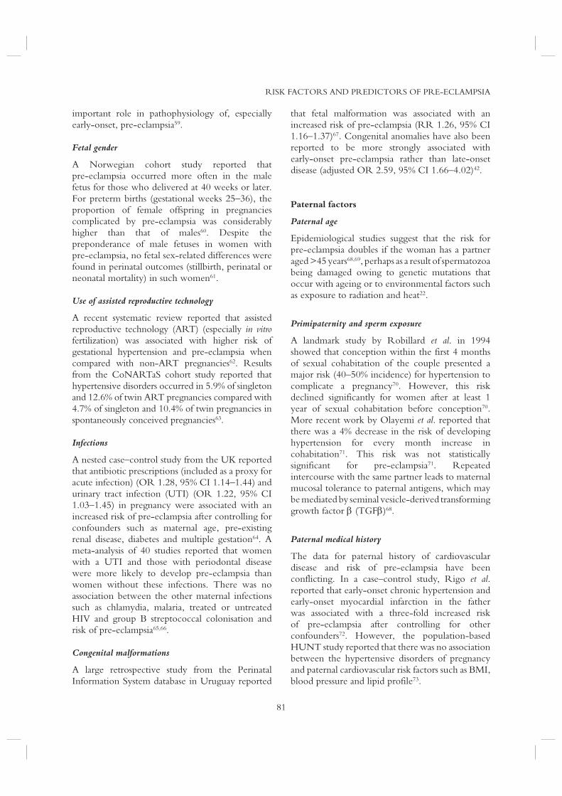
RISK FACTORS AND PREDICTORS OF PRE-ECLAMPSIA
81
important role in pathophysiology of, especially
early-onset, pre-eclampsia59.
Fetal gender
A Norwegian cohort study reported that
pre-eclampsia occurred more often in the male
fetus for those who delivered at 40 weeks or later.
For preterm births (gestational weeks 25–36), the
proportion of female offspring in pregnancies
complicated by pre-eclampsia was considerably
higher than that of males60. Despite the
preponderance of male fetuses in women with
pre-eclampsia, no fetal sex-related differences were
found in perinatal outcomes (stillbirth, perinatal or
neonatal mortality) in such women61.
Use of assisted reproductive technology
A recent systematic review reported that assisted
reproductive technology (ART) (especially in vitro fertilization) was associated with higher risk of
gestational hypertension and pre-eclampsia when
compared with non-ART pregnancies62. Results
from the CoNARTaS cohort study reported that
hypertensive disorders occurred in 5.9% of singleton
and 12.6% of twin ART pregnancies compared with
4.7% of singleton and 10.4% of twin pregnancies in
spontaneously conceived pregnancies63.
Infections
A nested case–control study from the UK reported
that antibiotic prescriptions (included as a proxy for
acute infection) (OR 1.28, 95% CI 1.14–1.44) and
urinary tract infection (UTI) (OR 1.22, 95% CI
1.03–1.45) in pregnancy were associated with an
increased risk of pre-eclampsia after controlling for
confounders such as maternal age, pre-existing
renal disease, diabetes and multiple gestation64. A
meta-analysis of 40 studies reported that women
with a UTI and those with periodontal disease
were more likely to develop pre-eclampsia than
women without these infections. There was no
association between the other maternal infections
such as chlamydia, malaria, treated or untreated
HIV and group B streptococcal colonisation and
risk of pre-eclampsia65,66.
Congenital malformations
A large retrospective study from the Perinatal
Information System database in Uruguay reported
that fetal malformation was associated with an
increased risk of pre-eclampsia (RR 1.26, 95% CI
1.16–1.37)67. Congenital anomalies have also been
reported to be more strongly associated with
early-onset pre-eclampsia rather than late-onset
disease (adjusted OR 2.59, 95% CI 1.66–4.02)42.
Paternal factors
Paternal age
Epidemiological studies suggest that the risk for
pre-eclampsia doubles if the woman has a partner
aged >45 years68,69, perhaps as a result of spermatozoa
being damaged owing to genetic mutations that
occur with ageing or to environmental factors such
as exposure to radiation and heat22.
Primipaternity and sperm exposure
A landmark study by Robillard et al. in 1994
showed that conception within the first 4 months
of sexual cohabitation of the couple presented a
major risk (40–50% incidence) for hypertension to
complicate a pregnancy70. However, this risk
declined significantly for women after at least 1
year of sexual cohabitation before conception70.
More recent work by Olayemi et al. reported that
there was a 4% decrease in the risk of developing
hypertension for every month increase in
cohabitation71. This risk was not statistically
significant for pre-eclampsia71. Repeated
intercourse with the same partner leads to maternal
mucosal tolerance to paternal antigens, which may
be mediated by seminal vesicle-derived transforming
growth factor (TGF)68.
Paternal medical history
The data for paternal history of cardiovascular
disease and risk of pre-eclampsia have been
conflicting. In a case–control study, Rigo et al. reported that early-onset chronic hypertension and
early-onset myocardial infarction in the father
was associated with a three-fold increased risk
of pre-eclampsia after controlling for other
confounders72. However, the population-based
HUNT study reported that there was no association
between the hypertensive disorders of pregnancy
and paternal cardiovascular risk factors such as BMI,
blood pressure and lipid profile73.

THE FIGO TEXTBOOK OF PREGNANCY HYPERTENSION
82
Miscellaneous factors
Smoking
Cigarette smoking is known to have adverse effects
on all organ systems. However, a systematic review
of 48 epidemiological studies reported that smoking
during pregnancy approximately halves the risk
of pre-eclampsia74. This protective effect was
consistently seen irrespective of parity and severity
of disease74. The pathophysiology of this relationship
is not well established. However, it is proposed that
smoking might have effects on angiogenic factors,
endothelial function and the immune system,
which may contribute to the lowered risk of
pre-eclampsia74. In an attempt to establish causality
between smoking and pre-eclampsia, data from the
National Swedish Birth Register showed that
smoking in two pregnancies again halves the risk of
pre-eclampsia, compared with the risk borne by
women who did not smoke in either pregnancy75.
No significant associations have been
observed between smokeless tobacco use and
pregnancy-associated hypertension in various
studies76,77. Therefore, it is proposed that
combustion products from cigarette smoke other
than nicotine may be responsible for the decreased
pre-eclampsia risk seen amongst smokers76.
Physical activity
Exercise and physical activity is recommended
during pregnancy to improve maternal health. In
their systematic review, Kasavara et al. reported that
physical activity had a protective effect on the
development of pre-eclampsia (OR 0.77, 95% CI
0.64–0.91), while this effect was not seen in cohort
studies (OR 0.99, 95% CI 0.93–1.05)78. However,
a recent meta-analysis conducted by Aune et al. reported that those women who engaged in high
levels of physical activity pre-pregnancy and
continued to do so during early pregnancy, were
less likely (by 35% and 21%, respectively) to
develop pre-eclampsia, compared with those who
participated in low levels of physical activity79.
Micronutrient deficiencies
Vitamin D deficiency is commonly reported in
women and has been investigated to assess its link
with pre-eclampsia. There have been conflicting
results regarding the serum concentrations of
25-hydroxy vitamin D and the subsequent risk of
developing pre-eclampsia80,81, mainly owing to
small sample size of these studies. A recent large
case–control study has reported that maternal
vitamin D deficiency, defined as 25-hydroxy
vitamin D <30 nmol/L, was associated with double
the risk of pre-eclampsia when compared with
concentrations >50 nmol/L82.
The Vitamins in Preeclampsia (VIP) Trial
reported that vitamin C (1000 mg) and vitamin E
(400 IU) supplements given prophylactically from
the second trimester of pregnancy have no effect
on reduction in the rate of pre-eclampsia in women
at risk83. Similar findings have been reported by the
WHO multicountry vitamin supplementation
survey from India, South Africa and Vietnam84.
Mental health
Depression and anxiety in the first trimester of
pregnancy are known to increase the risk of
pre-eclampsia by two- to three-fold85. In addition,
lifetime stress and perceived stress during pregnancy
may double the risk of developing pre-eclampsia;
an interaction that may be mediated by the
neuropsychoimmunological pathway86.
Socioeconomic status
In developing countries, rural dwellers were twice
as likely to develop pre-eclampsia compared with
those living in urban areas. Furthermore, women
with concurrent anaemia and poor intake of fruits
and vegetables were at higher risk of pre-eclampsia87.
Severe anaemia (haemoglobin <70 g/L) was
associated with a three-fold greater risk of
pre-eclampsia in women living in less-developed
countries88. A lack of antenatal care and less than
secondary-level education were pertinent risk
factors for risk of pre-eclampsia in these regions88.
PREDICTION (APPENDICES 5.1–5.3)
At present, maternal characteristics which include
well-established risk factors discussed above such
as maternal age, nulliparity, pre-existing medical
conditions and history of pre-eclampsia, are
mostly used to screen for pre-eclampsia by clinicians
during antenatal visits56,57,89. However, these risk
factors are not sufficient as only approximately 30%
of women who subsequently develop pre-eclampsia
are identified by their use90. Pre-eclampsia research
is now tailored towards development of a predictive
model utilising the risk factors mentioned above

RISK FACTORS AND PREDICTORS OF PRE-ECLAMPSIA
83
along with measurable clinical and laboratory
biomarkers to predict the onset of pre-eclampsia.
In the context of this chapter, we are talking
about the prediction of a diagnosis of pre-eclampsia
(or other placental complications) occurring at
some point in the future, not the prediction of
complications (prognosis), or risk stratification, in
either individual or populations of women whose
pregnancies have been complicated by the clinical
syndrome of pre-eclampsia (the focus of much of
the Chapter 3). According to WHO, a prediction
test should be simple, non-invasive, inexpensive,
rapid, easy to carry out early in gestation, impose
minimal discomfort or risk on the woman, be a
widely available technology, and the test results
must be valid, reliable and reproducible91,92.
The performance of predictive tests is generally
summarised in the text and tables as being poorly
associated, moderately associated and strongly
associated when the positive likelihood ratio (LR+)
is <5, 5–9.9 and 10, respectively (Appendix 5.1).
Similarly, for tests that poorly, moderately or
strongly exclude risk, their performance is
summarised as negative likelihood ratios (LR-) of
>0.2, 0.11–0.2 and 0.1, respectively. Other
summary statistics used in this chapter are the
sensitivity (“true positive rate”, the proportion of
positives that are correctly identified as such, e.g.,
the percentage of women who will develop the
complication who are correctly identified) and
specificity (“true negative rate”, the proportion of
negatives who are correctly identified as such, e.g.,
the percentage of women who are correctly
identified as not developing the condition) of the
test to predict the outcome, namely pre-eclampsia,
as well as the area under the receiver-operator
characteristic curve (AUC ROC)93–95.
It should be remembered that nearly all the studies
referred to in this section relate to women in
more-developed countries. Their relevance to
women in less-developed countries is uncertain. It is
women in less-developed countries who carry the
greatest burden of risk for the complications of
pre-eclampsia.
PREDICTORS (UNIVARIABLE ANALYSES)
Clinical examination
Blood pressure
Blood pressure, which forms the basis of diagnosis
for pre-eclampsia in all international guidelines, is
routinely measured during pregnancy89. The
Society of Obstetrics and Gynaecologists of Canada
(SOGC) recommends measurement of blood
pressure using a mercury sphygmomanometer, a
validated automated blood pressure device or a
calibrated aneroid device56,57. As high blood
pressure is an indication of the increased vascular
resistance observed in pre-eclampsia, there have
been studies examining the value of blood pressure
measurements using systolic blood pressure,
diastolic blood pressure, or MAP indices for the
prediction of pre-eclampsia96–99.
A systematic review and meta-analysis by
Cnossen et al. evaluated the predictive accuracy of
using blood pressure measurements in the second
trimester for pre-eclampsia. This review included
34 studies reporting the use of blood pressure
measurements (systolic blood pressure, diastolic
blood pressure and MAP) in predicting
pre-eclampsia for women at low risk96. The pooled
LRs were weakly associated with developing
pre-eclampsia. The authors concluded that no
index of blood pressure measurement predicted
pre-eclampsia well enough to be clinically useful.
Urine
Proteinuria
Proteinuria is routinely measured during pregnancy,
especially in women with new-onset hypertension
occurring after 20 weeks’ gestation to establish the
diagnosis of pre-eclampsia56,57 (see Chapter 2).
Underlying renal disease is a recognised clinical risk
factor for pre-eclampsia and as such, documentation
of proteinuria early in pregnancy is associated with
an increased risk of pre-eclampsia (see Pre-existing
medical conditions, above). Recently, significant
attention has been devoted to the role of
albuminuria, and more specifically for lower levels
of albuminuria (or ‘microalbuminuria’) for the
prediction of pre-eclampsia. In a review of the
published studies retrieved from a structured
literature search (1980 to mid-March 2008), a total
of seven studies were performed in early pregnancy
(defined as <20 weeks) and 13 studies in late
pregnancy (20 weeks)100. Overall, the negative
predictive value of ‘microalbuminuria’ was high
but the test performance was not good enough for
clinical use, which is consistent with most other
individual prediction tests described in this section.
The largest study (N = 2486 women) performed at

THE FIGO TEXTBOOK OF PREGNANCY HYPERTENSION
84
11+0–13+6 weeks demonstrated an increased
albumin : creatinine ratio in women who later
developed pre-eclampsia compared with those
who did not; however, the combined prediction
models incorporating the albumin : creatinine ratio
results did not yield to significantly improved
AUCs over maternal variables alone101. Prediction
of pre-eclampsia in early pregnancy (17–20 weeks)
by estimating the albumin : creatinine ratio was
also performed using high-performance liquid
chromatography (HPLC)102. In this cohort of 265
women with singleton pregnancy, six developed
pre-eclampsia; the AUC to predict pre-eclampsia
was 0.753. Although the interpretation is of a good
predictive test, this study has not been replicated
and, in addition, the impact is limited by accessibility
to HPLC in clinical practice, especially in
less-resourced settings.
Podocyturia (podocyte : creatinine ratio)
Glomerular epithelial cells (podocytes) are involved
in the maintenance of the function and structure
of the filtration barrier in the kidney103. As a
consequence of endothelial dysfunction and
disruption of the selective filtration barrier in the
kidney associated with pre-eclampsia, these
podocytes proteins which include podocin,
nephrin, synaptopodin and podocalyxin, lose their
functional ability and are shed in urine (i.e.,
podocyturia)104,105. Podocyturia is expressed as
podocytes : creatinine ratio and has been shown to
be associated with manifestation of renal dysfunction
in women with pre-eclampsia103.
A case–control study by Kelder et al. analysed
maternal urine mRNA levels of three markers
of podocytes (VEGF, nephrin and podocin)
using quantitative polymerase chain reaction
(qPCR-based analysis)103. The urine measurements
were collected in the early third trimester. None of
the three podocyte markers were strong predictors
of pre-eclampsia independently, but a combination
of all the markers showed a moderate performance
in predicting the occurrence of pre-eclampsia.
Craici et al. examined the predictive accuracy of
podocyturia in the second trimester using only
podocin as a marker in a prospective cohort
study105. In contrast to the study by Kelder et al., this study reported 100% sensitivity (95% CI
78–100) and 100% specificity (95% CI 92–100) in
predicting pre-eclampsia, using podocin staining of
blood and urine samples. In addition, this study
reported a strong LR+ for predicting the occurrence
of any hypertensive disorder of pregnancy.
Another prospective study carried out by Jim
et al. examined the predictive accuracy of
podocyturia (using podocin), nephrinuria and
albuminuria in the second and third trimesters from
urine samples using cytospin technique104. Only
albuminuria in this study showed a moderate LR+
for the prediction of pre-eclampsia.
The differences in the predictive accuracy for
podocin and nephrin in the three studies above
may be owing to different analytic techniques,
prevalence of pre-eclampsia, and population (e.g.
high-risk women versus unspecified). The study
designs and gestational age also differed in the
studies. Except in the study by Craici et al., none of
the urine markers attained the required predictive
LR values for clinical use. Further research is
needed to make conclusive statements on the use of
podocyturia as a screening test for pre-eclampsia.
Calcium (calcium : creatinine ratio)
As a result of renal dysfunction (decreasing
glomerular filtration rate) which occurs in
pre-eclampsia, there is an increase in serum
creatinine and decrease in calcium, thus a decrease
in calcium : creatinine ratio has been reported in
some studies106. Vahdat et al. studied the predictive
accuracy of urine calcium : creatinine ratio of 150
women during late second trimester. Using a cut-off
value of 0.071 in this study, calcium : creatinine
ratio was a poor predictor for pre-eclampsia.
Inositol phosphoglycan-P (IPG : creatinine ratio)
Inositol phosphoglycan-P type (IPG-P) which
belongs to the insulin mediator family has been
reported to be high in urine in pre-eclampsia107. A
prospective longitudinal study investigated the use
of IPG-P : creatinine ratio as a predictive screening
test for pre-eclampsia 2 weeks prior to its onset in
416 women. IPG-P : creatinine ratio had moderate
LR- and LR+ and may become a useful screening
test for pre-eclampsia up to 2 weeks before diagnosis.
Ultrasound markers
Uterine artery Doppler ultrasonography
Doppler ultrasound is a non-invasive technique,
and, in this setting, is used to study the uteroplacental

RISK FACTORS AND PREDICTORS OF PRE-ECLAMPSIA
85
circulation and changes in blood flow resistance108.
The flow change can be measured as pulsatility
index (PI) or resistance index (RI)108,109.
As an uncomplicated pregnancy progresses,
blood flow resistance in the uterine arteries
decreases with gestation owing to invasion of the
spiral arteries by the trophoblasts109–111. The
corollary is that increased impedance to blood flow
in the uterine arteries has been observed in
pregnancies complicated by impaired trophoblast
invasion of the spiral arteries, as occurs with
placental pre-eclampsia and IUGR of placental
origin111.
The change in uterine artery blood flow between
the first and second trimesters has been examined
by screening studies to identify pregnancies at risk
of pre-eclampsia and fetal growth restriction109.
The increase in impedance in the uterine arteries is
more reflective of preterm pregnancy complications
than those at term, as poor placentation is more
associated with early-onset pre-eclampsia110,111.
Abnormal uterine artery Doppler velocimetry
may be defined as bilateral notching with or no
notching with mean resistance index (RI) >0.70
(>95th centile), mean RI >0.55 (i.e., >50th
centile), or unilateral notching with mean RI
>0.65 (>90th centile), at 22–24 weeks56,57.
A few studies have examined abnormal uterine
artery resistance during the first and second
trimesters for prediction of pre-eclampsia108,112.
Using a case–control design, Bolin et al. measured
uterine artery PI in the first and second trimesters as
part of routine antenatal screening109. Uterine
artery PI was expressed in multiples of median
(MoM) values and the predictive accuracy for
preterm pre-eclampsia was assessed. The uterine
artery PI had a poor predictive ability for identifying
women at risk of preterm pre-eclampsia.
However, a retrospective observational study by
Napolitano et al. evaluated uterine artery Doppler
PI as a predictor of early-onset and preterm
pre-eclampsia in the first and second trimesters112.
The uterine artery PI was adjusted for gestational
age and the PI MoM ratio between the second and
first trimesters (uterine artery ratio) was compared
with the PI MoM mean difference between the
second and the first trimesters (uterine artery
difference). For the prediction of early-onset
pre-eclampsia, the AUC ROC values were 0.786
(95% CI 0.703–0.869) for the uterine artery ratio
and 0.851 (95% CI 0.753–0.950) for the mean
uterine artery difference. For the prediction of
preterm pre-eclampsia, the AUC ROC of the
uterine artery ratio and mean uterine artery
difference were 0.701 (95% CI 0.626–0.776) and
0.705 (95% CI 0.599–0.812), respectively. The
study concluded that the mean uterine artery
difference was the best index for predicting
pre-eclampsia and a better predictor of early-onset
pre-eclampsia.
Two reviews examining Doppler studies as an
individual predictor of pre-eclampsia were found.
The review by Papageorghiou et al. investigated
the use of uterine artery Doppler measurement in
the second trimester for the prediction of
pre-eclampsia using findings from 15 studies108.
The sensitivities reported in these studies ranged
from 26 to 89% and the specificities ranged from 86
to 96%. The pooled LR+ and LR- were 5.9 and
0.55, respectively, suggesting that second trimester
Doppler measurement had a moderate predictive
value for pre-eclampsia. However, the studies
included in this review differed in Doppler
techniques, definition of abnormal flow velocity
and pre-eclampsia, populations and disease
incidence.
The latter review by Cnossen et al. evaluated the
accuracy of uterine artery Doppler for predicting
pre-eclampsia in low- and high-risk women96.
Seventy-four studies that reported uterine artery
Doppler data in the first and/or second trimesters
were included in the review. The review concluded
that uterine artery Doppler velocimetry was more
accurate in second trimester for prediction than in
the first trimester and PI with notching had the best
predictive accuracy for pre-eclampsia in both high-
and low-risk women. Again, the review was limited
by the different Doppler indices used by the
included studies.
In conclusion, uterine artery Doppler PI may be
a moderate predictor of pre-eclampsia and may be
used to ‘rule in’ pre-eclampsia risk in the second
trimester. However, owing to inconsistencies
reported in the studies, further studies are required.
Laboratory markers
The markers of pre-eclampsia risk that become
available in the second and third trimesters are
based on the pathophysiological changes that
characterise pre-eclampsia and precede clinical
disease. Many have been evaluated, and they

THE FIGO TEXTBOOK OF PREGNANCY HYPERTENSION
86
include measures of the following: placental
perfusion and vascular resistance (e.g., mean second
trimester blood pressure, 24-hour ambulatory
blood pressure monitoring, Doppler ultrasound);
cardiac output and systemic vascular resistance;
fetoplacental unit endocrinology (e.g.,
pregnancy-associated plasma protein-A (PAPP-A)
and placental growth factor (PlGF) in the first
trimester, and alpha fetoprotein, hCG and inhibin
A in the early second trimester); renal function
(e.g., serum uric acid or microalbuminuria);
endothelial function and endothelial–platelet
interaction (e.g., platelet count, antiphospholipid
antibodies, or homocysteine); oxidative stress
(e.g., serum lipids); and circulating pro- and
anti-angiogenic factors113,114.
There have been many systematic reviews of
primary studies evaluating clinically available
biomarkers as well as Doppler ultrasound
interrogation of the uterine and umbilical
arteries115–117. Tests have been chosen for study
based on their association with adverse pregnancy
outcomes, including pre-eclampsia. Notable
examples are serum analytes involved in maternal
serum screening for trisomy 21 (4 studies, 427,751
women)118, serum uric acid measured before 25
weeks (5 studies, 572 women)119, and Doppler
ultrasonographic interrogation of the uterine artery
(74 studies, ~80,000 women)96. The methodological
quality of the primary studies is very variable,
related to women enrolled, gestational age at
testing, test performance (such as different Doppler
sampling techniques or definitions of abnormal
flow velocity waveform), and criteria for the
diagnosis of pre-eclampsia.
As markers of the fetoplacental unit are
commonly used for trisomy 21 screening, it has
been proposed that these markers be used for
pre-eclampsia risk estimation, in combination with
clinical markers. It will be necessary to evaluate
whether maternal serum screening for the sole
purpose of pre-eclampsia screening, in combination
with clinical markers and possibly uterine artery
Doppler, leads to improved outcomes. For the
moment, further studies are needed before
widespread clinical use of serum screening for
pre-eclampsia can be advocated, either in high- or
low-risk populations. In addition, it must be
acknowledged that with the development of
non-invasive prenatal testing120, the use of maternal
serum screening might soon become obsolete.
Endothelial dysfunction tests/placental proteins
High-sensitivity C-reactive protein (hs-CRP)
hs-CRP is a systemic inflammatory marker which
is produced by the placenta and released into the
maternal circulation121. This marker can be found
in fetal urine and amniotic fluid, and is sensitive
to inflammation and tissue damage. Studies have
reported an observed increase in maternal hs-CRP
level in pre-eclampsia and other adverse pregnancy
outcomes. Kashanian et al. conducted a prospective
cohort study of 394 women evaluating the
predictive accuracy of serum hs-CRP for
pre-eclampsia in the first trimester121. The result
from this study show poor LRs of hs-CRP for the
prediction of pre-eclampsia.
Fibronectin
Fibronectin, which is released by the placenta,
is associated with endothelial damage and
inflammation in pre-eclampsia. Higher plasma
levels of fibronectin have been reported in women
with pre-eclampsia compared to uncomplicated
pregnancies leading to research on its predictive
ability for pre-eclampsia. A systematic review by
Leeflang et al. evaluated the predictive ability of
fibronectin in five studies122. These studies measured
total and/or cellular fibronectin in the first or
second trimesters. Fibronectin had a pooled
moderate LR+ and, therefore, may be a useful test
for predicting pre-eclampsia.
Angiogenic factors
Placental growth factor
PlGF, which is a member of the vascular endothelial
growth factor (VEGF) family, is a pro-angiogenic
factor produced by the syncytiotrophoblast123,124.
PlGF is at lower maternal circulating concentrations
at time of disease with pre-eclampsia, compared
with normal pregnancy123–125. PIGF assessment
point-of-care platforms currently are available3,126,127.
Ghosh et al. evaluated maternal serum PlGF as a
predictive test in the second trimester for predicting
early-onset pre-eclampsia in a prospective cohort
of 722 women123. In this study, PlGF was poorly
associated with pre-eclampsia as a predictive test.
Another study by Ghosh et al. compared serum
PlGF measurements in the first trimester with
measurements in the second trimester for predicting

RISK FACTORS AND PREDICTORS OF PRE-ECLAMPSIA
87
early-onset pre-eclampsia128. Although PlGF
performed better in the second trimester compared
with the first trimester measurements, both
performances were poor for the prediction of
pre-eclampsia, as confirmed in an independent
cohort of high-risk women in their second trimester
to predict early-onset pre-eclampsia128,129.
In all the aforementioned studies by Ghosh et al., PlGF concentrations were measured by enzyme
linked immunosorbent assay (ELISA) technique
using the DRG PlGF Enzyme Immunoassay
Kit and early-onset pre-eclampsia was defined
as pre-eclampsia diagnosed by 32 weeks’
gestation123,128,129.
Chappell et al. assessed PlGF in 625 women in
their second and third trimester for the prediction
of pre-eclampsia with delivery within 14 days125. In
this prospective cohort multicentre study, plasma
PlGF concentration was measured using the Alere
Triage® assay. Using a cut-off of PlGF below the
5th centile, PlGF was strongly associated with a
negative likelihood of pre-eclampsia with delivery
within 14 days of diagnosis.
There are notable differences in the predictive
performance and quantification methods of PlGF
and in the incidence of pre-eclampsia among these
studies investigating PlGF as an independent
predictor of pre-eclampsia.
Soluble fms-like tyrosine kinase 1 (sFlt-1 : PlGF ratio)
sFlt-1 is an anti-angiogenic factor produced by the
placenta. It antagonises the activities of VEGF and
PlGF by binding to them90,124. This results in
reduction of the free circulating levels of VEGF
and PlGF, as observed in women with pre-eclampsia.
Some studies have reported that the sFlt-1 : PlGF
ratio can be used to identify patients at risk of
pre-eclampsia90,130.
One of these studies was conducted by Engels
et al. and measured serum samples of sFlt1 : PlGF
ratio using automated Elecsys system and assessed
its utility in the second and third trimester for
predicting pre-eclampsia and HELLP syndrome130.
Compared with PlGF alone or sFlt1 alone,
sFlt1 : PlGF ratio gave the best predictive accuracy
for pre-eclampsia and was strongly associated with
a positive likelihood of developing pre-eclampsia.
Teixeira et al. investigated the predictive value
of PLGF, sFlt-1 and sFlt-1 : PLGF ratio in a
prospective longitudinal study131. Maternal plasma
concentrations were measured in 71 high-risk
women using a commercial kits (R&D Systems) in
their second trimester. In this study, the
sFlt-1 : PLGF ratio had a better discriminative
ability (AUC ROC 0.95) for the prediction of
pre-eclampsia compared with only PlGF (AUC
ROC of 0.90) or sFlt-1 (AUC ROC 0.78).
Another prospective study evaluated serum
sFlt-1 : PlGF ratio as a predictor of pre-eclampsia
in high-risk women132. Blood samples were
measured in the second and third trimester using
electrochemiluminescence technology (Roche). The
third trimester sFlt-1 : PlGF ratio performed better
than second trimester and was a moderately associated
with a negative likelihood for pre-eclampsia.
A nested case–control study by Forest et al. evaluated the serum sFlt-1 : PlGF ratio in the second
and early third trimesters133. sFlt-1 was measured by
ELISA using the Quantikine Human Immunoassay
(R&D Systems) and PlGF was measured using
an automated immunoassay analyzer (DELFIA
System, PerkinElmer ). The sFlt-1 : PlGF ratio was
moderately well associated with later early-onset
pre-eclampsia. This contrasts with a similar nested
case–control study evaluating serum sFlt-1 : PlGF
ratio measured in the late second trimester using
the R&D Systems assay and which reported strong
associations for predicting early-onset pre-eclampsia
in high risk women134.
Except for a study by McElrath et al.124, all other
included studies in this chapter support growing
evidence that sFlt1 : PlGF ratio has good potential
as a predictive test for pre-eclampsia in the third
trimester, especially in high risk women. The study
by McElrath et al. quantified plasma sFlt-1 and
PlGF concentrations measured in the second
trimester using the Abbott Diagnostics’ platform
and showed poor association for the prediction of
pre-eclampsia124. The contradicting results from
this study may have been owing to the use of a
different measurement platform and the use of
PlGF : sFlt-1, in contrast to the sFlt1 : PlGF ratio
used in other studies.
PREDICTING PRE-ECLAMPSIA (MULTIVARIABLE ANALYSES)
No single clinical, blood or ultrasonographic test
reaches the ideal of 90% sensitivity and specificity
for the prediction of pre-eclampsia. Only Doppler
ultrasound (i.e., any or unilateral notching and/or

THE FIGO TEXTBOOK OF PREGNANCY HYPERTENSION
88
elevated RI) has a sensitivity >60%, particularly
when testing is performed in women at increased
risk (vs. low risk) of developing pre-eclampsia, in
the second (vs. the first) trimester, and for predicting
severe and early-onset pre-eclampsia (rather than
milder forms of the disease).
Therefore, as there is no single test that predicts
pre-eclampsia with sufficient accuracy to be useful
clinically91, interest has grown in the development
of multivariable models that include both clinical
and laboratory predictors, available at booking and
thereafter in pregnancy135.
The largest relevant studies have been performed
by investigators at King’s College, London, UK101.
For example, at 11–14 weeks, a combination of
MAP, uterine artery PI, PAPP-A and PlGF was
able to identify 93% of early-onset pre-eclampsia,
36% of late-onset pre-eclampsia, and 18% of
gestational hypertension such that 20% of women
identified as being screen positive developed a
hypertensive disorder of pregnancy; this is consistent
with other studies136.
In specialised clinics, women at increased risk of
pre-eclampsia may benefit from this type of risk
stratification followed by specific preventative
intervention(s); however, this is yet to be proven.
Similarly, screening of nulliparous or otherwise low
risk women is not yet recommended. Prospective
longitudinal studies are needed to assess the validity
of published observations in other patient
populations where some models have performed
differently137. Future studies should also distinguish
the ability of screening approaches to predict
pre-eclampsia that is more severe or that which
occurs early (vs. at term). Clinicians are encouraged
to support clinics investigating predictive models.
In the SCOPE (Screening for Pregnancy
Endpoints) Consortium cohort138, nine clinical
predictors of (almost exclusively, late-onset)
pre-eclampsia (many of which were identified by
PRECOG139 and NICE 2008140) were identified
among nulliparous women carrying singleton
pregnancies: one protective (miscarriage at 10
weeks’ gestation with the same partner) and eight
associated with increased risk (younger maternal
age, higher mean arterial blood pressure, higher
BMI, family history of pre-eclampsia, family history
of coronary heart disease, woman with lower birth
weight, vaginal bleeding during early pregnancy
and short duration of sexual relationship). Of note,
the performance of this model was not enhanced
by knowledge of uterine artery Doppler results.
Using the model, which remains to be validated,
the probability of pre-eclampsia would increase
from 5 to 10% and half of women who go on to
develop pre-eclampsia would be detected.
Clinical history/maternal characteristics
The most common clinical risk factors associated
with pre-eclampsia include first pregnancy/
primigravidity, maternal age, diabetes, history of
pre-eclampsia and other hypertensive disorders of
pregnancy, family history of pre-eclampsia, and
long inter-pregnancy interval56,57,89,90. Women with
a history of pre-eclampsia are at increased risk
(16–65%) of developing pre-eclampsia in a
subsequent pregnancy depending on the onset or
severity of the disease in the previous pregnancy140.
The risk of recurrence for women who had
pre-eclampsia is approximately 25% for women
who also had HELLP syndrome, about 55% for
those who had preterm delivery (<28 weeks)140 and
approximately 65% for women who had early-onset
pre-eclampsia141. In addition, the increased risk for
developing pre-eclampsia for women with a history
of gestational hypertension ranges from 2 to
9%140,141 and the reported RR of pre-eclampsia for
a woman with a history of chronic or gestational
hypertension is RR 8.9, CI 5.7–13.8 and RR 9.8,
CI 4.9–19.1, respectively141.
However, only a few studies have reported the
predictive abilities of the clinical factors for
pre-eclampsia, most often in combination with
other potential predictive markers as clinical
risk factors are not very useful predictors
individually90.
Maternal characteristics with biomarkers (placental
protein 13, PAPP-A and free beta subunit of hCG)
Placental protein (PP)-13, PAPP-A and free beta
subunit of hCG (-hCG) are produced and secreted
by the syncytiotrophoblast and are involved in
implantation, trophoblast invasion and remodelling
of the spiral arteries97,142,143. In healthy pregnancies,
there is an increase in PP-13 and free -hCG from
the first trimester which decreases with gestation,
whereas in pre-eclampsia, PP-13 and free -hCG
are reportedly lower in the first trimester, but

RISK FACTORS AND PREDICTORS OF PRE-ECLAMPSIA
89
significantly higher in the second and third trimesters.
Low concentration of PAPP-A in the first semester
has also been reported to be associated with
pregnancy complications97,142. The potential role of
these placental proteins as a predictor of pre-eclampsia
was assessed in a prospective cohort by Schneuer
et al.142. Serum samples in the first trimester in
combination with maternal characteristics (previous
hypertension, parity, weight and age) and other
biomarkers (-hCG, PAPP-A) using multivariate
models showed a moderate LR+ association for the
prediction of early-onset pre-eclampsia.
Maternal characteristics with MAP
In a prospective screening study of 17,383 cases,
Gallo et al. combined maternal characteristics
(gestational age at screening, maternal weight and
height, Afro-Caribbean racial origin, family history
of pre-eclampsia, prior personal history of
pre-eclampsia, cigarette smoking and chronic
hypertension) with MAP measured in the first and
second trimesters98. The model predicted
pre-eclampsia with moderate test performance.
Maternal characteristics with biomarkers (serum
PIGF and free -hCG)
First trimester maternal serum PIGF, free -hCG
and maternal history were evaluated for the
prediction of pre-eclampsia in a prospective cohort
study of 2118 women144. Serum blood
concentrations of PlGF and free -hCG were
quantified by DELFIA Xpress (Perkin Elmer) and
adjusted for gestational age and maternal BMI. The
multivariate model with serum PlGF, free -hCG
and chronic hypertension had a moderate LR+ for
predicting early-onset pre-eclampsia.
Maternal characteristics with MAP and biomarkers
(hyperglycosylated human chorionic gonadotrophin
and PAPP-A)
A nested case–control study developed a regression
model combining parity, MAP and first trimester
hyperglycosylated hCG (hCG-h) and PAPP-A for
the prediction of early-onset pre-eclampsia145. The
study reported a moderate performance of the
model for predicting the development of early-onset
pre-eclampsia.
Maternal characteristics with MAP and biomarkers
(taurine, PAPP-A, ADAM12 and PlGF)
Kuc et al. studied the utility of taurine, an amino
acid which is involved in trophoblast invasion, the
levels of which are altered at the time of disease
with pre-eclampsia, in combination with MAP,
maternal characteristics (parity, weight) and other
biomarkers (PAPP-A, ADAM12 and PlGF) using
a multiple logistic regression model97. Maternal
serum samples were collected in the first
trimester in 667 women in the nested
case–control study. Early-onset pre-eclampsia
with small-for-gestational age (SGA) was predicted
moderately with the multivariate model with a
strong LR-. A second model developed by in
another case–control study showed moderate
performance for the prediction of only early-onset
pre-eclampsia143.
Maternal characteristics with uterine artery PI
A review by Kleinrouweler et al. evaluated the
value of adding second trimester uterine artery
Doppler to patient characteristics in identification
of nulliparous women at high risk for
pre-eclampsia111. A logistic regression model
combining blood pressure, uterine artery and
bilateral uterine artery notching showed a good
discriminatory predictive performance with an
AUC of 0.85 (95% CI 0.67–1.00) in the study.
In another study, combining uterine artery PI
expressed as log10 MoM and maternal characteristics
(weight, height, race, parity and chronic
hypertension) used in the early third trimester
showed moderate association for predicting
late-onset pre-eclampsia146.
Maternal characteristics with blood pressure and
uterine artery PI
Prediction of early onset pre-eclampsia in a
prospective multicentre cohort study in 627
women was evaluated using demographic, clinical
and ultrasound data in the first trimester99. The
multivariate model included age, weight, systolic
blood pressure, diastolic blood pressure and MAP
at enrolment, parity, history of pre-eclampsia or
hypertension, diabetes mellitus, log (uterine artery
PI) and a history of preterm labour. The model

THE FIGO TEXTBOOK OF PREGNANCY HYPERTENSION
90
strongly predicted the development of early-onset
pre-eclampsia in the development study. The
performance of the model was moderate on
external validation by Oliveira et al. in a prospective,
observational study of 2669 women recruited in
their first trimester147.
Maternal characteristics with uterine artery Doppler
and biomarkers (ADAM12 and PAPP-A)
In a prospective cohort study, Goetzinger et al. assessed the accuracy of a multivariate model
combining first trimester bilateral uterine artery
notching and PAPP-A, and maternal characteristics
(chronic hypertension, history of pre-eclampsia,
pre-gestational diabetes, obesity) for the prediction
of pre-eclampsia148. The model was developed in
one-half of a prospective cohort of 1200 patients
in first trimester and validated in the second-half.
The validated model had a moderate predictive
accuracy for pre-eclampsia. It is worth mentioning
that the split-half method of validation used has
been reported to have some major drawbacks in
prediction modelling.
Maternal characteristics with MAP and biomarkers
(PlGF)
PlGF combined with MAP and maternal
characteristics (a sister with a history of pre-eclampsia
and a history of previous fertility treatment) were
assessed for predictive accuracy for preterm
pre-eclampsia in low-risk nulliparous women149. In
the prospective multicentre cohort study, plasma
PlGF was measured in second trimester using the
triage assay. The model predicted pre-eclampsia
with moderate performance.
Maternal characteristics with uterine artery PI and
biomarkers (PlGF)
Serum concentrations of PlGF collected during the
first, second and third trimesters of pregnancy and
quantified using R&D were assessed in a case–
control study of 541 women for the prediction of
pre-eclampsia150. In a logistic regression model
combining relative difference of PlGF from the
first to the second trimester with BMI, second
trimester uterine artery PI was a moderate predictor
of pre-eclampsia.
Other multivariate studies
Circulating proteins and angiogenic factors
In a prospective study by Katsipi et al., second
trimester measurements of pulse wave velocity
(PWV), which is a measure of aortic stiffness, was
combined with serum levels of sFlt-1 in a regression
model151. The model was a strong predictor of
pre-eclampsia in high-risk pregnant women.
Park et al. assessed the predictive accuracy of the
sFlt-1 : PlGF ratio (measured in the second and
third trimesters (Roche Elecsys)) in combination
with PAPP-A for late-onset pre-eclampsia in low
risk women90. In this study, the third trimester
sFlt-1 : PlGF ratio had a better predictive accuracy
than the second trimester ratio and was a strong
predictor of developing late-onset pre-eclampsia in
low-risk women90.
Myatt et al. combined first-trimester
measurements of biomarkers (using Luminex
assays152) with risk factors in an observational study
in 2434 nulliparous women at low risk137. The best
multivariable model included African-American
race, systolic blood pressure, BMI, education level,
ADAM-12, PAPP-A and PlGF, but performed
poorly in predicting pre-eclampsia.
In a prospective cohort of 235 women,
second-trimester uterine artery Doppler, serum
biomarkers and lipid-related markers were
evaluated for the prediction of pre-eclampsia152.
The final model included maternal age, nulliparity,
bilateral uterine artery notch, PlGF, sFlt-1, leptin
and triglycerides, and was a poor predictor of
pre-eclampsia in the high-risk cohort.
Histidine-rich glycoprotein (HRG), a
multi-domain protein which has both pro- and
anti-angiogenic properties, was studied as a
predictor for preterm pre-eclampsia in combination
with uterine artery pulsatility index (expressed as
MoM) in a case–control study of 175 women109.
The multivariate model showed a moderate LR-
for predicting preterm pre-eclampsia.
In conclusion, most of the multivariate models
combining maternal characteristics or biomarkers
with other variables were either poor predictors of
pre-eclampsia or did not give sufficient information
to confirm the predictive ability of the test. Only a
few seemed promising, especially those combining
angiogenic factors with some other markers151,155.

RISK FACTORS AND PREDICTORS OF PRE-ECLAMPSIA
91
BEST PRACTICE POINTS
(Please see Appendix 5.5 for the evaluation of the strength of evidence.)
1. Women should be screened for clinical risk markers of pre-eclampsia from early pregnancy.
2. Consultation with an obstetrician or an obstetric internist/physician should be offered to women
with a history of previous pre-eclampsia or another strong clinical marker of increased pre-eclampsia
risk, particularly multiple pregnancy, antiphospholipid antibody syndrome, significant proteinuria at
booking, or a pre-existing condition of hypertension, diabetes mellitus, or renal disease.
3. Screening for non-clinical risk markers cannot be recommended routinely at present for women at
low or increased risk of pre-eclampsia until such screening has been shown to improve pregnancy
outcome.
WHAT INTERNATIONAL GUIDELINES SAY (APPENDIX 5.4)
Abbreviations for Clinical Practice Guidelines are as
follows ACOG (American College of Obstetricians
and Gynecologists)155, NICE (National Institutes of
Clinical Excellence)140, SOGC (Society of
Obstetricians and Gynaecologists of Canada)56,57,
AOM (Association of Ontario Midwives)156.
In a systematic review of international clinical
practice guidelines on the hypertensive disorders of
pregnancy89, only three (ACOG, AOM, SOGC)
out of 13 guidelines gave recommendations for
the screening or prediction of pre-eclampsia or
other hypertensive disorders of pregnancy.
Well-established clinical risk markers such as
medical history were the only recommended
markers for screening. None of the guidelines
recommended the use of ultrasonography or
biomarkers; however, two guidelines (NICE,
SOGC) suggested that a combination of these tests
with clinical risk markers may be useful but require
further studies to make sufficient conclusions.
SUMMARY
The ability to predict pre-eclampsia will facilitate
early recognition of the disease, risk stratification
and better management of these women to prevent
associated severe complications, whilst making
optimum use of limited resources93,94. In addition,
predicting pre-eclampsia may provide more
clarification of the pathogenesis and mechanisms
involved in pre-eclampsia and might result in
strategies for developing better prophylactic
interventions and treatment92. There is need for
large studies to validate the clinical value of these
predictors and models before they can be applicable
in clinical care for the prediction of pre-eclampsia.
PRIORITIES FOR FUTURE RESEARCH
Key priorities when conducting research on
predicting pre-eclampsia include:
• Large prospective studies with adequate sample
sizes as many of the studies reviewed in this
chapter had small sample sizes with very low
rates of pre-eclampsia.
• Studies reporting predictive accuracy according
to disease onset and population risk will be
beneficial in risk assessment and screening so as to
allocate interventions to women who need them
most. More focus should be targeted at predicting
pre-eclampsia in the first trimester so that
prophylactic interventions can be commenced.
• Standardisation of definitions and analytical
methods will be useful for comparison and
meta-analysis of results from prediction studies.
• Multivariate models need to be validated
externally before they can be used in clinical
practice. Only two studies among all the studies
with multivariate models mentioned external
validation99,148. Research has shown that the
performance of a model can be overoptimistic
when assessed in the same population used for
building the model owing to overfitting157,158.
Assessing the validity of these models in other
population needs to be carried out to assure
validity and reliability of predictive performance.
• There is a large knowledge deficit related to risk
prediction for women in less-developed countries.
An urgent priority is to diminish this deficit.
PRIORITIES IN UNDER-RESOURCED SETTINGS (TABLE 5.2)
Delays in disease identification and treatment are
major contributing factors to the increased burden

THE FIGO TEXTBOOK OF PREGNANCY HYPERTENSION
92
of the hypertensive disorders of pregnancy in
LMICs159. Pre-eclampsia is a heterogeneous
condition with different phenotypes. Future
research is required to identify the risk factors and
disease presentation for pre-eclampsia in low-income
settings, which may differ from that in high-resourced
settings, to allow for early interventions. Also, there
might be need for earlier screening and initiation of
preventative treatments in LMICs owing to the
severity of outcomes in these settings.
A shortage of well-trained health professional
and financial costs remain a significant risk burden
for women in LMICs160. Therefore, for any
predictive test to be beneficial in such settings, it
should be a cheap, easy-to-use, point-of-care test
that requires minimal training. Potential
predictive tests that quickly measure angiogenic
imbalance and glycosylated fibronectin during
pregnancy are now available; however, further
research is required to ascertain their use as a
predictive tool.
Cost-effectiveness studies of these potential
predictors are important especially for the LMICs
which suffer most of the burden from
pre-eclampsia93. An economic analysis of screening
for pre-eclampsia using placenta markers (PP-13
and PlGF) and uterine artery Doppler compared
with standard care has shown that screening for
pre-eclampsia may be cost-effective161. Though the
feasibility of uptake of this screening in a LMIC
setting has not been studied, it represents a possible
area of future research162–169.
REFERENCES
1. Villar J, Ba’aqeel H, Piaggio G, Lumbiganon P, Belizán JM, Farnot U, et al. WHO antenatal care randomised trial for the evaluation of a new model of routine antenatal care. Lancet 2001;357(9268):1551–1564
2. Redman CW, Sargent IL. Latest Advances in Understanding Preeclampsia. Science 2005;308(5728):1592–1594
3. Benton SJ, Hu Y, Xie F, Kupfer K, Lee SW, Magee LA, et al. Can placental growth factor in maternal circulation identify fetuses with placental intrauterine growth restriction? Am J Obstet Gynecol 2012;206(2):163–167
4. Stampfer MJ, Ridker PM, Dzau VJ. Risk factor criteria. Circulation 2004;109(25 Suppl 1):IV3–IV5
5. Hutcheon J, Lisonkova S, Joseph K. Epidemiology of pre-eclampsia and the other hypertensive disorders of pregnancy. Best Pract Res Clin Obstet Gynaecol 2011;25(4):391–403
6. Trogstad L, Magnus P, Stoltenberg C. Pre-eclampsia: Risk factors and causal models. Best Pract Res Clin Obstet Gynaecol 2011;25(3):329–342
7. Mogren I, Hogberg U, Winkvist A, Stenlund H. Familial occurrence of preeclampsia. Epidemiology 1999;10(5):518–522
8. Chesley LC, Cooper DW. Genetics of hypertension in pregnancy: possible single gene control of pre-eclampsia and eclampsia in the descendants of eclamptic women. Br J Obstet Gynaecol 1986;93(9):898–908
9. English FA, Kenny LC, McCarthy FP. Risk factors and effective management of preeclampsia. Integr Blood Press Control 2015;8:7–12
10. Dawson LM, Parfrey PS, Hefferton D, Dicks EL, Cooper JM, Young D, et al. Familial Risk of Preeclampsia in Newfoundland: A Population-Based Study. J Am Soc Nephrol 2002;13(7):1901–1906
11. Boyd HA, Tahir H, Wohlfahrt J, Melbye M. Associations of personal and family preeclampsia history with the risk of early-, intermediate- and late-onset preeclampsia. Am J Epidemiol 2013;178(11):1611–1619
Table 5.2 Priorities for prediction of hypertensive disorder of pregnancy in under-resourced settings
Antepartum and postpartum
Initial priority Ultimate goal
Early screening for pregnant women using risk factors and readily
available predictive tests
Identification of high-risk women and initiation of
preventative therapies
Cost-effectiveness studies for potential predictors Use of cheap, point-of-care tests for early prediction
Recognition of recurrence risk factors based on prior pregnancy Monitor and early treatment in future pregnancy

RISK FACTORS AND PREDICTORS OF PRE-ECLAMPSIA
93
12. Esplin M, Fausett M, Fraser A, Kerber R, Mineau G, Carrillo J, et al. Paternal and maternal components of the predisposition to preeclampsia. N Engl J Med 2001;344(12):867–872
13. Qiu C, Williams MA, Leisenring WM, Sorensen TK, Frederick IO, Dempsey JC, et al. Family history of hypertension and type 2 diabetes in relation to preeclampsia risk. Hypertension 2003;41(3):408–413
14. Parker C, Doherty D, Walters B. PP030. Cardiovascular disease and risk in a pregnant woman’s father as a risk factor for preeclampsia. Pregnancy Hypertens 2012;2(3):258
15. Khalil A, Syngelaki A, Maiz N, Zinevich Y, Nicolaides K. Maternal age and adverse pregnancy outcome: a cohort study. Ultrasound Obstet Gynecol 2013;42(6):634–643
16. Abalos E, Cuesta C, Carroli G, Qureshi Z, Widmer M, Vogel JP, et al. Pre-eclampsia, eclampsia and adverse maternal and perinatal outcomes: a secondary analysis of the World Health Organization Multicountry Survey on Maternal and Newborn Health. BJOG 2014 Mar;121 Suppl 1:14–24
17. Wright D, Akolekar R, Syngelaki A, Poon LC, Nicolaides KH. A competing risks model in early screening for preeclampsia. Fetal Diagn Ther 2012;32(3):171–178
18. Goodwin AA, Mercer BM. Does maternal race or ethnicity affect the expression of severe preeclampsia? Am J Obstet Gynecol 2005;193(3 Pt 2):973–978
19. Dempsey JC, Williams MA, Luthy DA, Emanuel I, Shy K. Weight at birth and subsequent risk of preeclampsia as an adult. Am J Obstet Gynecol 2003;189(2):494–500
20. á Rogvi R, Forman J, Damm P, Greisen G. Women born preterm or with inappropriate weight for gestational age are at risk of subsequent gestational diabetes and pre-eclampsia. PLOS ONE 2012;7(3):e34001
21. Sohlberg S, Stephansson O, Cnattingius S, Wikstrom A. Maternal body mass index, height, and risks of preeclampsia. Am J Hypertens 2011;25(1):120–125
22. Shamsi U, Saleem S, Nishter N. Epidemiology and risk factors of preeclampsia; an overview of observational studies. Epidemiology 2013;6(4):368–374
23. Wang Z, Wang P, Liu H, He X, Zhang J, Yan H, et al. Maternal adiposity as an independent risk factor for pre-eclampsia: a meta-analysis of prospective cohort studies. Obes Rev 2013;14(6):508–521
24. Pare E, Parry S, McElrath TF, Pucci D, Newton A, Lim K. Clinical risk factors for preeclampsia in the 21st century. Obstet Gynecol 2014;124(4):763–770
25. Bodnar LM, Ness RB, Markovic N, Roberts JM. The risk of preeclampsia rises with increasing prepregnancy body mass index. Ann Epidemiol 2005;15(7):475–482
26. Sibai B, Caritis S, Hauth J, Lindheimer M, VanDorsten J, MacPherson C, et al. Risks of preeclampsia and adverse neonatal outcomes among women with pregestational diabetes mellitus. National Institute of Child Health and Human Development Network of Maternal-Fetal Medicine Units. Am J Obstet Gynecol 2000;182(2):364–369
27. Feig DS, Razzaq A, Sykora K, Hux JE, Anderson GM. Trends in deliveries, prenatal care, and obstetrical complications in women with pregestational diabetes: a population-based study in Ontario, Canada, 1996–2001. Diabetes Care 2006;29(2):232–235
28. Bigelow CA, Pereira GA, Warmsley A, Cohen J, Getrajdman C, Moshier E, et al. Risk factors for new-onset late postpartum preeclampsia in women without a history of preeclampsia. Am J Obstet Gynecol 2013;210(4):338e1-8
29. Lecarpentier E, Tsatsaris V, Goffinet F, Cabrol D, Sibai B, Haddad B. Risk factors of superimposed preeclampsia in women with essential chronic hypertension treated before pregnancy. PLoS One 2013;8(5):e62140
30. Bramham K, Parnell B, Nelson-Piercy C, Seed PT, Poston L, Chappell LC. Chronic hypertension and pregnancy outcomes: systematic review and meta-analysis. BMJ 2014 Apr 15;348:g2301
31. Yanit KE, Snowden JM, Cheng YW, Caughey AB. The impact of chronic hypertension and pregestational diabetes on pregnancy outcomes. Am J Obstet Gynecol 2012;207(4):333.e1-e6
32. Hirose N, Ohkuchi A, RieUsui S. Risk of Preeclampsia in Women with CKD, Dialysis or Kidney Transplantation. Med J Obstet Gynecol 2014;2(2):1028
33. Combs CA, Rosenn B, Kitzmiller JL, Khoury JC, Wheeler BC, Miodovnik M. Early-pregnancy proteinuria in diabetes related to preeclampsia. Obstet Gynecol 1993 Nov;82(5):802–807
34. Bramham K, Briley AL, Seed PT, Poston L, Shennan AH, Chappell LC. Pregnancy outcome in women with chronic kidney disease: a prospective cohort study. Reprod Sci 2011;18(7):623–630
35. Spracklen CN, Smith CJ, Saftlas AF, Robinson JG, Ryckman KK. Maternal hyperlipidemia and the risk of

THE FIGO TEXTBOOK OF PREGNANCY HYPERTENSION
94
preeclampsia: a meta-analysis. Am J Epidemiol 2014; 180(4):346–358
36. Lin J, August P. Genetic thrombophilias and preeclampsia: a meta-analysis. Obstet Gynecol 2005;105(1):182–192
37. Cohen D, Berger SP, Steup-Beekman GM, Bloemenkamp KW, Bajema IM. Diagnosis and management of the antiphospholipid syndrome. BMJ 2010 May 14;340:c2541
38. Abou-Nassar K, Carrier M, Ramsay T, Rodger MA. The association between antiphospholipid antibodies and placenta mediated complications: a systematic review and meta-analysis. Thromb Res 2011;128(1):77–85
39. American College of Obstetricians and Gynecologists Committee on Practice Bulletins-Obstetrics. ACOG Practice Bulletin No. 111: Inherited thrombophilias in pregnancy. Obstet Gynecol 2010;115(4):877
40. American College of Obstetricians and Gynecologists Committee on Practice Bulletins-Obstetrics. ACOG Practice Bulletin No. 118: antiphospholipid syndrome. Obstet Gynecol 2011;117(1):192
41. Odegard RA, Vatten LJ, Nilsen ST, Salvesen KA, Austgulen R. Risk factors and clinical manifestations of pre-eclampsia. BJOG 2000 Nov;107(11):1410–1416
42. Lisonkova S, Joseph KS. Incidence of preeclampsia: risk factors and outcomes associated with early- versus late-onset disease. Am J Obstet Gynecol 2013 Dec; 209(6):544.e1-544.e12
43. Skjaerven R, Wilcox AJ, Lie RT. The interval between pregnancies and the risk of preeclampsia. N Engl J Med 2002;346(1):33–38
44. Mostello D, Kallogieri D, Tungsiripat R, Leet T. Recurrence of preeclampsia: effects of gestational age at delivery of the first pregnancy, body mass index, paternity, and interval between births. Am J Obstet Gynecol 2008;199(1):55.e1-55.e7
45. Trogstad L, Magnus P, Moffett A, Stoltenberg C. The effect of recurrent miscarriage and infertility on the risk of pre-eclampsia. BJOG 2009;116(1):108–113
46. Xiong X, Fraser WD, Demianczuk NN. History of abortion, preterm, term birth, and risk of preeclampsia: a population-based study. Am J Obstet Gynecol 2002;187(4):1013–1018
47. Trogstad L, Skrondal A, Stoltenberg C, Magnus P, Nesheim B, Eskild A. Recurrence risk of preeclampsia in twin and singleton pregnancies. Am J Med Genet A 2004;126A(1):41–45
48. Li DK, Wi S. Changing paternity and the risk of preeclampsia/eclampsia in the subsequent pregnancy. Am J Epidemiol 2000;151(1):57
49. Lie RT, Rasmussen S, Brunborg H, Gjessing HK, Lie-Nielsen E, Irgens LM. Fetal and Maternal Contributions to Risk of Pre-eclampsia: Population Based Study. BMJ 1998;316(7141):1343–1347
50. Basso O, Christensen K, Olsen J. Higher Risk of Pre-eclampsia after Change of Partner. An Effect of Longer Interpregnancy Intervals? Epidemiology 2001;12(6):624–629
51. Trogstad LI, Eskild A, Magnus P, Samuelsen SO, Nesheim BI. Changing paternity and time since last pregnancy; the impact on pre-eclampsia risk. A study of 547 238 women with and without previous pre-eclampsia. Int J Epidemiol 2001;30(6):1317–1322
52. Hjartardottir S, Leifsson BG, Geirsson RT, Steinthorsdottir V. Recurrence of hypertensive disorder in second pregnancy. Am J Obstet Gynecol 2006;194(4):916–920
53. Zhang J, Troendle JF, Levine RJ. Risks of hypertensive disorders in the second pregnancy. Paediatr Perinat Epidemiol 2001;15(3):226–231
54. Brown, Mackenzie, Dunsmuir, Roberts, Ikin, Matthews, et al. Can we predict recurrence of pre-eclampsia or gestational hypertension? BJOG 2007;114(8):984–993
55. Andersgaard AB, Acharya G, Mathiesen EB, Johnsen SH, Straume B, Øian P. Recurrence and long-term maternal health risks of hypertensive disorders of pregnancy: a population-based study. Am J Obstet Gynecol 2012;206(2):143.e1-143.e8
56. Magee LA, Pels A, Helewa M, Rey E, von Dadelszen P. Diagnosis, evaluation, and management of the hypertensive disorders of pregnancy: executive summary. J Obstet Gynaecol Can 2014 05;36(5):416–441
57. Magee LA, Pels A, Helewa M, Rey E, von Dadelszen P. Diagnosis, evaluation, and management of the hypertensive disorders of pregnancy. Pregnancy Hypertens 2014 2015/01;4(2):105–145
58. Sibai B, Hauth J, Caritis S, Lindheimer M, MacPherson C, Klebanoff M, et al. Hypertensive disorders in twin versus singleton gestations. National Institute of Child Health and Human Development Network of Maternal-Fetal Medicine Units. Am J Obstet Gynecol 2000;182(4):938–942
59. Bdolah Y, Lam C, Rajakumar A, Shivalingappa V, Mutter W, Sachs BP, et al. Twin pregnancy and

RISK FACTORS AND PREDICTORS OF PRE-ECLAMPSIA
95
the risk of preeclampsia: bigger placenta or relative ischemia? Am J Obstet Gynecol 2008;198(4):428e1-6
60. Vatten LJ, Skjaerven R. Offspring sex and pregnancy outcome by length of gestation. Early Hum Dev 2004;76(1):47–54
61. Aliyu MH, Salihu HM, Lynch O, Alio AP, Marty PJ. Fetal sex and differential survival in preeclampsia and eclampsia. Arch Gynecol Obstet 2012;285(2):361–365
62. Thomopoulos C, Tsioufis C, Michalopoulou H, Makris T, Papademetriou V, Stefanadis C. Assisted reproductive technology and pregnancy-related hypertensive complications: a systematic review. J Hum Hypertens 2013;27(3):148–157
63. Opdahl S, Henningsen A, Tiitinen A, Bergh C, Pinborg A, Romundstad P, et al. Risk of hypertensive disorders in pregnancies following assisted reproductive technology: a cohort study from the CoNARTaS group. Hum Reprod 2015;30(7):1724–1731
64. Minassian C, Thomas SL, Williams DJ, Campbell O, Smeeth L. Acute maternal infection and risk of pre-eclampsia: a population-based case-control study. PLoS One 2013;8(9):e73047
65. Conde-Agudelo A, Villar J, Lindheimer M. Maternal infection and risk of preeclampsia: systematic review and metaanalysis. Am J Obstet Gynecol 2008;198(1):7–22
66. Mulla ZD, Carrillo T, Kalamegham R, Hernandez LL, Portugal E, Nuwayhid BS. Is maternal colonization with group B streptococci a risk factor for preeclampsia? J Reprod Med 2015 Mar-Apr;60(3–4):117–126
67. Conde-Agudelo A, Belizan JM. Risk factors for pre-eclampsia in a large cohort of Latin American and Caribbean women. BJOG 2000 Jan;107(1):75–83
68. Dekker G, Robillard P, Roberts C. The etiology of preeclampsia: the role of the father. J Reprod Immunol 2011;89(2):126–132
69. Chen X, Wen S, Smith G, Leader A, Sutandar M, Yang Q, et al. Maternal age, paternal age and new-onset hypertension in late pregnancy. Hypertens Pregnancy 2006;25(3):217–227
70. Robillard P, Hulsey T, Perianin J, Janky E, Miri E, Papiernik E. Association of pregnancy-induced hypertension with duration of sexual cohabitation before conception. Lancet 1994;344(8928):973–975
71. Olayemi O, Strobino D, Aimakhu C, Adedapo K, Kehinde A, Odukogbe A, et al. Influence of duration of sexual cohabitation on the risk of hypertension in
nulliparous parturients in Ibadan: A cohort study. Aust N Z J Obstet Gynaecol 2010;50(1):40–44
72. Rigo J, Boze T, Derzsy Z, Derzbach L, Treszl A, Lazar L, et al. Family history of early-onset cardiovascular disorders is associated with a higher risk of severe preeclampsia. Eur J Obstet Gynecol Reprod Biol 2006;128(1–2):148–151
73. Myklestad K, Vatten L, Salvesen K, Smith G, Romundstad P. Hypertensive disorders in pregnancy and paternal cardiovascular risk: a population-based study. Ann Epidemiol 2011;21(6):407–412
74. England L, Zhang J. Smoking and risk of preeclampsia: a systematic review. Front Biosci 2007;12:2471–2483
75. Perni UC, Wikstrom A, Cnattingius S, Villamor E. Interpregnancy change in smoking habits and risk of preeclampsia: a population-based study. Am J Hypertens 2012;25(3):372–378
76. England LJ, Kim SY, Shapiro-Mendoza CK, Wilson HG, Kendrick JS, Satten GA, et al. Effects of maternal smokeless tobacco use on selected pregnancy outcomes in Alaska Native women: a case-control study. Acta Obstet Gynecol Scand 2013;92(6):648–655
77. Wikstrom A, Stephansson O, Cnattingius S. Tobacco use during pregnancy and preeclampsia risk: effects of cigarette smoking and snuff. Hypertension 2010;55(5):1254–1259
78. Kasawara K, do Nascimento S, Costa M, Surita F, e Silva J. Exercise and physical activity in the prevention of pre-eclampsia: systematic review. Acta Obstet Gynecol Scand 2012;91(10):1147–1157
79. Aune D, Saugstad O, Henriksen T, Tonstad S. Physical activity and the risk of preeclampsia: a systematic review and meta-analysis. Epidemiology 2014;25(3):331–343
80. Shand A, Nassar N, Dadelszen VP, Innis S, Green T. Maternal vitamin D status in pregnancy and adverse pregnancy outcomes in a group at high risk for pre-eclampsia. BJOG 2010;117(13):1593–1598
81. Bodnar LM, Catov JM, Simhan HN, Holick MF, Powers RW, Roberts JM. Maternal vitamin D deficiency increases the risk of preeclampsia. J Clin Endocrinol Metab 2007;92(9):3517–3522
82. Achkar M, Dodds L, Giguere Y, Forest J, Armson AB, Woolcott C, et al. Vitamin D status in early pregnancy and risk of preeclampsia. Am J Obstet Gynecol 2014;212(4):511e-7
83. Poston L, Briley A, Seed P, Kelly F, Shennan A. Vitamin C and vitamin E in pregnant women at risk for pre-eclampsia (VIP trial): randomised

THE FIGO TEXTBOOK OF PREGNANCY HYPERTENSION
96
placebo-controlled trial. Lancet 2006;367(9517):1145–1154
84. Villar J, Purwar M, Merialdi M, Zavaleta N, Ngoc TN, Anthony J, et al. World Health Organisation multicentre randomised trial of supplementation with vitamins C and E among pregnant women at high risk for pre-eclampsia in populations of low nutritional status from developing countries. BJOG 2009;116(6):780–788
85. Kurki T, Hiilesmaa V, Raitasalo R, Mattila H, Ylikorkala O. Depression and anxiety in early pregnancy and risk for preeclampsia. Obstet Gynecol 2000;95(4):487–490
86. Yu Y, Zhang S, Wang G, Hong X, Mallow EB, Walker SO, et al. The combined association of psychosocial stress and chronic hypertension with preeclampsia. Am J Obstet Gynecol 2013;209(5):438.e1-e12
87. Endeshaw M, Abebe F, Bedimo M, Asart A. Diet and Pre-eclampsia: A Prospective Multicentre Case-Control Study in Ethiopia. Midwifery 2015;31(6):617–624
88. Bilano V, Ota E, Ganchimeg T, Mori R, Souza J. Risk factors of pre-eclampsia/eclampsia and its adverse outcomes in low- and middle-income countries: a WHO secondary analysis. PLOS ONE 2014;9(3):e91198
89. Gillon TE, Pels A, von Dadelszen P, MacDonell K, Magee LA. Hypertensive disorders of pregnancy: a systematic review of international clinical practice guidelines. PLoS One 2014;9(12):e113715
90. Park FJ, Leung CHY, Poon LCY, Williams PF, Rothwell SJ, Hyett JA. Clinical evaluation of a first trimester algorithm predicting the risk of hypertensive disease of pregnancy. Aust N Z J Obstet Gynaecol 2013;53(6):532–539
91. Conde-Agudelo A, Villar J, Lindheimer M. World Health Organization systematic review of screening tests for preeclampsia. Obstet Gynecol 2004;104(6):1367–1391
92. Leslie K, Thilaganathan B, Papageorghiou A. Early prediction and prevention of pre-eclampsia. Best Pract Res Clin Obstet Gynaecol 2011;25(3):343–354
93. Thangaratinam S, Langenveld J, Mol BW, Khan KS. Prediction and primary prevention of pre-eclampsia. Best Pract Res Clin Obstet Gynaecol 2011;25(4):419–433
94. Payne B, Magee LA, von Dadelszen P. Assessment, surveillance and prognosis in pre-eclampsia. Best Pract Res Clin Obstet Gynaecol 2011;25(4):449–462
95. Carvajal D, Rowe P. Sensitivity, Specificity, Predictive Values, and Likelihood Ratios. Pediatr Rev 2010;31(12):511–513
96. Cnossen JS, MD, Morris RK, MD, ter Riet G,MD PhD, Mol BWJ,MD PhD, van der Post, Joris A.M., MD PhD, Coomarasamy A, MD, et al. Use of uterine artery Doppler ultrasonography to predict pre-eclampsia and intrauterine growth restriction: a systematic review and bivariable meta-analysis. Can Med Assoc J 2008;178(6):701–711
97. Kuc S, Maria P H Koster, Franx A, C J I Schielen, Gerard H A Visser. Maternal Characteristics, Mean Arterial Pressure and Serum Markers in Early Prediction of Preeclampsia: e63546. PLos One 2013;8(5):e63546
98. Gallo D, Poon L, Fernandez M, Wright D, Nicolaides K. Prediction of Preeclampsia by Mean Arterial Pressure at 11–13 and 20–24 Weeks’ Gestation. Fetal Diagn Ther 2014;36(1):28–37
99. Caradeux J, Serra R, Nien J, Pérez-Sepulveda A, Schepeler M, Guerra F, et al. First trimester prediction of early onset preeclampsia using demographic, clinical, and sonographic data: a cohort study. Prenat Diagn 2013;33(8):732–736
100. Cote A, von Dadelszen P, Moutquin J, Ardilouze J, Magee LA. Microalbuminuria and the Hypertensive Disorders of Pregnancy. Curr Hypertens Rev 2010;6(1):8–19
101. Poon LCY, Kametas NA, Maiz N, Akolekar R, Nicolaides KH. First-Trimester Prediction of Hypertensive Disorders in Pregnancy. Hypertension 2009;53(5): 812–818
102. Baweja S, Kent A, Masterson R, Roberts S, McMahon L. Prediction of pre-eclampsia in early pregnancy by estimating the spot urinary albumin: creatinine ratio using high-performance liquid chromatography. BJOG 2011;118(9):1126–1132
103. Kelder TP, Penning ME, Uh H, Cohen D, Bloemenkamp KWM, Bruijn JA, et al. Quantitative Polymerase Chain Reaction–Based Analysis of Podocyturia Is a Feasible Diagnostic Tool in Preeclampsia. Hypertension 2012;60(6):1538–1544
104. Jim B, Mehta S, Qipo A, Kim K, Cohen HW, Moore RM, et al. A Comparison of Podocyturia, Albuminuria and Nephrinuria in Predicting the Development of Preeclampsia: A Prospective Study: e101445. PLos One 2014;9(7):e101445
105. Craici IM, Wagner SJ, Bailey KR, Fitz-Gibbon PD, Wood-Wentz CM, Turner ST, et al. Podocyturia Predates Proteinuria and Clinical Features of

RISK FACTORS AND PREDICTORS OF PRE-ECLAMPSIA
97
Preeclampsia: Longitudinal Prospective Study. Hypertension 2013;61(6):1289–1296
106. Vahdat M, Kashanian M, Sariri E, Mehdinia M. Evaluation of the value of calcium to creatinine ratio for predicting of pre-eclampsia. J Matern Fetal Neonatal Med 2012;25(12):2793–2794
107. Dawonauth L, Rademacher L, L’Omelette A, Jankee S, Yan M, Jeeawoody R, et al. Urinary inositol phosphoglycan-P type: Near patient test to detect preeclampsia prior to clinical onset of the disease. A study on 416 pregnant Mauritian women. J Reprod Immunol 2014;101:148–152
108. Papageorghiou AT, Yu CKH, Cicero S, Bower S, Nicolaides KH. Second-trimester uterine artery Doppler screening in unselected populations: a review J Matern Fetal Neonatal Med 2002;12(2):78–88
109. Bolin M, Wikström A, Wiberg-Itzel E, Olsson A, Ringvall M, Sundström-Poromaa I, et al. Prediction of Preeclampsia by Combining Serum Histidine-Rich Glycoprotein and Uterine Artery Doppler. Am J Hypertens 2012;25(12):1305–1310
110. Lai J, Poon L, Pinas A, Bakalis S, Nicolaides K. Uterine Artery Doppler at 30–33 Weeks’ Gestation in the Prediction of Preeclampsia. Fetal Diagn Ther 2013;33(3):156–163
111. Kleinrouweler CE, Bossuyt PMM, Thilaganathan B, Vollebregt KC, Arenas Ramírez J, Ohkuchi A, et al. Value of adding second-trimester uterine artery Doppler to patient characteristics in identification of nulliparous women at increased risk for pre-eclampsia: an individual patient data meta-analysis. Ultrasound Obstet Gynecol 2013;42(3):257–267
112. Napolitano R, Melchiorre K, Arcangeli T, Dias T, Bhide A, Thilaganathan B. Screening for pre-eclampsia by using changes in uterine artery Doppler indices with advancing gestation. Prenat Diagn 2012;32(2):180–184
113. Levine RJ, Maynard SE, Qian C, Lim K, England LJ, Yu KF, et al. Circulating Angiogenic Factors and the Risk of Preeclampsia. N Engl J Med 2004;350(7):672–683
114. Lindheimer MD, Umans JG. Explaining and Predicting Preeclampsia. N Engl J Med 2006;355(10):1056–1058
115. Cnossen JS, Riet GT, Mol BW, Van Der Post JA, Leeflang MM, Meads CA, et al. Are tests for predicting pre-eclampsia good enough to make screening viable? A review of reviews and critical appraisal. Acta Obstet Gynecol Scand 2009;88(7):758–765
116. Giguere Y, Charland M, Bujold E, Bernard N, Grenier S, Rousseau F, et al. Combining Biochemical and
Ultrasonographic Markers in Predicting Preeclampsia: A Systematic Review. Clin Chem 2010;56(3):361–375
117. Magee LA, Helewa M, Moutquin JM, von Dadelszen P. Diagnosis, evaluation, and management of the hypertensive disorders of pregnancy. J Obstet Gynaecol Can 2008 03;30(3):S1–48
118. Gagnon A, Wilson RD, Audibert F, Allen VM, Blight C, Brock J, et al. Obstetrical complications associated with abnormal maternal serum markers analytes. J Obstet Gynaecol Can 2008;30(10):918–49
119. Cnossen JS, Mol BWJ, Ruyter-Hanhijärvi d, H., Riet t, G., Post vd, J.A.M., Khan KS. Accuracy of serum uric acid determination in predicting pre-eclampsia: a systematic review. Acta Obstet Gynecol Scand 2006; 85(5):519–525
120. Lo YMD, Chiu RWK. Genomic Analysis of Fetal Nucleic Acids in Maternal Blood. Annu Rev Genomics Hum Genet 2012;13(1):285–306
121. Kashanian M, Aghbali F, Mahali N. Evaluation of the diagnostic value of the first-trimester maternal serum high-sensitivity C-reactive protein level for prediction of pre-eclampsia. J Obstet Gynaecol Res 2013;39(12): 1549–1554
122. Leeflang MMG, Cnossen JS, van der Post JAM, Mol BWJ, Khan KS, ter Riet G. Accuracy of fibronectin tests for the prediction of pre-eclampsia: a systematic review. Eur J Obstet Gynecol 2007;133(1):12–19
123. Ghosh SK, Raheja S, Tuli A, Raghunandan C, Agarwal S. Can maternal serum placental growth factor estimation in early second trimester predict the occurrence of early onset preeclampsia and/or early onset intrauterine growth restriction? A prospective cohort study. J Obstet Gynaecol Res 2013;39(5):881–890
124. McElrath T, Lim K, Pare E, Rich-Edwards J, Pucci D, Troisi R, et al. Longitudinal evaluation of predictive value for preeclampsia of circulating angiogenic factors through pregnancy. Am J Obstet Gynecol 2012;207(5): 407.e1-e7
125. Chappell LC, Duckworth S, Seed PT, Griffin M, Myers J, Mackillop L, et al. Diagnostic Accuracy of Placental Growth Factor in Women With Suspected Preeclampsia: A Prospective Multicenter Study. Circulation 2013;128(19):2121–2131
126. Benton SJ, Hu Y, Xie F, Kupfer K, Lee SW, Magee LA, et al. Angiogenic factors as diagnostic tests for preeclampsia: a performance comparison between two commercial immunoassays. Am J Obstet Gynecol 2011 11;205(5):469–8

THE FIGO TEXTBOOK OF PREGNANCY HYPERTENSION
98
127. Smith SA, Morris JM, Gallery EDM. Methods of assessment of the arterial pulse wave in normal human pregnancy. Am J Obstet Gynecol 2004;190(2):472–476
128. Ghosh SK, Raheja S, Tuli A, Raghunandan C, Agarwal S. Is serum placental growth factor more effective as a biomarker in predicting early onset preeclampsia in early second trimester than in first trimester of pregnancy? Arch Gynecol Obstet 2013; 287(5):865–873
129. Ghosh S, Raheja S, Tuli A, Raghunandan C, Agarwal S. Serum placental growth factor as a predictor of early onset preeclampsia in overweight/obese pregnant women. J Am Soc Hypertens 2013;7(2):137–148
130. Engels T, Pape J, Schoofs K, Henrich W, Verlohren S. Automated measurement of sFlt1, PlGF and sFlt1/PlGF ratio in differential diagnosis of hypertensive pregnancy disorders. Hypertens Pregnancy 2013;32(4):459–473
131. Teixeira PG, Reis ZSN, Andrade SP, Rezende CA, Lage EM, Velloso EP, et al. Presymptomatic prediction of preeclampsia with angiogenic factors, in high risk pregnant women. Hypertens Pregnancy 2013;32(3):312–320
132. Hanita O, Alia N, Zaleha A, Azlin M. Serum soluble FMS-like tyrosine kinase 1 and placental growth factor concentration as predictors of preeclampsia in high risk pregnant women. Malays J Pathol 2014;36(1):19–26
133. Forest J, Thériault S, Massé J, Bujold E, Giguère Y. Soluble Fms-like tyrosine kinase-1 to placental growth factor ratio in mid-pregnancy as a predictor of preterm preeclampsia in asymptomatic pregnant women. Clin Chem Lab Med 2014;52(8):1169–1178
134. Villa P, Hamalainen E, Maki A, Raikkonen K, Pesonen A, Taipale P, et al. Vasoactive agents for the prediction of early- and late-onset preeclampsia in a high-risk cohort. BMC Pregnancy Childbirth 2013;13(1):110
135. Espinoza J, Romero R, Nien JK, Gomez R, Kusanovic JP, Gonçalves LF, et al. Identification of patients at risk for early onset and/or severe preeclampsia with the use of uterine artery Doppler velocimetry and placental growth factor. Am J Obstet Gynecol 2007;196(4):326.e1-326.e13
136. Audibert F, Boucoiran I, An N, Aleksandrov N, Delvin E, Bujold E, et al. Screening for preeclampsia using first-trimester serum markers and uterine artery Doppler in nulliparous women. Am J Obstet Gynecol 2010;203(4):383.e1-383.e8
137. Myatt L, Clifton RG, Roberts JM, Spong CY, Hauth JC, Varner MW, et al. First-trimester prediction of preeclampsia in nulliparous women at low risk. Obstet Gynecol 2012;119(6):1234
138. North RA, McCowan LME, Dekker GA, Poston L, Chan EHY, Stewart AW, et al. Clinical risk prediction for pre-eclampsia in nulliparous women: development of model in international prospective cohort. BMJ 2011;342(7803):909–909
139. Milne F, Redman C, Walker J, Baker P, Bradley J, Cooper C, et al. The pre-eclampsia community guideline (PRECOG): how to screen for and detect onset of pre-eclampsia in the community. BMJ 2005 Mar 12;330(7491):576–80
140. National Collaborating Centre for Women’s and Children’s Health (UK). CG107: Hypertension in pregnancy: The management of hypertensive disorders during pregnancy. NICE: Guidance 2010 Aug
141. Mahande M, Daltveit A, Mmbaga B, Masenga G, Obure J, Manongi R, et al. Recurrence of Preeclampsia in Northern Tanzania: A Registry-Based Cohort Study. PLoS One 2013;8(11):e79116
142. Schneuer F, Nassar N, Khambalia A, Tasevski V, Ashton A, Morris J, et al. First trimester screening of maternal placental protein 13 for predicting preeclampsia and small for gestational age: In-house study and systematic review. Placenta 2012;33(9):735–740
143. Kuc S, Maria P H Koster, Jeroen L A Pennings, Hankemeier T, Berger R, Harms AC, et al. Metabolomics Profiling for Identification of Novel Potential Markers in Early Prediction of Preeclampsia. PLoS One 2014;9(5):e98540
144. Di Lorenzo G, Ceccarello M, Cecotti V, Ronfani L, Monasta L, Brumatti LV, et al. First trimester maternal serum PIGF, free [beta]-hCG, PAPP-A, PP-13, uterine artery Doppler and maternal history for the prediction of preeclampsia. Placenta 2012;33(6):495
145. Keikkala E, Vuorela P, Laivuori H, Romppanen J, Heinonen S, Stenman U. First trimester hyperglycosylated human chorionic gonadotrophin in serum – A marker of early-onset preeclampsia. Placenta 2013; 34(11):1059–1065
146. Lai J, Pinas A, Poon L, Agathokleous M, Nicolaides K. Maternal Serum Placental Growth Factor, Pregnancy-Associated Plasma Protein-A and Free beta-Human Chorionic Gonadotrophin at 30–33 Weeks in the Prediction of Pre-eclampsia. Fetal Diagn Ther 2013;33(3):164–172

RISK FACTORS AND PREDICTORS OF PRE-ECLAMPSIA
99
147. Oliveira N, Magder LS, Blitzer MG, Baschat AA. First-trimester prediction of pre-eclampsia: external validity of algorithms in a prospectively enrolled cohort. Ultrasound Obstet Gynecol 2014;44(3):279–285
148. Goetzinger K, Tuuli M, Cahill A, Macones G, Odibo A. Development and Validation of a Risk Factor Scoring System for First-Trimester Prediction of Preeclampsia. Am J Perinatol 2014;31(12):1049–1055
149. Myers J, Kenny L, McCowan L, Chan E, Dekker G, Poston L, et al. Angiogenic factors combined with clinical risk factors to predict preterm pre-eclampsia in nulliparous women: a predictive test accuracy study. BJOG 2013;120(10):1215–1223
150. Rizos D, Eleftheriades M, Karampas G, Rizou M, Haliassos A, Hassiakos D, et al. Placental growth factor and soluble fms-like tyrosine kinase-1 are useful markers for the prediction of preeclampsia but not for small for gestational age neonates: a longitudinal study. Eur J Obstet Gynecol Reprod Biol 2013;171(2):225–230
151. Katsipi I, Stylianou K, Petrakis I, Passam A, Vardaki E, Parthenakis F, et al. The use of pulse wave velocity in predicting pre-eclampsia in high-risk women. Hypertens Res 2014;37(8):733–740
152. Rules-Based Medicine. Myriad RBM: Innovative Biomarker Solutions. 2015; Available at: http://rbm.myriad.com. Accessed Dec/23, 2015
153. Diguisto C, Le Gouge A, Piver E, Giraudeau B, Perrotin F. Second-trimester uterine artery Doppler, PlGF, sFlt-1, sEndoglin, and lipid-related markers for predicting preeclampsia in a high-risk population. Prenat Diagn 2013;33(11):1070–1074
154. Park H, Kim S, Jung Y, Shim S, Kim J, Cho Y, et al. Screening models using multiple markers for early detection of late-onset preeclampsia in low-risk pregnancy. BMC Pregnancy Childbirth 2014;14(1):35
155. American College of Obstetricians and Gynecologists, Task Force on Hypertension in Pregnancy. Hypertension in pregnancy. Report of the American College of Obstetricians and Gynecologists’ Task Force on Hypertension in Pregnancy. Obstet Gynecol 2013 Nov; 122(5):1122–1131
156. Salehi P, Association of Ontario Midwives HDP CPG, Working Group. Hypertensive disorders of pregnancy (Clinical Practice Guideline 15). 2012. Available at http://www.aom.on.ca/Health_Care_Professionals/Clinical_Practice_Guidelines/)
157. Steyerberg EW, Vickers AJ, Cook NR, Gerds T, Gonen M, Obuchowski N, et al. Assessing the Performance of Prediction Models: A Framework for
Traditional and Novel Measures. Epidemiology 2010;21(1):128–138
158. Neeman T. Clinical Prediction Models: A Practical Approach to Development, Validation, and Updating by Ewout W. Steyerberg. Int Stat Rev 2009;77(2):320–321
159. Payne BA, Hutcheon JA, Ansermino JM, Hall DR, Bhutta ZA, Bhutta SZ, et al. A risk prediction model for the assessment and triage of women with hypertensive disorders of pregnancy in low-resourced settings: the miniPIERS (Pre-eclampsia Integrated Estimate of RiSk) multi-country prospective cohort study. PLoS Med 2014 01;11(1):e1001589
160. Firoz T, Sanghvi H, Merialdi M, von Dadelszen P. Pre-eclampsia in low and middle income countries. Best Pract Res Clin Obstet Gynaecol 2011;25(4): 537–548
161. Shmueli A, Meiri H, Gonen R. Economic assessment of screening for pre-eclampsia. Prenat Diagn 2012; 32(1):29–38
162. Afrakhteh M, Moeini A, Taheri MS, Haghighatkhah HR, Fakhri M, Masoom N. Uterine Doppler velocimetry of the uterine arteries in the second and third trimesters for the prediction of gestational outcome. Rev Bras Ginecol Obstet 2014;36(1):35–9
163. Delic R, Štefanovi M, Krivec Š, Weber V. Statistical regression model of standard and new laboratory markers and its usefulness in prediction of preeclampsia. J Matern Fetal Neonatal Med 2014;27(4): 388–392
164. Abdelaziz A, Maher MA, Sayyed TM, Bazeed MF, Mohamed NS. Early pregnancy screening for hypertensive disorders in women without a-priori high risk. Ultrasound Obstet Gynecol 2012;40(4): 398–405
165. Kleinrouweler C, Wiegerinck M, Ris-Stalpers C, Bossuyt P, van der Post J, von Dadelszen P, et al. Accuracy of circulating placental growth factor, vascular endothelial growth factor, soluble fms-like tyrosine kinase 1 and soluble endoglin in the prediction of pre-eclampsia: a systematic review and meta-analysis. BJOG 2012;119(7):778–787
166. Kuc S, Wortelboer EJ, van Rijn BB, Franx A, Visser GHA, Schielen PCJI. Evaluation of 7 Serum Biomarkers and Uterine Artery Doppler Ultrasound for First-Trimester Prediction of Preeclampsia: A Systematic Review. Obstet Gynecol Surv 2011;66(4): 225–239
167. Martell-Claros N, Blanco-Kelly F, Abad-Cardiel M, Torrejón MJ, Alvarez-Alvarez B, Fuentes ME, et al. Early predictors of gestational hypertension in a

THE FIGO TEXTBOOK OF PREGNANCY HYPERTENSION
100
low-risk cohort. Results of a pilot study. J Hypertens 2013;31(12):2380–2385
168. Siljee JE, Wortelboer EJ, Koster MPH, Imholz S, Rodenburg W, Visser GHA, et al. Identification of interleukin-1 beta, but no other inflammatory proteins, as an early onset pre-eclampsia biomarker in
first trimester serum by bead-based multiplexed immunoassays. Prenat Diagn 2013;33(12):1183–1188
169. Yang H, Tang W, Zhu C, Guo C. Platelets, inflammation, and prediction of the hypertension disorders of pregnancy. J Matern Fetal Neonatal Med 2012;25(1):99–103

101
6Preventing pre-eclampsia and its complications
A Han, E Bujold, M Belizán, J Jaime, J Belizán, S Sharma, LA Magee
WOMEN AT ‘LOW RISK’
Women at ‘low risk’ of pre-eclampsia are most
commonly those from unselected obstetric
populations and may be nulliparous or multiparous.
(Please see Appendix 6.1 for details of individual
randomised controlled trials or systematic
reviews of randomised controlled trials that
reported on the outcomes of pre-eclampsia,
gestational hypertension, maternal morbidity,
small-for-gestational-age (SGA) infants, or neonatal
morbidity such as neonatal intensive care stay.)
Abstention from alcohol
There are no trials studying the effect of alcohol
abstention on the incidence of hypertensive
disorders of pregnancy. Reduced consumption is
recommended to reduce blood pressure in
non-pregnant individuals4, but in pregnancy,
SYNOPSIS
There is a considerable literature devoted to the prevention of pre-eclampsia in order to avoid the associated maternal and perinatal complications. However, pre-eclampsia, at least in its non-severe form, may serve some adaptive function in terms of improved neonatal outcomes in the neonatal intensive care unit1 or neurodevelopmental outcome2. Therefore, we have based our preventative recommendations on the prevention of pre-eclampsia and/or the prevention of its associated complications where literature permits.
Preventative interventions may be best started before 16 weeks’ gestation when most of the physiologic transformation of uterine spiral arteries occurs, or even before pregnancy. Such early intervention has the greatest potential to decrease the early forms of pre-eclampsia that are associated with incomplete transformation of uterine spiral arteries3.
Pregnant women have been classified as being at ‘low’ or ‘increased’ risk of pre-eclampsia most commonly by the presence or absence of one or more of the risk markers (see Chapter 5, Table 5.1). Although the strength of evidence around various interventions to prevent pre-eclampsia varies, there is strong evidence that low-risk women who have low dietary intake of calcium (<600 mg/d) may benefit from calcium supplementation (of at least 1 g/d, orally) to prevent pre-eclampsia. High-risk women are recommended to take calcium supplementation (of at least 1 g/d) if calcium intake is low, and are also recommended to initiate low-dose aspirin (75–100 mg/d) at bedtime before 16 weeks of gestation. Widespread implementation of these interventions is recommended to help prevent pre-eclampsia and its complications.

THE FIGO TEXTBOOK OF PREGNANCY HYPERTENSION
102
abstention is recommended as there is no proven
safe level of alcohol intake in pregnancy5.
Aspirin (low dose)
There is weak evidence that low-dose aspirin can
prevent pre-eclampsia in moderate-risk women
(RR 0.86, 95% CI 0.79–0.95; 25 trials, 28,469
women)6. However, no trials have evaluated the
effect of low-dose aspirin started in the first
trimester, something that may be more effective
among women at increased risk (see Women at
increased risk below).
Calcium
At a population level, there is an inverse relationship
between dietary calcium intake and both blood
pressure among non-pregnant individuals and the
incidence of pre-eclampsia7. Dietary calcium intake
may mediate this effect by inhibiting parathyroid
activity thereby decreasing intracellular calcium
and causing vasodilatation8.
Although one trial found no decrease in pre-
clampsia with 1.5 g/d oral calcium supplementation
(RR 0.91, 95% CI 0.69–1.19; 357 women)9, other
reviews found that oral calcium supplementation
(of at least 1 g/d) decreased the incidence of
pre-eclampsia in low-risk women (8 trials, 15,143
women; RR 0.45, 95% CI 0.41–0.83), gestational
hypertension (RR 0.71, 95% CI 0.57–0.89; 8 trials,
15,143 women)7 and preterm birth (RR 0.76, 95%
CI 0.60–0.97; 10 trials, 15,275 women)10. Maternal
death or serious morbidity (which included severe
hypertension) is also reduced (RR 0.80, 95% CI
0.65–0.97; 2 trials, 9732 women) which more than
offsets the increase in HELLP (haemolysis, elevated
liver enzymes, low platelets) syndrome (RR 2.67,
95% CI 1.05–6.82; 2 trials, 12,901 women)
reported in the calcium supplementation arms of
the two trials that reported HELLP syndrome10; it
is possible that the blood pressure lowering effect of
calcium supplementation permitted more time for
pre-eclampsia to progress to HELLP syndrome.
Oral calcium supplementation of <1 g/d has
been trialed in mixed populations of women at low
and high risk (e.g. pregnant teenage girls, women
with previous pre-eclampsia or women with
positive roll over test); see Women at increased risk
below.
The benefits of calcium supplementation in
women at low risk of pre-eclampsia are most likely
restricted to women with low calcium intake;
potential harms in this population have not been
ruled out and in a supplementation trial of 1.5 g/d
in The Gambia, calcium treatment was associated
with lower bone mineral content throughout
lactation11. An alternative to supplementation may
be to increase dietary calcium intake, by 3–4 dairy
servings per day (as one serving corresponds to
250–300 mg of calcium).
Dietary changes
A variety of dietary and lifestyle interventions can
reduce the risk of pre-eclampsia (overall RR 0.81,
95% CI 0.69–0.94; 18 trials, 8712 women): by
dietary change (RR 0.67, 95% CI 0.53–0.85;
6 trials, 2695 women), not but by essential fatty
acid supplementation alone (RR 0.92, 95% CI
0.71–1.18; 6 trials, 4579 women) or by mixed
interventions of diet, physical activity and lifestyle
(RR 0.93, 95% CI 0.66–1.32; 6 trials, 1438
women)12.
Dietary salt restriction (with confirmed
compliance) does not affect the incidence of
gestational hypertension (RR 0.98, 95% CI
0.49–1.94; 2 trials, 242 women) or pre-eclampsia
specifically (RR 1.11, 95% CI 0.46–2.66; 2 trials,
603 women13). No trials were identified of a
heart-healthy diet that was associated with a lower
risk of pre-eclampsia in a single case–control
study14. However, there is a strong belief in many
jurisdictions that decreasing dietary salt is a prudent
action to take, as illustrated by the following quote:
“We advise her to eat less salt and not to eat
oily food, pickles.”
Auxiliary Nurse Midwife/Nurse, Belgaum,
India (from CLIP Feasibility Study)
Nutritional education counselling was associated
with a reduction in preterm birth (RR 0.46, 95%
CI 0.21–0.98; 2 trials, 449 women), and a reduction
in low birth weight babies (RR 0.04, 95% CI
0.01–0.14; 1 trial, 300 women)15. Specifically
within undernourished women, nutritional advice
was found to increase birth weight (mean difference
489.76, 95% CI 427.93–551.59; 2 trials, 320
women). Balanced protein/energy supplementation
in pregnancy did not affect pre-eclampsia incidence
(RR 1.48, 95% CI 0.82–2.66; 2 trials, 463 women),
but both stillbirth (RR 0.60, 95% CI 0.39–0.94;
5 trials, 3408 women) and SGA babies (RR 0.79,

PREVENTING PRE-ECLAMPSIA AND ITS COMPLICATIONS
103
95% CI 0.69–0.90; 7 trials, 4408 women)
were reduced in incidence15. High-protein
supplementation may have been associated with
harm by increasing the risk of SGA babies (RR
1.58, 95% CI 1.03–2.41; 1 trial, 505 women),
although weight at 1 year of age did not differ
between the high- and low/no supplementation
groups15. Isocaloric protein supplementation was
found to be unlikely to benefit pregnant women
or their infants; it did not affect birth weight
(mean difference 108.25 g, 95% CI 220.89–437.40,
I2 = 84%) or weekly gestational weight gain (mean
difference 110.45 g/week, 95% CI 82.87–303.76,
I2 = 85%; 2 trials, 184 women)15. Theoretical
concerns about the effect of starvation ketosis
on fetal neurodevelopment have led to
recommendations that women should not pursue
weight-loss dieting in pregnancy16.
No trials of probiotics were identified, but the
consumption of milk-based probiotics was
associated with a lower risk of pre-eclampsia in a
Norwegian population-based cohort study of
33,399 primiparous women; the decrease was
marked for severe pre-eclampsia (aOR 0.79, 95%
CI 0.66–0.96; 32,158 women)17.
A preventative strategy with considerable
potential appeal to women is administration of
flavanoids, antioxidants found in citrus fruits, dark
chocolates and tea. The idea is based on the inverse
relationship between higher chocolate intake and
lower blood pressure in pregnancy in a prospective
cohort study of 2291 women18. Two small trials
have found conflicting effects of flavanol-rich
chocolate on blood pressure in pregnancy; one trial
(90 women) found that blood pressure was lower
when high-cocoa-content chocolate was ingested
from 11 to 13 weeks’ gestation19, whereas another
trial (44 women) found that blood pressure (and
endothelial function) were unchanged among
normotensive women at baseline20. Another trial
(160 women) has finished recruiting but the impact
of the intervention on endothelial function has not
yet been reported21. We await adequately powered
trials that examine the impact of flavonoids on
pre-eclampsia or maternal or perinatal morbidity.
Folate-containing multivitamins
It is accepted that women should take a
folate-containing multivitamin when planning
pregnancy and into early pregnancy for primary
prevention of neural tube and, possibly, other
congenital anomalies22. However, periconceptional
and ongoing regular use of multivitamins has also
been associated with prevention of gestational
hypertension (1 trial, 138 women)23 and
pre-eclampsia in women with a body mass index
(BMI) <25 kg/m2 (prospective cohort, 1835
women)24. The international Folic Acid Clinical
Trial (FACT) is focused on women at increased risk
of pre-eclampsia, and is discussed below25.
Lifestyle changes
Low- to moderate-intensity regular exercise is
beneficial for general health reasons to maintain or
improve physical fitness (11 trials, 472 women)26,
and observational studies have associated exercise
with a reduced risk of pre-eclampsia in a
‘dose-dependent’ fashion27–34. Overweight women
who exercised from early pregnancy had improved
exercise capacity (1 trial, 132 women)35, but we
were unable to identify trials of exercise for
pre-eclampsia prevention among women at low
risk.
Greater workload31,36 and stress have been
associated with pre-eclampsia37, although the
quality of studies is not high38. We were unable to
identify randomised trials of workload reduction to
prevent pre-eclampsia, despite this being a common
obstetric intervention.
Micronutrients other than calcium
Micronutrient deficiencies (other than calcium) are
common in pregnancy when one takes a global
perspective. Deficiencies of magnesium, zinc and
pyridoxine have been associated with an increase in
hypertensive disorders of pregnancy and/or their
complications39–41.
Magnesium supplementation (various
preparations), primarily in women at low risk, did
not affect the incidence of pre-eclampsia (RR 0.87,
95% CI 0.58–1.32; 3 trials, 1042 women), preterm
birth <37 weeks’ gestational age (RR 0.89, 95%
CI 0.69–1.14; 7 trials, 5981 women), low birth
weight <2500 g (RR 0.95, 95% CI 0.83–1.09; 5
trials, 5577 women) or SGA infants (RR 0.76, 95%
CI 0.54–1.07; 3 trials, 1291 women)40. A subsequent
trial also found that magnesium supplementation
(of 300 mg/d from 25 weeks) prevented an increase
in diastolic blood pressure during the last weeks of
pregnancy (1 trial, 59 women)42.
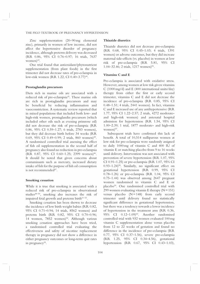
THE FIGO TEXTBOOK OF PREGNANCY HYPERTENSION
104
Zinc supplementation (20–90 mg elemental
zinc), primarily in women of low income, did not
affect the hypertensive disorder of pregnancy
incidence, although preterm delivery was decreased
(RR 0.86, 95% CI 0.76–0.97; 16 trials, 7637
women)43.
One trial found that antioxidant/phytonutrient
supplementation (from plant foods) in the first
trimester did not decrease rates of pre-eclampsia in
low-risk women (RR 1.22, CI 0.40–3.77)44.
Prostaglandin precursors
Diets rich in marine oils are associated with a
reduced risk of pre-eclampsia45. These marine oils
are rich in prostaglandin precursors and may
be beneficial by reducing inflammation and
vasoconstriction. A systematic review reported that
in mixed populations that included both low- and
high-risk women, prostaglandin precursors (which
included other oils such as evening primrose oil)
did not decrease the risk of pre-eclampsia (RR
0.86, 95% CI 0.59–1.27; 6 trials, 2783 women),
but they did decrease birth before 34 weeks (RR
0.69, 95% CI 0.49–0.99; 2 trials, 860 women)45.
A randomised controlled trial assessing the effect
of fish oil supplementation in the second half of
pregnancy also found no reduction in pre-eclampsia
(RR 0.87, 95% CI 0.60–1.25; 2399 women)46.
It should be noted that given concerns about
contaminants such as mercury, increased dietary
intake of fish for the purpose of fish oil consumption
is not recommended47.
Smoking cessation
While it is true that smoking is associated with a
reduced risk of pre-eclampsia in observational
studies48–50, smoking also increases the risk of
impaired fetal growth and preterm birth51–53.
Smoking cessation has been shown to decrease
the incidence of low birth weight babies (RR 0.82,
95% CI 0.71–0.94; 14 trials, 8562 women) and
preterm birth (RR 0.82, 95% CI 0.70–0.96;
14 women, 7852 women)54. Although various
smoking cessation approaches have been tried,
a randomised controlled trial evaluating the
effectiveness and safety of nicotine replacement
therapy in pregnancy did not show a difference in
either pregnancy outcomes or long-term quit rates
in pregnancy55.
Thiazide diuretics
Thiazide diuretics did not decrease pre-eclampsia
(RR 0.68, 95% CI 0.45–1.03; 4 trials, 1391
women) or adverse outcomes, but they did increase
maternal side-effects (vs. placebo) in women at low
risk of pre-eclampsia (RR 5.81, 95% CI
1.04–32.46; 2 trials, 1217 women)56.
Vitamins C and E
Pre-eclampsia is associated with oxidative stress.
However, among women at low risk given vitamins
C (1000 mg/d) and E (400 international units/day)
therapy from either the first or early second
trimester, vitamins C and E did not decrease the
incidence of pre-eclampsia (RR 0.85, 95% CI
0.48–1.51; 4 trials, 2441 women). In fact, vitamins
C and E increased use of any antihypertensive (RR
1.77, 95% CI 1.22–2.57; 2 trials, 4272 moderate-
and high-risk women) and antenatal hospital
admission for hypertension (RR 1.54, 95% CI
1.00–2.39; 1 trial, 1877 moderate- and high-risk
women)57.
Subsequent trials have confirmed this lack of
benefit. A total of 10,514 nulliparous women at
low risk for pre-eclampsia were randomly assigned
to daily 1000 mg of vitamin C and 400 IU of
vitamin E or matching placebo from 9 to 16 weeks
until delivery. Intervention was not associated with
prevention of severe hypertension (RR 1.07, 95%
CI 0.91–1.25) or pre-eclampsia (RR 1.07, 95% CI
0.93–1.24)58. Similarly, no significant effect on
gestational hypertension (RR 0.99, 95% CI
0.78–1.26) or pre-eclampsia (RR 1.04, 95% CI
0.75–1.44) was observed among 2647 pregnant
women randomised to vitamin C and E or
placebo59. One randomised controlled trial with
299 women evaluating vitamin E therapy (N = 151)
versus placebo (N = 148) from early second
trimester until delivery found no statistically
significant difference in gestational hypertension,
but there was a tendency towards a lower incidence
of hypertension in the treatment arm (RR 0.36,
95% CI 0.12–1.09)60. Another randomised
controlled trial with 932 women evaluated 100 mg
vitamin C supplementation alone versus placebo
from 12 to 22 weeks of gestation and found no
difference in the incidence of pre-eclampsia (RR
0.77, 95% CI 0.37–1.56), severe pre-eclampsia
(RR 1.25, 95% CI 0.34–4.56), gestational
hypertension (RR 0.67, 95% CI 0.43–1.03),

PREVENTING PRE-ECLAMPSIA AND ITS COMPLICATIONS
105
preterm delivery (RR 0.92, 95% CI 0.63–1.34) or
low birth weight (RR 1.07, 95% CI 0.72–1.59)61.
Vitamin D
Vitamin D may play a protective role against
pre-eclampsia through beneficial effects on immune
modulation and vascular function62–64. A significant
relationship between vitamin D deficiency and
increased risk of pre-eclampsia has been shown
by systematic reviews and meta-analyses of
observational studies65,66. This represents an area
where further studies are required.
Other interventions for which no recommendation can be made
Interest in supplementation with iron and/or folate
(beyond 10 weeks’ gestation) stems from the
importance of anaemia in developing countries
and further progressive anaemia associated with
pregnancy67. There is insufficient evidence on the
effect on pre-eclampsia of either routine (vs. no
routine) iron supplementation (usually 60–100 mg
elemental iron/day) (1 trial, 47 women) or routine
iron with/without folic acid supplementation
(1 trial, 48 women)68.
Pyridoxine has many roles, including neurological
development and function. Although in a systematic
review, pyridoxine supplementation did not
decrease the risk of pre-eclampsia, the trials were of
poor quality with poor reporting of substantive
outcomes, making it impossible to draw conclusions
(oral pyridoxine RR 1.71, 95% CI 0.85–3.45; 2
trials, 1197 women) (pyridoxine lozenges RR 1.43,
95% CI 0.64–3.22; 1 trial, 944 women)69.
Garlic may lower blood pressure70, reduce
oxidative stress71 and inhibit platelet aggregation72,
but a systematic review found no clear effect on
pre-eclampsia (RR 0.78, 95% CI 0.31–1.93). As
only one trial with 100 women was included,
further trials are needed to draw any reliable
conclusions about garlic and its effect on
pre-eclampsia73.
We were unable to identify trials administering
the following agents for primary prevention of
pre-eclampsia: vitamin A, selenium, copper and
iodine.
It must be noted that in some regions, there is
strong interest in traditional medicines for
pre-eclampsia prevention. Evidence is lacking to
support or refute these practices.
“About snails, we use the fluid from a snail to
prepare traditional medicine to treat patients
with high blood pressure . . . we use the snail’s
fluid to prepare a traditional medicine for them
. . . and they would use a teaspoon to take the
medicine . . . those that always have high blood
pressure . . . people whose blood pressure is
always high . . . people like that . . . within 3
months or so . . . they would be lying on a sick
bed . . . they would rolling on the floor in
pains . . . and be doing all sorts of things . . . so
we treat them so that the high blood pressure
wouldn’t cause complications for them”.
Head Traditional Birth Attendant, Yewa South,
Nigeria (from CLIP Feasibility Study)
WOMEN AT INCREASED RISK
Women identified as being at ‘increased risk’ of
pre-eclampsia have been most commonly those
with a personal or family history of a hypertensive
disorder of pregnancy, chronic medical disease
(including hypertension), and/or an abnormal
uterine artery Doppler velocimetry before 24 weeks.
However, there was variability between studies in
inclusion criteria (including use of the roll-over test
reflecting increased sensitivity to angiotensin-II) and
other characteristics of the population, including
ethnicity, parity, socioeconomic status and access to
prenatal care. No study identified used only the
roll-over test to enroll women.
A growing literature suggests that combining
clinical, biochemical and/or ultrasonographic risk
markers may better identify women at increased
risk of pre-eclampsia (as as discussed in Chapter 5);
however, to date no intervention trial has used such
an approach to evaluate a preventative therapy74–76.
The ASPRE trial is doing so for aspirin (150 mg/d
at bedtime), as discussed below77. (Please see
Appendix 6.2 for details of individual randomised
controlled trials or systematic reviews of randomised
controlled trials that reported on the outcomes of
pre-eclampsia, gestational hypertension, maternal
morbidity, SGA infants, or neonatal morbidity such
as neonatal intensive care stay.)
Antihypertensive therapy
Antihypertensive therapy does not prevent
pre-eclampsia (RR 0.93, 95% CI 0.80–1.08; 23
trials, 2851 women) or the associated adverse
perinatal outcomes, but it decreases by half the

THE FIGO TEXTBOOK OF PREGNANCY HYPERTENSION
106
incidence of development of severe hypertension
(RR 0.49, 95% CI 0.40–0.60; 2 trials, 2558
women)78. Antihypertensive therapy cannot be
recommended for pre-eclampsia prevention until it
can be demonstrated that the decrease in maternal
blood pressure is not outweighed by a negative
impact on perinatal outcomes79,80. (Antihypertensive
therapy for treatment of elevated blood pressure is
discussed in Chapter 8)
Aspirin (low dose)
In women identified as at increased risk of
pre-eclampsia based on clinical characteristics,
low-dose aspirin results in a small decrease in
pre-eclampsia (RR 0.75, 95% CI 0.66–0.85; 18
trials; 4121 women for this outcome), preterm
delivery <37 weeks’ gestation (RR 0.89, 95% CI
0.81–0.97; I2 32%; 10 trials, 3252 women for this
outcome), perinatal death (RR 0.69, 95% CI
0.53–0.9; 17 trials, 4443 women for this outcome)
(40 trials, 33,098 women overall)6, and intrauterine
growth restriction (RR 0.80, 95% CI 0.65–0.99; I2
36.9%, 13 trials, 12,504 women for this outcome)81.
There is low level evidence that low-dose aspirin
may help to prevent pre-eclampsia (RR 0.67, 95%
CI 0.48–0.94; 5 trials, 898 women) in multiple
gestations82. The ASPRE trial is doing so for aspirin
(150 mg/d at bedtime) started in the first-trimester
in women identified as being at increased risk77.
Aspirin does not increase or decrease miscarriage
risk83. There is no evidence of short- or long-term
adverse effects on the mother or newborn.
Who should receive aspirin, in what dose,
and when are unclear. Subgroup analyses in
meta-analyses suggest a number of important
considerations. First, aspirin is more effective in decreasing pre-eclampsia among women at high risk
(NNT 19, 95% CI 13–34) compared with those at
moderate risk (NNT 119, 95% CI 73–333), though
a recent meta-analysis did not show any effect of
preconceptionally started aspirin in reducing
hypertensive pregnancy complications in IVF
women84. Second, aspirin may be more effective at decreasing the following outcomes when it is initiated before 16 weeks’ gestation: severe pre-eclampsia85, preterm
pre-eclampsia, preterm delivery, perinatal death
and SGA infants81,86–90. Preconception-initiated
low-dose aspirin was associated with the outcome
of higher live birth rates in women with a single
documented loss at less than 20 weeks’ gestation
during the previous year91. However, a recent
secondary analysis showed that 60 mg of aspirin
daily, initiated before or after 16 weeks’ gestation
was not effective for the prevention of
pre-eclampsia92. Therefore, aspirin may be more effective when used at a higher dose6,93. Approximately
one-third of pregnant women are both resistant to
the effects of 75–80 mg of aspirin and at increased
risk of adverse pregnancy outcomes94,95. A
retrospective controlled study (270 women)
suggested that adjusting aspirin dosage based
on platelet function testing may improve the
effectiveness of aspirin without a demonstrated
increase in adverse neonatal outcomes96.
Furthermore, two randomised controlled trials
found that taking aspirin at bedtime (instead of the morning) resulted in lower blood pressure and fewer adverse pregnancy outcomes such as pre-eclampsia,
SGA babies and preterm birth97,98. Finally, aspirin may be continued until delivery as was prescribed in
most trials; however, some care providers of
women in these trials stopped aspirin prior to
delivery and the benefits of continuing aspirin
throughout the third trimester have also been
questioned99 (see Chapters 8 and 10).
Calcium
Oral calcium supplementation (of at least 1 g/d) in
high-risk women (e.g. teenagers or women older
than 40 years, women with previous pre-eclampsia,
women with increased sensitivity to angiotensin II,
women with pre-existing hypertension) was found
to decrease the incidence of pre-eclampsia (RR
0.22, 95% CI 0.12–0.42; 5 trials, 587 women),
gestational hypertension (RR 0.47, 95% CI
0.22–0.97; 4 trials, 327 women) and preterm
delivery (RR 0.45, 95% CI 0.24–0.83; 4 trials, 583
women)7. Three of the five relevant trials were
conducted in low calcium intake populations. No
trial included women with previous pre-eclampsia.
There were no documented adverse effects of
calcium supplementation, but none of these trials
of women at high risk of pre-eclampsia reported
the outcome of HELLP syndrome. An alternative
to supplementation may be an increase in dietary
calcium intake, by 3–4 dairy servings per day (as
one serving corresponds to 250–300 mg of calcium).
Oral calcium supplementation of <1 g/d is also
effective in mixed populations of women at low
and increased risk of pre-eclampsia, but the effect

PREVENTING PRE-ECLAMPSIA AND ITS COMPLICATIONS
107
within each of these populations is not known. The
Calcium and Pre-eclampsia (CAP) Study is an
ongoing randomised trial of low-dose calcium
supplementation among women at high risk of
pre-eclampsia)100.
Aspirin (low-dose) combined with calcium
Two small trials (91 women) have looked at the
combined effect of low-dose aspirin and calcium
supplementation (one <1 g/d101 and one more than
1 g/d102). The combined therapy from 20 to 27
weeks’ gestation was associated with a
non-significant decrease in pre-eclampsia (52.5%
vs. 73.1%, p = 0.11) and IUGR (25.0% vs. 4.8%,
p = 0.07) that may warrant further study, particularly
as both therapies are currently recommended
individually102. In particular, it is not known what
the effect would be of supplementation before
16–20 weeks of gestation, and bioavailability studies
are required to determine how much aspirin and
calcium are actually being absorbed by study
participants102. The other trial of aspirin and
low-dose calcium found that combined therapy
was associated with significant improvement in
pro-inflammatory factors of highly sensitive
C-reactive protein (hs-CRP), plasma total
antioxidant capacity (TAC) and total glutathione
(GSH)101.
Dietary changes
We were unable to identify trials of dietary salt
restriction on the incidence of pre-eclampsia among
women at increased risk. Women with pre-existing
hypertension who are already following a dietary
approached to stop hypertension (DASH) diet may
continue this diet during pregnancy, but there is no
evidence to support this practice.
We were unable to identify trials of a
heart-healthy diet for pre-eclampsia prevention.
Obesity is both a major public health problem
and a risk marker for pre-eclampsia. No effect
on gestational hypertension (or pre-eclampsia
specifically) has been demonstrated when
overweight women have received dietary
counselling during pregnancy to curb the rate of
weight gain (3 trials, 384 women)15. No trials have
addressed the impact of pre-pregnancy or early
pregnancy weight reduction on pre-eclampsia;
there are theoretical concerns about the impact of
starvation ketosis on fetal neurodevelopment16.
Garlic may decrease lipid peroxidation and
platelet aggregation. One small trial of 100 women
at increased risk of pre-eclampsia based on a positive
roll-over test found that garlic supplementation in
the third trimester of pregnancy reduced the
occurrence of gestational hypertension (18% vs.
36%, p = 0.04), but not of pre-eclampsia (14% vs.
18%, p = 0.80)103. Another small trial (N = 235)
found that coenzyme Q10 supplementation from
20 weeks until delivery (compared to placebo)
reduces the risk of developing pre-eclampsia (14.4%
vs. 25%, p = 0.035, RR 0.56, 95% CI 0.33–0.96)104.
Folate-containing multivitamin
Periconceptional and ongoing regular use of
multivitamins was associated with higher birth
weight centiles in a secondary analysis of the
Vitamins in Pre-eclampsia (VIP) (vitamin C and E
trial) in the UK105. Periconceptional use of a
folate-containing multivitamin is recommended
for all women of child-bearing age for prevention
of neural tube and, possibly, other birth defects.
The Canadian FACT trial of folic acid 0–1.1 mg
versus 4–5.1 mg (4.0 mg folic acid as the
intervention) from 10 to 14 weeks for the
prevention of pre-eclampsia has recently completed
recruitment and the results are anticipated106.
Heparin
Heparin may improve placentally mediated
outcomes through anticoagulant and/or
potentially non-anticoagulant actions, such as
endothelium-dependent vasodilation107 and/or
reversal of the anti-angiogenic actions of explanted
placental villi on cultured endothelial cells108.
A number of small randomised controlled trials
have studied prophylactic doses of heparin (mostly
low molecular weight heparin (LMWH)) for
women with a history of various placental
complications in previous pregnancies. The 2013
Cochrane review (9 trials, 979 women) found that
prophylactic doses of heparin (of any type)
compared with no treatment, decreased perinatal
mortality (2.9% vs. 8.6%; RR 0.40, 95% CI
0.20–0.78), preterm delivery before 34 weeks
(8.9% vs. 19.4%; RR 0.46, 95% CI 0.29–0.73), and
SGA infants (7.6% vs. 19.0%; RR 0.41, 95% CI
0.27–0.61) in women at high risk of placentally
mediated complications109. In another review
focused on only LMWH (6 trials, 848 women),

THE FIGO TEXTBOOK OF PREGNANCY HYPERTENSION
108
LMWH, compared with no treatment, reduced the
risk of ‘severe’ or early-onset pre-eclampsia (1.7%
vs. 13.4%; RR 0.16, 95% CI 0.07–0.36), preterm
delivery before 37 weeks (32.1% vs. 47.7%; RR
0.77, 95% CI 0.62–0.96), and SGA infants (10.1%
vs. 29.4%; RR 0.42, 95% CI 0.29–0.59), without a
significant effect on perinatal mortality (pregnancy
loss >20 weeks 1.9% vs. 5.3%; RR 0.41, 95% CI
0.17–1.02)110. In both analyses, a significant decrease
in any pre-eclampsia was seen, but there was more
between-trial difference in pre-eclampsia incidence
than could be expected by chance alone, as was the
case in the LMWH analysis for a composite of
placentally mediated pregnancy complications (i.e.,
pre-eclampsia, placenta abruption, SGA infants, or
fetal loss after 12 weeks) (18.7% vs. 42.9%; RR
0.52, 95% CI 0.32–0.86). However, a recent trial
with 292 women observed no impact of antepartum
prophylactic dose dalteparin (5000 IU once daily
up to 20 weeks’ gestation and twice daily thereafter
until at least 37 weeks’ gestation) on a composite
outcome of severe or early onset pre-eclampsia,
SGA infants, pregnancy loss, or venous
thromboembolism in women with thrombophilia
at high risk of complications (venous
thromboembolism, pregnancy loss, or placentally
mediated pregnancy complications) in both
an intention-to-treat analysis (17.1%, 95% CI
11.4–24.2% vs. 18.9%, 95% CI 12.8–26.3%; risk
difference 1.8%, 95% CI 10.6–7.1%) and an
on-treatment analysis (19.6% vs. 17.0%; risk
difference +2.6%, 95% CI 6.4–11.6%), but there
was an increased risk of minor bleeding associated
with LMWH (19.6% vs. 9.2%; risk difference
10.4%, 95% CI 2.3–18.4; p = 0.01)111.
Pending the results of larger trials powered for
perinatal mortality or severe maternal morbidity, or
individual patient data meta-analysis of greater
numbers of smaller trials, LMWH for pre-eclampsia
prevention should be used cautiously. The
independent role of concomitant treatment with
aspirin also remains to be elucidated.
LMWH in prophylactic subcutaneous doses is
associated with minimal risks for the mother and,
theoretically, none for the fetus as it does not cross
the placenta. In a meta-analysis of 64 studies (2777
women), major allergic reactions were uncommon
(1.2%) and no woman developed heparin-induced
thrombocytopaenia. LMWH in prophylactic doses
was associated with very low risks of antenatal
bleeding (0.42%), intrapartum bleeding (0.92%)
and wound haematoma after either Caesarean or
vaginal delivery (0.65%)112. In the randomised
controlled trial cited above, LMWH was associated
with an increase in minor bleeding compared with
no treatment111. LMWH to prevent recurrent
early-onset pre-eclampsia and/or IUGR could be
stopped at 34–36 weeks’ gestation, so the potential
side-effects of LMWH intrapartum and postpartum
are not as relevant. However, a recent international
audit on maternal and fetal safety of tinzaparin (at
therapeutic and prophylactic doses), the adjudication
committee considered that serious bleeding events
(before, during and after delivery) were probably related to tinzaparin therapy in 2.3% of pregnancies,
and possibly related to tinzaparin in 7.7% (1256
pregnancies in 1109 women)113. There was no
reported spinal haematoma; 10.4% of the women
received tinzaparin within 24 hours of epidural or
spinal anaesthesia with a median tinzaparin injection
to delivery interval of 12.9 hours (range 0–23.5).
Osteoporotic fractures occurred in 0.2% of women,
although all had other risk factors for osteoporosis.
Neonatal haemorrhage did not occur. Major allergic
reactions were uncommon (1.8%). No women
developed heparin-induced thrombocytopaenia.
L-arginine
Supplements containing L-arginine and ‘antioxidant
vitamins’ have been shown to reduce diastolic blood
pressure114 or both systolic and diastolic blood
pressure, and the incidence of pre-eclampsia in a
population at high risk of the condition (2 trials, 672
women)115,116. Another systematic review supported
that L-arginine supplements reduced the incidence
of pre-eclampsia in high-risk women (RR 0.34,
95% CI 0.21–0.55), as well as risk of preterm birth
(RR 0.48, 95% CI 0.28–0.81). The protective effect
was greater in women with established hypertensive
disease (RR 0.21, 95% CI 0.05–0.98)117. Data from
several small randomised trials suggests that
L-arginine given to women with already diagnosed
gestational hypertension (with or without
proteinuria) or with IUGR can lead to improvement
of maternal blood pressure and uteroplacental
circulation118–123.Optimal dosage needs to be defined
and large randomised trials are required.
Lifestyle changes
There are robust epidemiological data that weight
gain between pregnancies (even in non-obese

PREVENTING PRE-ECLAMPSIA AND ITS COMPLICATIONS
109
women) is associated with significantly more
pre-eclampsia and other pregnancy complications,
such as Caesarean delivery and gestational diabetes124.
Physical activity is associated with a reduced
incidence of pre-eclampsia125,126. In women at
increased risk of pre-eclampsia, it is not known
whether exercise (to improve or maintain fitness) is
of greater benefit than risk. No impact of exercise
was seen on gestational hypertension or
pre-eclampsia (2 trials, 45 women), although the
trials were small and the confidence intervals were
wide126,127. Similar results were seen in another
small trial of 79 sedentary women with previous
pre-eclampsia, among whom walking exercise vs.
stretching exercise during pregnancy did not
decrease the incidence of pre-eclampsia128.
Physically demanding work is associated with a
higher risk of gestational hypertension and
pre-eclampsia (OR 1.60, 95% CI 1.30–1.96; 4
observational studies, 5837 women)36. Although
workload reduction is a common obstetric
intervention, we were unable to identify randomised
studies of workload or stress reduction on the
incidence of pre-eclampsia. These are unlikely to
be forthcoming given the nature of the interventions.
Increased rest at home (varying from 30 minutes
to 6 hours/day) in the third trimester of pregnancy
decreased the incidence of pre-eclampsia (RR
0.05, 95% CI 0.00–0.83; 1 trial, 32 women for
increased rest alone; RR 0.13, 95% CI 0.03–0.51
for rest plus a nutrient supplement; 1 trial, 74
women)129. Other substantive outcomes (such as
adverse effects of rest and women’s views) were not
reported. There is a lack of clarity about the
definition of bed rest and uncertainty about
whether women comply with activity restriction130.
Metformin
One trial (N = 105 women) observed that women
with polycystic ovarian syndrome (PCOS)
randomised to receive metformin (vs. placebo)
from the first-trimester of pregnancy showed
significant improvement of the uterine artery
Doppler pulsatility index to a similar extent as that
observed with low-dose aspirin131. This trial did
not have the power to observe a significant
difference in the rate of pre-eclampsia (5.7% with
metformin, 5.7% with aspirin, and 11.4% in the
placebo group, p = 0.58). However, in a secondary
analysis of another trial of 400 obese non-diabetic
women (BMI >35 kg/m2) randomised to
metformin (1–3 g daily, gradually titrated over 4
weeks) or placebo, metformin was associated with
a significant decrease in pre-eclampsia (2.0% vs.
8.2%, p = 0.005)132. Further studies are warranted.
Micronutrients other than calcium
Magnesium supplementation (various preparations)
administered to a mixed population of women at
low and high risk in (7 trials, 2689 women) did not
decrease the risk of pre-eclampsia, but decreases
were seen in preterm birth (RR 0.73, 95% CI
0.57–0.94), low birth weight (RR 0.67, 95% CI
0.46–0.96) and incidence of SGA infants (RR
0.70, 95% CI 0.53–0.93)40. However, no
conclusions can be drawn because only one
included trial was of high quality.
In one trial (100 women), selenium
supplementation in the third trimester was reported
to decrease gestational hypertension, but this was
not defined133. Another small trial (166 women)
found no significant decrease in the rate of
pre-eclampsia134.
One study found that daily ingestion of a
phytonutrient supplement did not decrease rates of
pre-eclampsia in high-risk women135.
We did not identify trials of zinc, pyridoxine,
iron (with/without folic acid), zinc, multivitamins
with/without micronutrients, vitamin A, iodine,
or copper for pre-eclampsia prevention in women
at increased risk.
Prostaglandin precursors
According to the most recent Cochrane systematic
review, prostaglandin precursors did not decrease
the risk of pre-eclampsia in mixed populations of
women at low and high risk (RR 0.87, 95% CI
0.59–1.28; 5 trials, 1683 women)45. Birth before
34 weeks was marginally decreased (RR 0.69, 95%
CI 0.49–0.99). However, a recent trial including
pregnant women with previous pregnancy
complications showed that fish oil supplementation
was associated with a more advanced gestational
age at delivery in low and middle (but not high)
fish consumers136.
Vitamins C and E
In five trials (3005 women) of women at increased
risk of pre-eclampsia for various reasons,

THE FIGO TEXTBOOK OF PREGNANCY HYPERTENSION
110
antioxidants (usually combined therapy with
vitamins C 1000 mg/d and E 400 international
units/day) did not decrease the risk of pre-eclampsia
(RR 0.56, 95% CI 0.29–1.11)56. These findings
were supported by subsequent trials137,138. Vitamins
C and E have been associated with adverse
outcomes, including increased use of intravenous
antihypertensive therapy (RR 1.94, 95% CI
1.07–3.53)139, low birth weight babies (28%
(N = 387) vs. 24% (N = 335), RR 1.15, 95% CI
1.02–1.30; 2395 women)131, fetal loss or perinatal
death (RR 2.20, 95% CI 1.02–4.73; 2536 women),
preterm prelabour rupture of membranes (RR
1.97, 95% CI 1.31–2.98; 2363 women)140.
Nitric oxide donors
Nitric oxide (NO) donors like pentaerithrityl-
tetranitrate (PETN) have protective effects on the
endothelium. One trial found no decrease in
pre-eclampsia from NO-donor PETN, but a
decrease in IUGR and/or perinatal death (adjusted
RR 0.41, 95% CI 0.18–0.91) and for IUGR
(adjusted RR 0.44, 95% CI 0.20–0.97), and
preterm birth before 32 weeks’ gestational age
(adjusted RR 0.20, 95% CI 0.05–0.80)141.
Other
Treatment of periodontal disease is not associated
with a reduced risk of pre-eclampsia (4 trials)142,143.
However, it is possible that the type of treatment
(scaling vs. chlorexidine mouthwash) could
influence its impact, as it has been seen for
prevention of preterm birth144.
RESOURCE-CONSTRAINED SETTINGS
Pre-eclampsia is “. . . considerably more prevalent
in LMICs [low- and middle-income countries]
than in affluent communities”145. Furthermore,
over 99% of pre-eclampsia and eclampsia-related
mortality occurs in LMICs, particularly in
sub-Sahara Africa and on the Indian subcontinent146.
Factors that determine the potential impact of an
intervention on prevention of pre-eclampsia
include its availability, acceptability and
cost-effectiveness, as well as the strength of the
infrastructure of a health care system. The latter is
where LMICs face the greatest challenge. Their
unique challenges to intervention implementation
include:
• Low rates of antenatal visits and low levels of
education in the population, which could be
addressed by community engagement and
educational activities146,147.
• Poverty and weak public infrastructure (such as
paved roads and available transportation) which
prevent access to health care148. Addressing these
issues will require engagement of government
and policy makers.
• A shortage of trained health care workers146,149.
Major efforts have been directed towards ‘task
shifting’, training and employing community
health care workers to play a fundamental role
within the health care system in LMICs. In this
way, it is hoped that women with hypertensive
disorders of pregnancy will receive timely care.
This strategy recognises that the majority of
pre-eclampsia related deaths in LMICs occur in
the community146.
• A lack of high quality guidance material such as
protocols and guidelines150, something that this
book aims to address for the hypertensive
disorders of pregnancy.
What follows here are implications relevant
to resource-constrained settings for the given
recommendations to help prevent pre-eclampsia.
Aspirin (low dose)
A decision analytic model comparing aspirin
prophylaxis (vs. no prophylaxis) in a theoretical
cohort of 100,000 pregnant women concluded
that it was a more cost-effective strategy than no
prophylaxis. Lower costs ($18,720 vs. $18,804) and
marginal difference in quality-adjusted life years
(26.7417 vs. 26.7422) favours aspirin prophylaxis
– a better choice than no prophylaxis151.
Calcium supplementation
The impact of calcium in reducing pre-eclampsia is
dependent on the baseline calcium intake of the
population and pre-existing risk factors152,153. Global
trends of dietary calcium intake typically show
lower intake in LMICs (ranging from 300 to
600 mg/day) compared with high-income countries
(e.g., 969 mg for France)154. Although these data
suggest that calcium supplementation is particularly
important for women in developing countries,
suboptimal global implementation of this
intervention remains. In a study of women

PREVENTING PRE-ECLAMPSIA AND ITS COMPLICATIONS
111
receiving antenatal care in Brazilian public hospitals,
over 90% of women consumed less than 1 g of
calcium per day, yet less than 6% of women
received a prescription for calcium supplements155.
Similar results were observed in a teaching public
hospital in Argentina156.
Implementation of the recommended high-dose
calcium supplementation (1 g calcium/day or more)
in settings of low-dietary calcium is problematic to
policy-makers and programme managers in LMICs
for a number of reasons157. Lack of infrastructure
challenges the procurement of the preparation,
transportation of the heavy tablets, storage, quality
control and compliance assurance158. The cost
implications of the recommended calcium
supplementation dose may be a financial barrier; for
example, chewable calcium carbonate tablets cost
US $3–6/pregnancy159. A cost–benefit ratio must
be considered by ministries of health in decisions
to scale up this intervention149. Also, there are
potential risks of calcium supplementation,
particularly in excess, associated with
supplementation in pregnancy, such as HELLP
syndrome7 and rebound postnatal bone
demineralisation following supplementation in
pregnancy160.
Potential solutions to the problems discussed
include a recently developed micronutrient powder
designed to optimise absorption of all its contents
(calcium, iron and folic acid)161 and low-dose
calcium supplementation for which there are
limited data suggesting effectiveness in reducing
pre-eclampsia risk7. Until these findings are
confirmed by larger, sufficiently powered
randomised trials100,162, lower-dose supplements
(500–600 mg/day) may be considered in preference
to no supplementation in settings of low dietary
calcium where high-dose supplementation is not
feasible7,152.
Cost-effectiveness analyses of increasing calcium
intake should consider increasing dietary intake
versus calcium supplementation, with consideration
that many countries do not have sufficient availability
of dairy products to meet dietary needs163.
Folate-containing multivitamin
An average of 20–30% of pregnant women have
a vitamin deficiency of some kind. Without
supplementation, approximately 75% of these
women would show a deficit of at least one vitamin.
In India, for example, about 25% of pregnant
women are folate deficient164.
Global periconceptional folic acid supplement
use is low, taken by fewer than 50% of women in
many countries165. A study of American women
found that 29.7% used periconceptional folic acid
supplement166. A study of 21,889 women in
Tanzania in a geographical area with a high
prevalence of anaemia found a prenatal intake of
folic acid of 17.2%; notably, women were less likely
to take folic acid supplements if they had
pre-eclampsia/eclampsia during pregnancy (OR
0.48, 0.38–0.61)167.
Factors associated with lower preconceptional
use of folic acid are younger age, lower levels of
maternal formal education, single marital status and
unplanned pregnancy. In Canada, folic acid
supplementation has been shown to vary according
to maternal country of origin. In comparison with
Canadian born women, immigrants from Northern
African, Middle Eastern, Caribbean, Latin
American, a South Pacific country or from China
were significantly less likely to use supplements168.
It may be that certain groups of immigrant women
engage in less family planning and have more
unintended pregnancies, lack knowledge regarding
the benefit of folic acid supplementation, or cannot
afford tablet supplements165. Policy makers and
health practitioners can be aware of risk factors for
low use and help increase folic acid supplementation
in these populations.
Exercise
Literature suggests that physical activity declines
during pregnancy169,170. Barriers to activity during
pregnancy reported by women include pregnancy
symptoms, lack of time, access to child care and
concerns about their safety and that of their unborn
baby171–175 and lack of advice from health
professionals176. Conversely, significant enablers
included positive psychological feelings, family
influence and receiving advice from health
professionals177.
A lack of information by certain populations
may contribute to low levels of exercise in
pregnancy. A very recent study in a developing
country found that only 36.6% of women thought
that regular exercise was not harmful during
pregnancy178. Low-income African American
women report several factors that prevent them

THE FIGO TEXTBOOK OF PREGNANCY HYPERTENSION
112
BEST PRACTICE POINTS
(Please see Appendix 6.3 for the evaulation of the strength of recommendations and the quality of the
evidence on which they are based.)
Prevention of pre-eclampsia in women at low risk
1. Calcium supplementation (of at least 1 g/d, orally) is recommended for women with low dietary
intake of calcium (<600 mg/d, corresponding to less than two dairy servings per day).
2. The following are recommended for other established beneficial effects in pregnancy: abstention
from alcohol for prevention of fetal alcohol effects, exercise for maintenance of fitness, periconceptional
use of a folate-containing multivitamin for prevention of neural tube defects and smoking cessation
for prevention of low birth weight and preterm birth.
3. The following may be useful: periconceptional and ongoing use of a folate-containing multivitamin
or exercise.
4. The following are not recommended for pre-eclampsia prevention, but may be useful for prevention
of other pregnancy complications: prostaglandin precursor or supplementation with magnesium or
zinc.
5. The following are not recommended: dietary salt restriction during pregnancy, calorie restriction
during pregnancy for overweight women, low-dose aspirin, vitamins C and E or thiazide diuretics.
6. There is insufficient evidence to make a recommendation about the following: a heart-healthy diet,
workload or stress reduction, supplementation with iron with/without folate, pyridoxine, or food
rich in flavanoids.
from exercising, including a lack of information
about safe types, frequency and duration of exercise.
Cultural myths also exist about certain types of
movements that are believed to potentially cause
problems with pregnancy. For example, placing
arms over their heads raised concerns that the
umbilical cord would wrap around and strangulate
the baby’s neck179. This population also reports
both a lack of motivation to exercise in pregnancy
and a decreased level of physical activity in
pregnancy180. Health care providers can be aware
that cultural myths that may decrease exercise, and
can pose questions to understand the beliefs of their
patients regarding physical activity in pregnancy. It
may be beneficial to provide information to help
dispel misperceptions and ensure women
understand the role of exercise in contributing to
health pregnancy outcomes.
Low molecular weight heparin
If LMWH were effective for prevention of placental
complications, a dalteparin study (116 women)
found that the incremental cost of preventing one
case of severe pre-eclampsia or a SGA infant was
$54.00181. Further research is needed to clarify
whether LMWH can be considered a cost-effective
intervention in resource-constrained settings.
Lifestyle changes
Although no randomised trials exist on stress
reduction on the incidence of pre-eclampsia, studies
do suggest possible benefits for women at increased
risk. Proximity to city parks has been shown to be
associated with a beneficial impact on blood pressure
during the first trimester of pregnancy182. Further
research is needed to elucidate the mechanism
accounting for this benefit and to determine
whether the recommendation of visiting a green
area is an effective and cost-effective intervention.
Yoga is a method associated with stress reduction.
High-risk pregnant women in a controlled trial that
were randomised into a yoga versus control group
showed a significant reduction in pre-eclampsia
(p = 0.042). Further research is needed to determine
whether this is a cost-effective intervention for
women183.

PREVENTING PRE-ECLAMPSIA AND ITS COMPLICATIONS
113
WHAT INTERNATIONAL GUIDELINES SAY
A systematic review of 13 international clinical
practice guidelines (CPGs) on hypertensive
disorders of pregnancy184 summarises international
consensus regarding definitions for women at low
and at increased risk of pre-eclampsia.
Women at low risk are recommended NOT
to restrict dietary salt or take vitamins C and/or
E by four guidelines185–188 and NOT to take
diuretics by three guidelines186–188. Only two
guidelines recommend calcium supplementation
(1–2 g/day)187,188. Only one guideline (SOGC,
Society of Obstetricians and Gynaecologists of
Canada) mentioned low-dose aspirin as an
intervention that was NOT recommended187. One
guideline (SOGC) reported several interventions
with insufficient evidence to make a
recommendation, including a heart-healthy diet,
workload or stress reduction, iron supplementation
with/without folate, vitamin D, pyridoxine and
food rich in flavonoids187.
Women at increased risk of pre-eclampsia are
recommended to take calcium supplementation
(1–2.5 g/d) if they have low calcium intake by
three guidelines187–189. Five guidelines recommended
low-dose aspirin (60–162 mg/d)185–189 with
initiation in early pregnancy185–189, and three
guidelines recommend that it continue until
delivery186,187,189. Women at increased risk are
recommended NOT to restrict dietary salt by three
guidelines185,186,188 or to take vitamins C and/or E
by four guidelines185–188.
SUMMARY
Pre-eclampsia and its complications represent an
important cause of maternal and perinatal morbidity
and mortality. Optimising primary prevention efforts
in the periconceptional and antenatal period are
essential to reduce this burden. This chapter
summarises the most current evidence-based
recommendations regarding lifestyle changes and
drugs that have been shown to help prevent
pre-eclampsia and its complications. Health care
providers should promote these recommendations to
help minimise the deleterious effects of pre-eclampsia
and its complications. Considerations unique to
LMIC and to marginalised populations that may affect
implementation of recommended interventions are
also presented. Reducing the impact of pre-eclampsia
in LMIC countries and marginalised populations will
require health systems capacity building, strengthening
of infrastructure, and implementation of interventions
appropriate to low-resource settings.
Prevention of pre-eclampsia in women at increased risk
1. The following are recommended for prevention of pre-eclampsia: low-dose aspirin and calcium
supplementation (of at least 1 g/d) for women with low calcium intake.
2. Low-dose aspirin (75–100 mg/d) should be administered at bedtime and initiated after diagnosis of
pregnancy but before 16 weeks’ gestation and may be continued until delivery.
3. Prophylactic doses of LMWH may be considered in women with previous placental complications
(including pre-eclampsia) to prevent the recurrence of ‘severe’ or early-onset pre-eclampsia, preterm
delivery, and/or SGA infants.
4. The following may be useful: L-arginine, metformin in PCOS and/or overweight women, increased
rest at home in the third trimester and reduction of workload or stress.
5. The following may be useful for prevention of other pregnancy complications: prostaglandin
precursors, magnesium supplementation and heparin thromboprophylaxis.
6. The following are recommended for other established beneficial effects in pregnancy (as discussed
for women at low risk of pre-eclampsia): abstention from alcohol, periconceptional use of a
folate-containing multivitamin and smoking cessation.
7. The following are not recommended: calorie restriction in overweight women during pregnancy,
weight maintenance in obese women during pregnancy, antihypertensive therapy specifically to
prevent pre-eclampsia, vitamins C and E.
8. There is insufficient evidence to make a recommendation about the usefulness of the following: the
heart-healthy diet, exercise, selenium, garlic, zinc, pyridoxine, iron (with or without folate), or
multivitamins with/without micronutrients all.

THE FIGO TEXTBOOK OF PREGNANCY HYPERTENSION
114
PRIORITIES FOR FUTURE RESEARCH
This chapter identifies gaps in knowledge regarding
the prevention of pre-eclampsia. The effectiveness
of prevention efforts relies on the dissemination of
knowledge among health care providers and
women with subsequent uptake of given
recommendations. To help identify barriers and
help achieve these objectives, there is a need for
further implementation research.
Further research is also needed to elucidate the
effects of the following in preventing pre-eclampsia
in low-risk women: a heart-healthy diet; workload
or stress reduction; supplementation with iron
without or without folate; and pyridoxine or food
rich in flavonoids. In women at increased risk,
further investigation is required regarding the
effects of the heart-healthy diet; exercise;
selenium; garlic; zinc; pyridoxine; iron (with or
without folate); and multivitamins with/without
micronutrients.
REFERENCES
1. von Dadelszen P, Magee LA, Taylor EL, Muir JC, Stewart SD, Sherman P, et al. Maternal Hypertension and Neonatal Outcome Among Small for Gestational Age Infants. Obstet Gynecol 2005;106:335–9
2. McCowan LM, Pryor J, Harding JE. Perinatal predictors of neurodevelopmental outcome in small-for-gestational-age children at 18 months of age. Am J Obstet Gynecol 2002;186:1069–75
3. Ogge G, Chaiworapongsa T, Romero R, Hussein Y, Kusanovic JP, Yeo L, et al. Placental lesions associated with maternal underperfusion are more frequent in early-onset than in late-onset pre-eclampsia. J Perinat Med 2011 Nov;39(6):641–52
4. Khan NA, McAlister FA, Rabkin SW, Padwal R, Feldman RD, Campbell NR, et al. The 2006 Canadian Hypertension Education Program recommendations for the management of hypertension: Part II – Therapy. Can J Cardiol 2006;22:583–93
5. Alcohol, nicotine, substance use. Motherisk Program, March 14, 2007. Available at: http://www.motherisk.org/prof/alcohol.jsp. Accessed January 23, 2008
6. Duley L, Henderson-Smart DJ, Meher S, King JF. Antiplatelet agents for preventing pre-eclampsia and its complications. Cochrane Database Syst Rev 2007 Apr 18;(2):CD004659
7. Hofmeyr GJ, Lawrie TA, Atallah AN, Duley L, Torloni MR. Calcium supplementation during
pregnancy for preventing hypertensive disorders and related problems. Cochrane Database Syst Rev 2014;Issue 6:CD001059
8. Belizan JM, Villar J. The relationship between calcium intake and edema-, proteinuria-, and hypertension-getosis: an hypothesis. Am J Clin Nutr 1980;33:2202-10
9. Villar J, Abdel-Aleem H, Marialdi M, Mathai M, Ali MM, Zavaleta et al. World Health Organization randomized trial of calcium supplementation among low calcium intake pregnant women. Am J Obstet Gynecol 2006;194:639–49
10. Imdad A, Bhutta ZA. Effects of calcium supplementation during pregnancy on maternal, fetal and birth outcomes. Paediatric and Perinatal Epidemiology 2012;26(Suppl 1):138–152
11. Jariou LM, Laskey MA, Sawo Y, Goldberg GR, Cole TJ, Prentice A. Effect of calcium supplementation in pregnancy on maternal bone outcomes in women with a low calcium intake. Am J Clin Nutr 2010 Aug;92(2):450–7
12. Allen R, Rogozinska E, Sivarajasingam P, Khan A, Thangaratinam S. Effect of diet and life style based metabolic risk modifying interventions on preeclampsia: A meta-analysis. Acta Obstetricia et Gynecologica Scandinavica 2014;93(10):973–985
13. Duley L, Henderson-Smart D, Meher S. Altered dietary salt for preventing pre-eclampsia, and its complications. Cochrane Database Syst Rev 2005;CD005548
14. Frederick IO, Williams MA, Dashow E, Kestin M, Zhang C, Leisenring WM. Dietary fiber, potassium, magnesium and calcium in relation to the risk of preeclampsia. J Reprod Med 2005;50:332–44
15. Ota E, Hori M, Mori R, Tobe-Gai R, Farrar D. Antenatal dietary advice and supplementation to increase energy and protein intake. Cochrane Database Syst Rev 2015;CD000032
16. Rudolf MC, Sherwin RS. Maternal ketosis and its effects on the fetus. Clin Endocrinol Metab 1983;12:413–28
17. Brantsaeter AL, Myhre R, Haugen M, Myking S, Sengpiel V, Magnus P, et al. Intake of probiotic food and risk of preeclampsia in primiparous women: the Norwegian Mother and Child Cohort Study. Am J Epidemiol 2011 Oct 1;174(7):807–15
18. Saftlas AF, Triche EW, Beydoun H, Bracken MB. Does chocolate intake during pregnancy reduce the risks of preeclampsia and gestational hypertension? Ann Epidemiol 2010 Aug;20(8):584–91

PREVENTING PRE-ECLAMPSIA AND ITS COMPLICATIONS
115
19. DiRenzo GC, Brillo E, Romanelli M, Porcaro G, Capanna F, Kanninen TT, et al. Potential effects of chocolate on human pregnancy: a randomized controlled trial. J Matern Fetal Neonatal Med 2012 Oct;25(10):1860–7
20. Mogollon JA, Bujold E, Lemieux S, Bourdages M, Blanchet C, Bazinet L, et al, Blood pressure and endothelial function in healthy, pregnant women after acute and daily consumption of flavanol-rich chocolate: a pilot randomized controlled trial. Nutrition Journal 2013;12:41
21. Dodin S. Consumption of Chocolate in Pregnant Women. (CHOCENTA) In: ClinicalTrials.gov [Internet]. Bethesda (MD): National Library of Medicine (US). 2000–2016. Available from: URL of the record NLM Identifier: NCT01431443
22. Goh YI, Bollano E, Einarson TR, Koren G. Prenatal multivitamin supplementation and rates of congenital anomalies: a meta-analysis. J Obstet Gynaecol Can 2006;28:680–9
23. Kubik P, Kowalska B, Laskowska-Klita T, Chelchowska M, Leibschang J. Effect of vitamin-mineral supplementation on the status of some microelements in pregnant women [article in Polish]. Przegl Lek 2004;61:764–8
24. Bodnar LM, Tang G, Ness RB, Harger G, Roberts JM. Periconceptional multivitamin use reduces the risk of preeclampsia. Am J Epidemiol 2006;164:470–7
25. Wen SW, Champagne J, Rennicks White R, Coyle D, Fraser W, Smith G, Fergusson D, Walker MC. Effect of folic acid supplementation in pregnancy on preeclampsia: the folic acid clinical trial study. J Pregnancy 2013;2013:294–312
26. Kramer MS, McDonald SW. Aerobic exercise for women during pregnancy. Cochrane Database Syst Rev 2006;3:CD000180
27. Lombardi W, Wilson S, Peniston PB. Wellness intervention with pregnant soldiers. Mil Med 1999;164:22–9
28. Rudra CB, Williams MA, Lee IM, Miller RS, Sorensen TK. Perceived exertion during prepregnancy physical activity and preeclampsia risk. Med Sci Sports Exerc 2005;37:1836–41
29. Saftlas AF, Logsden-Sackett N, Wang W, Woolson R, Bracken MB. Work, leisure-time physical activity, and risk of preeclampsia and gestational hypertension. Am J Epidemiol 2004;160:758–65
30. Sorensen TK, Williams MA, Lee IM, Dashow EE, Thompson ML, Luthy DA. Recreational physical
activity during pregnancy and risk of preeclampsia. Hypertension 2003;41:1273–80
31. Marcoux S, Brisson J, Fabia J. The effect of leisure time physical activity on the risk of pre-eclampsia and gestational hypertension. J Epidemiol Community Health 1989;43:147–52
32. Landsbergis PA, Hatch MC. Psychosocial work stress and pregnancy-induced hypertension. Epidemiology 1996;7:346–51
33. Aune D, Saugstad OD, Henriksen T, Tonstad S. Physical activity and the risk of preeclampsia: a systematic review and meta-analysis. Epidemiology 2014;25(3):331–43
34. Kasawara KT, do Nascimento SL, Costa ML, Surita FG, e Silva JL. Exercise and physical activity in the prevention of pre-eclampsia: systematic review. Acta Obstet Gynecol Scand 2012;91(10):1147–57
35. Santos IA, Stein R, Fuchs SC, Duncan BB, Ribeiro JP, Kroeff LR, et al. Aerobic exercise and submaximal functional capacity in overweight pregnant women: a randomized trial. Obstet Gynecol 2005;106:243–9
36. Mozurkewich EL, Luke B, Avni M, Wolf FM. Working conditions and adverse pregnancy outcome: a meta-analysis. Obstet Gynecol 2000;95:623–35
37. Klonoff-Cohen HS, Cross JL, Pieper CF. Job stress and preeclampsia. Epidemiology 1996;7:245–9
38. Bonzini M, Coggon D, Palmer KT. Risk of prematurity, low birthweight and pre-eclampsia in relation to working hours and physical activities: a systematic review. Occup Environ Med 2007;64:228–43
39. Mahomed K. Zinc supplementation in pregnancy. Cochrane Database Syst Rev 2000;CD000230165
40. Makrides M, Crosby DD, Bain E, Crowther CA. Magnesium supplementation in pregnancy. Cochrane Database Syst Rev 201401;Issue 4:CD000937
41. Thaver D, Saeed MA, Bhutta ZA. Pyridoxine (vitamin B6) supplementation in pregnancy. Cochrane Database Syst Rev 2006;CD000179
42. Bullarbo M, Odman N, Nestler A, Nielsen T, KolisekM, Vormann J, Rylander R. Magnesium supplementation to prevent high blood pressure in pregnancy: a randomized placebo control trial. Arch Gynecol Obstet 2013;288(6):1269–74
43. Mori R, Ota E, Middleton P, Tobe-Gai R, Mahomed K, Bhutta ZA. Zinc supplementation for improving pregnancy and infant outcome. Cochrane Database of Syst Rev 2012;CD000230

THE FIGO TEXTBOOK OF PREGNANCY HYPERTENSION
116
44. Parrish MR, Martin JN Jr, Lamarca BB, Ellis B, Parrish SA, Owens MY, May WL. Randomized, placebo controlled, double blind trial evaluating early pregnancy phytonutrient supplementation in the prevention of preeclampsia. J Perinatol 2013;33(8):593–9
45. Makrides M, Duley L, Olsen SF. Marine oil, and other prostaglandin precursor, supplementation for pregnancy uncomplicated by pre-eclampsia or intrauterine growth restriction. Cochrane Database Syst Rev 2006;3:CD003402
46. Zhou SJ, Yelland L, McPhee AJ, Quinlivan J, Gibson RA, Makrides M. Fish-oil supplementation in pregnancy does not reduce the risk of gestational diabetes or preeclampsia. Am J Clin Nutr 2012;95:1378–84
47. Health Canada: Potential chemical contamination of food [Internet]. Available at: [cited 2008 Jan 23]. Available from http://www.hc-sc.gc.ca/fn-an/nutrition/prenatal
48. Akolekar R, Syngelaki A, Sarquis R, Zvanca M, Nicolaides KH. Prediction of early, intermediate and late pre-eclampsia from maternal factors, biophysical and biochemical markers at 11–13 weeks. Prenat Diagn 2011 Jan;31(1):66–74
49. Hammoud AO, Bujold E, Sorokin Y, Schild C, Krapp M, Baumann P. Smoking in pregnancy revisited: findings from a large population-based study. Am J Obstet Gynecol 2005 Jun;1 92(6):1856–62; discussion 1862–3
50. Conde-Agudelo A, Althabe F, Belizán JM, Kafury-Goeta AC. Cigarette smoking during pregnancy and risk of preeclampsia: a systematic review. Am J Obstet Gynecol 1999 Oct;181(4):1026–35
51. Hammoud AO, Bujold E, Sorokin Y, Schild C, Krapp M, Baumann P. Smoking in pregnancy revisited: findings from a large population-based study. Am J Obstet Gynecol 2005 Jun;1 92(6):1856–62; discussion 1862–3
52. Salihu HM, Wilson RE. Epidemiology of prenatal smoking and perinatal outcomes. Early Human Development 2007;83(11):713–20
53. Institute of Medicine (US) Committee on Understanding Premature Birth and Assuring Healthy Outcomes. 2007 http://www.ncbi.nlm.nih.gov/pubmed/20669423 US DHHS 2004
54. Chamberlain C, O’Mara-Eves A, Oliver S, Caird JR, Perlen SM, Eades SJ, Thomas J. Psychosocial interventions forsupporting women to stop smoking
in pregnancy. Cochrane Database Syst Rev 2013; Issue 10: CD001055
55. Coleman T, Cooper S, Thornton JG, Grainge MJ, Watts K, Britton J, et al. A randomized trial of nicotine-replacement therapy patches in pregnancy. N Engl J Med 2012 Mar 1;366(9):808–18
56. Churchill D, Beevers G, Meher S, Rhodes C. Diuretics for preventing pre-eclampsia. Cochrane Database Syst Rev 2007;CD004451
57. Rumbold A, Duley L, Crowther CA, Haslam, RR. Antioxidants for preventing pre-eclampsia. Cochrane Database Syst Rev 2008 Jan 23;(1):CD004227
58. Roberts JM, Myatt L, Spong CY, Thom EA, Hauth JC, Leveno KJ, et al. Vitamins C and E to prevent complications of pregnancy-associated hypertension. N Engl J Med 2010 Apr 8;362(14):1282–91
59. Xu H, Perez-Cuevas R, Xiong X, Reyes H, Roy C, Julien P, et al. An international trial of antioxidants in the prevention of preeclampsia (INTAPP). Am J Obstet Gynecol 2010 Mar;202(3):239.e1–239.e10
60. Mahdy ZA, Siraj HH, Khaza’ai H, Mutalib MS, Azwar MH, Wahab MA, Dali AZ, Jaafar R, Ismail NA, Jamil MA, Adeeb N. Does pal oil vitamin E reduce the risk of pregnancy induced hypertension? Acta Medica 2013;56(3):104–9
61. Kiondo P, Wamuyu-Maina G, Wandabwa J, Bimenya GS, Tumwesigye NM, Okong P. The effects of vitamin C supplementation on pre-eclampsia in Mulago Hospital, Kampala, Uganda: a randomized placebo controlled clinical trial. BMC Pregnancy and Childbirth 2014;14(283)
62. Hypponen E: Vitamin D for the prevention of preeclampsia? A hypothesis. Nutr Rev 2005;63:225–232
63. Zehnder D, Bland R, Chana RS, Wheeler DC, Howie AJ, Williams MC, Stewart PM, Hewison M. Synthesis of 1,25-dihydroxyvitamin D(3) by human endothelial cells is regulated by inflammatory cytokines: a novel autocrine determinant of vascular cell adhesion. J Am Soc Nephrol 2002;13:621–629
64. Evans KN, Bulmer JN, Kilby MD, Hewison M. Vitamin D and placental-decidual function. J Soc Gynecol Investig 2004;11:263–271
65. Tabesh M, Salehi-Abarguoei A, Tabesh M, Esmaillzadeh A. Maternal vitamin D status and risk of pre-eclampsia: a systematic review and meta-analysis. J Clin Endocrinol Metab 2013;98(8):3165–3173
66. Hypponen E, Cavadino A, Williams D, Fraser A, Vereczkey A, Fraser WD, Banhidy F, Lawlor D,

PREVENTING PRE-ECLAMPSIA AND ITS COMPLICATIONS
117
Czeizel AE. Vitamin D and pre-eclampsia: original data, systematic review and meta-analysis. Ann Nutr Metab 2013;63:331–340
67. Steer PJ. Maternal hemoglobin concentration and birth weight. Am J Clin Nutr 2000 May;71(5 Suppl):1285S-7S
68. Pena-Rosas JP, Viteri FE. Effects of routine oral iron supplementation with or without folic acid for women during pregnancy. Cochrane Database Syst Rev 2006;3:CD004736
69. Salam RA, Zuberi NF, Bhutta ZA. Pyridoxine (vitamin B6) supplementation during pregnancy or labour for maternal and neonatal outcomes. Cochrane Database Syst Rev 2015; Issue 6: CD000179
70. Silagy CA, Neil HAW. A meta-analysis of the effect of garlic on blood pressure. Journal of Hypertension 1994;12(4):463–8
71. Borek C. Antioxidant health effects of aged garlic extract. Journal of Nutrition 2001;131:1010–5
72. Ali M, Bordia T, Mustafa T. Effect of raw versus boiled aqueous extract of garlic and onion on platelet aggregation. Prostaglandins, Leukotrienes, and Essential Fatty Acids 1999;60(1):43–7
73. Meher S, Duley L. Garlic for preventing pre-eclampsia and its complications. Cochrane Database Syst Rev 2006; Issue 3:CD006065
74. Akolekar R, Syngelaki A, Poon L, Wright D, Nicolaides KH. Competing Risks Model in Early Screening for Preeclampsia by Biophysical and Biochemical Markers. Fetal Diagn Ther 2013;33(1):8–15
75. Audibert F, Boucoiran I, An N, Aleksandrov N, Delvin E, Bujold E, et al. Screening for preeclampsia using first-trimester serum markers and uterine artery Doppler in nulliparous women. Am J Obstet Gynecol 2010 Oct;203(4):383.e1–8
76. Scazzocchio E, Figueras F, Crispi F, Meler E, Masoller N, Mula R, et al. Performance of a first-trimester screening of preeclampsia in a routine care low-risk setting. Am J Obstet Gynecol 2012 Dec 11. pii: S0002-9378(12)02226-0
77. Kypros N. Combined multi-marker screening and randomised patient treatment with ASpirin for evidence-based PRE-eclampsia prevention. 2010- cited 2016 Mar. Available from the ISRCTN registry [Internet] http://www.isrctn.com/ ISRCTN13633058. DOI 10.1186/ISRCTN13633058
78. Abalos E, Duley L, Steyn DW, Henderson-Smart DJ. Antihypertensive drug therapy for mild to moderate
hypertension during pregnancy. Cochrane Database Syst Rev 2001;CD002252
79. Magee LA, von Dadelszen P, Chan S, Gafni A, Gruslin A, Helewa ME, et al. The CHIPS Pilot Trial Collaborative Group. The Control of Hypertension In Pregnancy Study pilot trial. BJOG 2007;114:770-e20
80. Magee LA, von Dadelszen P, Chan S, Gafni A, Gruslin A, Helewa ME, et al. The CHIPS Pilot Trial (Control of Hypertension In Pregnancy Study). Hypertens Pregnancy 2006;25:21
81. Henderson JT, Whitlock EP, O’Connor E, Senger CA, Thompson JH, Rowland MG. Low-Dose Aspirin for the Prevention of Morbidity and Mortality From Preeclampsia: A Systematic Evidence Review for the U.S. Preventive Services Task Force. Ann Intern Med 2014 May 20;160(10):695–703
82. Bergeron TS, Roberge S, Carpentier C, Sibai B, McCaw-Binns A, Bujold E. Prevention of preeclampsia with aspirin in multiple gestations: a systematic review and meta-analysis. Am J Perinatol 2016; [Epub ahead of print]
83. Keim SA, Klebanoff MA. Aspirin use and miscarriage risk. Epidemiology 2006;17:435–9
84. Groeneveld E, Lambers MJ, Lambalk CB, Broeze KA, Haapsamo M, de Sutter P, Schoot BC, Schats R, Mol BWJ and Hompes PGA. Preconceptional low-dose aspirin for the prevention of hypertensive pregnancy complications and preterm delivery after IVF: a meta-analysis with individual patient data. Human Reproduction 2013:28(6):1480–1488
85. Villa PM, Kajantie E, Raikkonen K, Presonen A-K, Hamalainen E, Vainio M, Taipale P,Laivuori H. Aspirin in the prevention of pre-eclampsia in high-risk women: a randomized placebo-controlled PREDO trial and a meta-analysis of randomized trials. BJOG 2013;120:64–74
86. Bujold E, Roberge S, Lacasse Y, Bureau M, Audibert F, Marcoux S, et al. Prevention of preeclampsia and intrauterine growth restriction with aspirin started in early pregnancy: a meta-analysis. Obstet Gynecol 2010 Aug;116(2 Pt 1):402–14
87. Bujold E, Roberge S, Tapp S, Giguère Y. Opinion & hypothesis could early aspirin prophylaxis prevent against preterm birth? J Matern Fetal Neonatal Med 2011 Jul;24(7):966–7
88. Roberge S, Villa P, Nicolaides K, Giguère Y, Vainio M, Bakthi A, et al. Early administration of low-dose aspirin for the prevention of preterm and term preeclampsia: a systematic review and meta-analysis. Fetal Diagn Ther 2012;31(3):141–6

THE FIGO TEXTBOOK OF PREGNANCY HYPERTENSION
118
89. Roberge S, Giguère Y, Villa P, Nicolaides K, Vainio M, Forest JC, et al. Early administration of low-dose aspirin for the prevention of severe and mild preeclampsia: a systematic review and meta-analysis. Am J Perinatol 2012 Aug;29(7):551–6
90. Roberge S, Nicolaides KH, Demers S, Villa P, Bujold E. Prevention of perinatal death and adverse perinatal outcome using low-dose aspirin: a meta-analysis. Ultrasound Obstet Gynecol 2013;41(5):491–9
91. Schisterman EF, Silver RM, Lesher LL, Faraggi D, Wactawski-Wende J, Townsend JM, Lynch AM, Perkins NJ, Mumford SL, Galai N. Preconception low-dose aspirin and pregnancy outcomes: results from the EAGeR randomised trial. The Lancet 2014;384(9937):29–36
92. Cantu JA, Jauk VR, Owen J, Biggio JR, Abramovici AR, Edwards RK and Tita AT. Is low-dose aspirin therapy to prevent preeclampsia more efficacious in non-obese women or when initiated early in pregnancy? J Matern Fetal Neonatal Med 2015 Jul;28(10):1128–32
93. Leitich H, Egarter C, Husslein P, Kaider A, Schemper M. A meta-analysis of low dose aspirin for the prevention of intrauterine growth retardation. BJOG 1997;104(4):450–459
94. Caron N, Rivard GE, Michon N, Morin F, Pilon D, Moutquin JM, et al. Low-dose ASA response using the PFA-100 in women with high-risk pregnancy. J Obstet Gynaecol Can 2009 Nov;31(11):1022–7
95. Wojtowicz A, Undas A, Huras H, Musiał J, Rytlewski K, Reron A, et al. Aspirin resistance may be associated with adverse pregnancy outcomes. Neuro Endocrinol Lett 2011;32(3):334–9
96. Rey E, Rivard GE. Is testing for aspirin response worthwhile in high-risk pregnancy? Eur J Obstet Gynecol Reprod Biol 2011 Jul;157(1):38–42
97. Hermida RC, Ayala DE, Iglesias M. Administration time-dependent influence of aspirin on blood pressure in pregnant women. Hypertension 2003;41:651–6
98. Ayala DE, Ucieda R, Hermida RC. Chronotherapy With Low-Dose Aspirin for Prevention of Complications in Pregnancy. Chronobiol Int 2012 Sep 24
99. de Swiet M, Redman CW. Aspirin, extradural anaesthesia and the MRC Collaborative Low-dose Aspirin Study in Pregnancy (CLASP). Br J Anaesth 1992;69:109–10
100. World Health Organization; University of Witwaterstrand; University of British Columbia. WHO randomized trial of calcium supplementation
before pregnancy to reduce recurrent pre-eclampsia. Registry In: Pan African Clinical Trials Registry [Internet]. Tygerberg (SA): South African Medical Research Council, South African Cochrane Centre. 2010-[cited 2016 Mar 7]. Available from: http://www.pactr.org/ATMWeb/appmanager/atm/atmregistry?dar=true&tNo=PACTR201105000267371 PACTR identifier: PACTR20110500026737
101. Asemi Z, Samimi M, Heidarzadeh Z, Khorrammian H, Tabassi Z. Randomized controlled trial investigating the effect of calcium supplementation plus low-dose aspirin on hs-CRP, oxidative stress and insulin resistance in pregnancy women at risk for pre-eclampsia. Pakistan Journal of Biological Sciences 2012;15(10):469–476
102. Souza EV, Torloni MR, Atallah AN, dos Santos GMS, Kulay Jr L, Sass N. Aspirin plus calcium supplementation to prevent superimposed preeclampsia: a randomized trial. Braz J Med Biol Res 2014;47(5):419–425
103. Ziaei S, Hantoshzadeh S, Rezasoltani P, Lamyian M. The effect of garlic tablet on plasma lipids and platelet aggregation in nulliparous pregnants at high risk of preeclampsia. Eur J Obstet Gynecol Reprod Biol 2001;99:201–6
104. Teran E, Hernandez I, Nieto B, Tavara R, Ocampo JE, Calle A. Coenzyme Q10 supplementation during pregnancy reduces the risk of pre-eclampsia. Int J Gynaecol Obstet 2009 Apr;105(1):43–5
105. Briley AL, Poston L, Seed PT, Shennan AH. Use of commercially available micronutrient preparations amongst high risk pregnant women taking part in the Vitamins in Pre-eclampsia trial (VIP); relationship to pregnancy outcome. Hypertens Pregnancy 2006;25:62
106. Wen SW, Champagne J, Rennicks White R, Coyle D, Fraser W, Smith G, et al. Effect of folic acid supplementation in pregnancy on preeclampsia: the folic acid clinical trial study. J Pregnancy 2013; 2013:294312
107. Tasatargil A, Ogutman C, Golbasi I, Karasu E, Dalaklioglu S. Comparison of the vasodilatory effect of nadroparin, enoxaparin, dalteparin, and unfractioned heparin in human internal mammary artery. J Cardiovasc Pharmacol 2005 Jun;45(6):550–4
108. Sobel ML, Kingdom J, Drewlo S. Angiogenic response of placental villi to heparin. Obstet Gynecol 2011 Jun;117(6):1375–83
109. Dodd JM, McLeod A, Windrim RC, Kingdom J. Antithrombotic therapy for improving maternal or infant health outcomes in women considered at risk of placental dysfunction. Cochrane Database of Systematic Reviews 2013; Issue 7: CD006780

PREVENTING PRE-ECLAMPSIA AND ITS COMPLICATIONS
119
110. Rodger MA, Carrier M, Le Gal G, Martinelli I, Perna A, Rey E, et al. Meta-analysis of low-molecular weight heparin to prevent recurrent placenta-mediated pregnancy complications. Blood 2014:123(6):822–828
111. Rodger MA, Hague WM, Kingdom J, Kahn S, Karovitch A, Sermer M, et al. Antepartum dalteparin versus no antepartum dalteparin for the prevention of pregnancy complications in pregnant women with thrombophilia (TIPPS): a multinational open-label randomised trial. Lancet 2014;384(9955):1673–1683
112. Greer IA, Nelson-Piercy C. Low-molecular-weight heparins for thromboprophylaxis and treatment of venous thromboembolism in pregnancy: a systematic review of safety and efficacy. Blood 2005;106:401–7
113. Nelson-Piercy C, Powrie R, Borg JY, Rodger M, Talbot DJ, Stinson J, et al. Tinzaparin use in pregnancy: an international, retrospective study of the safety and efficacy profile. Eur J Obstet Gynecol Reprod Biol 2011;159:293–9
114. Zhu Q, Yue X, Tian QY, Saren G, Wu MH, Zhang Y, Liu TT. Effect of L-arginine supplementation on blood pressure in pregnant women: a meta-analysis of placebo-controlled trials. Hypertens Pregnancy 2013;32(1):32–41
115. Neri I, Monari F, Sgarbi L, Berardi A, Masellis G, Facchinetti F. L-arginine supplementation in women with chronic hypertension: impact on blood pressure and maternal and neonatal complications. J Matern Fetal Neonatal Med 2010 Dec;23(12):1456–60
116. Vadillo-Ortega F, Perichart-Perera O, Espino S, Avila-Vergara MA, Ibarra I, Ahued R, et al. Effect of supplementation during pregnancy with L-arginine and antioxidant vitamins in medical food on pre-eclampsia in high risk population: randomised controlled trial. BMJ 2011 May 19;342:d2901
117. Dorniak-Wall T, Grivell RM, Dekker GA, Hague W, Dodd JM. The role of L-arginine in the prevention and treatment of pre-eclampsia: a systematic review or randomized trials. J Hum Hypertens 2014;28(4):230–5
118. Facchinetti F, Saade GR, Neri I, Pizzi C, Longo M, Volpe A. L-arginine supplementation in patients with gestational hypertension: a pilot study. Hypertens Pregnancy 2007;26(1):121–30
119. Rytlewski K, Olszanecki R, Lauterbach R, Grzyb A, Kiec-Wilk B, Dembinska-Kiec A, et al. Effects of oral L-arginine on the pulsatility indices of umbilical artery and middle cerebral artery in preterm labor. Eur J Obstet Gynecol Reprod Biol 2008 May;138(1):23–8
120. Winer N, Branger B, Azria E, Tsatsaris V, Philippe HJ, Rozé JC, et al. L-Arginine treatment for severe
vascular fetal intrauterine growth restriction: a randomized double-bind controlled trial. Clin Nutr 2009 Jun;28(3):243–8
121. Zhang N, Xiong AH, Xiao X, Li LP. [Effect and mechanism of L-arginine therapy for fetal growth retardation due to pregnancy-induced hypertension]. Nan Fang Yi Ke Da Xue Xue Bao 2007 Feb;27(2):198–200
122. Rytlewski K, Olszanecki R, Lauterbach R, Grzyb A, Basta A. Effects of oral L-arginine on the foetal condition and neonatal outcome in preeclampsia: a preliminary report. Basic Clin Pharmacol Toxicol 2006 Aug;99(2):146–52
123. Rytlewski K, Olszanecki R, Korbut R, Zdebski Z. Effects of prolonged oral supplementation with l-arginine on blood pressure and nitric oxide synthesis in preeclampsia. Eur J Clin Invest 2005 Jan;35(1):32–7
124. Villamor E, Cnattingius S. Interpregnancy weight change and risk of adverse pregnancy outcomes: a population-based study. Lancet 2006;368:1164–1170
125. Impact of physical activity during pregnancy and postpartum on chronic disease risk. Med Sci Sports Exerc 2006;38:989–1006
126. Meher S, Duley L.Exercise or other physical activity for preventing pre-eclampsia and its complications. Cochrane Database Syst Rev 2006 Apr 19;(2):CD005942
127. Yeo S. A randomized comparative trial of the efficacy and safety of exercise during pregnancy: design and methods. Contemp Clin Trials 2006;27:531–40
128. Yeo S, Davidge S, Ronis DL, Antonakos CL, Hayashi R, O’Leary S. A comparison of walking versus stretching exercises to reduce the incidence of preeclampsia: a randomized clinical trial. Hypertens Pregnancy 2008;27(2):113–30
129. Meher S, Duley L. Rest during pregnancy for preventing pre-eclampsia and its complications in women with normal blood pressure. Cochrane Database Syst Rev 2006;(2):CD005939
130. Josten LE, Savik K, Mullett SE, Campbell R, Vincent P. Bedrest compliance for women with pregnancy problems. Birth 1995;22:1–12
131. Jamal A, Milani F, Al-Yasin A. Evaluation of the effect of metformin and aspirin on utero placental circulation of pregnancy women with PCOS. Iran J Reprod Med 2012;10(3):265–270
132. Data presented at the Fetal Medicine Foundation World meeting – Creta, Greece, June 2015, https://clinicaltrials.gov/ct2/show/NTC01273584

THE FIGO TEXTBOOK OF PREGNANCY HYPERTENSION
120
133. Han L, Zhou SM. Selenium supplement in the prevention of pregnancy induced hypertension. Chin Med J (Engl) 1994;107:870–1
134. Tara F, Maamouri G, Rayman MP, Ghayour-Mobarhan M, Sahebkar A, Yazarlu O, et al. Selenium supplementation and the incidence of preeclampsia in pregnant Iranian women: a randomized, double-blind, placebo-controlled pilot trial. Taiwan J Obstet Gynecol 2010 Jun;49(2):181–7
135. Parrish MR, Martin JN Jr, Lamarca BB, Ellis B, Parrish SA, Owens MY, May WL. Randomized, placebo controlled, double blind trial evaluating early pregnancy phytonutrient supplementation in the prevention of preeclampsia. J Perinatol 2013 Aug;33(8):593–9
136. Olsen SF, Østerdal ML, Salvig JD, Weber T, Tabor A, Secher NJ. Duration of pregnancy in relation to fish oil supplementation and habitual fish intake: a randomised clinical trial with fish oil. Eur J Clin Nutr 2007 Aug;61(8):976–85
137. Villar J, Purwar M, Merialdi M, Zavaleta N, Thi Nhu Ngoc N, Anthony J et al. World Health Organisation multicentre randomised trial of supplementation with vitamins C and E among pregnant women at high risk for pre-eclampsia in populations of low nutritional status from developing countries. BJOG 2009 May;116(6):780–8
138. Spinnato JA 2nd, Freire S, Pinto E Silva JL, Cunha Rudge MV, Martins-Costa S, Koch MA, et al. Antioxidant therapy to prevent preeclampsia: a randomized controlled trial. Obstet Gynecol 2007 Dec;110(6):1311–8
139. Poston L, Briley AL, Seed PT, Kelly FJ, Shennan AH. Vitamin C and vitamin E in pregnant women at risk for pre-eclampsia (VIP trial): randomised placebo-controlled trial. Lancet 2006;367:1145–54
140. Xu H, Perez-Cuevas R, Xiong X, Reyes H, Roy C, Julien P, et al. An international trial of antioxidants in the prevention of preeclampsia (INTAPP). Am J Obstet Gynecol 2010 Mar;202(3):239.e1-239.e10
141. Schleussner E, Lehmann T, Kahler C, Schneider U, Schlembach D, Groten T. Impact of the nitric oxide-donor pentaerythrityl-tetranitrate on perinatal outcome in risk pregnancies: a prospective, randomized, double-blinded trial. J Perinat Med 2014;42(4):507–14
142. Kunnen A, van Doormaal JJ, Abbas F, Aarnoudse JG, van Pampus MG, Faas MM. Periodontal disease and pre-eclampsia: a systematic review. J Clin Periodontol 2010 Dec;37(12):1075–87
143. Niederman R, Periodontal treatment did not prevent complications of pregnancy. Evid Based Dent 2010;11(1):18–9
144. Boutin A, Demers S, Roberge S, Roy-Morency A, Chandad F, Bujold E. Treatment of periodontal disease and prevention of preterm birth: systematic review and meta-analysis. Am J Perinatol 2013 Aug;30(7):537–44
145. Langer A, Villar J, Tell K, Kim T, Kennedy S. Reducing eclampsia-related deaths—a call to action. Lancet 2008;371:705–6
146. Firoz T, Sanghvi H, Merialdi M, von Dadelszen P. Pre-eclampsia in low and middle income countries.Best Pract Res Clin Obstet Gynaecol 2011 Aug;25(4):537–48
147. von Dadelszen P, Ansermino JM, Dumont G, Hofmeyr GJ, Magee LA, Mathai M, et al. Improving maternal and perinatal outcomes in the hypertensive disorders of pregnancy: a vision of a community-focused approach. Int J Gyncol Obstet 2012;119:S30–S34
148. Dantas EM, Pereira FV, Queiroz JW, Dantas DL, Monteiro GR, Duggal P, et al. BMC Pregnancy Childbirth 2013;13:159
149. von Dadelszen P, Firoz T, Donnay F, Gordon R, Hofmeyr GJ, Lalani S, et al. Preeclampsia in Low and Middle Income Countries-Health Services Lessons Learned From the PRE-EMPT (PRE-Eclampsia-Eclampsia Monitoring, Prevention and Treatment) Project. J Obstet Gynaecol Can 2012; 34(10):917–26
150. Bazant E, Rakotovao JP, Rasolofomanana JR, Tripathi V, Gomez P, Favero R, et al. Quality of care to prevent and treat postpartum hemorrhage and pre-eclampsia/eclampsia: an observational assessment in Madagascar’s hospitals. Med Sante Trop 2013;23(2):168–75
151. Vogel SA, Rajali R, Ottaviano G, Kim L, Yeaton-Massey A, Caughey AB. Low-dose aspirin for prevention of pre-eclampsia and its complications: a cost-effective analysis. Arch Dis Child Fetal Neonatal Ed 2010;95:Suppl 1
152. Hofmeyr GJ, Belizan JM, von Dadelszen P. Low-dose calcium supplementation for preventing pre-eclampsia: a systematic review and commentary. BJOG 2014;121(8):951–7
153. Trumbo PR, Ellwood KC. Supplemental calcium and risk reuction of hypertension, pregnancy-induced hypertension, and preeclampsia: an evidence-based review by the US Food and Drug Administration. Nutr Rev 2007;65(2):78–87

PREVENTING PRE-ECLAMPSIA AND ITS COMPLICATIONS
121
154. Food and Agriculture Organisation of the United Nations. Production Yearbook 1990. Vol. 44. Rome: FAO, 1991
155. Camargo EB, Moraes LF, Souza CM, Akutsu R, Barreto JM, da Silva EM, et al. Survey of calcium supplementation to prevent preeclampsia: the gap between evidence and practice in Brazil. BMC Pregnancy Childbirth 2013;13:206
156. Cormick G, Zhang NN, Andrade SP, Quiroga MJ, Di Marco I, Porta A, Althabe F, Belizán JM. Gaps between calcium recommendations to prevent pre-eclampsia and current intakes in one hospital in Argentina. BMC Res Notes 2014 Dec 16;7:920
157. Villar J, Say L, Shennan A, Lindheier M, Duley L, Conde-Agudelo A, et al. Methodological and technical issues related to the diagnosis, screening, prevention, and treatment of pre-eclampsia and eclampsia. Int J Gynecol Obstet 2004;85 Suppl 1:S28-S41
158. Mora JO. Iron supplementation: overcoming technical and practical barriers. J Nutr 2002;132(4 Suppl):853S-855S
159. Currie S, de Graft-Johnson J, Galloway R, Sheehan C, Smith J. Interventions for impact in essential obstetric and newborn care. Asia Regional Meeting. 2012 May 3–6; Dhaka, Bangladesh; Meeting Report. [www.mchip.net/si tes/default/f i les/EONC_AsiaRegionalMeeting_web.pdf] Accessed on 22 January 2015
160. Jarjou LM, Laskey MA, Sawo Y, Goldberg GR, Cole TJ, Prentice A. Effect of calcium supplementation in pregnancy on maternal bone outcomes in women with a low calcium intake. Am J Clin Nutr 2010;92: 450–7
161. Phillips AM, Zlotkin SH, Baxter JA, Martinuzzi F, Kadria T, Roth DE. Design and development of a combined calcium-iron-folic acid prenatal supplement to support implementation of the new World Health Organization recommendations for calcium supplementation during pregnancy. Food Nutr Bull 2014;35(2):221–9
162. Torloni MR. Low Dose Calcium to Prevent Preeclampsia. In: ClinicalTrials.gov [Internet]. Bethesda (MD): National Library of Medicine (US). 2000–2016. Available from: URL of the record NLM Identifier: NCT02338687
163. Palacios C, Pena-Rosas JP: Calcium supplementation during pregnancy for preventing hypertensive disorders and related problems: RHL commentary (last revised: 1 February 2010). Geneva: The WHO Reproductive Health Library: World Health Organization; 2010
164. Hovdenak N, Haram K. Influence of mineral and vitamin supplements on pregnancy outcome. Eur J Obstet Gynecol Reprod Biol 2012;164(2):127–32
165. Ray JG, Singh G, Burrows RF. Evidence for suboptimal use of periconceptional folic acid supplements globally. BJOG 2004;111:1–10
166. Robbins CL, Zapata LB, Farr SL, Kroelinger CD, Morrow B, Ahluwalia I, et al. Core state preconception health indicators – pregnancy risk assessment monitoring system and behavioral risk factor surveillance system, 2009. Morb Mortal Wkly Rep Surveill Summ 2014;63(3):1–62
167. Ogundipe O, Hoyo C, Ostbye T, Oneko O, Manongi R, Lie RT, et al. Factors associated with prenatal folic acid and iron supplementation among 21,889 pregnant women in Northern Tanzania: a cross-sectional hospital-based study. BMC Public Health 2012;12:481
168. Han A, Rotermann M, Fuller-Thomson E, Ray J. Pre-conceptional folic acid supplement use according to maternal country of birth. J Obstet Gynaecol Can 2009;31(3):222–226
169. Gaston A, Cramp A. Exercise during pregnancy: a review of patterns and determinants. J Sci Med Sport 2011;14(4):299–305
170. Clarke PE, Gross H. Women’s behaviour, beliefs and information sources about physical exercise in pregnancy. Midwifery 2004;20(2):133–141
171. Clarke PE, Gross H. Women’s behaviour, beliefs and information sources about physical exercise in pregnancy. Midwifery 2004;20(2):133–141
172. Duncombe D, Wertheim EH, Skouteris H, Paxton SJ, Kelly L. Factors related to exercise over the course of pregnancy including women’s beliefs about the safety of exercise during pregnancy. Midwifery 2007;25(4):430–438
173. Evenson KR, Moos MK, Carrier K, Siega-Riz AM. Perceived barriers to physical activity among pregnant women. Matern Child Health J 2009;13(3):364–375
174. Pereira MA, Rifas-Shiman SL, Kleinman KP, Rich-Edwards JW, Peterson KE, Gillman MW. Predictors of change in physical activity during and after pregnancy: Project Viva. Am J Prev Med 2007;32(4):312–319
175. Symons Downs D, Hausenblas HA. Women’s exercise beliefs and behaviors during their pregnancy and postpartum. J Midwifery Womens Health 2004;49(2):138–144
176. Mills A, Schmied VA, Dahlen HG. ‘Get alongside us’, women’s experiences of being overweight and

THE FIGO TEXTBOOK OF PREGNANCY HYPERTENSION
122
pregnant in Sydney, Australia. Matern Child Nutr 2011 Dec 13
177. Sui Z, Dodd JM. Exercise in obese pregnant women: positive impacts and current perceptions. Int J Womens Health 2013;5:389–98
178. Alkaabi MS, Alsenaidi LK, Mirghani H. Women’s knowledge and attitude towards pregnancy in a high-income developing country. J Perinatal Med 2014 Jan 27:1–4
179. Krans EE, Chang JC. Low-income African American women’s beliefs regarding exercise during pregnancy. Maternal & Child Health Journal 2012;16(6):1180–7
180. Groth SW, Morrison-Beedy D. Low-income, pregnancy African American women’s views on physical activity and diet. J Midwifery Womens Health 2013;58(2):195–202
181. Rey E, David M, Gauthier R, Leduc L, Michon N, Morin F. Cost analysis of the prevention of severe preeclampsia/fetal restriction by dalteparin. Can J Pharmacol 16 (1) Winter 2009;e214
182. Grazuleviciene R, Dedele A, Danileviciute A, Vencloviene J, Grazulevicius T, Andrusaityte S, et al. The influence of proximity to city parks on blood pressure in early pregnancy. International Journal of Environmental Research and Public Health 2014;11(3):2958–72
183. Rakhshani A, Nagarathna R, Mhaskar R, Mhaskar A, Thomas A, Gunasheela S. The effects of yoga in
prevention of pregnancy complications in high-risk pregnancies: a randomized controlled trial. Prev Med 2012;55(4):333–40
184. Gillon TER, Pels A, von Dadelszen P, MacDonell K, Magee LA. Hypertensive Disorders of Pregnancy: A Systematic Review of International Clinical Practice Guidelines. PLoS One 2014;9(12):e113715
185. Roberts JM, August PA, Bakris G, Barton JR, Bernstin IM. The American College of Obstetricians and Gynecologists’ Task Force on Hypertension in Pregnancy. Hypertension in Pregnancy. Obstetrics & Gynecology 2013;122(5):1122–1131
186. National Institute for Health and Clinical Excellence. Hypertension in pregnancy: the management of hypertensive disorders during pregnancy. National Collaborating Centre for Women’s and Children’s Health, 2010
187. Magee LA, Pels A, Helewa M, Rey E, von Dadelszen P, et al. Diagnosis, Evaluation, and Management of the Hypertensive Disorders of Pregnancy: executive Summary. J Obstet Gynaecol Can 36(5):416–438
188. WHO. WHO recommendations for prevention and treatment of pre-eclampsia and eclampsia. 2011
189. HDP CPG Working Group, Association of Ontario Midwives (2012). Hypertensive disorders of pregnancy. (Clinical Practice Guideline no. 15). Paula Salehi, RM. Available: http://www.aom.on.ca/Health_Care_Professionals/Clinical_Practice_Guidelines/
123
SYNOPSIS
Non-pharmacological management of women with the hypertensive disorders of pregnancy involves consideration of dietary interventions, lifestyle and place of care. There is scant literature on the role of dietary interventions or lifestyle change (including bed rest and stress reduction) for women with established hypertensive disorders of pregnancy; the limited literature has focused on these practices as preventative measures against pre-eclampsia (discussed in Chapter 5). As such, the bulk of this chapter focuses on place of care, including transport from community to facility. There is enormous potential benefit of addressing delays in transport to facility in LMICs, where more than 99% of hypertensive disorders of pregnancy-related maternal deaths occur. Communities have a critical role to play in ensuring that women and their families are prepared for birth and hypertensive disorder of pregnancy-related or any other emergencies that may arise.
7Diet, lifestyle and place of care
LA Magee, S Mundle, JM Moutquin, S Sharma, P von Dadelszen
DIET
Hydration
It is recognised that in LMICs, women may
intentionally dehydrate themselves during the work
day when toileting facilities are not readily available.
There is some concern that this may lead to
erroneous proteinuria. There is no relevant
literature in pregnancy, although there is some
supporting evidence from high-performance
athletics. This exercise literature has associated
dehydration with an increase in proteinuria in
proportion to the intensity of that exercise1,2.
Dietary change
There are no specific guidelines for diet during
pregnancy for women with pre-eclampsia.
Dietary modifications that effectively lower
blood pressure among non-pregnant individuals are
weight loss, reduced salt intake, increased potassium
intake, moderation of alcohol consumption and an
overall healthy dietary pattern3. Many of these
interventions have been evaluated as preventative
therapy among women at increased risk for
pre-eclampsia, and this approach is discussed in
Chapter 6.
Calorie restriction among overweight or obese hypertensive women
Dietary interventions have been studied to curb
weight gain in pregnancy, primarily among
overweight and obese women4,5. (Table 7.1 shows
the 2009 Institute of Medicine guidelines that
recommend a total weight gain of 15–25 lb
(6.8–11.3 kg) for overweight women and 11–20 lb
(5.0–9.1 kg) for obese women6.) The dietary
interventions studied reduced maternal weight gain
and the incidence of pre-eclampsia. However, the
objective was to prevent pre-eclampsia or other
pregnancy complications rather than treat women

THE FIGO TEXTBOOK OF PREGNANCY HYPERTENSION
124
who had a hypertensive disorder of pregnancy. We
were unable to identify randomised controlled
trials of weight loss among overweight or obese
pregnant women who were already hypertensive
and they are the focus of this chapter. A Cochrane
systematic review for search of randomised
controlled trials, quasi randomised trials and cluster
randomised trials was unable to identify relevant
trials in any overweight or obese pregnant women7.
Actual weight loss is not recommended during
pregnancy because of the potential adverse effects
of catabolism and ketosis on fetal brain development.
Salt intake
Salt in the diet comes from added table salt, as well
as that added to foods as a preservative. The
recommended level of salt intake is 140 mmol/d
(~3200 mg/day), equivalent to just under one and a
half teaspoons of table salt per day from any source.
In a trial of sodium reduction and the DASH diet
(i.e., Dietary Approaches to Stop Hypertension),
both were shown to decrease blood pressure8. The
DASH diet was a modification of the North
American diet, and involved a reduction in red
meat and sugar, and an increase in whole grains,
low-fat dairy products, fruits, vegetables, fish, nuts
and poultry. The levels of salt intake studied were
high (~150 mmol/d, consistent with a usual North
American diet), intermediate (~100 mmol/d), or
low (~50 mmol/d). Among non-pregnant subjects
of whom 59% were women, the DASH diet
lowered blood pressure in all subjects, particularly
those who were already hypertensive, and the
blood pressure reduction occurred regardless of
pre-trial salt intake (that was high, intermediate, or
low). Reducing the sodium intake from the high
to the intermediate level reduced the sBP by
2.1 mmHg (p < 0.001) during the control diet and
by 1.3 mmHg (p = 0.03) during the DASH diet.
Reducing the sodium intake from the intermediate
to the low level caused additional reductions of
4.6 mmHg during the control diet (p < 0.001) and
1.7 mmHg during the DASH diet (p < 0.01). A
reduction in salt intake and the DASH diet were
independently effective in lowering blood pressure,
and the effects of both were greater than the effects
of either intervention alone.
We were unable to identify trials of salt restriction
or dietary change among already hypertensive
pregnant women. This was true of ongoing
salt restriction among women with chronic
hypertension and new, severe salt restriction among
women with any hypertensive disorder of
pregnancy.
LIFESTYLE
Physical activity
We were unable to identify studies of the impact of
exercise on outcomes in any hypertensive disorder
of pregnancy. However, pre-eclampsia is considered
by some authorities to be a contraindication to
vigorous exercise9.
It is common practice to recommend workload
reduction or cessation, or stress management (e.g
meditation) when non-severe elevations in blood
pressure are found in association with chronic or
gestational hypertension, or pre-eclampsia and
outpatient care is continued. There are no
randomised controlled trial data to support this
practice, although it may be practical, facilitating
maternal and fetal monitoring. Outside pregnancy,
stress management by relaxation techniques may be
useful to improve blood pressure control if stress
appears to be exacerbating hypertension10.
Although blood pressure may be improved by
workload reduction/cessation or stress management
in women with any hypertensive disorder of
pregnancy, there is currently no evidence that these
lifestyle changes improve pregnancy outcomes.
Table 7.1 Recommendations for total and rate of weight gain during pregnancy, by pre-pregnancy BMI6
Pre-pregnancy BMI BMI (kg/m2) (WHO) Total weight gain range (lbs)Rate of weight gain in 2nd and 3rd trimesters
(mean range in lbs/week)
Underweight <18.5 28–40 1 (1–1.3)
Normal weight 18.5–24.9 25–35 1 (0.8–01)
Overweight 25.0–29.9 15–25 0.6 (0.5–0.7)
Obese 30.0 11–20 0.5 (0.4–0.6)

DIET, LIFESTYLE AND PLACE OF CARE
125
Since its introduction in 195211, bed rest has
become standard therapy for women with a
hypertensive disorder of pregnancy, as either
primary or adjunctive therapy12. How bed rest is
defined has varied widely, and compliance with
recommendations has been questioned. However,
bed rest should be determined to be clearly
beneficial before it can be recommended, in hospital
or at home, because it may have harmful physical,
psychosocial and financial effects13. There is limited
randomised controlled trial evidence to consider.
For women with gestational hypertension
(without evidence of pre-eclampsia), routine
activity at home (compared with at least some bed
rest in hospital) is associated with more severe
hypertension (RR 1.72, 95% CI 1.12–2.63) and
preterm birth (RR 1.89, 95% CI 1.01–3.45;
2 trials, 304 women). It is unclear whether the
beneficial effect of bed rest in hospital is derived
from the bed rest or the hospitalisation. It is clear,
however, that women prefer routine activity at
home14,15.
We found no studies on the cost-effectiveness
of dietary and lifestyle changes for the treatment of
any hypertensive disorder of pregnancy.
PLACE OF CARE
Organising out-of-hospital care for women with
pre-eclampsia must follow a full assessment of
maternal and fetal well-being. Ideally, this
assessment would be performed in hospital. Women
eligible for out-of-hospital care must not have
severe disease, as classified by the Canadian HDP
Working Group (see Chapter 3). Of note, published
outpatient studies have excluded women with
severe hypertension or severe pre-eclampsia (by
multiple definitions that are less serious than the
Canadian definition) from evaluation of alternatives
to inpatient care.
Options for outpatient care include obstetric day
units and antepartum home care that is delivered
through structured antepartum home care
programmes. A woman’s eligibility is dependent
on the proximity of the hospital to her residence,
a home environment that allows the home care
team to provide the necessary maternal and fetal
surveillance, a woman’s likelihood of compliance,
the lability of a woman’s blood pressure, the
absence of comorbid conditions, and no evidence
of active progression of pre-eclampsia.
Hospital day units
Many women are not eligible for care in day units.
Eligibility has varied from 30% to 60% of women
assessed16,17. The target group in these studies has
been women with gestational hypertension, and
care in hospital day units has been compared with
inpatient care (2 trials, 449 women)17,18. The
likelihood of re-admission to hospital and actual
days in hospital were reduced by care in day units
and maternal and perinatal outcomes were similar,
but so were costs19,20. However, women preferred
out-of-hospital care when asked in the context of
trials17 or in previous observational studies21.
Home care
Most women are not eligible for formal home
care programmes. Although eligibility criteria have
varied, published estimates suggest that no more
than 25% of women assessed can be cared for in
this way22. Women can accurately measure blood
pressure at home using an automated device23.
Although blood pressure at home is not consistently
different from that in hospital, values for individual
women vary widely, particularly for those on
antihypertensive therapy24.
In observational studies, the definition of home
care has varied with regards to prescriptions for bed
rest; proportion of assessments that are done by the
women versus those done by a nurse or midwife;
and communication in person, by telephone, or
electronically25,26. However, all involved some
component of daily contact with the woman and,
in most cases, a weekly outpatient visit to the
hospital or office22,25,26.
In observational studies of antepartum home
care compared with care in hospital, hospital
admission26 and re-admission rates22 were quite
high (i.e., 25% and 44%, respectively). However,
home care resulted in similar maternal and perinatal
outcomes among women with mild pre-eclampsia
(321 women)22 or gestational hypertension (592
women)27 and at lower cost26. Women were
satisfied with home care28.
No randomised controlled trials have compared
care at home through formal antepartum home
care programme with care in hospital. All of these
programmes include some component of bed rest.
The potential beneficial effect of bed rest associated
with hospitalisation, and uncertainty about which
component is important, are discussed above.

THE FIGO TEXTBOOK OF PREGNANCY HYPERTENSION
126
Community-based care
In under-resourced settings, particularly LMICs,
maternal and newborn care services have been
increasingly delivered through community-based
packages delivered by outreach workers – residents
trained to provide basic maternal and newborn
health services. A 2015 Cochrane review of 26
cluster-randomised and quasi-randomised trials
provided encouraging evidence that these
community-based interventions may improve
outcomes for mothers and babies29. Packages of
community care had a possible decrease in maternal
mortality (RR 0.80, 95% CI 0.64–1.00; 11 studies,
167,311 women), and a definite decrease in
maternal morbidity overall (RR 0.75, 95% CI
0.61–0.92; 4 studies, 138,290 women); the
reduction was driven by decreases in some
morbidities (such as postpartum haemorrhage, RR
0.63, 95% CI 0.52–0.76; 1 study, 19,525 women)
but not others (such as puerperal sepsis, RR 0.84,
95% CI 0.65–1.08; 1 study, 19,525 women). The
interventions were not focused on hypertensive
pregnancy, but there was no reduction in eclampsia
(RR 0.74, 95% CI 0.43–1.27; 1 study, 19,525
women). Community-based packages of care
reduced perinatal mortality overall and stillbirth
and neonatal death specifically, although all pooled
estimates showed more between-study difference
than could be expected by chance alone. The
authors concluded that there is sufficient evidence
to scale up community-based care through packages
that are delivered by community-based workers.
Drugs and therapeutics that could be used at the
community-level for management of hypertension
or other hypertensive disorder of pregnancy-related
complications are discussed in Chapter 8.
TRANSPORT TO FACILITY
Care in the community is predicated on the
assumption that ill women and babies can be
transported to facility in a timely and safe manner.
In well-resourced settings, eligibility for
antepartum home care programmes is based on an
individualised assessment of transport plans and
their safety. Formal guidelines are published to
guide transport from community facilities to
higher-level care30.
In under-resourced settings, women and their
families spend a considerable time waiting for
transportation and travelling to the health facility,
and this contributes to maternal and perinatal death
and illness; this delay in transport has been described
in a ‘three-delay model’ along with delays in triage
and treatment31. As such, reliable, suitable and
affordable transport has the potential to play a key
role in enabling expectant mothers and newborn
children to receive necessary care at local health
centres, district hospitals, or regional referral centres.
“Those places are too remote for them to say
they want to quickly rush the pregnant woman
to a hospital . . . even if the health care worker
writes a referral note for the pregnant woman
. . . how would they transport the pregnant
woman? . . . maybe there’s no road network
. . . or maybe there’s no vehicle or even a
motorcycle . . . the pregnant woman might
die while they are scrambling around”.
Community Director, Ogun, Nigeria
(from the CLIP Feasibility Study)32
Barriers to timely transportation are mediated by
several factors, such as permission to seek transport,
access, risk perceptions and choice, financial costs,
excessive travel time and distance, adequate road
infrastructure and inadequate neonatal care in
the transport system31,33. Barriers vary in their
importance depending on the individual settings.
For example, access to road infrastructure is poorest
in Sub-Saharan Africa where, overall, only 30%
of the rural population has adequate access to
transport33. Ultimately, the impact that these
barriers may have on outcome depends on a
woman’s individual circumstances. For example, it
has been estimated that the time interval from the
onset of antepartum haemorrhage to death can be
12 hours, while the interval from postpartum
haemorrhage to death can be as short as 2 hours34.
Many of these barriers must be addressed by the
health care system (e.g., ambulance services) or
government (e.g., road infrastructure) in order to
improve outcomes. In rural Niger, prior to the
introduction of the ambulance, the only way for a
woman with obstructed labour to reach the hospital
was to walk 75 km or go by camel35, but after
introduction of the radio-ambulance system, the
number of emergency transports from the periphery
or health centre to the district hospital increased by
20-fold35 and in 14% of cases, the obstetric or
medical problems could be dealt with by the
ambulance team without evacuating the patient to
the district hospital. This highlights the importance
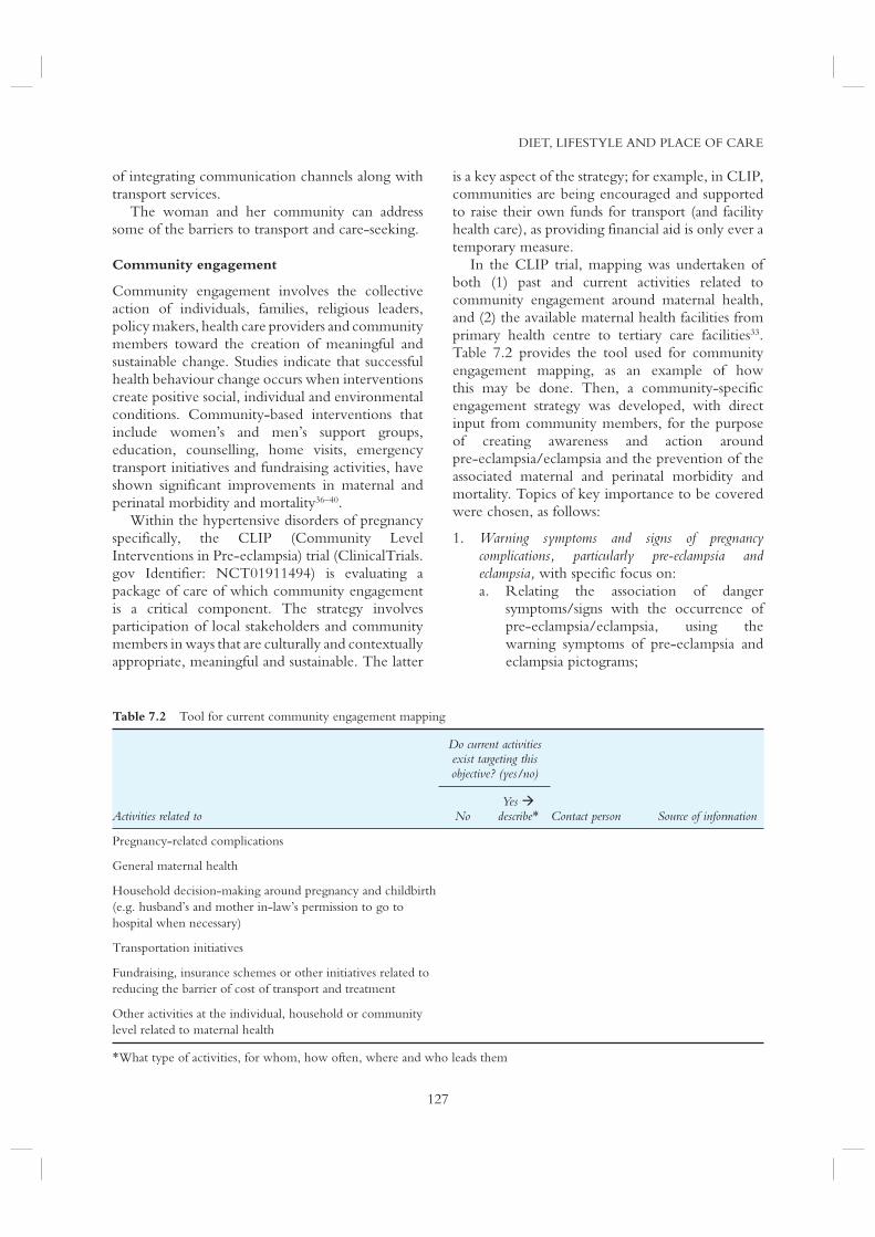
DIET, LIFESTYLE AND PLACE OF CARE
127
of integrating communication channels along with
transport services.
The woman and her community can address
some of the barriers to transport and care-seeking.
Community engagement
Community engagement involves the collective
action of individuals, families, religious leaders,
policy makers, health care providers and community
members toward the creation of meaningful and
sustainable change. Studies indicate that successful
health behaviour change occurs when interventions
create positive social, individual and environmental
conditions. Community-based interventions that
include women’s and men’s support groups,
education, counselling, home visits, emergency
transport initiatives and fundraising activities, have
shown significant improvements in maternal and
perinatal morbidity and mortality36–40 .
Within the hypertensive disorders of pregnancy
specifically, the CLIP (Community Level
Interventions in Pre-eclampsia) trial (ClinicalTrials.
gov Identifier: NCT01911494) is evaluating a
package of care of which community engagement
is a critical component. The strategy involves
participation of local stakeholders and community
members in ways that are culturally and contextually
appropriate, meaningful and sustainable. The latter
is a key aspect of the strategy; for example, in CLIP,
communities are being encouraged and supported
to raise their own funds for transport (and facility
health care), as providing financial aid is only ever a
temporary measure.
In the CLIP trial, mapping was undertaken of
both (1) past and current activities related to
community engagement around maternal health,
and (2) the available maternal health facilities from
primary health centre to tertiary care facilities33.
Table 7.2 provides the tool used for community
engagement mapping, as an example of how
this may be done. Then, a community-specific
engagement strategy was developed, with direct
input from community members, for the purpose
of creating awareness and action around
pre-eclampsia/eclampsia and the prevention of the
associated maternal and perinatal morbidity and
mortality. Topics of key importance to be covered
were chosen, as follows:
1. Warning symptoms and signs of pregnancy complications, particularly pre-eclampsia and eclampsia, with specific focus on:
a. Relating the association of danger
symptoms/signs with the occurrence of
pre-eclampsia/eclampsia, using the
warning symptoms of pre-eclampsia and
eclampsia pictograms;
Table 7.2 Tool for current community engagement mapping
Activities related to
Do current activities exist targeting this objective? (yes/no)
Contact person Source of informationNoYes
describe*
Pregnancy-related complications
General maternal health
Household decision-making around pregnancy and childbirth
(e.g. husband’s and mother in-law’s permission to go to
hospital when necessary)
Transportation initiatives
Fundraising, insurance schemes or other initiatives related to
reducing the barrier of cost of transport and treatment
Other activities at the individual, household or community
level related to maternal health
*What type of activities, for whom, how often, where and who leads them

THE FIGO TEXTBOOK OF PREGNANCY HYPERTENSION
128
b. Identifying the need for referral when
danger symptoms/signs associated with
pre-eclampsia/eclampsia occur; and
c. Considering discussion of PPH, a ‘visible’
cause of maternal death, as a segue into
discussion of pre-eclampsia/eclampsia as a
‘silent’ killer of pregnant and postpartum
women.
2. Permission for women to seek carea. Recognising the need for decision-making
power and/or prior permissions in the
event of obstetric emergencies; and
b. Discussion of how women can obtain
prior permission to seek that care
3. Identification of a skilled birth attendant4. Identification of a facility for delivery5. Identification of transport and treatment funds
a. Recognising the need to develop plans
for financial resources when required in
emergency conditions associated with
pre-eclampsia/eclampsia. Funds may be
personal or from the community. The
community engager should facilitate the
individual communities to form their own
plan for raising transport and treatment
funds for women in their communities;
b. Encouraging the identification of existing
community resources (if applicable) and
the development of community funds for
seeking emergency care. In CLIP, the
community is told that the trial will
supplement any existing funds, but
fundraising activities must build on those
funds to make this sustainable; and
c. Identifying available and appropriate
modes of transport, their associated costs,
and the means by which these modes can
be accessed in emergencies.
6. Feedback mechanisms about adverse outcomes and ‘great saves’a. In CLIP, the team requests that families of
sufferers share their experiences with the
community.
The CLIP trial will complete recruitment in 2017,
and findings should be available in 2018.
BEST PRACTICE POINTS
(Please see Appendix 7.1 for the evaluation of the strength of the recommendation and the quality of the
evidence on which they are based.)
1. There is insufficient evidence to make a recommendation about the usefulness of the following:
ongoing salt restriction among women with pre-existing hypertension, new severe dietary salt
restriction for women with any hypertensive disorder of pregnancy, and a heart-healthy diet or
calorie restriction for obese women specifically.
2. There is insufficient evidence to make a recommendation about the usefulness of exercise, workload
reduction, or stress reduction.
3. For women with gestational hypertension (without pre-eclampsia), some bed rest in hospital
(compared with unrestricted activity at home) may be useful to decrease severe hypertension and
preterm birth.
4. For women with pre-eclampsia who are hospitalised, strict bed rest is not recommended.
5. For all other women with hypertensive disorders of pregnancy, the evidence is insufficient to make
a recommendation about the usefulness of some bed rest, which may nevertheless, be advised based
on practical considerations.
6. Inpatient care should be provided for women with severe hypertension or severe pre-eclampsia,
however, defined.
7. A component of care through hospital day units or home care can be considered for women with
non-severe pre-eclampsia or non-severe (pre-existing or gestational) hypertension.
8. In under-resourced settings, transport from community to facility must be considered a responsibility
of women, their families, their communities, civil society and their care providers.

DIET, LIFESTYLE AND PLACE OF CARE
129
PRIORITIES FOR UNDER-RESOURCED SETTINGS
Table 7.3 outlines priorities for care for women in
under-resourced settings. Key issues relate to place
of care given the high proportion of women who
deliver at home without a skilled birth attendant or
plans for transport to facility should complications
arise.
Communities should play a key role in birth
preparedness and complication-readiness, including
how to arrange for transport to the primary health
centre or higher-level facility; obtaining prior
permission for transport should an emergency arise
between antenatal visits; saving money for obstetric
care; identifying a skilled birth attendant; and
identifying a facility for delivery.
Those facilitating community engagement
should consider covering broader issues in maternity
care, including appropriate timing of pregnancy
in order to avoid hypertensive disorder
of pregnancy-related (and other) pregnancy
complications. Avoidance of teen pregnancy
(discussed in Chapter 5) and food security for
adolescent girls (related to malnutrition, a risk
factor for pregnancy complications in general) must
be addressed by communities as culturally
appropriate priorities.
WHAT INTERNATIONAL GUIDELINES SAY (APPENDIX 7.2)41
Abbreviations for Clinical Practice Guidelines:
ACOG (American College of Obstetricians and
Gynecologists)42, NICE (National Institutes of
Clinical Excellence)43, PRECOG (Pre-eclampsia
Community Guideline)44, QLD (Queensland,
Australia)45,46, SOGC (Society of Obstetricians
and Gynaecologists of Canada)47, SOMANZ
(Society of Obstetric Medicine of Australia
and New Zealand)48, WHO (World Health
Organization)49.
There is little agreement, and a lack of detail,
about diet, lifestyle change and place of care in
international guidelines. In a review of guidelines
that cited recommendations that were either based
on high-quality evidence leading to a strong
recommendation, or were cited by at least included
Table 7.3 Priorities for non-pharmacological management of women with a hypertensive disorder of pregnancy (HDP)
by level of health care system at which care is delivered
Antepartum & postpartum
Initial priority Ultimate goal
Community
Primary health care
centre
(detect, stabilise and
refer)
Clear communication with referral unit
regarding transport to appropriate place of care
Clear communication with referral unit
regarding transport to appropriate place of care
Clear transport plan to secondary or
tertiary-level facilities
Clear transport service to secondary or
tertiary-level facilities
Community engagement about maternal health*
Facility
Secondary-level facility
(detect, manage and
refer if necessary)
Clear transport service to tertiary-level facility Clear transport service to tertiary-level facility
Hospitalisation of women with severe
hypertension or severe pre-eclampsia†
Hospitalisation of women with severe
hypertension or severe pre-eclampsia†
Tertiary-level (referral)
facility (detect and
manage definitively)
Hospitalisation of women with
severe hypertension or severe pre-eclampsia†
Hospitalisation of women with
severe hypertension or severe pre-eclampsia†
Outreach to communities for women with
HDP managed in the outpatient setting
* Community engagement should cover topics about timing of pregnancy, prior permissions to seek care, birth
preparedness and transport to facility† ‘Severe’ pre-eclampsia is defined according to the definition by the Canadian HDP Working Group – pre-eclampsia with
one/more serious complications. For details, see the Chapter 3

THE FIGO TEXTBOOK OF PREGNANCY HYPERTENSION
130
guidelines (of 13 included), only bed rest and
indications for hospital admission were discussed.
Recommendations against bed rest were made
by four guidelines, although no high-quality or
consistent recommendations were made (NICE,
WHO, ACOG, SOGC). Two exceptions were
made by type of hypertensive disorder of pregnancy:
(1) women with gestational hypertension may
benefit from bed rest in hospital (SOGC), and (2)
women with severe pre-eclampsia were excluded
from the ACOG bed rest recommendations.
For place of care, the only indication for hospital
admission that was consistently recommended was
severe hypertension (QLD, NICE, PRECOG,
SOGC, SOMANZ).
PRIORITIES FOR FUTURE RESEARCH
In well-resourced settings, research must address
the role, if any, of bed rest for women who are at
home with hypertensive disorders of pregnancy
and those who are in hospital, as well as the safety
of antepartum home care for women with
hypertensive disorders of pregnancy.
In under-resourced settings, where more than
99% of women with hypertensive disorders of
pregnancy die, lives lost from pre-eclampsia and
eclampsia result from delays in triage, transport
and treatment. Previous research in the field of
hypertensive disorders of pregnancy has focused
on institutional-level interventions, such as MgSO4
for eclampsia prevention and treatment, or
antihypertensive therapy for severe hypertension.
However, if we are to address the tragedies of
women dying prior to reaching facility or arriving
there moribund, or women being irreversibly
affected by pre-eclampsia by suffering complications
prior to arriving at facility, we need to address care
in the community and transportation issues.
There is now sufficient evidence of the
effectiveness of community-based care packages in
maternal and newborn care that research is required
specifically for the hypertensive disorders of
pregnancy. The CLIP trial is a singular step towards
addressing the excess maternal and perinatal
mortality that derive from the failure to identify
and rapidly manage pre-eclampsia and eclampsia at
the community level in LMICs. The intervention
is a combination of community engagement and
community-based triage, transport and treatment
by community-based workers.
REFERENCES
1. Abián-Vicén J, Del Coso J, González-Millán C, Salinero JJ, Abián P. Analysis of dehydration and strength in elite badminton players. PLoS One 2012; 7(5):e37821
2. Mydlík M, Derzsiová K, Bohus B. Renal function abnormalities after marathon run and 16-kilometre long-distance run. Przegl Lek 2012;69(1):1–4
3. Appel LJ, Moore TJ, Obarzanek E, Vollmer WM, Svetkey LP, Sacks FM, et al. A clinical trial of the effects of dietary patterns on blood pressure. N Engl J Med 1997;336(16):1117–1124
4. Thangaratinam S, Rogozinska E, Jolly K, Glinkowski S, Roseboom T, Tomlinson JW, et al. Effects of interventions in pregnancy on maternal weight and obstetric outcomes: meta-analysis of randomised evidence. BMJ 2012;344:e2088
5. Muktabhant B, Lawrie TA, Lumbiganon P, Laopaiboon M. Diet or exercise, or both, for preventing excessive weight gain in pregnancy. Cochrane Database Syst Rev 2015;6:CD007145
6. Institute of Medicine and National Research Council. Weight Gain During Pregnancy: Reexamining The Guidelines. 2009
7. Furber CM, McGowan L, Bower P, Kontopantelis E, Quenby S, Lavender T. Antenatal interventions for reducing weight in obese women for improving pregnancy outcome. Cochrane Database Syst Rev 2013(1):CD009334
8. Sacks FM, Svetkey LP, Vollmer WM, Appel LJ, Bray GA, Harsha D, et al. Effects on Blood Pressure of Reduced Dietary Sodium and the Dietary Approaches to Stop Hypertension (DASH) Diet. N Engl J Med 2001;344(1):3–10
9. Davies GAL, Wolfe LA, Mottola MF, MacKinnon C, Arsenault M, Bartellas E, et al. Exercise in pregnancy and the postpartum period. J Obstet Gynaecol Can 2003;25(6):516–29
10. Daskalopoulou S, Khan N, Quinn R, Ruzicka M, Mckay D, Hackam D, et al. The 2012 Canadian Hypertension Education Program Recommendations for the Management of Hypertension: Blood Pressure Measurement, Diagnosis, Assessment of Risk, and Therapy. Can J Cardiol 2012;28(3):270–287
11. Hamlin RHJ. The prevention of eclampsia and pre-eclampsia. Lancet 1952;259(6698):64–68
12. Meher S, Abalos E, Carroli G. Bed rest with or without hospitalisation for hypertension during pregnancy. Cochrane Database Syst Rev 2005(4):CD003514

DIET, LIFESTYLE AND PLACE OF CARE
131
13. Greer IA. 1 Epidemiology, risk factors and prophylaxis of venous thrombo-embolism in obstetrics and gynaecology. Baillieres Clin Obstet Gynaecol 1997;11(3):403–430
14. Crowther CA, Bouwmeester AM, Ashurst HM. Does admission to hospital for bed rest prevent disease progression or improve fetal outcome in pregnancy complicated by non-proteinuric hypertension? Br J Obstet Gynaecol 1992;99(1):13
15. Leung KY, Sum TK, Tse CY, Law KW, Chan MY. Is in-patient management of diastolic blood pressure between 90 and 100 mm Hg during pregnancy necessary? Hong Kong Med J 1998 Jun;4(2):211–217
16. Rosenberg K, Twaddle S. Screening and surveillance of pregnancy hypertension--an economic approach to the use of daycare. Baillieres Clin Obstet Gynaecol 1990;4(1):89–107
17. Turnbull DA, Wilkinson C, Gerard K, Shanahan M, Ryan P, Griffith EC, et al. Clinical, psychosocial, and economic effects of antenatal day care for three medical complications of pregnancy: a randomised controlled trial of 395 women. Lancet 2004;363(9415):1104–1109
18. Tuffnell AJ, Tuffnell DJ, Lilford RJ, Buchan PC, Prendiville VM, Holgate MP, et al. Randomised controlled trial of day care for hypertension in pregnancy. Lancet 1992;339(8787):224–227
19. Bergel E, Carroli G, Althabe F. Ambulatory versus conventional methods for monitoring blood pressure during pregnancy. Cochrane Database Syst Rev 2002(2):CD001231
20. Kröner C, Turnbull D, Wilkinson C. Antenatal day care units versus hospital admission for women with complicated pregnancy. Cochrane Database Syst Rev 2001(4):CD001803
21. Dunlop L, Umstad M, McGrath G, Reidy K, Brennecke S. Cost-effectiveness and patient satisfaction with pregnancy day care for hypertensive disorders of pregnancy. Aust N Z J Obstet Gynaecol 2003;43(3):207–212
22. Helewa M, Heaman M, Robinson MA, Thompson L. Community-based home-care program for the management of pre-eclampsia: an alternative. Can Med Assoc J 1993;149(6):829–834
23. Waugh J, Habiba MA, Bosio P, Boyce T, Shennan A, Halligan AWF. Patient Initiated Home Blood Pressure Recordings Are Accurate in Hypertensive Pregnant Women. Hypertens Pregnancy 2003;22(1):93–97
24. Walker S, Permezel M, Brennecke S, Tuttle L, Ugoni A, Higgins J. The effect of hospitalisation on ambulatory
blood pressure in pregnancy. Aust N Z J Obstet Gynaecol 2002;42(5):490–493
25. Waugh J, Bosio P, Shennan A, Halligan A. Inpatient monitoring on an outpatient basis: managing hypertensive pregnancies in the community using automated technologies. J Soc Gynecol Investig 2001;8(1):14–17
26. Barton JR, Istwan NB, Rhea D, Collins A, Stanziano GJ. Cost-savings analysis of an outpatient management program for women with pregnancy-related hypertensive conditions. Dis Manag 2006;9(4):236–241
27. Barton JR, Stanziano GJ, Sibai BM. Monitored outpatient management of mild gestational hypertension remote from term. Am J Obstet Gynecol 1994;170(3):765–769
28. Heaman M, Robinson MA, Thompson L, Helewa M. Patient satisfaction with an antepartum home-care program. J Obstet Gynecol Neonatal Nurs 1994;23(8):707–713
29. Lassi ZS, Bhutta ZA. Community-based intervention packages for reducing maternal and neonatal morbidity and mortality and improving neonatal outcomes. Cochrane Database Syst Rev 2015(3):CD007754
30. Wilson AK, Martel MJ. Maternal Transport Policy (SOGC policy statement no. 165). J Obstet Gynaecol Can 2005;27(10):956–958
31. Thaddeus S, Maine D. Too far to walk: maternal mortality in context. Soc Sci Med 1994 Apr;38(8):1091–1110
32. Akeju DO, Vidler M, Sotunsa JO, Osiberu MO, Orenuga EO, Olufemi OT, Adetoro OO, von Dadelszen P, Dada OA and the CLIP Nigeria Feasibility Working Group [Akinmade Adepoju, Diane Sawchuck, Rahat Qureshi, Beth Payne, Sharla Drebit, Chirag Kariya, Zulfiqar Bhutta, and Laura Magee]. Human resource constraints and the prospect of task-shifting among community health workers for emergency management of pre-eclampsia in Nigeria. Reproductive Health. 2016;(S2)
33. Babinard J, Roberts P. Maternal and Child Mortality Development Goals: What Can the Transport Sector Do. World Bank; 2006
34. Krasovec K. Auxiliary technologies related to transport and communication for obstetric emergencies. Int J Gynaecol Obstet 2004;85(1):S14-S23
35. Bossyns P, Abache R, Abdoulaye MS, Lerberghe WV. Unaffordable or cost-effective?: introducing an emergency referral system in rural Niger. Trop Med Int Health 2005;10(9):879–887

THE FIGO TEXTBOOK OF PREGNANCY HYPERTENSION
132
36. Colbourn T, Pulkki-Brännström A, Nambiar B, Kim S, Bondo A, Banda L, et al. Cost-effectiveness and affordability of community mobilisation through women’s groups and quality improvement in health facilities (MaiKhanda trial) in Malawi. Cost Eff Resour Alloc 2015;13(1):1
37. Younes L, Houweling T, Azad K, Kuddus A, Shaha S, Haq B, et al. The effect of participatory women’s groups on infant feeding and child health knowledge, behaviour and outcomes in rural Bangladesh: a controlled before-and-after study. J Epidemiol Community Health 2015;69(4):374–381
38. Younes L, Houweling TA, Azad K, Costello A, Fottrell E. Estimating coverage of a women’s group intervention among a population of pregnant women in rural Bangladesh. BMC Pregnancy Childbirth 2012;12:60
39. Lewycka S, Mwansambo C, Rosato M, Kazembe P, Phiri T, Mganga A, et al. Effect of women’s groups and volunteer peer counselling on rates of mortality, morbidity, and health behaviours in mothers and children in rural Malawi (MaiMwana): a factorial, cluster-randomised controlled trial. Lancet 2013;381(9879):1721–1735
40. Farnsworth SK, Böse K, Fajobi O, Souza PP, Peniston A, Davidson LL, et al. Community Engagement to Enhance Child Survival and Early Development in Low- and Middle-Income Countries: An Evidence Review. J Health Commun 2014;19(sup1):67–88
41. Gillon TE, Pels A, von Dadelszen P, MacDonell K, Magee LA. Hypertensive disorders of pregnancy: a systematic review of international clinical practice guidelines. PLoS One 2014;9(12):e113715
42. American College of Obstetricians and Gynecologists, Task Force on Hypertension in Pregnancy. Hypertension in pregnancy. Report of the
American College of Obstetricians and Gynecologists’ Task Force on Hypertension in Pregnancy. Obstet Gynecol 2013 Nov;122(5):1122–1131
43. National Collaborating Centre for Women’s and Children’s Health (UK). CG107: Hypertension in pregnancy: The management of hypertensive disorders during pregnancy. NICE: Guidance 2010 Aug. Available at https://www.nice.org.uk/guidance/cg107
44. Milne F, Redman C, Walker J, Baker P, Bradley J, Cooper C, et al. The pre-eclampsia community guideline (PRECOG): how to screen for and detect onset of pre-eclampsia in the community. BMJ 2005 Mar 12;330(7491):576–80
45. Queensland Maternity and Neonatal Clinical,Guidelines Program. Hypertensive disorders of pregnancy. 2013; MN10.13-V4-R15
46. Queensland Maternity and Neonatal Clinical,Guidelines Program. Supplement: hypertensive disorders of pregnancy. 2013;MN10.15.V4-R15
47. Magee LA, Pels A, Helewa M, Rey E, von Dadelszen P. Diagnosis, evaluation, and management of the hypertensive disorders of pregnancy. Pregnancy Hypertens 2014;4(2):105–145
48. SOMANZ: Lowe SA, Bowyer L, Lust K, McMahon LP, Morton MR, North RA, et al. The SOMANZ Guidelines for the Management of Hypertensive Disorders of Pregnancy 2014. Aust N Z J Obstet Gynaecol 2015;55:11–16 (executive summary). Available at https://somanz.org/documents/ HTPregnancyGuidelineJuly2014.pdf (full guideline)
49. World Health Organization. WHO recommendations for prevention and treatment of pre-eclampsia and eclampsia. 2011. Avaiable at http://www.who.int/reproductivehealth/publications/maternal_perinatal_health/9789241548335/en/
133
SYNOPSIS
The management of the hypertensive disorders of pregnancy encompasses far more than use of antihypertensive therapy. Women with pre-existing or gestational hypertension are at risk of it evolving into pre-eclampsia, a multisystem disorder of endothelial dysfunction. As such, attention must be paid to judicious fluid management, antihypertensive therapy of severe and non-severe hypertension with oral or parenteral agents, magnesium sulphate (MgSO4) for eclampsia prevention and treatment as well as fetal neuroprotection with birth at <34 weeks, antenatal corticosteroids for acceleration of fetal pulmonary maturity, and various therapies for HELLP (haemolysis, elevated liver enzyme, low platelet) syndrome, including transfusion of blood products and, possibly, corticosteroids. The WHO Model List of Essential Medicines includes all of the aforementioned interventions other than fluid therapy for pregnant women. It is our responsibility to ensure that we advocate the use of effective interventions whether we practice in well- or under-resourced settings.
8Fluids, drugs and transfusion
LA Magee, D Hall, JL van der Merwe, R Qureshi, E Rey, MF Escobar Vidarte
INTRODUCTION
At present, timed delivery of the placenta is the
only cure for the hypertensive disorders of
pregnancy. Care aims to optimise outcome for the
fetus and reduce maternal risk related to end-organ
complications (Table 8.1).
Fluid management
Plasma volume expansion
Plasma volume expansion is not recommended for
women with pre-eclampsia. The rationale for this
practice was that women with pre-eclampsia are
intravascularly volume contracted and sympathetic
tone is high. Observational studies suggested that
plasma volume expansion (with crystalloid or
colloid) improved maternal haemodynamics,
umbilical blood flow velocities, fetal growth and
perinatal mortality. However, trials (of colloid
solution) demonstrated no improvement in
maternal or perinatal outcomes (4 trials, 277
women)2,3. In the largest trial (216 women), plasma
volume expansion was associated with harm
– namely more Caesarean deliveries, a
(non-significantly) shorter pregnancy prolongation,
and a (non-significant) increase in pulmonary
oedema3. Also, there was no evidence of benefit as
measured by an increase in fetal middle cerebral or
umbilical artery blood flow velocity4, a decrease
in sympathetic tone5, or an improvement in
neurodevelopmental outcomes at the age of 1 year6.
KEY POINT
Use fluids judiciously in the hypertensive
disorders of pregnancy, particularly pre-eclampsia

THE FIGO TEXTBOOK OF PREGNANCY HYPERTENSION
134
Table 8.1 Management of pre-eclampsia. (Adapted from Mol et al., Lancet 2015 Sep 2. pii: S0140-6736(15)00070-71 with
permission)
Antepartum (regardless of gestational age) and postpartum (unless otherwise specified)
Place of care Inpatient care when there is severe hypertension or maternal symptoms, signs, or abnormal laboratory
tests
Outpatient care can be considered, recognising that many women are not eligible and hospital
re-admission rates are high following home care
Consultation Obstetrics to ensure that pre-eclampsia risk is recognised and appropriate maternal and fetal
surveillance is put in place
Anaesthesia to plan maternal monitoring and plan neuraxial analgesia/anaesthesia in labour to assist
with blood pressure control and facilitate Caesarean delivery (should it be necessary)
Fluid management Restrict to a maximum of 80 mL/h when an IV is in place
Antihypertensive
therapy
Severe hypertension (blood pressure ≥160/110 mmHg):Consider oral or parenteral agents that can be repeated in 30 min if blood pressure remains at
160 mmHg systolic or 110 mmHg diastolic:
• Nifedpine capsule (10 mg orally without biting to a maximum of 30 mg)
• Nifedipine tablet (10 mg orally to a maximum of 30 mg)
• Hydralazine (5 mg IV bolus then if needed, 5–10 mg IV to a maximum of 45 mg)
• Labetalol (20 mg IV then if needed, 40 mg then 80 mg to a maximum of 300 mg)
Consider alternative oral agents that can be repeated in 1 h (supported by less evidence in pregnancy):
• Labetalol (200 mg orally)
• Clonidine (0.1–0.2 mg orally)*†
• Only postpartum – Captopril (6.25–12.5 mg orally)*
Non-severe hypertension• Methyldopa (500–2000 mg/d in 3 or 4 divided doses)
• Labetalol (300–2400 mg/d in 3 or 4 divided doses)
• Nifedipine (20–120 mg/d once daily)
MgSO4 Eclampsia treatment
• 4 g IV (over 5 min) then 1 g/h IV
• If already on MgSO4, administer another 2–4 g IV (over 5 min) and increase infusion to 2 g/h IV
Eclampsia prevention among women with pre-eclampsia• 4 g IV (over 5 min) then 1 g/h IV
Fetal neuroprotection4 g IV (with/without 1 g/h until delivery or 24 h maximum) for women with imminent delivery at
<34+0 weeks who do not otherwise quality for eclampsia prevention or treatment
Corticosteroids Antenatally only, for fetal pulmonary maturity when delivery is anticipated within the next 7 days
and at <340–6 weeks
HELLP syndrome (10 mg dexamethasone IV every 12 h for 48 h) if improvement in laboratory
parameters alone will change management, such as eligibility for neuroaxial anaesthesia/analgesia or
platelet transfusion
Platelet transfusion
for HELLP
syndrome
Recommended for counts: <20 109/L, 20–49 109/L prior to Caesarean, or 50 109/L ( packed
red blood cells) with excessive active bleeding, platelet dysfunction, a rapidly falling platelet count, or
coagulopathy2
* Captopril (25 mg) and clonidine (0.1 mg) are being compared in a postpartum randomised controlled trial
(NCT01761916) based on the effectiveness of these medications for severe hypertension treatment outside pregnancy† Clonidine therapy is not recommended during breastfeeding (http://toxnet.nlm.nih.gov/newtoxnet/lactmed.htm)
FLUIDS, DRUGS AND TRANSFUSION
135
Fluid restriction
Women with pre-eclampsia who are on delivery
suite, for stabilisation or delivery, require IV access.
In an international benchmarking study, restricting
IV fluids was associated with lower rates of
pulmonary oedema without an increase in acute
renal failure7. As such, IV fluid of no more than
80 mL/h is recommended8.
Oliguria (<15 mL of urine/h for 6 consecutive
hours) is common in pre-eclampsia, particularly
postpartum. Reasons include oxytocin
administration and high levels of antidiuretic
hormone following surgery. In the absence of
pre-existing renal disease or a rising creatinine that
mandate fluid challenge to rule out a component of
pre-renal failure as a cause of renal dysfunction,
oliguria should be tolerated and observed, at least
over hours because fluid administration can
precipitate pulmonary oedema in a dose-dependent
fashion3,7. Furosemide should not be administered
unless there is pulmonary oedema or the woman
has oliguric renal failure (in which case increasing
urine output simplifies management but does not
improve prognosis in renal failure). ‘Renal-dose’
dopamine is not recommended; although it appears
to increase postpartum urine output in women
with pre-eclampsia; this is of uncertain clinical
importance (1 trial, 40 women)9.
Antihypertensive treatment of severe hypertension (blood pressure of 160 mmHg
systolic or 110 mmHg diastolic)
The following discussion applies to women with
either pre-existing or gestational hypertension,
with or without evidence of pre-eclampsia.
In the WHO Prevention and Treatment of
Pre-eclampsia and Eclampsia recommendations,
antihypertensive treatment of severe hypertension
during pregnancy was strongly recommended10.
This seems very reasonable despite the fact that the
quality of evidence on which the recommendation
was based was graded as ‘very low’. First, there
are no relevant placebo-controlled randomised
controlled trials that prove that women randomised
to antihypertensive therapy more frequently have
their blood pressure lowered compared with those
randomised to placebo; however, such randomised
controlled trials would be unethical and will never
be done. Second, severe systolic hypertension is a
independent risk marker for stroke in pregnancy11.
Third, an individual short-acting antihypertensive
agent is successful at lowering maternal blood
pressure in at least 80% of women, based on
randomised controlled trials of one antihypertensive
drug versus another (as discussed below). Finally, a
recent report of the Confidential Enquiries into
Maternal Deaths in the UK that covered the
hypertensive disorders of pregnancy (2005–2008)
identified the failure to treat the severe (particularly
systolic) hypertension of pre-eclampsia as the single
most serious failing in the clinical care of the
women who died12,13. It is of note that concerted
efforts in the UK to address treatment of severe
hypertension have been associated with a fall in the
contribution of the hypertensive disorders of
pregnancy to maternal mortality, based on
2009–2012 data14. Similarly, in South Africa that
has a legislated Confidential Enquiries into
Maternal Deaths process, maternal deaths owing
to complications of hypertension have featured
prominently, and recommendations for
antihypertensive therapy have been associated with
a reduction of deaths in this category15.
In deciding on the need for treatment and the
urgency with which blood pressure should be
lowered, both the absolute level of blood pressure
(i.e., severe or non-severe) and the rate with which
it has risen should be considered. Experimental
evidence from cats suggests that an abrupt (versus
step-wise) increase in blood pressure is associated
with more permeability of the cerebral vessels,
taken as a measure of vascular injury16. Presumably,
abrupt increases in intraluminal pressure may result
in mechanical distension of the cerebral vessel wall
which may adapt better to gradual or step-wise
increases.
Women with a hypertensive ‘urgency’ (i.e.,
acute rise in blood pressure that is not associated
with end-organ dysfunction) may be treated with
oral antihypertensive agents that have peak drug
effects in 1–2 hours (e.g., oral labetalol), recognising
that gastric emptying may be delayed or unreliable
among women in active labour. Choice of agents is
discussed below.
In contrast to a hypertensive ‘urgency’, a
hypertensive ‘emergency’ is associated with
end-organ complications, such as eclampsia,
pulmonary oedema and renal failure. Whether
headache and visual symptoms should be considered

THE FIGO TEXTBOOK OF PREGNANCY HYPERTENSION
136
end-organ complications of a hypertensive
‘emergency’ is not known. They are non-specific
and common, being documented in about 30% of
women who are hospitalised with pre-eclampsia17.
There is a general appreciation that the goal of
antihypertensive therapy for severe hypertension is
not normalisation of blood pressure, but rather,
lowering of blood pressure to a non-severe level of
hypertension that decreases the risk of stroke18.
Also, there is recognition that lowering of blood
pressure, even to levels that remain outside the
hypertensive range has the potential to precipitate
fetal distress and fetal heart rate monitoring (FHR)
monitoring is advised8,18.
Based on extrapolation of the approach outside
pregnancy, hypertensive emergencies should be
treated with short-acting antihypertensive agents
and an arterial line when possible aimed at lowering
mean arterial blood pressure by no more than 25%
over minutes to hours; this is equivalent to taking a
blood pressure of 220/130 mmHg to 165/98 over
1–2 hours, and then further lowering blood pressure
below 160/100 mmHg over the next 2 hours.
Outside pregnancy, American19, British20 and
European guidelines21 all recommend that
antihypertensive therapy be initiated with two oral
agents when blood pressure is 20 mmHg systolic
or 10 mmHg diastolic above target. The American
(JNC VII) guidelines stress that initial therapy of
severe hypertension should be with two oral agents.
This recommendation is based on the multifactorial
nature of the blood pressure elevation and the
limited (but variable) average blood pressure
reduction of 9.1 mmHg systolic and 5.5 mmHg
diastolic achieved after treatment with any one
agent, given compensatory mechanisms in response
to any single agent of a given class22. However,
these recommendations are based on treatment in
the setting of chronic hypertension, outside
pregnancy, and following long-term therapy19. In
pregnancy, initiating antihypertensive therapy with
one agent may be more appropriate given the
intravascular volume depletion associated with
both severe hypertension and pre-eclampsia, and
the potential for fetal compromise if blood pressure
is acutely lowered too much.
Choice of antihypertensive agent
Table 8.2 presents the antihypertensive agents used
most commonly for hypertensive urgencies in
pregnancy, as well as alternatives that have a
different pharmacology. Only hydralazine is on the
WHO Model List of Essential Medicines (2015) for
treatment of severe hypertension, although
nifedipine capsules (10 mg) are listed as a tocolytic23.
The treatment approach recommended here is
cautious, in an attempt to lower blood pressure
progressively, over hours and to minimise the risk
of maternal hypotension and/or fetal distress. First,
although nifedipine capsules have been
recommended in doses of 20 mg by the American
College of Obstetricians and Gynecologists if a
10 mg dose fails18, this dosing approach is not
recommended here because few of the relevant
trials have administered nifedipine in this way24,25.
Second, none of the agents recommended here are
to be repeated prior to 30 min unless there is a
hypertensive emergency, although some societies
recommend more frequent administration (i.e.,
every 10 min for labetalol and every 20 min for
either hydralazine or nifedipine)18.
Recommendations about antihypertensive
therapy for severe hypertension in pregnancy come
from 47 trials (4322 women) that have compared
one short-acting antihypertensive with another26–28.
Just over half of these trials (i.e., 28/47) involved
comparisons between parenteral hydralazine (usually
5 mg), parenteral labetalol (usually 20 mg) and
calcium channel blockers (usually oral nifedipine
10 mg capsules). Each of these three agents is
a reasonable choice for treatment of severe
hypertension (in doses listed in Table 8.2). Some
antihypertensive agents may be more or less
appropriate for some women based on associated
medical conditions (such as asthma) or therapies
(such as current treatment with full doses of labetalol
as an outpatient). Hydralazine may be associated
with more adverse effects for the mother and labetalol
with neonatal bradycardia, as discussed below.
KEY POINTS
• Women with severe hypertension in
pregnancy (or postpartum) should be treated
with antihypertensive therapy
• The antihypertensive agents used most
commonly are oral nifedipine (capsules or
tablets) or IV labetalol or hydralazine (see
Table 8.1 and Appendix 8.1, Figures S8.1
and S8.2)

FLUIDS, DRUGS AND TRANSFUSION
137
Most published trials have compared parenteral
hydralazine (usually 5 mg IV) with either calcium
channel blockers (N = 11 trials, 699 women, usually
nifedipine 10 mg capsules orally)26,27,29 or parenteral
labetalol (N = 8 trials, 384 women, usually 20 mg
IV)26,27, with repeat doses administered every
15–20 minutes to achieve blood pressure control in
at least 80% of women; in nine other trials,
hydralazine was compared with drugs used
regionally or infrequently: mini-dose diazoxide
(1 trial, 124 women)30, ketanserin (4 trials, 210
women)26, urapidil (3 trials, 101 women)27,31 and
prostacyclin (1 trial, 47 women)26.
Compared with calcium channel blockers
(usually nifedipine), hydralazine may be a less effective antihypertensive and also associated with
more maternal side-effects (11 trials of which 9
studied oral nifedipine 10 mg, one nifedipine 5 mg,
and one parenteral isradipine26,27,32. There is no
published review of all relevant trials, so one
summary statistic is not available.
Compared with labetalol, hydralazine may be a
more effective antihypertensive but also associated
with more maternal hypotension and maternal
side-effects (8 trials, 384 women)26,27. Most of the
published hydralazine trials were included in a 2003
meta-analysis that compared hydralazine with any
other short-acting antihypertensive agent;
hydralazine was found to be associated with more
adverse effects, including maternal hypotension,
Caesarean delivery and adverse FHR effects26. It
should be noted that in two hydralazine versus
labetalol trials, parenteral labetalol was associated
with more neonatal bradycardia (which required
intervention in one of six affected babies in one
trial26,33,34.
Compared with labetalol, oral nifedipine (N = 7
trials, 363 women)28,35–39 appears to be similarly
effective for blood pressure control (RR 0.42, 95%
CI 0.18–0.96), as does parenteral nicardipine (60
women)40, although there is only one such trial.
In the trials discussed above, labetalol was
administered parenterally; however, it has been
given orally for hypertensive urgencies. In a dose of
200 mg, oral labetalol has been used with good effect
as part of a regional pre-eclampsia protocol41. In a
clinical trial of preterm severe hypertension, 100 mg
of oral labetalol every 6 hours achieved the stated
blood pressure goal (of about 140/90 mmHg) in
47% of women42. We believe that these data
are insufficient to support the NICE 2010
recommendation to use oral labetalol as initial
therapy for severe hypertension in pregnancy43;
however, if severe hypertension is detected in the
office setting, an oral dose of labetalol or another
antihypertensive may be useful to administer while
the woman is being transported to hospital for
further evaluation and treatment44. Other than oral
nifedipine (discussed above), methyldopa may be
suitable although probably starting with a 750 mg
dose rather than the 250 mg used in the one relevant
randomised controlled trial42; IV methyldopa is
manufactured for women who are unable to take the
medication by mouth. Prazosin may be associated
with an increase in stillbirth and is not recommended45.
The nifedipine preparations that are appropriate
for treatment of severe hypertension are the capsule
and the PA tablet29,46. The PA tablets have been
withdrawn from some markets. Most authors of
randomised trials did not specify whether nifedipine
capsules were bitten (prior to swallowing), which
may have a greater effect on blood pressure. The
10 mg tablet may be associated with less maternal
hypotension than the 10 mg capsule when bitten/
punctured (2 trials, 87 women)29,46. Theoretically,
the 5 mg (instead of the 10 mg) capsule may reduce
the risk of a precipitous fall in blood pressure,
although there are only two published reports
comparing nifedipine 5 mg with hydralazine 5 mg
IV (250 women)34,47.
Nifedipine or other calcium channel blockers
can be used together with MgSO4. The risk of
neuromuscular blockade with contemporaneous
use of nifedipine and MgSO4 is <1%, based on a
single-centre, controlled study and a complete data
synthesis from the literature48,49. Blockade is
reversed with 10 g of IV calcium gluconate.
MgSO4 is not an antihypertensive agent50.
However, transient decreases in blood pressure
may be seen. Observational literature describes no
decrease51 or a transient decrease in blood
pressure52–55 30 minutes after 2–5 g of IV MgSO4
(with or without ongoing infusion), usually in
patients with pre-eclampsia. In randomised
controlled trials of MgSO4 for fetal neuroprotection,
an excess of hypotension was seen (i.e., 9.7% with
MgSO4 versus 6.5% with placebo, RR 1.51, 95%
CI 1.09–2.09)56. When MgSO4 was compared
directly with parental nimodipine, MgSO4 was less
effective in lowering blood pressure (2 trials, 1683
women)27 or parenteral labetalol (1 trial, 177
women)57. Therefore, although a sustained

THE FIGO TEXTBOOK OF PREGNANCY HYPERTENSION
138
Tab
le 8
.2
Agen
ts u
sed m
ost
com
monly
for
trea
tmen
t of a
blo
od p
ress
ure
≥160/110 m
mH
g
Age
ntM
echa
nism
of a
ction
Dos
age
Phar
mac
okin
etics
*
Com
men
tsO
nset
Peak
Dur
atio
n
Mos
t com
mon
ly re
com
men
ded
Lab
etal
ol
†P
erip
her
al a
lpha-
1 a
nd
(non-s
elec
tive)
bet
a-1
and 2
rec
epto
r
anta
gonis
t
Inte
rmitte
nt
dosi
ng
Sta
rt w
ith 2
0 m
g I
V o
ver
2 m
in
Rep
eat
with 4
0 m
g t
hen
80 m
g I
V
(eac
h o
ver
2 m
in)
q 3
0 m
in
Continuous
infu
sion
1–2 m
g/m
in
(max
dosa
ge
300 m
g)
5 m
in 30 m
in≥4 h
Bes
t av
oid
ed in w
om
en w
ith a
sthm
a or
hea
rt fai
lure
Neo
nat
olo
gy s
hould
be
info
rmed
if th
e w
om
an is
in
labour,
as
par
ente
ral la
bet
alol m
ay c
ause
neo
nat
al
bra
dyca
rdia
Par
ente
ral th
erap
y s
hould
be
follow
ed b
y o
ngoin
g
ora
l th
erap
y t
o m
ainta
in B
P
Nifed
ipin
eC
alci
um
chan
nel
blo
cker
(vas
odilat
or)
Cap
sule
5–10 m
g t
o s
wal
low
without
bitin
g
Rep
eat
ever
y 3
0 m
in
5–10 m
in 30 m
in≥6 h
Ther
e ar
e th
ree
types
of nifed
ipin
e pre
par
atio
ns
with
whic
h a
ll s
taff
must
be
fam
ilia
r: c
apsu
les,
inte
rmed
iate
-rel
ease
tab
lets
(PA
, SR
or
reta
rd t
able
t)
and s
low
-rel
ease
tab
lets
(X
L, M
R o
r LA
)
Nifed
ipin
e m
ay b
e giv
en a
t th
e sa
me
tim
e as
MgSO
4
PA
, SR
or
reta
rd t
able
t
10 m
g t
o s
wal
low
Rep
eat
ever
y 3
0 m
in
(max
dosa
ge
30 m
g)
30 m
in240 m
in≥6 h
Hydra
lazi
ne
Dir
ect-
acting
vas
odilat
or
Inte
rmitte
nt
dosi
ng
5 m
g I
V
Rep
eat
5–10 m
g I
V e
ver
y 3
0 m
in
(may
be
giv
en I
M b
ut
unusu
al)
Continuous
infu
sion
0.5
–10 m
g/h I
V
(max
dosa
ge
45 m
g)
5 m
in 30 m
in3–8 h
May
incr
ease
the
risk
of m
ater
nal
hypote
nsi
on

FLUIDS, DRUGS AND TRANSFUSION
139
Lab
etal
ol
†P
erip
her
al a
lpha-
1 a
nd
(non-s
elec
tive)
bet
a-1
and -
2 r
ecep
tor
anta
gonis
t
200 m
g o
rally
Rep
eat
in 4
h
(max
dosa
ge
2400 m
g/day
in 4
div
ided
dose
s‡)
20–120 m
in1–4 h
8–12 h
Dura
tion is
dose
-dep
enden
t
Met
hyld
opa
Cen
tral
ly a
ctin
g
alpha-
2 r
ecep
tor
agonis
t
750 m
g o
rally
Rep
eat
in 6
h
(max
dosa
ge
2000 m
g/day
in 4
div
ided
dose
s‡)
Not
know
n4–6 h
24–48 h
Les
s ef
fect
ive
than
ora
l nifed
ipin
e
Clo
nid
ine¶
Cen
tral
ly a
ctin
g
alpha-
2 r
ecep
tor
agonis
t
0.1
–0.2
mg o
rally
Rep
eat
in 1
h
(max
dosa
ge
0.8
mg
‡)
30–60 m
in2–4 h
6–10 h
Clo
nid
ine
ther
apy is
not
reco
mm
ended
duri
ng
bre
astf
eedin
g§
Cap
topri
l¶ o
nly
postp
artu
mA
ngio
tensi
n-c
onver
ting
enzy
me
inhib
itor
6.2
5–12.5
mg o
rally
Rep
eat
in 1
h
(max
dosa
ge
75 m
g)
30 m
in60–90 m
in8
hC
apto
pri
l m
ust
NO
T b
e ad
min
iste
red b
efore
del
iver
y,
but
it is
acce
pta
ble
for
use
duri
ng b
reas
tfee
din
g§
Dura
tion is
dose
-dep
enden
t
Nitro
gly
ceri
n
infu
sion
Dir
ect
vas
odilat
ors
that
has
its
aff
ects
vei
ns
more
than
art
erio
les
5
g/m
in, in
crea
sed e
ver
y 5
min
(max
rat
e 100
g/m
in)
2–5 m
in 5 m
in5–10 m
inM
ain s
ide-
effe
cts
are
hea
dac
he
(due
to d
irec
t
vas
odilat
ion)
and t
achyca
rdia
(fr
om
ref
lect
sym
pat
het
ic
activat
ion)
Met
hae
moglo
bin
aem
ia h
as b
een r
eport
ed a
fter
24 h
of
trea
tmen
t
BP, blo
od p
ress
ure
; IM
, in
tram
usc
ula
r; I
V, in
trav
enous;
MgSO
4, m
agnes
ium
sulp
hat
e
* G
ener
al r
efer
ence
ww
w.d
rugs.
com
† B
eta-
blo
ckad
e is
3–7 t
imes
more
than
alp
ha-
blo
ckad
e, e
spec
ially a
t lo
wer
dose
s‡ D
osi
ng o
f th
is d
rug m
ay c
ontinue
afte
r th
e se
ver
e hyper
tensi
on h
as r
esolv
ed, as
it
is u
sed for
chro
nic
tre
atm
ent
of non-s
ever
e hyper
tensi
on
¶ C
apto
pri
l (2
5 m
g)
and c
lonid
ine
(0.1
mg)
are
bei
ng c
om
par
ed in a
post
par
tum
ran
dom
ised
contr
olled
tri
al (
NC
T01761916)
bas
ed o
n t
he
effe
ctiv
enes
s of th
ese
med
icat
ions
for
sever
e hyper
tensi
on t
reat
men
t outs
ide
pre
gnan
cy§ h
ttp:/
/to
xnet
.nlm
.nih
.gov/new
toxnet
/la
ctm
ed.h
tm

THE FIGO TEXTBOOK OF PREGNANCY HYPERTENSION
140
lowering of blood pressure cannot be anticipated
following a loading dose of MgSO4, the potential
for a transient lowering of blood pressure 30
minutes after administration should be considered
when antihypertensives are co-administered.
Nitrogylcerin (by infusion) compared favourably
with oral nifedipine in one small trial (32 women)58
and no adverse clinical effects were demonstrated
in other small studies58.60.
Mini-dose diazoxide (i.e., 15 mg IV every 3
minutes) was associated with less persistent severe
hypertension compared with parenteral hydralazine
(5 mg) in another small trial (124 women)30.
For refractory hypertension in an intensive care
setting, consideration can be given to using sodium
nitroprusside or higher dose diazoxide. The
theoretical concerns about nitroprusside are well
known: light-sensitivity, the need for careful
monitoring and the potential to cause fetal cyanide
toxicity. A published review of case reports (22
women, 24 fetuses) documented stillbirths among
five of 18 women (27.8%) treated antenatally with
nitroprusside, although the authors could not
attribute these deaths to fetal cyanide toxicity61.
High dose diazoxide (i.e., 75 mg IV every 30 min)
was associated in one trial (90 women) with an
excess of maternal hypotension (17.8%) compared
with IV labetalol (0%)62.
Observational literature illustrates that
hypotension may result with any short-acting
antihypertensive agent administered to women
with pre-eclampsia, because they are intravascularly
volume depleted. Therefore, it is prudent to
continuously monitor FHR until blood pressure
has stabilised.
Postpartum, hydralazine, labetalol, nifedipine
and methyldopa are appropriate for use during
breastfeeding, although only two trials have
compared hydralazine with either labetalol63 or
nifedipine64 for treatment of severe hypertension.
Nitroglycerine and diazoxide have not been studied
in breastfeeding, although treatment with one of
these agents would be expected to be very brief.
Nitroprusside is not advised in breastfeeding because
of the potential for toxic metabolites (thiocyanate
and cyanide) to cross into breast milk65. Captopril
could also be administered orally for severe
hypertension based on its effectiveness for this
indication outside pregnancy66 and its acceptability
during breastfeeding65. Although neonatologists
may express concerns about this in babies born
preterm or of low birth weight, no reports of
adverse effects were identified. Oral clonidine
which is effective for severe hypertension outside
pregnancy is not advocated for use in breastfeeding
because of high serum levels in breastfed infants65.
No relevant economic analyses were identified.
Antihypertensive treatment of non-severe hypertension (blood pressure of 140–159/
90–109 mmHg)
Management of a pregnant woman with a blood
pressure of 140–159/90–109 mmHg is much
debated. Any antihypertensive therapy will,
compared with placebo or no therapy, decrease the
risk of transient, severe hypertension (RR 0.49, 95%
CI 0.40–0.60; 20 trials, 2558 women; NNT 10,
95% CI 8–13) without a clear difference in other
maternal or perinatal outcomes, such as stroke,
perinatal death, or preterm delivery (29 trials, 3350
women)67. The results of a small pilot randomised
controlled trial (132 women)68 and a meta-regression
of randomised controlled trials (42 trials, 3892
women69,70) raised concerns that antihypertensive
therapy may be harmful. The meta-regression of
randomised controlled trials found a significant
relationship between the antihypertensive-induced
fall in mean arterial pressure and the risk of SGA
infants or lower birth weight. On the other hand, a
small trial of 125 women with mild essential or
gestational hypertension found that ‘very tight’ (goal
blood pressure <130/80 mmHg) versus ‘tight’
control (goal blood pressure 130–139/80–89 mmHg)
was associated with fewer antenatal hospitalisations
and a later gestational age at delivery71.
The results of a large definitive trial, CHIPS
(Control of Hypertension In Pregnancy Study), has
provided evidence that non-severe hypertension in
pregnancy should be treated with antihypertensive
therapy72. ‘Tight’ blood pressure control (target
diastolic 85 mmHg) (versus ‘less tight’ control,
target diastolic 100 mmHg) achieved a lower blood
pressure by 5.8/4.6 mmHg (p < 0.001). ‘Tight’
(versus ‘less tight’) control resulted in similar rates
of adverse perinatal outcome: the primary outcome
of perinatal death or high level neonatal care for
>48 hours (30.7% versus 31.4%; aOR 0.98, 95%
CI 0.74–1.30) and birth weight <10th percentile
for gestational age and gender (19.7% versus 16.1%;
aOR 1.28, 95% CI 0.93–1.79). However, ‘tight’
(versus ‘less tight’) control resulted in fewer adverse

FLUIDS, DRUGS AND TRANSFUSION
141
maternal outcomes: a significant decrease in severe
maternal hypertension (27.5% versus 40.6%; aOR
0.56, 95% CI 0.42–0.75) but similar rates of serious
maternal complications (2.0% versus 3.7%; aOR
0.57, 95% CI 0.26–1.27).
Although there is ongoing debate about whether
blood pressure should be lowered below a diastolic
blood pressure of 80 mmHg in the setting of
proteinuria (compared with non-proteinuric)
patients, a goal of <130/80 mmHg is specified only
for patients with diabetes mellitus in order to
decrease the risk of long-term cardiovascular disease
and diabetic nephropathy73.
As blood pressure is lowest at about 20 weeks’,
women may be able to discontinue antihypertensives
in early pregnancy. Medication should be restarted
as blood pressure rises again later in pregnancy.
There is no evidence that blood pressure should
be managed differently in women with
pre-eclampsia compared with those with
pre-existing or isolated gestational hypertension. It
should be noted that 47.3% of women developed
pre-eclampsia in the CHIPS trial, and the diastolic
blood pressure goal to which women were
randomised continued to be applied until delivery72.
Guidance on treatment of secondary causes of
hypertension is available from general hypertension
sources73.
When a decision is made to lower blood
pressure, antihypertensive therapy is warranted.
Relaxation techniques (such as guided imagery)
were not successful in lowering blood pressure in
one trial (69 women)74.
Therapy is usually initiated with one
antihypertensive agent, although this will not be
sufficient if blood pressure is more than 20/10 above
the target19. It is important to be familiar with a
number of antihypertensive options. Outside
pregnancy, only 30–50% of patients respond to a
particular antihypertensive drug. Also, women may
have another medical problem that is a contraindication
to a specific medication (such as severe asthma and
beta-blockers) or a characteristic that makes one type
of agent more likely to be effective (such as Black
race and calcium channel blockers).
Choice of antihypertensive agent
Table 8.3 presents the most commonly used
antihypertensive agents for non-severe pregnancy
hypertension.
There is little to guide the choice of
antihypertensive agent, including effects on FHR
and pattern, maternal and perinatal outcomes,
and long-term paediatric neurodevelopment.
Methyldopa, labetalol and nifedipine are the most
commonly recommended antihypertensives in
international practice guidelines, although oral
labetalol is not widely available in LMICs75. Only
methyldopa is on the WHO Model List of Essential
Medicines (2015) for non-severe pregnancy
hypertension23, and it appears to be a reasonable
antihypertensive choice; in the CHIPS trial,
women treated with methyldopa (versus labetalol)
may have had better outcomes, although this
comparison was non-randomised and subject to the
possibility of residual confounding76. Angiotensin
converting enzyme inhibitors and receptor blockers
should not be used later in pregnancy, and prazosin
and atenolol may be best avoided, as discussed
below.
KEY POINTS
• Antihypertensive therapy for non-severe
pregnancy hypertension does not affect
outcomes for the baby, but does decrease
severe hypertension and therefore, risk, for
the mother
• Oral methyldopa and oral labetalol are used
most frequently for treatment of non-severe
hypertension, but there are a wide variety of
agents that can be used
• ACE inhibitors and ARBs should NOT be
used in pregnancy
Whether pre-eclampsia haemodynamics (either
high cardiac output or peripheral vascular resistance)
should be used to guide therapy is unclear;
although haemodynamics may interact with the
pharmacodynamics of antihypertensives to
influence development of fetal growth restriction
or pre-eclampsia77, it is unknown if individualised
therapy would improve outcomes and be
cost-effective.
FHR and pattern
Oral antihypertensives do not appear to change
FHR or pattern, but the quality of the data is
poor78. A prudent approach would be to regard
changes in FHR or pattern to evolution of the

THE FIGO TEXTBOOK OF PREGNANCY HYPERTENSION
142
underlying hypertensive disorder of pregnancy, and
not to the antihypertensive agent that the woman is
taking.
Maternal and perinatal outcomes
In randomised controlled trials, usually of
women without comorbidities, a wide variety of
antihypertensive agents (started after the first
trimester of pregnancy) have been compared with
placebo or no therapy and shown to decrease the
risk of severe hypertension (as discussed above):
methyldopa, labetalol, other pure beta-blockers
(acebutolol, mepindolol, metoprolol, pindolol and
propranolol), calcium channel blockers (isradipine,
nicardipine, nifedipine and verapamil), hydralazine,
prazosin and ketanserin (29 trials, 3350 women)67.
In comparative trials of one antihypertensive
agent versus another, meta-analysis has revealed no
clear differences in maternal and perinatal outcomes
(22 trials, 1723 women)67, and small trials published
subsequently have been consistent with these
conclusions (2 trials, 163 women)79,80. Most trials
have compared beta-blockers with methyldopa.
Although alternative drugs may be more effective
at reducing the risk of severe hypertension than
methyldopa (RR 0.54, 95% CI 0.30–0.95; 11 trials,
638 women), and beta-blockers and calcium
channel blockers considered together may decrease
the risk of proteinuria (as a surrogate for
pre-eclampsia) (RR 0.73, 95% CI 0.54–0.99; 11
trials, 997 women), the significance of these
findings is unclear. The effects on both severe
hypertension and proteinuria are not seen in
individual drug comparisons.
Thiazide diuretics can be considered for use in
hypertensive women, but they are used mainly in
specific circumstances identified before pregnancy,
such as medullary sponge kidney for which a
decrease in renal calcium excretion is advantageous.
Table 8.3 Agents used most commonly for a blood pressure of 140–159/90–109 mmHg
Agent Mechanism of action Dosage Comments
Methyldopa Centrally acting alpha-2
receptor agonist decreased
sympathetic outflow
decreased peripheral vascular
resistance
250–500 mg PO BID-QID
(max dosage 2000 mg/day)
There is no evidence to support a loading dose of
methyldopa
Psychological side-effects (e.g., drowsiness or
depression) may occur but women do not change
drugs more frequently than with other medication
Within first 6 weeks of therapy, <10% may
develop hepatitis or cholestasis that can be detected
by laboratory testing; abnormalities should reverse
with discontinuation, but liver failure is rare
After 6 months of therapy, 10–20% develop a
positive direct Coombs test, but it does not
interfere with typing or cross matching and
associated haemolytic anaemia is rare
Labetalol *Peripheral alpha-1 and
(non-selective) beta-1 and 2
receptor antagonist
decreased peripheral vascular
resistance with no reflex
increase in heart rate
100–400 mg PO BID-QID
(max 2400 mg/day)
Some experts recommend a starting dose of
100 mg PO TID because the half-life of labetalol is
shorter in pregnancy
May be associated with postural hypotension,
especially at higher doses
Nifedipine Calcium channel blocker
vascular smooth muscle
relaxation decrease
peripheral vascular resistance
PA, SR or retard tablets
10–20 mg PO BID-TID
(max 180 mg/day)
XL, MR or LA preparation
20–60 mg PO OD-BID
(max 120 mg/day)
Peripheral oedema as a side-effect may be more
common at doses of 120 mg/day or more
BID, twice/day; PO, per os; QID, four times/day; TID, three times/day)
* Beta-blockade is 3–7 times more than alpha-blockade, especially at lower doses

FLUIDS, DRUGS AND TRANSFUSION
143
Despite concerns that they may inhibit the normal
plasma volume expansion of pregnancy, thiazides
used after the first trimester in randomised
controlled trials for pre-eclampsia prevention did
not (negatively or positively) affect maternal or
perinatal outcomes, including pre-eclampsia (5
trials, 1836 women)81.
ACE inhibitors and angiotensin receptor
blockers (ARBs) should not be used in pregnancy
as they are fetotoxic. The hypertensive disorders of
pregnancy guidelines in the UK have identified
advising women about these risks as a key priority
for implementation43. If used prior to pregnancy for
renoprotection among women with diabetes
mellitus and pre-pregnancy microalbuminuria,
there is no reasonable alternative available in
pregnancy. However, most renoprotection is
afforded by good control of blood pressure. Some
ACE inhibitors are acceptable during breastfeeding
and, as such, can be restarted after delivery65.
There are a number of drugs that may be best
not to use in pregnancy. It is not clear why atenolol
(in contrast to other beta-blockers, even
cardioselective) may be associated with adverse
effects on fetal growth81–86, an effect that has not
been consistently observed87. Until further data are
available on the risks of atenolol in pregnancy,
other agents may be preferable to use. More
stillbirths were reported in the prazosin arm of one
trial of early severe pre-eclampsia (150 women)45.
Oral hydralazine is not recommended because of
maternal side-effects when used alone88.
For women with pre-existing hypertension,
antihypertensive choice for pregnancy is best made
pre-pregnancy. However, 50% of pregnancies are
unplanned. Relative to the baseline risk of major
malformations (1–5%), most antihypertensives are
not teratogenic but the quality of the evidence is
only fair and controversies remain. As blood
pressure falls in early pregnancy (reaching its nadir
at 20 weeks), many women may be able to
discontinue their antihypertensive therapy and
maintain normotension, thereby avoiding first
trimester exposure of the fetus to antihypertensive
agents. If this is not possible, it should be noted that
methyldopa, labetalol and nifedipine are used
commonly in early pregnancy. Although clinical
practice guidelines from the UK state that thiazides
are teratogenic, no specific reference was provided43.
There is even controversy over whether ACE
inhibitors increase the risk of major malformations
following first trimester exposure. A high-impact
study that found ACE inhibitors were teratogenic89,
but the study was criticised because of potential
residual confounding of the drug–outcome
relationship. A subsequent prospective cohort study
did not find ACE inhibitors (or ARBs) to be
teratogenic following first trimester exposure, but
they were associated with an increase in miscarriage90.
A meta-analysis of controlled cohort studies found
that any antihypertensive therapy (and not just
treatment with ACE inhibitors or ARBs) was
associated with heightened teratogenic risk,
although the quality of the evidence was not high
(five cohort studies involving 786 infants exposed to
ACE inhibitors or ARBs, 1723 exposed to other
antihypertensives, and 1,091,472 unexposed)91.
Whether to replace ACE inhibitors, ARBs,
atenolol, or less commonly used antihypertensives
before or in early pregnancy, and if so with what, is
uncertain. Conception may take up to 12 months,
but women over 30 years suffer more subfertility.
Long-term paediatric neurodevelopment
There is very little published research on the
potential long-term developmental effects of
antihypertensive therapy and the hypertensive
disorders of pregnancy for which they are
prescribed. Unfortunately, different studies have
focused on either the hypertensive disorders of
pregnancy or the antihypertensive treatment, each
type of study focusing on different confounders.
Most studies are observational cohort studies and
cannot address effectively both known and
unknown confounders of the relationship between
outcomes and either the hypertensive disorder of
pregnancy or its antihypertensive therapy. Also,
few existing studies have been published over a
35-year period, making it difficult to synthesise
them owing to major changes in methods of
treatment for hypertensive disorders of pregnancy,
paediatric follow-up and neurodevelopmental
testing methods.
What can be said is that follow-up data from
placebo-controlled randomised controlled trials
have not revealed clear adverse effects on health or
neurodevelopment of nifedipine at 1 year of age
(110 children)92, atenolol at 18 months of age (190
children)93, or methyldopa at 7.5 years (242
children)94. Data from a controlled observational
study were reassuring for labetalol (N = 32

THE FIGO TEXTBOOK OF PREGNANCY HYPERTENSION
144
pregnancies), but compared with women exposed
to medications without known neurodevelopmental
effects (N = 42), women who took methyldopa in
pregnancy (N = 25) had children with lower scores
on measures of Full-Scale IQ (105.2 12.5 vs.
111.911.4, p = 0.04) and Performance IQ
(98.816.2 vs. 110.212.9, p = 0.002); although
the mean scores were within the normal range, the
duration of treatment with methyldopa was an
independent predictor of children’s Performance
IQ95.
What is important to note is that the hypertensive
disorders of pregnacy do appear to be associated
with some effects on neurodevelopment,
independent of any antihypertensive therapy. We
were unable to identify literature on the impact on
child development of pre-existing hypertension
itself (compared with normotensive pregnancy).
However, the children of women with gestational
hypertension or pre-eclampsia appear to have a
relatively modest, inconsistent increase in
neurodevelopmental problems, such as inattention
and externalising behaviours (e.g., aggressiveness),
fine or gross motor function, or verbal ability96–99.
These studies are presented in detail elsewhere8.
The reader should also be aware of a growing
literature describing adverse effects of pre-eclampsia
on offspring health, particularly cardiovascular100,
reproductive101 and even cognitive at advanced
age102.
No relevant analyses were found about the
cost-effectiveness of antihypertensive therapy (or
not) for non-severe hypertension in pregnancy,
although an economic analysis of the CHIPS trial
(see above) is anticipated for publication in 2016103.
No economic analyses were identified for
comparisons of different antihypertensive agents.
Magnesium sulphate therapy for eclampsia prevention and treatment, and fetal neuroprotection
Magnesium sulphate (MgSO4) is listed on the
WHO Model List of Essential Medicines (2015) for
treatment of eclampsia and severe pre-eclampsia23.
Benzodiazepines are listed as anticonvulsants, but
not specifically for eclampsia.
For eclampsia treatment
MgSO4 is effective for eclampsia treatment, more
than halving the risk of recurrent seizures compared
with phenytoin (7 trials, 972 women)104, diazepam
(7 trials, 1396 women)105, or a lytic cocktail (usually
chlorpromazine, promethazine and pethidine) (3
trials, 397 women)106. Also, MgSO4 was associated
with a reduction in some other adverse maternal
outcomes, such as death (compared with diazepam
or a lytic cocktail) or pneumonia and ventilatory
support (compared with phenytoin or a lytic
cocktail). Of note, the protocol for women in the
MgSO4 arm of the largest of these trials, the
Collaborative Eclampsia Trial, did not include
administration of benzodiazepines for seizure
termination. The initial intravenous treatment
protocol was MgSO4 4 g IV (or 5 g in South Africa)
over 5 minutes, followed by an infusion of 1 g/h; a
recurrent seizure was treated with another 2–4 g IV
over 5 minutes. Serum magnesium levels were not
measured, but women were followed clinically for
adverse magnesium-related effects. Algorithms
have been published to improve eclampsia care.
We were unable to identify a cost-effectiveness
analysis of MgSO4 for eclampsia treatment.
For pre-eclampsia (eclampsia prevention)
MgSO4 is more effective than placebo/no therapy
for eclampsia prevention among women with
pre-eclampsia, more than halving the occurrence of
seizures (RR 0.41, 95% CI 0.29–0.58; 6 trials, 11,444
women)107. In the Magpie Trial, the largest of the
prevention trials, pre-eclampsia was defined as
hypertension and 1+ proteinuria108. The initial
treatment protocol was MgSO4 4 g IV over 10–15
minutes, followed by an infusion of 1 g/h. The
number needed to treat (NNT) (95% CI) to prevent
one seizure among women with severe pre-eclampsia
was 50 (34–100) and for non-severe pre-eclampsia
100 (100–500). (Severe pre-eclampsia was defined
as severe hypertension (systolic blood pressure
170 mmHg or diastolic 110 mmHg, measured
twice) and proteinuria 3+ by dipstick, or more
moderate hypertension (systolic blood pressure
150 mmHg or diastolic 100 mmHg, measured
twice) and proteinuria (2+), as well as TWO or
more symptoms/signs of ‘imminent eclampsia’
(unspecified).) MgSO4 also decreased the risk of
abruption (RR 0.64, 95% CI 0.50–0.83; NNT of 100
(50–1000)) but increased the risk of Caesarean delivery
(50% vs. 47%; RR 1.05, 95% CI 1.01–1.10). MgSO4
was more frequently associated with side-effects
(24% vs. 5%; RR 5.26, 95% CI 4.59–6.03).

FLUIDS, DRUGS AND TRANSFUSION
145
MgSO4 is more effective than other agents for
eclampsia prevention among women with
pre-eclampsia (9 trials, 6301 women). MgSO4
compared with phenytoin reduced eclampsia (RR
0.08, 95% CI 0.01–0.60) but increased Caesarean
delivery (RR 1.21, 95% CI 1.05–1.41; 4 trials,
2343 women)107. MgSO4 compared with
nimodipine reduced eclampsia, but there were
more maternal respiratory problems (1.3% vs. 0.4%;
RR 3.61, 95% CI 1.01–12.91) and the need for
additional antihypertensive therapy (54% vs. 46%;
RR 1.19, 95% CI 1.08–1.31; 1 trial, 1650
women)109. Other trials comparing MgSO4 with
other agents (diazepam in 2 trials, 2241 women;
methyldopa in 1 trial, 31 women; and nitrates in 1
trial, 36 women) were too small for conclusions to
be drawn107.
Although MgSO4 is effective for eclampsia
prevention in women with pre-eclampsia, the
challenge remains how to use MgSO4
cost-effectively for this purpose. MgSO4 for
eclampsia prevention is costly110. In high income
countries, the number of women who need to
receive MgSO4 to prevent one case of eclampsia is
324 (95% CI 122–), compared with 43 (95% CI
30–68) in low-income countries110. The
incremental cost of preventing each case of
eclampsia in 2001 US$ was $21,202 in high-income
and $456 in low-income countries, driven by the
costs of maternal surveillance in high-income
settings and by the drug cost in low-income ones.
If only women with severe pre-eclampsia were to
be treated with MgSO4, the incremental cost would
be US$12,942 in high- and $263 in low-income
countries.
The high costs of MgSO4 for eclampsia
prevention has generated controversy about
whether women with non-severe pre-eclampsia
should receive MgSO4, particularly as MgSO4 is
associated with more Caesarean deliveries and
maternal adverse effects110. Potential solutions to
this challenge include restricting treatment to
‘severe’ pre-eclampsia and lowering the MgSO4
dose and/or duration of therapy.
Restricting therapy to ‘severe’ pre-eclampsia only
There are a number of concerns about this
approach. First, in a comprehensive review of
eclampsia (21,149 women with eclampsia from 26
countries contributing to at least one variable of
interest), a significant proportion lacked evidence
of ‘severe pre-eclampsia’ based on severe
hypertension (32% of 3443 women), headache
(66% of 2163 women), visual disturbances (27% of
2163 women), or epigastric pain (25% of 2053
women); 25% (of 3443 women) were actually
normotensive and 25% (of 1092 women)
asymptomatic111. Second, in a large American
centre that changed its policy from universal
prophylaxis of all women with gestational
hypertension to a selective approach for only
women with severe gestational hypertension, there
was more eclampsia and, in those women, more
general anaesthesia and adverse neonatal outcomes,
although absolute rates of these complications were
very low112. Finally, whether we could successfully
target at least 80% of women with severe
pre-eclampsia if we tried is questionable; only 62%
of women who were hospitalised with pre-eclampsia
and also suffered an adverse maternal outcome
were treated with MgSO4 in an international
prospective cohort study17. Also, if we chose this
approach, cost-savings would be offset by the need
to administer MgSO4 for fetal neuroprotection
when women with non-severe pre-eclampsia
deliver at <32 weeks (see below)113.
Lowering the dose or duration of MgSO4 therapy
Interest in MgSO4 dose reduction has been fuelled
by fear of serious maternal side-effects and the
perception that women must have serum
magnesium levels, as illustrated by the following
quote:
“We know that the gold standard is magnesium
sulphate, but you know the problem associated
with that, monitoring level and so on and so
forth. But then the diazepam that can be used
without much monitoring.”
Society of Obstetricians and Gynaecologists of
Nigeria, Nigeria
However, in a comprehensive review of 143
publications (including 21 randomised controlled
trials, total of 23,916 women), appropriate
administration of MgSO4 was not associated with
an increase in maternal death or cardiorespiratory
arrest, and estimates from non-randomised studies
largely supported those from randomised controlled
trials114. In a review specifically of 24 studies (9556
women) conducted in LMICs, serious side-effects

THE FIGO TEXTBOOK OF PREGNANCY HYPERTENSION
146
were infrequent (i.e., one maternal death associated
with a serum magnesium level of 24 mEq/L; 1.3%
respiratory arrest; cardiac arrest not reported) and
when concerns arose (e.g., absent patellar reflex,
1.6%), a delay in repeat administration (3.6%) was
generally sufficient to mitigate the effect; calcium
gluconate was administered to <0.2% of treated
women115.
Dose reduction is of particular interest in LMICs,
where women tend to have lower body weight and
the cost of MgSO4 itself drives the cost of treatment;
22/25 published studies of MgSO4 administration
in LMICs used a modified dosing regimen that
decreased overall dose and was associated with a
median eclampsia rate of 3.0%, even when studies
of eclampsia treatment were included116. However,
an important consideration is that global obesity
rates are rising and women with a BMI >30 kg/m2
may need higher than standard doses of MgSO4117.
Modified regimens for eclampsia treatment have
been studied in six trials (899 women). Two trials
(481 women) compared a MgSO4 loading dose
with loading dose plus maintenance therapy for
24 hours; there were no clear between-group
differences in recurrent seizures or other outcomes
but the 95% CIs were wide118,119. Four trials (359
women) compared low dose MgSO4 with standard
dosing over 24 hours; the studies were small but at
least one found that lower doses were associated
with a higher risk of recurrent seizures120–123. One
trial (98 women) evaluated a postpartum course of
MgSO4 shortened to two intramuscular doses given
4 hours apart; there was no difference in
outcomes124.
Modified regimens for eclampsia prevention
among women with pre-eclampsia have been
evaluated in six trials (685 women)125–127; an
additional trial (60 women) that compared 1 g/h
versus 2 g/h maintenance dosing antenatally (and
found no difference in outcomes) was not
considered to have studied a reduced dosing
regimen128. One trial (17 women) compared an IV
with an IM maintenance regimen for 24 hours; no
reliable conclusions could be drawn129. Five trials
(668 women) evaluated shortened maintenance
regimens of postpartum MgSO4, compared with
continuing the MgSO4 for 24 hours after the birth;
eclampsia was not more common in the abbreviated
treatment groups but the trials were too small for
reliable conclusions to be drawn125,130–132. Given a
rate of 0.75% of eclampsia in the MgSO4 arm of
women in eclampsia prevention trials, a sample size
of 3285/group would be required to rule out a
doubling of the eclampsia rate (from 0.75% to
1.5%) with a modified MgSO4 therapy regimen
(assuming an alpha of 0.05 and power of 80%).
Therefore, there are insufficient data to evaluate
the effectiveness of a modified (reduced dose)
regimen of MgSO4 for eclampsia prevention.
All MgSO4 data presented thus far relate
to administration in facilities. In-community
administration of MgSO4 for eclampsia decreased
recurrence in one randomised controlled trial (265
women)133, and administration for pre-eclampsia is
being studied in a cluster randomised controlled
trial in four LMICs134 (pre-empt.cfri.ca).
For fetal neuroprotection
At <32 weeks, MgSO4 decreased the risk of
cerebral palsy (RR 0.68, 95% CI 0.52–0.91) or
‘death or cerebral palsy’ (RR 0.86, 95% CI
0.74–1.00) (3 trials, 3981 infants). As such, MgSO4
is recommended for fetal neuroprotection in the
setting of imminent preterm birth (i.e., within the
next 24 hours) at gestational ages up to 31+6 weeks56.
Women with pre-existing or gestational
hypertension who are at risk of imminent preterm
birth at up to 33+6 weeks would be candidates to
KEY POINTS
MgSO4 for eclampsia treatment and prevention
• IV only: 4 g MgSO4 IV (over 5 min), then
maintenance dose of 1 g/h
• IV & IM: 4 g MgSO4 IV (over 5 min) + 5 g
IM into each buttock (total 10 g IM), then
5 g IM every 4 h
• Administer an additional 2–4 g IV (over 5
min) if there is a seizure while on MgSO4
• There are insufficient data to evaluate the
effectiveness of a modified (reduced dose)
regimen of MgSO4 for eclampsia prevention
Fetal neuroprotection
• 4 g MgSO4 IV (with/without 1 g/h until
delivery or 24 h maximum) for women with
imminent delivery at <34 weeks who do not
otherwise quality for eclampsia prevention or
treatment
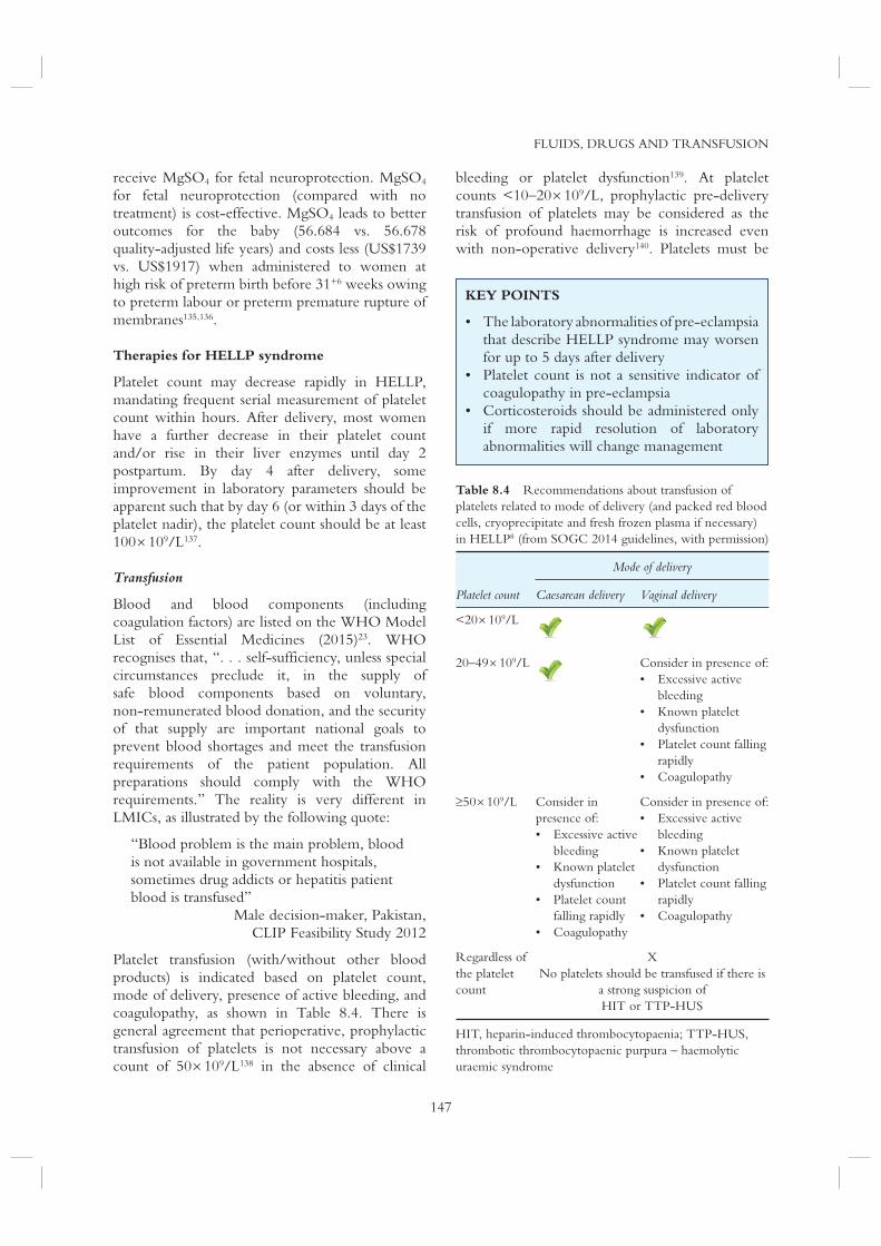
FLUIDS, DRUGS AND TRANSFUSION
147
receive MgSO4 for fetal neuroprotection. MgSO4
for fetal neuroprotection (compared with no
treatment) is cost-effective. MgSO4 leads to better
outcomes for the baby (56.684 vs. 56.678
quality-adjusted life years) and costs less (US$1739
vs. US$1917) when administered to women at
high risk of preterm birth before 31+6 weeks owing
to preterm labour or preterm premature rupture of
membranes135,136.
Therapies for HELLP syndrome
Platelet count may decrease rapidly in HELLP,
mandating frequent serial measurement of platelet
count within hours. After delivery, most women
have a further decrease in their platelet count
and/or rise in their liver enzymes until day 2
postpartum. By day 4 after delivery, some
improvement in laboratory parameters should be
apparent such that by day 6 (or within 3 days of the
platelet nadir), the platelet count should be at least
100109/L137.
Transfusion
Blood and blood components (including
coagulation factors) are listed on the WHO Model
List of Essential Medicines (2015)23. WHO
recognises that, “. . . self-sufficiency, unless special
circumstances preclude it, in the supply of
safe blood components based on voluntary,
non-remunerated blood donation, and the security
of that supply are important national goals to
prevent blood shortages and meet the transfusion
requirements of the patient population. All
preparations should comply with the WHO
requirements.” The reality is very different in
LMICs, as illustrated by the following quote:
“Blood problem is the main problem, blood
is not available in government hospitals,
sometimes drug addicts or hepatitis patient
blood is transfused”
Male decision-maker, Pakistan,
CLIP Feasibility Study 2012
Platelet transfusion (with/without other blood
products) is indicated based on platelet count,
mode of delivery, presence of active bleeding, and
coagulopathy, as shown in Table 8.4. There is
general agreement that perioperative, prophylactic
transfusion of platelets is not necessary above a
count of 50109/L138 in the absence of clinical
bleeding or platelet dysfunction139. At platelet
counts <10–20109/L, prophylactic pre-delivery
transfusion of platelets may be considered as the
risk of profound haemorrhage is increased even
with non-operative delivery140. Platelets must be
Table 8.4 Recommendations about transfusion of
platelets related to mode of delivery (and packed red blood
cells, cryoprecipitate and fresh frozen plasma if necessary)
in HELLP8 (from SOGC 2014 guidelines, with permission)
Platelet count
Mode of delivery
Caesarean delivery Vaginal delivery
<20 109/L
20–49 109/L Consider in presence of:
• Excessive active
bleeding
• Known platelet
dysfunction
• Platelet count falling
rapidly
• Coagulopathy
50 109/L Consider in
presence of:
• Excessive active
bleeding
• Known platelet
dysfunction
• Platelet count
falling rapidly
• Coagulopathy
Consider in presence of:
• Excessive active
bleeding
• Known platelet
dysfunction
• Platelet count falling
rapidly
• Coagulopathy
Regardless of
the platelet
count
X
No platelets should be transfused if there is
a strong suspicion of
HIT or TTP-HUS
HIT, heparin-induced thrombocytopaenia; TTP-HUS,
thrombotic thrombocytopaenic purpura – haemolytic
uraemic syndrome
KEY POINTS
• The laboratory abnormalities of pre-eclampsia
that describe HELLP syndrome may worsen
for up to 5 days after delivery
• Platelet count is not a sensitive indicator of
coagulopathy in pre-eclampsia
• Corticosteroids should be administered only
if more rapid resolution of laboratory
abnormalities will change management

THE FIGO TEXTBOOK OF PREGNANCY HYPERTENSION
148
thawed prior to administration, and a standard unit
of apheresis platelets can be expected to raise the
platelet count by at least 5109/L, with a peak at
10–60 minutes post-transfusion. Four units of
platelets can contain as much as 2 mL of RBCs
to which women who are anti-D(Rho)-negative
may become sensitised. Therefore, women
who are anti-D negative and receive a platelet
transfusion should receive a 300g dose of anti-D
immune globulin, a dose sufficient to prevent
sensitisation following transfusion of up to 30 units
of platelets140.
Although a platelet count <150109/L is
associated with a heightened risk of abnormal
coagulation, platelet count is not a sensitive
indicator of coagulopathy. Coagulation should be
assessed independently of platelet count in
pre-eclampsia prior to neuraxial analgesia/
anaesthesia or surgery141.
Corticosteroids
Dexamethasone is listed on the WHO Model List
of Essential Medicines (2015) for maternal
administration to benefit the neonate23, based on
evidence that the drug accelerates fetal pulmonary
maturation when indicated at <34 weeks142.
When given specifically for HELLP syndrome,
corticosteroids (particularly dexamethasone) more
rapidly improve platelet count and other
haematological and biochemical indices of the
HELLP syndrome (ALT, AST, LDH), especially
when the treatment is initiated before delivery (11
trials, 550 women)143; however, no significant
impact was seen on major maternal (death or severe
morbidity) or perinatal (death or severe morbidity)
outcomes, and transfusion requirements and rates
of regional anaesthesia were not reported. In a
small retrospective study of 37 women, regional
anaesthesia was more often achieved (in 42% of
women vs. 0%) when steroids were given to
women with platelet counts <90109/L144. When
dexamethasone for HELLP was incorporated into
the local treatment protocol (along with MgSO4
and antihypertensive therapy), one centre noted a
reduction in severe maternal morbidity and a low
rate of disease progression145. However, these data
are not sufficient to guide practice. The COHELLP
trial (NCT00711841) will determine whether
postpartum dexamethasone decreases the key
clinical outcome – severe maternal morbidity146.
Other
HELLP syndrome must be differentiated from
other ‘imitators’, as discussed in Chapter 3. Women
with progressive HELLP syndrome, particularly
postpartum, have been described in observational
studies to improve with plasma therapies that are
effective for thrombotic thrombocytopaenic
purpura (TTP), a HELLP mimicker147. No
randomised controlled trials were identified.
Thromboprophylaxis
Unfractionated heparin (sodium) is listed on the
WHO Model List of Essential Medicines (2015)23.
Thromboprophylaxis (with unfractionated or
low molecular weight heparin) should be considered
when thromboembolic risk is at least 1%. This risk
level is reached antenatally, when pre-eclampsia is
associated with two or more other risk markers,
and postnatally, when either pre-eclampsia is
associated with at least one other risk marker (e.g.,
obesity or maternal age >35 years) or women with
any hypertensive disorder of pregnancy were on
antenatal bedrest for at least 7 days (regardless of
mode of delivery)148,149. Whether emergency
Caesarean delivery warrants thromboprophylaxis in
all women is not consistent between guidelines. It
must be noted that guidelines are based largely on
observational data. Although the influential Royal
College of Obstetricians and Gynaecologists
Guidelines149 have been associated with a decline in
thromboembolism-related maternal deaths in the
UK, there are insufficient data from randomised
controlled trials on which to base guideline
recommendations150.
Novel therapies for pre-eclampsia
Novel therapies for pre-eclampsia target various
aspects of pre-eclampsia pathogenesis and are in
development151. Most of these therapies ultimately
target increased nitric oxide (NO) production and
vasodilatation. There is insufficient information to
evaluate their effects, and their use in clinical
practice is not yet recommended.
Agents under active investigation and that
show promise include pravastatin, L-arginine,
S-nitrosoglutathione (GSNO), sildenafil,
esomeprazole152 and antithrombin.
Pravastatin is being evaluated in a randomised
controlled trial for prevention of severe

FLUIDS, DRUGS AND TRANSFUSION
149
complications in women with early ‘severe’
pre-eclampsia153 (STaMP, EudraCT Number:
2009-012968-13). The rationale is that statins
reduce antiangiogenic factors and increase NO
production (Figure 8.1). With an ageing obstetric
population, these medications will be used more
frequently for cardiovascular disease prevention;
although questioned as being teratogenic,
particularly with regards to central nervous system
and limb anomalies, a recent large retrospective
cohort study failed to find that statins are
teratogenic154.
In multiple small randomised controlled trials,
women with gestational hypertension or
pre-eclampsia were administered L-arginine, a NO
precursor, as it is an amino acid required for the
body’s production of NO. L-arginine is available as
a powder, tablet, or intravenous infusion. L-arginine
increased the time to delivery (mean difference
11.5 days, 95% CI 5.2–17.9; 2 trials, 135 women)
and reduced blood pressure, diastolic (mean
difference 4.9 mmHg, 95% CI 4.2–5.5; 4 trials, 204
women) more than systolic (mean difference
3.2 mmHg, 95% CI -1.5–7.9; 4 trials, 204 women)
(7 trials in total, 916 women)155.
S-nitrosoglutathione (GSNO) is a NO donor
that causes vascular relaxation. When given to
women with severe pre-eclampsia, GSNO
improved blood pressure, platelet count and uterine
artery Doppler resistance. This, in addition to the
fact that it does not appear to induce tolerance,
makes it an interesting drug for future study151.
Sildenafil is a phosphodiesterase type-5 inihbitor
that increases concentrations of cGMP, resulting in
relaxation of vascular smooth muscle (Figure 8.1).
It has been marketed extensively for treatment of
erectile dysfunction in men. Sildenafil is currently
being studied in four randomised controlled trials
Figure 8.1 Overview of the mechanisms of action of various novel therapies for pre-eclampsia (modified from Everett
et al. J Matern Fetal Neonatal Med 2012;25(1):50–52). 2O, secondary; cGMP, cyclic guanosine monophosphate; CO, carbon
monoxide; GMP, guanosine monophosphate; GSNO, S-nitrosoglutathione; GTP, guanosine triphosphate; PDE5,
phosphodiesterase-5; PlGF, placental growth factor; sEng, soluble endoglin; sFlt-1, soluble fms-like tyrosine kinase;
sGC, soluble guanylyl cyclase; VEGF, vascular endothelial growth factor
CO bilirubin statins
sEng sFlt-1
VEGF PLGF
eNOS
NO
sGC
GSNO
GTP cGMP GMP
sildenafil (via PDE5 inhibition)
end effects via 2⁰ messengers smooth muscle relaxation platelet downregulation
biological effectors positive / catalyst negative / inhibitor

THE FIGO TEXTBOOK OF PREGNANCY HYPERTENSION
150
for treatment of severe, early-onset IUGR156. The
randomised controlled trial of sildenafil for
pre-eclampsia did not improve maternal or perinatal
outcomes, but the pre-eclampsia was of late-onset,
the type less likely to have the abnormal placentation
that sildenafil aims to target (see Chapter 3).
Esomeprazole is a proton pump inhibitor used
to treat gastric reflux. Preclinical laboratory studies
have demonstrated that esomeprazole decreases
sFlt-1, soluble endoglin, and measures of oxidative
stress157.
Recombinant antithrombin (ATryn®) is being
studied for the treatment of preterm pre-eclampsia
at <31+0 weeks158.
Remote literature describes potentially beneficial
effects of abdominal decompression, by application
of intermittent negative pressure over the abdomen
for 30 minutes, once to three times daily (3 trials,
367 women)159. Each trial was potentially biased,
and only one enrolled women with pre-eclampsia
or pre-existing hypertension. However, abdominal
decompression was associated with beneficial
effects: a reduction in pre-eclampsia or worsening
pre-eclampsia (RR 0.36, 95% CI 0.18–0.72; 1
trial, 80 women), low birth weight babies (RR
0.50, 95% CI 0.40–0.63; 2 trials, 304 women), and
perinatal mortality (RR 0.39, 95% CI 0.22–0.71;
3 trials, 367 women).
Sleep-disordered breathing has been linked
with gestational hypertension. Treatment of that
sleep-disordered breathing did not improve blood
pressure, but the one relevant trial (24 women)
treated women for only one night, so it is impossible
to draw conclusions160.
Also, immediate postpartum curettage, usually
under ultrasound guidance, was associated with
lower blood pressure, higher platelet count and
higher urine output, but differences in harder
clinical outcomes (such as hospitalisation or need
for transfusion) were not demonstrated (3 trials,
497 women)161–163. Uterine perforation was not
documented to have occurred.
Agents that have shown disappointing results in
studies to date include Digibind and recombinant
activated protein C.
Digibind (anti-digoxin antibody) was studied in
a randomised controlled trial (NCT00158743) of
postpartum women with severe pre-eclampsia.
The rationale was that binding of endogenous
digitalis-like factors would lead to vasodilatation.
Deterioration in creatinine clearance was blunted
in the Digibind group, but there was no difference
in hard clinical outcomes, including blood
pressure164.
Activated protein C (APC) is a serine protease
that was studied as a disease-modifying
treatment for critically ill subjects. Despite its
anti-inflammatory, antithrombotic and fibrinolytic
properties, APC did not improve mortality in sepsis
and it was withdrawn from the market. In a
controlled series of nine women with antenatal,
severe pre-eclampsia, APC increased urine output
(consistent with initiation of disease resolution),
but did not improve other clinical outcomes165.
Evidence-based care in under-resourced settings
The hypertensive disorders of pregnancy rate
among the four top causes of maternal mortality
and morbidity worldwide, but more than 99%
of hypertensive disorder of pregnancy-related
maternal deaths occur in under-resourced settings,
particularly sub-Saharan Africa and South Asia166.
There, efforts to improve outcomes by promoting
evidence-based care in facility have taken many
approaches, including practice audit and
development of practice guidance and tools15.
Care in the community, including task-shifting to
community health workers is complementing this
approach. These approaches are discussed in detail
below, but it should be noted that their application
in well-resourced settings could improve care there
as well.
Audit of practice and outcomes
Introducing quality of care indicators for
pre-eclampsia/eclampsia appears to be acceptable
to hospital-based practitioners (South Thailand)167.
Practice audit according to those indicators can
identify case management problems; however, the
quality of the analysis, clarity of recommendations
for improvement, and follow-up to confirm
implementation of solutions are related to their
effectiveness (Benin, West Africa)168. When done
properly, criteria-based audit at university teaching
hospitals has improved pregnancy outcomes,
including maternal mortality (Tanzania)169.
Whether high-quality practice audit works
equally well at all levels of the health care system
has been questioned. After a multifaceted
intervention, adherence with practice indicators

FLUIDS, DRUGS AND TRANSFUSION
151
increased, but variably, being substantially lower at
district (for approximately 70% of indicators) than
at referral hospitals (>90%) (South Thailand)167.
Similar results were seen in a cluster randomised
controlled trial (Senagal and Mali); the intervention
of maternal death reviews combined with best
practice implementation for emergency obstetric
care, was supported by regular visits by trained
facilitators. Hospital-based maternal mortality was
decreased (OR 0.85, 95% CI 0.73–0.98), but only
at first-level referral hospitals and not at regional
referral hospitals170.
Various audit data collection sheets have been
published, although they have been designed to
comply with either local guidelines171 or national
guidelines43. As such, they may be less applicable at
other sites or in other countries, especially as many
criteria are not based on high-quality evidence but
rather, on what is achievable in that particular
setting.
Emergency drills (also known as ‘fire drills’)
provide a simulated experience for participants to
practice problem-solving and decision-making
skills in the management of an obstetric or newborn
emergency, with emphasis on thinking quickly,
reacting (intervening) rapidly, and working as a
team. Also, they provide opportunities to both
revise essential skills and develop confidence in
dealing with emergencies that do not occur
frequently. Formal programmes have been
developed, such as the Essential Steps in Managing
Obstetric Emergencies (ESMOE) – Emergency
Obstetric Simulation Training (EOST) and then
adapted for use in countries such as South Africa.
This programme’s drills for eclampsia and
pre-eclampsia (N = 2) have been provided in
Appendix 8.2.
Standardising care in facility
The lack of easy to use protocols and monitoring
charts in the management of pre-eclampsia/
eclampsia are felt to contribute to substandard care
of women in resource-poor settings, particularly
when care is provided by those with less experience.
Even when the necessary drugs and supplies are
available for high-quality pre-eclampsia/eclampsia
management, there is a lack of provider knowledge
and experience (Afghanistan)172.
Although developing guidance is hampered by
the lack of high-quality evidence in some areas of
care, a variety of tools have been studied to improve
evidence-based hypertensive disorder of pregnancy
care, including monitoring and treatment guides
and emergency medical kits, building on the
popularity of the ‘eclampsia box’ in the
Collaborative Eclampsia Trial. A tool that provided
a visual record of monitoring and treatment, as well
as treatment guidance of women with severe
pre-eclampsia/eclampsia, was viewed as potentially
useful in clinical care by the majority of skilled
birth attendants surveyed and an implementation
study has been planned (sub-Saharan Africa)173.
Single-use obstetric emergency medical kits made
available for in-hospital care were used frequently
for care of women with pre-eclampsia/eclampsia
(in 52/192 cases of kit use), and there was an
associated (non-significant) 30% decrease in
all-cause maternal mortality (Kenya)174. Lack of IV
pumps for administration of MgSO4 maintenance
therapy was addressed by a single trial (300 women);
women allocated to IV MgSO4 using a mechanical,
flow-controlled pump (Springfusor®) experienced
less pain and fewer other side-effects than women
allocated to IV and IM MgSO4 loading with IM
maintenance175. More than 90% of women in both
groups completed their full course of therapy.
The NICE guidelines published detailed
algorithms for care in well-resourced settings.
These were based on the 2010 NICE guidelines,
UK, but the algorithms could be adapted for local
use43.
Initiating treatment in the community
At the primary health centre level, fewer than half
of centres initiated treatment for pre-eclampsia
(40.0%) or eclampsia (28.0%) prior to transfer to
facility (rural Nigeria)176. Taken in the context of
the ‘three delays’ model of maternal mortality, this
represents a lost opportunity for improving
maternal outcome.
The nine manuals of the Perinatal Education
Programme (PEP) in South Africa have been
produced and distributed by the Perinatal Education
Trust, a non-profit organisation that aims to
improve outcomes for pregnant women and their
babies, especially in poor, rural communities
(pepcourse.co.za). PEP is self-help training for
health professionals who are responsible for their
own education. The course is cheap and does not
require a teacher. Material is presented in a series of

THE FIGO TEXTBOOK OF PREGNANCY HYPERTENSION
152
manuals that learners can either download for free
or purchase from suppliers of medical books.
Learners usually study in groups of 5–10 to foster
co-operative learning. The group studies the
chapters independently, usually meeting every 2–3
weeks to allow for discussion of the units or
demonstration of specific skills. Since the inception
of PEP in 1988, approximately 50,000 manuals
have been distributed and an estimated 80,000
health care providers have used PEP course work.
Course evaluation takes the form of self-assessed
multiple choice tests before and after each chapter,
and a final multiple-choice examination by the
Perinatal Education Trust for each manual. By
2014, over 20,000 PEP certificates had been
awarded to more than 10,000 participants in South
Africa.
The Community-Level Interventions for
Pre-eclampsia (CLIP) Trial is a cluster randomised
controlled trial that is evaluating a community-based
package of triage, treatment and transport for
women identified with hypertensive pregnancy
(2013–2017) in four LMICs (India, Nigeria,
Mozambique and Pakistan)134 (pre-empt.cfri.ca).
Community health workers are being instructed to
administer oral methyldopa for severe hypertension
and MgSO4 IM for eclampsia prevention and
treatment (Appendix 8.1).
BEST PRACTICE POINTS
(Please see Appendix 8.3 for the evaluation of the strength of the recommendation and the quality of the
evidence on which they are based.)
Fluid
1. Plasma volume expansion is not recommended for women with pre-eclampsia.
2. IV fluid intake should be minimised to 80 mL/h in women with pre-eclampsia to avoid pulmonary
oedema.
3. Fluid should not be routinely administered to treat oliguria (<15 mL/h for 6 consecutive hours) for
the sole purpose of increasing urine output.
4. For treatment of persistent oliguria, neither dopamine nor furosemide is recommended.
Antihypertensive therapy for severe hypertension
1. Blood pressure should be lowered to <160 mmHg systolic and <110 mmHg diastolic.
2. Initial antihypertensive therapy in the hospital setting should be with nifedipine short-acting
(capsules), parenteral hydralazine, or parenteral labetalol.
3. Alternative antihypertensive medications include oral methyldopa, oral labetalol, oral clonidine, oral
captopril (only postpartum), or a nitroglycerin infusion (for doses, see Table 8.2).
4. Refractory hypertension may be treated with sodium nitroprusside.
5. Nifedipine and MgSO4 can be used contemporaneously.
6. MgSO4 is not recommended solely as an antihypertensive agent.
7. Continuous FHR monitoring is advised until blood pressure is stable.
Antihypertensive therapy for non-severe hypertension
1. Antihypertensive drug therapy should aim for a diastolic blood pressure of 85 mmHg.
2. The choice of antihypertensive agent for initial treatment should be based on characteristics of the
patient, contraindications to a particular drug, and physician and patient preference.

FLUIDS, DRUGS AND TRANSFUSION
153
3. Initial therapy in pregnancy can be with one of a variety of antihypertensive agents methyldopa,
labetalol, other beta-blockers (acebutolol, metoprolol, pindolol, and propranolol) and calcium
channel blockers (nifedipine).
4. ACE inhibitors and ARBs should not be used during pregnancy.
5. Atenolol and prazosin are not recommended prior to delivery.
6. Captopril, enalapril, or quinapril may be used postpartum, even during breastfeeding.
7. There is no compelling evidence that antihypertensive treatment of hypertension (with labetalol,
nifedipine, and probably methyldopa) is associated with adverse effects on child development.
8. Gestational hypertension and pre-eclampsia may each be associated with an increase in adverse
paediatric neurodevelopmental effects, such as inattention and externalising behaviours.
MgSO4
1. MgSO4 is recommended for first-line treatment of eclampsia.
2. MgSO4 is recommended for eclampsia prevention in women with severe pre-eclampsia.
3. MgSO4 may be considered for eclampsia prevention in women with non-severe pre-eclampsia based
on cost considerations.
4. MgSO4 should be used in standard dosing, usually 4 g IV loading dose followed by 1 g/h.
5. Routine monitoring of serum magnesium levels is not recommended.
6. Phenytoin and benzodiazepines should not be used for eclampsia prophylaxis or treatment, unless
there is a contraindication to MgSO4 or it is ineffective.
7. In women with pre-existing or gestational hypertension, MgSO4 should be considered for fetal
neuroprotection in the setting of imminent preterm birth within the next 24 hours at ≤33+6 weeks.
Therapies for HELLP syndrome
Recommendations
1. Every obstetrical centre should be aware of the local delay between ordering and receiving platelets
units.
2. For a platelet count <20109/L, platelet transfusion is recommended, regardless of mode of delivery.
3. For a platelet count 20–49109/L platelet transfusion is recommended prior to Caesarean delivery.
4. For a platelet count 20–49109/L, platelet transfusion should be considered prior to vaginal delivery
if there is excessive active bleeding, known platelet dysfunction, a rapidly falling platelet count, or
coagulopathy).
5. For a platelet count of 50109/L, platelet transfusion should be considered prior to either Caesarean
or vaginal delivery if there is excessive active bleeding, known platelet dysfunction, a rapidly falling
platelet count, or coagulopathy.
6. We do not recommend corticosteroids for treatment of HELLP until they have been proven to
decrease maternal morbidity.
7. We recommend against plasma exchange or plasmapheresis for HELLP, particularly within the first
4 days postpartum.
Other therapies for treatment of pre-eclampsia
1. Women with pre-eclampsia before 34 weeks’ gestation should receive antenatal corticosteroids for
acceleration of fetal pulmonary maturity.
2. Thromboprophylaxis may be considered antenatally among women with pre-eclampsia who have
two or more additional thromboembolic risk markers, postnatally among women with pre-eclampsia
who have at least one additional thromboembolic risk marker, or postnataly among women any
hypertensive disorder of pregnancy who were on antenatal bed rest for at least 7 days.

THE FIGO TEXTBOOK OF PREGNANCY HYPERTENSION
154
PRIORITIES FOR UNDER-RESOURCED SETTINGS
Table 8.5 outlines priorities for care in the
community (to prevent eclampsia and
hypertension-related stroke prior to referral to
facility177) and in facilities (to prevent and treat
severe acute maternal morbidity and decrease
maternal and perinatal mortality, particularly for
the periviable fetus)178–180.
All of the interventions relevant specifically to
the hypertensive disorders of pregnancy and
recognised by the WHO as essential medicines are
included here: antihypertensive therapy for severe
or non-severe hypertension, MgSO4 for eclampsia
prevention or treatment, blood products, and
Table 8.5 Priorities for management of women with a hypertensive disorder of pregnancy (HDP) by level of health care
system at which care is delivered
Antepartum & postpartum
Initial priority Ultimate goal
Community
Primary health
care centre
(detect, stabilise
and refer)
Antihypertensives for
severe hypertension
Antihypertensives for
severe or non-severe hypertension
MgSO4 administered before referral in order
to prevent or treat eclampsia
MgSO4 administered before referral in order to prevent or
treat eclampsia
Clear communication with referral unit
regarding transport and medication
Clear communication with referral unit regarding transport
and medication (including individualisation of antenatal
corticosteroid therapy)
Facility
Secondary-level
facility
(detect, manage
and refer if
necessary)
In women with a HDP, appropriate use of
antihypertensive therapy, MgSO4
In women with a HDP, appropriate use of antihypertensive
therapy, MgSO4, fluids (restricted), and corticosteroids
Appropriate triage of women for referral to
tertiary-level care (including those eligible
for expectant care* and those at high risk of
or with severe maternal morbidity)
Appropriate triage of women for referral to tertiary-level
care (including those eligible for expectant care and those
with or at high risk of severe maternal morbidity)
Availability of pRBCs Availability of pRBCs,
platelets, and clotting factors
Tertiary-l evel
(referral) facility
(detect and
manage
definitively)
Appropriate use of antihypertensive therapy,
MgSO4, fluids (restricted) and corticosteroids
in women with a HDP
Appropriate use of antihypertensive therapy, MgSO4, fluids
(restricted), and corticosteroids in women with a HDP
Appropriate triage and care of women
eligible for expectant care* and those at high
risk of or with severe maternal morbidity
Appropriate triage and care of women eligible for
expectant care and those at high risk of or with severe
maternal morbidity
Availability of pRBCs, platelets, and clotting
factors
Availability of pRBCs, platelets, and clotting factors
Management of the periviable neonate Management of the periviable fetus and neonate
Advanced management options including the establishment
of Obstetric Critical Care Units in close proximity to
labour wards to provide advanced monitoring (e.g.,
intra-arterial BP measurement) and treatment (e.g.,
ventilatory support) of complicated cases
pRBCs, packed red blood cells; BP, blood pressure
* For a discussion about timing of delivery, see Chapter 9

FLUIDS, DRUGS AND TRANSFUSION
155
antenatal corticosteroids for acceleration of fetal
pulmonary maturity. Sample policy statements for
antihypertensive therapy and MgSO4 are provided
for local adaptation (Appendix 8.4).
An initial focus should be on the early
administration of antihypertensive agents and
MgSO4 in the community prior to transfer to
facility, or in secondary-level facilities prior to
transfer to tertiary-level facility. Reluctance to care
for these women prior to their arrival at tertiary-level
facilities is illustrated by the following quote:
“Many doctors also don’t like to treat
eclampsia. If the lady has eclampsia, or
imminent eclampsia or severe pre-eclampsia
because of the risk with the morbidity and the
mortality to both the baby and the mother they
try to shift the patient to the higher centres”
Obstetrician, CLIP Feasibility Study,
Bagalkot, India
WHAT INTERNATIONAL GUIDELINES SAY ( APPENDIX 8.5)
Abbreviations for Clinical Practice Guidelines:
ACOG (American College of Obstetricians and
Gynecologists)181, AOM (Association of Ontario
Midwives), NICE (National Institute for Health
and Clinical Excellence)43, NVOG (National
Obstetrics and Gynaecology Society, The
Netherlands)182, PRECOG II (Pre-eclampsia
Community Guideline) and PRECOG II
(Pre-eclampsia Community Guideline II), QLD
(Queensland, Australia)183,184, SOGC (Society of
Obstetricians and Gynaecologists of Canada)22,
SOMANZ (Society of Obstetric Medicine of
Australia and New Zealand)185, WHO (World
Health Organization)10.
Fluid management
Multiple guidelines recommend against plasma
volume expansion (SOGC, NICE, SOMANZ).
Fluid restriction in pre-eclampsia is recommended
by two guidelines (SOGC, NICE), one of which
recommends administration of no more than
80 mL/h of IV fluids (NICE).
Antihypertensive therapy
Seven guidelines discuss antihypertensive therapy
(SOGC, WHO, NICE, ACOG, NVOG,
SOMANZ, QLD).
For severe hypertension
There is uniform agreement in all seven guidelines
that severe hypertension should be treated,
although most guidelines do not rate the
recommendation highly because of the lack of
randomised controlled trials of antihypertensive
versus placebo/no therapy (as discussed above
under ‘Antihypertensive therapy for severe
hypertension’). Most guidelines recommend a
blood pressure goal of <160/110 mmHg (SOGC,
ACOG, QLD), but a goal of <150/80–100 mmHg
is recommended in the UK (NICE),
<160/100 mmHg in Australasia (SOMANZ), and
ACOG makes a specific recommendation for
women with chronic hypertension for whom
blood pressure should be <160/105 mmHg.
Recommended drugs of first choice are IV labetalol
(SOGC, NICE, NVOG, SOMANZ), oral
nifedipine (SOGC, NICE, NVOG, SOMANZ),
and IV hydralazine (SOGC, NICE, SOMANZ);
two CPGs leave the choice to the clinician (WHO,
QLD). Two guidelines highlight that MgSO4
should not be used as an antihypertensive (SOGC,
SOMANZ).
For non-severe hypertension
Guidance for treatment of non-severe hypertension
is reported by five guidelines and is highly variable,
in part based on associated comorbidities and/or the
type of hypertensive disease of pregnancy. All
guidelines were published prior to release of the
CHIPS Trial results (see ‘Antihypertensive therapy
for non-severe hypertension’, above) which have
clarified optimal management and will be
incorporated into future updates. For women with
end-organ dysfunction that can be exacerbated
by elevated blood pressure, treatment to
<140/90 mmHg is recommended (SOGC, NICE).
For women without target-organ damage, treatment
targets are: (1) for any hypertensive disorder
of pregnancy, <150/80–100 mmHg (NICE),
130–159/80–105 mmHg (SOGC), 140–160/
90–100 mmHg (SOMANZ), or <160/110 mmHg
(NVOG); (2) for women with chronic hypertension,
120–159/80–104 mmHg (ACOG); and (3) for
women with gestational hypertension or non-severe
pre-eclampsia <160/110 mmHg (ACOG). Oral
methyldopa (SOGC, NICE, ACOG, NVOG,
SOMANZ), oral labetalol (SOGC, NICE, ACOG,
NVOG, SOMANZ), and nifedipine (SOGC,

THE FIGO TEXTBOOK OF PREGNANCY HYPERTENSION
156
NICE, ACOG, NVOG, SOMANZ) are most
commonly recommended.
ACE inhibitors and ARBs should not be used in
pregnancy. For women with antihypertensive-treated
chronic hypertension who are planning pregnancy,
counselling should be undertaken (SOGC, NICE,
NVOG, QLD). Alternatives to ACE inhibitors and
ARBs should be discussed, and women should be
instructed to stop ACE inhibitors and ARBs if
inadvertently taken in early pregnancy (SOGC,
NICE, ACOG, NVOG).
MgSO4
There is general agreement that MgSO4 is indicated
for treatment of eclampsia (SOGC, WHO, NICE,
ACOG, NVOG, QLD) and severe pre-eclampsia
(SOGC, WHO, NICE, ACOG, NVOG),
although ACOG recommends only intrapartum
and postpartum treatment. There is less certainty
about recommending MgSO4 for non-severe
pre-eclampsia (SOGC, ACOG, NVOG), although
no guideline recommended against it. One guideline
recommended that units define their own protocols
for eclampsia prophylaxis (SOMANZ). MgSO4 is
otherwise indicated for fetal neuroprotection if
women are delivering imminently at <34 weeks
(SOGC, SOMANZ).
Therapies for HELLP
Corticosteroids are not recommended to improve
clinical outcomes in HELLP syndrome (SOGC,
WHO, NICE, ACOG, SOMANZ), but one
guidelines suggests considering this therapy if an
improvement in platelet count would be useful
(ACOG).
One guideline discusses platelet thresholds for
platelet transfusion (SOGC).
PRIORITIES FOR FUTURE RESEARCH
Significant progress has been and is being made to
reduce the impact of pre-eclampsia in LMICs, but
it remains a priority focus as we continue to struggle
to achieve the 75% reduction in maternal mortality
– the goal set in Millennium Development Goal 5
with a target date of 2015)166.
Global priorities for hypertensive disorder
of pregnancy management include: whether
nifedipine is superior to parenteral agents for
treatment of severe pregnancy hypertension; how
to improve the cost-effectiveness of MgSO4 for
eclampsia prevention with regards to an abbreviated
treatment course or reduced dose; and whether
dexamethasone reduces severe maternal morbidity
in HELLP syndrome without increasing maternal
risk.
In general, hypertensive disorder of
pregnancy management research has focused
on institutional-level interventions. However,
maternal lives lost from pre-eclampsia and eclampsia
result from delays in triage, transport and treatment,
such that if we limit ourselves to studying inpatient,
facility-level interventions, many women will die
or be irreversibly affected by pre-eclampsia
complications prior to arriving at the inpatient
facility. The future lies in getting diagnosis and care
into the community, and improving transport to
facility for definitive treatment.
REFERENCES
1. Mol BW, Roberts CT, Thangaratinam S, Magee LA, de Groot CJ, Hofmeyr GJ. Pre-eclampsia. Lancet 2015; pii: S0140-6736(15)00070-7. doi: 10.1016/S0140-6736(15)00070-7 [Epub ahead of print]
2. Duley L, Williams J, Henderson-Smart DJ. Plasma volume expansion for treatment of women with pre-eclampsia. Cochrane Database Syst Rev 2000;(2)(2):CD001805
3. Ganzevoort W, Rep A, Bonsel GJ, Fetter WP, van Sonderen L, De Vries JI, et al. A randomised controlled trial comparing two temporising management strategies, one with and one without plasma volume expansion, for severe and early onset pre-eclampsia. BJOG 2005 Oct;112(10):1358–1368
4. Ganzevoort W, Rep A, Bonsel GJ, De Vries JI, Wolf H, PETRA investigators. A randomized trial of plasma volume expansion in hypertensive disorders of pregnancy: influence on the pulsatility indices of the fetal umbilical artery and middle cerebral artery. Am J Obstet Gynecol 2005 Jan;192(1):233–239
5. Metsaars WP, Ganzevoort W, Karemaker JM, Rang S, Wolf H. Increased sympathetic activity present in early hypertensive pregnancy is not lowered by plasma volume expansion. Hypertens Pregnancy 2006;25(3):143–157
6. Rep A, Ganzevoort W, Van Wassenaer AG, Bonsel GJ, Wolf H, De Vries JI, et al. One-year infant outcome in women with early-onset hypertensive disorders of pregnancy. BJOG 2008 Jan;115(2):290–298

FLUIDS, DRUGS AND TRANSFUSION
157
7. Thornton C, Hennessy A, von Dadelszen P, Nishi C, Makris A, Ogle R. An international benchmarking collaboration: measuring outcomes for the hypertensive disorders of pregnancy. J Obstet Gynaecol Can 2007 Oct;29(10):794–800
8. Magee LA, Pels A, Helewa M, Rey E, von Dadelszen P. Diagnosis, evaluation, and management of the hypertensive disorders of pregnancy. Pregnancy Hypertens 2014;4(2):105–145
9. Mantel GD, Makin JD. Low dose dopamine in postpartum pre-eclamptic women with oliguria: a double-blind, placebo controlled, randomised trial. Br J Obstet Gynaecol 1997 Oct;104(10):1180–1183
10. World Health Organization. WHO recommendations for prevention and treatment of pre-eclampsia and eclampsia. 2011
11. Martin JN,Jr, Thigpen BD, Moore RC, Rose CH, Cushman J, May W. Stroke and severe preeclampsia and eclampsia: a paradigm shift focusing on systolic blood pressure. Obstet Gynecol 2005 Feb;105(2):246–254
12. Reidy J, Russell R. Cmace 2006–2008. Int J Obstet Anesth 2011 Jul;20(3):208–212
13. Draycott T, Lewis G, Stephens I. Eighth report of the Confidential Enquiries into Maternal Deaths in the UK (Executive Summary). BJOG 2011;118(Suppl 1): e12-e21
14. Knight M, Kenyon S, Brocklehurst P, Neilson J, Shakespeare J, Kurinczuk JJ, eds. on behalf of MBRRACEUK. Saving Lives, Improving Mothers’ Care – Lessons learned to inform future maternity care from the UK and Ireland Confidential Enquiries into Maternal Deaths and Morbidity 2009–12. Oxford: National Perinatal Epidemiology Unit, University of Oxford 2014
15. National Committee on Confidential Enquiries into Maternal Deaths. Saving Mothers 2005–2007: Fourth Report on Confidential Enquiries into Maternal Deaths in South Africa. Available at: http://www0.s u n . a c . z a / r u r a l h e a l t h / u k w a n d a h o m e /rudasaresources2009/DOH/savingmothers%2005-07%5B1%5D.pdf. Accessed June/10, 2015
16. Haggendal E, Johansson B. On the pathophysiology of the increased cerebrovascular permeability in acute arterial hypertension in cats. Acta Neurol Scand 1972;48(3):265–270
17. von Dadelszen P, Payne B, Li J, Ansermino JM, Broughton Pipkin F, Cote AM, et al. Prediction of adverse maternal outcomes in pre-eclampsia: development and validation of the fullPIERS model. Lancet 2011 Jan 15;377(9761):219–227
18. Committee on Obstetric Practice. Committee Opinion No. 623: Emergent therapy for acute-onset, severe hypertension during pregnancy and the postpartum period. Obstet Gynecol 2015 Feb;125(2):521–525
19. Chobanian AV, Bakris GL, Black HR, Cushman WC, Green LA, Izzo JL,Jr, et al. The Seventh Report of the Joint National Committee on Prevention, Detection, Evaluation, and Treatment of High Blood Pressure: the JNC 7 report. JAMA 2003 May 21;289(19):2560–2572
20. Williams B, Poulter NR, Brown MJ, Davis M, McInnes GT, Potter JF, et al. Guidelines for management of hypertension: report of the fourth working party of the British Hypertension Society, 2004-BHS IV. J Hum Hypertens 2004 Mar;18(3):139–185
21. Mancia G, De Backer G, Dominiczak A, Cifkova R, Fagard R, Germano G, et al. 2007 ESH-ESC Practice Guidelines for the Management of Arterial Hypertension: ESH-ESC Task Force on the Management of Arterial Hypertension. J Hypertens 2007 Sep;25(9): 1751–1762
22. Gradman AH, Basile JN, Carter BL, Bakris GL, American Society of Hypertension Writing Group. Combination therapy in hypertension. J Am Soc Hypertens 2010 Jan-Feb;4(1):42–50
23. World Health Organization. 19th WHO Model List of Essential Medicines (April 2015). 2015; Available at: http://www.who.int/medicines/publications/essentialmedicines/EML2015_8-May-15.pdf. Accessed June/10, 2015
24. Rezaei Z, Sharbaf FR, Pourmojieb M, Youefzadeh-Fard Y, Motevalian M, Khazaeipour Z, et al. Comparison of the efficacy of nifedipine and hydralazine in hypertensive crisis in pregnancy. Acta Med Iran 2011;49(11):701–706
25. Fenakel K1, Fenakel G, Appelman Z, Lurie S, Katz Z, Shoham Z. Nifedipine in the treatment of severe preeclampsia. Obstet Gynecol. 1991 Mar;77(3):331–7
26. Magee LA, Cham C, Waterman EJ, Ohlsson A, von Dadelszen P. Hydralazine for treatment of severe hypertension in pregnancy: meta-analysis. BMJ 2003 Oct 25;327(7421):955–960
27. Duley L, Meher S, Jones L. Drugs for treatment of very high blood pressure during pregnancy. Cochrane Database Syst Rev 2013 Jul 31;7:CD001449
28. Shekhar S, Gupta N, Kirubakaran R, Pareek P. Oral nifedipine versus intravenous labetalol for severe hypertension during pregnancy: a systematic review and meta-analysis. BJOG. 2016 Jan;123(1):40–7

THE FIGO TEXTBOOK OF PREGNANCY HYPERTENSION
158
29. Saudan P, Billieux M-, Pechere A, Irion O, Savoldelli G, Boulvain M. OS014. Which first-line drug to control severe hypertension in pregnancy? A pilot study. Pregnancy Hypertension: An International Journal of Women’s Cardiovascular Health 2012;2(3):182
30. Hennessy A, Thornton CE, Makris A, Ogle RF, Henderson-Smart DJ, Gillin AG, et al. A randomised comparison of hydralazine and mini-bolus diazoxide for hypertensive emergencies in pregnancy: the PIVOT trial. Aust N Z J Obstet Gynaecol 2007 Aug;47(4): 279–285
31. Wacker JR, Wagner BK, Briese V, Schauf B, Heilmann L, Bartz C, et al. Antihypertensive therapy in patients with pre-eclampsia: A prospective randomised multicentre study comparing dihydralazine with urapidil. Eur J Obstet Gynecol Reprod Biol 2006 Aug;127(2):160–165
32. Maharaj B, Khedun SM, Moodley J, Madhanpall N, van der Byl K. Intravenous isradipine in the management of severe hypertension in pregnant and nonpregnant patients. A pilot study. Am J Hypertens 1994 Jul;7(7 Pt 2):61S-63S
33. Vigil-De Gracia P, Lasso M, Ruiz E, Vega-Malek JC, de Mena FT, Lopez JC, et al. Severe hypertension in pregnancy: hydralazine or labetalol. A randomized clinical trial. Eur J Obstet Gynecol Reprod Biol 2006 Sep-Oct;128(1–2):157–162
34. Garden A, Davey DA, Dommisse J. Intravenous labetalol and intravenous dihydralazine in severe hypertension in pregnancy. Clin Exp Hypertens B 1982;1(2–3):371–383
35. Vermillion ST, Scardo JA, Newman RB, Chauhan SP. A randomized, double-blind trial of oral nifedipine and intravenous labetalol in hypertensive emergencies of pregnancy. Am J Obstet Gynecol 1999 Oct;181(4):858–861
36. Shekhar S, Sharma C, Thakur S, Verma S. Oral nifedipine or intravenous labetalol for hypertensive emergency in pregnancy: a randomized controlled trial. Obstet Gynecol 2013 Nov;122(5):1057–1063
37. Lakshmi BS, Dasari P. Oral nifedipine versus intravenous labetalol in hypertensive urgencies and emergencies of pregnancy: a randomized clinical trial. Obstetric Medicine: The Medicine of Pregnancy 2012 12/01;5(4):171–175
38. Raheem IA, Saaid R, Omar SZ, Tan PC. Oral nifedipine versus intravenous labetalol for acute blood pressure control in hypertensive emergencies of pregnancy: a randomised trial. BJOG 2012 Jan;119(1):78–85
39. Aswathkumar R, Gilvas S. Management of severe hypertension in pregnancy: prospective comparison of labetalol vs. nifedipine [abstract]. 49th All India Congress of Obstetrics and Gynaecology:38
40. Elatrous S, Nouira S, Ouanes Besbes L, Marghli S, Boussarssar M, Sakkouhi M, et al. Short-term treatment of severe hypertension of pregnancy: prospective comparison of nicardipine and labetalol. Intensive Care Med 2002 Sep;28(9):1281–1286
41. Tuffnell DJ, Jankowicz D, Lindow SW, Lyons G, Mason GC, Russell IF, et al. Outcomes of severe pre-eclampsia/eclampsia in Yorkshire 1999/2003. BJOG 2005 Jul;112(7):875–880
42. Moore MP, Redman CWG. The treatment of hypertension in pregnancy. Curr Med Res Opin 1982 01/01; 2015/04;8:39–46
43. National Collaborating Centre for Women’s and Children’s Health (UK). CG107: Hypertension in pregnancy: The management of hypertensive disorders during pregnancy. NICE: Guidance 2010 Aug; 2010 Aug
44. Firoz T, Magee L, MacDonell K, Payne B, Gordon R, Vidler M, et al. Oral antihypertensive therapy for severe hypertension in pregnancy and postpartum: a systematic review. BJOG 2014;121(10):1210–1218
45. Hall DR, Odendaal HJ, Steyn DW, Smith M. Nifedipine or prazosin as a second agent to control early severe hypertension in pregnancy: a randomised controlled trial. BJOG 2000 Jun;107(6):759–765
46. Brown MA, Buddle ML, Farrell T, Davis GK. Efficacy and safety of nifedipine tablets for the acute treatment of severe hypertension in pregnancy. Am J Obstet Gynecol 2002 Oct;187(4):1046–1050
47. Jegasothy R, Paranthaman S. Sublingual nifedipine compared with intravenous hydralazine in the acute treatment of severe hypertension in pregnancy: potential for use in rural practice. J Obstet Gynaecol Res 1996 Feb;22(1):21–4
48. Magee LA, Miremadi S, Li J, Cheng C, Ensom MH, Carleton B, et al. Therapy with both magnesium sulfate and nifedipine does not increase the risk of serious magnesium-related maternal side effects in women with preeclampsia. Am J Obstet Gynecol 2005 Jul;193(1):153–163
49. Bhalla AK, Dhall GI, Dhall K. A safer and more effective treatment regimen for eclampsia. Aust N Z J Obstet Gynaecol 1994 May;34(2):144–148
50. Caetano M, Ornstein MP, Von Dadelszen P, Hannah ME, Logan AG, Gruslin A, et al. A survey of Canadian

FLUIDS, DRUGS AND TRANSFUSION
159
practitioners regarding the management of the hypertensive disorders of pregnancy. Hypertens Pregnancy 2004;23(1):61–74
51. Scardo JA, Hogg BB, Newman RB. Favorable hemodynamic effects of magnesium sulfate in preeclampsia. Am J Obstet Gynecol 1995 Oct;173(4):1249–1253
52. Cotton DB, Gonik B, Dorman KF. Cardiovascular alterations in severe pregnancy-induced hypertension: acute effects of intravenous magnesium sulfate. Am J Obstet Gynecol 1984 Jan 15;148(2):162–165
53. Mroczek WJ, Lee WR, Davidov ME. Effect of magnesium sulfate on cardiovascular hemodynamics. Angiology 1977 Oct;28(10):720–724
54. Pritchard JA. The use of the magnesium ion in the management of eclamptogenic toxemias. Surg Gynecol Obstet 1955 Feb;100(2):131–140
55. Young BK, Weinstein HM. Effects of magnesium sulfate on toxemic patients in labor. Obstet Gynecol 1977 Jun;49(6):681–685
56. Magee L, Sawchuck D, Synnes A, von Dadelszen P. SOGC Clinical Practice Guideline. Magnesium sulphate for fetal neuroprotection. J Obstet Gynaecol Can 2011 May;33(5):516–529
57. Warren J, Lacoursiere Y, Varner M, Silver R, Anthony J, Belfort M. First interim report on the labetalol versus magnesium sulfate for the prevention of eclampsia trial (LAMPET) [abstract]. Hypertens Pregn 2004;23(Suppl 1):9
58. Manzur-Verastegui S, Mandeville PB, Gordillo-Moscoso A, Hernandez-Sierra JF, Rodriguez-Martinez M. Efficacy of nitroglycerine infusion versus sublingual nifedipine in severe pre-eclampsia: a randomized, triple-blind, controlled trial. Clin Exp Pharmacol Physiol 2008 May;35(5–6):580–585
59. Cetin A, Yurtcu N, Guvenal T, Imir AG, Duran B, Cetin M. The effect of glyceryl trinitrate on hypertension in women with severe preeclampsia, HELLP syndrome, and eclampsia. Hypertens Pregnancy 2004;23(1):37–46
60. Neri I, Valensise H, Facchinetti F, Menghini S, Romanini C, Volpe A. 24-Hour Ambulatory Blood Pressure Monitoring: a Comparison between Transdermal Glyceryl-Trinitrate and Oral Nifedipine. Hypertens Pregnancy 1999;18(1):107–113
61. Sass N, Itamoto CH, Silva MP, Torloni MR, Atallah AN. Does sodium nitroprusside kill babies? A systematic review. Sao Paulo Med J 2007 Mar 1;125(2):108–111
62. Michael CA. Intravenous labetalol and intravenous diazoxide in severe hypertension complicating pregnancy. Aust N Z J Obstet Gynaecol 1986 Feb;26(1):26–29
63. Vigil-De Gracia P, Ruiz E, Lopez JC, de Jaramillo IA, Vega-Maleck JC, Pinzon J. Management of severe hypertension in the postpartum period with intravenous hydralazine or labetalol: a randomized clinical trial. Hypertens Pregnancy 2007;26(2):163–171
64. Walss Rodriguez RJ, Villarreal Ordaz F. Severe pre-eclampsia management during puerperium. Comparative study between sublingual nifedipine and hydralazine [Manejo de preeclampsia severa en el puerperio]. Ginec Obstet Mex 1991;26(2):163
65. National Institutes of Health. Drugs and Lactation Database (LactMed). 2015; Available at: http://toxnet.nlm.nih.gov/newtoxnet/lactmed.htm. Accessed March 16, 2015
66. Souza LM, Riera R, Saconato H, Demathe A, Atallah AN. Oral drugs for hypertensive urgencies: systematic review and meta-analysis. Sao Paulo Med J 2009 Nov;127(6):366–372
67. Abalos E, Duley L, Steyn DW. Antihypertensive drug therapy for mild to moderate hypertension during pregnancy. Cochrane Database Syst Rev 2014 Feb 6;2:CD002252
68. Magee LA, von Dadelszen P, Chan S, Gafni A, Gruslin A, Helewa M, et al. The Control of Hypertension In Pregnancy Study pilot trial. BJOG 2007 Jun;114(6): 770, e13–20
69. von Dadelszen P, Ornstein MP, Bull SB, Logan AG, Koren G, Magee LA. Fall in mean arterial pressure and fetal growth restriction in pregnancy hypertension: a meta-analysis. Lancet 2000 Jan 8;355(9198):87–92
70. von Dadelszen P, Magee LA. Fall in mean arterial pressure and fetal growth restriction in pregnancy hypertension: an updated metaregression analysis. J Obstet Gynaecol Can 2002 Dec;24(12):941–945
71. El Guindy AA, Nabhan AF. A randomized trial of tight vs. less tight control of mild essential and gestational hypertension in pregnancy. J Perinat Med 2008;36(5):413–418
72. Magee LA, von Dadelszen P, Rey E, Ross S, Asztalos E, Murphy KE, et al. Less-tight versus tight control of hypertension in pregnancy. N Engl J Med 2015 Jan 29;372(5):407–417
73. Daskalopoulou SS, Khan NA, Quinn RR, Ruzicka M, McKay DW, Hackam DG, et al. The 2012 Canadian hypertension education program

THE FIGO TEXTBOOK OF PREGNANCY HYPERTENSION
160
recommendations for the management of hypertension: blood pressure measurement, diagnosis, assessment of risk, and therapy. Can J Cardiol 2012 May;28(3):270–287
74. Moffatt FW, Hodnett E, Esplen MJ, Watt-Watson J. Effects of guided imagery on blood pressure in pregnant women with hypertension: a pilot randomized controlled trial. Birth 2010 Dec;37(4):296–306
75. Lalani S, Firoz T, Magee LA, Sawchuck D, Payne B, Gordon R, et al. Pharmacotherapy for preeclampsia in low and middle income countries: an analysis of essential medicines lists. J Obstet Gynaecol Can 2013 Mar;35(3):215–223
76. Magee LA (for the CHIPS Study Group), von Dadelszen P, Singer J, Lee T, Rey E, Ross S, Asztalos E, Murphy KE, Menzies J, Sanchez J, Gafni A, Gruslin A, Helewa M, Hutton E, Koren G, Lee SK, Logan AG, Ganzevoort W, Welch R, Thornton JG, Moutquin J-M. The Control of Hypertension In Pregnancy Study (CHIPS) randomised controlled trial – are the results dependent on the choice of labetalol or methyldopa. BJOG 2015 Aug 11; doi: 10.1111/1471-0528.13568. [Epub ahead of print] PMID: 26259808
77. Easterling TR. Pharmacological management of hypertension in pregnancy. Semin Perinatol 2014 Dec;38(8):487–495
78. Waterman EJ, Magee LA, Lim KI, Skoll A, Rurak D, von Dadelszen P. Do commonly used oral antihypertensives alter fetal or neonatal heart rate characteristics? A systematic review. Hypertens Pregnancy 2004;23(2): 155–169
79. Vigil-De Gracia P, Dominguez L, Solis A. Management of chronic hypertension during pregnancy with furosemide, amlodipine or aspirin: a pilot clinical trial. J Matern Fetal Neonatal Med 2014 Sep;27(13):1291–1294
80. Aparna J. A randomized, double-blind, comparative trial of nifedipine and methyldopa in moderate pregnancy induced hypertension. Der Pharmacia Lettre 2013;5(4):274–277
81. Churchill D, Beevers GD, Meher S, Rhodes C. Diuretics for preventing pre-eclampsia. Cochrane Database Syst Rev 2007 Jan 24;(1)(1):CD004451
82. Churchill D, Bayliss H, Beevers G. Fetal growth restriction. Lancet 2000 Apr 15;355(9212):1366–1367
83. Easterling TR, Brateng D, Schmucker B, Brown Z, Millard SP. Prevention of preeclampsia: a randomized trial of atenolol in hyperdynamic patients before
onset of hypertension. Obstet Gynecol 1999 May;93(5 Pt 1):725–733
84. Easterling TR, Carr DB, Brateng D, Diederichs C, Schmucker B. Treatment of hypertension in pregnancy: effect of atenolol on maternal disease, preterm delivery, and fetal growth. Obstet Gynecol 2001 Sep;98(3):427–433
85. Lip GY, Beevers M, Churchill D, Shaffer LM, Beevers DG. Effect of atenolol on birth weight. Am J Cardiol 1997 May 15;79(10):1436–1438
86. Lydakis C, Lip GY, Beevers M, Beevers DG. Atenolol and fetal growth in pregnancies complicated by hypertension. Am J Hypertens 1999 Jun;12(6):541–547
87. Orbach H, Matok I, Gorodischer R, Sheiner E, Daniel S, Wiznitzer A, et al. Hypertension and antihypertensive drugs in pregnancy and perinatal outcomes. Am J Obstet Gynecol 2013 Apr;208(4):301.e1–301.e6
88. Rosenfeld J, Bott-Kanner G, Boner G, Nissenkorn A, Friedman S, Ovadia J, et al. Treatment of hypertension during pregnancy with hydralazine monotherapy or with combined therapy with hydralazine and pindolol. Eur J Obstet Gynecol Reprod Biol 1986 Aug; 22(4):197–204
89. Cooper WO, Hernandez-Diaz S, Arbogast PG, Dudley JA, Dyer S, Gideon PS, et al. Major congenital malformations after first-trimester exposure to ACE inhibitors. N Engl J Med 2006 Jun 8;354(23):2443–2451
90. Moretti ME, Caprara D, Drehuta I, Yeung E, Cheung S, Federico L, et al. The Fetal Safety of Angiotensin Converting Enzyme Inhibitors and Angiotensin II Receptor Blockers. Obstet Gynecol Int 2012;2012:658310
91. Walfisch A, Al-maawali A, Moretti ME, Nickel C, Koren G. Teratogenicity of angiotensin converting enzyme inhibitors or receptor blockers. J Obstet Gynaecol 2011 Aug;31(6):465–472
92. Bortolus R, Ricci E, Chatenoud L, Parazzini F. Nifedipine administered in pregnancy: effect on the development of children at 18 months. BJOG 2000 Jun;107(6):792–794
93. Reynolds B, Butters L, Evans J, Adams T, Rubin PC. First year of life after the use of atenolol in pregnancy associated hypertension. Arch Dis Child 1984 Nov;59(11):1061–1063
94. Cockburn J, Moar VA, Ounsted M, Redman CW. Final report of study on hypertension during

FLUIDS, DRUGS AND TRANSFUSION
161
pregnancy: the effects of specific treatment on the growth and development of the children. Lancet 1982 Mar 20;1(8273):647–649
95. Chan WS, Koren G, Barrera M, Rezvani M, Knittel-Keren D, Nulman I. Neurocognitive development of children following in-utero exposure to labetalol for maternal hypertension: a cohort study using a prospectively collected database. Hypertens Pregnancy 2010;29(3):271–283
96. Ounsted M, Cockburn J, Moar VA, Redman CW. Maternal hypertension with superimposed pre-eclampsia: effects on child development at 71/2 years. Br J Obstet Gynaecol 1983 Jul;90(7):644–649
97. Robinson M, Mattes E, Oddy WH, de Klerk NH, Li J, McLean NJ, et al. Hypertensive diseases of pregnancy and the development of behavioral problems in childhood and adolescence: the Western Australian Pregnancy Cohort Study. J Pediatr 2009 Feb;154(2):218–224
98. Whitehouse AJ, Robinson M, Newnham JP, Pennell CE. Do hypertensive diseases of pregnancy disrupt neurocognitive development in offspring? Paediatr Perinat Epidemiol 2012 Mar;26(2):101–108
99. Mutch LM, Moar VA, Ounsted MK, Redman CW. Hypertension during pregnancy, with and without specific hypotensive treatment. II. The growth and development of the infant in the first year of life. Early Hum Dev 1977 Oct;1(1):59–67
100. Davis EF, Lazdam M, Lewandowski AJ, Worton SA, Kelly B, Kenworthy Y, et al. Cardiovascular risk factors in children and young adults born to preeclamptic pregnancies: a systematic review. Pediatrics 2012 Jun; 129(6):e1552–61
101. Boivin A, Luo ZC, Audibert F, Masse B, Lefebvre F, Tessier R, et al. Pregnancy complications among women born preterm. CMAJ 2012 Nov 6;184(16):1777–1784
102. Tuovinen S, Raikkonen K, Kajantie E, Henriksson M, Leskinen JT, Pesonen AK, et al. Hypertensive disorders in pregnancy and cognitive decline in the offspring up to old age. Neurology 2012 Oct 9;79(15):1578–1582
103. Magee, L.A. for the CHIPS Study Group. The CHIPS Trial (Control of Hypertension in Pregnancy Study) - Protocol. 2009; Available at: http://www.thelancet.com/protocol-reviews/09PRT-3980. Accessed Mar/ 16, 2015
104. Duley L, Henderson-Smart DJ, Chou D. Magnesium sulphate versus phenytoin for eclampsia. Cochrane Database Syst Rev 2010 Oct 6;(10):CD000128. doi(10): CD000128
105. Duley L, Henderson-Smart DJ, Walker GJ, Chou D. Magnesium sulphate versus diazepam for eclampsia. Cochrane Database Syst Rev 2010 Dec 8;(12): CD000127. doi(12): CD000127
106. Duley L, Gulmezoglu AM, Chou D. Magnesium sulphate versus lytic cocktail for eclampsia. Cochrane Database Syst Rev 2010 Sep 8;(9):CD002960. doi(9): CD002960
107. Duley L, Gulmezoglu AM, Henderson-Smart DJ, Chou D. Magnesium sulphate and other anticonvulsants for women with pre-eclampsia. Cochrane Database Syst Rev 2010 Nov 10;(11):CD000025. doi(11): CD000025
108. Altman D, Carroli G, Duley L, Farrell B, Moodley J, Neilson J, et al. Do women with pre-eclampsia, and their babies, benefit from magnesium sulphate? The Magpie Trial: a randomised placebo-controlled trial. Lancet 2002 Jun 1;359(9321):1877–1890
109. Belfort MA, Anthony J, Saade GR, Allen JC,Jr, Nimodipine Study Group. A comparison of magnesium sulfate and nimodipine for the prevention of eclampsia. N Engl J Med 2003 Jan 23;348(4):304–311
110. Simon J, Gray A, Duley L, Magpie Trial Collaborative Group. Cost-effectiveness of prophylactic magnesium sulphate for 9996 women with pre-eclampsia from 33 countries: economic evaluation of the Magpie Trial. BJOG 2006 Feb;113(2):144–151
111. Berhan Y, Berhan A. Should magnesium sulfate be administered to women with mild pre-eclampsia? A systematic review of published reports on eclampsia. J Obstet Gynaecol Res. 2015 Jun;41(6):831-42. doi: 10.1111/jog.12697. Epub 2015 Apr 1
112. Alexander JM, McIntire DD, Leveno KJ, Cunningham FG. Selective magnesium sulfate prophylaxis for the prevention of eclampsia in women with gestational hypertension. Obstet Gynecol 2006 Oct;108(4):826–832
113. Chang E. Preterm birth and the role of neuroprotection. BMJ 2015 Jan 20;350:g6661
114. Bain ES, Middleton PF, Crowther CA. Maternal adverse effects of different antenatal magnesium sulphate regimens for improving maternal and infant outcomes: a systematic review. BMC Pregnancy Childbirth 2013 Oct 21;13:195-2393-13-195
115. Smith JM, Lowe RF, Fullerton J, Currie SM, Harris L, Felker-Kantor E. An integrative review of the side effects related to the use of magnesium sulfate for pre-eclampsia and eclampsia management. BMC Pregnancy Childbirth 2013 Feb 5;13:34-2393-13-34

THE FIGO TEXTBOOK OF PREGNANCY HYPERTENSION
162
116. Gordon R, Magee LA, Payne B, Firoz T, Sawchuck D, Tu D, et al. Magnesium sulphate for the management of preeclampsia and eclampsia in low and middle income countries: a systematic review of tested dosing regimens. J Obstet Gynaecol Can 2014 Feb;36(2):154–163
117. Tudela CM, McIntire DD, Alexander JM. Effect of maternal body mass index on serum magnesium levels given for seizure prophylaxis. Obstet Gynecol 2013 Feb;121(2 Pt 1):314–320
118. Begum MR, Begum A, Quadir E. Loading dose versus standard regime of magnesium sulfate in the management of eclampsia: a randomized trial. J Obstet Gynaecol Res 2002 Jun;28(3):154–159
119. Regmi MC, Aggrawal A, Pradhan T, Rijal P, Subedi A, Uprety D. Loading dose versus standard regimen of magnesium sulphate in eclampsia--a randomized trial. Nepal Med Coll J 2010 Dec;12(4):244–247
120. Shilva, Saha SC, Kalra J, Prasad R. Safety and efficacy of low-dose MgSO4 in the treatment of eclampsia. Int J Gynaecol Obstet 2007 May;97(2):150–151
121. Bhattacharjee N, Saha SP, Ganguly RP, Patra KK, Dhali B, Das N, et al. A randomised comparative study between low-dose intravenous magnesium sulphate and standard intramuscular regimen for treatment of eclampsia. J Obstet Gynaecol 2011 May;31(4):298–303
122. Malapaka SV, Ballal PK. Low-dose magnesium sulfate versus Pritchard regimen for the treatment of eclampsia imminent eclampsia. Int J Gynaecol Obstet 2011 Oct;115(1):70–72
123. Abdul MA, Nasir UI, Khan N, Yusuf MD. Low-dose magnesium sulphate in the control of eclamptic fits: a randomized controlled trial. Arch Gynecol Obstet 2013 Jan;287(1):43–46
124. Chama CM, Geidam AD, Bako B, Mairiga AG, Atterwahmie A. A shortened versus standard matched postpartum magnesium sulphate regimen in the treatment of eclampsia: a randomised controlled trial. Afr J Reprod Health 2013 Sep;17(3):131–136
125. Duley L, Matar HE, Almerie MQ, Hall DR. Alternative magnesium sulphate regimens for women with pre-eclampsia and eclampsia. Cochrane Database Syst Rev 2010 Aug 4;(8):CD007388. doi(8):CD007388
126. Darngawn L, Jose R, Regi A, Bansal R, Jeyaseelan L. A shortened postpartum magnesium sulfate prophylaxis regime in pre-eclamptic women at low risk of eclampsia. Int J Gynaecol Obstet 2012 Mar;116(3):237–239
127. Maia SB, Katz L, Neto CN, Caiado BV, Azevedo AP, Amorim MM. Abbreviated (12-hour) versus traditional (24-hour) postpartum magnesium sulfate therapy in severe pre-eclampsia. Int J Gynaecol Obstet 2014 Sep;126(3):260–264
128. Charoenvidhya D, Manotaya S. Magnesium sulfate maintenance infusion in women with preeclampsia: a randomized comparison between 2 gram per hour and 1 gram per hour. J Med Assoc Thai 2013 Apr;96(4):395–398
129. Chissell S, Botha JH, Moodley J, McFadyen L. Intravenous and intramuscular magnesium sulphate regimens in severe pre-eclampsia. S Afr Med J 1994 Sep;84(9):607–610
130. Suneja A, Sinha S, Vaid N, Ahuja S. A prospective randomized controlled trial to individualize the duration of post partum magnesium sulfate therapy [abstract]. Hypertens Pregnancy 2008;27(4):504
131. Wang Y, Zhang Y, Canzoneri BJ, Gu Y, Philibert L, Lewis DF. Prostacyclin and thromboxane levels in women with severe preeclampsia undergoing magnesium sulfate therapy during antepartum and postpartum periods. Hypertens Pregnancy 2008;27(1):17–27
132. Ehrenberg HM, Mercer BM. Abbreviated postpartum magnesium sulfate therapy for women with mild preeclampsia: a randomized controlled trial. Obstet Gynecol 2006 Oct;108(4):833–838
133. Shamsuddin L, Nahar K, Nasrin B, Nahar S, Tamanna S, Kabir RM, et al. Use of parenteral magnesium sulphate in eclampsia and severe pre-eclampsia cases in a rural set up of Bangladesh. Bangladesh Med Res Counc Bull 2005 Aug;31(2):75–82
134. von Dadelszen P, Magee LA, Payne B, Sharma S, Vidler M. PRE-EMPT: Pre-eclampsia and eclampsia monitoring, prevention, and treatment. 2015; Available at: www.pre-empt.cfri.ca. Accessed March/16, 2015
135. Cahill AG, Odibo AO, Stout MJ, Grobman WA, Macones GA, Caughey AB. Magnesium sulfate therapy for the prevention of cerebral palsy in preterm infants: a decision-analytic and economic analysis. Am J Obstet Gynecol 2011 Dec;205(6):542.e1-542.e7
136. Bickford CD, Magee LA, Mitton C, Kruse M, Synnes AR, Sawchuck D, et al. Magnesium sulphate for fetal neuroprotection: a cost-effectiveness analysis. BMC Health Serv Res 2013 Dec 19;13:527-6963-13-527
137. Martin JN,Jr, Blake PG, Perry KG,Jr, McCaul JF, Hess LW, Martin RW. The natural history of HELLP

FLUIDS, DRUGS AND TRANSFUSION
163
syndrome: patterns of disease progression and regression. Am J Obstet Gynecol 1991 Jun;164(6 Pt 1):1500–9; discussion 1509–13
138. Rebulla P. Platelet transfusion trigger in difficult patients. Transfus Clin Biol 2001 Jun;8(3):249–254
139. American Society of Anesthesiologists Task Force on Perioperative Blood Transfusion and Adjuvant Therapies. Practice guidelines for perioperative blood transfusion and adjuvant therapies: an updated report by the American Society of Anesthesiologists Task Force on Perioperative Blood Transfusion and Adjuvant Therapies. Anesthesiology 2006 Jul;105(1):198–208
140. ACOG technical bulletin. Blood component therapy. Number 199--November 1994 (replaces no. 78, July 1984). Committee on Technical Bulletins of the American College of Obstetricians and Gynecologists. Int J Gynaecol Obstet 1995 Feb;48(2):233–238
141. Laskin S, Payne B, Hutcheon JA, Qu Z, Douglas MJ, Ford J, et al. The role of platelet counts in the assessment of inpatient women with preeclampsia. J Obstet Gynaecol Can 2011 Sep;33(9):900–908
142. Brownfoot FC, Gagliardi DI, Bain E, Middleton P, Crowther CA. Different corticosteroids and regimens for accelerating fetal lung maturation for women at risk of preterm birth. Cochrane Database Syst Rev 2013 Aug 29;8:CD006764
143. Woudstra DM, Chandra S, Hofmeyr GJ, Dowswell T. Corticosteroids for HELLP (hemolysis, elevated liver enzymes, low platelets) syndrome in pregnancy. Cochrane Database Syst Rev 2010 Sep 8;(9):CD008148. doi(9):CD008148
144. O’Brien JM, Shumate SA, Satchwell SL, Milligan DA, Barton JR. Maternal benefit of corticosteroid therapy in patients with HELLP (hemolysis, elevated liver enzymes, and low platelet count) syndrome: impact on the rate of regional anesthesia. Am J Obstet Gynecol 2002 Mar;186(3):475–479
145. Martin JN,Jr, Owens MY, Keiser SD, Parrish MR, Tam Tam KB, Brewer JM, et al. Standardized Mississippi Protocol treatment of 190 patients with HELLP syndrome: slowing disease progression and preventing new major maternal morbidity. Hypertens Pregnancy 2012;31(1):79–90
146. Katz L, Amorim M, Souza JP, Haddad SM, Cecatti JG, COHELLP Study Group. COHELLP: collaborative randomized controlled trial on corticosteroids in HELLP syndrome. Reprod Health 2013 May 22;10:28-4755-10-28
147. Nguyen TC, Stegmayr B, Busund R, Bunchman TE, Carcillo JA. Plasma therapies in thrombotic syndromes. Int J Artif Organs 2005 May;28(5):459–465
148. Chan WS, Rey E, Kent NE, VTE in Pregnancy Guideline Working Group, Chan WS, Kent NE, et al. Venous thromboembolism and antithrombotic therapy in pregnancy. J Obstet Gynaecol Can 2014 Jun;36(6):527–553
149. Royal College of Obstetricians and Gynaecologists, UK. Reducing the Risk of Venous Thromboembolism during Pregnancy and the Puerperium. Green-top Guideline No. 37a, April 2015 (www.rcog.org.uk)
150. Bain E, Wilson A, Tooher R, Gates S, Davis LJ, Middleton P. Prophylaxis for venous thromboembolic disease in pregnancy and the early postnatal period. Cochrane Database Syst Rev 2014 Feb 11;2:CD001689
151. Everett TR, Wilkinson IB, Lees CC. Drug development in preeclampsia: a ‘no go’ area? J Matern Fetal Neonatal Med 2012 01/01; 2015/04;25(1):50–52
152. Onda K, Hannan N, Beard S, Binder N, Brownfoot F, Kaitu’u-Lino T, et al. Proton pump inhibitors for treatment of preeclampsia. Pregnancy Hypertens 2015 Jan;5(1):3
153. A Proof of Principle, Double-Blind, Randomised Placebo-Controlled, Multi-centre Trial of pravaStatin to Ameliorate Early Onset Pre-eclampsia (STaMP). Available at: https://www.clinicaltrialsregister.eu/ctr-search/search?query=2009-012968-13. Accessed Mar/16, 2015
154. Bateman BT, Hernandez-Diaz S, Fischer MA, Seely EW, Ecker JL, Franklin JM, Desai RJ, Allen-Coleman C, Mogun H, Avorn J, Huybrechts KF. Statins and congenital malformations: cohort study. BMJ. 2015 Mar 17;350:h1035. doi: 0.1136/bmj.h1035
155. Gui S, Jia J, Niu X, Bai Y, Zou H, Deng J, et al. Arginine supplementation for improving maternal and neonatal outcomes in hypertensive disorder of pregnancy: a systematic review. J Renin Angiotensin Aldosterone Syst 2014 Mar;15(1):88–96
156. Ganzevoort W, Alfirevic Z, von Dadelszen P, Kenny L, Papageorghiou A, van Wassenaer-Leemhuis A, et al. STRIDER: Sildenafil Therapy In Dismal prognosis Early-onset intrauterine growth Restriction – a protocol for a systematic review with individual participant data and aggregate data meta-analysis and trial sequential analysis. Syst Rev 2014 Mar 11;3:23-4053-3-23

THE FIGO TEXTBOOK OF PREGNANCY HYPERTENSION
164
157. Cluver CA, Walker SP, Mol BW, Theron GB, Hall DR, Hiscock R, Hannan N, Tong S. Double blind, randomised, placebo-controlled trial to evaluate the efficacy of esomeprazole to treat early onset pre-eclampsia (PIE Trial): a study protocol. BMJ Open. 2015 Oct 28;5(10):e008211. doi: 10.1136/bmjopen-2015-008211
158. Pharmacokinetics, Safety and Efficacy Study of Recombinant Antithrombin Versus Placebo in Preterm Preeclampsia (PRESERVE-1). Available at: https://clinicaltrials.gov/ct2/show/NCT02059135. Accessed Mar/16, 2015
159. Hofmeyr GJ. Abdominal decompression for suspected fetal compromise/pre-eclampsia. Cochrane Database Syst Rev 2012 Jun 13;6:CD000004
160. Reid J, Taylor-Gjevre R, Gjevre J, Skomro R, Fenton M, Olatunbosun F, et al. Can gestational hypertension be modified by treating nocturnal airflow limitation? J Clin Sleep Med 2013 Apr 15;9(4):311–317
161. Magann EF, Martin JN,Jr, Isaacs JD, Perry KG,Jr, Martin RW, Meydrech EF. Immediate postpartum curettage: accelerated recovery from severe preeclampsia. Obstet Gynecol 1993 Apr;81(4):502–506
162. Magann EF, Bass JD, Chauhan SP, Perry KG,Jr, Morrison JC, Martin JN,Jr. Accelerated recovery from severe preeclampsia: uterine curettage versus nifedipine. J Soc Gynecol Investig 1994 Jul-Sep;1(3):210–214
163. Ragab A, Goda H, Raghib M, Barakat R, El-Samanoudy A, Badawy A. Does immediate postpartum curettage of the endometrium accelerate recovery from preeclampsia-eclampsia? A randomized controlled trial. Arch Gynecol Obstet 2013 Nov;288(5):1035–1038
164. Adair CD, Buckalew VM, Graves SW, Lam GK, Johnson DD, Saade G, et al. Digoxin immune fab treatment for severe preeclampsia. Am J Perinatol 2010 Sep;27(8):655–662
165. Benton SJ, von Dadelszen P, Payne BA, Hutcheon JA, Li J, Qu F, et al. T10.3 Clinical analysis of activated protein C as an antenatal therapy for early onset pre-eclampsia: a safety and efficacy trial. Pregnancy Hypertension: An International Journal of Women’s Cardiovascular Health 2015/04;1:S20
166. von Dadelszen P, Firoz T, Donnay F, Gordon R, Hofmeyr GJ, Lalani S, et al. Preeclampsia in low and middle income countries-health services lessons learned from the PRE-EMPT (PRE-Eclampsia-Eclampsia Monitoring, Prevention and Treatment) project. J Obstet Gynaecol Can 2012 Oct;34(10):917–926
167. Talungchit P, Liabsuetrakul T, Lindmark G. Multifaceted intervention to implement indicators of quality of care for severe pre-eclampsia/eclampsia. Int J Gynaecol Obstet 2014 Feb;124(2):106–111
168. Borchert M, Goufodji S, Alihonou E, Delvaux T, Saizonou J, Kanhonou L, et al. Can hospital audit teams identify case management problems, analyse their causes, identify and implement improvements? A cross-sectional process evaluation of obstetric near-miss case reviews in Benin. BMC Pregnancy Childbirth 2012 Oct 11;12:109-2393-12-109
169. Kidanto HL, Wangwe P, Kilewo CD, Nystrom L, Lindmark G. Improved quality of management of eclampsia patients through criteria based audit at Muhimbili National Hospital, Dar es Salaam, Tanzania. Bridging the quality gap. BMC Pregnancy Childbirth 2012 Nov 21;12:134-2393-12-134
170. Dumont A, Fournier P, Abrahamowicz M, Traore M, Haddad S, Fraser WD, et al. Quality of care, risk management, and technology in obstetrics to reduce hospital-based maternal mortality in Senegal and Mali (QUARITE): a cluster-randomised trial. Lancet 2013 Jul 13;382(9887):146–157
171. Browne JL, van Nievelt SW, Srofenyoh EK, Grobbee DE, Klipstein-Grobusch K. Criteria-Based Audit of Quality of Care to Women with Severe Pre-Eclampsia and Eclampsia in a Referral Hospital in Accra, Ghana. PLoS ONE 2015 04/29;10(4):e0125749
172. Kim YM, Ansari N, Kols A, Tappis H, Currie S, Zainullah P, et al. Prevention and management of severe pre-eclampsia/eclampsia in Afghanistan. BMC Pregnancy Childbirth 2013 Oct 12;13:186-2393-13-186
173. Ameh CA, Ekechi CI, Tukur J. Monitoring severe pre-eclampsia and eclampsia treatment in resource poor countries: skilled birth attendant perception of a new treatment and monitoring chart (LIVKAN chart). Matern Child Health J 2012 Jul;16(5):941–946
174. Ouma MN, Chemwolo BT, Pastakia S, Christoffersen-Deb A, Washington S. Pilot study of single-use obstetric emergency medical kits to reduce maternal mortality. Int J Gynaecol Obstet 2012 Oct;119(1):49–52
175. Mundle S, Regi A, Easterling T, Bivas B, Bracken H, Khedekar V, et al. Treatment approaches for preeclampsia in low-resource settings: A randomized trial of the Springfusor pump for delivery of magnesium sulfate. Pregnancy Hypertens 2012;2(1):32–38
176. Okoli U, Abdullahi MJ, Pate MA, Abubakar IS, Aniebue N, West C. Prenatal care and basic emergency obstetric care services provided at primary healthcare

FLUIDS, DRUGS AND TRANSFUSION
165
facilities in rural Nigeria. Int J Gynaecol Obstet 2012 Apr;117(1):61–65
177. Payne BA, Hutcheon JA, Ansermino JM, Hall DR, Bhutta ZA, Bhutta SZ, Biryabarema C, Grobman WA, Groen H, Haniff F, Li J1, Magee LA, Merialdi M, Nakimuli A, Qu Z, Sikandar R, Sass N, Sawchuck D, Steyn DW, Widmer M, Zhou J, von Dadelszen P; miniPIERS Study Working Group. A risk prediction model for the assessment and triage of women with hypertensive disorders of pregnancy in low-resourced settings: the miniPIERS (Pre-eclampsia Integrated Estimate of RiSk) multi-country prospective cohort study. PLoS Med. 2014 Jan;11(1):e1001589. doi: 10.1371/journal.pmed.1001589. Epub 2014 Jan 21
178. Hall DR, Grové D, Carstens E. Early pre-eclampsia: what proportion of women qualify for expectant management and if not, why not? Eur J Obstet Gynecol Reprod Biol 2006;128:169-174
179. Langenegger E, Dalla S, Petro G, Hall D. Invasive versus non-invasive monitoring of acute severe hypertension in women with pre-eclampsia. Preg Hypertens 2012;2:374-379
180. Langenegger E, Hall DR. The impact of a new South African Obstetric Critical Care Unit at Tygerberg Hospital: A comparison of patient outcomes before and after. S Afr J Obstet Gynaecol (SAJOG) 2012;18:60
181. American College of Obstetricians and Gynecologists, Task Force on Hypertension in Pregnancy.
Hypertension in pregnancy. Report of the American College of Obstetricians and Gynecologists’ Task Force on Hypertension in Pregnancy. Obstet Gynecol 2013 Nov;122(5):1122–1131
182. Nederlandse Vereniging voor Obstetrie en Gynaecologie. Hypertensieve aandoeningen in de zwangerschap. 2011
183. Queensland Maternity and Neonatal Clinical, Guidelines Program. Hypertensive disorders of pregnancy. 2013;MN10.13-V4-R15
184. Queensland Maternity and Neonatal Clinical, Guidelines Program. Supplement: hypertensive disorders of pregnancy. 2013;MN10.15.V4-R15
185. Lowe SA, Brown MA, Dekker GA, Gatt S, McLintock CK, McMahon LP, et al. Guidelines for the management of hypertensive disorders of pregnancy 2008. Aust N Z J Obstet Gynaecol 2009 Jun;49(3):242–246.
186. HDP CPG Working Group, Association of Ontario Midwives (2012) Hypertensive Disorders of Pregnancy. (Clinical Practice Guideline no. 15). Paula Salehi, RM. Available: http://www aom on ca/Health_Care_Professionals/Clinical_Practice_Guidelines/
187. Gillon TE, Pels A, von Dadelszen P, MacDonell K, Magee LA. Hypertensive disorders of pregnancy: a systematic review of international clinical practice guidelines. PLoS One 2014;9(12):e113715


167
9Timing and mode of delivery
A Pels, P von Dadelszen, S Engelbrecht, H Ryan, M Bellad, A Lalonde, LA Magee
TIMING OF DELIVERY
Optimising the timing of delivery involves striking
a balance between the benefits and risks of pregnancy
prolongation compared with those of induction or
elective Caesarean delivery. Birth of the baby is
always in the best interest of the woman. For her,
pregnancy prolongation has no direct benefit, but
for the baby, the benefits may be large at gestational
ages remote from term. This can be a heart-wrenching
decision for families and their care providers.
SYNOPSIS
The phrase ‘planned childbirth on the best day in the best way’ alludes to the fact that there is a myriad of considerations regarding timing (and mode of) childbirth in women with a hypertensive disorder of pregnancy, particularly pre-eclampsia1. Complicating this decision-making are inaccurate determination of gestational age, difficulty identifying those women who are at particular risk of an adverse outcome if pregnancy is prolonged, and the fact that ‘severe’ pre-eclampsia has been variably defined by international organisations and, yet, all list ‘severe’ pre-eclampsia as an indication for interventionist management, i.e. delivery.
Nevertheless, the past decade has seen publication of a significant body of work that informs our decisions about timing of delivery in women with a hypertensive disorder of pregnancy, particularly pre-eclampsia. Childbirth is recommended for women with pre-eclampsia or gestational hypertension at term for maternal benefit, although expectant care is recommended for women with any hypertensive disorder of pregnancy at late preterm gestational ages to reduce neonatal respiratory morbidity (associated with labour induction and Caesarean delivery). Small trials suggest that expectant care of women with pre-eclampsia from fetal viability to 33+6 weeks reduces neonatal morbidity, but the magnitude of maternal risk has not been fully quantified. To date, there are no trials to inform management of women with chronic hypertension.
Mode of delivery is usually determined by obstetric indications; however, if there is evidence of fetal compromise at a gestational age remote from term, women with a hypertensive disease of pregnancy may benefit from delivery by Caesarean. It is particularly important for women with a hypertensive disease of pregnancy to have the third stage of labour actively managed, particularly in the presence of thrombocytopaenia or coagulopathy. Ergometrine maleate should not be administered to women with any hypertensive disorder of pregnancy given its potential to precipitate severe hypertension.

THE FIGO TEXTBOOK OF PREGNANCY HYPERTENSION
168
“I remember asking one of the doctors to
please be honest with me and to tell me how
soon they thought I would deliver . . . would it
be three weeks or three days? I will never
forget that doctor as she pulled up a chair next
to my bed and held my hand as I cried when
she told me that I would probably only make it
three days. I was 28 weeks along.”
Melissa M
Assessing gestational age
Accurate knowledge of gestational age is critical
to decisions about timing of childbirth, diagnosis
of intrauterine growth retardation (IUGR) and
decisions about whether to administer antenatal
corticosteroids for fetal lung maturity. This is of
particular importance in low-resource settings
where care for preterm infants may be limited to
specialised health care facilities not easily accessible
to all women.
The most accurate estimation of gestational age
can be achieved by ultrasonographic examination in
the first trimester. However, ultrasound is not always
available in under-resourced settings and, when it is,
many women do not present for their first antenatal
care visit until the second trimester or later, when
ultrasonographic examination is less accurate.
In the absence of an early ultrasonographic
assessment of gestational age, it is advisable to use
multiple methods. In addition to ultrasonographic
assessment in the second trimester (or later),
providers may estimate gestational age using last
menstrual period (LMP) or clinical examination
(abdominal palpation before 24 weeks’ gestational
age and symphysis–fundal height (SFH) after 24
weeks’ gestational age). All of these are less
accurate than first trimester ultrasonographic
examination2 (Table 9.1). For example, gestational
age estimates were within 7 days when assessed by
LMP (65%) or SFH (75%) in a prospective,
population-based study in Pakistan3. Accuracy was
improved by an algorithm that took LMP-based
dating only when ultrasound-based values were
not available2. Memory aids have been developed
to assist women in remembering their LMP, such
as those relating dates to festivals in Pakistan. In
addition, job aids and algorithms have been
developed to assist providers in accurately
estimating gestational age.
INTERVENTIONIST VERSUS EXPECTANT CARE
When considering timing of delivery, the decision
must be made between delivery (i.e., interventionist
Table 9.1 Comparison of methods to estimate gestational age
Method Accuracy Limitations
Ultrasonographic
examination (US)
5 days if first
trimester
7 days after first
trimester
Controversial whether all women should undergo routine US screening in
the first trimester
May be less accurate if fetal malformation, severe IUGR, or maternal obesity
If a single late examination is performed, it cannot reliably distinguish
between a pregnancy that is misdated and younger than expected, and a
pregnancy that is complicated by fetal growth restriction
Last menstrual period
(LMP)
14 days May be inaccurate if the woman is not sure of the date of her LMP or does
not have regular 28-day cycles
There is lower accuracy in settings with low literacy
Inaccurate assumption of the date of ovulation may be due to early
pregnancy bleeding, implantation bleeding, non-ovulatory menstrual cycles,
or use of hormonal contraceptives in the preceding 3 months
Symphysis–fundal
height (SFH)
3 weeks Many factors interfere with accurate assessment, such as leiomyoma, obesity,
other factors affecting uterine size or the ability to palpate the uterus (e.g.,
retroverted position), fetal anomalies affecting fetal size (e.g., hydrocephalus),
IUGR, racial differences in SFH growth rates
Inter- and intra-observer error
Dating based on a single measurement is not recommended and might easily
be inaccurate
IUGR, intrauterine fetal growth restriction

TIMING AND MODE OF DELIVERY
169
care) and pregnancy prolongation (i.e., expectant
care).
• Interventionist care (also known as ‘active
management’, ‘aggressive management’, or
‘early delivery’): Childbirth by either induction
of labour or Caesarean delivery after antenatal
corticosteroids have been given to improve fetal
lung maturation, which in practice, is after
24–48 hours.
• Expectant care: Administration of corticosteroids
to improve fetal lung maturation, stabilisation of
the woman’s condition and then, if possible,
delay of childbirth.
The goal of expectant management is to achieve
fetal maturation in utero, thereby preventing
or minimising complications associated with
prematurity; there are no maternal benefits to
expectant management. A decision to proceed with
expectant management follows a period of maternal
and fetal observation, assessment and maternal
stabilisation. The latter may involve control of
maternal blood pressure, magnesium sulphate for
eclampsia prophylaxis (among women with
pre-eclampsia), and corticosteroids to accelerate
fetal pulmonary maturation if delivery is anticipated
within the next 7 days and current gestational age is
34+6 weeks4.
Expectant management with inpatient
monitoring of maternal and fetal status may improve
perinatal outcomes, but women should be chosen
carefully and provided with counselling on the
likelihood of perinatal survival and the risks of
maternal complications. Ideally, candidates for
expectant management are women who have been
appropriately counselled, have made an informed
choice for expectant management, have a viable
fetus that is less than 37+0/7 weeks’ gestational age,
and have no contraindications (see below) to
expectant management.
Although lists have been published of indications
for delivery in pre-eclampsia, criteria will vary
based on gestational age. These women have
indictions for delivery that are consistent with
expert opinion and study protocols5,6:
• Eclampsia or another serious maternal
complications associated with pre-eclampsia5
• Severe end-organ complications
• Uncontrolled severe maternal hypertension
• Intrauterine fetal demise
• Fetal compromise that would be an indication
for delivery in general obstetric practice (e.g.,
reversed end-diastolic flow in the umbilical
artery)7
• Term gestational age.
There appears to be some agreement that risks of
expectant management, regardless of gestational
age, outweigh any potential benefits in the setting
of severe pre-eclampsia, as defined in this book and
by SOGC, the Canadian Society of Obstetrics
and Gynaecology8,9. A pragmatic schema for
consideration, and local modification, summarising
the place, timing and mode of delivery is presented
in Table 9.210.
Appropriate level of care
The place of care for women with a hypertensive
disorder of pregnancy will depend on the woman’s
disorder and associated complications (if any), her
gestational age, and the status of her fetus. Different
levels of health care systems have different capacities
to support the care of sick women and babies, based
on levels of staffing, cadres of providers available,
infrastructure and the availability of equipment,
medications, or laboratory tests. Women with a
hypertensive disorder of pregnancy, particularly
non-severe pre-eclampsia, must be managed at a
facility that can provide at least basic emergency
obstetric and neonatal care (EmONC); women
with severe pre-eclampsia, eclampsia, or severe
hypertension, whether managed expectantly or
with interventionist management, should be
managed at facilities that can provide comprehensive
EmONC; women with severe complications of a
hypertensive disorder of pregnancy (e.g., oliguria
that persists for 48 hours after delivery, coagulopathy,
haemolysis, HELLP (haemolysis, elevated liver
enzymes, low platelet) syndrome, persistent coma
after convulsion) should be managed at a tertiary
care facility. Recognised standards for basic and
comprehensive EmONC have been published by
the UNFPA11.
Women with pre-eclampsia
Women with pre-eclampsia must be recognised as
having the potential to develop life-threatening or
life-altering complications. This has been
emphasised by the Confidential Enquiries into
Maternal Death (UK), which have consistently
identified the failure to appreciate risk in

THE FIGO TEXTBOOK OF PREGNANCY HYPERTENSION
170
Tab
le 9
.2
Tim
ing o
f del
iver
y a
ccord
ing t
o g
esta
tional
age
at p
rese
nta
tion w
ith p
re-e
clam
psi
a (r
epro
duce
d fro
m S
teeg
ers
EA
et a
l., L
ance
t 2010 A
ug 2
1;3
76(9
741):
631–44
10 w
ith p
erm
issi
on fro
m E
lsev
ier)
Ges
tatio
nal a
ge a
t dia
gnos
is
20+
0 –
viab
ility
Via
bilit
y –
29+
630
+0 –
33+
634
+0 –
36+
63
7+0
Per
inat
al p
rognosi
sSurv
ival
: 18–50%
Inta
ct s
urv
ival
: 2–45%
Surv
ival
: 60–95%
Inta
ct s
urv
ival
: 15–90%
Surv
ival
: 98%
Inta
ct s
urv
ival
:
88–96%
Surv
ival
: >
99%
Inta
ct s
urv
ival
: >
96%
Mat
ernal
ris
ks
(rel
ativ
e to
norm
ote
nsi
ve
pre
gnan
cy)
Sig
nific
antly
incr
ease
d
Sig
nific
antly
incr
ease
d
Sig
nific
antly
incr
ease
d
Moder
atel
y
incr
ease
d
Min
imal
ly
incr
ease
d
In u
tero
tra
nsf
er t
o
tert
iary
cen
tre
NO
as
a ro
utine,
but
centr
e sh
ould
be
com
pet
ent
with 2
nd t
rim
este
r te
rmin
atio
n
and/or
expec
tant
man
agem
ent
YE
S if st
able
for
tran
sfer
Idea
lly,
but
per
inat
al
outc
om
es u
nch
anged
if p
ost
par
tum
tra
nsf
er
NO
, but
centr
e sh
ould
be
com
pet
ent
with e
xpec
tant
man
agem
ent
NO
Expec
tant
man
agem
ent
NO
as
a ro
utine,
but
at 2
2–23 w
eeks
som
e
may
att
empt
to a
ttai
n p
erin
atal
surv
ival
YE
S r
ate
of ad
ver
se m
ater
nal
outc
om
es s
ame
with
exped
ited
del
iver
y; si
gnific
ant
per
inat
al g
ains
YE
S a
cute
morb
idity a
nd s
chool
age
issu
es a
re a
ssoci
ated
with
late
pre
term
bir
th
NO
post
-
HY
PIT
AT
38
Bet
amet
has
one
for
feta
l
lungs
NO
YE
SY
ES
YE
S if non-l
aboure
d C
aesa
rean
NO
Ass
essm
ent
&
surv
eillan
ce
Min
imum
sta
ndar
d: on a
dm
issi
on, day
aft
er a
dm
issi
on, ev
ery M
onday
& T
hurs
day
until del
iver
y, a
nd o
n d
ay o
f del
iver
y; ad
ditio
nal
tes
ting a
s
indic
ated
by c
han
ges
in c
linic
al s
tate
NO
TE
: th
is a
ppro
ach h
as b
een a
ssoci
ated
with >
80%
red
uct
ion in a
dver
se m
ater
nal
outc
om
es
Mat
ernal
Blo
od: C
BC
, IN
R, A
PT
T, fibri
nogen
, cr
eatinin
e, e
lect
roly
tes,
uri
c ac
id, A
ST
, LD
H, bilir
ubin
, al
bum
in, glu
cose
(to
R/O
AFLP
)
Uri
ne:
dip
stic
k, pro
tein
:cre
atin
ine
ratio; pulse
oxim
etry
Fet
alU
ltra
sound: A
FI,
um
bilic
al a
rter
y D
opple
r, d
uct
us
ven
osu
s D
opple
r; N
ST
Dec
idin
g w
hen
to
del
iver
Wom
en w
ith ‘se
ver
e pre
-ecl
ampsi
a, a
s def
ined
in t
his
tex
tbook, sh
ould
be
del
iver
edD
eliv
ery,
post
-
HY
PIT
AT
38
Route
of del
iver
yV
agin
al
(mis
opro
stol IO
L)
Pro
bab
le C
esar
ean, unle
ss
IUFD
Vag
inal
; fe
tal or
ute
rine
stat
us
may
pre
clude
vag
inal
del
iver
y
AFI,
am
nio
tic
fluid
index
; A
FLP, ac
ute
fat
ty liv
er o
f pre
gnan
cy; A
PT
T, ac
tivat
ed p
artial
thro
mbopla
stin
tim
e; A
ST
, as
par
tate
am
inotr
ansf
eras
e; C
BC
, co
mple
te b
lood
count; I
NR
, in
tern
atio
nal
norm
alis
ed r
atio
; IO
L, in
duct
ion o
f la
bour;
IU
FD
, in
trau
teri
ne
feta
l dea
th; LD
H, la
ctat
e deh
ydro
gen
ase;
NST
, non-s
tres
s te
st; P
TB
, pre
term
bir
th; R
/O
, ro
le o
f

TIMING AND MODE OF DELIVERY
171
pre-eclampsia as responsible for potentially
avoidable mortality or morbidity. As a result,
subspecialty consultation has been advised12 by
telephone if necessary depending on the availability
of obstetricians in the practice setting.
The optimal timing of birth for women with
pre-eclampsia depends on evolving manifestations
of pre-eclampsia in one/more organ systems for the
woman and baby (Table 9.2). There is no tool
available to guide the clinician in balancing the
multitude of factors to consider, including the
maternal and perinatal benefits and risks as perceived
by the physician and the family, availability of
personnel and conditions to monitor the woman
and fetus, availability of specialist care for a preterm
infant, and the preferences of the family. However,
tools are available to identify women at increased
risk of maternal complications.
Predicting adverse outcomes
Ideally, clinicians would identify women at
particular risk of adverse maternal outcomes and
undertake interventionist care. Models have not
yet been developed and validated that will allow
this to be done with a high degree of accuracy. This
is discussed in detail in Chapter 3.
In brief, many individual factors (clinical,
laboratory, or ultrasonographic) continue to be
identified as related to latency (e.g., angiographic
factor profile and shorter admission-delivery
intervals13) or adverse clinical outcomes (e.g.,
higher uric acid and more adverse perinatal
outcomes14). However, systematic study is unusual,
particularly examinations of their added value over
and above information from history and physical
examination, with or without basic laboratory
testing.
The Pre-eclampsia Integrated Estimate of RiSk
(PIERS) score can identify women with
pre-eclampsia who are at increased risk of adverse
maternal outcomes in the subsequent 7 days, based
on maternal history, symptoms, signs and laboratory
parameters within the first 48 hours of hospital
assessment with suspected pre-eclampsia. (Efforts
to predict adverse outcomes farther into the future
have not been successful15.) The fullPIERS model
was developed in well-resourced settings and the
miniPIERS model in under-resourced settings,
with areas under the receiver operating curves
(AUC ROC) of 0.76 (95% CI 0.72–0.80) for
fullPIERS16, and 0.88 (95% CI 0.47–0.80) for
miniPIERS17. (These models are discussed in detail
in Chapter 3.)
If laboratory testing is available, then in addition
to the clinical features of gestational age on
admission and oxygen saturation, the following
laboratory tests should be used as they were
predictive of adverse maternal outcome in
fullPIERS: platelet count, serum creatinine
and alanine aminotransferase (https://piers.cfri.ca/
PIERSCalculatorH.aspx)16. If laboratory testing is
NOT available, then the focus should be on those
clinical features that were independently predictive
of adverse maternal outcome in the miniPIERS
study: parity and gestational age on admission,
headache/visual symptoms, chest pain/dyspnoea,
systolic blood pressure and proteinuria (dipstick)17.
An online calculator (cfri.ca/piers) is available for
entry of continuous variables (such as gestational
age) into the miniPIERS model to provide
real-time personalised risks to all women whose
caregivers have access to the internet.
Consideration of the severity of pre-eclampsia
The timing of birth literature on pre-eclampsia is
heavily focused on the distinction between ‘severe’
and non-severe pre-eclampsia. Yet, there is little
consistency between international guidelines in the
definition of ‘severe’ pre-eclampsia8,9,18–24.
Chapter 3 discusses the definition of ‘severe’
pre-eclampsia. In brief, when proteinuria is a
mandatory criterion for pre-eclampsia in
international guidelines18–20, ‘severe’ pre-eclampsia
is defined as the development of: (1) pre-eclampsia
at <34 weeks18, (2) one/more features of maternal
end-organ dysfunction that is either not defined18,19
or listed as ‘symptoms’20, (3) heavy proteinuria18,20,
or severe hypertension18,20, or (4) one/more
relevant fetal abnormalities8,9,18. When proteinuria
is not a mandatory criterion for pre-eclampsia
(which can be otherwise defined by hypertension
and one/more pre-eclampsia-related maternal
symptoms, signs, or abnormal laboratory tests or
fetal monitoring abnormalities)8,9,21–24, ‘severe’
pre-eclampsia is defined as the development of:
(1) pre-eclampsia at <34 weeks21, (2) proteinuria
plus one/more feature(s) that alone would
signify pre-eclampsia (cerebral/visual disturbances,
pulmonary oedema, platelet count <100 109/L,
renal insufficiency, or elevated liver enzymes)24, or

THE FIGO TEXTBOOK OF PREGNANCY HYPERTENSION
172
(3) one/more features of end-organ dysfunction
described as: heavy proteinuria21, one/more
features of HELLP22,23, new persistent and otherwise
unexplained right upper quadrant/epigastric
abdominal pain24, severe hypertension21,24, or those
dysfunctions requiring delivery8,9.
What further complicates timing of delivery
related to the severity of pre-eclampsia is that there
are women with non-severe pre-eclampsia who
should be delivered (e.g., those at ≥37+0 weeks),
and those with ‘severe’ pre-eclampsia (by all but
Canadian guidelines8,9) who may reasonably
undertake pregnancy prolongation (e.g., heavy
proteinuria). This is why the Canadian guidelines
have tried to single out as ‘severe’ pre-eclampsia, a
group of women who are particularly ‘severe’ and
require delivery by all guidelines. However,
clinicians cannot be faulted for finding all of the
‘severe’ pre-eclampsia definitions difficult to follow.
What can be said is that the woman with
pre-eclampsia who is at least 34 weeks’ gestation
and who is without symptoms, heavy proteinuria,
laboratory evidence of end-organ complications, or
fetal compromise has non-severe pre-eclampsia by
all international guidelines. Also, the woman with
pre-eclampsia with proteinuria and one or more
end-organ manifestations of pre-eclampsia has
‘severe’ pre-eclampsia. The only exception is the
Canadian guidelines that have tried to single out a
particularly high risk group of women (within the
women designated as ‘severe’ by other guidelines)
who are inappropriate for ongoing pregnancy
prolongation and should give birth.
Indications for delivery in pre-eclampsia vary
with gestational age, and are discussed by gestational
age below.
Gestational age <24+0 weeks
Expectant management of pre-eclampsia at <24+0
weeks (prior to fetal viability in well-resourced
settings) is associated with high perinatal mortality
(>80%) and maternal complication rates that have
varied from 27 to 71% (including one maternal
death; >40 studies, >4700 women)6,25. Given these
risks, experts have recommended extensive
counselling, which should include as an option
termination of pregnancy regardless of the setting6.
In under-resourced settings where there are limited
neonatal services, this approach could be undertaken
at gestational ages at which the fetus is ‘non-viable’
or unlikely to achieve viability within 1 or 2
weeks18.
Gestational age 24+0–33+6 weeks
Observational studies suggest that approximately
40% of women are eligible for expectant care
following an initial period of observation and
stabilisation (39 cohort studies, 4650 women)5. If
women are eligible for expectant management of
pre-eclampsia at 24+0–33+6 weeks, such an approach
may decrease neonatal morbidity, although the
magnitude of maternal risk is unclear. Rates of
serious maternal complications are very low
(median <5%) in uncontrolled observational studies
in well-resourced settings5.
In the relevant Cochrane review (4 trials, 425
women26), interventionist care (i.e., antenatal
corticosteroids if possible, followed by labour
induction or emergency Caesarean delivery)
compared with expectant care was associated with
earlier birth by an average of 9.91 days (95% CI
16.37 to 3.45) and birth by Caesarean (4 trials,
425 women; RR 1.09, 95% CI 1.01–1.18), as well
as more of the following adverse neonatal outcomes:
neonatal intensive care admission (2 trials, 125
women; RR 1.35, 95% CI 1.16–1.58) and a longer
stay there (2 trials, 125 women; mean difference of
11.14 days, 95% CI 1.57–20.72), respiratory distress
syndrome (2 trials, 133 women; RR 2.30, 95% CI
1.39–3.81), ventilation (2 trials, 300 women; RR
1.50, 95% CI 1.11–2.02), neonatal intraventricular
haemorrhage (1 trial, 262 women; RR 1.82,
95% CI 1.06–3.14), and necrotising enterocolitis
(3 trials, 395 women; RR 2.10, 95% CI 0.93–4.79).
The excess of morbidity associated with
interventionist (vs. expectant) care occurred despite
interventionist care being associated with fewer
small for gestational age (SGA) babies (2 trials, 125
women; RR 0.30, 95% CI 0.14–0.65). There was
no significant difference in adverse maternal
outcomes between interventionist (vs. expectant)
care, but the event rates were very low and the
trials underpowered to find differences that would
be clinically significant.
Subsequent to the most recent update of the
Cochrane review discussed above, an additional
randomised controlled trial (267 women) has been
published that both failed to find neonatal benefit
associated with expectant care and demonstrated
increased maternal risk27. This trial was similar to

TIMING AND MODE OF DELIVERY
173
others in that women had to qualify for expectant
care following a period of stabilisation, and
interventionist care was associated with delivery an
average of 8.1 days earlier (2.2 days in the prompt
delivery group versus 10.3 days for the expectant
management group). SGA babies were less common
in the intervention (vs. expectant care) group (9.4%
vs. 21.7%; RR 0.44, 95% CI 0.24–0.83), as in
previous trials. However, interventionist (vs.
expectant) care was not associated with more
neonatal morbidity (56.4% vs. 55.6%; RR 1.01,
95% CI 0.81–1.26) or maternal morbidity (20.3%
vs. 25.2%; RR 0.81, 95% CI 0.52–1.27). In fact,
interventionist (vs. expectant) care was associated
with fewer women with placental abruption (1.5%
vs. 7.6%; RR 0.20, 95% CI 0.04–0.88). What
makes the results of this trial different from others is
not clear. The trial was carried out in South
America in tertiary perinatal units, although others
have been carried out in similar units in low- and
middle-income countries28. However, following
treatment of severe hypertension, only some units
used oral antihypertensive therapy, something that
may have been associated with the excess of
placental abruption in expectant care and the failure
to demonstrate less neonatal morbidity in babies
born an average of 8.1 days later, compared with
babies born in the interventionist care group.
In observational studies, expectant care of
pre-eclampsia at 24+0–33+6 weeks is associated with
pregnancy prolongation of approximately 14 days.
However, if pre-eclampsia is complicated by
HELLP syndrome, only a median of 5 days are
gained, and serious maternal morbidity is higher
(median 15%). Therefore, brief expectant care
would be appropriate if disseminated intravascular
coagulation (DIC) is absent29 and either regional
anaesthesia or vaginal birth may be possible if there
is temporary improvement of HELLP, something
that is observed in more than 50% of women so
managed5.
Pending the results of a definitive randomised
controlled trial powered to examine perinatal and
maternal benefits and risks, timing of delivery in
women with pre-eclampsia at 24+0–33+6 weeks
must be individualised. It would seem prudent to
follow advice to clearly document a care plan that
outlines the nature of fetal monitoring, indications
for delivery, when corticosteroids should be given,
and when discussions should take place with
neonatology and obstetric anaesthesia staff19.
Gestational age 34+0–36+6 weeks
At these gestational ages, pregnancy prolongation is
not expected to have substantial perinatal survival
benefits. However, there may be advantages with
regards to reduction in neonatal morbidity
(particularly central nervous system30) and maternal
morbidity. There are two published randomised
controlled trials that inform timing of delivery at
these late preterm gestational ages.
In HYPITAT II31, 703 women with
pre-eclampsia (60.2%, de novo or superimposed),
gestational hypertension (25.9%), or pre-existing
hypertension that was deteriorating (13.9%) were
randomised to interventionist care (i.e., labour
induction or Caesarean birth) or expectant care.
Interventionist (vs. expectant) care was associated
with possible maternal benefit, but definite perinatal
risk. Women assigned to interventionist (vs.
expectant) care experienced fewer adverse maternal
outcomes (of thromboembolic disease, pulmonary
oedema, eclampsia, HELLP syndrome, placental
abruption, or maternal death, 1.1% vs. 3.1%; RR
0.36, 95% CI 0.12–1.11) without an increase in
Caesarean delivery (30.4% vs. 32.5%; RR 0.94,
95% CI 0.75–1.16). However, interventionist (vs.
expectant) care was associated with more admissions
to neonatal intensive care (7.4% vs. 3.7%; RR 2.0,
95% CI 1.0–3.8) attributable to neonatal respiratory
distress syndrome (5.7% versus 1.7%; RR 3.3, 95%
CI 1.4–8.2). These findings did not differ by type
of hypertensive disorder of pregnancy.
In a second, smaller randomised controlled trial
of 169 women with mild pre-eclampsia without
severe features, interventionist (vs. expectant) care
was associated with fewer women who progressed
to pre-eclampsia with severe features within 72
hours of randomisation (3.2% vs. 41.3%; RR
0.36, 95% CI 0.27–0.47), without an associated
increase in Caesarean delivery (44.7% vs. 37.3%;
RR not provided, p = 0.35) or neonatal intensive
care unit admission (21.3% vs. 18.7%; RR not
provided, p = 0.89)32. This trial was not of high
quality, having been stopped early for unstated
reasons.
In summary, it would appear that interventionist
care may decrease the risk of adverse maternal
outcome, however defined, among women who
are stable and eligible for expectant care. However,
the potential for interventionist (vs. expectant) care
to increase neonatal respiratory morbidity justifies a

THE FIGO TEXTBOOK OF PREGNANCY HYPERTENSION
174
strategy of expectant care at these late preterm
gestational ages.
Specific comment must be made about the
impact of interventionist (vs. expectant) care on
mode of delivery. Caesarean delivery rates have
been about 70% in trials comparing one
antihypertensive with another near or at term
among women with pre-eclampsia who were not
delivered immediately33–37. Although it has been
long-believed that delaying childbirth may allow
time for cervical ripening and successful vaginal
birth (the preferred mode for all women if possible,
including those with HELLP syndrome29), neither
of the interventionist (vs. expectant) care trials
mentioned above associated pregnant prolongation
with lower rates of Caesarean delivery. Also, the
large HYPITAT trial of women with pre-eclampsia
at term (see below) failed to demonstrate this
association38.
Gestational age 37+0–42+0 weeks
In the HYPITAT trial (756 women), interventionist
(vs. expectant) care was associated with a decrease
in progression of maternal disease (31.0% vs. 43.8%;
RR 0.71, 95% CI 0.59–0.86); although primarily
due to a decrease in severe hypertension (16.4% vs.
27.2%), a similar impact was seen on other serious
maternal complications such as HELLP syndrome
(1.1% vs. 2.9%)38. (Although women were recruited
from 36+0 weeks, they consisted of only 9.9% of the
trial population, so the results of the HYPITAT
trial are not considered to be applicable to women at
this gestational age.) Interventionist (vs. expectant)
care was not associated with an increase in
Caesarean birth (RR 0.75, 95% CI 0.55–1.04) or
impact on long-term health-related quality of life39.
Secondary analyses revealed that the benefits of
labour induction (with regards to decreasing
maternal complications) were even greater among
women with an unfavourable cervix within the
expectant care group and unrelated to those
complications in the interventionist group40.
Women with gestational hypertension (without pre-eclampsia)
Like those with pre-eclampsia, women with
gestational hypertension at 37+0–42+0 weeks
probably benefit from labour induction by
decreasing a composite measure of maternal
morbidity38. Women with gestational hypertension
comprised 65.6% of the relevant HYPITAT trial
cohort, and the effect was similar in the gestational
hypertension subgroup, although it did not reach
statistical significance on its own (RR 0.81, 95%
0.63–1.03). The UK guidelines have interpreted
these data as reflecting some uncertainty about
whether labour induction is effective for women
with gestational hypertension19. As discussed above,
there was no increase in Caesarean births with
labour induction (RR 0.75, 95% CI 0.55–1.04).
Using observational data from a multicentre
American database of 3588 women with gestational
hypertension at ≥36+0 weeks (1.6% of 228,668
deliveries), labour induction between 38+0 and 39+6
weeks appeared to offer the best balance between
maternal and neonatal complications41.
Women with pre-existing (chronic) hypertension
There are no randomised controlled trial data that
inform timing of delivery in women with
pre-existing hypertension.
Using observational data from an American
population-based database of 179,669 women with
otherwise uncomplicated pre-existing hypertension
at 36+0–41+6 weeks (half of all women with
pre-existing hypertension who represented 1% of
all deliveries), labour induction at 38+0–39+6 weeks
appeared to optimise the trade-off between the risk
of adverse fetal (stillbirth) or maternal complications
(superimposed pre-eclampsia and abruption) that
increase in incidence with gestational age, and the
adverse neonatal outcomes (neonatal mortality and
morbidity) that decrease in incidence with
gestational age42.
Cost-effectiveness of interventionist management
We were unable to identify data on the
cost-effectiveness of interventionist (vs. expectant)
care for women with any of the hypertensive
disorders of pregnancy before 34+0 weeks.
For women with pre-eclampsia or gestational
hypertension near term (at 34+0–36+6 weeks), we
were unable to identify analyses from randomised
controlled trials. The relevant analysis identified
data from a retrospective controlled study of 4293
pregnant women of whom 1064 developed
gestational hypertension or pre-eclampsia; although
not recommended by randomised controlled trial

TIMING AND MODE OF DELIVERY
175
data31, a policy of labour induction was cost-effective
based on neonatal and maternal morbidity; labour
induction cost CAD$299 more but was associated
with better quality of life19,43.
For women with pre-eclampsia or gestational
hypertension (without pre-eclampsia) at term,
labour induction was effective and cost-saving (by
CAD$1065 overall) owing to less resource use
antepartum44.
MODE OF DELIVERY
While associated with greater than average rates of
Caesarean delivery, the presence of a hypertensive
disorder complicating a woman’s pregnancy is not
an automatic indication for Caesarean delivery.
Randomised controlled trial data from India suggest
that even women who have experienced antenatal
eclampsia at or beyond 34+0 weeks of gestation can
be considered for induction45. However, we do
recognise that women with severe pre-eclampsia
remote from term with clinical evidence indicative
of fetal compromise (e.g., absent or reversed
end-diastolic flow by umbilical artery Doppler)
may best be delivered by Caesarean section. A
randomised controlled trial conducted in India of
200 women with eclampsia identified an almost
significant, but clinically important, improvement
in adverse neonatal events with a policy of
Caesarean delivery (9.90% vs. 19.19%; RR 0.52,
95% CI 0.25–1.05)45.
Labour induction
Induction of labour in women with severe
pre-eclampsia takes more time46 and is less successful
than in women with normotensive pregnancies47.
However, an unfavourable cervix does not preclude
successful induction48, and neither IUGR nor
oligohydramnios are contraindications to induction
of labour49. Indeed, and against widely held opinion,
the HYPITAT trial identified that women with
gestational hypertension or mild pre-eclampsia at
term who have an unfavourable cervix may benefit
more from labour induction than other women40.
For induction of labour, cervical ripening is
recommended to increase the chance of successful
vaginal delivery, recognising that this statement is
supported by data derived from normotensive,
rather than hypertensive pregnancies50. Cervical
ripening could be by either mechanical (e.g.,
intracervical Foley balloon51) or prostaglandin-based
(e.g., misoprostol, PGE2); the use of vaginal PGE2
is limited owing to both cost and cold chain
requirements and may be less effective than oral
misoprostol52,53. Adding vaginal oestradiol (50 g)
may improve the labour induction properties of
vaginal misoprostol54. In women with asthma,
mechanical approaches to labour induction may be
safer and as effective, and do not appear to carry the
excess maternal and perinatal morbidity previously
associated with this method55.
Fetal status
When considering the mode of delivery, both the
gestational age and the fetal status should be
considered. The rate of successful induction of
labour with vaginal delivery is 47.5% at 28–32
weeks and 68.8% at 32–34 weeks of gestation. A
success rate of 30% can be achieved even when birth
weight is <1500 g48,49. Conversely, the success of
induction at 24–28 weeks of gestation ranges from
6.7% to 10% suggesting that the potential maternal
and fetal benefits to be derived by labour induction
be carefully considered against the requirements for
urgent or emergency delivery56–58. When there is
increased resistance to diastolic flow in the umbilical
artery, the vaginal delivery rate is significantly lower
but still greater than 50%59,60. Most babies with
absent or reversed end-diastolic flow by Doppler
velocimetry of the umbilical artery, abnormal
biophysical profile scores and abnormal sequential
changes in Doppler studies of the fetal arterial and
venous systems (e.g., appearance of ductal A waves)
are delivered by Caesarean61–64. It should be
remembered that the biophysical profile appears to
be falsely reassuring when pregnancies are
complicated by either pre-eclampsia65 or IUGR66,67.
In observational studies of women with severe
pre-eclampsia, induction of labour (compared with
Caesarean delivery) is associated with either similar
or lower rates of adverse maternal and fetal
outcomes49,57,68,69. For example, there was a 52%
decrease in the odds for bronchopulmonary
dysplasia and shorter duration of ventilator support
in the infants born following labour induction
compared with those delivered by elective
Caesarean section. In addition, there are
longer-term considerations relevant to Caesarean
delivery, such as the risk of uterine rupture with
subsequent pregnancies or morbidity associated
with repeat Caesarean deliveries70.

THE FIGO TEXTBOOK OF PREGNANCY HYPERTENSION
176
Potential for bleeding
Women with pre-eclampsia are at risk of
thrombocytopaenia and coagulopathy (either
antepartum or de novo postpartum), and all standard
measures including the active management of
the third stage of labour71 should be taken to
avoid postpartum haemorrhage. Oxytocin is the
uterotonic drug of choice for such active
management. Ergometrine (ergonovine maleate) is
contraindicated in all forms of hypertensive disorder
of pregnancy, particularly pre-eclampsia and
gestational hypertension. If oxytocin is not available,
safer alternative uterotonic drugs that have
significantly fewer side-effects, especially acute
elevations in blood pressure, are recommended19,72–76.
Antenatal corticosteroids
Where delivery is believed to be in the best
maternal and/or fetal interest, there are no clinical
signs of maternal infection, and gestational age is
between 24+0 and 34+6, the clinician should offer a
single course of antenatal corticosteroids (either IM
dexamethasone or IM betamethasone – a total of
24 mg in two divided doses given 12 hours apart)77.
The beneficial effects of antenatal corticosteroids
can be observed within 4 hours of the first dose77.
A single repeat course of corticosteroids can be
considered if iatrogenic preterm birth at 34+6
weeks still seems likely within the next 7 days, and
at least 7 days have transpired since the initial course
of antenatal corticosteroids78.
BEST PRACTICE POINTS
(Please see Appendix 9.1 for the evaluation of the strength of the recommendation and the quality of the
evidence on which they are based.)
Management should be based on the understanding that giving birth is the only cure for pre-eclampsia,
and women with gestational hypertension or pre-existing hypertension may develop pre-eclampsia
antepartum or postpartum. Mode of delivery is usually driven by the usual obstetric indications, unless
there is evidence of substantial fetal compromise or gestational age is <30 weeks. Recommendations for
delivery or ongoing pregnancy are outlined in Table 9.2.
Place of delivery
1. All women with a hypertensive disorder of pregnancy of any type require delivery in a centre that
can provide EmONC.
2. Women with a hypertensive disorder of pregnancy and serious maternal complications require
delivery in a centre capable of providing CEmONC.
Timing of delivery
Women with pre-eclampsia
1. Consultation with an obstetrician is advised in women with pre-eclampsia. (If an obstetrician is not
available in under-resourced settings, consultation with at least a physician is recommended.)
2. All women with severe pre-eclampsia or eclampsia should be delivered within 24 hours, regardless
of gestational age.*3. For women with non-severe pre-eclampsia at <24+0 weeks’ gestation, counselling should include
information about delivery within days as an option.
4. For women with non-severe pre-eclampsia at 24+0–33+6 weeks’ gestation, expectant management
should be considered, but only in centres capable of caring for very preterm infants.
5. For women with non-severe pre-eclampsia at 34+0–36+6 weeks’ gestation, expectant management is
advised.
6. For women with pre-eclampsia at ≥37+0 weeks’ gestation, delivery within 24 hours is recommended.
7. For women with non-severe pre-eclampsia complicated by HELLP syndrome at 24+0–34+6 weeks’
gestation, consider delaying delivery long enough to administer antenatal corticosteroids for
acceleration of fetal pulmonary maturity as long as there is temporary improvement in maternal
laboratory testing.

TIMING AND MODE OF DELIVERY
177
8. All women with HELLP syndrome at ≥35+0 weeks’ gestation should be considered for delivery
within 24 hours.
*“Severe”pre-eclampsia is defined according to Canadian criteria of potentially life-altering complications
included within all other definitions of severe pre-eclampsia. There is consensus that these represent
indications for delivery: (1) uncontrolled maternal hypertension; (2) maternal end-organ complications
of the central nervous, cardiorespiratory, haematological, renal, or hepatic systems; or (3) stillbirth or
substantial fetal compromise of abruption with maternal/fetal compromise or reversed ductus venosus A
wave. Although these conditions are included in the WHO definition of severe pre-eclampsia, WHO
also includes other criteria for severe pre-eclampsia that are not clear indications for delivery: heavy
proteinuria, gestational age <34 weeks and evidence of any ‘fetal morbidity’.
Women with gestational hypertension (without pre-eclampsia)
1. For women with gestational hypertension at <34+0 weeks, expectant management is advised.
2. For women with gestational hypertension at 34+0–36+6 weeks’, expectant management is advised.
3. For women with gestational hypertension at ≥37+0 weeks’, childbirth within days should be discussed.
Women with pre-existing hypertension
1. For women with pre-existing hypertension at <34+0 weeks, expectant management is advised.
2. For women with pre-existing hypertension at 34+0–36+6 weeks, expectant management is advised,
even if women require treatment with antihypertensive therapy.
3. For women with uncomplicated pre-existing hypertension who are otherwise well at ≥37+0 weeks’
gestation, childbirth should be considered at 38+0–39+6 weeks’ gestation.
Mode of delivery
1. For women with any hypertensive disorder of pregnancy, vaginal delivery should be considered
unless a Caesarean delivery is required for the usual obstetric indications.
2. If vaginal delivery is planned and the cervix is unfavourable, then cervical ripening should be used
to increase the chance of a successful vaginal delivery.
3. At a gestational age remote from term, women with a hypertensive disorder of pregnancy with
evidence of fetal compromise may benefit from delivery by emergent Caesarean.
4. Antihypertensive treatment should be continued throughout labour and delivery to maintain systolic
blood pressure at <160 mmHg and diastolic blood pressure at <110 mmHg.
5. The third stage of labour should be actively managed with oxytocin 5 units IV or 10 units IM,
particularly in the presence of thrombocytopaenia or coagulopathy.
6. Ergometrine maleate should not be administered to women with any hypertensive disorder of
pregnancy, particularly pre-eclampsia or gestational hypertension; alternative oxytocics should be
considered.
PRIORITIES FOR UNDER-RESOURCED SETTINGS
A challenge with expectant management in
low-resource settings is inadequate resources
(human and material) to accurately assess gestational
age or monitor the woman and fetus intensively.
The minimum technology, staffing and
infrastructure requirements by level of the health
care system (beyond the need for EmONC) are yet
to be determined. Also, although many technologies
for assessing gestational age, maternal well-being
and fetal well-being meet requirements for use in
low-resource settings and many have been tested in
those settings, there is no clear consensus on
cost-effectiveness of their introduction into health
systems and potential impact on maternal and
perinatal mortality. Ministries of health must
consider their budgetary constraints and multiple

THE FIGO TEXTBOOK OF PREGNANCY HYPERTENSION
178
priorities when making decisions about introducing
new technologies that require capital investments,
training interventions and maintenance costs. What
is needed at this time is a guide that includes
information on how they perform in relation to
requirements for low-resource settings: portability,
cost, ease of use, ability to record/print images,
frequencies, power requirements, battery life,
durability, frame rate, screen settings, user interface
and ability to communicate with a variety of
devices79. This will provide ministries of health
with guidance for choosing and scaling up use of
the technologies.
The authors have suggested priorities for
different levels of the health care system in
Table 9.3.
WHAT INTERNATIONAL GUIDELINES SAY (APPENDIX 9.280)
Abbreviations for Clinical Practice Guidelines:
ACOG (American College of Obstetricians and
Gynecologists)24, NICE (National Institutes of
Clinical Excellence)19, NVOG (National Obstetrics
and Gynaecology Society, Netherlands)20, QLD
(Queensland, Australia)22,23, SOGC (Society of
Obstetricians and Gynaecologists of Canada)8,9,
SOMANZ (Society of Obstetric Medicine of
Australia and New Zealand)81, WHO (World
Health Organization)18, ESC (European Society
of Cardiology)82, ASH (American Society of
Hypertension)83, AOM (Association of Onario
Midwives)21.
Timing of delivery
Seven international guidelines (NICE, NVOG,
ESC, WHO, ACOG, SOGC, SOMANZ) make
recommendations regarding timing of delivery.
Recommendations for delivery (and
administration of antenatal corticosteroids, if
appropriate) focus on women with pre-eclampsia
(ACOG, NICE, NVOG, SOGC, WHO,
SOMANZ). Uncontrolled severe hypertension is
the most widely regarded maternal indication for
delivery (and treatment) (NICE, WHO, ACOG,
SOMANZ). Expectant care is considered
appropriate depending on the type of hypertensive
disorder and gestational age, assuming that women
and fetuses can be appropriately managed and cared
for when delivered.
Table 9.3 Priorities for timing and mode of delivery by level of health care system at which care is delivered
Antepartum and postpartum
Initial priority Ultimate goal
Community
Primary health care
centre (detect,
stabilise and refer)
Assess gestational age accurately mHealth-guided decision-making
Use miniPIERS) model ( pulse oximetry to assess risk for
individual women with HDPs14
Facility
Secondary-level
facility (detect,
manage and refer
if necessary)
Assess gestational age accurately mHealth-guided decision-making
Monitor maternal well-being with additional testing (blood, urine
and pulse oximetry) to derive personalised risk through fullPIERS
model (https://piers.cfri.ca/PIERSCalculatorH.aspx)13
Monitor fetal well-being with
NST and ultrasonographic
assessment
Tertiary-level
(referral) facility
(detect and manage
definitely)
Assess gestational age accurately mHealth-guided decision-making
Monitor maternal well-being with additional testing (blood, urine
and pulse oximetry) to derive personalised risk through fullPIERS
model (https://piers.cfri.ca/PIERSCalculatorH.aspx)13
Monitor fetal well-being with NST and ultrasonographic assessment
NST, non-stress test

TIMING AND MODE OF DELIVERY
179
There is general consensus that women with
pre-eclampsia should be delivered if pre-eclampsia
is ‘severe’ or gestational age is either prior to fetal
viability (WHO, ACOG, SOGC, SOMANZ
2014) or term (NICE, WHO, ACOG,
SOGC, SOMANZ 2014). Definitions of severe
pre-eclampsia vary, but none of the guidelines that
have gestational age <34 weeks as a severity
criterion indicate that women at <34 weeks with
pre-eclampsia must be delivered (WHO, ASH
2008, AOM 2012). It should be noted that of 14
guidelines, only four indicate that ‘heavy
proteinuria’ is a pre-eclampsia severity criterion; if
applied strictly, it would mean that women with
pre-eclampsia and heavy proteinuria should be
delivered (WHO, ASH 2008, NVOG 2011, AOM
2012). There is consensus that women with
pre-eclampsia should be considered for expectant
management if they are at a gestational age
associated with fetal viability and <34 weeks
(WHO, NICE, ACOG, SOGC, SOMANZ 2014).
Women with gestational hypertension should
be delivered at term (WHO, ACOG, SOGC),
although this remains a controversial
recommendation, with some guidelines
recommending expectant care pending future
studies (NICE, SOMANZ 2014).
There is no consistent guidance for women with
chronic hypertension.
Mode of delivery
In terms of mode of delivery, the related issues have
been addressed by five of the nine clinical practice
guidelines (ACOG, AOM, QLD, NICE, SOGC).
In pregnancies complicated by pregnancy
hypertension, but without fetal compromise, the
mode of delivery should be based on the clinical
circumstances and usual obstetric indications
(N = 4) (ACOG, QLD, NICE, SOGC). If a vaginal
delivery is planned, and the cervix is unfavourable,
then two guidelines recommend cervical ripening
(QLD, SOGC). Active management of the third
stage of labour with oxytocin is recommended
(N = 2) (AOM, SOGC).
PRIORITIES FOR FUTURE RESEARCH
There is a need for better mechanisms for assessing
gestational age in under-resourced settings where
there is substantial reliance on inaccurate methods,
such as LMP and SFH.
REFERENCES
1. Tuffnell DJ, Jankowicz D, Lindow SW, Lyons G, Mason GC, Russell IF, et al. Outcomes of severe pre-eclampsia/eclampsia in Yorkshire 1999/2003. BJOG 2005;112(7):875–880
2. Traisathit P, Le Cœur S, Mary JY, Kanjanasing A, Lamlertkittikul S, Lallemant M. Gestational age determination and prevention of HIV perinatal transmission. Int J Gynaecol Obstet 2006;92(2):176–180
3. Jehan I, Zaidi S, Rizvi S, Mobeen N, McClure EM, Munoz B, et al. Dating gestational age by last menstrual period, symphysis-fundal height, and ultrasound in urban Pakistan. Int J Gynaecol Obstet 2010;110(3):231–234
4. Royal College of Obstetricians & Gynaecologists. Green-top Guideline No. 7: Antenatal corticosteroids to reduce neonatal morbidity and mortality. 2010; Available at: https://www.rcog.org.uk/globalassets/documents/guidelines/gtg_7.pdf. Accessed Oct/28, 2015
5. Magee LA, Yong PJ, Espinosa V, Côté AM, Chen I, von Dadelszen P. Expectant Management of Severe Preeclampsia Remote from Term: A Structured Systematic Review. Hypertens Pregnancy 2009;28(3):312–347
6. Sibai BM, Barton JR. Expectant management of severe preeclampsia remote from term: patient selection, treatment, and delivery indications. Am J Obstet Gynecol 2007;196(6):514.e1-514.e9
7. Liston R, Sawchuck D, Young D, Society of Obstetrics and Gynaecologists of Canada, British Columbia Perinatal Health Program. Fetal health surveillance: antepartum and intrapartum consensus guideline. J Obstet Gynaecol Can 2007;29(9 Suppl 4):S3–S56
8. Magee LA, Pels A, Helewa M, Rey E, von Dadelszen P. Diagnosis, evaluation, and management of the hypertensive disorders of pregnancy: executive summary. J Obstet Gynaecol Can 2014 05;36(5):416–441
9. Magee LA, Pels A, Helewa M, Rey E, von Dadelszen P. Diagnosis, evaluation, and management of the hypertensive disorders of pregnancy. Pregnancy Hypertens 2014 2015/01;4(2):105–145
10. Steegers EA, von Dadelszen P, Duvekot JJ, Pijnenborg R. Pre-eclampsia. Lancet 2010 08/21;376(1474–547; 9741):631–644
11. United Nations Population Fund. Featured Publications. 2015; Available at: http://www.unfpa.org/publications. Accessed Oct/28, 2015

THE FIGO TEXTBOOK OF PREGNANCY HYPERTENSION
180
12. Reidy J, Russell R. Cmace 2006–2008. Int J Obstet Anesth 2011 Jul;20(3):208–212
13. Gómez-Arriaga PI, Herraiz I, López-Jiménez EA, Gómez-Montes E, Denk B, Galindo A. Uterine artery Doppler and sFlt-1/PlGF ratio: usefulness in diagnosis of pre-eclampsia. Ultrasound Obstet Gynecol 2014;43(5):525–532
14. Urato AC, Bond B, Craigo SD, Norwitz ER, Paulus JK, Strohsnitter WC. Admission uric acid levels and length of expectant management in preterm preeclampsia. J Perinatol 2012;32(10):757–762
15. Ganzevoort W, Rep A, de Vries JIP, Bonsel GJ, Wolf H, PETRA-investigators. Prediction of maternal complications and adverse infant outcome at admission for temporizing management of early-onset severe hypertensive disorders of pregnancy. Am J Obstet Gynecol 2006;195(2):495–503
16. von Dadelszen P, Payne B, Li J, Ansermino JM, Broughton Pipkin F, Cote AM, et al. Prediction of adverse maternal outcomes in pre-eclampsia: development and validation of the fullPIERS model. Lancet 2011 Jan 15;377(9761):219–227
17. Payne BA, Hutcheon JA, Ansermino JM, Hall DR, Bhutta ZA, Bhutta SZ, et al. A risk prediction model for the assessment and triage of women with hypertensive disorders of pregnancy in low-resourced settings: the miniPIERS (Pre-eclampsia Integrated Estimate of RiSk) multi-country prospective cohort study. PLoS Med 2014 01;11(1):e1001589
18. World Health Organization. WHO recommendations for prevention and treatment of pre-eclampsia and eclampsia. 2011
19. National Collaborating Centre for Women’s and Children’s Health (UK). CG107: Hypertension in pregnancy: The management of hypertensive disorders during pregnancy. NICE: Guidance 2010 Aug
20. Nederlandse Vereniging voor Obstetrie en Gynaecologie. Hypertensieve aandoeningen in de zwangerschap. 2011
21. Salehi P, Association of Ontario Midwives HDP CPG, Working Group. Hypertensive disorders of pregnancy (Clinical Practice Guideline 15). 2012; Available: http://www aom on ca/Health_Care_Professionals/Clinical_Practice_Guidelines/
22. Queensland Maternity and Neonatal Clinical,Guidelines Program. Hypertensive disorders of pregnancy. 2013; MN10.13-V4-R15
23. Queensland Maternity and Neonatal Clinical, Guidelines Program. Supplement: hypertensive disorders of pregnancy. 2013;MN10.15.V4-R15
24. American College of Obstetricians and Gynecologists, Task Force on Hypertension in Pregnancy. Hypertension in pregnancy. Report of the American College of Obstetricians and Gynecologists’ Task Force on Hypertension in Pregnancy. Obstet Gynecol 2013 Nov;122(5):1122–1131
25. Ganzevoort W, Sibai BM. Temporising versus interventionist management (preterm and at term). Best Pract Res Clin Obstet Gynaecol 2011;25(4):463–476
26. Churchill D, Duley L, Thornton JG, Jones L. Interventionist versus expectant care for severe pre-eclampsia between 24 and 34 weeks’ gestation. Cochrane Database Syst Rev 2013 Jul 26;7:CD003106
27. Vigil-De Gracia P, Reyes Tejada O, Calle Miñaca A, Tellez G, Chon VY, Herrarte E, et al. Expectant management of severe preeclampsia remote from term: the MEXPRE Latin Study, a randomized, multicenter clinical trial. Am J Obstet Gynecol 2013;209(5):425.e1-e8
28. Odendaal HJ, Pattinson RC, Bam R, Grove D, Kotze TJ. Aggressive or Expectant Management for Patients With Severe Preeclampsia Between 28–34 Weeks’ Gestation: A Randomized Controlled Trial. Obstet Gynecol 1990;76(6):1070–1075
29. Aloizos S, Seretis C, Liakos N, Aravosita P, Mystakelli C, Kanna E, et al. HELLP syndrome: Understanding and management of a pregnancy-specific disease. J Obstet Gynaecol 2013;33(4):331–337
30. Adams-Chapman I. Neurodevelopmental outcome of the late preterm infant. Clin Perinatol 2006;33(4):947
31. Broekhuijsen K, van Baaren G, van Pampus MG, Ganzevoort W, Sikkema JM, Woiski MD, et al. Immediate delivery versus expectant monitoring for hypertensive disorders of pregnancy between 34 and 37 weeks of gestation (HYPITAT-II): an open-label, randomised controlled trial. Lancet 2015;385(9986): 2492–2501
32. Owens MY, Thigpen B, Parrish MR, Keiser SD, Sawardecker S, Wallace K, et al. Management of preeclampsia when diagnosed between 34–37 weeks gestation: deliver now or deliberate until 37 weeks? J Miss State Med Assoc 2014;55(7):208
33. Gul A, Aslan H, Cebeci A, Polat I, Ulusoy S, Ceylan Y. Maternal and Fetal Outcomes in HELLP Syndrome Complicated with Acute Renal Failure. Ren Fail 2004;26(5):557–562
34. Sagen N, Haram K, Nilsen ST. Serum Urate as a Predictor of Fetal Outcome in Severe Pre-Eclampsia. Acta Obstet Gynecol Scand 1984;63(1):71–75

TIMING AND MODE OF DELIVERY
181
35. Varma TR. Serum uric acid levels as an index of fetal prognosis in pregnancies complicated by pre-existing hypertension and pre-eclampsia of pregnancy. Int J Gynaecol Obstet 1982;20(5):401–408
36. Hjertberg R, Faxelius G, Lagercrantz H. Neonatal adaptation in hypertensive pregnancy--a study of labetalol vs hydralazine treatment. J Perinat Med 1993;21(1):69
37. Montan S, Anandakumar C, Arulkumaran S, Ingemarsson I, Ratnam S. Randomised controlled trial of methyldopa and isradipine in preeclampsia--effects on uteroplacental and fetal hemodynamics. J Perinat Med 1996;24(2):177
38. Koopmans CM, Bijlenga D, Groen H, Vijgen SM, Aarnoudse JG, Bekedam DJ, et al. Induction of labour versus expectant monitoring for gestational hypertension or mild pre-eclampsia after 36 weeks’ gestation (HYPITAT): a multicentre, open-label randomised controlled trial. Lancet 2009;374(9694): 979–988
39. Bijlenga D, Koopmans CM, Birnie E, Mol BJ, van der Post JA, Bloemenkamp KW, et al. Health-Related Quality of Life after Induction of Labor versus Expectant Monitoring in Gestational Hypertension or Preeclampsia at Term. Hypertens Pregnancy 2011;30(3): 260–274
40. Tajik P, van der Tuuk K, Koopmans C, Groen H, van Pampus M, van der Berg P, et al. Should cervical favourability play a role in the decision for labour induction in gestational hypertension or mild pre-eclampsia at term? An exploratory analysis of the HYPITAT trial. BJOG 2012;119(9):1123–1130
41. Cruz MO, Gao W, Hibbard JU. What is the optimal time for delivery in women with gestational hypertension? Am J Obstet Gynecol 2012;207(3):214.e1-e6
42. Hutcheon JA, Lisonkova S, Magee LA, von Dadelszen P, Woo HL, Liu S, et al. Optimal timing of delivery in pregnancies with pre-existing hypertension. BJOG 2011;118(1):49–54
43. Habli M, Levine RJ, Qian C, Sibai B. Neonatal outcomes in pregnancies with preeclampsia or gestational hypertension and in normotensive pregnancies that delivered at 35, 36, or 37 weeks of gestation. Am J Obstet Gynecol 2007 10;197(4):406.e1-406.e7
44. Vijgen S, Koopmans C, Opmeer B, Groen H, Bijlenga D, Aarnoudse J, et al. An economic analysis of induction of labour and expectant monitoring in women with gestational hypertension or pre-eclampsia at term (HYPITAT trial). BJOG 2010;117(13):1577–1585
45. Seal SL, Ghosh D, Kamilya G, Mukherji J, Hazra A, Garain P. Does route of delivery affect maternal and perinatal outcome in women with eclampsia? A randomized controlled pilot study. Am J Obstet Gynecol 2012;206(6):484.e1-484.e7
46. Griffiths AN, Hikary N, Suzer AR. Induction to delivery time interval in patients with and without preeclampsia: a retrospective analysis. Acta Obstet Gynecol Scand 2002;81(9):867–869
47. Xenakis EM-, Piper JM, Field N, Conway D, Langer O. Preeclampsia: Is induction of labor more successful? Obstet Gynecol 1997;89(4):600–603
48. Regenstein AC, Laros J,R K., Wakeley A, Kitterman JA, Tooley WH. Mode of delivery in pregnancies complicated by preeclampsia with very low birth weight infants. J Perinatol 1995;15(1):2–6
49. Alexander JM, Bloom SL, McIntire DD, Leveno KJ. Severe preeclampsia and the very low birth weight infant: is induction of labor harmful? Obstet Gynecol 1999;93(4):485–488
50. Crane J. SOGC Clinical Practice Guideline, No. 107: Induction of Labour at term. 2001; Available at: http://s o g c . o r g / w p - c o n t e n t / u p l o a d s / 2 0 1 3 / 0 1 /gui107ECPG0108.pdf. Accessed Oct/28, 2015
51. Mozurkewich EL, Chilimigras JL, Berman DR, Perni UC, Romero VC, King VJ, et al. Methods of induction of labour: a systematic review. BMC Pregnancy Childbirth 2011;11(1):84
52. Alfirevic Z, Aflaifel N, Weeks A. Oral misoprostol for induction of labour. Cochrane Database Syst Rev 2014; 6:CD001338
53. Alfirevic Z, Keeney E, Dowswell T, Welton NJ, Dias S, Jones LV, et al. Labour induction with prostaglandins: a systematic review and network meta-analysis. BMJ 2015;350(feb05 10):h217-h217
54. Dasgupta E, Singh G. Vaginal Misoprostol vs Vaginal Misoprostol With Estradiol for Labor Induction: A Prospective Double Blind Study. J Obstet Gynaecol India 2012;62(1):47–51
55. McMaster K, Sanchez-Ramos L, Kaunitz AM. Evaluation of a Transcervical Foley Catheter as a Source of Infection: A Systematic Review and Meta-analysis. Obstet Gynecol 2015;126(3):539–551
56. Alanis MC, Robinson CJ, Hulsey TC, Ebeling M, Johnson DD. Early-onset severe preeclampsia: induction of labor vs elective cesarean delivery and neonatal outcomes. Am J Obstet Gynecol 2008 9;199(3):262.e1-262.e6

THE FIGO TEXTBOOK OF PREGNANCY HYPERTENSION
182
57. Blackwell SC, Redman ME, Tomlinson M, Jr JBL, Tuynman M, Gonik B, et al. Labor induction for the preterm severe pre-eclamptic patient: is it worth the effort? J Matern Fetal Med 2001;10(5):305–311
58. Baschat AA, Galan HL, Bhide A, Berg C, Kush ML, Oepkes D, et al. Doppler and biophysical assessment in growth restricted fetuses: distribution of test results. Ultrasound Obstet Gynecol 2006;27(1):41–47
59. Li H, Gudmundsson S, Olofsson P. Prospect for vaginal delivery of growth restricted fetuses with abnormal umbilical artery blood flow. Acta Obstet Gynecol Scand 2003;82(9):828–833
60. Skinner J, Greene RA, Gardeil F, Stuart B, Turner MJ. Does increased resistance on umbilical artery Doppler preclude a trial of labour? Eur J Obstet Gynecol Reprod Biol 1998;79(1):35–38
61. Weiss E, Ulrich S, Berle P. Condition at birth of infants with previously absent or reverse umbilical artery end-diastolic flow velocities. Arch Gynecol Obstet 1992; 252(1):37–43
62. Hiett AK, Brown HL, Britton KA. Outcome of infants delivered between 24 and 28 weeks’ gestation in women with severe pre-eclampsia. J Matern Fetal Med 2001;10(5):301–304
63. Wallace EM, Baker LS. Effect of antenatal betamethasone administration on placental vascular resistance. Lancet 1999;353(9162):1404–1407
64. Turan OM, Turan S, Berg C, Gembruch U, Nicolaides KH, Harman CR, et al. Duration of persistent abnormal ductus venosus flow and its impact on perinatal outcome in fetal growth restriction. Ultrasound Obstet Gynecol 2011;38(3):295–302
65. Payne BA, Kyle PM, Lim K, Lisonkova S, Magee LA, Pullar B, et al. An assessment of predictive value of the biophysical profile in women with preeclampsia using data from the fullPIERS database. Pregnancy Hypertens 2013;3(3):166–171
66. Shalev E, Zalel Y, Weiner E. A comparison of the nonstress test, oxytocin challenge test, Doppler velocimetry and biophysical profile in predicting umbilical vein pH in growth-retarded fetuses. Int J Gynaecol Obstet 1993;43(1):15–19
67. Kaur S, Picconi JL, Chadha R, Kruger M, Mari G. Biophysical profile in the treatment of intrauterine growth-restricted fetuses who weigh <1000 g. Am J Obstet Gynecol 2008;199(3):264.e1-264.e4
68. Nassar AH, Adra AM, Chakhtoura N, Gómez-Marín O, Beydoun S. Severe preeclampsia remote from term: Labor induction or elective cesarean delivery? Am J Obstet Gynecol 1998;179(5):1210–1213
69. Coppage KH, Polzin WJ. Severe preeclampsia and delivery outcomes: Is immediate cesarean delivery beneficial? Am J Obstet Gynecol 2002;186(5):921–923
70. Liu S, Liston RM, Joseph KS, Heaman M, Sauve R, Kramer MS, et al. Maternal mortality and severe morbidity associated with low-risk planned cesarean delivery versus planned vaginal delivery at term. Can Med Assoc J 2007;176(4):455–460
71. Schuurmans N, MacKinnon C, Lane C, Etches D. Prevention and Management of Postpartum Haemorrhage. 2000; Available at: http://sogc.org/wp-content/uploads/2013/01/88E-CPG-April2000.pdf. Accessed Oct/28, 2015
72. Leung S, Ng P, Wong W, Cheung T. A randomised trial of carbetocin versus syntometrine in the management of the third stage of labour. BJOG 2006;113(12):1459–1464
73. Ergonovine Maleate Injection USP. Therapeutic Category: Oxytocic. Product Information Insert. March 2009
74. Leduc D, Senikas V, Lalonde AB, Ballerman C, Biringer A, Delaney M, et al. Active management of the third stage of labour: prevention and treatment of postpartum hemorrhage. J Obstet Gynaecol Can 2009;31(10):980–993
75. Mitchell GG, Elbourne DR. The Salford Third Stage Trial. Oxytocin plus ergometrine versus oxytocin alone in the active management of the third stage of labor. Online J Curr Clin Trials 1993;No. 83:2305–2332
76. National Collaborating Centre for Women’s and Children’s Health (UK). CG55: Intrapartum care: care of healthy women and their babies during childbirth. 2014
77. Roberts D, Dalziel S. Antenatal corticosteroids for accelerating fetal lung maturation for women at risk of preterm birth. Cochrane Database Syst Rev 2006(3): CD004454
78. Peltoniemi OM, Kari MA, Hallman M. Repeated antenatal corticosteroid treatment: a systematic review and meta-analysis. Acta Obstet Gynecol Scand 2011;90(7):719
79. World Health Organization, Regional Office for the Western Pacific. 2.1 Essential Medical Equipment, 2.3.3.1 (2) Diagnostic imaging equipment. District Health Facilities: Guidelines for development and operations Manila, Philippines: WHO Regional Office for the Western Pacific; 1998. p. 145
80. Gillon TE, Pels A, von Dadelszen P, MacDonell K, Magee LA. Hypertensive disorders of pregnancy: a

TIMING AND MODE OF DELIVERY
183
systematic review of international clinical practice guidelines. PLoS One 2014;9(12):e113715
81. Lowe SA, Bowyer L, Lust K, McMahon LP, Morton MR, North RA, et al. The SOMANZ guideline for the management of hypertensive disorders of pregnancy. Sydney: SOMANZ; 2014
82. Regitz-Zagrosek V, Blomstrom Lundqvist C, Borghi C, Cifkova R, Ferreira R, Foidart J, et al. Guidelines
on the management of cardiovascular diseases during pregnancy: the Task Force on the Management of Cardiovascular Diseases during Pregnancy of the European Society of Cardiology (ESC). Eur Heart J 2011;32(24):3147–3197
83. Lindheimer MD, Taler SJ, Cunningham FG. Hypertension in pregnancy. J Am Soc Hypertens 2010;4(2):68–78


185
SYNPOSIS
This chapter is not designed to be an anaesthetic text but focuses on anaesthetic issues specifically related to parturients with hypertensive disorders of pregnancy. Early consultation and involvement of anaesthesia will result in the best possible outcome for women with a hypertensive disorder of pregnancy and their babies. Provision of effective analagesia for labour will not only decrease pain, but also attenuate its effects on blood pressure and cardiac output. In addition, epidural analgesia benefits the fetus by decreasing maternal respiratory alkalosis, compensatory metabolic acidosis, and release of catecholamines. An effective labour epidural can be used should a Caesarean delivery be required, avoiding the need for general anaesthesia. Both neuraxial (epidural, spinal, continuous spinal and combined spinal epidural) and general anaesthesia are appropriate for Caesarean delivery. The choice of technique will depend on the overall condition of the parturient, the urgency of the situation and whether there are contraindications to any particular technique. Challenges associated with anaesthesia include maintaining haemodynamic stability during laryngoscopy and intubation with general anaesthesia, or after sympathetic block secondary to neuraxial anaesthesia. Although neuraxial anaesthesia is preferred to general anaesthesia, owing to potential problems with the airway in the woman with pre-eclampsia, neuraxial anaesthesia may not be possible in the presence of a low platelet count or other coagulation abnormality. The interaction of non-depolarising muscle relaxants (as part of general anaesthesia) and magnesium sulphate will limit their use in the woman with pre-eclampsia. Adequate analgesia and ongoing monitoring are important components of overall postpartum management.
10Anaesthesia
ABR Aarvold, J Wiggins, I Sebbag, A Kintu, V Mung’ayi, J Douglas
INTRODUCTION
A recurring lesson following investigation of
maternal mortality secondary to complications of
pre-eclampsia is the importance of teamwork and,
in particular, the early involvement of anaesthesia.
When possible, the anaesthetic team should be
notified when a woman with pre-eclampsia is
admitted to hospital. This notification allows for
anaesthetic assessment, as well as clinical
optimisation and care planning, all well in advance
of anaesthetic intervention. Early anaesthetic
consultation is associated with a reduction in both
fetal and maternal morbidity1.
Basic equipment and medications must be
available in every labour and delivery area, and
operating room in order to monitor maternal and
fetal well-being, and resuscitate both should
complications arise (Table 10.1). Essential

THE FIGO TEXTBOOK OF PREGNANCY HYPERTENSION
186
equipment includes oxygen, suction and a means of
monitoring maternal blood pressure and heart rate.
Ideally, one also would be able to monitor oxygen
saturation and end-tidal carbon dioxide. Equipment
for maternal resuscitation should always be
immediately available (Table 10.1)2. A means of
monitoring the fetus and equipment for newborn
resuscitation also are required. While all of these
resources are considered essential in well-resourced
settings, they may not be available in less
well-resourced areas. Some of the agents, techniques
and equipment discussed in this chapter may not be
available, but the basic principles of working as a
team and using available resources to ensure the
best possible outcome still apply.
The perspective taken in this chapter is that the
anaesthetist should participate in a team-based
multidisciplinary approach, that includes midwifery,
obstetrics, nursing, neonatology and other medical
specialties (e.g., haematology) or intensive care, as
appropriate.
This chapter aims to highlight the potential issues
faced by the anaesthetist when managing a patient
with a hypertensive disorder of pregnancy, although
the focus of this best anaesthetic practice is on
pre-eclampsia. Throughout the chapter, analgesia
refers to pain relief which may be provided through
pharmacological means (e.g., opioids or gases, such
as nitrous oxide) or through a central nerve
(neuraxial) block (e.g., epidural) (Figure 10.1)
whereby local anaesthetic is deposited close to the
spinal cord and nerves to block the sensation of pain.
Anaesthesia allows surgery to be performed and may
be provided by a neuraxial block (e.g., epidural,
spinal that can be continuous or ‘single shot’, or
combined spinal epidural) which uses a stronger
local anaesthetic than that used for analgesia.
Neuraxial anaesthesia provides a denser sensory
block in addition to muscle relaxation. Another way
of providing anaesthesia is the use of a general
anaesthetic that obtunds sensation to the whole body
and the brain, resulting in unconsciousness, amnesia,
analgesia, muscle relaxation and the inhibition of
reflex activity. (For further information on overall
anaesthetic concerns, the reader is referred to a basic
text on anaesthesia, e.g., Miller’s Anaestheia3.)
INITIAL ASSESSMENT
The aim of the initial assessment is to plan in
advance all aspects of both routine and emergency
care, anticipate possible problems and the potential
for anaesthetic intervention, and discuss any issues
identified with the maternity care providers, the
woman and her family. The risks and benefits
associated with each anaesthetic technique can be
explained, with the aim of expediting the process
of informed consent should the need arise for an
emergency procedure.
Anaesthetic planning should cover all aspects of
prenatal maternal optimisation, including provision
of analgesia for labour (as applicable), the appropriate
choice of anaesthesia for assisted delivery or
Caesarean delivery, and a plan for general
postpartum care and pain management. Quotes
from pre-eclampsia survivors illustrate just how
much a multidisciplinary team is needed:
“My blood pressure was 256/120 [mmHg] and
doctors couldn’t get it lowered. The doctors
decided to put me into a medically induced
coma to help stop the swelling of my brain and
to try and lower my blood pressure. They
Table 10.1 Essential equipment for maternal
resuscitation
Ventilation Oxygen source
Bag/mask
Oral, nasopharyngeal airways
Intubation Laryngoscope and different blades
Different sizes of endotracheal tubes
Gum elastic bougie
McGill forceps
Supraglottic airway device for rescue if failed
intubation (e.g., laryngeal mask airway)
Intravenous
access
Intravenous catheters (different gauges)
Intravenous fluids (e.g., normal saline)
Intravenous tubing
Emergency
medications
Vasopressors (e.g., phenylephrine or
ephedrine)
Atropine
Epinephrine
MgSO4
Cardiac medications such as amiodarone
Nalaoxone (for neonatal and maternal
resuscitation)
Other Some means of providing left uterine
displacement (e.g., wedge, blankets)
Suction
Defibrillator
Equipment to perform perimortem
Caesarean delivery

ANAESTHESIA
187
didn’t successfully wake me until Friday
morning when I was in labor and it was time to
push. They had tried several times to wake me
up but . . . I would thrash and struggle and try
to pull the intubation tubes from my throat.
The first thing I heard when I woke was that I
was in the hospital and my baby had died. I was
so sedated and drugged all I could say was,
‘that’s sad’ . . . Looking back I realize I had no
idea what was actually happening”.
Shelly S., HELLP syndrome survivor
A full anaesthetic history and physical examination
should be completed, paying particular attention to
the maternal airway in case emergency intubation
is required, concurrent disease (which is common
in this population, Table 10.2), drug history
(including potential drug interactions) and
end-organ involvement from pre-eclampsia (or
another hypertensive disorder of pregnancy).
Women with a hypertensive disorder of
pregnancy often have medical comorbidities for
which they are receiving therapy, and women with
pre-eclampsia have a multisystem disorder by
definition (Table 10.3)4,5. Baseline haemoglobin,
platelet count, tests of coagulation, renal function
and liver enzymes should all be performed whether
or not neuraxial analgesia/anaesthesia is considered.
The initial results will guide the frequency of further
investigations. Also, women with hypertensive
disorders of pregnancy (including pre-eclampsia)
are often treated with medications that may have
implications for anaesthetic management, such as
MgSO4 for eclampsia prophylaxis or nifedipine for
hypertension (Table 10.4)6,7.
ONGOING MONITORING
A basic standard of monitoring should be maintained
throughout a woman’s hospital stay. At minimum,
blood pressure, heart rate, oxygen saturation and
level of consciousness should be monitored
Figure 10.1 Schematic of spinal neuroanatomy,
illustrating sites of needle insertion for neuraxial
anaesthesia. (Image by DA de Silva © GLOWM)
Table 10.2 Common comorbidities associated with the
hypertensive disorders of pregnancy
Comorbidity Anaesthetic implications
Diabetes mellitus Regular blood glucose monitoring and
BP <140/90 mmHg
Chronic
hypertension
Consider the possibility of end-organ
damage (e.g., renal disease, left
ventricular hypertrophy, or coronary
artery disease)
Renal disease Consider the medication(s) given and
their doses
Avoid non-steroidal anti- inflammatory
drugs
Seek specialist advice as required (such
as for dialysis)
Obesity Difficulty with non-invasive
monitoring (e.g., correct BP cuff size
for accurate measurement, challenging
venous access)
May require invasive monitoring
Increased failure/complications
associated with neuraxial techniques
Increased incidence of airway problems
Need for thromboprophylaxis
Anti-phospholipid
antibody
syndrome
Patient likely to have been on
prophylactic heparin therapy
throughout pregnancy
Connective tissue
disorders
Wide variety of effects that may
require specialist advice
BP, blood pressure

THE FIGO TEXTBOOK OF PREGNANCY HYPERTENSION
188
Tab
le 1
0.3
Impac
t of th
e sy
stem
ic e
ffec
ts o
f pre
-ecl
ampsi
a on a
nae
sthes
ia
Syste
mE
ffect
Ana
esth
etic
impl
icatio
nsPl
anni
ng co
nsid
erat
ions
Vas
cula
r (B
P)
Hyper
tensi
on
Pote
ntial
for
extr
eme
hyper
tensi
on d
uri
ng lab
our,
in r
esponse
to intu
bat
ion, or
duri
ng e
mer
gen
ce
from
gen
eral
anae
sthes
ia
Reg
ula
r B
P m
onitori
ng, in
vas
ive
monitori
ng m
ay b
e re
quir
ed
Mai
nta
in s
BP
<160 m
mH
g
Hypote
nsi
on
Exag
ger
ated
hypote
nsi
on s
econdar
y t
o n
eura
xia
l
or
gen
eral
anae
sthes
ia g
iven
hig
h s
ym
pat
het
ic t
one
Incr
ease
d s
ensi
tivity t
o v
asoac
tive
dru
gs
Lat
eral
ute
rine
dis
pla
cem
ent
to a
void
aort
o-c
aval
com
pre
ssio
n
Cau
tious
bolu
ses
of ep
hed
rine
or
phen
yle
phri
ne
to m
ainta
in s
BP
within
10%
of
pat
ient’s
bas
elin
e
Gen
eral
ised
oed
ema
Antici
pat
e th
at g
ener
alis
ed o
edem
a m
ay m
ake
ven
ous
acce
ss c
hal
lengin
g
May
nee
d c
entr
al lin
e
Airw
ay
Lar
yngea
l oed
ema
Incr
ease
d r
isk o
f dif
ficu
lt intu
bat
ion o
r ven
tila
tion
Ear
ly a
nd c
ontinuous
asse
ssm
ent
Antici
pat
e poss
ible
nee
d for
inte
rven
tion, an
d iden
tify
exper
ience
d a
ssis
tance
if
poss
ible
Dif
ficu
lt a
irw
ay e
quip
men
t sh
ould
be
avai
lable
Pra
ctic
e ‘c
an’t intu
bat
e, c
an’t v
entila
te’ dri
ll4
Car
diac
Pulm
onar
y o
edem
aM
ay r
equir
e C
PA
P o
r in
vas
ive
ven
tila
tory
support
Pote
ntial
ly d
ifficu
lt v
entila
tion
Car
eful in
put/
outp
ut
monitori
ng a
nd flu
id r
estr
iction
Car
dia
c fa
ilure
with p
rese
rved
ejec
tion fra
ctio
n5
Consi
der
non-i
nvas
ive
card
iac
monitori
ng o
r tr
anst
hora
cic
echoca
rdio
gra
phy
Ear
ly involv
emen
t of in
tensi
ve
care
, if indic
ated
Ren
al
Pro
tein
uri
aP
ulm
onar
y, c
ereb
ral, o
r gen
eral
ised
oed
ema,
even
in t
he
abse
nce
of nep
hro
tic-
range
pro
tein
uri
a
Input/
outp
ut
monitori
ng
Oliguri
aFlu
id r
estr
iction (
to a
max
imum
of 80 m
L/h in p
re-e
clam
psi
a)
Acu
te k
idney
inju
ry a
nd e
lect
roly
te
imbal
ance
Alter
ed d
rug c
lear
ance
Pote
ntial
arr
hyth
mia
s
Avoid
NSA
IDs
and o
ther
nep
hro
toxic
agen
ts
Ele
ctro
lyte
monitori
ng (
incl
udin
g m
agnes
ium
) an
d t
reat
men
t of hyper
kal
aem
ia

ANAESTHESIA
189
Hep
atic
Epig
astr
ic o
r R
UQ
pai
n
Ele
vat
ed liv
er e
nzy
mes
Sub-c
apsu
lar
hae
mat
om
a or
ruptu
re
May
hav
e ra
pid
ly d
eter
iora
ting funct
ion
Mai
nly
hae
mat
olo
gic
al im
plica
tions
(see
bel
ow
)
May
req
uir
e su
rgic
al inte
rven
tion
Ear
ly inves
tigat
ion o
f R
UQ
pai
n
Fre
quen
t re
-ass
essm
ent
of fu
nct
ion
Pre
par
atio
n for
lapar
oto
my if re
quir
ed
Hae
mat
olog
y
Hae
moly
sis
Thro
mbocy
topae
nia
Coag
ulo
pat
hy
Incr
ease
d r
isk o
f neu
raxia
l hae
mat
om
a w
ith
neu
raxia
l te
chniq
ues
Ris
k o
f m
assi
ve
hae
morr
hag
e
May
req
uir
e blo
od p
roduct
s
Reg
ula
r pla
tele
t m
onitori
ng in r
espec
t to
per
form
ing n
eura
xia
l an
aest
hes
ia
Tim
ely r
emoval
of ep
idura
l ca
thet
ers,
wei
ghin
g t
he
risk
of re
moval
with t
he
risk
of le
avin
g t
he
cath
eter
in s
ituA
void
non-s
tero
idal
anti-i
nflam
mat
ori
es if th
ere
is t
hro
mbocy
topae
nia
Monitor
fibri
nogen
lev
els
duri
ng h
aem
orr
hag
e
Ear
ly a
dvic
e fr
om
hae
mat
olo
gis
ts
Pre
par
atio
n o
f blo
od p
roduct
s if r
equir
ed
Neu
rolo
gy
Ecl
ampsi
a or
PR
ES
Alter
ed c
onsc
iousn
ess
Str
oke
Subar
achnoid
hae
morr
hag
e
Em
ergen
cy m
anag
emen
t of se
izure
s
An o
ngoin
g d
efic
it a
nd/or
evolv
ing c
linic
al p
ictu
re
may
aff
ect
info
rmed
conse
nt
or
anae
sthet
ic c
hoic
e
(e.g
., if th
ere
is r
aise
d intr
acra
nia
l pre
ssure
)
Sei
zure
pre
ven
tion p
refe
rable
(w
ith M
gSO
4)
Avoid
ance
or
pro
mpt
trea
tmen
t of se
ver
e hyper
tensi
on
Any p
ersi
sten
t hea
dac
he/
neu
rolo
gic
al d
efic
it w
arra
nts
furt
her
inves
tigat
ion
Plac
enta
Incr
ease
d r
isk o
f ab
ruption c
ausi
ng
DIC
Maj
or
obst
etri
c hae
morr
hag
e
Imm
edia
te C
aesa
rean
del
iver
y m
ay b
e in
dic
ated
See
Hae
mat
olo
gic
al s
yst
em
Inad
equat
e tim
e fo
r neu
raxia
l te
chniq
ue
BP, blo
od p
ress
ure
; C
PA
P, co
ntinuous
posi
tive
airw
ay p
ress
ure
; D
IC, dis
sem
inat
ed intr
avas
cula
r co
agula
tion; sB
P, sy
stolic
blo
od p
ress
ure
; R
UQ
, ri
ght
upper
quad
rant;
PR
ES, post
erio
r re
ver
sible
ence
phal
opat
hy s
yndro
me;
NSA
IDs,
non-s
tero
idal
anti-i
nflam
mat
ory
dru
gs;
MgSO
4, m
agnes
ium
sulp
hat
e

THE FIGO TEXTBOOK OF PREGNANCY HYPERTENSION
190
regularly. It is important to stress that
postanaesthetic monitoring and documentation
should be equivalent to postoperative monitoring
seen in non-obstetric surgical patients8.
Blood pressure monitoring should be frequent
(and at times continuous) following neuraxial
anaesthesia or general anaesthesia, regardless of the
severity of hypertension pre-intervention. It may be
appropriate to monitor it continuously using an
intra-arterial line which is particularly useful when
the woman has a very high systolic blood pressure, a
very labile blood pressure, or when it cannot be
accurately measured (such as in the obese woman
for whom a large cuff is required but which fits
poorly due to a short upper arm). An arterial line is
also useful when frequent blood sampling is required.
Oliguria is a common finding in women with
pre-eclampsia, given high sympathetic tone and
intravascular volume depletion. There is currently
no way of identifying women who will respond
adversely to a fluid challenge with pulmonary
oedema, so fluid restriction (i.e., administration of
no more than 80 mL/h of intravenous fluid) is
recommended in pre-eclampsia. (See Chapter 8 for
a more detailed discussion of fluid management.)
Early warning systems – integrating routine observations
‘Early warning systems’ are red and yellow
colour-coded observation charts onto which a
patient’s routine observations are plotted and
Table 10.4 Pharmacological agents in pre-eclampsia and their impact on anaesthesia
Name of drug Effect Anaesthetic considerations
MgSO4 Eclampsia prevention and treatment Awareness of potential toxicity and reversal by calcium gluconate
Increased risk of Caesarean delivery
Prolonged effect of non-depolarising muscle relaxants
No proven increase in the need for neonatal resuscitation6
Nifedipine Effective, rapid hypertensive control
Prolongation of pregnancy
Calcium channel antagonist
Rebound tachycardia on induction of anaesthesia
Caution when used with magnesium may have increased
antihypertensive and negative inotropic effects
Labetalol Well tolerated, good hypertensive
control
Specific alpha 1 and non-specific beta
adrenoreceptor antagonist
Avoid in asthmatics
Fatigue bronchospasm
May cause neonatal hypotension and hypoglycaemia
Hydralazine Increases intracellular cGMP causing
decrease in intracellular calcium
producing vasodilation
Tachycardia
SLE-like syndrome
Peripheral neuropathy with longer term use – assess prior to
neuraxial block
Delayed hypotension with fetal bradycardia
Alpha methyldopa Central alpha-2 receptor blocker May cause bradycardia and haemolytic anaemia
Aspirin Reduced risk of pre-eclampsia Low dose (<160 mg/d) should not preclude neuraxial technique
in the absence of other clotting abnormalities7
Oxytocin Augmentation of labour
Reduced blood loss after delivery
May cause hypotension and should be given slowly and
cautiously
Ergometrine Increases uterine tone after delivery,
reduces blood loss
Avoid – may cause severe hypertension
Misoprostol Increases uterine tone and reduces
blood loss after delivery
Hypertension but to a lesser degree than ergometrine
Carboprost Increases uterine tone after delivery,
reduces blood loss
Caution in asthmatics
cGMP, cyclic guanosine monophosphate; MgSO4, magnesium sulphate; SLE, systemic lupus erythematosus
ANAESTHESIA
191
deviations from norms of vital signs, symptoms, or
signs are flagged for review. These early warning
systems have been used widely to trigger early
review of ‘at risk’ medical and surgical patients and,
in some circumstances, have been validated as a
means of identifying patients who will require
critical care9.
There is growing enthusiasm for the use of early
warning systems to monitor women in pregnancy
and postpartum. Although evidence is lacking to
fully support the implementation of early warning
systems in maternity care, it seems logical that a
standardised mechanism to enable early detection
and appropriate reporting of the ‘at risk’ parturient
is a prerequisite to reducing maternal morbidity
and mortality1. Table 10.5 presents one published
example of a Modified Early Obstetric Warning
Systems (MEOWS), with one reading within the
‘red zone’ or two within the ‘yellow zone’
triggering urgent review by a consultant10.
Central venous catheters
Central venous pressure (CVP) correlates poorly
with left atrial pressures in severe pre-eclampsia,
making absolute values of CVP measurements
inaccurate11. However, central venous access may
be required for the safe delivery of vasoactive drugs
or if generalised oedema makes peripheral access
impossible. Under those circumstances, the inserted
central venous line may be used to measure trends in CVP as a guide to a woman’s response to any
fluid administered.
Although pulmonary artery catheters are the
gold standard for measurement of left and right
ventricular filling pressures, there is no evidence
from randomised controlled trials to support their
use in pre-eclampsia12. This is unlikely to change
owing to the highly invasive nature of the
intervention, the significant risk of complications,
and the lack of physicians skilled in pulmonary
arterial catheter insertion given their infrequent use.
Transthoracic echocardiography
Transthoracic echocardiography (TTE) provides a
quick, non-invasive, accurate assessment of fluid
status and contractility5,13. Right heart pre-load can
be estimated from right and left ventricular
end-diastolic volumes, and variations in inferior
vena caval diameter with spontaneous respirations.
As anaesthetists become more skilled in TTE use,
there is potential to have additional information
with which to care for women with hypertensive
disorders of pregnancy.
Minimally invasive cardiac output monitoring devices
There is increasing availability of devices to estimate
cardiac output at the bedside (e.g., PiCCO,
LiDCO, FloTrac/Vigileo systems) and these are
used widely in intensive care settings for
non-pregnant patients. Although most techniques
require that patients be undergoing positive pressure
ventilation for accurate results, these devices may
provide information about trends that can be used
to guide fluid replacement when neuraxial
anaesthesia is used. At present, unlike TTE14, use of
non-invasive cardiac output assessment has not
been validated for use in maternity care.
Other
The addition of end tidal carbon dioxide monitoring
is mandatory during general anaesthesia.
LABOUR ANALGESIA
Maternal pain has physiological effects that may be
harmful to the mother and her fetus(es). Pain is
associated with increased maternal cardiac output
and blood pressure15–17. Maternal pain is also
associated with the following effects that can harm
the fetus: (1) respiratory alkalosis (that can shift the
maternal haemoglobin-oxygen dissociation curve
to the left and reduce oxygenation of umbilical
venous blood, as well as cause vasoconstriction and
restrict uterine artery blood flow); (2) compensatory
metabolic acidosis (that is readily transferred to the
fetus); and (3) release of catecholamines that are
associated with uncoordinated uterine contractions18.
Labour analgesia may benefit the hypertensive
parturient by attenuating the pain-induced
sympathetic response that may contribute to
uncontrolled hypertension. In the past, lay
individuals and some health care providers assumed
that any form of medicinal pain relief was deleterious
to the fetus. However, studies focusing on
parameters of fetal well-being such as acid–base
status, Apgar scores, fetal oxygen saturation and
perinatal mortality have demonstrated that effective
maternal pain relief (such as with epidural analgesia)
is beneficial rather than harmful18.

THE FIGO TEXTBOOK OF PREGNANCY HYPERTENSION
192
Labour analgesia can be provided
pharmacologically through three different forms of
administration: inhalation, parenteral (by
intravenous (IV) or intramuscular (IM) injection),
or neuraxial.
Inhalation analgesia
Inhalation analgesia is not used commonly with the
exception of the 50:50 mix of nitrous oxide/
oxygen (N2O/O2). The benefits of N2O/O2
include minimal placental transfer, minimal
haemodynamic effects, and rapid onset and offset
of analgesia. Disadvantages described include
nausea and vomiting, and maternal sedation. A
meta-analysis summarised the effectiveness of
analgesia in 19 randomised controlled trials that
compared N2O/O2 with other forms of analgesia
– placebo, other gases and mixtures, or
transcutaneous nerve stimulation (TENS)19; 17 of
these studies were of poor quality and two of fair
quality. In the N2O/O2 arms, maternal satisfaction
with analgesia ranged from 30 to 80%. In a
prospective cohort study of good quality, 54% of
women who had had N2O/O2 were satisfied with
their pain relief compared with 94% of women
who received epidural analgesia19.
Some hospitals have N2O/O2 ‘piped-in’ to
labour and delivery areas. Those that provide the
50:50 mixture in tanks should ensure that the tanks
are stored and handled correctly to ensure that the
correct mixture is delivered. When administering
the N2O/O2 mixture, a demand valve system
should be used to ensure that further gas will not be
delivered if the woman becomes drowsy. Also, the
room in which inhalational analgesia is used should
be well ventilated and, ideally, there should be a
system for scavenging waste gases. Simple
scavenging systems can be made by connecting a
corrugated tube that collects the exhaled gases to a
vent or exhaust system.
Many centres no longer use N2O/O2 given
these environmental concerns and the perceived
lack of efficacy20.
Parenteral analgesia
Parenteral analgesia is administered commonly in
many centres where neuraxial analgesia is not
readily available.
Table 10.5 Parameters to trigger a response in an early warning system. (Adapted from CEMACH recommended early
warning system10)
Red trigger Yellow trigger
Observation Low High Low High
Systolic BP (mmHg) <90 >160 90–100 150–160
Diastolic BP (mmHg) — >100 – 90–100
Heart rate (beats per minute) <40 >120 40–50 100–120 or 40–50
Respiratory rate (breaths per min) <10 >30 – 21–30
Oxygen saturation (%) <95 Not applicable – Not applicable
Temperature (degrees C) <35 >38 35–36 –
Pain score Not applicable – Not applicable 2–3
Neurological response Unresponsive, but
responds to pain
Not applicable Depressed responsiveness,
but responds to voice
–
Lochia Foul smelling – Not applicable –
Proteinuria >2+ >2+ – Not applicable –
Amniotic fluid Green – Not applicable –
Looks unwell Not applicable – Yes –
BP, blood pressure

ANAESTHESIA
193
Historically, pethidine (meperidine) has been
the opioid of choice for labour analgesia, but this
practice has changed with the recognition that
pethidine has both adverse fetal and neonatal
side-effects, including depression of fetal muscular
activity, reduction in fetal aortic blood flow,
decreased short-term fetal heart rate variability, low
Apgar scores, neonatal respiratory depression,
reduced neonatal neurobehavioural scores, and
weak suckling that could affect breastfeeding18. Of
note, neonatal side-effects may occur up to 72
hours after birth owing to accumulation of
pethidine’s active metabolite, norpethidine.
The fact remains, however, that all opioids
administered parenterally have undesirable
maternal and neonatal side-effects. However,
IM-administered opioid (pethidine, tramadol, or
diamorphine) is not particularly effective compared
with placebo21, making IV opioid administration
the route of choice.
When used during early labour, morphine and
fentanyl have minimal neonatal effects. However,
owing to their long half-lives, neither drug is
recommended for routine use in advanced stages of
labour or during delivery, as maternal sedation and
neonatal respiratory depression may result21.
Remifentanil is an ultra short-acting opioid that
has been investigated for use in IV patient-controlled
labour analgesia (PCA). In one meta-analysis of
three studies (233 subjects), remifentanil (compared
with pethidine) was more effective (as measured by
a reduction of mean visual analogue scale scores for
labour pain after 1 hour) and associated with higher
patient satisfaction22. In another meta-analysis
that reviewed 12 randomised controlled trials
(2001–2011) comparing remifentanil with any
other form of labour analgesia23, 269 women
received remifentanil, 209 pethidine, 10 nitrous
oxide and 54 epidural analgesia. Remifentanil
(compared with pethidine) provided superior
analgesia, better patient satisfaction, and lower
conversion rates to epidural analgesia23. However,
compared with epidural analgesia, remifentanil was
associated with poorer pain control as well as
maternal respiratory depression (defined as a
maternal oxygen saturation <95%); long-term
adverse neonatal effects were not increased23.
In summary, parenteral opioids should be used
when neuraxial analgesia is contraindicated or
unavailable. When parenteral opioids are used,
careful attention must be paid to maternal
respiration and oxygen saturation, and neonatal
resuscitation may be required.
Neuraxial analgesia
Neuraxial analgesia provides the highest quality of
pain relief and can be obtained through four
different techniques: epidural, combined
spinal-epidural, continuous spinal, or ‘single-shot’
spinal anaesthesia (Figure 10.1). Neuraxial
analgesia/anaesthesia is contraindicated when: (1)
the patient refuses to consent; (2) there is infection
at the proposed site of insertion of the catheter or
there is evidence of systemic infection; (3) there is
haemodynamic compromise (severe hypotension);
or (4) there is evidence of coagulopathy (see Table
10.6, discussed below)24.
Neuraxial anaesthesia/analgesia may be
contraindicated in women with pre-eclampsia
owing to the presence of coagulopathy. There is
debate about the lowest platelet count that is safe
for neuraxial anaesthesia, even in normotensive
patients25. Many anaesthetists will administer
neuraxial anaesthesia when the platelet count is
>75,000/mm3 and relatively stable, and there is no
clinical evidence of coagulopathy. Not only do
platelets need to be sufficient for insertion of a
neuraxial catheter, they need to be sufficient at
the time of catheter removal. In all cases, the
risk–benefit profile of removal versus leaving the
epidural in situ needs to be addressed. In some cases
of pre-eclampsia, the platelet count may take days
to normalise; therefore, the risk of epidural infection
or trauma from the catheter may outweigh the risk
of neuraxial haematoma. Also, as a decreased
platelet count may be an indication of disseminated
intravascular coagulation (possibly secondary to
placental abruption) or other co-existing conditions,
one has to consider the relative merits of providing
neuraxial anaesthesia with the potential risk of a
neuraxial haematoma in each individual patient26.
Epidural analgesia
Maternal pain can be treated effectively with
epidural analgesia, without an associated increase in
adverse fetal or neonatal effects among normotensive
or hypertensive women15,27. In fact, when given to
normotensive women, epidural analgesia (compared
with either no labour analgesia or opioids, as
discussed above) was associated with better fetal
acid–base status and neonatal Apgar scores18.

THE FIGO TEXTBOOK OF PREGNANCY HYPERTENSION
194
Among hypertensive pregnant women, epidural
analgesia attenuates pain-induced elevations in
blood pressure and cardiac output16, as well as
providing an option for neuraxial anaesthesia
should urgent/emergent Caesarean delivery be
necessary for maternal or fetal reasons. An extension
of this effect is the potential for hypotension among
women with pre-eclampsia who have systemic
vasoconstriction and intravascular volume
depletion. Although most studies have
demonstrated no hypotension among women with
pre-eclampsia compared with normotensive
women27–29, a recent retrospective controlled
cohort study (200 women, 100 with severe
pre-eclampsia) did demonstrate more frequent
hypotension, late decelerations and vasopressor
administration following epidural analgesia
compared with normotensive controls30. Although
this study did not use hypertensive controls
provided with alternative analgesia, or ideally,
randomise women, it highlights the theoretical risk
of neuraxial-related hypotension among women
with ‘severe’ pre-eclampsia.
It is of particular note that even among women
with severe pre-eclampsia, epidural analgesia/
anaesthesia does not increase the risk of Caesarean
delivery31.
Medications administered through the epidural
catheter consist of a combination of a low-dose
local anaesthestic (e.g., bupivacaine 0.08%) and an
opioid (e.g., fentanyl 2 g/mL) that provides
effective labour analgesia with minimal
haemodynamic effect.
Combined spinal-epidural analgesia
Combined spinal-epidural analgesia (CSE) combines
the advantages of spinal analgesia (i.e., rapid onset of
Table 10.6 Coagulation and neuraxial anaesthesia (reproduced from Magee et al. J Obstet Gynaecol Can 2014;36:
416–441, with permission)
Treatment with ASA or heparin Normal platelet count
Low platelet count & normal INR and aPTT
Abnormal INR or aPTT(regardless of platelet count)*
None orLow dose ASA
if platelets >75109/L
Unclear if platelets 50–75109/L
X if platelets <50109/L
UFH
10,000 IU/d (SC) 0–4 h after last dose Unclear
XContraindicated
>10,000 IU/d (SC) 4 h after last dose and aPTT normal Unclear
Therapeutic dose (IV) 4 h after last dose and aPTT normal Unclear
LMWH
Prophylactic dose† 10–12 h after last dose Unclear
Therapeutic dose‡ 24 h after last dose Unclear
Low dose
ASA + prophylactic
UFH or LMWH**
Unclear†† Unclear
ASA, aspirin; aPTT, activated partial thromboplastin time; INR, international normalised ratio;LMWH, low molecular
weight heparin; SC, subcutaneous; UFH, unfractionated heparin
These recommendations are based on the absence of a rapidly falling platelet count or KNOWN platelet dysfunction (e.g.,
von Willebrand’s disease)
* Other than a lupus anticoagulant† Prophylactic dosing is defined as 10,000 IU/d‡ Therapeutic dosing (SC) is defined as >10,000 IU/d
** Prophylactic doses of unfractionated heparin are defined as ≤10,000 IU/d†† Unless ASA is stopped 7 days or more before delivery

ANAESTHESIA
195
pain relief and good analgesia from the insertion of
medications into the subarachnoid space) with those
of epidural analgesia so that one can provide
ongoing continuous pain relief 32. CSE analgesia is
an acceptable technique for labour analgesia and
anaesthesia, but there is some evidence that the use
of intrathecal opioids may lead to transient fetal
bradycardia (odds ratio 1.8, 95% confidence interval
1.0–3.1)33,34. This fetal bradycardia does not lead to
the need for emergency Caesarean delivery (6.0%
versus 7.8% for any non-intrathecal opioid
technique)34. The proposed mechanism is that the
rapid onset of analgesia causes a rapid decrease in
beta-adrenergic agonists leading to a predominance
of alpha activity. As a result, there is increased
uterine contractility and reduced uteroplacental
perfusion, with subsequent fetal bradycardia34.
Continuous spinal analgesia
Continuous spinal analgesia (CSA) is more effective
than epidural analgesia, as the medications for
analgesia (opioid and local anaesthetic) are injected
directly into the subarachnoid (intrathecal) space
around the spinal cord and cauda equina. This
technique is performed by inserting a needle
directly into the subarachnoid space and then
threading a catheter through that needle. Visual
confirmation of cerebrospinal fluid in the hub of
the needle identifies correct placement of the
needle in the subarachnoid space (prior to insertion
of the catheter), making it an easier technique to
learn and perform in contrast to inserting a needle
into the epidural space where loss of resistance is
used to identify the space.
CSA is used in some patient populations as an
initial labour analgesia technique (e.g., morbid
obesity) as there is a defined end-point (i.e.
cerebrospinal fluid) when the space is identified.
One of the concerns in identifying the epidural
space is that one might inadvertently puncture the
dura, but obviously that is not a concern when one
is deliberately puncturing it. Increasingly, CSA is
used as a rescue technique after accidental dural
puncture has occurred inadvertently during
attempted epidural insertion; attempting again to
identify the epidural space may result in multiple
attempts and a second dural puncture.
CSA has the same side-effect profile as epidural
analgesia, including risk of postdural puncture
headache (see below). However, CSA is associated
with a greater risk of neuraxial infection, as the
catheter is in the subarachnoid space and
cerebrospinal fluid is an excellent culture medium.
Similar to the CSE technique, injection of
intrathecal opioids may result in transient fetal
bradycardia (see above). In addition, there is greater
potential for drug error owing to the injection of
medication, intended for the epidural space,
through the spinal catheter (because the same type
of catheter may be used by most anaesthetists for
both techniques). As less medication is required in
the subarachnoid space (1–2 mL) than in the
epidural space (10–20 mL), inadvertent injection of
medication intended for the epidural space into the
subarachnoid space can result in a high anaesthetic
block and cardiovascular and respiratory
impairment. This mandates that an intrathecal
catheter for CSA be well labelled and that
information is communicated to all care givers that
the catheter is intrathecal.
Although controversial, animal studies suggest
that use of an indwelling spinal catheter may be
associated with a lower incidence of postdural
puncture headache. Although two recent
meta-analyses did not find a lower incidence of
postdural puncture headache in an intrathecal
catheter group, compared with a group that had
the epidural reinserted, nevertheless, fewer women
in the intrathecal catheter group required an
epidural blood patch35,36.
‘Single-shot’ spinal analgesia
Low dose, single-shot spinal analgesia is easy to
perform and provides rapid pain relief of a limited
duration, depending on the medications used (i.e.,
1–3 hours)37,38. This technique is used occasionally
in women who are expected to deliver within an
hour (because, for example, they are at full cervical
dilatation).
Single-shot spinal analgesia is the neuraxial
technique that uses the smallest needle (i.e., 24 gauge
or smaller, compared with 16–18 gauge needles with
other techniques). Some anaesthetists consider the
single-shot spinal technique safer than the epidural
technique, as the incidence of postdural puncture
headache is lower and there is potentially less risk of
trauma to neuraxial blood vessels39,40. However, the
potential adverse effects are similar to combined
spinal-epidural, and include maternal hypotension
(that is dose-dependent) and fetal bradycardia34.

THE FIGO TEXTBOOK OF PREGNANCY HYPERTENSION
196
CAESAREAN DELIVERY
Cesarean delivery is common, and particularly
so among women with pre-eclampsia given
uteroplacental dysfunction and the need to deliver
some such women at early gestational ages.
Table 10.7 presents the principles of managing
an anaesthetic in women with a hypertensive
disorder of pregnancy. All neuraxial anaesthetic
techniques are appropriate to consider, even in the
woman with eclampsia. Among 66 conscious
women who had suffered eclampsia but were
stable, no major complications occurred in the 37
who underwent epidural anaesthesia and the 27
who underwent general anaesthesia, although the
1-minute Apgar score was higher in the epidural
group (related to the temporary depressive effects
of the medication used for general anaesthesia)41.
This latter finding is not unexpected as babies born
to healthy mothers having general anaesthesia have
a lower 1-minute Apgar that recovers by 5 minutes.
Factors that influence anaesthetic choice include
the need for an emergent Caesarean delivery owing
to maternal or fetal concerns, any contraindication
to neuraxial anaesthesia, the presence of an existing
epidural or spinal catheter in a labouring woman,
and maternal choice.
General anaesthesia
The indications for general anaesthesia are similar
to those in the general obstetric population. (For
further details about mode of delivery, see Chapter
9.) These indications include insufficient time to
induce neuraxial anaesthesia (generally owing to
fetal concerns or maternal haemodynamic
instability) or contraindication(s) to neuraxial
anaesthesia, including coagulopathy (Table 10.6),
systemic infection, cardiovascular instability, failure
to obtain consent, and allergy to any of the
anaesthetic agents.
The challenges associated with general anaesthesia
are similar to those in normotensive women but the
risks may be higher in women with pre-eclampsia.
These challenges include the possibility of a difficult
airway or failed intubation, the hypertensive
response to endotracheal intubation, and
haemodynamic instability. If a woman is comatose
and has increased intracranial pressure, any further
increase in blood pressure during intubation could
cause irreversible brain damage. Similarly a drop in
blood pressure secondary to sympathetic block with
neuraxial anaesthesia, could compromise cerebral
perfusion42. The risks associated with pulmonary
aspiration secondary to gastric regurgitation have
declined due to the almost universal administration
of aspiration prophylaxis (with histamine2 receptor
antagonists and prokinetic agents) and ‘nothing by
mouth’ policies once the possibility of Caesarean
delivery is raised.
Difficult airway
Although obstetric anaesthetists are concerned
about the risks of difficult or failed intubation in
any parturient undergoing general anaesthesia, this
is a particular concern in women with pre-eclampsia
because they may have airway oedema43,44. Airway
difficulties are responsible for a substantial part of
the increased maternal morbidity and mortality
associated with general anaesthesia in women
with pre-eclampsia45. The Obstetric Anaesthetists’
Association and the Difficult Airway Society of the
UK have published guidelines for the management
of difficult and failed intubation in obstetrics46. The
algorithms and tables which summarize the
management of this situation are available on the
websites of the OAA47 and the DAS48.
Hypertensive response to intubation
In normotensive and hypertensive patients, tracheal
intubation may trigger an increase in heart rate and
blood pressure49. In women with a hypertensive
disorder of pregnancy, this hypertensive response
may precipitate severe hypertension and an adverse
cerebrovascular event (e.g., stroke). As such, it is
important to make every effort to attenuate this
hypertensive response.
Various pharmacologic approaches to prevention
of the hypertensive response to tracheal intubation
have been studied in non-obstetric patients, with
fewer studied in pregnancy. Randomised controlled
trials have compared various agents (with or
without control therapy) in patients with
pre-eclampsia of various severity. These agents
have included opioids (i.e., alfentanil, fentanyl,
remifentanil), antihypertensive agents (i.e.,
nitroglycerin, labetalol, nifedipine), magnesium
and lidocaine. The number of subjects in the
individual studies were small and the patient
populations were heterogeneous; although most
women had received antihypertensive medications
preoperatively, others had not and not all were

ANAESTHESIA
197
receiving magnesium. The agents and doses for
induction of anaesthesia also varied.
These studies are presented in detail in Appendix
10.150–60. The bottom-line is that the differences in
the protocols of these studies make it difficult to
draw firm conclusions as to the best method of
attenuating the hypertensive response to intubation,
in terms of the individual agent, combination of
agents, and/or dose(s). Further study is needed of this
important topic. In the meantime, a prudent
approach would be to ensure that an optimum dose
of an induction agent is used (e.g., thiopental 5–7 mg/
kg or propofol 2–3 mg/kg) as well as an opioid.
There is little to choose between the opioids, but the
advantages of rapid onset and offset of remifentanil
suggest it be used in a dose of 1 g/kg, where it
is available. Alfentanil (10 g/kg) and fentanyl
(50–100 g) are alternatives to remifentanil. As most
women with pre-eclampsia will be on magnesium,
further magnesium should not be given. However,
one could consider administering a loading dose of
magnesium pre-induction in the circumstance where
it has not been given. Sublingual nifedipine 10 mg or
labetalol (20 mg loading dose followed by 10 mg
increments) to control blood pressure preoperatively
are other possible agents. Caution should be
exercised, however, as many women with
pre-eclampsia may already be on antihypertensive
medication; additional antihypertensive medication
may lead to hypotension.
Non-depolarising muscle relaxants
Non-depolarising muscle relaxants are used to
produce muscle relaxation to facilitate surgery
and mechanical ventilation. In women with
pre-eclampsia, the combination of a standard dose
of a non-depolarising muscle relaxant and MgSO4
results in prolonged motor block61, so many
consider MgSO4 to be a relative contraindication
to the use of a non-depolarising muscle relaxant. If
this combination of therapies cannot be avoided,
the dose of the non-depolarising muscle relaxant
must be titrated carefully according to monitoring
by a peripheral nerve stimulator62. Calcium
gluconate (or, alternatively, calcium chloride) is the
antidote to magnesium and can be used if prolonged
neuromuscular block occurs.
Haemodynamic management (i.e., hypotension and hypertension)
Maintenance of a stable blood pressure is key to the
successful management of women with a
hypertensive disorder of pregnancy. Ideally, the
blood pressure will have been stabilised prior to the
need for Caesarean delivery. Thereafter, assuming a
baseline blood pressure that is <160/110 mmHg
(i.e., not severely elevated), one should strive to
avoid both falls and rises in blood pressure relative
to that baseline.
Table 10.7 Principles of anaesthetic management for Caesarean delivery in women with a hypertensive disorder of
pregnancy
Stabilise the woman’s BP prior to surgery, if possible. If the BP is severely elevated and uncontrolled, the risks of anaesthesia
and surgery will have to be balanced against any potential benefits
Aim for haemodynamic stability perioperatively (avoid hypotension/hypertension)
For spinal anaesthesia, use the smallest available spinal needle, e.g., 24-gauge or smaller, preferably with a pencil point in
order to decrease the risk of postdural puncture headache
Consider prophylactic phenylephrine infusion to prevent hypotension during neuraxial anaesthesia (possibly use lower dose
than for normotensive parturients)
Avoid a hypertensive response to intubation during general anaesthesia by administering an opioid, remifentanil
(0.0–1.0 g/kg) or fentanyl (50–150 g), with or without labetalol (20 mg followed by 10 mg increments until the dBP is
decreased to <100 mmHg or mean arterial BP has decreased by 20% from baseline) prior to induction with an adequate
dose of thiopental (5–7 mg/kg) or propofol (2–3 mg/kg). Esmolol (1.5 mg/kg) or nitroglycerin (2 g/kg) combined with
propofol (2 mg/kg) have been recommended56
Monitor:
• Standard monitors: electrocardiogram, non-invasive BP, oxygen saturation with the addition of temperature and end
tidal CO2 for general anaesthesia
• Consider intra-arterial BP monitoring if woman requires repeated blood sampling or there is difficulty controlling BP
Administer oxytocin as an infusion, rather than as a bolus
BP, blood pressure; dBP, diastolic blood pressure; MAP, mean arterial pressure

THE FIGO TEXTBOOK OF PREGNANCY HYPERTENSION
198
Maternal blood pressure can be monitored
during Caesarean delivery either non-invasively or
invasively. If blood pressure monitoring is
undertaken non-invasively, the cuff size has to be
appropriate for the size of arm. (For more
information, see Chapter 1.) Shivering, which
often occurs in women having neuraxial anaesthesia
(either epidural or spinal), may interfere with
non-invasive blood pressure monitoring. Placing
the cuff on the wrist (rather than on the upper arm)
may decrease interference, although blood pressure
measurements taken in this fashion would be used
to evaluate trends in blood pressure as wrist
measurement is known to overestimate blood
pressure63. Invasive blood pressure monitoring
achieved by insertion of an arterial line can provide
continuous measurement when blood pressure
proves difficult to stabilise, and provides the added
benefit of allowing repetitive blood sampling.
Some authors suggest that it is more important
to focus on cardiac output, rather than blood
pressure, during neuraxial anaesthesia for Caesarean
delivery in women with severe pre-eclampsia64.
This can be done using minimally invasive
cardiac output monitors64,65 or transthoracic
echocardiography5, as discussed above. The authors
who recommend the use of cardiac output suggest
that it reflects uterine perfusion better than arterial
blood pressure, but this is controversial66.
For many years, anaesthetists routinely
administered an intravenous bolus (1000–2000 mL)
of crystalloid prior to neuraxial block for Caesarean
deliveries to avoid hypotension. As most crystalloid
intravenous fluid exits the vascular system within
20 minutes of administration, this therapy is
ineffective67,68. Most authorities now recommend
limiting the amount of intravenous fluid (unless
there is ongoing bleeding) in healthy women,
administering fluid as a co-load (i.e., administering
intravenous fluid after rather than before induction
of spinal anaesthesia)69–71. Hypotension is avoided
and/or treated through the use of vasopressors
(generally phenylephrine). (For further discussion
on fluid management, see Chapter 8.)
Vasopressors
In normotensive women, phenylephrine has
become the vasopressor of choice to prevent and/
or treat hypotension, given its rapid onset and offset
that allow for moment-to-moment control of
blood pressure66,72. Ephedrine’s popularity as a
vasopressor in the setting of neuraxial anaesthesia
declined following a study comparing prophylactic
infusions of ephedrine and phenylephrine in
elective Caesarean deliveries. This study found that
umbilical arterial pH was significantly lower in the
group receiving the ephedrine (vs. phenylephrine)
infusion, even in the presence of good blood
pressure control66,72.
In women with pre-eclampsia, there are no
studies comparing ephedrine with phenylephrine
to prevent or treat hypotension. Although the
majority of studies of neuraxial block for Caesarean
delivery in women with pre-eclampsia used
ephedrine as the vasopressor, the literature is not
extensive (as summarised in Appendix 10.2). A
prudent approach would be to start with a lower
dose of medication (either ephedrine 3–5 mg bolus
or phenylephrine 25–50 g bolus) than one would
for normotensive women and titrate the dose to
the blood pressure13.
Neuraxial anaesthesia
Neuraxial anaesthesia and hypotension
Henke et al. reviewed prospective studies that
compared haemodynamic changes following spinal
anaesthesia, or CSE anaesthesia, with epidural and/
or general anaesthesia for Caesarean delivery in
severe pre-eclampsia68. They reported on three
trials that studied the haemodynamic changes after
spinal anaesthesia for non-emergency Caesarean
delivery, comparing women with severe
pre-eclampsia (N = 115) with normotensive women
(N = 121). In all three of these studies, the group
with severe pre-eclampsia had a lower incidence of
hypotension and required less ephedrine to treat
hypotension than did the normotensive controls68.
In two of these studies, many of the women with
severe pre-eclampsia delivered at an earlier
gestational age (lower fetal weight), raising the
possibility that women with severe pre-eclampsia
had less hypotension because their fetuses were
smaller and caused less aortocaval compression73,74.
Therefore, the third study compared normotensive
women delivering at an earlier gestational age (fetal
weight was similar); once again, the incidence of
hypotension requiring treatment was lower in the
pre-eclampsia group (p < 0.03).
In addition to the three studies identified by
Henke et al., a study by Tihtonen et al. used

ANAESTHESIA
199
non-invasive whole-body impedance cardiography
to assess the impact of spinal anaesthesia for
Caesarean delivery on maternal haemodynamics in
women with pre-eclampsia (N = 10; 6 were severe)
with comparative data from a historic cohort of
normotensive (N = 10) women75. The incidence of
hypotension differed between the groups
(normal = 80% vs. pre-eclampsia = 30%). Women
with pre-eclampsia had a low cardiac index
and high systemic vascular resistance index
preoperatively and, while cardiac index remained
stable after induction of spinal anaesthesia, systemic
vascular resistance index decreased. Following
delivery, the mean cardiac index increased due to
an increase in heart rate (with no associated increase
in stroke index). The authors raised the possibility
that women with pre-eclampsia were unable to
increase stroke index and that this might increase
the risk of pulmonary oedema75. One observational
study looked at cardiac output using minimally
invasive monitoring in 15 women with severe
pre-eclampsia undergoing Caesarean delivery
under spinal anaesthesia65; there was a modest
afterload reduction with minimal cardiac output
change following spinal anaesthesia in women with
severe pre-eclampsia. This led them to conclude
that spinal anaesthesia is associated with adequate
haemodynamic stability65.
Spinal anaesthesia versus epidural anaesthesia in
women with pre-eclampsia
Two prospective studies compared epidural
anaesthesia (N = 57) with spinal anaesthesia (N = 64)
in women with severe pre-eclampsia76,77. The
larger of these studies found a higher incidence of
hypotension (systolic blood pressure <100 mmHg)
in the spinal group compared with the epidural
group (51% vs. 23%) but the duration of hypotension
was short (median 1 min vs. 0 min). From induction
of anaesthesia to delivery of the fetus, there was a
significant difference in the lowest systolic blood
pressure (p < 0.001), diastolic blood pressure
(p < 0.005) and mean blood pressure (p < 0.001)
between the epidural and spinal groups. The total
dose of ephedrine to treat hypotension, although
greater in the spinal group, was nevertheless small
(12 mg – spinal; 6 mg – epidural)76. The smaller
study (total of 21 subjects) found a similar incidence
of hypotension and ephedrine dose between the
two groups77.
In addition to these prospective studies identified
by Henke et al., there have been two retrospective
studies comparing spinal with epidural
anaesthesia78,79. The study by Hood et al. looked at
women with severe pre-eclampsia who received
spinal anaesthesia (N = 103) or epidural anaesthesia
(N = 35) for Caesarean delivery78. Similar to the
prospective studies, the lowest mean blood pressure
and ephedrine use were similar between the groups.
In a similar retrospective study, the incidence of
hypotension and ephedrine use were similar
between spinal (N = 70) and epidural (N = 51)
anaesthesia in women with pre-eclampsia79; the
only difference in this study was that there were
some women in each group who were classified as
having mild-moderate pre-eclampsia (41 in the
spinal group vs. 18 in the epidural group).
Only one prospective study compared CSE
(N = 27) with epidural (N = 27) and general
anaesthesia (N = 26)80. It is important to note that
the dose of hyperbaric bupivacaine administered in
the spinal component of the CSE in this study is
similar to that used by many anaesthetists for
single-shot spinal (11 mg). The authors do not
report on whether the CSE group required epidural
top-up doses but given the intrathecal dose, one
could consider this group as receiving spinal
anaesthesia. Blood pressure was lower at the time of
skin incision (p < 0.003) and treatment with
ephedrine boluses was higher in the CSE and
epidural groups (compared to the general
anaesthesia group) (0 mg general anaesthesia vs.
8 mg epidural vs. 6 mg CSE) (p < 0.009). As well,
significantly more IV fluid was administered
in the neuraxial groups (2387110 epidural,
2255102 mL spinal) compared to the general
anaesthetic (1537101 mL) group (p < 0.001)80.
Spinal anaesthesia (CSE) versus general anaesthesia
in women with pre-eclampsia
In Henke et al.’s review68 there was one study that
compared general anaesthesia (N = 35) to spinal
anaesthesia (N = 35) (other than for the Wallace
study that used CSE, rather than single shot
spinal)81. The primary outcome in this study was
umbilical arterial base deficit; they considered a
difference of >8 mEq/L to be clinically significant81.
Of note, this study was done in women undergoing
emergent Caesarean delivery for a non-reassuring
fetal heart rate. Although there was a higher base

THE FIGO TEXTBOOK OF PREGNANCY HYPERTENSION
200
deficit (7.1 vs. 4.7 mEq/L, p = 0.02) and lower
median umbilical artery pH (7.20 vs. 7.23,
p = 0.046) in the spinal group compared to the
general anaesthesia group, the authors felt that the
clinical significance was uncertain, as there was
no difference in requirement for neonatal
resuscitation81. As with other studies, the dose of
ephedrine used to treat hypotension was higher in
the spinal group (14 vs. 8 mg, p = 0.002). The dose
of ephedrine did not correlate with umbilical artery
base deficit.
In conclusion, spinal anaesthesia is considered to
be safe in women with severe pre-eclampsia,
providing there are no contraindications to its use
(such as coagulopathy). More research is needed in
this area, particularly with respect to vasopressor
use. Although phenylephrine is used by most
anaesthetists for prophylaxis and treatment of
hypotension in normotensive women receiving
neuraxial anaesthesia, only one study used
phenylephrine to treat hypotension65; the rest used
ephedrine. Studies are required comparing
phenylephrine with ephedrine in women with
severe pre-eclampsia.
Spinal anaesthesia
Spinal (vs. epidural) anaesthesia provides more
rapid onset, more profound block, and a lower
incidence of patchy/failed anaesthesia82. Also, if a
blood vessel is in advertently punctured during
spinal anaesthesia, the hole is smaller than that from
an epidural needle, potentially decreasing the risk
of a neuraxial haematoma.
In the past, spinal anaesthesia was considered to
be contraindicated in women with pre-eclampsia
owing to the fear of precipitating potentially fatal
hypotension. However, several randomised
controlled trials of women with pre-eclampsia have
shown that the incidence of hypotension following
spinal anaesthesia is actually lower than among
healthy women, and that hypotension is easier to
treat73,74,83.
Some anaesthetists now use continuous spinal
anaesthesia for Caesarean delivery. With continuous
spinal anaesthesia, the local anaesthetic can be
titrated to achieve the desired level of anaesthesia.
Although spinal microcatheters (27–32 gauge) have
been used in the past for continuous spinal
anaesthesia, many anaesthetists now use a
macrocatheter (20–22 gauge epidural catheter) as it
is easier to insert than the microcatheter. Not only
will some anaesthetists insert a standard epidural
catheter if an accidental dural puncture occurs
during attempted epidural or CSE anaesthesia, but
some will electively insert it in a patient when
difficulty with insertion is anticipated, such as a
morbidly obese parturient. Potentially, continuous
spinal anaesthesia using a macrocatheter carries a
greater risk of postdural puncture headache and
neuraxial haematoma, than when single-shot spinal
anaesthesia is done with a smaller needle, although
this is controversial.
Epidural anaesthesia
Epidural anaesthesia is not used commonly for
Caesarean delivery unless the woman already has
an epidural catheter in place for labour analgesia.
The larger epidural needle, the slower onset of
anaesthesia, the higher incidence of shivering, and
the higher incidence of patchy/failed anaesthesia
are potential disadvantages of epidural anaesthesia.
The major advantage of epidural anaesthesia,
compared to single-shot spinal anaesthesia, is the
slower onset of sympathetic block, making it easier
to titrate vasopressors to avoid/treat hypotension.
Combined spinal-epidural analgesia
CSE combines the advantages and disadvantages of
spinal and epidural anaesthesia. Some studies
suggest that combined spinal-epidural is
advantageous in women with pre-eclampsia as one
can use a lower intrathecal dose to initiate
anaesthesia and then use the epidural catheter to
adjust the height of the block84,85. The effective
dose of medication does not appear to be different
in women with (as opposed to those without)
pre-eclampsia86.
Local anaesthetic infiltration
Rarely general anaesthesia and neuraxial anaesthesia
may not be available for Caesarean delivery owing
to a lack of anaesthetic services or contraindications
to both techniques related to the parturient’s
underlying disease. Under these circumstances, the
only option is for the obstetrician to infiltrate the
layers of the wound with local anaesthetic87,88. This
technique uses a dilute concentration of local
anaesthetic (e.g., 0.5% lidocaine) combined with
epinephrine (i.e., 1:200,000) to limit absorption

ANAESTHESIA
201
and decrease the risk of local anaesthetic toxicity.
As there is less dense anaesthesia, the surgeon has to
handle the tissues gently or the patient will complain
of pain.
Other considerations
Ergometrine is contraindicated in women with
pre-eclampsia.
Oxytocin is usually administered after delivery
of the baby to prevent postpartum haemorrhage.
However, oxytocin should be administered by
infusion and carefully titrated to effect13. Oxytocin
should not be administered by bolus injection;
using continuous minimally invasive haemodynamic
monitoring in 18 women with ‘severe’
pre-eclampsia, 5 IU of oxytocin by IV bolus was
associated with an increase in heart rate, increase in
systemic vascular resistance, and fall in blood
pressure89. Five of the 18 women had a decrease in
cardiac output as they could not increase their
stroke volume.
POSTPARTUM ISSUES
It is essential that postpartum, anaesthesia assesses
every woman who has received anaesthetic care for
potential complications and pain control. In women
whose labour and delivery was uncomplicated,
little or no analgesia may be required, but those
who have had a long, difficult labour or operative
delivery may require a more complex plan.
Anaesthetic complications – early and delayed
Depending on the type of anaesthesia given, there
is a range of complications that need to be addressed
should they arise in the postoperative period.
Early
In the immediate postoperative period, respiratory
depression, labile blood pressure, oxygen
desaturation and cardiac changes can all occur. The
woman should be closely monitored in a recovery
unit by someone trained in recovery care until the
patient is fully awake (i.e., able to appropriately
answer questions and maintain her own airway)
and stable from a cardiovascular perspective8. The
type of monitoring should include, at minimum,
measurement of blood pressure non-invasively,
heart rate, oxygen saturation and level of
consciousness. Also, as most women undergoing
general anaesthesia will have received a
neuromuscular blocking agent, it is important to
monitor return of neuromuscular function. Several
maternal deaths have occurred in the immediate
postoperative period secondary to respiratory
failure (sometime owing to inadequate reversal of
neuromuscular blockade90.
While postoperative nausea and vomiting may
occur following neuraxial opioids, they are more
likely to occur following general anaesthesia and
postoperative orders should include provision for
administration of anti-emetic medications.
The challenge of balancing the need to induce
general anaesthesia rapidly for the sake of the fetus/
newborn while anaesthetising the mother, may lead
to maternal awareness (i.e., recall of events when
the patient was thought to have been anaesthetised)91.
Caesarean delivery is one of the most common
surgical procedures that lead to awareness given the
lower doses of anaesthetic agents used in an effort
to minimise effects on the fetus(es)92. Specific
questioning of the mother in the postoperative
period should be undertaken following any obstetric
general anaesthetic in order to detect awareness93. If
awareness is detected, a full explanation should be
provided and an appropriate referral should be
made for psychological assistance as post-traumatic
stress disorder may otherwise arise94.
Also, if a complication(s) has(have) occurred
during the provision of general anaesthesia (such as
dental damage or difficult intubation), the woman
should be informed about these. Consideration
should be given to providing written information
about the nature of the complication and how it
was managed. Also, the woman should be instructed
to give the letter to her anaesthetist prior to any
future anaesthetic.
Delayed
Potentially delayed complications of regional
anaesthesia vary in their severity and incidence
(Table 10.8).
Dural puncture is one of the most common
complications. A meta-analysis of obstetric studies
in this area found the incidence of accidental dural
puncture to be 1.5%, with 50% of these patients
going on to develop a postdural puncture
headache95. The incidence varies greatly from one
centre to another, with the number of epidurals
inversely related to the number of complications96.

THE FIGO TEXTBOOK OF PREGNANCY HYPERTENSION
202
Serious complications are rare. For example,
spinal haematoma occurs in about 1:168,000
epidurals97, and even less frequently following
spinal anaesthesia. Nerve damage can occur
temporarily in about 1 in 3000 patients, and
permanently (i.e., for more than 6 months) in
about 1 in 15,000 patients98. Meningitis following
neuraxial anaesthesia is also a very rare complication,
ranging in reported incidence from 1:50,00099 to
fewer than 1:200,000100.
Pain
Once the patient is stable and she has been
transferred to the ward, the main challenge will be
pain management, especially in the absence of any
supplemental regional anaesthesia.
Pain is defined as an unpleasant sensory and
emotional experience associated with actual or
potential tissue damage or described in terms of
such damage. Table 10.9 outlines the physiological
effects of pain which include, but are not limited
to, an increase in blood pressure.
There is scant literature on postpartum analgesia
in women with a hypertensive disorder of
pregnancy, so the management approach is based
on general principles of postpartum care. Ideally,
the plan for pain control will include a means of
preventing pain (prophylaxis) and a means of
treating breakthrough pain. Regularly administered
medication via a variety of routes (multimodal)
provides a baseline level of analgesia and is generally
ordered for 24–48 hours postpartum. Additional
oral or IV analgesics are ordered for breakthrough
pain. For women planning an elective Caesarean
delivery, some form of preoperative patient
education may be useful in managing patients’
expectations and advising on coping strategies (e.g.,
finding alternative ways of performing tasks that
may cause pain and limiting certain activities, such
as lifting).
A commonly used approach to pharmacological
management is the WHO analgesic ladder,
beginning with: (1) non-opioid analgesic, then
adding (2) opioid for mild to moderate pain, and
then (3) spinal/epidural opioid or patient-controlled
analgesia, with or without other techniques, as
necessary (e.g., local wound infiltration with
anaesthetic) (Figure 10.2)101.
Table 10.8 Postoperative complications secondary to regional anaesthesia
Procedure Complication Sign
Epidural or spinal Spinal haematoma Back pain, neurological signs
Epidural haematoma Back pain, neurological signs
Dural puncture Severe postural fronto-occipital headache/neck ache, visual disturbances
Direct nerve damage History of pain on injection, neurological signs
Epidural abscess Back pain, neurological signs, fever
Meningitis Fever, headache
Local anaesthetic
wound infiltration
Infection Fever
Displacement Pain
Local anaesthetic toxicity Neurological and cardiac symptoms (end result cardiac arrest)
Table 10.9 The physiological effects of pain
System Effect
Cardiovascular Increased heart rate
Increased blood pressure
Increased peripheral vascular resistance
Increased myocardial oxygen consumption
potential for myocardial ischaemia
Respiratory Diaphragmatic splinting
Gastrointestinal Delayed gastric emptying
Decreased bowel motility
Psychological Anxiety
Sleeplessness
Low morale
Postpartum depression
Neurological Chronic pain (in up to 10% of patients
postCaesarean delivery

ANAESTHESIA
203
Oral/rectal analgesics
The non-opioid oral analgesics, paracetamol and
non-steroidal anti-inflammatory drugs (NSAIDs)
(Step 1, Figure 10.2) give a good base on which to
build further medication needs. Both drugs are
acceptable for use in breastfeeding mothers.
Paracetamol (acetaminophen) is well-tolerated
and demonstrates excellent synergy with many
other analgesics. NSAIDs are very useful
analgesics, but caution should be exercised in
women with a hypertensive disorder of pregnancy.
NSAIDs (e.g., ibuprofen, diclofenac, ketorolac)
have antiplatelet effects (which may be an issue in
the face of thrombocytopaenia); they may increase
blood pressure102, and they may reduce renal
perfusion (and should therefore, not be used in
the woman with renal dysfunction from
pre-eclampsia).
Oral opioids (e.g., morphine, hydromorphone,
oxycodone) are effective in managing moderate
pain (Step 2, Figure 10.2). The American Academy
of Pediatrics, the European Medicines Agency and
the UK’s Medicines and Healthcare Products
Regulatory Agency all recommend against use of
codeine during breastfeeding103,104 as a maternal
rapid acetylator phenotype may result in excessive
levels of active metabolites in breast milk.
Non-opioids and opioids are usually administered
orally, but an alternative route of administration is
rectal. Of course, opioids can be administered by
IV injection.
IV analgesics
When oral/rectal administration is not an option,
pain is severe (e.g., postoperatively), or a rapid-onset
of analgesia is required, IV analgesics may be used
either alone or as a supplement to oral analgesics
(Step 3, Figure 10.2). IV analgesics act more quickly
because IV administration avoids ‘first-pass’ hepatic
metabolism. IV analgesics are also often more
potent than those administered via other routes.
The commonly used IV medications are
morphine based. They are administered by the
nurse or physician, or via a patient-controlled
analgesia (PCA) pump. While IV opioids are an
excellent option for urgent analgesia, their use
should be limited as there is the potential for
tolerance and addiction. In addition to IV opioids,
some oral analgesics (e.g., paracetamol, ketorolac,
NSAIDs) are available for intravenous use.
Some IV medications, that were not used
previously for analgesia, such as ketamine and
magnesium, are being investigated for their analgesic
properties. When ketamine is used in low doses
during general anaesthesia105, it has a morphine-sparing
effect that lasts longer (i.e., up to 24 hours) than one
would anticipate based on the half-life of the drug.
MgSO4 also has some analgesic effect106, an ‘added
bonus’ in women who are administered it for
eclampsia prophylaxis or treatment, or fetal
neuroprotection; at present, MgSO4 is not
recommended for administration as an analgesic per se. (For more information, see Chapter 8.)
Figure 10.2 WHO analgesic ladder101. (Adapted for use with permission)

THE FIGO TEXTBOOK OF PREGNANCY HYPERTENSION
204
Neuraxial analgesia
A neuraxial catheter generally is removed
postoperatively unless there are concerns about
haemostasis or coagulation. Without a supplemental
opioid, analgesia can be anticipated for 1–2 hours
after spinal anaesthesia and 1–4 hours after epidural
anaesthesia/analgesia, depending on the local
anaesthetic used and the dose injected. When an
opioid (e.g., morphine or diamorphine) is included
in the spinal injectate or when an opioid is injected
through the epidural catheter, effective postpartum
analgesia may last up to 24 hours; the actual
duration is dependent on the dose107.
The commonly used opioids for this purpose are
morphine, fentanyl and diamorphine, although
some countries use pethidine (such as Australia).
Other medications under investigation for neuraxial
analgesia include MgSO4 for its morphine-sparing
action108 and clonidine for prolonging spinal
anaesthesia and improving early analgesia109.
Another way of providing postoperative
analgesia in a patient is to administer a continuous
local anaestheticopioid mixture through a
pre-existing neuraxial catheter (epidural or spinal)
by continuous infusion or patient-controlled
epidural analgesia. While use of a neuraxial catheter
is an effective way of providing postpartum
analgesia, it is more complex. The catheter may
limit patient mobility and the longer it is in place,
the greater the risk of infection. This approach is
generally avoided in obstetric cases unless extensive
surgery has been required (e.g., laparotomy for
complications) and postoperative pain control is a
concern (e.g., in patients with contraindications to
opioids and NSAIDs). Even in these rare cases,
nurses caring for these patients postoperatively
must be experienced in the management of
neuraxial analgesia.
Other methods
Following general or neuraxial anaesthesia for
Caesarean delivery, wound infiltration and
abdominal nerve blocks reduce opioid consumption
postpartum (20 trials, 1150 women)110.
Bupivacaine-soaked sponges have been described
to reduce postoperative opioid and diclofenac
consumption111.
Postoperative wound infusion is a technique
whereby a catheter is inserted superficially into the
abdominal wound and local anaesthetic is
continuously infused. It is a technique with mixed
reports of success112–114. A recent study suggests that
subfascial placement is superior and that multiholed
catheters provide better analgesic outcomes115. The
catheter is generally placed intraoperatively just
before closure of the fascia and should block
superficial nerves around the wound. Inadvertent
intravascular injection could result in cardiovascular
and central nervous system collapse.
Transversus abdominis plane blocks often are
used when neuraxial analgesia is unavailable (e.g.,
following general anaesthesia). They are often
placed under ultrasound guidance into the
transversus abdominal plane and are performed
bilaterally. This is a single-shot technique and more
nerves are blocked than during wound infusion. A
correctly placed transversus abdominis plane block
should block intercostal nerves (T7–T11), the
subcostal nerves (T12) and the iliohypogastric and
ilioinguinal nerves (L1). A meta-analysis by
Mishriky et al. (9 trials, 554 patients) found that
bilateral transversus abdominis plane blocks, in the
absence of intrathecal morphine, are effective for
post-Caesarean analgesia; however, when
intrathecal (spinal) morphine has been used, there is
no additional benefit of a transversus abdominis
plane block. Intrathecal morphine alone provides
better analgesia than transversus abdominis plane
blocks alone, although this is at the expense of
morphine-related side-effects116. A randomised
controlled trial comparing transverse abdominal
plane blocks with wound infiltration (both
combined with paracetamol and NSAIDs) found
no difference in cumulative morphine consumption
following Caesarean delivery117. The authors
recommended wound infiltration over transverse
abdominal plane blocks owing to the resources and
time required to do transverse abdominal plane
blocks, but they acknowledged that further studies
are required.
Intrathecal opioid followed by postoperative
bilateral ilioinguinal nerve blocks is an approach
associated with reduced morphine use postpartum118,
although there is no reduction in morphine-related
side-effects119.

ANAESTHESIA
205
PRIORITIES FOR UNDER-RESOURCED SETTINGS
While all of the resources discussed in this chapter
are considered to be essential for care of women
with hypertensive disorders of pregnancy in
well-resourced settings, these materials may not be
available in less well-resourced areas. However,
basic principles apply in all settings – working as a
team to provide multidisciplinary care, and using
available resources to ensure the best possible
outcome for mother and baby(ies). Table 10.10
outlines suggested priorities according to the level
of the health care service, with primary health
centres designed to provide BEmONC and facilities
designed to provide CEmONC.
A key feature of any priority-setting exercise is
action and evaluation. As such, routine monitoring
and evaluation of obstetric anaesthesia services must
be undertaken to help improve the quality of
maternity care120. A key component of future
priorities is the proper training of non-physician
anaesthesia providers, with emphasis on provision
of resuscitation and regional anaesthesia techniques,
since most of anaesthetics in sub-Saharan Africa are
provided by this cadre of people121; also, these
individuals can assist in providing adequate pain
management for both Caesarean deliveries and
vaginal deliveries, utilising simple and inexpensive
methods such as single-shot spinals122 (Figure 10.3).
The availability of blood products is discussed in
Chapter 8, but transfusion protocols for blood loss
antenatally or postnatally should be in place in
every unit123,124.
WHAT INTERNATIONAL GUIDELINES SAY
In a review of international guidelines, only the
Canadian guidelines24,125 present a detailed list of
recommendations for anaesthetic management.
The latest update from the National Institute for
BEST PRACTICE POINTS
(Please see Appendix 10.3 for the evaluation of the strength of the recommendation and the quality of
the evidence on which they are based.)
1. The anaesthetist should be informed when a woman with pre-eclampsia is admitted to the delivery
suite.
2. Women with pre-eclampsia should have a platelet count on admission to the delivery suite.
3. Planning for the care of women with pre-eclampsia should include members of the multi- disciplinary
team.
4. The anaesthetist should assess the woman with pre-eclampsia from the standpoint of possible
anaesthetic care and as her status may change, she should be reassessed.
5. Arterial line insertion may be used for continuous arterial blood pressure monitoring when blood
pressure control is difficult or there is severe bleeding. An arterial line also is useful when repetitive
blood sampling is required, e.g., in women with HELLP (haemolysis, elevated liver enzymes, low
platelet) syndrome.
6. Central venous pressure monitoring is not routinely recommended and, if a central venous catheter
is inserted, it should be used to monitor trends and not absolute values.
7. Pulmonary artery catheterisation is not recommended unless there is a specific associated indication
and then only in an intensive care setting.
8. Early insertion of an epidural catheter (in the absence of contraindications) is recommended for
control of labour pain.
9. In the absence of contraindications, all of the following are acceptable methods of anaesthesia for
women undergoing Caesarean section: epidural, spinal, continuous spinal, combined spinal epidural
and general anaesthesia.
10. A routine, fixed intravenous fluid bolus should not be administered prior to neuraxial anaesthesia.
11. Neuraxial analgesia and/or anaesthesia are appropriate in women with hypertensive disorders of
pregnancy provided there are no associated coagulation concerns (Table 10.5) or specific
contraindications as noted earlier in the text.

THE FIGO TEXTBOOK OF PREGNANCY HYPERTENSION
206
Table 10.10 Priorities for obstetric anaesthesia by level of health care system at which care is delivered
Antepartum and postpartum
Initial priority Ultimate goal
Community
Primary health care centre
for provision of BEmONC
Availability of essential equipment for monitoring,
consisting of a means of measuring BP and heart rate
Some means of providing left uterine displacement
(e.g., wedge, blankets)
Availability of essential equipment for maternal
resuscitation, consisting of oxygen, suction, and
intravenous access (see Table 10.1 for details)
Provision of pain relief (inhalational or systemic
opioids) for vaginal delivery
Ability of oxygen saturation
monitoring
Facility
Secondary-level (for
provision of EmONC
Assess gestational age accurately
Availability of essential equipment for monitoring,
consisting of a means of measuring BP and heart rate
Ability to monitor maternal well-being with
laboratory testing* (blood and urine)
Some means of providing left uterine displacement
(e.g. wedge, blankets)
Ability to monitor fetus with NST
Availability of essential equipment for maternal
resuscitation, consisting of oxygen, suction,
equipment for intubation and ventilation, intravenous
access, and emergency medications (see Table 10.1)
Defibrillator
Equipment to perform peri-mortem Caesarean
delivery
Provision of adequate pain relief for vaginal delivery
and postCaesarean delivery (by inhalational of
systemic means)
Ability to monitor oxygen saturation
and end-tidal carbon dioxide
Ability to monitor fetus with
ultrasonographic assessment
Provision of anaesthetic management
(including neuraxial analgesia such as
single-shot spinal) by non-physician
provider
Transfusion protocol
Tertiary-level (referral) for
provision of EmONC
Assess gestational age accurately
Availability of essential equipment for monitoring,
consisting of a means of measuring BP and heart rate
Ability to monitor maternal well-being with
laboratory testing* (blood and urine)
Some means of providing left uterine displacement
(e.g., wedge, blankets)
Monitor fetal well-being with NST and
ultrasonographic assessment
Ability to monitor oxygen saturation
and end-tidal carbon dioxide
Provision of anaesthetic management
(including neuraxial analgesia) by
non- physician provider
Transfusion protocol
continued

ANAESTHESIA
207
Health and Care Excellence (NICE) in the UK
includes references to the use of remifentanil for
labour analgesia and to ablate the hypertensive
response to intubation126. The recommendations
from this review of guidelines are presented in
Appendix 10.4127. In addition, the Australasian
guideline presents discussion of anaesthetic issues
that are in agreement with the Canadian guideline,
in terms of early involvement of the anaesthetist in
the care of women with pre-eclampsia on delivery
suite, no pre-loading with fluid prior to neuraxial
anaesthesia, epidural analgesia as an adjunct to
antihypertensive therapy, and low-dose aspirin as
compatible with regional analgesia/anaesthesia; also
the Australasian guidelines do a particularly good
job of highlighting the potential airway problems
associated with pre-eclampsia and the importance
of attenuating the hypertensive response to
endotracheal intubation128.
PRIORITIES FOR FUTURE RESEARCH
Priorities for future research include:
• How can we improve maternal monitoring
intrapartum, including maternal fluid status?
• Does haemodynamic monitoring during
antihypertensive therapy improve maternal and
perinatal outcomes?
• What is a safe platelet count for neuraxial block?
• What is the most appropriate vasopressor (and
dose) for the prevention and treatment of
hypotension following neuraxial block?
• What is the most appropriate strategy to manage
postpartum pain?
REFERENCES
1. Wilkinson H. Saving Mothers’ Lives: Reviewing maternal deaths to make motherhood safer: 2006–2008. BJOG 2011;118(S1):1–203
2. Lipman S, Cohen S, Einav S, Jeejeebhoy F, Mhyre JM, Morrison LJ, et al. The Society for Obstetric Anesthesia and Perinatology consensus statement on the management of cardiac arrest in pregnancy. Anesth Analg 2014 May;118(5):1003–1016
3. Miller RD, Eriksson L, Fleisher L, Wiener-Kronish J, Cohen N, Young W. Miller’s Anesthesia, 8th edn. Philadelphia, PA: Elsevier/Saunders; 2015
4. Apfelbaum JL, Hagberg CA, Caplan RA, Blitt CD, Connis RT, Nickinovich DG, et al. Practice guidelines for management of the difficult airway: an updated report by the American Society of Anesthesiologists Task Force on Management of the Difficult Airway. Anesthesiology 2013 Feb;118(2):251–270
Antepartum and postpartum
Initial priority Ultimate goal
Availability of essential equipment for maternal
resuscitation, consisting of oxygen, suction,
equipment for intubation and ventilation, intravenous
access, and emergency medications (see Table 10.1)
Defibrillator
Equipment to perform peri-mortem Caesarean
delivery
BP, blood pressure; NST, non-stress test
* Complete blood count, coagulation, serum creatinine, and liver enzymes, at minimum
Figure 10.3 Performing single-shot spinal anaesthesia in
Uganda
THE FIGO TEXTBOOK OF PREGNANCY HYPERTENSION
208
5. Dennis AT, Castro JM. Transthoracic echocardiography in women with treated severe pre-eclampsia. Anaesthesia 2014 May;69(5):436–444
6. Weisz DE, Shivananda S, Asztalos E, Yee W, Synnes A, Lee SK, et al. Intrapartum magnesium sulfate and need for intensive delivery room resuscitation. Arch Dis Child Fetal Neonatal Ed 2015 Jan;100(1):F59–65
7. Horlocker TT. Regional anaesthesia in the patient receiving antithrombotic and antiplatelet therapy. Br J Anaesth 2011;107(suppl_1):i96–i106
8. Saving Lives, Improving Mothers’ Care – Lessons learned to inform future maternity care from the UK and Ireland Confidential Enquiries into Maternal Deaths and Morbidity 2009–12. 2014
9. Subbe CP, Kruger M, Rutherford P, Gemmel L. Validation of a modified Early Warning Score in medical admissions. QJM 2001 Oct;94(10):521–526
10. Saving Mothers’ Lives: reviewing maternal deaths to make motherhood safer – 2003–2005. The Seventh Report on Confidential Enquiries into Maternal Deaths in the United Kingdom. 2007
11. Bolte AC, Dekker GA, van Eyck J, van Schijndel RS, van Geijn HP. Lack of agreement between central venous pressure and pulmonary capillary wedge pressure in preeclampsia. Hypertens Pregnancy 2000;19(3):261–271
12. Li YH, Novikova N. Pulmonary artery flow catheters for directing management in pre-eclampsia. Cochrane Database Syst Rev 2012 Jun 13;6:CD008882
13. Dennis AT. Management of pre-eclampsia: issues for anaesthetists. Anaesthesia 2012 Sep;67(9):1009–1020
14. Dennis AT. Transthoracic echocardiography in obstetric anaesthesia and obstetric critical illness. Int J Obstet Anesth 2011 Apr;20(2):160–168
15. Moore TR, Key TC, Reisner LS, Resnik R. Evaluation of the use of continuous lumbar epidural anesthesia for hypertensive pregnant women in labor. Am J Obstet Gynecol 1985 Jun 15;152(4):404–412
16. Newsome LR, Bramwell RS, Curling PE. Severe preeclampsia: hemodynamic effects of lumbar epidural anesthesia. Anesth Analg 1986 Jan;65(1):31–36
17. Shnider SM, Abboud TK, Artal R, Henriksen EH, Stefani SJ, Levinson G. Maternal catecholamines decrease during labor after lumbar epidural anesthesia. Am J Obstet Gynecol 1983 Sep 1;147(1):13–15
18. Reynolds F. Labour analgesia and the baby: good news is no news. Int J Obstet Anesth 2011 Jan;20(1):38–50
19. Likis FE, Andrews JC, Collins MR, Lewis RM, Seroogy JJ, Starr SA, et al. Nitrous oxide for the management of labor pain: a systematic review. Anesth Analg 2014 Jan;118(1):153–167
20. King TL, Wong CA. Nitrous oxide for labor pain: is it a laughing matter? Anesth Analg 2014 Jan;118(1):12–14
21. Ullman R, Smith LA, Burns E, Mori R, Dowswell T. Parenteral opioids for maternal pain relief in labour. Cochrane Database Syst Rev 2010 Sep 8;(9):CD007396. doi(9):CD007396
22. Leong WL, Sng BL, Sia AT. A comparison between remifentanil and meperidine for labor analgesia: a systematic review. Anesth Analg 2011 Oct;113(4):818–825
23. Schnabel A, Hahn N, Broscheit J, Muellenbach RM, Rieger L, Roewer N, et al. Remifentanil for labour analgesia: a meta-analysis of randomised controlled trials. Eur J Anaesthesiol 2012 Apr;29(4):177–185
24. Magee LA, Pels A, Helewa M, Rey E, von Dadelszen P. Diagnosis, evaluation, and management of the hypertensive disorders of pregnancy: executive summary. J Obstet Gynaecol Can 2014 05;36(5):416–441
25. Beilin Y, Bodian CA, Haddad EM, Leibowitz AB. Practice patterns of anesthesiologists regarding situations in obstetric anesthesia where clinical management is controversial. Anesth Analg 1996 Oct;83(4):735–741
26. Horlocker TT. What’s a nice patient like you doing with a complication like this? Diagnosis, prognosis and prevention of spinal hematoma. Can J Anaesth 2004 Jun-Jul;51(6):527–534
27. Ramos-Santos E, Devoe LD, Wakefield ML, Sherline DM, Metheny WP. The effects of epidural anesthesia on the Doppler velocimetry of umbilical and uterine arteries in normal and hypertensive patients during active term labor. Obstet Gynecol 1991 Jan;77(1):20–26
28. Hughes AB, Devoe LD, Wakefield ML, Metheny WP. The effects of epidural anesthesia on the Doppler velocimetry of umbilical and uterine arteries in normal term labor. Obstet Gynecol 1990 May;75(5):809–812
29. Jouppila P, Jouppila R, Hollmen A, Koivula A. Lumbar epidural analgesia to improve intervillous blood flow during labor in severe preeclampsia. Obstet Gynecol 1982 Feb;59(2):158–161
30. Vricella LK, Louis JM, Mercer BM, Bolden N. Epidural-associated hypotension is more common

ANAESTHESIA
209
among severely preeclamptic patients in labor. Am J Obstet Gynecol 2012 Oct;207(4):335.e1–335.e7
31. Hogg B, Hauth JC, Caritis SN, Sibai BM, Lindheimer M, Van Dorsten JP, et al. Safety of labor epidural nesthesia for women with severe hypertensive disease. National Institute of Child Health and Human Development Maternal-Fetal Medicine Units Network. Am J Obstet Gynecol 1999 Nov;181(5 Pt 1):1096–1101
32. Heesen M, Van de Velde M, Klohr S, Lehberger J, Rossaint R, Straube S. Meta-analysis of the success of block following combined spinal-epidural vs epidural analgesia during labour. Anaesthesia 2014 Jan;69(1):64–71
33. Abrao KC, Francisco RP, Miyadahira S, Cicarelli DD, Zugaib M. Elevation of uterine basal tone and fetal heart rate abnormalities after labor analgesia: a randomized controlled trial. Obstet Gynecol 2009 Jan;113(1):41–47
34. Mardirosoff C, Dumont L, Boulvain M, Tramer MR. Fetal bradycardia due to intrathecal opioids for labour analgesia: a systematic review. BJOG 2002 Mar;109(3):274–281
35. Apfel CC, Saxena A, Cakmakkaya OS, Gaiser R, George E, Radke O. Prevention of postdural puncture headache after accidental dural puncture: a quantitative systematic review. Br J Anaesth 2010 Sep;105(3):255–263
36. Heesen M, Klohr S, Rossaint R, Walters M, Straube S, van de Velde M. Insertion of an intrathecal catheter following accidental dural puncture: a meta-analysis. Int J Obstet Anesth 2013 Jan;22(1):26–30
37. Minty RG, Kelly L, Minty A, Hammett DC. Single-dose intrathecal analgesia to control labour pain: is it a useful alternative to epidural analgesia? Can Fam Physician 2007 Mar;53(3):437–442
38. Viitanen H, Viitanen M, Heikkila M. Single-shot spinal block for labour analgesia in multiparous parturients. Acta Anaesthesiol Scand 2005 Aug;49(7):1023–1029
39. Sachs A, Smiley R. Post-dural puncture headache: the worst common complication in obstetric anesthesia. Semin Perinatol 2014 Oct;38(6):386–394
40. Beilin Y, Abramovitz S. The anticoagulated parturient. Int Anesthesiol Clin 2007 Winter;45(1):71–81
41. Moodley J, Jjuuko G, Rout C. Epidural compared with general anaesthesia for caesarean delivery in conscious women with eclampsia. BJOG 2001 Apr;108(4):378–382
42. Wang LP, Paech MJ. Neuroanesthesia for the pregnant woman. Anesth Analg 2008 Jul;107(1):193–200
43. Rocke DA, Scoones GP. Rapidly progressive laryngeal oedema associated with pregnancy-aggravated hypertension. Anaesthesia 1992 Feb;47(2):141–143
44. Tillmann Hein HA. Cardiorespiratory arrest with laryngeal oedema in pregnancy-induced hypertension. Can Anaesth Soc J 1984 Mar;31(2):210–212
45. Quinn AC, Milne D, Columb M, Gorton H, Knight M. Failed tracheal intubation in obstetric anaesthesia: 2 yr national case-control study in the UK. Br J Anaesth 2013 Jan;110(1):74–80
46. Mushambi MC, Kinsella SM, Popat M, Swales H, Ramaswamy KK, Winton AL, et al. Obstetric Anaesthetists’ Association and Difficult Airway Society guidelines for the management of difficult and failed tracheal intubation in obstetrics. Anaesthesia 2015;70(11):1286–1306
47. Obstetric Anaesthetists’ Association. OAA DAS obstetric airway guidelines 2015. 2015; Available at: http://www.oaa-anaes.ac.uk/ui/content/content.aspx?id=3447. Accessed 12/23, 2015
48. Difficult Airway Society. Downloads. 2015; Available at: http://www.das.uk.com/guidelines/downloads.html. Accessed Dec/23, 2015
49. King BD, Harris LC, Jr., Greifenstein FE, Elder JDJ, Dripps RD. Reflex circulatory responses to direct laryngoscopy and tracheal intubation performed during general anesthesia. Anesthesiology 1951 Sep;12(5):556–566
50. Yoo KY, Jeong CW, Park BY, Kim SJ, Jeong ST, Shin MH, et al. Effects of remifentanil on cardiovascular and bispectral index responses to endotracheal intubation in severe pre-eclamptic patients undergoing Caesarean delivery under general anaesthesia. Br J Anaesth 2009 Jun;102(6):812–819
51. Park BY, Jeong CW, Jang EA, Kim SJ, Jeong ST, Shin MH, et al. Dose-related attenuation of cardiovascular responses to tracheal intubation by intravenous remifentanil bolus in severe pre-eclamptic patients undergoing Caesarean delivery. Br J Anaesth 2011 Jan;106(1):82–87
52. Pournajafian A, Rokhtabnak F, Kholdbarin A, Ghodrati M, Ghavam S. Comparison of remifentanil and fentanyl regarding hemodynamic changes due to endotracheal intubation in preeclamptic parturient candidate for cesarean delivery. Anesth Pain Med 2012 Fall;2(2):90–93
53. Allen RW, James MF, Uys PC. Attenuation of the pressor response to tracheal intubation in hypertensive

THE FIGO TEXTBOOK OF PREGNANCY HYPERTENSION
210
proteinuric pregnant patients by lignocaine, alfentanil and magnesium sulphate. Br J Anaesth 1991 Feb;66(2):216–223
54. Rout CC, Rocke DA. Effects of alfentanil and fentanyl on induction of anaesthesia in patients with severe pregnancy-induced hypertension. Br J Anaesth 1990 Oct;65(4):468–474
55. Yoo KY, Kang DH, Jeong H, Jeong CW, Choi YY, Lee J. A dose-response study of remifentanil for attenuation of the hypertensive response to laryngoscopy and tracheal intubation in severely preeclamptic women undergoing caesarean delivery under general anaesthesia. Int J Obstet Anesth 2013 Jan;22(1):10–18
56. Ashton WB, James MF, Janicki P, Uys PC. Attenuation of the pressor response to tracheal intubation by magnesium sulphate with and without alfentanil in hypertensive proteinuric patients undergoing caesarean section. Br J Anaesth 1991 Dec;67(6):741–747
57. Hood DD, Dewan DM, James FM, 3rd, Floyd HM, Bogard TD. The use of nitroglycerin in preventing the hypertensive response to tracheal intubation in severe preeclampsia. Anesthesiology 1985 Sep;63(3):329–332
58. Ramanathan J, Sibai BM, Mabie WC, Chauhan D, Ruiz AG. The use of labetalol for attenuation of the hypertensive response to endotracheal intubation in preeclampsia. Am J Obstet Gynecol 1988 Sep;159(3):650–654
59. Kumar N, Batra YK, Bala I, Gopalan S. Nifedipine attenuates the hypertensive response to tracheal intubation in pregnancy-induced hypertension. Can J Anaesth 1993 Apr;40(4):329–333
60. Pant M, Fong R, Scavone B. Prevention of peri-induction hypertension in preeclamptic patients: a focused review. Anesth Analg 2014 Dec;119(6):1350–1356
61. Ghoneim MM, Long JP. The interaction between magnesium and other neuromuscular blocking agents. Anesthesiology 1970 Jan;32(1):23–27
62. Dean C, Douglas J. Magnesium and the obstetric anaesthetist. Int J Obstet Anesth 2013 Jan;22(1):52–63
63. Sebbag I, Massey SR, Albert AY, Dube A, Gunka V, Douglas MJ. A Prospective Observational Comparison Between Arm and Wrist Blood Pressure During Scheduled Cesarean Delivery. Anesth Analg 2015 Sep;121(3):767–775
64. Langesaeter E. Is it more informative to focus on cardiac output than blood pressure during spinal anesthesia for cesarean delivery in women with severe
preeclampsia? Anesthesiology 2008 May;108(5):771–772
65. Dyer RA, Piercy JL, Reed AR, Lombard CJ, Schoeman LK, James MF. Hemodynamic changes associated with spinal anesthesia for cesarean delivery in severe preeclampsia. Anesthesiology 2008 May;108(5):802–811
66. Cooper DW. Caesarean delivery vasopressor management. Curr Opin Anaesthesiol 2012 Jun;25(3):300–308
67. Morgan PJ. The Effect of Increasing Central Blood Volume to Decrease the Incidence of Hypotension Following Spinal Anesthesia for Cesarean Section. Blackwell Publishing Ltd; 2007:89–100
68. Henke VG, Bateman BT, Leffert LR. Focused review: spinal anesthesia in severe preeclampsia. Anesth Analg 2013 Sep;117(3):686–693
69. Banerjee A, Stocche RM, Angle P, Halpern SH. Preload or coload for spinal anesthesia for elective Cesarean delivery: a meta-analysis. Can J Anaesth 2010 Jan;57(1):24–31
70. Kee W. Prevention of maternal hypotension after regional anaesthesia for caesarean section. Current Opinion In Anesthesiology 2010;23(3):304–309
71. Dyer R, Farina Z, Joubert I, Du Toit P, Meyer M, Torr G, et al. Crystalloid preload versus rapid crystalloid administration after induction of spinal anaesthesia (coload) for elective Caesarean section. Anaesth Intensive Care 2004;32(3):351–357
72. Heesen M, Stewart A, Fernando R. Vasopressors for the treatment of maternal hypotension following spinal anaesthesia for elective caesarean section: past, present and future. Anaesthesia 2015 Mar;70(3):252–257
73. Aya AG, Mangin R, Vialles N, Ferrer JM, Robert C, Ripart J, et al. Patients with severe preeclampsia experience less hypotension during spinal anesthesia for elective cesarean delivery than healthy parturients: a prospective cohort comparison. Anesth Analg 2003 Sep;97(3):867–872
74. Clark VA, Sharwood-Smith GH, Stewart AV. Ephedrine requirements are reduced during spinal anaesthesia for caesarean section in preeclampsia. Int J Obstet Anesth 2005 Jan;14(1):9–13
75. Tihtonen K, Koobi T, Yli-Hankala A, Huhtala H, Uotila J. Maternal haemodynamics in pre-eclampsia compared with normal pregnancy during caesarean delivery. BJOG 2006 Jun;113(6):657–663

ANAESTHESIA
211
76. Visalyaputra S, Rodanant O, Somboonviboon W, Tantivitayatan K, Thienthong S, Saengchote W. Spinal versus epidural anesthesia for cesarean delivery in severe preeclampsia: a prospective randomized, multicenter study. Anesth Analg 2005 Sep;101(3):862–8
77. Sharwood-Smith G, Clark V, Watson E. Regional anaesthesia for caesarean section in severe preeclampsia: spinal anaesthesia is the preferred choice. Int J Obstet Anesth 1999 Apr;8(2):85–89
78. Hood DD, Curry R. Spinal versus epidural anesthesia for cesarean section in severely preeclamptic patients: a retrospective survey. Anesthesiology 1999 May;90(5):1276–1282
79. Chiu CL, Mansor M, Ng KP, Chan YK. Retrospective review of spinal versus epidural anaesthesia for caesarean section in preeclamptic patients. Int J Obstet Anesth 2003 Jan;12(1):23–27
80. Wallace DH, Leveno KJ, Cunningham FG, Giesecke AH, Shearer VE, Sidawi JE. Randomized comparison of general and regional anesthesia for cesarean delivery in pregnancies complicated by severe preeclampsia. Obstet Gynecol 1995 Aug;86(2):193–199
81. Dyer RA, Els I, Farbas J, Torr GJ, Schoeman LK, James MF. Prospective, randomized trial comparing general with spinal anesthesia for cesarean delivery in preeclamptic patients with a nonreassuring fetal heart trace. Anesthesiology 2003 discussion 5A-6A; Sep;99(3):561–569
82. Riley ET, Cohen SE, Macario A, Desai JB, Ratner EF. Spinal versus epidural anesthesia for cesarean section: a comparison of time efficiency, costs, charges, and complications. Anesth Analg 1995 Apr;80(4):709–712
83. Aya AG, Vialles N, Tanoubi I, Mangin R, Ferrer JM, Robert C, et al. Spinal anesthesia-induced hypotension: a risk comparison between patients with severe preeclampsia and healthy women undergoing preterm cesarean delivery. Anesth Analg 2005 Sep;101(3):869–75
84. Ramanathan J, Vaddadi AK, Arheart KL. Combined spinal and epidural anesthesia with low doses of intrathecal bupivacaine in women with severe preeclampsia: a preliminary report. Reg Anesth Pain Med 2001 Jan-Feb;26(1):46–51
85. Berends N, Teunkens A, Vandermeersch E, Van de Velde M. A randomized trial comparing low-dose combined spinal-epidural anesthesia and conventional epidural anesthesia for cesarean section in severe preeclampsia. Acta Anaesthesiol Belg 2005;56(2):155–162
86. Tyagi A, Kakkar A, Kumar S, Sethi AK, Salhotra R. ED50 of hyperbaric bupivacaine with fentanyl for cesarean delivery under combined spinal epidural in normotensive and preeclamptic patients. Reg Anesth Pain Med 2012 Jan-Feb;37(1):40–44
87. Mellor DJ, Bodenham A. Infiltration anaesthesia in the management of Caesarean section in a patient with peripartum cardiomyopathy. Anaesthesia 1996;51(4):409–409
88. Moir DD. Local anaesthetic techniques in obstetrics. Br J Anaesth 1986;58(7):747–759
89. Langesaeter E, Rosseland LA, Stubhaug A. Haemodynamic effects of oxytocin in women with severe preeclampsia. Int J Obstet Anesth 2011 Jan;20(1):26–29
90. Mhyre JM, Riesner MN, Polley LS, Naughton NN. A series of anesthesia-related maternal deaths in Michigan, 1985–2003. Anesthesiology 2007 Jun;106(6):1096–1104
91. Robins K, Lyons G. Intraoperative awareness during general anesthesia for cesarean delivery. Anesth Analg 2009 Sep;109(3):886–890
92. Pandit JJ, Andrade J, Bogod DG, Hitchman JM, Jonker WR, Lucas N, et al. The 5th National Audit Project (NAP5) on accidental awareness during general anaesthesia: summary of main findings and risk factors. Anaesthesia 2014 Oct;69(10):1089–1101
93. Sebel PS, Bowdle TA, Ghoneim MM, Rampil IJ, Padilla RE, Gan TJ, et al. The incidence of awareness during anesthesia: a multicenter United States study. Anesth Analg 2004 Sep;99(3):833–9
94. Cook TM, Andrade J, Bogod DG, Hitchman JM, Jonker WR, Lucas N, et al. The 5th National Audit Project (NAP5) on accidental awareness during general anaesthesia: patient experiences, human factors, sedation, consent and medicolegal issues. Anaesthesia 2014 Oct;69(10):1102–1116
95. Choi PT, Galinski SE, Takeuchi L, Lucas S, Tamayo C, Jadad AR. PDPH is a common complication of neuraxial blockade in parturients: a meta-analysis of obstetrical studies. Can J Anaesth 2003 May;50(5):460–469
96. Gleeson CM, Reynolds F. Accidental dural puncture rates in UK obstetric practice. Int J Obstet Anesth 1998 Oct;7(4):242–246
97. Ruppen W, Derry S, McQuay H, Moore RA. Incidence of epidural hematoma, infection, and neurologic injury in obstetric patients with epidural analgesia/anesthesia. Anesthesiology 2006 Aug;105(2):394–399

THE FIGO TEXTBOOK OF PREGNANCY HYPERTENSION
212
98. Reynolds F. Damage to the conus medullaris following spinal anaesthesia. Anaesthesia 2001 Mar;56(3):238–247
99. Moen V, Dahlgren N, Irestedt L. Severe neurological complications after central neuraxial blockades in Sweden 1990–1999. Anesthesiology 2004 Oct;101(4):950–959
100. The Royal College of Anaesthetists. National Audit of Major Complications of Central Neuraxial Block in the United Kingdom. 2009; Available at: http://www.rcoa.ac.uk/nap3. Accessed Apr/13, 2015
101. World Health Organization (WHO). WHO’s pain ladder. 2012; Available at: http://www.who.int/cancer/palliative/painladder/en/. Accessed 07/19, 2015
102. Makris A, Thornton C, Hennessy A. Postpartum hypertension and nonsteroidal analgesia. Am J Obstet Gynecol 2004 Feb;190(2):577–578
103. Sachs HC, Committee On Drugs. The transfer of drugs and therapeutics into human breast milk: an update on selected topics. Pediatrics 2013 Sep;132(3):e796–809
104. Lazaryan M, Shasha-Zigelman C, Dagan Z, Berkovitch M. Codeine should not be prescribed for breastfeeding mothers or children under the age of 12. Acta Paediatr 2015 Jun;104(6):550–556
105. Haliloglu M, Ozdemir M, Uzture N, Cenksoy PO, Bakan N. Perioperative low-dose ketamine improves postoperative analgesia following Cesarean delivery with general anesthesia. J Matern Fetal Neonatal Med 2015 Apr 7:1–5
106. Seyhan TO, Tugrul M, Sungur MO, Kayacan S, Telci L, Pembeci K, et al. Effects of three different dose regimens of magnesium on propofol requirements, haemodynamic variables and postoperative pain relief in gynaecological surgery. Br J Anaesth 2006 Feb;96(2):247–252
107. Fuller JG, McMorland GH, Douglas MJ, Palmer L. Epidural morphine for analgesia after caesarean section: a report of 4880 patients. Can J Anaesth 1990 Sep;37(6):636–640
108. Albrecht E, Kirkham KR, Liu SS, Brull R. The analgesic efficacy and safety of neuraxial magnesium sulphate: a quantitative review. Anaesthesia 2013 Feb;68(2):190–202
109. van Tuijl I, van Klei WA, van der Werff DB, Kalkman CJ. The effect of addition of intrathecal clonidine to hyperbaric bupivacaine on postoperative pain and morphine requirements after Caesarean section: a
randomized controlled trial. Br J Anaesth 2006 Sep;97(3):365–370
110. Bamigboye AA, Hofmeyr GJ. Local anaesthetic wound infiltration and abdominal nerves block during caesarean section for postoperative pain relief. Cochrane Database Syst Rev 2009 Jul 8;(3):CD006954. doi(3):CD006954
111. Simavli S, Kaygusuz I, Kinay T, Akinci Baylan A, Kafali H. Bupivacaine-soaked absorbable gelatin sponges in caesarean section wounds: effect on postoperative pain, analgesic requirement and haemodynamic profile. Int J Obstet Anesth 2014 Nov;23(4):302–308
112. Kainu JP, Sarvela J, Halonen P, Puro H, Toivonen HJ, Halmesmaki E, et al. Continuous wound infusion with ropivacaine fails to provide adequate analgesia after caesarean section. Int J Obstet Anesth 2012 Apr;21(2):119–124
113. Bamigboye AA, Hofmeyr GJ. Caesarean section wound infiltration with local anaesthesia for postoperative pain relief - any benefit? S Afr Med J 2010 May 4;100(5):313–319
114. Zohar E, Shapiro A, Eidinov A, Fishman A, Fredman B. Postcesarean analgesia: the efficacy of bupivacaine wound instillation with and without supplemental diclofenac. J Clin Anesth 2006 Sep;18(6):415–421
115. Rackelboom T, Le Strat S, Silvera S, Schmitz T, Bassot A, Goffinet F, et al. Improving continuous wound infusion effectiveness for postoperative analgesia after cesarean delivery: a randomized controlled trial. Obstet Gynecol 2010 Oct;116(4):893–900
116. Mishriky BM, George RB, Habib AS. Transversus abdominis plane block for analgesia after Cesarean delivery: a systematic review and meta-analysis. Can J Anaesth 2012 Aug;59(8):766–778
117. Telnes A, Skogvoll E, Lonnée H. Transversus abdominis plane block vs. wound infiltration in Caesarean section: a randomised controlled trial. Acta Anaesthesiol Scand 2015;59(4):496–504
118. Wolfson A, Lee AJ, Wong RP, Arheart KL, Penning DH. Bilateral multi-injection iliohypogastric-ilioinguinal nerve block in conjunction with neuraxial morphine is superior to neuraxial morphine alone for postcesarean analgesia. J Clin Anesth 2012 Jun;24(4): 298–303
119. Bell EA, Jones BP, Olufolabi AJ, Dexter F, Phillips-Bute B, Greengrass RA, et al. Iliohypogastric-ilioinguinal peripheral nerve block for post-Cesarean delivery analgesia decreases morphine

ANAESTHESIA
213
use but not opioid-related side effects. Can J Anaesth 2002 Aug-Sep;49(7):694–700
120. Hussein J, Goodburn EA, Damisoni H, Lema V, Graham W. Monitoring obstetric services: putting the ‘UN Guidelines’ into practice in Malawi: 3 years on. Int J Gynaecol Obstet 2001 Oct;75(1):63–73;discussion 74
121. Fenton PM, Whitty CJ, Reynolds F. Caesarean section in Malawi: prospective study of early maternal and perinatal mortality. BMJ 2003 Sep 13;327(7415):587
122. Olufolabi AJ, Atito-Narh E, Eshun M, Ross VH, Muir HA, Owen MD. Teaching neuraxial anesthesia techniques for obstetric care in a Ghanaian referral hospital: achievements and obstacles. Anesth Analg 2015 Jun;120(6):1317–1322
123. Okafor UV, Aniebue U. Anaesthesia for uterine rupture in a Nigerian teaching hospital: maternal and fetal outcome. Int J Obstet Anesth 2006 Apr;15(2):124–128
124. Ismail S, Siddiqui S, Shafiq F, Ishaq M, Khan S. Blood transfusion in patients having caesarean section: a
prospective multicentre observational study of practice in three Pakistan hospitals. Int J Obstet Anesth 2014 Aug;23(3):253–259
125. Magee LA, Pels A, Helewa M, Rey E, von Dadelszen P. Diagnosis, evaluation, and management of the hypertensive disorders of pregnancy. Pregnancy Hypertens 2014 2015/01;4(2):105–145
126. National Collaborating Centre for Women’s and Children’s Health (UK). CG107: Hypertension in pregnancy: The management of hypertensive disorders during pregnancy. NICE: Guidance 2010 Aug; 2010 Aug
127. Gillon TE, Pels A, von Dadelszen P, MacDonell K, Magee LA. Hypertensive disorders of pregnancy: a systematic review of international clinical practice guidelines. PLoS One 2014;9(12):e113715
128. Lowe SA, Bowyer L, Lust K, McMahon LP, Morton MR, North RA, et al. The SOMANZ guideline for the management of hypertensive disorders of pregnancy 2014. Aust N Z J Obstet Gynaecol 2015;55(1):11–6


215
SYNOPSIS
Hypertension may worsen transiently postpartum, especially between days 3 and 6 when blood pressure peaks. Hypertension and pre-eclampsia may even develop for the first time postpartum. Hypertension, proteinuria and the biochemical changes of pre-eclampsia begin to resolve by 6 weeks postpartum but may persist for longer, especially when those changes have been extreme. Care in the 6 weeks postpartum includes management of hypertension, ensuring resolution of biochemical changes, and screening for secondary causes of hypertension in women with resistant hypertension, impaired renal function, or abnormal urinalysis. Care providers should be aware of the mental health implications of the hypertensive disorders of pregnancy, such as anxiety, depression and post-traumatic stress disorder. The hypertensive disorders of pregnancy are also associated with a number of long-term complications and the postpartum period provides an ideal window of opportunity to address these risks, such as premature cardiovascular disease and chronic kidney disease. Women with a history of a hypertensive disorders of pregnancy should adopt a heart-healthy lifestyle and should be screened and treated for traditional cardiovascular risk factors according to locally accepted guidelines.
11Treatment postpartum – immediate and long term
T Firoz, J Sotunsa, OO Adetoro
CARE IN THE FIRST 6 WEEKS AFTER BIRTH
Women and their maternity care providers may
assume that, because delivery is the cure for
pre-eclampsia, all aspects of the disease will improve
postpartum. As such, it is important to manage
expectations and prepare women for an alternative
outcome.
Hypertension may antedate delivery in up to
50% of women with postpartum hypertension.
Women with pre-existing hypertension who did
not require antihypertensive medication antenatally
may require such therapy after delivery1. Those at
greatest risk of postpartum hypertension are those
who delivered preterm and, for multiparous
women, those with higher urate levels2,3.
Postpartum deterioration of maternal end-organ
function occurs in up to 25% of women with
a hypertensive disorder of pregnancy; this
deterioration usually occurs early in the puerperium,
especially when women have had severe disease4.
Hypertension that appears for the first time
postpartum does so most commonly on days 3–65,
when there is mobilisation of extracellular fluid and
expansion of intravascular volume2. Postpartum
hypertension may be isolated or associated with
pre-eclampsia-related end-organ dysfunction.
Two-thirds of women with postpartum
pre-eclampsia have no antenatal hypertensive
disorder of pregnancy and their postpartum

THE FIGO TEXTBOOK OF PREGNANCY HYPERTENSION
216
pre-eclampsia/eclampsia usually develops within
days, but occasionally up to 3 weeks, after delivery6.
Pre-eclampsia mimickers should be considered in
women in whom pre-eclampsia worsens postpartum
or in women who develop severe hypertension and
HELLP (haemolysis, elevated liver enzymes, low
platelet) syndrome in the postpartum period (see
Chapter 3, for further details). The differential
diagnosis includes disorders such as thrombotic
thrombocytopaenic purpura/haemolytic uraemic
syndrome (TTP/HUS), systemic lupus
erythematosus and exacerbation of pre-existing
renal disease. They are important to recognise
because they require individualised therapeutic
interventions. Key issues to consider are the urinary
sediment (which must be collected by urinary
catheter because of lochia), the time course of the
abnormalities relative to delivery, and manifestations
that may point to disease processes other than
pre-eclampsia, such as a skin rash of lupus.
Management of hypertension
At minimum, blood pressure should be measured
during the time of peak postpartum blood pressure
elevation (for all postnatal women), on days 3–6
after delivery so that the rise in blood pressure,
which may be to severe levels, does not go
undetected5,7. All severe hypertension should be
treated, be it antenatally or postpartum8.
There are no reliable data to guide whether
antenatal antihypertensive therapy should be
continued postpartum. The potential advantages of
continuing antihypertensives postpartum would be
to decrease the risk of non-severe or severe
hypertension postpartum. However, postnatal
antihypertensive therapy has not been shown to
decrease the development of postnatal severe
hypertension, shorten hospital stay, or result in any
other beneficial or adverse effects in very small
published, randomised controlled trials (3 trials,
313 women)9. Based on data outside pregnancy,
blood pressure should be treated to <140/90 mmHg
but possibly to <130/80 mmHg in women with
pre-gestational diabetes mellitus10. Generally,
antihypertensives are needed longer in women
with pre-eclampsia (approximately 2 weeks) versus
gestational hypertension (about 1 week)11.
There is no clear best choice of antihypertensive
agent9. Any antihypertensive agent used should
be based on a clinician’s familiarity with the
drug. Antihypertensives used most commonly in
pregnancy, as well as captopril and enalapril are
‘usually acceptable’ for breastfeeding12,13. The
available studies have been small and evaluated
maternal serum/plasma drug and/or active
metabolite concentrations, the same levels in breast
milk, and infant serum/plasma/urine levels; few
case reports or series have described any clinical
adverse effects in infants. However, any breastfed
baby who is potentially exposed to drugs through
breast milk should be observed for any behavioural
(e.g., excessive crying) and/or physiological (e.g.,
diarrhoea) concerns. Caution may be exercised in
preterm and low birth weight infants owing to
immature drug clearance and/or increased
susceptibility to drug effects. There is particular
concern about the angiotensin converting enzyme
(ACE) inhibitors, at least initially until the
premature baby is stabilised, but the concerns
expressed by neonatologists are largely theoretical14.
Analgesia
Control of postpartum pain is discussed in
Chapter 10.
Thromboprophylaxis
Guidelines vary in their recommendations about
risk factors, the number of risk factors that should
prompt thromboprophylaxis, and the duration of
that thromboprophylaxis. Risk factors agreed upon
by most guidelines include pre-eclampsia, advanced
maternal age, obesity, prolonged antenatal bed
rest, postpartum haemorrhage and emergency
Caesarean delivery15–17. Pre-eclampsia is associated
with an increased risk of venous thromboembolism
(VTE) (adjusted odds ratio 2.9–3.1)17. The risk is
further increased in women with pre-eclampsia and
fetal growth restriction (adjusted odds ratio 5.8)17.
The duration of thromboprophylaxis from delivery
may vary from treatment until full mobilisation, to
4–6 weeks postpartum.
CARE BEYOND THE FIRST 6 WEEKS AFTER BIRTH
Work-up to rule out underlying disease
After gestational hypertension
Gestational hypertension usually resolves by 6
weeks postpartum18. If it persists, particularly
beyond 6 months postpartum, the woman has
‘pre-existing’ or ‘chronic’ hypertension, either

TREATMENT POSTPARTUM
217
essential or secondary to another aetiology. Further
investigation is warranted at that time.
Hypertension that is difficult to control (such as
with three agents) in particular should prompt
evaluation for a secondary cause of hypertension.
After pre-eclampsia
The hypertension of severe pre-eclampsia may take
up to 3–6 months to resolve18. As following
gestational hypertension, hypertension that persists
beyond 6 months after delivery indicates chronic
hypertension and warrants consideration of
secondary causes.
Screening for underlying causes of pre-eclampsia
may better inform management of the woman’s
health after the current pregnancy, between
pregnancies, and/or in subsequent pregnancies.
These efforts are best undertaken at 3–6 months
postpartum when pregnancy-related physiology
can be relied upon to have resolved.
Screening for pre-existing hypertension and
underlying renal disease should be undertaken if
pre-eclampsia was: (1) of onset before 34 weeks or
‘severe’, or (2) was followed at 3–6 months
postpartum by ongoing proteinuria, estimated
glomerular filtration rate (eGFR) <60 mL/min, or
abnormal urinary sediment. While it is essential
to ensure resolution of target organ damage
(e.g., proteinuria), routine measurement of
microalbuminuria after pre-eclampsia resolution is
not recommended without a specific renal
indication. Appropriate specialist referral (e.g.,
internal medicine or nephrology) should be
considered for women in whom blood pressure is
difficult to control or a secondary cause (including
renal disease) is suspected.
Special mention of thrombophilia screening is
warranted. Thrombophilia confers, at most, a
weakly increased risk of pre-eclampsia (and other
placentally mediated pregnancy complications).
Routine thrombophilia screening following
pre-eclampsia is not recommended20 because of
this weak association and, also, because treatment
with thromboprophylaxis has not been
demonstrated to improve outcomes21; an individual
patient data meta-analysis is being performed to
examine whether there is a high-risk subgroup of
women who may benefit from thromboprophylaxis
and therefore, could be screened for thrombophilia22.
Until such time, the one subgroup of women who
may benefit from antiphospholipid antibody
screening is pre-eclampsia with delivery at <34
weeks, as these women would meet criteria for the
antiphospholipid antibody syndrome (APAS) and
the diagnosis would influence future management
outside pregnancy (e.g., choice of contraception) at
minimum23.
Future pregnancy planning
The recurrence risk of a hypertensive disorder of
pregnancy depends in part on the disorder and its
characteristics in the previous pregnancy and
characteristics of the woman, particularly obesity.
Recurrence is discussed in detail in Chapter 5. In
brief, gestational hypertension is followed by a
hypertensive disorder of pregnancy risk of about
25% in the subsequent pregnancy, and almost all is
gestational hypertension (21%) rather than
pre-eclampsia (4%). In contrast, pre-eclampsia is
followed by a higher hypertensive disorder of
pregnancy risk of 40%, with just over half as
pre-eclampsia (22%) and the rest as gestational
hypertension (15%); recurrence rates are higher
(exceeding 50% in some reports) when
pre-eclampsia was ‘severe’ or associated with
HELLP syndrome specifically.
Implications for long-term paediatric health
The short-term implications for the fetus and
newborn are discussed in Chapter 3. Discussed
here is the fact that the hypertensive disorders of
pregnancy may have long-term implications for the
child beyond the complications of preterm delivery
and/or fetal growth restriction. Although any
potential impact on neurodevelopment is of keen
interest to practitioners, there are other chronic
diseases associated with the hypertensive disorders
of pregnancy and/or preterm delivery of which the
clinician should be aware.
Neurodevelopment
Pre-eclampsia superimposed on pre-existing
hypertension (versus pre-existing hypertension
alone) has no adverse effect on (or slightly better)
intellectual development24. There is no literature
available on the independent impact of
antihypertensive therapy.
Gestational hypertension and pre-eclampsia may
predict generally modest long-term effects on
child development. Children of women with
pre-eclampsia had better outcomes (i.e., reduced

THE FIGO TEXTBOOK OF PREGNANCY HYPERTENSION
218
internalising morbidity such as anxiety) at ages 5
and 8 years, but children of women with gestational
hypertension were more likely to have poorer
behaviour from 8 years onwards, with the largest
difference seen at 14 years; no information was
provided on the potential impact of antihypertensive
therapy25. Both types of hypertensive disorders of
pregnancy were associated with a small reduction
in verbal ability of uncertain clinical significance26.
The neurodevelopmental effects of pre-eclampsia
persisted even when matched or adjusted for
gestational age and growth restriction27. Although
placental abruption is an additional risk factor for
adverse neurodevelopmental outcomes, it has not
been studied in conjunction with pre-eclampsia28.
It should be noted that not all studies provide a
consistent picture of the association between the
hypertensive disorders of pregnancy and paediatric
cognitive function. The mixed pattern of results
likely arises from methodological differences,
particularly varying study populations and study
designs27. The lack of much information on
antihypertensive therapy is a major drawback to
this literature. There are a handful of small
randomised controlled trials that have examined
paediatric neurodevelopment. Babies of
antihypertensive (mainly methyldopa)-treated
mothers (versus normotensive controls) more often
had delayed fine-motor function at 6 months of
age, while those of placebo-treated hypertensive
mothers more frequently had ‘questionable’
neurological assessment and delayed gross-motor
function at 12 months29. In other small randomised
controlled trials, antihypertensive therapy was not
associated with negative effects on child
development when assessed at 1 year, 18 months,
or 7.5 years (methyldopa, 242 children)30, 18
months (atenolol, 190 children)31, or 7.5 years
(nifedipine, 110 children)32. In contrast, in an
observational controlled study, methyldopa (25
exposed children) (but not labetalol, 32 exposed
children) was associated with lower intelligence
quotient (IQ) scores, but in multivariable regression,
IQ was associated with maternal IQ and duration
of antihypertensive treatment33.
Other long-term outcomes
Higher blood pressure A systematic review and
meta-analysis found that children exposed to
pre-eclampsia had higher systolic and diastolic
blood pressure values during childhood and young
adulthood when compared with controls34,35. The
degree of elevated blood pressure is related to
preterm birth and higher body mass index (BMI) of
the children34,35. The blood pressure effects were
present at age 21 years36.
Stroke In the Helsinki birth cohort (6410
pregnancies), pre-eclampsia was associated with an
increased risk of stroke in the adult offspring37.
Pregnancy complications of their own In a
population-based cohort study (24,119 women),
hypertensive disorders of pregnancy and gestational
diabetes mellitus (GDM) were increased in women
who were themselves born preterm, especially
before 32 weeks38. Although this was a study of
preterm birth in general, the hypertensive disorders
of pregnancy were found to be an important cause
of iatrogenic preterm birth.
Implications for long-term maternal health
Pregnancy is considered a biological ‘stress test’ that
can predict a woman’s health in later life39. The
hypertensive disorders of pregnancy, particularly
pre-eclampsia, are associated with a number of
future health risks. Identifying women at risk by
virtue of the physiologic stress test of a pregnancy
complicated by pre-eclampsia is a unique
opportunity to address and prevent chronic
illnesses.
Cardiovascular risk factors and disease
The American Heart Association has recognised
pre-eclampsia and gestational hypertension as
‘major’ cardiovascular risk factors for women40–42.
Pre-eclampsia has a pathophysiology remarkably
similar to cardiovascular disease, in terms of
metabolic abnormalities (such as hyperlipidaemia
and insulin resistance), a heightened inflammatory
response, a hypercoagulable state and endothelial
dysfunction43.
It is likely that some women are predisposed to
pre-eclampsia because of an adverse pre-pregnancy
cardiovascular risk profile, which lowers the
threshold for a hypertensive response to placentally
derived products44. The alternative hypothesis is
that pre-eclampsia itself damages a woman’s
endothelium and produces permanent metabolic
sequelae, leading to increased long-term
cardiovascular risk39,43.
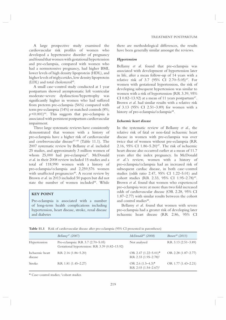
TREATMENT POSTPARTUM
219
A large prospective study examined the
cardiovascular risk profiles of women who
developed a hypertensive disorder of pregnancy
and found that women with gestational hypertension
and pre-eclampsia, compared with women who
had a normotensive pregnancy, had higher BMI,
lower levels of high density lipoprotein (HDL), and
higher levels of triglycerides, low density lipoprotein
(LDL) and total cholesterol45.
A small case–control study conducted at 1 year
postpartum showed asymptomatic left ventricular
moderate–severe dysfunction/hypertrophy was
significantly higher in women who had suffered
from preterm pre-eclampsia (56%) compared with
term pre-eclampsia (14%) or matched controls (8%;
p <0.001)46. This suggests that pre-eclampsia is
associated with persistent postpartum cardiovascular
impairment.
Three large systematic reviews have consistently
demonstrated that women with a history of
pre-eclampsia have a higher risk of cardiovascular
and cerebrovascular disease47–49 (Table 11.1). The
2007 systematic review by Bellamy et al. included
25 studies, and approximately 3 million women of
whom 25,000 had pre-eclampsia47. McDonald
et al. in their 2008 review included 15 studies and a
total of 118,990 women with a history of
pre-eclampsia/eclampsia and 2,259,576 women
with unaffected pregnancies49. A recent review by
Brown et al. in 2013 included 50 papers but did not
state the number of women included48. While
there are methodological differences, the results
have been generally similar amongst the reviews.
Hypertension
Bellamy et al. found that pre-eclampsia was
associated with development of hypertension later
in life, after a mean follow-up of 14 years with a
relative risk of 3.7 (95% CI 2.70–5.05)47. For
women with gestational hypertension, the risk of
developing subsequent hypertension was similar to
women with a risk of hypertension (RR 3.39, 95%
CI 0.82–13.92) at a mean of 11 years postpartum47.
Brown et al. had similar results with a relative risk
of 3.13 (95% CI 2.51–3.89) for women with a
history of pre-eclampsia/eclampsia48.
Ischaemic heart disease
In the systematic review of Bellamy et al., the
relative risk of fatal or non-fatal ischaemic heart
disease in women with pre-eclampsia was over
twice that of women without pre-eclampsia (RR
2.16, 95% CI 1.86–5.20)47. The risk of ischaemic
heart disease also occurred earlier at a mean of 11.7
years after the index pregnancy. In McDonald
et al.’s review, women with a history of
pre-eclampsia/eclampsia had an increased risk of
subsequent cardiac disease, in both case–control
studies (odds ratio 2.47, 95% CI 1.22–5.01) and
cohort studies (RR 2.33, 95% CI 1.95–2.78)49.
Brown et al. found that women who experienced
pre-eclampsia were at more than two fold increased
odds of cardiovascular disease (OR 2.28, 95% CI
1.87–2.77) with similar results between the cohort
and control studies48.
Bellamy et al. found that women with severe
pre-eclampsia had a greater risk of developing later
ischaemic heart disease (RR 2.86, 95% CI
KEY POINT
Pre-eclampsia is associated with a number
of long-term health complications including
hypertension, heart disease, stroke, renal disease
and diabetes
Table 11.1 Risk of cardiovascular disease after pre-eclampsia (95% CI presented in parentheses)
Bellamy47 (2007) McDonald49 (2008) Brown48 (2013)
Hypertension Pre-eclampsia: RR 3.7 (2.70–5.05)
Gestational hypertension: RR 3.39 (0.82–13.92)
Not analysed RR 3.13 (2.51–3.89)
Ischaemic heart
disease
RR 2.16 (1.86–5.20) OR 2.47 (1.22–5.01)*RR 2.33 (1.95–2.78)†
OR 2.28 (1.87–2.77)
Stroke RR 1.81 (1.45–2.27) OR 2.6 (1.5–4.3)*RR 2.03 (1.54–2.67)†
OR 1.77 (1.43–2.21)
* Case–control studies; †cohort studies

THE FIGO TEXTBOOK OF PREGNANCY HYPERTENSION
220
1.65–2.24) compared to women who had mild
pre-elampsia (RR 1.92, 95% CI 1.65–2.24)
particularly if pre-eclampsia occurred before 37
weeks (RR 7.71, 95% CI 4.40–13.52)47. This ‘dose
response’ effect was not, however, demonstrated in
the review by Brown. The review by Brown
compared outcomes for pre-eclampsia both with
and without preterm birth using three studies
whilst the data from Bellamy came from only one
study48.
The risk of cardiovascular disease may be further
increased in the presence of poor fetal outcomes. In
a population-based retrospective cohort study of
women with maternal placental syndrome (defined
as pre-eclampsia, gestational hypertension, placental
abruption, and/or placental infarction), the risk of
premature cardiovascular disease was higher in the
presence of poor fetal growth (adjusted hazard ratio
(aHR) 3.1, 95% CI 2.2–4.5) or intrauterine fetal
death (aHR 4.4, 95% CI 2.4–7.9)50. A Canadian
population-based retrospective cohort study of
1985 women found that in middle-aged women
(mean age 45 years) who underwent coronary
revascularisation, prior maternal placental syndrome
(such as pre-eclampsia) doubled the risk of death
(aHR 1.61, 95% CI 1.00–2.58)51.
Stroke
Bellamy’s review found that the overall risk of fatal
and non-fatal cerebrovascular disease (stroke or
non-fatal stroke) after pre-eclampsia was 1.81 (95%
CI 1.45–2.27) compared with women who had
not developed pre-eclampsia. Subgroup analysis
showed that the risk of fatal stroke (RR 2.98, 95%
CI 1.11–7.96) was greater than that of non-fatal
stroke (RR 1.76, 95% CI 1.40–2.2) after
pre-eclampsia47. A diagnosis of pre-eclampsia
before 37 weeks was associated with a further
elevation in risk (RR 5.08, 95% CI 2.09–12.35)
compared with pre-eclampsia after 37 weeks (RR
0.98, 95% CI 0.50–1.92). McDonald included only
one eligible case–control study which reported an
increased risk of 2.6 (95% CI 1.5–4.3) consistent
with the pooled estimate in the six cohort studies
(RR 2.03, 95% CI 1.54–2.67)49.
Renal disease
Pre-eclampsia has been associated with an increased
risk of end-stage kidney disease in large observational
studies.
A large retrospective cohort study from Norway
(20,918 women with pre-eclampsia) found that
pre-eclampsia in the first pregnancy was associated
with a relative risk of end-stage kidney disease of
4.7 (95% CI 3.6–6.1). This risk increased further to
6.7 (95% CI 4.3–10.6) among women who also
had pre-eclampsia only in their second pregnancy,
and increased again if women had pre-eclampsia in
both their first and second pregnancies or in three
pregnancies (RR 15.5, 95% CI 7.8–30.8)52.
In a population-based study from Taiwan (8653
women with gestational hypertension, 17,998
women with pre-eclampsia), having had either
gestational hypertension or pre-eclampsia was
associated with a greater risk of chronic kidney
disease (aHR 9.4, 95% CI 7.1–12.4) and end-stage
renal disease (aHR 12.4, 95% CI 8.5–18.0), even
after controlling for several factors including
coronary artery disease, congestive heart failure,
hyperlipidaemia and abruption53. The greatest risk
of end-stage renal disease was associated with
having had pre-eclampsia or eclampsia (aHR 14.0,
95% CI 9.4–20.7) compared with gestational
hypertension (aHR 9.0, 95% CI 5.2–15.7)53.
Other chronic diseases
Diabetes In a population-based study (50,598
women with a hypertensive disorder of pregnancy),
women with a prior hypertensive disorder of
pregnancy had a two-fold increased risk of
developing diabetes when followed up to 16.5
years after pregnancy, even in the absence of a prior
history of gestational diabetes54. A history of a
hypertensive disorder of pregnancy and gestational
diabetes together increased the risk associated with
GDM alone54.
Elevated thyroid stimulating hormone (TSH) In a
nested case–control study of women with
pre-eclampsia from two large cohort studies
(Calcium for Pre-eclampsia Prevention trial55 and
the Nord-Trondelag Health Study56), women with
prior pre-eclampsia had higher thyroid stimulating
hormone (TSH) levels compared with controls
who had no history of a hypertensive disorder of
pregnancy57. Of note, women with prior
pre-eclampsia were less likely to have thyroid
peroxidase antibodies, suggesting that their
hypothyroidism was occurring in the absence of an
autoimmune process. The association between

TREATMENT POSTPARTUM
221
pre-eclampsia and elevated TSH was especially
strong (adjusted OR 5.8, 95% CI 1.3–25.5) if
pre-eclampsia had occurred in both the first and the
second pregnancies57.
Central nervous system white-matter
lesions Several studies have shown that women
whose pregnancies were complicated by
pre-eclampsia or eclampsia are more likely than
controls to have white matter lesions; although
these may reflect a predisposition to vascular
disease, the significance of these lesions is currently
unknown58,59.
Mental health Pre-eclampsia can be very stressful
for women and their partners60, especially relative
to expectations of a routine, normal pregnancy and
no prior significant illness60. Women may have to
deal with postpartum recovery from hypertension,
end-organ complications, and frequently, Caesarean
delivery. In addition, women may have to deal
with perinatal loss or illness, such as care in the
neonatal intensive care unit (NICU).
A systematic review showed that while the
evidence is not entirely consistent, in general, there
is an association with prior pre-eclampsia or HELLP
syndrome and more anxiety, depression and
post-traumatic stress disorder (6 studies, 5636
women)61. Women who experienced severe
maternal morbidity were shown to be at particular
risk of post-traumatic stress disorder (1824
women)62. Women with severe pre-eclampisa who
must deal with a perinatal death or NICU admission
are at particular risk of poor health-related
(especially mental health-related) quality of life63.
These effects of pre-eclampsia on mental health
may persist beyond the short term; women with
preterm birth owing to severe, early-onset
pre-eclampsia (compared with women with
preterm birth for other reasons) more often
experienced post-traumatic stress symptoms an
average of 7 years postpartum64.
Postpartum care of women with pre-eclampsia
should include evaluation and referral for
postpartum psychological care. It is important for
women to receive relevant information about not
only their medical condition, but their psychological
condition as well. It is also necessary to examine
coping strategies after pre-eclampsia and offer
adequate supportive interventions when they are
needed60.
Investigations and interventions to improve long-term health
Pregnancy and the immediate postpartum period
may be one of the few times in a woman’s life when
she accesses the health care system regularly.
Therefore, the postpartum period provides a unique
window of opportunity for early identification and
reduction of primary cardiovascular risk.
Education/awareness
Among women A small study using focus groups
found that women with prior pre-eclampsia were
unaware of the link between pre-eclampsia and
future cardiovascular disease, but were eager to
learn about the link and motivated to achieve a
healthy lifestyle65. Another study found that women
generally had a low level of cardiovascular risk
factor knowledge66.
Although some guidelines, such as those from
NICE (UK), recommend that future cardiovascular
risk should be communicated to the woman before
discharge from maternal services, some argue that it
may be too early for most women67,68. Women may
be recovering from serious morbidity whilst
balancing the demands of the neonate; this may
limit the efficacy of the communication. Women’s
reactions to learning about the link between
pre-eclampsia and future cardiovascular disease
included both positive feelings (i.e., motivation,
empowerment) and negative ones (i.e., being
scared, angry, guilty, or isolated)65. Therefore, it is
important that women are followed on an ongoing
basis and receive information about cardiovascular
health over time.
Recently, an educational intervention to
promote cardiovascular knowledge and awareness
was tested amongst women with a history of
pre-eclampsia. The intervention, delivered by
telephone given that postpartum women have
many demands on their time, had several
components: diet, exercise, medication compliance,
screening for risk factors and symptoms of
myocardial infarction. It was found to be a practical
and effective method of contacting postpartum
women following pre-eclampsia and increasing
perception of cardiovascular risk66.
Among health care providers There is also a lack
of awareness amongst health care providers and
gaps in identification and routine follow-up.

THE FIGO TEXTBOOK OF PREGNANCY HYPERTENSION
222
Though the American Heart Association
recommends that health professionals disclose the
future risk of cardiovascular disease to women with
a history of pre-eclampsia, a study revealed that
only a third of health professionals provide that
counselling69. A review of the existing literature on
engaging obstetricians and gynaecologists in
cardiovascular risk reduction found that while they
agreed that their role extended beyond reproductive
care, there was variation in practice and they were
unlikely to manage hypertension or elevated
cholesterol. Obstetricians and gynaecologists
identified knowledge and skill deficits, concerns
about liability, and barriers to prevention presented
by their practice structure. Some providers also
emphasised difficulties completing referrals to
primary care providers70. A Canadian survey of
maternity care providers found that only 54% of
participants were familiar with the long-term
cardiovascular risks of pre-eclampsia71. A small
retrospective review from The Netherlands
examined cardiovascular risk factor management
and found that only 50% of women with
pre-eclampsia had their blood pressure measured
by 3 months postpartum. Blood glucose and lipids
were infrequently checked even though some of
the women had cardiovascular risk factors prior to
the index pregnancy72. Appendix 11.1 contains
training materials for health care providers including
multiple choice questions and a case study.
Knowledge translation tools for health care
providers are included in Appendix 11.2.
Cardiovascular risk factor screening
At present, there are no guidelines advising when
to screen women with a prior hypertensive disorder
of pregnancy or pre-eclampsia for cardiovascular
risk factors. (Cardiovascular risk factor screening in
women with pre-existing or chronic hypertension
has been published by all national societies.) The
very earliest would be at 3–6 months postpartum
when the metabolic changes of pregnancy (such as
dyslipidaemia) have resolved.
Traditional cardiovascular markers appear to be
more abnormal in women who have suffered from
pre-eclampsia, as early as 1 year postpartum73. A
small study from the Maternal Health Clinic, a
postpartum cardiovascular risk reduction clinic in
Kingston, Canada, sought to determine whether
women with a history of pre-eclampsia (N = 99)
compared with those without pre-eclampsia
(N = 118) had 10-year, 30-year and lifetime
cardiovascular risk estimates that were high enough
at 1 year postpartum to identify them as warranting
further counselling and follow-up regarding
lifestyle modification and/or pharmacotherapy74.
Using traditional cardiovascular risk markers (i.e.,
sex, age, smoking, serum total cholesterol, serum
LDL cholesterol, serum HDL cholesterol, fasting
plasma glucose, systolic blood pressure, diastolic
blood pressure and antihypertensive use), the study
found that women who had suffered from
pre-eclampsia (versus those who had not) more
often had elevated 10-year cardiovascular risk (i.e.,
18.2% vs. 1.7%, respectively; OR 13.1, 95% CI
3.4–85.5), 30-year cardiovascular risk (i.e., 31.3%
vs. 5.1%, respectively, OR 8.4, 95% CI 3.5–23.2),
and lifetime cardiovascular risk (i.e., 41.4% vs.
17.8%, respectively; OR 3.3, 95% CI 1.8–6.1)74. A
follow-up study showed that the Maternal Health
Clinic could identify a population of postpartum
patients with increased 10- and 30-year
cardiovascular risk75.
It remains unclear whether traditional risk
factors, such as those used in the Maternal Health
Clinic, are sufficient for cardiovascular risk
screening. First, global risk assessment tools like the
Framingham Risk Score may not accurately
estimate cardiovascular risk in young women,
especially in the short term; a study of 2333 women
using the Framingham Offspring cohort showed
that the 10-year model estimates negligible risks for
young women, whereas the 30-year model suggests
a risk that is 10 times higher76. Second, cardiovascular
risk factors used in prediction models like the
Framingham or Reynolds Risk Score may not fully
explain the risk of cardiovascular disease after
pre-eclampsia; in a literature based study that
included 16 studies, a major part of the observed
OR of cardiovascular disease after pre-eclampsia
remained after adjustment for these traditional
cardiovascular risk factors77.
In summary, it remains unclear both when
cardiovascular risk screening should take place
KEY POINT
Following pre-eclampsia, it would seem prudent
to screen women for traditional cardiovascular
risk markers according to national guidelines
which should also dictate intervention for
abnormal results

TREATMENT POSTPARTUM
223
following pre-eclampsia, and at what threshold
treatment should begin. There are public health
implications and costs to consider. Although it may
be appropriate to intervene at earlier stages in this
population, we should await supportive research
findings. In the meantime, it would seem prudent
to screen women who have had pre-eclampsia for
traditional cardiovascular risk markers according to
national guidelines which should also dictate
intervention for abnormal results.
A simple approach that has been proposed in this
population of women is the identification of any of
the components of metabolic syndrome as the
syndrome does not predict clinical outcomes better
than its individual components78. These components
include:
• Blood pressure
• Weight and height are measured to calculate the
BMI
• Lipid panel
• Glucose intolerance screening.
All women with gestational diabetes should
undergo postpartum glucose tolerance testing. In
addition, some recommend that postpartum testing
for glucose intolerance should be ordered for
all women with one of the other components
of metabolic syndrome, such as obesity78.
Abnormalities in any of the above components
should be treated as per current national
guidelines or prompt referral to an internal
medicine specialist.
Lifestyle change
What is appropriate and evidence-based for all is
adoption of a heart-healthy diet and lifestyle to
decrease cardiovascular risk (Table 11.2)40. Of
course, neither is an easy intervention and there are
barriers to change that are specific to postpartum
women.
Women may be motivated by the knowledge
that weight gain between pregnancies predicts
pre-eclampsia and other pregnancy complications
(e.g., gestational diabetes and Caesarean delivery)80,81,
and weight loss between pregnancies may improve
future pregnancy outcome in addition to long-term
cardiovascular risk. However, there are many
competing demands on a new mother’s time.
Major perceived barriers to lifestyle change
identified in a qualitative (American) study of 20
postpartum women were lack of time, cost of healthy
foods and family responsibilities65. Another (Dutch)
study of 36 women identified additional barriers
of poor postpartum physical and psychological
recovery, and lack of postpartum medical and
psychological support from health care providers82.
Perceived facilitators have included knowledge of
the link between pre-eclampsia and cardiovascular
disease, a desire to stay healthy, and creating a healthy
home for their children65. This link to child health
may be a key motivator of change.
Currently, postpartum lifestyle interventions
tailored specifically for women following a
hypertensive disorder of pregnancy are lacking,
although those demonstrated to be effective outside
Table 11.2 Dietary and lifestyle modifications recommended for all women40 (with permission from Society of
Obstetricians and Gynaecologists of Canada)
Intervention Details
Heart-healthy diet Maintain a healthy balanced diet (high in fruits, vegetables, low-fat dairy products, reduced
in saturated fat and cholesterol) in addition to dietary and soluble fibre, whole grains and
protein from plant sources79
Regular physical activity Undertake 150 minutes/week of moderate to vigorous-intensity aerobic physical activity
(such as walking, jogging, cycling or swimming)
Alcohol consumption Reduce alcohol consumption to <2 drinks/day and <9/week
Weight reduction Attain and maintain ideal body weight (i.e., BMI 18.5–24.9 kg/m2)
Reduce waist circumference Attain and maintain a waist circumference of <88 cm
Salt intake Reduce intake to <1500 mg/d
Smoking cessation Quit smoking in addition to ensuring a smoke-free environment
BMI, body mass index

THE FIGO TEXTBOOK OF PREGNANCY HYPERTENSION
224
pregnancy have been tested in unselected postpartum
populations83. Among postpartum women in general
(21 studies, 6288 women), most weight loss
interventions (6 of 8) were effective, as were most
smoking cessation interventions (4 of 5). Also
effective were individualised tailoring of counselling,
group counselling sessions, and use of diaries or
other correspondence material. Of note, the
Maternal Health Clinic (Kingston, ON) has designed
‘The Postpartum Mother’s Health Record’, a card
that allows women to set goals and track weight
loss84. The timing of data collection coincides with
the infant’s scheduled visits and immunisations,
linking maternal and child health going forward –
a practical and potentially more feasible approach
for the new mother. Appendix 11.2 highlights
knowledge translation tools for women including
mobile apps, programmes and research studies.
Focus groups with women with prior
pre-eclampsia indicated potential interest in a
web-based programme focused on lifestyle
strategies to decrease cardiovascular risk65. This
approach was tested in a small feasibility study (20
women) of a web-based, tailored health education
intervention related to diet and exercise, in
conjunction with counselling by a psychologist
(Dutch ProActive study, Postpartum Rotterdam
Appraisal of Cardiovascular Health and Tailored
Intervention)85. The intervention was initiated at 6
months postpartum and continued for 3 months. In
all 60% of women participated and anthropometric
measurements at 13 months postpartum improved
significantly, although metabolic parameters did
not85.
Bariatric surgery for women with morbid obesity
Women who are morbidly obese and have failed
lifestyle interventions to achieve weight loss are
candidates for bariatric surgery without considering
potential effects on future pregnancy, which are
mixed. There are no randomised controlled trials
for women planning pregnancy, but in a retrospective
cohort study of insurance claims data (585 women)86
and two controlled registry studies (identifying 1085
women with prior bariatric surgery)87,88, women
who had undergone bariatric surgery (vs. those who
had not) experienced lower rates of all hypertensive
disorders of pregnancy (including pre-eclampsia)86,
gestational diabetes and large-for-gestational
infants87, as well as fewer emergency Caesarean
deliveries88 following adjustment for confounders.
However, these benefits have not been consistently
demonstrated, with one registry study demonstrating
higher rates of hypertension and gestational diabetes
following bariatric surgery88. In addition, potential
benefits appear to come at the price of more
gastrointestinal problems88,89, lower birth weight88,
more small-for-gestational age (SGA) infants87,
earlier delivery87 and, possibly, higher perinatal
mortality87,88 and admission to NICU88. Therefore,
at present, there are insufficient data to support
recommendations to undergo bariatric surgery to
favourably affect future pregnancy outcomes.
BEST PRACTICE POINTS
(Please see Appendix 11.3 for the evaluation of the strength of the recommendations and the quality of
the evidence on which they are based.)
Care in the 6 weeks after birth
1. Blood pressure should be measured during the time of peak postpartum blood pressure, at days 3–6
after delivery.
2. Women with postpartum hypertension should be evaluated for pre-eclampsia (either arising de novo or worsening from the antenatal period).
3. Antihypertensive therapy may be continued postpartum, particularly in women with antenatal
pre-eclampsia and those who delivered preterm.
4. Severe postpartum hypertension must be treated with antihypertensive therapy, to keep systolic
blood pressure <160 mmHg and diastolic blood pressure <110 mmHg.
5. Antihypertensive therapy may be used to treat non-severe postpartum hypertension, to keep blood
pressure at <140/90 mmHg for all but women with pre-gestational diabetes mellitus among whom
the target should be <130/80 mmHg.

TREATMENT POSTPARTUM
225
PRIORITIES FOR UNDER-RESOURCED SETTINGS
The priorities for postpartum care of women
with hypertensive disorders of pregnancy in
under-resourced settings are outlined in Table
11.3. A sample policy brief that focuses on postnatal
care is contained in Appendix 11.4.
In LMICs, routine postnatal care has the
potential to improve both maternal and neonatal
outcomes. In LMICs, almost 40% of women
experience complications after delivery and an
estimated 15% develop potentially life-threatening
problems90. More than half of maternal deaths
occur postpartum and the vast majority (i.e., 80%)
of those occur in the first week postpartum91. Such
visits are most likely to detect early complications
that may be addressed by referral for specialist care.
Also, postnatal care has the potential to identify
neonatal sepsis and asphyxia/hypothermia, the
leading causes of neonatal death in LMICs. Finally,
postnatal care helps to promote healthy maternal
behaviours, such as exclusive breastfeeding and
proper care of babies with low birth weight.
“By the 6th week the child is due for
pentavalent. That is . . . the diphtheria, tetanus
and all . . . at that 6 week [mark]. That is the
time (when) the lady is also due for the 6 week
postnatal review. But many a time the postnatal
review is not done . . . because of lack of
manpower at the PHC. You have a lot of
children and . . . so sometimes to attend to
mother, and educate and counsel takes time.”
Stakeholder, Local Government in Ogun,
Nigeria
Postnatal care is reported at much lower rates than
for other maternal and infant health services92. In a
review of Demographic and Health Survey (DHS)
data from 1990 to 2009 in 38 countries in four
regions (i.e., sub-Saharan Africa; North Africa/
West Asia/Europe; South/Southeast Asia; and
Latin America and the Caribbean), approximately
half of the countries with data (i.e., 8 of 18)
provided at least one demonstrated postnatal visit to
more than half (64–92%) of postpartum women
within 41 days after giving birth93. Even within
those countries, postnatal visits varied from 64% to
6. Antihypertensive agents acceptable for use in breastfeeding include nifedipine XL, labetalol,
methyldopa, captopril and enalapril.
7. There should be confirmation that end-organ dysfunction of pre-eclampsia has resolved.
8. Non-steroidal anti-inflammatory druds (NSAIDs) should not be given postpartum if hypertension is
difficult to control, there is evidence of kidney injury (oliguria and/or an elevated creatinine)
(≥90 mol/L) or platelets are <50109/L.
9. Postpartum thromboprophylaxis should be considered in women with pre-eclampsia who have
other risk factors for thromboembolism.
Care beyond the first 6 weeks after birth
1. Women with a history of severe pre-eclampsia (particularly those who presented or delivered at <34
weeks) should be screened for pre-existing hypertension and underlying renal disease.
2. Referral for internal medicine or nephrology consultation should be considered for women with
postpartum hypertension that is difficult to control, or women who had pre-eclampsia and have at
3–6 months postpartum ongoing proteinuria, decreased eGFR (<60 mL/min), or another indication
of renal disease (such as abnormal urinary sediment).
3. Women who are overweight should be encouraged to attain a healthy body mass index to decrease
risk in future and for long-term health.
4. Women with pre-existing hypertension or persistent postpartum hypertension should undergo the
following investigations (if not done previously): urinalysis; serum sodium, potassium and creatinine;
fasting glucose; fasting lipid profile; and standard 12-lead electrocardiography.
5. Women who are normotensive but who have had a hypertensive disorder of pregnancy, may benefit
from assessment of traditional cardiovascular risk markers.
6. All women who have had a hypertensive disorder of pregnancy should pursue a healthy diet and
lifestyle.

THE FIGO TEXTBOOK OF PREGNANCY HYPERTENSION
226
92% of relevant women. Although there was a
strong relationship between receiving postnatal
care and both more antenatal visits and having
skilled birth attendance, the major determinant of
postnatal care was delivery in facility. (A recent
systematic review found that inequities in the use
of postnatal services is also based on socioeconomic
status, education, ethnicity and geographical
location94.) Following delivery in facility, at least
two-thirds of women reported postnatal care in all
countries except Uganda and Zimbabwe where less
than half of the women reported postnatal checkups.
Women who delivered in a health facility
(compared with those who did not) were more
likely to report postnatal visits, to have the first visit
within 2 days after birth, and to receive postnatal
care from a doctor, nurse, or midwife. Among
women who did not deliver at a health facility,
postnatal care was reported for less than 50% with
the exception of a few countries in South/Southeast
Asia (i.e., Cambodia and the Philippines),
sub-Saharan Africa (i.e., Ghana and Madagascar),
and Latin America and the Caribbean (i.e., Bolivia,
the Domincan Republic and Peru).
In recognition of the postnatal period as “. . . a
critical phase in the lives of mothers and newborn
babies”, the 2013 WHO guidelines on postnatal
care95 recommend the following:
• A health care visit within 24 hours after the birth
and then again at least three more times – on day
3, in the second week, and again at 6 weeks;
• Blood pressure measurement shortly after birth
and at 6 hours, although there is no guidance
around blood pressure measurement during the
rest of the postpartum period;
• Counselling about the signs and symptoms of
pre-eclampsia at each postpartum visit.
It is important to recognise that in a LMIC,
postpartum care may be provided in the community
rather than in facility, and by varying cadres of
health care workers. For example, lay health
workers may undertake promotion of postpartum
care, while nurses and midwives may initiate
treatment of pre-eclampsia96. All of these workers
need to be trained accordingly.
Global initiatives that address the link between a
woman’s reproductive health and long-term health
are lacking. To prevent non-communicable diseases
in the offspring, the International Federation of
Gynaecology and Obstetrics (FIGO) recently
announced that it is partnering with other agencies
and organisations that focus on enforcing
interventions, such as good nutrition and
minimisation of harmful environmental exposures
during pregnancy97. While this initiative
Table 11.3 Priorities for postpartum care in under-resourced settings
Initial priority Ultimate goal
Community
Primary health care
centre (detect,
stabilise and refer)
A health care visit within 24 hours after the birth A health care visit within 24 hours after the birth
and then again at least three more times – on day
3, in the second week, and again at 6 weeks
BP measurement shortly after birth and at 6 hours BP measurement shortly after birth and at 6 hours
Counselling about the signs and symptoms of
pre-eclampsia at each postpartum visit
Counselling about the signs and symptoms of
pre-eclampsia at each postpartum visit
Facility
Secondary-level
facility (detect,
manage and refer
if necessary)
Tertiary-level
(referral) facility
(detect and manage
definitively)
Delivery in facility of all women with a HDP Delivery in facility of all women with a HDP
Management of women with HDPs, including
postpartum pre-eclampsia (see Table 4.4,
Chapter 4)
Management of women with HDPs, including
postpartum pre-eclampsia (see Table 4.4,
Chapter 4)
Counselling about BP monitoring as well as
heart-healthy diet and lifestyle following a HDP
BP, blood pressure; HDP, hypertensive disorder of pregnancy

TREATMENT POSTPARTUM
227
acknowledges the role of non-communicable
diseases within the reproductive, maternal, neonatal
and child health continuum, it does not address the
long-term chronic illnesses that may develop as a
result of a complicated pregnancy.
WHAT INTERNATIONAL GUIDELINES SAY (APPENDIX 11.5)
Abbreviations for Clinical Practice Guidelines:
ACOG (American College of Obstetricians and
Gynecologists)99, AOM (Association of Ontario
Midwives), NICE (National Institutes of Clinical
Excellence)67, NVOG (National Obstetrics and
Gynaecology Society, Netherlands)100, QLD
(Queensland, Australia)101,102, SOGC (Society of
Obstetricians and Gynaecologists of Canada)103,104,
SOMANZ (Society of Obstetric Medicine of
Australia and New Zealand)105,106, WHO (World
Health Organization)107.
Most international guidelines highlighted that
pre-eclampsia may develop de novo or worsen in
the postpartum period (AOM, ACOG, NICE,
SOGC, QLD).
The majority of guidelines stated that blood
pressure may increase postpartum and recommended
continuing antihypertensive therapy that women
had been taking antepartum (NICE, ACOG,
SOGC). Importantly, no guideline recommended
that antenatal antihypertensive therapy be stopped.
Although the treatment of severe hypertension
followed similar recommendations to those
antenatally, treatment targets for non-severe
hypertension were generally lower and varied
amongst guidelines (NICE, SOGC, ACOG); most
commonly, clinicians are recommended to aim for
a blood pressure <150/100 mmHg for women
with gestational hypertension or pre-eclampsia
(NICE, ACOG).
Most of the guidelines specifically mentioned
the association between the hypertensive disorders
of pregnancy (particularly pre-eclampsia) and future
cardiovascular health, and suggested lifestyle
counselling as the logical response (AOM, ACOG,
NICE, SOGC, QLD, SOMANZ).
SUMMARY
Care in the immediate postpartum period should
focus on the management of hypertension using
treatment options that are acceptable during
breastfeeding. Consideration should be given to
postpartum thromboprophylaxis in women with a
hypertensive disorder of pregnancy if other risk
factors are present. NSAIDs should be avoided if
hypertension is difficult to control or there is either
a coagulopathy or renal dysfunction. All women
with pre-eclampsia should be followed closely after
delivery to ensure resolution of end-organ damage.
Women should be screened for underlying disease
that may have predisposed to pre-eclampsia and
pre-eclampsia mimickers should also be ruled out.
Finally, the postpartum period offers a unique
window of opportunity to address short- and
long-term risks of hypertensive disorders of
pregnancy. Women should be counselled about the
ideal inter-pregnancy interval as well as risks in
future pregnancies. Women should be evaluated
for risk factors for premature cardiovascular disease
as well as other complications such as chronic
kidney disease. Currently, the focus should be on
the adoption of a heart-healthy lifestyle and
screening for traditional cardiovascular risk factors.
PRIORITIES FOR FUTURE RESEARCH
Given the global rise in non-communicable diseases
and the risk stratification that pregnancy appears to
provide, there is an urgent need to identify how a
woman’s pregnancy history can be added to
currently available cardiovascular disease risk
scoring systems to identify and manage women at
increased risk for cardiovascular disease.
REFERENCES
1. Firoz T, Melnik T. Postpartum evaluation and long term implications. Best Pract Res Clin Obstet Gynaecol 2011;25(4):549–561
2. Ferrazzani S, De Carolis S, Pomini F, Testa AC, Mastromarino C, Caruso A. The duration of hypertension in the puerperium of preeclamptic women: relationship with renal impairment and week of delivery. Am J Obstet Gynecol 1994;171(2):506–512
3. Tan LK, de Swiet M. The management of postpartum hypertension. BJOG 2002;109(7):733–736
4. Deruelle P, Coudoux E, Ego A, Houfflin-Debarge V, Codaccioni X, Subtil D. Risk factors for post-partum complications occurring after preeclampsia and HELLP syndrome. A study in 453 consecutive pregnancies. Eur J Obstet Gynecol Reprod Biol 2006;125(1):59–65
5. Bayliss H, Beevers D, Churchill D. A study of puerperal blood pressure in hypertensive and

THE FIGO TEXTBOOK OF PREGNANCY HYPERTENSION
228
normotensive pregnancies. Hypertens Pregnancy 2002;21(Suppl 1)
6. Al-Safi Z, Imudia AN, Filetti LC, Hobson DT, Bahado-Singh RO, Awonuga AO. Delayed postpartum preeclampsia and eclampsia: demographics, clinical course, and complications. Obstet Gynecol 2011 Nov;118(5):1102–1107
7. Davison JM, Dunlop W. Changes in renal hemodynamics and tubular function induced by normal human pregnancy. Semin Nephrol 1984;4(3):198–207
8. Martin JN,Jr, Thigpen BD, Moore RC, Rose CH, Cushman J, May W. Stroke and severe preeclampsia and eclampsia: a paradigm shift focusing on systolic blood pressure. Obstet Gynecol 2005 Feb;105(2):246–254
9. Magee L, von Dadelszen P. Prevention and treatment of postpartum hypertension. Cochrane Database Syst Rev 2013 Apr 30;4:CD004351
10. Daskalopoulou SS, Khan NA, Quinn RR, Ruzicka M, McKay DW, Hackam DG, et al. The 2012 Canadian hypertension education program recommendations for the management of hypertension: blood pressure measurement, diagnosis, assessment of risk, and therapy. Can J Cardiol 2012 May;28(3):270–287
11. Denolle T, Weber J, Calvez C, Daniel J, Cheve M, Marechaud M, et al. Home blood pressure measured telemetrically in hypertensive pregnant women. Am J Hypertens 2001;14(11):43A
12. American Academy of Pediatrics Committee on Drugs. Transfer of drugs and other chemicals into human milk. Pediatrics 2001 Sep;108(3):776–789
13. National Institutes of Health. Drugs and Lactation Database (LactMed). 2015; Available at: http://toxnet.nlm.nih.gov/newtoxnet/lactmed.htm. Accessed March 16, 2015
14. Shannon ME, Malecha SE, Cha AJ. Angiotensin converting enzyme inhibitors (ACEIs) and angiotensin II receptor blockers (ARBs) and lactation: an update. J Hum Lact 2000 May;16(2):152–155
15. Mathews DD, Agarwal V, Shuttleworth TP. A randomized controlled trial of complete bed rest versus ambulation in the management of proteinuric hypertension during pregnancy. Br J Obstet Gynaecol 1982 Feb;89(2):128–131
16. Jacobsen AF, Skjeldestad FE, Sandset PM. Ante- and postnatal risk factors of venous thrombosis: a hospital-based case-control study. J Thromb Haemost 2008 Jun;6(6):905–912
17. Royal College of Obstetricians & Gynaecologists. Green-top Guideline 37a: Reducing the risk of thrombosis and embolism during pregnancy and the puerperium. 2015; Available at: https://www.rcog.org.uk/en/guidelines-research-services/guidelines/gtg37a/. Accessed Jul/19, 2015
18. Karumanchi S, August P, Podymow T. Renal complications in normal pregnancy. In: Jurgen F, Johnson RJ, Feehally J, eds. Comprehensive Clinical Nephrology, 4th edn. New York: Elsevier; 2010
19. Daskalopoulou SS, Rabi DM, Zarnke KB, Dasgupta K, Nerenberg K, Cloutier L, et al. The 2015 Canadian Hypertension Education Program Recommendations for Blood Pressure Measurement, Diagnosis, Assessment of Risk, Prevention, and Treatment of Hypertension. Can J Cardiol 2015 5;31(5):549–568
20. American College of Obstetricians and Gynecologists Committee on Practice Bulletins-Obstetrics. ACOG Practice Bulletin No. 111: Inherited thrombophilias in pregnancy. Obstet Gynecol 2010 Apr;115(4):877–887
21. Rodger MA, Hague WM, Kingdom J, Kahn SR, Karovitch A, Sermer M, et al. Antepartum dalteparin versus no antepartum dalteparin for the prevention of pregnancy complications in pregnant women with thrombophilia (TIPPS): a multinational open-label randomised trial. Lancet 2014 Nov 8;384(9955): 1673–1683
22. Rodger MA, Langlois NJ, de Vries JI, Rey E, Gris JC, Martinelli I, et al. Low-molecular-weight heparin for prevention of placenta-mediated pregnancy complications: protocol for a systematic review and individual patient data meta-analysis (AFFIRM). Syst Rev 2014 Jun 26;3:69-4053-3-69
23. Miyakis S, Lockshin MD, Atsumi T, Branch DW, Brey RL, Cervera R, et al. International consensus statement on an update of the classification criteria for definite antiphospholipid syndrome (APS). J Thromb Haemost 2006 Feb;4(2):295–306
24. Ounsted M, Cockburn J, Moar VA, Redman CW. Maternal hypertension with superimposed pre-eclampsia: effects on child development at 71/2 years. Br J Obstet Gynaecol 1983 Jul;90(7):644–649
25. Robinson M, Mattes E, Oddy WH, de Klerk NH, Li J, McLean NJ, et al. Hypertensive diseases of pregnancy and the development of behavioral problems in childhood and adolescence: the Western Australian Pregnancy Cohort Study. J Pediatr 2009 Feb;154(2):218–224
26. Whitehouse AJ, Robinson M, Newnham JP, Pennell CE. Do hypertensive diseases of pregnancy disrupt

TREATMENT POSTPARTUM
229
neurocognitive development in offspring? Paediatr Perinat Epidemiol 2012 Mar;26(2):101–108
27. Tuovinen S, Eriksson JG, Eero K, Räikkönen K. Maternal hypertensive pregnancy disorders and cognitive functioning of the offspring: a systematic review. Am Soc Hypertens 2014;8(11):8328470
28. Ananth CV, Friedman AM. Ischemic placental disease and risks of perinatal mortality and morbidity and neurodevelopmental outcomes. Semin Perinatol 2014;38(3):151–158
29. Mutch LM, Moar VA, Ounsted MK, Redman CW. Hypertension during pregnancy, with and without specific hypotensive treatment. II. The growth and development of the infant in the first year of life. Early Hum Dev 1977 Oct;1(1):59–67
30. Cockburn J, Moar VA, Ounsted M, Redman CW. Final report of study on hypertension during pregnancy: the effects of specific treatment on the growth and development of the children. Lancet 1982 Mar 20;1(8273):647–649
31. Reynolds B, Butters L, Evans J, Adams T, Rubin PC. First year of life after the use of atenolol in pregnancy associated hypertension. Arch Dis Child 1984;59(11):1061–1063
32. Bortolus R, Ricci E, Chatenoud L, Parazzini F. Nifedipine administered in pregnancy: effect on the development of children at 18 months. BJOG 2000;107(6):792–794
33. Chan WS, Koren G, Barrera M, Rezvani M, Knittel-Keren D, Nulman I. Neurocognitive development of children following in-utero exposure to labetalol for maternal hypertension: a cohort study using a prospectively collected database. Hypertens Pregnancy 2010;29(3):271–283
34. Davis EF, Lazdam M, Lewandowski AJ, Worton SA, Kelly B, Kenworthy Y, et al. Cardiovascular Risk Factors in Children and Young Adults Born to Preeclamptic Pregnancies: A Systematic Review. Pediatrics 2012;129(6):e1552-e1561
35. Lazdam M, de lH, Diesch J, Kenworthy Y, Davis E, Lewandowski AJ, et al. Unique blood pressure characteristics in mother and offspring after early onset preeclampsia. Hypertension 2012;60(5):1338–1345
36. Mamun AA, Kinarivala MK, O’Callaghan M, Williams G, Najman J, Callaway L. Does hypertensive disorder of pregnancy predict offspring blood pressure at 21 years? Evidence from a birth cohort study. J Hum Hypertens 2012;26(5):288–294
37. Kajantie E, Eriksson JG, Osmond C, Thornburg K, Barker DJ. Pre-eclampsia is associated with increased
risk of stroke in the adult offspring: the Helsinki birth cohort study. Stroke 2009;40(4):1176–1180
38. Boivin A, Luo ZC, Audibert F, Mâsse B, Lefebvre F, Tessier R, et al. Pregnancy complications among women born preterm. Can Med Assoc J 2012;184(16):1777–1784
39. Williams D. Pregnancy: a stress test for life. Curr Opin Obstet Gynecol 2003 Dec;15(6):465–471
40. Mosca L, Appel LJ, Benjamin EJ, Berra K, Chandra-Strobos N, Fabunmi RP, et al. Evidence-based guidelines for cardiovascular disease prevention in women. Circulation 2004 Feb 10;109(5):672–693
41. Mosca L, Banka CL, Benjamin EJ, Berra K, Bushnell C, Dolor RJ, et al. Evidence-based guidelines for cardiovascular disease prevention in women: 2007 update. Circulation 2007 Mar 20;115(11):1481–1501
42. Mosca L, Benjamin EJ, Berra K, Bezanson JL, Dolor RJ, Lloyd-Jones DM, et al. Effectiveness-based guidelines for the prevention of cardiovascular disease in women--2011 update: a guideline from the American Heart Association. J Am Coll Cardiol 2011 Mar 22;57(12):1404–1423
43. Craici I, Wagner S, Garovic VD. Preeclampsia and future cardiovascular risk: formal risk factor or failed stress test? Ther Adv Cardiovasc Dis 2008 Aug;2(4):249–259
44. Magnussen EB, Vatten LJ, Lund-Nilsen TI, Salvesen KA, Davey Smith G, Romundstad PR. Prepregnancy cardiovascular risk factors as predictors of pre-eclampsia: population based cohort study. BMJ 2007 Nov 10;335(7627):978
45. Magnussen EB, Vatten LJ, Smith GD, Romundstad PR. Hypertensive disorders in pregnancy and subsequently measured cardiovascular risk factors. Obstet Gynecol 2009 Nov;114(5):961–970
46. Melchiorre K, Sutherland GR, Liberati M, Thilaganathan B. Preeclampsia is associated with persistent postpartum cardiovascular impairment. Hypertension 2011 Oct;58(4):709–715
47. Bellamy L, Casas JP, Hingorani AD, Williams DJ. Pre-eclampsia and risk of cardiovascular disease and cancer in later life: systematic review and meta-analysis. BMJ 2007 Nov 10;335(7627):974
48. Brown MC, Best KE, Pearce MS, Waugh J, Robson SC, Bell R. Cardiovascular disease risk in women with pre-eclampsia: systematic review and meta-analysis. Eur J Epidemiol 2013;28(1):1–19

THE FIGO TEXTBOOK OF PREGNANCY HYPERTENSION
230
49. McDonald SD, Malinowski A, Zhou Q, Yusuf S, Devereaux PJ. Cardiovascular sequelae of preeclampsia/eclampsia: a systematic review and meta-analyses. Am Heart J 2008 Nov;156(5):918–930
50. Ray JG, Vermeulen MJ, Schull MJ, Redelmeier DA. Cardiovascular health after maternal placental syndromes (CHAMPS): population-based retrospective cohort study. Lancet 2005 Nov 19;366(9499): 1797–1803
51. Ray JG, Booth GL, Alter DA, Vermeulen MJ. Prognosis after maternal placental events and revascularization: PAMPER study. Am J Obstet Gynecol 2015; 214(1):106.e1-e14
52. Vikse BE, Irgens LM, Leivestad T, Skjaerven R, Iversen BM. Preeclampsia and the risk of end-stage renal disease. N Engl J Med 2008 Aug 21;359(8):800–809
53. Wang IK, Muo CH, Chang YC, Liang CC, Chang CT, Lin SY, et al. Association between hypertensive disorders during pregnancy and end-stage renal disease: a population-based study. CMAJ 2013 Feb 19;185(3):207–213
54. Feig DS, Shah BR, Lipscombe LL, Wu CF, Ray JG, Lowe J, et al. Preeclampsia as a risk factor for diabetes: a population-based cohort study. PLoS Med 2013;10(4):e1001425
55. Levine RJ, Esterlitz JR, Raymond EG, DerSimonian R, Hauth JC, Ben Curet L, et al. Trial of Calcium for Preeclampsia Prevention (CPEP): rationale, design, and methods. Control Clin Trials 1996 Oct;17(5):442–469
56. Holmen J, Midthjell K, Krüger Ø, Langhammer A, Holmen TL, Bratberg GH, et al. The Nord-Trøndelag Health Study 1995–97 (HUNT 2): Objectives, contents, methods and participation. Norsk Epidemiologi 2003;13(1):19–32
57. Levine RJ, Vatten LJ, Horowitz GL, Qian C, Romundstad PR, Yu KF, et al. Pre-eclampsia, soluble fms-like tyrosine kinase 1, and the risk of reduced thyroid function: nested case-control and population based study. BMJ 2009 Nov 17;339:b4336
58. Aukes AM, De Groot JC, Wiegman MJ, Aarnoudse JG, Sanwikarja GS, Zeeman GG. Long-term cerebral imaging after pre-eclampsia. BJOG 2012 Aug;119(9):1117–1122
59. Wiegman MJ, Zeeman GG, Aukes AM, Bolte AC, Faas MM, Aarnoudse JG, et al. Regional distribution of cerebral white matter lesions years after preeclampsia and eclampsia. Obstet Gynecol 2014 Apr;123(4):790–795
60. Mautner E, Stern C, Deutsch M, Nagele E, Greimel E, Lang U, et al. The impact of resilience on psychological outcomes in women after preeclampsia: an observational cohort study. Health Qual Life Outcomes 2013;11:194
61. Delahaije DH, Dirksen CD, Peeters LL, Smits LJ. Anxiety and depression following preeclampsia or hemolysis, elevated liver enzymes, and low platelets syndrome. A systematic review. Acta Obstet Gynecol Scand 2013;92(7):746–761
62. Furuta M, Sandall J, Cooper D, Bick D. The relationship between severe maternal morbidity and psychological health symptoms at 6–8 weeks postpartum: a prospective cohort study in one English maternity unit. BMC Pregnancy Childbirth 2014;14(1):133
63. Hoedjes M, Berks D, Vogel I, Franx A, Duvekot JJ, Steegers EA, et al. Poor health-related quality of life after severe preeclampsia. Birth 2011 Sep;38(3):246–255
64. Gaugler-Senden IP, Duivenvoorden HJ, Filius A, De Groot CJ, Steegers EA, Passchier J. Maternal psychosocial outcome after early onset preeclampsia and preterm birth. J Matern Fetal Neonatal Med 2012 Mar;25(3):272–276
65. Seely EW, Rich-Edwards J, Lui J, Nicklas JM, Saxena A, Tsigas E, et al. Risk of future cardiovascular disease in women with prior preeclampsia: a focus group study. BMC Pregnancy Childbirth 2013;13(1):240
66. Spratling PM, Pryor ER, Moneyham LD, Hodges AL, White-Williams CL, Martin JN,Jr. Effect of an educational intervention on cardiovascular disease risk perception among women with preeclampsia. J Obstet Gynecol Neonatal Nurs 2014 Mar-Apr;43(2):179–189
67. National Collaborating Centre for Women’s and Children’s Health (UK). CG107: Hypertension in pregnancy: The management of hypertensive disorders during pregnancy. NICE: Guidance 2010 Aug
68. Brown MC, Bell R, Collins C, Waring G, Robson SC, Waugh J, et al. Women’s perception of future risk following pregnancies complicated by preeclampsia. Hypertens Pregnancy 2013;32(1):60–73
69. Young B, Hacker MR, Rana S. Physicians’ knowledge of future vascular disease in women with preeclampsia. Hypertens Pregnancy 2012;31(1):50–58
70. Ehrenthal DB, Catov JM. Importance of engaging obstetrician/gynecologists in cardiovascular disease prevention. Curr Opin Cardiol 2013 Sep;28(5):547–553

TREATMENT POSTPARTUM
231
71. MacDonald SE, Walker M, Ramshaw H, Godwin M, Chen XK, Smith G. Hypertensive disorders of pregnancy and long-term risk of hypertension: what do Ontario prenatal care providers know, and what do they communicate? J Obstet Gynaecol Can 2007 Sep;29(9):705–710
72. Nijdam ME, Timmerman MR, Franx A, Bruinse HW, Numans ME, Grobbee DE, et al. Cardiovascular risk factor assessment after pre-eclampsia in primary care. BMC Fam Pract 2009 Dec 8;10:77-2296-10-77
73. Smith GN, Walker MC, Liu A, Wen SW, Swansburg M, Ramshaw H, et al. A history of preeclampsia identifies women who have underlying cardiovascular risk factors. Am J Obstet Gynecol 2009 Jan;200(1): 58.e1-58.e8
74. Smith GN, Pudwell J, Walker M, Wen SW. Ten-year, thirty-year, and lifetime cardiovascular disease risk estimates following a pregnancy complicated by preeclampsia. J Obstet Gynaecol Can 2012 Sep;34(9):830–835
75. Cusimano MC, Pudwell J, Roddy M, Cho CK, Smith GN. The maternal health clinic: an initiative for cardiovascular risk identification in women with pregnancy-related complications. Am J Obstet Gynecol 2014 May;210(5):438.e1-438.e9
76. Pencina MJ, D’Agostino RB S, Larson MG, Massaro JM, Vasan RS. Predicting the 30-year risk of cardiovascular disease: the framingham heart study. Circulation 2009 Jun 23;119(24):3078–3084
77. Berks D, Hoedjes M, Raat H, Duvekot JJ, Steegers EA, Habbema JD. Risk of cardiovascular disease after pre-eclampsia and the effect of lifestyle interventions: a literature-based study. BJOG 2013 Jul;120(8):924–931
78. Carson MP. Society for maternal and fetal medicine workshop on pregnancy as a window to future health: Clinical utility of classifying women with metabolic syndrome. Semin Perinatol 2015 Jun;39(4):284–289
79. Health Canada. Eating well with Canada’s food guide. 2011; Available at: http://www.hc-sc.gc.ca/fn-an/alt_formats/hpfb-dgpsa/pdf/food-guide-aliment/view_eatwell_vue_bienmang-eng.pdf. Accessed Jan/ 05, 2016
80. Villamor E, Cnattingius S. Interpregnancy weight change and risk of adverse pregnancy outcomes: a population-based study. Lancet 2006 Sep 30;368(9542):1164–1170
81. Bogaerts A, Van den Bergh BR, Ameye L, Witters I, Martens E, Timmerman D, et al. Interpregnancy weight change and risk for adverse perinatal outcome. Obstet Gynecol 2013 Nov;122(5):999–1009
82. Hoedjes M, Berks D, Vogel I, Franx A, Duvekot JJ, Oenema A, et al. Motivators and barriers to a healthy postpartum lifestyle in women at increased cardiovascular and metabolic risk: a focus-group study. Hypertens Pregnancy 2012;31(1):147–155
83. Hoedjes M, Berks D, Vogel I, Franx A, Visser W, Duvekot JJ, et al. Effect of postpartum lifestyle interventions on weight loss, smoking cessation, and prevention of smoking relapse: a systematic review. Obstet Gynecol Surv 2010 Oct;65(10):631–652
84. The MotHERS Program. The MotHERS Program: Mothers’ Health Education, Research and Screening. 2015; Available at: http://www.themothersprogram.ca. Accessed Aug/10, 2015
85. Berks D, Hoedjes M, Franx A, Habbema D, Raat H, Duvekot H, et al. T14.2 Postpartum lifestyle intervention after complicated pregnancy proves feasible. Pregnancy Hypertension: An International Journal of Women’s Cardiovascular Health 2010;1:S25
86. Bennett WL, Gilson MM, Jamshidi R, Burke AE, Segal JB, Steele KE, et al. Impact of bariatric surgery on hypertensive disorders in pregnancy: retrospective analysis of insurance claims data. BMJ 2010 Apr 13;340:c1662
87. Johansson K, Cnattingius S, Naslund I, Roos N, Trolle Lagerros Y, Granath F, et al. Outcomes of pregnancy after bariatric surgery. N Engl J Med 2015 Feb 26;372(9):814–824
88. Berlac JF, Skovlund CW, Lidegaard O. Obstetrical and neonatal outcomes in women following gastric bypass: a Danish national cohort study. Acta Obstet Gynecol Scand 2014 May;93(5):447–453
89. Armstrong C. ACOG Guidelines on Pregnancy After Bariatric Surgery. 2010;81(7)
90. Rahman MM, Haque SE, Zahan MS. Factors affecting the utilisation of postpartum care among young mothers in Bangladesh. Health Soc Care Community 2011;19(2):138–147
91. Li XF, Fortney JA, Kotelchuck M, Glover LH. The postpartum period: the key to maternal mortality. Int J Gynaecol Obstet 1996 Jul;54(1):1–10
92. Fort AL. Coverage of post-partum and post-natal care in Egypt in 2005–2008 and Bangladesh in 2004–2007: levels, trends and unmet need. Reprod Health Matters 2012;20(39):81–92
93. Wang W, Alva S, Wang S, Fort A. Levels and Trends in the use of Maternal Health Services in Developing Countries. DHS Comparative Reports 2011;26

THE FIGO TEXTBOOK OF PREGNANCY HYPERTENSION
232
94. Langlois E, Miszkurka M, Zunzunegui M, Ghaffar G, Ziegler D, Karp I. Inequities in postnatal care in low- and middle-income countries: a systematic review and meta-analysis. Bull World Health Organ 2015;93(4)
95. World Health Organization. WHO Recommendations on postnatal care of the mother and newborn. 2013; Available at: http://www.who.int/maternal_c h i l d _ a d o l e s c e n t / d o c u m e n t s /postnatal-care-recommendations. Accessed Aug/10, 2015
96. World Health Organization. WHO Recommendations – OPTIMIZEMNH. 2012; Available at: http://www.optimizemnh.org/. Accessed Aug/10, 2015
97. Roura L, Arulkumaran S. Facing the noncommunicable disease (NCD) global epidemic – The battle of prevention starts in utero – The FIGO challenge. Best Pract Res Clin Obstet Gynaecol 2015;29(1):514
98. Gillon TE, Pels A, von Dadelszen P, MacDonell K, Magee LA. Hypertensive disorders of pregnancy: a systematic review of international clinical practice guidelines. PLoS One 2014;9(12):e113715
99. American College of Obstetricians and Gynecologists, Task Force on Hypertension in Pregnancy. Hypertension in pregnancy. Report of the American College of Obstetricians and Gynecologists’ Task Force on Hypertension in Pregnancy. Obstet Gynecol 2013 Nov;122(5):1122–1131
100. Nederlandse Vereniging voor Obstetrie en Gynaecologie. Hypertensieve aandoeningen in de zwangerschap. 2011
101. Queensland Maternity and Neonatal Clinical, Guidelines Program. Hypertensive disorders of pregnancy. 2013;MN10.13-V4-R15
102. Queensland Maternity and Neonatal Clinical, Guidelines Program. Supplement: hypertensive disorders of pregnancy. 2013;MN10.15.V4-R15
103. Magee LA, Pels A, Helewa M, Rey E, von Dadelszen P. Diagnosis, evaluation, and management of the hypertensive disorders of pregnancy. Pregnancy Hypertens 2014;4(2):105–145
104. Magee LA, Pels A, Helewa M, Rey E, von Dadelszen P, SOGC Hypertension Guideline Committee. Diagnosis, evaluation, and management of the hypertensive disorders of pregnancy: executive summary. J Obstet Gynaecol Can 2014 Jul;36(7):575–576
105. Lowe SA, Bowyer L, Lust K, McMahon LP, Morton MR, North RA, et al. The SOMANZ Guidelines for the Management of Hypertensive Disorders of Pregnancy 2014. Aust N Z J Obstet Gynaecol 2015;55:11–16
106. Lowe SA, Brown MA, Dekker GA, Gatt S, McLintock CK, McMahon LP, et al. Guidelines for the management of hypertensive disorders of pregnancy 2008. Aust N Z J Obstet Gynaecol 2009 Jun;49(3):242–246
107. World Health Organization. WHO recommendations for prevention and treatment of pre-eclampsia and eclampsia. 2011

233
Appendices
The reference numbers cited in the appendices refer to the
references given at the end of the relevant chapter in
the main section of the textbook


APPENDICES FOR CHAPTER 1
235
The CRADLE BP device (Microlife 3AS1-2) is a
hand-held, upper-arm, semi-automated blood
pressure device that has been successfully validated
for use in a non-pregnant population108 and for use
in pregnancy (including pre-eclampsia)109. It is
being used for BP measurement in the community
setting in the CLIP Trial.
Instructions for use of the Microlife 3AS1-2 by
community health care workers are as follows:
1. Have the woman rest for at least 5 minutes.
She should be seated, without talking or
reading.
2. Position the woman properly. She should be
seated with her back against a chair. Both feet
should be on the floor.
3. Place the cuff on her arm. Either arm may be
used. Ensure that there is no tight clothing
around her upper arm. The cuff should be
placed so that the bottom is 1–2 cm above the
elbow. The arm should then rest on a table or
the arm rest of the chair if the arm rest is high
enough. The woman must remain still, with
no movement and no talking.
4. Take the blood pressure. Turn on machine and
inflate the cuff by hand, the cuff will then
deflate automatically. Keep the device as still as
possible during cuff deflation or alternatively,
let it rest on the table during deflation. If the
cuff has not been inflated to the correct
pressure, the device will indicate this with a
‘beeping’ sound; if this occurs, inflate the cuff
to 30 mmHg higher than the previous inflation
pressure that caused the beeping and then try
letting the cuff deflate again.
5. Record the first blood pressure measurement.
6. Wait 1 minute during which time the woman
should remain still, without moving, talking,
or reading.
7. Repeat the blood pressure measurement (i.e.,
step #4). All women will receive two blood
pressure measurements, and an average of the
two measurements should be used to indicate
the blood pressure for that visit (i.e., the two
measurements are added and divided by two).
8. If the second measurement differs significantly
(>10 mmHg) from the first, a third
measurement is required. In this case the
second and third measurements will be
averaged to determine the blood pressure.
9. If at any time an ‘error’ message is received,
repeat the measurement.
Appendix 1.1
Training module for automated blood pressure measurement by community
health care workers – adapted from materials from the CLIP Trial for use with
the Microlife 3AS1-2 with guidance from the Piers On the Move (POM) app

THE FIGO TEXTBOOK OF PREGNANCY HYPERTENSION
236
Appendix 1.2
GRADE evaluation of best practice points regarding hypertension
RecommendationQuality of evidence*
Strength of recommendation†
Diagnosis of hypertension
1. The diagnosis of hypertension should be confirmed by health facility BP measurements. Low Strong
2. Hypertension in pregnancy should be defined as a sBP 140 mmHg and/or dBP 90 mmHg,
based on the average of at least two measurements, taken at least 15 minutes apart, using the
same arm.
Low Weak (sBP)
Strong (dBP)
3. For the purposes of defining superimposed pre-eclampsia in women with pre-existing
hypertension, resistant hypertension should be defined as the need for three antihypertensive
medications for BP control at 20 weeks’ gestation.
Low Weak
4. A ‘transient’ hypertensive effect should be defined as a sBP 140 mmHg or a dBP 90 mmHg
which is not confirmed on the same visit after the woman rests, or on subsequent visits.
Very low Weak
5. A ‘white coat’ hypertensive effect refers to BP that is elevated in a health facility (i.e., sBP
140 mmHg or dBP 90 mmHg) but by ABPM or HBPM, sBP is <135 mmHg and dBP is
<85 mmHg.
Very low Strong
6. ‘Masked’ hypertension refers to BP that is normal in the health facility (i.e., sBP <140 mmHg
and dBP <90 mmHg) but elevated by ABPM or HBPM (i.e., sBP of 135 mmHg or dBP
85 mmHg).
Very low Weak
7. Severe hypertension should be defined as a sBP of 160 mmHg or a dBP of 110 mmHg
based on the average of at least two measurements, taken at least 15 minutes apart, using the
same arm.
Low Strong
BP measurement
1. BP should be measured using standardised technique, particularly with the woman seated and
her arm at the level of the heart.
Low Strong
2. An appropriately sized cuff (i.e., length of 1.5 times the circumference of the arm) should be
used.
Low Strong
3. Korotkoff phase V (marked as disappearance of Korotkoff sounds) should be used to
designate dBP.
Moderate Strong
4. If BP is consistently higher in one arm, the arm with the higher values should be used for all
BP measurements.
Very low Weak
5. BP can be measured using a mercury sphygmomanometer, calibrated aneroid device, or an
automated BP device that has been validated for use in pre-eclampsia.
Low Strong
6. Automated BP machines that have not been validated for use in pre-eclampsia may under- or
over-estimate BP, so those readings should be compared with those using mercury
sphygmomanometry or a calibrated aneroid device.
Low Strong
continued

APPENDICES FOR CHAPTER 1
237
Appendix 1.2 continued
RecommendationQuality of evidence*
Strength of recommendation†
7. In a health facility setting, when BP elevation is non-severe and pre-eclampsia is not
suspected, ABPM or HBPM is useful to confirm persistently elevated BP.
Very low Weak
8. When HBPM is used, maternity care providers should ensure that women have adequate
training in measuring their BP and interpreting the readings taken.
Very low Strong
9. The accuracy of all BP measurement devices used in health facilities should be checked
regularly (e.g. annually) against a calibrated device.
Very low Strong
10. The accuracy of all automated devices used for HBPM should be checked regularly against a
calibrated device (e.g., at multiple ANC for an individual woman).
Very low Strong
* The judgements about the quality of evidence is based on the confidence that available evidence reflects the true effect of
the intervention or service. Evidence is considered to be of high quality when the true effect is thought to lie close to that of
the estimate of the effect (e.g., if there is a wide range of studies included in the analyses with no major limitations, there is
little variation between studies, and the summary estimate has a narrow confidence interval). Evidence is considered to be
of moderate quality when the true effect is likely to be close to the estimate of the effect, but there is a possibility that it is
substantially different (e.g., if there are only a few studies and some have limitations but not major flaws, there is some
variation between studies, or the confidence interval of the summary estimate is wide). Evidence is considered to be of low quality when the true effect may be substantially different from the estimate of the effect (e.g., the studies have major flaws,
there is important variation between studies, or the confidence interval of the summary estimate is very wide)† A strong recommendation should be interpreted as meaning that most people in this situation would want the recommended
course of action and only a small number would not. Clinicians should regard the recommendation as applying to most
individuals. Policy-makers can adopt the recommendation as policy in most situations. Adherence to this recommendation
according to the guideline could be used as a quality criterion or performance indicator. A weak recommendation should be
interpreted as meaning that the majority of people in this situation would want the recommended course of action, but
many would not; patients’ values and preferences should be considered in reaching a decision. Decision aids may support
people in reaching these decisions. Policy-making will require substantial debate and involvement of various stakeholders.
An appropriately documented decision making process could be used as a quality indicator

THE FIGO TEXTBOOK OF PREGNANCY HYPERTENSION
238
ELEVATED BLOOD PRESSURE IS AN ESSENTIAL DIAGNOSTIC CRITERIA FOR THE HYPERTENSIVE DISORDERS OF PREGNANCY
Approximately 99% of all global maternal deaths
occur in resource-constrained regions. Between
one-third and one-half of those deaths result from
the hypertensive disorders of pregnancy which
cannot be diagnosed if blood pressure is not
measured.
WE ARE FALLING SHORT OF OUR BLOOD PRESSURE MEASUREMENT TARGETS
Routine blood pressure measurement is part of
prescribed antenatal and postnatal care in all
countries for the purpose of detecting the
hypertensive disorders of pregnancy and preventing
complications for mothers and babies. WHO
recommends blood pressure measurement at each
antenatal visit, shortly after birth, and again within
6 hours after birth. Furthermore, hypertension may
worsen transiently postpartum, especially between
days 3 and 6 when blood pressure peaks. Monitoring
of blood pressure should continue in the 6 weeks
postpartum to prevent long-term complications.
Although blood pressure measurement is one of
the more commonly received components of
antenatal care in LMICs, many women still do not
have their blood pressure measured and rates are as
low as 40%.
WHICH BLOOD PRESSURE MEASUREMENT DEVICE SHOULD BE USED?
There are three types of blood pressure measurement
devices available: mercury sphygmomanometers,
aneroid (dial) devices and automated devices.
Availability and accuracy in pregnancy are the key
concepts that need to be considered when choosing
a device.
Mercury manometers and aneroid (dial) devices
require a trained health care provider to use a
stethoscope. For health and safety reasons, mercury
devices are largely unavailable outside of biomedical
departments that check the accuracy of institutional
blood pressure measurement devices. Those devices
are usually aneroid. These need to be checked
(‘calibrated’) at least once every 2 years, something
that is often not done.
Automated blood pressure measurement devices
can be used without stethoscopes by all health care
workers, or in the home by the woman herself.
While training/instruction in their use is necessary,
they do not demand the skill required to use a
stethoscope, therefore enabling task-sharing across
health worker cadres. They maintain their accuracy
over time and many are inexpensive. A critically
important point is that devices used must be
accurate for use in pregnancy; most devices have
been neither tested nor found to be accurate.
Furthermore, devices must be validated for use
specifically in pre-eclampsia, the most dangerous of
the hypertensive disorders of pregnancy; many of
the devices used in pregnancy that have been tested
have not been found to be accurate for this purpose.
Microlife and OMRON have marketed devices
suitable for use in pre-eclampsia. The Microlife
3AS1-2 is a low-cost device suitable for use in
pre-eclampsia as well as in under-resourced settings.
ACTIONS
Ensure provision of accurate blood pressure devices
at the primary and all health care levels.
Integrate blood pressure measurement into
routine antenatal and postnatal care, especially at
the primary health centre level.
Task shift to enable midwives, nurses and
lower-level workers to correctly measure and
Appendix 1.3
Sample policy brief for blood pressure measurement

APPENDICES FOR CHAPTER 1
239
interpret blood pressure, and subsequently refer
women to the appropriate level of care.
Update national protocols and clinical guidelines
to facilitate education and training about blood
pressure measurement by health care workers,
including all of those in the community.
Integrate blood pressure measurement into
quality assurance checklists and initiatives.

THE FIGO TEXTBOOK OF PREGNANCY HYPERTENSION
240
Appendix 1.4
Recommendations for blood pressure measurement and diagnosis from
international clinical guidelines*
SOMANZ 2014 PRECOG II (DAU) 2009PRECOG2005
Measurement of BP
Position BP should be measured with the
woman seated comfortably with her
legs resting on a flat surface
Cuff size An appropriately sized cuff (i.e., use
large cuff with inflatable bladder
covering 80% of arm circumference
when upper arm circumference is
greater than 33 cm) should be used
Measure BP with equipment that is accurate
in individual hypertensive pregnant women
Use appropriate cuff size–thigh cuffs
(18×36 cm) for women with an arm
circumference of 41 cm or more.
Follow PRECOG recommendation 6 for
reducing errors in BP measurement
Korotkoff phase for
BP
Disappearance of Korotkoff (K) phase
V should be used to designate diastolic
BP
First sound heard of K phase I defines
the systolic BP
Which arm to use Measurements should be undertaken
in both arms at the initial visit to
exclude vascular abnormalities
Type of device Mercury sphygomomanometers
remain the gold standard. Other
devices that may be used are
automated BP recorder and aneroid
devices
Choice of automated
BP device
Automated BP recorders and aneroid
devices are prone to errors and each
unit should maintain a mercury
sphygmomanometer for validation of
those devices

APPENDICES FOR CHAPTER 1
241
NICE2010
NVOG2011
WHO2011 ACOG 2013 SOGC 2014
BP should be measured with the woman in the
sitting position with the arm at the level of the
heart
An appropriately sized cuff (i.e., length of 1.5
times the circumference of the arm) should be
used Weak
Korotkoff phase V should be used to designate
diastolic BP Weak
If BP is consistently higher in one arm, the arm
with the higher values should be used for all BP
measurements
BP can be measured using a mercury
sphygmomanometer, calibrated aneroid device,
or an automated BP device that has been
validated for use in PE
Automated BP machines that have not been
validated for use in PE may under- or
over-estimate BP in those women and
comparison of readings using mercury
sphygmomanometry or a calibrated aneroid
device is recommended
continued

THE FIGO TEXTBOOK OF PREGNANCY HYPERTENSION
242
Appendix 1.4 continued
SOMANZ 2014 PRECOG II (DAU) 2009PRECOG2005
Measurement of BP
Home and
ambulatory BP
monitoring
24-h ambulatory BP monitoring or
repeated home BP monitoring can be
used to diagnose white coat
hypertension in early pregnancy
Precautions to take
when choosing
HBPM
Maintenance of
hospital BP
measurement devices
All devices should be calibrated on a
regular basis (ideally monthly)
Maintenance of
home BP
measurement devices
Diagnosis of hypertension
Location/type for
measurements
Defining
hypertension
Defined as sBP 140 mmHg and/or
dBP 90 mmHg confirmed by
repeated readings over several hours
dBP 90 mmHg
Defining resistant
hypertension

APPENDICES FOR CHAPTER 1
243
NICE2010
NVOG2011
WHO2011 ACOG 2013 SOGC 2014
For women with GH we suggest BP be
monitored at least once weekly with
proteinuria assessment in the office and with
an additional weekly measurement of BP at home or in the office. For pregnant women
with chronic hypertension and poorly
controlled BP we suggest the use of HBPM
For women with suspected white coat
hypertension, we suggest the use of ABPM
to confirm the diagnosis before the
initiation of antihypertensive therapy
In the office setting, when BP elevation is
non-severe and PE is not suspected, ambulatory
BP monitoring (ABPM) or home BP
monitoring (HBPM) are useful to confirm
persistently elevated BP
When HBPM is used, maternity care providers
should ensure that patients have adequate
training in measuring their BP and interpreting
the readings taken
The accuracy of all BP measurement devices
used in hospitals or offices should be checked
regularly against a calibrated device
The accuracy of all automated devices used for
HBPM should be checked regularly against a
calibrated device
The diagnosis of hypertension should be based
on office or in-hospital BP measurements
sBP 140 mmHg and/or a dBP 90 mmHg
Two occasions at least 4 h apart
Hypertension in pregnancy should be defined
as an office (or hospital) sBP 140 mmHg and/
or dBP 90 mmHg, based on the average of at
least two measurements, taken at least 15
minutes apart, using the same arm
For the purposes of defining superimposed PE
in women with pre-existing hypertension,
resistant hypertension should be defined as the
need for three antihypertensive medications for
BP control at 20 weeks’ gestation
continued

THE FIGO TEXTBOOK OF PREGNANCY HYPERTENSION
244
Appendix 1.4 continued
SOMANZ 2014 PRECOG II (DAU) 2009PRECOG2005
Diagnosis of hypertension
Defining transient
hypertension
Defined as women referred for
assessment of new onset hypertension
with normal BP and investigations
Repeat assessment should be arranged within 3–7 daysSynonymous for labile hypertension
Defining white coat
hypertension
Defined as hypertension in a clinical
setting with normal BP away from
this setting assessed by 24-h ABPM
Defining masked
hypertension
Defining severe
hypertension
Defined as a sBP 170 mmHg or a
dBP of 110 mmHg
PE, pre-eclampsia
* SOMANZ 2014 is included in the chapter text, but not in this table adapted from Gillon 2014118
† Techniques for measurement of BP in pregnancy are described in ‘Antenatal care’ (NICE clinical guidance 62)
ACOG 2013: American College of Obstetricians and Gynecologists, Task Force on Hypertension in Pregnancy.
Hypertension in pregnancy. Report of the American College of Obstetricians and Gynecologists’ Task Force on
Hypertension in Pregnancy. Obstet Gynecol 2013 Nov; 122(5):1122–1131
NICE 2010: National Collaborating Centre for Women’s and Children’s Health (UK). CG107: Hypertension in
pregnancy: The management of hypertensive disorders during pregnancy. NICE: Guidance 2010 Aug
NVOG 2011: Nederlandse Vereniging voor Obstetrie en Gynaecologie. Hypertensieve aandoeningen in de zwangerschap.
2011

APPENDICES FOR CHAPTER 1
245
NICE2010
NVOG2011
WHO2011 ACOG 2013 SOGC 2014
A ‘transient’ hypertensive effect should be
defined as office sBP 140 mmHg or a dBP
90 mmHg which is not confirmed after rest,
on repeat measurement on the same or on
subsequent visits
A ‘white coat’ hypertensive effect refers to BP
that is elevated in the office (i.e., sBP
140 mmHg or dBP 90 mmHg) but ABPM or
HBPM sBP is <135 mmHg and dBP is
<85 mmHg
A ‘masked’ hypertensive effect refers to BP that
is normal in the office (i.e., sBP <140 mmHg
and dBP <90 mmHg) but elevated by ABPM
or HBPM (i.e., sBP of 135 mmHg or dBP
85 mmHg)
sBP 160 mmHg and/or a dBP 110 mmHg
Diagnosis can be confirmed within a shorter
interval (even minutes) to facilitate timely
antihypertensive therapy
Severe hypertension should be defined, in any
setting, as a sBP of 160 mmHg or a dBP of
110 mmHg based on the average of at least two measurements, taken at least 15 minutes
apart, using the same arm
PRECOG: Milne F, Redman C, Walker J, Baker P, Bradley J, Cooper C, et al. The pre-eclampsia community guideline
(PRECOG): how to screen for and detect onset of pre-eclampsia in the community. BMJ 2005 Mar 12;330(7491):576–80
PRECOG II: Milne F, Redman C, Walker J, Baker P, Black R, Blincowe J et al. Assessing the onset of pre-eclampsia in
the hospital day unit: summary of the pre-eclampsia guideline (PRECOG II). BMJ 2009; 339:b3129
SOGC 2014: Magee LA, Pels A, Helewa M, Rey E, von Dadelszen P. Diagnosis, evaluation, and management of the
hypertensive disorders of pregnancy. Pregnancy Hypertens 2014;4(2):105–145
SOMANZ 2014: Lowe SA, Bowyer L, Lust K, McMahon LP, Morton MR, North RA, et al. The SOMANZ guideline
for the management of hypertensive disorders of pregnancy. Sydney: SOMANZ; 2014
WHO 2011: World Health Organization. WHO recommendations for prevention and treatment of pre-eclampsia and
eclampsia. 2011


APPENDICES FOR CHAPTER 2
247
Appendix 2.1
Proteinuria – policy brief

THE FIGO TEXTBOOK OF PREGNANCY HYPERTENSION
248
Appendix 2.2
Methods of proteinuria assessment
Advantages Disadvantages Comments*
Random urine samples
Easy to perform Excretion may vary over a 24-hour period
Dipstick testing
For protein Widely used in
pregnancy
Poor sensitivity and specificity for
quantification of proteinuria
Results vary according to urine
concentration
Results vary according to test strips and
analyser used; testing using automated
analyser may decrease reading bias
For albumin More specific for
glomerular proteinuria
Results vary according to urine
concentration
No studies for diagnosis of significant
proteinuria
For PrCr Urinary creatinine
‘correction’ for
concentration
No information in pregnancy No studies for diagnosis of significant
proteinuria
For ACR More specific for
glomerular proteinuria
Less information and validation for use
in pregnancy compared with urinary
dipstick
Available on strips for visual read, point of
care or on laboratory automated analyser
More costly than urinary dipstick for protein
Spot testing*
Urinary PrCr Widely studied Less reliable at high range proteinuria Current cut-off is 30 mg/mmol to detect
0.3 g/d of proteinuria but optimal threshold
may be slightly higher and published
cut-offs range from 17 to 71 mg/mmol
Urinary ACR More specific for
glomerular proteinuria
Less information and validation for use
in pregnancy compared with PrCr
Ideal cut-off to identify 0.3 g/d of
proteinuria unclear, possibly within the
range of 2–8 mg/mmol.
Other methods
Heat
coagulation
test
Low cost Requires test tubes, burner, and test
reference card
This is an alternative to urinary dipstick
testing when test strips are not available and
pre-eclampsia (or renal disease) is suspected
Sulfosalicylic
acid test
Low cost False positive in alkaline or dilute
urine
Same as for heat coagulation test
continued

APPENDICES FOR CHAPTER 2
249
Appendix 2.2 continued
Advantages Disadvantages Comments*
Timed urine collections*
Reflect total 24 h excretion in complete collection
InconvenientInaccurate when incomplete
Urinary creatinine excretion is helpful to estimate under or over-collection
24 hour
For
proteinuria
Traditional gold
standard for
quantification of
proteinuria
For
albuminuria
Less studied in pregnancy compared
with total proteinuria
2–12 hour
For
proteinuria or
albuminuria
Less studied and used in clinical
practice
ACR, albumin : creatinine ratio; PrCr, protein : creatinine ratio
* The values of proteinuria and albuminuria vary according to local laboratory methods; urinary creatinine reporting is
now standardized in many laboratories

THE FIGO TEXTBOOK OF PREGNANCY HYPERTENSION
250
Quality of evidence*
Strength of recommendation†
1. All pregnant women should be assessed for proteinuria, at minimum, at their first antenatal
visit.
Low Weak
2. Urinary dipstick testing (or SSA or heat coagulation testing if dipsticks are not available) may
be used for screening for proteinuria when the suspicion of pre-eclampsia is low.
Low Weak
3. Significant proteinuria should be strongly suspected when urinary dipstick proteinuria is 2+. Moderate Strong
4. Definitive testing for proteinuria (by urinary protein : creatinine ratio or 24-hour urine
collection) is encouraged when there is a suspicion of pre-eclampsia.
Moderate Strong
5. Significant proteinuria is 0.3 g/d in a complete 24-hour urine collection or 30 mg/mmol
(0.3 mg/mg) urinary creatinine in a random urine sample.
Moderate Strong
6. There is insufficient information to make a recommendation about the accuracy of the
urinary albumin:creatinine ratio, although values <2 mg/mmol (<18 mg/g) are normal and all
values 8 mg/mmol (71 mg/g) are elevated..
Low Strong
7. In well-resourced settings with sophisticated fetal monitoring, proteinuria testing does not
need to be repeated once the significant proteinuria of pre-eclampsia has been confirmed.
Moderate Strong
8. In under-resourced settings, proteinuria testing should be repeated to detect 4+ dipstick
proteinuria that is associated with an increased risk of stillbirth.
Low Weak
GRADE, Grades of Recommendation, Assessment, Development, and Evaluation; SSA, sulfosalicylic acid
* The judgements about the quality of evidence is based on the confidence that available evidence reflects the true effect
of the intervention or service. Evidence is considered to be of high quality when the true effect is thought to lie close to
that of the estimate of the effect (e.g., if there is a wide range of studies included in the analyses with no major limitations,
there is little variation between studies, and the summary estimate has a narrow confidence interval). Evidence is
considered to be of moderate quality when the true effect is likely to be close to the estimate of the effect, but there is a
possibility that it is substantially different (e.g., if there are only a few studies and some have limitations but not major
flaws, there is some variation between studies, or the confidence interval of the summary estimate is wide). Evidence is
considered to be of low quality when the true effect may be substantially different from the estimate of the effect (e.g., the
studies have major flaws, there is important variation between studies, or the confidence interval of the summary estimate
is very wide)† A strong recommendation should be interpreted as meaning that most people in this situation would want the
recommended course of action and only a small number would not. Clinicians should regard the recommendation as
applying to most individuals. Policy-makers can adopt the recommendation as policy in most situations. Adherence to this
recommendation according to the guideline could be used as a quality criterion or performance indicator. A weak
recommendation should be interpreted as meaning that most people in this situation would want the recommended course
of action, but many would not; patients’ values and preferences should be considered in reaching a decision. Decision aids
may support people in reaching these decisions. Policy-making will require substantial debate and involvement of various
stakeholders. An appropriately documented decision making process could be used as a quality indicator
Appendix 2.3
GRADE evaluation of best practice points regarding proteinuria

APPENDICES FOR CHAPTER 2
251
Appendix 2.4
Recommendations for proteinuria diagnosis in international pregnancy
hypertension guidelines*
See next page – this appendix requires a double-page layout

THE FIGO TEXTBOOK OF PREGNANCY HYPERTENSION
252
PRECOG II (DAU) 2009 PRECOG 2005 AOM 2012
General
considerations
Urinary protein should also be
reassessed by dipstick at the time of
the second BP measurement
Screening
means/
method
Estimate proteinuria by dipsticks and
follow PRECOG recommendation 7 to
improve reliability; 6 Accuracy is not
increased by retesting a new sample. Use
the higher of the dipstick results from the
community and the day assessment unit
Definition of
significant
proteinuria
Exclude significant proteinuria by
calculating the urinary protein to
creatinine ratio from a random sample or
confirm and quantify by 24 hour urine
collection. Use a threshold ratio of 30 to
exclude significant proteinuria
1+ (300 mg/L) on dipstick
testing, a protein : creatinine
ratio of 30 mg/mmol on a
random sample, or a urine
protein excretion of
300 mg/24 h
For urine dipstick values equivalent
to 0.3 g/L (+1 on urine dipstick)
in addition to other signs or
symptoms of pre-eclampsia, further
investigation and/or a prompt
medical consult should be arranged
Reading
urinary
dipstick tests
BP, blood pressure; PE, pre-eclampsia; PET, pre-eclamptic toxaemia
* SOMANZ 2014 is included in the chapter text, but not in this table adapted from Gillon 201463
ACOG 2013: American College of Obstetricians and Gynecologists, Task Force on Hypertension in Pregnancy.
Hypertension in pregnancy. Report of the American College of Obstetricians and Gynecologists’ Task Force on
Hypertension in Pregnancy. Obstet Gynecol 2013 Nov; 122(5):1122–1131
AOM 2012: Salehi P, Association of Ontario Midwives HDP CPG, Working Group. Hypertensive disorders of pregnancy
(Clinical Practice Guideline 15). 2012; Available: http://www aom on ca/Health_Care_Professionals/Clinical_Practice_
Guidelines/
NICE 2010: National Collaborating Centre for Women’s and Children’s Health (UK). CG107: Hypertension in
pregnancy: The management of hypertensive disorders during pregnancy. NICE: Guidance 2010 Aug
NVOG 2011: Nederlandse Vereniging voor Obstetrie en Gynaecologie. Hypertensieve aandoeningen in de zwangerschap.
2011

APPENDICES FOR CHAPTER 2
253
NICE 2010 QLD 2013NVOG 2011
WHO 2011
ACOG 2013 SOGC 2014
All pregnant women should
be assessed for proteinuria
ideally at each routine
antenatal visit
Use an automated reagent-strip
reading device or a spot urinary
protein : creatinine ratio for estimating
proteinuria in a secondary care setting
Urinary dipstick testing (by
visual or automated testing)
may be used for screening
for proteinuria when the
suspicion of PE is low
Diagnose significant proteinuria if the
urinary protein : creatinine ratio is
>30 mg/mmol or a validated 24-hour
urine collection shows >300 mg
protein
– Definition
of PET lists
0.3 g/d
Definition
of PET lists
0.3 g/d
– Significant proteinuria
should be defined as 0.3 g/d
in a complete 24-hour urine
collection or 30 mg/mmol
urinary creatinine in a spot
(random) urine sample
Where 24-hour urine collection is
used to quantify proteinuria, there
should be a recognized method of
evaluating completeness of the sample
If an automated reagent-strip reading
device is used to detect proteinuria
and a result of 1+ is obtained, use a
spot urinary protein : creatinine ratio
or 24-hour urine collection to
quantify proteinuria
Proteinuria should
be strongly suspected
when urinary
dipstick proteinuria
is “2+”
Significant proteinuria
should be suspected when
urinary dipstick proteinuria is
1+
QLD 2013: Queensland Maternity and Neonatal Clinical, Guidelines Program. Hypertensive disorders of pregnancy.
2013;MN10.13-V4-R15
PRECOG: Milne F, Redman C, Walker J, Baker P, Bradley J, Cooper C, et al. The pre-eclampsia community guideline
(PRECOG): how to screen for and detect onset of pre-eclampsia in the community. BMJ 2005 Mar 12;330(7491):576–80
PRECOG II: Milne F, Redman C, Walker J, Baker P, Black R, Blincowe J et al. Assessing the onset of pre-eclampsia in the
hospital day unit: summary of the pre-eclampsia guideline (PRECOG II). BMJ 2009; 339:b3129
SOGC 2014: Magee LA, Pels A, Helewa M, Rey E, von Dadelszen P. Diagnosis, evaluation, and management of the
hypertensive disorders of pregnancy. Pregnancy Hypertens 2014;4(2):105–145
WHO 2011: World Health Organization. WHO recommendations for prevention and treatment of pre-eclampsia and
eclampsia. 2011


APPENDICES FOR CHAPTER 3
255
Appendix 3.1
GRADE evaluation of best practice points regarding classification of
hypertensive disorders of pregnancy
Quality of evidence*Strength of
recommendation†
1. HDPs should be classified as pre-existing hypertension or gestational
hypertension with or without pre-eclampsia, or ‘other’ hypertension on the basis
of different diagnostic and therapeutic considerations.
Low Strong
2. The presence or absence of pre-eclampsia must be ascertained, given its clear
association with more adverse maternal and perinatal outcomes.
Low Strong
3. In women with pre-existing hypertension, pre-eclampsia should be defined as
resistant hypertension, new or worsening proteinuria, one or more adverse
conditions, or one or more severe complications.
Low Strong
4. In women with gestational hypertension, pre-eclampsia should be defined as
new-onset proteinuria, one or more adverse conditions, or one or more severe
complications.
Low Strong
5. The assessment of maternal angiogenic factor balance appears to inform the
diagnosis of pre-eclampsia, and other placental complications of pregnancy, where
uncertainty exists, especially when ‘superimposed pre-eclampsia’ is suspected.
Moderate Strong
6. Severe pre-eclampsia should be defined as pre-eclampsia complicated by one or
more severe complications.
Low Strong
7. For women with pre-existing hypertension, serum creatinine, fasting blood
glucose, serum potassium, and urinalysis should be performed in early pregnancy if
not previously documented.
Low Weak
8. Among women with pre-existing hypertension or those with a strong clinical
risk marker for pre-eclampsia, additional baseline laboratory testing may be based
on other considerations deemed important by health care providers.
Very low Weak
9. Women with suspected pre-eclampsia should undergo the maternal laboratory
and a schedule of pertinent fetal testing described in Table 3.3.
Moderate Strong
10. Doppler velocimetry-based assessment of the fetal circulation may be useful to
support a placental origin for hypertension, proteinuria, and/or adverse conditions
(including IUGR), and for timing of delivery.
Moderate except for
timing of delivery
which is high
Weak except for
timing of delivery
which is strong
11. The BPP is not recommended as part of a schedule of fetal testing in women
with a HDP.
Moderate Weak
12. If initial testing is reassuring, maternal and fetal testing should be repeated if
there is ongoing concern about pre-eclampsia (e.g., change in maternal and/or
fetal condition).
Low Weak
continued

THE FIGO TEXTBOOK OF PREGNANCY HYPERTENSION
256
Appendix 3.1 continued
Quality of evidence*Strength of
recommendation†
13. In resource-constrained settings, the miniPIERS model can provide
personalised risk estimation for women with any HDP. In many of these women,
the ultimate diagnosis cannot be confirmed until at least three months after
delivery.
High Strong
14. Health care providers should be alert to symptoms of post-traumatic stress
following a HDP; and refer women for appropriate evaluation and treatment.
Low Weak
15. Health care providers should inform their patients, antepartum and postpartum,
about pre-eclampsia, its signs and symptoms, and the importance of timely
reporting of symptoms to health care providers.
Very low Weak
16. Information should be re-emphasised at subsequent visits. Very low Weak
GRADE, Grades of Recommendation, Assessment, Development, and Evaluation; HDP, hypertensive disorder of
pregnancy; BPP, biophysical profile
* The judgements about the quality of evidence is based on the confidence that available evidence reflects the true effect of
the intervention or service. Evidence is considered to be of high quality when the true effect is thought to lie close to that
of the estimate of the effect (e.g., if there is a wide range of studies included in the analyses with no major limitations, there
is little variation between studies, and the summary estimate has a narrow confidence interval). Evidence is considered to be
of moderate quality when the true effect is likely to be close to the estimate of the effect, but there is a possibility that it is
substantially different (e.g., if there are only a few studies and some have limitations but not major flaws, there is some
variation between studies, or the confidence interval of the summary estimate is wide). Evidence is considered to be of low
quality when the true effect may be substantially different from the estimate of the effect (e.g., the studies have major flaws,
there is important variation between studies, or the confidence interval of the summary estimate is very wide).† A strong recommendation should be interpreted as meaning that most people in this situation would want the recommended
course of action and only a small number would not. Clinicians should regard the recommendation as applying to most
individuals. Policy-makers can adopt the recommendation as policy in most situations. Adherence to this recommendation
according to the guideline could be used as a quality criterion or performance indicator. A weak recommendation should
be interpreted as meaning that most people in this situation would want the recommended course of action, but many
would not; patients’ values and preferences should be considered in reaching a decision. Decision aids may support people
in reaching these decisions. Policy-making will require substantial debate and involvement of various stakeholders. An
appropriately documented decision making process could be used as a quality indicator.

APPENDICES FOR CHAPTER 3
257
Appendix 3.2
Classification of the hypertensive disorders of pregnancy according to
international clinical practice guidelines*
See next page – this appendix requires a double-page layout

THE FIGO TEXTBOOK OF PREGNANCY HYPERTENSION
258
PRECOG 2005PRECOG II 2009 QLD 2013 NICE 2010 WHO 2011
Pre-existing (chronic) hypertension
Definition dBP 90 mmHg
before
pregnancy or at
booking before
20 weeks
dBP 90 mmHg
before
pregnancy or at
booking before
20 weeks
(specify essential without
known cause)
BP >140/90 mmHg before
pregnancy or 20 weeks or if
woman taking
antihypertensive(s) when
she conceives
“Hypertension” at
booking or before
20 weeks or if
woman taking
antihypertensives
when referred to
maternity services.
With comorbid
conditions
“Secondary” causes are
listed
Superimposed
PET
New features
of ET (includes
women with
pre-existing
proteinuria)
New features
of PET
New systemic features of
PET after 20 weeks
Includes women
with pre-existing
proteinuria
Superimposed
PET without
severe features
Superimposed
PET with severe
features
Resistant
hypertension

APPENDICES FOR CHAPTER 3
259
NVOG 2011 AOM 2012 ACOG 2013 SOGC 2014
BP 140/90 mmHg
before pregnancy or
20 weeks
Hypertension before
pregnancy or 20
weeks
Hypertension (140/90) before pregnancy
or 20 weeks
Hypertension (140/90)
before pregnancy or 20 weeks
Comorbid
conditions are listed
and some include
some secondary
causes (e.g., CKD)
Comorbid conditions are
listed and some include some
secondary causes (e.g., CKD)
Symptoms of PET after
20 weeks
One/more at 20
weeks: resistant
hypertension or new
or worsening
proteinuria or one
or more other
adverse conditions
“More likely” when:
New proteinuria after 20 weeks
Sudden, substantial, and sustained increase
in proteinuria
AND
(1) sudden increase in BP or need to
increase antihypertensive dose;
sudden signs and symptoms of PET, such as
(2) abnormal liver enzymes;
(3) platelet count <100,000 cells/mm3;
(4) PET symptoms such as right upper
quadrant pain and severe headaches;
(5) pulmonary congestion or edema;
(6) renal insufficiency (creatinine level
doubling or rising to 1.1 mg/dL (97.2 μM)
in women without other renal disease
One/more at 20 weeks:
Resistant hypertension, orNew or worsening
proteinuria, orOne/more adverse
condition(s), orOne/more severe
complication(s)
Without organ system dysfunction #2–6
above (i.e., only hypertension and
proteinuria)
With one/more organ dysfunctions (#2–6
above)
Need for three
antihypertensives for BP
control at 20 weeks
continued

THE FIGO TEXTBOOK OF PREGNANCY HYPERTENSION
260
PRECOG 2005PRECOG II 2009 QLD 2013 NICE 2010 WHO 2011
Gestational or ‘new’ hypertension
Definition New
hypertension at
20 weeks
New
hypertension at
20 weeks
New hypertension at >20
weeks, without features of
PET, with normal BP by 12
weeks postpartum
New hypertension
at >20 weeks
without proteinuria
With comorbid
conditions
With evidence
of pre-eclampsia
Pre-eclampsia
Definition Gestational
hypertension
and quantified
proteinuria that
resolves after
delivery
Gestational
hypertension
and proteinuria
that resolves
after delivery
Gestational hypertension
(confirmed twice) and
proteinuria or one/more of:
renal involvement (creat
90 mol/L or oliguria),
haematological involvement
(thrombocytopaenia,
haemolysis, DIC), liver
involvement (raised
transaminases, severe
epigastric or RUQ pain),
neurological involvement
(severe headache, persistent
visual disturbances of
photopsia, scotomata, or
cortical blindness, retinal
vasospasm, hyperreflexia
with sustained clonus,
convulsions (eclampsia),
stroke, pulmonary oedema,
IUGR, placental abruption
Gestational
hypertension and
proteinuria
Gestational
hypertension and
proteinuria
(>0.3 g/24 h)
Appendix 3.2 continued
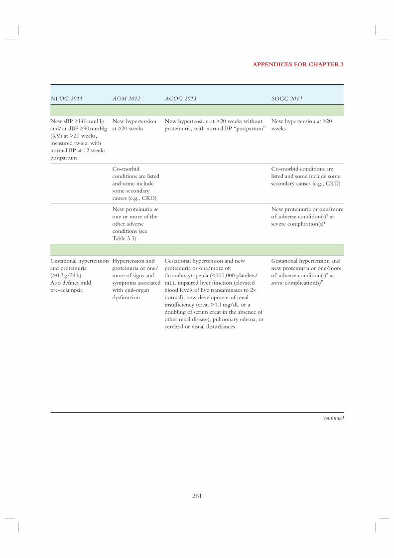
APPENDICES FOR CHAPTER 3
261
NVOG 2011 AOM 2012 ACOG 2013 SOGC 2014
New sBP 140 mmHg
and/or dBP 90 mmHg
(KV) at >20 weeks,
measured twice, with
normal BP at 12 weeks
postpartum
New hypertension
at 20 weeks
New hypertension at >20 weeks without
proteinuria, with normal BP “postpartum”
New hypertension at 20
weeks
Co-morbid
conditions are listed
and some include
some secondary
causes (e.g., CKD)
Co-morbid conditions are
listed and some include some
secondary causes (e.g., CKD)
New proteinuria or one or more of the
other adverse
conditions (see
Table 3.3)
New proteinuria or one/more
of: adverse condition(s)¥ or severe complication(s)¥
Gestational hypertension
and proteinuria
(>0.3 g/24 h)
Also defines mild
pre-eclampsia
Hypertension and
proteinuria or one/
more of signs and
symptoms associated
with end-organ
dysfunction
Gestational hypertension and new
proteinuria or one/more of:
thrombocytopenia (<100,000 platelets/
mL), impaired liver function (elevated
blood levels of live transaminases to 2
normal), new development of renal
insufficiency (creat >1.1 mg/dL or a
doubling of serum creat in the absence of
other renal disease), pulmonary edema, or
cerebral or visual disturbances
Gestational hypertension and
new proteinuria or one/more
of: adverse condition(s)¥ or severe complication(s)¥
continued

THE FIGO TEXTBOOK OF PREGNANCY HYPERTENSION
262
PRECOG 2005PRECOG II 2009 QLD 2013 NICE 2010 WHO 2011
Pre-eclampsia
Eclampsia With PET, one/more
seizures
With PET, a
convulsive
condition
With PET,
generalized seizures
not attributable to
other causes
Severe
pre-eclampsia
One/more of: platelet
count <100,00109/L,
elevated transaminases,
microangiopathic
haemolytic anaemia with
fragments/schistocytes on
blood film (essentially
HELLP syndrome)
Severe hypertension
and/or symptoms,
and/or biochemical
and/or
haematological
impairment
One/more of:
severe
hypertension, heavy
proteinuria, and
substantial maternal
organ dysfunction
Onset at <32–34
weeks and fetal
morbidity are used
in some parts of the
world
HELLP
syndrome
HELLP spelled out
Highlighted as variant of
severe pre-eclampsia
HELLP spelled out
Other ‘hypertensive effects’
Transient
hypertensive
effect
White-coat
effect
BP that is elevated in a
clinical setting but normal
in a non-clinical setting by
(24 h) ABPM or HBPM
using an appropriately
validated device
Masked
hypertensive
effect
Hypertension
(sBP and/or
dBP)
dBP 90 mmHg dBP 90 mmHg sBP 140 mmHg
and/or
dBP 90 mmHg
dBP 90 mmHg (on
two occasions, >4
hours apart)
or
dBP >110 mmHg
(measured once)
-
Appendix 3.2 continued
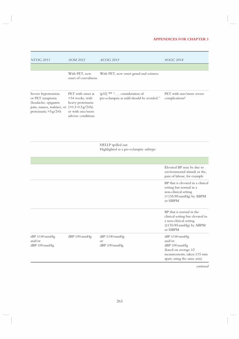
APPENDICES FOR CHAPTER 3
263
NVOG 2011 AOM 2012 ACOG 2013 SOGC 2014
With PET, new
onset of convulsions
With PET, new onset grand mal seizures
Severe hypertension
or PET symptoms
(headache, epigastric
pain, nausea, malaise), or
proteinuria >5 g/24 h
PET with onset at
<34 weeks, with
heavy proteinuria
(>0.3–0.5 g/24 h)
or with one/more
adverse conditions
(p32) ** “. . . consideration of
pre-eclampsia as mild should be avoided.”
PET with one/more severe
complications‡
HELLP spelled out
Highlighted as a pre-eclamptic subtype
Elevated BP may be due to
environmental stimuli or the,
pain of labour, for example
BP that is elevated in a clinical
setting but normal in a
non-clinical setting
(<135/85 mmHg) by ABPM
or HBPM
BP that is normal in the
clinical setting but elevated in
a non-clinical setting
(135/85 mmHg) by ABPM
or HBPM
sBP 140 mmHg
and/or
dBP 90 mmHg
dBP 90 mmHg sBP 140 mmHg
or
dBP 90 mmHg
sBP 140 mmHg
and/or
dBP 90 mmHg
(based on average 2
measurements, taken 15 min
apart, using the same arm)
continued

THE FIGO TEXTBOOK OF PREGNANCY HYPERTENSION
264
PRECOG 2005PRECOG II 2009 QLD 2013 NICE 2010 WHO 2011
Other ‘hypertensive effects’
Mild sBP 140–
149 mmHg
dBP 90–99 mmHg
Moderate sBP 150–
159 mmHg
dBP 100–
109 mmHg
Severe 160/ and/or 110 mmHg 160/110 mmHg
Late postpartum hypertension
Definition
ABPM, ambulatory blood pressure monitoring); ACOG, American College of Obstetricians and Gynecologists; AOM,
Association of Ontario Midwives; BP, blood pressure; CKD, chronic kidney disease; Creat, creatinine; dBP, diastolic blood
pressure; DIC, disseminated intravascular coagulation; HBPM, home blood pressure monitoring; HELLP syndrome,
Haemolysis, Elevated Liver enzymes and Low Platelet count syndrome; NICE, National Institute for Health and Clinical
Excellence; NVOG, Nederlandse Vereniging voor Obstetrie en Gynaecologie; PET, pre-eclampsia; PRECOG,
pre-eclampsia community guideline; QLD, Queensland Maternity and Neonatal Clinical Guidelines Program; RUQ, right
upper quadrant; sBP, systolic blood pressure; SOGC, Society of Obstetricians and Gynaecologists of Canada; WHO, World
Health Organisation
* SOMANZ 2014 is included in the chapter text, but not in this table adapted from Gillon 201458
ACOG 2013: American College of Obstetricians and Gynecologists, Task Force on Hypertension in Pregnancy.
Hypertension in pregnancy. Report of the American College of Obstetricians and Gynecologists’ Task Force on
Hypertension in Pregnancy. Obstet Gynecol 2013 Nov; 122(5):1122–1131
AOM 2012: Salehi P, Association of Ontario Midwives HDP CPG, Working Group. Hypertensive disorders of pregnancy
(Clinical Practice Guideline 15). 2012; Available: http://www aom on ca/Health_Care_Professionals/Clinical_Practice_
Guidelines/
Appendix 3.2 continued

APPENDICES FOR CHAPTER 3
265
NVOG 2011 AOM 2012 ACOG 2013 SOGC 2014
sBP 140–159 mmHg
or
dBP 90–109 mmHg
sBP 140–159 mmHg
or
dBP 90–109 mmHg
160/or 110 mmHg 160/or 110 mmHg 160/or 110 mmHg
(as greater than mild)
160/or 110 mmHg
(based on average 2
measurements, taken 15 min
apart, using the same arm)
Hypertension (usually mild) that develops 2
weeks to-6 mos postpartum, usually
normalizing by the end of the first year
NICE 2010: National Collaborating Centre for Women’s and Children’s Health (UK). CG107: Hypertension in
pregnancy: The management of hypertensive disorders during pregnancy. NICE: Guidance 2010 Aug
NVOG 2011: Nederlandse Vereniging voor Obstetrie en Gynaecologie. Hypertensieve aandoeningen in de zwangerschap.
2011
QLD 2013: Queensland Maternity and Neonatal Clinical, Guidelines Program. Hypertensive disorders of pregnancy.
2013;MN10.13-V4-R15
PRECOG: Milne F, Redman C, Walker J, Baker P, Bradley J, Cooper C, et al. The pre-eclampsia community guideline
(PRECOG): how to screen for and detect onset of pre-eclampsia in the community. BMJ 2005 Mar 12;330(7491):576–80
PRECOG II: Milne F, Redman C, Walker J, Baker P, Black R, Blincowe J et al. Assessing the onset of pre-eclampsia in
the hospital day unit: summary of the pre-eclampsia guideline (PRECOG II). BMJ 2009; 339:b3129
SOGC 2014: Magee LA, Pels A, Helewa M, Rey E, von Dadelszen P. Diagnosis, evaluation, and management of the
hypertensive disorders of pregnancy. Pregnancy Hypertens 2014;4(2):105–145
WHO 2011: World Health Organization. WHO recommendations for prevention and treatment of pre-eclampsia and
eclampsia. 2011

THE FIGO TEXTBOOK OF PREGNANCY HYPERTENSION
266
Define pre-eclampsia in association with hypertension
PRECOG2005
PRECOG II2009
QLD2013
NICE2010
WHO2011
NVOG2011
AOM2012
ACOG2013
SOGC2014
Proteinuria
Heavy proteinuria
Proteinuria is not mandatory
– one/more other
manifestations sufficient
Gestational age at onset <34
weeks
Maternal symptoms
Headache/visual symptoms
Chest pain/dyspnoea
Nausea/vomiting
Right upper quadrant/
epigastric pain
Maternal signs
Cardiac/cardiovascular
Severe hypertension
Uncontrolled severe
hypertension
Appendix 3.3
Definitions of pre-eclampsia and severe pre-eclampsia

APPENDICES FOR CHAPTER 3
267
Define SEVERE pre-eclampsia
NotesPRECOG
2005PRECOG II
2009QLD2013
NICE2010
WHO2011
NVOG2011
AOM2012
ACOG2013
SOGC2014
1. Not mandatory. In absence
of proteinuria, one or more of
1. <32–34 weeks
2. Mentioned in text as risk
factor for poor outcome
1. Cerebral or visual
disturbances
2. Cerebral or visual
disturbances (with proteinuria)
1. Severe, persistent,
unresponsive to medication, not
otherwise explained (with
proteinuria)
( ) ( )
continued

THE FIGO TEXTBOOK OF PREGNANCY HYPERTENSION
268
Define pre-eclampsia in association with hypertension
PRECOG2005
PRECOG II2009
QLD2013
NICE2010
WHO2011
NVOG2011
AOM2012
ACOG2013
SOGC2014
Maternal signs
Positive inotropic support
Myocardial ischaemia/
infarction
Neurologic
Eclampsia
PRES
Cortical blindness or retinal
detachment
Glasgow coma scale <13
Stroke, TIA or RIND
Hyperreflexia (with clonus)
Pulmonary
Oxygen saturation <97%
Oxygen saturation <90%
Pulmonary oedema
Need for ≥50% oxygen for
>1 h
Intubation (other than for
Caesarean delivery),
Renal
Oliguria
Appendix 3.3 continued

APPENDICES FOR CHAPTER 3
269
Define SEVERE pre-eclampsia
NotesPRECOG
2005PRECOG II
2009QLD2013
NICE2010
WHO2011
NVOG2011
AOM2012
ACOG2013
SOGC2014
( )
( )
( )
( )
( )
( )
( )
( )
( )
1. With proteinuria
( )
( )
continued

THE FIGO TEXTBOOK OF PREGNANCY HYPERTENSION
270
Appendix 3.3 continued
Define pre-eclampsia in association with hypertension
PRECOG2005
PRECOG II2009
QLD2013
NICE2010
WHO2011
NVOG2011
AOM2012
ACOG2013
SOGC2014
Abnormal maternal laboratory tests
Haematology/coagulation
Elevated WBC count
Platelet count decreased but
50109/L
Platelet count decreased but
<50109/L
Elevated INR or aPTT
1
Renal
Elevated serum uric acid
Elevated serum creatinine
Acute kidney injury
(creatinine >150 μM with no
prior renal disease)
New indication for dialysis
Hepatic
Elevated serum AST, ALT,
LDH or bilirubin
Hepatic dysfunction (INR >2
in absence of DIC or warfarin)
Low plasma albumin
Hepatic haematoma or
rupture

APPENDICES FOR CHAPTER 3
271
Define SEVERE pre-eclampsia
NotesPRECOG
2005PRECOG II
2009QLD2013
NICE2010
WHO2011
NVOG2011
AOM2012
ACOG2013
SOGC2014
1. “Microangiopathic
haemolytic anaemia”
( ) ( )
1. Thrombocytopaenia
2. <100,000/mL
3. <100,000/mL with
proteinuria
1. Haemolysis and DIC
( ) ( )
1. Progressive renal
insufficiency (serum creatinine
>1.1 mg/dL or a doubling of
serum creatinine concentration
in absence of other renal
disease)
2. With proteinuria
( ) ( )
1. Twice normal
2. With proteinuria
( ) ( )
( ) ( )
continued

THE FIGO TEXTBOOK OF PREGNANCY HYPERTENSION
272
Define pre-eclampsia in association with hypertension
PRECOG2005
PRECOG II2009
QLD2013
NICE2010
WHO2011
NVOG2011
AOM2012
ACOG2013
SOGC2014
Fetoplacental manifestations
Non-reassuring FHR
IUGR
Oligohydramnios
Absent/reversed end-diastolic
flow by Doppler velocimetry
Abruption without evidence of
maternal or fetal compromise ( ) ( )
Abruption with evidence of
maternal or fetal compromise ( ) ( )
Reverse ductus venosus A
wave
Stillbirth
Interventions
Transfusion of any blood
product
ACOG, American College of Obstetricians and Gynecologists; AOM, Association of Ontario Midwives; aPTT, activated
partial thromboplastic time; ASH, American Society of Hypertension; AST, aspartate aminotransferase; ALT, alanine
aminotransferase; FHR, fetal heart rate; INR, international normalised ratio; IUGR, intrauterine fetal growth restriction;
LDH, lactate dehydrogenase; NICE, National Institute for Health and Clinical Excellence; NVOG, Nederlandse
Vereniging voor Obstetrie en Gynaecologie; PRECOG, pre-eclampsia community guideline; PRES, posterior reversible
encephalopathy syndrome; QLD, Queensland Maternity and Neonatal Clinical Guidelines Program; RIND, reversible
ischaemic neurological deficit; SOGC, Society of Obstetricians and Gynaecologists of Canada; TIA, transient ischaemic
attack; WBC, white blood cell count; WHO, World Health Organization
* A checkmark indicates that the diagnostic criterion was listed by the guideline. A checkmark in brackets indicates that
although not listed specifically, the criterion could reasonably be interpreted as being part of the definition in the relevant
guideline† The NICE 2010 guidelines include “symptoms, and/or biochemical and/or haematological impairment” as part of the
definition of severe pre-eclampsia. It is assumed that those complications indicated by ( ) would meet this definition
** The WHO 2011 guidelines include “substantial maternal end-organ dysfunction” as part of the definition of severe
pre-eclampsia. It is assumed that those complications indicated by ( ) would meet this definition. “Fetal morbidity” also
required interpretation
*** Pre-eclampsia with severe feature
Appendix 3.3 continued

APPENDICES FOR CHAPTER 3
273
Define SEVERE pre-eclampsia
NotesPRECOG
2005PRECOG II
2009QLD2013
NICE2010
WHO2011
NVOG2011
AOM2012
ACOG2013
SOGC2014
1. Not included (as IUGR with
PET managed the same way as
IUGR w/o PET)
( )
( ) ( )
ACOG 2013: American College of Obstetricians and Gynecologists, Task Force on Hypertension in Pregnancy.
Hypertension in pregnancy. Report of the American College of Obstetricians and Gynecologists’ Task Force on
Hypertension in Pregnancy. Obstet Gynecol 2013 Nov; 122(5):1122–1131
AOM 2012: Salehi P, Association of Ontario Midwives HDP CPG, Working Group. Hypertensive disorders of pregnancy
(Clinical Practice Guideline 15). 2012; Available: http://www aom on ca/Health_Care_Professionals/Clinical_Practice_
Guidelines/
NICE 2010: National Collaborating Centre for Women’s and Children’s Health (UK). CG107: Hypertension in
pregnancy: The management of hypertensive disorders during pregnancy. NICE: Guidance 2010 Aug
NVOG 2011: Nederlandse Vereniging voor Obstetrie en Gynaecologie. Hypertensieve aandoeningen in de zwangerschap.
2011
QLD 2013: Queensland Maternity and Neonatal Clinical, Guidelines Program. Hypertensive disorders of pregnancy.
2013;MN10.13-V4-R15
PRECOG: Milne F, Redman C, Walker J, Baker P, Bradley J, Cooper C, et al. The pre-eclampsia community guideline
(PRECOG): how to screen for and detect onset of pre-eclampsia in the community. BMJ 2005 Mar 12;330(7491):576–80
PRECOG II: Milne F, Redman C, Walker J, Baker P, Black R, Blincowe J et al. Assessing the onset of pre-eclampsia in
the hospital day unit: summary of the pre-eclampsia guideline (PRECOG II). BMJ 2009; 339:b3129
SOGC 2014: Magee LA, Pels A, Helewa M, Rey E, von Dadelszen P. Diagnosis, evaluation, and management of the
hypertensive disorders of pregnancy. Pregnancy Hypertens 2014;4(2):105–145
WHO 2011: World Health Organization. WHO recommendations for prevention and treatment of pre-eclampsia and
eclampsia. 2011


APPENDICES FOR CHAPTER 4
275
PREGNANCY-INDUCED HYPERTENSION SEARCHES: COMPLICATIONS, EPIDEMIOLOGY
Removed duplicates, non-Eng, non-Fre, animal
research using EndNote searches and de-duping
function.
COMPLICATIONS
Database(s): Ovid MEDLINE(R) In-Process & Other Non-Indexed Citations and Ovid MEDLINE(R) 1946 to Present Search Strategy:
Appendix 4.1
Literature searches
# Searches Results
1 complications.fs. or exp Infant, Newborn, Diseases/et, cn or exp Pregnancy Outcome/ or (sequel* or later
life or late life).mp. or Pregnancy Complications/et, cn or exp Abortion, Spontaneous/et, cn or exp Chorea
Gravidarum/et or exp Diabetes, Gestational/et or exp Fetal Death/et or exp Fetal Diseases/et, cn or exp
Maternal Death/et or exp Morning Sickness/et or exp Nuchal Cord/et or exp Obstetric Labor
Complications/et or exp Oligohydramnios/et or exp Pelvic Floor Disorders/et or exp Pemphigoid
Gestationis/et, cn or exp Perinatal Death/et or exp Phenylketonuria, Maternal/et or exp Placenta Diseases/et,
cn or exp Polyhydramnios/et, cn or exp Pregnancy Complications, Cardiovascular/et or exp Pregnancy
Complications, Hematologic/et, cn or exp Pregnancy Complications, Infectious/et or exp Pregnancy
Complications, Neoplastic/et, cn or exp Pregnancy in Diabetics/et or exp Pregnancy, Ectopic/et or exp
Pregnancy, Prolonged/et or exp Prenatal Injuries/et or exp Puerperal Disorders/et, cn
1759552
2 exp Hypertension, Pregnancy-Induced/ or ((exp Pregnancy/ or exp Pregnancy Complications/) and exp
Hypertension/)
34751
3 1 and 2 10375
4 limit 3 to yr=”2011 -Current” 1499
5 limit 4 to (humans and (english or french)) 1358
6 (complicat* and (((pregnan* or gestation* or obstetric*) and hypertens*) or (pre-eclamp* or preeclamp* or
toxemia* or toxaem* or gestosis or pre eclamp* or eclamp* or EPH Complex))).ti,ab.
9813
7 limit 6 to yr=”2013 -Current” 1505
8 5 or 7
EXPORTED THESE
2668
Database(s): EBM Reviews – Cochrane Central Register of Controlled Trials March 2015,
EBM Reviews – Database of Abstracts of Reviews of Effects 1st Quarter 2015 Search
Strategy:
# Searches Results
1 (complicat* and (((pregnan* or gestation* or obstetric*) and hypertens*) or (pre-eclamp* or preeclamp* or
toxemia* or toxaem* or gestosis or pre eclamp* or eclamp* or EPH Complex))).mp.
735
2 limit 1 to yr=”2011 -Current” [Limit not valid in DARE; records were retained]
EXPORTED THESE
221

THE FIGO TEXTBOOK OF PREGNANCY HYPERTENSION
276
EPIDEMIOLOGY
Database(s): Ovid MEDLINE(R) In-Process & Other Non-Indexed Citations and Ovid MEDLINE(R) 1946 to Present Search Strategy:
# Searches Results
1 exp Hypertension, Pregnancy-Induced/ or ((exp Pregnancy/ or exp Pregnancy Complications/) and exp
Hypertension/)
34751
2 (epidemiology or ethnology).fs. 1328387
3 exp Epidemiology/ 22151
4 exp incidence/ or exp prevalence/ 368169
5 (incidence or prevalen* or epidemiol*).ti,ab. 1178435
6 2 or 3 or 4 or 5 2053499
7 1 and 6 6092
8 limit 7 to (yr=”2010 -Current” and (english or french)) 1736
9 (((pregnan* or gestation* or obstetric*) and hypertens*) or (pre-eclamp* or preeclamp* or toxemia* or
toxaem* or gestosis or pre eclamp* or eclamp* or EPH Complex)).ti,ab.
41767
10 (incidence or prevalen* or epidemiol*).ti,ab. 1178435
11 9 and 10 5706
12 limit 11 to yr=”2013 -Current” 945
13 8 or 12
EXPORTED THESE
2383
Database(s): EBM Reviews – Cochrane Central Register of Controlled Trials March 2015,
EBM Reviews – Database of Abstracts of Reviews of Effects 1st Quarter 2015 Search
Strategy:
# Searches Results
1 (((pregnan* or gestation* or obstetric*) and hypertens*) or (pre-eclamp* or preeclamp* or toxemia* or toxaem*
or gestosis or pre eclamp* or eclamp* or EPH Complex)).mp.
2235
2 (incidence or prevalen* or epidemiol*).mp. 76380
3 1 and 2 447
4 limit 3 to yr=”2010 -Current” [Limit not valid in DARE; records were retained]
EXPORTED THESE
208

APPENDICES FOR CHAPTER 5
277
Appendix 5.1
Studies of predictive tests for pre-eclampsia
See next page – this appendix requires a double-page layout

THE FIGO TEXTBOOK OF PREGNANCY HYPERTENSION
278
Risk
facto
r¥ o
r pre
dicto
rFi
rst a
utho
r (ye
ar)
Stud
y de
sign
Typ
e of
HD
PN
o. o
f wom
en
(rate
%)
RR
or
OR
*A
UC
R
OC
Sens
itivi
ty
(%)
Spec
ificit
y (%
)L
R+
LR
Pred
ictor
s
Clin
ical e
xam
inat
ion
BP
Cnoss
en† (
2008)9
6Syst
emat
ic r
evie
w
and m
eta-
anal
ysi
s
Any P
ET
60,5
99 (
5.5
1%
)0.7
65
535.1
2590.1
2503.5
50.7
2
UA
DC
noss
en† (
2008)9
6Syst
emat
ic r
evie
w
and b
ivar
iable
met
a-an
alysi
s
Any P
ET
79,5
47 (
3.1
4 %
)–
590.1
2570.1
2503.0
50.1
4
Pap
ageo
rghio
u† (
2002)1
08
Rev
iew
Any P
ET
18,6
83 (
2.5
%)
24–89
86–96
505.9
00.5
5
Afr
akhte
h† (
2014)1
62
Pro
spec
tive
PE
T, st
illb
irth
,
pla
centa
l
abru
ption a
nd
pre
term
lab
our
205 (
17.5
%)
557.5
5598.2
5531.9
50.4
3
Nap
olita
no
† (
2012)1
12
Ret
rosp
ective
obse
rvat
ional
Any P
ET
Ear
ly-o
nse
t
pre
-ecl
ampsi
a
Pre
term
PE
T
3549 (
3.6
%)
(0.6
%)
(1.2
%)
0.6
82
0.8
51
0.7
86
––
––
Lab
orat
ory
mar
kers
Podocy
turi
aJi
m† (
2014)1
04
Pro
spec
tive
Any P
ET
91 (
15.4
%)
536
596.1
2509.1
20.6
7
Cra
ici†
(2013)1
05
Pro
spec
tive
Any P
ET
Any H
DP
(PE
T a
nd G
H)
267
(5.6
%)
(11.2
%)
1.7
86
0.7
86
100.1
2
154.1
2
100.1
2
100.1
2
0.1
2
0.4
6
Kel
der
† (
2012)1
03
Cas
e–co
ntr
ol
Any P
ET
69 (
50.7
%)
0.8
25
568.6
5588.2
5505.8
10.3
6
IPG
/cr
eatinin
e ra
tio
Daw
onau
th† (
2014)1
07
Pro
spec
tive
longitudin
al
Any P
ET
416 (
8.2
%)
0.8
62
584.2
5583.6
5505.1
30.1
9
Cal
cium
/cr
eatinin
e ra
tio
Vah
dat
† (
2012)1
06
Pro
spec
tive
cohort
Any P
ET
150 (
9.3
%)
–577.1
2578.1
2503.5
50.2
9

APPENDICES FOR CHAPTER 5
279
Hs-
CR
PK
ashan
ian
† (
2013)1
21
Pro
spec
tive
cohort
Any P
ET
394 (
10.7
%)
0.8
55
578.1
5572.1
5502.8
00.3
0
Fib
ronec
tin
Lee
flan
g (
2007)1
22
Syst
emat
ic r
evie
wA
ny P
ET
573 (
19.0
%)
–550–100
43–88
1.2
4–11.5
0–0.7
4
Pla
tele
tsY
ang
† (
2011)1
69
Cas
e–co
ntr
ol
Any H
DP
(P
ET
and G
H)
1288 (
35.8
%)
0.6
62
547.1
281.6
7502.5
70.6
5
Ang
ioge
nic f
acto
rs
PlG
F
sFLT
1
sEN
G
Kle
inro
uw
eler
(2012)1
65
Syst
emat
ic r
evie
w
and m
eta-
anal
ysi
s
Any P
ET
29 t
o 3
098
–
–532
526
518
595.1
2
595.1
2
595.1
2
506.4
5
505.2
5
503.6
5
0.7
2
0.7
8
0.8
6
PlG
FG
hosh
† (
2013)1
28
Pro
spec
tive
cohort
EO
-PE
T (
1st
vs
2nd t
rim
este
r)
1244 (
1.5
%)
18.8
3*
52.7
6*
0.9
85
0.9
75
584
558
578.1
2
566.1
2
503.8
2
501.7
1
0.2
1
0.6
7
Ghosh
† (
2013)1
29
Pro
spec
tive
cohort
EO
-PE
T in
Obes
e/over
wei
ght
1678 (
1.7
%)
57.6
4*
0.9
85
579.1
2568.1
2502.4
60.3
1
Chap
pel
l (2
013)1
25
Pro
spec
tive
PE
T d
eliv
ered
within
14 d
ays
625 (
55%
)0.8
75
596.1
2555.1
2502.1
30.0
7
Ghosh
† (
2013)1
23
Pro
spec
tive
cohort
EO
-PE
T
EO
-IU
GR
722 (
1.5
%)
58.3
5*
10.7
3*
0.9
82
0.9
82
582.1
2
584.1
2
565.1
2
567.1
2
502.3
6
502.5
4
0.2
8
0.2
4
Sflt/
PlG
FH
anita
(2014)1
32
Pro
spec
tive
Any P
ET
84 (
14.3
%)
0.8
73
592.1
2568.1
2502.8
90.1
2
Engel
s (2
013)1
30
Pro
spec
tive
PE
T/H
ELLP
338 (
64%
)0.9
64
570.3
5595.1
5514.3
50.3
1
Tei
xei
ra† (
2013)1
31
Pro
spec
tive
longitudin
al
Any P
ET
71 (
16.9
%)
0.9
55
––
––
Del
ic† (
2014)1
63
Pro
spec
tive
Any P
ET
69 (
49.2
%)
0.8
95
––
––
Villa
(2013)1
34
Nes
ted c
ase–
contr
ol
EO
-PE
T106 (
5.7
%)
1100.1
2599.8
5500.1
20.1
2
Fore
st† (
2014)1
33
Nes
ted c
ase–
contr
ol
EO
-PE
T
7929 (
0.2
%)
0.9
77
588.9
5590.1
2508.8
90.1
2
PlG
F/sF
lt-1
McE
lrat
h† (
2012)1
24
Pro
spec
tive
longitudin
al
Any P
ET
2243 (
6.2
%)
0.7
45
561.1
2575.1
5502.4
50.5
2
cont
inue
d

THE FIGO TEXTBOOK OF PREGNANCY HYPERTENSION
280
Risk
facto
r¥ o
r pre
dicto
rFi
rst a
utho
r (ye
ar)
Stud
y de
sign
Typ
e of
HD
PN
o. o
f wom
en
(rate
%)
RR
or
OR
*A
UC
R
OC
Sens
itivi
ty
(%)
Spec
ificit
y (%
)L
R+
LR
Pred
ictor
s
Mul
tivar
iabl
es
BP
and U
AD
Kle
inro
uw
eler
(2013)1
11
Indiv
idual
pat
ient
dat
a m
eta-
anal
ysi
s
Any P
ET
6708 (
4.5
%)
0.8
55
MC
, P
P-1
3, -
hC
GSch
neu
er† (
2012)1
42
In-h
ouse
stu
dy a
nd
syst
emat
ic r
evie
w
Any P
ET
EO
-PE
T
2678 (
2.7
%)
(0.2
%)
0.7
25
0.8
25
524.1
2
545.1
2
595.1
2
595.1
2
504.8
5
509.1
2
0.8
5
0.5
8
MC
, U
AD
, B
iom
arker
sK
uc
(2011)1
43
Syst
emat
ic r
evie
wA
ny P
ET
138,5
71 (
2.6
%)
–543–100
590.1
2–
–
MC
, U
AD
and P
AP
P-A
Goet
zinger
† (
2014)1
48
Pro
spec
tive
cohort
Any P
ET
1200 (
8.5
%)
0.7
65
536.7
5593.2
5505.4
50.6
8
MC
and U
AD
Lai
† (
2013)1
10
Pro
spec
tive
scre
enin
g
Inte
rmed
iate
PE
T
LO
-PE
T
4,2
94 (
0.9
%)
(3.4
%)
0.8
38
0.7
92
570.3
5
554.6
5
590.1
2
590.1
2
507.0
3
505.4
6
0.3
3
0.5
1
MC
and B
PG
allo
† (
2014)9
8P
rosp
ective
Any P
ET
EO
-PE
T
Pre
term
PE
T
17,3
83 (
3.1
%)
(0.4
%)
(0.8
%)
0.8
93
0.8
85
0.8
13
552.5
5
584.3
5
565.7
5
590.1
2
590.1
2
590.1
2
505.2
5
508.4
3
508.1
3
0.1
2
0.1
7
0.2
1
MC
, B
P a
nd U
AD
Car
adeu
x† (
2013)9
9P
rosp
ective
cohort
EO
-PE
T627 (
1.5
%)
0.8
95
562.5
5595.5
5513.9
50.3
9
MC
, P
lGF, B
P a
nd U
AD
M
yer
s† (
2013)1
49
Pro
spec
tive
cohort
Pre
term
PE
T3529 (
1.3
%)
50.6
5*
0.8
15
545.1
2595.1
2509.1
20.5
8
MC
, P
IGF a
nd fre
e -
hC
GD
i Lore
nzo
† (
2012)1
44
Pro
spec
tive
cohort
EO
-PE
T2118 (
0.5
7%
)0.8
93
575.1
2590.1
2507.5
50.2
8
MC
, B
P P
AP
PA
, A
DA
M12
and P
lGF
Myat
t† (
2012)1
37
Obse
rvat
ional
stu
dy
Any P
ET
2218 (
7.9
%)
0.7
35
546.1
5580.1
2502.3
10.6
7
MC
, hC
G-h
, P
AP
P-A
and
BP
Kei
kkal
a† (
2013)1
45
Nes
ted c
ase–
contr
ol
EO
-PE
T707 (
4.1
%)
0.8
63
569.1
2590.1
2506.9
50.3
4

APPENDICES FOR CHAPTER 5
281
MC
, U
AD
, P
lGF, sF
lt-1
and
lipid
-rel
ated
mar
ker
s
Dig
uis
to† (
2013)1
53
Pro
spec
tive
obse
rvat
ional
stu
dy
Any P
ET
235 (
23.8
%)
0.7
95
539.6
5590.1
2503.9
60.6
7
MC
, P
lGF, an
d U
AD
R
izos†
(2013)1
50
Cas
e–co
ntr
ol
Any P
ET
116 (
10.3
%)
–546.1
2599.1
2519.1
20.5
6
PA
PP
-A a
nd s
Flt-1
/P
lGF
Par
k† (
2014)9
0P
rosp
ective
cohort
LO
-t P
ET
262 (
3%
)0.9
69
587.5
5595.1
2517.5
50.1
3
PW
V a
nd s
Flt-1
Kat
sipi†
(2014)1
51
Pro
spec
tive
Any P
ET
EO
-PE
T
118 (
17.8
%)
(9.3
%)
0.9
65
0.9
63
590.1
2
592.1
2
590.1
2
590.1
2
509.1
2
509.2
5
0.1
1
0.0
9
IPG
/cr
eatinin
eD
awonau
th† (
2014)1
07
Pro
spec
tive
longitudin
al
Any P
ET
416
(8.2
%)
0.8
62
584.2
5583.6
5505.1
30.1
9
MC
, B
P, PA
PPA
,
AD
AM
12 a
nd P
lGF
Kuc†
(2013)9
7N
este
d c
ase–
contr
ol
EO
-PE
T
LO
-PE
T
EO
-PE
T +
SG
A
LO
-PE
T+
SG
A
667 (
10.2
%)
(14.8
%)
(1.9
%)
(7.3
%)
0.8
85
0.8
85
0.9
55
0.9
55
572.1
2
549.1
2
592.1
2
557.1
2
590.1
2
590.1
2
590.1
2
590.1
2
507.2
5
504.9
5
509.2
5
505.7
5
0.3
1
0.5
7
0.0
9
0.4
8
MC
, B
P a
nd t
auri
ne
Kuc†
(2014)1
43
Cas
e–co
ntr
ol
EO
-PE
T167 (
10.2
%)
0.9
35
555.1
2590.1
2505.5
50.5
5
BP
and s
EN
GA
bdel
aziz
† (
2012)1
62
Nes
ted c
ase–
contr
ol
Any H
DP
EO
-PE
T
LO
-PE
T
1898 (
4.6
9%
)
(0.8
4%
)
(3.1
6%
)
0.8
35
0.8
65
0.8
35
571.8
5
583.8
5
580.3
5
590.1
2
590.1
2
590.1
2
507.1
8
508.3
8
508.0
3
0.3
1
0.1
8
0.2
2
HR
G a
nd U
AD
Bolin
† (
2012)1
09
Cas
e–co
ntr
ol
Pre
term
PE
T175 (
15.4
%)
0.8
55
591.1
2562.1
2502.3
90.1
5
PlG
F, P
P13, PA
PP
A a
nd
IL-1
Silje
e† (
2013)1
68
Ret
rosp
ective
case
–co
ntr
ol
EO
-PE
T70 (
50%
)0.8
30
555.9
5590.1
2505.5
90.4
9
Uri
c ac
id a
nd B
PM
arte
ll-C
laro
s (2
013)1
67
Pro
spec
tive
cohort
GH
283 (
6.0
%)
54.0
2*
3.6
*0.7
55
550.1
2584.2
5503.1
60.5
9
† S
tudie
s in
cludin
g p
rote
inuri
a fo
r th
e def
initio
n o
f pre
-ecl
ampsi
a; ¥
Ris
k fac
tors
, *O
dd r
atio
s
PE
T, pre
-ecl
ampsi
a; G
H, ges
tational
hyper
tensi
on; E
O, ea
rly o
nse
t; L
O, la
te o
nse
t; U
AD
, ute
rine
arte
ry D
opple
r; B
P, blo
od p
ress
ure
; SG
A, sm
all fo
r ges
tational
age;
MC
, m
ater
nal
char
acte
rist
ics

THE FIGO TEXTBOOK OF PREGNANCY HYPERTENSION
282
Appendix 5.2
Predictors of pre-eclampsia
Demographics and family history Past medical or obstetric history Current pregnancy
Independent predictors
Maternal
Clinical examination
First trimester Second or third trimester
• MAP • MAP
• Uterine artery Doppler
Laboratory markers
First trimester Second or third trimester
• Fibronectin • sFlt-1:PlGF
• hs-CRP • Podocyturia
• Platelets • PlGF
• PlGF • Calcium:creatinine ratio
• sFLT-1 • Fibronectin
• sENG
Multivariable predictors
Maternal
Maternal age Previous pre-eclampsia Multiple pregnancy Excessive weight gain in
pregnancy
Afro-Caribbean or South
Asian race
Pre-existing medical
condition(s)
Overweight/obesity (BMI)
Family history of
pre-eclampsia (mother)
Pre-existing hypertension First ongoing pregnancy
Education level Pre-existing diabetes mellitus
Preterm labour/delivery
Non-smoking
Clinical examination
First trimester Second or third trimester
• MAP • MAP
• sBP • Uterine artery Doppler
• dBP
• Uterine artery Doppler
continued

APPENDICES FOR CHAPTER 5
283
Appendix 5.3
Performance of predictors
(summary of evidence by trimester)
Appendix 5.2 continued
Demographics and family history Past medical or obstetric history Current pregnancy
Multivariable predictors
Laboratory markers
First trimester Second or third trimester
• PIGF • PIGF
• Uric acid • sFlt-1
• PP-13 • PAPP-A
• sENG • HRG
• -hCG • PWV
• PAPP-A • Leptin,
• ADAM12 • Triglycerides
• Taurine
• IL-1
See next page – this appendix requires a double-page layout

THE FIGO TEXTBOOK OF PREGNANCY HYPERTENSION
284
App
endi
x 5.
3
Pred
ictor
First
aut
hor (
year
)St
udy
desig
nT
ype
of H
DP
No.
of w
omen
(ra
te %
)A
UC
R
OC
Sens
itivi
ty
(%)
Spec
ificit
y (%
)L
R+
LR
First
trim
este
r pre
dicto
rs
Indi
vidu
al p
redi
ctors
Clin
ical e
xam
inat
ion
BP
(M
AP
)C
noss
en† (
2008)9
6Syst
emat
ic r
evie
w a
nd
met
a-an
alysi
s
Any P
ET
60,5
99 (
5.5
1 %
)0.7
95
––
––
Lab
orat
ory
mar
kers
hs-
CR
PK
ashan
ian
† (
2013)1
21
Pro
spec
tive
cohort
Any P
ET
394 (
10.7
%)
0.8
55
578.1
5572.1
5552.8
00.3
0
Fib
ronec
tin
Lee
flan
g (
2007)1
22
Syst
emat
ic r
evie
wA
ny P
ET
573 (
19.0
%)
–550–100
63–88
1.4
7–4.0
5.5
40–0.7
4
Pla
tele
tsY
ang
† (
2011)1
69
Cas
e–co
ntr
ol
Any H
DP
(P
ET
and G
H)
1288 (
35.8
%)
0.6
62
547.1
2581.6
7552.5
70.6
5
Ang
ioge
nic f
acto
rs
PlG
F
sFLT
-1
sEN
G
Kle
inro
uw
eler
(2012)1
65
Syst
emat
ic r
evie
w a
nd
met
a-an
alysi
s
Any P
ET
29 t
o 3
098
––
532.1
2
526.1
2
518.1
2
595.1
6
595.1
6
595.1
6
556.4
5
555.2
5
553.6
5
0.7
2
0.7
8
0.8
6
PlG
FG
hosh
† (
2013)1
28
Pro
spec
tive
cohort
EO
-PE
T1244 (
1.5
%)
0.9
72
558.1
2566.1
6551.7
10.6
4
Mul
tivar
iabl
es
MC
, P
P-1
3, -
hC
GSch
neu
er† (
2012)1
42
In-h
ouse
stu
dy a
nd
syst
emat
ic r
evie
w
Any P
ET
EO
-PE
T
2678 (
2.7
%)
(0.2
%)
0.7
25
0.8
25
524.1
2
545.1
2
595.1
6
595.1
6
554.8
5
559.1
8
0.8
5
0.5
8
MC
, U
AD
, B
iom
arker
sK
uc
(2011)1
66
Syst
emat
ic r
evie
wA
ny P
ET
138,5
71 (
2.6
%)
–543–100
590.1
6–
–
MC
, U
AD
and P
AP
P-A
Goet
zinger
†
(2014)1
48
Pro
spec
tive
cohort
Any P
ET
1200 (
8.5
%)
0.7
65
536.7
5593.2
5555.4
50.6
8
MC
, B
P a
nd U
AD
Car
adeu
x† (
2013)9
9P
rosp
ective
cohort
EO
-PE
T627 (
1.5
%)
0.8
95
562.5
5595.5
5513.9
50.3
9
MC
, P
IGF a
nd fre
e b-h
CG
Di Lore
nzo
†
(2012)1
44
Pro
spec
tive
cohort
EO
-PE
T2118 (
0.5
7%
)0.8
93
575.1
2590.1
6557.5
50.2
8
MC
, B
P P
AP
PA
,
AD
AM
12 a
nd P
lGF
Myat
t† (
2012)1
37
Obse
rvat
ional
stu
dy
Any P
ET
2218 (
7.9
%)
0.7
35
546.1
5580.1
6552.3
10.6
7

APPENDICES FOR CHAPTER 5
285
MC
, hC
G-h
, PA
PP
-A a
nd
BP
Kei
kkal
a† (
2013)1
45
Nes
ted c
ase–
contr
ol
EO
-PE
T
707 (
4.1
%)
0.8
63
569.1
2590.1
6556.9
50.3
4
MC
, P
lGF, an
d U
AD
Riz
os†
(2013)1
50
Cas
e–co
ntr
ol
Any P
ET
116 (
10.3
%)
–546.1
2599.1
6519.6
90.5
6
MC
, B
P, PA
PP
-A,
AD
AM
12 a
nd P
lGF
Kuc†
(2013)9
7N
este
d c
ase–
contr
ol
EO
-PE
T
LO
-PE
T
EO
-PE
T +
SG
A
LO
-PE
T +
SG
A
667 (
10.2
%)
(14.8
%)
(1.9
%)
(7.3
%)
0.8
85
0.8
85
0.9
55
0.9
55
572.1
2
549.1
2
592.1
2
557.1
2
590.1
6
590.1
6
590.1
6
590.1
6
557.2
5
554.9
5
559.2
5
555.7
5
0.3
1
0.5
7
0.0
9
0.4
8
MC
, B
P a
nd t
auri
ne
Kuc†
2014
143
Cas
e–co
ntr
ol
EO
-PE
T167 (
10.2
%)
0.9
35
555.1
2590.1
6555.5
50.5
5
BP
and s
EN
GA
bdel
aziz
† (
2012)1
64
Nes
ted c
ase–
contr
ol
Any H
DP
EO
-PE
T
LO
-PE
T
1898 (
4.6
9%
)
(0.8
4%
)
(3.1
6%
)
0.8
35
0.8
65
0.8
35
571.8
5
583.8
5
580.3
5
590.1
6
590.1
6
590.1
6
557.1
8
558.3
8
558.0
3
0.3
1
0.1
8
0.2
2
PlG
F, P
P13, PA
PP
A a
nd
IL-1
Silje
e† (
2013)1
68
Ret
rosp
ective
case
–co
ntr
ol
EO
-PE
T70 (
50%
)0.8
30
555.9
5590.1
6555.5
90.4
9
Uri
c ac
id a
nd B
PM
arte
ll-C
laro
s
(2013)1
67
Pro
spec
tive
cohort
GH
283 (
6.0
%)
0.7
55
550.1
2584.2
5553.1
60.5
9
Seco
nd a
nd th
ird tr
imes
ter p
redi
ctors
(sum
mar
y of
the
evid
ence
)
Indi
vidu
al p
redi
ctors
Clin
ical e
xam
inat
ion
BP
(M
AP
)C
noss
en† (
2008)9
6Syst
emat
ic r
evie
w a
nd
met
a-an
alysi
s
Any P
ET
60,5
99 (
5.5
1 %
)0.7
65
535.1
2590.1
6553.5
50.7
2
UA
DC
noss
en† (
2008)9
6Syst
emat
ic r
evie
w a
nd
biv
aria
ble
met
a-an
alysi
s
Any P
ET
79,5
47 (
3.1
4 %
)–
590.1
2570.1
6553.0
50.1
4
Pap
ageo
rghio
u†
(2002)1
08
Rev
iew
Any P
ET
18683 (
2.5
%)
24–89
86–96
555.9
00.5
5
Afr
akhte
h† (
2014)1
62
Pro
spec
tive
PE
T, st
illb
irth
, pla
centa
l
abru
ption a
nd p
rete
rm lab
or
205 (
17.5
%)
557.5
5598.2
5531.9
50.4
3
Nap
olita
no
†
(2012)1
12
Ret
rosp
ective
obse
rvat
ional
Any P
ET
Ear
ly-o
nse
t pre
-ecl
ampsi
a
Pre
term
PE
T
3549 (
3.6
%)
(0.6
%)
(1.2
%)
0.6
82
0.8
51
0.7
86
––
––
cont
inue
d

THE FIGO TEXTBOOK OF PREGNANCY HYPERTENSION
286
App
endi
x 5.
3 co
ntin
ued
Pred
ictor
First
aut
hor (
year
)St
udy
desig
nT
ype
of H
DP
No.
of w
omen
(ra
te %
)A
UC
R
OC
Sens
itivi
ty
(%)
Spec
ificit
y (%
)L
R+
LR
Seco
nd a
nd th
ird tr
imes
ter p
redi
ctors
(sum
mar
y of
the
evid
ence
)
Indi
vidu
al p
redi
ctors
Lab
orat
ory
mar
kers
Podocy
turi
aJi
m† (
2014)1
04
Pro
spec
tive
Any P
ET
91 (
15.4
%)
536.1
2596.1
6559.1
90.6
7
Cra
ici†
(2013)1
05
Pro
spec
tive
Any P
ET
Any H
DP
(PE
T a
nd G
H)
267
(5.6
%)
(11.2
%)
1.7
86
0.7
86
100.1
2
554.1
2
100.1
6
100.1
6
0.4
6
0.4
6
Kel
der
† (
2012)1
03
Cas
e–co
ntr
ol
Any P
ET
69 (
50.7
%)
0.8
25
568.6
5588.2
5555.8
10.3
6
Cal
cium
: cr
eatinin
e ra
tio
Vah
dat
† (
2012)1
06
Pro
spec
tive
cohort
Any P
ET
150 (
9.3
%)
–577.1
2578.1
6553.5
50.2
9
Fib
ronec
tin
Lee
flan
g (
2007)1
22
Syst
emat
ic r
evie
wA
ny P
ET
573 (
19.0
%)
–50–85
43–96
1.2
4–11.5
0.2
0–0.6
9
Ang
ioge
nic f
acto
rs
PlG
FG
hosh
† (
2013)1
28
Pro
spec
tive
cohort
EO
-PE
T1244 (
1.5
%)
0.9
85
584.1
2578.1
6553.8
20.2
1
Ghosh
† (
2013)1
29
Pro
spec
tive
cohort
EO
-PE
T in o
bes
e/
over
wei
ght
1678 (
1.7
%)
0.9
85
579.1
2568.1
6552.4
60.3
1
Chap
pel
l (2
013)1
25
Pro
spec
tive
PE
T d
eliv
ered
within
14 d
ays
625 (
55%
)0.8
75
596.1
2555.1
6552.1
30.0
7
Ghosh
† (
2013)1
23
Pro
spec
tive
cohort
EO
-PE
T722 (
1.5
%)
0.9
82
582.1
2565.1
6552.3
60.2
8
sFlt-1
:PlG
FH
anita
(2014)1
30
Pro
spec
tive
Any P
ET
84 (
14.3
%)
0.8
73
592.1
2568.1
6552.8
90.1
2
Engel
s (2
013)1
30
Pro
spec
tive
PE
T/H
ELLP
338 (
64%
)0.9
64
570.3
5595.1
5514.3
50.3
1
Tei
xei
ra† (
2013)1
31
Pro
spec
tive
longitudin
al
Any P
ET
71 (
16.9
%)
0.9
55
––
––
Del
ic† (
2014)1
63
Pro
spec
tive
Any P
ET
69 (
49.2
%)
0.8
95
––
––
Villa
(2013)1
34
Nes
ted c
ase–
contr
ol
EO
-PE
T106 (
5.7
%)
1.8
95
100.1
2599.8
5500.8
90.1
9
Fore
st† (
2014)1
33
Nes
ted c
ase–
contr
ol
EO
-PE
T
7929 (
0.2
%)
0.9
77
588.9
5590.1
6558.8
90.1
2
PlG
F/sF
lt-1
McE
lrat
h† (
2012)1
24
Pro
spec
tive
longitudin
al
Any P
ET
2243 (
6.2
%)
0.7
45
561.1
2575.1
5552.4
5 0
.52

APPENDICES FOR CHAPTER 5
287
Mul
tivar
iabl
es
BP
and U
AD
Kle
inro
uw
eler
(2013)1
11
Indiv
idual
pat
ient
dat
a
met
a-an
alysi
s
Any P
ET
6708 (
4.5
%)
0.8
55
MC
, P
lGF, B
P a
nd U
AD
M
yer
s† (
2013)1
49
Pro
spec
tive
cohort
Pre
term
PE
T3529 (
1.3
%)
0.8
15
545.1
2595.1
6559.8
90.5
8
MC
, U
AD
, P
lGF, sF
lt-1
and lip
id-r
elat
ed m
arker
s
Dig
uis
to† (
2013)1
53
Pro
spec
tive
obse
rvat
ional
stu
dy
Any P
ET
235 (
23.8
%)
0.7
95
539.6
5590.1
6553.9
60.6
7
PA
PP
-A a
nd s
Flt-1
/P
lGF
Par
k† (
2014)9
0P
rosp
ective
cohort
LO
-PE
T262 (
3%
)0.9
69
587.5
5595.1
6517.5
50.1
3
PW
V a
nd s
Flt-1
Kat
sipi†
(2014)1
51
Pro
spec
tive
Any P
ET
EO
-PE
T
118 (
17.8
%)
(9.3
%)
0.9
65
0.9
63
590.1
2
592.1
2
590.1
6
590.1
6
559.9
6
559.2
5
0.1
1
0.0
9
HR
G a
nd U
AD
Bolin
† (
2012)1
09
Cas
e–co
ntr
ol
Pre
term
PE
T175 (
15.4
%)
0.8
55
591.1
2562.1
6552.3
90.1
5
Not
spe
cifie
d (a
ny) t
rimes
ter p
redi
ctors
(sum
mar
y of
the
evid
ence
)
IPG
/cr
eatinin
e ra
tio (
2
wee
ks
pri
or
to d
iagnosi
s)
Daw
onau
th†
(2014)1
07
Pro
spec
tive
longitudin
al
Any P
ET
416 (
8.2
%)
0.8
62
584.2
5583.6
51555.1
30.1
9
PlG
F
sFlt-1
sEN
G
(Com
bin
atio
n o
f tr
imes
ters
)
Kle
inro
uw
eler
(2012)1
65
Syst
emat
ic r
evie
w a
nd
met
a-an
alysi
s
Any P
ET
29 t
o 3
098
–
–532.1
2
526.1
2
518.1
2
595.1
6
595.1
6
595.1
6
556.4
5
555.2
5
553.6
5
0.7
2
0.7
8
0.8
6
PlG
F
(confirm
ed P
ET
and
del
iver
ed w
ithin
14 d
ays)
Chap
pel
l (2
013)1
25
Pro
spec
tive
PE
T625 (
55%
)0.8
75
596.1
2555.1
6552.1
30.0
7
MC
and B
P
(Com
bin
atio
n o
f 1st
and
2nd t
rim
este
r)
Gal
lo† (
2014)9
8P
rosp
ective
Any P
ET
EO
-PE
T
Pre
term
PE
T
17,3
83 (
3.1
%)
(0.4
%)
(0.8
%)
0.8
93
0.8
85
0.8
13
552.5
5
584.3
5
565.7
5
590.1
6
590.1
6
590.1
6
555.2
5
558.4
3
558.1
3
0.1
2
0.1
7
0.2
1
MC
, P
lGF, an
d U
AD
(Com
bin
atio
n o
f tr
imes
ters
)
Riz
os†
(2013)1
50
Cas
e–co
ntr
ol
Any P
ET
116 (
10.3
%)
–546.1
2599.1
6519.1
30.5
6
† S
tudie
s in
cludin
g p
rote
inuri
a fo
r th
e def
initio
n o
f pre
-ecl
ampsi
a
PE
T, pre
-ecl
ampsi
a; G
H, ges
tational
hyper
tensi
on; E
O, ea
rly o
nse
t; L
O, la
te o
nse
t; U
AD
, ute
rine
arte
ry D
opple
r; B
P, blo
od p
ress
ure
; SG
A, sm
all fo
r ges
tational
age;
MC
,
mat
ernal
char
acte
rist
ics

THE FIGO TEXTBOOK OF PREGNANCY HYPERTENSION
288
Appendix 5.4
Recommendations for prediction of pre-eclampsia from
international clinical guidelines
PRECOG 2005 NICE 2010
Prediction
Risk assessment
Prediction
Clinical risk markers
for pre-eclampsia
History of previous PET
Multiple pregnancy
Antiphospholipid antibodies
Significant proteinuria at booking or pre-existing renal disease
Pre-existing diabetes mellitus
Pre-existing hypertension
First pregnancy
10 years since last baby
Age 40 years
BMI 35
Family history of preeclampsia (mother/sister)
Booking diastolic BP 80 mmHg
“High” risk markers:
HDP in prior pregnancy
Autoimmune disease (e.g., SLE)
Antiphospholipid syndrome
Renal disease
Pre-existing diabetes mellitus
Pre-existing hypertension
“Moderate” risk factors:
Multiple pregnancy
First pregnancy
Age 40 years
>10 years since 1st baby
BMI 35 kg/m2 at first visit
Family history of PET
10 year since last baby
ACOG, American Congress of Obstetricians and Gynecologists; AOM, Association of Ontario Midwives; BMI, body mass
index; DM, diabetes mellitus; GP, general practitioner; GPP, good practice point; NICE, National Institute for Health and
Clinical Excellence; PET, pre-eclampsia; PRECOG, pre-eclampsia community guideline; WHO, World Health
Organization; SOGC, Society of Obstetricians and Gynaecologists of Canada
ACOG 2013: American College of Obstetricians and Gynecologists, Task Force on Hypertension in Pregnancy.
Hypertension in pregnancy. Report of the American College of Obstetricians and Gynecologists’ Task Force on
Hypertension in Pregnancy. Obstet Gynecol 2013 Nov; 122(5):1122–1131
AOM 2012: Salehi P, Association of Ontario Midwives HDP CPG, Working Group. Hypertensive disorders of pregnancy
(Clinical Practice Guideline 15). 2012; Available: http://www aom on ca/Health_Care_Professionals/Clinical_Practice_
Guidelines/

APPENDICES FOR CHAPTER 5
289
WHO 2011 AOM 2012 ACOG 2013 SOGC 2014
Screening for PET should
be assessed by known clinical
risk factors assessment in
early pregnancy, and decide
whether or not to undertake
preventive measures
(IIIB)
(IIIA/B)
Screening for PET except
the use of medical history
is not recommended
(Moderate, Strong)
Screening for PET risk should be
offered by clinical risk assessment
in early pregnancy
(II-2C/Low, Strong)
Screening using biomarkers or
Doppler ultrasound velocimetry
of uteroplacental circulation, is
not recommended
(II-2C/Very low,Weak)
Obesity, chronic
hypertension, DM,
nulliparity, adolescent
pregnancy, conditions
leading to hyperplacentation
and large placentas (e.g.,
twin pregnancy)
Presence of antiphospholipid
antibodies, previous PET,
pre-existing DM, multiple
pregnancy, nulliparity, family
history of PET, raised
pre-pregnancy BMI,
maternal age 40 years
First degree relative with
history of PET, PET in
previous PET, multiple
gestation, maternal age
40 years, DM, obesity,
pre-existing hypertension
History of previous pre-eclampsia
Multiple pregnancy
Antiphospholipid antibody
syndrome
Significant proteinuria at booking
or pre-existing renal disease
Pre-existing diabetes mellitus
Pre-existing hypertension
(II-2 B/Very low, Strong)
NICE 2010: National Collaborating Centre for Women’s and Children’s Health (UK). CG107: Hypertension in
pregnancy: The management of hypertensive disorders during pregnancy. NICE: Guidance 2010 Aug
PRECOG: Milne F, Redman C, Walker J, Baker P, Bradley J, Cooper C, et al. The pre-eclampsia community guideline
(PRECOG): how to screen for and detect onset of pre-eclampsia in the community. BMJ 2005 Mar 12;330(7491):576–80
SOGC 2014: Magee LA, Pels A, Helewa M, Rey E, von Dadelszen P. Diagnosis, evaluation, and management of the
hypertensive disorders of pregnancy. Pregnancy Hypertens 2014;4(2):105–145
WHO 2011: World Health Organization. WHO recommendations for prevention and treatment of pre-eclampsia and
eclampsia. 2011

THE FIGO TEXTBOOK OF PREGNANCY HYPERTENSION
290
Appendix 5.5
GRADE evaluation of best practice points
Quality of evidence*
Strength of recommendation†
1. Women should be screened for clinical risk markers of pre-eclampsia from early pregnancy. Low Strong
2. Consultation with an obstetrician or an obstetric internist/physician should be offered to
women with a history of previous pre-eclampsia or another clinical marker of increased risk,
particularly multiple pregnancy, antiphospholipid antibody syndrome, significant proteinuria at
booking, or a pre-existing condition of hypertension, diabetes mellitus, or renal disease.
Very Low Strong
3. Screening for non-clinical risk markers cannot be recommended routinely at present for
women at low or increased risk of pre-eclampsia until such screening has been shown to
improve pregnancy outcome.
Very Low Weak
* The judgments about the quality of evidence is based on the confidence that available evidence reflects the true effect of
the intervention or service. Evidence is considered to be of high quality when the true effect is thought to lie close to that of
the estimate of the effect (e.g., if there is a wide range of studies included in the analyses with no major limitations, there is
little variation between studies, and the summary estimate has a narrow confidence interval). Evidence is considered to be
of moderate quality when the true effect is likely to be close to the estimate of the effect, but there is a possibility that it is
substantially different (e.g., if there are only a few studies and some have limitations but not major flaws, there is some
variation between studies, or the confidence interval of the summary estimate is wide). Evidence is considered to be of low quality when the true effect may be substantially different from the estimate of the effect (e.g., the studies have major flaws,
there is important variation between studies, or the confidence interval of the summary estimate is very wide)† A strong recommendation should be interpreted as meaning that most people in this situation would want the recommended
course of action and only a small number would not. Clinicians should regard the recommendation as applying to most
individuals. Policy-makers can adopt the recommendation as policy in most situations. Adherence to this recommendation
according to the guideline could be used as a quality criterion or performance indicator. A weak recommendation should be
interpreted as meaning that most people in this situation would want the recommended course of action, but many would
not; patients’ values and preferences should be considered in reaching a decision. Decision aids may support people in
reaching these decisions. Policy-making will require substantial debate and involvement of various stakeholders. An
appropriately documented decision making process could be used as a quality indicator

APPENDICES FOR CHAPTER 6
291
Appendix 6.1
Randomised trials and systematic reviews of trials of interventions in
pregnancy to prevent pre-eclampsia in women at low (to moderate) risk
(unless indicated by an ‘*’ when all women were presented together)
See next page – this appendix requires a double-page layout

THE FIGO TEXTBOOK OF PREGNANCY HYPERTENSION
292
Outcomes (summary statistic [95% CI]) (N trials, N women for systematic reviews)
Maternal outcomes
Author (study design) N trials (N women) Quality of trials Intervention (N women) Controls (N women)
Aspirin
Duley 20076
(systematic review of 59
trials, 37,500 women with
only moderate-risk
women included here
when possible; see
Appendix 6.2 for data on
high-risk women)
25 trials
(N = 28,469)
“. . . wide variation in study quality. The poorer quality studies were mostly the small early trials, with the more recent large studies tending to be of higher quality.”
Low-dose aspirin or
dipyridamole
(N = 14,326)
Placebo or no
anti-platelet agent
(N = 14,143)
Henderson 201481
(systematic review of 23
trials, 22,988 women with
both low and high-risk
women included here; see
Appendix 6.2 for data on
high-risk women)
23 trials
(N = 22,988)
8 trials
(average-risk
women)
(N not specified)
(Of 23 trials) “18 described adequate randomisation, with 2 trials not clearly reporting appropriate allocation concealment”OAB: “all RCTs reported valid outcome measures”LFU <20%: 14/23 trials
Aspirin (50–150 mg/d)
(N = not specified)
Placebo or no
treatment
(N = not specified)
Calcium
Hofmeyr 20147
(systematic review of 24
trials, 17,954 women with
only low-risk women
included here when
possible; see Appendix 6.2
for data on high-risk
women. Data on women
at unclear risk not
presented)
HIGH-DOSE
8 trials
(N = 15,143)
Alloc con low risk:
11/19 trials.
OAB low risk: 12/19 trials
IOD low risk: 10/19 trials
HIGH-DOSE
(≥1 g/d)
(N = 7821)
Placebo or no
calcium
(N = 8935)
LOW DOSE
10 trials
(N = 2234 with
low and high risk
women
combined)
LOW DOSE
(<1 g/d)
(N = 1178)

APPENDICES FOR CHAPTER 6
293
Outcomes (summary statistic [95% CI]) (N trials, N women for systematic reviews)
Maternal outcomes Neonatal outcomes
PET Other SGA infants Other
RR 0.86 [0.79–0.95]
NNT 119 [73,333]
(25 trials, N = 28,469)
Eclampsia
RR 0.94 [0.59–1.48]*(9 trials, N = 22,584)
GH
RR 1.00 [0.92–1.08]
(22 trials, N = 19,863)
Abruption
RR 1.17 [0.93–1.48]
(12 trials, N = 2 2,272)
Maternal death
RR 2.57 [0.39–17.06]*(3 trials, N = 12,709)
CS
RR 1.02 [0.98–1.06]*(24 trials, N = 31,834)
IOL
RR 1.03 [0.98–1.08]*(5 trials, N = 19,295)
Hospital admission during pregnancy
RR 1.03 [0.97–1.10]*(3 trials, N = 12,964)
RR 0.91 [0.83–0.99]
(23 trials, N = 19,399)
Perinatal death
RR 0.92 [0.80–1.07]
NNT 243 [131–1666]
(23 trials, N = 28655)
PTB <37 weeks
RR 0.93 [0.88–0.99]
(19 trials, N = 27,899)
Abruption
RR 1.17 [0.93–1.48]*(8 trials, N = 22,988)
Perinatal death
RR 0.92 [0.76–1.11]*(14 trials, N = 22,848)
HIGH-DOSE
RR 0.59 [0.41–0.83]
(8 trials, N = 15,143)
HIGH-DOSE
Hypertension
(+/PET)
RR 0.71 [0.57–0.89]
(8 trials, N = 15,143)
Death or serious morbidity
RR 0.80 [0.65–0.97]
(4 trials, N = 9732)
HELLP
RR 2.67 [1.05–6.82]
(2 trials, N = 12,901)
HIGH-DOSE
RR 1.05
[0.86–1.29]*(4 trials, N = 13,615)
LOW DOSE
Calcium alone
RR 0.36 [0.23–0.57]*(4 trials, N = 980)
Calcium supplements
RR 0.38 [0.28–0.52]
(9 trials, N = 2234)
LOW DOSE
Calcium with or without
co-supplements
Hypertension ( PET)
RR 0.53 [0.38–0.74]*(5 trials, N = 665)
LOW DOSE
Calcium alone
Not estimable
Calcium plus supplements
RR 0.81 [0.54–1.21]*(4 trials, N = 854)
continued

THE FIGO TEXTBOOK OF PREGNANCY HYPERTENSION
294
Outcomes (summary statistic [95% CI]) (N trials, N women for systematic reviews)
Maternal outcomes
Author (study design) N trials (N women) Quality of trials Intervention (N women) Controls (N women)
Calcium
Imdad 201210
(systematic review)
15 trials
(N = 16,754)
“The studies included in this review were in general of good methodological quality… allocation concealment [was] adequate in most of the studies.”
500 mg–2 g/d starting at
<32 weeks
(N = 8,367)
Placebo or no
calcium
(N = 8387)
Villar 20069
(single trial)
N = 8325 Alloc con: yes.
OAB: yes
LFU <20%: yes
Calcium (1.5 g/d)
(N = 4157)
Placebo
(N = 4168)
Dietary changes
Duley 200513
(systematic review)
2 trials
(N = 603)
Alloc con low risk: 1/2
trials.
OAB: NR.
IOD low risk: 2/2 trials.
Advice to reduce dietary
salt intake to 20 or
50 mmol/d
(N = 294)
Advice to continue
normal diet
(N = 309)
Appendix 6.1 continued

APPENDICES FOR CHAPTER 6
295
Outcomes (summary statistic [95% CI]) (N trials, N women for systematic reviews)
Maternal outcomes Neonatal outcomes
PET Other SGA infants Other
Any PET
RR 0.48, [0.34–0.67]
(15 trials, N = 16,490)
Severe PET
RR 0.75, [0.57–0.98]
(5 trials, N = 13,724)
Mortality/severe morbidity
RR 0.80 [0.65–0.97]
(2 trials, N = 9732)
(“No increased risk of kidney stones”)
RR 1.01 [0.84–1.21]
(7 trials, N = 14,438]
LBW
RR 0.85 [0.72–1.01]
(6 trials, N = 14,479)
BWt (g)
Mean difference 85.75
[37.91–133.58]
(13 trials, N = 8574)
PTB <37 weeks
RR 0.76 [0.60–0.96]
(10 trials, N = 15,275)
Perinatal mortality
RR 0.90 [0.74–1.09]
(11 trials, N = 15,665]
PET/eclampsia
RR 0.91 [0.69–1.19]
Severe PET/eclampsia
RR 0.73 [0.49–1.07]
Early onset PET or
eclampsia
RR 0.77 [0.54–1.11]
Eclampsia
RR 0.68 [0.48–0.97]
Abruption
RR 0.77 [0.43–1.39]
GH
RR 0.96 [0.86–1.06]
Severe GH
RR 0.71 [0.61–0.82]
Gestational proteinuria
RR 1.04 [0.93–1.17]
Severe PET complications^
RR 0.76 [0.66–0.89]
Any ICU/SCBU admission
RR 0.85 [0.75–0.95]
ICU admission 2 d
RR 0.84 [0.57–1.21]
Maternal death
RR 0.17 [0.03–0.76]
Severe maternal M&M index+
RR 0.80 [0.70–0.91]
PTB <37 weeks
RR 0.91 [0.79-1.05]
PTB <32 weeks
RR 0.82 [0.71–0.93]
Stillbirth
RR 0.93 [0.74–1.17]
NND
RR 0.70 [0.56–0.88]
RR 1.11 [0.46–2.66]
(2 trials, N = 603)
GH
RR 0.98 [0.49–1.94]
(2 trials, N = 242)
Visit to day care unit
RR 1.05 [0.48–2.32]
(1 trial, N = 361)
Antenatal hospital admission
RR 0.82 [0.56–1.22]
(1 trial, N = 361)
Abruption
RR 0.19 [0.01–3.98]
(1 trial, N = 361)
CS
RR 0.75 [0.44–1.27]
(1 trial, N = 361)
RR 1.5 [0.73–3.07]
(1 trial, N = 242)
Perinatal death
RR 1.92 [0.18–21.03]
(2 trials, N = 409)
PTB
RR 1.08 [0.46–2.56]
(1 trial, N = 242)
5 min Apgar <7
RR 1.37 [0.53–3.53]
(1 trial, N = 361)
NICU admission
RR 0.98 [0.69–1.40]
(1 trial, N = 361)
continued

THE FIGO TEXTBOOK OF PREGNANCY HYPERTENSION
296
Outcomes (summary statistic [95% CI]) (N trials, N women for systematic reviews)
Maternal outcomes
Author (study design) N trials (N women) Quality of trials Intervention (N women) Controls (N women)
Dietary changes
Ota 201515
(systematic review)
17 trials
(N = 9030)
Alloc con low risk: 6/17
trials.
OAB low risk: 3/17 trials.
IOD low risk 11/17 trials.
Nutritional education to
increase energy and
protein intake or actual
energy and protein
supplementation
No education, no
supplement or
placebo
NUTRITIONAL
EDUCATION
(5 trials, N = 553)
No nutritional
education
(5 trials, N = 544)
BALANCED ENERGY
AND PROTEIN
(12 trials, N = 2856)
No intervention
(12 trials, N = 2684)
HIGH-PROTEIN
(1 trial, N = 259)
Low or no protein
supplement
(1 trial, N = 270)
Appendix 6.1 continued

APPENDICES FOR CHAPTER 6
297
Outcomes (summary statistic [95% CI]) (N trials, N women for systematic reviews)
Maternal outcomes Neonatal outcomes
PET Other SGA infants Other
– Protein intake (g/d)
Mean difference 6.99 [3.02–10.97]
(e trials, N = 632]
Energy intake (kcal/d)
Mean difference 105.61
[18.94–230.15]
RR 0.97 [0.45–2.11]
(1 trial, N = 404)
LBW
RR 0.04 [0.01–0.14]
(1 trial, N = 300)
BWt (g)
Undernourished
Mean difference +489.76
[427.93–551.59]
(2 trials, N = 320)
BWt (g)
Adequately nourished
Mean difference +15.0
[76.30–+106.30]
(1 trial, N = 406
PTB
RR 0.46 [0.21–0.98]
(2 trials, N = 449)
Stillbirth
RR 0.37 [0.07–1.90]
(1 trial, N = 431)
Neonatal death
RR 1.28 [0.35–4.72]
(1 trial, N = 448)
RR 1.48 [0.82–2.66]
(2 trials, N = 263)
Weekly gestational weight gain
Mean difference 18.63
[1.81–39.07]
(9 trials, N = 2391)
RR 0.79 [0.69–0.90]
(7 trials, N = 4408)
BWt (g)
Mean difference +40.96
[4.66–77.26]
(11 trials, N = 5385)
PTB
RR 0.96 [0.80–1.16]
(5 trials, N = 3384)
Stillbirth
RR 0.60 [0.39–0.94]
(5 trials, N = 3408)
NND
RR 0.68 [0.43–1.07]
(5 trials, N = 3381)
Bayley Mental Score at 1 year
Mean difference of 0.74
[1.95–0.47]
(1 trial, N = 411)
– Weekly gestational weight gain
(g/week)
Mean difference 4.5 [33.55–42.55]
(1 trial, N = 486)
RR 1.58 [1.03–2.41]
(1 trial, N = 505)
BWt (g)
Mean difference 73.0
[171.26–+25.26]
(1 trial, N = 504)
Weight at 1 year (g)
Mean difference 61.0
[184.60–+306.60]
(1 trial, N = 409)
PTB
RR 1.14 [0.83–1.56]
(1 trial, N = 505)
Stillbirth
RR 0.81 [0.31–2.15]
(1 trial, N = 529)
NND
RR 2.78 [0.75–10.36]
(1 trial, N = 529)
continued

THE FIGO TEXTBOOK OF PREGNANCY HYPERTENSION
298
Outcomes (summary statistic [95% CI]) (N trials, N women for systematic reviews)
Maternal outcomes
Author (study design) N trials (N women) Quality of trials Intervention (N women) Controls (N women)
Dietary changes
ISOCALORIC
PROTEIN
Protein replaced by
an equal quantity of
non-protein energy
(2 trials, N = 93)
Allen 201412
(systematic review of 18
trials, 8712 women with
low and high-risk women
presented together here; 7
of the trials were with
women with no risk
factors for preeclampsia;
see Appendix 6.2 for data
on high-risk women. Data
for women at unclear risk
not presented)
18 trials
(N = 8712)
Alloc con: low risk 9/18
trials
OAB: low risk 7/18 trials
IOD low risk: 17/18 trials
Dietary change alone or
with other change
Placebo or no
dietary change
DIET
(6 trials, N = 1334)
Control
(6 trials, N = 1361)
MIXED
(Diet, physical activity &
lifestyle)
(6 trials, N = 733)
Control (not
specified)
(6 trials, N = 705)
ESSENTIAL ACIDS
(6 trials, N = 2275)
Control (not
specified)
(6 trials, N = 2304)
Micronutrients other than calcium
Kubik 200423
(single trial)
N = 138 “double blinded trial” Vitamin and mineral
supplement containing
15 mg zinc, 2 mg copper,
and 20 g selenium
Placebo
Appendix 6.1 continued

APPENDICES FOR CHAPTER 6
299
Outcomes (summary statistic [95% CI]) (N trials, N women for systematic reviews)
Maternal outcomes Neonatal outcomes
PET Other SGA infants Other
– Weekly gestational weight gain
(g/week)
Mean difference 110.45
[82.77–303.76]
(2 trials, N = 184)
BWt (g)
Mean difference 108.25
[220.89–437.40]
(2 trials, N = 184)
ANY DIETARY
CHANGE
RR 0.81 [0.69–0.94]
(18 trials, N = 8712)
(I2 = 0%)
RR 0.67 [0.53–0.85]
(6 trials, N = 2695)
– – –
RR 0.93 [0.66–1.32
(6 trials, N = 1438)
– – –
RR 0.92 [0.71–1.18]
(6 trials, N = 4579)
– – –
“6.25% vs. 7.7%” SVD (“natural deliveries”)
“75.0% vs. 53.8%”
continued

THE FIGO TEXTBOOK OF PREGNANCY HYPERTENSION
300
Outcomes (summary statistic [95% CI]) (N trials, N women for systematic reviews)
Maternal outcomes
Author (study design) N trials (N women) Quality of trials Intervention (N women) Controls (N women)
Micronutrients other than calcium
Makrides 201440
(systematic review of 10
trials, 9090 low and high
risk women for whom
outcomes were not
reported by risk)
(Low and high
risk women
reported
together)
10 trials
(N = 9090)
Alloc con low risk: 2/10
trials.
OAB low risk: 7/10 trials.
IOD low risk: 3/10 trials.
Oral Mg
(N = 4516)
“compositions of the Mg supplements, gestational ages at commencement, and doses administered varied”
Placebo (8 trials,
3241) or no therapy
(2 trials, N = 939)
(Total N = 4180)
Appendix 6.1 continued

APPENDICES FOR CHAPTER 6
301
Outcomes (summary statistic [95% CI]) (N trials, N women for systematic reviews)
Maternal outcomes Neonatal outcomes
PET Other SGA infants Other
RR 0.87 [0.58–1.32]
(3 trials, N = 1042)*Eclampsia
RR 0.14 [0.01–2.70]
(1 trial, N = 100)
Hospitalisation during pregnancy
RR 0.65 [0.48–0.86]*(3 trials, N = 1158)
Abruption
RR 0.96 [0.48–1.94]
(1 trial, N = 4082)
Pregnancy-induced HTN
RR 0.39 [0.11–1.41]
(3 trials, N = 4284)
RR 0.76
[0.54–1.07]*(3 trials, N = 1291 infants)
Stillbirth
RR 0.73 [0.43–1.25]*(4 trials, N = 5526]
Perinatal mortality
RR 1.10 [0.72–1.67]*(5 trials, N = 5903 infants
NND before hospital discharge
RR 2.21 [1.02–4.75]‡*(4 trials, N = 5373 infants)
Miscarriage <20 weeks
RR 0.85 [0.49–1.49]*(6 trials, N = 3704]
(6 trials, N = 3704)
Gestational age at birth (weeks)
Mean difference 0.06
[0.07–0.20]*(5 trials, N = 5564]
PTB <37 weeks
RR 0.89 [0.69–1.14]*(7 trials, N = 5981]
LBW <2500 g
RR 0.95 [0.83–1.09]*(5 trials, N = 5577)
NICU admission
RR 0.74 [0.50–1.11]*(3 trials, N = 1435)
Apgar <5 at 5 min
RR 0.83 [0.41–1.67]*(1 trial, N = 377)
Apgar <7 at 5 min
RR 0.34 [0.15–0.80]*(4 trials, 1083 infants)
Meconium-stained liquor
RR 0.79 [0.63–0.99]*(1 trial, 4082 infants)
Late FH decelerations
RR 0.68 [0.53–0.88]*(1 trial, 4082 infants)
Mild HIE
RR 0.38 [0.15–0.98]*(3 trials, 4082 infants)
Breech presentation
RR 1.25 [0.81–1.92]*(1 trial, N = 4082)
continued

THE FIGO TEXTBOOK OF PREGNANCY HYPERTENSION
302
Outcomes (summary statistic [95% CI]) (N trials, N women for systematic reviews)
Maternal outcomes
Author (study design) N trials (N women) Quality of trials Intervention (N women) Controls (N women)
Micronutrients other than calcium
Bullarbo 201342
(single trial)
N = 59 “double-blind randomisa-tion”
Magnesium
(300 mg/d from 25
weeks)
(N = 29)
Placebo
(N = 30)
Mori 201243
(systematic review)
20 trials
“over 15,000 women and their babies”
Alloc con low risk: 10/20
trials.
OAB low risk: 13/20
trials.
IOD low risk: 5/20 trials.
ZINC
(5–90 mg/d) starting
before conception to 26
weeks
(N not specified)
Placebo or no zinc
(N not specified)
Parrish 201344
(single trial of 684 low
and high-risk women
with data on low-risk
women reported here; see
Appendix 6.2 for data on
high risk women)
N = 113 Alloc con: yes
OAB: yes
Loss to follow up <20%:
No
(f/u was available for
N = 267 low and high risk
combined)
Fruit and vegetable juice
powder concentrate
(N = 56)
Placebo
(N = 57)
Appendix 6.1 continued
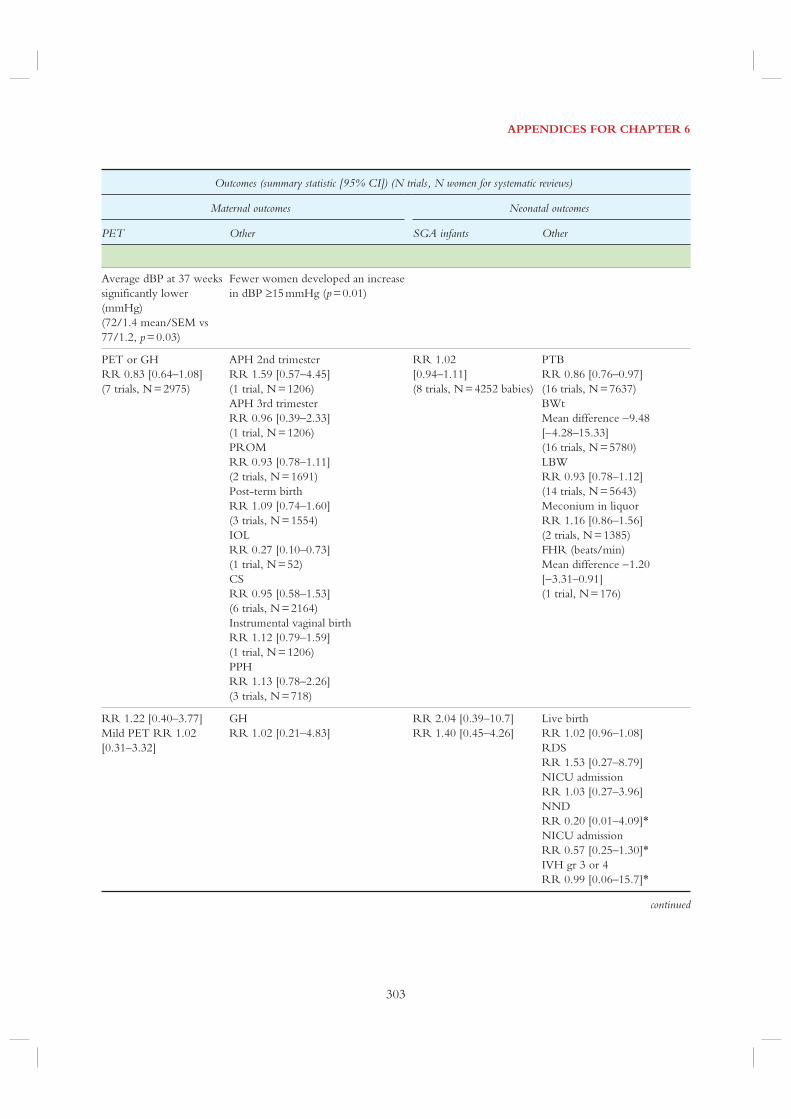
APPENDICES FOR CHAPTER 6
303
Outcomes (summary statistic [95% CI]) (N trials, N women for systematic reviews)
Maternal outcomes Neonatal outcomes
PET Other SGA infants Other
Average dBP at 37 weeks
significantly lower
(mmHg)
(72/1.4 mean/SEM vs
77/1.2, p = 0.03)
Fewer women developed an increase
in dBP 15 mmHg (p = 0.01)
PET or GH
RR 0.83 [0.64–1.08]
(7 trials, N = 2975)
APH 2nd trimester
RR 1.59 [0.57–4.45]
(1 trial, N = 1206)
APH 3rd trimester
RR 0.96 [0.39–2.33]
(1 trial, N = 1206)
PROM
RR 0.93 [0.78–1.11]
(2 trials, N = 1691)
Post-term birth
RR 1.09 [0.74–1.60]
(3 trials, N = 1554)
IOL
RR 0.27 [0.10–0.73]
(1 trial, N = 52)
CS
RR 0.95 [0.58–1.53]
(6 trials, N = 2164)
Instrumental vaginal birth
RR 1.12 [0.79–1.59]
(1 trial, N = 1206)
PPH
RR 1.13 [0.78–2.26]
(3 trials, N = 718)
RR 1.02
[0.94–1.11]
(8 trials, N = 4252 babies)
PTB
RR 0.86 [0.76–0.97]
(16 trials, N = 7637)
BWt
Mean difference 9.48
[4.28–15.33]
(16 trials, N = 5780)
LBW
RR 0.93 [0.78–1.12]
(14 trials, N = 5643)
Meconium in liquor
RR 1.16 [0.86–1.56]
(2 trials, N = 1385)
FHR (beats/min)
Mean difference 1.20
[3.31–0.91]
(1 trial, N = 176)
RR 1.22 [0.40–3.77]
Mild PET RR 1.02
[0.31–3.32]
GH
RR 1.02 [0.21–4.83]
RR 2.04 [0.39–10.7]
RR 1.40 [0.45–4.26]
Live birth
RR 1.02 [0.96–1.08]
RDS
RR 1.53 [0.27–8.79]
NICU admission
RR 1.03 [0.27–3.96]
NND
RR 0.20 [0.01–4.09]*NICU admission
RR 0.57 [0.25–1.30]*IVH gr 3 or 4
RR 0.99 [0.06–15.7]*
continued

THE FIGO TEXTBOOK OF PREGNANCY HYPERTENSION
304
Outcomes (summary statistic [95% CI]) (N trials, N women for systematic reviews)
Maternal outcomes
Author (study design) N trials (N women) Quality of trials Intervention (N women) Controls (N women)
Prostaglandin precursors
Makrides 200645
(systematic review of 2783
low- and high-risk
women with data on
low-risk women reported
here; see Appendix 6.2 for
data on high risk women)
4 trials
(N = 2056)
Alloc con low risk: 3/6
trials
OAB: NR
IOD “Most trials reported outcome for at least 83% of all women recruited”
Marine oil
(N = 1024)
Placebo or no
marine oil
(N = 1032)
Zhou 201246
(single trial)
N = 2399 Alloc con: yes
OAB: NR
Loss to f/u <20%: NR
Fish oil (800 mg DHA/d
in second half of
pregnancy)
(N = 1197)
Placebo
(N = 1202)
Appendix 6.1 continued

APPENDICES FOR CHAPTER 6
305
Outcomes (summary statistic [95% CI]) (N trials, N women for systematic reviews)
Maternal outcomes Neonatal outcomes
PET Other SGA infants Other
RR 1.01 [0.52–1.98]
(3 trials, N = 1130)
GH
RR 1.09 [0.90–1.33]
(5 trials, N = 1831)
RR 1.12 [0.93–1.35]
(1 trial, N = 1111)
PTB <37 weeks
RR 0.95 [0.80–1.13]
(3 trials, N = 1393)
Length of gestation (days)
Mean difference 2.23
[0.67–3.80]
(3 trials, N = 1393)
Prolonged gestation (>42
weeks)
RR 1.19 [0.73–1.93]
(1 trial, N = 533)
BWt (g)
Mean difference 55.79
[4.83–106.74]
(3 trials, N = 1946)
LBW <2500 g
RR 0.99 [0.87–1.13]
(2 trials, N = 1413)
Stillbirth ≥24 weeks)
RR 1.00 [0.06–15.96]
(1 trial, N = 533)
NND
RR 2.01 [0.18–22.01]
(1 trial, N = 579)
PET
aRR 1.03 (0.72–1.48]
(N = 2399)
Clinical PET
aRR 0.87 [0.60–1.25]
GH
aRR 0.93 [0.71–1.21]
GDM
aRR 1.04 [0.75–1.44]
Clinical GDM aRR 0.97
[0.74–1.27]
For weight
aRR 0.90 [0.66–1.22]
For length
aRR 0.93 [0.75–1.16]
(N = 2399)
For head circum
aRR 0.96
[0.78–1.19] (N = 2399)
LBW
aRR 0.65 [0.44–0.96]
Macrosomia
aRR 1.27 [1.05–1.55]
continued

THE FIGO TEXTBOOK OF PREGNANCY HYPERTENSION
306
Outcomes (summary statistic [95% CI]) (N trials, N women for systematic reviews)
Maternal outcomes
Author (study design) N trials (N women) Quality of trials Intervention (N women) Controls (N women)
Smoking cessation
Chamberlain 201354
(systematic review)
86 trials
(N = >29,000
women)
Alloc con low risk: 10/86
trials.
OAB:
“not calculable due to insufficient numbers of studies with low risk of bias”IOD low risk: 22/86 trials.
Smoking cessation
interventions
(N = 4298)
Routine care
(N = 4264)
Coleman 201255
(single trial)
N = 1050 Alloc con: yes
OAB: yes
Loss to f/u <20%:
yes (18.5%)
Nicotine patches (15 mg
every 16 h for 8 weeks)
(N = 521)
Placebo
(N = 529)
Appendix 6.1 continued

APPENDICES FOR CHAPTER 6
307
Outcomes (summary statistic [95% CI]) (N trials, N women for systematic reviews)
Maternal outcomes Neonatal outcomes
PET Other SGA infants Other
LBW <2500 g
RR 0.87 [0.70–1.08]
(6 trials, N = 3836)
Very LBW
RR 1.27 [0.60–2.71]
(2 trials, N = 1666)
Mean BWt
Mean difference 36.72
[0.70–72.74]
(9 trials, N = 4846)
PTB <37 weeks
RR 0.82 [0.70v0.96]
(14 trials, N = 7852)
Stillbirths
RR 1.08 [0.51–2.30]
(4 trials, N = 2212]
NND
RR 2.06 [0.61–6.92]
(3 trials, N = 2095)
NICU admission
RR 0.82 [0.52–1.29]
(2 trials, N = 1140)
PET or eclampsia
3 (0.6%) vs. 5 (0.9%),
p = NR
BP >140/90 mmHg on at least 2
occasions
24 (4.6%) vs. 25 (4.7%), p = NR
Caesarean
OR 1.45 [1.05–2.01]
(N = 1024)
LBW
OR 1.38 [0.90–2.09]
BWt, unadjusted (kg)
0.02 [0.10–0.05]
Miscarriage
OR 1.52 [0.25–9.13]
Stillbirth
OR 2.59 [0.50–13.4]
(N = 1041)
PTB
OR 0.90 [0.58–1.41]
(N = 1024)
NICU admission
OR 0.95 [0.58–1.57]
(N = 1024)
5 min Apgar <7
OR 0.91 [0.45–1.80]
(N = 1024)
Cord blood arterial pH <7
OR 0.57 [0.17–1.97]
(N = 1024)
IVH
OR 0.67 [0.11–4.05]
(N = 1024)
Neonatal convulsions
OR 1.02 [0.29–3.54]
(N = 1024)
NEC
OR 0.50 [0.12–2.03]
(N = 1024)
continued

THE FIGO TEXTBOOK OF PREGNANCY HYPERTENSION
308
Outcomes (summary statistic [95% CI]) (N trials, N women for systematic reviews)
Maternal outcomes
Author (study design) N trials (N women) Quality of trials Intervention (N women) Controls (N women)
Thiazide diuretics
Churchill 200756
(systematic review of 5
trials, N = 1836 low and
high-risk women of
which low and high-risk
women are reported
together here; see
Appendix 6.2 for data on
high-risk women)
5 trials
(N = 1836)
“The quality of all five studies was unclear”Alloc con: unclear
OAB: 4/5 trials
LFU <20%: 5/5 trials
Thiazide diuretic
(N = 1016)
Placebo or no
thiazide
(N = 820)
Vitamins C & E
Rumbold 200857
(systematic review of 10
trials, N = 6533 low/
moderate- and high-risk
women, of which the
low/ moderate-risk
women are presented here
when possible; see
Appendix 6.2 for data on
the high-risk women)
5 trials
(N = 3307)
Alloc con low risk: 3/5
trials.
OAB low-risk: 5/5 trials
(explicitly stated in 4).
OAB low risk: 3/5 trials.
One/more antioxidants
(N = 1858 as calculated
from tables)
Placebo or no
antioxidant
(N = 1449)
Appendix 6.1 continued

APPENDICES FOR CHAPTER 6
309
Outcomes (summary statistic [95% CI]) (N trials, N women for systematic reviews)
Maternal outcomes Neonatal outcomes
PET Other SGA infants Other
RR 0.68 [0.45–1.03]*(4 trials, N = 1391)
Severe PET
RR 1.56 [0.26–9.17]*(2 trials, N = 1297)
HTN (new or worsening)
RR 0.85 [0.68–1.08]*(2 trials, N = 1475)
Nausea and vomiting
RR 5.81 [1.04–32.46]*(2 trials, N = 1217)
CS
RR 1.0 [0.26–3.81]*(1 trial, N = 20)
None in the 1 trial that
reported this outcome
Perinatal death
RR 0.72 [0.40–1.27]*(5 trials, N = 1836)
Stillbirth
RR 0.60 [0.27–1.34]*(5 trials, N = 1836)
NND
RR 0.88 [0.40–1.97]*(4 trials, N = 1816)
PTB
RR 0.67 [0.32–1.41]*(2 trials, N = 465)
BWt
Mean difference 139.0
[484.40–762.40]*(1 trial, N = 20)
Gestation at birth
Mean difference 0.70
[0.71–2.11]*(1 trial, N = 20)
Postmaturity >42 weeks
RR 7.0 [0.41–120.16]*(1 trial, N = 20)
5 min Apgar <7
RR 3.0 [0.14–65.90]*(1 trial, N = 20)
RR 0.85 [0.48–1.51]
(4 trials, N = 2441)
Antihypertensive therapy
RR 1.77 [1.22–2.57]*(2 trials, N = 4272)
Require antenatal hospital admission
for HTN RR 1.54 [1.00–2.39]*(1 trial, N = 1877)
RR 0.71 [0.42–1.19]*(2 trials, N = 2104)
PTB
RR 1.17 [0.92–1.48]*(2 trials, N = 2067)
Any baby death
RR 0.90 [0.53–1.51]*(2 trials, N = 2077)
continued
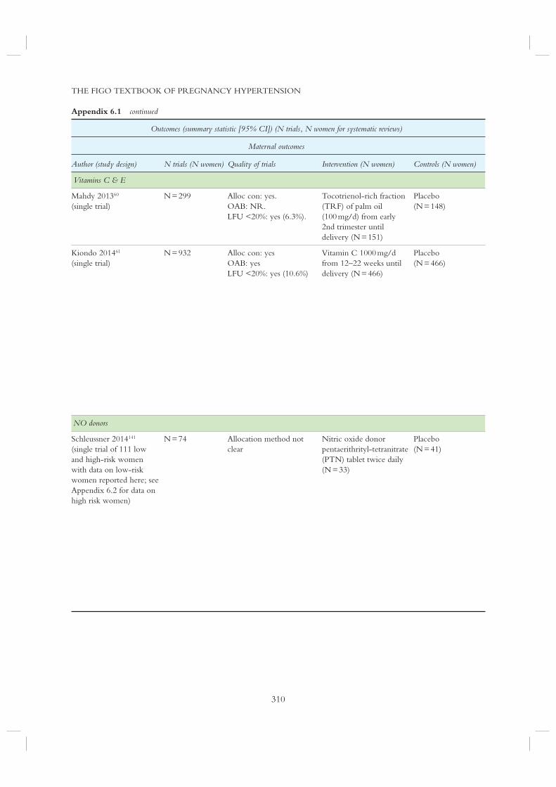
THE FIGO TEXTBOOK OF PREGNANCY HYPERTENSION
310
Outcomes (summary statistic [95% CI]) (N trials, N women for systematic reviews)
Maternal outcomes
Author (study design) N trials (N women) Quality of trials Intervention (N women) Controls (N women)
Vitamins C & E
Mahdy 201360
(single trial)
N = 299 Alloc con: yes.
OAB: NR.
LFU <20%: yes (6.3%).
Tocotrienol-rich fraction
(TRF) of palm oil
(100 mg/d) from early
2nd trimester until
delivery (N = 151)
Placebo
(N = 148)
Kiondo 201461
(single trial)
N = 932 Alloc con: yes
OAB: yes
LFU <20%: yes (10.6%)
Vitamin C 1000 mg/d
from 12–22 weeks until
delivery (N = 466)
Placebo
(N = 466)
NO donors
Schleussner 2014141
(single trial of 111 low
and high-risk women
with data on low-risk
women reported here; see
Appendix 6.2 for data on
high risk women)
N = 74 Allocation method not
clear
Nitric oxide donor
pentaerithrityl-tetranitrate
(PTN) tablet twice daily
(N = 33)
Placebo
(N = 41)
Appendix 6.1 continued

APPENDICES FOR CHAPTER 6
311
Outcomes (summary statistic [95% CI]) (N trials, N women for systematic reviews)
Maternal outcomes Neonatal outcomes
PET Other SGA infants Other
RR 0.20
[0.02–1.66]
PET or GH
RR 0.36
[0.12–1.09]
Any PET
RR 0.77 [0.37–1.56]
Severe PET
RR 1.25 [0.34–4.65]
GH
RR 0.67 [0.43–1.03]
APH
RR 0.78 [0.29–2.1]
PROM
RR 0.79 [0.41–1.54]
Abruption
RR 0.5 [0.04–5.53]
Vaginal delivery
RR 1.0 [0.82–1.22]
LBW
RR 1.07 [0.72–1.59]
BWt <2500 g
RR 1.07 [0.72–1.59]
Apgar <7 RR 1.17
[0.76–1.81]
Admission to SCU
RR 1.53 [0.95–2.43]
Stillbirth
RR 1.01 [0.54–1.87]
Early NND
RR 0.71 [0.27–1.83]
Abortion
RR 1.01 [0.40–2.51]
PTB
RR 0.92 [0.63–1.34]
Stillbirth
RR 1.01 [0.54–1.87]
PET/HELLP
6(21.2%) vs. 8 (19.5%)
PET <32 weeks
3 (50%) vs. 5( 62.5%)
Abruption
0 vs. 4(9.8%)
CS
14 (41.2%) vs. 21 (53.8%)
IUGR or perinatal death
9 (27.3%) vs. 17 (41.5%)
PTD <37 weeks
10 (30.3%) vs. 12 (29.3%)
PTD <32 weeks
1 (3%) vs. 8 (19.5%)
1 min Apgar score
7.7 (+/1.9) vs. 7.4 (+/2.2)
5 min Apgar score
8.5 (+/1.4) vs. 8.7 (+/1.1)
UA pH
7.3 (+/0.1) vs. 7.3 (+/0.1)
BWt (g)
2734 (+/889) vs. 2460
(+/01004)
Ventilation (NICU)
9 (30) vs. 7 (20.0)
continued

THE FIGO TEXTBOOK OF PREGNANCY HYPERTENSION
312
Outcomes (summary statistic [95% CI]) (N trials, N women for systematic reviews)
Maternal outcomes
Author (study design) N trials (N women) Quality of trials Intervention (N women) Controls (N women)
Lifestyle changes
Meher 200673
(systematic review)
2 trials
(N = 106)
Alloc con low risk:
“inadequately reported”.
OAB: “not possible” LFU
<20%: “completeness of
follow-up was not
reported in either trial”
4–6 h rest/d
(N = 16)
Normal activity
(N = 16)
4–6 h rest/d + Nutrient
supplementation
(N = 37)
Normal
activity + placebo
(N = 37)
Kramer 200626
(systematic review)
14 trials
(N = 1014)
Alloc con: “in most of the
trials, the method of
treatment allocation was
either by alternation or
was not described”.
OAB: not specified
LFU<20%: not specified
Increase in exercise in
sedentary women
(N = 280)
Maintain activity
level
(N = 276)
Reduction in exercise in
physically fit women
(N = 28)
Maintain activity
level
(N = 33)
Increase then reduction in
exercise in physically fit
women
(N = 25)
Maintain activity
level
(N = 24)
Appendix 6.1 continued

APPENDICES FOR CHAPTER 6
313
Outcomes (summary statistic [95% CI]) (N trials, N women for systematic reviews)
Maternal outcomes Neonatal outcomes
PET Other SGA infants Other
RR 0.05 [0.00–0.83]
(1 trial, N = 32)
GH
RR 0.25 [0.03–2.00]
(1 trial, N = 32)
RR 0.13 [0.03–0.51]
(1 trial, N = 74)
GH
RR 0.15 [0.04–0.63]
(1 trial, N = 74)
CS
RR 0.82 [0.48–1.41]
(1 trial, N = 74)
RR 1.17 [0.44–3.08]
(2 trials, N = 82)
CS
RR 0.96 [0.60–1.53]
(3 trials, N = 386]
Total gestational weight gain (kg)
Mean difference 0.79 [0.73–2.31]
(4 trials, N = 122)
Change in maternal fat mass (kg)
Mean difference 1.51 [3.06–0.04]
(1 trial, N = 41)
Change in maternal lean mass (kg)
Mean difference 1.59 [0.38–2.80]
(1 trial, N = 41)
BWt (g)
Mean difference 49.49
[27.74–126.73]
(6 trials, N = 556)
PTB RR 1.82 [0.35–9.57]
( 3 trials, N = 111)
1 min Apgar
Mean difference 1.0
[1.37–3.37]
(1 trial, N = 20)
5 min Apgar
Mean difference 0.15
[0.10–0.39]
(4 trials, N = 462)
PTB
RR 1.18 [0.08–17.99]
(1 trial, N = 61)
BWt (g)
Mean difference 135.0
[368.66, 98.66]
(1 trial, N = 61)
Gestational weight gain (kg)
Mean difference 0.90
[1.59–3.39]
(1 trial, N = 49)
Bwt (g)
Mean difference 460.0
[251.63–668.37]
(1 trial, N = 49)
continued

THE FIGO TEXTBOOK OF PREGNANCY HYPERTENSION
314
Outcomes (summary statistic [95% CI]) (N trials, N women for systematic reviews)
Maternal outcomes
Author (study design) N trials (N women) Quality of trials Intervention (N women) Controls (N women)
Lifestyle changes
Reduction, then increase
in exercise in physically fit
women
(N = 26)
Maintain activity
level
(N = 24)
Increase in exercise in
over weight women
(N = 37)
Maintain activity
level
(N = 35)
Periodontal therapy
Niederman 2010143 N = 1082 Alloc con: yes
OAB: yes
LFU <20%: yes
Periodontal treatment in
midpregnancy
(N = 542)
Periodontal
treatment after
pregnancy
(N = 540)
Alloc con, allocation concealment; APH, antepartum haemorrhage; aRR, adjusted relative risk; BWt, birth weight; CI,
confidence interval; circum, circumference; CS, Caesarean section; ctx, contraction; dBP, diastolic blood pressure; DHA,
docosahexanenoic acid; FHR, fetal heart rate; FM, fetal movement; GDM, gestational diabetes mellitus; GH, gestational
hypertension; HELLP, haemolysis, elevated liver enzymes, low platelets; HIE, hypoxic ischaemic encephalopathy; IOD,
incomplete outcome data; IOL, induction of labour; LBW, low birth weight; LFU, loss to follow up; IUGR, intrauterine
growth restriction; IVH, intraventricular haemorrhage; LBW, low birth weight; Mg, magnesium; NEC, necrotising
enterocolitis; NICU, neonatal intensive care unit; NND, neonatal death; NNT, number needed to treat; NR, not
reported; OAB, outcome assessment blinding; OR, odds ratio; PET, pre-eclampsia; PPH, postpartum haemorrhage;
PROM, premature rupture of membranes; PTB, preterm birth; RDS, respiratory distress syndrome; RR, relative risk;
SEM, standard error of mean; SGA, small-for-gestational age; SVP, spontaneous vaginal delivery
Appendix 6.1 continued

APPENDICES FOR CHAPTER 6
315
Outcomes (summary statistic [95% CI]) (N trials, N women for systematic reviews)
Maternal outcomes Neonatal outcomes
PET Other SGA infants Other
Gestational weight gain (kg)
Mean difference 2.60
[4.96-9-0.24)]
(1 trial, N = 50)
BWt (g)
Mean difference 100.0
[308.39–108.39]
(1 trial, N = 50)
PTB
RR 1.89 [0.18–19.95]
(1 trial, N = 72)
BWt (g)
Mean difference 5.0
[241.27–231.27]
OR 0.82 [0.44–1.56] BWt
3450 vs. 3410 g (p = 0.12)
PTB
OR 1.05 [0.7–1.58]
† “Sensitivity analysis after excluding women with GDM showed that the reduction in pre-eclampsia did not persist by
combining all interventions (RR 0.91 [0.75–1.11]) or in diet only group (RR 0.86 [0.45–1.64]).” “2 studies on women
with GDM had . . . insulin. We cannot rule out the possibility that insulin use could have been an important contributor to
the beneficial effect observed”‡ These results should be interpreted with caution as a large number of severe congenital anomalies and deaths of two sets of
twins (with birth weights <750 g) in the supplemented group likely accounted for the increased risk of death observed.
When deaths due to severe congenital abnormalities were excluded from the meta-analysis, no increased risk of NND was
seen.
^ Severe PET complications: 1+ of the following outcomes: severe pre-eclampsia or early onset pre-eclampsia (32 weeks
gestation), eclampsia, HELLP syndrome, placental abruption, severe gestational HTN (160 mmHg and/or 110 mmHg
systolic and diastolic pressures, respectively)

THE FIGO TEXTBOOK OF PREGNANCY HYPERTENSION
316
Appendix 6.2
Randomised trials and systematic reviews of trials of interventions to
prevent pre-eclampsia in women at increased risk (unless indicated
by an ‘*’ when all women were presented together)
Author (study design) N trials (N women) Quality of trials Intervention (N women) Controls (N women)
Antihypertensive drugs
Abalos 201478
(systematic review)
49 trials
(N = 4723)
Alloc con low risk: 17/49
trials.
OAB low risk: 10/49 trials.
IOD low risk: 45/49 trials.
ANY HYPERTENSIVE
DRUG
(N = 1476)
NO DRUG OR
PLACEBO
(N = 1375)
ANY
ANTIHYPERTENSIVE
DRUG
(N = 689)
METHYLDOPA
(N = 650)
ANY
ANTIHYPERTENSIVE
DRUG
(N = 74)
CALCIUM
CHANNEL
BLOCKER
(N = 62)

APPENDICES FOR CHAPTER 6
317
PET Other SGA infants Other
Proteinuria/PET
RR 0.93 [0.80–1.08]
(23 trials, N = 2851)
Severe PET
RR 0.54 [0.24–1.23]
(3 trials, N = 416)
Eclampsia
RR 0.34 [0.01–8.15]
(5 trials, N = 578)
Maternal death
RR 1.08 [0.24–4.83]
(5 trials, N = 525)
Severe HTN
RR 0.49 [0.40–0.60]
(20 trials, N = 2558)
HELLP
RR 2.02 [0.38–10.78]
(1 trial, N = 197)
RR 0.97 [0.80–1.17]
(20 trials, N = 2586)
RR 0.71 [0.49–1.02]
(27 trials, N = 3230)
Proteinuria/PET
RR 0.73 [0.54–0.99]
(11 trials, N = 997)
Severe HTN
RR 0.54 [0.30–0.95]
(11 trials, N = 638)
Antenatal hospital admission
RR 0.77 [0.58–1.03]
(2 trials, N = 275)
CS
RR 0.93 [0.78–1.12]
(10 trials, N = 878)
Abruption
RR 2.02 [0.19–21.90]
(1 trial, N = 173)
RR 0.80 [0.53–1.21]
(7 trials, N = 597)
Perinatal death
RR 0.73 [0.42–1.27]
(19 trials, N = 1339)
PTB< 37 weeks
RR 0.76 [0.55–1.05]
(9 trials, N = 623)
Admission to SCBU
RR 0.92 [0.67–1.26]
(4 trials, N = 478)
Proteinuria/PET
RR 2.15 [0.73–6.38]
(2 trials, N = 128)
Severe HTN
RR 2.09 [0.96–4.57]
(2 trials, N = 136)
HELLP
RR 1.5 [0.26–8.60]
(1 trial, N = 100)
CS
RR 1.57 [0.91–2.71]
(1 trial, N = 100)
RR 1.0 [0.10–9.96]
(1 trial, N = 36)
Total fetal or NND
RR 1.0 [0.06–15.55]
(2 trials, N = 136)
PTB <37 weeks
RR 0.63 [0.20–1.91]
(1 trial, N = 36)
Admission to SCBU
RR 1.47 [0.44–4.89]
(1 trial, N = 99)
continued

THE FIGO TEXTBOOK OF PREGNANCY HYPERTENSION
318
Author (study design) N trials (N women) Quality of trials Intervention (N women) Controls (N women)
Antihypertensive drugs
Magee 200779
(single trial)
N = 132 Less tight BP control
(N = 66)
Tight BP control
(N = 65)
Aspirin
Duley 20076
(systematic review of 59
trials, 37,500 women with
only high-risk women
included here; see
Appendix 6.1 for data on
moderate-risk women)
18 trials
(N = 4121)
“. . . wide variation in study quality. The poorer quality studies were mostly the small early trials, with the more recent large studies tending to be of higher quality.”
Low-dose aspirin or
dipyridamole
(N = 14,326)
Placebo or no
anti-platelet agent
(N = 14,143)
Bujold 201086
(systematic review and
meta-analysis)
27 trials
(N = 11,348)
Alloc con: 12/12 trials.
OAB: 4/12 trials.
LFU <20%: 12/12 trials.
Low-dose (50–150 mg/d)
aspirin started 16 weeks
or earlier
(N = 389)
Placebo or no
treatment
(N = 375)
Alloc con: 22/22 trials.
OAB: double blinding
16/22 trials
LFU <20%: 22/22 trials
Low-dose aspirin
(50–150 mg/d) started
16 weeks
(N = 5691)
Placebo or no
treatment
(N = 5657)
Groeneveld 201384
(meta-analysis)
4 trials
(N = 268)
Alloc con: 4/4 trials.
OAB: 4/4 trials.
LFU <20%: ?
(No information provided)
Aspirin 100 mg/d in IVF
patients
(N = 131)
Singletons (N = 96)
Twins (N = 24)
Placebo
(N = 137)
Singletons
(N = 91)
Twins
(N = 41)
Appendix 6.2 continued

APPENDICES FOR CHAPTER 6
319
PET Other SGA infants Other
16 (24.2) vs. 20 (30.8) Serious maternal complications
3 (4.6%) vs. 2 (3.1%)
CS
35 (53.0%) vs. 37 (56.9%)
Antenatal corticosteroids for fetal
lung maturation
16 (24.2%) vs. 15 (23.1%)
MgSO4 for PET
10 (15.2%) vs. 12 (18.5%)
GA at delivery
36.9 ± 3.0 vs. 36.3 3.3
BWt (g)
2675 ± 858 vs. 2501 ± 855
5 min Apgar <7
0 (0.0) vs. 2 (3.1)
5 min serious perinatal
complications
9 (13.6%) vs. 14 (21.5%)
NICU stay
15 (22.7%) vs. 22 (34.4%)
RR 0.75 [0.66–0.85]
(18 trials, N = 4121)
GH
RR 0.54 [0.41–0.70]
(12 trials, N = 838)
Abruption
RR 0.75 [0.42–1.34]
(4 trials, N = 2710)
RR 0.89 [0.74–1.08]
(13 trials, N = 4239)
Fetal and neonatal death
RR 0.69 [0.53–0.90]
(17 trials, N = 4443)
PTB <37 weeks
RR 0.89 [0.81–0.97]
(10 trials, N = 3252)
RR 0.47 [0.34–0.65]
(9 trials, N = 765)
Severe PET
RR 0.09 [0.02–0.37]
(3 trials, N = 278)
GH
RR 0.62 [0.45–0.84]
(3 trials, N = 278)
Abruption
RR 0.62 [0.08–5.03]
(4 trials, N = 360)
IUGR (any definition)
16 weeks or less:
RR 0.44 (0.30–0.65)
(9 trials, N = 853)
>16 weeks:
RR 0.98 (0.87–1.10)
(15 trials, N = 7027)
PTB
RR 0.22 [0.10–0.49]
(4 trials, N = 387)
RR 0.81 [0.63–1.03)
(18 trials, N = 10,584)
Severe PET
RR 0.26 [0.05–1.26]
(2 trials, N = 669)
GH
RR 0.63 [0.47–0.85)
(14 trials, N = 4303)
Abruption
RR 1.56 [0.96–2.55]
(6 trials, N = 3583)
IUGR
RR 0.98 [0.87–1.10]
(15 trials, N = 7027)
PTB
RR 0.90 [0.83–0.97]
(16 trials, N = 10,398)
“Hypertensive pregnancy
complications”
Singletons:
OR 0.62 [0.22–1.7]
Twins:
OR 1.2 [0.35–4.4]
PTB
Singletons:
OR 0.52 [0.16–1.7]
(N = 180)
Twins:
OR 1.6 [0.51–5.0)
continued

THE FIGO TEXTBOOK OF PREGNANCY HYPERTENSION
320
Author (study design) N trials (N women) Quality of trials Intervention (N women) Controls (N women)
Aspirin
Villa 201385
(single
trial + meta-analysis)
Single trial
(N = 152)
Meta-analysis
2 trials: Vainio
2002, Ebrashy
2005
(N = 346)
Alloc con: yes
OAB: “double-blinded”
LFU <20%: no (20.4%)
Aspirin (100 mg/d)
(N = 61)
Placebo
(N = 60)
Roberge et al 201288
(systematic review and
meta-analysis)
4 trials
(N = 392)
“Studies with high risk of bias were considered for exclusion”
Aspirin (50–150 mg/d)
(16 weeks)
(N = 201)
Placebo or no
treatment
(N = 191)
Henderson 201481
(systematic review of 23
trials, 22,988 women with
only high-risk women
included here; see
Appendix 6.1 for data on
moderate-risk women
15 trials
(N = 12,656)
(Reported only for all 23
trials of low and high risk
women together – See
‘Henderson 2014’,
Appendix 6.1)
Aspirin (50–150 mg/d)
(N = 6123)
Placebo or no
treatment
(N = 6522)
Cantu 201592
(secondary analysis of
single trial)
Stratification by
initiation (< or
>16 weeks)
N = 2539
Stratification by
BMI
N = 2479
Alloc con: not specified.
OAB: no.
LFU <20%: yes.
Aspirin (60 mg/d) <16
weeks
(N = 225)
Aspirin (60 mg/d) >16
weeks
(N = 1029)
BMI <30
(N = 756)
BMI 30
(N = 487)
Initiation Placebo
<16 weeks
(N = 236)
Placebo >16 weeks
(N = 1013)
BMI <30
(N = 756)
BMI 30
(N = 480)
Bergeron 201682
(systematic review of 6
trials, 898 women with
multiple gestations)
6 trials
(N = 898)
Alloc con low risk: 5/6
trials.
OAB low risk: 5/6 trials.
IOD low risk: 4/6 trials
Aspirin (61–100 mg/d) Placebo
Appendix 6.2 continued

APPENDICES FOR CHAPTER 6
321
PET Other SGA infants Other
SINGLE TRIAL
RR 0.70 [0.30–1.7]
Severe PET
RR 0.4 [0.1–1.2]
Early onset PET
RR 0.2 [0.03–2.1]
META-ANALYSIS
2 trials (N = 346)
RR 0.6 [0.37–0.83]
Severe PET
RR 0.3 [0.11–0.69)
Preterm PET
RR 0.2 [0.02–1.26]
Term PET
RR 1.0 [0.25–4.26]
GH
RR 1.6 [0.6–4.2]
RR 0.3 [0.1–1.6]
Severe PET
RR 0.22 [0.08–0.57]
Mild PET
RR 0.81 [0.33–1.96]
RR 0.76 [0.62–0.95]
(13 trials, N = 12,184)
Abruption
RR 1.12 [0.86–1.46]
(3 trials, N = 12,366)
IUGR
RR 0.80 [0.65–0.99]
(13 trials, N = 12,504)
Perinatal death
RR 0.81 [0.65–1.01]
(10 trials, N = 12,240)
PTB
RR 0.86 [0.76–0.98]
(10 trials, N = 11,779)
LDA <16 weeks
RR 0.93 [0.67–1.31]
LDA >16 weeks
RR 0.90 [0.75–1.08]
BMI <30
RR 0.91 [0.7–1.13]
BMI 30
RR 0.89 [0.7–1.13]
RR 0.67 [0.48–0.94]
(5 trials, N = 898)
Mild PET
RR 0.44 [0.24–0.82]
(# trials not specified,
N = 724)
Severe PET
RR 1.02 [0.61–1.72]
(# trials not specified,
N = 724)
RR 1.09 [0.80–1.47]
(4 trials, N = 1573
neonates)
PTB <37 weeks
RR 1.11 [0.83–1.49]
(# trials not specified, N = 1554
neonates)
continued

THE FIGO TEXTBOOK OF PREGNANCY HYPERTENSION
322
Author (study design) N trials (N women) Quality of trials Intervention (N women) Controls (N women)
Calcium
Hofmeyr 20147
(systematic review of 24
trials, 17,954 women with
only high-risk women
included here when
possible; see Appendix 6.1
for data on low-risk
women. Data on women
at unclear risk not
presented.)
5 trials
(N = 587)
Alloc con low risk: 4/5
trials.
OAB low risk: 4/5 trials.
IOD low risk: 3/5 trials
Calcium (1 g/d)
(N = 281)
Placebo or no
calcium
(N = 306)
Calcium + Aspirin
Asemi 2012101
(single trial)
N = 42 Alloc con: yes.
OAB: no.
LFU <20%: yes
Calcium carbonate
(500 mg/d) + aspirin
(80 mg/d) for 9 weeks
(N = 20)
Placebo
(N = 22)
Souza 2014102
(single trial)
N = 49 Alloc con: yes.
OAB: yes
LFU <20%: yes
Calcium (2 g/d) + aspirin
(100 mg/d)
(N = 23)
Placebo
(N = 26)
Dietary Changes
Allen 201412
(systematic review of 18
trials, 8712 women with
low and high-risk women
presented together here;
see Appendix 6.1 for data
on low-risk women. Data
for women at unclear risk
not presented.)
18 trials
(N = 8712)
Alloc con: low risk 9/18
trials
OAB: low risk 7/18 trials
IOD low risk: 17/18 trials
Dietary change alone or
with other change
(N = 4342)
Placebo or no
dietary change
(N = 4370)
DIET
(6 trials, N = 1334)
Control (not
specified)
(6 trials, N = 1361)
MIXED
(Diet, physical activity &
lifestyle)
(6 trials, N = 733)
Control (not
specified)
(6 trials, N = 705)
Appendix 6.2 continued

APPENDICES FOR CHAPTER 6
323
PET Other SGA infants Other
RR 0.22 [0.12–0.42]
(5 trials, N = 587)
Hypertension
( PET)
RR 0.47 [0.22–0.97]
(4 trials, N = 327)
PTB
RR 0.45 [0.24–0.83]
(4 trials, N = 568)
Admission to NICU
RR 0.29 [0.03–2.48]
(1 trial, N = 63)
Stillbirth or death before
hospital discharge
RR 0.39 [0.02–9.20]
(3 trials, N = 512)
Serum hs-CRP
102.87 1828.52 vs.
3227.75 4760.70 (p = 0.001)
Plasma TAC
68.96 236.39 vs. 74.46 199.07
(p = 0.04)
GSH
304.33 709.32 vs.
39.33 174/33 (p = 0.03)
Superimposed PET
42.2 vs. 73.1% (p = 0.112)
IUGR
25.0% vs. 2.8% (p = 0.07)
BWt (g)
2563 0 1033 vs.
2604 811 (p = 0.88)
PTB
33.3% in both treatment and
placebo groups
LBW (<2500 g) 11 (42.3%) vs.
7 (30.4%) (p = 0.40)
Very LBW (<1500 g)
5 (19.2%) vs. 3 (13.0%)
(p = 0.71)
ANY DIETARY
CHANGE
RR 0.81 [0.69–0.94]†
(18 trials, N = 8712)
(I2 = 0%)
RR 0.67 [0.53–0.85]*(6 trials, N = 2695)
RR 0.93 [0.66–1.32]*(6 trials, N = 1438)
continued

THE FIGO TEXTBOOK OF PREGNANCY HYPERTENSION
324
Author (study design) N trials (N women) Quality of trials Intervention (N women) Controls (N women)
Dietary Changes
ESSENTIAL FATTY
ACIDS
(6 trials, N = 2275)
Control (not
specified)
(6 trials, N = 2304)
Ziaei 2001103
(single trial)
N = 100 Alloc con: not specified.
OAB: no.
LFU <20%: not specified
Allicin (100 mcg/d) in 3rd
trimester
(N = 50)
Placebo
(N = 50)
Teran 2009104
(single trial)
N = 235 Alloc con: yes.
OAB: yes.
LFU <20%: yes
CoQ10 (200 mg/d) (20
weeks GA to delivery
(N = 118)
Placebo
(N = 117)
Heparin
Rodger 2014111
(single trial)
N = 292 Alloc con: yes.
LFU <20%: yes
Antepartum dalteparin
N = 146
No antepartum
dalteparin
N = 143
On-treatment analysis
(N = 143)
On-treatment
analysis
(N = 141)
Appendix 6.2 continued

APPENDICES FOR CHAPTER 6
325
PET Other SGA infants Other
RR 0.92 [0.71–1.18]*(6 trials, N = 6579)
7 (14%) vs. 9 (18%)
(p = 0.799)
9 (18%) vs. 18 (36%) (p = 0.043)
RR 0.56 [0.33–0.96] –
8 (5.5%) vs. 5 (3.5%)
difference 0.7
[3.1–1.6]
Severe or early onset
PET
7 (4.8%) vs. 4 (2.8%)
difference 2.0 (2.8–6.8)
Symptomatic major VTE 1 (0.7%)
vs. 2 (1.4%) difference 0.7
(3.1–1.6)
Abruption 4 (2.7%) vs. 3 (2.1%)
difference 0.6 (2.9–4.2)
SGA <10%
9 (6.2%) vs. 12(8.4%)
difference 2.2
(8.2–3.8)
SGA <5%
2 (1.4%) vs. 3 (2.1%)
Pregnancy loss (any) 12 (8.2%)
vs. 10 (7.0%) difference 1.2
(4.9–7.3)
Early pregnancy loss (≥3 at <10
weeks) 4 (2.7%) vs. 5 (3.5%)
difference 0.8 (4.8–3.2)
Late pregnancy loss (2 at >10
weeks or 1 at >16 weeks) 6
(4.1%) vs. 2 (1.4%) difference
2.7(1.0–6.5)
PTB <37 weeks (23 (15.8%)
vs. 17 (11.9%) difference 3.9
(4.1–11.8)
BWt of live births (g) 3186.2
vs. 3241.4 difference 55.2
[238.6–128.1]
Major bleeding
3 (2.1%) vs. 2 (1.4%) difference
0.7(2.4–3.7)
Minor bleeding (non-major) 28
(19.6% vs. 13 (9.2%) difference
10.4 (2.3–18.4)
BMD 6 weeks postpartum
2.16 (0.35) vs. 2.23 (0.42)
difference 0.07(0.19–0.04)
continued

THE FIGO TEXTBOOK OF PREGNANCY HYPERTENSION
326
Author (study design) N trials (N women) Quality of trials Intervention (N women) Controls (N women)
Heparin
Rodger 2014110
(systematic review)
6 trials
(N = 848)
Alloc con low risk: 5/6
trials.
AB low risk: 3/6 trials.
IOD low risk: 5/6 trials
Prophylactic LMWH
(N = 425)
No LMWH
(N = 423)
Lifestyle
Meher 2006126
(systematic review)
2 trials
(N = 45)
Alloc con low risk: 2/2
trials.
OAB: 1/2 trials.
IOD low risk:2/2 trials
Moderate intensity aerobic
exercise program
(N = 23)
Normal physical
activity
(N = 22)
Yeo 2008128
(single trial)
N = 79
(only have access
to abstract)
Walking
(N = 41)
Stretching
(N = 38)
Periodontal therapy
Kunnen 2010142
(systematic review of 12
observational studies and
3 RCTs, of which results
for 3 RCTs are reported
here)
N = 3650 Alloc con: methods not
reported. OAB: methods
not reported.
LFU <20% not reported
Periodontal treatment in
midpregnancy
(N = 1827)
Periodontal
treatment after
delivery
(N = 1823)
Appendix 6.2 continued

APPENDICES FOR CHAPTER 6
327
PET Other SGA infants Other
RR 0.46 [0.28–0.75]
(N = 739)
Severe or early PET
RR 0.16 [0.07–0.36]
(N = 665)
Abruption
RR 0.42 [0.13–1.4]
(N = 756)
SGA <10th centile
RR 0.42 [0.29–0.59]
(N = 713)
SGA <5th centile
RR 0.52 [0.28–0.94]
(N = 604)
Pregnancy loss <20 weeks
RR 0.89 [0.50–1.6]
(N = 591)
Pregnancy loss >20 weeks
RR 0.41 [0.17–1.02]
(N = 611)
NND
RR 0.31 [0.07–1.3]
(N = 623)
PTB <37 weeks
RR 0.77 [0.62–0.96]
(N = 556)
PTB <34 weeks
RR 0.45 [0.30–0.69]
(N = 678)
RR 0.31 [0.01–7.09]
(2 trials, N = 45)
GH
RR 1.00 [0.07–13.37]
(1 trial, N = 16)
CS
RR 0.93 [0.22–3.88]
(1 trial, N = 29)
RR 3.00 [0.14–64.26]
(1 trial, N = 16)
PTB
RR 1.00 [0.07–13.37]
(1 trial, N = 45)
14.6% [5.6–29.2] vs.
2.6% [0.07–13.8]
RR 1.0 [0.78–1.28]
(3 trials, N = 3650)
continued

THE FIGO TEXTBOOK OF PREGNANCY HYPERTENSION
328
Author (study design) N trials (N women) Quality of trials Intervention (N women) Controls (N women)
Micronutrients other than calcium
Kubik 200423
(single trial)
N = 138 “double blinded trial” Vitamin and mineral
supplement containing
15 mg zinc, 2 mg copper,
and 20 g selenium
Placebo
Makrides 201440
(systematic review of 10
trials, 9090 low and high
risk women for whom
outcomes were not
reported by risk)
(Low and high
risk women
reported
together)
10 trials
(N = 9090)
Alloc con adequate: 2/10
trials.
OAB adequate: 7/10 trials.
IOD: low risk of
attribution bias 3/10 trials
ORAL Mg
(N = 4516)
“compositions of the Mg supplements, gestational ages at commencement, and doses administered varied”
Placebo
(8 trials, 3241)
or no therapy
(2 trials, 939
women)
(Total N = 4180)
Bullarbo 201342
(single trial)
N = 59 “double-blind randomisation” Mg
(300 mg/d from 25 weeks)
(N = 29)
Placebo
(N = 30)
Mori 201243
(systematic review)
20 trials
“over 15,000 women and their babies”
Alloc con adequate: 10/20
trials.
OAB adequate: 13/20
trials.
LFU “ranged from 1% to 40%.Attrition bias was judged to be at high risk in only 3 trials
ZINC
(5–90 mg/d) starting before
conception to 26 weeks
(N not specified)
Placebo or no zinc
(N not specified)
Parrish 201344
(single trial of 684 low
and high-risk women
with data on high-risk
women reported here; see
Appendix 6.1 for data on
moderate risk women)
N = 154 Alloc con: yes
OAB: yes
LFU <20%: No
(f/u was available for
N = 267 low and high risk
combined)
Fruit and vegetable juice
powder concentrate
(N = 76)
Placebo
(N = 78)
Appendix 6.2 continued

APPENDICES FOR CHAPTER 6
329
PET Other SGA infants Other
“6.25% vs. 7.7%” SVD (“natural deliveries”)
“75.0% vs. 53.8%”
RR 0.87 [0.58–1.32]*(3 trials, N = 1042)
Hospitalisation during pregnancy
RR 0.65 [0.48–0.86]*(3 trials, N = 1158)
RR 0.76 [0.54–1.07]*(3 trials, N = 1291 infants)
Perinatal mortality
RR 1.10 [0.72–1.67]*(5 trials, N = 5903 infants
NND before hospital discharge
RR 2.21 [1.02–4.75]‡*(4 trials, N = 5373 infants)
Apgar <7 at 5 min
RR 0.34 [0.15–0.80]*(4 trials, 1083 infants)
Meconium-stained liquor
RR 0.79 [0.63–0.99]*(1 trial, 4082 infants)
Late FH decelerations
RR 0.68 [0.53–0.88]*(1 trial, 4082 infants)
Mild HIE
RR 0.38 [0.15–0.98]*(3 trials, 4082 infants)
Average dBP at 37 weeks
significantly lower
(mmHg)
(72/1.4 mean/SEM vs.
77/1.4, p = 0.03)
Fewer women developed an
increase in dBP 15 mmHg
(p = 0.01)
PET or GH
RR 0.83 [0.64–1.08]
(7 trials, N = 2975)
IOL
RR 0.27 [0.10–0.73]
(1 trial, N = 52)
RR 1.02 [0.94–1.11]
(8 trials, N = 4252 babies)
PTB
RR 0.86 [0.76–0.97]
(16 trials, N = 7637)
PET
RR 0.91 [0.49–1.68]
Mild PET
RR 1.03 [0.07–16.1]
Severe PET
RR 1.37 [0.32–5.91]
Superimposed PET
RR 0.71 [0.32–1.56]
(N = 154)
GH
RR 1.37 [0.32–5.91]
RR 0.77 [0.17–3.32] Live birth
RR 104 [0.95–1.14]
NND
RR 0.21 [0.01–4.31]
RDS
RR 0.34 [0.12–1.01]
NICU admission
RR 0.34 [0.12–1.01]
continued

THE FIGO TEXTBOOK OF PREGNANCY HYPERTENSION
330
Author (study design) N trials (N women) Quality of trials Intervention (N women) Controls (N women)
Prostaglandin precursors
Makrides 200645
(systematic review of 2783
low and high risk women
with high risk women
reported here. See
Appendix 6.1 for data on
low risk women)
3 trials
(N = 1725)
Alloc con low risk: 3/6
trials
OAB: NR
LFU <20%: “Most trials reported outcome for at least 83% of all women recruited”
Marine oil
(N = 858)
Placebo or no
marine oil
(N = 877)
Zhou 201246
(single trial)
N = 2399 Alloc con: yes
OAB: NR
LFU <20%: NR
Fish oil (800 mg DHA/d
in second half of
pregnancy)
(N = 1197)
Placebo
(N = 1202)
Smoking cessation
Chamberlain 201354
(systematic review)
86 trials
(N = >29,000
women)
Alloc con: low risk of bias
10/86 trials
OAB:
“not calculable due to insufficient numbers of studies with low risk of bias”Incomplete outcome data
attrition bias: low risk
22/86 trials
Smoking cessation
interventions
(N = 4298)
Routine care
(N = 4264)
Coleman 201255
(single trial)
N = 1050 Alloc con: yes
OAB: yes
LFU <20%: yes (18.5%)
Nicotine patches (15 mg
every 16 h for 8 weeks)
(N = 521)
Placebo
(N = 529)
Appendix 6.2 continued

APPENDICES FOR CHAPTER 6
331
PET Other SGA infants Other
RR 0.80 [0.50–1.29]
(2 trials, N = 553)
RR 1.17 [0.81–1.69]
(1 trial, N = 263)
PTB <37 weeks
RR 0.82 [0.60–1.12]
(3 trials, N = 523)
BWt
47 g [1–93 g]
(3 trials, N = 2440)
LBW
RR 1.03 [0.80–1.33]
3 trials, N = 789)
Stillbirth (24 weeks)
RR 0.68 [0.11–4.08]
(2 trials, N = 295)
NND
RR 1.01 [0.32–3.24]
(3 trials, N = 1724)
PET
aRR 1.03 (0.72–1.48]
(N = 2399)
GH
aRR 0.93 [0.71–1.21]
For wt
aRR 0.90 [0.66–1.22]
For length
aRR 0.93 [0.75–1.16]
(N = 2399)
For head circum
aRR 0.96 [0.78–1.19]
(N = 2399)
LBW <2500 g
RR 0.82 [0.71–0.94]
(14 trials, N = 8562)
PTB <37 weeks
RR 0.82 [0.70–0.96]
(14 trials, N = 7852)
PET or eclampsia
3 (0.6%) vs. 5 (0.9%),
p = NR
BP >140/90 mmHg on at least 2
occasions
24 (4.6%) vs. 25 (4.7%), p = NR
CS
OR 1.45 [1.05–2.01]
LBW
OR 1.38 [0.90–2.09]
PTB
OR 0.90 [0.58–1.41]
continued

THE FIGO TEXTBOOK OF PREGNANCY HYPERTENSION
332
Author (study design) N trials (N women) Quality of trials Intervention (N women) Controls (N women)
Thiazide diuretics
Churchill 200756
(systematic review of 5
trials, N = 1836 low and
high-risk women of
which low and high-risk
women are reported here;
see Appendix 6.1 for data
on low-risk women)
5 trials
(N = 1836)
“The quality of all five studies was unclear”Alloc con: unclear
OAB: 4/5 trials
LFU <20%: 5/5 trials
Thiazide diuretic
(N = 1016)
Placebo or no
thiazide
(N = 820)
Vitamins C & E
Rumbold 200857
(systematic review of 10
trials, 6533 low/
moderate-and high-risk
women, of which the
high-risk women are
presented here when
possible; see Appendix 6.1
for data on the low/
moderate-risk
5 trials
(N = 3226)
Alloc con low risk: 2/5
trials (3/5 trials “unclear, as
no information was
provided about the
methods of randomsation
and alloc con”)
OAB low risk: 4/5 trials
(“degree of blinding, if any,
was unclear for 1 trial”)
LFU <20%: 3/5 trials. (2/5
did not mention any losses
to follow-up)
One/more antioxidants
(N = 1858 as calculated
from tables)
Placebo or no
antioxidant
(N = 1449)
Appendix 6.2 continued

APPENDICES FOR CHAPTER 6
333
PET Other SGA infants Other
RR 0.68 [0.45–1.03]*(4 trials, N = 1391)
Severe PET
RR 1.56 [0.26–9.17]
(2 trials, N = 1297)
HTN (new or worsening)
RR 0.85 [0.68–1.08]*(2 trials, N = 1475)
Nausea and vomiting
RR 5.81 [1.04–32.46]*(2 trials, N = 1217)
CS
RR 1.0 [0.26–3.81]*(1 trial, N = 20)
None in the 1 trial that
reported this outcome
Perinatal death
RR 0.72 [0.40–1.27]*(5 trials, N = 1836)
Stillbirth
RR 0.60 [0.27–1.34]*(5 trials, N = 1836)
NND
RR 0.88 [0.40–1.97]*(4 trials, N = 1816)
PTB
RR 0.67 [0.32–1.41]*(2 trials, N = 465)
BWt
Mean difference 139.0
[484.40–762.40]*(1 trial, N = 20)
Gestation at birth
Mean difference 0.70
[0.71–2.11]*(1 trial, N = 20)
Postmaturity >42 weeks
RR 7.0 [0.41–120.16]*(1 trial, N = 20)
5 min Apgar <7
RR 3.0 [0.14–65.90]*(1 trial, N = 20)
RR 0.56 [0.29–1.11]
(5 trials, N = 3005)
Severe PET
RR 1.25 [0.89–1.76]
(2 trials, N = 2495)
Antihypertensive therapy
RR 1.77 [1.22–2.57]*(2 trials, N = 4272)
Require antenatal hospital
admission for HTN
RR 1.54 [1.00–2.39]*(1 trial, 1877 women)
RR 0.92 [0.63–1.34]
(3 trials, N = 3167)
PTB
RR 1.09 [0.97–1.22]
(3 trials, N = 3131)
Any baby death
RR 1.27 [0.85–1.90]
(2 trials, N = 3067)
continued

THE FIGO TEXTBOOK OF PREGNANCY HYPERTENSION
334
Author (study design) N trials (N women) Quality of trials Intervention (N women) Controls (N women)
Vitamins C & E
Villar 2009137
(single trial)
N = 1365 Alloc con: method yes.
OAB: not specified.
LFU <20%: yes
Vitamin C (1000 mg/d)
and Vitamin E (400 IU/d)
(N = 687)
Placebo
(N = 678)
Appendix 6.2 continued

APPENDICES FOR CHAPTER 6
335
PET Other SGA infants Other
RR 1.0 [0.9–1.3]
(N = 1355)
Severe PET
RR 0.8 [0.4–1.3]
(N = 1355)
Eclampsia
RR 1.5 [0.2–8.9)
(N = 1355)
HELLP
RR 1.2 [0.5–3.1]
(N = 1355)
Abruption RR0.7 [0.2–1.8]
(N = 1355)
GH
RR 1.2 [0.9–1.7]
(N = 1355)
Severe GH
RR 0.9 [0.5–1.8]
(N = 1355)
Maternal ICU admission
RR 0.2 [0.02–1.7]
(N = 1355)
PTB <37 weeks
RR 0.9 [0.7–1.0]
(N = 1343)
Delivery for PET <37 weeks
RR 0.9 [0.6–1.2]
(N = 1343)
PTB <34 weeks
RR 0.8 [0.6–1.0]
(N = 1343)
Delivery for PET <34 weeks
RR 0.9 [0.6–1.5]
(N = 1343)
LBW <2500 g
RR 0.9 [0.8–1.0]
(N = 1515)
LBW <1500 g
RR 0.8 [0.6–1.1]
(N = 1515)
Any admission to NICU
RR 0.8 [0.6–1.1]
(N = 1515)
>7 days in NICU
(RR 0.9 [0.5–1.4]
(N = 1515)
Perinatal death
RR 0.8 [0.6–1.2]
(N = 1515)
Any congenital malformation
RR 1.6 [0.8–3.3]
(N = 1515)
continued
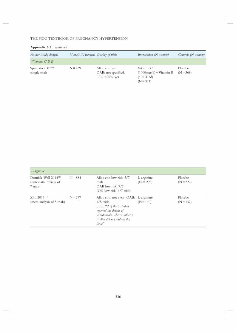
THE FIGO TEXTBOOK OF PREGNANCY HYPERTENSION
336
Appendix 6.2 continued
Author (study design) N trials (N women) Quality of trials Intervention (N women) Controls (N women)
Vitamins C & E
Spinnato 2007138
(single trial)
N = 739 Alloc con: yes.
OAB: not specified.
LFU <20%: yes
Vitamin C
(1000 mg/d) + Vitamin E
(400 IU/d)
(N = 371)
Placebo
(N = 368)
L-arginine
Dorniak-Wall 2014117
(systematic review of
7 trials)
N = 884 Alloc con low risk: 3/7
trials.
OAB low risk: 7/7.
IOD low risk: 4/7 trials.
L-arginine
(N = 228)
Placebo
(N = 222)
Zhu 2013114
(meta-analysis of 5 trials)
N = 277 Alloc con: not clear. OAB:
4/5 trials.
LFU: “2 of the 5 studies reported the details of withdrawals, whereas other 3 studies did not address this issue”
L-arginine
(N = 140)
Placebo
(N = 137)

APPENDICES FOR CHAPTER 6
337
PET Other SGA infants Other
aRR 0.78 [0.61–1.25] Fetal and NND
aRR 1.00 [0.53–1.87]
PTD <37 weeks
aRR 1.15 [0.89–1.50]
PTD <34 weeks
aRR 1.10 [0.65–1.84]
LBW <2500 g
aRR 0.98 [0.71-–1.36]
Very LBW <1500 g
aRR 1.08 [0.58–2.00]
Apgar <4 at 1 min
aRR 0.72 [0.37–1.39]
Apgar <7 at 5 min
aRR 0.72 [0.29–1.77]
Baby died before discharge, or
received NICU care
aRR 0.93 [0.61–1.43]
RDS
aRR 1.11 [0.72–1.71]
Ventilator support
aRR 1.29 (0.60–2.74]
Seizures
aRR 2.08 [0.10–134.08)
PET or eclampsia
OR 0.34 [0.21–0.55]
(1 trial, N = 450)
PTB
OR 0.48 [0.28–0.81]
(1 trial, N = 450)
Change in dBP
Mean difference fixed 3.07
[5.17–(0.98)]
(5 trials, 177)
GA at delivery
Mean difference fixed 1.23
[0.46–1.99]
(5 trials, N = 289)
continued

THE FIGO TEXTBOOK OF PREGNANCY HYPERTENSION
338
Appendix 6.2 continued
Author (study design) N trials (N women) Quality of trials Intervention (N women) Controls (N women)
NO donors
Schleussner 2014141
(single trial of 111 low
and high-risk women
with high-risk women
reported here; see
Appendix 6.1 for data on
low-risk women)
N = 36 Allocation method not
clear
Nitric oxide donor
pentaerithrityl-tetranitrate
(PTN) tablet twice daily
(N = 20)
Placebo
(N = 16)
Alloc con, allocation concealment; APH, antepartum haemorrhage; aRR, adjusted relative risk; BMI, body mass index;
BWt, birth weight; CI, confidence interval; circum, circumference; CS, Caesarean section; ctx, contraction; dBP, diastolic
blood pressure; DHA, docosahexanenoic acid; FH, fetal heart; FHR, fetal heart rate; FM, fetal movement; GA, gestational
age; GH, gestational hypertension; GSH, total glutathione; HELLP, haemolysis, elevated liver enzymes, low platelets; HIE,
hypoxic ischaemic encephalopathy; hs-CRP, high-sensitivity C-reactive protein; HTN, hypertension; IOD, incomplete
outcome data; IOL, induction of labour; LBW; low birth weight; IUGR, intrauterine growth restriction; IVH,
intraventricular haemorrhage; LBW, low birth weight; LFU, loss to follow-up; M&M, morbidity and mortality; Mg,
magnesium; MgSO4, magnesium sulphate; NEC, necrotizing enterocolitis; NICU, neonatal intensive care unit; NND,
neonatal death; NNT, number needed to treat; NR, not reported; OAB, outcome assessment blinding; OR, odds ratio;
PET, pre-eclampsia; PPH, postpartum haemorrhage; PROM, premature rupture of membranes; PTB, preterm birth; RDS,
respiratory distress syndrome; RR, relative risk; SCBU, special care baby unit; SEM, standard error of mean; SGA,
small-for-gestational-age infants; SVP, spontaneous vaginal delivery; TAC, total antioxidant capacity; UA, umbilical artery;
VTE, venous thromboembolism); WMD, weighted mean difference

APPENDICES FOR CHAPTER 6
339
PET Other SGA infants Other
PET/HELLP
6 (30%) vs. 6 (37.5%)
PET <32 weeks
5 (62.5%) vs. 1 (16.7%)
0 vs. 1 (6.2%)
CS
12 (63.2%) vs. 7 (38.9%)
IUGR or perinatal death
7 (35%) vs. 11 (68.8%)
PTD <37 weeks
4 (20%) vs. 7 (43.85)
PTD <32 weeks
2 (10%) vs. 4 (25%)
‡ These results should be interpreted with caution as a large number of severe congenital anomalies and deaths of two sets of
twins (with birth weights <750 g) in the supplemented group likely accounted for the increased risk of death observed.
When deaths due to severe congenital abnormalities were excluded from the meta-analysis, no increased risk of NND was
seen

THE FIGO TEXTBOOK OF PREGNANCY HYPERTENSION
340
Appendix 6.3
GRADE evaluation of best practice points for preventing pre-eclampsia
Recommendation
Prevention of pre-eclampsia in women at low risk
1. Calcium supplementation (of at least 1 g/d, orally) is recommended for women with low dietary intake of calcium
(<600 mg/d, corresponding to less than two dairy servings per day
2. The following are recommended for other established beneficial effects in pregnancy: abstention from alcohol for
prevention of fetal alcohol effects, exercise for maintenance of fitness, periconceptional use of a folate-containing
multivitamin for prevention of neural tube defects, and smoking cessation for prevention of low birth weight and preterm
birth
3. The following may be useful: periconceptional and ongoing use of a folate-containing multivitamin or exercise
4. The following are not recommended for pre-eclampsia prevention, but may be useful for prevention of other
pregnancy complications: prostaglandin precursors or supplementation with magnesium or zinc
5. The following are not recommended: dietary salt restriction during pregnancy, calorie restriction during pregnancy for
overweight women, low-dose aspirin, vitamins C and E, or thiazide diuretics
6. There is insufficient evidence to make a recommendation about the following: a heart-healthy diet, workload or stress
reduction, supplementation with iron with/without folate, vitamin D, pyridoxine, or food rich in flavanoids.
Prevention of pre-eclampsia in women at increased risk
1. The following are recommended for prevention of pre-eclampsia: low-dose aspirin and calcium supplementation (of at
least 1 g/d) for women with low calcium intake
2. Low-dose aspirin (75–100 mg/d) should be administered at bedtime (I-B) and initiated after diagnosis of pregnancy but
before 16 weeks’ gestation (I-B) and may be continued until delivery (I-C)
3. Prophylactic doses of LMWH may be considered in women with previous placental complications (including
pre-eclampsia) to prevent the recurrence of ‘severe’ or early-onset pre-eclampsia, preterm delivery, and/or SGA infants
(I-B)

APPENDICES FOR CHAPTER 6
341
Quality of the evidence* Strength of the recommendation†
High Strong
Low (alcohol), moderate (exercise for fitness), moderate
(folate-containing vitamin), high (smoking cessation)
Strong (for all)
Low (folate-containing vitamin), Very low (exercise) Weak (for both)
Low (prostaglandin precursors), low (magnesium), low (zinc) Weak (for all)
Moderate (salt restriction), moderate (calorie restriction in
obesity), moderate (low-dose aspirin), high (vitamins C & E),
moderate (thiazides)
Strong (for all but aspirin)
Weak ( for aspirin)
Very low (heart healthy diet), very low (workload/stress
reduction), low (iron supplementation), very low (pyridoxine),
low (vitamin D), very low (food rich in flavonoids)
Weak (for all)
High (low-dose aspirin), high (calcium) Strong (for both)
Moderate (for aspirin at bedtime), high (aspirin initiated after
diagnosis of pregnancy but before 16 weeks’ gestation),
moderate (aspirin continued until delivery)
Weak (for aspirin initiated after diagnosis of pregnancy but before 16 weeks’ gestation and for aspirin continued until delivery)Strong (for aspirin at bedtime)
High Weak
continued

THE FIGO TEXTBOOK OF PREGNANCY HYPERTENSION
342
Appendix 6.3 continued
Recommendation
Prevention of pre-eclampsia in women at increased risk
4. The following may be useful: L-arginine (I-B), metformin in PCOS and/or overweight women (1-C), increased rest at
home in the third trimester (I-C), and reduction of workload or stress (III-C)
5. The following may be useful for prevention of other pregnancy complications: prostaglandin precursors (I-B),
magnesium supplementation (I-C), and heparin thromboprophylaxis (I-B)
6. The following are recommended for other established beneficial effects in pregnancy (as discussed for women at low
risk of pre-eclampsia): abstention from alcohol (II-2E) , periconceptional use of a folate-containing multivitamin (I-A),
and smoking cessation (I-E)
7. The following are not recommended: calorie restriction in overweight women during pregnancy (I-D), weight
maintenance in obese women during pregnancy (III-D), antihypertensive therapy specifically to prevent pre-eclampsia
(I-D), vitamins C and E (I-E)
8. There is insufficient evidence to make a recommendation about the usefulness of the following: the heart-healthy diet
(III-L), exercise (I-L), selenium (I-L), garlic (I-L); zinc, pyridoxine, iron (with or without folate), or multivitamins with/
without micronutrients all (III-L)
* The judgements about the quality of evidence is based on the confidence that available evidence reflects the true effect
of the intervention or service. Evidence is considered to be of high quality when the true effect is thought to lie close to
that of the estimate of the effect (e.g., if there is a wide range of studies included in the analyses with no major limitations,
there is little variation between studies, and the summary estimate has a narrow confidence interval). Evidence is
considered to be of moderate quality when the true effect is likely to be close to the estimate of the effect, but there is a
possibility that it is substantially different (e.g., if there are only a few studies and some have limitations but not major
flaws, there is some variation between studies, or the confidence interval of the summary estimate is wide). Evidence is
considered to be of low quality when the true effect may be substantially different from the estimate of the effect (e.g., the
studies have major flaws, there is important variation between studies, or the confidence interval of the summary estimate
is very wide)

APPENDICES FOR CHAPTER 6
343
Quality of the evidence* Strength of the recommendation†
High (L-arginine), high (metformin), high (rest), low
(workload or stress reduction)
Weak (for all)
Moderate (prostaglandin), moderate (magnesium), moderate
(heparin)
Weak (for all)
Moderate (alcohol), moderate (folate), high (smoking) Strong (for all)
Moderate (calorie restriction), moderate (weight maintenance),
high (antihypertensive therapy), moderate (vitamins C and E)
Weak (for calorie restriction and weight maintenance)
Strong (for antihypertensive therapy and vitamins C and E)
Low (heart healthy diet), moderate (exercise), moderate (selenium), moderate (garlic), low (zinc, pyridoxine, iron, multivitamins)
Weak (for all)
† A strong recommendation should be interpreted as meaning that most people in this situation would want the recommended
course of action and only a small number would not. Clinicians should regard the recommendation as applying to most
individuals. Policy-makers can adopt the recommendation as policy in most situations. Adherence to this recommendation
according to the guideline could be used as a quality criterion or performance indicator. A weak recommendation should be
interpreted as meaning that most people in this situation would want the recommended course of action, but many would
not; patients’ values and preferences should be considered in reaching a decision. Decision aids may support people in
reaching these decisions. Policy-making will require substantial debate and involvement of various stakeholders. An
appropriately documented decision making process could be used as a quality indicator


APPENDICES FOR CHAPTER 7
345
Appendix 7.1
GRADE evaluation of best practice points for diet, lifestyle and place of care
Quality ofevidence*
Strength of recommendation†
1. There is insufficient evidence to make a recommendation about the usefulness of
the following: ongoing salt restriction among women with pre-existing hypertension,
new severe dietary salt restriction for women with any HDP, and a heart-healthy diet
or calorie restriction for obese women specifically.
Very low Weak
2. There is insufficient evidence to make a recommendation about the usefulness of:
exercise, workload reduction, or stress reduction.
Very low Weak
3. For women with gestational hypertension (without pre-eclampsia), some bed rest in
hospital (compared with unrestricted activity at home) may be useful to decrease
severe hypertension and preterm birth.
Low Weak
4. For women with pre-eclampsia who are hospitalised, strict bed rest is not
recommended.
Moderate Weak
5. For all other women with HDP, the evidence is insufficient to make a
recommendation about the usefulness of some bed rest, which may nevertheless, be
advised based on practical considerations.
Very low Weak
6. Inpatient care should be provided for women with severe hypertension or severe
pre-eclampsia, however defined.
Low Strong
7. A component of care through hospital day units or home care can be considered
for women with non-severe pre-eclampsia or non-severe (pre-existing or gestational)
hypertension.
Moderate (day units)
Low
(home care)
Strong
8. Transport from community to facility must be considered a responsibility of
women, their communities, and their care providers.
Moderate Strong
* The judgments about the quality of evidence is based on the confidence that available evidence reflects the true effect of
the intervention or service. Evidence is considered to be of high quality when the true effect is thought to lie close to that
of the estimate of the effect (e.g., if there is a wide range of studies included in the analyses with no major limitations, there
is little variation between studies, and the summary estimate has a narrow confidence interval). Evidence is considered to be
of moderate quality when the true effect is likely to be close to the estimate of the effect, but there is a possibility that it is
substantially different (e.g., if there are only a few studies and some have limitations but not major flaws, there is some
variation between studies, or the confidence interval of the summary estimate is wide). Evidence is considered to be of low
quality when the true effect may be substantially different from the estimate of the effect (e.g., the studies have major flaws,
there is important variation between studies, or the confidence interval of the summary estimate is very wide)† A strong recommendation should be interpreted as meaning that most people in this situation would want the recommended
course of action and only a small number would not. Clinicians should regard the recommendation as applying to most
individuals. Policy-makers can adopt the recommendation as policy in most situations. Adherence to this recommendation
according to the guideline could be used as a quality criterion or performance indicator. A weak recommendation should be
interpreted as meaning that most people in this situation would want the recommended course of action, but many would
not; patients’ values and preferences should be considered in reaching a decision. Decision aids may support people in
reaching these decisions. Policy-making will require substantial debate and involvement of various stakeholders. An
appropriately documented decision making process could be used as a quality indicator

THE FIGO TEXTBOOK OF PREGNANCY HYPERTENSION
346
PRECOG II 2009 QLD 2013 NICE 2010 WHO 2011
Dietary & lifestyle change
General comments
Dietary changes For women with chronic
hypertension, ongoing salt
restriction recommended
Exercise
Workload reduction
Stress reduction
Bed rest For women with GH, (any)
NOT recommended
For women
with any HDP,
(strict) NOT
recommended
(Low, Weak)
Place of care
Transfer of care from
midwifery
Assessment in 2 care
setting by health care
provider trained in HDP
Women with GH
Hospital day unit or
antepartum home care
Admit to hospital Women with any HDP and BP
170/110 mmHg
PET & protein-ur3a of 2+, or
protein : creatinine ratio of 30
Women with any
HDP and severe
hypertension or
severe PET
Women with GH
Refer to critical care
setting
Women with any HDP and
severe hypertension or
severe PET with specific
end-organ dysfunction
Appendix 7.2
Diet, lifestyle and place of care recommendations from international guidelines*

APPENDICES FOR CHAPTER 7
347
NVOG 2011 AOM 2012 ACOG 2013 SOGC 2014
For women
with chronic
hypertension,
ongoing salt
restriction
recommended
For women with chronic
hypertension, extreme salt
restriction NOT recommended
For women with chronic
hypertension, weight loss NOT
recommended)
For women with chronic hypertension, insufficient
evidence to recommend ongoing salt restriction or
extreme (new) salt restriction
For women with chronic hypertension and obesity,
insufficient evidence to recommend calorie restriction or
heart healthy diet
For women with chronic
hypertension and BP that is
controlled, ongoing exercise
recommended
For women with any HDP, insufficient evidence to
recommend
For women with any HDP, insufficient evidence to
recommend
Stress reduction for any HDP – insufficient evidence to
recommend
For women with GH or PET
without severe features, (strict)
NOT recommended
(Low, Qualified)
For women with GH, (In hospital vs. unrestricted activity at
home) may be useful
For women with PET, (in hospital) NOT recommended
For women with chronic hypertension or any HDP out of
hospital, Insufficient evidence to recommend
Women
with PET
Consider for women with non-severe pre-existing
hypertension, GH, or PET
Women with any HDP and severe hypertension or “severe”
PET
continued

Appendix 7.2 continued
BP, blood pressure; GH, gestational hypertension; HDP, hypertensive disorder of pregnancy; PET, pre-eclampsia
* SOMANZ 2014 is included in the chapter text, but not in this table adapted from Gillon 201441
ACOG 2013: American College of Obstetricians and Gynecologists, Task Force on Hypertension in Pregnancy.
Hypertension in pregnancy. Report of the American College of Obstetricians and Gynecologists’ Task Force on
Hypertension in Pregnancy. Obstet Gynecol 2013 Nov; 122(5):1122–1131
AOM 2012: Salehi P, Association of Ontario Midwives HDP CPG, Working Group. Hypertensive disorders of pregnancy
(Clinical Practice Guideline 15). 2012; Available: http://www aom on ca/Health_Care_Professionals/Clinical_Practice_
Guidelines/
NICE 2010: National Collaborating Centre for Women’s and Children’s Health (UK). CG107: Hypertension in
pregnancy: The management of hypertensive disorders during pregnancy. NICE: Guidance 2010 Aug
NVOG 2011: Nederlandse Vereniging voor Obstetrie en Gynaecologie. Hypertensieve aandoeningen in de zwangerschap.
2011
QLD 2013: Queensland Maternity and Neonatal Clinical, Guidelines Program. Hypertensive disorders of pregnancy.
2013;MN10.13-V4-R15
PRECOG II: Milne F, Redman C, Walker J, Baker P, Black R, Blincowe J et al. Assessing the onset of pre-eclampsia in
the hospital day unit: summary of the pre-eclampsia guideline (PRECOG II). BMJ 2009; 339:b3129
SOGC 2014: Magee LA, Pels A, Helewa M, Rey E, von Dadelszen P. Diagnosis, evaluation, and management of the
hypertensive disorders of pregnancy. Pregnancy Hypertens 2014;4(2):105–145
WHO 2011: World Health Organization. WHO recommendations for prevention and treatment of pre-eclampsia and
eclampsia. 2011

APPENDICES FOR CHAPTER 8
349
Figure S8.1 Wall chart for treatment of hypertension
Appendix 8.1
Treatment wall charts

THE FIGO TEXTBOOK OF PREGNANCY HYPERTENSION
350
Figure S8.2 Wall chart for intramuscular administration of magnesium sulphate (MgSO4)

APPENDICES FOR CHAPTER 8
351
The three drills listed here for eclampsia/
pre-eclampsia are part of a more comprehensive set
of drills designed for training in South Africa for a
variety of obstetric emergencies, both maternal and
neonatal. The instructions listed for these drills
were adapted, with permission, from the
ESMOE-EOST training manual.
Emergency drills (also known as ‘fire drills’)
provide a simulated experience for participants to
practice problem-solving and decision-making
skills in the management of an obstetric or newborn
emergency, with emphasis on thinking quickly,
reacting (intervening) rapidly, and working as a
team. Also, they provide opportunities to both
revise essential skills and develop confidence in
dealing with emergencies that do not occur
frequently. Enquiries into poor outcomes from
obstetric emergencies revealed the following
common errors:
• Confusion in roles and responsibilities
• Failure to prioritise
• Failure to perform clinical tasks in a structured
coordinated manner
• Poor communication
• Lack of organisational support?
Emergency drills should be carried out in the most
realistic setting possible, such as the labour and
delivery area of a hospital, clinic or maternity
centre, where equipment and supplies are available
for emergency interventions.
Drills should occur every 3 or 6 months. Try to
avoid postponing a drill. The same drills should be
repeated regularly to help health care workers to
‘keep on their toes’. Ask yourself the following
questions when you prepare your schedule:
• How will I/we ensure that all the emergency
drills take place on time?
• How will I/we ensure that all staff are covered
for each topic?
N Day staff?
N Night staff?
• Which skills do I/we not feel confident enough
about?
• What will I/we do to improve my/our skills
before doing the relevant emergency drill with
the rest of the staff?
• How am I/are we going to improve the skills
of staff members not feeling comfortable with
certain skills after an emergency drill has been
conducted?
The drills provided here cover eclampsia and
pre-eclampsia.
• Scenario 1 (Eclampsia), version 1.2
• Scenario 2 (Pre-eclampsia), version 1.2
• Scenario 3 (Pre-eclampsia)
Start with Scenario 1 and proceed in order.
Complete one scenario sheet for EVERY
emergency drill. Please ensure that you complete
the back page of the sheet where all participants
should sign the attendance register.
Start a file for each scenario. Each time you have
completed a drill, add that scenario sheet on top of
the others in that file. Complete the summary
sheets that should be kept in the front of this file.
Prepare for the drill by:
• Familiarising yourself with the requirements in
terms of skills to demonstrate and materials to
prepare for each scenario.
• Read the scenarios carefully before conducting
the emergency drills. You must be comfortable
and familiar with the different scenarios.
• Prepare all materials, medications, equipment,
and manikins. Each scenario sheet has a list of
materials needed for that drill on the first page.
Conducting an emergency drill: You or someone
else should act as the “director” or “conductor”
who facilitates the drill (10–15 min). The different
roles for participants are illustrated on the diagram
found on the first page of each scenario sheet.
Appendix 8.2
Essential Steps in Managing Obstetric Emergencies (ESMOE) –
Emergency Obstetric Simulation Training (EOST)

THE FIGO TEXTBOOK OF PREGNANCY HYPERTENSION
352
Before beginning the drill, instruct the participants
on which role they will play: (1) Team leader, (2)
Helper 1, (3) Helper 2, or (4) Helper 3. The
discoverer can become the team leader or a helper.
• One participant plays the role of patient.
• Invasive procedures are practised on the manikin/
model that serves as the patient’s “body”.
• Procedures such as starting an IV and giving
oxygen should be role played, using the
appropriate equipment.
• A second participant plays the role of the
“discoverer”, while other participants are called
on to assist the provider. It is important that
during different drills, participants change their
roles.
• The idea is to create a simulation that is as near
as possible to a real emergency. Do not prompt
participants as they participate in the drill and
do not interrupt the drill with any discussion.
However, throughout, participants should
demonstrate what they would do and explain
what they are doing and why they are doing it.
The facilitator/director/conductor uses the
scenario sheet to orchestrate the drill. For
information on how to do this and how to score
the drill, see the instructions below under the
heading “How to use the scenario sheets”.
After the drill has been completed, give feedback
(5–10 minutes) about how the team carried out the
emergency drill (clinical skills and skills in
conducting the drill). Facilitate an interactive
discussion with participants who “acted” in the
drill by asking them to:
• Comment on their performance, starting with
strengths and then working towards areas that
need improvement. Include aspects relating to
clinical skills and to teamwork. Ask questions
and encourage participants to ask questions.
Review roles of providers who assist with the
emergency, discuss what order there was, how
the order could be improved and get participants
to understand how to work as a team.
• Then, calculate the score for the drill (see
scenario sheet) and review strengths and areas
needing improvement based on the scenario
sheet (where the column is blank).
Demonstrate each clinical skill with which problems
were identified (clinical and teamwork) (5–10
minutes). Give participants a chance to return and
demonstrate the skill(s) (10–30 minutes). Identify
participants who need additional time to practise
specific skills and arrange time after the session to
work with each one.
Repeat the same drill (10 minutes) to give
participants a chance to put together all of the skills
in a repeat simulation. If you still identify serious
problems with the drill (especially teamwork),
repeat it for a third time.
Participants are evaluated by their ability to
respond to an emergency as a team. Ideally this
score will be 80% or higher. If one member of the
team does well, the whole team will do well. If
one member of the team is not performing to
standard, the entire team will not pass. Participants
must understand that they have a responsibility
to themselves, team members, and women and
newborns.
How to use the scenario sheet
• At the top of each scenario sheet, complete the
line that indicates the topic of the scenario. (The
number of the Module relates only to the
ESMOE-EOST programme and can be ignored
if not part of that programme in South Africa.)
• Complete the page of the sheet which is your
summary record of what has happened in the
drill, with space provided for: the before- and
after-scores; observations and remarks on
follow-up needed (e.g. for improvement of
skills); and an attendance list to be signed by
each participant.
• The scenarios are presented in a table with four
columns:
1. Information provided and questions asked
The scenario starts with information about the patient’s condition written in italics across
the first two columns. Give the information
in the first block in italics to the participant
who will act as “discoverer” (in front of
the other participants) and ask him/her to
repeat the information before starting with
the drill. Provide the rest of the information
in the blocks in italics as the drill progresses.
Each block with information in italics is
followed by a question in bold that you
should ask the participants. There are also
discussion questions to use during feedback
on the initial drill to push participants to
problem-solve and give you an opportunity

APPENDICES FOR CHAPTER 8
353
to provide additional information about
the condition and/or care provision.
2. Key reactions/responses expected from
participants
Key reactions/responses expected from
participants are provided in the second
column of each scenario. The participant
should demonstrate and explain what he/
she is doing and also talk to the patient/
family member of the patient.
Participants are expected to think quickly
and react (intervene) rapidly when you
provide information and ask questions.
3. Before (B)
Complete this during the initial drill.
Put a “Y” or “√” beside each step or task
that the team performed correctly. If the
team did NOT perform the step/task or
did not perform it correctly, leave that
space blank.
After the drill is complete, add up the
number of “Y”s or “√”s and calculate the
score for the drill.
4. After (A)
Complete this during the repeat drill.
Put a “Y” or “√” beside each step or task
that the team performed correctly. If the
team did NOT perform the step/task or
did not perform it correctly, leave that
space blank.
After the drill is complete, add up the
number of “Y”s or “√”s and calculate the
score for the drill.
Table S8.1 contains a fictitious example of a
template for scoring a drill. Overall, the evaluation
Table S8.1 Example of how to score a drill

THE FIGO TEXTBOOK OF PREGNANCY HYPERTENSION
354
of the response to an emergency is graded according
to the sum of two sub-scores: a ‘clinical score’ and
‘execution of drill score’ (made up of scores for A.
Activation/communication skills, B. Response/
team work, C. Sign-out/documentation, and D.
Sequence of activities). To calculate the score, add
the two subs-cores together to get the total score.
You can get the percentage by dividing the score
received by the possible score and then multiplying
by 100. In the example in Table S8.1, the “before”
drill was scored as follows: total score: 23 + 6 = 29
(for clinical + execution) out of a possible total of
43 + 13 = 56 points, giving a percentage score of
(29/56) 100 = 52%. The “after” drill was scored as
32 + 13 = 45 points out of a possible 56 points,
giving a percentage score of (45/56) 100 = 80%, a
passing score.

APPENDICES FOR CHAPTER 8
355
ESMOE-EOST Module 4: Pre-eclampsia and eclampsia: Scenario 1 Version 1.2
Date: ……………………………. Name of health facility: ………………................................
Name(s) of evaluator(s): Signature(s):
…………………………………………………...… ……………………………………………………
…………………………………………………...… ……………………………………………………
BEFORE AFTER SCORE:
NOTES AND FOLLOW-UP
ATTENDANCE
Name Rank Ward Signature
1.
2.
3.
4.
5.
6.
7.
8.

THE FIGO TEXTBOOK OF PREGNANCY HYPERTENSION
356
ESMOE-EOST Module 4: Pre-eclampsia and eclampsia: Scenario 1 Version 1.2
MATERIALS TO BE READY AND AVAILABLE BEFORE STARTING THE SESSION:
General Ask one of the participants to be the patient. Brief the “patient” on the scenario.
Blank clinical notes sheet Clock
Drugs and supplies Syringes and needles IV giving sets and IV pole Test tubes for taking blood samples Ringer’s Lactate
Learning materials Flip charts Module 4
Equipment Sphygmomanometer Stethoscope Pulse oximeter if available A supplemental oxygen source.
o If cylinders are used, check that they have adequate oxygen o Flow meter and air oxygen blender o Tubing
Ambu bag and mask Oxygen mask Oxygen tubing Oropharyngeal airway Yankauer sucker Pinardfetal stethoscope Patellar hammer
CALL FOR CALL FOR HELP!HELP!
Care for babyApprise family
Comfort patient
Equipment & documents
Medications & monitoring
SBAR SBAR approachapproach
Interventions
(Mostly 1 to 3 helpers available)
LOUD INSTRUCTIONS, role allocations LOUD INSTRUCTIONS, role allocations and distribution of functions and distribution of functions
to to ……
For all of the steps, please demonstrate what you would do. Explain what you are doing as you do it and why you are doing it.

APPENDICES FOR CHAPTER 8
357
ESMOE-EOST Module 4: Pre-eclampsia and eclampsia: Scenario 1 Version 1.2
•
•

THE FIGO TEXTBOOK OF PREGNANCY HYPERTENSION
358
ESMOE-EOST Module 4: Pre-eclampsia and eclampsia: Scenario 1 Version 1.2
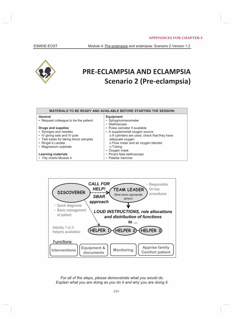
APPENDICES FOR CHAPTER 8
359
ESMOE-EOST Module 4: Pre-eclampsia and eclampsia: Scenario 2 Version 1.2
MATERIALS TO BE READY AND AVAILABLE BEFORE STARTING THE SESSION:
General Request colleague to be the patient
Drugs and supplies Syringes and needles IV giving sets and IV pole Test tubes for taking blood samples Ringer’s Lactate Magnesium sulphate
Learning materials Flip charts Module 4
Equipment Sphygmomanometer Stethoscope Pulse oximeter if available A supplemental oxygen source.
o If cylinders are used, check that they have adequate oxygen o Flow meter and air oxygen blender o Tubing
Oxygen mask Pinard fetal stethoscope Patellar hammer
CALL FOR CALL FOR HELP!HELP!
Apprise familyComfort patient
Equipment & documents Monitoring
SBAR SBAR approachapproach
Interventions
(Mostly 1 to 3 helpers available)
LOUD INSTRUCTIONS, role allocations LOUD INSTRUCTIONS, role allocations and distribution of functions and distribution of functions
to to ……
For all of the steps, please demonstrate what you would do. Explain what you are doing as you do it and why you are doing it.

THE FIGO TEXTBOOK OF PREGNANCY HYPERTENSION
360
ESMOE-EOST Module 4: Pre-eclampsia and eclampsia: Scenario 2 Version 1.2
°
•
•

APPENDICES FOR CHAPTER 8
361
ESMOE-EOST Module 4: Pre-eclampsia and eclampsia: Scenario 2 Version 1.2

THE FIGO TEXTBOOK OF PREGNANCY HYPERTENSION
362
PRE ECLAMPSIAScenario 3
MATERIALS TO BE READY AND AVAILABLE BEFORE STARTING THE SESSION:
General • “Actor” • Blank clinical notes sheet • Clock Drugs and supplies • Syringes and needles • IV giving sets and IV pole • Test tubes for taking blood samples • Ringer’s Lactate Learning materials • Flip charts Module
Equipment • Sphygmomanometer • Stethoscope • Pulse oximeter if available • A supplemental oxygen source.
o If cylinders are used, check that they have adequate oxygen o Flow meter and air oxygen blender o Tubing
• Ambu bag and mask • Oxygen mask • Oxygen tubing • Oropharyngeal airway • Yankauer sucker • Model of larynx • Defibrillator if available
For all of the steps, please demonstrate what you would do. Explain what you are doing as you do it and why you are doing it. As you perform
each step the facilitator will give you the results of your actions
CALL FOR CALL FOR HELP!HELP!
DISCOVERERDISCOVERER TEAM LEADERTEAM LEADER
• Quick diagnosis• Basic management
of patient
Apprise familyComfort patient
Equipment & documents Monitoring
• Responsible for key procedures
SBAR SBAR approachapproach
(Most senior appropriate person)
Interventions
Functions:Functions:
(Mostly 1 to 3 helpers available) HELPER 3HELPER 3HELPER 2HELPER 2
LOUD INSTRUCTIONS, role allocations LOUD INSTRUCTIONS, role allocations and distribution of functions and distribution of functions
to to ……
HELPER 1HELPER 1
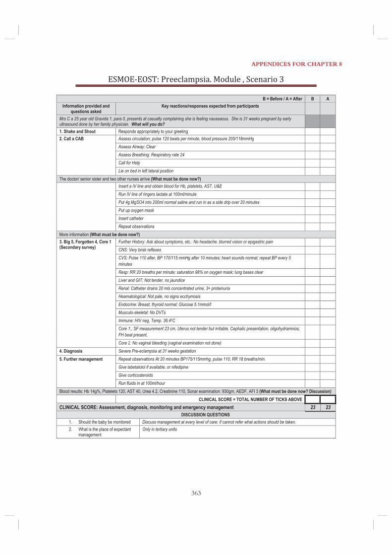
APPENDICES FOR CHAPTER 8
363
B = Before / A = After B A Information provided and
questions asked Key reactions/responses expected from participants
Mrs C a 25 year old Gravida 1, para 0, presents at casualty complaining she is feeling nauseaous. She is 31 weeks pregnant by early ultrasound done by her family physician. What will you do?
1. Shake and Shout Responds appropriately to your greeting 2. Call a CAB Assess circulation; pulse 120 beats per minute, blood pressure 205/118mmHg Assess Airway: Clear Assess Breathing: Respiratory rate 24 Call for Help Lie on bed in left lateral position The doctor/ senior sister and two other nurses arrive (What must be done now?) Insert a IV line and obtain blood for Hb, platelets, AST, U&E
Run IV line of ringers lactate at 100ml/minute Put 4g MgSO4 into 200ml normal saline and run in as a side drip over 20 minutes Put up oxygen mask Insert catheter Repeat observations
More information (What must be done now?) 3. Big 5, Forgotten 4, Core 1 (Secondary survey)
Further History: Ask about symptoms, etc.: No headache, blurred vision or epigastric pain CNS: Very brisk reflexes CVS: Pulse 110 after, BP 170/115 mmHg after 10 minutes; heart sounds normal; repeat BP every 5 minutes
Resp: RR 20 breaths per minute; saturation 98% on oxygen mask; lung bases clear Liver and GIT; Not tender, no jaundice Renal: Catheter drains 20 mls concentrated urine, 3+ proteinuria Heamatological: Not pale, no signs ecchymosis Endocrine: Breast, thyroid normal; Glucose 5.1mmol/l Musculo-skeletal: No DVTs Immune: HIV neg, Temp. 36.40C Core 1:, SF measurement 23 cm, Uterus not tender but irritable, Cephalic presentation, oligohydramnios, FH beat present,
Core 2: No vaginal bleeding (vaginal examination not done) 4. Diagnosis Severe Pre-eclampsia at 31 weeks gestation 5. Further management Repeat observations At 20 minutes BP175/115mmhg, pulse 110, RR 18 breaths/min. Give labetalolol if available, or nifedipine Give corticosteroids Run fluids in at 100ml/hour Blood results: Hb 14g%, Platelets 120, AST 40, Urea 4.2, Creatinine 110, Sonar examination: 930gm, AEDF, AFI 3 (What must be done now? Discussion) CLINICAL SCORE = TOTAL NUMBER OF TICKS ABOVE CLINICAL SCORE: Assessment, diagnosis, monitoring and emergency management 23 23
DISCUSSION QUESTIONS 1. Should the baby be monitored Discuss management at every level of care; if cannot refer what actions should be taken. 2. What is the place of expectant
management Only in tertiary units

THE FIGO TEXTBOOK OF PREGNANCY HYPERTENSION
364
EXECUTION OF DRILL SCORE: Before (B) After (A) A. Activation/Communication skills 1. Appropriate equipment brought (emergency trolley) 2. Discoverer exchanges information with team leader and helpers using SBAR approach 3. Team leader assigns essential roles to helpers (care for the woman, calling a doctor, etc.) 4. Team leader addresses team members by name 5. All observations are communicated clearly and loudly 6. Communication done correctly: instruction repeat instruction inform team when instruction is completed 7. The delegated helper informs the patient and family of what is happening and what will be done for the woman B. Response/Team work 8. Team responds appropriately to team leaders’ instructions 9. Team members cooperate with each other 10. The team determines the disposition of the patient (transfer, plan for further management) C. Sign out/Documentation 11. Person allocated to do documentation 12. Care (actions) completely documented (timing of intervention and administration of drugs) D. Sequence of activities 13. Activities performed in the correct order of priority
EXECUTION OF DRILL SCORE (A-D above) 13 13 EXECUTION OF DRILL SCORE (A-D above): Number of boxes ticked
TOTAL SCORE (CLINICAL SCORE + EXECUTION OF DRILL SCORE) Out of a possible score of 36 36
DISCUSSION POINTS 1. Remember to replace drugs etc (on emergency trolley) 2. Equipment to be cleaned and sterilised appropriately 3. During drill there are no arguments or in-between discussions of opinions on
how something should be done. Only the necessary actions are performed as swiftly and efficiently as possible
4. The environment should be quiet. Only instructions and feedback allowed
5. Observations are given clearly and loudly 6. Importance of the correct sequence of events 7. Documentation

APPENDICES FOR CHAPTER 8
365
Appendix 8.3
GRADE evaluation of best practice points regarding fluids,
drugs and transfusion
Quality of evidence*Strength of recommendation†
Fluid therapy
1. Plasma volume expansion is not recommended for women with
pre-eclampsia.
Moderate Strong
2. IV fluid intake should be minimized to 80 mL/h in women with
pre-eclampsia to avoid pulmonary oedema.
Low Strong
3. Fluid should not be routinely administered to treat oliguria
(<15 mL/h for 6 consecutive hours) for the sole purpose of increasing
urine output.
Very low Weak
4. For treatment of persistent oliguria, neither dopamine nor
furosemide is recommended.
Moderate Strong
Antihypertensive therapy for severe hypertension
1. BP should be lowered to <160 mmHg systolic and <110 mmHg
diastolic.
Low Strong
2. Initial antihypertensive therapy in the hospital setting should be with
nifedipine short-acting (capsules), parenteral hydralazine, or parenteral
labetalol
High Strong
3. Alternative antihypertensive medications include oral methyldopa,
oral labetalol, oral clonidine, oral captopril (only postpartum), or a
nitroglycerin infusion
Moderate (labetalol, nitroglycerin)
Low (clonidine, captopril
postpartum)
Very low (methyldopa)
Weak
4. Refractory hypertension may be treated with sodium nitroprusside Low Weak
5. Nifedipine and MgSO4 can be used contemporaneously Moderate Weak
6. MgSO4 is not recommended solely as an antihypertensive agent. High Strong
7. Continuous FHR monitoring is advised until BP is stable. Very low Weak
Antihypertensive therapy for non-severe hypertension
1. Antihypertensive drug therapy should aim for a dBP of 85 mmHg. High Strong
2. The choice of antihypertensive agent for initial treatment should be
based on characteristics of the patient, contraindications to a particular
drug, and physician and patient preference.
Very low Weak
3. Initial therapy in pregnancy can be with one of a variety of
antihypertensive agents methyldopa, labetalol, other beta-blockers
(acebutolol, metoprolol, pindolol, and propranolol and calcium channel
blockers (nifedipine).
High (methyldopa, labetalol,
nifedipine), moderate (other
beta-blockers)
Strong
continued

THE FIGO TEXTBOOK OF PREGNANCY HYPERTENSION
366
Appendix 8.3 continued
Quality of evidence*Strength of recommendation†
Antihypertensive therapy for non-severe hypertension
4. ACE inhibitors and ARBs should not be used during pregnancy. Moderate Strong
5. Atenolol and prazosin are not recommended prior to delivery. Low Weak
6. Captopril, enalapril, or quinapril may be used postpartum, even
during breastfeeding.
Low Weak
7. There is no compelling evidence that antihypertensive treatment of
hypertension (with labetalol, nifedipine, and probably methyldopa) is
associated with adverse effects on child development.
Low Weak
8. Gestational hypertension and pre-eclampsia may each be associated
with an increase in adverse paediatric neurodevelopmental effects, such
as inattention and externalising behaviours.
Very low Weak
MgSO4
1. MgSO4 is recommended for first-line treatment of eclampsia. High Strong
2. MgSO4 is recommended for eclampsia prevention in women with
severe pre-eclampsia.
High Strong
3. MgSO4 may be considered for eclampsia prevention in women with
non-severe pre-eclampsia based on cost considerations.
Moderate (based on effectiveness;
cost from only one trial)
Strong
4. MgSO4 should be used in standard dosing, usually 4 g IV loading
dose followed by 1 g/h
Moderate Strong
5. Routine monitoring of serum Mg levels is not recommended. Low Strong
6. Phenytoin and benzodiazepines should not be used for eclampsia
prophylaxis or treatment, unless there is a contraindication to MgSO4
or it is ineffective.
High (phenytoin)
Moderate (diazepam)
Strong
7. In women with pre-existing or gestational hypertension, MgSO4
should be considered for fetal neuroprotection in the setting of
imminent preterm birth within the next 24 hours at 33+6 weeks.
Moderate (extrapolating from
preterm labour)
Strong
Therapies for HELLP syndrome
1. Every obstetrical centre should be aware of the local delay between
ordering and receiving platelets units
Very low Strong
2. For a platelet count <20 109/L, platelet transfusion is
recommended, regardless of mode of delivery.
Low Strong
3. For a platelet count 20–49 109/L platelet transfusion is
recommended prior to Caesarean delivery.
Low Strong
4. For a platelet count 20–49 109/L, platelet transfusion should be
considered prior to vaginal delivery if there is excessive active bleeding,
known platelet dysfunction, a rapidly falling platelet count, or
coagulopathy.
Low Weak
continued

APPENDICES FOR CHAPTER 8
367
Appendix 8.3 continued
Quality of evidence*Strength of recommendation†
Therapies for HELLP syndrome
5. For a platelet count of 50 109/L, platelet transfusion should be
considered prior to either Caesarean or vaginal delivery if there is
excessive active bleeding, known platelet dysfunction, a rapidly falling
platelet count, or coagulopathy.
Low Weak
6. We do not recommend corticosteroids for treatment of HELLP until
they have been proven to decrease maternal morbidity
Moderate/Low (RCTs did not
show change in hard outcomes
but underpowered)
Weak
7. We recommend against plasma exchange or plasmapheresis for
HELLP, particularly within the first 4 days postpartum.
Low Strong
Other therapies for treatment of pre-eclampsia (from 2008 document)
1. Women with pre-eclampsia before 34 weeks’ gestation should
receive antenatal corticosteroids for acceleration of fetal pulmonary
maturity.
High Strong
2. Thromboprophylaxis may be considered antenatally among women
with pre-eclampsia who have two or more additional thromboembolic
risk markers, postnatally among women with pre-eclampsia who have
at least one additional thromboembolic risk marker, or postnatally
among women any HDP who were on antenatal bed rest for at least 7
days
Low Weak
FHR, fetal heart rate; GRADE, Grades of Recommendation, Assessment, Development, and Evaluation; HELLP,
Haemolysis, Elevated Liver enzyme, Low Platelet syndrome; MgSO4, magnesium sulphate
* The judgements about the quality of evidence is based on the confidence that available evidence reflects the true effect of
the intervention or service. Evidence is considered to be of high quality when the true effect is thought to lie close to that
of the estimate of the effect (e.g., if there is a wide range of studies included in the analyses with no major limitations, there
is little variation between studies, and the summary estimate has a narrow confidence interval). Evidence is considered to be
of moderate quality when the true effect is likely to be close to the estimate of the effect, but there is a possibility that it is
substantially different (e.g., if there are only a few studies and some have limitations but not major flaws, there is some
variation between studies, or the confidence interval of the summary estimate is wide). Evidence is considered to be of low
quality when the true effect may be substantially different from the estimate of the effect (e.g., the studies have major flaws,
there is important variation between studies, or the confidence interval of the summary estimate is very wide).† A strong recommendation should be interpreted as meaning that most people in this situation would want the
recommended course of action and only a small number would not. Clinicians should regard the recommendation as
applying to most individuals. Policy-makers can adopt the recommendation as policy in most situations. Adherence to this
recommendation according to the guideline could be used as a quality criterion or performance indicator. A weak
recommendation should be interpreted as meaning that most people in this situation would want the recommended course
of action, but many would not; patients’ values and preferences should be considered in reaching a decision. Decision aids
may support people in reaching these decisions. Policy-making will require substantial debate and involvement of various
stakeholders. An appropriately documented decision making process could be used as a quality indicator.

THE FIGO TEXTBOOK OF PREGNANCY HYPERTENSION
368
Appendix 8.4
Sample policy briefs
ANTIHYPERTENSIVE THERAPY– Policy brief
WHY IS ANTIHYPERTENSIVE THERAPY IMPORTANT?Women with severe hypertensiondiastolic in pregnancy (or postpartum), should be treated with antihypertensive therapy. The World Health Organization (WHO) ‘Prevention and Treatment of Pre-eclampsia and Eclampsia’ recommendations strongly recommend use of antihypertensive therapy for treatment of severe hypertension during pregnancy, because treatment of severe hypertension in pregnancy or postpartum decreases maternal risk, particular that of stroke. This has been demonstrated in the‘Confidential Enquiries into Maternal Deaths in the UK (2009-12) and through a similar process in South Africa.
Antihypertensive therapy for non-severe pregnancy hypertension decreases the risk of severe hypertension and the associated risks.
WHICH ANTIHYPERTENSIVE SHOULD BE USED?The choice of antihypertensive agent for initial treatment should be based on characteristics of the patient, contraindications to a particular drug, and physician and patient preference.
Severe hypertension
The antihypertensive agents used most commonly are oral nifedipine (capsules or tablets) or intravenous labetalol or hydralazine. Hydralazine is on the WHO Model List of Essential Medicines (2015) for treatment of severe hypertension, although nifedipine capsules (10mg) are listed as a tocolytic. Both of these medications are on the essential medicines lists of most LMICs.
Oral agents (such as methyldopa or labetalol) are far better-suited to management of severe hypertension than are parenteral agents, especially in resource-limited settings, as they do not require an investment in either physical resources (i.e. intravenous tubing, syringes and needles)or human resources (as administration of parenteral agents is by nurses or often, doctors). Also, oral antihypertensive agents do not mandate the same level of monitoring given a lower risk of dropping the blood pressure quickly and causing fetal compromise.
Non-severe hypertension
Oral methyldopa and oral labetalol are used most frequently for treatment of non-severe hypertension, but there are a wide variety of agents that can be used. Only methyldopa is on the WHO Model List of Essential Medicines (2015) for non-severe pregnancy hypertension.
ACTIONS
Create regulatory efficiency by updating the National Essential Medicines List to include antihypertensive agents for treatment of severe and non-severe hypertension.Identify and promote opportunities where maternal health commodities can be integrated into the broader Health Management Information System.Task-shift to enable midwives, nurses, and lower-level providers to prescribe and safely administer the appropriate antihypertensive agent.Strengthen the treatment at the community level where few centers initiate treatment for pre-eclampsia and eclampsia. Taken in the context of the ‘three delays’ model of maternal mortality, this represents a lost opportunity for improving maternal outcome.Update national protocols and clinical guidelines to facilitate education, training and proper use of antihypertensive therapy among health care workers, particularly those in the community Materials should include a standardised toolkit that includes treatment guidance such as a visual record of monitoring and treatment, as well as other drugs needed for women with severe pre-eclampsia/eclampsia.
“… the report of the‘Confidential Enquiries into Maternal Deaths in the UK’ that covered the hypertensive disorders of pregnancy (2005-8) identified the failure to treat the severe (particularly systolic)hypertension of pre-eclampsia as the single most serious failing in the clinical care of these women who died.”
Above: An instructional chart for Mozambique health workers showing the administration of methyldopa to a woman who has non-severe hypertension in pregnancy

APPENDICES FOR CHAPTER 8
369
MAGNESIUM SULFATE (MgSO4) – Policy brief WHY USE MAGNESIUM SULFATE? Magnesium sulfate (MgSO4) has been on the World Health Organization(WHO) Model List of Essential Medicines since 1996. MgSO4 is recommended by the WHO as the most effective, safe, and low-cost treatment for eclampsia prevention and treatment.
First-line treatment of eclampsiaMgSO4 more than halves the risk of recurrent eclampsia compared with other agents. Also, MgSO4 is associated with a lower risk of both maternal death (compared with either diazepam or a lytic cocktail) and maternal pneumonia and respiratory support (compared with either phenytoin or a lytic cocktail). Although the WHO Model List of Essential Medicines (2015) also lists benzodiazepines as anticonvulsants, they are not recommended for eclampsia treatment. First-line therapy for eclampsia prevention in severe pre-eclampsiaCompared with placebo or no treatment, MgSO4 more than halves the risk of eclampsia among women with pre-eclampsia. MgSO4 may be considered for eclampsia prevention in women with non-severe pre-eclampsia based on cost considerations. In under-resourced settings, 43 women with pre-eclampsia need to be treated to prevent one case of eclampsia, for a cost (in 2001 US dollars) of $456. Prevention of cerebral palsy in infants born before 34 weeks’ gestationMgSO4 decreases the risk of cerebral palsy by 30% when infants are born before 34 weeks’ gestation, based on the results of four trials and over 4,000 babies. MgSO4 may be administered before delivery in the same way as for eclampsia prevention.
ACTIONS
Standardise MgSO4 concentrations in order to address complicated dosage preparations and variations in dosing regimens that are among the major barriers to use of MgSO4according to the Maternal Health Technical Resource Team of the UN Commission on Life-Saving Commodities. The WHO is advocating use of a 50% solution, equivalent to 50 g of MgSO4 in 100mL of solution; as each ampule contains 10mL of solution, each vial contains 5 g of MgSO4. National or institutional essential medicine lists (EMLs) should be updated to include this standardised concentration (50%) of MgSO4.Strengthen supply chains by offering results-based financing of maternal health commodities that rewards providers when they meet performance standards for MgSO4administration.Ensure procurement by providing advanced market commitments or pooled procurements at the regional/central level to incentivise manufacturers to supply MgSO4 and create a more sustainable marketUpdate national protocols to facilitate education, training and proper use of MgSO4 among health care workers, including community midwives and health care workers. Materials should include a standardised toolkit which includes treatment guidance such as visual record of monitoring and treatment, as well as other drugs needed for women with severe pre-eclampsia/eclampsia.Strengthen the treatment at the community level where few centres initiate treatment for pre-eclampsia and eclampsia. Ready-to-use packs comprising a loading dose pack, a maintenance dose pack, of appropriate strengths of MgSO4, in addition to critical items such as lidocaine and a 20mL syringe, could enhance the use of MgSO4 at the community level. Taken in the context of the ‘three delays’ model of maternal mortality, this represents a lost opportunity for improving maternal outcomeDispel myths about the safety of MgSO4. MgSO4 is a safe drug with a very low incidence of severe side effects (1-2%). These are usually attributable to medication errors that would be addressed by standardising use of 50% MgSO4, as discussed above. Even when adverse effects occur, delaying the next scheduled dose is generally sufficient to mitigate the effect.
“…(F)ewer than half of centres initiated treatment for pre-eclampsia (40.0%) or eclampsia (28.0%) prior to transfer to facility (rural Nigeria). Taken in the context of the ‘three delays’ model of maternal mortality (delays in triage, treatment, transport), this represents a lostopportunity for improving maternal outcome”
Above: An instructional chart showing the procedure of im MgSO4administration.© PRE-EMPT Project

THE FIGO TEXTBOOK OF PREGNANCY HYPERTENSION
370
Appendix 8.5
Recommendations for fluids, drugs and transfusion from
international clinical guidelines*
QLD 2013 NICE 2010 WHO 2011
Antihypertensive therapy (antenatally or postnatally)
Antihypertensive therapy for
severe Hypertension (defined)
(160/110 mmHg) (160/110 mmHg)
Treatment recommended For women with
any HDP, treat
severe hypertension
For women with any HDP, treat severe
hypertension (immediately) during
pregnancy or postpartum
For women with any
HDP, treat severe
hypertension
Target BP level (level at
which treatment may be
unchanged; level above which
treatment should be started;
below which treatment should
be decreased if on
antihypertensive therapy)
For women with
any HDP, goal of
160/100 mmHg
For women with any HDP (in critical
care), goal of <150/80–100 mmHg is
recommended
Initial antihypertensive
therapy/first choice
Initial
anti-hypertensive
therapy can be with
one of a variety of
antihypertensive
drugs
Labetalol (oral or IV, hydralazine (IV) or
nifedipine (oral) are recommended for
women in a critical care setting
Consider administration of up to 500 mL of
crystalloid before or with the first dose of
hydralazine IV
Should be based on
clinician’s experience,
cost and local
availability
Alternative antihypertensives
Antihypertensives NOT to
use
Other considerations For women with PET, consider side-effect
profiles if giving treatment other than
labetalol
For women with severe hypertension treated
in critical care setting, monitor response to
treatment, ensure BP falls, identify adverse
effects, and modify treatment according to
response

APPENDICES FOR CHAPTER 8
371
NVOG 2011 AOM 2012 ACOG 2013 SOGC 2014
(160/110 mmHg) (160/
110 mmHg)
(160/110 mmHg) (160/110 mmHg)
For women with any
HDP, treat severe
hypertension
For women with any HDP, treat severe
hypertension
For women with any HDP, treat severe
hypertension
For women with chronic hypertension, goal
of <160/105 mmHg is recommended
For women with PET, goal of
<160/110 mmHg
For women with any HDP, goal of
<160/110 mmHg is recommended
Methyldopa, labetalol
and nifedipine
Labetalol (IV), hydralazine (IV) or
nifedipine (oral capsules) recommended
Nifedipine and MgSO4 can be used
contemporaneously
Alternatives are nitroglycerin (IV)
methyldopa (oral), labetalol (oral),
clonidine (oral), or captopril (oral) only
postpartum
Sodium nitroprusside recommended
for refractory hypertension
ACE inhibitors, ARBs
and direct renin
inhibitors during
pregnancy
MgSO4 as an antihypertensive
FHR monitoring (until stable BP)
recommended
continued

THE FIGO TEXTBOOK OF PREGNANCY HYPERTENSION
372
Appendix 8.5 continued
QLD 2013 NICE 2010 WHO 2011
For non-severe hypertension
Target BP level (level at
which treatment may be
unchanged; level above which
treatment should be started or
increased; level below which
any antihypertensive therapy
should be decreased)
For women with uncomplicated chronic
hypertension, goal of <150/100 mmHg
(without lowering dBP to <80 mmHg) is
recommended
For women with chronic hypertension and
target organ damage, goal of <140/90 mmHg
recommended
Antihypertensives to use For women with chronic hypertension,
choose an agent(s) based on pre-existing
treatment, side-effect profiles and
teratogenicity
For women with GH, offer antihypertensive
medication (other than labetalol) ONLY
after considering side-effect profiles
Alternatives include methyldopa† and
nifedipine
Antihypertensives NOT to
use during pregnancy (and
should be stopped)
For women with any HDP, ACE, ARBs or
chlorothiazide (as they are associated with an
increased risk of major malformations)
For women with chronic hypertension, stop
ACE inhibitors or ARBs in pregnancy
(preferably within 2 working days of
notification of pregnancy) and offer
alternatives
Tell women who took ACE inhibitors or
ARBs “during pregnancy” that these
medications increase the risk of congenital
abnormalities
Tell women who took chlorothiazide
“during pregnancy” that this medication may
increase the risk of congenital abnormalities
and neonatal complications

APPENDICES FOR CHAPTER 8
373
NVOG 2011 AOM 2012 ACOG 2013 SOGC 2014
For any HDP, goal of
<160/110 mmHg is
recommended
For women with uncomplicated chronic
hypertension, goal of 120–159/80–
104 mmHg is recommended
For women with mild GH or PET, goal of
<160/110 mmHg is recommended
For any HDP, goal of 130–155/80–
105 mmHg is recommended
For women with any HDP and a
comorbid condition(s), goal of
<140/90 mmHg is recommended
For women with any
HDP, methyldopa,
labetalol, and
nifedipine
recommended as
agents of first choice
For women with chronic hypertension,
methyldopa, labetalol, and nifedipine
recommended as agents of first choice
For women with any HDP, the choice
of antihypertensive agent should be
based on patient characteristics,
contraindications and physician and
patient preference
For women with any HDP,
methyldopa, labetalol, nifedipine, other
beta-blockers, or other calcium channel
blockers are reasonable as agents of
first choice
Methyldopa, labetalol and nifedipine
are acceptable choices in the 1st
trimester of pregnancy
For women with any
HDP, ACE inhibitors,
ARBs, and direct renin
inhibitors
For women with uncomplicated chronic
hypertension, ACE inhibitors, ARBs, renin
inhibitors, and mineralcorticoid receptor
antagonists are NOT recommended
For women with any HDP, atenolol
and prazosin are not acceptable for use
For women with any HDP, ACE
inhibitors and ARBs (which should be
stopped) – not acceptable for use
continued

THE FIGO TEXTBOOK OF PREGNANCY HYPERTENSION
374
Appendix 8.5 continued
QLD 2013 NICE 2010 WHO 2011
Antenatal corticosteroids
“34 weeks” – FIRST dose “Between 24 and 34 weeks”
For women with PET who are likely to
deliver within 7 days
REPEAT dosing
“35–36 weeks” “35–36 weeks”
For women with PET who are likely to
deliver within 7 days
38+6 weeks gestation and
elective Caesarean

APPENDICES FOR CHAPTER 8
375
NVOG 2011 AOM 2012 ACOG 2013 SOGC 2014
“Before 34 weeks”
For women with any
HDP who are likely to
delivery within 2-10
days
“At 34+0 weeks”
For women with severe PET or
superimposed PET who are receiving
expectant care
“33+6/7 weeks”
For women with severe PET who require
delivery, without delivery being delayed
NOTE: Listed were: uncontrollable severe
hypertension, eclampsia, pulmonary oedema,
abruption placentae, disseminated
intravascular coagulation, evidence of
non-reassuring feta status, intrapartum fetal
demise
“33+6/7 weeks”
For women with severe PET who are stable
enough to have delivery delayed by 48 h
NOTE: Criteria specified were: low platelet
count (<100,000/mL), persistently abnormal
hepatic enzyme concentrations (twice or
more the upper normal values), fetal growth
restriction (less than the fifth percentile),
severe oligohydramnios (amniotic fluid index
<5 cm), reversed end-diastolic flow on
umbilical artery Doppler studies, new-onset
renal dysfunction or increasing renal
dysfunction
“At 34+6 weeks”
For women with PET
“34+6 weeks”
For women with GH who may deliver
within the next 7 days
“Before 33 weeks”
For women with any
HDP, ONLY if first
does were given at <30
weeks and >14 days prior
“34+6 weeks”
For women with any HDP, if first dose
7 days prior
“38+6 weeks”
May consider for women with any
HDP who are delivered by elective
Caesarean
continued

THE FIGO TEXTBOOK OF PREGNANCY HYPERTENSION
376
Appendix 8.5 continued
QLD 2013 NICE 2010 WHO 2011
Antenatal corticosteroids
Fluid administration
(including management of
oliguria)
For women with severe PET, do NOT
administer a fixed IV fluid bolus routinely
prior to neuraxial analgesia
For women with severe PET, limit ongoing
fluid administration to 80 mL/h (unless
ongoing fluid losses)
Treatment of oliguria
Aspects of care for women with pre-existing hypertension
General considerations Advice and treatment should be in line with
‘Hypertension: the management of
hypertension in adults in primary care’
(NICE clinical guideline 34), unless it
specifically differs from recommendations in
this guideline
Schedule additional antenatal consultations
based on needs of woman and baby
Specialist referral (Specialist in hypertensive disorders)
For women with secondary chronic
hypertension
Antihypertensive therapy
– BEFORE pregnancy
For women with
any prior HDP,
preconceptional
advice should be
offered at a formal
postnatal review
Tell women of reproductive age who take
ACE inhibitors or ARBs that these
medications increase the risk of congenital
abnormalities if they are taken “during
pregnancy”
Tell women who take chlorothiazide that
this medication may increase the risk of
congenital abnormalities and neonatal
complications if the drug is taken “during
pregnancy”
Discuss alternatives to ACE inhibitors,
ARBs, and chlorothiazide for women
planning pregnancy

APPENDICES FOR CHAPTER 8
377
NVOG 2011 AOM 2012 ACOG 2013 SOGC 2014
For women with any HDP, do NOT
administer a fixed IV fluid bolus
routinely prior to neuraxial anaesthesia
For women with PET, minimise IV
and oral fluid intake
For women with any HDP, do NOT
routinely administer fluid to treat
oligura (<15 mL/h for 6 consecutive
hours)
For women with any HDP, do NOT
treat oliguria with dopamine or
furosemide
Discuss alternatives to
ACE inhibitors, ARBs
and direct renin
inhibitors for women
planning pregnancy
Women of reproductive age should not be
prescribed ACE inhibitors, ARBs, renin
inhibitors, and/or mineralocorticoid receptor
antagonists unless there is a compelling
indication
Pre-conceptional counselling is
recommended
Discuss alternatives to ACE inhibitors
and ARBs for women planning
pregnancy
Changes to antihypertensive therapy
should be made when planning
pregnancy
continued

THE FIGO TEXTBOOK OF PREGNANCY HYPERTENSION
378
Appendix 8.5 continued
QLD 2013 NICE 2010 WHO 2011
Aspects of care for women with pre-eclampsia
MgSO4
Indications Eclampsia (drug of
first choice)
Eclampsia
Previous eclampsia in women with severe
hypertension or severe PET in a critical care
setting
Severe PET in a critical care setting when
birth is planned within 24 h
Severe PET
NOTE: features listed: severe hypertension
and proteinuria or mild or moderate
hypertension and proteinuria with one or
more of the following: symptoms of severe
headache, problems with vision, such as
blurring or flashing before the eyes, severe
pain just below the ribs or vomiting,
papilloedema, signs of clonus (3 beats), liver
tenderness, HELLP syndrome, platelet count
falling to below 100 109/L, abnormal liver
enzymes (ALT or AST rising to above
70 IU/L)
Eclampsia (drug of first
choice)
Severe PET
Dosage Loading dose: 4 g IV over 5 min
Maintenance dose: 1 g/h for 24 h
Recurrent seizure dose: 2–4 g IV over 5 min
“Full IV or IM”
regimens
When full IV or IM
regimens cannot be
administered,
administer loading
dose and transfer
immediately to a
higher level health care
facility
Monitoring
Alternatives to MgSO4 Do NOT use diazepam, phenytoin or lytic
cocktail in preference to MgSO4 in women
with eclampsia
Do NOT use
diazepam, phenytoin
or lytic cocktail in
preference to MgSO4
in women with
eclampsia or severe
PET

APPENDICES FOR CHAPTER 8
379
NVOG 2011 AOM 2012 ACOG 2013 SOGC 2014
Eclampsia (drug of
first choice)
Severe PET
Mild/moderate PET
(“can be considered”)
Eclampsia (drug of first choice)
Severe PET and superimposed PET with
severe features, intrapartum and postpartum
for severe PET for superimposed PET with
severe features
NOT routinely for PET with BP
<160/110 mmHg and no symptoms
Any PET intraoperatively during Caesarean
delivery
Postpartum, PET with severe hypertension
or new-onset hypertension with headaches/
blurred vision
Eclampsia (drug of first choice)
“Severe PET”
“Non-severe PET” (“can be
considered based on cost
considerations”)
Fetal neuroprotection for women with
any HDP when imminent preterm
birth at 31+6 weeks
Loading dose: “standard dosing”,
usually 4 mg IV
Maintenance dose: “standard dosing”,
usually 1 g/h
Monitor mothers
according to local
protocol
Do NOT routinely monitor serum Mg
levels
Do NOT use diazepam or phenytoin
in preference to MgSO4 in women
with eclampsia or PET
continued

THE FIGO TEXTBOOK OF PREGNANCY HYPERTENSION
380
Appendix 8.5 continued
QLD 2013 NICE 2010 WHO 2011
Plasma volume expansion
Pre-eclampsia NOT recommended for women with severe
PET (unless hydralazine is the antenatal
antihypertensive)
Therapies for HELLP
Platelet transfusion
Corticosteroids NOT recommended NOT recommended
Plasma exchange or
plasmapheresis
* SOMANZ 2014 is included in the chapter text, but not in this table adapted from Gillon187
ACOG 2013: American College of Obstetricians and Gynecologists, Task Force on Hypertension in Pregnancy.
Hypertension in pregnancy. Report of the American College of Obstetricians and Gynecologists’ Task Force on
Hypertension in Pregnancy. Obstet Gynecol 2013 Nov; 122(5):1122–1131
AOM 2012: Salehi P, Association of Ontario Midwives HDP CPG, Working Group. Hypertensive disorders of pregnancy
(Clinical Practice Guideline 15). 2012; Available: http://www aom on ca/Health_Care_Professionals/Clinical_Practice_
Guidelines/
NICE 2010: National Collaborating Centre for Women’s and Children’s Health (UK). CG107: Hypertension in
pregnancy: The management of hypertensive disorders during pregnancy. NICE: Guidance 2010 Aug

APPENDICES FOR CHAPTER 8
381
NVOG 2011 AOM 2012 ACOG 2013 SOGC 2014
NOT recommended for women with
PET
Platelet count <20 109/L
Platelet count 20–49 109/L prior to
Caesarean
Platelet count 20–49 109/L prior to
vaginal delivery if there is: excessive
active bleeding, known platelet
dysfunction, a rapidly falling platelet
count, or coagulopathy
Platelet count 50 109/L if there is:
excessive active bleeding, known
platelet dysfunction, a rapidly falling
platelet count, or coagulopathy
Every obstetrical centre should be
aware of the local delay between
ordering and receiving platelets units
NOT recommended to improve clinical
outcomes
(footnote)
Can be considered if improvement in platelet
count would be useful (footnote)
NOT recommended
NOT recommended
NVOG 2011: Nederlandse Vereniging voor Obstetrie en Gynaecologie. Hypertensieve aandoeningen in de zwangerschap.
2011
QLD 2013: Queensland Maternity and Neonatal Clinical, Guidelines Program. Hypertensive disorders of pregnancy.
2013;MN10.13-V4-R15
SOGC 2014: Magee LA, Pels A, Helewa M, Rey E, von Dadelszen P. Diagnosis, evaluation, and management of the
hypertensive disorders of pregnancy. Pregnancy Hypertens 2014;4(2):105–145
WHO 2011: World Health Organization. WHO recommendations for prevention and treatment of pre-eclampsia and
eclampsia. 2011


APPENDICES FOR CHAPTER 9
383
Appendix 9.1
GRADE evaluation of best practice points regarding timing and
mode of delivery
Quality of evidence*
Strength of recommendation†
Place of delivery
1. All women with a HDP of any type require delivery in a centre that can provide EmONC Low Strong
2. Women with a HDP and serious maternal complications require delivery in a centre capable
of providing CEmONC
Low Strong
Timing of delivery
Women with pre-eclampsia
1. Consultation with an obstetrician is advised in women with pre-eclampsia. (If an obstetrician
is not available in under-resourced settings, consultation with at least a physician is
recommended.)
Low Strong
2. All women with severe pre-eclampsia as defined by the SOGC should be delivered
immediately (either vaginally or by Caesarean), regardless of gestational age‡
Low Strong
3. For women with non-severe pre-eclampsia at <24+0 weeks’ gestation, counselling should
include information about delivery within days as an option
Low Weak
4. For women with non-severe pre-eclampsia at 24+0–33+6 weeks’ gestation, expectant
management should be considered, but only in perinatal centres capable of caring for very
preterm infants
Moderate Weak
5. For women with non-severe pre-eclampsia at 34+0–36+6 weeks’ gestation, expectant
management is advised
High Strong
6. For women with pre-eclampsia at ≥37+0 weeks’ gestation, immediate delivery is
recommended
High Strong
7. For women with non-severe pre-eclampsia complicated by HELLP syndrome at 24+0–34+6
weeks’ gestation, consider delaying delivery long enough to administer antenatal corticosteroids
for acceleration of fetal pulmonary maturity if there is temporary improvement in maternal
laboratory testing (II-2B)
Low Weak
8. All women with HELLP syndrome at ≥35+0 weeks’ gestation should be considered for
delivery within 24 hours
Moderate Strong
Women with gestational hypertension without pre-eclampsia
1. For women with gestational hypertension at <34+0 weeks, expectant management is advised Low Weak
2. For women with gestational hypertension at 34–36+6 weeks, expectant management is
advised
Low Weak
3. For women with gestational hypertension at ≥37+0 weeks’, childbirth within days should be
discussed
Low Weak
continued

THE FIGO TEXTBOOK OF PREGNANCY HYPERTENSION
384
Appendix 9.1 continued
Quality of evidence*
Strength of recommendation†
Timing of delivery
Women with pre-existing hypertension
1. For women with pre-existing hypertension at <34+0 weeks, expectant management is advised Low Weak
2. For women with pre-existing hypertension at 34–36+6 weeks, expectant management is
advised, even if women require antihypertensive therapy
Low Weak
3. For women with uncomplicated pre-existing hypertension who are otherwise well at ≥37+0
weeks’ gestation, delivery should be considered at 38+0–39+6 weeks’ gestation.
Low Weak
Mode of delivery
1. For women with any HDP, vaginal delivery should be considered unless a Caesarean delivery
is required for the usual obstetric indications
Low Strong
2. If vaginal delivery is planned and the cervix is unfavourable, then cervical ripening should be
used to increase the chance of a successful vaginal delivery
Moderate Strong
3. At a gestational age remote from term, women with HDP with evidence of fetal compromise
may benefit from delivery by emergent Caesarean
Low Strong
4. Antihypertensive treatment should be continued throughout labour and delivery to maintain
sBP at <160 mmHg and dBP at <110 mmHg
Low Strong
5. The third stage of labour should be actively managed with oxytocin 5 units IV or 10 units
IM, particularly in the presence of thrombocytopaenia or coagulopathy
Moderate Strong
6. Ergometrine maleate should not be administered to women with any HDP, particularly
pre-eclampsia or gestational hypertension; alternative oxytocics should be considered
Low Strong
CEmONC, comprehensive emergency obstetric and neonatal care; BPP, biophysical profile; GRADE, Grades of
Recommendation, Assessment, Development, and Evaluation; HELLP, haemolysis, elevated liver enzymes, low platelet; HDP,
hypertensive disorder of pregnancy
* The judgements about the quality of evidence are based on the confidence that available evidence reflects the true effect of the
intervention or service. Evidence is considered to be of high quality when the true effect is thought to lie close to that of the
estimate of the effect (e.g., if there are a wide range of studies included in the analyses with no major limitations, there is little
variation between studies, and the summary estimate has a narrow confidence interval). Evidence is considered to be of moderate
quality when the true effect is likely to be close to the estimate of the effect, but there is a possibility that it is substantially different
(e.g., if there are only a few studies and some have limitations but not major flaws, there is some variation between studies, or the
confidence interval of the summary estimate is wide). Evidence is considered to be of low quality when the true effect may be
substantially different from the estimate of the effect (e.g., the studies have major flaws, there is important variation between
studies, or the confidence interval of the summary estimate is very wide).† A strong recommendation should be interpreted as meaning that most people in this situation would want the recommended
course of action and only a small number would not. Clinicians should regard the recommendation as applying to most individuals.
Policy-makers can adopt the recommendation as policy in most situations. Adherence to this recommendation according to the
guideline could be used as a quality criterion or performance indicator. A weak recommendation should be interpreted as meaning
that most people in this situation would want the recommended course of action, but many would not; patients’ values and
preferences should be considered in reaching a decision. Decision aids may support people in reaching these decisions.
Policy-making will require substantial debate and involvement of various stakeholders. An appropriately documented decision
making process could be used as a quality indicator.‡ Severe pre-eclampsia is defined according to Canadian criteria of potentially life-altering complications included within all other
definitions of severe pre-eclampsia. There is consensus that these represent indications for delivery: (1) uncontrolled severe
maternal hypertension; (2) maternal end-organ complications of the central nervous, cardiorespiratory, haematological, renal, or
hepatic systems; or (3) stillbirth or substantial fetal compromise of abruption with maternal/fetal compromise or reversed ductus
venosus A wave. Although these conditions are included in the WHO definition of severe pre-eclampsia, WHO also includes
other criteria for severe pre-eclampsia that are not clear indications for delivery: heavy proteinuria, gestational age <34 weeks, and
evidence of any ‘fetal morbidity’.

APPENDICES FOR CHAPTER 9
385
Appendix 9.2
Timing and mode of delivery according to international clinical
practice guidelines*
See next page – this appendix requires a double-page layout

THE FIGO TEXTBOOK OF PREGNANCY HYPERTENSION
386
PRECOG II 2009 QLD 2013 NICE 2010 WHO 2011
Timing of delivery
General
comments
For women with PET at “before 34
weeks”, consultant obstetric staff
should document maternal and fetal
indications for elective birth
Delivery
indicated
(indications)
For women with any HDP
(regardless of GA) who have
refractory severe hypertension after
BP has been controlled and a course
of antenatal corticosteroids has been
completed (if appropriate)
For women with PET “before 34
weeks” who have a maternal or fetal
indication for delivery (as specified
by the care plan), after discussion
with neonatal and anaesthetic teams,
and after a course of antenatal
corticosteroids has been “given”
For women with PET “after 37+0
wks” who have mild to moderate
hypertension
For women with severe PET
before fetal viability (and at a
GA at which fetus not viable
or unlikely to achieve
viability in 1–2 weeks)
For women with severe PET
“before 34 weeks” or
“between 34 and 36 (+6
days) weeks” who cannot be
monitored or who have
uncontrolled maternal
hypertension, increasing
maternal organ dysfunction
or fetal distress
In women with mild GH or
mild PET “at term”
For women with severe PET
“at term”
Expectant care
ONLY until
steroids have been
administered
For women with HELLP
syndrome “from fetal viability
to 33+6/7 weeks” with stable
maternal and fetal conditions

APPENDICES FOR CHAPTER 9
387
NVOG 2011 AOM 2012 ACOG 2013 SOGC 2014
For women with any
HDP, indications
should be based on
care provider’s own
knowledge and
experience
For women with “severe PET”,
consultation must be undertaken
(by telephone is necessary) with
an obstetrician
For women with
severe PET
(including HELLP)
or any HDP with an
abnormal Doppler
For women with severe PET or
HELLP syndrome before fetal
viability (after maternal stabilisation)
for severe PET for HELLP
For women with PET or
superimposed PET at any GA who
have unstable maternal or fetal
conditions (after maternal
stabilisation)
NOTE: Listed were uncontrollable
severe hypertension, eclampsia,
pulmonary edema, abruption
placentae, disseminated intravascular
coagulation, non-reassuring fetal
status
For women with severe PET or
HELLP syndrome “≥34 0/7 wks”,
or superimposed PET with severe
features “beyond 34 0/7 wks” (after
maternal stabilisation)
For women with mild GH or mild
PET at “≥37 0/7 wks” who have
no severe features
For women with uncomplicated
chronic hypertension, consider
delivery at 38+0/7 to 39+6/7 weeks
For women with GH at ≥37
weeks, delivery within days
should be discussed
For women with PET at <24+0
weeks, delivery should be
discussed as an option
For women with “severe PET”
regardless of GA
For women with PET at ≥37
weeks
For women with HELLP at ≥350
wks
For women with HELLP
syndrome at 24+0–34+6 weeks
If there is temporary
improvement in maternal
laboratory testing
continued

THE FIGO TEXTBOOK OF PREGNANCY HYPERTENSION
388
PRECOG II 2009 QLD 2013 NICE 2010 WHO 2011
Timing of delivery
Expectant care For women with PET “until 34
weeks”
For women with chronic
hypertension at <37 weeks and BP
<160/110mmHg
For women with GH “before 37
wks” who have BP <160/110
mmHg (even on antihypertensive
treatment)
For women with PET at 34+0 to
36+6 weeks who have mild or
moderate hypertension, depending
on maternal and fetal condition, risk
factors and availability of neonatal
intensive care
For women with severe PET
“before 34 weeks” who have
a viable fetus and can be
monitored
For women with severe PE
“between 34 and 36 weeks
(+6 days)” who have a viable
fetus and can be monitored
Care plan For women with severe GH or
PET, write a care plan that includes:
timing and mode of delivery,
indications for delivery, timing of
antenatal corticosteroids, and when
discussion should take place with
neonatology and obstetric
anaesthesia
Evidence
insufficient to
make a
recommendation
about delivery or
expectant care
For women with chronic
hypertension at ≥37 weeks and BP
<160/110 mmHg (“timing of birth
and indications for birth to be
agreed upon between woman and
specialist”)
For women with GH “after 37
weeks” who have BP
<160/110 mmHg (even on
antihypertensive therapy) (“timing
of birth, and maternal and fetal
indications for birth should be
agreed between the woman and the
senior obstetrician”)
Appendix 9.2 continued

APPENDICES FOR CHAPTER 9
389
NVOG 2011 AOM 2012 ACOG 2013 SOGC 2014
For women with severe PET or
severe superimposed PET at
<34+0/7 weeks who have stable
maternal and fetal conditions and
who can be monitored at facilities
with adequate intensive care
resources(Moderate, Strong) for
PET
For women with superimposed
PET “at <37+0/7 weeks” who have
no severe features and stable
maternal and fetal conditions
For women with mild GH or PET
at “<37+0/7 weeks” who have no
severe features or indication for
delivery, and can be monitored
For women with uncomplicated
chronic hypertension at <38 weeks
For women with PET regardless of
the amount or change in
proteinuria
For women with non-severe
PET at 24+0–33+6 weeks, at
centres capable of caring for very
preterm infants
For women with non-severe
PET at 34+0–36+6 weeks
For women with GH at <37
weeks
continued

THE FIGO TEXTBOOK OF PREGNANCY HYPERTENSION
390
Appendix 9.2 continued
PRECOG II 2009 QLD 2013 NICE 2010 WHO 2011
Labour and delivery
Intrapartum care Advice and treatment should be in
line with ‘Intrapartum care:
management and delivery of care to
women in labour’ (NICE clinical
guideline 55), unless it specifically
differs from recommendations in this
guideline
BP management For women with any HDP,
continue antihypertensive therapy
For women any HDP, monitor BP
continuously in women who have
severe hypertension, and hourly in
women who have non-severe
hypertension
Investigations (for PET)
For women with any HDP and
non-severe hypertension, perform
haematological and biochemical tests
using the same criteria as those used
antenatally, whether regional
anaesthesia is being considered
Vaginal or
Caesarean
delivery
For women with any
HDP, Caesarean
should be reserved for
the usual obstetric
indications
If vaginal birth is
planned and the
cervix is unfavourable,
cervical ripening is
recommended
For women with any HDP and
severe hypertenison, severe PET, or
eclampsia, choice should be based
on clinical circumstances and
woman’s preference
Second stage (of
labour)
For women with any HDP with
severe hypertension whose BP is not
meeting treatment targets,
recommend operative birth.
Otherwise, do NOT limit second
stage of labour
Third stage

APPENDICES FOR CHAPTER 9
391
NVOG 2011 AOM 2012 ACOG 2013 SOGC 2014
For women with any HDP,
continue antihypertensive
therapy
For women with PET, platelet
count should be done upon
admission to delivery suite
For women with any HDP,
Caesarean need not be the mode of
delivery, depending on the GA,
fetal presentation, cervical status
and maternal and fetal conditions
For women with any HDP and
evidence of fetal compromise,
Caesarean delivery may be
beneficial
For women with any HDP
without fetal compromise,
Caesarean should be reserved for
the usual obstetric indications
If vaginal birth is planned and the
cervix is unfavourable, cervical
ripening is recommended
For women with any HDP,
active management with
oxytocin recommender
Ergonovine maleate should
NOT be used to prevent/
treat PPH if other suitable
uterotonic drugs are available
For women with any HDP,
active management with
oxytocin (5 units IV or 10 units
IM) recommended
Ergonovine maleate NOT be
used to prevent/treat PPH
continued

Appendix 9.2 continued
* SOMANZ 2014 is included in the chapter text, but not in this table adapted from Gillon 201480
ACOG 2013: American College of Obstetricians and Gynecologists, Task Force on Hypertension in Pregnancy.
Hypertension in pregnancy. Report of the American College of Obstetricians and Gynecologists’ Task Force on
Hypertension in Pregnancy. Obstet Gynecol 2013 Nov; 122(5):1122–1131
AOM 2012: Salehi P, Association of Ontario Midwives HDP CPG, Working Group. Hypertensive disorders of pregnancy
(Clinical Practice Guideline 15). 2012; Available: http://www aom on ca/Health_Care_Professionals/Clinical_Practice_
Guidelines/
NICE 2010: National Collaborating Centre for Women’s and Children’s Health (UK). CG107: Hypertension in
pregnancy: The management of hypertensive disorders during pregnancy. NICE: Guidance 2010 Aug
NVOG 2011: Nederlandse Vereniging voor Obstetrie en Gynaecologie. Hypertensieve aandoeningen in de zwangerschap.
2011
QLD 2013: Queensland Maternity and Neonatal Clinical, Guidelines Program. Hypertensive disorders of pregnancy.
2013;MN10.13-V4-R15
PRECOG II: Milne F, Redman C, Walker J, Baker P, Black R, Blincowe J et al. Assessing the onset of pre-eclampsia in
the hospital day unit: summary of the pre-eclampsia guideline (PRECOG II). BMJ 2009; 339:b3129
SOGC 2014: Magee LA, Pels A, Helewa M, Rey E, von Dadelszen P. Diagnosis, evaluation, and management of the
hypertensive disorders of pregnancy. Pregnancy Hypertens 2014;4(2):105–145
WHO 2011: World Health Organization. WHO recommendations for prevention and treatment of pre-eclampsia and
eclampsia. 2011

APPENDICES FOR CHAPTER 10
393
Appendix 10.1
Randomised controlled trials (RCTs) of prevention of the hypertensive
response to intubation in women with pre-eclampsia
AuthorStudy type Population N Methods (n women) Results Other
Rout &
Rocke
199054
RCT ‘Severe’
pre-eclampsia
40 Alfentanil 10 g/kg 3 min
before induction (N = 20)
Fentanyl 2.5 g/kg 1 min
before induction (N = 20)
All induced with lidocaine,
etomidate 0.3 mg/kg,
succinylcholine
Both groups had HR
after intubation.
No significant difference
MAP before induction and
after intubation
9 fentanyl, 8 alfentanil
received magnesium
2 alfentanil group had
no treatment for
hypertension, rest
had various
anti-hypertensives
Hood et al. 198557
RCT ‘Severe’
pre-eclampsia
19 Nitroglycerin infusion
200 g/mL (N = 9)
Control (N = 10)
Induction: thiopental
4 mg/kg, succinylcholine
Maximum HR occurred 2
min after intubation in both
groups
Nitroglycerin: MAP 20%
before induction – 2 min
after intubation but
significantly more in
control group
All received magnesium
preoperatively
No information re
anti-hypertensive
medication
Ramanathan
et al. 198858
RCT ‘Mild-moderate’
pre-eclampsia
25 Labetalol 20 mg – then
10 mg increments to total
1 mg/kg (N = 15) –
administered until
DBP<100 or MAP 20%
from baseline
Control (N = 10)
Induced 10 min after BP
stabilised
Induction: thiopental
4 mg/kg, succinylcholine
Baseline values similar
Labetalol mean MAP &
HR before induction
After intubation MAP significantly both groups
but significantly > control
Mean HR significantly
more in control group
All received magnesium
pre-operatively
No antihypertensive
medication
3 subjects in labetalol
group did not achieve
BP goals in spite of
maximum dose
Allen et al. 199153
RCT ‘Moderate’
(N = 5) to
‘severe’ (N = 64)
pre-eclampsia
69 Lidocaine 1.5 mg/kg
(N = 21)
Magnesium 40 mg/kg
(N = 24)
Alfentanil 10 g/kg
(N = 24)
Study drug given after
induction with thiopental
5 mg/kg. Succinylcholine
given after study drug
SBP, dBP, MAP post
intubation > lidocaine group
compared to other 2 groups
10 subjects did not
receive antihypertensive
therapy – various
antihypertensives used
in other 59 – many in
combination
Some in other groups
received magnesium
No control group so
difficult to determine
effect of lidocaine but
authors felt should not
be used alone
continued
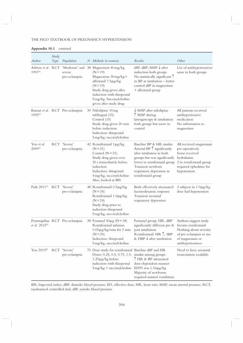
THE FIGO TEXTBOOK OF PREGNANCY HYPERTENSION
394
Appendix 10.1 continued
AuthorStudy Type Population N Methods (n women) Results Other
Ashton et al. 199156
RCT ‘Moderate’ and
severe
pre-eclampsia
38 Magnesium 40 mg/kg
(N = 19)
Magnesium 30 mg/kg +
alfentanil 7.5 g/kg
(N = 19)
Study drug given after
induction with thiopental
5 mg/kg. Succinylcholine
given after study drug
sBP, dBP, MAP after
induction both groups
No statistically significant in BP at intubation – better
control sBP in magnesium
+ alfentanil group
Use of antihypertensives
same in both groups
Kumar et al. 199359
RCT Pre-eclampsia 30 Nifedipine 10 mg
sublingual (15)
Control (15)
Study drug given 20 min
before induction
Induction: thiopental
5 mg/kg, succinylcholine
MAP after nifedipine
MAP during
laryngoscopy & intubation
both groups but more in
control
All patients received
antihypertensive
medication
No information re.
magnesium
Yoo et al. 200950
RCT ‘Severe’
pre-eclampsia
42 Remifentanil 1 g/kg
(N = 21)
Control (N = 21)
Study drug given over
30 s immediately before
induction
Induction: thiopental
4 mg/kg, succinylcholine
Also, looked at BIS
Baseline BP & HR similar
Arterial BP significantly
after intubation in both
groups but was significantly
lower in remifentanil group
Transient newborn
respiratory depression in
remifentanil group
All received magnesium
pre-operatively
Some received
hydralazine
2 in remifentanil group
required ephedrine for
hypotension
Park 201151 RCT ‘Severe’
pre-eclampsia
48 Remifentanil 0.5 g/kg
(N = 24)
Remifentanil 1.0 g/kg
(N = 24)
Study drug prior to
induction thiopental
5 mg/kg, succinylcholine
Both effectively attenuated
haemodynamic response
Transient neonatal
respiratory depression
3 subjects in 1.0 g/kg
dose had hypotension
Pournajafian
et al. 201252
RCT Pre-eclampsia 38 Fentanyl 50 g (N = 18)
Remifentanil infusion
0.05 g/kg/min for 3 min
(N = 20)
Induction: thiopental
5 mg/kg, succinylcholine
Fentanyl group: HR, dBP
significantly different pre &
post intubation
Remifentanil: HR SBP
& DBP after intubation
Authors suggest study
favours remifentanil
Nothing about severity
of pre-eclampsia or use
of magnesium or
antihypertensives
Yoo 201355 RCT ‘Severe’
pre-eclampsia
75 Dose study for remifentanil
Doses: 0.25, 0.5, 0.75, 1.0,
1.25 g/kg before
induction with thiopental
5 mg/kg + succinylcholine
Baseline sBP and HR
similar among groups
HR & BP attenuated
dose-dependent manner
ED95 was 1.34 g/kg
Majority of newborns
required assisted ventilation
Need to have neonatal
resuscitation available.
BIS, bispectral index; dBP, diastolic blood pressure; ED, effective dose; HR, heart rate; MAP, mean arterial pressure; RCT,
randomised controlled trial; sBP, systolic blood pressure

APPENDICES FOR CHAPTER 10
395
Appendix 10.2
Anaesthesia for Caesarean delivery in women with pre-eclampsia
See next page – this appendix requires a double-page layout
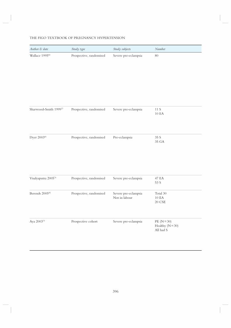
THE FIGO TEXTBOOK OF PREGNANCY HYPERTENSION
396
Author & date Study type Study subjects Number
Wallace 199580 Prospective, randomised Severe pre-eclampsia 80
Sharwood-Smith 199977 Prospective, randomised Severe pre-eclampsia 11 S
10 EA
Dyer 200381 Prospective, randomised Pre-eclampsia 35 S
35 GA
Visalyaputra 200576 Prospective, randomised Severe pre-eclampsia 47 EA
53 S
Berends 200585 Prospective, randomised Severe pre-eclampsia
Not in labour
Total 30
10 EA
20 CSE
Aya 200373 Prospective cohort Severe pre-eclampsia PE (N = 30)
Healthy (N = 30)
All had S

APPENDICES FOR CHAPTER 10
397
Methods Results
3 groups – GA (N = 26), EA (N = 27), CSE (N = 27)
All received magnesium, intermittent IV hydralazine as
needed
IV fluid limited to 60 mL/h but did preload
GA: IV hydralazine – dBP 100 mmHg preintubation;
lidocaine, NTG; RSI: thiopental 4–5 mg/kg,
succinylcholine – nitrous oxide, oxygen, isoflurane
EA: preload 1000 mL LR; incremental 2% lidocaine or 3%
chloroprocaine
CSE: preload 1000 mL LR; hyperbaric 0.75% bupivacaine;
epidural supplements 3 mL boluses 0.5% bupivacaine
Ephedrine 5 mg doses for hypotension S & EA groups
GA: shortest induction to skin incision time (3 min vs. 25–35
min)
Hypotension requiring ephedrine similar in CSE and EA
BP significantly over time in all groups
IV fluids > EA & CSE groups than GA group
Concluded: all techniques acceptable for CS
All required antihypertensive therapy
S: 2.75 mL hyperbaric 0.5% bupivacaine
EA: 4 mL + 16 mL 0.5% bupivacaine
Preload 250 mL LR, otherwise fluids restricted to
80 mL/h + losses
Ephedrine – 6 mg increments if hypotension
Poor anaesthesia in EA group
Ephedrine use similar
All had non-reassuring FHR trace
Severe PE had magnesium sulphate
Dihydralazine IV used for BP control
GA: Preload <750 mL LR; thiopental 5 mg/kg then
30–45 mg/kg magnesium sulphate to ablate hypertensive
response to intubation, followed by succinylcholine; nitrous
oxide, oxygen isoflurane
S: Preload <750 mL LR; 1.8 mL hyperbaric 0.5%
bupivacaine + 10 g fentanyl
Groups similar at baseline
HR, sBP, dBP, MAP significantly lower in S group
> umbilical arterial base deficit & lower median umbilical
arterial pH in S group
More ephedrine used in S group
Questioned the clinical significance of this
EA: 18–23 mL 2% lidocaine with epinephrine
S: 2.2 mL 0.5% hyperbaric bupivacaine + morphine
Hypothesis MAP 10 mm < S group during delivery
Hypotension > S than EA (51% vs. 23%)
Duration short both groups
More ephedrine in spinal group
Compared EA vs. CSE with 2 prophylactic regimens
EA + fluid preload (N = 10) – preload 10 mL/kg RL
CSE + fluid preload (N = 10) – preload 10 mL/kg RL
CSE prophylactic ephedrine (N = 10) 15 mg ephedrine in
150 mL LR given over 5 min
Primary outcome: incidence hypotension
Shorter time induction to surgery both CSE groups
7 EA group needed supplemental analgesics – only 2 CSE
groups
MAP similar between groups during surgery
More ephedrine, < LR in CSE prophylactic ephedrine group
No hypertension
All had magnesium
After each PE enrolled the next normotensive was the
control
Preload 1500–2000 mL LR
S: hyperbaric 0.5% bupivacaine 8–12 mg + sufentanil/
morphine
Hypotension treated with ephedrine
PE group – more nulliparas, younger gestational age, less IV
fluid, 12 had magnesium, 11 had nicardipine, 2 urapidil, 8
had both magnesium & nicardipine
Bupivacaine > PE group;
in dBP, MAP < PE group; sBP similar both groups
Ephedrine 16.6% PE vs. 53.3% control
continued

THE FIGO TEXTBOOK OF PREGNANCY HYPERTENSION
398
Appendix 10.2 continued
Author & date Study type Study subjects Number
Aya 200583 Case–controlled study Severe pre-eclampsia
Healthy controls
PE 65
Control 71
Tihtonen 200675 Prospective Pre-eclampsia
Healthy
6 severe, 4 mild or moderate PE
10 healthy
Clark 200574 Observational Normotensive
Severe pre-eclampsia
40–20/group
Dyer 200865 Observational Severe pre-eclampsia 15 S
Hood 199978 Retrospective Severe pre-eclampsia
Not in labour
103 S
35 EA
Chiu 200379 Retrospective Pre-eclampsia 70 S
51 EA
RCT, randomised controlled trial; GA, general anaesthesia; EA, epidural; CSE, combined spinal-epidural; IV, intravenous;
dBP, diastolic BP; NTG, nitroglycerin; RSI, rapid sequence induction; LR, lactated Ringer’s; CS, Caesarean delivery; S,
spinal; OB, obstetrician; BP, blood pressure; MAP, mean arterial pressure; FHR, fetal heart rate; sBP, systolic BP; CO,
cardiac output; HR, heart rate; bpm, beats per minute; SVR, systemic vascular resistance; SVRI, systemic vascular
resistance index; SI, stroke index; CI, cardiac index

APPENDICES FOR CHAPTER 10
399
Methods Results
All subjects were preterm (<35 weeks) patients
Consecutive enrollment
Pharmacologic treatment of BP before inclusion
Nicardipine was 1st line antihypertensive
All were on magnesium sulphate
Neonatal weight 1100–1900 g
Preload 1500–2000 mL LR over 20 min
Spinal anaesthesia (8–12 mg hyperbaric bupivacaine,
sufentanil, morphine)
Primary outcome – 25% difference in hypotension
All had effective anaesthesia
PE group: heavier, more nulliparas, 7 had only magnesium,
11 only nicardipine, 18 both drugs
Hypotension treated with ephedrine < in PE group
Magnitude sBP, dBP and MAP similar – time to nadir of
MAP longer in PE group
PE group less ephedrine
Risk of hypotension almost 2 times < PE group
PE: 4 received labetalol
All had whole-body impedance cardiography
S = 2.4–2.7 mL 0.5% hyperbaric bupivacaine
Hypotension treated with ephedrine infusion
Baseline: mean MAP and SVRI were significantly in PE, SI
and CI significantly lower in PE
S group: SVRI & MAP Hypotension: 30% PE vs. 80% controls
Ephedrine MAP & SVRI both groups
Concluded PE a state of low CO, high SVR. At delivery PE
could not increase SI
All spinal anaesthesia: 2.5 mL hyperbaric 0.5%
bupivacaine + fentanyl 12.5 g
Preload 250 mL
Primary outcome: Difference in ephedrine use of 11 mg
with more used in normotensives
All PE subjects were stabilised on antihypertensive drugs
before study
Mean ephedrine in normotensives 27.911.6 mg vs. PE
group 16.3515.0 mg (p < 0.01)
All received magnesium sulphate
IV-300–500 mL hydroxyethyl starch before IV dihydralazine
then crystalloid 120 mL/h
Measured cardiac output with LiDCOplus
S: co-hydration 10 mL/kg LR; 2.0 mL hyperbaric 0.5%
bupivacaine + fentanyl 10 g
Hypotension: 50 g phenylephrine every minute until
within 20% baseline; if MAP 30% from baseline 100 g
phenylephrine given
If CO didn’t respond with target MAP then ephedrine 5 or
10 mg was given
If HR <55 bpm + hypotension then 0.5 mg atropine and
10 mg ephedrine were given
All patients were haemodynamically stable
Mean baseline SVR was above normal in spite of
antihypertensive therapy
Mean baseline CO was normal
CO changes intraoperatively were clinically insignificant
Induction of S was followed by significant in MAP and
SVR
Main effect of S was modest afterload reduction
7 did not require phenylephrine before delivery; only 1
required 100 g before delivery, 7 received 50 g
Of the 8 who had phenylephrine pre-delivery, 4 also required
it after delivery
5 required ephedrine pre-delivery
Database reviewed
Ephedrine, IV fluids at discretion of anaesthetist
Antihypertensive therapy discretion of OB or anaesthetist
EA more likely to receive antihypertensive therapy
More IV fluids S group
Ephedrine use similar
BP similar both groups
5 year review: Mild, moderate, severe PE
Not in labor having CS
S = 1.7–2.5 mL 0.5% hyperbaric bupivacaine
EA: Incremental boluses 3–10 mL 0.5% bupivacaine with
50–100 g fentanyl
Labetalol most commonly used antihypertensive, then
hydralazine
No magnesium in mild or moderate group
BP similarly S and EA
Ephedrine use similar EA & S groups & in mild/moderate or
severe PE
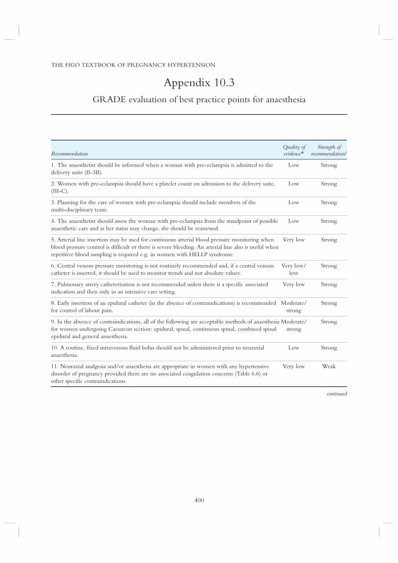
THE FIGO TEXTBOOK OF PREGNANCY HYPERTENSION
400
Appendix 10.3
GRADE evaluation of best practice points for anaesthesia
RecommendationQuality of evidence*
Strength of recommendation†
1. The anaesthetist should be informed when a woman with pre-eclampsia is admitted to the
delivery suite (II-3B).
Low Strong
2. Women with pre-eclampsia should have a platelet count on admission to the delivery suite.
(III-C).
Low Strong
3. Planning for the care of women with pre-eclampsia should include members of the
multi-disciplinary team.
Low Strong
4. The anaesthetist should assess the woman with pre-eclampsia from the standpoint of possible
anaesthetic care and as her status may change, she should be reassessed.
Low Strong
5. Arterial line insertion may be used for continuous arterial blood pressure monitoring when
blood pressure control is difficult or there is severe bleeding. An arterial line also is useful when
repetitive blood sampling is required e.g. in women with HELLP syndrome.
Very low Strong
6. Central venous pressure monitoring is not routinely recommended and, if a central venous
catheter is inserted, it should be used to monitor trends and not absolute values.
Very low/
low
Strong
7. Pulmonary artery catheterisation is not recommended unless there is a specific associated
indication and then only in an intensive care setting.
Very low Strong
8. Early insertion of an epidural catheter (in the absence of contraindications) is recommended
for control of labour pain.
Moderate/
strong
Strong
9. In the absence of contraindications, all of the following are acceptable methods of anaesthesia
for women undergoing Caesarean section: epidural, spinal, continuous spinal, combined spinal
epidural and general anaesthesia.
Moderate/
strong
Strong
10. A routine, fixed intravenous fluid bolus should not be administered prior to neuraxial
anaesthesia.
Low Strong
11. Neuraxial analgesia and/or anaesthesia are appropriate in women with any hypertensive
disorder of pregnancy provided there are no associated coagulation concerns (Table 6.6) or
other specific contraindications.
Very low Weak
continued

APPENDICES FOR CHAPTER 10
401
Appendix 10.3 continued
aPTT, activated partial thromboplastin time; ASA, aspirin; GRADE, Grades of Recommendation, Assessment,
Development, and Evaluation; HELLP, Haemolysis, Elevated Liver enzyme, Low Platelet syndrome; LMWH,
low-molecular weight heparin; UFH, unfractionated heparin
* The judgements about the quality of evidence is based on the confidence that available evidence reflects the true effect of
the intervention or service. Evidence is considered to be of high quality when the true effect is thought to lie close to that of
the estimate of the effect (e.g., if there is a wide range of studies included in the analyses with no major limitations, there is
little variation between studies, and the summary estimate has a narrow confidence interval). Evidence is considered to be
of moderate quality when the true effect is likely to be close to the estimate of the effect, but there is a possibility that it is
substantially different (e.g., if there are only a few studies and some have limitations but not major flaws, there is some
variation between studies, or the confidence interval of the summary estimate is wide). Evidence is considered to be of low quality when the true effect may be substantially different from the estimate of the effect (e.g., the studies have major flaws,
there is important variation between studies, or the confidence interval of the summary estimate is very wide).† A strong recommendation should be interpreted as meaning that most people in this situation would want the recommended
course of action and only a small number would not. Clinicians should regard the recommendation as applying to most
individuals. Policy-makers can adopt the recommendation as policy in most situations. Adherence to this recommendation
according to the guideline could be used as a quality criterion or performance indicator. A weak recommendation should be
interpreted as meaning that most people in this situation would want the recommended course of action, but many would
not; patients’ values and preferences should be considered in reaching a decision. Decision aids may support people in
reaching these decisions. Policy-making will require substantial debate and involvement of various stakeholders. An
appropriately documented decision making process could be used as a quality indicator.

THE FIGO TEXTBOOK OF PREGNANCY HYPERTENSION
402
Appendix 10.4
Recommendations for anaesthesia from international guidelines127
PRECOG II 2009 QLD 2013 NICE 2010 WHO 2011 NVOG 2011
General principles

APPENDICES FOR CHAPTER 10
403
AOM 2012 ACOG 2013 SOGC 2014
For women with PET,
neuraxial analgesia or
anaesthesia (spinal or
epidural) is recommended
(Moderate, Strong)
For women with PET who are admitted to delivery suite, the anaesthesiologist
should be informed
(Low, Strong)
Early insertion of an epidural catheter for analgesia is recommended
(Moderate, Strong)
Acceptable methods of anaesthesia include epidural, spinal, combined
spinal-epidural and general anaesthesia
(Moderate, Strong)
For women with any HDP, neuraxial analgesia and/or anaesthesia are
appropriate:
a) With PET, provided there are no associated coagulation concerns.
(Low, Strong);
b) With a platelet count 75109/L
(Very low, Weak);
c) Taking low-dose ASA in the presence of an adequate platelet count.
(Moderate/High, Strong);
d) Receiving UFH in a dose of 10,000 IU/d subcutaneously, 4 h after the last
dose and possibly IV after the last dose without any delay (Very low, Weak);
e) Receiving UFH in a dose of 10,000 IU/d subcutaneously if they have a
normal aPTT 4 h after the last dose (Very low, Weak);
f) Receiving IV heparin in a therapeutic dose if they have a normal aPTT 4 h
after the last dose (Very low, Weak); or
g) Receiving low-molecular weight heparin (LMWH) a minimum of 10–12 h
after a prophylactic dose, or 24 h after a therapeutic dose
(Very low, Weak)
For women with any HDP, phenylephrine or ephedrine may be used to treat
hypotension during neuraxial anaesthesia
(Moderate, Strong)
continued

THE FIGO TEXTBOOK OF PREGNANCY HYPERTENSION
404
Appendix 10.4 continued
PRECOG II 2009 QLD 2013 NICE 2010 WHO 2011 NVOG 2011
General principles
Fluid administration
(including management
of oliguria)
For women with severe
PET, do NOT administer a
fixed IV fluid bolus routinely
prior to neuraxial analgesia
For women with severe
PET, limit ongoing fluid
administration to 80 mL/h
(unless ongoing fluid losses)
Treatment of oliguria
Anesthesia – monitoring
Invasive
haemodynamic
monitoring
ACOG, American College of Obstetricians and Gynecologists; ASA, aspirin; BP, blood pressure; GA, gestational age;
GH, gestational hypertension; BPP, good practice point; HDP, hypertensive disorders of pregnancy; HELLP, haemolysis,
elevated liver enzyme, low platelet syndrome; LMWH, low molecular weight heparin; MgSO4, magnesium sulphate;
NICE, National Institute for Health and Clinical Excellence; NVOG, Nederlandse Vereniging voor Obstetrie en
Gynaecologie; PET, pre-eclampsia; PRECOG, pre-eclampsia community guideline; QLD, Queensland Maternity and
Neonatal Clinical Guidelines Program; SOGC, Society of Obstetricians and Gynaecologists of Canada; UFH,
unfractionated heparin; WHO, World Health Organization
ACOG 2013: American College of Obstetricians and Gynecologists, Task Force on Hypertension in Pregnancy.
Hypertension in pregnancy. Report of the American College of Obstetricians and Gynecologists’ Task Force on
Hypertension in Pregnancy. Obstet Gynecol 2013 Nov; 122(5):1122–1131
AOM 2012: Salehi P, Association of Ontario Midwives HDP CPG, Working Group. Hypertensive disorders of pregnancy
(Clinical Practice Guideline 15). 2012; Available: http://www aom on ca/Health_Care_Professionals/Clinical_Practice_
Guidelines/

APPENDICES FOR CHAPTER 10
405
AOM 2012 ACOG 2013 SOGC 2014
For women with any HDP, do NOT administer a fixed IV fluid bolus routinely
prior to neuraxial anaesthesia
(Low, Strong)
For women with PET, minimize iv and oral fluid intake
(Low, Strong)
For women with any HDP, do NOT routinely administer fluid to treat oligura
(<15 mL/h for 6 consecutive hours)
(Very low, Weak)
For women with any HDP, do NOT treat oliguria with dopamine or furosemide
(Moderate, Strong)
For women with severe
PET, do NOT routinely
use invasive
haemodynamic
monitoring
(Low, Qualified)
For women with any HDP, do NOT routinely use central venous pressure
monitoring
(Very low/Low, Strong)
If a central venous monitoring is used, trends (and not absolute values) should be
monitored
(Very low/Low, Strong)
For women with any HDP, an arterial line may be used when BP is difficult to
control or there is severe bleeding
(Very low, Strong)
For women with any HDP, pulmonary artery catheterisation is NOT
recommended unless there is a specific indication
(Very low, Strong)
If used, a pulmonary catheter should be used only in a critical care setting
(Very low, Strong)
NICE 2010: National Collaborating Centre for Women’s and Children’s Health (UK). CG107: Hypertension in
pregnancy: The management of hypertensive disorders during pregnancy. NICE: Guidance 2010 Aug
NVOG 2011: Nederlandse Vereniging voor Obstetrie en Gynaecologie. Hypertensieve aandoeningen in de zwangerschap.
2011
QLD 2013: Queensland Maternity and Neonatal Clinical, Guidelines Program. Hypertensive disorders of pregnancy.
2013;MN10.13-V4-R15
PRECOG II: Milne F, Redman C, Walker J, Baker P, Black R, Blincowe J et al. Assessing the onset of pre-eclampsia in
the hospital day unit: summary of the pre-eclampsia guideline (PRECOG II). BMJ 2009; 339:b3129
SOGC 2014: Magee LA, Pels A, Helewa M, Rey E, von Dadelszen P. Diagnosis, evaluation, and management of the
hypertensive disorders of pregnancy. Pregnancy Hypertens 2014;4(2):105–145
WHO 2011: World Health Organization. WHO recommendations for prevention and treatment of pre-eclampsia and
eclampsia. 2011


APPENDICES FOR CHAPTER 11
407
MULTIPLE CHOICE QUESTIONS
1. When does blood pressure reach its peak
during the postpartum period?
a. Immediately after delivery
b. Within the first 24 hours after delivery
c. Days 3–6 postpartum
d. Within 14 days postpartum
e. Blood pressure remains the same
throughout the postpartum period
2. Which of the following are acceptable
antihypertensive choices during breastfeeding?
a. Enalapril
b. Labetalol
c. Nifedipine
d. Methyldopa
e. All of the above
3. Of the following groups of women, which one
has the highest risk for premature cardiovascular
disease?
a. A woman who develops gestational
hypertension
b. A woman who develops pre-eclampsia at
36 weeks’ gestational age
c. A woman who develops severe
pre-eclampsia at 38 weeks’ gestational age
d. A woman who develops mild pre-eclampsia
at 38 weeks’ gestational age
e. A woman with pre-existing hypertension
who does not develop a hypertensive
disorder of pregnancy
4. In which of the following scenarios, should a
woman who developed pre-eclampsia be
investigated for underlying renal disease?
a. Persistent proteinuria at 6 months
postpartum
b. Urine analysis persistently showing
leukocytes
c. Hypertension at 4 weeks requiring 2 agents
d. Ongoing hypertension at 6 weeks
postpartum
e. Delivery at 37 weeks’ gestational age
5. In the postpartum cardiovascular evaluation of
a woman with a history of pre-eclampsia,
which of the following should be undertaken:
a. Screening for traditional cardiovascular
risk factors
b. Counselling about a heart-healthy lifestyle
c. Treating blood pressure, dyslipidaemia
and blood sugar according to locally
accepted guidelines
d. Discussion about postpartum weight loss
e. All of the above
Answers
1) c 2) e 3) b 4)a 5) e
CASE STUDY
A 34 year-old G1P1 previously healthy woman
developed pre-eclampsia at 33 weeks’ gestation.
She developed severe hypertension, elevated liver
enzymes and proteinuria with a protein to creatinine
ratio of 257. She is now 3 months postpartum and
has been referred for evaluation of ongoing
postpartum hypertension. Her blood pressure is
135/85 mmHg on labetalol 200 mg TID.
1. The patient has been having difficulty taking
antihypertensives three times a day and asks
about other options that are dosed once daily
and acceptable in breastfeeding.
Adalat and Enapril are two antihypertensives that
are dosed daily and are acceptable in breastfeeding.
2. What would prompt you to screen this patient
for underlying renal disease?
This patient should be screened for renal disease
given that she developed severe pre-eclampsia and
delivered before 34 weeks. Other factors that
should prompt evaluation for underlying renal
disease include proteinuria that persists beyond 3–6
months postpartum, glomerular filtration rate
(GFR) <60 or abnormal urinary sediment.
Appendix 11.1
Training material for health care providers

THE FIGO TEXTBOOK OF PREGNANCY HYPERTENSION
408
3. How would you confirm that end organ
dysfunction related to pre-eclampsia has
resolved?
The patient had three manifestations of end organ
dysfunction: hypertension, proteinuria and elevated
liver enzymes. In women with severe pre-eclampsia,
blood pressure may take about 3–6 months to
resolves. Liver enzymes should normalise by 6
weeks. Proteinuria should resolve by 3–6 months
postpartum and can be evaluated using albumin to
creatinine ratio (ACR).
4. What are the long-term risks of
pre-eclampsia?
Pre-eclampsia is associated with a number of
long-term risks. These include cardiovascular
disease (hypertension, ischaemic heart disease,
stroke), end stage renal disease and diabetes.
5. What is her risk of developing ischaemic heart
disease in the future?
Women who develop early onset pre-eclampsia are
at the greatest risk of developing ischaemic heart
disease in the future. The risk is almost 8 times
higher than in women who developed pre-eclampsia
after 37 weeks. She is at risk of developing
premature disease as disease occurred as early as 12
years after the index pregnancy.
6. When should be screened and how should she
be managed?
There are no specific guidelines for timing and type
of screening for this group of high risk women. She
should be screened for traditional cardiovascular
risk factors according to local guidelines. There is
also no evidence to suggest preventive therapies at
an earlier age than usual. However, a heart-healthy
lifestyle should be prescribed, as we know that
there is evidence for lifestyle intervention for
the prevention of cardiovascular disease. The
postpartum period provides a unique window of
opportunity to begin this important discussion.
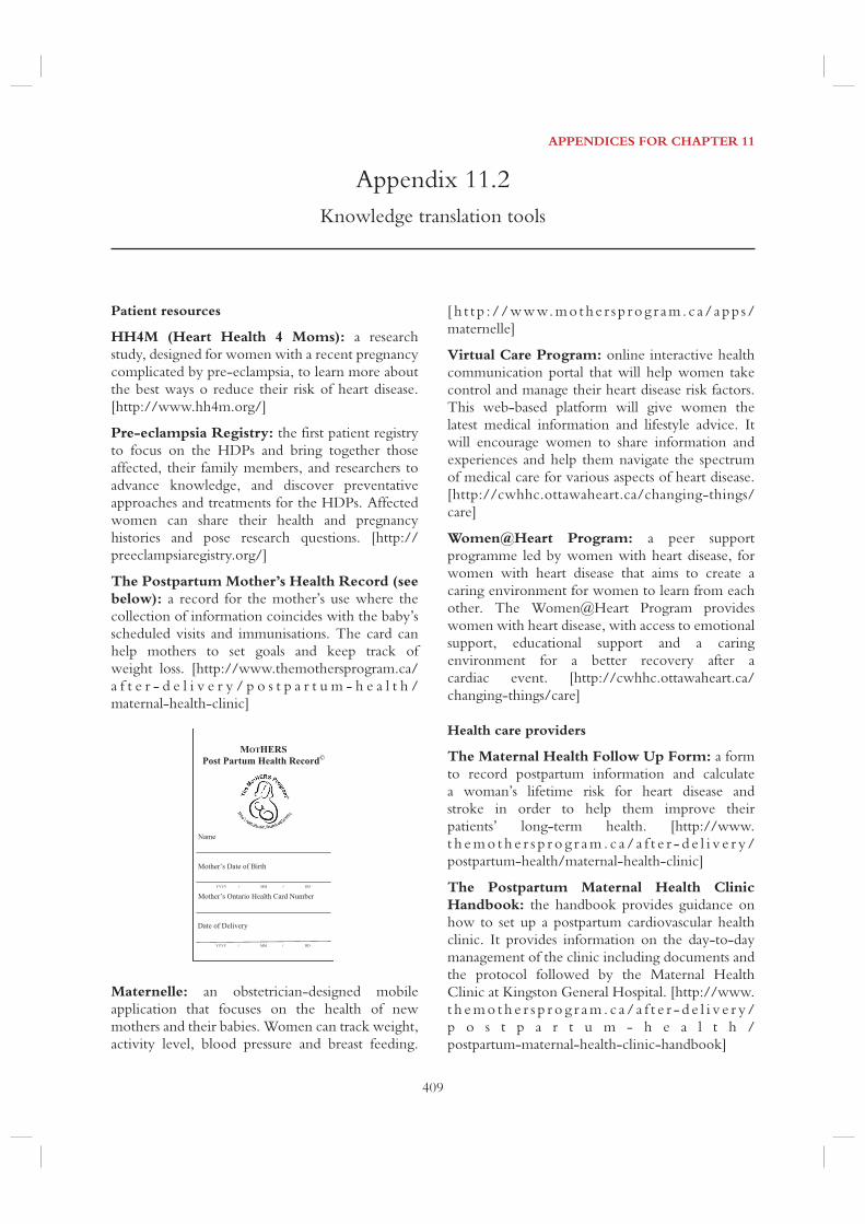
APPENDICES FOR CHAPTER 11
409
Appendix 11.2
Knowledge translation tools
Patient resources
HH4M (Heart Health 4 Moms): a research
study, designed for women with a recent pregnancy
complicated by pre-eclampsia, to learn more about
the best ways o reduce their risk of heart disease.
[http://www.hh4m.org/]
Pre-eclampsia Registry: the first patient registry
to focus on the HDPs and bring together those
affected, their family members, and researchers to
advance knowledge, and discover preventative
approaches and treatments for the HDPs. Affected
women can share their health and pregnancy
histories and pose research questions. [http://
preeclampsiaregistry.org/]
The Postpartum Mother’s Health Record (see below): a record for the mother’s use where the
collection of information coincides with the baby’s
scheduled visits and immunisations. The card can
help mothers to set goals and keep track of
weight loss. [http://www.themothersprogram.ca/
a f t e r - d e l i v e r y / p o s t p a r t u m - h e a l t h /
maternal-health-clinic]
Maternelle: an obstetrician-designed mobile
application that focuses on the health of new
mothers and their babies. Women can track weight,
activity level, blood pressure and breast feeding.
[ h t t p : / /www.mothe r s p rog r am . c a/ app s /
maternelle]
Virtual Care Program: online interactive health
communication portal that will help women take
control and manage their heart disease risk factors.
This web-based platform will give women the
latest medical information and lifestyle advice. It
will encourage women to share information and
experiences and help them navigate the spectrum
of medical care for various aspects of heart disease.
[http://cwhhc.ottawaheart.ca/changing-things/
care]
Women@Heart Program: a peer support
programme led by women with heart disease, for
women with heart disease that aims to create a
caring environment for women to learn from each
other. The Women@Heart Program provides
women with heart disease, with access to emotional
support, educational support and a caring
environment for a better recovery after a
cardiac event. [http://cwhhc.ottawaheart.ca/
changing-things/care]
Health care providers
The Maternal Health Follow Up Form: a form
to record postpartum information and calculate
a woman’s lifetime risk for heart disease and
stroke in order to help them improve their
patients’ long-term health. [http://www.
t h e m o t h e r s p r o g r a m . c a / a f t e r - d e l i v e r y /
postpartum-health/maternal-health-clinic]
The Postpartum Maternal Health Clinic Handbook: the handbook provides guidance on
how to set up a postpartum cardiovascular health
clinic. It provides information on the day-to-day
management of the clinic including documents and
the protocol followed by the Maternal Health
Clinic at Kingston General Hospital. [http://www.
t h e m o t h e r s p r o g r a m . c a / a f t e r - d e l i v e r y /
p o s t p a r t u m - h e a l t h /
postpartum-maternal-health-clinic-handbook]
MOTHERS Post Partum Health Record©
Name
Mother’s Date of Birth
Date of Delivery
Mother’s Ontario Health Card Number YYYY / MM / DD
YYYY / MM / DD

THE FIGO TEXTBOOK OF PREGNANCY HYPERTENSION
410
Appendix 11.3
GRADE evaluation of best practice points for postpartum care
Quality of evidence*
Strength of recommendation†
Care in the 6 weeks after birth
1. Blood pressure should be measured during the time of peak postpartum blood pressure, at
days 3–6 after delivery.
Low Strong
2. Women with postpartum hypertension should be evaluated for pre-eclampsia (either arising
de novo or worsening from the antenatal period).
Low Weak
3. Antihypertensive therapy may be continued postpartum, particularly in women with
antenatal pre-eclampsia and those who delivered preterm.
Low Weak
4. Severe postpartum hypertension must be treated with antihypertensive therapy, to keep
systolic blood pressure <160 mmHg and diastolic blood pressure <110 mmHg.
Moderate Strong
5. Antihypertensive therapy may be used to treat non-severe postpartum hypertension, to keep
blood pressure at <140/90 mmHg for all but women with pre-gestational diabetes mellitus
among whom the target should be <130/80 mmHg.
Very low Weak
6. Antihypertensive agents acceptable for use in breastfeeding include nifedipine XL
(slow-release), labetalol, methyldopa, captopril and enalapril.
Moderate Weak
7. There should be confirmation that end-organ dysfunction of pre-eclampsia has resolved. Very low Strong
8. Non-steroidal anti-inflammatory drugs should not be given postpartum if hypertension is
difficult to control, there is evidence of kidney injury (oliguria and/or an elevated creatinine)
(90 mol/L) or platelets are <50 109/L.
Low Weak
9. Postpartum thromboprophylaxis should be considered in women with pre-eclampsia who
have other risk factors for thromboembolism.
Low Weak
Care beyond the first 6 weeks after birth
1. Women with a history of severe pre-eclampsia (particularly those who presented or
delivered at <34 weeks) should be screened for pre-existing hypertension and underlying renal
disease.
Low Weak
2. Referral for internal medicine or nephrology consultation should be considered for women
with postpartum hypertension that is difficult to control, or women who had pre-eclampsia
and have at 3–6 months postpartum ongoing proteinuria, decreased eGFR (<60 mL/min), or
another indication of renal disease (such as abnormal urinary sediment).
Low Weak
3. Women who are overweight should be encouraged to attain a healthy body mass index to
decrease risk in future and for long-term health.
Low/
moderate
Strong
4. Women with pre-existing hypertension or persistent postpartum hypertension should
undergo the following investigations (if not done previously): urinalysis; serum sodium,
potassium and creatinine; fasting glucose; fasting lipid profile; and standard 12-lead
electrocardiography.
Low Weak
continued

APPENDICES FOR CHAPTER 11
411
Appendix 11.3 continued
Quality of evidence*
Strength of recommendation†
Care beyond the first 6 weeks after birth
5. Women who are normotensive but who have had a hypertensive disorder of pregnancy, may
benefit from assessment of traditional cardiovascular risk markers.
Low/
moderate
Weak
6. All women who have had a hypertensive disorder of pregnancy should pursue a healthy diet
and lifestyle.
Low Strong
eGFR, estimated glomerular filtration rate
* The judgements about the quality of evidence is based on the confidence that available evidence reflects the true effect of
the intervention or service. Evidence is considered to be of high quality when the true effect is thought to lie close to that of
the estimate of the effect (e.g., if there is a wide range of studies included in the analyses with no major limitations, there is
little variation between studies, and the summary estimate has a narrow confidence interval). Evidence is considered to be
of moderate quality when the true effect is likely to be close to the estimate of the effect, but there is a possibility that it is
substantially different (e.g., if there are only a few studies and some have limitations but not major flaws, there is some
variation between studies, or the confidence interval of the summary estimate is wide). Evidence is considered to be of low quality when the true effect may be substantially different from the estimate of the effect (e.g., the studies have major flaws,
there is important variation between studies, or the confidence interval of the summary estimate is very wide)† A strong recommendation should be interpreted as meaning that most people in this situation would want the recommended
course of action and only a small number would not. Clinicians should regard the recommendation as applying to most
individuals. Policy-makers can adopt the recommendation as policy in most situations. Adherence to this recommendation
according to the guideline could be used as a quality criterion or performance indicator. A weak recommendation should be
interpreted as meaning that most people in this situation would want the recommended course of action, but many would
not; patients’ values and preferences should be considered in reaching a decision. Decision aids may support people in
reaching these decisions. Policy-making will require substantial debate and involvement of various stakeholders. An
appropriately documented decision making process could be used as a quality indicator

THE FIGO TEXTBOOK OF PREGNANCY HYPERTENSION
412
Postnatal period remains the most neglected
period for provision of critical care for mothers
and babies. In low-income countries, an
estimated 70% of women do not receive any
postnatal care.
Literature suggests that in sub Saharan Africa,
15.2% maternal deaths occurred in the postnatal
period.
Appendix 11.4
Postnatal care care – Policy brief
Postnatal care (PNC) is considered to be an essential intervention for reducing maternal mortality. In LMICs, almost 40% of women
experience complications after delivery and in 15%
of women, those complications are life-threatening.
A 2013 WHO systematic analysis of the causes of
maternal deaths (2003–09) determined that 480,000
or 19.7% of maternal deaths worldwide occurred
postpartum. Most of those deaths occur in the first
week postpartum.
Postpartum care has the potential to optimise future pregnancy outcomes and the long-term health of the mother. PNC affords the
opportunity to counsel women about birth
spacing and contraception. Also, the HDPs, and
pre-eclampsia in particular, are associated with an
increase in many adverse maternal health conditions,
including hypertension, heart disease, stroke, renal
disease, and diabetes mellitus. Postpartum care
offers care providers the opportunity to educate
women about these risks as well as changes in diet,
lifestyle, and medical management that may modify
them.
ACTIONS
Advancing PNC policy and implementing
evidence-based programmatic changes in the
national and state level health policies is crucial to
improving access to care and reducing maternal
mortality and morbidity.
• Increase demand for PNC care by engagement
with women and communities
• Engage relevant stakeholders at the
community and state levels in order to establish
leadership for integration of a PNC package at
the community level
• Develop a local PNC package adapted that
includes all of the STRONG recommendations
from the WHO 2013 Postnatal Care guidelines’
recommendations (see Table S11.1 below), as
follows:
N PNC care beginning within 24 h of birth,
consisting of at least three visits, and occurring
ideally at home
N Exclusive breastfeeding
N Assessments of the mother that include
physical and mental health evaluations, as
well as targeted approaches for family
planning needs. Providers need to be trained
about the mental health implications of the
HDPs, such as anxiety, depression, and
post-traumatic stress disorder
• Engage traditional birth attendants in
delivery of PNC

APPENDICES FOR CHAPTER 11
413
Table S11.1 Recommendations graded as STRONG in the WHO Postnatal Care Guidelines 201395
Nature of recommendation Details
Postnatal contact
Timing (as early as possible within 24 h)
Number (3 visits)
Place (home visits are recommended)
Exclusive breastfeeding of baby
Maternal counselling to encourage and support
Maternal assessment
Physical
Within 24 h of birth Starting shortly after birth and taken again at 6 h: Blood pressure
Starting from the first hour after birth and continuing routinely during the first 24 h: Assessment for vaginal bleeding, uterine contraction, fundal height, temperature
and heart rate (pulse) routinely during the first 24 hours
Beyond 24 h of birth Ongoing assessment of: general symptoms (headache, fatigue, back pain); uterine
tenderness and lochia; voiding (i.e., micturition and urinary incontinence, bowel
function); healing of any perineal wound, perineal pain, and perineal hygiene;
breast pain and breastfeeding progress
Counselling of mother on: warning signs and symptoms of PPH, infection, and
pre-eclampsia/eclampsia; good nutrition, hygiene, especially hand washing; birth
spacing and family planning; gentle exercise, iron and folic acid supplementation
Mental health Emotional well-being
Psychosocial support For women who have lost her baby
Accounting for experiences in hospital

THE FIGO TEXTBOOK OF PREGNANCY HYPERTENSION
414
Appendix 11.5
Recommendations for partartum care of women with hypertensive disorders of
pregnancy from international clinical guidelines*
QLD NICE 2010 WHO 2011
BP monitoring For women with chronic hypertension or GH, measure BP
daily for first 2 days, once/day on days 3–5, and as indicated if
antihypertensive therapy is changed
For women with PET, measure BP 4x/day in hospital, once/
day on days 3–5, and if abnormal then, on alternate days (until
normal)
In women with PET who took antihypertensive therapy,
measure BP 4x/day in hospital, then every 1–2 days for 2
weeks until off treatment and normotensive
PET may appear or
worsen
For women with
pre-eclampsia, serial
surveillance of
maternal well-being
is recommended
For women with severe PET, ask about severe headache and
epigastric pain when BP is measured
For women with PET with non-severe hypertension or those
who have received critical care, measuring creatinine
transaminases within 48–72 h
If creatinine and transaminases are normal at 48–72 h after
birth, they do NOT need to be retested
For women with PET, repeat platelet count, transaminases
and serum creatinine “as clinically indicated” and at the 6–8
weeks postnatal review
For women with PET who have stepped down from critical
care (level 2), do NOT measure fluid balance if creatinine is
normal
Continuation of
antenatal
antihypertensive
therapy
For women with chronic hypertension, continue antenatal
antihypertensive therapy
In women with GH or PET who were taking antenatal
antihypertensive therapy, continue therapy
If methyldopa was the antenatal antihypertensive, stop it
within 2 days of birth. For women with chronic hypertension,
restart the antihypertensive agent that was taken before
planning pregnancy
For women with
any HDP,
continue
antenatal
antihypertensive
therapy
Treatment of severe
hypertension
For women with any HDP, treat severe hypertension For women with
any HDP, treat
severe
hypertension with
antihypertensive
drugs

APPENDICES FOR CHAPTER 11
415
AOM 2012 ACOG 2013 SOGC 2014
Inform women with any HDP that
elevated BP may take time to resolve
Inform women with GH that
hypertension may worsen “during the
postpartum period”
For women with GH, PET,
or superimposed PET,
measure BP in hospital (or
equivalent setting) for
72 h and at some point on
days 7–10 or earlier if PET
symptoms occur
For women with any HDP, measure BP at some point
on days 3–6 postpartum
Inform women with any HDP to
report any symptoms or signs of PET
Inform women with any
HDP about symptoms and
signs of PET which they
should report immediately
if they arise
Women with new/worsening postpartum
hypertension should be evaluated for PET
For women with PET, there should be confirmation
that end-organ dysfunction has resolved
For women with any HDP, especially with PET or
preterm delivery, continue antihypertensive therapy
For women with any HDP,
treat severe hypertension
(BP 160/110 mmHg)
within 1 hour
For women with any HDP, treat severe hypertension
with antihypertensive drugs
For women with any HDP, goal of <160/110 mmHg
continued

THE FIGO TEXTBOOK OF PREGNANCY HYPERTENSION
416
Appendix 11.5 continued
QLD NICE 2010 WHO 2011
Treatment of
non-severe
hypertension
For women with “chronic hypertension”, goal of
<140/90 mmHg
In women with GH or PET goal of <150/100 mmHg
In women with GH or PET consider a reduced dose if BP
<140/90 mmHg. Reduce the dose if BP is <130/80 mmHg
Antihypertensive
agents and
breastfeeding
Acceptable agents are nifedipine, labetalol, captopril, enalapril,
atenolol and metoprolol
Do NOT prescribe diuretics to women who are breastfeeding
or expressing milk
Insufficient evidence to comment on the neonatal safety of
the following during breastfeeding: ACE inhibitors (other
than enalapril and captopril), ARBs and amlodipine
Discharge planning
for community care
For women with chronic hypertension, review long-term
antihypertensive treatment 2 weeks after the birth
Offer women with PET transfer to community care if they
have no symptoms, BP <150/100 mmHg, and laboratory
abnormalities are stable/improving
For women with GH or PET, write a detailed care plan
before transfer to community care
A care plan should include the following details: who will
provide follow-up care, including medical review if needed,
frequency of BP monitoring needed, thresholds for reducing
or stopping treatment, indications for referral to primary care
for BP review, and self-monitoring for symptoms
At midwifery visits
between discharge
and formal 6–8
weeks postnatal
review
Offer medical review (with the pre-pregnancy team) at the
6–8 weeks postnatal review for women with chronic
hypertension
Offer medical review at the 6–8 weeks postnatal review for
women with GH or PET, especially if they are still on
antihypertensive treatment 2 weeks after transfer to
community care
Formal medical
postnatal review at
6–8 weeks after
delivery
In women with PET, perform urinary reagent-strip testing. If
proteinuria 1+, offer further review at 3 months postpartum
If women with PET had improving but still abnormal
haematological or biochemical indices at hospital discharge,
repeat testing
For women with PET, do NOT routinely perform
thrombophilia screening

APPENDICES FOR CHAPTER 11
417
AOM 2012 ACOG 2013 SOGC 2014
For women with any HDP
goal of <150/100 mmHg
For women with uncomplicated chronic hypertension,
consider goal of <140/90 mmHg
For women with chronic hypertension and
comorbidities other than pre-gestational diabetes
mellitus, consider goal of <140/90 mmHg
For women with chronic hypertension and
pre-gestational diabetes mellitus, goal of
<130/80 mmHg
Acceptable agents are nifedipine XL, labetalol,
captopril and enalapril, and methyldopa
For women with any HDP postpartum, captopril,
enalapril or quinapril may be used
For women with any HDP, monitor
BP at “all regular postpartum visits”
in first 2 weeks postpartum, or until
normal BP measured twice
For women with any HDP who has
an elevated BP upon discharge from
hospital, ensure plan is in place for
physician follow-up in the event that
BP remains elevated (or increases
further)
Upon discharge from midwifery care,
communicate information about any
HDP to the primary care provider
For women with PET, there should be confirmation
that end-organ dysfunction has resolved
For women with chronic hypertension or any HDP
with persistent postpartum hypertension, perform the
following (if not done previously): urinalysis, serum
Na/K and creatinine, fasting glucose and lipid profile
and standard ECG recommended
For women with severe PET (particularly with
presentation at <34 weeks), screen for chronic
hypertension and underlying renal disease)
For women with any HDP, consider screening for
traditional cardiovascular risk markers
continued

THE FIGO TEXTBOOK OF PREGNANCY HYPERTENSION
418
Appendix 11.5 continued
QLD NICE 2010 WHO 2011
Counselling about
future pregnancy
risks
For women with
any HDP, offer
preconceptual advice
Counselling about
long-term health
risks
For women with
any HDP, offer
“screening” and
lifestyle counselling
Advise women with GH or PET (and their primary care
physicians) that they are at increased risk of future
hypertension and cardiovascular disease in later life
Advise women with PET with proteinuria (that has resolved)
that they are still at increased risk kidney disease but the
absolute risk is very low and follow-up is not necessary
Advise women with PET to keep their BMI within healthy
range (18.5–24.8 kg/m2, NICE clinical guideline 43)
Specialist referral
(e.g., renal, etc.)
Hypertension specialist – for women with GH or PET who
still need antihypertensive therapy 6–8 weeks after delivery
Kidney specialist – for women with PET who have
proteinuria 1+ at 6–8 weeks after delivery (although
clinicians can reassess at 3 months post-delivery to confirm)
NSAIDs
Thromboprophylaxis
* SOMANZ 2014 is included in the chapter text, but not in this table adapted from Gillon 201498. PRECOG II (2009) and
NVOG (2011) did not provide postpartum guidance and are not included in this table
ACOG 2013: American College of Obstetricians and Gynecologists, Task Force on Hypertension in Pregnancy.
Hypertension in pregnancy. Report of the American College of Obstetricians and Gynecologists’ Task Force on
Hypertension in Pregnancy. Obstet Gynecol 2013 Nov; 122(5):1122–1131
AOM 2012: Salehi P, Association of Ontario Midwives HDP CPG, Working Group. Hypertensive disorders of pregnancy
(Clinical Practice Guideline 15). 2012; Available: http://www aom on ca/Health_Care_Professionals/Clinical_Practice_
Guidelines/

APPENDICES FOR CHAPTER 11
419
AOM 2012 ACOG 2013 SOGC 2014
Advise women with any HDP to keep their BMI
within healthy range to decrease risk in future
pregnancy
Advise women with any HDP that
they may be at increased risk of future
hypertension and cardiovascular
disease in later life
Advise women with any HDP of the
benefits of a heart healthy diet and
lifestyle
For women with PET and
preterm birth (<37 0/7
weeks) or recurrent PET,
consider yearly assessment
of BP, lipids, fasting blood
glucose and BMI
Advise women with any HDP to pursue a healthy diet
and lifestyle
Advise women with any HDP to keep their BMI
within healthy range for long-term health
Offer in hospital specialist assessment with internal
medicine – for women with any HDP when
postpartum hypertension is difficult to control
Offer outpatient renal assessment – for women who
had PET who have proteinuiria, decreased eGFR
(<60 mL/min) or another indication of renal disease at
3–6 months after delivery
For women with any HDP, limit use
of NSAIDs and offer acetaminophen
as an effective alternative (albeit with
limited information about side-effects)
For women with any HDP, NSAIDs are NOT
recommended if BP is difficult to control, there is
kidney injury (oliguria and/or an elevated creatinine)
(90 M), or platelets are <50 109/L
Consider for women with PET, especially when there
are other risk factors
NICE 2010: National Collaborating Centre for Women’s and Children’s Health (UK). CG107: Hypertension in
pregnancy: The management of hypertensive disorders during pregnancy. NICE: Guidance 2010 Aug
QLD 2013: Queensland Maternity and Neonatal Clinical, Guidelines Program. Hypertensive disorders of pregnancy.
2013;MN10.13-V4-R15
SOGC 2014: Magee LA, Pels A, Helewa M, Rey E, von Dadelszen P. Diagnosis, evaluation, and management of the
hypertensive disorders of pregnancy. Pregnancy Hypertens 2014;4(2):105–145
WHO 2011: World Health Organization. WHO recommendations for prevention and treatment of pre-eclampsia and
eclampsia. 2011


421
Index
1-antitrypsin, 19
-methyldopa, impact on anaesthesia, 190
-fetoprotein, 86
abruption, see placental abruption
access to antenatal care, 10
access to laboratory facilities, 47
accuracy of BPM devices, 5
ACE inhibitors, 143
acidosis, compensatory metabolic, 191
activated protein C, 143
active management, see interventionist care
acute renal failure, 68
ADAM-12 as part of multivariable analysis, 89, 90
adverse conditions of pre-eclampsia, 39
age,
as a risk factor, 78
maternal, pre-eclampsia risk, 39
paternal, 81
see also gestational age
aggressive management, see interventionist care
aggressiveness, 144
airway,
diffi cult, 196
examination at initial assessment, 187
albumin, 19
pre-eclampsia risk, 39
albuminuria, 22
alcohol, 101, 123
alfentanil, 197
alkalosis, respiratory, 191
ALT,
serum levels, 43
timing of delivery, 171
ambulance services, 126
ambulatory blood pressure measurement, 6
use in community, 3
vs. HBPM, 7
anaesthesia, 185–207
awareness during, 201
blood pressure monitoring after, 190
central venous catheters, 191
delayed complications, 201
diffi cult airway, 196
dural puncture, 201
early assessment, 185
early complications, 201
early warning systems, 190
for Caesarean delivery, 196, 395–9
general anaesthesia, 196
GRADE evaluation of best practice points, 400
haemodynamic management, 197
hypertensive response to intubation, 196
initial assessment, 186
international guidelines on recommendations for,
402–5
intravenous bolus of crystalloid, 198
local anaesthetic infi ltration, 200
minimally invasive cardiac output monitoring
devices, 191
monitoring for, 187
non-depolarising muscle relaxants, 197
pharmacological agents used in pre-eclampsia and,
190
post-traumatic stress disorder after, 201
postoperative complications, 202
postpartum issues, 201
prevention of hypertensive response to intubation,
393
priorities for, 206
prolonged neuromuscular block, 197
resuscitation equipment, 185
systemic effects of pre-eclampsia on, 188–9
transthoracic echocardiography, 191
use of vasopressors, 198
analgesia,
fetal well-being and, 191
in labour, 191
inhalation, 192
intravenous, 203
methods for, 186
neuraxial, 204
oral, 203
pain-induced sympathetic response and
hypertension, 191
parenteral, 192
patient-controlled, 193
rectal, 203
WHO analgesic ladder, 203
wound infi ltration, 204
angiogenic factor imbalance, 36, 86, 44
future research priorities, 52
pre-eclampsia defi nition and, 37

THE FIGO TEXTBOOK OF PREGNANCY HYPERTENSION
422
angiogenic factor imbalance continuedwith circulating proteins, 90
angiotensin receptor blockers, 143
antenatal care,
access to, 10
attendance at, 9
development of surveillance, 76
fi rst trimester ultrasound, 168
health care worker staffi ng levels for, 9
late presentation for, 47
number of visits, 8
pre-eclampsia prevention and low rates of, 110
recommended components of, 9
Scottish paradigm, 33
screening for proteinuria, 20
timing of initiation, 9
antiangiogenic factors, 86
antihypertensives,
choice of agent, 136, 141
commonly used agents, 142
for pre-eclampsia prevention, 105, 316
GRADE evaluation of best practice points, 365–7
in non-severe hypertension, 140–4
in severe hypertension, 135–40
international clinical guidelines, 155, 370–81
maternal outcomes after use, 142
maternal side-effects, 143
policy briefs, 368
treatment wall chart, 349
use during breastfeeding, 140
antioxidants, 103
antiphospholipid syndrome, 79,
antiphospholipid antibody syndrome, 43
antithrombin, 148, 150
antithrombin III defi ciency, 79
anxiety, 82
aspirin, 292, 318–321
impact on anaesthesia, 190
low-dose,
and hypertensive disorders in pregnancy, 102
combined with calcium supplementation,
106
for pre-eclampsia prevention, 102, 105
use in under-resourced settings, 110
assisted reproductive treatment, as a risk factor, 81
AST, serum levels, 43
asthma, 136, 141
atenolol, 143
paediatric neurodevelopment and, 143
attendance at antenatal care, 9
audit of practice, 150
auscultatory methods of blood pressure measurement,
4
automated blood pressure measurement devices, 5
automated testing of urinary dipsticks, 22
awareness, during anaesthesia, 201
-lipoprotein, 19
bariatric surgery for obesity, 224
bed rest, 109, 125
behaviour and paediatric neurodevelopment, 218
best practice points,
antihypertensives
in non-severe hypertension, 152
in severe hypertension, 152
blood pressure measurement, 11
care beyond 6 weeks after birth, 225
care in 6 weeks after birth, 224
diagnosis of hypertension, 11
fl uids, 152
for anaesthesia and analgesia, 205
for classifi cation of pre-eclampsia, 46
for diet lifestyle and place of care, 128
for fl uids drugs and transfusions, 365–7
for risk factors and predictors, 91
GRADE evaluation
for anaesthesia, 400
for classifi cation of hypertensive disorders of
pregnancy, 255–6
for diet, 345
for hypertension, 236–7
for lifestyle, 345
for mode of delivery, 383
for place of care, 345
for postpartum care, 410
for preventing pre-eclampsia, 340–3
for proteinuria, 250
for timing of delivery, 383
in proteinuria, 27
mode of delivery, 177
place of delivery, 176
prevention of pre-eclampsia in low-risk women,
112
prevention of pre-eclampsia in women at increased
risk, 113
timing of delivery, 176, 177
beta blockers, 141
use with methyldopa, 142
betamethasone, 176
biomarkers,
for pre-eclampsia, 44
future research priorities, 51
in multivariable prediction analyses, 88
in univariable prediction analyses, 86, 87
birth weight, maternal, 79
see also large for gestational age, small for gestational
age
blindness, 68
blood fi lm, for pre-eclampsia, 42
blood pressure,
access to monitoring facilities, 47
as a predictor, 83
changes with gestational age, 1, 2

INDEX
423
changes with parity, 2
chocolate and, 103
circadian rhythm, 1
importance of repeat measurement, 2
in offspring after pre-eclampsia, 218
invasive monitoring, 198
maintenance in anaesthesia, 197
monitoring after anaesthesia, 190
timing of delivery, 171
with uterine artery PI as predictor, 89
blood pressure measurement,
ABPM, 6
accuracy of devices, 5
aneroid devices, 5
auscultatory methods, 4
automated devices, 4
choice of device during pregnancy, 5
choice of device for setting, 5, 6
comparison of ABPM and HBPM, 7
cuff size for, 3, 240
HBPM, 6
importance of repeat, 2
in the community, 3
in under-resourced settings, 7–10
international guidelines, 240–6
position for, 3
recalibration of devices, 5
sample policy brief, 238
settings, health facility, 5
smartphone and tablet technologies, 8
solar-powered devices, 8
techniques for, 3–7
body mass index,
pre-eclampsia risk, 40
pre-pregnancy, 79
bradycardia,
fetal, 195
neonatal, 136, 137
brain natriuretic peptide, 52
breastfeeding,
antihypertensive use during, 140
hypertension management and, 216
bronchopulmonary dysplasia, 69
after vaginal cf Caesarean delivery, 175
bupivacaine-soaked sponges, 204
C-reactive protein, 86
Caesarean section,
after MgSO4 use, 145
anaesthesia for, 196, 395–9
at 37+0–42+0 weeks
bronchopulmonary dysplasia and, 175
choice of anaesthesia for, 199
combined spinal–epidural for, 200
epidural anaesthesia for, 200
local anaesthetic infi ltration, 200
postoperative pain management, 202–4
principles for anaesthetic management for, 197
spinal anaesthesia for, 200
spinal vs. epidural, 199
spinal vs. general, 199
uterine rupture after, 175
calcium, 292–295, 322
calcium : creatinine ratio, 84
calcium channel blockers, 136, 137, 141
calcium gluconate, antidote to MgSO4, 197
calcium supplementation, 75
combined with low-dose aspirin, 106
for pre-eclampsia prevention, 102, 105
use in under-resourced settings, 110
captopril, 139, 140
carboprost, impact on anaesthesia, 190
cardiac output monitoring devices, 191
cardiovascular disease,
antihypertensive use and, 141
paternal, 81
risk, 35
risk factors after pre-eclampsia, 218, 219
catastrophic antiphospholipid antibody syndrome, 43
catecholamine release, 191
central nervous system,
proteinuria and, 20
white matter lesions, 221
central venous catheters, 191
cerebral palsy, 146
cerebrovascular disease, 35
cervix,
ripening of, 175
unfavourable, 174, 175
chest pain, 171
CHIPS (Control of Hypertension in Pregnancy Study),
140
chlamydia, 81
chlorpromazine promethazine pethidine, cf MgSO4,
144
chocolate, higher intake lowers blood pressure, 103
chronic hypertension, 33
as a risk factor, 79
estimates for incidence, 64
renal disease, 187
see also pre-existing hypertension
temporal trends in, 65
timing of delivery in, 174
chronic kidney disease, 79
circadian rhythm of blood pressure, 1
circulating proteins, 90
climate and pre-eclampsia trends, 66
CLIP (Community-Level Interventions for
Pre-eclampsia) trial, 64, 127
clonidine, 139, 140
co-enzyme Q10 supplementation, 107, 109

THE FIGO TEXTBOOK OF PREGNANCY HYPERTENSION
424
coagulation,
neuraxial analgesia and, 194
tests for pre-eclampsia, 42
coagulopathy, 176
communication,
doctor–patient, 45
with patient, 46
community,
blood pressure measurement in, 3
postpartum care, 226
community engagement, 127
community-based care, 126
comorbidity,
commonly linked to gestational hypertension, 187
in hypertension, 34
compensatory metabolic acidosis, 191
complete blood count, 42
congenital malformations, 81
congestive cardiac disease and proteinuria, 20
Congo red dot test, 28
connective tissue disorders, 187
continuous spinal analgesia, 195
copper, 105, 109
corticosteroids,
antenatal, 176
for fetal lung maturity, 168
use in expectant care, 169
use in HELLP syndrome, 148
cost-effectiveness,
of interventionist care, 174
of MgSO4, 144, 145
of prediction, 92
of proteinuria testing, 26
counselling,
for expectant vs. interventionist care, 169
nutritional education, 102
CRADLE BP device, 235
creatinine,
calcium : creatinine ratio, 84
IPG : creatinine ratio, 84
pre-eclampsia risk, 40
serum levels, 43
timing of delivery, 171
crystalloid, intravenous bolus, 198
cuff,
correct position, 3
correct size, 3
importance in anaesthesia, 198
dalteparin, 108
DASH (dietary approached to stop hypertension) diet,
107, 124
defi nitions,
bed rest, 109, 125
HELLP syndrome, 38
hypertension, 1
pre-eclampsia, 37, 266–73
severe hypertension, 2
severe pre-eclampsia, 266–73
signifi cant proteinuria, 26
systolic and diastolic criteria, 2
delivery,
indications for in pre-eclampsia, 169, 169
see also Caesarean section; vaginal delivery
timing and mode, 167–79
demographic risk factors, 78
depression, 82
dexamethasone, 148, 176
diabetes, 78, 79
antihypertensive use and, 141
as a risk factor, 79
chronic hypertension, 187
pre-gestational, 79
risk after pre-eclampsia, 220
diabetic nephropathy, 79
antihypertensive use and, 141
diamorphine, 193
diazepam, cf MgSO4, 144
diazoxide, 137, 140
diet,
GRADE evaluation for best practice points, 345
international guidelines for recommendations, 346–8
modifi cations recommended for all women, 223
role in pre-eclampsia, 123–4
weight loss in obesity, 123
dietary changes, 123, 294–299
for pre-eclampsia prevention, 102, 107, 322–325
salt restriction, 102
diffi cult airway, 187, 196
dipsticks,
for spot albumin : creatinine ratio, 25
urinary, 21
diuretics, 104, 142, 308, 332
dopamine, renal dose, 135
Doppler,
fetal status and delivery, 175
ophthalmic artery, 40
uterine artery, 40, 84
velocimetry, 43
with anogiogenic imbalance measures, 45
ductus venosus, 45
dural puncture, 201
dyspnoea, 171
early delivery, see interventionist care
early warning systems, 190
response triggering parameters in, 192
eclampsia,
incidence estimates, 68
MgSO4 use for, 144

INDEX
425
prevention, 145, 146
thrombocytopaenia, 189
‘eclampsia box’, 151
education,
nutritional counselling, 102
of health care providers, 221
of women, 46, 221
pre-eclampsia prevention and low rates of, 110
strengthening pre-eclampsia knowledge in women, 49
EmONC, 169
end tidal CO2 monitoring, 191
end-diastolic fl ow in umbilical artery, 45
end-organ function,
complications, 135
pre-existing hypertension, 215
endothelial dysfunction tests, 86
endothelial–platelet interactions, 86
endotheliosis, glomerular, 19
EOST (Emergency Obstetric Simulation Training),
151, 351–64
ephedrine, 198
epidemiology, 63–9
epidural analgesia, 193
combined spinal–epidural, 194
continuous spinal analgesia, 195
vs. parenteral, 193
vs. spinal in pre-eclampsia, 199
epigenome, 51
epinephrine, 200
equipment,
availability and choice of care level, 169
availability and use of local anaesthetic infi ltration
for Caesarean section, 200
availability in under-resourced settings, 7
early warning tool, 7
essential equipment for resuscitation, 186
for fetal monitoring, 186
see also devices
semi-automated, 7
ergometrine, 176
contraindicated in pre-eclampsia, 201
impact on anaesthesia, 190
ESMOE (Essential Steps in Managing Obstetric
Emergencies), 151, 351–64
esomeprazole, 148, 150
ethnicity, 141
as a risk factor, 78
evidence-based care in under-resourced setting, 150
exercise,
and risk, 82
pre-eclampsia prevention and, 103, 109
proteinuria and, 20
role in pre-eclampsia, 124
stretching, 109
use in under-resourced settings, 111
walking, 109
expectant care,
at 24+0–33+6 weeks, 173
at 34+0–36+6 weeks, 173
at <24+0 weeks, 172
counselling, 169
health care system and, 169
of HELLP syndrome, 65
risks of, 169
vs. interventionist care, 168
externalising behaviour, 144
factor V Leiden mutation, 79
familial risk factors, 76–8
fentanyl, 197
fetal distress, 136
fetal growth,
antihypertensives and, 143
restriction, 69
fetal heart pattern, 141
fetal heart rate, 141
monitoring, 136
fetal lung maturity and corticosteroid use, 168, 169, 176
fetal monitoring, 186
in pre-eclampsia, 45
fetal neuroprotection, 146
fetal testing for pre-eclampsia, 43
fetal well-being and analgesia, 191
fetotoxic agents, 143
fever and proteinuria, 20
fi brinogen for pre-eclampsia, 42
fi bronectin, 86
glycosylated, 45
fl avanoids, 103
fl uid management,
GRADE evaluation of best practice points, 365–7
in pre-eclampsia, 133–5
international guidelines, 155, 370–81
restriction in pre-eclampsia, 135
folate supplementation, 105
folate-containing multivitamins, 107
pre-eclampsia prevention and, 103
use in under-resourced settings, 111
fullPIERS model, 39, 68
timing of delivery, 171
furosemide, 135
garlic supplementation, 105, 107, 113
gender, fetal, as a risk factor, 81
general anaesthesia, 145, 196
vs. spinal in pre-eclampsia, 199
genetics,
familial risk factors for pre-eclampsia, 76
maternal risk factors, 77
paternal risk factors, 78

THE FIGO TEXTBOOK OF PREGNANCY HYPERTENSION
426
genetics continuedthrombophilias, 79
gestational age,
assessment of, 168
in under-resourced settings, 168
methods for, 168
at diagnosis and timing of delivery, 170
blood pressure changes with, 1, 2
mode of delivery and, 175
stillbirth and, 69
timing of delivery, 167, 171
at <24+0 weeks, 172
at 24+0–33+6 weeks, 172
at 34+0–36+6 weeks, 173
at 37+0–42+0 weeks, 174
gestational hypertension,
classifi cation, 34
defi nition, 35
estimates for incidence, 64
previous, as a risk factor, 80
progression to pre-eclampsia, 67
work up after, 216
glomerular endotheliosis, 19
glomerular proteins, 19
glucose serum levels, 43
GRADE evaluation for best practice points,
classifi cation of hypertensive disorders of pregnancy,
255–6
for anaesthesia, 400
for diet, 345
for fl uids drugs and transfusions, 365–7
for hypertension, 236–7
for lifestyle, 345
for mode of delivery, 383
for place of care, 345
for postpartum care, 410
for preventing pre-eclampsia, 340–3
for proteinuria, 250
for timing of delivery, 383
gross motor function, 144
group B streptococci, 81
haematoma, spinal, 202
haemodynamic management, 197
haemoglobin for pre-eclampsia diagnosis, 42
haemorrhage,
potential for, 176
risk, 76
HBPM (home blood pressure monitoring), 6
comprehensive list, 5
use in community, 3
vs. ABPM, 7
headache,
postdural puncture headache, 195, 200, 201
pre-eclampsia risk, 39
timing of delivery, 171
health care workers,
education of, 221
knowledge translation tools, 409
pre-eclampsia prevention and training, 110
self-help training, 151
shortage of, 67, 92
staffi ng levels, 9
training for blood pressure measurement, 235
training material for, 407–8
health facility, blood pressure measurement in, 5
heart-healthy diet, 107
heat coagulation test, 22
example, 24
interpretation, 23
height, 79
HELLP syndrome,
corticosteroids for, 148
defi nition, 38
estimates for incidence, 65
ethnicity and, 78
expectant management, 65
imitators, 148
international guidelines for therapy, 156
level of care required, 169
management at 24+0–33+6 weeks, 173
postpartum appearance, 216
therapies for, 147–8, 153
transfusion for, 147
heparin, 148
for pre-eclampsia prevention, 107, 323
hepatic haematoma, 68
hepatic rupture, 68
high-sensitivity C-reactive protein, 86
histidine-rich glycoprotein, 90
HIV, 81
home care, 125
hospital day units, 125
human chorionic gonadotrophin, 86, 88, 89
as part of multivariable analysis, 88, 89
hydralazine, 136, 138, 143
impact on anaesthesia, 190
hydration, 123
hyperlipidaemia, 79
hypertension,
best practice points, 236
chronic, 33
classifi cation of disorders of pregnancy, 34
defi nition, 1
diagnosis, international guidelines, 240–6
hypertensive response to intubation, 196
mean arterial pressure, 3
measurement of, 1–13
pain-induced sympathetic response, 191
postpartum appearance, 215
postpartum treatment, 215

INDEX
427
refractory, 140
risk after pre-eclampsia, 219
severe,
antihypertensive treatment, 135
defi nition, 2
treatment wall chart, 349
hypertensive disorders of pregnancy,
abstention from alcohol and, 101
associated morbidity and mortality, 66
calcium supplementation, 102
classifi cation, 33–53
common comorbidities, 187
epidemiology, 63–9
estimates on incidence, 63
folate-containing multivitamins, 103
GRADE evaluation for best practice points, 255–6
international guidelines on classifi cation, 257–65
lifestyle changes, 103
low-dose aspirin, 102
management of, 154
non-pharmacological management, 129
risk factors for, 66
timing and mode of delivery, 167–79
hypertensive emergency, 135
hypertensive response to intubation, 196, 393
hypertensive urgency, 135
HYPITAT trial, 40
HYPITAT II trial, 173, 174
hypotension, 188
avoidance of, 198
neuraxial anaesthesia and, 198
spinal analgesia and, 195
hypothyroidism, risk after pre-eclampsia, 220
ICU, refractory hypertension in, 140
immigrant status,
pre-eclampsia risk, 39
pre-eclampsia trends and, 66
immune thrombocytopenia, pre-existing hypertension
and, 41
immunoglobulins, 19
immunosuppression, 44
inattention, 144
incidence,
chronic hypertension, 64
estimates, 63
gestational hypertension, 64
in HIC, 64
in LMIC, 64
late-onset vs. early-onset, 65
pre-eclampsia, 64
temporal trends in, 65
indwelling spinal catheter, 195
infection,
as a risk factor, 81
treatment of for pre-eclampsia prevention, 110
information provision to patient, 46
see also education
inhalation analgesia, 192
inhibin, 86
inositol phosphoglycan-P, 84
interferon-, 45
international guidelines,
classifi cation of hypertensive disorders of pregnancy,
257–65
for anaesthesia and analgesia, 205
for classifi cation of pre-eclampsia, 50
for diet, lifestyle and place of care, 129
for fl uid, drugs and transfusions, 155
for pre-eclampsia prediction, 288–9
for prevention of pre-eclampsia, 113
for risk factors and predictors, 91
hypertension identifi cation, 12
in proteinuria, 28
mode of delivery, 179, 385–392
postpartum care, 227
recommendations,
for anaesthesia, 402–5
for blood pressure measurement and diagnosis,
240–6
for fl uids drugs and transfusions, 370–81
for postpartum care, 414–19
for proteinuria diagnosis, 251–3
on diet, 346–8
on lifestyle, 346–8
on place of care, 346–8
timing of delivery, 178, 385–92
interventionist care,
at 24+0–33+6 weeks, 172
at 34+0–36+6 weeks, 173
cost-effectiveness of, 174
counselling, 169
vs. expectant, 168, 173
intracerebral haemorrhage and proteinuria, 20
intracervical Foley balloon, 175
intrathecal catheter, 195
intrathecal morphine, 204
intrauterine growth restriction, 36, 45
diagnosis, 168
early-onset, 150
induction of labour and, 175
intubation,
hypertensive response to, 196
prevention of, 393
iodine, 105, 109
IPG : creatinine ratio, 84
iron, 109
iron supplements, 105
ischaemic heart disease risk after pre-eclampsia,
219
isradipine, 137

THE FIGO TEXTBOOK OF PREGNANCY HYPERTENSION
428
ketamine, 203
ketanserin, 137
knowledge translation tools, 409
L-arginine, 148, 149
for pre-eclampsia prevention, 108, 336
labetalol, 135, 136, 138, 139, 141, 142
hypertensive response to intubation, 197
impact on anaesthesia, 190
paediatric neurodevelopment and, 143
laboratory markers, 85
labour induction, 175
in women with gestational hypertension, 174
lactate dehydrogenase, serum levels, 43
large for gestational age, risk in pre-eclampsia, 35
last menstrual period,
assessment of gestational age, 168
memory aids for, 168
leptin, 45
lidocaine, 200
lifestyle,
changes, 312
changes for cardiovascular risk, 223
changes for pre-eclampsia prevention, 103, 108, 326
GRADE evaluation for best practice points, 345
international guidelines for recommendations, 346–8
modifi cations recommended for all women, 223
role in pre-eclampsia, 124
use in under-resourced settings, 112
literature searches, 275–6
LMICs, see under-resourced settings
long-term paediatric neurodevelopment, 143
low-dose aspirin, 75
low-molecular-weight heparin, 107, 108
use in under-resourced settings, 112
lupus nephropathy, 79
magnesium sulphate, 137
analgesic effect, 203
antihypertensives in non-severe hypertension, 153
Caesarean delivery after, 145
contraindication for non-depolarising muscle
relaxants, 197
cost-effectiveness, 144, 145
dose reduction, 145
for eclampsia treatment, 144
for pre-eclampsia treatment, 144
impact on anaesthesia, 190
implications for anaesthesia, 187
international guidelines, 156
intramuscular administration, 350
obesity and dose required, 146
policy brief, 369
treatment wall chart, 350
use in expectant care, 169
magnesium supplementation, 103, 109
malaria, 81
Malawi Demographic Health Survey, equipment
availability, 7
MAP (mean arterial pressure), 3, 88, 89
as part of multivariable analysis, 89
masked hypertension, 34, 40
maternal birth weight, 79
maternal placental syndrome, 220
mean arterial pressure in hypertension in pregnancy, 3,
88, 89
measurements,
importance of repeat blood pressure measurements, 2
of hypertension, 1–13
of proteinuria, 19–29
medical history,
as a risk factor, 79
paternal, 81
medullary sponge kidney, 142
memory aids for remembering last menstrual period,
168
meningitis, 202
mental health,
as a risk factor, 82
risks after pre-eclampsia, 221
MEOWS (Modifi ed Early Obstetric Warning Systems),
191
mercury sphygmomanometer, 5
metformin, for pre-eclampsia prevention, 109
methyldopa, 137, 139, 141
paediatric neurodevelopment and, 143
use of, 142
Microlife 3AS1-2 hand-held device, 10, 235
micronutrients, 298–303
defi ciencies and risk, 82
for pre-eclampsia prevention, 103, 109, 328
minimally invasive cardiac output monitoring devices,
191
miniPIERS model, 48, 68
timing of delivery, 171
miscarriage, previous, as a risk factor, 80
misoprostol, 175
impact on anaesthesia, 190
mode of delivery,
at 34+0–36+6 weeks, 174
choice of, 175–6
priorities for, 178
see also Caesarean section; vaginal delivery
monitoring for anaesthesia, 187
morbidity,
associated with hypertensive disorders of pregnancy,
66
early anaesthetic assessment and reduction in, 185
excess in interventionist care at 24+0–33+6 weeks,
172

INDEX
429
interventionist care and, 173
maternal,
associated with hypertensive disorders of
pregnancy, 66
rates of, 68
timing of delivery at 34+0–36+6 weeks, 173
neonatal,
respiratory, 173
timing of delivery at 34+0–36+6 weeks, 173
perinatal, 69
mortality,
in LMICs, 66
maternal,
associated with hypertensive disorders of
pregnancy, 66
rate in Sri Lanka, 49
rate in UK, 48
neonatal, 69
perinatal, 69
MTHFR single nucleotide polymorphism, 79
multiple pregnancy,
as a risk factor, 80
pre-eclampsia risk, 39
multivitamins, folate-containing, 107
myocardial infarction, markers for, 52
near-miss morbidity, 67
neonatal bradycardia, 136, 137
neonatal death, 69
nephropathy,
diabetic, 79
lupus, 79
nerve damage after neuraxial analgesia, 202
neuraxial analgesia,
cf parenteral, 193
choice of route in pre-eclampsia, 199
combined spinal–epidural for, 194, 200
contraindications, 193
epidural, 193
for Caesarean section, 199, 200
hypotension and, 198
neuroanatomy for needle placement, 187
postoperative, 204
postoperative complications, 202
single-shot spinal analgesia, 195
neurodevelopment,
long-term, 143
paediatric, 217
neuroprotection, 146
neutrophil gelatinase-associated lipocalin, 45
nifedipine, 136, 138, 141
hypertensive response to intubation, 197
impact on anaesthesia, 190
implications for anaesthesia, 187
paediatric neurodevelopment and, 143
use of, 142
nitric oxide donors for pre-eclampsia prevention, 110,
310, 338
nitric oxide production, 148
nitroglycerin infusion, 139
nitrous oxide/oxygen, 192
non-alcoholic steatohepatitis, pre-existing hypertension
and, 41
non-depolarising muscle relaxants, 197
non-pre-eclampsia glomerular disease, persistent
proteinuria and, 20
non-steroidal anti-infl ammatory drugs, 203
obesity,
and anaesthesia, 187
bariatric surgery for, 224
blood pressure changes with, 2
choice of cuff size, 3
dietary changes for pre-eclampsia prevention, 107
dose of MgSO4 and, 146
pre-existing hypertension and, 41
weight loss in, 123
oedema,
laryngeal, 188
of airway, 196
pre-eclampsia defi nition and, 37
oestradiol, 175
oligohydramnios, 45, 69
induction of labour and, 175
oliguria, 135
and anaesthesia, 188
pre-eclampsia risk, 39
online calculator, 48
for pre-eclampsia risk, 39
ophthalmic artery, Doppler imaging, 40
opioids,
for parenteral analgesia, 193
hypertensive response to intubation, 197
intrathecal, 204
intravenous, 203
oral, 203
orthostasis, 20
out-of-hospital care, 125
outpatient care, 125
oxidative stress, 86, 104
oxygen saturation,
for pre-eclampsia, 42
pre-eclampsia risk, 39
oxytocin, 176, 201
impact on anaesthesia, 190, 190
pain,
analgesia in labour, 191
control of, 202

THE FIGO TEXTBOOK OF PREGNANCY HYPERTENSION
430
pain continuedphysiological effects of, 202
postoperative management, 202–4
PAPP-A, 86, 88, 89
as part of multivariable analysis, 88, 89, 90
paracetamol, 203
parenteral analgesia, 192
parity,
as a risk factor, 80
blood pressure changes with, 2
pre-eclampsia risk, 39
timing of delivery, 171
paternal risk factors, 78
patient advocacy, 45
patient perspective in pre-eclampsia, 45
patient-controlled analgesia, 193, 203
PELICAN study, 44
periodontal disease, 81, 110, 314
persistent proteinuria, 20
pethidine, 193
phaeochromocytoma, 43
phenotype for pre-eclampsia, 44
phenylephrine, 198
phenytoin, cf MgSO4, 144
physical activity and pre-eclampsia, 124
phytonutrient supplementation, 109
placental abruption, 39
risk in pre-existing hypertension, 35
timing of delivery in chronic hypertension, 174
placental growth factor, 44, 86, 86, 88, 89
as part of multivariable analysis, 89
ratio to sFlt-1, 87
role in pre-eclampsia, 36
placental perfusion, 86
plasma volume expansion, 133
platelet count,
tests for pre-eclampsia, 42
timing of delivery, 171
platelet distribution width, 39
platelet transfusion, 147
podocyturia as a predictor, 84
point-of-care paper-based microfl uidic diagnostic
stamps, 28
policy briefs,
for antihypertensive therapy, 368
for blood pressure measurement, 238
for MgSO4 treatment, 369
for postpartum care, 412
for proteinuria, 247
sample policy brief for blood pressure measurement,
238
position for blood pressure measurement, 3
post-traumatic stress disorder, 45, 201
risks after pre-eclampsia, 221
postdural puncture headache, 195, 200, 201
posterior reversible leukoencephalopathy syndrome, 39
postpartum care, 215–27
GRADE evaluation of best practice points, 410
international guidelines, 414–19
policy brief, 412
priorities for in under-resourced settings, 226
potassium, increased intake, 123
poverty and pre-eclampsia prevention, 110
PP-13 as part of multivariable analysis, 88
pravastatin, 148
prazosin, 143
pre-eclampsia,
adverse conditions of, 39
adverse conditions that defi ne it, 38
biomarkers for, 44
choice of anaesthesia for, 199
choice of antihypertensive treatment in, 136
choice of neuraxial anaesthesia for, 199
classifi cation, 34
defi nitions, 35, 37, 266–73
diet and, 123–4
early-onset, causes, 36
effect on offspring health, 144
estimates for incidence, 64
familial risk factors, 76–8
fl uid management in, 133–5
fl uid restriction, 135
health issues in offspring, 218
imitators of, 43
impact of classifi cation, 52
indications for delivery, 169
international clinical guidelines for prediction, 288–9
late-onset, causes, 37
late-onset vs. early-onset incidence, 65
long-term maternal health after, 218–21
management of, 134
maternal testing in suspected, 41, 42
MgSO4 use for, 144
model for, 36
novel therapies for, 148
origins, 35
paediatric neurodevelopment after, 217
paternal risk factors, 78
performance of predictive tests for pre-eclampsia,
277–87
phenotypes of, 44
planning future pregnancies after, 217
post-traumatic stress after, 45
postpartum work up after, 217
prediction, 82–90
predictors for, 75–92
prevention, 101–13
best practice points, 340–3
in intermediate-risk women, 105
previous as a risk factor, 80
recognition of risks, 169
risk factors for, 75–92

INDEX
431
risk markers for, 77
risks of general anaesthesia in, 196
severe,
defi nition, 37, 171, 266–73
risks of expectant care, 169
systemic effects on anaesthesia, 188–9
trials and reviews of interventions for prevention,
291–315, 316–39
pre-existing hypertension,
classifi cation, 34
defi nition, 35
investigations for, 41
postpartum treatment, 215
screening for after pre-eclampsia, 217
pre-existing medical conditions, 79
pre-gestational diabetes, 79
prediction, 82–90
multivariable analyses, 87–90
priorities for, 92
univariable analyses, 83–7
blood pressure, 83
calcium, 84
laboratory markers, 85
podocyturia, 84
proteinuria, 83
predictors,
angiogenic factors, 86
endothelial dysfunction tests, 86
for adverse outcomes, 171
for pre-eclampsia, 75–92
maternal characteristics plus biomarkers, 88
performance of predictive tests for pre-eclampsia,
277–87
warning signs and symptoms, 127
pregnancy,
balancing prolongation with delivery risk, 167
choice of BPM device for, 5
complications in offspring after pre-eclampsia, 218
planning future pregnancies, 217
recommended weight gain during, 124
trials and reviews of interventions for prevention of
pre-eclampsia, 291–315, 316–39
pregnancy interval as a risk factor, 80
pregnancy-induced hypertension and confusion, 35
prematurity, complications associated with, 169
preterm delivery,
complications associated with, 169
risk in pre-existing hypertension, 35
prevention,
in women at low-risk, 101–5
of pre-eclampsia, 101–13
trials and reviews of interventions for prevention of
pre-eclampsia, 291–315, 316–39
primipaternity, 81
priorities in urine testing, 27, 28
priorities for future research,
biomarkers, 51
for anaesthesia and analgesia, 207
for classifi cation of pre-eclampsia, 52
for diet lifestyle and place of care, 130
for epidemiology, 69
for fl uid, drugs and transfusions, 156
for hypertension identifi cation, 13
for prevention of pre-eclampsia, 114
for risk factors and predictors, 91
in proteinuria, 29
postpartum care, 227
precision medicine, 52
timing and mode of delivery, 179
priorities for under-resourced settings,
diagnosis of hypertensive disorders of pregnancy, 47
for anaesthesia and analgesia, 205
for classifi cation of pre-eclampsia, 47
for diet lifestyle and place of care, 129
for fl uid drugs and transfusions, 154
for risk factors and predictors, 91–2
hypertension identifi cation, 12
in proteinuria, 27
postpartum treatment, 225
timing and mode of delivery, 177
prioritisation of urine testing, 28
proangiogenic factors, 86
probiotics, 103
prolonged labour, 76
prolonged neuromuscular block, 197
prophylaxis, 75
propofol, 197
prostacyclin, 137
prostaglandin E2, 175
prostaglandin precursors, 104, 304
for pre-eclampsia prevention, 109, 330
proteinuria,
and anaesthesia, 188
antihypertensive use and, 141
as a predictor, 83
best practice points, 250
causes of, 19, 20
cost considerations for testing, 26
cost-effective testing, 26
defi nition of signifi cant proteinuria, 26
detection of, 20
dipstick testing, 21
heat coagulation test, 22
international guidelines for diagnosis, 251–3
measurement of, 19–29
in antenatal care, 9
methods of assessment, 248
normal levels, 19
persistent, 20
policy brief, 247
prioritisation of urine testing, 28
screening for, 20

THE FIGO TEXTBOOK OF PREGNANCY HYPERTENSION
432
proteinuria continuedself-test, 27
spot albumin : creatinine ratio, 25
spot protein : creatinine ratio, 24
sulfosalicylic acid testing, 23
timed urine collection, 26
timing of delivery, 171
transient, 19
prothrombin gene mutation, 79
proton pump inhibitors, 150
pulmonary artery catheters, 191
pulmonary oedema, 39, 135
and anaesthesia, 188
pyridoxine, 105, 109
rainy season and pre-eclampsia trends, 66
relative rise in blood pressure, 3
relaxation techniques, 141
remifentanil, 193, 197
renal disease,
and anaesthesia, 187
exacerbation postpartum, 216
pre-existing and proteinuria, 20
risk after pre-eclampsia, 220
renal function, 86
renal parenchymal disease, 35
resistant hypertension, defi nition, 37
respiratory alkalosis, 191
respiratory failure, 201
rest,
bed rest, 109, 125
increased, 109
restricting therapy, 145
resuscitation,
after parenteral analgesia, 193
equipment for, 185
essential equipment for, 186
maternal, 186
neonatal, 186
retinal detachment, 68
risk factors,
age, 78
balancing pregnancy prolongation with delivery risk,
167
demographic, 78
ethnicity, 78
familial, 76–8
for cardiovascular disease after pre-eclampsia, 218,
219
for hypertensive disorders of pregnancy, 66
for pre-eclampsia, 75–92
identifi cation of for timing of delivery, 171
identifi cation of women at risk in LMICs, 75
laboratory markers, 85
past medical history, 79
paternal, 81
performance of predictive tests for pre-eclampsia,
277–87
pregnancy-associated, 80
risk markers for pre-eclampsia, 77
screening for cardiovascular risk factors, 222
road infrastructure, 126
S100B, 45, 51
S-nitrosoglutathione, 148, 149
salt, reduced intake, 107, 123, 124
pre-eclampsia prevention and, 102
sample policy brief for blood pressure measurement,
238
SCOPE Consortium cohort, 88
Scottish paradigm for antenatal care, 33
screening,
cost-effectiveness in proteinuria, 26
for cardiovascular risk factors, 222
for pre-existing hypertension after pre-eclampsia,
217
for proteinuria, 20
for thrombophilia after pre-eclampsia, 217
seizures and proteinuria, 20
selenium supplementation, 105, 109
sepsis,
proteinuria and, 20
risk, 76
serum chemistry for pre-eclampsia, 43
settings,
appropriate levels of care, 169
best practice points for place of care, 345
choice of device for, 6
community-based care, 126
community-based engagement, 127
facility vs. community, 48
for blood pressure measurement, 5
home care, 125
hospital day units, 125
international guidelines for recommendations on
place of care, 346–8
out-of-hospital care, 125
outpatient care, 125
postpartum care in community, 226
transport to facility, 126
severe hypertension, see hypertension, severe
sFlt-1, 44
levels in multiple pregnancy, 80
ratio to PlGF, 87
shivering,
cuff size and, 198
with epidural anaesthesia, 200
sildenafi l, 148
single nucleotide polymorphisms,
MTHFR, 79

INDEX
433
prothrombin, 79
single-shot spinal analgesia, 195
small for gestational age,
antihypertensive use and, 140
interventionist care at 240–336 weeks, 172
risk in pre-eclampsia, 35
risk in pre-existing hypertension, 35
smartphone technology, links to BPM devices, 8
smoking,
as a risk factor, 82
cessation, 306
pre-eclampsia prevention and cessation, 104, 330
snails, 105
socioeconomic status, 82
sodium intake, 124
solar power for BPM devices, 8
sperm exposure, 81
spinal anaesthesia,
vs. epidural in pre-eclampsia, 199
vs. general anaesthesia in pre-eclampsia, 199
spinal neuroanatomy, 187
spot albumin : creatinine ratio, 25
spot protein : creatinine ratio, 24
standardising care, 151
stature, 79
stillbirth, 39
antihypertensives and, 143
incidence, 69
risk and proteinuria, 26
risk in pre-existing hypertension, 35
timing of delivery in chronic hypertension, 174
stress,
management and pre-eclampsia, 124
post-traumatic, 45
reduction, 112
stroke, 68, 196
incidence, 68
risk after pre-eclampsia, 220
risk markers for, 135
thrombocytopaenia, 189
subarachnoid haemorrhage, 189
proteinuria and, 20
sulfosalicylic acid testing, 23
supplementation,
calcium, 75
folate-containing multivitamins, 103
high-protein, 102
isocaloric protein, 102
magnesium, 103
micronutrients, 103
phytonutrients, 104
protein/energy supplements in undernourished
women, 102
pyridoxine, 104
selenium, 105, 109
vitamins, 104, 105
zinc, 104
surveillance, for risk factors, 75
symphysis–fundal height, assessment of gestational age,
168
systemic lupus erythematosus, 216
tablet technology, links to BPM devices, 8
Tamm–Horsfall protein, 19
taurine as part of multivariable analysis, 89
temporal trends, 65
thiazide diuretics, 142, 308
pre-eclampsia prevention and, 104, 352
thiopental, 197
three-delay model, 126
thrombocytopaenia, 176
and anaesthesia, 189
thromboembolic disease, interventionist care, 173
thrombophilias,
as a risk factor, 79
screening for after pre-eclampsia, 217
thromboprophylaxis, 148
postpartum, 216
thrombosis, 43
thrombotic thrombocytopaenic purpura, 148
thrombotic thrombocytopaenic purpura–haemolytic
uraemic syndrome, 43, 216
thyroid-stimulating hormone, elevated, 220
thyroxine-binding prealbumin, 19
timed urine collection, 26
timing of delivery, 167
at 24+0–33+6 weeks, 172
at 34+0–36+6 weeks, 173
at 37+0–42+0 weeks, 174
at <24+0 weeks, 172
consideration of pre-eclampsia severity, 171
gestational age at diagnosis and, 170
identifi cation of risk factors and, 171
in women with chronic hypertension, 174
in women with gestational hypertension, 174
in women with pre-existing hypertension, 174
predicting adverse outcome, 171
priorities for, 178
tinzaparin, 108
traditional medicines, 105
traffi c light early warning system, 8
training,
emergency obstetric simulation training, 351–64
for health care workers, 10
health care workers for blood pressure measurement,
235
knowledge translation tools, 409
material for health care workers, 407–8
of health workers, 67
pre-eclampsia prevention and training of health care
workers, 110

THE FIGO TEXTBOOK OF PREGNANCY HYPERTENSION
434
tramadol, 193
transferrin, 19
transfusion,
GRADE evaluation of best practice points, 365–7
international clinical guidelines for, 370–81
use in HELLP syndrome, 147
transient hypertensive effect, classifi cation, 34
transient proteinuria, 19
transport,
delays and mortality, 67
delays in, 126
road infrastructure, 126
to care facility, 126
transthoracic echocardiography, 191
transversus abdominis plane blocks, 204
treatment,
delays and mortality, 67
delays in, 126
standardising care, 151
treatment postpartum, 215–27
triage,
delays and mortality, 67
delays in, 126
trisomy 21, 85
tubular disease, persistent proteinuria and, 20
tubular proteins, 19
ultrasound,
assessment of gestational age, 168
availability in LMICs, 168
umbilical artery, end-diastolic fl ow in, 45
under-resourced settings,
antenatal care in, 8
assessment of gestational age in, 168
blood pressure measurement in, 7–10
heat coagulation test for proteinuria in, 22
road infrastructure, 126
traffi c light early warning system, 8
transport to facility in, 126
undernourished women, 102
urapidil, 137
uric acid,
pre-eclampsia risk, 39, 39, 40
serum levels, 43
urinary dipsticks,
automated testing of, 22
visual interpretation of, 21
urinary sediment, 216
urine testing,
for pre-eclampsia, 42
for proteinuria, 21–6
uterine artery, 43
as part of multivariable analysis, 90
Doppler imaging, 40, 84
Doppler imaging and delivery, 175
pulsatility index, 88, 89
uterine rupture, 175
vaginal delivery at 34+0–36+6 weeks, 174
vascular resistance, 86
vasopressors, 198
verbal ability, 144
visual disturbances and pre-eclampsia risk, 39
visual interpretation of urinary dipsticks, 21
visual symptoms, timing of delivery, 171
vitamin A, 105, 109
vitamin C, 308
defi ciency and risk, 82
pre-eclampsia prevention and, 104, 109, 332
vitamin D,
defi ciency and risk, 82
pre-eclampsia prevention and, 105
vitamin E, 308
defi ciency and risk, 82
pre-eclampsia prevention and, 104, 109, 332
wall chart,
hypertension treatment, 349
MgSO4 treatment, 350
weight,
extremes of and pre-eclampsia risk, 39
recommended weight gain during pregnancy,
124
weight gain,
between pregnancies, 108
pre-eclampsia defi nition and, 37
white blood cell count for pre-eclampsia, 42
white coat hypertensive effect, classifi cation, 34
white-coat effect, 40
WHO analgesic ladder, 202
WHO prediction test, 83
WHO recommendations, for antenatal care, 9
work, physically demanding, 109
workload,
reduction, 109
role in pre-eclampsia, 124
wound infi ltration, 204
yoga, 112
zinc supplementation, 104, 109