
Ectopic pregnancy: a review
13
1 23 Archives of Gynecology and Obstetrics ISSN 0932-0067 Volume 288 Number 4 Arch Gynecol Obstet (2013) 288:747-757 DOI 10.1007/s00404-013-2929-2 Ectopic pregnancy: a review Poonam Rana, Imran Kazmi, Rajbala Singh, Muhammad Afzal, Fahad A. Al- Abbasi, Ali Aseeri, Rajbir Singh, Ruqaiyah Khan & Firoz Anwar
-
Upload
independent -
Category
Documents
-
view
4 -
download
0
Transcript of Ectopic pregnancy: a review

1 23
Archives of Gynecology andObstetrics ISSN 0932-0067Volume 288Number 4 Arch Gynecol Obstet (2013)288:747-757DOI 10.1007/s00404-013-2929-2
Ectopic pregnancy: a review
Poonam Rana, Imran Kazmi, RajbalaSingh, Muhammad Afzal, Fahad A. Al-Abbasi, Ali Aseeri, Rajbir Singh,Ruqaiyah Khan & Firoz Anwar

1 23
Your article is protected by copyright and
all rights are held exclusively by Springer-
Verlag Berlin Heidelberg. This e-offprint is
for personal use only and shall not be self-
archived in electronic repositories. If you wish
to self-archive your article, please use the
accepted manuscript version for posting on
your own website. You may further deposit
the accepted manuscript version in any
repository, provided it is only made publicly
available 12 months after official publication
or later and provided acknowledgement is
given to the original source of publication
and a link is inserted to the published article
on Springer's website. The link must be
accompanied by the following text: "The final
publication is available at link.springer.com”.

REPRODUCTIVE MEDICINE
Ectopic pregnancy: a review
Poonam Rana • Imran Kazmi • Rajbala Singh •
Muhammad Afzal • Fahad A. Al-Abbasi • Ali Aseeri •
Rajbir Singh • Ruqaiyah Khan • Firoz Anwar
Received: 24 December 2012 / Accepted: 22 May 2013 / Published online: 21 June 2013
� Springer-Verlag Berlin Heidelberg 2013
Abstract
Purpose Ectopic pregnancy (EP) presents a major health
problem for women of child-bearing age. EP refers to the
pregnancy occurring outside the uterine cavity that con-
stitutes 1.2–1.4 % of all reported pregnancies. All identi-
fied risk factors are maternal: pelvic inflammatory disease,
Chlamydia trachomatis infection, smoking, tubal surgery,
induced conception cycle, and endometriosis. These
developments have provided the atmosphere for trials
using methotrexate as a non-surgical treatment for EP. The
diagnosis measure of EP is serum human chorionic gona-
dotropin, urinary hCGRP/i-hCG, progesterone measure-
ment, transvaginal ultrasound scan, computed tomography,
vascular endothelial growth factor, CK, disintegrin and
metalloprotease-12 and hysterosalpingography. The treat-
ment option of EP involves surgical treatment by
laparotomy or laparoscopy, medical treatment is usually
systemic or through local route, or by expectant treatment.
Results It was concluded that review data reflect a
decrease in surgical treatment and not an actual decline in
EP occurrence so that further new avenues are needed to
explore early detection of the EP.
Keywords b-hCG � TVS � Methotrexate � Laparotomy
Abbreviations
EP Ectopic pregnancy
CEP Cervical ectopic pregnancy
OEP Ovarian ectopic pregnancy
CSEP Cesarean scar ectopic pregnancy
IP Interstitial pregnancy
PID Pelvic inflammatory disease
PROKR Prokineticin receptor
IVF In vitro fertilization
ART Assisted reproductive technology
b-hCG Serum human chorionic gonadotropin
TVS Transvaginal ultrasound scan
CT Computed tomography
VEGF Vascular endothelial growth factor
ADAM-12 Disintegrin and metalloprotease-12
Hsg Hysterosalpingography
MTX Methotrexate
PPV Positive predictive value
Introduction
Ectopic pregnancy (EP) or extra uterine pregnancy,
accepted from the Greek word ‘‘ektopos’’ meaning out of
place [1], refers to the blastocyst implantation outside the
P. Rana � I. Kazmi � R. Singh (&) � M. Afzal (&) � R. Khan �F. Anwar (&)
Siddhartha Institute of Pharmacy, Dehradun 248001,
Uttarakhand, India
e-mail: [email protected]
M. Afzal
e-mail: [email protected]
F. Anwar
e-mail: [email protected]
F. A. Al-Abbasi
Department of Biochemistry, Faculty of Science,
King Abdulaziz University, Jeddah, Saudi Arabia
A. Aseeri
Lab Director, Jeddah Eye Hospital, Ministry of Health,
Jeddah, Saudi Arabia
R. Singh
Alchemist Hospital, Panchkula, Haryana, India
123
Arch Gynecol Obstet (2013) 288:747–757
DOI 10.1007/s00404-013-2929-2
Author's personal copy
uterine cavity endometrium with over 95.5 % implanting in
the fallopian tube [2–6]; where fetus or embryo is often
absent or stops growing. The other most common
implantation sites are ovarian (3.2 %) and abdominal
(1.3 %) sites [7]. This is a major track and significant cause
of morbidity and mortality with associated risks of tubal
rupture and intra abdominal hemorrhage in women and can
lead to substantial future reproductive morbidity, including
subsequent ectopic pregnancy and infertility [8–12].
Hence, it is a medical emergency that requires immediate
treatment [13].
The annual incidence of EP has increased over the past
30 years [14]. In the western world 4–10 % of pregnancy-
related deaths have been observed [15, 16], from this issue
and now it is a growing problem in developing countries
also [17]. Although advances in diagnostic methods have
allowed for earlier diagnosis, it still remains a life threat-
ening condition. Approximately, 75 % of deaths in the first
trimester and 9 % of all pregnancy-related deaths are due
to EP [12].
Around 10,000 EP are diagnosed annually in the UK.
The incidence of EP in the UK (11.1/1,000 pregnancies) is
similar to that in other countries, such as Norway (14.9/
1,000) and Australia (16.2/1,000) [18–20] from 1994, the
overall rate of EP and resulting mortality (0.35/1,000 EP in
2003–2005) has been static in the UK [20]. A French
population study undertaken from 1992 to 2002 found that,
over the duration of the study, the rate of reproductive
failure EP increased by 17 %. Haifa et al. studied that there
is an increasing trend in terms of EP in the eastern coun-
tries like Saudi Arabia [21]. Calderon et al. [22] reported an
EP rate in California of 11.2 per 1,000 pregnancies during
1991–2000; Sewell and Cundiff [23] noted a rate in
Maryland of 5.2 per 10,000 women aged 15–44 years
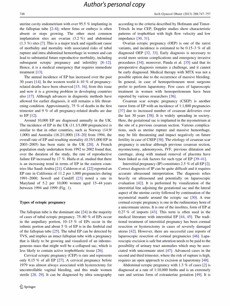
between 1994 and 1999 (Fig. 1).
Types of ectopic pregnancy
The fallopian tube is the dominant site [24] in the majority
of cases of tubal ectopic pregnancy. 75–80 % of EPs occur
in the ampullary portion, 10–15 % of EPs occur in the
isthmic portion and about 5 % of EP is in the fimbrial end
of the fallopian tube [25]. The tubal EP can be detected by
TVS, and implies an intact fallopian tube with a pregnancy
that is likely to be growing and visualized of an inhomo-
geneous mass that might well be a collapsed sac, which is
less likely to contain active trophoblastic tissue [26].
Cervical ectopic pregnancy (CEP) is rare and represents
only 0.15 % of all EP [27]. A cervical pregnancy before
1979 was almost always associated with hysterectomy for
uncontrollable vaginal bleeding, and this made women
sterile [28, 29]. It can be diagnosed by ultra sonography
according to the criteria described by Hofmann and Timor-
Tritsch. In true CEP, Doppler studies show characteristic
patterns of trophoblast with high flow velocity and low
impedance [30, 31].
Ovarian ectopic pregnancy (OEP) is one of the rarest
variants, and incidence is estimated to be 0.15–3 % of all
diagnosed OEP [32, 33]. Early diagnosis is necessary to
avoid more serious complications and emergency invasive
procedures [34]; moreover, Panda et al. [35] said that its
preoperative diagnosis remains a challenge, and it cannot
be early diagnosed. Medical therapy with MTX was not a
possible option due to the occurrence of massive bleeding.
In general, in case of hemoperitoneum most surgeons
prefer to perform laparotomy. Few cases of laparoscopic
treatment in women with hemoperitoneum have been
reported by various researchers [36].
Cesarean scar ectopic pregnancy (CSEP) is another
rarest form of EP with an incidence of 1:1,800 pregnancies
[37] due to increased number of cesarean deliveries over
the last 30 years [38]. It is widely spreading in society.
Here, the gestational sac is implanted in the myometrium at
the site of a previous cesarean section. Various complica-
tions, such as uterine rupture and massive hemorrhage,
may be life threatening and impact negatively on future
fertility in case of CSEP [38]. The etiology of cesarean scar
pregnancy is unclear although previous cesarean section,
myomectomy, adenomyosis, IVF, previous dilatation and
curettage, along with manual removal of placenta have
been linked as risk factors for such type of EP [39–41].
Interstitial pregnancy (IP) constitutes 2.5 % of all EP [2].
Correct diagnosis of IP can be quite difficult and it requires
accurate ultrasound interpretation. The diagnosis relies
heavily on ultrasound and potentially on laparoscopic
evaluation [42]. It is performed by visualization of the
interstitial line adjoining the gestational sac and the lateral
aspect of the uterine cavity followed by continuation of the
myometrial mantle around the ectopic sac [30]. A true
cornual ectopic pregnancy is one in the rudimentary horn of
a unicornuate uterus. It is one of the insolites, form of EP at
0.27 % of imports [43]. This term is often used in the
medical literature with interstitial EP [44, 45]. The tradi-
tional treatment of interstitial pregnancy has been cornual
resection or hysterectomy in cases of severely damaged
uterus [42]. However, there are successful case reports of
laparoscopic resection of cornual pregnancies [46]. Lapa-
roscopic excision is safe but attention needs to be paid to the
possibility of urinary tract anomalies which may be asso-
ciated with unicornuate uteri [47]. Advanced cases in the
second and third trimester, where the risk of rupture is high,
requires an open approach to excision at laparotomy [48].
Abdominal ectopic pregnancy with 1.3 % of cases [2] is
diagnosed at a rate of 1:10,000 births and is an extremely
rare and serious form of extrauterine gestation [49]. It is
748 Arch Gynecol Obstet (2013) 288:747–757
123
Author's personal copy

described as primary or secondary abdominal ectopic
pregnancy and usually results from an implantation fol-
lowing tubal rupture or abortion through the fimbricated
end of the fallopian tube. The fetus continues to grow
following attachment to an abdominal structure, using its
blood source, which may be extensive. It usually attaches
to the surface of the uterus, broad ligaments, or ovaries, but
may also attach to the liver, spleen, or intestines [24, 50].
The traditional management involves a laparotomy with
removal of the fetus with or without placental tissue [51].
One of the problems associated with the removal of
abdominal pregnancies after the first trimester is that the
risk of uncontrolled bleeding from the placental bed [52].
A heterotopic ectopic pregnancy is diagnosed when
women have any of the above said EP in conjunction with
an intra uterine pregnancy. It occurs with a rate\1:30,000
naturally occurring pregnancies, and 1:100 couples who
conceive through assisted reproduction [53]. It is also more
common (1–3 %) in in vitro fertilization and fertility
treatments involve superovulatory drugs [54, 55]. A high-
resolution transvaginal ultrasound with color Doppler will
be helpful to locate the trophoblastic tissue in the adnexa in
a case of heterotopic EP [56]. Different sites for ectopic
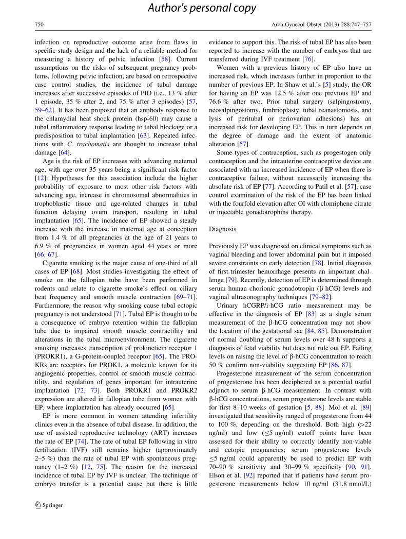
pregnancy are depicted in Fig. 2.
Risk factor
EP is further common in women who have suffered with
pelvic inflammatory disease (PID) and more than 50 % of
women who have been infected are unaware of the expo-
sure of PID [57]. Moreover, it is due to difficulties in
determining the effect of female genital chlamydial
Tubal EP (90-95%)
Cervical EP (0.15%)
Ovarian EP (0.15%-3%)
Caesarean scar EP (6%)
Interstitial EP (2.5%)
Abdominal EP (1.3%)
Heterotopic EP (1-3%)
Pelvic inflammatory disease (PID)
Age
Cigarette smoking
History of ART and IVF
Previous history of EP
Contraception pills
Light vaginal bleeding
Nausea and Vomiting
Lower abdominal pain
Sharp abdominal cramps
Pain on one side of the body
Dizziness or weakness
Pain in the shoulder, neck, rectum
PREGNANCY (EP)
Serum β-human chorionic gonadotropin (b-hCG) test
Urinary hCGRP/i-hCG ratio
Progesterone measurement
Transvaginal ultrasonography (TVS)
Computed Tomography (CT) or MRI
Vascular Endothelial Growth Factor (VEGF)
Creatine kinase (CK)
Disintegrin and Metalloprotease-12 (ADAM-12)
Hysterosalpingography (Hsg)
Expectant treatment
Medical treatment (systemic or local
route)
Surgical treatment (laparotomy or
laparoscopy)
Fig. 1 Summary of ectopic
pregnancy (EP)
Fig. 2 Different site for ectopic pregnancy
Arch Gynecol Obstet (2013) 288:747–757 749
123
Author's personal copy
infection on reproductive outcome arise from flaws in
specific study design and the lack of a reliable method for
measuring a history of pelvic infection [58]. Current
assumptions on the risks of subsequent pregnancy prob-
lems, following pelvic infection, are based on retrospective
case control studies, the incidence of tubal damage
increases after successive episodes of PID (i.e., 13 % after
1 episode, 35 % after 2, and 75 % after 3 episodes) [57,
59–62]. It has been proposed that an antibody response to
the chlamydial heat shock protein (hsp-60) may cause a
tubal inflammatory response leading to tubal blockage or a
predisposition to tubal implantation [63]. Repeated infec-
tions with C. trachomatis are thought to increase tubal
damage [64].
Age is the risk of EP increases with advancing maternal
age, with age over 35 years being a significant risk factor
[12]. Hypotheses for this association include the higher
probability of exposure to most other risk factors with
advancing age, increase in chromosomal abnormalities in
trophoblastic tissue and age-related changes in tubal
function delaying ovum transport, resulting in tubal
implantation [65]. The incidence of EP showed a steady
increase with the increase in maternal age at conception
from 1.4 % of all pregnancies at the age of 21 years to
6.9 % of pregnancies in women aged 44 years or more
[66, 67].
Cigarette smoking is the major cause of one-third of all
cases of EP [68]. Most studies investigating the effect of
smoke on the fallopian tube have been performed in
rodents and relate to cigarette smoke’s effect on ciliary
beat frequency and smooth muscle contraction [69–71].
Furthermore, the reason why smoking cause tubal ectopic
pregnancy is not understood [71]. Tubal EP is thought to be
a consequence of embryo retention within the fallopian
tube due to impaired smooth muscle contractility and
alterations in the tubal microenvironment. The cigarette
smoking increases transcription of prokineticin receptor 1
(PROKR1), a G-protein-coupled receptor [65]. The PRO-
KRs are receptors for PROK1, a molecule known for its
angiogenic properties, control of smooth muscle contrac-
tility, and regulation of genes important for intrauterine
implantation [72, 73]. Both PROKR1 and PROKR2
expression are altered in fallopian tube from women with
EP, where implantation has already occurred [65].
EP is more common in women attending infertility
clinics even in the absence of tubal disease. In addition, the
use of assisted reproductive technology (ART) increases
the rate of EP [74]. The rate of tubal EP following in vitro
fertilization (IVF) still remains higher (approximately
2–5 %) than the rate of tubal EP with spontaneous preg-
nancy (1–2 %) [12, 75]. The reason for the increased
incidence of tubal EP by IVF is unclear. The technique of
embryo transfer is a potential cause but there is little
evidence to support this. The risk of tubal EP has also been
reported to increase with the number of embryos that are
transferred during IVF treatment [76].
Women with a previous history of EP also have an
increased risk, which increases further in proportion to the
number of previous EP. In Shaw et al.’s [5] study, the OR
for having an EP was 12.5 % after one previous EP and
76.6 % after two. Prior tubal surgery (salpingostomy,
neosalpingostomy, fimbrioplasty, tubal reanastomosis, and
lysis of peritubal or periovarian adhesions) has an
increased risk for developing EP. This in turn depends on
the degree of damage and the extent of anatomic
alteration [57].
Some types of contraception, such as progestogen only
contraception and the intrauterine contraceptive device are
associated with an increased incidence of EP when there is
contraceptive failure, without necessarily increasing the
absolute risk of EP [77]. According to Patil et al. [57], case
control examination of the risk of the EP has been linked
with the fourfold elevation after OI with clomiphene citrate
or injectable gonadotrophins therapy.
Diagnosis
Previously EP was diagnosed on clinical symptoms such as
vaginal bleeding and lower abdominal pain but it imposed
severe constraints on early detection [78]. Initial diagnosis
of first-trimester hemorrhage presents an important chal-
lenge [79]. Recently, detection of EP is determined through
serum human chorionic gonadotropin (b-hCG) levels and
vaginal ultrasonography techniques [79–82].
Urinary hCGRP/i-hCG ratio measurement may be
effective in the diagnosis of EP [83] as a single serum
measurement of the b-hCG concentration may not show
the location of the gestational sac [84, 85]. Demonstration
of normal doubling of serum levels over 48 h supports a
diagnosis of fetal viability but does not rule out EP. Failing
levels on raising the level of b-hCG concentration to reach
50 % confirm non-viability suggesting EP [86, 87].
Progesterone measurement of the serum concentration
of progesterone has been deciphered as a potential useful
adjunct to serum b-hCG measurement. In contrast with
b-hCG concentrations, serum progesterone levels are stable
for first 8–10 weeks of gestation [5, 88]. Mol et al. [89]
investigated that sensitivity ranged of progesterone from 44
to 100 %, depending on the threshold. Both high ([22
ng/ml) and low (B5 ng/ml) cutoff points have been
assessed for their ability to correctly identify non-viable
and ectopic pregnancies; serum progesterone levels
B5 ng/ml could apparently be used to predict EP with
70–90 % sensitivity and 30–99 % specificity [90, 91].
Elson et al. [92] reported that if patients have serum pro-
gesterone measurements below 10 ng/ml (31.8 nmol/L)
750 Arch Gynecol Obstet (2013) 288:747–757
123
Author's personal copy

and b-hCG levels below 1,500 mIU/L are more likely to
demonstrate spontaneous resolution of EP.
Transvaginal ultrasound scan (TVS) is very popular
from 1980, and by the mid 1990 sensitivity and specificity
were calculated at 84.4 and 98.9 %, respectively. It
remains the gold standard for diagnosis of EP [30, 93]. A
b-hCG level that has elevated above the detestable
threshold in the absence of sonographic signs of early
pregnancy is considered concomitant conformation of an
EP. With the evolution in ultrasound technology, the
detestable threshold has dropped from 6,500 IU/L with a
transabdominal approach to between 1,000 and 2,000 IU/L
with transvaginal imaging [94]. The spectrum of sono-
graphic findings in EP is broad. Identification of an extra-
uterine gestational sac containing a yolk sac (with or
without an embryo) confirms the diagnosis for EP [95].
Pregnant women generally do not undergo computed
tomography (CT) and MRI examination, due to radiation
but should be ruled out in all young women complaining of
the abdominal pain. CT findings of the ruptured EP are
sporadic and extremely rare. In emergency situations, the
role of CT imaging of the abdominal and pelvic cavity has
been evaluated: it remains the first-line treatment in such
situations, [96–98]. Usually, CT diagnosis is reported in the
context of suspected cases when the patient is extremely
unstable. The CT scans clearly identified the site of
bleeding and helped to differentiate and characterize other
various causes of acute abdominal pain [98, 99]. Some-
times, an MRI can be helpful as well; moreover, this is not
a first-line examination. It is rather used for a better pre-
operative planning, or as a problem-solving tool in preg-
nant patients, or for imaging of fetal anatomy and
pathology [100, 101].
Vascular endothelial growth factor (VEGF) is a potent
angiogenic factor that acts as a modulator of vascular
growth, remodeling, and permeability in the endometrium,
decidua, and trophoblast, as well as during vascular
development in the embryo, all of which are crucial pro-
cesses related to normal implantation and placentation
[102]. Serum values of VEGF were significantly increased
in EP. Daponte et al. [79] described higher serum VEGF
concentrations in women with EP (median 227.2 pg/ml)
than with abnormal intrauterine pregnancy (median
107.2 pg/ml) (p \ 0.001) and it concluded that VEGF
serum concentrations might be a useful marker for EP, and
suggested 174 pg/ml as the cut-off value for EP diagnosis.
On the other hand, some groups have found conflicting
results on whether serum measurement of VEGF could be
used for differentiation of EP [81, 103].
Existing evidence suggests elevated creatine kinase
(CK) as a tool for diagnosis of EP. The trophoblast usually
invades the muscle layer and maternal blood vessels are
eroded, allowing muscle cell products such as CK to enter
the circulation [104]; therefore, increased serum CK levels
are normal during EP [104, 105]. Saha et al. [105] per-
formed a study comprising 40 women; total serum CK
levels were found to be significantly higher in the EP group
as compared to the controls (p \ 0.001), suggesting that
this test might be used as a indicator for EP. Similarly,
Katsikis et al. [106] studied 40 women with EP; and con-
cluded that women with EP had significantly higher CK
concentrations compared to women with intrauterine
abortive pregnancies and controls, suggesting that CK
concentrations could be used to predict EP.
Disintegrin and metalloprotease-12 (ADAM-12), a
proteomics evaluation of serum from women with EP, is
diagnosed with the presence of latter has both novel
marker disintegrin and metalloprotease-12. It has both an
adhesion and protease domain, plays a role in myoblast
fusion [107] as well as giant cell macrophage and osteo-
clast formation in bone [108]. In humans, ADAM-12 is
expressed in placenta, and potently provokes myogenesis.
In first-trimester placentas, it is localized to the cyto-
trophoblasts as well as the apical side of the synctio-
trophoblasts and to play a role in syncytial fusion in the
trophoblast [109]. If ADAM-12 is involved in the normal
implantation of pregnancy, and decreased levels are a
harbinger of an abnormal pregnancy or the abnormal
implantation of pregnancy, then decreased levels in ecto-
pic pregnancy may be biologically plausible; the ADAM-
12 test would be more sensitive in the group of EP with
lower b-hCG levels [110–112].
Hysterosalpingography (Hsg) is the radiographic eval-
uation of the uterine cavity and fallopian tubes after the
administration of a radio opaque medium through the
cervical canal. The Hsg was first practiced in 1910 and was
considered to be the special radiologic procedure. A
properly performed Hsg can decipher the contour of the
uterine cavity and the width of the cervical canal. Further
contrast medium injection will outline the cornua isthmic
and ampullary portions of the tubes and will show the
degree of spillage [113, 114]. There is a high probability
that tubal obstruction really exist because of high speci-
ficity of Hsg, while the observation of tubal permeability
shown after the examination does not exclude tubal
pathology, since it does not assess its function. In addition,
Hsg is a safe and inexpensive procedure [115, 116]; being
the most cost effective method in the study of the fallopian
tubes EP [117].
Medical management
The treatment option of EP involves surgical treatment by
laparotomy or laparoscopy, and medical treatment is usu-
ally systemic or through local route, or by expectant
treatment [118, 119].
Arch Gynecol Obstet (2013) 288:747–757 751
123
Author's personal copy

Expectant treatment
Expectant treatment can be applied in a selected subset of
patients with self-limiting ectopic pregnancy; the propor-
tion over treated must be accepted until a marker that
identifies this subgroup of patients is found [120, 121].
Studies evaluating expectant management of ectopic
pregnancy are primarily based on this concept of tropho-
blast in regression, and therefore exposed to the uncer-
tainties of definite primary EP which are diagnosis [122].
According to the most recent guideline, published by the
American College of Obstetricians and Gynecologists,
there may be a role for expectant management when the
b-hCG level is \200 mIU/ml and which is further in
decline phase. It should only be offered when TVS remains
non-diagnostic and b-hCG levels continue to decline.
Tubal rupture has occurred with low or declining b-hCG
levels. However, almost all EPs resolve spontaneously
when the b-hCG level reaches 15 mIU/ml [123, 124].
Another multivariate analysis has shown that the favorable
prognostic signs for successful expectant management of
ectopic pregnancy are the following—absent or minimal
clinical symptoms with no evidence of haemodynamic
compromise: evidence of ectopic resolution by declining
b-hCG levels preceding expectant treatment can be used
for such dilation; low initial serum b-hCG: successful
expectant management occurs in 98 % of cases for hCG
\200 IU/L, in 73 % for b-hCG\500 IU/L and in 25 % for
b-hCG \2,000 IU/L. Overall, if initial serum b-hCG
\1,000 IU/L then successful expectant management might
occur in most patients (88 %) with an ectopic pregnancy
size of \4 cm, without a fetal heart beat on transvaginal
sonography; followed by haemoperitoneum \50 ml. Evi-
dence of ectopic resolution on scan is another way to
diagnosis. A decrease in ectopic pregnancy size on day 7
had a sensitivity of 84 % and specificity of 100 % in pre-
dicting spontaneous resolution [122].
Medical treatment
Medical treatment of EP is quite less expensive than sur-
gery [125]. Many different agents have been used to treat
ectopic pregnancies including systemic and local metho-
trexate (MTX), local potassium chloride, hyperosmolar
glucose, prostaglandins, danazol, etoposide, and mifepri-
stone (RU486) [126–128]. Current therapies focus pri-
marily on MTX treatments. A better understanding of the
pathogenesis of the disease could avoid the risk in women
by providing better prediction and prevention [9, 65]. MTX
was first used in diagnosed EP in the 1960 to aide safe
surgical removal of the placenta from its abdominal
implantation sites in second and third trimester cases [129].
Patients treated with MTX should be monitored closely
because as mentioned earlier, it causes severe abdominal
pain and side effect too. The serum b-hCG concentration
should be measured weekly. If the serum b-hCG concen-
tration has not declined by at least 25 % in first week after
MTX administration, a second dose should be given which
is only required 15–20 % of patients [6]. Two common
regimens are available for MTX, multidose (MTX 1.0 mg/kg
i.m daily; days 0, 2, 4, and 6 alternated with folinic acid
0.1 mg/kg orally on days 1, 3, 5, 7) and single dose (MTX
0.4 to 1.0 mg/kg or 50 mg/m2 i.m. without folinic acid)
[129]. The multidose regimen alternates an every other day
dose of intramuscular MTX 1.0 mg/kg with an every other
day dose of intramuscular leucovorin calcium 0.1 mg/kg, a
folic acid antagonist antidote, up to four doses of each until
the b-hCG level decreases by 15 % on two consecutive
days. The single-dose regimen is an intramuscular injection
of MTX, 50 mg/m2, based on the patient’s body surface,
and does not include leucovorin rescue. If b-hCG levels do
not decline by 15 % on days 4 and 7 after treatment, a
second dose of MTX may be given after 1 week. About
20 % of women will need a second treatment cycle [130–
133]. Many side-effects associated with MTX treatment are
nausea and vomiting, stomatitis, diarrhea, abdominal dis-
comfort, pneumonitis, photosensitivity skin reaction,
impaired liver function, reversible, severe neutropenia
(rare), reversible alopecia (rare) [122].
Gabbur et al. reported that on its retrospective analysis
of stable women with small unruptured EP treated with
single-dose intramuscular MTX concluded that day 4 post
treatment b-hCG levels do not predict successful treatment
or need for surgery. Only day 7 b-hCG levels were asso-
ciated with successful single-dose MTX treatment [134].
Barnhart et al. [135] investigated in their meta-analysis
of both regimens (multi dose and single dose) and con-
cluded that the multi-dose regimen was more effective than
the single-dose regimen, with success rate reported as 93 %
for the multi-dose regimen and 88 % for the single-dose
regimen.
Kirk et al. evaluated that the TVS is a non-surgical
workup logarithm of patients with suspected EP. From
1993, a monitoring protocol has been developed based on
serial serum b-hCG taken evaluated on day 1, 4, 7, and
weekly until resolution. Efficacy of treatment is determined
when there is a C15 % fall in serum b-hCG between days 4
and 7. This definition of treatment success has a positive
predictive value (PPV) of 93 %, with a sensitivity of 93 %
and a specificity of 84.2 % [136].
Barnhart et al. was attempted by the challenge to
develop an optimum regimen that balances efficacy and
safety on the one hand and convenience on the other hand,
and he first described what is called the ‘‘double-dose
protocol’’. In a study that included 101 patients, two doses
752 Arch Gynecol Obstet (2013) 288:747–757
123
Author's personal copy

of MTX were administered on days 0 and 4 without
measuring b-hCG between doses. The authors reported a
success rate of 76 % after two doses and 87 % after a
further two doses [137].
Hossam et al. found that the double-dose protocol was
an efficient and safe alternative to the single-dose regimen.
It has the advantage of a shorter follow-up duration that
improves patient compliance, treatment satisfaction, and
costs [138].
Surgical treatment
Surgical treatment is the preferred treatment for EP when
there is rupture, hypotension, anemia, diameter of the
gestational sac greater than 4 cm on ultrasonography, or
pain persisting beyond 24 h [139]. In America, the first
abdominal surgery for EP was performed in 1759 by John
Bard, and became increasingly attempted in the nineteenth
century. Robert Lawson Tait, an eminent British surgeon,
described treatment of ruptured EP by ligating bleeding
vessels at laparotomy in 1884. This was a major
advancement in development of effective surgical man-
agement of this condition [140]. Surgical treatment of EP
should be reserved for those patients who have contrain-
dications to medical treatment or to whom medical treat-
ment has failed and those who are hemodynamically
unstable. Two techniques are described to remove the EP
from the fallopian tube—(1) salpingectomy: the pregnancy
is removed en bloc with the tube, (2) salpingostomy: an
incision is made on the fallopian tube over the swelling, the
EP carefully removed with forceps or irrigation and the
incision should be either closed or left to heal by secondary
intention [125, 140].
The preferred method of surgical treatment of EP today
is diagnostic laparoscopy with salpingostomy and tubal
conservation [130, 141]. Laparotomy is indicated in the
case of hemodynamic instability because it allows rapid
access to pelvic structures [130]. The success rate of sal-
pingostomy is 92 % and failure cases can be managed with
MTX [142]. Serial b-hCG measurements should be taken
until undetectable to be certain that there is no persistence
of trophoblastic tissue. Sometimes a prophylactic dose of
MTX is given with salpingosotomy [130].
Persistent EP occurs as a result of incomplete removal of
trophoblastic tissue [143], the most common complication
of laparoscopic salpingostomy, occurs at a frequency of
5–20 % [139, 144]. It is diagnosed during follow-up when
b-hCG concentrations measured once a week plateau
or rise. Factors increasing risk are small ectopic pregnan-
cies (\2 cm diameter), early therapy (\42 days from
last menstrual period), high concentrations of b-hCG
([3,000 IU/L) preoperatively, and implantation medial to
the salpingostomy site [145]. In high-risk cases, a single
dose of MTX (1 mg/kg) can be administered postopera-
tively for prophylaxis [144, 145]. In one randomized con-
trolled trial of laparoscopic surgery, prophylactic MTX
lowered the rate of persistent ectopic pregnancy from 14.5
to 1.9 %. The major benefit was in the shorter duration of
postoperative monitoring [144]. Since experience is lim-
ited, there is no single optimum treatment as on date. In the
largest series, all of 19 patients with persistent ectopic
pregnancies were successfully treated with single-dose
systemic MTX (50 mg/m2) [143].
Discussion and conclusion
Ectopic pregnancy in developing countries is a serious
threat, just because of poor medical facility so that a sig-
nificant morbidity rate and the potential for maternal death
generally are seen. Many patients have no documented risk
factors and no physical indications of EP, yet they suffer
from the complication. On the other hand, in developed
countries, it is now not so threatening as in past because
they have advanced technique of diagnosis and women are
much more aware of their health. Management is dictated
by the clinical presentation, serum b-hCG levels and TVS
findings. Expert consultation with radiologists and gyne-
cologists is recommended whenever ectopic pregnancy is
suspected. The use of MTX for treatment of early unrup-
tured EP reported to be safe and effective. Surgical treat-
ment is particularly appropriate for women who are
hemodynamically unstable or unlikely to be compliant with
post treatment monitoring and those who do not have
immediate access to medical care. The choice of treatment
should be guided by the patient’s preference, after a
detailed discussion about monitoring, outcome, risks, and
benefits of the approaches. The radiologists and gynecol-
ogists should have been firstly the identification of clinical
features or biomarkers predictive of MTX success and the
secondly is the use of additional medical treatments or
novel adjuncts that reduce treatment failures. The current
analysis of EP would suggest declining trends over time.
However, this reflects a decrease in surgical treatment and
not an actual decline in EP occurrence. Further, new ave-
nues are needed to explore early detection and less side
effect medication of the EP.
Conflict of interest None declared.
References
1. Kirk E, Bourne T (2011) Ectopic pregnancy. Obstet Gynecol
Reprod Med 21:207–211
Arch Gynecol Obstet (2013) 288:747–757 753
123
Author's personal copy

2. Khan KS, Wojdyla D, Say L, Gulmezoglu AM, Van Look PF
(2006) WHO analysis of causes of maternal death: a systematic
review. Lancet 367:1066–1074
3. Walker JJ (2007) Ectopic pregnancy. Clin Obstet Gynecol
50:89–99
4. Varma R, Gupta J (2009) Tubal ectopic pregnancy. Clin Evid
20:406
5. Shaw JL, Dey SK, Critchley HO, Horne AW (2010) Current
knowledge of the aetiology of human tubal ectopic pregnancy.
Hum Reprod Update 16:432–444
6. Sivalingam VN, Duncan WC, Kirk E, Shephard LA, Horne AW
(2011) Diagnosis and management of ectopic pregnancy. J Fam
Plann Reprod Health Care 37:231–240
7. Bouyer J, Coste J, Fernandez H (2002) Sites of ectopic preg-
nancy: a 10 year population-based study of 1800 cases. Hum
Reprod 17:3224–3230
8. Musa J (2009) Ectopic pregnancy in Jos Northern Nigeria:
prevalence and impact on subsequent fertility. Niger J Med
18:8–35
9. Barnhart KT (2009) Clinical practice. Ectopic pregnancy.
N Engl J Med 361:379–387
10. Stovall TG, Ling FW, Carson SA, Buster JE (1990) Nonsurgical
diagnosis and treatment of tubal pregnancy. Fertil Steril
54:537–538
11. Chandrasekhar C (2008) Ectopic pregnancy: a pictorial review.
Clin Imaging 32:468–473
12. Farquhar CM (2005) Ectopic pregnancy. Lancet 366:583–591
13. Dickens BM, Faundes A, Cook RJ (2003) Ectopic pregnancy
and emergency care: ethical and legal issues. Int J Gynecol
Obstet 82:121–126
14. Gamzu R, Almog B, Levin Y, Avni A, Jaffa A, Lessing J (2002)
Efficacy of methotrexate treatment in extrauterine pregnancies
defined by stable or increasing human chorionic gonadotropin
concentrations. Fertil Steril 77:761–765
15. Valley VT, Mateer JR, Aiman EJ (1998) Serum progesterone
endovaginal sonography by emergency physicians in the eval-
uation of ectopic pregnancy. Acad Emerg Med 5:309–313
16. Marion LL, Meeks GR (2012) Ectopic pregnancy: history,
incidence, epidemiology, and risk factors. Clin Obstet Gynecol
55:376–386
17. Wedderburn CJ, Warner P, Graham B, Duncan WC, Critchley
HO, Horne AW (2010) Economic evaluation of diagnosing and
excluding ectopic pregnancy. Hum Reprod 25:328–333
18. Bakken IJ, Skjeldestad FE (2003) Incidence and treatment of
extrauterine pregnancies in Norway 1990–2001. Tidsskr Nor
Laegeforen 123:3016–3020
19. Boufous S, Quartararo M, Mohsin M (2001) Trends in the
incidence of ectopic pregnancy in New South Wales between
1990–1998. Aust N Z J Obstet Gynaecol 41:436–438
20. Lewis G (2007) Saving mothers’ lives: reviewing maternal
deaths to make motherhood safer 2003–2005. CEMACH,
London
21. Al-Turki HA (2013) Trends in ectopic pregnancies in Eastern
Saudi Arabia. ISRN Obstet Gynecol, article ID 975251
22. Calderon JL, Shaheen M, Pan D, Teklehaimenot S, Robinson
PL, Baker RS (2005) Multi-cultural surveillance for ectopic
pregnancy: California 1991–2000. Ethn Dis 15:S4–S5
23. Sewell CA, Cundiff GW (2002) Trends for inpatient treatment
of tubal pregnancy in Maryland. Am J Obstet Gynecol
186:404–408
24. Condous G (2004) The management of early pregnancy com-
plications. Best Pract Res Clin Obstet Gynaecol 18:37–57
25. Ackerman TE, Levi CS, Dashefsky SM (1993) Interstitial line:
sonographic finding in interstitial (cornual) ectopic pregnancy.
Radiology 189:83–87
26. Kirk E, Daemen A, Papageorghiou AT (2008) Why are some
ectopic pregnancies characterized as pregnancies of unknown
location at the initial transvaginal ultrasound examination? Acta
Obstet Gynecol Scand 87:1150–1154
27. Webb EM, Green GE, Scoutt LM (2004) Adnexal mass with
pelvic pain. Radiol Clin North Am 42:329–348
28. Ushakov FB, Elchalal U, Aceman PJ (1996) Cervical preg-
nancy: past and future. Obstet Gynecol Surv 52:45–59
29. Leeman LM, Wendland CL (2000) Cervical ectopic pregnancy:
diagnosis with endocervical ultrasound examination and suc-
cessful treatment with methotrexate. Arch Fam Med 9:72–77
30. Jurkovic D, Marvelos D (2007) Catch me if you can: ultrasound
diagnosis of ectopic pregnancy. Ultrasound Obstet Gynecol
30:1–7
31. Lemus JF (2000) Ectopic pregnancy: an update. Curr Opin
Obstet Gynecol 12:369–375
32. Odejinmi F, Rizzuto MI, MacRae R, Olowu O, Hussain M
(2009) Diagnosis and laparoscopic management of 12 consec-
utive cases of ovarian pregnancy and review of literature.
J Minim Invasive Gynecol 16:354–359
33. Gon S (2011) Two cases of primary ectopic ovarian pregnancy.
OJHAS 10(1):26
34. Plotti F, Di GA, Oliva C, Battaglia FG (2008) Plotti, ‘‘Bilateral
ovarian pregnancy after intrauterine insemination and controlled
ovarian stimulation’’. Fertil Steril 90(5):2015.e3–2015.e5
35. Panda S, Darlong LM, Singh S, Borah T (2009) Case report of a
primary ovarian pregnancy in a primigravida. J Hum Reprod Sci
2:90–92
36. Odejinmi F, Sangrithi M, Olowu O (2011) Operative laparos-
copy as the mainstay method in management of hemodynami-
cally unstable patients with ectopic pregnancy. J Minim Invasive
Gynecol 18:179–183
37. Seow K, Hang L, Lin Y (2004) Cesarean scar pregnancy: issues
in management. Ultrasound Obstet Gynecol 23:247–253
38. Rotas MA, Haberman S, Levgur M (2006) Cesarean scar ectopic
pregnancies: etiology, diagnosis, and management. Obstet
Gynecol 107:1373–1381
39. Jin H, Shou J, Yu Y (2004) Intramural pregnancy, a report of
two cases. J Reprod Med 49:569–572
40. Graesslin O, Dedecker F, Quereux C (2005) Conservative
treatment of ectopic pregnancy in a cesarean scar. Obstet
Gynecol 105:869–871
41. Shufaro Y, Nadjari M (2001) Implantation of a gestational sac in
a cesarean section scar. Fertil Steril 75:1217
42. Katz DL, Barrett JP, Sanfilippo JS, Badway DM (2003) Com-
bined hysteroscopy and laparoscopy in the treatment of inter-
stitial pregnancy. Am J Obstet Gynecol 188:1113–1114
43. Nahum GG (2002) Rudimentary uterine horn pregnancy. The
20th century worldwide experience of 588 cases. J Reprod Med
47:151–163
44. Malinowski A, Bates SK (2006) Semantics and pitfalls in the
diagnosis of cornual/interstitial pregnancy. Fertil Steril 86:e11–e14
45. Kun WM, Tung WK (2001) On the look out for a rarity—
interstitial/cornual pregnancy. Eur J Emerg Med 8:147–150
46. Moon HS, Choi YJ, Park VH, Kim SG (2000) New simple
endoscopic operations for interstitial pregnancies. Am J Obstet
Gynecol 152:114–121
47. Sonmezer M, Taskin S, Atabekoglu C (2006) Laparoscopic
management of rudimentary uterine horn pregnancy: case report
and literature review. JSLS 10:396–399
48. Panayotidis C, Abdel FM, Leggott M (2004) Rupture of rudi-
mentary uterine horn of a unicornuate uterus at 15 weeks’ges-
tation. J Obstet Gynecol 24:323–324
49. Yildizhan R, Kurdoglu M, Kolusari A, Erten R (2008) Primary
omental pregnancy. Saudi Med J 29:606–609
754 Arch Gynecol Obstet (2013) 288:747–757
123
Author's personal copy

50. Sarwat A, Nadia A (2011) Abdominal pregnancy: a diagnostic
dilemma. Prof Med J 18:479–484
51. Ayinde OA, Aimakhu CO, Adeyanju OA (2005) Abdominal
pregnancy at the University College Hospital, Ibadan: a ten-year
review. Afr J Reprod Health 9:123–127
52. Oki T, Baba Y, Yoshinaga M (2008) Super-selective arterial
embolization for uncontrolled bleeding in abdominal pregnancy.
Obstet Gynecol 112:427–429
53. Ludwig M (1999) Heterotopic pregnancy in a spontaneous
cycle: do not forget about it. Eur J Obstet Gynecol Reprod Biol
87:91–103
54. Rojansky N, Schenker JG (1996) Heterotopic pregnancy and
assisted reproduction—an update. J Assist Reprod Genet
13:594–601
55. Condous G, Okaro E, Bourne T (2003) The conservative man-
agement of early pregnancy complications: a review of the lit-
erature. Ultrasound Obstet Gynecol 22:420–430
56. Glassner MJ, Aron E, Eskin BA (1990) Ovulation induction
with clomiphene and the rise in heterotopic pregnancies: a report
of two cases. J Reprod Med 35:175–178
57. Madhuri P (2012) Ectopic pregnancy after infertility treatment.
J Hum Reprod Sci 5:154–165
58. Onan MA, Turp AB, Saltik A, Akyurek N, Taskiran C, Him-
metoglu O (2005) Primary omental pregnancy: case report. Hum
Reprod 20:807–809
59. Bakken IJ (2008) Chlamydia trachomatis and ectopic preg-
nancy: recent epidemiological findings. Curr Opin Infect Dis
21:77–82
60. Bjartling C, Osser S, Persson K (2007) Deoxyribonucleic acid of
Chlamydia trachomatis in fresh tissue from the fallopian tubes
of patients with ectopic pregnancy. Eur J Obstet Gynecol Re-
prod Biol 134:95–100
61. Low N, Egger M, Sterne JA, Harbord RM, Ibrahim F, Lindblom
B, Herrmann B (2006) Incidence of severe reproductive tract
complications associated with diagnosed genital chlamydial
infection: the Uppsala Women’s Cohort Study. Sex Transm
Infect 82:212–218
62. Van Valkengoed IG, Morre SA, van den Brule AJ, Meijer CJ,
Bouter LM, Boeke AJ (2004) Overestimation of complication
rates in evaluations of Chlamydia trachomatis screening pro-
grammes—implications for cost-effectiveness analyses. Int J
Epidemiol 33:416–425
63. Ault KA, Statland BD, King MM, Dozier DI, Joachims ML,
Gunter J (1998) Antibodies to the chlamydial 60 kilodalton heat
shock protein in women with tubal factor infertility. Infect Dis
Obstet Gynecol 6:163–167
64. Rank RG, Dascher C, Bowlin AK, Bavoil PM (1995) Systemic
immunization with Hsp60 alters the development of chlamydial
ocular disease. Invest Ophthalmol Vis Sci 36:1344–1351
65. Shaw JL, Oliver E, Lee KF (2010) Cotinine exposure increases
fallopian tube PROKR1 expression via nicotinic AChRalpha-7:
a potential mechanism explaining the link between smoking and
tubal ectopic pregnancy. Am J Pathol 177:2509–2515
66. Nybo Andersen AM, Wohlfahrt J, Christens P, Olsen J, Melbye
M (2000) Maternal age and fetal loss: population based register
linkage study. BMJ 320(7251):1708–1712
67. Goddijn M, van der Veen F, Schuring Blom GH, Ankum WM,
Leschot NJ (1996) Cytogenetic characteristics of ectopic preg-
nancy. Hum Reprod 11:2769–2771
68. Bouyer J, Coste J, Shojaei T (2003) Risk factors for ectopic
pregnancy: a comprehensive analysis based on a large case-
control, population-based study in France. Am J Epidemiol
157:185–194
69. Knoll M, Shaoulian R, Magers T, Talbot P (1995) Ciliary beat
frequency of hamster oviducts is decreased in vitro by exposure
to solutions of mainstream and sidestream cigarette smoke. Biol
Reprod 53:29–37
70. Riveles K, Roza R, Arey J, Talbot P (2004) Pyrazine derivatives
in cigarette smoke inhibit hamster oviductal functioning. Reprod
Biol Endocrinol 2:23
71. Talbot P, Riveles K (2005) Smoking and reproduction: the
oviduct as a target of cigarette smoke. Reprod Biol Endocrinol
3:52
72. Li YY, Li L, Hwang IS, Tang F, O WS (2008) Coexpression of
adrenomedullin and its receptors in the reproductive system of
the rat: effects on steroid secretion in rat ovary. Biol Reprod
79:200–208
73. Evans J, Catalano RD, Morgan K, Critchley HO, Millar RP,
Jabbour HN (2008) Prokineticin 1 signaling and gene regulation
in early human pregnancy. Endocrinology 149:2877–2887
74. Clayton HB, Schieve LA, Peterson HB, Jamieson DJ, Reynolds
MA, Wright VC (2006) Ectopic pregnancy risk with assisted
reproductive technology procedures. Obstet Gynecol 107:595–
604
75. Strandell A, Thorburn J, Hamberger L (1999) Risk factors for
ectopic pregnancy in assisted reproduction. Fertil Steril
71:282–286
76. Weigert M, Gruber D, Pernicka E, Bauer P, Feichtinger W
(2009) Previous tubal ectopic pregnancy raises the incidence of
repeated ectopic pregnancies in in vitro fertilization-embryo
transfer patients. J Assist Reprod Genet 26:13–17
77. Furlong LA (2002) Ectopic pregnancy risk when contraception
fails. A review. J Reprod Med 47:881–885
78. McCord ML, Muram D, Buster JE, Arheart KL, Stovall TG,
Carson SA (1996) Single serum progesterone as a screen for
ectopic pregnancy: exchanging specificity and sensitivity to
obtain optimal test performance. Fertil Steril 66:513–516
79. Daponte A, Pournaras S, Zintzaras E, Kallitsaris A, Lialios G,
Maniatis AN (2005) The value of a single combined measure-
ment of VEGF, glycodelin, progesterone, PAPP-A, HPL and
LIF for differentiating between ectopic and abnormal intra-
uterine pregnancy. Hum Reprod 20:3163–3166
80. Miller WC, Ford CA, Morris M, Handcock MS, Schmitz JL,
Hobbs MM, Cohen MS, Harris KM, Udry JR (2004) Prevalence
of chlamydial and gonococcal infections among young adults in
the United States. JAMA 291:2229–2236
81. Kucera-Sliutz E, Schiebel I, Konig F, Leodolter S, Sliutz G,
Koelbl H (2002) Vascular endothelial growth factor (VEGF) and
discrimination between abnormal intrauterine and ectopic
pregnancy. Hum Reprod 17:3231–3234
82. Felemban A, Sammour A, Tulandi T (2002) Serum vascular
endothelial growth factor as a possible marker for early ectopic
pregnancy. Hum Reprod 17:490–492
83. Jae KL, Min JOh, Joong SS, Kyung JL, Jung HN, Jung HC, Jin
DC, Dong HC, In-Soo K, Paul IL (2005) Clinical effectiveness
of urinary human chorionic gonadotropin related protein
(hCGRP) quantification for diagnosis of ectopic pregnancy.
J Korean Med Sci 20:461–467
84. Kaplan BC, Dart RG, Moskos M, Kuligowska E, Chun B, Adel
HM (1996) Ectopic pregnancy: prospective study with improved
diagnostic accuracy. Ann Emerg Med 28:10–17
85. Kohn MA, Kerr K, Malkevich D, ONeil N, Kerr MJ, Kaplan BC
(2003) Beta-human chorionic gonadotropin levels and the like-
lihood of ectopic pregnancy in emergency department patients
with abdominal pain or vaginal bleeding. Acad Emerg Med
10:119–126
86. Barnhart KT, Sammel MD, Rinaudo PF, Zhou L, Hummel AC,
Guo W (2004) Symptomatic patients with an early viable
intrauterine pregnancy: hCG curves redefined. Obstet Gynecol
104:50–55
Arch Gynecol Obstet (2013) 288:747–757 755
123
Author's personal copy

87. Heather M, Hanadi B, Trevor B, Togas T (2005) Diagnosis and
treatment of ectopic pregnancy progesterone measurement.
CMAJ 173(8):905–912
88. Stovall TG, Ling FW, Gray LA, Carson SA, Buster JE (1991)
Methotrexate treatment of unruptured ectopic pregnancy: a
report of 100 cases. Obstet Gynecol 77(5):749–753
89. Mol BW, Hajenius PJ, Engelsbel S, Ankum WM, van der Veen
F, Hemrika DJ (1999) Can noninvasive diagnostic tools predict
tubal rupture or active bleeding in patients with tubal preg-
nancy? Fertil Steril 71:167–173
90. Dart R, Ramanujam P, Dart L (2002) Progesterone as a predictor
of ectopic pregnancy when the ultrasound is indeterminate. Am
J Emerg Med 20:575–579
91. Buckley RG, King KJ, Disney JD, Riffenburgh RH, Gorman JD,
Klausen JH (2000) Serum progesterone testing to predict ectopic
pregnancy in symptomatic first-trimester patients. Ann Emerg
Med 36:95–100
92. Elson J, Tailor A, Banerjee S, Salim R, Hillaby K, Jurkovic D
(2004) Expectant management of tubal ectopic pregnancy:
prediction of successful outcome using decision tree analysis.
Ultrasound Obstet Gynecol 23:552–556
93. Condous G (2006) Ectopic pregnancy—risk factors and diag-
nosis. Aust Fam Physician 35:854–857
94. Mehta TS, Levine D, Beckwith B (1997) Treatment of ectopic
pregnancy: is a human chorionic gonadotropin level of 2,000
mIU/mL a reasonable threshold. Radiology 205:569–573
95. Murray H, Baakdah H, Bardell T, Tulandi T (2005) Diagnosis
and treatment of ectopic pregnancy. CMAJ 173:905–912
96. Kirsch JD, Scoutt LM (2010) Imaging of ectopic pregnancy.
Appl Radiol 39:10–25
97. Cano AR, Borruel NS, Dıez MP (2009) Role of multidetector
CT in the management of acute female pelvic disease. Emerg
Radiol 16:453–472
98. Pham H, Lin EC (2007) Adnexal ring of ectopic pregnancy
detected by contrast-enhanced CT. Abdom Imaging 32(1):56–58
99. Shin BS, Park MH (2010) Incidental detection of interstitial
pregnancy on CT imaging. Korean J Radiol 11(1):123–125
100. Tamai K, Koyama T, Togashi K (2007) MR features of ectopic
pregnancy. Eur Radiol 17(12):3236–3246
101. Yoshigi J, Yashiro N, Kinoshita T (2006) Diagnosis of ectopic
pregnancy with MRI: efficacy of T2 weighted imaging. Magn
Reson Med Sci 5(1):25–32
102. Torry DS, Torry RJ (1997) Angiogenesis and the expression of
vascular endothelial growth factor in endometrium and placenta.
Am J Reprod Immunol 37:21–29
103. Ugurlu EN, Ozaksit G, Karaer A, Zulfikaroglu E, Atalay A,
Ugur M (2008) The value of vascular endothelial growth factor,
pregnancy-associated plasma protein-A, and progesterone for
early differentiation of ectopic pregnancies, normal intrauterine
pregnancies, and spontaneous miscarriages. Fertil Steril
91(5):1657–1661
104. Chandra L, Jain A (1995) Maternal serum creatine kinase as a
biochemical marker of tubal pregnancy. Int J Gynaecol Obstet
49:21–23
105. Saha PK, Gupta I, Ganguly NK (1999) Evaluation of serum
creatine kinase as a diagnostic marker for tubal pregnancy. Aust
N Z J Obstet Gynaecol 39:366–367
106. Katsikis I, Rousso D, Farmakiotis D, Kourtis A, Diamanti KE,
Zournatzi KV (2006) Creatine phosphokinase in ectopic preg-
nancy revisited: significant diagnostic value of its MB and MM
isoenzyme fractions. Am J Obstet Gynecol 194:86–91
107. Yagami HT, Sato T, Kurisaki T, Kamijo K, Nabeshima Y,
Fujisawa SA (1995) A metalloprotease-disintegrin participating
in myoblast fusion. Nature 377:652–656
108. Abe E, Mocharla H, Yamate T, Taguchi Y, Manolagas
SC (1999) Meltrin-alpha, a fusion protein involved in
multinucleated giant cell and osteoclast formation. Calcif Tissue
Int 64:508–515
109. Huppertz B, Bartz C, Kokozidou M (2006) Trophoblast fusion:
fusogenic proteins, syncytins and ADAMs, and other prerequi-
sites for syncytial fusion. Micron 37:509–517
110. Poon LC, Chelemen T, Granvillano O, Pandeva I, Nicolaides
KH (2008) First-trimester maternal serum a disintegrin and
metalloprotease 12 (ADAM12) and adverse pregnancy outcome.
Obstet Gynecol 112:1082–1090
111. Laigaard J, Cuckle H, Wewer UM, Christiansen M (2006)
Maternal serum ADAM12 levels in Down and Edwards’ syn-
drome pregnancies at 9–12 weeks’ gestation. Prenat Diagn
26:689–691
112. Spencer K, Cowans NJ, Stamatopoulou A (2008) ADAM12s in
maternal serum as a potential marker of pre-eclampsia. Prenat
Diagn 28:212–216
113. Swart P, Mol BWJ, Vander VF, Van BM, Redekop WK,
Bossuyt PMM (1995) The accuracy of hysterosalpingography in
the diagnosis of tubal pathology: a meta-analysis. Fertil Steril
64:486–491
114. Elito J Jr, Han KK, Camano L (2005) Tubal patency after
clinical treatment of unruptured ectopic pregnancy. Int J
Gynecol Obstet 88:309–313
115. Mol BWJ, Swart P, Bossuyt PMM, Van BM, Vander VF (1996)
Reproducibility of the interpretation of hysterosalpingography in
the diagnosis of tubal pathology. Hum Reprod 11:1204–1208
116. Papaioannou S, Afnan M, Jafettas J (2007) Tubal assessment
tests: still have not found what we are looking for. Reprod Bio
Med Online 15:376–382
117. Fertility Assessment and Treatment for People with Fertility
Problems (2004) Clinical Guideline. RCOG Press, London
118. Sowter M, Farquhar C, Petrie K, Gudex G (2001) A randomized
trial comparing single dose systemic methotrexate and laparo-
scopic surgery for the treatment of unruptured tubal pregnancy.
Brit J Obstet Gynaecol 108:192–203
119. Seror V, Gelfucci F, Gerbaud L, Pouly JL, Fernandez H, Job
Spira N, Bouyer J, Coste J (2007) Care pathways for ectopic
pregnancy: a population-based cost-effectiveness analysis. Fertil
Steril 87:737–748
120. Carson SA, Stovall TG, Ling FW, Buster JE (1991) Low human
chorionic somatomammotropin fails to predict spontaneous
resolution of unruptured ectopic pregnancies. Fertil Steril
55:629–630
121. Quasim SM, Trias A, Sachdev R, Kenmann E (1996) Evaluation
of serum creatinine kinase levels in ectopic pregnancy. Fertil
Steril 65:443–445
122. Rajesh V, Lawrence M (2002) Evidence-based management of
ectopic pregnancy. Curr Obstet Gynaecol 12:191–199
123. Barnhart KT, Fay CA, Suescum M, Sammel MD, Appleby D,
Shaunik A, Dean AJ (2011) Clinical factors affecting the
accuracy of ultrasonography in symptomatic first-trimester
pregnancy. Obstet Gynecol 117:299–306
124. American College of Obstetricians and Gynecologists (1998)
Medical management of tubal pregnancy. ACOG Practice Bul-
letin No. 3. Obstet Gynecol 92:1–7
125. Rodrigues SP, de Burlet KJ, Hiemstra E, Twijnstra AR, van
Zwet EW, Trimbos-Kemper TC, Jansen FW (2012) Ectopic
pregnancy: when is expectant management safe? Gynecol Surg
9:421–426
126. Raughley MJ, Frishman GN (2007) Local treatment of ectopic
pregnancy. Semin Reprod Med 25(2):99–115
127. van Mello NM, Mol F, Mol BW, Hajenius PJ (2009) Conser-
vative management of tubal ectopic pregnancy. Best Pract Res
23:509–518
128. Hajenius PJ, Mol BWJ, Ankum WM, Van der Veen F (2003)
Systemic and local medical therapy of tubal pregnancy. In:
756 Arch Gynecol Obstet (2013) 288:747–757
123
Author's personal copy

Timmerman D, Deprest J, Bourne T (eds) Ultrasound and
endoscopic surgery in obstetrics and gynaecology. A combined
approach to diagnosis and treatment. Springer, London. ISBN
3540762124
129. Condous G, Okaro E, Khalid A, Lu C, Van HS, Timmerman D
(2005) A prospective valuation of a single-visit strategy to
manage pregnancies of unknown location. Hum Reprod
20:1398–1403
130. Seeber BE, Barnhart KT (2006) Suspected ectopic pregnancy.
Obstet Gynecol 107:399–413
131. Jeng CJ, Ko ML, Shen J (2007) Transvaginal ultrasound-guided
treatment of cervical pregnancy. Obstet Gynecol 109:1076–
1082
132. Lin CY, Chang CY, Chang HM, Tsai EM (2008) Cervical
pregnancy treated with systemic methotrexate administration
and resectoscopy. Taiwan J Obstet Gynecol 47:4
133. Sijanovic S, Vidosavljevic D, Sijanovic I (2011) Methotrexate in
local treatment of cervical heterotopic pregnancy with success-
ful perinatal outcome: case report. J Obstet Gynaecol Res
37:1241–1245
134. Gabbur N, Sherer DM, Hellmann M (2006) Do serum beta-
human chorionic gonadotropin levels on day 4 following
methotrexate treatment of patients with ectopic pregnancy pre-
dict successful single-dose therapy? Am J Perinatol 23:193–196
135. Barnhart KT, Gosman G, Ashby R, Sammel M (2003) The
medical management of ectopic pregnancy: a meta-analysis
comparing ‘‘single dose’’ and ‘‘multidose’’ regimens. Obstet
Gynecol 101:778–784
136. Kirk E, Condous G, Van Calster B (2007) A validation of the
most commonly used protocol to predict the success of single-
dose methotrexate in the treatment of ectopic pregnancy. Hum
Reprod 22:858–863
137. Barnhart K, Hummel AC, Sammel MD, Menon S, Jain J, Cha-
khtoura N (2007) Use of ‘‘2-dose’’ regimen of methotrexate to
treat ectopic pregnancy. Fertil Steril 87:250–256
138. Hossam O, Hamed A, Salah R, Ahmed A, Abdullah A (2012)
Comparison of double- and single-dose methotrexate protocols
for treatment of ectopic pregnancy. Alghasham Int J Gynecol
Obstet 116:67–71
139. Buster JE, Carson SA (1995) Ectopic pregnancy; new advances
in diagnosis and treatment. Curr Opinion Obstet Gynecol
7:168–176
140. Fritz MA, Speroff L (2011) Clinical gynecologic endocrinology
and infertility, 8th edn. Wolters Kluwer Health/Lippincott
Williams & Wilkins, Philadelphia
141. Lozeau AM, Potter B (2005) Diagnosis and management of
ectopic pregnancy. Am Fam Physician 1707–14(19):20
142. Hajenius PJ, Mol BW, Bossuyt PM, Ankum WM, Vander VF
(2000) Interventions for tubal ectopic pregnancy. Cochrane
Database Syst Rev (2):CD000324
143. Hoppe DE, Bekkar BE, Nager CW (1994) Single-dose systemic
methotrexate for the treatment of persistent ectopic pregnancy
after conservative surgery. Obstet Gynecol 83:51–54
144. Graczykowski JW, Mishell DR (1997) Methotrexate prophy-
laxis for persistent ectopic pregnancy after conservative treat-
ment by salpingostomy. Obstet Gynecol 89:118–122
145. Seifer DB (1997) Persistent ectopic pregnancy: an argument for
heightened vigilance and patient compliance. Fertil Steril
68:402–404
Arch Gynecol Obstet (2013) 288:747–757 757
123
Author's personal copy