Mutations in Breast Cancer Exome Sequences Predict Susceptibility to Infections and Converge on the...
28
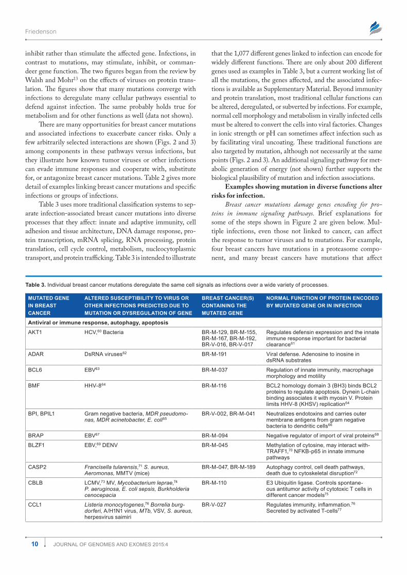
1 JOURNAL OF GENOMES AND EXOMES 2015:4 Mutations in Breast Cancer Exome Sequences Predict Susceptibility to Infections and Converge on the Same Signaling Pathways Bernard Friedenson Department of Biochemistry and Molecular Genetics, College of Medicine, University of Illinois Chicago, Chicago, IL, USA. ABSTRACT: Many mutations in breast cancer exome sequences alter susceptibility to infections. An exhaustive analysis of all the mutations in exomes from 103 breast cancer cases found that more than 1,000 genes have a published association with some kind of infection, including all known tumor viruses. Altered susceptibility to infection was identified as a common thread connecting breast cancer mutations in genes traditionally classified as coding for diverse functions, including cell immunity, cell architectural barriers, stromal interactions, cell adhesion, DNA damage responses, translation, cell cycle control, metabolism, homeostasis, transport, and neurosensing. Infections and mutations can both contribute to cancer because they deregulate the same pathways. In many cases, infections make a contribution to cancer that is either known or biologically plausible. Interventions may be possible to prevent occult infections from cooperating with mutations to cause further cancer, metastasis, or other complications. e emerging list of infection–gene mutation associations is readily scalable to routine testing of large human data sets. KEYWORDS: breast cancer, infection, viral cancer, cancer genome, cancer infection, breast cancer mutation CITATION: Friedenson. Mutations in Breast Cancer Exome Sequences Predict Susceptibility to Infections and Converge on the Same Signaling Pathways. Journal of Genomes and Exomes 2015:4 1–28 doi:10.4137/JGE.S30058. TYPE: Original Research RECEIVED: May 28, 2015. RESUBMITTED: July 8, 2015. ACCEPTED FOR PUBLICATION: July 14, 2015. ACADEMIC EDITOR: Stephen F. Kingsmore, Editor in Chief PEER REVIEW: Seven peer reviewers contributed to the peer review report. Reviewers’ reports totaled 2,237 words, excluding any confidential comments to the academic editor. FUNDING: This work was funded by the University of Illinois–Chicago and by personal funds. The author confirms that the funder had no influence over the study design, content of the article, or selection of this journal. COMPETING INTERESTS: Author discloses no potential conflicts of interest. COPYRIGHT: © the authors, publisher and licensee Libertas Academica Limited. This is an open-access article distributed under the terms of the Creative Commons CC-BY-NC 3.0 License. CORRESPONDENCE: [email protected]; [email protected] Paper subject to independent expert blind peer review. All editorial decisions made by independent academic editor. Upon submission manuscript was subject to anti- plagiarism scanning. Prior to publication all authors have given signed confirmation of agreement to article publication and compliance with all applicable ethical and legal requirements, including the accuracy of author and contributor information, disclosure of competing interests and funding sources, compliance with ethical requirements relating to human and animal study participants, and compliance with any copyright requirements of third parties. This journal is a member of the Committee on Publication Ethics (COPE). Provenance: The author was invited to submit this paper. Published by Libertas Academica. Learn more about this journal. Introduction Lesions are thought to become malignant because mutations accumulate over long periods to disable or deregulate essential cellular controls. Some mutations may activate proto-oncogenes to become uncontrolled oncogenes, and other mutations may inactivate tumor suppressor genes. About 15%–20% of cancers are known to be caused by tumor viruses or other infections. 1 Infectious and noninfectious cancers are considered as sepa- rate diseases and are even studied in separate disciplines. Rela- tionships between infections and cancers have produced some notable successes, such as the ability to prevent some cancers of the cervix and the liver. In other organs, associations between a single individual infection and cancer have been difficult to reproduce. For example, breast cancers have been associated with very different infections including retroviruses (mouse mammary tumor virus [MMTV] and human endogenous ret- rovirus [HERV]), a large double-stranded DNA virus (Epstein- Barr virus [EBV]), and a small double-stranded DNA virus (human papilloma virus [HPV]). Results linking any one of these infections to breast cancer are contradictory and difficult to reproduce. 2 Asymptomatic infections have spread tumor viruses through the population so that virtually everyone has been inoc- ulated with tumor viruses such as EBV and HPV. Tumor viruses have been widely reported in normal breasts, so if viruses cause cancer, most women should probably develop breast cancer. One reason this does not occur is because breast cancers may require mutations in genes that lead to compromised immunity. 3,4 Gene mutations that deregulate the immune system, cellular architecture, or underlying metabolic sup- port create errors in the signals that prevent viral infection and in signals that maintain resident tumor viruses in a latent state. In addition, some bacterial infections may cause chronic inflammation with continual cell proliferation in the pres- ence of mutagens or the infecting bacteria may even release carcinogenic metabolites. 5–7 Under these scenarios, no mat- ter what the infectious agent, mutations that damage host cell–protective mechanisms or normal cell functions would increase risks for cancers. In breast cancer, gene mutations can alter the ability of the immune system to control cancer-causing infections in multiple ways and high percentages of mutations can be linked to dam- age to protective signals. 3,4 Some host mutations interfere with signals that cells are under attack and that protective boundaries have become abnormal. Signals connecting innate and adaptive immunity may not work properly. Communication between cells and the extracellular matrix may be damaged by mutation. Changes in cellular morphology and metabolism are needed to convert normal cells into cancer or viral factories. Mutations in genes encoding proteins essential for transcription, mRNA splicing, or translation can facilitate or inhibit viral takeover.
Transcript of Mutations in Breast Cancer Exome Sequences Predict Susceptibility to Infections and Converge on the...
1Journal of Genomes and exomes 2015:4
Mutations in Breast Cancer Exome Sequences Predict Susceptibility to Infections and Converge on the Same Signaling Pathways
Bernard friedensonDepartment of Biochemistry and Molecular Genetics, College of Medicine, University of Illinois Chicago, Chicago, IL, USA.
ABSTR ACT: Many mutations in breast cancer exome sequences alter susceptibility to infections. An exhaustive analysis of all the mutations in exomes from 103 breast cancer cases found that more than 1,000 genes have a published association with some kind of infection, including all known tumor viruses. Altered susceptibility to infection was identified as a common thread connecting breast cancer mutations in genes traditionally classified as coding for diverse functions, including cell immunity, cell architectural barriers, stromal interactions, cell adhesion, DNA damage responses, translation, cell cycle control, metabolism, homeostasis, transport, and neurosensing. Infections and mutations can both contribute to cancer because they deregulate the same pathways. In many cases, infections make a contribution to cancer that is either known or biologically plausible. Interventions may be possible to prevent occult infections from cooperating with mutations to cause further cancer, metastasis, or other complications. The emerging list of infection–gene mutation associations is readily scalable to routine testing of large human data sets.
KEY WORDS: breast cancer, infection, viral cancer, cancer genome, cancer infection, breast cancer mutation
CITATION: friedenson. mutations in Breast Cancer exome sequences Predict susceptibility to Infections and Converge on the same signaling Pathways. Journal of Genomes and Exomes 2015:4 1–28 doi:10.4137/JGe.s30058.
TYPE: original research
RECEIVED: may 28, 2015. RESUBMITTED: July 8, 2015. ACCEPTED FOR PUBLICATION: July 14, 2015.
ACADEMIC EDITOR: stephen f. Kingsmore, editor in Chief
PEER REVIEW: seven peer reviewers contributed to the peer review report. reviewers’ reports totaled 2,237 words, excluding any confidential comments to the academic editor.
FUNDING: This work was funded by the university of Illinois–Chicago and by personal funds. The author confirms that the funder had no influence over the study design, content of the article, or selection of this journal.
COMPETING INTERESTS: Author discloses no potential conflicts of interest.
COPYRIGHT: © the authors, publisher and licensee libertas academica limited. This is an open-access article distributed under the terms of the Creative Commons CC-BY-nC 3.0 license.
CORRESPONDENCE: [email protected]; [email protected]
Paper subject to independent expert blind peer review. all editorial decisions made by independent academic editor. upon submission manuscript was subject to anti-plagiarism scanning. Prior to publication all authors have given signed confirmation of agreement to article publication and compliance with all applicable ethical and legal requirements, including the accuracy of author and contributor information, disclosure of competing interests and funding sources, compliance with ethical requirements relating to human and animal study participants, and compliance with any copyright requirements of third parties. This journal is a member of the Committee on Publication ethics (CoPe). Provenance: The author was invited to submit this paper.
Published by libertas academica. learn more about this journal.
IntroductionLesions are thought to become malignant because mutations accumulate over long periods to disable or deregulate essential cellular controls. Some mutations may activate proto-oncogenes to become uncontrolled oncogenes, and other mutations may inactivate tumor suppressor genes. About 15%–20% of cancers are known to be caused by tumor viruses or other infections.1 Infectious and noninfectious cancers are considered as sepa-rate diseases and are even studied in separate disciplines. Rela-tionships between infections and cancers have produced some notable successes, such as the ability to prevent some cancers of the cervix and the liver. In other organs, associations between a single individual infection and cancer have been difficult to reproduce. For example, breast cancers have been associated with very different infections including retroviruses (mouse mammary tumor virus [MMTV] and human endogenous ret-rovirus [HERV]), a large double-stranded DNA virus (Epstein-Barr virus [EBV]), and a small double-stranded DNA virus (human papilloma virus [HPV]). Results linking any one of these infections to breast cancer are contradictory and difficult to reproduce.2 Asymptomatic infections have spread tumor viruses through the population so that virtually everyone has been inoc-ulated with tumor viruses such as EBV and HPV. Tumor viruses have been widely reported in normal breasts, so if viruses cause cancer, most women should probably develop breast cancer.
One reason this does not occur is because breast cancers may require mutations in genes that lead to compromised immunity.3,4 Gene mutations that deregulate the immune system, cellular architecture, or underlying metabolic sup-port create errors in the signals that prevent viral infection and in signals that maintain resident tumor viruses in a latent state. In addition, some bacterial infections may cause chronic inflammation with continual cell proliferation in the pres-ence of mutagens or the infecting bacteria may even release carcinogenic metabolites.5–7 Under these scenarios, no mat-ter what the infectious agent, mutations that damage host cell–protective mechanisms or normal cell functions would increase risks for cancers.
In breast cancer, gene mutations can alter the ability of the immune system to control cancer-causing infections in multiple ways and high percentages of mutations can be linked to dam-age to protective signals.3,4 Some host mutations interfere with signals that cells are under attack and that protective boundaries have become abnormal. Signals connecting innate and adaptive immunity may not work properly. Communication between cells and the extracellular matrix may be damaged by mutation. Changes in cellular morphology and metabolism are needed to convert normal cells into cancer or viral factories. Mutations in genes encoding proteins essential for transcription, mRNA splicing, or translation can facilitate or inhibit viral takeover.
Journal name: Journal of Genomes and Exomes
Journal type: Original Research
Year: 2015
Volume: 4
Running head verso: Friedenson
Running head recto: Mutations in breast cancer exome sequences

Friedenson
2 Journal of Genomes and exomes 2015:4
Essential signals to metabolism underlying pathogen clearance and the intracellular environment may be abnormal or become abnormal because of mutations. Mutations may alter a gene product enough to disable its normal host cell function but not enough to prevent a pathogen from using it anyway.
Do gene mutations in breast cancer cells affect the same signaling pathways as pathogens? Do different breast cancer gene mutations cause signaling errors that alter responses to different infections? Because each breast cancer likely has dam-age to different signals, are cancers in different patients likely associated with different sets of infections? Would a compre-hensive search for infections in breast cancer cells or their sur-roundings find that cancer cells are predisposed to infections? Do viruses or other infections contribute to cancers that are not now classified as infectious in origin? To explore these ques-tions, 4,985 exome mutations in 3,807 different genes from 103 different breast cancer patients were examined in detail to determine whether cancer mutations systematically associate with infections, especially infections known to cause cancer.
Materials and MethodsData used. Breast cancers blindly used for analysis were
from publicly available data for sporadic breast cancers.50 As previously described,3,4,49 studies selected were heavily weighted for ductal cancers because ductal cancer is the most common form. The whole-exome sequences came from 103 matched sporadic female breast cancer/normal pairs from Mexico (54 tumors; median age, 54) and Vietnam (49 tumors; median age, 48).50 Eighty-seven of these 103 sporadic breast cancers were invasive ductal. Sixteen cancers from the group were tubular, medullary, mucinous, mixed, lobular, and duc-tal carcinoma in situ (DCIS). Over 60% of the breast cancers were stage II, but about 20% were stage III. Eight (15%) of the cancers from Mexico and three (6%) from Vietnam were stage I. Nine (17%) of the breast cancers were stage 0 (DCIS). These breast cancer exomes had 4,985 candidate somatic gene muta-tions that involved 3,807 different genes.50 Twelve women from Vietnam were postmenopausal and the remaining 27 were premenopausal. The menopause status of the women from Mexico is not known but 21 were younger than 50 years. The ages of the women ranged from 31 to 92.50 Lists of onco-genes and tumor suppressors were taken from lists compiled for the CancerGenes website.
Databases used and methods of functional analysis have been previously described.3,4 Briefly, the functions of each test-able gene with an exome mutation were determined by search-ing through all the information published about the gene on PubMed, Google scholar, and/or The Online Mendelian Inheritance of Man. Many original papers were also consulted. Functional analyses were limited to the most recent 100 refer-ences published. After the normal function of a gene was deter-mined, further searches tested the name of the gene against “infection, virus, bacteria,” etc. In many cases the relationships among genes and infections could only be found by studying
publications describing the life cycle of candidate microorgan-isms. Based on similarities to retroviruses, retrotransposons were tentatively classified as infectious in origin. About six ret-rotransposons were associated with mutations and had no sig-nificant effect on the results. An initial classification of genes related to innate immunity was obtained by comparing genes listed in innate immune databases.57–59 Statistical analyses were done with Excel and StatsDirect.
ResultsMutations focus on immune signaling. Many dif-
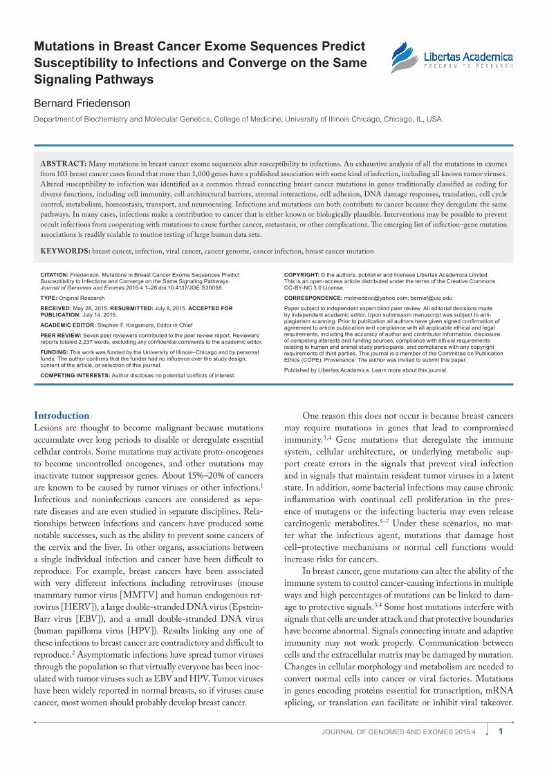
ferent signaling pathways are affected by mutations in dif-ferent breast cancers but a common thread is that they alter responses to infection. In many cases, the altered responses are to infections known to cause cancer. This is based on study-ing 4,985 mutations involving a total of 3,807 genes in 103 sporadic breast cancer exomes. Figure 1 is a pie chart showing the numbers of mutated genes placed into broad functional categories. Of the 4,985 total exome mutations, most of them (3,427 mutations) had some relationship to signals essential for immunity or for structural and architectural barriers needed to prevent or sequester infections.
Of the 3,807 different genes with mutations, only 2,947 could be tested (Fig. 1). Among these 2,947 genes with muta-tions are 1,077 different genes (36.5%) that are known to respond to some infection. In all, 774 mutations occurred in genes encoding products for more diverse cellular functions: homeostasis, metabolism, hormonally mediated phenomena, cell cycle, replication, transcription, translation, etc. Among these 774 mutations, at least 287 were associated with some kind of infection.
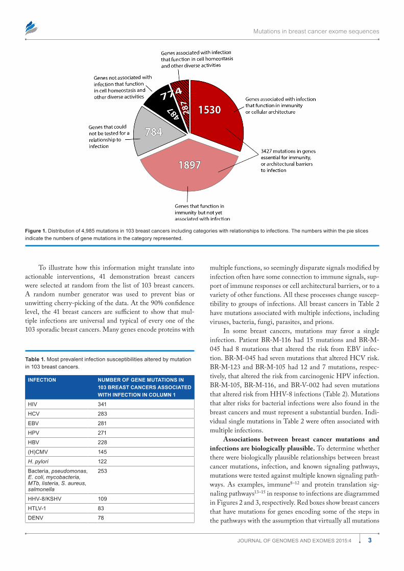
Table 1 shows how the mutations are distributed among the most prevalent infections. There are many opportunities for associations among mutations and known cancer-causing microbes. All known cancer-causing microbes are represented among these infections. Human immunodeficiency virus (HIV) appears most frequently, but this may merely reflect the intensity with which AIDS has been studied. Nonetheless, in the presence of a damaged immune system, associations between gene mutations and HIV infection probably raise the risk for AIDS-defining malignancies such as Kaposi sarcoma, non-Hodgkin lymphoma, and cervical cancer. Other can-cer causing viruses including EBV, hepatitis B virus (HBV), hepatitis C virus (HCV), and HPV are all represented about equally in Table 1. Helicobacter pylori occurs roughly half as often; human herpes virus type 8 (HHV-8), Dengue virus (DENV), and human T-cell leukemia virus (HTLV) slightly less than that. Associations with other viruses such as human cytomegalovirus (HCMV), influenza A virus (IAV), and with bacteria, mycobacteria, fungi, parasites, and prions. There were a few infections associated with mutations in transpo-son and retrotransposon genes (Tables 1 and 2). (Gene sym-bols and microorganism abbreviations are inserted before the author contribution section of this paper).
Mutations in breast cancer exome sequences
3Journal of Genomes and exomes 2015:4
Figure 1. distribution of 4,985 mutations in 103 breast cancers including categories with relationships to infections. The numbers within the pie slices indicate the numbers of gene mutations in the category represented.
Table 1. most prevalent infection susceptibilities altered by mutation in 103 breast cancers.
INFECTION NUMBER OF GENE MUTATIONS IN 103 BREAST CANCERS ASSOCIATED WITH INFECTION IN COLUMN 1
HIV 341
HCV 283
eBV 281
HPV 271
HBV 228
(H)CmV 145
H. pylori 122
Bacteria, pseudomonas, E. coli, mycobacteria, MTb, listeria, S. aureus, salmonella
253
HHV-8/KsHV 109
HTlV-1 83
denV 78
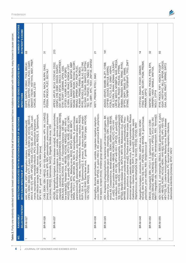
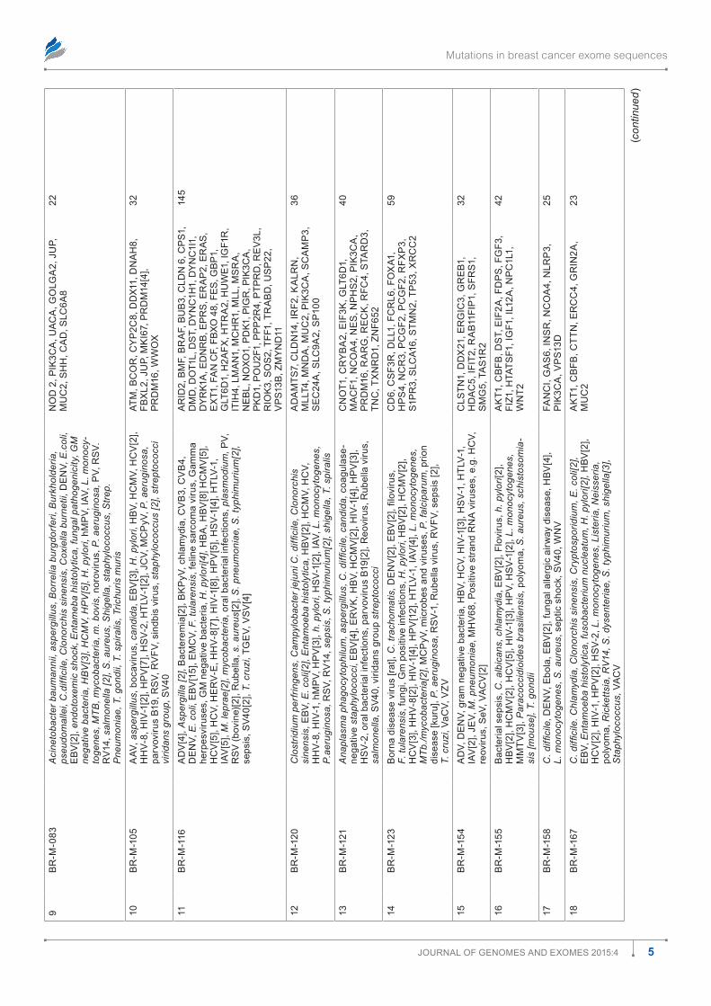
To illustrate how this information might translate into actionable interventions, 41 demonstration breast cancers were selected at random from the list of 103 breast cancers. A random number generator was used to prevent bias or unwitting cherry-picking of the data. At the 90% confidence level, the 41 breast cancers are sufficient to show that mul-tiple infections are universal and typical of every one of the 103 sporadic breast cancers. Many genes encode proteins with
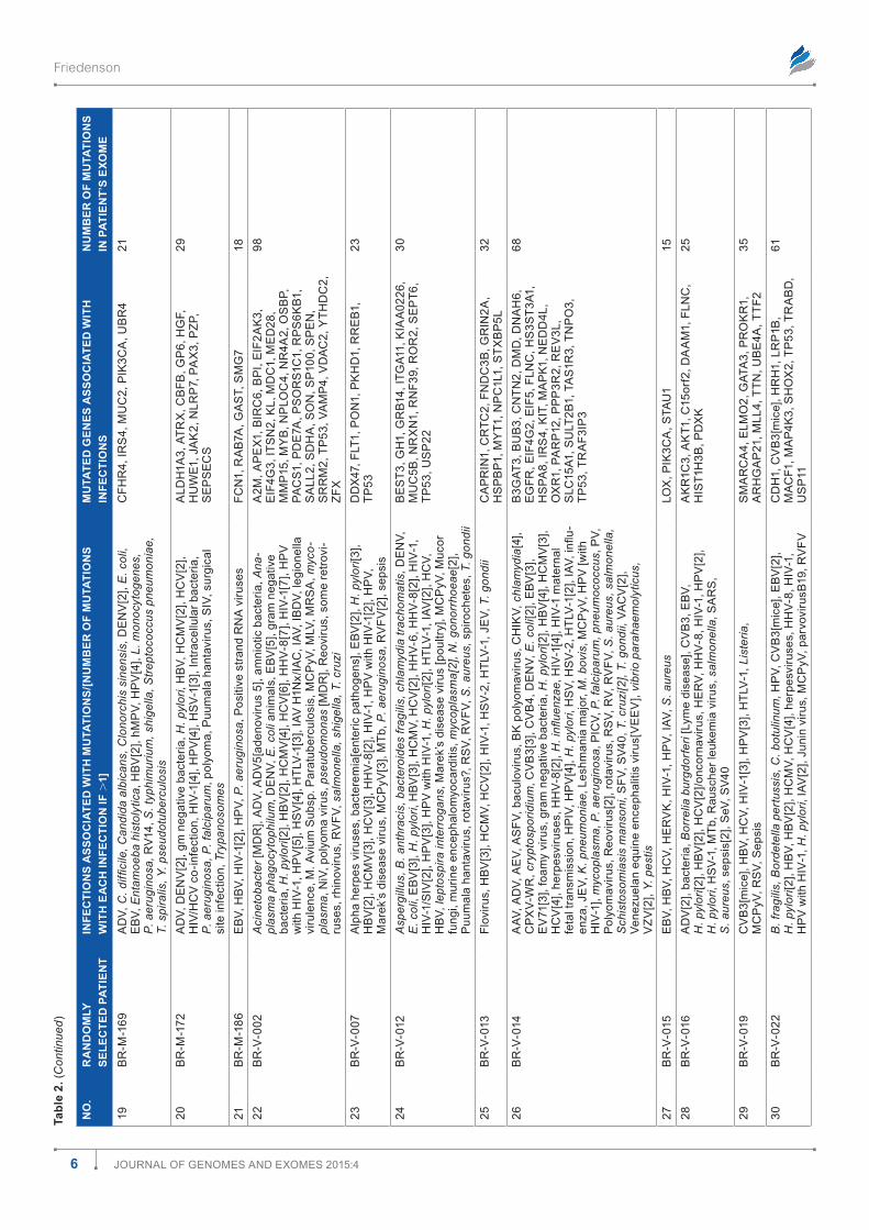
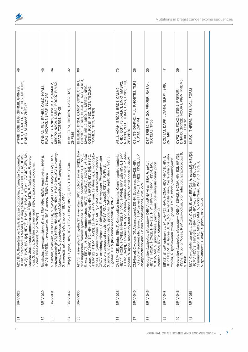
multiple functions, so seemingly disparate signals modified by infection often have some connection to immune signals, sup-port of immune responses or cell architectural barriers, or to a variety of other functions. All these processes change suscep-tibility to groups of infections. All breast cancers in Table 2 have mutations associated with multiple infections, including viruses, bacteria, fungi, parasites, and prions.
In some breast cancers, mutations may favor a single infection. Patient BR-M-116 had 15 mutations and BR-M-045 had 8 mutations that altered the risk from EBV infec-tion. BR-M-045 had seven mutations that altered HCV risk. BR-M-123 and BR-M-105 had 12 and 7 mutations, respec-tively, that altered the risk from carcinogenic HPV infection. BR-M-105, BR-M-116, and BR-V-002 had seven mutations that altered risk from HHV-8 infections (Table 2). Mutations that alter risks for bacterial infections were also found in the breast cancers and must represent a substantial burden. Indi-vidual single mutations in Table 2 were often associated with multiple infections.
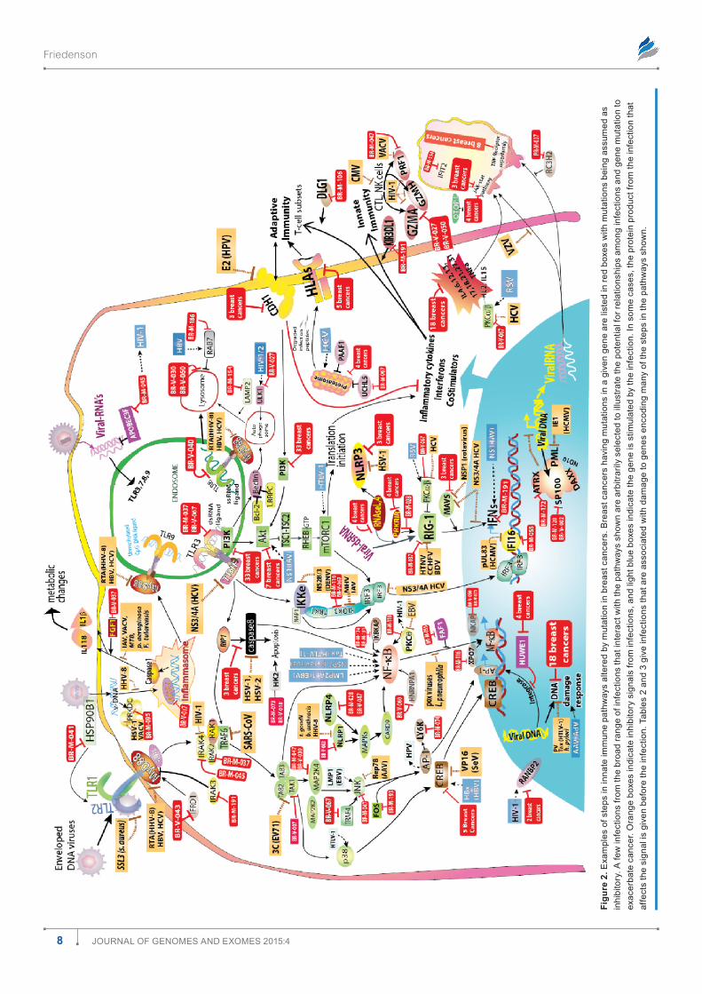
Associations between breast cancer mutations and infections are biologically plausible. To determine whether there were biologically plausible relationships between breast cancer mutations, infection, and known signaling pathways, mutations were tested against multiple known signaling path-ways. As examples, immune8–12 and protein translation sig-naling pathways13–15 in response to infections are diagrammed in Figures 2 and 3, respectively. Red boxes show breast cancers that have mutations for genes encoding some of the steps in the pathways with the assumption that virtually all mutations
Friedenson
4 Journal of Genomes and exomes 2015:4
Tabl
e 2.
for
ty-o
ne ra
ndom
ly s
elec
ted
spor
adic
bre
ast c
ance
rs e
xam
ples
sho
win
g th
at a
ll br
east
can
cers
test
ed h
ave
mut
atio
ns a
ssoc
iate
d w
ith in
fect
ions
, man
y kn
own
to c
ause
can
cer.
NO
.R
AN
DO
MLY
SE
LEC
TED
PAT
IEN
TIN
FEC
TIO
NS
ASS
OC
IATE
D W
ITH
MU
TATI
ON
S/[N
UM
BER
OF
MU
TATI
ON
S W
ITH
EA
CH
INFE
CTI
ON
IF .
1]M
UTA
TED
GEN
ES A
SSO
CIA
TED
WIT
H
INFE
CTI
ON
SN
UM
BER
OF
MU
TATI
ON
S IN
PAT
IEN
T’S
EXO
ME
1B
r-m
-027
aaV
, ad
V, a
eV,
as
fV, A
sper
gillu
s ni
dula
ns, C
. tra
chom
atis
/psi
ttaci
[4],
CPx
V, d
en
V, e
BV[2
], e
V71
, h p
ylor
i, H
BV[3
], H
Cm
V [2
], H
CV
[3],
He
rV-
e,
HH
V-8,
HIV
-1[7
], H
PIV,
HP
V[2
], H
sV
[2],
HTl
V-1[
2], I
aV, J
CV,
lC
mV,
Les
h-m
ania
, myc
opla
sma,
nem
atod
e in
fect
ion,
PIC
V, p
lasm
odiu
m fa
lcip
arum
, pr
ion
infe
ctio
n sc
rapi
e, r
aBV
, reo
viru
s, rh
inov
irus,
rs
V[3
], S
. Typ
him
uriu
m,
sfV
, sIV
, T. g
ondi
i, T.
cru
zi, V
aCV,
Wn
V [m
ice]
eGfr
, Ga
P43,
Il13
ra1
, TY
K2,
Br
Ca1
, C
rTC
2, e
nTP
d1,
fo
xo4,
PlK
3, C
3ar
1,
dn
aH
17, f
sC
n1,
HIs
T1H
1a, m
Id1,
Pr
eP,
C
WC
25, I
ns
r, l
ZTs1
68
2B
r-m
-028
CV
B3,
de
nV,
eBV
, HBV
[2],
HIV
-1[5
] alp
ha h
erpe
sviru
ses,
HP
V, Ia
V, l
Cm
V[2
] [m
ice]
, Les
hman
ia, L
iste
ria, M
Tb, n
dV,
pla
smod
ium
, pne
umoc
cus,
pne
umov
i-ru
s (m
ice)
, rsV
[2],
Sal
mon
ella
, seV
[2],
Shi
gella
, sin
dbis
viru
s, V
sV
Il27
ra
, KIf
1a, n
aCa
, nlr
P4, o
as
2,
Pd
s5a
, PIK
3Ca
, Ple
C, s
eC14
l131
3B
r-m
-037
ad
V[6
], A
sper
gillu
s fu
mig
atus
, Bor
na d
isea
se v
irus[
rat],
Bor
relia
spi
roch
etes
, B
HV
-1[3
], B
urkh
olde
ria c
enoc
epac
ia, c
.jeju
ni, c
andi
da, c
anin
e pa
rvov
irus,
ch
lam
ydia
, clo
strid
ium
, CV/
CV
B3,
Cm
V[2
]/HC
mV
[3],
CV
B3m
ice[
2], d
en
V[6
], e
Bo
la[3
], e
BV[1
4], e
CH
o30
, E. c
oli,
Fran
cise
lla tu
lare
nsis
, Gm
neg
a-tiv
e ba
cter
ia[2
], H
anta
an V
irus,
HBV
[9],
HC
V[1
6], H
er
V[2
], H
eV,
HH
V-8,
H
IV[2
2], H
PV
[5],
H. p
ylor
i[4],
Hs
V-1/
2[8]
, HTl
V-1[
3], I
aV[6
], Je
V[4
], lC
mV,
lu
ng in
fect
ions
, mTb
., m
arbu
rg v
irus,
mV
[2],
mC
PyV
, mH
V-08
, mm
TV,
mr
sa
, HTl
V-1,
mTb
, mH
V68
, mV,
nd
V, N
. men
ingi
tides
, N. g
onor
rhoe
ae,
para
myx
oviru
s[3]
, pla
smod
ium
[3],
Pol
iovi
rus,
pol
y-m
icro
bial
sep
sis,
BK
V,
prio
n In
fect
ion,
PV,
rabi
es, r
ecur
rent
infe
ctio
ns, r
eovi
rus,
rota
viru
s, r
sV
[3],
rV
fV, s
V-a
, S. T
yphi
mur
ium
[2],
S. a
ureu
s, S
. aur
eus
(mic
e), s
eV, s
trept
o-co
ccus
[2],
sV4
0, s
win
e fe
ver v
irus,
T. g
ondi
i, TB
eV,
TG
eV
(pig
s), V
aCV
[4],
Vs
V, V
ZV[2
], W
nV
[3],
Yers
inia
aP
ex
2, a
PlP
2, B
Cl6
, CC
dC
40, C
d22
, C
en
PJ, C
fB, d
dB1
, dH
x29
, dIa
PH
1,
ds
T, e
ef2
K, e
If3a
, er
CC
5, e
rV
frd
e1,
eYa
1, f
ox
f1, G
em
In5,
GPx
4, G
ss
, G
Tf2e
1, H
Jur
P, H
la-a
, IG
f2B
P2,
Il
15r
a, I
l21r
, Ir
aK
2, K
dm
3a, l
YsT,
m
ad
1l1,
maG
I3, m
aP
2, m
aP
3K1,
mB
P,
mG
am
, mIP
eP,
mu
sK
, nC
oa
2, n
Co
r1,
n
r4a
2, o
Pr
m1,
PG
K1, P
IGP,
Ple
C,
Plx
na1
, Pr
dm
2, P
sm
e3,
ro
Bo
3,
rY
r2,
sH
3Gl2
, sIn
3B, s
la2,
slC
1a6,
s
lC30
a1, s
lC38
a5,
sn
aP
C2,
su
PT5
H,
TeC
, Tf,
Tfa
P2C
, Tlr
3, T
nn
C2,
Tr
Io,
TTn
, uB
r1,
xP
C, Y
es1,
Zn
f136
, Zn
f652
213
4B
r-m
-038
Asp
ergi
llus,
Bor
relia
spi
roch
aete
s, c
andi
da, c
oagu
lase
-neg
ativ
e st
aphy
lo-
cocc
i, d
en
V, e
BV, H
PV
[3],
H. p
ylor
i, H
sV-
2, Ia
V, p
arvo
viru
s B1
9, p
olym
icro
-bi
al s
epsi
s, s
crap
ie (m
ice)
, viri
dans
gro
up s
trept
ococ
ci
ms
T1, P
rd
m16
, rTe
l1, s
mo
21
5B
r-m
-045
ad
V, A
napl
asm
a ph
agoc
ytop
hilu
m, b
acte
rem
ia w
ith e
nter
ic p
atho
gens
, BK
po
lyom
a vi
rus,
Cam
pylo
bact
er je
juni
, can
dida
, chl
amyd
ia, C
mV,
Cox
sack
i-ev
irus
a9,
Cox
sack
ievi
rus,
den
tal a
bsce
ss, d
en
V, d
srn
a v
iruse
s, e
BV[8
], E
. col
i[2],
Gra
m p
ositi
ve b
acte
ria, H
BV[4
], H
Cm
V[5
], H
CV
[7],
HC
V-H
IV
co-in
fect
ion,
HH
V-8[
3], H
IV-1
[6],
HP
V w
ith H
IV-1
, HP
V[3
], H
. pyl
ori[2
], H
sV-
1[3]
, HTl
V-1[
2], I
aV[3
], Le
gion
ella
pne
umop
hila
, Lis
teria
mon
ocyt
ogen
es[2
], m
Tb[2
], m
arek
’s d
isea
se v
irus,
mC
PyV
, mH
V-08
, mol
lusc
ipox
viru
s, M
RS
A,
mH
V68
, Myc
obac
teriu
m a
ssili
ense
, pic
orno
viru
ses,
pla
smod
ium
[2],
Pne
u-m
ococ
cus,
pol
iovi
rus,
pol
yom
a vi
rus[
2], P
ositi
ve s
trand
rn
a v
iruse
s, p
seu-
dom
onas
aer
ugin
osa,
pse
udo
rabi
es v
irus,
PV,
rabb
it m
yxom
aviru
s, r
hesu
s rh
adin
oviru
s, ro
tavi
rus,
rs
V[2
], r
VfV
, S. a
ureu
s[2]
, Sal
mon
ella
, sep
sis.
S
taph
yloc
occu
s, d
erm
atop
hyte
fung
i, Va
cV[2
], V
sV
[2],
VZV
[2],
Wn
V
ad
ad
2, a
ra
P2,
Ba
mB
I, B
lZf1
, CY
BB
, C
YP1
B1, d
uo
x2,
er
o1l
B, e
sr
rG
, fC
rl5
, fC
Tl5,
flG
, Gr
m1,
Hs
PH
1,
Ide
, Il3
1ra
, Ir
aK
4, K
dm
4a, l
dlr
aP1
, lr
P1B
, me
d13
, mY
BB
P1a
, nH
s, P
de
2a,
PIK
fYV
e, P
KH
d1,
Po
lr3a
, PTP
rn
, r
aB
6a, r
na
se4
, rP
n2,
rr
m2,
rY
r2,
s
fTP
C, s
KP1
, sm
G5,
sP1
, sr
ms
, s
Tam
2, T
aP
BP,
TG
fBr
aP1
, TP
53, Z
nf7
141
6B
r-m
-048
Asp
ergi
llus
fum
igat
us, c
lost
ridiu
m, d
en
V, d
srn
a v
irus,
eBV
[2],
HC
mV,
H
CV
[3],
HH
V-8,
HIV
-1[3
], H
IV-1
, HP
V[3
], H
sV-
2, m
HV
68, N
. gon
orrh
oeae
, pl
asm
odiu
m, p
rion
infe
ctio
n, r
sV,
Stre
ptoc
occu
s B
CY
P21
a2,
dn
aH
3, f
oxo
1, G
rIn
2d,
ITs
n2,
mlx
IPl,
nC
or
1, P
IGm
, Pr
dm
5,
Pr
Kr
Ir, u
sP1
2
14
7B
r-m
-050
eBV
[3],
chla
myd
ia[2
], e
BV, H
TlV-
1, S
. typ
him
uriu
m[2
], T.
gon
dii,
CV
B3
(mic
e), e
BV[2
], H
BV[3
], H
Cm
V[2
], H
CV
[6],
HIV
-1, H
PV
[2],
HTl
V-1[
2], I
aV,
mm
TV in
tegr
atio
n si
te, m
ycob
acte
ria, P
V, re
trotra
nspo
son,
rs
V, Y
ersi
nia[
2]
ma
P3K
1, m
ed
14, P
IK3C
a, P
Ten
, ePx
, H
nr
nP
u, H
as1
, ma
ml2
, Wn
T10a
, n
olC
1, u
TP14
32
8B
r-m
-055
ad
V, d
en
V[3
], E
. col
i men
ingi
tis, e
Bo
la, e
BV[3
], gi
ardi
a m
uris
[2],
H. p
ylor
i, H
PV,
HBV
[3],
HC
mV,
HC
V[3
], H
er
V, H
HV-
8[2]
, HIV
-1[3
], H
PV,
Hs
V-1[
2],
He
V71
[2],
IaV,
Lis
teria
mon
ocyt
ogen
es, m
arbu
rg v
irus,
mar
ek’s
dis
ease
vi
rus
(pou
ltry)
, pla
smod
ium
, PV,
recu
rren
t sin
o-re
spira
tory
infe
ctio
ns,
Sal
mon
ella
ent
eriti
dis
[chi
cken
s], m
Tb?,
VaC
V
IfI1
6, P
IK3C
a, f
af1
, Hd
aC9,
Po
u2f
1,
GG
a1, H
IsT1
H1C
, mll
2, r
PG
r, d
dx
20,
Ga
a, G
lud
2, r
nG
TT, s
rr
m2,
sIr
T2
93
Mutations in breast cancer exome sequences
5Journal of Genomes and exomes 2015:4
9B
r-m
-083
Aci
neto
bact
er b
aum
anni
i, as
perg
illus
, Bor
relia
bur
gdor
feri,
Bur
khol
deria
, ps
eudo
mal
lei,
C.d
iffici
le, C
lono
rchi
s si
nens
is, C
oxie
lla b
urne
tii, d
en
V, E
.col
i, e
BV[2
], en
doto
xem
ic s
hock
, Ent
ameb
a hi
stol
ytic
a, fu
ngal
pat
hoge
nici
ty, G
M
nega
tive
bact
eria
, HBV
[3],
HC
MV,
HP
V[5
], H
. pyl
ori,
hmP
V, Ia
V, L
. mon
ocy-
toge
nes,
MTB
, myc
obac
teria
, m. b
ovis
, nor
oviru
s, P
. aer
ugin
osa,
PV,
rs
V.
rV1
4, s
alm
onel
la [2
], S
. aur
eus,
Shi
gella
, sta
phyl
ococ
cus,
Stre
p.
Pne
umon
iae,
T. g
ondi
i, T.
spi
ralis
, Tric
huris
mur
is
no
d 2
, PIK
3Ca
, uaC
a, G
olG
a2,
Ju
P,
mu
C2,
sH
H, C
ad
, slC
6a8
22
10B
r-m
-105
aaV
, asp
ergi
llus,
boc
aviru
s, c
andi
da, e
BV[3
], H
. pyl
ori,
HBV
, HC
mV,
HC
V[2
], H
HV-
8, H
IV-1
[2],
HP
V[7
], H
sV-
2, H
TlV-
1[2]
, JC
V, m
CP
yV, P
. aer
ugin
osa,
pa
rvov
irus
B19,
rs
V, r
VfV
, sin
dbis
viru
s, s
taph
yloc
occu
s [2
], st
rept
ococ
ci
virid
ans
grou
p, s
V40
aTm
, BC
or
, CY
P2C
8, d
dx1
1, d
na
H8,
fB
xl2
, Ju
P, m
KI6
7, P
rd
m14
[4],
Pr
dm
16, W
Wo
x
32
11B
r-m
-116
ad
V[4
], A
sper
gilla
[2],
Bac
tere
mia
[2],
BK
PyV
, chl
amyd
ia, C
VB
3, C
VB
4,
de
nV,
E. c
oli,
eBV
[15]
, em
CV,
F. t
ular
ensi
s, fe
line
sarc
oma
viru
s, G
amm
a he
rpes
viru
ses,
Gm
neg
ativ
e ba
cter
ia, H
. pyl
ori[4
], H
Ba
, HBV
[8] H
Cm
V[5
], H
CV
[5],
HC
V, H
er
V-e
, HH
V-8[
7], H
IV-1
[8],
HP
V[5
], H
sV-
1[4]
, HTl
V-1,
Ia
V[5
], M
. lep
rae[
2], m
ycob
acte
ria, o
ral b
acte
rial i
nfec
tions
, pla
smod
ium
, PV,
r
sV
(bov
ine)
[2],
rub
ella
, s. a
ureu
s[2]
, S. p
neum
onia
e, S
. typ
him
uriu
m[2
], se
psis
, sV4
0[2]
, T. c
ruzi
, TG
eV,
Vs
V[4
]
ar
Id2,
Bm
f, B
ra
f, B
uB
3, C
ldn
6, C
Ps1,
d
md
, do
T1l,
dsT
, dY
nC
1H1,
dY
nC
1I1,
d
Yr
K1a
, ed
nr
B, e
Prs
, er
aP
2, e
ra
s,
ex
T1, f
an
Cf,
fBx
o 4
8, f
es, G
BP1
, G
lT6d
1, H
2afx
, HTr
a2,
Hu
We1
, IG
f1r
, IT
IH4,
lm
an
1, m
CH
r1,
mll
, ms
ra
, n
eBl,
no
xo1,
Pd
K1, P
IGr
, PIK
3Ca
, PK
d1,
Po
u2f
1, P
PP2r
4, P
TPr
d, r
eV3
l,
rIo
K3, s
os2
, Tff
1, T
ra
Bd
, us
P22
, V
Ps13
B, Z
mY
nd
11
145
12B
r-m
-120
Clo
strid
ium
per
fring
ens,
Cam
pylo
bact
er je
juni
C. d
iffici
le, C
lono
rchi
s si
nens
is, e
BV, E
. col
i[2],
Ent
amoe
ba h
isto
lytic
a, H
BV[2
], H
Cm
V, H
CV,
H
HV-
8, H
IV-1
, hm
PV,
HP
V[3
], h.
pyl
ori,
Hs
V-1[
2], I
aV, L
. mon
ocyt
ogen
es,
P.ae
rugi
nosa
, rs
V, r
V14,
sep
sis,
S. t
yphi
mur
ium
[2],
shig
ella
, T. s
pira
lis
ad
am
Ts7,
Cld
n14
, Ir
f2, K
alr
n,
mll
T4, m
nd
a, m
uC
2, P
IK3C
a, s
Ca
mP
3,
seC
24a
, slC
9a2,
sP1
00
36
13B
r-m
-121
Ana
plas
ma
phag
ocyt
ophi
lum
, asp
ergi
llus,
C. d
iffici
le, c
andi
da, c
oagu
lase
-ne
gativ
e st
aphy
loco
cci,
eBV
[4],
er
VK
, HBV
, HC
mV
[2],
HIV
-1[4
], H
PV
[3],
Hs
V-2,
ora
l bac
teria
l inf
ectio
ns, p
arvo
viru
s B1
9[2]
, reo
viru
s, r
ubel
la v
irus,
sa
lmon
ella
, sV4
0, v
irida
ns g
roup
stre
ptoc
occi
Cn
oT1
, Cr
YB
a2,
eIf
3K, G
lT6d
1,
maC
f1, n
Co
a4,
nes
, nP
Hs
2, P
IK3C
a,
Pr
dm
16, r
ar
G, r
eCK
, rfC
4, s
Tar
d3,
Tn
C, T
xn
rd
1, Z
nf6
52
40
14B
r-m
-123
Bor
na d
isea
se v
irus
[rat],
C. t
rach
omat
is, D
EN
V[2
], E
BV[2
], fil
oviru
s,
F. tu
lare
nsis
, fun
gi, G
m p
ositi
ve in
fect
ions
, H. p
ylor
i, H
BV[2
], H
Cm
V[2
], H
CV
[3],
HH
V-8[
2], H
IV-1
[4],
HP
V[1
2], H
TlV-
1, Ia
V[4
], L.
mon
ocyt
ogen
es,
MTb
./myc
obac
teria
[2],
mC
PyV
, mic
robe
s an
d vi
ruse
s, P
. fal
cipa
rum
, prio
n di
seas
e [k
uru]
, P. a
erug
inos
a, r
sV-
1, r
ubel
la v
irus,
rV
fV, s
epsi
s [2
],
T. c
ruzi
, VaC
V, V
ZV
Cd
6, C
sf3
r, d
ll1,
fC
rl6
, fo
xa1
, H
Ps
4, n
Cr
3, P
CG
f2, P
CG
f2, r
fxP
3,
s1P
r3,
slC
a16,
sTm
n2,
TP
53, x
rC
C2
59
15B
r-m
-154
ad
V, d
en
V, g
ram
neg
ativ
e ba
cter
ia, H
BV, H
CV,
HIV
-1[3
], H
sV-
1, H
TlV-
1,
IaV
[2],
JeV,
M. p
neum
onia
e, m
HV
68, P
ositi
ve s
trand
rn
a v
iruse
s, e
.g. H
CV,
re
oviru
s, s
eV, V
aCV
[2]
Cls
Tn1,
dd
x21
, er
GIC
3, G
re
B1,
Hd
aC5,
IfIT
2, r
aB1
1fIP
1, s
frs1
, s
mG
5, T
as1
r2
32
16B
r-m
-155
Bac
teria
l sep
sis,
C. a
lbic
ans,
chl
amyd
ia, e
BV[2
], fl
oviru
s, h
. pyl
ori[2
], H
BV[2
], H
Cm
V[2
], H
CV
[5],
HIV
-1[3
], H
PV,
Hs
V-1[
2], L
. mon
ocyt
ogen
es,
mm
TV[3
], P
arac
occi
dioi
des
bras
ilien
sis,
pol
yom
a, S
. aur
eus,
sch
isto
som
ia-
sis
[mou
se],
T. g
ondi
i
aK
T1, C
BfB
, ds
T, e
If2a
, fd
Ps
, fG
f3,
fIZ1
, HTa
Tsf1
, IG
f1, I
l12a
, nP
C1l
1,
Wn
T2
42
17B
r-m
-158
C. d
iffici
le, d
en
V, e
bola
, eBV
[2],
fung
al a
llerg
ic a
irway
dis
ease
, HBV
[4],
L.
mon
ocyt
ogen
es, S
. aur
eus,
sep
tic s
hock
, sV4
0, W
nV
fan
CI,
Ga
s6,
Ins
r, n
Co
a4,
nlr
P3,
PI
K3C
a, V
Ps1
3d25
18B
r-m
-167
C. d
iffici
le. C
hlam
ydia
, Clo
norc
his
sine
nsis
, Cry
ptos
porid
ium
, E. c
oli[2
], e
BV, E
ntam
oeba
his
toly
tica,
fuso
bact
eriu
m n
ucle
atum
, H. p
ylor
i[2],
HBV
[2],
HC
V[2
], H
IV-1
, HP
V[2
], H
sV-
2, L
. mon
ocyt
ogen
es, L
iste
ria, N
eiss
eria
, po
lyom
a, R
icke
ttsia
, RV1
4, S
. dys
ente
riae,
S. t
yphi
mur
ium
, shi
gella
[3],
Sta
phyl
ococ
cus,
VaC
V
aK
T1, C
BfB
, CTT
n, e
rC
C4,
Gr
In2a
, m
uC
223
(con
tinue
d)
Friedenson
6 Journal of Genomes and exomes 2015:4
Tabl
e 2.
(Con
tinue
d)
NO
.R
AN
DO
MLY
SE
LEC
TED
PAT
IEN
TIN
FEC
TIO
NS
ASS
OC
IATE
D W
ITH
MU
TATI
ON
S/[N
UM
BER
OF
MU
TATI
ON
S W
ITH
EA
CH
INFE
CTI
ON
IF .
1]M
UTA
TED
GEN
ES A
SSO
CIA
TED
WIT
H
INFE
CTI
ON
SN
UM
BER
OF
MU
TATI
ON
S IN
PAT
IEN
T’S
EXO
ME
19B
r-m
-169
ad
V, C
. diffi
cile
, Can
dida
alb
ican
s, C
lono
rchi
s si
nens
is, d
en
V[2
], E
. col
i, e
BV, E
ntam
oeba
his
toly
tica,
HBV
[2],
hmP
V, H
PV
[4],
L. m
onoc
ytog
enes
, P.
aer
ugin
osa,
rV1
4, S
. typ
him
uriu
m, s
hige
lla, S
trept
ococ
cus
pneu
mon
iae,
T.
spi
ralis
, Y. p
seud
otub
ercu
losi
s
CfH
r4,
Irs
4, m
uC
2, P
IK3C
a, u
Br
421
20B
r-m
-172
ad
V, d
en
V[2
], gm
neg
ativ
e ba
cter
ia, H
. pyl
ori,
HBV
, HC
mV
[2],
HC
V[2
], H
IV/H
CV
co-
infe
ctio
n, H
IV-1
[4],
HP
V[4
], H
sV-
1[3]
, Int
race
llula
r bac
teria
, P.
aer
ugin
osa,
P. f
alci
paru
m, p
olyo
ma,
Puu
mal
a ha
ntav
irus,
sIV
, sur
gica
l si
te in
fect
ion,
Try
pano
som
es
ald
H1a
3, a
Trx
, CB
fB, G
P6,
HG
f,
Hu
We1
, Ja
K2,
nlr
P7,
Pa
x3,
PZP
, s
eP
seC
s
29
21B
r-m
-186
eBV
, HBV
, HIV
-1[2
], H
PV,
P. a
erug
inos
a, P
ositi
ve s
trand
rn
a v
iruse
sfC
n1,
ra
B7a
, Ga
sT,
sm
G7
1822
Br
-V-0
02A
cine
toba
cter
[md
r],
ad
V, a
dV
5[ad
enov
irus
5], a
mni
otic
bac
teria
, Ana
-pl
asm
a ph
agoc
ytop
hilu
m, d
en
V, E
. col
i ani
mal
s, e
BV[5
], gr
am n
egat
ive
bact
eria
, H. p
ylor
i[2],
HBV
[2],
HC
mV
[4],
HC
V[6
], H
HV-
8[7]
, HIV
-1[7
], H
PV
w
ith H
IV-1
, HP
V[5
], H
sV
[4],
HTl
V-1[
3], I
aV H
1nx/
IaC
, IaV
, IB
dV,
legi
onel
la
viru
lenc
e, m
. avi
um s
ubsp
. Par
atub
ercu
losi
s, m
CP
yV, m
lV, m
rs
a, m
yco-
plas
ma,
niV
, pol
yom
a vi
rus,
pse
udom
onas
[md
r],
reo
viru
s, s
ome
retro
vi-
ruse
s, rh
inov
irus,
rV
fV, s
alm
onel
la, s
hige
lla, T
. cru
zi
a2m
, aP
ex1
, BIr
C6,
BPI
, eIf
2aK
3,
eIf4
G3,
ITs
n2,
Kl,
md
C1,
me
d28
, m
mP1
5, m
YB
, nP
loC
4, n
r4a
2, o
sB
P,
PaC
s1, P
de
7a, P
so
rs1
C1,
rP
s6K
B1,
sa
ll2,
sd
Ha
, so
n, s
P100
, sP
en
, s
rr
m2,
TP
53, V
am
P4, V
daC
2, Y
THd
C2,
Zf
x
98
23B
r-V
-007
alp
ha h
erpe
s vi
ruse
s, b
acte
rem
ia[e
nter
ic p
atho
gens
], e
BV[2
], H
. pyl
ori[3
], H
BV[2
], H
Cm
V[3
], H
CV
[3],
HH
V-8[
2], H
IV-1
, HP
V w
ith H
IV-1
[2],
HP
V,
mar
ek’s
dis
ease
viru
s, m
CP
yV[3
], m
Tb, P
. aer
ugin
osa,
rV
fV[2
], se
psis
dd
x47
, flT
1, P
on
1, P
KH
d1,
rr
eB1
, TP
5323
24B
r-V
-012
Asp
ergi
llus,
B. a
nthr
acis
, bac
tero
ides
frag
ilis,
chl
amyd
ia tr
acho
mat
is, d
en
V,
E. c
oli,
eBV
[3],
H. p
ylor
i, H
BV[3
], H
Cm
V, H
CV
[2],
HH
V-6,
HH
V-8[
2], H
IV-1
, H
IV-1
/sIV
[2],
HP
V[3
], H
PV
with
HIV
-1, H
. pyl
ori[2
], H
TlV-
1, Ia
V[2
], H
CV,
H
BV, l
epto
spira
inte
rrog
ans,
mar
ek’s
dis
ease
viru
s [p
oultr
y], m
CP
yV, m
ucor
fu
ngi,
mur
ine
ence
phal
omyo
card
itis,
myc
opla
sma[
2], N
. gon
orrh
oeae
[2],
Puu
mal
a ha
ntav
irus,
rota
viru
s?, r
sV,
rV
fV, S
. aur
eus,
spi
roch
etes
, T. g
ondi
i
Bes
T3, G
H1,
Gr
B14,
ITG
a11,
KIa
a02
26,
mu
C5B
, nr
xn
1, r
nf3
9, r
or
2, s
eP
T6,
TP53
, us
P22
30
25B
r-V
-013
flov
irus,
HBV
[3],
HC
mV,
HC
V[2
], H
IV-1
, Hs
V-2,
HTl
V-1,
Je
V, T
. gon
dii
Ca
Pr
In1,
Cr
TC2,
fn
dC
3B, G
rIn
2a,
Hs
PB
P1, m
YT1
, nP
C1l
1, s
TxB
P5l
32
26B
r-V
-014
aaV
, ad
V, a
eV,
as
fV, b
acul
oviru
s, B
K p
olyo
mav
irus,
CH
IKV,
chl
amyd
ia[4
], C
PxV-
Wr
, cry
ptos
porid
ium
, CV
B3[
3], C
VB
4, d
en
V, E
. col
i[2],
eBV
[3],
eV
71[3
], fo
amy
viru
s, g
ram
neg
ativ
e ba
cter
ia, H
. pyl
ori[2
], H
BV[4
], H
Cm
V[3
], H
CV
[4],
herp
esvi
ruse
s, H
HV-
8[2]
, H. i
nflue
nzae
, HIV
-1[4
], H
IV-1
mat
erna
l fe
tal t
rans
mis
sion
, HPI
V, H
PV
[4],
H. p
ylor
i, H
SV,
HS
V-2,
HTL
V-1[
2], I
AV, i
nflu-
enza
, Je
V, K
. pne
umon
iae,
les
hman
ia m
ajor
, M. b
ovis
, mC
PyV
, HP
V [w
ith
HIV
-1],
myc
opla
sma,
P. a
erug
inos
a, P
ICV,
P. f
alci
paru
m, p
neum
ococ
cus,
PV,
P
olyo
mav
irus,
reo
viru
s[2]
, rot
aviru
s, r
sV,
rV,
rV
fV, S
. aur
eus,
sal
mon
ella
, S
chis
toso
mia
sis
man
soni
, sfV
, sV4
0, T
. cru
zi[2
], T.
gon
dii,
VaC
V[2
], Ve
nezu
elan
equ
ine
ence
phal
itis
viru
s[V
ee
V],
vibr
io p
arah
aem
olyt
icus
, V
ZV[2
], Y.
pes
tis
B3G
aT3,
Bu
B3,
Cn
Tn2,
dm
d, d
na
H6,
eG
fr, e
If4G
2, e
If5,
fln
C, H
s3s
T3a1
, H
sPa
8, Ir
s4,
KIT
, ma
PK1
, ne
dd
4l,
ox
r1,
Pa
rP1
2, P
PP
3r2,
re
V3l
, s
lC15
a1, s
ulT
2B1,
Ta
s1r
3, T
nP
o3,
TP
53, T
ra
f3IP
3
68
27B
r-V
-015
eBV
, HBV
, HC
V, H
er
VK
, HIV
-1, H
PV,
IaV,
S. a
ureu
slo
x, P
IK3C
a, s
Tau
115
28B
r-V
-016
ad
V[2
], ba
cter
ia, B
orre
lia b
urgd
orfe
ri [l
yme
dise
ase]
, CV
B3,
eBV
, H
. pyl
ori[2
], H
BV[2
], H
CV
[2]/o
ncor
navi
rus,
He
rV,
HH
V-8,
HIV
-1, H
PV
[2],
H
. pyl
ori,
Hs
V-1,
mTb
, rau
sche
r leu
kem
ia v
irus,
sal
mon
ella
, sa
rs
, S
. aur
eus,
sep
sis[
2], s
eV, s
V40
aK
r1C
3, a
KT1
, C15
orf2
, da
am
1, f
lnC
, H
IsT1
H3B
, Pd
xK
25
29B
r-V
-019
CV
B3[
mic
e], H
BV, H
CV,
HIV
-1[3
], H
PV
[3],
HTl
V-1,
Lis
teria
, m
CP
yV, r
sV, s
epsi
ss
ma
rC
a4,
elm
o2,
GaT
a3,
Pr
oK
r1,
a
rH
Ga
P21
, mll
4, T
Tn, u
Be4
a, T
Tf2
35
30B
r-V
-022
B. f
ragi
lis, B
orde
tella
per
tuss
is, C
. bot
ulin
um, H
PV,
CV
B3[
mic
e], e
BV[2
],
H. p
ylor
i[2],
HBV
, HBV
[2],
HC
mV,
HC
V[4
], he
rpes
viru
ses,
HH
V-8,
HIV
-1,
HP
V w
ith H
IV-1
, H. p
ylor
i, Ia
V[2
], Ju
nin
viru
s, m
CP
yV, p
arvo
viru
sB19
, rV
fV
Cd
H1,
CV
B3[
mic
e], H
rH
1, l
rP1
B,
maC
f1, m
aP4
K3,
sH
ox
2, T
P53
, Tr
aB
d,
us
P11
61
Mutations in breast cancer exome sequences
7Journal of Genomes and exomes 2015:4
31B
r-V
-028
ad
V, B
lV, B
orde
tella
per
tuss
is, B
orre
lia b
urgd
orfe
ri, c
hlam
ydia
trac
hom
atis
, d
enV,
Ent
eroc
occu
s fa
ecal
is, G
m n
eg b
acte
ria, H
. pyl
ori,
HB
e, H
BV, H
Cm
V,
HC
V[4
], H
Vs, H
IV-1
[2],
HP
V[3
], H
sV-1
[3],
HsV
-2, H
TlV-
1, Ia
V, J
CV,
Je
V, m
ol-
lusc
ipox
viru
s, m
ouse
gam
ma
herp
es, m
rs
a. m
Tb, P
. fal
cipa
rum
, P. a
erug
i-no
sa, S
. aur
eus,
Sch
isto
som
a m
anso
ni, s
eV,
stre
ptoc
occu
s py
ogen
es,
T. c
ruzi
, tin
ea c
orpo
ris, V
sV, W
nV
[2]
aTf6
B, d
se
, flG
, GP
nm
B, G
rIn
2B,
Hr
H1,
ITG
ax
, lr
IG1,
meC
P2,
no
TCH
2,
ra
B13,
slC
38a
5, Z
nf2
17
49
32B
r-V
-030
Bacu
lovi
rus,
cry
ptos
porid
ium
, E. c
oli,
eBV,
HBV
, HC
mV,
Hen
dra
viru
s, H
HV-
6,
HH
V-8,
HIV
-1[3
], m
. pne
umon
iae,
niV
, S. a
ureu
s, T
me
V, T
. bru
cei
CTn
na
3, e
ln, e
PH
B2,
Ga
lC, H
Yal1
, K
lK8,
nC
oa
3, n
sm
af,
slC
15a1
40
33B
r-V
-031
C. a
lbic
ans,
chl
amyd
ia, d
enV,
eBV
[4],
H. p
ylor
i[4],
HBV
, HC
mV[
2], H
CV[
3], h
er-
pesv
irus
saim
iri [H
Vs],
Her
V, H
IV-1
[2],
HPV
, HsV
-1, H
sV-2
, IaV
, JC
V, L
. mon
ocy-
toge
nes,
MTb
, N. g
onor
rhoe
ae, P
arac
occi
dioi
des
bras
iliens
is, P
. aer
ugin
osa,
S.
aur
eus[
2], s
alm
onel
la, s
eV, T
. gon
dii, T
BeV,
Wn
V
aTx
n1,
CTn
nB1
, Il1
2a, K
rT2
, ma
ml2
, m
eCP
2, n
aCa
, na
rs
2, P
dC
d7,
rIm
s2,
s
PaG
1, T
Ce
rG
1, T
nKs
34
34B
r-V
-032
eBV[
2], H
. pyl
ori,
HBV
, HC
V, H
eV3,
HH
V-8,
HIV
-1[2
], H
PV, H
TlV-
1, s
V40
Bu
B1, e
lP3,
Hn
rn
Pu
, laT
s2,
TaT
, Zn
f185
32
35B
r-V
-033
ad
V[3
], as
perg
illus
fum
igat
us[2
], as
perg
illus
, BK
V [p
olyo
mav
irus
BK
][2],
Bor
-re
lia b
urgd
orfe
ri, B
orre
lia s
piel
man
ii, C
. diffi
cile
, can
dida
alb
ican
s, d
en
V[3
], E
. col
i, e
BV[8
], H
. pyl
ori,
Han
taan
viru
s, H
BV[3
], H
Cm
V[2
], H
CV
[7],
H. i
nflu-
enza
e, H
eV,
HH
V-8[
2],H
IV-1
[8],
HP
V w
ith H
IV-1
, HP
V[5
], H
sV-
1[4]
, Hs
V-2,
H
TlV-
1[2]
, HTl
V-1,
IaV
[3],
Leis
hman
ia in
fant
ium
, Lei
shm
ania
, L. m
onoc
yto-
gene
s, M
. hyo
pneu
mon
iae,
MTb
, mC
PyV
, mm
TV, o
ral b
acte
rial i
nfec
tions
, o
ro
V, o
rtho
buny
aviru
ses,
P. f
alci
paru
m, P
. aer
ugin
osa,
Pla
smod
ium
cha
-ba
udi,
pneu
moc
occa
l sep
sis,
ra
BV, r
na
viru
s[3]
, rs
V[3
], r
VfV
[2],
S
. aur
eus,
S. p
neum
onia
e, S
. pyo
gene
s, S
alm
onel
la, s
eptic
sho
ck, s
eV[3
],
T. g
ondi
i, TB
eV
[tic
k bo
rne
ence
phal
itis]
, Wn
V
BH
lHe4
0, B
rd
4, C
an
d1,
Cd
38, C
fHr
1,
GH
rH
r, G
lT6d
1, H
la-a
, Hla
-G, K
lKB1
, m
aVs
, mB
l2, m
ed
13l,
mP
eG1,
nfI
C,
oG
T[2]
, PlC
e1, P
sd
, saT
1, T
Go
ln2,
To
m1l
2, T
P53
, VTn
[3]
80
36B
r-V
-036
CV
B3[
mIC
e],
ebo
la, e
BV[2
], G
m p
ositi
ve b
acte
ria, G
ram
neg
ativ
e ba
cter
ia,
HBV
[4],
HC
mV,
HC
V[5
], H
HV-
8[2]
, HIV
-1[6
], H
PV
[4],
HP
V w
ith H
IV-1
, Hs
V-1,
H
TlV-
1, Ia
V[3
], le
gion
ella
, mC
PyV
, mol
oney
mur
ine
leuk
emia
viru
s, P
. aer
u-gi
nosa
, H. p
ylor
i, re
spira
tory
trac
t inf
ectio
ns, r
VfV
, sep
tic s
hock
, T. c
ruzi
aB
l1, a
oa
H, B
rC
a1, B
rd
4, C
alm
2,
CH
d6,
ds
T, f
8, K
alr
n, l
ImK1
, ma
sP
2,
nr
xn
3, P
leC
, TG
m3,
TP
53, T
Px2,
TTn
, Zf
YV
e20
70
37B
r-V
-040
Cm
V[m
ice]
, Cry
pton
s [d
na
trans
poso
ns],
den
V, e
bola
viru
s, e
BV[2
], H
BV
[hot
spo
t of r
ecur
rent
inte
grat
ion
of H
BV g
enes
], H
HV-
8, H
IV-1
[2],
HsV
[2],
JeV,
la
ryng
otra
chei
tis v
irus,
Lis
teria
mon
ocyt
ogen
es, V
sV, V
ZV
C8o
rf79
, dIa
PH
2, m
ll, r
Ho
BTB
2, T
lr8,
Tu
B1a1
, Zm
Ym
426
38B
r-V
-045
Asp
ergi
llus
fum
igat
us, c
lost
ridiu
m, d
en
V, d
srn
a v
irus,
eBV
, H. p
ylor
i, H
BV[2
], H
Cm
V, H
CV
[2],
HH
V-8[
2], H
IV1,
HP
V w
ith H
IV-1
, Hs
V-1,
IaV,
m
CP
yV, m
V, N
. gon
orrh
oeae
, pla
smod
ium
, pne
umoc
ystis
car
nii,
prio
n in
fect
ion,
rs
V, r
VfV
, Stre
ptoc
occu
s B
ds
T, e
rB
B2I
P, P
IGo
, Pr
Kr
Ir, r
as
a4,
s
lC12
a3,
TP
5320
39B
r-V
-047
eBV
[2],
E. c
oli,
ente
rovi
rus,
H. p
ylor
i[2],
HaV
, HC
mV,
Hd
V, H
HV-
8, H
IV-1
, H
PV,
HTl
V-1,
M. l
epra
e, M
.Tb.
, mou
se h
epat
itis
viru
s, P
aste
urel
la p
neum
o-tro
pica
, S. t
yphi
mur
ium
(mic
e), T
. gon
dii,
Tme
V, V
ibrio
par
ahae
mol
ytic
us
Co
l13a
1, d
aP
K1, l
Ta4H
, nlr
P4, s
rf,
Zn
f148
17
40B
r-V
-048
Asp
ergi
llus
fum
igat
us, c
lost
ridiu
m, d
en
V, e
BV[2
], H
Cm
V, H
IV-1
[2],
HP
V[3
], H
sV-
2, N
. gon
orrh
oeae
, rs
V, p
lasm
odiu
m, p
rion
infe
ctio
n, S
trept
ococ
cus
B,
mH
V68
CY
P21
a2,
fo
xo1,
ITs
n2,
Pr
Kr
Ir,
dn
aH
3, G
rIn
2d, n
Co
r1,
PIG
m, P
rd
m5,
m
lxIP
l, u
sP1
2
39
41B
r-V
-051
BK
V, C
ampy
loba
cter
jeju
ni, C
mV,
CV
B3,
E. c
oli,
eBV
[2],
H. p
ylor
i[2],
HBV
[2],
HC
mV,
HC
V[2
], H
HV-
8[2]
, HIV
-1[2
], H
PV
with
HIV
-1, H
PV,
Hs
V, J
CV,
Le
shm
ania
maj
or, M
Tb, m
CP
yV, m
mTV
, Ric
ketts
iae,
rV
fV, S
. aur
eus,
S
. typ
him
uriu
m, T
. cru
zi, T
. gon
dii,
Vs
V, V
aCV
Klr
K1, T
nfs
f9, T
P53
, VC
l, f
Gf2
315
Friedenson
8 Journal of Genomes and exomes 2015:4
Figu
re 2
. exa
mpl
es o
f ste
ps in
inna
te im
mun
e pa
thw
ays
alte
red
by m
utat
ion
in b
reas
t can
cers
. Bre
ast c
ance
rs h
avin
g m
utat
ions
in a
giv
en g
ene
are
liste
d in
red
boxe
s w
ith m
utat
ions
bei
ng a
ssum
ed a
s in
hibi
tory
. a fe
w in
fect
ions
from
the
broa
d ra
nge
of in
fect
ions
that
inte
ract
with
the
path
way
s sh
own
are
arbi
traril
y se
lect
ed to
illu
stra
te th
e po
tent
ial f
or re
latio
nshi
ps a
mon
g in
fect
ions
and
gen
e m
utat
ion
to
exac
erba
te c
ance
r. o
rang
e bo
xes
indi
cate
inhi
bito
ry s
igna
ls fr
om in
fect
ions
, and
ligh
t blu
e bo
xes
indi
cate
the
gene
is s
timul
ated
by
the
infe
ctio
n. In
som
e ca
ses,
the
prot
ein
prod
uct f
rom
the
infe
ctio
n th
at
affe
cts
the
sign
al is
giv
en b
efor
e th
e in
fect
ion.
Tab
les
2 an
d 3
give
infe
ctio
ns th
at a
re a
ssoc
iate
d w
ith d
amag
e to
gen
es e
ncod
ing
man
y of
the
step
s in
the
path
way
s sh
own.
Mutations in breast cancer exome sequences
9Journal of Genomes and exomes 2015:4
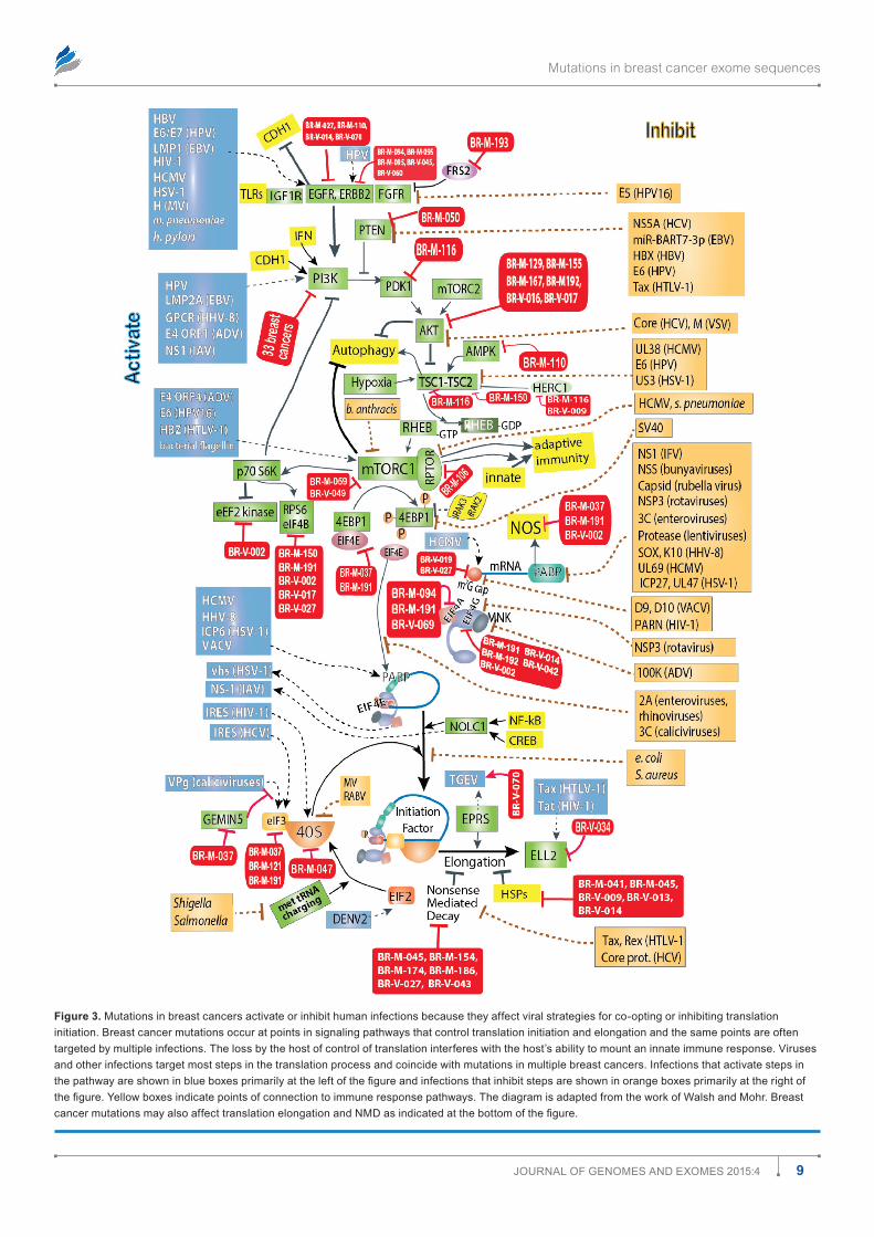
Figure 3. mutations in breast cancers activate or inhibit human infections because they affect viral strategies for co-opting or inhibiting translation initiation. Breast cancer mutations occur at points in signaling pathways that control translation initiation and elongation and the same points are often targeted by multiple infections. The loss by the host of control of translation interferes with the host’s ability to mount an innate immune response. Viruses and other infections target most steps in the translation process and coincide with mutations in multiple breast cancers. Infections that activate steps in the pathway are shown in blue boxes primarily at the left of the figure and infections that inhibit steps are shown in orange boxes primarily at the right of the figure. Yellow boxes indicate points of connection to immune response pathways. The diagram is adapted from the work of Walsh and Mohr. Breast cancer mutations may also affect translation elongation and NMD as indicated at the bottom of the figure.
Friedenson
10 Journal of Genomes and exomes 2015:4
inhibit rather than stimulate the affected gene. Infections, in contrast to mutations, may stimulate, inhibit, or comman-deer gene function. The two figures began from the review by Walsh and Mohr13 on the effects of viruses on protein trans-lation. The figures show that many mutations converge with infections to deregulate many cellular pathways essential to defend against infection. The same probably holds true for metabolism and for other functions as well (data not shown).
There are many opportunities for breast cancer mutations and associated infections to exacerbate cancer risks. Only a few arbitrarily selected interactions are shown (Figs. 2 and 3) among components in these pathways versus infections, but they illustrate how known tumor viruses or other infections can evade immune responses and cooperate with, substitute for, or antagonize breast cancer mutations. Table 2 gives more detail of examples linking breast cancer mutations and specific infections or groups of infections.
Table 3 uses more traditional classification systems to sep-arate infection-associated breast cancer mutations into diverse processes that they affect: innate and adaptive immunity, cell adhesion and tissue architecture, DNA damage response, pro-tein transcription, mRNA splicing, RNA processing, protein translation, cell cycle control, metabolism, nucleocytoplasmic transport, and protein trafficking. Table 3 is intended to illustrate
that the 1,077 different genes linked to infection can encode for widely different functions. There are only about 200 different genes used as examples in Table 3, but a current working list of all the mutations, the genes affected, and the associated infec-tions is available as Supplementary Material. Beyond immunity and protein translation, most traditional cellular functions can be altered, deregulated, or subverted by infections. For example, normal cell morphology and metabolism in virally infected cells must be altered to convert the cells into viral factories. Changes in ionic strength or pH can sometimes affect infection such as by facilitating viral uncoating. These traditional functions are also targeted by mutation, although not necessarily at the same points (Figs. 2 and 3). An additional signaling pathway for met-abolic generation of energy (not shown) further supports the biological plausibility of mutation and infection associations.
Examples showing mutation in diverse functions alter risks for infection.
Breast cancer mutations damage genes encoding for pro-teins in immune signaling pathways. Brief explanations for some of the steps shown in Figure 2 are given below. Mul-tiple infections, even those not linked to cancer, can affect the response to tumor viruses and to mutations. For example, four breast cancers have mutations in a proteasome compo-nent, and many breast cancers have mutations that affect
Table 3. Individual breast cancer mutations deregulate the same cell signals as infections over a wide variety of processes.
MUTATED GENE IN BREAST CANCER
ALTERED SUSCEPTIBILITY TO VIRUS OR OTHER INFECTIONS PREDICTED DUE TO MUTATION OR DYSREGULATION OF GENE
BREAST CANCER(S) CONTAINING THE MUTATED GENE
NORMAL FUNCTION OF PROTEIN ENCODED BY MUTATED GENE OR IN INFECTION
Antiviral or immune response, autophagy, apoptosis
aKT1 HCV,60 Bacteria Br-m-129, Br-m-155, Br-m-167, Br-m-192, Br-V-016, Br-V-017
regulates defensin expression and the innate immune response important for bacterial clearance61
adar dsrna viruses62 Br-m-191 Viral defense. adenosine to inosine in dsrna substrates
BCl6 eBV63 Br-m-037 regulation of innate immunity, macrophage morphology and motility
Bmf HHV-864 Br-m-116 BCl2 homology domain 3 (BH3) binds BCl2 proteins to regulate apoptosis. dynein l-chain binding associates it with myosin V. Protein limits HHV-8 (KHsV) replication64
BPI, BPIl1 Gram negative bacteria, MDR pseudomo-nas, MDR acinetobacter, E. coli65
Br-V-002, Br-m-041 neutralizes endotoxins and carries outer membrane antigens from gram negative bacteria to dendritic cells66
BraP eBV67 Br-m-094 negative regulator of import of viral proteins68
BlZf1 eBV,69 denV Br-m-045 methylation of cytosine, may interact with-Traff1,70 nfKB-p65 in innate immune pathways
CasP2 Francisella tularensis,71 S. aureus, Aeromonas, mmTV (mice)
Br-m-047, Br-m-189 autophagy control, cell death pathways, death due to cytoskeletal disruption72
CBlB lCmV,73 mV, Mycobacterium leprae,74 P. aeruginosa, E. coli sepsis, Burkholderia cenocepacia
Br-m-110 e3 ubiquitin ligase. Controls spontane-ous antitumor activity of cytotoxic T cells in different cancer models75
CCl1 Listeria monocytogenes,76 Borrelia burg-dorferi, a/H1n1 virus, MTb, VsV, S. aureus, herpesvirus saimiri
Br-V-027 Regulates immunity, inflammation.76 secreted by activated T-cells77
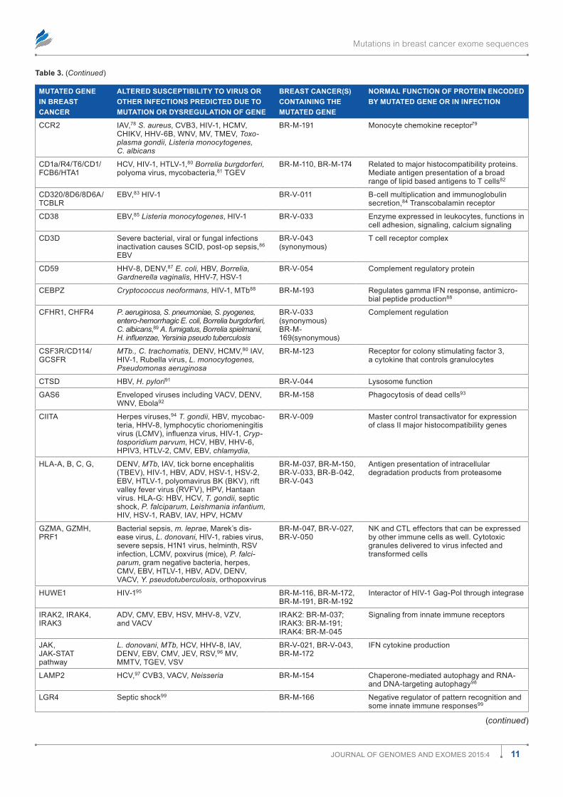
Mutations in breast cancer exome sequences
11Journal of Genomes and exomes 2015:4
Table 3. (Continued)
MUTATED GENE IN BREAST CANCER
ALTERED SUSCEPTIBILITY TO VIRUS OR OTHER INFECTIONS PREDICTED DUE TO MUTATION OR DYSREGULATION OF GENE
BREAST CANCER(S) CONTAINING THE MUTATED GENE
NORMAL FUNCTION OF PROTEIN ENCODED BY MUTATED GENE OR IN INFECTION
CCr2 IaV,78 S. aureus, CVB3, HIV-1, HCmV, CHIKV, HHV-6B, WnV, mV, TmeV, Toxo-plasma gondii, Listeria monocytogenes, C. albicans
Br-m-191 monocyte chemokine receptor79
Cd1a/r4/T6/Cd1/fCB6/HTa1
HCV, HIV-1, HTlV-1,80 Borrelia burgdorferi, polyoma virus, mycobacteria,81 TGeV
Br-m-110, Br-m-174 related to major histocompatibility proteins. mediate antigen presentation of a broad range of lipid based antigens to T cells82
Cd320/8d6/8d6a/TCBlr
eBV,83 HIV-1 Br-V-011 B-cell multiplication and immunoglobulin secretion,84 Transcobalamin receptor
Cd38 eBV,85 Listeria monocytogenes, HIV-1 Br-V-033 enzyme expressed in leukocytes, functions in cell adhesion, signaling, calcium signaling
Cd3d severe bacterial, viral or fungal infections inactivation causes sCId, post-op sepsis,86 eBV
Br-V-043 (synonymous)
T cell receptor complex
Cd59 HHV-8, denV,87 E. coli, HBV, Borrelia, Gardnerella vaginalis, HHV-7, HsV-1
Br-V-054 Complement regulatory protein
CeBPZ Cryptococcus neoformans, HIV-1, mTb88 Br-m-193 regulates gamma Ifn response, antimicro-bial peptide production88
CfHr1, CHfr4 P. aeruginosa, S. pneumoniae, S. pyogenes, entero-hemorrhagic E. coli, Borrelia burgdorferi, C. albicans,89 A. fumigatus, Borrelia spielmanii, H. influenzae, Yersinia pseudo tuberculosis
Br-V-033 (synonymous)Br-m-169(synonymous)
Complement regulation
Csf3r/Cd114/GCsfr
MTb., C. trachomatis, denV, HCmV,90 IaV, HIV-1, rubella virus, L. monocytogenes, Pseudomonas aeruginosa
Br-m-123 receptor for colony stimulating factor 3, a cytokine that controls granulocytes
CTsd HBV, H. pylori91 Br-V-044 lysosome function
Gas6 enveloped viruses including VaCV, denV, WnV, ebola92
Br-m-158 Phagocytosis of dead cells93
CIITa Herpes viruses,94 T. gondii, HBV, mycobac-teria, HHV-8, lymphocytic choriomeningitis virus (LCMV), influenza virus, HIV-1, Cryp-tosporidium parvum, HCV, HBV, HHV-6, HPIV3, HTlV-2, CmV, eBV, chlamydia,
Br-V-009 master control transactivator for expression of class II major histocompatibility genes
Hla-a, B, C, G, denV, MTb, IaV, tick borne encephalitis (TBeV), HIV-1, HBV, adV, HsV-1, HsV-2, eBV, HTlV-1, polyomavirus BK (BKV), rift valley fever virus (rVfV), HPV, Hantaan virus. Hla-G: HBV, HCV, T. gondii, septic shock, P. falciparum, Leishmania infantium, HIV, HsV-1, raBV, IaV, HPV, HCmV
Br-m-037, Br-m-150, Br-V-033, Br-B-042, Br-V-043
antigen presentation of intracellular degradation products from proteasome
GZma, GZmH, Prf1
Bacterial sepsis, m. leprae, marek’s dis-ease virus, L. donovani, HIV-1, rabies virus, severe sepsis, H1n1 virus, helminth, rsV infection, lCmV, poxvirus (mice), P. falci-parum, gram negative bacteria, herpes, CmV, eBV, HTlV-1, HBV, adV, denV, VaCV, Y. pseudotuberculosis, orthopoxvirus
Br-m-047, Br-V-027, Br-V-050
nK and CTl effectors that can be expressed by other immune cells as well. Cytotoxic granules delivered to virus infected and transformed cells
HuWe1 HIV-195 Br-m-116, Br-m-172, Br-m-191, Br-m-192
Interactor of HIV-1 Gag-Pol through integrase
IraK2, IraK4, IraK3
adV, CmV, eBV, HsV, mHV-8, VZV, and VaCV
IraK2: Br-m-037; IraK3: Br-m-191; IraK4: Br-m-045
signaling from innate immune receptors
JaK,JaK-sTaT pathway
L. donovani, MTb, HCV, HHV-8, IaV, denV, eBV, CmV, JeV, rsV,96 mV, mmTV, TGeV, VsV
Br-V-021, Br-V-043, Br-m-172
Ifn cytokine production
lamP2 HCV,97 CVB3, VaCV, Neisseria Br-m-154 Chaperone-mediated autophagy and rna- and dna-targeting autophagy98
lGr4 septic shock99 Br-m-166 negative regulator of pattern recognition and some innate immune responses99
(continued)
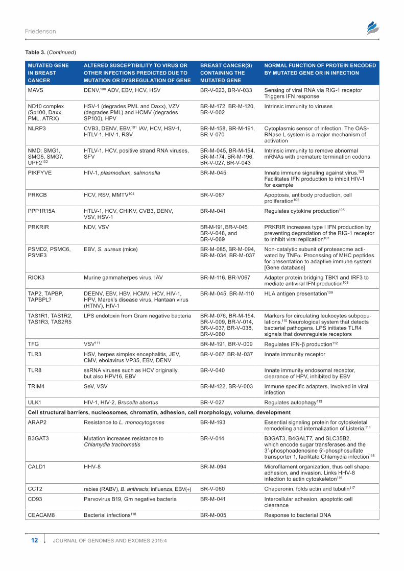
Friedenson
12 Journal of Genomes and exomes 2015:4
Table 3. (Continued)
MUTATED GENE IN BREAST CANCER
ALTERED SUSCEPTIBILITY TO VIRUS OR OTHER INFECTIONS PREDICTED DUE TO MUTATION OR DYSREGULATION OF GENE
BREAST CANCER(S) CONTAINING THE MUTATED GENE
NORMAL FUNCTION OF PROTEIN ENCODED BY MUTATED GENE OR IN INFECTION
maVs denV,100 adV, eBV, HCV, HsV Br-V-023, Br-V-033 sensing of viral rna via rIG-1 receptor Triggers Ifn response
nd10 complex (sp100, daxx, Pml, aTrx)
HsV-1 (degrades Pml and daxx), VZV (degrades Pml) and HCmV (degrades sP100), HPV
Br-m-172, Br-m-120, Br-V-002
Intrinsic immunity to viruses
nlrP3 CVB3, denV, eBV,101 IaV, HCV, HsV-1, HTlV-1, HIV-1, rsV
Br-m-158, Br-m-191, Br-V-070
Cytoplasmic sensor of infection. The oas-rnase l system is a major mechanism of activation
nmd: smG1, smG5, smG7, uPf2102
HTlV-1, HCV, positive strand rna viruses, sfV
Br-m-045, Br-m-154, Br-m-174, Br-m-196, Br-V-027, Br-V-043
Intrinsic immunity to remove abnormal mrnas with premature termination codons
PIKfYVe HIV-1, plasmodium, salmonella Br-m-045 Innate immune signaling against virus.103 facilitates Ifn production to inhibit HIV-1 for example
PrKCB HCV, rsV, mmTV104 Br-V-067 apoptosis, antibody production, cell proliferation105
PPP1r15a HTlV-1, HCV, CHIKV, CVB3, denV, VsV, HsV-1
Br-m-041 regulates cytokine production106
PrKrIr ndV, VsV Br-m-191, Br-V-045, Br-V-048, and Br-V-069
PrKrIr increases type I Ifn production by preventing degradation of the rIG-1 receptor to inhibit viral replication107
Psmd2, PsmC6, Psme3
eBV, S. aureus (mice) Br-m-085, Br-m-094, Br-m-034, Br-m-037
non-catalytic subunit of proteasome acti-vated by Tnfa. Processing of mHC peptides for presentation to adaptive immune system [Gene database]
rIoK3 murine gammaherpes virus, IaV Br-m-116, Br-V067 adapter protein bridging TBK1 and Irf3 to mediate antiviral Ifn production108
TaP2, TaPBP, TaPBPl?
deenV, eBV, HBV, HCmV, HCV, HIV-1, HPV, marek’s disease virus, Hantaan virus (HTnV), HIV-1
Br-m-045, Br-m-110 Hla antigen presentation109
Tas1r1, Tas1r2, Tas1r3, Tas2r5
lPs endotoxin from Gram negative bacteria Br-m-076, Br-m-154. Br-V-009, Br-V-014, Br-V-037, Br-V-038, Br-V-060
markers for circulating leukocytes subpopu-lations.110 neurological system that detects bacterial pathogens. lPs initiates Tlr4 signals that downregulate receptors
TfG VsV111 Br-m-191, Br-V-009 regulates Ifn-b production112
Tlr3 HsV, herpes simplex encephalitis, JeV, CmV, ebolavirus VP35, eBV, denV
Br-V-067, Br-m-037 Innate immunity receptor
Tlr8 ssrna viruses such as HCV originally, but also HPV16, eBV
Br-V-040 Innate immunity endosomal receptor, clearance of HPV, inhibited by eBV
TrIm4 seV, VsV Br-m-122, Br-V-003 Immune specific adapters, involved in viral infection
ulK1 HIV-1, HIV-2, Brucella abortus Br-V-027 regulates autophagy113
Cell structural barriers, nucleosomes, chromatin, adhesion, cell morphology, volume, development
araP2 resistance to L. monocytogenes Br-m-193 essential signaling protein for cytoskeletal remodeling and internalization of listeria.114
B3GaT3 mutation increases resistance to Chlamydia trachomatis
Br-V-014 B3GaT3, B4GalT7, and slC35B2, which encode sugar transferases and the 3′-phosphoadenosine 5′-phosphosulfate transporter 1, facilitate Chlamydia infection115
Cald1 HHV-8 Br-m-094 Microfilament organization, thus cell shape, adhesion, and invasion. links HHV-8 infection to actin cytoskeleton116
CCT2 rabies (raBV), B. anthracis, influenza, EBV(+) Br-V-060 Chaperonin, folds actin and tubulin117
Cd93 Parvovirus B19, Gm negative bacteria Br-m-041 Intercellular adhesion, apoptotic cell clearance
CeaCam8 Bacterial infections118 Br-m-005 response to bacterial dna
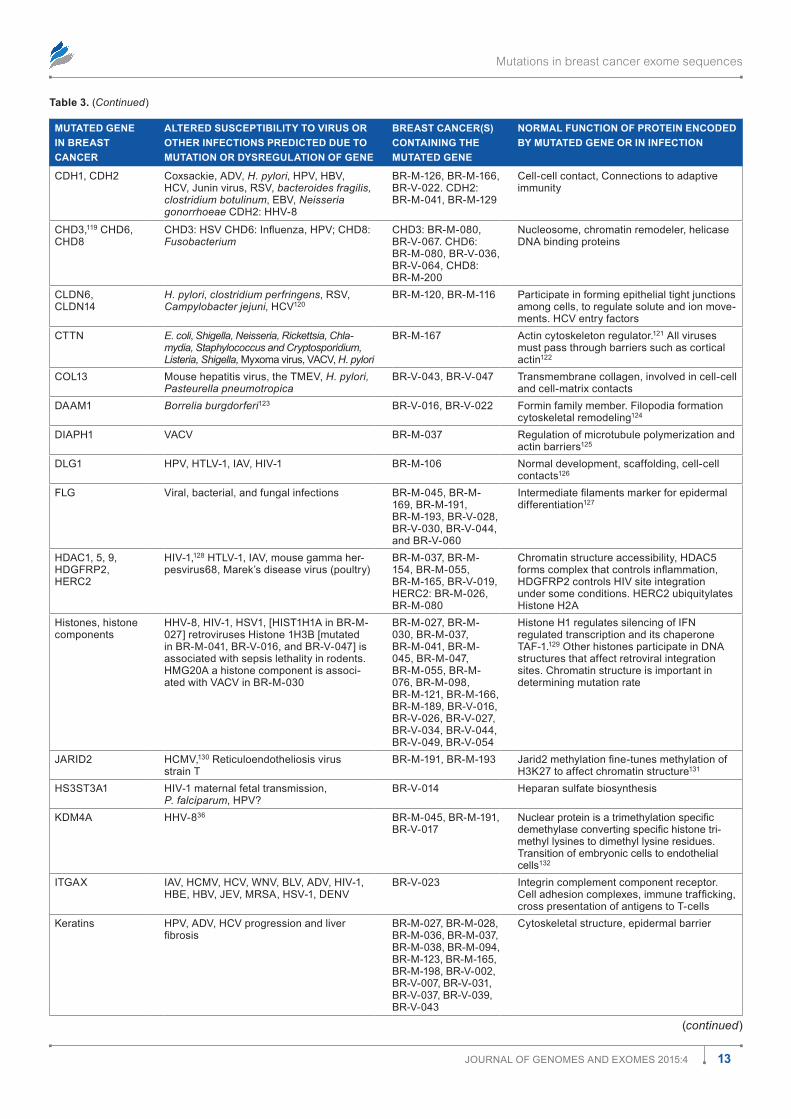
Mutations in breast cancer exome sequences
13Journal of Genomes and exomes 2015:4
Table 3. (Continued)
MUTATED GENE IN BREAST CANCER
ALTERED SUSCEPTIBILITY TO VIRUS OR OTHER INFECTIONS PREDICTED DUE TO MUTATION OR DYSREGULATION OF GENE
BREAST CANCER(S) CONTAINING THE MUTATED GENE
NORMAL FUNCTION OF PROTEIN ENCODED BY MUTATED GENE OR IN INFECTION
CdH1, CdH2 Coxsackie, adV, H. pylori, HPV, HBV, HCV, Junin virus, rsV, bacteroides fragilis, clostridium botulinum, eBV, Neisseria gonorrhoeae CdH2: HHV-8
Br-m-126, Br-m-166, Br-V-022. CdH2: Br-m-041, Br-m-129
Cell-cell contact, Connections to adaptive immunity
CHd3,119 CHd6, CHd8
CHD3: HSV CHD6: Influenza, HPV; CHD8: Fusobacterium
CHd3: Br-m-080, Br-V-067. CHd6: Br-m-080, Br-V-036, Br-V-064, CHd8: Br-m-200
nucleosome, chromatin remodeler, helicase dna binding proteins
Cldn6,Cldn14
H. pylori, clostridium perfringens, rsV, Campylobacter jejuni, HCV120
Br-m-120, Br-m-116 Participate in forming epithelial tight junctions among cells, to regulate solute and ion move-ments. HCV entry factors
CTTn E. coli, Shigella, Neisseria, Rickettsia, Chla-mydia, Staphylococcus and Cryptosporidium, Listeria, Shigella, myxoma virus, VaCV, H. pylori
Br-m-167 actin cytoskeleton regulator.121 all viruses must pass through barriers such as cortical actin122
Col13 mouse hepatitis virus, the TmeV, H. pylori, Pasteurella pneumotropica
Br-V-043, Br-V-047 Transmembrane collagen, involved in cell-cell and cell-matrix contacts
daam1 Borrelia burgdorferi123 Br-V-016, Br-V-022 formin family member. filopodia formation cytoskeletal remodeling124
dIaPH1 VaCV Br-m-037 regulation of microtubule polymerization and actin barriers125
dlG1 HPV, HTlV-1, IaV, HIV-1 Br-m-106 normal development, scaffolding, cell-cell contacts126
flG Viral, bacterial, and fungal infections Br-m-045, Br-m-169, Br-m-191, Br-m-193, Br-V-028, Br-V-030, Br-V-044, and Br-V-060
Intermediate filaments marker for epidermal differentiation127
HdaC1, 5, 9, HdGfrP2, HerC2
HIV-1,128 HTlV-1, IaV, mouse gamma her-pesvirus68, marek’s disease virus (poultry)
Br-m-037, Br-m-154, Br-m-055, Br-m-165, Br-V-019, HerC2: Br-m-026, Br-m-080
Chromatin structure accessibility, HdaC5 forms complex that controls inflammation, HdGfrP2 controls HIV site integration under some conditions. HerC2 ubiquitylates Histone H2a
Histones, histone components
HHV-8, HIV-1, HsV1, [HIsT1H1a in Br-m-027] retroviruses Histone 1H3B [mutated in Br-m-041, Br-V-016, and Br-V-047] is associated with sepsis lethality in rodents. HmG20a a histone component is associ-ated with VaCV in Br-m-030
Br-m-027, Br-m-030, Br-m-037, Br-m-041, Br-m-045, Br-m-047, Br-m-055, Br-m-076, Br-m-098, Br-m-121, Br-m-166, Br-m-189, Br-V-016, Br-V-026, Br-V-027, Br-V-034, Br-V-044, Br-V-049, Br-V-054
Histone H1 regulates silencing of Ifn regulated transcription and its chaperone Taf-1.129 other histones participate in dna structures that affect retroviral integration sites. Chromatin structure is important in determining mutation rate
JarId2 HCmV,130 reticuloendotheliosis virus strain T
Br-m-191, Br-m-193 Jarid2 methylation fine-tunes methylation of H3K27 to affect chromatin structure131
Hs3sT3a1 HIV-1 maternal fetal transmission, P. falciparum, HPV?
Br-V-014 Heparan sulfate biosynthesis
Kdm4a HHV-836 Br-m-045, Br-m-191, Br-V-017
Nuclear protein is a trimethylation specific demethylase converting specific histone tri-methyl lysines to dimethyl lysine residues. Transition of embryonic cells to endothelial cells132
ITGax IaV, HCmV, HCV, WnV, BlV, adV, HIV-1, HBe, HBV, JeV, mrsa, HsV-1, denV
Br-V-023 Integrin complement component receptor. Cell adhesion complexes, immune trafficking, cross presentation of antigens to T-cells
Keratins HPV, adV, HCV progression and liver fibrosis
Br-m-027, Br-m-028, Br-m-036, Br-m-037, Br-m-038, Br-m-094, Br-m-123, Br-m-165, Br-m-198, Br-V-002, Br-V-007, Br-V-031, Br-V-037, Br-V-039, Br-V-043
Cytoskeletal structure, epidermal barrier
(continued)
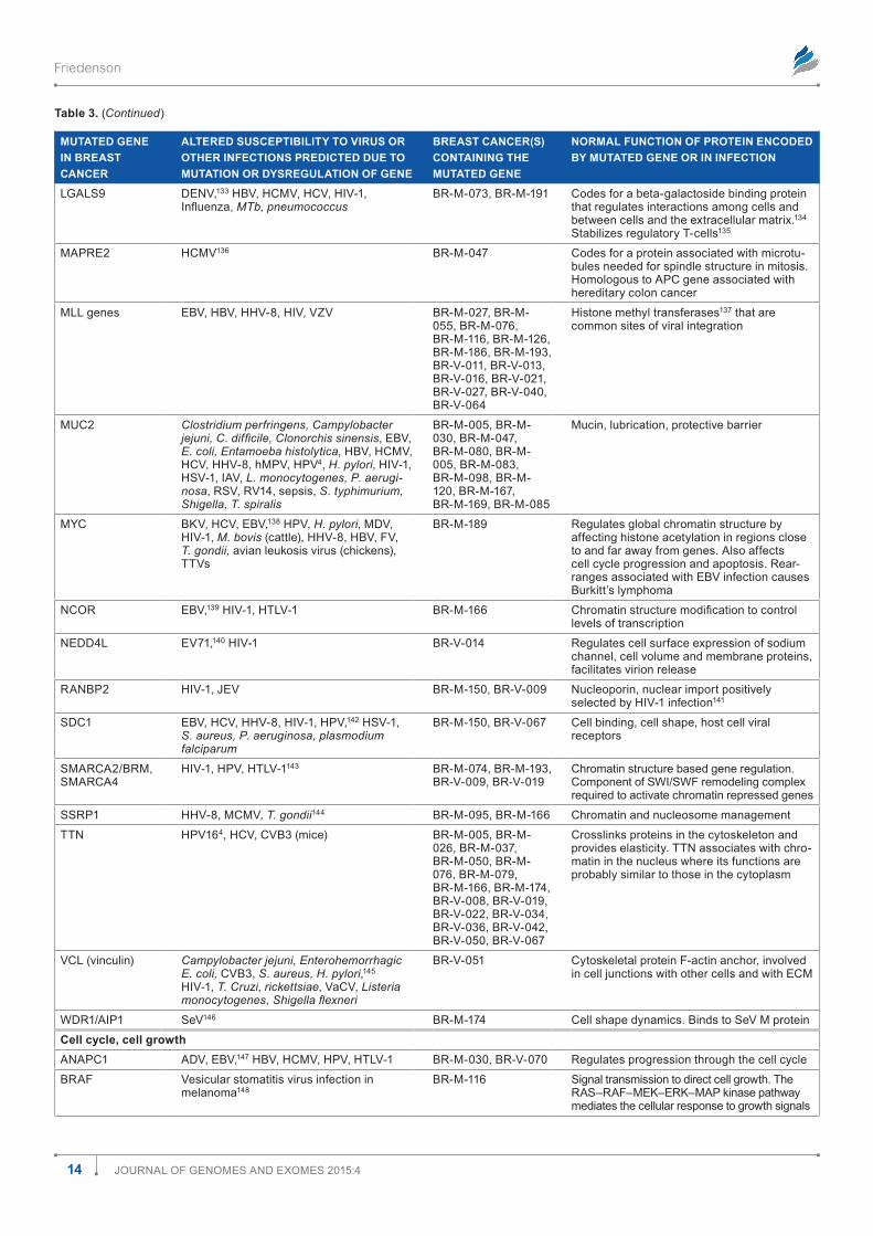
Friedenson
14 Journal of Genomes and exomes 2015:4
Table 3. (Continued)
MUTATED GENE IN BREAST CANCER
ALTERED SUSCEPTIBILITY TO VIRUS OR OTHER INFECTIONS PREDICTED DUE TO MUTATION OR DYSREGULATION OF GENE
BREAST CANCER(S) CONTAINING THE MUTATED GENE
NORMAL FUNCTION OF PROTEIN ENCODED BY MUTATED GENE OR IN INFECTION
lGals9 denV,133 HBV, HCmV, HCV, HIV-1, Influenza, MTb, pneumococcus
Br-m-073, Br-m-191 Codes for a beta-galactoside binding protein that regulates interactions among cells and between cells and the extracellular matrix.134 stabilizes regulatory T-cells135
maPre2 HCmV136 Br-m-047 Codes for a protein associated with microtu-bules needed for spindle structure in mitosis. Homologous to aPC gene associated with hereditary colon cancer
mll genes eBV, HBV, HHV-8, HIV, VZV Br-m-027, Br-m-055, Br-m-076, Br-m-116, Br-m-126, Br-m-186, Br-m-193, Br-V-011, Br-V-013, Br-V-016, Br-V-021, Br-V-027, Br-V-040, Br-V-064
Histone methyl transferases137 that are common sites of viral integration
muC2 Clostridium perfringens, Campylobacter jejuni, C. difficile, Clonorchis sinensis, eBV, E. coli, Entamoeba histolytica, HBV, HCmV, HCV, HHV-8, hmPV, HPV4, H. pylori, HIV-1, HsV-1, IaV, L. monocytogenes, P. aerugi-nosa, rsV, rV14, sepsis, S. typhimurium, Shigella, T. spiralis
Br-m-005, Br-m-030, Br-m-047, Br-m-080, Br-m-005, Br-m-083, Br-m-098, Br-m-120, Br-m-167, Br-m-169, Br-m-085
mucin, lubrication, protective barrier
mYC BKV, HCV, eBV,138 HPV, H. pylori, mdV, HIV-1, M. bovis (cattle), HHV-8, HBV, fV, T. gondii, avian leukosis virus (chickens), TTVs
Br-m-189 regulates global chromatin structure by affecting histone acetylation in regions close to and far away from genes. also affects cell cycle progression and apoptosis. rear-ranges associated with eBV infection causes Burkitt’s lymphoma
nCor eBV,139 HIV-1, HTlV-1 Br-m-166 Chromatin structure modification to control levels of transcription
nedd4l eV71,140 HIV-1 Br-V-014 regulates cell surface expression of sodium channel, cell volume and membrane proteins, facilitates virion release
ranBP2 HIV-1, JeV Br-m-150, Br-V-009 nucleoporin, nuclear import positively selected by HIV-1 infection141
sdC1 eBV, HCV, HHV-8, HIV-1, HPV,142 HsV-1, S. aureus, P. aeruginosa, plasmodium falciparum
Br-m-150, Br-V-067 Cell binding, cell shape, host cell viral receptors
smarCa2/Brm, smarCa4
HIV-1, HPV, HTlV-1143 Br-m-074, Br-m-193, Br-V-009, Br-V-019
Chromatin structure based gene regulation. Component of sWI/sWf remodeling complex required to activate chromatin repressed genes
ssrP1 HHV-8, mCmV, T. gondii144 Br-m-095, Br-m-166 Chromatin and nucleosome management
TTn HPV164, HCV, CVB3 (mice) Br-m-005, Br-m-026, Br-m-037, Br-m-050, Br-m-076, Br-m-079, Br-m-166, Br-m-174, Br-V-008, Br-V-019, Br-V-022, Br-V-034, Br-V-036, Br-V-042, Br-V-050, Br-V-067
Crosslinks proteins in the cytoskeleton and provides elasticity. TTn associates with chro-matin in the nucleus where its functions are probably similar to those in the cytoplasm
VCl (vinculin) Campylobacter jejuni, Enterohemorrhagic E. coli, CVB3, S. aureus, H. pylori,145 HIV-1, T. Cruzi, rickettsiae, VaCV, Listeria monocytogenes, Shigella flexneri
Br-V-051 Cytoskeletal protein f-actin anchor, involved in cell junctions with other cells and with eCm
Wdr1/aIP1 seV146 Br-m-174 Cell shape dynamics. Binds to seV m protein
Cell cycle, cell growthanaPC1 adV, eBV,147 HBV, HCmV, HPV, HTlV-1 Br-m-030, Br-V-070 regulates progression through the cell cycle
Braf Vesicular stomatitis virus infection in melanoma148
Br-m-116 signal transmission to direct cell growth. The ras–raf–meK–erK–maP kinase pathway mediates the cellular response to growth signals
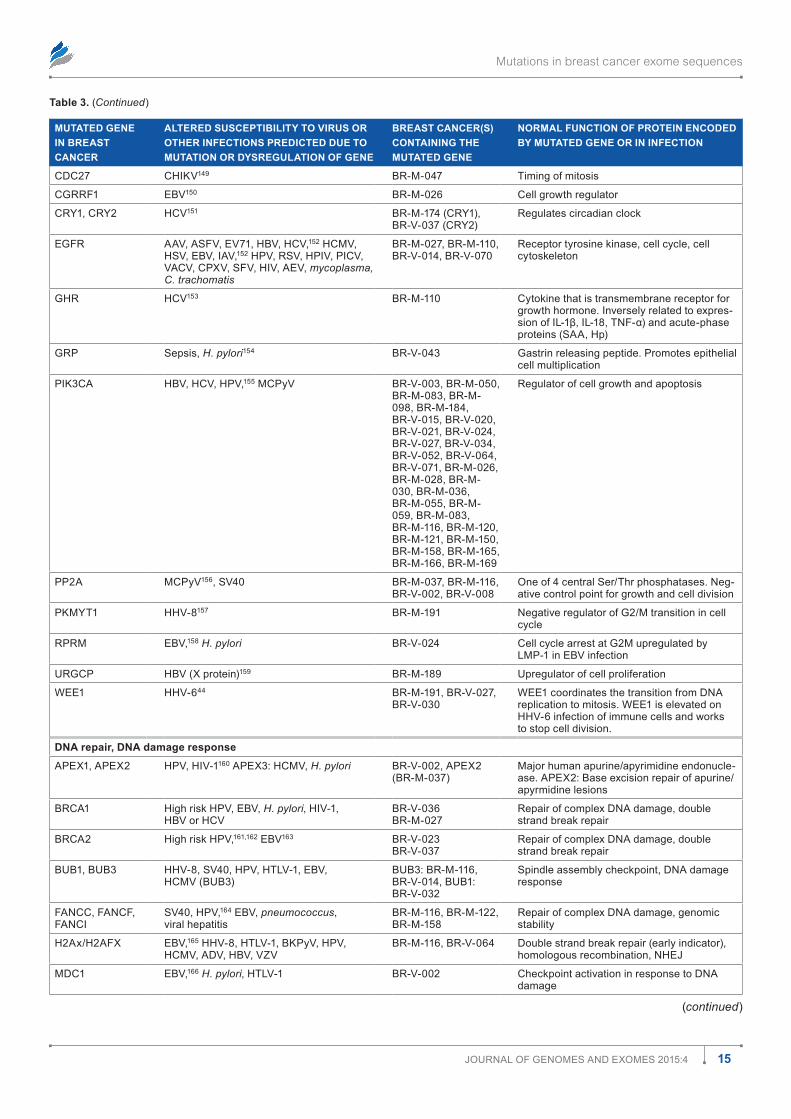
Mutations in breast cancer exome sequences
15Journal of Genomes and exomes 2015:4
Table 3. (Continued)
MUTATED GENE IN BREAST CANCER
ALTERED SUSCEPTIBILITY TO VIRUS OR OTHER INFECTIONS PREDICTED DUE TO MUTATION OR DYSREGULATION OF GENE
BREAST CANCER(S) CONTAINING THE MUTATED GENE
NORMAL FUNCTION OF PROTEIN ENCODED BY MUTATED GENE OR IN INFECTION
CdC27 CHIKV149 Br-m-047 Timing of mitosis
CGrrf1 eBV150 Br-m-026 Cell growth regulator
CrY1, CrY2 HCV151 Br-m-174 (CrY1), Br-V-037 (CrY2)
regulates circadian clock
eGfr aaV, asfV, eV71, HBV, HCV,152 HCmV, HsV, eBV, IaV,152 HPV, rsV, HPIV, PICV, VaCV, CPxV, sfV, HIV, aeV, mycoplasma, C. trachomatis
Br-m-027, Br-m-110, Br-V-014, Br-V-070
receptor tyrosine kinase, cell cycle, cell cytoskeleton
GHr HCV153 Br-m-110 Cytokine that is transmembrane receptor for growth hormone. Inversely related to expres-sion of IL-1β, IL-18, TNF-α) and acute-phase proteins (saa, Hp)
GrP sepsis, H. pylori154 Br-V-043 Gastrin releasing peptide. Promotes epithelial cell multiplication
PIK3Ca HBV, HCV, HPV,155 mCPyV Br-V-003, Br-m-050, Br-m-083, Br-m-098, Br-m-184, Br-V-015, Br-V-020, Br-V-021, Br-V-024, Br-V-027, Br-V-034, Br-V-052, Br-V-064, Br-V-071, Br-m-026, Br-m-028, Br-m-030, Br-m-036, Br-m-055, Br-m-059, Br-m-083, Br-m-116, Br-m-120, Br-m-121, Br-m-150, Br-m-158, Br-m-165, Br-m-166, Br-m-169
regulator of cell growth and apoptosis
PP2a mCPyV156, sV40 Br-m-037, Br-m-116, Br-V-002, Br-V-008
one of 4 central ser/Thr phosphatases. neg-ative control point for growth and cell division
PKmYT1 HHV-8157 Br-m-191 negative regulator of G2/m transition in cell cycle
rPrm eBV,158 H. pylori Br-V-024 Cell cycle arrest at G2m upregulated by lmP-1 in eBV infection
urGCP HBV (x protein)159 Br-m-189 upregulator of cell proliferation
Wee1 HHV-644 Br-m-191, Br-V-027, Br-V-030
Wee1 coordinates the transition from dna replication to mitosis. Wee1 is elevated on HHV-6 infection of immune cells and works to stop cell division.
DNA repair, DNA damage response
aPex1, aPex2 HPV, HIV-1160 aPex3: HCmV, H. pylori Br-V-002, aPex2 (Br-m-037)
major human apurine/apyrimidine endonucle-ase. aPex2: Base excision repair of apurine/apyrmidine lesions
BrCa1 High risk HPV, eBV, H. pylori, HIV-1, HBV or HCV
Br-V-036Br-m-027
repair of complex dna damage, double strand break repair
BrCa2 High risk HPV,161,162 eBV163 Br-V-023Br-V-037
repair of complex dna damage, double strand break repair
BuB1, BuB3 HHV-8, sV40, HPV, HTlV-1, eBV, HCmV (BuB3)
BuB3: Br-m-116, Br-V-014, BuB1: Br-V-032
spindle assembly checkpoint, dna damage response
fanCC, fanCf, fanCI
sV40, HPV,164 eBV, pneumococcus, viral hepatitis
Br-m-116, Br-m-122, Br-m-158
repair of complex dna damage, genomic stability
H2ax/H2afx eBV,165 HHV-8, HTlV-1, BKPyV, HPV, HCmV, adV, HBV, VZV
Br-m-116, Br-V-064 double strand break repair (early indicator), homologous recombination, nHeJ
mdC1 eBV,166 H. pylori, HTlV-1 Br-V-002 Checkpoint activation in response to dna damage
(continued)
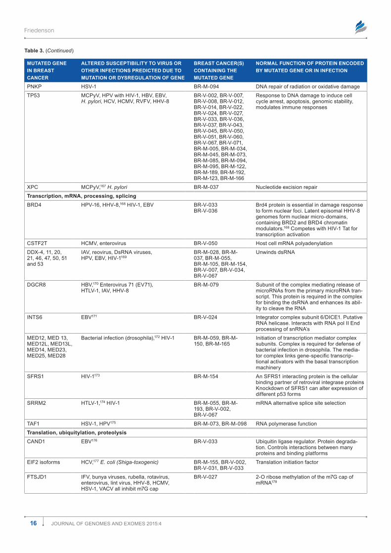
Friedenson
16 Journal of Genomes and exomes 2015:4
Table 3. (Continued)
MUTATED GENE IN BREAST CANCER
ALTERED SUSCEPTIBILITY TO VIRUS OR OTHER INFECTIONS PREDICTED DUE TO MUTATION OR DYSREGULATION OF GENE
BREAST CANCER(S) CONTAINING THE MUTATED GENE
NORMAL FUNCTION OF PROTEIN ENCODED BY MUTATED GENE OR IN INFECTION
PnKP HsV-1 Br-m-094 dna repair of radiation or oxidative damage
TP53 mCPyV, HPV with HIV-1, HBV, eBV, H. pylori, HCV, HCmV, rVfV, HHV-8
Br-V-002, Br-V-007, Br-V-008, Br-V-012, Br-V-014, Br-V-022, Br-V-024, Br-V-027, Br-V-033, Br-V-036, Br-V-037, Br-V-043, Br-V-045, Br-V-050, Br-V-051, Br-V-060, Br-V-067, Br-V-071, Br-m-005, Br-m-034, Br-m-045, Br-m-073, Br-m-085, Br-m-094, Br-m-095, Br-m-122, Br-m-189, Br-m-192, Br-m-123, Br-m-166
response to dna damage to induce cell cycle arrest, apoptosis, genomic stability, modulates immune responses
xPC mCPyV,167 H. pylori Br-m-037 nucleotide excision repair
Transcription, mRNA, processing, splicingBrd4 HPV-16, HHV-8,168 HIV-1, eBV Br-V-033
Br-V-036Brd4 protein is essential in damage response to form nuclear foci. latent episomal HHV-8 genomes form nuclear micro-domains, containing Brd2 and Brd4 chromatin modulators.168 Competes with HIV-1 Tat for transcription activation
CsTf2T HCmV, enterovirus Br-V-050 Host cell mrna polyadenylation
ddx-4, 11, 20, 21, 46, 47, 50, 51 and 53
IaV, reovirus, dsrna viruses, HPV, eBV, HIV-1169
Br-m-028, Br-m-037, Br-m-055, Br-m-105, Br-m-154, Br-V-007, Br-V-034, Br-V-067
unwinds dsrna
dGCr8 HBV,170 enterovirus 71 (eV71), HTlV-1, IaV, HHV-8
Br-m-079 subunit of the complex mediating release of micrornas from the primary microrna tran-script. This protein is required in the complex for binding the dsrna and enhances its abil-ity to cleave the rna
InTs6 eBV171 Br-V-024 Integrator complex subunit 6/dICe1. Putative rna helicase. Interacts with rna pol II end processing of snrna’s
med12, med 13, med12l, med13l, med14, med23, med25, med28
Bacterial infection (drosophila),172 HIV-1 Br-m-059, Br-m-150, Br-m-165
Initiation of transcription mediator complex subunits. Complex is required for defense of bacterial infection in drosophila. The media-tor complex links gene-specific transcrip-tional activators with the basal transcription machinery
sfrs1 HIV-1173 Br-m-154 an sfrs1 interacting protein is the cellular binding partner of retroviral integrase proteins Knockdown of sfrs1 can alter expression of different p53 forms
srrm2 HTlV-1,174 HIV-1 Br-m-055, Br-m-193, Br-V-002, Br-V-067
mrna alternative splice site selection
Taf1 HsV-1, HPV175 Br-m-073, Br-m-098 rna polymerase function
Translation, ubiquitylation, proteolysisCand1 eBV176 Br-V-033 ubiquitin ligase regulator. Protein degrada-
tion. Controls interactions between many proteins and binding platforms
eIf2 isoforms HCV,177 E. coli (Shiga-toxogenic) Br-m-155, Br-V-002, Br-V-031, Br-V-033
Translation initiation factor
fTsJd1 IfV, bunya viruses, rubella, rotavirus, enterovirus, lint virus, HHV-8, HCmV, HsV-1, VaCV all inhibit m7G cap
Br-V-027 2-o ribose methylation of the m7G cap of mrna178
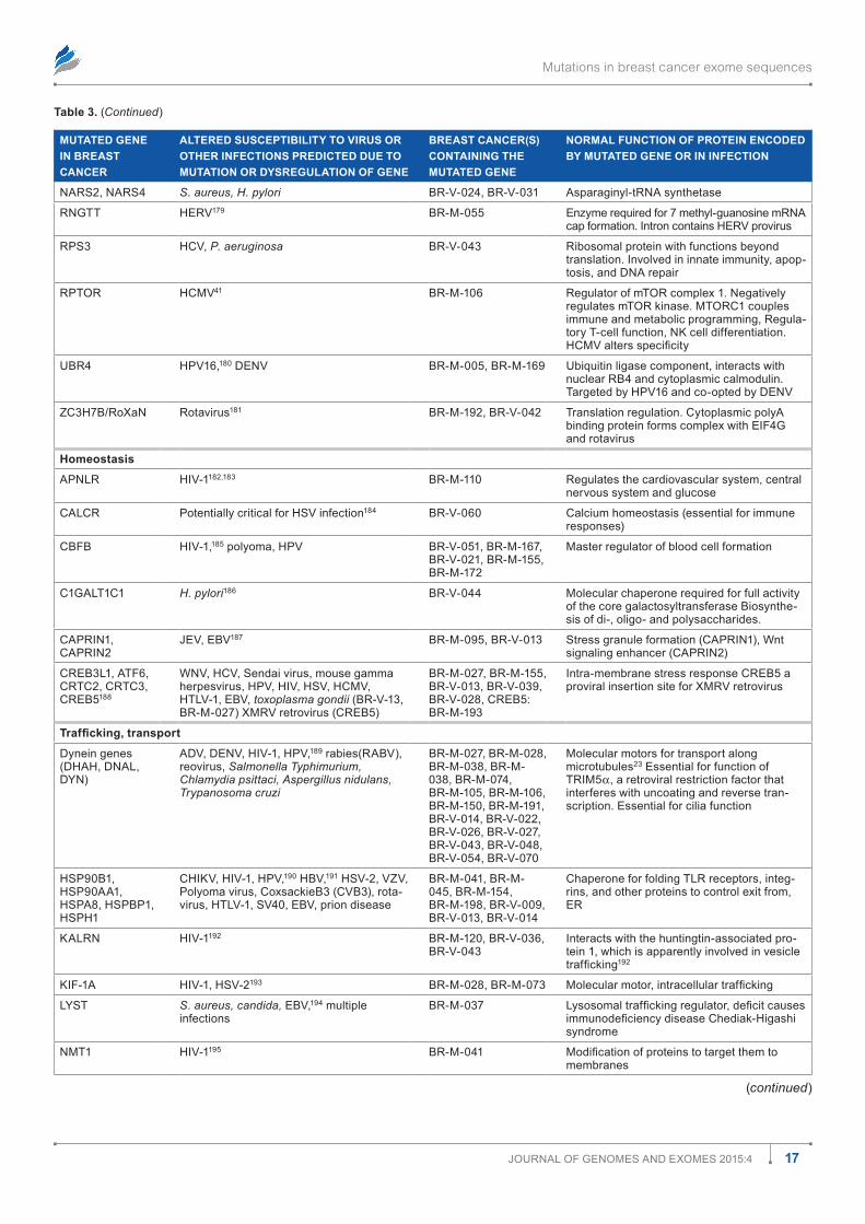
Mutations in breast cancer exome sequences
17Journal of Genomes and exomes 2015:4
Table 3. (Continued)
MUTATED GENE IN BREAST CANCER
ALTERED SUSCEPTIBILITY TO VIRUS OR OTHER INFECTIONS PREDICTED DUE TO MUTATION OR DYSREGULATION OF GENE
BREAST CANCER(S) CONTAINING THE MUTATED GENE
NORMAL FUNCTION OF PROTEIN ENCODED BY MUTATED GENE OR IN INFECTION
nars2, nars4 S. aureus, H. pylori Br-V-024, Br-V-031 asparaginyl-trna synthetase
rnGTT HerV179 Br-m-055 enzyme required for 7 methyl-guanosine mrna cap formation. Intron contains HerV provirus
rPs3 HCV, P. aeruginosa Br-V-043 ribosomal protein with functions beyond translation. Involved in innate immunity, apop-tosis, and dna repair
rPTor HCmV41 Br-m-106 regulator of mTor complex 1. negatively regulates mTor kinase. mTorC1 couples immune and metabolic programming, regula-tory T-cell function, nK cell differentiation. HCMV alters specificity
uBr4 HPV16,180 denV Br-m-005, Br-m-169 ubiquitin ligase component, interacts with nuclear rB4 and cytoplasmic calmodulin. Targeted by HPV16 and co-opted by denV
ZC3H7B/roxan rotavirus181 Br-m-192, Br-V-042 Translation regulation. Cytoplasmic polya binding protein forms complex with eIf4G and rotavirus
Homeostasis
aPnlr HIV-1182,183 Br-m-110 regulates the cardiovascular system, central nervous system and glucose
CalCr Potentially critical for HsV infection184 Br-V-060 Calcium homeostasis (essential for immune responses)
CBfB HIV-1,185 polyoma, HPV Br-V-051, Br-m-167, Br-V-021, Br-m-155, Br-m-172
master regulator of blood cell formation
C1GalT1C1 H. pylori186 Br-V-044 molecular chaperone required for full activity of the core galactosyltransferase Biosynthe-sis of di-, oligo- and polysaccharides.
CaPrIn1, CaPrIn2
JeV, eBV187 Br-m-095, Br-V-013 stress granule formation (CaPrIn1), Wnt signaling enhancer (CaPrIn2)
CreB3l1, aTf6, CrTC2, CrTC3, CreB5188
WnV, HCV, sendai virus, mouse gamma herpesvirus, HPV, HIV, HsV, HCmV, HTlV-1, eBV, toxoplasma gondii (Br-V-13, Br-m-027) xmrV retrovirus (CreB5)
Br-m-027, Br-m-155, Br-V-013, Br-V-039, Br-V-028, CreB5: Br-m-193
Intra-membrane stress response CreB5 a proviral insertion site for xmrV retrovirus
Trafficking, transport
dynein genes (dHaH, dnal, dYn)
adV, denV, HIV-1, HPV,189 rabies(raBV), reovirus, Salmonella Typhimurium, Chlamydia psittaci, Aspergillus nidulans, Trypanosoma cruzi
Br-m-027, Br-m-028, Br-m-038, Br-m-038, Br-m-074, Br-m-105, Br-m-106, Br-m-150, Br-m-191, Br-V-014, Br-V-022, Br-V-026, Br-V-027, Br-V-043, Br-V-048, Br-V-054, Br-V-070
molecular motors for transport along microtubules23 essential for function of TrIm5a, a retroviral restriction factor that interferes with uncoating and reverse tran-scription. essential for cilia function
HsP90B1, HsP90aa1, HsPa8, HsPBP1, HsPH1
CHIKV, HIV-1, HPV,190 HBV,191 HsV-2, VZV, Polyoma virus, CoxsackieB3 (CVB3), rota-virus, HTlV-1, sV40, eBV, prion disease
Br-m-041, Br-m-045, Br-m-154, Br-m-198, Br-V-009, Br-V-013, Br-V-014
Chaperone for folding Tlr receptors, integ-rins, and other proteins to control exit from, er
Kalrn HIV-1192 Br-m-120, Br-V-036, Br-V-043
Interacts with the huntingtin-associated pro-tein 1, which is apparently involved in vesicle trafficking192
KIf-1a HIV-1, HsV-2193 Br-m-028, Br-m-073 Molecular motor, intracellular trafficking
lYsT S. aureus, candida, eBV,194 multiple infections
Br-m-037 Lysosomal trafficking regulator, deficit causes immunodeficiency disease Chediak-Higashi syndrome
nmT1 HIV-1195 Br-m-041 Modification of proteins to target them to membranes
(continued)
Friedenson
18 Journal of Genomes and exomes 2015:4
Table 3. (Continued)
MUTATED GENE IN BREAST CANCER
ALTERED SUSCEPTIBILITY TO VIRUS OR OTHER INFECTIONS PREDICTED DUE TO MUTATION OR DYSREGULATION OF GENE
BREAST CANCER(S) CONTAINING THE MUTATED GENE
NORMAL FUNCTION OF PROTEIN ENCODED BY MUTATED GENE OR IN INFECTION
PaCs1 HIV-1,196 HPV, mlV (murine leukemia), rd114, HCmV
Br-V-002 regulated sorting of proteins in trans Golgi network to proper compartment, an important component of their in vivo activity Crucial for assembly of some retroviruses and essen-tial interactions with some herpesviruses envelopes
raB7a HBV,197 HIV-1 Br-m-186 Molecular switches, traffickingslC6a3 HIV-1198 Br-V-003 dopamine transportersTard3 subverted by a bacterial infection
Anaplasma phagocytophilum for a bacterial inclusion membrane199
Br-m-121 Cholesterol export
sTau1 HCV,200 HIV-1, HerV-K, IaV Br-V-015 mrna transport to different organellesTf (transferrin) HIV, HCV, HBV,201 Neisseria meningitidis,
Burkholderia cenocepacia, Enteropatho-genic E. coli, streptococcus, MTb, canine parvovirus, plasmodium
Br-m-037 Within their host, pathogenic bacteria acquire iron essential for infection and growth from Tf, a crucial innate immune defense protein
MetabolismadPrHl2 Bacteria202 Br-V-020 removes bacterial adP ribosylation added
by toxins from host proteinsaoaH Gram negative bacteria203 Br-V-036 acyloxyacyl hydrolase hydrolyzes fatty acyl
chains from bacterial lipopolysaccharides, to detoxify them. The aoaH protein may modify host inflammatory responses to gram-negative bacteria
Ca4 renal bacteria, many Co2 sensing bacteria204
Br-m-079 reversible hydration of carbon dioxide
CPT1a lower respiratory tract infection, hemophilus influenzae, pneumococcus, C. trachomatis,205 HCV
Br-V-043 long chain fatty acid metabolism, transport into mitochondria
CYP2C8 HPV206 Br-m-105 steroid hydroxylation e.g. estradiolfasn denV, HCV, HHV-8,207 rotavirus, HIV-1 Br-V-023, Br-V-052 long chain fatty acid synthesisG6Pd HPV, HBV,208 HBe, denV, HIV-1,
P. falciparum, P. vivaxBr-m-122 needed for pentose phosphate pathway to
supply reducing energy as nadPH to cellsGrB14 N. gonorrhoeae, pathogenic bacteria209 Br-V-012, Br-V-070 Interacts with insulin and insulin-like growth
factor receptors. regulates response to infection with pathogenic bacteria
lCT/lactase rotavirus,210 Giardia lamblia, HIV-1 Br-m-191 lactose metabolismldlraP1 E. coli uropathogenic211 Br-m-045 ldl receptor adaptor. used as an alternate
receptor by e. coli211
mGam denV,212 eBola, HCV, marburg virus, mV, VZV
Br-m-037, Br-m-106, Br-V-034
alpha glucosidase, starch digestion. Inhibi-tion of this enzyme can impair the assembly of viral structural proteins and viral particles
nr1d1 MTb, salmonella, bacteria47 Br-m-079 Circadian clockPaH HCV impairs activity, HIV-1,213 P. falciparum,
viral encephalitis, yellow fever, bacterial infections
Br-m-191 Hydroxylation of phenylalanine converting it to tyrosine
PC HCmV inhibits, HsV-1 induces214 Br-m-191 Pyruvate carboxylase converts pyruvate to oxaloacetate
PdxK HIV-1,215 HBV, rauscher leukemia virus, oncornaviral dna polymerases
Br-V-016 Phosphorylates vitamin B6, required to convert B6 to pyridoxal phosphate
uPrT eBV216 Br-m-174 Pyrimidine salvage pathway
the Absent In Melanoma 2 (AIM2) inflammasome. AIM2 inflammasome–mediated defenses can be blunted by multiple tumor viruses, by other viruses, by bacterial infections,16 and by many breast cancer mutations as well (Fig. 1 and Table 3).
MyD88 (myeloid differentiation primary response 88) is an adaptor for Toll-like receptors (TLRs) on the cell and
endosomal membrane and is essential to produce inflam-matory cytokines and Interferons (IFN; Fig. 2). The RTA pro tein (the transcription activator and lytic switch) from the HHV-8 virus degrades MyD88 and blocks TLR signaling (Fig. 2). HBV and HCV also inhibit MyD88 and alter its downstream signaling.17,18 Other viruses also target
Mutations in breast cancer exome sequences
19Journal of Genomes and exomes 2015:4
MyD88. Breast cancer mutations in MyD88 itself were not found, but breast cancer mutations affected many of the steps downstream of the MyD88 gene product (Fig. 2). IRAK2 and IRAK3 (Interleukin 1 Receptor-Associated Kinases) are mutated in different breast cancers, damaging the control of inflammatory cytokine production (Fig. 2). IRAK3 interacts with IRAK2 and inhibits IRAK2-mediated phosphoryla-tion of eIF4E, establishing a connection with protein transla-tion. This prevents translation of inflammatory cytokines and downregulates TLR responses.19 These genes are associated with multiple infections (Table 3).20
The endosomal TLR3 gene has missense mutations in two breast cancers (Table 3). In endosomes (green circle in Fig. 2), TLR3 binds tumor virus double-stranded RNA (dsRNA) such as those from lysed pathogens and activates signaling to the nucleus to produce antiviral cytokines such as IFNs (Fig. 2).21,22 TLR3 is essential to respond to other viruses listed in Table 3.9
RIG-1 (retinoic acid–inducible gene 1–like helicase) is a cytoplasmic recognition sensor for viral RNAs, and RIG-1–mediated pathways attract both mutation and infection (Fig. 2 and Table 3). Multiple infections and breast cancer mutations focus on RIG-1–mediated signaling (Fig. 2).
Twenty mutations in 17 different breast cancers involved dynein heavy chains (Table 3), which transport cargo along microtubules and maintain cytoplasmic architecture. Innate cellular defenses against retroviral infection include restric-tion factors such as Tripartite Motif-Containing Protein 5 (TRIM5 protein). TRIM5a is present in the cytoplasm, where it interferes with retroviruses shortly after they enter the cell. The restriction factor then inhibits viral uncoating and reverse transcription. The dynein complex is essential to transport TRIM5a protein or complexes containing TRIM5a along microtubules to interrupt cytoplasmic retroviral infections. Crippling the activity of dynein motors or dynein complexes or interfering with microtubule structure decreases the ability of TRIM5a to protect against retroviral infection.23
IFNs can transmit signals to the Janus Kinase – Signal Transducer and Activator of Transcription (JAK-STAT) path-way via receptors in uninfected cells24 to protect them from infection. Abnormal regulation of the JAK-STAT pathway occurs in human cancers in diverse organs25 and at least seven breast cancers have mutations affecting JAK-STAT signaling (Fig. 2). The JAK-STAT pathway is inhibited by many viruses including Varicella zoster virus (VZV),26 HBV,27 and HCV.28
Breast cancer mutations interfere with intrinsic immunity. Nonsense-mediated decay (NMD) factors sense premature termination codons associated with translation of some viral sequences. Figure 3 (bottom) shows inhibition or activation of NMD by several viruses. Six breast cancers have mutations in factors required for NMD (Fig. 3 and Table 3).
Infection and mutation can both affect the DNA damage response. Damage to genes encoding pathways to repair DNA damage occurs in at least 18 different breast cancers (Table 3
and Fig. 2). Viral infections can break chromosomes, activating innate immune responses to target infected or transformed cells. Viruses can pervert host DNA repair to promote inte gration of viral DNA into host DNA and can limit other infections. Chro-mosomes in virally infected cells can sometimes have a remark-able resemblance to hereditary cancer-prone diseases that have inactive genes required for DNA double strand break repairs. Mutations affecting genomic stability increase infection risks.
The protein signals required for DNA damage responses include ATM, ATR, BRCA1, BRCA2, PALB2, RAD50, RAD51, Fanconi proteins, XPC, and PRKC1. Mutations in many of these genes can be inherited and lead to hereditary cancer predispositions. Chromatid exchanges and aberrations typical of Fanconi anemia and BRCA2 infections can result from viral infections. Mutations in ATM (four breast can-cers), BRCA1, BRCA2, and interacting partners (seven breast cancers) and Fanconi proteins (three breast cancers) render the genome less stable by influencing DNA repair. Homozygous ATM mutations are well known to increase infection risks. Table 3 shows infection risks increased by BRCA and Fan-coni gene mutations. Breast cancer mutations affecting base excision and nucleotide excision repair pathways also increase risks for infections (Table 3). In turn, infection by H. pylori, for one, causes mutations by several mechanisms including downregulating major DNA repair pathways.
Infections associated with mutations affect innate immu-nity signals. Granzymes are serine proteases without antigen receptors that kill target cells and pathogens directly. Perforin (PRF1) is the chief effector for Natural Killer cell (NK)-mediated cytolysis. Table 3 shows that granzyme and perforin deficits are associated with many infections, both viral and nonviral. Granzyme A (GZMA) and Granzyme H (GZMH) genes are mutated in two breast cancers and PRF1 is mutated in another (Fig. 2, right side).
Damage to genes essential for neutrophil functions. In addition to granzymes, at least 10 other genes (AKT1, AOC3, BIRC6, BPI, CEACAM8, CYBB/NOX2, DBNL, KLK15, MPO, and NCF2) contain mutations that could affect neutrophil effectiveness in 12 different breast cancers. Many infec tions seen in cancer patients are related to weakened function of neu-trophils/phagosome–containing cells. Cancer-causing infec-tions related to the mutations listed above include EBV, HCV, HPV, and H. pylori. The CYBB/NOX2 (cytochrome B-245, beta polypeptide) gene mutated in BR-M-045 breast cancer encodes a product that acts in host defense in phagosomes by generating H2O2.29 Multiple noncancer-associated infections are specifically linked to CYBB/NOX2 activity and might exacerbate effects of mutation and of cancer-causing infections.
Breast cancer mutations affecting connections to immune responses and neurological sensing alter risks for infections. In at least 18 of the 103 breast cancers, a gene mutation affects the code for an inflammatory cytokine such as an interleukin (IL). These cytokines are essential signals to activate a long-term adaptive immune response (Fig. 2). Other connections
Friedenson
20 Journal of Genomes and exomes 2015:4
to innate and adaptive immunity are also damaged including Human Leukocyte Antigens (HLA) or the class II major his-tocompatibility complex transactivator (CIITA), an important regulator of histocompatibility genes (a total of six breast can-cers in Table 3). Damage to these gene products can impair response to a long list of infections. Table 3 also contains many instances of specific individual signals essential for immunity that have sustained serious mutations in their genes. Even a neuroimmune response is damaged when genes encoding taste receptors (TAS1R and TAS2R genes) that sense bacte-rial endotoxins30 are mutated in seven breast cancers (Table 3).
Damage to cell adhesion and architecture genes associates with infections. In epithelial cells, CDH1 (cadherin 1, type 1) dynami-cally complexes with catenins, which regulates signalling path-ways such as Phosphoinositide 3 kinase (PIK3)/Akt and nuclear factor kappa-light-chain-enhancer of activated B cells (NF-KB). CDH1 also regulates multiple connections to the innate and adaptive immune systems31 (Fig. 2). CDH1 gene mutations occur in three different breast cancers, and CDH1 protein is associated with a wide range of infections including some known to cause cancer (Table 3). Maintaining epithelial barriers is a prime immunologic function of CDH1 because it participates in walling off infections and harmful agents to protect underlying structures. Adherens junctions formed by cadherins are known to inhibit lentivirus entry into cells.32 In all the breast cancers, there were 16 mutations in 13 different cadherins. Many more mutations affected proteins that interact with cadherins.
Cell architecture and structure must be changed in order to accommodate viral infections. Every one of the 103 breast cancers have mutations affecting actin, the cytoskeleton, or its regulation. In order to establish a persistent infection, viruses must escape an immune response and pass through collagen-rich extracellular structures, unfavorable environments, and structural barriers to reach targets.3,4 Many viruses have evolved the ability to manipulate the actin/membrane net-work in host cells to facilitate viral replication and then spread
to new cells. Over 1/3 (35/103) breast cancers have damage to genes encoding one or more of three families of structural molecules (laminins, collagens, and fibronectin). Collagen alone is dependent on dozens of genes that have mutations in the breast cancers. Defects in these structural proteins could increase susceptibility to a broad range of microbial infections. In addition to viruses, infections that degrade these proteins include gram-positive bacteria, gram-negative bacteria, fungi, anaerobic bacteria, and parasites (Table 4). Vaccinia virus (VACV), Rickettsia, and facultative intracellular bacteria Lis-teria monocytogenes and Shigella flexneri pervert host intracellu-lar actin to promote transmission to new cells.33 This suggests that the breast cancer genomes with deregulated cytoskeletal proteins or with considerable damage to the genes encoding cytoskeletal and extracellular signaling connections present many attractive targets for infection either before or after the cancer develops. In this instance, mutation and infection often work in the same direction in breast cancer cells.
Some ion channels have evolved to maintain homeostasis during infections. Because breast and other cancer cells have highly abnormal morphology and size, ion channel mutations may be essential for cancer cells to survive. In many breast can-cers, ion channels with known associations to cell volume or morphology are mutated including NEDD4L (Table 3), BEST3, CHRNA9, HBS1L, KCNA4, KCNA5, KCNH4, KCNH8, KCNV2, ACCN1l, TTYH1, and WNK3. Damage to host cell ion channels may facilitate virally enhanced cell permeabil-ity caused by incorporation of ion channel proteins encoded by viruses. A growing list of viruses encode their own ion channels (viroporins), including HCV, HIV-1, IAV, Infectious Bronchitis Virus (IBV), poliovirus (PV), Severe Acute Respiratory Syn-drome virus (SARS), Sindbis Virus, Semliki Forest virus (SFV),34 HTLV-1, John Cunningham virus (JCV), and rotavirus.
The gene for intermediate filament–associated protein filaggrin (FLG) was mutated in eight breast cancers (Table 3). FLG aggregates intermediate filaments and helps determine
Table 4. Breast cancers with mutations in major structural proteins that protect against infection.
structural barrier molecules
Pathogens that produce enzymes that degrade this molecule or adhere to it or Viruses with capsid proteins or antigens that interact with structural barrier molecules217
Breast cancers with damage to gene(s) encoding the barrier molecule(s)
laminins, collagens, fibronectin
Bacteria, fungi, parasites: Actinobacillus actinomycetem-comitans, Bacillus cereus, Clostridium histolyticum, Clostridium perfringens, Porphyromonas gingivalis, Pseudomonas aeruginosa, P. gingivalis, Streptococcus pyogenes (or group A Streptococcus; GAS), Streptococcus gordonii, Treponema denticola, Vibrio alginolyticus, Vibrio vulnificus, Vibrio parahaemolyticus, Aspergillus fumigatus, Cryptococcus neoformans, C. albicans, Candida spp., Paracoccidioides brasiliensis, Trichophyton schoenleinii, Acanthamoeba castellanii, Acanthamoeba healyi, Balamu-thia mandrillaris, Entamoeba histolytica, Giardia duode-nalis, Trichomonas vaginalis, Trypanosoma spp. Viruses: Influenza, HPV, retroviruses, human polyoma virus
Br-m-028, Br-m-037, Br-m-041, Br-m-055, Br-m-076, Br-m-085, Br-m-095, Br-m-098, Br-m-106, Br-m-110, Br-m-120, Br-m-122, Br-m-123, Br-m-126, Br-m-174, Br-m-191, Br-m-193, Br-V-002, Br-V-009, Br-V-014, Br-V-017, Br-V-022, Br-V-023, Br-V-024, Br-V-026, Br-V-027, Br-V-034, Br-V-037, Br-V-043, Br-V-044, Br-V-045, Br-V-047, Br-V-064, Br-V-067, Br-V-071
actin, actin related every virus known interacts with the actin cytoskeleton,218 L. donovani, C.trachomatis, salmonella, Borrelia burgdorferi
all 103 breast cancers. see also reference 4
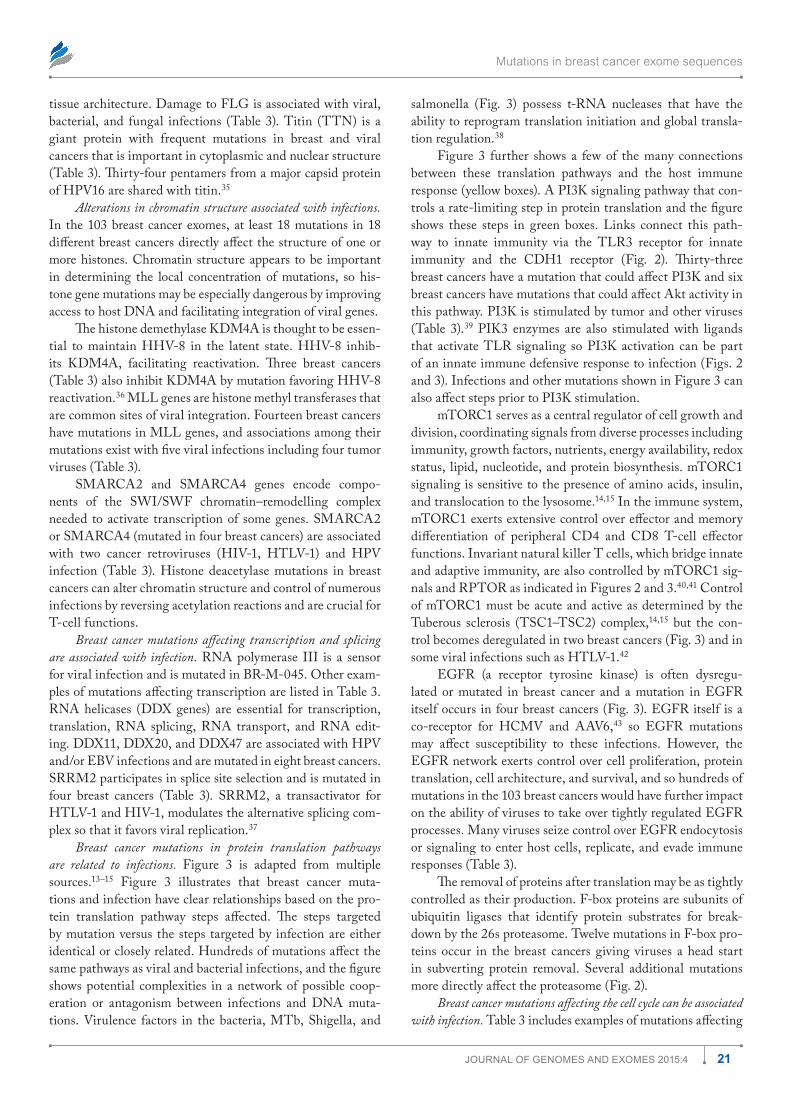
Mutations in breast cancer exome sequences
21Journal of Genomes and exomes 2015:4
tissue architecture. Damage to FLG is associated with viral, bacterial, and fungal infections (Table 3). Titin (TTN) is a giant protein with frequent mutations in breast and viral cancers that is important in cytoplasmic and nuclear structure (Table 3). Thirty-four pentamers from a major capsid protein of HPV16 are shared with titin.35
Alterations in chromatin structure associated with infections. In the 103 breast cancer exomes, at least 18 mutations in 18 different breast cancers directly affect the structure of one or more histones. Chromatin structure appears to be important in determining the local concentration of mutations, so his-tone gene mutations may be especially dangerous by improving access to host DNA and facilitating integration of viral genes.
The histone demethylase KDM4A is thought to be essen-tial to maintain HHV-8 in the latent state. HHV-8 inhib-its KDM4A, facilitating reactivation. Three breast cancers (Table 3) also inhibit KDM4A by mutation favoring HHV-8 reactivation.36 MLL genes are histone methyl transferases that are common sites of viral integration. Fourteen breast cancers have mutations in MLL genes, and associations among their mutations exist with five viral infections including four tumor viruses (Table 3).
SMARCA2 and SMARCA4 genes encode compo-nents of the SWI/SWF chromatin–remodelling complex needed to activate transcription of some genes. SMARCA2 or SMARCA4 (mutated in four breast cancers) are associated with two cancer retroviruses (HIV-1, HTLV-1) and HPV infection (Table 3). Histone deacetylase mutations in breast cancers can alter chromatin structure and control of numerous infections by reversing acetylation reactions and are crucial for T-cell functions.
Breast cancer mutations affecting transcription and splicing are associated with infection. RNA polymerase III is a sensor for viral infection and is mutated in BR-M-045. Other exam-ples of mutations affecting transcription are listed in Table 3. RNA helicases (DDX genes) are essential for transcription, translation, RNA splicing, RNA transport, and RNA edit-ing. DDX11, DDX20, and DDX47 are associated with HPV and/or EBV infections and are mutated in eight breast cancers. SRRM2 participates in splice site selection and is mutated in four breast cancers (Table 3). SRRM2, a transactivator for HTLV-1 and HIV-1, modulates the alternative splicing com-plex so that it favors viral replication.37
Breast cancer mutations in protein translation pathways are related to infections. Figure 3 is adapted from multiple sources.13–15 Figure 3 illustrates that breast cancer muta-tions and infection have clear relationships based on the pro-tein translation pathway steps affected. The steps targeted by mutation versus the steps targeted by infection are either identical or closely related. Hundreds of mutations affect the same pathways as viral and bacterial infections, and the figure shows potential complexities in a network of possible coop-eration or antagonism between infections and DNA muta-tions. Virulence factors in the bacteria, MTb, Shigella, and
salmonella (Fig. 3) possess t-RNA nucleases that have the ability to reprogram translation initiation and global transla-tion regulation.38
Figure 3 further shows a few of the many connections between these translation pathways and the host immune response (yellow boxes). A PI3K signaling pathway that con-trols a rate-limiting step in protein translation and the figure shows these steps in green boxes. Links connect this path-way to innate immunity via the TLR3 receptor for innate immunity and the CDH1 receptor (Fig. 2). Thirty-three breast cancers have a mutation that could affect PI3K and six breast cancers have mutations that could affect Akt activity in this pathway. PI3K is stimulated by tumor and other viruses (Table 3).39 PIK3 enzymes are also stimulated with ligands that activate TLR signaling so PI3K activation can be part of an innate immune defensive response to infection (Figs. 2 and 3). Infections and other mutations shown in Figure 3 can also affect steps prior to PI3K stimulation.
mTORC1 serves as a central regulator of cell growth and division, coordinating signals from diverse processes including immunity, growth factors, nutrients, energy availability, redox status, lipid, nucleotide, and protein biosynthesis. mTORC1 signaling is sensitive to the presence of amino acids, insulin, and translocation to the lysosome.14,15 In the immune system, mTORC1 exerts extensive control over effector and memory differentiation of peripheral CD4 and CD8 T-cell effector functions. Invariant natural killer T cells, which bridge innate and adaptive immunity, are also controlled by mTORC1 sig-nals and RPTOR as indicated in Figures 2 and 3.40,41 Control of mTORC1 must be acute and active as determined by the Tuberous sclerosis (TSC1–TSC2) complex,14,15 but the con-trol becomes deregulated in two breast cancers (Fig. 3) and in some viral infections such as HTLV-1.42
EGFR (a receptor tyrosine kinase) is often dysregu-lated or mutated in breast cancer and a mutation in EGFR itself occurs in four breast cancers (Fig. 3). EGFR itself is a co-receptor for HCMV and AAV6,43 so EGFR mutations may affect susceptibility to these infections. However, the EGFR network exerts control over cell proliferation, protein translation, cell architecture, and survival, and so hundreds of mutations in the 103 breast cancers would have further impact on the ability of viruses to take over tightly regulated EGFR processes. Many viruses seize control over EGFR endocytosis or signaling to enter host cells, replicate, and evade immune responses (Table 3).
The removal of proteins after translation may be as tightly controlled as their production. F-box proteins are subunits of ubiquitin ligases that identify protein substrates for break-down by the 26s proteasome. Twelve mutations in F-box pro-teins occur in the breast cancers giving viruses a head start in subverting protein removal. Several additional mutations more directly affect the proteasome (Fig. 2).
Breast cancer mutations affecting the cell cycle can be associated with infection. Table 3 includes examples of mutations affecting
Friedenson
22 Journal of Genomes and exomes 2015:4
the cell cycle that have known effects on infection. WEE1 (WEE1 G2 checkpoint kinase) normally coordinates the tran-sition from DNA replication to mitosis. WEE1 is elevated on HHV-6 infection and arrests the cell cycle.44 WEE1 muta-tion in three breast cancers prevents cell cycle arrest, disabling this defense mechanism against infection. Cell cycle arrest in infected cells is a major host defense against infections.45 Other examples include the LATS2 (large tumor suppressor kinase 2) gene and protein phosphatase 2A (Table 3).
Gene mutations affecting metabolism can be associated with infection. Many breast cancer mutations encode proteins that affect metabolism such as glycolytic enzymes, lipid biosynthe-sis, tissue-repair mediators, and NF-KB. There are often clear signaling connections to the immune system. Aerobic glycol-ysis controlled by an AKT-mTORC-HIF-1a pathway is the metabolic basis that enables myeloid cells to protect against secondary infections.46
The breast cancer BR-M-079 has a mutation that causes the NR1D1 gene to begin transcription out of frame. NR1D1 (nuclear receptor subfamily 1, group D, member 1) encodes for a core member of the circadian clock emerging as a regu-lator of the immune response and metabolic pathways. The NR1D1 gene product is involved in the response to several microbes (Table 3).47 Even the prion disease Creutzfeld-Jakob disease48 is increased by mutation in phospholipase PLCXD3 (lipid catabolism and signal transduction), which is mutated in BR-V-011.
DiscussionDifferent breast cancers contain mutations that alter responses to microbial infections, and microbial infections can alter responses to mutations and suppress the immune system. Even infections that are not directly linked to cancer may exacer-bate damage from mutations and from other infections that do cause cancer. It is likely that there are many more mutations associated with infection because many mutations that affect the immune system have not been studied in the context of risks for infection (Fig. 1). The goal of the present work is the ability to scan mutations in genomes for altered responses to a wide variety of infections. This can be done in few seconds. An emerging list of infection–mutation associations is readily scalable to routine human cancer genome analysis and may be helpful to determine infection susceptibility in other human genome analyses as well.
Mutation of the genes for host regulatory proteins can damage their control by the host, yet help infections bypass host regulatory circuits. This represents an alternative to the view that mutations cause cancer independently from infec-tions. Instead, mutations caused by environmental or genetic damage increase the risks from both bacterial and viral infec-tions and vice versa. One infection can increase damage from another or help control it. Genes encoding the signals needed to perform immune functions and to maintain cell barriers against infection represent most of the gene mutations found
in this work and in hundreds of breast and other cancers.3,4,49 Mutations in other host genes such as those encoding transla-tion are not normally considered as part of innate immunity, but there are multiple and very clear connections. Mutations in host genes controlling rate-limiting steps in translation have clear connections to immune defenses and innate immu-nity but are not normally considered as antiviral defenses.
Based on Figures 2 and 3, it is difficult to imagine that infec-tions do not participate in the cancer process along with muta-tions. Infections interfere with corrections of errors; removal of damaged cells; immunity to known cancer viruses; control of the cell cycle, cell size, and shape; cell adhesion; cell metabolism, etc. Signaling pathways that are known to be involved in pro-ducing cancer are inhibited or damaged by infections as well as by mutations. In treating cancers, effects from infections should probably be considered along with effects from mutations. The population of cancer patients50 included in this study contained nine patients with precancerous lesions such as DCIS. Patients with DCIS were not specifically identified in the original DNA sequencing report,50 but all 103 patients, even those with only a few mutations, had associations with infections. These associa-tions persisted despite the large range in age of the population of female sporadic cancer patients (31–92). There were differences as well in common tumor markers such as estrogen receptors, progesterone receptors, and HER2 status.50 Moreover, there are probably many more mutation–infection associations because Figure 1 shows that thousands of mutations with likely associa-tions could not be evaluated.
Infections such as HPV and EBV are known cancer viruses that are almost universal in the human population. Patient populations from the developing world who contrib-uted the DNA sequences are at high risk from hepatitis viruses. Mutations that interfere with the control of these known cancer-causing infections would be reasonably expected to increase the number of cancers caused by these infections. High-risk gene mutations in BRCA1 and BRCA2 may have clear links to infection. In prophylactic tissue removed from high-risk BRCA1/2 patients, signs of infection are present even when there are no signs of cancer (B. Friedenson, unpublished observation). Histology photos of breast cancer cells suggest that they are often infected. Many breast cancers contain what look like hollow cells with gutted cytoplasm, a zone of clearing around the nucleus, changes in the cell cytoskeleton, and dam-age to primary cilia. Breast cancer cells also often contain other signs of viral infection: nuclear and cytoplasmic inclusion bod-ies, altered shapes, strangling of cytoplasm causing tentacles and projections, and chromatin redistribution or margination (B. Friedenson, unpublished observation).
Defenses against microbial infection are multilayered and depend on exposure and the condition of the immune system. Cancer gene mutations gather on common functions needed to control pathogens, so mutations create gaps in defenses. Responses to pathogens requiring multiple diverse host genes and proteins suggest how mutations could contribute to cancer
Mutations in breast cancer exome sequences
23Journal of Genomes and exomes 2015:4
in infected individuals and how specific groups of infections might contribute to cancer in populations with particular gene mutations. In most cases, the gene with a mutation was essen-tial to prevent the infection or was required in order for the infection to proceed. This argues that many infections can-not be due to random associations between mutated genes and microorganisms. Despite the above arguments, some infec-tions probably occur after the cancer has developed and could represent cancer symptoms.
A practical application of this new genomic and structural evidence is that it may be feasible to eradicate occult infections and perhaps compensate for some gaps in immune defenses. Vaccinations against likely potential infections may be possible and helpful. Breast cancer cells with their damaged genes are easier to infect with some microbes than cells that do not have damage to the same genes. Therapy with oncolytic microbes that can take advantage of the deficits in breast cancer cells may destroy them with minimal damage to normal cells.
Many viral proteins target host proteins that have major impacts on the cell cycle of host infection targets. Viral proteins also target major regulatory proteins such as p53, RB, and the anaphase-promoting complex via diverse mechanisms.51 Short stretches of viral protein amino acids (short linear motifs of 3–10 amino acids)52 interact with such major host regulatory proteins. Short viral interacting sequences are abundant in the human proteome. They make viral interactions with critical host proteins much less specific and much less sensitive to host gene mutation than the complex processes that normally regu-late host cells.
Tables 1–4 suggest that altered responses to multiple infections seem to be the rule. The presence of weakened defenses in the face of large numbers of possible infections may factor into explanations for why cancer cells become resis-tant to targeted therapies. Removal of cancer cells related to one set of infections/mutations may clear the way for cancers associated with alternate different infections/mutations. Tar-geted therapy then clears the way for competing infections in the group of abnormal vulnerable cells so targeted therapy may fail. The diversity of exome mutations and infections that can potentially participate in carcinogenesis shows that cancer therapy should not consider cancer as a single-gene disease.
At least four well-studied cancer viruses (EBV, HBV, HCV, and HPV) are all represented equally in Table 1. HIV-1 occurs about 50% more often and H. pylori about half as often. HPV cancer is thought to be stimulated by coexist-ing infections such as chlamydia and HSV-2. Figures 2 and 3 show abundant opportunities for more than one infection to become involved in cancer. Breast cancers share the same frac-tion of their gene mutations with HPV viral cancers of the cervix and with small-cell lung cancer,4 a “non-viral” cancer attributed to smoking in almost all cases.53 The results shows that multiple infections can be associated with breast cancers depending on the mutations present. DNA sequencing and microorganism associations were not randomly selected from
the general population of breast cancer patients. Thus, the idea that these particular microorganisms are associated in general with breast cancer in roughly these proportions would depend on the mutations and exposures to infection within the popu-lation. This requires further study.
The disease stage and type of cancer were not controlled in the 103 breast cancers so infection–mutation associations cannot yet be assigned to any specific stage of the disease. The present study was also quite small but should be easy to expand to larger groups. Another limitation is that the results are biased by the amount of study that each mutation–infection association has received so that genes or infections having the greatest interest are more likely to be represented. The number of infections that can potentially infect humans is probably close to limitless, but there are only a finite number of critical infections in the groups of breast cancer patients from Mexico and Vietnam.
Damage to host cell–protective signals account for high percentages of mutations found in breast cancer cells but must be rationalized with the widely held belief that 10 mutations are sufficient to cause cancer and that additional mutations occur as time passes.54 However, the vast differences in the numbers of mutations in a series of breast cancers show no relationship to the age of the patient,55,56 suggesting vastly different mutation rates. Evidence supporting 10 mutations comes largely from experi-ments using relatively homogeneous cell cultures that contrast with heterogeneous cancers. Highly important and significant advances have resulted from cancer cell culture experiments, and cancer cell cultures are an invaluable resource. However, rely-ing only on events within a single cell might limit the conclu-sions. In cell culture, there are only fragments of an immune system; natural protection is largely gone against infection or against abnormal cells; many anatomical and cellular barriers to infection are removed and important protective interactions with the extracellular matrix are not possible. Nonetheless, anti-biotics and sterile technique preclude infection. In many experi-ments, architectural barriers within the cell to cancer-causing infections are overcome by forcible DNA transfection. Normal host cells in a culture without systems that protect from cancer-causing infections may require far fewer mutations to develop cancer. Moreover, cancers strategically placed in the immune system may require fewer mutations than other cancers because the malignancy itself impairs immunity. The success of the drug Gleevec for CML with reciprocal translocated chromosome fragments, has not been widely duplicated in other cancers, sug-gesting that most cancers are different from CML.
ConclusionsMany mutations in breast cancer alter susceptibility to infec-tion. Change in infection susceptibility is a common thread connecting cancer mutations in diverse functions. Infections and mutations can both contribute to cancer because they deregulate the same pathways. Interventions may be possible to prevent infections from cooperating with mutations to cause further cancer, metastasis, or other complications.
Friedenson
24 Journal of Genomes and exomes 2015:4
The emerging list of infection–gene mutation associations is readily scalable to routine testing of large human data sets.
Gene SymbolsA useful nomenclature resource for current gene names and gene symbols is Entrez Gene. The HUGO Gene Nomencla-ture Committee (e-mail: [email protected]) and http://www.genenames.org provide human gene symbols.
Microorganism AbbreviationsAAF, Adeno-associated virus; ADV, Adenovirus; AEV, African Epidemic virus; AFSV, African swine fever virus; BHV-1, bovine herpesvirus-1; BLV, bovine leukemia virus; CHIKV, Chikungunya virus; CVB3, Coxsackie virus; DENV, Dengue virus; EBV, Epstein-Barr virus; ECHO30, Echo-virus30; EMCV, encephalomyocarditis virus; FV, Foamy virus; HBV, Hepatitis B virus; HBE, hepatitis E virus; (H)CMV, (Human) cytomegalovirus; HCV, Hepatitis C virus; HERV, Human endogenous retrovirus; HEV71, human enterovirus 71; HPV, human papilloma virus; HHV-8, human herpes virus type 8/Kaposi sarcoma associated herpes virus; HIV, Human immunodeficiency virus; hMPV, human meta-pneumovirus; HSV, Herpes simplex virus; HVS, herpesvirus saimiri; HMPV, human metapneumovirus; HPIV3, Human parainfluenza virus type 3; BKV, Human polyomavirus BK; HTLV-1, Human T-cell leukemia virus; IAV, Influenza A virus; JCV, John Cunningham virus; JEV, Japanese encephalitis virus; KSHV, Kaposi sarcoma-associated herpesvirus; LCMV, murine lymphocytic choriomeningitis virus; MDV, Marek,s disease virus; MCPyV, Merkel cell polyoma virus; MHV-08, Mouse hepatitis virus; MMTV, Mouse mammary tumor virus; MV, Measles virus; MHV68, murine gammaherpesvirus68; MLV, murine leukemia; MTb, mycobacterium tuberculosis; NDV, Newcastle disease virus; NiV, Nipah virus; OROV, Oro-pouche virus; PV, poliovirus; RVFV, Rift valley fever virus; RSV, Respiratory syncytial virus; RV, rhinovirus; SV-A, salivi-rus A; SeV, Sendai virus; SFV, Semliki Forest virus; SIV, sim-ian immunodeficiency virus; TBEV, tick borne encephalitis; TGEV, transmissible gastroenteritis virus; TMEV, Theiler’s murine encephalomyelitis virus; TTVs, Torque teno viruses; VACV, Vaccinia virus; VSV, Vesicular stomatitis virus; VZV, Varicella zoster virus; WNV, West Nile virus.
Author ContributionsConceived and designed the experiments: BF. Analyzed the data: BF. Wrote the first draft of the manuscript: BF. Con-tributed to the writing of the manuscript: BF. Agree with manuscript results and conclusions: BF. Jointly developed the structure and arguments for the paper: BF. Made critical revi-sions and approved final version: BF. Author reviewed and approved of the final manuscript.
Supplementary MaterialList of mutations in breast cancers vs associated infections.
REFERENCES 1. de Martel C, Ferlay J, Franceschi S, et al. Global burden of cancers attributable to
infections in 2008: a review and synthetic analysis. Lancet Oncol. 2012;13:607–615. 2. Joshi D, Buehring GC. Are viruses associated with human breast cancer? Scru-
tinizing the molecular evidence. Breast Cancer Res Treat. 2012;135:1–15. 3. Friedenson B. Mutations in components of antiviral or microbial defense as a
basis for breast cancer. Funct Integr Genomics. 2013;13:411–424. 4. Friedenson B. Many breast cancer mutations parallel mutations in known viral
cancers. J Genomes Exomes. 2014;3:17–35. 5. Oleastro M, Menard A. The role of helicobacter pylori outer membrane proteins
in adherence and pathogenesis. Biology (Basel). 2013;2:1110–1134. 6. Hatakeyama M. Helicobacter pylori CagA and gastric cancer: a paradigm for
hit-and-run carcinogenesis. Cell Host Microbe. 2014;15:306–316. 7. Louis P, Hold GL, Flint HJ. The gut microbiota, bacterial metabolites and
colorectal cancer. Nat Rev Microbiol. 2014;12:661–672. 8. Rossini G, Cerboni C, Santoni A, et al. Interplay between human cytomegalo-
virus and intrinsic/innate host responses: a complex bidirectional relationship. Mediators Inflamm. 2012;2012:607276.
9. Vercammen E, Staal J, Beyaert R. Sensing of viral infection and activation of innate immunity by toll-like receptor 3. Clin Microbiol Rev. 2008;21:13–25.
10. Song DH, Lee JO. Sensing of microbial molecular patterns by Toll-like recep-tors. Immunol Rev. 2012;250:216–229.
11. Paludan SR, Bowie AG, Horan KA, Fitzgerald KA. Recognition of herpesvi-ruses by the innate immune system. Nat Rev Immunol. 2011;11:143–154.
12. O’Neill LA, Golenbock D, Bowie AG. The history of Toll-like receptors—redefining innate immunity. Nat Rev Immunol. 2013;13:453–460.
13. Walsh D, Mohr I. Viral subversion of the host protein synthesis machinery. Nat Rev Microbiol. 2011;9:860–875.
14. Benjamin D, Hall MN. mTORC1: turning off is just as important as turning on. Cell. 2014;156:627–628.
15. Menon S, Dibble CC, Talbott G, et al. Spatial control of the TSC complex inte-grates insulin and nutrient regulation of mTORC1 at the lysosome. Cell. 2014; 156:771–785.
16. Lamkanfi M, Dixit VM. Modulation of inflammasome pathways by bacterial and viral pathogens. J Immunol. 2011;187:597–602.
17. Wu M, Xu Y, Lin S, Zhang X, Xiang L, Yuan Z. Hepatitis B virus polymerase inhibits the interferon-inducible MyD88 promoter by blocking nuclear translo-cation of Stat1. J Gen Virol. 2007;88:3260–3269.
18. Abe T, Kaname Y, Hamamoto I, et al. Hepatitis C virus nonstructural protein 5A modulates the toll-like receptor-MyD88-dependent signaling pathway in mac-rophage cell lines. J Virol. 2007;81:8953–8966.
19. Carpenter S, Ricci EP, Mercier BC, Moore MJ, Fitzgerald KA. Post-transcriptional regulation of gene expression in innate immunity. Nat Rev Immunol. 2014; 14:361–376.
20. Takeuchi O, Akira S. Innate immunity to virus infection. Immunol Rev. 2009; 227:75–86.
21. Lee BL, Barton GM. Trafficking of endosomal Toll-like receptors. Trends Cell Biol. 2014;24:360–369.
22. Sen GC, Sarkar SN. Transcriptional signaling by double-stranded RNA: role of TLR3. Cytokine Growth Factor Rev. 2005;16:1–14.
23. Pawlica P, Le Sage V, Poccardi N, Tremblay MJ, Mouland AJ, Berthoux L. Func-tional evidence for the involvement of microtubules and dynein motor complexes in TRIM5alpha-mediated restriction of retroviruses. J Virol. 2014;88:5661–5676.
24. Garcia-Sastre A, Biron CA. Type 1 interferons and the virus-host relationship: a lesson in detente. Science. 2006;312:879–882.
25. Arbouzova NI, Zeidler MP. JAK/STAT signalling in Drosophila: insights into conserved regulatory and cellular functions. Development. 2006;133:2605–2616.
26. Verweij MC, Wellish M, Whitmer T, et al. Varicella Viruses Inhibit Interferon-Stimulated JAK-STAT Signaling through Multiple Mechanisms. PLoS Pathog. 2015;11(5):e1004901.
27. Du LY, Cui YL, Chen EQ , Cheng X, Liu L, Tang H. Correlation between the suppressor of cytokine signaling-1 and 3 and hepatitis B virus: possible roles in the resistance to interferon treatment. Virol J. 2014;11:51.
28. Han T, Wan Y, Wang J, et al. Set7 facilitates hepatitis C virus replication via enzymatic activity-dependent attenuation of the IFN-related pathway. J Immu-nol. 2015;194:2757–2768.
29. Brown DI, Griendling KK. Nox proteins in signal transduction. Free Radic Biol Med. 2009;47:1239–1253.
30. Zhu X, He L, McCluskey LP. Ingestion of bacterial lipopolysaccharide inhibits peripheral taste responses to sucrose in mice. Neuroscience. 2014;258:47–61.
31. Van den Bossche J, Malissen B, Mantovani A, De Baetselier P, Van Ginderachter JA. Regulation and function of the E-cadherin/catenin complex in cells of the monocyte-macrophage lineage and DCs. Blood. 2012;119:1623–1633.
32. Padmashali R, You H, Karnik N, Lei P, Andreadis ST. Adherens junction for-mation inhibits lentivirus entry and gene transfer. PLoS One. 2013;8:e79265.
33. Van Kirk LS, Hayes SF, Heinzen RA. Ultrastructure of Rickettsia rickettsii actin tails and localization of cytoskeletal proteins. Infect Immun. 2000;68:4706–4713.
Mutations in breast cancer exome sequences
25Journal of Genomes and exomes 2015:4
34. Wang K, Xie S, Sun B. Viral proteins function as ion channels. Biochim Biophys Acta. 2011;1808:510–515.
35. Kanduc D. Potential cross-reactivity between HPV16 L1 protein and sudden death-associated antigens. J Exp Ther Oncol. 2011;9:159–165.
36. Chang PC, Fitzgerald LD, Hsia DA, et al. Histone demethylase JMJD2A regu-lates Kaposi’s sarcoma-associated herpesvirus replication and is targeted by a viral transcriptional factor. J Virol. 2011;85:3283–3293.
37. Wojcechowskyj JA, Didigu CA, Lee JY, et al. Quantitative phosphoproteomics reveals extensive cellular reprogramming during HIV-1 entry. Cell Host Microbe. 2013;13:613–623.
38. Winther KS, Gerdes K. Enteric virulence associated protein VapC inhibits trans-lation by cleavage of initiator tRNA. Proc Natl Acad Sci U S A. 2011;108:7403–7407.
39. Hazeki K, Nigorikawa K, Hazeki O. Role of phosphoinositide 3-kinase in innate immunity. Biol Pharm Bull. 2007;30:1617–1623.
40. Zhang L, Tschumi BO, Corgnac S, et al. Mammalian target of rapamycin com-plex 1 orchestrates invariant NKT cell differentiation and effector function. J Immunol. 2014;193:1759–1765.
41. Kudchodkar SB, Yu Y, Maguire TG, Alwine JC. Human cytomegalovirus infec-tion alters the substrate specificities and rapamycin sensitivities of raptor- and rictor-containing complexes. Proc Natl Acad Sci U S A. 2006;103:14182–14187.
42. Mukai R, Ohshima T. HTLV-1 HBZ positively regulates the mTOR signaling pathway via inhibition of GADD34 activity in the cytoplasm. Oncogene. 2014; 33:2317–2328.
43. Zheng K, Kitazato K, Wang Y. Viruses exploit the function of epidermal growth factor receptor. Rev Med Virol. 2014;24:274–286.
44. Li L, Gu B, Zhou F, et al. Human herpesvirus 6 suppresses T cell proliferation through induction of cell cycle arrest in infected cells in the G2/M phase. J Virol. 2011;85:6774–6783.
45. Belair C, Baud J, Chabas S, et al. Helicobacter pylori interferes with an embry-onic stem cell micro RNA cluster to block cell cycle progression. Silence. 2011;2:7.
46. Cheng SC, Quintin J, Cramer RA, et al. mTOR- and HIF-1alpha-mediated aero-bic glycolysis as metabolic basis for trained immunity. Science. 2014;345:1250684.
47. Curtis AM, Bellet MM, Sassone-Corsi P, O’Neill LA. Circadian clock proteins and immunity. Immunity. 2014;40:178–186.
48. Bishop MT, Sanchez-Juan P, Knight RS. Splice site SNPs of phospholipase PLCXD3 are significantly associated with variant and sporadic Creutzfeldt-Jakob disease. BMC Med Genet. 2013;14:91.
49. Friedenson B. A theory that explains the tissue specificity of BRCA1/2 related and other hereditary cancers. J Med Med Sci. 2010;1:372–384.
50. Banerji S, Cibulskis K, Rangel-Escareno C, et al. Sequence analysis of mutations and translocations across breast cancer subtypes. Nature. 2012;486:405–409.
51. Heilman DW, Green MR, Teodoro JG. The anaphase promoting complex: a crit-ical target for viral proteins and anti-cancer drugs. Cell Cycle. 2005;4:560–563.
52. Gould CM, Diella F, Via A, et al. ELM: the status of the 2010 eukaryotic linear motif resource. Nucleic Acids Res. 2010;38:D167–D180.
53. Moore PS, Chang Y. Why do viruses cause cancer? Highlights of the first cen-tury of human tumour virology. Nat Rev Cancer. 2010;10:878–889.
54. Vogelstein B, Papadopoulos N, Velculescu VE, Zhou S, Diaz LA Jr, Kinzler KW. Cancer genome landscapes. Science. 2013;339:1546–1558.
55. Nik-Zainal S, Alexandrov LB, Wedge DC, et al; Breast Cancer Working Group of the International Cancer Genome Consortium. Mutational processes molding the genomes of 21 breast cancers. Cell. 2012;149:979–993.
56. Nik-Zainal S, Van Loo P, Wedge DC, et al; Breast Cancer Working Group of the International Cancer Genome, Consortim. The life history of 21 breast cancers. Cell. 2012;149:994–1007.
57. Korb M, Rust AG, Thorsson V, et al. The innate immune database (IIDB). BMC Immunol. 2008;9:7.
58. Brucker RM, Funkhouser LJ, Setia S, Pauly R, Bordenstein SR. Insect innate immunity database (IIID): an annotation tool for identifying immune genes in insect genomes. PLoS One. 2012;7:e45125.
59. Chen S, Chinnaswamy A, Biswas SK, et al. Cell interaction knowledgebase: an online database for innate immune cells, cytokines and chemokines. In Silico Biol. 2007;7:569–574.
60. Huang H, Kang R, Wang J, Luo G, Yang W, Zhao Z. Hepatitis C virus inhib-its AKT-tuberous sclerosis complex (TSC), the mechanistic target of rapamycin (MTOR) pathway, through endoplasmic reticulum stress to induce autophagy. Autophagy. 2013;9:175–195.
61. Lindner HB, Zhang A, Eldridge J, et al. Anti-bacterial effects of poly-N-acetyl-glucosamine nanofibers in cutaneous wound healing: requirement for Akt1. PLoS One. 2011;6:e18996.
62. Tomaselli S, Galeano F, Locatelli F, Gallo A. ADARs and the balance game between virus infection and innate immune cell response. Curr Issues Mol Biol. 2014;17:37–52.
63. Kelly GL, Stylianou J, Rasaiyaah J, et al. Different patterns of Epstein-Barr virus latency in endemic Burkitt lymphoma (BL) lead to distinct variants within the BL-associated gene expression signature. J Virol. 2013;87:2882–2894.
64. Choi YB, Sandford G, Nicholas J. Human herpesvirus 8 interferon regulatory factor-mediated BH3-only protein inhibition via Bid BH3-B mimicry. PLoS Pathog. 2012;8:e1002748.
65. Wu ZC, Liu Y, Dong WH, et al. Identification of BPI protein produced in dif-ferent expression system and its association with Escherichia coli F18 susceptibil-ity. Genet Mol Res. 2015;14:1111–1123.
66. Schultz H, Weiss JP. The bactericidal/permeability-increasing protein (BPI) in infection and inflammatory disease. Clin Chim Acta. 2007;384:12–23.
67. Lee YH, Chiu YF, Wang WH, Chang LK, Liu ST. Activation of the ERK signal transduction pathway by Epstein-Barr virus immediate-early protein Rta. J Gen Virol. 2008;89:2437–2446.
68. Fulcher AJ, Roth DM, Fatima S, Alvisi G, Jans DA. The BRCA-1 binding pro-tein BRAP2 is a novel, negative regulator of nuclear import of viral proteins, dependent on phosphorylation flanking the nuclear localization signal. FASEB J. 2010;24:1454–1466.
69. Precopio ML, Sullivan JL, Willard C, Somasundaran M, Luzuriaga K. Differ-ential kinetics and specificity of EBV-specific CD4+ and CD8+ T cells during primary infection. J Immunol. 2003;170:2590–2598.
70. Pacaud R, Sery Q , Oliver L, Vallette FM, Tost J, Cartron PF. DNMT3L inter-acts with transcription factors to target DNMT3L/DNMT3B to specific DNA sequences: role of the DNMT3L/DNMT3B/p65-NFkappaB complex in the (de-)methylation of TRAF1. Biochimie. 2014;104:36–49.
71. Schwartz JT, Bandyopadhyay S, Kobayashi SD, et al. Francisella tularensis alters human neutrophil gene expression: insights into the molecular basis of delayed neutrophil apoptosis. J Innate Immun. 2013;5:124–136.
72. Ho LH, Read SH, Dorstyn L, Lambrusco L, Kumar S. Caspase-2 is required for cell death induced by cytoskeletal disruption. Oncogene. 2008;27:3393–3404.
73. Ou R, Zhang M, Huang L, Moskophidis D. Control of virus-specific CD8+ T-cell exhaustion and immune-mediated pathology by E3 ubiquitin ligase Cbl-b during chronic viral infection. J Virol. 2008;82:3353–3368.
74. Kumar S, Naqvi RA, Khanna N, Rao DN. Disruption of HLA-DR raft, deregu-lations of Lck-ZAP-70-Cbl-b cross-talk and miR181a towards T cell hypore-sponsiveness in leprosy. Mol Immunol. 2011;48:1178–1190.
75. Lutz-Nicoladoni C, Wolf D, Sopper S. Modulation of Immune Cell Functions by the E3 Ligase Cbl-b. Front Oncol. 2015;5:58.
76. Regan T, Nally K, Carmody R, et al. Identification of TLR10 as a key mediator of the inflammatory response to Listeria monocytogenes in intestinal epithelial cells and macrophages. J Immunol. 2013;191:6084–6092.
77. Thakur R, Anand R, Tiwari S, Singh AP, Tiwary BN, Shankar J. Cytokines induce effector T-helper cells during invasive aspergillosis; what we have learned about T-helper cells? Front Microbiol. 2015;6:429.
78. Lin SJ, Lo M, Kuo RL, et al. The pathological effects of CCR2+ inflammatory monocytes are amplified by an IFNAR1-triggered chemokine feedback loop in highly pathogenic influenza infection. J Biomed Sci. 2014;21:99.
79. Jung H, Mithal DS, Park JE, Miller RJ. Localized CCR2 activation in the bone marrow niche mobilizes monocytes by desensitizing CXCR4. PLoS One. 2015; 10:e0128387.
80. Inagaki S, Takahashi M, Fukunaga Y, Takahashi H. HTLV-I-infected breast milk macrophages inhibit monocyte differentiation to dendritic cells. Viral Immunol. 2012;25:106–116.
81. Van Rhijn I, Ly D, Moody DB. CD1a, CD1b, and CD1c in immunity against mycobacteria. Adv Exp Med Biol. 2013;783:181–197.
82. Birkinshaw RW, Pellicci DG, Cheng TY, et al. alphabeta T cell antigen receptor recognition of CD1a presenting self lipid ligands. Nat Immunol. 2015;16:258–266.
83. Morscio J, Dierickx D, Nijs J, et al. Clinicopathologic comparison of plasmablas-tic lymphoma in HIV-positive, immunocompetent, and posttransplant patients: single-center series of 25 cases and meta-analysis of 277 reported cases. Am J Surg Pathol. 2014;38:875–886.
84. Cho W, Choi J, Park CH, et al. Expression of CD320 in human B cells in addi-tion to follicular dendritic cells. BMB Rep. 2008;41:863–867.
85. Visco C, Falisi E, Young KH, et al. Epstein-Barr virus DNA load in chronic lymphocytic leukemia is an independent predictor of clinical course and survival. Oncotarget. June 10, 2015.
86. Hinrichs C, Kotsch K, Buchwald S, et al. Perioperative gene expression analysis for prediction of postoperative sepsis. Clin Chem. 2010;56:613–622.
87. Marinho CF, Azeredo EL, Torrentes-Carvalho A, et al. Down-regulation of complement receptors on the surface of host monocyte even as in vitro com-plement pathway blocking interferes in dengue infection. PLoS One. 2014;9: e102014.
88. Sow FB, Alvarez GR, Gross RP, et al. Role of STAT1, NF-kappaB, and C/EBPbeta in the macrophage transcriptional regulation of hepcidin by mycobac-terial infection and IFN-gamma. J Leukoc Biol. 2009;86:1247–1258.
89. Losse J, Zipfel PF, Jozsi M. Factor H and factor H-related protein 1 bind to human neutrophils via complement receptor 3, mediate attachment to Candida albicans, and enhance neutrophil antimicrobial activity. J Immunol. 2010;184:912–921.
90. Carlier J, Martin H, Mariame B, et al. Paracrine inhibition of GM-CSF signal-ing by human cytomegalovirus in monocytes differentiating to dendritic cells. Blood. 2011;118:6783–6792.
Friedenson
26 Journal of Genomes and exomes 2015:4
91. Raju D, Hussey S, Jones NL. Crohn disease ATG16L1 polymorphism increases susceptibility to infection with Helicobacter pylori in humans. Autophagy. 2012; 8:1387–1388.
92. Shimojima M, Takada A, Ebihara H, et al. Tyro3 family-mediated cell entry of Ebola and Marburg viruses. J Virol. 2006;80:10109–10116.
93. Morizono K, Chen IS. Role of phosphatidylserine receptors in enveloped virus infection. J Virol. 2014;88:4275–4290.
94. Zuo J, Rowe M. Herpesviruses placating the unwilling host: manipulation of the MHC class II antigen presentation pathway. Viruses. 2012;4:1335–1353.
95. Yamamoto SP, Okawa K, Nakano T, et al. Huwe1, a novel cellular interactor of Gag-Pol through integrase binding, negatively influences HIV-1 infectivity. Microbes Infect. 2011;13:339–349.
96. Wang S, Zheng G, Zhao L, Xu F, Qian J. Shp-2 contributes to anti-RSV activity in human pulmonary alveolar epithelial cells by interfering with the IFN-alpha induced Jak/Stat1 pathway. J Cell Mol Med. June 27, 2015.
97. Ke PY, Chen SS. Activation of the unfolded protein response and autophagy after hepatitis C virus infection suppresses innate antiviral immunity in vitro. J Clin Invest. 2011;121:37–56.
98. Wong J, Zhang J, Si X, et al. Autophagosome supports coxsackievirus B3 repli-cation in host cells. J Virol. 2008;82:9143–9153.
99. Du B, Luo W, Li R, et al. Lgr4/Gpr48 negatively regulates TLR2/4-associated pattern recognition and innate immunity by targeting CD14 expression. J Biol Chem. 2013;288:15131–15141.
100. Dalrymple NA, Cimica V, Mackow ER. Dengue virus NS proteins inhibit RIG-I/MAVS signaling by blocking TBK1/IRF3 phosphorylation: dengue virus serotype 1 NS4A is a unique interferon-regulating virulence determinant. MBio. 2015;6:e553–e515.
101. Haneklaus M, Gerlic M, Kurowska-Stolarska M, et al. Cutting edge: miR-223 and EBV miR-BART15 regulate the NLRP3 inflammasome and IL-1beta pro-duction. J Immunol. 2012;189:3795–3799.
102. Bono F. Juggling key players in NMD initiation. Structure. 2014;22:1074–1075. 103. Kawasaki T, Takemura N, Standley DM, Akira S, Kawai T. The second messen-
ger phosphatidylinositol-5-phosphate facilitates antiviral innate immune signal-ing. Cell Host Microbe. 2013;14:148–158.
104. Wallace JA, Pitarresi JR, Sharma N, et al. Protein kinase C Beta in the tumor microenvironment promotes mammary tumorigenesis. Front Oncol. 2014;4:87.
105. Teh CE, Horikawa K, Arnold CN, et al. Heterozygous mis-sense mutations in Prkcb as a critical determinant of anti-polysaccharide antibody formation. Genes Immun. 2013;14:223–233.
106. Clavarino G, Claudio N, Dalet A, et al. Protein phosphatase 1 subunit Ppp1r15a/GADD34 regulates cytokine production in polyinosinic:polycytidylic acid-stimulated dendritic cells. Proc Natl Acad Sci U S A. 2012;109:3006–3011.
107. Now H, Yoo JY. A protein-kinase, IFN-inducible double-stranded RNA depen-dent inhibitor and repressor of p58 (PRKRIR) enhances type I IFN-mediated antiviral response through the stability control of RIG-I protein. Biochem Biophys Res Commun. 2011;413:487–493.
108. Feng J, De Jesus PD, Su V, et al. RIOK3 is an adaptor protein required for IRF3-mediated antiviral type I interferon production. J Virol. 2014;88:7987–7997.
109. Huang P, Zhang Y, Lu X, et al. Association of polymorphisms in HLA anti-gen presentation-related genes with the outcomes of HCV infection. PLoS One. 2015;10:e0123513.
110. Malki A, Fiedler J, Fricke K, Ballweg I, Pfaffl MW, Krautwurst D. Class I odor-ant receptors, TAS1R and TAS2R taste receptors, are markers for subpopula-tions of circulating leukocytes. J Leukoc Biol. 2015;97:533–545.
111. Lee NR, Shin HB, Kim HI, Choi MS, Inn KS. Negative regulation of RIG-I-mediated antiviral signaling by TRK-fused gene (TFG) protein. Biochem Biophys Res Commun. 2013;437:168–172.
112. Wynne C, Lazzari E, Smith S, et al. TRIM68 negatively regulates IFN-beta production by degrading TRK fused gene, a novel driver of IFN-beta down-stream of anti-viral detection systems. PLoS One. 2014;9:e101503.
113. Chen Y, He J, Tian M, et al. UNC51–like kinase 1, autophagic regulator and cancer therapeutic target. Cell Prolif. 2014;47:494–505.
114. Gavicherla B, Ritchey L, Gianfelice A, Kolokoltsov AA, Davey RA, Ireton K. Critical role for the host GTPase-activating protein ARAP2 in InlB-mediated entry of Listeria monocytogenes. Infect Immun. 2010;78:4532–4541.
115. Rosmarin DM, Carette JE, Olive AJ, Starnbach MN, Brummelkamp TR, Ploegh HL. Attachment of Chlamydia trachomatis L2 to host cells requires sulfa-tion. Proc Natl Acad Sci U S A. 2012;109:10059–10064.
116. Mayanagi T, Sobue K. Diversification of caldesmon-linked actin cytoskeleton in cell motility. Cell Adh Migr. 2011;5:150–159.
117. Lee JY, Duan L, Iverson TM, Dima RI. Exploring the role of topological frustration in actin refolding with molecular simulations. J Phys Chem B. 2012;116:1677–1686.
118. Singer BB, Opp L, Heinrich A, et al. Soluble CEACAM8 interacts with CEACAM1 inhibiting TLR2-triggered immune responses. PLoS One. 2014;9:e94106.
119. Arbuckle JH, Kristie TM. Epigenetic repression of herpes simplex virus infec-tion by the nucleosome remodeler CHD3. MBio. 2014;5:e1027–e1013.
120. Haid S, Grethe C, Dill MT, Heim M, Kaderali L, Pietschmann T. Isolate-dependent use of claudins for cell entry by hepatitis C virus. Hepatology. 2014;59:24–34.
121. Tian Y, Tian X, Gawlak G, O’Donnell JJ III, Sacks DB, Birukova AA. IQGAP1 regulates endothelial barrier function via EB1-cortactin cross talk. Mol Cell Biol. 2014;34:3546–3558.
122. Irwin CR, Favis NA, Agopsowicz KC, Hitt MM, Evans DH. Myxoma virus oncolytic efficiency can be enhanced through chemical or genetic disruption of the actin cytoskeleton. PLoS One. 2013;8:e84134.
123. Hoffmann AK, Naj X, Linder S. Daam1 is a regulator of filopodia formation and phagocytic uptake of Borrelia burgdorferi by primary human macrophages. FASEB J. 2014;28:3075–3089.
124. Kuhn S, Geyer M. Formins as effector proteins of Rho GTPases. Small GTPases. 2014;5:e29513.
125. Pan J, Lordier L, Meyran D, et al. The formin DIAPH1 (mDia1) regulates megakaryocyte proplatelet formation by remodeling the actin and microtubule cytoskeletons. Blood. 2014;124:3967–3977.
126. Philippe M, Leger T, Desvaux R, Walch L. Discs large 1 (Dlg1) scaffolding pro-tein participates with clathrin and adaptator protein complex 1 (AP-1) in forming Weibel-Palade bodies of endothelial cells. J Biol Chem. 2013;288:13046–13056.
127. Haydock PV, Dale BA. Filaggrin, an intermediate filament-associated protein: structural and functional implications from the sequence of a cDNA from rat. DNA Cell Biol. 1990;9:251–261.
128. Das B, Dobrowolski C, Shahir AM, et al. Short chain fatty acids potently induce latent HIV-1 in T-cells by activating P-TEFb and multiple histone modifica-tions. Virology. 2015;474:65–81.
129. Kadota S, Nagata K. Silencing of IFN-stimulated gene transcription is regu-lated by histone H1 and its chaperone TAF-I. Nucleic Acids Res. 2014;42: 7642–7653.
130. Sourvinos G, Morou A, Sanidas I, et al. The downregulation of GFI1 by the EZH2-NDY1/KDM2B-JARID2 axis and by human cytomegalovirus (HCMV) associated factors allows the activation of the HCMV major IE promoter and the transition to productive infection. PLoS Pathog. 2014;10:e1004136.
131. Sanulli S, Justin N, Teissandier A, et al. Jarid2 methylation via the PRC2 com-plex regulates H3K27me3 deposition during cell differentiation. Mol Cell. 2015; 57(5):769–783.
132. Wu L, Wary KK, Revskoy S, et al. Histone demethylases KDM4A and KDM4C regulate differentiation of embryonic stem cells to endothelial cells. Stem Cell Reports. 2015;5(1):10–21.
133. Chagan-Yasutan H, Ndhlovu LC, Lacuesta TL, et al. Galectin-9 plasma levels reflect adverse hematological and immunological features in acute dengue virus infection. J Clin Virol. 2013;58:635–640.
134. Zhang ZY, Dong JH, Chen YW, et al. Galectin-9 acts as a prognostic factor with antimetastatic potential in hepatocellular carcinoma. Asian Pac J Cancer Prev. 2012;13:2503–2509.
135. Wu C, Thalhamer T, Franca RF, et al. Galectin-9-CD44 interaction enhances stability and function of adaptive regulatory T cells. Immunity. 2014;41:270–282.
136. Grey F, Tirabassi R, Meyers H, et al. A viral microRNA down-regulates mul-tiple cell cycle genes through mRNA 5′UTRs. PLoS Pathog. 2010;6:e1000967.
137. Ansari KI, Kasiri S, Mandal SS. Histone methylase MLL1 has critical roles in tumor growth and angiogenesis and its knockdown suppresses tumor growth in vivo. Oncogene. 2013;32:3359–3370.
138. Shin DY, Kim A, Kang HJ, Park S, Kim DW, Lee SS. Histone deacetylase inhibitor romidepsin induces efficient tumor cell lysis via selective down-regulation of LMP1 and c-myc expression in EBV-positive diffuse large B-cell lymphoma. Cancer Lett. 2015;364:89–97.
139. Portal D, Zhao B, Calderwood MA, Sommermann T, Johannsen E, Kieff E. EBV nuclear antigen EBNALP dismisses transcription repressors NCoR and RBPJ from enhancers and EBNA2 increases NCoR-deficient RBPJ DNA bind-ing. Proc Natl Acad Sci U S A. 2011;108:7808–7813.
140. Kuo RL, Lin YH, Wang RY, et al. Proteomics analysis of EV71-infected cells reveals the involvement of host protein NEDD4L in EV71 replication. J Pro-teome Res. 2015;14:1818–1830.
141. Meyerson NR, Rowley PA, Swan CH, Le DT, Wilkerson GK, Sawyer SL. Posi-tive selection of primate genes that promote HIV-1 replication. Virology. 2014; 45(4–455):291–298.
142. Surviladze Z, Dziduszko A, Ozbun MA. Essential roles for soluble virion-associated heparan sulfonated proteoglycans and growth factors in human papil-lomavirus infections. PLoS Pathog. 2012;8:e1002519.
143. Easley R, Carpio L, Guendel I, et al. Human T-lymphotropic virus type 1 tran-scription and chromatin-remodeling complexes. J Virol. 2010;84:4755–4768.
144. Gao XJ, Feng JX, Zhu S, Liu XH, Tardieux I, Liu LX. Protein phosphatase 2C of Toxoplasma gondii interacts with human SSRP1 and negatively regulates cell apoptosis. Biomed Environ Sci. 2014;27:883–893.
145. Moese S, Selbach M, Brinkmann V, et al. The Helicobacter pylori CagA protein disrupts matrix adhesion of gastric epithelial cells by dephosphorylation of vin-culin. Cell Microbiol. 2007;9:1161–1148.
146. Irie T, Inoue M, Sakaguchi T. Significance of the YLDL motif in the M protein and Alix/AIP1 for Sendai virus budding in the context of virus infection. Virology. 2010;405:334–341.
Mutations in breast cancer exome sequences
27Journal of Genomes and exomes 2015:4
147. Pan SH, Tai CC, Lin CS, et al. Epstein-Barr virus nuclear antigen 2 disrupts mitotic checkpoint and causes chromosomal instability. Carcinogenesis. 2009;30: 366–375.
148. Wollmann G, Davis JN, Bosenberg MW, van den Pol AN. Vesicular stomatitis virus variants selectively infect and kill human melanomas but not normal mela-nocytes. J Virol. 2013;87:6644–6659.
149. Saxena T, Tandon B, Sharma S, et al. Combined miRNA and mRNA signa-ture identifies key molecular players and pathways involved in chikungunya virus infection in human cells. PLoS One. 2013;8:e79886.
150. De Falco G, Leucci E, Lenze D, et al. Gene-expression analysis identifies novel RBL2/p130 target genes in endemic Burkitt lymphoma cell lines and primary tumors. Blood. 2007;110:1301–1307.
151. Benegiamo G, Mazzoccoli G, Cappello F, et al. Mutual antagonism between circadian protein period 2 and hepatitis C virus replication in hepatocytes. PLoS One. 2013;8:e60527.
152. Oshiumi H, Miyashita M, Okamoto M, et al. DDX60 is involved in RIG-I-dependent and independent antiviral responses, and its function is attenuated by virus-induced EGFR activation. Cell Rep. 2015;11:1193–1207.
153. Vespasiani Gentilucci U, Perrone G, Galati G, et al. Subcellular shift of the hepatic growth hormone receptor with progression of hepatitis C virus-related chronic liver disease. Histopathology. 2006;48:822–830.
154. Konturek PC, Konturek SJ, Bobrzynski A, et al. Helicobacter pylori and impaired gastric secretory functions associated with duodenal ulcer and atrophic gastritis. J Physiol Pharmacol. 1997;48:365–373.
155. Lou H, Villagran G, Boladn JF, et al. Genome Analysis of Latin American Cervical Cancer: Frequent Activation of the PIK3CA Pathway. Clin Cancer Res. 1837.2014 Published online first June 16, 2015.
156. Kwun HJ, Shuda M, Camacho CJ, et al. Restricted protein phosphatase 2A targeting by Merkel cell polyomavirus small T antigen. J Virol. 2015;89: 4191–4200.
157. Bryan BA, Dyson OF, Akula SM. Identifying cellular genes crucial for the reac-tivation of Kaposi’s sarcoma-associated herpesvirus latency. J Gen Virol. 2006;87: 519–529.
158. Yeo KS, Mohidin TB, Ng CC. Epstein-Barr virus-encoded latent membrane protein-1 upregulates 14-3-3sigma and Reprimo to confer G(2)/M phase cell cycle arrest. C R Biol. 2012;335:713–721.
159. Dodurga Y, Yonguc GN, Avci CB, Bagci G, Gunduz C, Satiroglu-Tufan NL. Investigation of microRNA expression changes in HepG2 cell line in presence of URG4/URGCP and in absence of URG4/URGCP suppressed by RNA inter-ference. Mol Biol Rep. 2012;39:11119–11124.
160. Guikema JE, Linehan EK, Tsuchimoto D, et al. APE1- and APE2-dependent DNA breaks in immunoglobulin class switch recombination. J Exp Med. 2007; 204:3017–3026.
161. Aceto GM, Solano AR, Neuman MI, et al. High-risk human papilloma virus infection, tumor pathophenotypes, and BRCA1/2 and TP53 status in juvenile breast cancer. Breast Cancer Res Treat. 2010;122:671–683.
162. Mighty KK, Laimins LA. p63 is necessary for the activation of human papillomavi-rus late viral functions upon epithelial differentiation. J Virol. 2011;85:8863–8869.
163. Yahia ZA, Adam AA, Elgizouli M, et al. Epstein Barr virus: a prime candidate of breast cancer aetiology in Sudanese patients. Infect Agent Cancer. 2014;9:9.
164. Hoskins EE, Morreale RJ, Werner SP, et al. The Fanconi anemia pathway limits human papillomavirus replication. J Virol. 2012;86:8131–8138.
165. Jha HC, MP AJ, Saha A, Banerjee S, Lu J, Robertson ES. Epstein-Barr virus essen-tial antigen EBNA3C attenuates H2AX expression. J Virol. 2014;88:3776–3788.
166. Yang J, Deng W, Hau PM, et al. Epstein-Barr virus BZLF1 protein impairs accumulation of host DNA damage proteins at damage sites in response to DNA damage. Lab Invest. 2015;95(8):937–950.
167. Demetriou SK, Ona-Vu K, Sullivan EM, Dong TK, Hsu SW, Oh DH. Defec-tive DNA repair and cell cycle arrest in cells expressing Merkel cell polyomavirus T antigen. Int J Cancer. 2012;131:1818–1827.
168. Hellert J, Weidner-Glunde M, Krausze J, et al. A structural basis for BRD2/4-mediated host chromatin interaction and oligomer assembly of Kaposi sarcoma-associated herpesvirus and murine gammaherpesvirus LANA proteins. PLoS Pathog. 2013;9:e1003640.
169. Yasuda-Inoue M, Kuroki M, Ariumi Y. Distinct DDX DEAD-box RNA heli-cases cooperate to modulate the HIV-1 Rev function. Biochem Biophys Res Com-mun. 2013;434:803–808.
170. Shan X, Ren M, Chen K, Huang A, Tang H. Regulation of the microRNA processor DGCR8 by hepatitis B virus proteins via the transcription factor YY1. Arch Virol. 2015;160:795–803.
171. Lei T, Yuen KS, Xu R, et al. Targeting of DICE1 tumor suppressor by Epstein-Barr virus-encoded miR-BART3* microRNA in nasopharyngeal carcinoma. Int J Cancer. 2013;133:79–87.
172. Kuuluvainen E, Hakala H, Havula E, et al. Cyclin-dependent kinase 8 module expression profiling reveals requirement of mediator subunits 12 and 13 for tran-scription of Serpent-dependent innate immunity genes in Drosophila. J Biol Chem. 2014;289:16252–16261.
173. Hare S, Shun MC, Gupta SS, Valkov E, Engelman A, Cherepanov P. A novel co-crystal structure affords the design of gain-of-function lentiviral integrase mutants in the presence of modified PSIP1/LEDGF/p75. PLoS Pathog. 2009;5: e1000259.
174. Youn HG, Matsumoto J, Tanaka Y, Shimotohno K. SR-related protein TAXREB803/SRL300 is an important cellular factor for the transactivational function of human T-cell lymphotropic virus type 1 Tax. J Virol. 2003;77: 10015–10027.
175. Centeno F, Ramirez-Salazar E, Garcia-Villa E, Gariglio P, Garrido E. TAF1 interacts with and modulates human papillomavirus 16 E2-dependent transcrip-tional regulation. Intervirology. 2008;51:137–143.
176. Gastaldello S, Chen X, Callegari S, Masucci MG. Caspase-1 promotes Epstein-Barr virus replication by targeting the large tegument protein deneddylase to the nucleus of productively infected cells. PLoS Pathog. 2013;9:e1003664.
177. Galmozzi E, Aghemo A, Colombo M. Eukaryotic initiation factor 5B: a new player for the anti-hepatitis C virus effect of ribavirin? Med Hypotheses. 2012;79:471–473.
178. Werner M, Purta E, Kaminska KH, et al. 2′-O-ribose methylation of cap2 in human: function and evolution in a horizontally mobile family. Nucleic Acids Res. 2011;39:4756–4768.
179. Gosenca D, Gabriel U, Steidler A, et al. HERV-E-mediated modulation of PLA2G4A transcription in urothelial carcinoma. PLoS One. 2012;7:e49341.
180. Thomas M, Tomaic V, Pim D, Myers MP, Tommasino M, Banks L. Interactions between E6AP and E6 proteins from alpha and beta HPV types. Virology. 2013; 435:357–362.
181. Harb M, Becker MM, Vitour D, et al. Nuclear localization of cytoplasmic poly(A)-binding protein upon rotavirus infection involves the interaction of NSP3 with eIF4G and RoXaN. J Virol. 2008;82:11283–11293.
182. Kleinz MJ, Davenport AP. Emerging roles of apelin in biology and medicine. Pharmacol Ther. 2005;107:198–211.
183. Langelaan DN, Reddy T, Banks AW, Dellaire G, Dupre DJ, Rainey JK. Struc-tural features of the apelin receptor N-terminal tail and first transmembrane seg-ment implicated in ligand binding and receptor trafficking. Biochim Biophys Acta. 2013;1828:1471–1483.
184. Abdelmagid N, Bereczky-Veress B, Guerreiro-Cacais AO, et al. The calcitonin receptor gene is a candidate for regulation of susceptibility to herpes simplex type 1 neuronal infection leading to encephalitis in rat. PLoS Pathog. 2012;8:e1002753.
185. Matsui Y, Shindo K, Nagata K, et al. Defining HIV-1 Vif residues that interact with CBFbeta by site-directed mutagenesis. Virology. 2014;449:82–87.
186. Yang M, Li FG, Xie XS, Wang SQ , Fan JM. CagA, a major virulence factor of Helicobacter pylori, promotes the production and underglycosylation of IgA1 in DAKIKI cells. Biochem Biophys Res Commun. 2014;444:276–281.
187. Riley KJ, Rabinowitz GS, Yario TA, Luna JM, Darnell RB, Steitz JA. EBV and human microRNAs co-target oncogenic and apoptotic viral and human genes during latency. EMBO J. 2012;31:2207–2221.
188. Dong B, Kim S, Hong S, et al. An infectious retrovirus susceptible to an IFN antiviral pathway from human prostate tumors. Proc Natl Acad Sci U S A. 2007; 104:1655–1660.
189. Nguyen CL, McLaughlin-Drubin ME, Munger K. Delocalization of the micro-tubule motor Dynein from mitotic spindles by the human papillomavirus E7 oncoprotein is not sufficient for induction of multipolar mitoses. Cancer Res. 2008; 68:8715–8722.
190. Castle PE, Ashfaq R, Ansari F, Muller CY. Immunohistochemical evaluation of heat shock proteins in normal and preinvasive lesions of the cervix. Cancer Lett. 2005;229:245–252.
191. Yang Z, Zhuang L, Szatmary P, et al. Upregulation of heat shock proteins (HSPA12A, HSP90B1, HSPA4, HSPA5 and HSPA6) in tumour tissues is associated with poor outcomes from HBV-related early-stage hepatocellular car-cinoma. Int J Med Sci. 2015;12:256–263.
192. Dziuba N, Ferguson MR, O’Brien WA, et al. Identification of cellular proteins required for replication of human immunodeficiency virus type 1. AIDS Res Hum Retroviruses. 2012;28:1329–1339.
193. Koshizuka T, Kawaguchi Y, Nishiyama Y. Herpes simplex virus type 2 mem-brane protein UL56 associates with the kinesin motor protein KIF1A. J Gen Virol. 2005;86:527–533.
194. Merino F, Henle W, Ramirez-Duque P. Chronic active Epstein-Barr virus infection in patients with Chediak-Higashi syndrome. J Clin Immunol. 1986;6: 299–305.
195. Ohta H, Takamune N, Kishimoto N, Shoji S, Misumi S. N-myristoyltrans-ferase 1 enhances human immunodeficiency virus replication through regula-tion of viral RNA expression level. Biochem Biophys Res Commun. 2015;463(4): 988–993.
196. Dikeakos JD, Thomas L, Kwon G, Elferich J, Shinde U, Thomas G. An interdo-main binding site on HIV-1 Nef interacts with PACS-1 and PACS-2 on endo-somes to down-regulate MHC-I. Mol Biol Cell. 2012;23:2184–2197.
197. Inoue J, Krueger EW, Chen J, Cao H, Ninomiya M, McNiven MA. HBV secre-tion is regulated through the activation of endocytic and autophagic compart-ments mediated by Rab7 stimulation. J Cell Sci. 2015;128:1696–1706.
Friedenson
28 Journal of Genomes and exomes 2015:4
198. Acharjee S, Branton WG, Vivithanaporn P, et al. HIV-1 Nef expression in microglia disrupts dopaminergic and immune functions with associated mania-like behaviors. Brain Behav Immun. 2014;40:74–84.
199. Xiong Q , Rikihisa Y. Subversion of NPC1 pathway of cholesterol transport by Anaplasma phagocytophilum. Cell Microbiol. 2012;14:560–576.
200. Blackham SL, McGarvey MJ. A host cell RNA-binding protein, Staufen1, has a role in hepatitis C virus replication before virus assembly. The Journal of general virology. 2013;94:2429–2436.
201. Gu JM, Lim SO, Oh SJ, Yoon SM, Seong JK, Jung G. HBx modulates iron regu-latory protein 1-mediated iron metabolism via reactive oxygen species. Virus Res. 2008;133:167–177.
202. Mueller-Dieckmann C, Kernstock S, Lisurek M, et al. The structure of human ADP-ribosylhydrolase 3 (ARH3) provides insights into the reversibility of pro-tein ADP-ribosylation. Proc Natl Acad Sci U S A. 2006;103:15026–15031.
203. Mehrzad J, Dosogne H, De Spiegeleer B, Duchateau L, Burvenich C. Bovine blood neutrophil acyloxyacyl hydrolase (AOAH) activity during endotoxin and coliform mastitis. Vet Res. 2007;38:655–668.
204. Cummins EP, Selfridge AC, Sporn PH, Sznajder JI, Taylor CT. Carbon dioxide-sensing in organisms and its implications for human disease. Cell Mol Life Sci. 2014;71:831–845.
205. Marangoni A, Fiorino E, Gilardi F, et al. Chlamydia pneumoniae acute liver infection affects hepatic cholesterol and triglyceride metabolism in mice. Athero-sclerosis. 2015;241:471–479.
206. Newfield L, Bradlow HL, Sepkovic DW, Auborn K. Estrogen metabolism and the malignant potential of human papillomavirus immortalized keratinocytes. Proc Soc Exp Biol Med. 1998;217:322–326.
207. Sharma-Walia N, Chandran K, Patel K, Veettil MV, Marginean A. The Kaposi’s sarcoma-associated herpesvirus (KSHV)-induced 5-lipoxygenase-leukotriene B4 cascade plays key roles in KSHV latency, monocyte recruitment, and lipo-genesis. J Virol. 2014;88:2131–2156.
208. Hu H, Ding X, Yang Y, et al. Changes in glucose-6-phosphate dehydrogenase expression results in altered behavior of HBV-associated liver cancer cells. Am J Physiol Gastrointest Liver Physiol. 2014;307:G611–G622.
209. Kopp K, Buntru A, Pils S, et al. Grb14 is a negative regulator of CEACAM3-mediated phagocytosis of pathogenic bacteria. J Biol Chem. 2012;287:39158–39170.
210. Boshuizen JA, Reimerink JH, Korteland-van Male AM, et al. Changes in small intestinal homeostasis, morphology, and gene expression during rotavirus infec-tion of infant mice. J Virol. 2003;77:13005–13016.
211. Eto DS, Gordon HB, Dhakal BK, Jones TA, Mulvey MA. Clathrin, AP-2, and the NPXY-binding subset of alternate endocytic adaptors facilitate FimH-mediated bacterial invasion of host cells. Cell Microbiol. 2008;10:2553–2567.
212. Plummer E, Buck MD, Sanchez M, et al. Dengue virus evolution under a host-targeted antiviral. J Virol. 2015;89:5592–5601.
213. Zangerle R, Kurz K, Neurauter G, Kitchen M, Sarcletti M, Fuchs D. Increased blood phenylalanine to tyrosine ratio in HIV-1 infection and correction follow-ing effective antiretroviral therapy. Brain Behav Immun. 2010;24:403–408.
214. Vastag L, Koyuncu E, Grady SL, Shenk TE, Rabinowitz JD. Divergent effects of human cytomegalovirus and herpes simplex virus-1 on cellular metabolism. PLoS Pathog. 2011;7:e1002124.
215. Salhany JM, Schopfer LM. Pyridoxal 5′-phosphate binds specifically to soluble CD4 protein, the HIV-1 receptor. Implications for AIDS therapy. The Journal of biological chemistry. 1993;268:7643–7645.
216. Kashuba E, Kashuba V, Sandalova T, Klein G, Szekely L. Epstein-Barr virus encoded nuclear protein EBNA-3 binds a novel human uridine kinase/uracil phosphoribosyltransferase. BMC Cell Biol. 2002;3:23.
217. Singh B, Fleury C, Jalalvand F, Riesbeck K. Human pathogens utilize host extracellular matrix proteins laminin and collagen for adhesion and invasion of the host. FEMS Microbiol Rev. 2012;36:1122–1180.
218. Taylor MP, Koyuncu OO, Enquist LW. Subversion of the actin cytoskeleton during viral infection. Nat Rev Microbiol. 2011;9:427–439.