Development and aging of the healthy human brain uncinate fasciculus across the lifespan using...
19
Development and Aging of the Healthy Human Brain Uncinate Fasciculus across the Lifespan using Diffusion Tensor Tractography Khader M. Hasan 1 , Amal Iftikhar 1 , Arash Kamali 1 , Larry A. Kramer 1 , Manzar Ashtari 5 , Paul T. Cirino 3,4 , Andrew C. Papanicolaou 2 , Jack M. Fletcher 3 , and Linda Ewing-Cobbs 2 1 Department of Diagnostic and Interventional Imaging, University of Texas Health Science Center at Houston-Medical School 2 Department of Pediatrics, University of Texas Health Science Center at Houston-Medical School 3 Department of Psychology, University of Houston 4 Texas Institute for Measurement, Evaluation, and Statistics Houston, Texas 5 Department of Radiology, The Children’s Hospital of Philadelphia, Philadelphia, PA 19104 Abstract The human brain uncinate fasciculus (UF) is an important cortico-cortical white matter pathway that directly connects the frontal and temporal lobes, although there is a lack of conclusive support for its exact functional role. Using diffusion tensor tractography, we extracted the UF, calculated its volume and normalized it with respect to each subject’s intracranial volume (ICV) and analyzed its corresponding DTI metrics bilaterally on a cohort of 108 right-handed children and adults aged 7– 68 years. Results showed inverted U-shaped curves for fractional anisotropy (FA) with advancing age and U-shaped curves for radial and axial diffusivities reflecting white matter progressive and regressive myelination and coherence dynamics that continue into young adulthood. The mean FA values of the UF were significantly larger on the left side in children (p=0.05), adults (p=0.0012) and the entire sample (p=0.0002). The FA leftward asymmetry (Left > Right) is shown to be due to increased leftward asymmetry in the axial diffusivity (p<0.0001) and a lack of asymmetry (p>0.23) for the radial diffusivity. This is the first study to provide baseline normative macro and microstructural age trajectories of the human UF across the lifespan. Results of this study may lend themselves to better understanding of UF role in future behavioral and clinical studies. Keywords Diffusion tensor imaging; fiber tracking; uncinate fasciculus; child; adult; brain development; aging; lifespan *Corresponding Author: Khader M. Hasan, Ph.D., Associate Professor of Diagnostic and Interventional Imaging, Department of Diagnostic and Interventional Imaging, University of Texas Medical School at Houston, 6431 Fannin Street, MSB 2.100, Houston, Texas 77030, Tel: Office (713) 500-7690, Fax: (713) 500-7684, Email: E-mail: [email protected]. Publisher's Disclaimer: This is a PDF file of an unedited manuscript that has been accepted for publication. As a service to our customers we are providing this early version of the manuscript. The manuscript will undergo copyediting, typesetting, and review of the resulting proof before it is published in its final citable form. Please note that during the production process errors may be discovered which could affect the content, and all legal disclaimers that apply to the journal pertain. NIH Public Access Author Manuscript Brain Res. Author manuscript; available in PMC 2010 June 18. Published in final edited form as: Brain Res. 2009 June 18; 1276: 67–76. doi:10.1016/j.brainres.2009.04.025. NIH-PA Author Manuscript NIH-PA Author Manuscript NIH-PA Author Manuscript
-
Upload
independent -
Category
Documents
-
view
0 -
download
0
Transcript of Development and aging of the healthy human brain uncinate fasciculus across the lifespan using...
Development and Aging of the Healthy Human Brain UncinateFasciculus across the Lifespan using Diffusion TensorTractography
Khader M. Hasan1, Amal Iftikhar1, Arash Kamali1, Larry A. Kramer1, Manzar Ashtari5, PaulT. Cirino3,4, Andrew C. Papanicolaou2, Jack M. Fletcher3, and Linda Ewing-Cobbs2
1 Department of Diagnostic and Interventional Imaging, University of Texas Health Science Center atHouston-Medical School
2 Department of Pediatrics, University of Texas Health Science Center at Houston-Medical School
3 Department of Psychology, University of Houston
4 Texas Institute for Measurement, Evaluation, and Statistics Houston, Texas
5 Department of Radiology, The Children’s Hospital of Philadelphia, Philadelphia, PA 19104
AbstractThe human brain uncinate fasciculus (UF) is an important cortico-cortical white matter pathway thatdirectly connects the frontal and temporal lobes, although there is a lack of conclusive support forits exact functional role. Using diffusion tensor tractography, we extracted the UF, calculated itsvolume and normalized it with respect to each subject’s intracranial volume (ICV) and analyzed itscorresponding DTI metrics bilaterally on a cohort of 108 right-handed children and adults aged 7–68 years. Results showed inverted U-shaped curves for fractional anisotropy (FA) with advancingage and U-shaped curves for radial and axial diffusivities reflecting white matter progressive andregressive myelination and coherence dynamics that continue into young adulthood. The mean FAvalues of the UF were significantly larger on the left side in children (p=0.05), adults (p=0.0012) andthe entire sample (p=0.0002). The FA leftward asymmetry (Left > Right) is shown to be due toincreased leftward asymmetry in the axial diffusivity (p<0.0001) and a lack of asymmetry (p>0.23)for the radial diffusivity. This is the first study to provide baseline normative macro andmicrostructural age trajectories of the human UF across the lifespan. Results of this study may lendthemselves to better understanding of UF role in future behavioral and clinical studies.
KeywordsDiffusion tensor imaging; fiber tracking; uncinate fasciculus; child; adult; brain development; aging;lifespan
*Corresponding Author: Khader M. Hasan, Ph.D., Associate Professor of Diagnostic and Interventional Imaging, Department ofDiagnostic and Interventional Imaging, University of Texas Medical School at Houston, 6431 Fannin Street, MSB 2.100, Houston, Texas77030, Tel: Office (713) 500-7690, Fax: (713) 500-7684, Email: E-mail: [email protected]'s Disclaimer: This is a PDF file of an unedited manuscript that has been accepted for publication. As a service to our customerswe are providing this early version of the manuscript. The manuscript will undergo copyediting, typesetting, and review of the resultingproof before it is published in its final citable form. Please note that during the production process errors may be discovered which couldaffect the content, and all legal disclaimers that apply to the journal pertain.
NIH Public AccessAuthor ManuscriptBrain Res. Author manuscript; available in PMC 2010 June 18.
Published in final edited form as:Brain Res. 2009 June 18; 1276: 67–76. doi:10.1016/j.brainres.2009.04.025.
NIH
-PA Author Manuscript
NIH
-PA Author Manuscript
NIH
-PA Author Manuscript
1. IntroductionThe human uncinate fasciculus (small hook-shaped bundle) connects the inferior frontal gyrusand the inferior surfaces of the frontal lobe with the anterior portions of the temporal lobe(Ebeling and Cramon, 1992; Kier et al., 2004; Schmahmann and Pandya, 2006). The exact roleof the UF’s function and its relevant human behavioral relations are not yet resolved (Cataniand Mesulam, 2008; Duffau et al., 2009; Parker et al., 2005). The UF is traditionally consideredto be part of the limbic system and is known for its involvment in human emotion processing,memory and language functions (Schmahmann et al., 2008).
To the best of our knowledge, Highley et al. (2002) is the only postmortem histological studyon the human UF that reported a rightward asymmetry of the area and fiber density for thisstructure in older adults. The UF has been used as a marker of tissue integrity in noninvasivemagnetic resonance imaging (MRI) studies in healthy (Catani et al., 2002; Mamata et al.,2002) and diseased (Antunes et al., 2002; Aralasmak et al., 2006; Borroni et al., 2008; Kier etal., 2004; Kezele et al., 2008; Hasan et al., 2009b) populations. Other DTI reports on the UFincludes using region-of-interest (Kubicki et al., 2002), voxel-based (Burns. et al., 2003; Parket al., 2004), and fiber tracking deterministic approaches (Catani et al., 2002; Mori et al.,2002) and probabilistic methods (Hagmann et al., 2003) to clarify changes related to tissuematuration (Dubois et al., 2006), development (Constable et al., 2008; Eluvathingal et al.,2007; Lebel et al., 2008) and normal aging (Zahr et al., 2009). The UF has also been studiedusing DTI methods in several clinical conditions that include frontotemporal dementia (Fujieet al., 2008; Matsu et al., 2008), Alzheimer’s (Taoka et al., 2006), mental retardation (Yu etal., 2008), social deprivation (Eluvathingal et al., 2006), depression (Sheline et al., 2008),bipolar disorder (Houenou et al., 2008), schizophrenia (Nakamura et al., 2005; Kanaan et al.,2005; Price et al., 2008; Szeszko et al., 2008), spina bifida (Hasan et al., 2008), traumatic braininjury (Niogi et al., 2008), temporal lobe epilepsy (Diehl et al., 2008; Rodrigo et al., 2007;Mcdonald et al., 2008) and multiple sclerosis as a marker of Wallerian degeneration (Hasan etal., 2009b).
In general, there is limited literature on the normal aging trajectories and normative values forUF macro (morphometry) and micro-structures (morphology) (Schmahmann and Pandya,2006). Age has been shown to be a confounding factor in the interpretation of clinical datafrom structures such as the UF (Jones et al., 2006). Due to differences in acquisition and analysismethods between studies with relatively small population size, the available DTI reports (seeTable 1) have provided a conflicting and incomplete account of the UF age, gender anddiffusion anisotropy asymmetry in both children and adults. To our knowledge, there has beenno cross-sectional quantitative MRI study on healthy controls across the lifespan for evaluationof the UF macrostructural (e.g. volume) and microstructure (e.g. diffusion indices).
In this work, we applied DTI fiber tracking methods as described previously (Eluvathingal etal., 2007) on a cohort of right-handed children and adults to quantify the spatiotemporaltrajectories of the UF volumes bilaterally along with its corresponding DTI indices such asfractional anisotropy, radial and axial diffusivities. Given previous DTI works on whole brainwhite matter (Hasan et al., 2007b) and regional white matter tracking trends (Hasan et al.,2009a) that identified nonlinear relationships of DTI metrics with age, we hypothesized thatthe microstructural attributes of the UF development and aging trajectories would be bestcharacterized by nonlinear curves across the human lifespan.
Hasan et al. Page 2
Brain Res. Author manuscript; available in PMC 2010 June 18.
NIH
-PA Author Manuscript
NIH
-PA Author Manuscript
NIH
-PA Author Manuscript
2. Results2.1 Uncinate Fasciculus Absolute and Normalized Volume: Group Differences and SexEffects
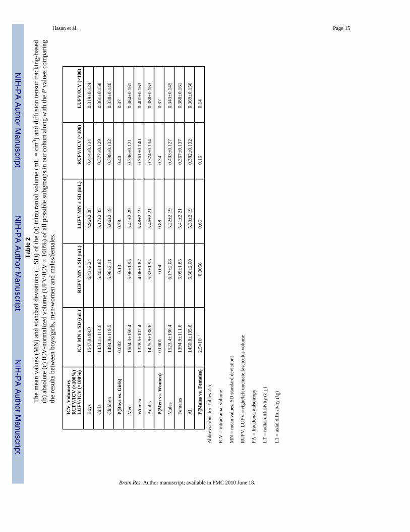
Group mean values and comparisons of the intracranial volume (ICV) and the right and leftUF volume (UFV) on all possible subgroups of study participants (boys, girls, men, women,children and adults) are summarized in Table 2. The ICV was not significantly differentbetween boys and men (p = 0.27). The ICV was not significantly different between girls andwomen (p=0.08). The ICV was significantly larger in males compared to females (p<0.000001;see Table 2). The ICV and right UFV were larger in males compared to females (p<0.005).The UF volume-to-ICV ratios (UFV/ICV × 100%) were not significantly different betweenmales and females (p>0.14).
2.2 Uncinate Fasciculus DTI Metrics: Group Differences and Sex EffectsThe UF corresponding FA, radial and axial diffusivity are summarized in Table 3. There wereno significant differences between males and females (p>0.14; see Table 3). Therefore, ageeffect results are from a pooled sample of males and females.
2.3 Age EffectsPearson correlation coefficients of the UFV/ICV, FA, radial and axial diffusivities and age forall children and adults are summarized in Table 4. Note the lack of dependence on age in thenormalized UFV between children (p>0.2) and adults (p>0.13). The DTI metrics in Table 4show that children have significant linear trends compared to adults which necessitatedquadratic least-squares modeling for the age effects. Table 5 summarizes the least-squarescoefficients using the quadratic least-squares (QLS) model, standard deviation andsignificance. The scatter data, least and quadratic least-squares fits of the normalized UFvolume, corresponding FA, radial and axial diffusivity are plotted in Figure 1 and Figure 2 forboth the right and left UF respectively.
2.4 Asymmetry effectsThere were no statistically significant age-by-sex or age-by-hemisphere interactions.Comparison of linear and quadratic coefficients for both males and females showed nodifferences between males and females (p>0.3). The absolute and ICV-normalized UF volumesfor the right side were larger as compared with the left in children (p=0.02) but not significantlydifferent either in adults (p>0.56) or in the entire population (p=0.27). The mean FA values ofthe UF were significantly larger on the left side in children (p=0.05), adults (p=0.0012) andthe entire sample (p=0.0002). This leftward asymmetry (Left > Right) in the diffusionanisotropy is explained by a leftward asymmetry in the axial diffusivity (p<0.0001) and a lackof asymmetry (p>0.23) in the radial diffusivity.
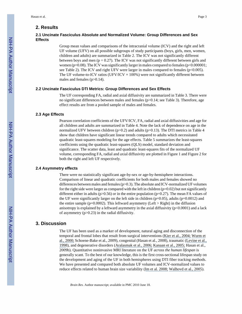
3. DiscussionThe UF has been used as a marker of development, natural aging and disconnection of thetemporal and frontal lobes that result from surgical interventions (Kier et al., 2004; Wurm etal., 2000; Schoene-Bake et al., 2009), congenital (Hasan et al., 2008), traumatic (Levine et al.,1998), and degenerative disorders (Aralasmak et al., 2006; Kanaan et al., 2005; Hasan et al.,2009b). Quantitative noninvasive MRI literature on the UF across the human lifespan isgenerally scant. To the best of our knowledge, this is the first cross-sectional lifespan study onthe development and aging of the UF in both hemispheres using DTI fiber tracking methods.We have presented and compared both absolute UF volumes and ICV-normalized values toreduce effects related to human brain size variability (Im et al. 2008; Walhovd et al., 2005).
Hasan et al. Page 3
Brain Res. Author manuscript; available in PMC 2010 June 18.
NIH
-PA Author Manuscript
NIH
-PA Author Manuscript
NIH
-PA Author Manuscript

The ICV values in our healthy cohort are comparable to those reported in other studies incontrols (Buckner et al., 2004). The fiber-tracked UF volume and its ICV-normalized valuesexhibited larger variability compared with the corresponding DTI metrics. Large variability infiber tract volume using DTI has also been reported on the cingulum (Heiervang et al., 2007),the corticospinal tract (Reich et al., 2006), and uncinate fasciculus (Wakana et al., 2007). Tothe best of our knowledge, only three DTI publications documented the UF volume and itscorresponding DTI metrics on healthy adults (Malykhin et al., 2009; Taoka et al., 2006; Wakanaet al., 2007; see Table 1). Some studies on the UF using coronal 2D ROIs may have includedthe inferior fronto-occipital fasciculus (Catani et al., 2002; see Kanaan et al., 2005 for details).
The volume measurements of the UF in our adult population (see Table 2) are larger than thosereported previously (Malykhin et al., 2009; Taoka et al., 2006;Wakana et al., 2007) in adultcontrols (see Table 1). These differences may be attributed to differences between samplesused (e.g. age, brain size), DTI acquisition paradigm (e.g. voxel size, signal-to-noise ratio) andanalysis procedures (e.g. tracking thresholds). Consistent with previous reports in the healthyUF (see Table 1), we report no sex differences on the normalized UF and its correspondingDTI metrics.
3.1 Age and Sex EffectsOur DTI results on the development and aging of the UF consolidate previous normative studies(see Table 1) that reported positive linear age trends in healthy children (Eluvathingal et al.,2007) and young adults (Lebel et al., 2008), and negative age trends in older adults (Jones etal., 2006). The current work UF DTI age trajectories resemble those published on whole brainwhite matter (Hasan et al., 2007b), lobar (Sowell et al., 2003) and subcortical volumes (e.g.hippocampus and amygdala see Walhovd et al., 2005).
The DTI-related metrics (FA, eigenvalues) provide complementary information about themicrostructural substrates of tissue organization (Beaulieu, 2002; Hasan et al., 2009b). Inparticular, the decrease in the radial eigenvalues during childhood and increase duringadulthood with advancing age may offer early predictors of the regional dynamics ofmyelination and demyelination (Song et al., 2005).
3.2 Uncinate fasciculus DTI AsymmetryOur DTI results reproduce an inconsistently reported UF leftward asymmetry of the diffusiontensor anisotropy (Table 1) in healthy children (Eluvathingal et al., 2007) and adults (Kubickiet al., 2002). This trend has not been reported in other studies (Taoka et al., 2006;Lebel et al.,2008; see Table 1) or was even reversed (Rodrigo et al., 2006). A previous postmortem studyon the UF cross-sectional area in older adults (Highley et al., 2002; see Table 1), however,reported a rightward asymmetry in UF fiber density, and no age or sex effects.
In our study, the UF diffusion tensor anisotropy leftward asymmetry is more specifically relatedto a statistically significant leftward asymmetry in the axial diffusivity which could result froma more coherent or less tortuous alignment of fibers (Beaulieu, 2002; Takahashi et al., 2000).Our lifespan cross-sectional study in healthy right-handed controls shows a dissociationbetween volume-based or macrostructural asymmetry and DTI-based or microstructuralasymmetry. Our results on the axial asymmetry should be interpreted carefully as the UF rightand left tract volumes were measured using diffusion tensor tractography which is limited bythe DTI acquisition spatial resolution and the tracking procedures adopted. The biophysicalsubstrates of this finding require histochemical and postmortem analyses (Beaulieu, 2002;Highley et al., 2002; Takahashi et al., 2000).
Hasan et al. Page 4
Brain Res. Author manuscript; available in PMC 2010 June 18.
NIH
-PA Author Manuscript
NIH
-PA Author Manuscript
NIH
-PA Author Manuscript
3.3 Implications and Significance of the of the Current StudyOur results provide useful baseline data on the effects of side, gender and age on the UF volumeand its corresponding DTI metrics. Our results should help in the experimental design of futureclinical investigation of the exact functional role of the U.
Access to anatomic information and the pattern of normal developmental trajectory of theuncinate fasciculus expands the breadth and depth of our knowledge of this structure(Schmahamn and Pandya, 2006). The significant role of these patterns of information may playan important role in evaluating fronto-temporal structural and functional connectivity inclinical or psychiatric populations. Since the UF originates from the anterior temporal lobe infront of the temporal horn and the cortical nuclei of amygdala and terminates in several criticalareas of the frontal lobes such as orbital frontal cortex. As such the uncinate fasciculus isespecially considered to be a critical structure in playing an important role in memory andemotion and our further understanding of these structures may offer new opportunities toprovide clinically significant services to patient populations with specific neurologic orpsychiatric dysfunctions. An example of such relationship has been recently shown by Fujieet al. (2008). Therefore, our current work on the normal structural development of uncinatefasciculus generates excitement for the expansion of our limited knowledge of brain fiberbundles and their mysterious functional significance.
3.4 Conclusions and Future PlansOur normative database has been formed by pooling cross-sectional data collected on healthychildren and adults using the same DTI protocol (study span ~ 3 years) to help in theinterpretation of data collected from patients (Hasan et al., 2008; Hasan et al., 2009b). Ourcross-sectional cohort and the lifespan experimental design accounted for the confounding andnonlinear age effects and warrant future longitudinal studies on the structural basis of functionaldisconnectivity.
Future extensions of the current studies include the investigation in larger samples of malesand females of the interplay between UF volume, DTI metrics and the corresponding corticalgray matter thickness and subcortical substrate volume (e.g. hippocampus and amygdala) inboth health (Sowell et al., 2003; Walhovd et al., 2005) and disease (Aralasmak et al., 2006;Highley et al., 2002; Kier et al., 2004).
4. Experimental Procedure4.1 Participants
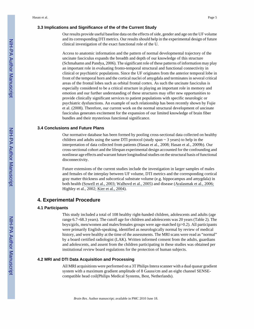
This study included a total of 108 healthy right-handed children, adolescents and adults (agerange 6.7–68.3 years). The cutoff age for children and adolescents was 20 years (Table 2). Theboys/girls, men/women and males/females groups were age-matched (p>0.2). All participantswere primarily English-speaking, identified as neurologically normal by review of medicalhistory, and were healthy at the time of the assessments. The MRI scans were read as “normal”by a board certified radiologist (LAK). Written informed consent from the adults, guardiansand adolescents, and assent from the children participating in these studies was obtained perinstitutional review board regulations for the protection of human subjects.
4.2 MRI and DTI Data Acquisition and ProcessingAll MRI acquisitions were performed on a 3T Philips Intera scanner with a dual quasar gradientsystem with a maximum gradient amplitude of 8 Gauss/cm and an eight channel SENSE-compatible head coil(Philips Medical Systems, Best, Netherlands).
Hasan et al. Page 5
Brain Res. Author manuscript; available in PMC 2010 June 18.
NIH
-PA Author Manuscript
NIH
-PA Author Manuscript
NIH
-PA Author Manuscript

The DTI studies utilized a high signal-to-noise ratio (SNR) whole brain DTI protocol that waskept under 7 minutes (Hasan et al., 2007a). The DTI data were acquired using a single-shotspin-echo diffusion sensitized and fat-suppressed echo-planar imaging sequence with thebalanced Icosa21 tensor encoding scheme (i.e. twenty-one uniformly-distributed directionsover the unit hemisphere) (Hasan, 2007), b-factor = 1000 sec mm−2, repetition time (TR)/echotime TE = 7100/65 msec. The echo-planar phase encoding used a SENSE k-spaceundersampling factor of two (R=2) with an effective k-space matrix of 112×112, image matrixafter zero-filling = 256×256. The field-of-view = 240 mm × 240 mm, number of axial sections= 44 with no gap and slice thickness = 3 mm. The number of b~0 magnitude image averageswas 6; in addition, each diffusion encoding was repeated twice and magnitude-averaged toenhance the SNR (Hasan, 2007). The SNR, DTI data spatial resolution (e.g. voxel size 2.2 mm× 2.2 mm × 3 mm) and image quality (e.g. reduced image distortions) were adequate to tracethe uncinate fasciculus (Eluvathingal et al. 2007; Fujiwara et al., 2008; Hasan et al., 2009a).
In this work, the DTI-derived rotationally-invariant metrics included the fractional anisotropy(FA), axial (L1 = λ||) and radial diffusivity (LT = λ⊥). The radial diffusivity is defined as theaverage of the second and third eigenvalues (λ⊥ = (λ2+λ3)/2) and has been shown by severalresearchers to be a marker of myelination (Beaulieu, 2002; Song et al., 2005; see also Hasan,2006). We performed distortion correction to remove eddy-current artifacts, then calculatedthe ICV from the non-diffusion weighted image (Hasan et al., 2009a). Additional details ofDTI image processing (Hasan et al., 2007b; Hasan et al., 2009a) and DTI quality controlmeasures are found elsewhere (Hasan, 2007a).
4.3 Fiber Tracking of the Uncinate FasciculusCompact white matter fiber tracking was performed using DTI Studio software (Jiang et al.,2006) based on the fiber assignment by continuous tracking (FACT) algorithm. Diffusiontensor tractography using anatomical landmarks (Kier et al., 2004) and multiple regions-of-interest (ROI) were utilized to trace the UF bilaterally as described elsewhere (Catani et al.,2002; Mori et al., 2002; Jones et al., 2006; Wakana et al., 2007; Yu et al., 2008). The UFtracking used a fractional anisotropy (FA) threshold of 0.15 and angle threshold of 60 degrees.These tracking thresholds were identical to those adopted to trace the UF in two previouspublications using the same DTI protocol (Eluvathingal et al., 2007; Hasan et al., 2008).Additional details of the fiber tracking procedures are provided elsewhere (see APPENDIX inEluvathingal et al., 2007).
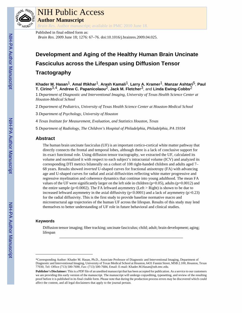
In brief, the anterior commissure (AC) identified on a coronal DTI color-coded plane was usedas an anatomical landmark to identify the two regions where the UF superior and inferiorsegments traverse the coronal plane (Figure 3A regions 1 and 2). Note that the seed ROIsselected (Figure 3A) border the external capsule, insula, putamen and the hippocampus(Ebeling and Cramon, 1992). A Boolean “OR” operation on region 1 (lower portion of externalcapsule) combined with an “AND” operation on region 2 (temporal lobe) were sufficient toconstruct the UF on each side avoiding any mixing with other tracts such as the inferior fronto-occipital and inferior longitudinal fascicule (Catani et al. 2002; Mori et al. 2002). Once the UFfiber tract is reconstructed, its volume and corresponding DTI attributes were recorded andvisualized in 3D (Figure 3C and 3D).
4.4 Statistical analysisAll analyses of UF absolute, normalized volumes and their corresponding DTI metricsvariation were conducted using a generalized linear model with effects of age, side and sex asdescribed elsewhere (Hasan et al., 2007b). Given previous reports on white matter macro andmicrostructural development across the lifespan (Sowell et al., 2003; Hasan et al., 2007b;Hasan et al., 2009a), both linear and quadratic age terms were included. The DTI metrics (e.g.,
Hasan et al. Page 6
Brain Res. Author manuscript; available in PMC 2010 June 18.
NIH
-PA Author Manuscript
NIH
-PA Author Manuscript
NIH
-PA Author Manuscript

fractional anisotropy = FA; radial or transverse diffusivity = λ⊥ = LT; axial diffusivity = λ|| =L1) were modeled for both males and females as yf= β0+β1*age+β2*age2, and then the generalleast-squares methods were used to compute the coefficients, standard errors and theirsignificance using analysis-of-variance methods (Hasan et al., 2009a). Interactions of sex withage (both terms) were examined, and trimmed where non-significant. If the quadratic age termwas not significant, age and sex interactions were examined without this term, and trimmed ifnon-significant. All statistical analyses were conducted using SAS 9.1 (SAS Institute Inc, NC)and MATLAB R12.1 Statistical Toolbox v 3.0 (The Mathworks Inc, Natick, MA).
AcknowledgmentsThis work was funded by NIH-NINDS R01 NS052505-04 awarded to KMH, NIH-NICHD R01 NS046308 awardedto LEC, P01 HD35946 awarded to JMF and 1 P01 NS46588 awarded to ACP. The authors wish to thank Vipul KumarPatel for helping in data acquisition.
References1. Antunes NL, Souweidane MM, Lis E, Rosenblum M, Steinherz P. Methotrexate leukoencephalopathy
presenting as Klüver-Bucy syndrome and uncinate seizures. Pediatr Neurol 2002;26:305–308.[PubMed: 11992760]
2. Aralasmak A, Ulmer JL, Kocak M, Salvan CV, Hillis AE, Yousem DM. Association, commissural,and projection pathways and their functional deficit reported in literature. J Comput Assist Tomog2006;30:695–715.
3. Beaulieu C. The basis of anisotropic water diffusion in the nervous system - a technical review. NMRBiomed 2002;15:435–455. [PubMed: 12489094]Review
4. Bendlin BB, Ries ML, Lazar M, Alexander AL, Dempsey RJ, Rowley HA, Sherman JE, Johnson SC.Longitudinal changes in patients with traumatic brain injury assessed with diffusion-tensor andvolumetric imaging. Neuroimage 2008;42:503–514. [PubMed: 18556217]
5. Borroni B, Alberici A, Premi E, Archetti S, Garibotto V, Agosti C, Gasparotti R, Di Luca M, PeraniD, Padovani A. Brain magnetic resonance imaging structural changes in a pedigree of asymptomaticprogranulin mutation carriers. Rejuvenation Res 2008;11:585–595. [PubMed: 18593276]
6. Buckner RL, Head D, Parker J, Fotenos AF, Marcus D, Morris JC, Snyder AZ. A unified approach formorphometric and functional data analysis in young, old, and demented adults using automated atlas-based head size normalization: reliability and validation against manual measurement of totalintracranial volume. Neuroimage 2004;23:724–738. [PubMed: 15488422]
7. Burns J, Job D, Bastin ME, Whalley H, Macgillivray T, Johnstone EC, Lawrie SM. Structuraldisconnectivity in schizophrenia: a diffusion tensor magnetic resonance imaging study. Br J Psychiatry2003;182:439–443. [PubMed: 12724248]
8. Catani M, Howard RJ, Pajevic S, Jones DK. Virtual in vivo interactive dissection of white matterfasciculi in the human brain. Neuroimage 2002;17:77–94. [PubMed: 12482069]
9. Catani M, Mesulam M. The arcuate fasciculus and the disconnection theme in language and aphasia:History and current state. Cortex 2008;44:953–961. [PubMed: 18614162]
10. Constable RT, Ment LR, Vohr BR, Kesler SR, Fulbright RK, Lacadie C, Delancy S, Katz KH,Schneider KC, Schafer RJ, Makuch RW, Reiss AR. Prematurely born children demonstrate whitematter microstructural differences at 12 years of age, relative to term control subjects: an investigationof group and gender effects. Pediatrics 2008;121:306–316. [PubMed: 18245422]
11. Diehl B, Busch RM, Duncan JS, Piao Z, Tkach J, Lüders HO. Abnormalities in diffusion tensorimaging of the uncinate fasciculus relate to reduced memory in temporal lobe epilepsy. Epilepsia2008;49:1409–1418. [PubMed: 18397294]
12. Dubois J, Hertz-Pannier L, Dehaene-Lambertz G, Cointepas Y, Le Bihan D. Assessment of the earlyorganization and maturation of infants’ cerebral white matter fiber bundles: a feasibility study usingquantitative diffusion tensor imaging and tractography. Neuroimage 2006;30:1121–1132. [PubMed:16413790]
Hasan et al. Page 7
Brain Res. Author manuscript; available in PMC 2010 June 18.
NIH
-PA Author Manuscript
NIH
-PA Author Manuscript
NIH
-PA Author Manuscript

13. Duffau H, Gatignol P, Moritz-Gasser S, Mandonnet E. Is the left uncinate fasciculus essential forlanguage?: A cerebral stimulation study. J Neurol. 200910.1007/s00415-009-0053-9
14. Ebeling U, Von Cramon D. Topography of uncinate fascicle and adjacent temporal fiber tracts. ActaNeurochir (Wien) 1992;115:143–148. [PubMed: 1605083]
15. Eluvathingal TJ, Chugani HT, Behen ME, Juhász C, Muzik O, Maqbool M, Chugani DC, Makki M.Abnormal brain connectivity in children after early severe socioemotional deprivation: a diffusiontensor imaging study. Pediatrics 2006;117:2093–2100. [PubMed: 16740852]
16. Eluvathingal TJ, Hasan KM, Kramer L, Fletcher JM, Ewing-Cobbs L. Quantitative diffusion tensortractography of association and projection fibers in normally developing children and adolescents.Cereb Cortex 2007;17:2760–2768. [PubMed: 17307759]
17. Fujie S, Namiki C, Nishi H, Yamada M, Miyata J, Sakata D, Sawamoto N, Fukuyama H, Hayashi T,Murai T. The role of the uncinate fasciculus in memory and emotional recognition in amnestic mildcognitive impairment. Dement Geriatr Cogn Disord 2008;26:432–439. [PubMed: 18957848]
18. Fujiwara S, Sasaki M, Kanbara Y, Inoue T, Hirooka R, Ogawa A. Feasibility of 1.6-mm isotropicvoxel diffusion tensor tractography in depicting limbic fibers. Neuroradiology 2008:131–136.[PubMed: 17938897]
19. Hagmann P, Thiran JP, Jonasson L, Vandergheynst P, Clarke S, Maeder P, Meuli R. DTI mappingof human brain connectivity: statistical fibre tracking and virtual dissection. Neuroimage2003;19:545–554. [PubMed: 12880786]
20. Hasan KM. Diffusion tensor eigenvalues or both mean diffusivity and fractional anisotropy arerequired in quantitative clinical diffusion tensor MR reports: fractional anisotropy alone is notsufficient. Radiology 2006;239:611–612. [PubMed: 16641362]
21. Hasan KM. A framework for quality control and parameter optimization in diffusion tensor imaging:theoretical analysis and validation. Magn Reson Imaging 2007a;25:1196–1202. [PubMed:17442523]
22. Hasan KM, Sankar A, Halphen C, Kramer LA, Brandt ME, Juranek J, Cirino PT, Fletcher JM,Papanicolaou AC, Ewing-Cobbs L. Development and Organization of Human Brain TissueCompartments across Lifespan using Diffusion Tensor Imaging. Neuroreport 2007b;18:1735–1739.[PubMed: 17921878]
23. Hasan KM, Eluvathingal TJ, Kramer LA, Ewing-Cobbs L, Dennis M, Fletcher JM. White mattermicrostructural abnormalities in children with spina bifida myelomeningocele and hydrocephalus: adiffusion tensor tractography study of the association pathways. J Magn Reson Imaging2008;27:700–709. [PubMed: 18302204]
24. Hasan KM, Kamali A, Iftikhar A, Kramer LA, Papanicolaou AC, Fletcher JM, Ewing-Cobbs L.Diffusion tensor tractography quantification of the human corpus callosum fiber pathways across thelifespan. Brain Res 2009a;1249:91–100. [PubMed: 18996095]
25. Hasan, KM.; Kamali, A.; Iftikhar, A.; Datta, S.; Nelson, F.; Wolinsky, JS.; Narayana, PA. DiffusionTensor Tractogrpahy Quantification of Wallerian Degeneration of the Uncinate Fasciculus inMultiple Sclerosis . Proceedings of the International scoiety for Magnetic Resonance in Medicine(ISMRM) 17th Scientific Meeting & Exhibition; 18–24 April, 2009; Honolulu, Hawai’i, USA. 2009.p. 3196
26. Heiervang E, Behrens TE, Mackay CE, Robson MD, Johansen-Berg H. Between sessionreproducibility and between subject variability of diffusion MR and tractography measures.Neuroimage 2006;33:867–877. [PubMed: 17000119]
27. Highley JR, Walker MA, Esiri MM, Crow TJ, Harrison PJ. Asymmetry of the uncinate fasciculus: apost-mortem study of normal subjects and patients with schizophrenia. Cereb Cortex 2002;12:1218–1224. [PubMed: 12379610]
28. Houenou J, Wessa M, Douaud G, Leboyer M, Chanraud S, Perrin M, Poupon C, Martinot JL, Paillere-Martinot ML. Increased white matter connectivity in euthymic bipolar patients: diffusion tensortractography between the subgenual cingulate and the amygdalo-hippocampal complex. MolPsychiatry 2007;12:1001–1010. [PubMed: 17471288]
29. Jiang H, van Zijl PC, Kim J, Pearlson GD, Mori S. DtiStudio: resource program for diffusion tensorcomputation and fiber bundle tracking. Comput Methods Programs Biomed 2006;81:106–116.[PubMed: 16413083]
Hasan et al. Page 8
Brain Res. Author manuscript; available in PMC 2010 June 18.
NIH
-PA Author Manuscript
NIH
-PA Author Manuscript
NIH
-PA Author Manuscript

30. Jones DK, Catani M, Pierpaoli C, Reeves SJ, Shergill SS, O’Sullivan M, Golesworthy P, McGuireP, Horsfield MA, Simmons A, Williams SC, Howard RJ. Age effects on diffusion tensor magneticresonance imaging tractography measures of frontal cortex connections in schizophrenia. Hum BrainMapp 2006;27:230–238. [PubMed: 16082656]
31. Im K, Lee JM, Lyttelton O, Kim SH, Evans AC, Kim SI. Brain size and cortical structure in the adulthuman brain. Cereb Cortex 2008;18:2181–2191. [PubMed: 18234686]
32. Kanaan RA, Kim JS, Kaufmann WE, Pearlson GD, Barker GJ, McGuire PK. Diffusion tensor imagingin schizophrenia. Biol Psychiatry 2005;58:921–929. [PubMed: 16043134]Review
33. Kezele IB, Arnold DL, Collins DL. Atrophy in white matter fiber tracts in multiple sclerosis is notdependent on tract length or local white matter lesions. Mult Scler 2008;14:779–785. [PubMed:18611990]
34. Kier EL, Staib LH, Davis LM, Bronen RA. MR imaging of the temporal stem: anatomic dissectiontractography of the uncinate fasciculus, inferior occipitofrontal fasciculus, and Meyer’s loop of theoptic radiation. AJNR Am J Neuroradiol 2004;25:677–691. [PubMed: 15140705]
35. Kubicki M, Westin CF, Maier SE, Frumin M, Nestor PG, Salisbury DF, Kikinis R, Jolesz FA,McCarley RW, Shenton ME. Uncinate fasciculus findings in schizophrenia: a magnetic resonancediffusion tensor imaging study. Am J Psychiatry 2002;159:813–820. [PubMed: 11986136]
36. Lebel C, Walker L, Leemans A, Phillips L, Beaulieu C. Microstructural maturation of the humanbrain from childhood to adulthood. Neuroimage 2008;40:1044–1055. [PubMed: 18295509]
37. Levine B, Black SE, Cabeza R, Sinden M, Mcintosh AR, Toth JP, Tulving E, Stuss DT. Episodicmemory and the self in a case of isolated retrograde amnesia. Brain 1998;121:1951–1973. [PubMed:9798749]
38. McDonald CR, Ahmadi ME, Hagler DJ, Tecoma ES, Iragui VJ, Gharapetian L, Dale AM, HalgrenE. Diffusion tensor imaging correlates of memory and language impairments in temporal lobeepilepsy. Neurology 2008;71:1869–1876. [PubMed: 18946001]
39. Malykhin N, Concha L, Seres P, Beaulieu C, Coupland NJ. Diffusion tensor imaging tractographyand reliability analysis for limbic and paralimbic white matter tracts. Psychiatry Res 2008;164:132–142. [PubMed: 18945599]
40. Mamata H, Mamata Y, Westin CF, Shenton ME, Kikinis R, Jolesz FA, Maier SE. High-resolutionline scan diffusion tensor MR imaging of white matter fiber tract anatomy. AJNR Am J Neuroradiol2002;23:67–75. [PubMed: 11827877]
41. Matsuo K, Mizuno T, Yamada K, Akazawa K, Kasai T, Kondo M, Mori S, Nishimura T, NakagawaM. Cerebral white matter damage in frontotemporal dementia assessed by diffusion tensortractography. Neuroradiology 2008;50:605–611. [PubMed: 18379765]
42. Mori S, Kaufmann WE, Davatzikos C, Stieltjes B, Amodei L, Fredericksen K, Pearlson GD, MelhemER, Solaiyappan M, Raymond GV, Moser HW, van Zijl PC. Imaging cortical association tracts inthe human brain using diffusion-tensor-based axonal tracking. Magn Reson Med 2002;47:215–223.[PubMed: 11810663]
43. Nakamura M, McCarley RW, Kubicki M, Dickey CC, Niznikiewicz MA, Voglmaier MM, SeidmanLJ, Maier SE, Westin CF, Kikinis R, Shenton ME. Fronto-temporal disconnectivity in schizotypalpersonality disorder: a diffusion tensor imaging study. Biol Psychiatry 2005;58:468–478. [PubMed:15978550]
44. Niogi SN, Mukherjee P, Ghajar J, Johnson CE, Kolster R, Lee H, Suh M, Zimmerman RD, ManleyGT, McCandliss BD. Structural dissociation of attentional control and memory in adults with andwithout mild traumatic brain injury. Brain 131:3209–3221. [PubMed: 18952679]
45. Park HJ, Westin CF, Kubicki M, Maier SE, Niznikiewicz M, Baer A, Frumin M, Kikinis R, JoleszFA, McCarley RW, Shenton ME. White matter hemisphere asymmetries in healthy subjects and inschizophrenia: a diffusion tensor MRI study. Neuroimage 2004;23:213–223. [PubMed: 15325368]
46. Parker GJ, Luzzi S, Alexander DC, Wheeler-Kingshott CA, Ciccarelli O, Lambon, Ralph MA.Lateralization of ventral and dorsal auditory language pathways in the human brain. NeuroImage2005;24:656–666. [PubMed: 15652301]
47. Price G, Cercignani M, Parker GJ, Altmann DR, Barnes TR, Barker GJ, Joyce EM, Ron MA. Whitematter tracts in first-episode psychosis: a DTI tractography study of the uncinate fasciculus.Neuroimage 2008;39:949–955. [PubMed: 17988894]
Hasan et al. Page 9
Brain Res. Author manuscript; available in PMC 2010 June 18.
NIH
-PA Author Manuscript
NIH
-PA Author Manuscript
NIH
-PA Author Manuscript

48. Reich DS, Smith SA, Jones CK, Zackowski KM, van Zijl PC, Calabresi PA, Mori S. Quantitativecharacterization of the corticospinal tract at 3T. AJNR Am J Neuroradiol 2006;27:2168–2178.[PubMed: 17110689]
49. Rodrigo S, Oppenheim C, Chassoux F, Golestani N, Cointepas Y, Poupon C, Semah F, Mangin JF,Le, Bihan D, Meder JF. Uncinate fasciculus fiber tracking in mesial temporal lobe epilepsy. Initialfindings Eur Radiol 2007;17:1663–1668.
50. Schmahmann, JD.; Pandya, DN. Fiber Pathways of the Brain. Oxford: New York; 2006.51. Schmahmann JD, Smith EE, Eichler FS, Filley CM. Cerebral white matter: neuroanatomy, clinical
neurology, and neurobehavioral correlates. Ann N Y Acad Sci 2008;1142:266–309. [PubMed:18990132]
52. Schoene-Bake JC, Faber J, Trautner P, Kaaden S, Tittgemeyer M, Elger CE, Weber B. Widespreadaffections of large fiber tracts in postoperative temporal lobe epilepsy. Neuroimage. 200910.1016/j.neuroimage.2009.03.013
53. Sheline YI, Price JL, Vaishnavi SN, Mintun MA, Barch DM, Epstein AA, Wilkins CH, Snyder AZ,Couture L, Schechtman K, McKinstry RC. Regional white matter hyperintensity burden in automatedsegmentation distinguishes late-life depressed subjects from comparison subjects matched forvascular risk factors. Am J Psychiatry 2008;165:524–532. [PubMed: 18281408]
54. Song SK, Yoshino J, Le TQ, Lin SJ, Sun SW, Cross AH, Armstrong RC. Demyelination increasesradial diffusivity in corpus callosum of mouse brain. Neuroimage 2005;26:132–140. [PubMed:15862213]
55. Sowell ER, Peterson BS, Thompson PM, Welcome SE, Henkenius AL, Toga AW. Mapping corticalchange across the human life span. Nat Neurosci 2003;6:309–315. [PubMed: 12548289]
56. Szeszko PR, Robinson DG, Ashtari M, Vogel J, Betensky J, Sevy S, Ardekani BA, Lencz T, MalhotraAK, McCormack J, Miller R, Lim KO, Gunduz-Bruce H, Kane JM, Bilder RM. Clinical andneuropsychological correlates of white matter abnormalities in recent onset schizophrenia.Neuropsychopharmacology 2008;33:976–984. [PubMed: 17581532]
57. Takahashi M, Ono J, Harada K, Maeda M, Hackney DB. Diffusional anisotropy in cranial nerveswith maturation: quantitative evaluation with diffusion MR imaging in rats. Radiology2000;216:881–885. [PubMed: 10966726]
58. Taoka T, Iwasaki S, Sakamoto M, Nakagawa H, Fukusumi A, Myochin K, Hirohashi S, Hoshida T,Kichikawa K. iffusion anisotropy and diffusivity of white matter tracts within the temporal stem inAlzheimer disease: evaluation of the “tract of interest” by diffusion tensor tractography. AJNR AmJ Neuroradiol 2006;27:1040–1045. [PubMed: 16687540]
59. Wakana S, Caprihan A, Panzenboeck MM, Fallon JH, Perry M, Gollub RL, Hua K, Zhang J, JiangH, Dubey P, Blitz A, van Zijl P, Mori S. Reproducibility of quantitative tractography methods appliedto cerebral white matter. Neuroimage 2007;36:630–644. [PubMed: 17481925]
60. Walhovd KB, Fjell AM, Reinvang I, Lundervold A, Dale AM, Eilertsen DE, Quinn BT, Salat D,Makris N, Fischl B. Effects of age on volumes of cortex, white matter and subcortical structures.Neurobiol Aging 2005;26:1261–1270. [PubMed: 16005549]
61. Wurm G, Wies W, Schnizer M, Trenkler J, Holl K. Advanced surgical approach for selectiveamygdalohippocampectomy through neuronavigation. Neurosurgery 2000;46:1377–82. [PubMed:10834642]discussion 1382–1383
62. Yu C, Li J, Liu Y, Qin W, Li Y, Shu N, Jiang T, Li K. White matter tract integrity and intelligencein patients with mental retardation and healthy adults. Neuroimage 2008;40:1533–1541. [PubMed:18353685]
63. Zahr NM, Rohlfing T, Pfefferbaum A, Sullivan EV. Problem solving, working memory, and motorcorrelates of association and commissural fiber bundles in normal aging: a quantitative fiber trackingstudy. Neuroimage 2009;44:1050–1062. [PubMed: 18977450]
Hasan et al. Page 10
Brain Res. Author manuscript; available in PMC 2010 June 18.
NIH
-PA Author Manuscript
NIH
-PA Author Manuscript
NIH
-PA Author Manuscript
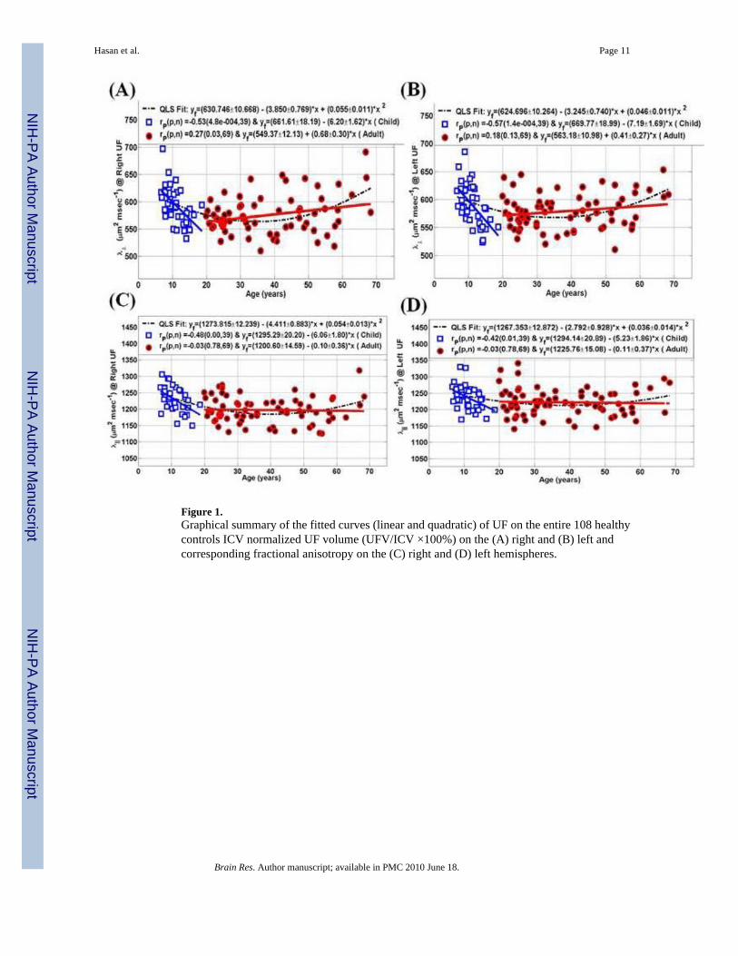
Figure 1.Graphical summary of the fitted curves (linear and quadratic) of UF on the entire 108 healthycontrols ICV normalized UF volume (UFV/ICV ×100%) on the (A) right and (B) left andcorresponding fractional anisotropy on the (C) right and (D) left hemispheres.
Hasan et al. Page 11
Brain Res. Author manuscript; available in PMC 2010 June 18.
NIH
-PA Author Manuscript
NIH
-PA Author Manuscript
NIH
-PA Author Manuscript

Figure 2.Graphical summary of the fitted curves of UF on the entire 108 healthy controls of the radialdiffusivity on the (A) right and (B) left and axial diffusivity on the (C) right and (D) lefthemispheres.
Hasan et al. Page 12
Brain Res. Author manuscript; available in PMC 2010 June 18.
NIH
-PA Author Manuscript
NIH
-PA Author Manuscript
NIH
-PA Author Manuscript

Figure 3.Illustration of the DTI-based fiber tracking of the uncinate fasciculus (A) The two ROIs (1rectangle, 2 circle) were placed after identifying the anterior commissure on a coronal DTIcolor coded map (e.g. principal eigenvector modulated by FA and fused with the meandiffusivity to identify cerebrospinal fluid (CSF) and gray matter) (B) An illustration of thesame coronal plane with the two seed ROIs on an FA map showing the gray matter borderingthe UF (1 caudate, 2 putamen, 3 insula, 4 amygdala/hippocampus). (C) a 3D postero-lateralview of the right and left UF on a DTI color coded (red = right-left; green = anterior-posterior,blue = inferior superior). Note the origin (temporal lobe) and termination (frontal lobe) of theUF (D) a 3D antero-lateral view of the right and left UF showing the UF position within thecortical and subcortical gray matter structures (e.g. hippocampus).
Hasan et al. Page 13
Brain Res. Author manuscript; available in PMC 2010 June 18.
NIH
-PA Author Manuscript
NIH
-PA Author Manuscript
NIH
-PA Author Manuscript

NIH
-PA Author Manuscript
NIH
-PA Author Manuscript
NIH
-PA Author Manuscript
Hasan et al. Page 14
Table 1A list of publications that used 10 or more healthy controls that reported some quantitative volumetric and DTI attributesof the uncinate fasciculus.
Study
Control Demographics
Methods
Results and Conclusions
N(sex/hand) ≥ 10 Age Age & Asymmetry
Dubois et al.,2006
18 (Infants) 12.9 ± 3.4wks
DT-FT&ROI p(Dav, age) = 0.001; FA(age) = n.s.
Eluvathingal etal., 2007
29 (13Boys/16Girls) 10.7 ± 2.9yrs
DT-FT FA (Left > Right); r(FA, age) > 0.4;p<0.03
Highley et al.,2002
11 Women10 Men
74.5 ± 12.5yrs70.0 ± 14.1yrs
Postmortem Histology Area, axons # and densityRight > Left (rightwardasymmetry)No age, sex effects
Jones et al.,2006
14 Rh (Adults) 19–57 yrs DT-VBA R (FA, age) = −0.53 (p=0.049)
Kubicki et al.,2002
18 Rh (Adults) 43± 6 yrs DT-ROI FA (Left > Right)Area (Left > Right)
Lebel et al.,2008
202 (98 Females; 187Rh)
15.2 ± 6.1yrs5.6–29.2 yrs
DT-FT r (FA, age) = 0.514Minimal sex and asymmetry effects
Malykhin et al.,2009
24 (6 Men) 32 ± 8 yrs DT-FT FA (Left ~ Right)UFV Right = 1.108 mLUFV Left = 0.802
Nakamura etal., 2007
15 (Men) 32.7 ± 12.4yrs
DT-ROI FA (Left UFV ~= Right UFV)Left UF-area (22.0 ± 6.8 mm2)Right UF-area (21.8 ± 7.4 mm2)
Park et al., 2004 32 (Men) 44 ± 5.6 yrs DT-VBA FA (Left > Right) superior portionFA (Right > Left) inferior portion
Rodrigo et al.,2006
10 (6 Men) Rh 20–33 yrs DT-FT FA (Right > Left)
Taoka et al.,2006
15 (11Women) ~ 72 ± 3 yrs DT-FT FA (Right ~= Left)Right UFV = 0.475 ± 0.211 mLLeft UFV = 0.502 ± 0.216 mL
Wakana et al.,2007
10 (5 Men) 26.1 ± 5.48yrs
DT-FT Left UFV (~ 3 ± 2 mL)Right UFV (~ 1.5 ± 1)Volume (Left > Right; p < 0.05)FA (Right ~= Left)
Abbreviations used in Table 1
Dav = Mean diffusivity; FA = fractional anisotropy; n. s. = not significant
Rh = Right-handed
DT-FT = diffusion tensor fiber tracking; DT-ROI = diffusion tensor region-of-interest; DT-VBA = diffusion tensor voxel based anisotropy
UFV = uncinate fasciculus volume in mL = cm3
~= approximately equal to or not statistically different
Brain Res. Author manuscript; available in PMC 2010 June 18.
NIH
-PA Author Manuscript
NIH
-PA Author Manuscript
NIH
-PA Author Manuscript
Hasan et al. Page 15Ta
ble
2Th
e m
ean
valu
es (M
N) a
nd s
tand
ard
devi
atio
ns (±
SD
) of t
he (a
) int
racr
ania
l vol
ume
(mL
= cm
3 ) a
nd d
iffus
ion
tens
or tr
acki
ng-b
ased
(b) a
bsol
ute
(c) I
CV
-nor
mal
ized
vol
ume
(UFV
/ICV
× 1
00%
) of a
ll po
ssib
le su
bgro
ups i
n ou
r coh
ort a
long
with
the
P va
lues
com
parin
gth
e re
sults
bet
wee
n bo
ys/g
irls,
men
/wom
en a
nd m
ales
/fem
ales
.
ICV
, Vol
umet
ryR
UFV
/IC
V (×
100%
)L
UFV
/IC
V (×
100%
)IC
V M
N ±
SD
(mL
)R
UFV
MN
± S
D (m
L)
LU
FV M
N ±
SD
(mL
)R
UFV
/IC
V (×
100)
LU
FV/I
CV
(×10
0)
Boy
s15
47.0
±99.
06.
43±2
.24
4.96
±2.0
80.
414±
0.13
40.
319±
0.12
4
Girl
s14
34.1
±114
.65.
40±1
.82
5.17
±2.3
50.
377±
0.12
90.
361±
0.15
8
Chi
ldre
n14
94.9
±119
.55.
96±2
.11
5.06
±2.1
90.
398±
0.13
20.
338±
0.14
0
P(B
oys v
s. G
irls
)0.
002
0.13
0.78
0.40
0.37
Men
1504
.3±1
50.4
5.96
±1.9
55.
41±2
.29
0.39
6±0.
121
0.36
4±0.
161
Wom
en13
78.5
±107
.44.
96±1
.87
5.48
±2.1
90.
361±
0.14
00.
401±
0.16
3
Adu
lts14
25.9
±138
.65.
33±1
.95
5.46
±2.2
10.
374±
0.13
40.
388±
0.16
3
P(M
en v
s. W
omen
)0.
0001
0.04
0.88
0.34
0.37
Mal
es15
23.4
±130
.46.
17±2
.08
5.22
±2.1
90.
403±
0.12
70.
343±
0.14
5
Fem
ales
1394
.9±1
11.6
5.09
±1.8
55.
41±2
.21
0.36
7±0.
137
0.38
8±0.
161
All
1450
.8±1
35.6
5.56
±2.0
05.
33±2
.19
0.38
2±0.
132
0.36
9±0.
156
P(M
ales
vs.
Fem
ales
)2.
5×10
−70.
0056
0.66
0.16
0.14
Abb
revi
atio
ns fo
r Tab
les 2
-5
ICV
= in
tracr
ania
l vol
ume
MN
= m
ean
valu
es, S
D st
anda
rd d
evia
tions
RU
FV, L
UFV
= ri
ght/l
eft u
ncin
ate
fasc
icul
us v
olum
e
FA =
frac
tiona
l ani
sotro
py
LT =
radi
al d
iffus
ivity
(λ⊥
)
L1 =
axi
al d
iffus
ivity
(λ||)
Brain Res. Author manuscript; available in PMC 2010 June 18.

NIH
-PA Author Manuscript
NIH
-PA Author Manuscript
NIH
-PA Author Manuscript
Hasan et al. Page 16Ta
ble
3Th
e m
ean
valu
es a
nd s
tand
ard
devi
atio
ns o
f FA
, rad
ial a
nd a
xial
diff
usiv
ities
of a
ll po
ssib
le s
ubgr
oups
in o
ur c
ohor
t alo
ng w
ith th
e P
valu
es c
ompa
ring
the
resu
lts b
etw
een
boys
/girl
s, m
en/w
omen
and
mal
es/fe
mal
es.
FA(R
UFV
) MN
±SD
(×10
00)
FA(L
UFV
) MN
±SD
(×10
00)
LT
(RU
FV) M
N ±
SD (m
2 /mse
c)L
T(L
UFV
) MN
±SD
(m2 /m
sec)
L1(
RU
FV) M
N ±
SD
(m2 /m
sec)
L1(
LU
FV) M
N ±
SD
(m2 /m
sec)
Boy
s42
7.2±
27.1
431.
2±29
.259
3.3±
41.0
594.
5±40
.612
27.7
±41.
712
39.6
±40.
1
Girl
s42
6.5±
20.5
434.
4±25
.259
5.4±
23.7
588.
3±31
.612
31.5
±29.
812
34.6
±31.
9
Chi
ldre
n42
6.8±
24.0
432.
7±27
.159
4.3±
33.8
591.
6±36
.412
29.4
±36.
312
37.3
±36.
1
P (B
oys v
s. G
irls
)0.
930.
720.
850.
600.
750.
67
Men
433.
1±28
.943
6.5±
23.5
571.
2±32
.458
3.4±
32.6
1196
.1±4
2.8
1229
.3±4
1.4
Wom
en42
7.4±
28.2
437.
6±23
.057
8.2±
36.7
576.
3±30
.311
97.3
±39.
912
17.1
±42.
2
Adu
lts42
9.5±
28.4
437.
1±23
.157
5.5±
35.1
579.
1±31
.111
96.8
±40.
712
21.7
±42.
1
P (M
en v
s.W
omen
)0.
430.
850.
430.
360.
910.
25
Mal
es43
0.4±
27.9
434.
1±26
.058
1.1±
37.8
588.
4±36
.412
10.2
±44.
712
33.9
±40.
7
Fem
ales
427.
1±26
.043
6.6±
23.5
583.
2±34
.257
9.8±
30.9
1207
.4±4
0.1
1222
.3±4
0.0
All
428.
6±26
.843
5.5±
24.6
582.
3±35
.658
3.6±
33.5
1208
.6±4
2.0
1227
.3±4
0.0
P (M
ales
vs.
Fem
ales
)0.
530.
600.
760.
190.
730.
14
Abb
revi
atio
ns fo
r Tab
les 2
-5
ICV
= in
tracr
ania
l vol
ume
MN
= m
ean
valu
es, S
D st
anda
rd d
evia
tions
RU
FV, L
UFV
= ri
ght,
left
unci
nate
fasc
icul
us v
olum
e
FA =
frac
tiona
l ani
sotro
py L
T =
radi
al d
iffus
ivity
(λ⊥
)
L1 =
axi
al d
iffus
ivity
(λ||)
Brain Res. Author manuscript; available in PMC 2010 June 18.

NIH
-PA Author Manuscript
NIH
-PA Author Manuscript
NIH
-PA Author Manuscript
Hasan et al. Page 17
Table 4The Pearson correlation coefficient and significance of the linear regression fit model in both children and adults forthe UFV/ICV, FA, radial and axial diffusivities. The linear regression curves are shown in Figure 2 and Figure 3.
Pearson Correlation Coefficients of age vs. UF volumetry and DTI metrics Children r(p) Adults r(p)
UFV (Right)/ICV vs. age 0.00 (0.98) 0.04 (0.74)
UFV (Left)/ICV vs. age 0.20 (0.22) 0.18 (0.13)
FA (Right) vs. age 0.35 (0.03) −0.32 (0.01)
FA (Left) vs. age 0.45 (< 0.0001) −0.25 (0.04)
LT (Right) vs. age −0.53 (< 0.0005) 0.27 (0.03)
LT (Left) vs. age −0.57 (< 0.0002) 0.18 (0.13)
L1 (Right) vs. age −0.48 (< 0.0001) −0.03 (0.78)
L1 (Left) vs. age −0.42 (0.01) −0.03 (0.78)
Abbreviations for Tables 2-5
ICV = intracranial volume
MN = mean values, SD standard deviations
RUFV, LUFV = right/left uncinate fasciculus volume
FA = fractional anisotropy
LT = radial diffusivity (λ⊥)
L1 = axial diffusivity (λ||)
Brain Res. Author manuscript; available in PMC 2010 June 18.

NIH
-PA Author Manuscript
NIH
-PA Author Manuscript
NIH
-PA Author Manuscript
Hasan et al. Page 18
Table 5Diffusion tensor fiber tracking-based estimation of the FA, radial and axial diffusivities corresponding to the right andleft UF fit statistics in all controls.
Diffusion TensorMetrics of the UncinateFasciculus
Quadratic Fit Model: y=β0+β1* age+β2
* age2
R2 β0±SD (p) β1±SD (p) β2±SD (p)
FA (× 1000) 0.084 413.512±8.542 1.524±0.616 −0.026±0.009
Right (p*) (0.015) (0.005)
FA (× 1000) 0.087 415.944±7.822 1.703±0.564 −0.026±0.008
Left (p*) (0.003) (0.002)
LT(μm2/msec) 0.193 630.746±10.668 −3.850±0.769 0.055±0.011
Right (p*) (p*) (p*)
LT (μm2/msec) 0.155 624.696±10.264 −3.245±0.740 0.046±0.011
Left (p*) (p*) (p*)
L1 (μm2/msec) 0.236 1273.815±12.239 −4.411±0.883 0.054±0.013
Right (p*) (p*) (p*)
L1 (μm2/msec) 0.093 1267.353±12.872 −2.792±0.928 0.036±0.014
Left (p*) (0.003) (0.010)
*P<0.00001
Abbreviations for Tables 2-5
ICV = intracranial volume
MN = mean values, SD standard deviations
RUFV, LUFV = right/left uncinate fasciculus volume
FA = fractional anisotropy
LT = radial diffusivity (λ⊥)
L1 = axial diffusivity (λ||)
Brain Res. Author manuscript; available in PMC 2010 June 18.
NIH
-PA Author Manuscript
NIH
-PA Author Manuscript
NIH
-PA Author Manuscript
Hasan et al. Page 19
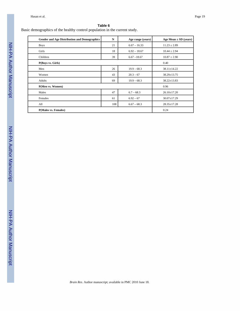
Table 6Basic demographics of the healthy control population in the current study.
Gender and Age Distribution and Demographics N Age range (years) Age Mean ± SD (years)
Boys 21 6.67 – 16.33 11.23 ± 2.89
Girls 18 6.92 – 18.67 10.44 ± 2.94
Children 39 6.67 –18.67 10.87 ± 2.90
P(Boys vs. Girls) 0.40
Men 26 19.9 – 68.3 38.11±14.22
Women 43 20.3 – 67 38.29±13.75
Adults 69 19.9 – 68.3 38.22±13.83
P(Men vs. Women) 0.96
Males 47 6.7 – 68.3 26.10±17.20
Females 61 6.92 – 67 30.07±17.29
All 108 6.67 – 68.3 28.35±17.28
P(Males vs. Females) 0.24
Brain Res. Author manuscript; available in PMC 2010 June 18.