Techniques Used for Morphological and Ultrastructural Description from
Upload
independentCategory
view
3download
0
This article appeared in a journal published by Elsevier. The attachedcopy is furnished to the author for internal non-commercial researchand education use, including for instruction at the authors institution
and sharing with colleagues.
Other uses, including reproduction and distribution, or selling orlicensing copies, or posting to personal, institutional or third party
websites are prohibited.
In most cases authors are permitted to post their version of thearticle (e.g. in Word or Tex form) to their personal website orinstitutional repository. Authors requiring further information
regarding Elsevier’s archiving and manuscript policies areencouraged to visit:
http://www.elsevier.com/copyright
Author's personal copy
Micron 41 (2010) 1005–1010
Contents lists available at ScienceDirect
Micron
journa l homepage: www.e lsev ier .com/ locate /micron
Morphological, morphometrical and ultrastructural characterization of Phrynopsgeoffroanus’ (Testudines: Chelidae) blood cells, in different environments
Carlos Eduardo Saranz Zagoc, Tiago Lucena da Silvac, Maria Isabel Afonso da Silvac,Larissa Paola Rodrigues Venancioc, Priscila Pasqüetto Mendoncac, Luiz Roberto Falleiros Juniora,Luiz Dino Vizottoc, Sebastião Roberto Tabogaa, Claudia Regina Bonini-Domingosc,Maria Tercília Vilela de Azeredo-Oliveirac,∗, Classius de Oliveirab
a Departamento de Biologia, Centro de Microscopia e Microanálise – UNESP-IBILCE-São José do Rio Preto, SP, Brazilb Departamento de Biologia, Laboratório de Anatomia Comparada – UNESP-IBILCE-São José do Rio Preto, SP, Brazilc Departamento de Biologia, Centro de Estudos de Quelônios – UNESP-IBILCE-São José do Rio Preto, SP, Brazil
a r t i c l e i n f o
Article history:Received 23 February 2010Received in revised form 2 June 2010Accepted 19 June 2010
Keywords:Phrynops geoffroanusTurtlesBlood cellsMorphologyUltrastructure
a b s t r a c t
The aim of this study was to evaluate the formed elements in the periferical blood of two amostralgroups of Phrynops geoffroanus: one from an urban environment under domestic sewage dumping, andanother from a non-contaminated environment. Blood samples of 36 animals (females and males) werecollected through cardiocentesis. Sixteen specimens were from the urban environment, and 20 werefrom a control environment. Samples of blood tissue were used for light microscopy analysis, and alsofor morphometric analysis of red blood cells. For the ultrastructural analysis, blood samples of 2 ani-mals were used. The formed elements found, using morphological and ultrastructural analysis were:nucleated red blood cells; thrombocytes; neutrophils, lymphocytes, monocytes, basophils; eosinophils;heterophils, and azurophils. The morphometric analysis of all red blood cells parameters examinated infemales showed a statistically significant difference, but in males just the nuclear area showed differ-ence between the specimens of the two environments. The elements identified by light microscopy wereelucidated by electron transmission microscopy. This P. geoffroanus study is the first one that makes acorrelation between these environments and the description of turtle’s blood cells, thus contributing tothe identification of the hematological characteristics of this group.
© 2010 Elsevier Ltd. All rights reserved.
1. Introduction
Geoffroy’s side-necked turtle (Phrynops geoffroanus) belongs tothe order Testudines and family Chelidae. They are small-sizedand diurnal animals, which are frequently found in rivers, lakesand ponds with slow currents, and have a wide distribution, inSouth America countries (Gans, 1980; Goulart, 2004; Pritchard andTrebbau, 1984).
The circulating blood of turtles has several primitive cells, suchas nucleated red blood cells, responsible for oxygen transporting,and the thrombocytes, which are involved in clotting process. Theproduction of the blood cells occurs in the spleen and bone mar-row. The white blood cells observed in turtles peripheral bloodare: neutrophils, lymphocytes, basophils, monocytes, eosinophils,
∗ Corresponding author at: CEQ, IBILCE-UNESP, Rua Cristóvão Colombo, 2265; Jd.Nazareth; CEP: 15054-000, Brazil. Tel.: +55 17 3221 2378; fax: +55 17 3221 2390.
E-mail address: [email protected] (M.T.V. de Azeredo-Oliveira).
heterophils and azurophils, this cells are responsible for defensemechanisms (Andrew, 1965; Canfield, 1998; Frye and Murphy,1991; Garcia-Navarro and Pachaly, 1994; Stacy and Whitaker,2000).
Environmental degradation linked to human population growthresults in ecological niches variation for several species. Thehuman impact on the environment becomes a major threat tothe organisms that lives there; however, some species can sur-vive even in completely impacted environments (Souza and Abe,1999).
São José do Rio Preto is located in the southeast region of Brazil,at the following geographic coordinates: 20◦49′12′′S 49◦22′44′′E.The city is physically divided by the Rio Preto river and its tribu-taries, including the Piedade and Felicidade stream. The Rio Pretoriver is approximately 120 km long, and is a tributary of the Turvoriver, which flows into the Rio Grande river, and is a componentof the Turvo Grande basin (Zago et al., 2010). With approxi-mately 450 thousand inhabitants, the city is a constant sourceof water pollution from dozens of streams that run though thecity.
0968-4328/$ – see front matter © 2010 Elsevier Ltd. All rights reserved.doi:10.1016/j.micron.2010.06.006
Author's personal copy
1006 C.E.S. Zago et al. / Micron 41 (2010) 1005–1010
The aim of this study were to analyze the blood cells of the ani-mals from the urban environment, which had been contaminatedby domestic sewage, as well as blood cells of the animals fromthe control environment using morphological, ultrastructural andmorphometric analysis.
2. Material and methods
2.1. Animals
We used blood samples of 36 P. geoffroanus specimens, 10females and 10 males from the “Reginaldo Uvo Leone” breedingfarm in Tabapuã, São Paulo (20◦59′47.4′′S, 49◦07′16.6′′W), and 8females and 8 males, from the Felicidade stream, located in SãoJosé do Rio Preto city (20◦46′20.6′′S, 49◦21′18.0′′W). This streamruns through an urban area and is a dump site of domestic sewageand has flat banks interspersed with predominantly herbaceousvegetation, slopes and a large number of P. geoffroanus.
The “Reginaldo Uvo Leone” breeding farm works with wild andexotic reptiles, amphibians and birds with commercial purpose.They work with a larger number of turtles species, including P.geoffroanus. The animals are fed with fish food (45% of protein),and the water of the artificial lakes is from an artesian well, devoidof contaminations. Because of that, these animals were chosen as acontrol group.
2.2. Sample collection
We collect 3 mL of blood by cardiocentesis, between the humeraland pectoral plastron scutes in each animal. We used a 40:12 sizeneedle (BD Precision Glide®) and sodium heparin as anticoagulant.After collecting the blood, the orifice was sealed with surgical glue(Vetband®), and the animals were kept under observation for 72 hbefore being returned to their habitats.
The blood smears were made in less than 24 h, in order to avoidfurther color impregnation caused by heparin (Brites and Rantin,2004; Mader, 1996).
2.3. Morphological analysis
Three blood smears of each animal were fixed in methanol andstained by Panotic (Hematocor®, Biology), according to the man-ufacturer’s instructions. These slides were used for analysis andidentification of thrombocytes, white and red blood cells underlight microscopy (Olympus BX 60), using the objective of 100×.
2.4. Ultrastructural analysis
We analyzed blood samples of two P. geoffroanus males fromthe urban environment using a electron transmission microscopy(Zeiss – Leo IN 910) from the Center of Microscopy and Micro-analysis in the Instituto de Biociências, Letras e Ciências Exatas– IBILCE/UNESP, São José do Rio Preto, Brazil. We used this tech-nique in order to characterize the ultrastructural aspects of thisspecie, having no intention to make correlations with structuraldata related to the environment, only the description of ultrastruc-tural morphology, because of that we used only two animals. Theblood sample was centrifuged at 1000 rpm for 10 min. The plasmawas discarded and the layer of leukocytes was set at 3% glutaralde-hyde in a 0.1 M phosphate buffer for 2 h at 4 ◦C. The material waspost fixed and then put into osmium tetroxide 1% for 2 h at 4 ◦C.It was then dehydrated in an increasing concentration series ofacetone and embedded in Araldite resin (Pellizzon et al., 2002).Ultrathin sections (75 nm) were made using a diamond knife inultramicrotome LKB, and mounted on copper grids and the cuts
were mixed in uranyl acetate and lead citrate. The material was ana-lyzed under a Zeiss – Leo IN 910 transmission electronic microscopeoperated at 80 kV.
2.5. Morphometric analysis
One hundred red cells were analyzed for each animal under anOlympus BX 60 light microscope (objective of 100×). The imageswere captured using the Image – Pro Plus computer program.The cell parameters used were: larger diameter, smaller diameter,nuclear area and total area of red cells (�m).
2.6. Statistical analysis
Statistical data from morphometric analysis were performedusing the software BioEstat 3.0. The resulting values were tested tonormality using the Shapiro–Wilk test and homoscedastic distribu-tion. The parametric test used to compare averages was Student’st-test, and the non-parametric test used was the Mann–WhitneyU-test, with the alpha of 5% (Ayres et al., 1998).
3. Results
3.1. Morphological analysis
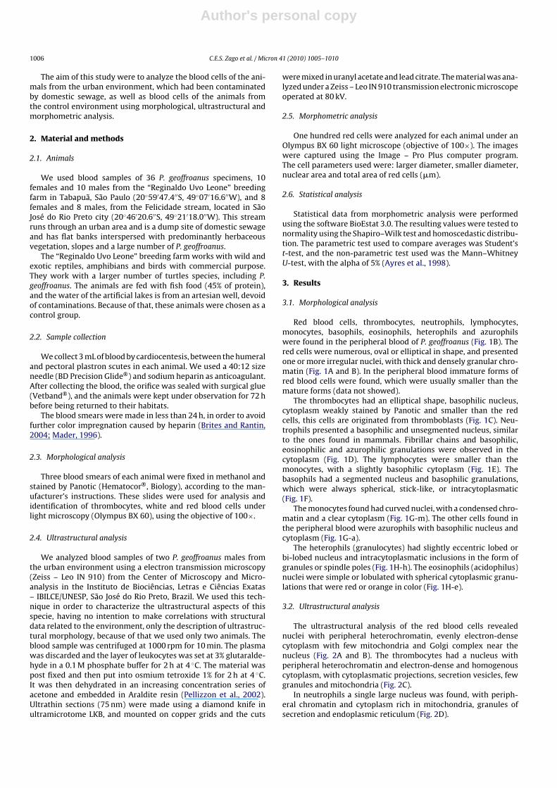
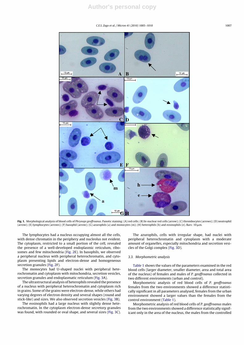
Red blood cells, thrombocytes, neutrophils, lymphocytes,monocytes, basophils, eosinophils, heterophils and azurophilswere found in the peripheral blood of P. geoffroanus (Fig. 1B). Thered cells were numerous, oval or elliptical in shape, and presentedone or more irregular nuclei, with thick and densely granular chro-matin (Fig. 1A and B). In the peripheral blood immature forms ofred blood cells were found, which were usually smaller than themature forms (data not showed).
The thrombocytes had an elliptical shape, basophilic nucleus,cytoplasm weakly stained by Panotic and smaller than the redcells, this cells are originated from thromboblasts (Fig. 1C). Neu-trophils presented a basophilic and unsegmented nucleus, similarto the ones found in mammals. Fibrillar chains and basophilic,eosinophilic and azurophilic granulations were observed in thecytoplasm (Fig. 1D). The lymphocytes were smaller than themonocytes, with a slightly basophilic cytoplasm (Fig. 1E). Thebasophils had a segmented nucleus and basophilic granulations,which were always spherical, stick-like, or intracytoplasmatic(Fig. 1F).
The monocytes found had curved nuclei, with a condensed chro-matin and a clear cytoplasm (Fig. 1G-m). The other cells found inthe peripheral blood were azurophils with basophilic nucleus andcytoplasm (Fig. 1G-a).
The heterophils (granulocytes) had slightly eccentric lobed orbi-lobed nucleus and intracytoplasmatic inclusions in the form ofgranules or spindle poles (Fig. 1H-h). The eosinophils (acidophilus)nuclei were simple or lobulated with spherical cytoplasmic granu-lations that were red or orange in color (Fig. 1H-e).
3.2. Ultrastructural analysis
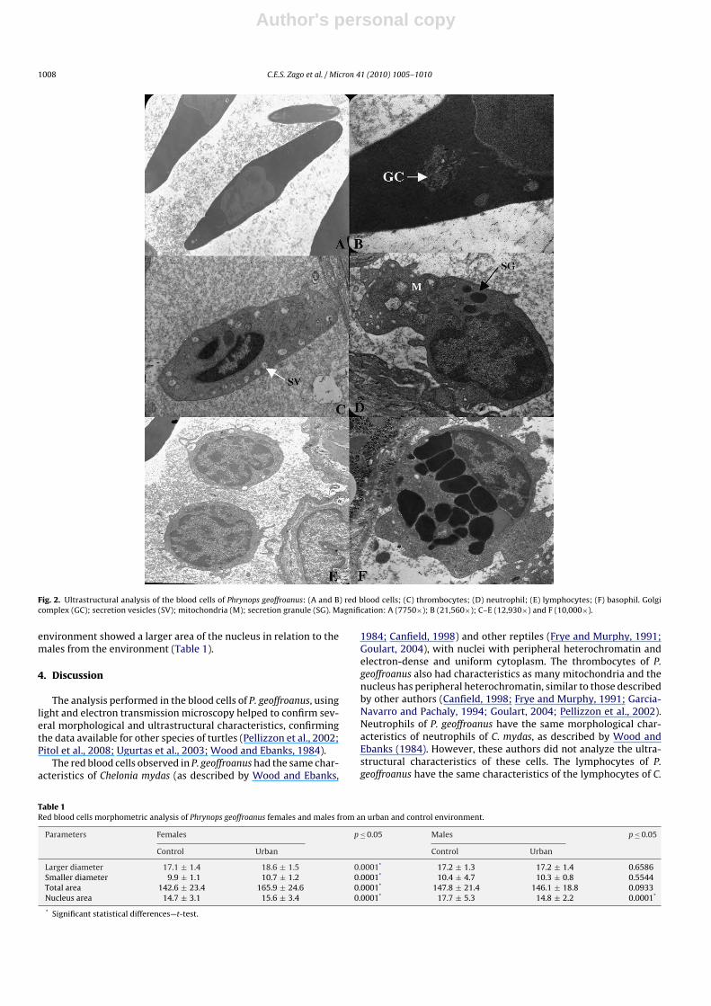
The ultrastructural analysis of the red blood cells revealednuclei with peripheral heterochromatin, evenly electron-densecytoplasm with few mitochondria and Golgi complex near thenucleus (Fig. 2A and B). The thrombocytes had a nucleus withperipheral heterochromatin and electron-dense and homogenouscytoplasm, with cytoplasmatic projections, secretion vesicles, fewgranules and mitochondria (Fig. 2C).
In neutrophils a single large nucleus was found, with periph-eral chromatin and cytoplasm rich in mitochondria, granules ofsecretion and endoplasmic reticulum (Fig. 2D).
Author's personal copy
C.E.S. Zago et al. / Micron 41 (2010) 1005–1010 1007
Fig. 1. Morphological analysis of blood cells of Phrynops geoffroanus. Panotic staining: (A) red cells; (B) bi-nuclear red cells (arrow); (C) thrombocytes (arrows); (D) neutrophil(arrow); (E) lymphocytes (arrows); (F) basophil (arrow); (G) azurophils (a) and monocytes (m); (H) heterophils (h) and eosinophils (e). Bars: 10 �m.
The lymphocytes had a nucleus occupying almost all the cells,with dense chromatin in the periphery and nucleolus not evident.The cytoplasm, restricted to a small portion of the cell, revealedthe presence of a well-developed endoplasmic reticulum, ribo-somes and few mitochondria (Fig. 2E). In basophils, we observeda peripheral nucleus with peripheral heterochromatin, and cyto-plasm presenting lipids and electron-dense and homogeneoussecretion granules (Fig. 2F).
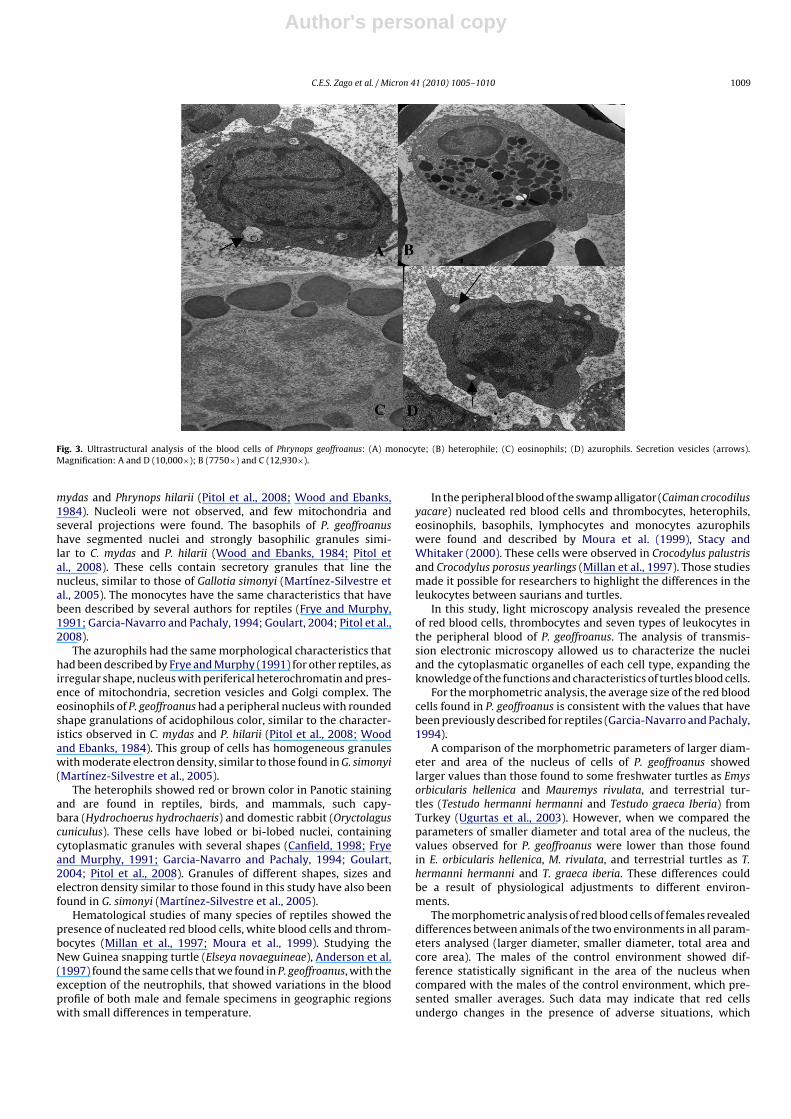
The monocytes had U-shaped nuclei with peripheral hete-rochromatin and cytoplasm with mitochondria, secretion vesicles,secretion granules and endoplasmatic reticulum (Fig. 3A).
The ultrastructural analysis of heterophils revealed the presenceof a nucleus with peripheral heterochromatin and cytoplasm richin grains. Some of the grains were electron-dense, while others hadvarying degrees of electron density and several shapes (round andstick-like) and sizes. We also observed secretion vesicles (Fig. 3B).
The eosinophils had a large nucleus with slightly dense hete-rochromatin. In the cytoplasm electron-dense secretory granuleswas found, with rounded or oval shape, and several sizes (Fig. 3C).
The azurophils, cells with irregular shape, had nuclei withperipheral heterochromatin and cytoplasm with a moderateamount of organelles, especially mitochondria and secretion vesi-cles of the Golgi complex (Fig. 3D).
3.3. Morphometric analysis
Table 1 shows the values of the parameters examined in the redblood cells (larger diameter, smaller diameter, area and total areaof the nucleus) of females and males of P. geoffroanus collected intwo different environments (urban and control).
Morphometric analysis of red blood cells of P. geoffroanusfemales from the two environments showed a difference statisti-cally significant in all parameters analysed, females from the urbanenvironment showed a larger values than the females from thecontrol environment (Table 1).
Morphometric analysis of red blood cells of P. geoffroanus malesfrom the two environments showed a difference statistically signif-icant only in the area of the nucleus, the males from the controlled
Author's personal copy
1008 C.E.S. Zago et al. / Micron 41 (2010) 1005–1010
Fig. 2. Ultrastructural analysis of the blood cells of Phrynops geoffroanus: (A and B) red blood cells; (C) thrombocytes; (D) neutrophil; (E) lymphocytes; (F) basophil. Golgicomplex (GC); secretion vesicles (SV); mitochondria (M); secretion granule (SG). Magnification: A (7750×); B (21,560×); C–E (12,930×) and F (10,000×).
environment showed a larger area of the nucleus in relation to themales from the environment (Table 1).
4. Discussion
The analysis performed in the blood cells of P. geoffroanus, usinglight and electron transmission microscopy helped to confirm sev-eral morphological and ultrastructural characteristics, confirmingthe data available for other species of turtles (Pellizzon et al., 2002;Pitol et al., 2008; Ugurtas et al., 2003; Wood and Ebanks, 1984).
The red blood cells observed in P. geoffroanus had the same char-acteristics of Chelonia mydas (as described by Wood and Ebanks,
1984; Canfield, 1998) and other reptiles (Frye and Murphy, 1991;Goulart, 2004), with nuclei with peripheral heterochromatin andelectron-dense and uniform cytoplasm. The thrombocytes of P.geoffroanus also had characteristics as many mitochondria and thenucleus has peripheral heterochromatin, similar to those describedby other authors (Canfield, 1998; Frye and Murphy, 1991; Garcia-Navarro and Pachaly, 1994; Goulart, 2004; Pellizzon et al., 2002).Neutrophils of P. geoffroanus have the same morphological char-acteristics of neutrophils of C. mydas, as described by Wood andEbanks (1984). However, these authors did not analyze the ultra-structural characteristics of these cells. The lymphocytes of P.geoffroanus have the same characteristics of the lymphocytes of C.
Table 1Red blood cells morphometric analysis of Phrynops geoffroanus females and males from an urban and control environment.
Parameters Females p ≤ 0.05 Males p ≤ 0.05
Control Urban Control Urban
Larger diameter 17.1 ± 1.4 18.6 ± 1.5 0.0001* 17.2 ± 1.3 17.2 ± 1.4 0.6586Smaller diameter 9.9 ± 1.1 10.7 ± 1.2 0.0001* 10.4 ± 4.7 10.3 ± 0.8 0.5544Total area 142.6 ± 23.4 165.9 ± 24.6 0.0001* 147.8 ± 21.4 146.1 ± 18.8 0.0933Nucleus area 14.7 ± 3.1 15.6 ± 3.4 0.0001* 17.7 ± 5.3 14.8 ± 2.2 0.0001*
* Significant statistical differences—t-test.
Author's personal copy
C.E.S. Zago et al. / Micron 41 (2010) 1005–1010 1009
Fig. 3. Ultrastructural analysis of the blood cells of Phrynops geoffroanus: (A) monocyte; (B) heterophile; (C) eosinophils; (D) azurophils. Secretion vesicles (arrows).Magnification: A and D (10,000×); B (7750×) and C (12,930×).
mydas and Phrynops hilarii (Pitol et al., 2008; Wood and Ebanks,1984). Nucleoli were not observed, and few mitochondria andseveral projections were found. The basophils of P. geoffroanushave segmented nuclei and strongly basophilic granules simi-lar to C. mydas and P. hilarii (Wood and Ebanks, 1984; Pitol etal., 2008). These cells contain secretory granules that line thenucleus, similar to those of Gallotia simonyi (Martínez-Silvestre etal., 2005). The monocytes have the same characteristics that havebeen described by several authors for reptiles (Frye and Murphy,1991; Garcia-Navarro and Pachaly, 1994; Goulart, 2004; Pitol et al.,2008).
The azurophils had the same morphological characteristics thathad been described by Frye and Murphy (1991) for other reptiles, asirregular shape, nucleus with periferical heterochromatin and pres-ence of mitochondria, secretion vesicles and Golgi complex. Theeosinophils of P. geoffroanus had a peripheral nucleus with roundedshape granulations of acidophilous color, similar to the character-istics observed in C. mydas and P. hilarii (Pitol et al., 2008; Woodand Ebanks, 1984). This group of cells has homogeneous granuleswith moderate electron density, similar to those found in G. simonyi(Martínez-Silvestre et al., 2005).
The heterophils showed red or brown color in Panotic stainingand are found in reptiles, birds, and mammals, such capy-bara (Hydrochoerus hydrochaeris) and domestic rabbit (Oryctolaguscuniculus). These cells have lobed or bi-lobed nuclei, containingcytoplasmatic granules with several shapes (Canfield, 1998; Fryeand Murphy, 1991; Garcia-Navarro and Pachaly, 1994; Goulart,2004; Pitol et al., 2008). Granules of different shapes, sizes andelectron density similar to those found in this study have also beenfound in G. simonyi (Martínez-Silvestre et al., 2005).
Hematological studies of many species of reptiles showed thepresence of nucleated red blood cells, white blood cells and throm-bocytes (Millan et al., 1997; Moura et al., 1999). Studying theNew Guinea snapping turtle (Elseya novaeguineae), Anderson et al.(1997) found the same cells that we found in P. geoffroanus, with theexception of the neutrophils, that showed variations in the bloodprofile of both male and female specimens in geographic regionswith small differences in temperature.
In the peripheral blood of the swamp alligator (Caiman crocodilusyacare) nucleated red blood cells and thrombocytes, heterophils,eosinophils, basophils, lymphocytes and monocytes azurophilswere found and described by Moura et al. (1999), Stacy andWhitaker (2000). These cells were observed in Crocodylus palustrisand Crocodylus porosus yearlings (Millan et al., 1997). Those studiesmade it possible for researchers to highlight the differences in theleukocytes between saurians and turtles.
In this study, light microscopy analysis revealed the presenceof red blood cells, thrombocytes and seven types of leukocytes inthe peripheral blood of P. geoffroanus. The analysis of transmis-sion electronic microscopy allowed us to characterize the nucleiand the cytoplasmatic organelles of each cell type, expanding theknowledge of the functions and characteristics of turtles blood cells.
For the morphometric analysis, the average size of the red bloodcells found in P. geoffroanus is consistent with the values that havebeen previously described for reptiles (Garcia-Navarro and Pachaly,1994).
A comparison of the morphometric parameters of larger diam-eter and area of the nucleus of cells of P. geoffroanus showedlarger values than those found to some freshwater turtles as Emysorbicularis hellenica and Mauremys rivulata, and terrestrial tur-tles (Testudo hermanni hermanni and Testudo graeca Iberia) fromTurkey (Ugurtas et al., 2003). However, when we compared theparameters of smaller diameter and total area of the nucleus, thevalues observed for P. geoffroanus were lower than those foundin E. orbicularis hellenica, M. rivulata, and terrestrial turtles as T.hermanni hermanni and T. graeca iberia. These differences couldbe a result of physiological adjustments to different environ-ments.
The morphometric analysis of red blood cells of females revealeddifferences between animals of the two environments in all param-eters analysed (larger diameter, smaller diameter, total area andcore area). The males of the control environment showed dif-ference statistically significant in the area of the nucleus whencompared with the males of the control environment, which pre-sented smaller averages. Such data may indicate that red cellsundergo changes in the presence of adverse situations, which
Author's personal copy
1010 C.E.S. Zago et al. / Micron 41 (2010) 1005–1010
cytoplasmatic is likely because of the constant interference of envi-ronmental change.
The morphometric analysis of red blood cells of P. geoffroanusallowed us to standardize the diameter and the total area of thenucleus, as well as the difference between these parameters interms of the environment from where the animal was collected.
This P. geoffroanus study is the first one that makes a correlationbetween these environments and the description of turtle’s bloodcells, thus contributing to the identification of the hematologicalcharacteristics of this group.
Acknowledgment
This study was funded by the Coordenacão de Aperfeicoamentode Pessoal de Nível Superior (CAPES) of Brazil.
References
Anderson, N.L., Wack, R.F., Hatcher, R., 1997. Hematology and clinical chemistry ref-erence ranges for clinically normal, captive New Guinea snapping turtle (Elseyanovaeguineae) and the effects of temperature, sex, and sample type. Journal ofZoo and Wildlife Medicine 28 (4), 394–403.
Andrew, W., 1965. Comparative Hematology. Grune & Stratton, New York and Lon-don.
Ayres, M., Junior, M.A., Ayres, D.L., Santos, A.S., 1998. Bioestat, third ed. MPEG/CNPq.Brites, V.L.C., Rantin, F.T., 2004. The influence of agricultural and urban contamina-
tion on leech infestation of freshwater turtles, Phrynops geoffroanus, taken fromtwo areas of the Uberabinha river. Environmental Monitoring and Assessment96, 273–281.
Canfield, P.J., 1998. Comparative cell morphology in the peripheral blood film fromexotic and native animals. Australian Veterinary Journal 76 (12), 793–800.
Frye, F.L., Murphy, J.B., 1991. Health and Welfare of Captive Reptiles. Clifford War-wick.
Gans, C., 1980. Répteis do Mundo. Universidade de São Paulo, São Paulo.
Garcia-Navarro, C.E.K., Pachaly, J.R., 1994. Manual de Hematologia Veterinária, firsted. Livraria Varela Ltda.
Goulart, C.E.S., 2004. Herpetologia, herpetocultura e medicina de répteis, first ed.L.F. Livros de Veterinária LTDA.
Mader, D.R., 1996. Reptile Medicine and Surgery. W.B. Saunders Company.Martínez-Silvestre, A., Marco, I., Rodriguez-Dominguez, M.A., Lavín, S., Cuenca, R.,
2005. Morphology, cytochemical staining, and ultrastructural characteristics ofthe blood cells of the giant lizard of El Hierro (Gallotia simonyi). Research inVeterinary Science 78, 127–134.
Millan, J.M., Janmaat, A., Richardson, K.C., Chambers, L.K., Fomiatti, K.R., 1997. Ref-erence ranger for biochemical and haematological values in farmed saltwatercrocodile, Crocodylus porosus yearlings. Australian Veterinary Journal 75 (11),814–817.
Moura, W.L., Matushima, E.R., Oliveira, L.W., Egami, M.I., 1999. Aspecto morfológicoe citoquímico dos glóbulos sangüíneos de Caiman crocodilus yacare (Daudin,1802) (Reptilia, Crocodilia). Brazilian Journal of Veterinary Research and AnimalScience 36 (1), 01–09.
Pellizzon, C.H., Azevedo, A., Casaletti, L., Lunardi, L.O., 2002. The thrombocyte aggre-gation process in the turtle Phrynops hilarii (Chelonia). An ultrastructural study.Journal of Submicroscopic Cytology and Pathology 34 (3), 323–327.
Pitol, D.L., Issa, J.P.M., Caetano, F.H., Lunardi, L.O., 2008. Radioautographic study of theseasonal distribution of leukocytes in turtles Phrynops hilarii (Chelonia Chelidae).Micron 1228, 1–6.
Pritchard, P.C.H., Trebbau, P., 1984. The Turtles of Venezuela, Fundación de Interna-dos Rurales (Venezuela). Society for the study of Amphibians and Reptiles.
Stacy, B.A., Whitaker, N., 2000. Hematology and blood biochemistry of captive mug-ger crocodiles (Crocodylus palustris). Journal of Zoo and Wildlife Medicine 31 (3),339–347.
Souza, F.L., Abe, A.S., 1999. Um sobrevivente em rios poluídos. Ciência Hoje 147,01–09.
Ugurtas, I.H., Sevinc, M., Yildirimhan, H.S., 2003. Erythrocyte size and morphologyof some tortoises and turtles from Turkey. Zoological Studies 42 (1), 173–178.
Wood, F.E., Ebanks, G.K., 1984. Blood cytology and hematology of the green sea turtle,Chelonia mydas. Herpetologica 40 (3), 331–336.
Zago, C.E.Z., Ferrarezi, A.L., Vizotto, L.D., Oliveira, C., Cabral, S.R.P., Tabaga,S.R., Bonilla-Rodriguez, G.O., Venancio, L.P.R., Bonini-Domingos, C.R., 2010.Hemoblobin polymorphism and hematological profile of Geoffroy’s side-neckedturtle (Phrynops geoffroanus, Testudines) in the northwestern region of São PauloState, Brazil. Genetics and Molecular Research 9, 721–726.
Copyright © 2022 FDOKUMEN