![Забытый энциклопедист: Михаил Игнатьевич Кулишер (1847-1919) [Forgotten Polymath: Mikhail Ignatyevich Kulisher (1847-1919)]](https://static.fdokumen.com/doc/165x107/63235ba2078ed8e56c0ad283/zabitiy-entsiklopedist-mikhail-ignatevich-kulisher.jpg)
Gross morphology and microstructure of type locality ossicles of Psephophorus polygonus Meyer, 1847...
16
Geol. Mag. 150 (5 ), 2013, pp. 767–782. c Cambridge University Press 2013 767 doi:10.1017/S001675681200091X Gross morphology and microstructure of type locality ossicles of Psephophorus polygonus Meyer, 1847 (Testudines, Dermochelyidae) MASSIMO DELFINO ∗ ‡†, TORSTEN M. SCHEYER§, FRANCESCO CHESI ¶, TAMARA FLETCHER , RICHARD GEMEL#, STEWART MACDONALD ∗∗ , MÁRTON RABI ‡‡§§ & STEVEN W. SALISBURY ∗ Dipartimento di Scienze della Terra, Università di Torino, Via Valperga Caluso 35, I-10125 Torino, Italy ‡Institut Català de Paleontologia Miquel Crusafont, Universitat Autònoma de Barcelona, Edifici ICP, Campus de la UAB s/n, E-08193 Cerdanyola del Vallès, Barcelona, Spain §Paläontologisches Institut und Museum, Universität Zürich, Karl Schmid-Strasse 4, CH-8006 Zürich, Switzerland ¶Dipartimento di Scienze della Terra, Università di Firenze, Via G. La Pira 4, I-50121 Firenze, Italy School of Biological Sciences, The University of Queensland, Brisbane, Qld 4072, Australia #Herpetologische Sammlung, Naturhistorisches Museum Wien, Burgring 7, A-1010 Wien, Austria ∗∗ Centre for Tropical Biodiversity and Climate Change, James Cook University, Townsville, Qld 4811, Australia ‡‡ ˝ Oslénytani Tanszék, Eötvös Loránd Tudomány Egyetem, H-1117 Pázmány Péter sétány 1/C, Budapest, Hungary §§Institut für Geowissenschaften, University of Tübingen, Sigwartstr. 10, D-72070 Tübingen, Germany (Received 22 April 2012; accepted 11 October 2012; first published online 22 February 2013) Abstract – Psephophorus polygonus Meyer, 1847, the first fossil leatherback turtle to be named, was described on the basis of shell ossicles from the middle Miocene (MN6–7/8?) of Slovakia. The whereabouts of this material is uncertain but a slab on display at the Naturhistorisches Museum Wien is considered the neotype. We rediscovered further type locality ossicles in four European institutions, re-evaluated their gross morphology and described for the first time their microstructure by comparing them with Dermochelys coriacea, the only living dermochelyid turtle. The gross morphology is congruent with that already described for P. polygonus, but with two significant exceptions: the ridged ossicles of P. polygonus may have a distinctly concave ventral surface as well as a tectiform shape in cross-section. They do not develop the external keel typical of many ossicles of D. coriacea. Both ridged and non-ridged ossicles of P. polygonus are characterized by compact diploe structures with an internal cortex consisting of a coarse fibrous meshwork, whereas the proportionately thinner ossicles of D. coriacea tend to lose the internal cortex, and thus their diploe, during ontogeny. The ossicles of both P. polygonus and D. coriacea differ from those of other lineages of amniotes whose carapace is composed of polygonal ossicles or platelets, in having growth centres situated at the plate centres just interior to the external bone surface and not within the cancellous core or closer to the internal compact layer. The new diagnosis of P. polygonus allows us to preliminarily re-evaluate the taxonomy of some of the Psephophorus-like species. Despite some macro- and micromorphological differences, it seems likely that Psephophorus was as cosmopolitan as extant Dermochelys and had a broadly similar ecology, with a possible difference concerning the dive depth. Keywords: Dermochelys coriacea, bone histology, middle Miocene, palaeoecology, petrographic thin- sections, taxonomy. 1. Introduction Although dermochelyid turtles are currently represen- ted by only a single living species, the leatherback turtle Dermochelys coriacea (Vandelli, 1761), they have an extensive fossil record that mirrors a complex evolution (Wood et al. 1996). Despite the fact that D. coriacea has a very short evolutionary history inscribed in its mtDNA (<1 Ma; Dutton et al. 1999), the earliest definite dermochelyids are known from the Late Paleocene to Early Eocene (Nielsen, 1959; Tong et al. 1999). Besides the Mesozoic Mesodermochelys Hirayama & Chitoku, 1996 (Late Cretaceous, Japan) and Corsochelys Zangerl, 1960 (Late Cretaceous, † Author for correspondence: massimo.delfi[email protected] North America), at least seven Cenozoic fossil der- mochelyid genera can be identified (Danilov, 2005; Bever & Joyce, 2005): (1) Arabemys crassiscutata Tong et al. 1999 (Paleocene–Eocene, Saudi Arabia), (2) Cosmochelys Andrews, 1919 (Eocene, Africa), (3) Egyptemys Wood et al. 1996 (Eocene, Africa), (4) Eosphargis Lydekker, 1889 (Eocene, Europe), (5) Cardiochelys Moody, 1993 (Oligocene, Europe), (6) Natemys Wood et al. 1996 (Oligocene, Peru) and (7) Psephophorus Meyer, 1847 (mainly Miocene, Europe and possibly North America). The two Mesozoic forms are closely related to dermochelyids according to Hirayama & Chitoku (1996) and Hirayama (1997); however, the relationships of these taxa await reassess- ment in a more comprehensive phylogenetic analysis. It is apparent that the basal members (e.g. Eosphargis
-
Upload
uni-tzuebingen -
Category
Documents
-
view
6 -
download
0
Transcript of Gross morphology and microstructure of type locality ossicles of Psephophorus polygonus Meyer, 1847...

Geol. Mag. 150 (5 ), 2013, pp. 767–782. c© Cambridge University Press 2013 767doi:10.1017/S001675681200091X
Gross morphology and microstructure of type locality ossiclesof Psephophorus polygonus Meyer, 1847
(Testudines, Dermochelyidae)
M A S S I M O D E L F I N O∗‡†, TO R S T E N M . S C H E Y E R § , F R A N C E S C O C H E S I¶,TA M A R A F L E T C H E R‖, R I C H A R D G E M E L # , S T E WA RT M AC D O NA L D‖∗∗,
M Á RTO N R A B I‡‡§ § & S T E V E N W. S A L I S B U RY‖∗Dipartimento di Scienze della Terra, Università di Torino, Via Valperga Caluso 35, I-10125 Torino, Italy
‡Institut Català de Paleontologia Miquel Crusafont, Universitat Autònoma de Barcelona, Edifici ICP, Campus de laUAB s/n, E-08193 Cerdanyola del Vallès, Barcelona, Spain
§Paläontologisches Institut und Museum, Universität Zürich, Karl Schmid-Strasse 4, CH-8006 Zürich, Switzerland¶Dipartimento di Scienze della Terra, Università di Firenze, Via G. La Pira 4, I-50121 Firenze, Italy
‖School of Biological Sciences, The University of Queensland, Brisbane, Qld 4072, Australia#Herpetologische Sammlung, Naturhistorisches Museum Wien, Burgring 7, A-1010 Wien, Austria∗∗Centre for Tropical Biodiversity and Climate Change, James Cook University, Townsville, Qld 4811, Australia
‡‡Oslénytani Tanszék, Eötvös Loránd Tudomány Egyetem, H-1117 Pázmány Péter sétány 1/C, Budapest, Hungary§§Institut für Geowissenschaften, University of Tübingen, Sigwartstr. 10, D-72070 Tübingen, Germany
(Received 22 April 2012; accepted 11 October 2012; first published online 22 February 2013)
Abstract – Psephophorus polygonus Meyer, 1847, the first fossil leatherback turtle to be named,was described on the basis of shell ossicles from the middle Miocene (MN6–7/8?) of Slovakia. Thewhereabouts of this material is uncertain but a slab on display at the Naturhistorisches Museum Wienis considered the neotype. We rediscovered further type locality ossicles in four European institutions,re-evaluated their gross morphology and described for the first time their microstructure by comparingthem with Dermochelys coriacea, the only living dermochelyid turtle. The gross morphology iscongruent with that already described for P. polygonus, but with two significant exceptions: the ridgedossicles of P. polygonus may have a distinctly concave ventral surface as well as a tectiform shape incross-section. They do not develop the external keel typical of many ossicles of D. coriacea. Bothridged and non-ridged ossicles of P. polygonus are characterized by compact diploe structures with aninternal cortex consisting of a coarse fibrous meshwork, whereas the proportionately thinner ossiclesof D. coriacea tend to lose the internal cortex, and thus their diploe, during ontogeny. The ossiclesof both P. polygonus and D. coriacea differ from those of other lineages of amniotes whose carapaceis composed of polygonal ossicles or platelets, in having growth centres situated at the plate centresjust interior to the external bone surface and not within the cancellous core or closer to the internalcompact layer. The new diagnosis of P. polygonus allows us to preliminarily re-evaluate the taxonomyof some of the Psephophorus-like species. Despite some macro- and micromorphological differences,it seems likely that Psephophorus was as cosmopolitan as extant Dermochelys and had a broadlysimilar ecology, with a possible difference concerning the dive depth.
Keywords: Dermochelys coriacea, bone histology, middle Miocene, palaeoecology, petrographic thin-sections, taxonomy.
1. Introduction
Although dermochelyid turtles are currently represen-ted by only a single living species, the leatherbackturtle Dermochelys coriacea (Vandelli, 1761), theyhave an extensive fossil record that mirrors a complexevolution (Wood et al. 1996). Despite the fact thatD. coriacea has a very short evolutionary historyinscribed in its mtDNA (<1 Ma; Dutton et al. 1999),the earliest definite dermochelyids are known from theLate Paleocene to Early Eocene (Nielsen, 1959; Tonget al. 1999). Besides the Mesozoic MesodermochelysHirayama & Chitoku, 1996 (Late Cretaceous, Japan)and Corsochelys Zangerl, 1960 (Late Cretaceous,
† Author for correspondence: [email protected]
North America), at least seven Cenozoic fossil der-mochelyid genera can be identified (Danilov, 2005;Bever & Joyce, 2005): (1) Arabemys crassiscutataTong et al. 1999 (Paleocene–Eocene, Saudi Arabia),(2) Cosmochelys Andrews, 1919 (Eocene, Africa),(3) Egyptemys Wood et al. 1996 (Eocene, Africa),(4) Eosphargis Lydekker, 1889 (Eocene, Europe), (5)Cardiochelys Moody, 1993 (Oligocene, Europe), (6)Natemys Wood et al. 1996 (Oligocene, Peru) and (7)Psephophorus Meyer, 1847 (mainly Miocene, Europeand possibly North America). The two Mesozoic formsare closely related to dermochelyids according toHirayama & Chitoku (1996) and Hirayama (1997);however, the relationships of these taxa await reassess-ment in a more comprehensive phylogenetic analysis.It is apparent that the basal members (e.g. Eosphargis

768 M . D E L F I N O A N D OT H E R S
spp.) still retained neural, costal and peripheral bonesand otherwise lacked an ‘epithecal’ shell (Nielsen,1963; see also Karl & Lindow, 2010). Conversely,in D. coriacea the primary ‘thecal’ shell, with theexception of the nuchal bone, is largely reduced.Instead a secondary ‘epithecal’ mosaic of dermalelements, the ossicles, embedded in the thick fattydermis, is exceptionally developed (e.g. Hay, 1898;Versluys, 1913; Deraniyagala, 1930; Zangerl, 1939,1969; Rieppel, 2001; Bever & Joyce, 2005; Frazier,Gramentz & Fritz, 2005). Seven longitudinal ridgesrun along the carapace, and the unique secondarycarapace consists of thousands of ‘epithecal’ polygonalossicles (e.g. Gervais, 1872). The ‘thecal’ elements ofthe plastron are still present, but only as thin bony rod-like elements. After Brongersma (1969), the plastronis referred to as being additionally covered in isolatedrows of small ‘epithecal’ platelets that coincide withsix plastral ridges (see also Bever & Joyce, 2005).
For several decades, it has been assumed that Der-mochelys evolved from the extinct taxon PsephophorusMeyer, 1847 from the middle Miocene StudienkaFormation of Devínska Nová Ves, close to Bratislava,Slovakia. However, according to the last comprehensiverevision of the taxonomy and phylogeny of fossilDermochelyidae (Wood et al. 1996), Dermochelysbelongs to a clade that is the sister group to P. polygonus(i.e. an unresolved trichotomy with ‘Psephophorus’calvertensis and a yet undescribed specimen fromthe Eocene Selma Formation of Alabama, USA).Wood et al. (1996) concluded that most of thespecies previously referred to Psephophorus are notassignable to this genus, which is represented by asingle species, Psephophorus polygonus Meyer, 1847.They distinguished Dermochelys and Psephophorus onthe basis of several characters. Particularly diagnosticfor Psephophorus should be the fact that longitudinalridges are not apically pointed in cross-section, andare expressed only on the external surface of thecarapace ossicles, their visceral surface being flat. Thiscontrasts with the condition in Dermochelys, wherethe osteoderms that comprise the longitudinal ridgesare tectiform, pointed in cross-section and expressedalso on the ventral surface of the shell. Moreover,ossicles of Dermochelys vary greatly in size but notin Psephophorus.
In the revision by Wood et al. (1996), the morphologyof the type species, Psephophorus polygonus Meyer,1847, was based only on what is considered to be theneotype (NHMW 2011/0330/0001) as it is doubtfulwhether Meyer’s specimens, which he did not figure,will ever become identifiable. In fact, the history of thedefinition of this species has not been straightforwardand several inaccuracies, related mostly to the natureof the type material and its whereabouts, have beenprogressively disseminated in the literature.
Meyer (1846) first briefly mentioned some isolatedossicles from Slovakia that he tentatively referred toan unidentified dasipodid mammal, without namingany new taxon. Then, the year after, he examined a
drawing of a slab coming from the Slovakian localityNeudorf an der March (= Dévényújfalu, now DevínskaNová Ves), containing ossicles in connection, and onthe basis of it – without studying the real fossil –confirmed the previous referral to a mammal, brieflydescribing the new taxon Psephophorus polygonusMeyer, 1847. Regrettably, he did not publish thedrawing and therefore we only know that the slabcontained about 70 ossicles, some of which formeda ridge. As already summarized by Seeley (1880,p. 406), who associated for the first time P. polygonusto chelonians, the slab was originally in Bratislava butwas later acquired by the Museum of the ImperialGeological Survey in Vienna, which also obtained asecond slab ‘containing a larger portion of the samecarapace’ that fitted onto the first (see also von Hauer,1868, 1870). This second slab is the only one ofthe two that is currently available: it is displayedat the Naturhistorisches Museum Wien (NHMW2011/0330/0001) and is considered the neotype of P.polygonus (Wood et al. 1996). Seeley (1880) describedin detail both slabs and figured them along with avertebra that he referred to the same taxon. He reportedthe presence of a fragment of a rib and five fragmentaryvertebrae, the best preserved of which, the one figured,‘is chelonian in its characters’ (Seeley, 1880, p. 410).On the basis of the description and the figure, it is notactually possible to confirm the identification of thevertebrae as chelonian, and possibly because of this,vertebrae were not mentioned in subsequent accounts(i.e. Wood et al. 1996; Gemel & Rauscher, 2000). Woodet al. (1996, p. 279) wrote that ‘the type and onlyspecimen of Psephophorus polygonus is housed in thecollections of the Natural History Museum in Vienna’but it is known that other shell remains were found inthe same locality (Abel, 1919; Szalai, 1934; Młynarski,1966). In particular, Abel (1919) described and figuredisolated ossicles from Dévényújfalu (the Hungarianname for Devínska Nová Ves) that were depositedat the Geologische Bundesanstalt in Vienna, at theInstitute of Palaeontology of Vienna University andin the Mineral and Palaeontological collections of theHungarian National Museum in Budapest. Młynarski(1966) later published catalogue numbers and a briefcomment on the available remains stored in Budapest.
In order to provide a sound basis for futureanalyses of fossil dermochelyids and to further ourunderstanding of the relationship among the fossiltaxa and the only living relative, D. coriacea, we heresummarize the current location of the remains of P.polygonus from the type locality that are availablein public collections. On the basis of this material,we re-evaluate the gross morphology of the ossiclesof P. polygonus and describe for the first time theirmicrostructure, both of which are compared with thoseof the ossicles of extant D. coriacea.
Institutional abbreviations. GBA – GeologischeBundesanstalt, Vienna, Austria; IPUW – Institut fürPaläontologie an der Universität Wien, Vienna, Austria;MDHC – Massimo Delfino Herpetological Collection,

Ossicle morphology of Psephophorus polygonus 769
deposited at Università di Torino, Italy; MTM – MagyarTermészettudományi Múzeum, Budapest, Hungary;NHMW – Naturhistorisches Museum Wien, Vienna,Austria; PIMUZ – Paläontologisches Institut undMuseum, Universität Zürich, Zürich, Switzerland;QM – Queensland Museum, Brisbane, Queensland,Australia.
2. Systematic palaeontology
Order TESTUDINES Batsch, 1788Family DERMOCHELYIDAE Fitzinger, 1843
Genus Psephophorus Meyer, 1847
Diagnosis. As for the only species ascribed to this genusby Wood et al. (1996).
Psephophorus polygonus Meyer, 1847
Neotype. NHMW 2011/0330/0001: a slab containingridge and non-ridge ossicles (Wood et al. 1996).
Topotype material. GBA 2008/93/1: 13 ossicles; IPUW6224/1–5: five ossicles; MTM V 61.1216: 37 ossicles,28 of which are almost complete (six from a ridge);all the ossicles are isolated except six (three from aridge) that have been joined in a major shell fragmentand two that constitute a minor one; MTM V.61.1215:46 ossicles (two from the ridge); MTM V.61.1218:17 ossicles (two from the ridge); MTM V.61.1219: aslab broken into two pieces containing 37 connectedossicles (all except ten are broken into two alongtheir horizontal plane); NHMW 1848/0015/a–z, aa–aq: 43 ossicles; NHMW 1857/0019/0010a–e: fiveossicles; NHMW 1857/0028/0006a–b: two ossicles;NHMW 1859/0005/0125a–b: two ossicles; NHMW1868/0008/0004: one ossicle; NHMW 2011/01/0149/001–002: two ossicles; NHMW 2011/0150/001–2: twoossicles; NHMW 2011/0151/0001–006: six ossicles(one from a ridge); NHMW 2011/0152/0001–0013:13 ossicles; NHMW 2011/137/0001–0010: ten ossicles(three from a ridge).
Type locality. Devínska Nová Ves, close to Bratislava(Slovakia). The locality reported on the label ofthe materials in the collections of the MTM isDévényujfalu/Neudorf (the Hungarian and Germannames for Devínska Nová Ves, respectively), upperlayers of Sandberg Hill. It is noteworthy that the samelocality was sometimes called Neudorf an der March inthe German literature (= Neudorf at the Morava river).The labels of the isolated ossicles at the NHMW alsoindicate Sandberg (Neudorf an der March), whereas thematerial at the GBA is known to come from Neudorfan der March but more precise collection data areunavailable. Based on the labels at the MTM andNHMW together with the sandstone matrix of MTMV.61.1219, we assume that all known material of P.polygonus from the type locality of Devínska Nová Vescomes from a single site and horizon (Sandberg). Thisis supported by the presence of very similar matrix inthe neotype specimen (NHMW 2011/0330/0001) and
in MTM V.61.1219. Sandberg is a sandstone hill justat the southern tip of Devínska Nová Ves (longitude:17◦ 00′, latitude: 48◦ 12′). The succession, classified asthe Sandberg Member of the Studienka Formation andinterpreted as the shallow marine marginal facies ofthe latter, consists of predominately clastic sediments,including sand, clayey sand, calcareous sandstone,limestone, breccia and gravel. During the middleMiocene, Sandberg was part of an archipelago orpeninsula in the Central Paratethys Sea as testified by arich vertebrate and invertebrate marine and terrestrialfauna (Švargovský, 1978; Baráth, Nagy & Kovác, 1994;Sabol & Holec, 2002; Holec & Emry, 2003; Kovác et al.2004).
Age. According to molluscan and foraminiferalbiostratigraphy, the age of the fossiliferous layers ofSandberg at Devínska Nová Ves is middle Miocene(Late Badenian or earliest Langhian). They representthe faciostratotype of the Bulimina–Bolivina Zone(Švargovský, 1974, 1978; Holec & Emry, 2003; Kovácet al. 2004). The large terrestrial mammal assemblageis typical of the Astaracian faunal unit that correspondsto basal MN6–MN7/8? (Sabol & Holec, 2002).
Diagnosis. Because only shell fragments are knownfrom the type locality, this diagnosis is based onlyon the morphology and arrangement of the ossiclesforming the shell. P. polygonus differs from all theother dermochelyid turtles provided with an ‘epithecal’shell formed by several small bony ossicles basedon the following combination of characters: thickossicles; ossicles not greatly varying in size and shape;ossicle edges with reduced sutural structures; ossiclesnot arranged in the ‘sunflower’ pattern typical ofNatemys; largest ossicles arranged in several longit-udinal ridges; ridge-forming ossicles often viscerallyflat (ridge expressed only on the external surface) anddorsally arched in cross-section, but in some casesviscerally concave (the ridge is also expressed onthe visceral surface) and tectiform in cross-section;ridge ossicles characterized by variable thicknesses butwithout evident external keels (present in Dermochelys;compare Fig. 1 with Fig. 2); presence of peaks andvalleys on the longitudinal ridges; peaks and bottomof the valleys corresponding to the sutures amongossicles; neighbouring longitudinal ridges separated byat least five ossicles (which are 13 on average in theremains from Southern Italy; Chesi et al. 2007).
From a histological perspective, P. polygonus differsat least from D. coriacea in having shell ossicles thatretain the diploe structure with an internal corticalcoarse fibrous meshwork throughout ontogeny (theossicles of D. coriacea do not have the internal cortex).
3. Gross morphology
The following description deals with the isolatedossicles and the poorly preserved slab MTM V.61.1219available in the four above-mentioned collections anddoes not take into consideration the slab NHMW
770 M . D E L F I N O A N D OT H E R S
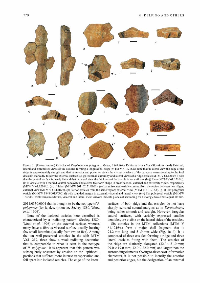
Figure 1. (Colour online) Ossicles of Psephophorus polygonus Meyer, 1847 from Devínska Nová Ves (Slovakia). (a–d) External,lateral and extremities views of the ossicles forming a longitudinal ridge (MTM V 61.1216/a); note that in lateral view the edge of theridge is approximately straight and that in anterior and posterior views the visceral surface of the carapace corresponding to the keeldoes not markedly follow the external surface. (e–g) External, extremity and lateral views of a ridge ossicle (MTM V 61.1216/b); notethat the ventral surface is nearly flat and that in lateral view the thickness of the ossicle is not uniform. (h–j) Idem (MTM V 61.1216/c).(k, l) Ossicle with a marked ventral concavity and a clear tectiform shape in cross-section; external and extremity views, respectively(MTM V 61.1216/d). (m, n) Idem (NHMW 2011/0151/0001). (o) Large isolated ossicle coming from the region between two ridges;external view (MTM V 61.1216/e). (p) Pair of ossicles from the same region; external view (MTM V 61.1216/f). (q–s) Flat polygonalossicle (NHMW 1848/0015/0001al) with rounded margin in external, visceral and lateral view. (t–v) Flat polygonal ossicle (NHMW1848/0015/0001am) in external, visceral and lateral view. Arrows indicate planes of sectioning for histology. Scale bars equal 10 mm.
2011/0330/0001 that is thought to be the neotype of P.polygonus (for its description see Seeley, 1880; Woodet al. 1996).
None of the isolated ossicles here described ischaracterized by a ‘radiating pattern’ (Seeley, 1880;Wood et al. 1996) on the external surface, whereasmany have a fibrous visceral surface usually hostingfew small foramina (usually from two to five). Amongthe ten well-preserved ossicles in the slab MTMV.61.1219, three show a weak radiating decorationthat is comparable to what is seen in the neotypeof P. polygonus. It is apparent that this pattern wassubsequently obscured by erosion on the ‘epithecal’portions that suffered more intense transportation andfell apart into isolated ossicles. The edge of the lateral
surfaces of both ridge and flat ossicles do not havesharply serrated sutural margins as in Dermochelys,being rather smooth and straight. However, irregularsutural surfaces, with variably expressed smallerdenticles, are visible on the lateral sides of the ossicles.
Six ossicles in the MTM collections (MTM V61.1216/a) form a major shell fragment that is94.2 mm long and 51.9 mm wide (Fig. 1a–d); it iscomposed of three ossicles forming a ridge and threelateral ossicles fitting with them. The ossicles ofthe ridge are distinctly elongated (32.0 × 21.0 mm;29.0 × 19.0 mm; 32.0 × 22.0 mm) and larger than thesurrounding elements. Owing to absence of informativecharacters, it is not possible to identify the anteriorand posterior edges, but the designation of an external
Ossicle morphology of Psephophorus polygonus 771
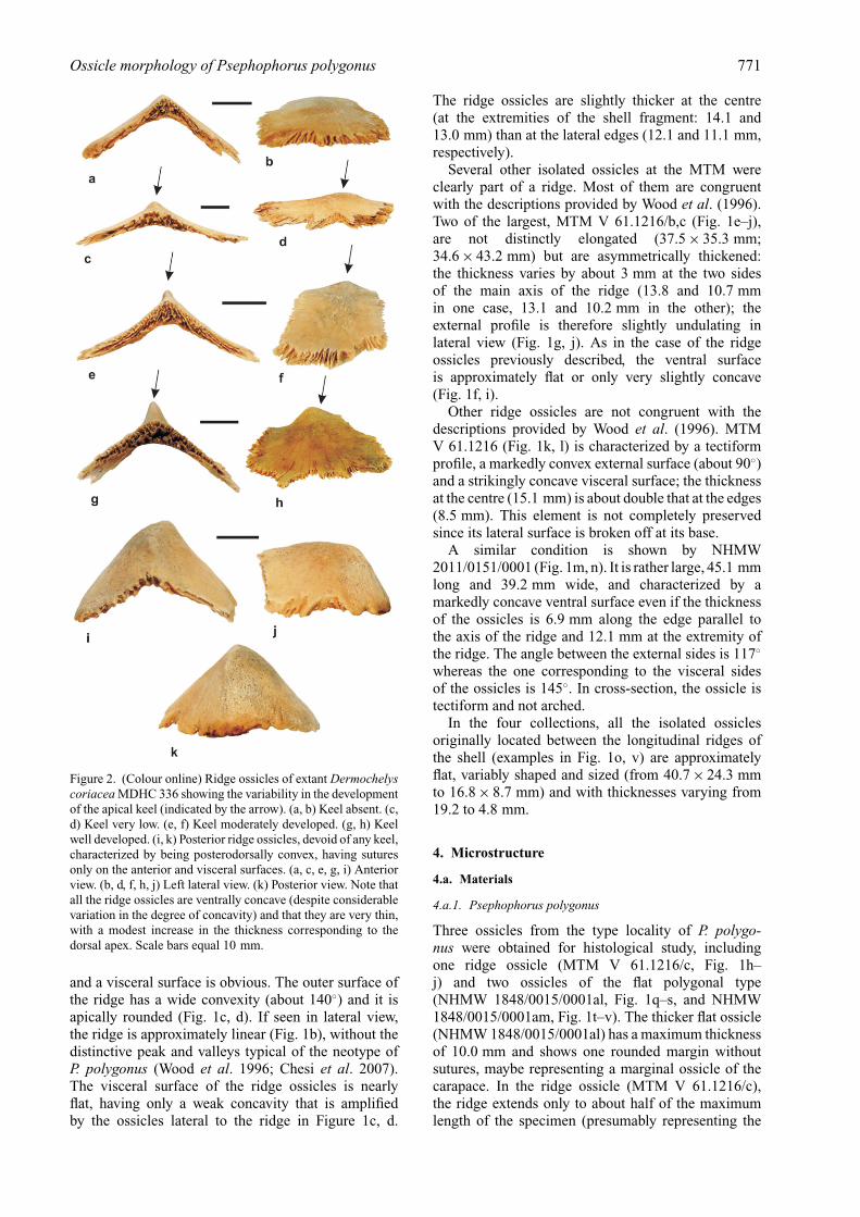
Figure 2. (Colour online) Ridge ossicles of extant Dermochelyscoriacea MDHC 336 showing the variability in the developmentof the apical keel (indicated by the arrow). (a, b) Keel absent. (c,d) Keel very low. (e, f) Keel moderately developed. (g, h) Keelwell developed. (i, k) Posterior ridge ossicles, devoid of any keel,characterized by being posterodorsally convex, having suturesonly on the anterior and visceral surfaces. (a, c, e, g, i) Anteriorview. (b, d, f, h, j) Left lateral view. (k) Posterior view. Note thatall the ridge ossicles are ventrally concave (despite considerablevariation in the degree of concavity) and that they are very thin,with a modest increase in the thickness corresponding to thedorsal apex. Scale bars equal 10 mm.
and a visceral surface is obvious. The outer surface ofthe ridge has a wide convexity (about 140◦) and it isapically rounded (Fig. 1c, d). If seen in lateral view,the ridge is approximately linear (Fig. 1b), without thedistinctive peak and valleys typical of the neotype ofP. polygonus (Wood et al. 1996; Chesi et al. 2007).The visceral surface of the ridge ossicles is nearlyflat, having only a weak concavity that is amplifiedby the ossicles lateral to the ridge in Figure 1c, d.
The ridge ossicles are slightly thicker at the centre(at the extremities of the shell fragment: 14.1 and13.0 mm) than at the lateral edges (12.1 and 11.1 mm,respectively).
Several other isolated ossicles at the MTM wereclearly part of a ridge. Most of them are congruentwith the descriptions provided by Wood et al. (1996).Two of the largest, MTM V 61.1216/b,c (Fig. 1e–j),are not distinctly elongated (37.5 × 35.3 mm;34.6 × 43.2 mm) but are asymmetrically thickened:the thickness varies by about 3 mm at the two sidesof the main axis of the ridge (13.8 and 10.7 mmin one case, 13.1 and 10.2 mm in the other); theexternal profile is therefore slightly undulating inlateral view (Fig. 1g, j). As in the case of the ridgeossicles previously described, the ventral surfaceis approximately flat or only very slightly concave(Fig. 1f, i).
Other ridge ossicles are not congruent with thedescriptions provided by Wood et al. (1996). MTMV 61.1216 (Fig. 1k, l) is characterized by a tectiformprofile, a markedly convex external surface (about 90◦)and a strikingly concave visceral surface; the thicknessat the centre (15.1 mm) is about double that at the edges(8.5 mm). This element is not completely preservedsince its lateral surface is broken off at its base.
A similar condition is shown by NHMW2011/0151/0001 (Fig. 1m, n). It is rather large, 45.1 mmlong and 39.2 mm wide, and characterized by amarkedly concave ventral surface even if the thicknessof the ossicles is 6.9 mm along the edge parallel tothe axis of the ridge and 12.1 mm at the extremity ofthe ridge. The angle between the external sides is 117◦
whereas the one corresponding to the visceral sidesof the ossicles is 145◦. In cross-section, the ossicle istectiform and not arched.
In the four collections, all the isolated ossiclesoriginally located between the longitudinal ridges ofthe shell (examples in Fig. 1o, v) are approximatelyflat, variably shaped and sized (from 40.7 × 24.3 mmto 16.8 × 8.7 mm) and with thicknesses varying from19.2 to 4.8 mm.
4. Microstructure
4.a. Materials
4.a.1. Psephophorus polygonus
Three ossicles from the type locality of P. polygo-nus were obtained for histological study, includingone ridge ossicle (MTM V 61.1216/c, Fig. 1h–j) and two ossicles of the flat polygonal type(NHMW 1848/0015/0001al, Fig. 1q–s, and NHMW1848/0015/0001am, Fig. 1t–v). The thicker flat ossicle(NHMW 1848/0015/0001al) has a maximum thicknessof 10.0 mm and shows one rounded margin withoutsutures, maybe representing a marginal ossicle of thecarapace. In the ridge ossicle (MTM V 61.1216/c),the ridge extends only to about half of the maximumlength of the specimen (presumably representing the
772 M . D E L F I N O A N D OT H E R S
anteroposterior axis of the element); thus, it mightrepresent the first or the last ossicle in a series of ridgeossicles in the carapace. The external bone surface issmooth in all three specimens. Only in the thinnerflat ossicle (NHMW 1848/0015/0001am; maximumthickness of 5.0 mm) and the ridge ossicle (thicknessranging between 10.0 mm at the margins and 12.0 mmbelow the ridge) are a few foramina visible on thevisceral bone surface.
4.a.2. Dermochelys coriacea
Several associated ossicles, including six irregularlyformed flat polygonal ossicles and two ridge ossiclesof a carapace fragment of a presumably juvenile tosubadult specimen (PIMUZ A/III 1288 collected alongthe Atlantic coast of northern Africa) of D. coriaceawere sampled (Fig. 3a, b). The carapace fragment wascompletely devoid of soft tissue, so the interdigitatingsutures between the ossicles were well visible. Theexternal bone surfaces of both ossicles were rugose,with the ridge ossicles also showing some faint striationradiating from the ossicle centre towards the margins.The visceral bone surfaces were also slightly rugoseand several foramina were scattered over the surfaceof the ossicles. The thickness of the ossicles variedbetween 1.5 mm (usually the flat ossicles) and upto 4.0 and 5.0 mm on each ridge ossicle. As notedby Deraniyagala (1939), ossification of ossicles startsduring the first year of life below the ridges (ossicleslarger in outer ridges, smaller in the more mediallysituated ones), and then spreads towards the areasbetween ridges. In an animal of 662 days, so less thantwo years of age, he further noted that the ‘epithecal’carapace has almost fully developed (see also Frazier,Gramentz & Fritz, 2005, p. 298, table 29). We hereassume that the specimens used for sectioning derivedfrom an individual that was thus at least two years old,because the carapace fragment shows well-developedand well-sutured ossicles both of ridge and intra-ridge areas. A more accurate age determination of thematerial is not possible owing to the lack of growthmarks (see below).
Additionally, ossicles from three wild-caught spe-cimens of D. coriacea from southeastern Queenslandwere sampled between 2004 and 2006 by three of theauthors (SM, TF and SWS): a flat, polygonal ossiclefrom a small adult male (QM J81592) and a ridgeossicle from a large adult (QM J73979; Fig. 3c, d).Because these ossicles were cut out of the integument,the margins of the plates were not sampled. Portionsof the carapace of a hatchling specimen (QM J58751)from a failed nest were also examined in order to covera wide range of ontogenetic development.
The young adult specimen (QM J81592; male,maximum curved carapace length 163.5 cm) was foundin a shark net off the Gold Coast, southeasternQueensland, on 25 September 2004. After collection,a portion of carapace was removed and kept on icefor few days before it was preserved in 90 % ethanol.
The exact location and orientation of the portion ofcarapace that was removed was not recorded, but it isassumed to have come from the caudal left side of theanimal. The ossicle that was sampled from this portionof carapace is situated just below the transition betweenthe external cuticle and the lipid-rich fibrous tissue ofthe dermis, where the dermal fibre bundles trend sub-parallel to the external surface of the integument. Athin layer of dermal connective tissue thus separatesthe ossicle from the epidermal tissue. In cross-section,the layer of horizontally arranged dermal fibre bundlesis followed internally by a thick, adipose-rich layerof integumentary tissue, where fibre bundles are moreloosely arranged. The fibre bundles extend in severaldirections, with a slight dominance of fibre bundles thatextend roughly diagonally towards the external surfaceof the integument.
The adult specimen (QM J73979; sex unknown,total length recorded as 200 cm, assumed to be curvedcarapace length but not specified) was found drownedin a fishing net in Moreton Bay, just off Skirmish Point,Bribie Island, southeastern Queensland, sometimeduring 2004. The carcass was taken ashore and leftfor several months, during which time portions ofthe carapace became desiccated. The extremely highlipid content of the integument prevented processingof good sections during this period. During thedesiccation process, the ‘epithecal’ (secondary) shelllocally separated from the ribs. Histological sampleswere subsequently taken from a ridge ossicle on theleft side of the desiccated carapace (Fig. 3c, d). Thesampled ridge ossicle is tectiform in cross-section.Because the bone was dissected out of the surroundingintegumentary tissue, its connection with the dermaltissue is not recorded.
The hatchling specimen (QM J58751; sex unknown,maximum carapace length 8.2 cm) from a failed nest,thought to be at Wreck Rock, between Gladstoneand Bundaberg, in the Wide Bay–Burnett region ofsoutheastern Queensland, was gutted and fixed informalin, and then kept in 70 % alcohol. Histologicalslides from this specimen were based on a transversesection through a caudal portion of the carapace, dorsalto a rib and extending from the lateral margin to3 mm past the median osteoderm ridge. Because thehatchling specimen shows largely unossified soft tissueintegumentary structures, i.e. subcutical adipose tissue,and the formation of the ribs and vertebral columnonly, with no ‘epithecal’ ossicles (which is again inorder with the developmental timing presented byDeraniyagala, 1939, see above), it was excluded fromthe following Results and Discussion sections.
4.b. Histological sampling
The sampling of the ossicles followed standardprocedures for petrographic thin-sections. The slideswere studied using a Leica DM2500 M compositemicroscope mounted with a Leica DFC 420 C digitalcamera.

Ossicle morphology of Psephophorus polygonus 773
Figure 3. (Colour online) Carapace fragments of Dermochelys coriacea sampled in the study. (a, b) Carapace fragment (PIMUZ A/III1288) in external (a) and visceral view (b), respectively. Note size and shape differences in the flat polygonal ossicles lateral to the ridgeossicles, as well as the presence of an anomalous, elongated, extra ossicle which is not seen in MDHC 336 (Fig. 2). Arrows indicateplanes of sectioning for histology. (c, d) Left part of the carapace of an adult specimen (QM J73979, cranial towards the top of image).The position of the removed part is indicated by the red box on the small sketch of the overall carapace. (d) Portions of carapace thatwere removed from the larger piece for histological sectioning. The sectioned ridge ossicle shown in Figures 6 and 7 derived from theslim rectangular portion in the upper left. Plane of sectioning is indicated by black arrow. Scale bars equal 10 mm in (a, b) and 10 cmin (c, d). Abbreviation: MR – medial ridge.
4.c. Results
4.c.1. Psephophorus polygonus
All three ossicles sampled show similar histologicalfeatures, thus warranting their description in one
section. Differences tied to variation in overall shapebetween the flat polygonal and the ridge ossiclesare pointed out where necessary. Although all threespecimens are quite compact bones, they still presenta diploe structure (Fig. 4), with external and internal

774 M . D E L F I N O A N D OT H E R S
Figure 4. (Colour online) Overview of thin-sections of theossicles of Psephophorus polygonus in normal transmittedlight. Note thickness of ossicles and the diploe structurethat is retained in all three specimens. (a) Flat polygonalossicle (NHMW 1848/0015/0001am). (b) Flat polygonal ossicle(NHMW 1848/0015/0001al). (c) Ridge ossicle (MTM V61.1216/c). Scale bars equal 5 mm.
cortices surrounding a cancellous core. On the otherhand, it is extremely difficult to differentiate betweenthe tissue present in the cancellous core and that of theinternal cortex, because both tissues share histologicalfeatures.
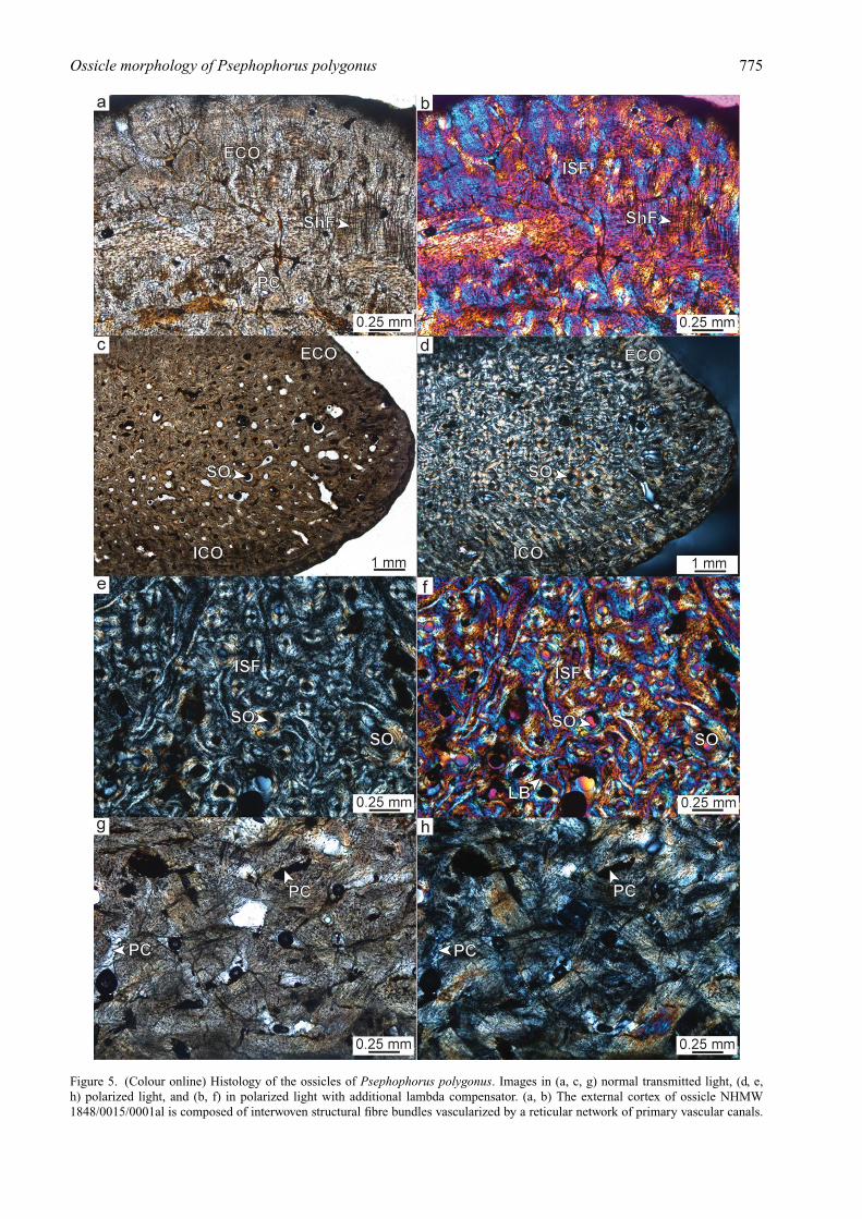
External cortex. The external surface of the ossiclesis even, disturbed only by the insertion of smallforamina. The cortex itself is composed of interwovenstructural fibre bundles, in which the bundles aremostly homogeneous in size and diameter, with aslight dominance of fibre bundles that extend at highangles to the external bone surface (Fig. 5a, b). Thebone tissue is vascularized by an extensive networkof primary vascular canals, scattered primary osteonsand few secondary osteons. The amount of secondaryosteons increases towards the internal cancellous bone.Bone cell lacunae are round or slightly flattenedand elongated and appear scattered or clustered. Inthe lamellar bone of the secondary osteons, the celllacunae are flattened and elongated and are alignedalong the secondary bone lamellae. The ridge ossicleshows patches in the cortex, in which the typical
interwoven structure is abruptly offset by a moreorganized lamellar structure reminiscent of parallel-fibred bone tissue. Although growth marks are visiblein all three specimens, it is only in these more organizedareas of the external cortex of the ridge ossicle thatthey are most pronounced (although not continuous). InNHMW 1848/0015/0001al, the external cortical boneis continuous all along the rounded margin, gradinginto the internal cortical tissue at the visceral side ofthe ossicle (Fig. 5c, d).
Cancellous bone and internal cortex. The interiorcore of the ossicles is more compact (Fig. 5e, f) inthe flat polygonal ossicles than in the ridge ossicle,with larger and longer trabeculae being completelyabsent. Even the ridge ossicle shows only a fewtrabeculae. The tissue is predominantly still primaryand of a coarse cancellous type. Remodelling of thebone appears to be restricted to scattered secondaryosteons and few larger erosion cavities. Internally, thetissue increasingly becomes coarsely fibrous, with theinternal cortex consisting completely of a meshwork ofcoarse fibre bundles (Fig. 5g, h). Although the coarsebundles form a three-dimensional meshwork, the wholetissue is dominated by fibre bundles extending from thegrowth centre of the ossicle (at the plate centre closeto the external bone surface) towards the internal bonesurface in a fan-shaped pattern. Growth marks are alsofaintly seen in the internal cortical bone.
Sutures. The sutural zones are narrow with short bonyprotrusions and sockets. The bone tissue adjacent to thesutures is similar to the tissue described for the externalcortex, but shows more signs of remodelling. Sharpey’sfibres that extend perpendicular to the margin dominatethe sutural bone tissue.
4.c.2. Dermochelys coriacea
All sampled ossicles of D. coriacea are similar in bonemicrostructure, so they will be described in one section(Fig. 6), with peculiarities being pointed out if applic-able. The ridge ossicles of the presumably juvenile tosubadult specimen (PIMUZ A/III 1288) show a diploestructure, with external and visceral cortices framing aninterior cancellous centre. In some of the flat polygonalossicles, the interior core is often restricted to a thinarea of vascular primary bone. Noteworthy, one of theridge ossicles of the juvenile to subadult specimenrevealed a small internal (visceral) ‘keel’, caused bya local thickening of the cancellous bone core belowthe external ridge. The bony ossicle of the young adultspecimen (QM J81592) comprises a layer of externalcortical bone and an internal layer of cancellous bone,with an internal cortical layer of compact bone beingalmost absent. The ossicle of the adult specimen (QMJ73979), marginally thicker than that of the young adultspecimen, is composed of two layers only, a morecompact external layer and a vascular internal layer;an internal cortical layer is not visible.
Ossicle morphology of Psephophorus polygonus 775
Figure 5. (Colour online) Histology of the ossicles of Psephophorus polygonus. Images in (a, c, g) normal transmitted light, (d, e,h) polarized light, and (b, f) in polarized light with additional lambda compensator. (a, b) The external cortex of ossicle NHMW1848/0015/0001al is composed of interwoven structural fibre bundles vascularized by a reticular network of primary vascular canals.

776 M . D E L F I N O A N D OT H E R S
External cortex. In all specimens (e.g. Fig. 6a, e, f),the external cortex consists of interwoven structuralfibre bundles. The bone tissue is highly vascularizedwith an extensive reticular network of primary vascularcanals and scattered primary osteons. The external bonesurface is rough and scalloped with deep foraminainserting into the cortical bone (Fig. 6a, e, g). Especiallyin the older individuals, Sharpey’s fibres are locallyfound as thick and dark fibrous strands inserting athigh angles (Fig. 6g) into the external-most layers ofthe cortex, whereas they are less conspicuous in thejuvenile to subadult specimen. In the ridge ossicle ofthe adult individual, the cortical bone itself resemblesonly a spongy meshwork of trabecular struts, withthe large vascular intertrabecular cavities being filledwith brown adipose tissue. In the juvenile to subadultspecimen, the external-most cortical layer usually doesnot show bone cell lacunae, followed by a second layerin which the cell lacunae are small and round andusually devoid of canaliculi. Only the deeper corticallayers then also house larger flattened osteocyte lacunaewith canaliculi. This stratification of the cortex wasnot observed in the two older specimens. Furthermore,growth marks (i.e. lines of arrested growth) were notobserved in any of the samples.
Cancellous bone. The internal cancellous bone com-prises trabeculae and scattered secondary osteons(Fig. 6b–d, g). Interstitial primary bone is still presentwithin the lamellar bone walls of many trabecularstruts and trabecular branching areas. Other trabeculae,especially in the ridge ossicle, are at various stagesof remodelling, resulting in secondary trabeculaecomposed solely of lamellar bone. Bone cell lacunaein the lamellar bone are flattened and elongated, whilethey are more round and plump in interstitial areas.The intertrabecular cavities are also filled with brownadipose tissue. Where the adipose tissue desiccated,small empty vascular spaces appeared.
Internal cortex. The ossicles of the presumably juvenileto subadult specimen all showed a thin internal cortex(Fig. 6b), with the cortical tissue being composedof parallel-fibred bone. The tissue is vascularizedby primary vascular canals extending predominantlyanteroposteriorly (parallel to the trend of the ridge).Abundant coarse Sharpey’s fibres insert into theinternal cortex perpendicularly or at moderate angles,especially in the flanks of the ridge ossicles. In theyoung adult specimen (QM J81592), only remnantsof this internal compact cortical layer are still visible,with other parts being already completely cancellous
(Fig. 6c, d). Unfortunately, most of the structural detailsin this specimen are obscured here because of the highfat content of the adjacent deeper dermal tissue. Thecortex is completely remodelled in the adult ossicle(Fig. 6h).
Sutures. The internal structure of sutural areas couldonly be studied in the purported juvenile to subadultspecimen (PIMUZ A/III 1288). As indicated alreadyby the irregular shape of the ossicles in external view,the bones show wide sutural areas in cross-section withlong protrusions and sockets. The bone tissue again issimilar to that of the external cortex, with Sharpey’sfibres inserting perpendicularly into the bone tissue.
5. Discussion
Wood et al. (1996) identified about 20 charactersfor the analysis of the phylogeny of dermochelyidturtles. The evaluation of some of them, such asthe number of ossicles between ridges, is restrictedto the fossils where portions of shell (with ossicles)are preserved in connection (i.e. in vivo). Conversely,other characters can be better evaluated on isolatedossicles. The type locality materials of P. polygonuslisted and described herein have been overlookedfor several decades by researchers who concentratedonly on the in vivo ossicles preserved on the slabdisplayed at the Naturhistorisches Museum Wien,NHMW 2011/0330/0001 (among others Wood et al.1996; Chesi et al. 2007). On the basis of the isolatedossicles here described, as well as those of theneotype, it is possible to re-evaluate the morphologicaldifferences between the extinct P. polygonus and theextant D. coriacea.
5.a. Macro-morphological differences betweenPsephophorus polygonus and Dermochelys coriacea
The main differences can be summarized as follows.According to Wood et al. (1996), P. polygonus
has ridge ossicles that are viscerally flat, whereasthe ridge ossicles of D. coriacea are characterizedby a concave visceral surface (see Wood et al.1996, fig. 19c, e). Such morphology has been onlypartly confirmed by our material. Some ridge ossiclesof P. polygonus are actually viscerally flat, unlikethose of D. coriacea (compare Fig. 1c, d, f, i withFig. 2a, c, e, g). However, two isolated ridge ossicles(MTM V 61.1216/d and NHMW 2011/0151/001) arecharacterized by a distinct visceral concavity (Fig. 1l,n). Even if the viscerally concave ossicles clearly
Sharpey’s fibres point perpendicular to the external bone surface. (c, d) Rounded margin of ossicle NHMW 1848/0015/0001al. Notehow the external cortical bone continues onward to the visceral side of the ossicle. (e, f) Interior core area of ossicle NHMW1848/0015/0001al showing interwoven structural fibre bundles and numerous secondary osteons. (g, h) Close-up of the irregularmeshwork of mainly longitudinally sectioned coarse fibre bundles of the internal cortex of ossicle NHMW 1848/0015/0001am. Thetissue is vascularized by simple primary vascular canals. Abbreviations: ECO – external cortex; ICO – internal cortex; ISF – interwovenstructural fibre bundles; LB – lamellar bone; PC – primary vascular canals; ShF – Sharpey’s fibres; SO – secondary osteons.

Ossicle morphology of Psephophorus polygonus 777
Figure 6. (Colour online) Histology of the ossicles of Dermochelys coriacea. Images (a–c, e, g) in normal transmitted light and (d, f,h) in polarized light. (a) External cortex of the flank of smaller ridge ossicle of PIMUZ A/III 1288. Note extensive vascular system andscalloped external bone surface. (b) Interior core area and internal cortical bone of smaller ridge ossicle of PIMUZ A/III 1288. (c, d)
778 M . D E L F I N O A N D OT H E R S
do not belong to D. coriacea because, among otherfactors, of their thickness, such polymorphism requiresa reconsideration of the phylogenetic analysis of thedermochelyid taxa performed by Wood et al. (1996),which was based on very few characters, some of whichare correlated with each other or are phylogeneticallyuninformative. It is worth noting that an unnamedEocene dermochelyid from Alabama (USNM 23699)has ossicles similarly concave viscerally, but (at leastfrom the only figured specimen; Wood et al. 1996,fig. 14) it is characterized by a nearly uniform thicknessof the ossicles. In P. polygonus, the thickness of theridge ossicles significantly decreases laterally.
Although the carapaces of both P. polygonus and D.coriacea have ridges that are not straight but undulated,the nature of the undulations is quite different. Thepeaks and valleys on the ridges of P. polygonuscorrespond to the sutures between contiguous ossiclesso that each convexity is formed by two contiguousossicles with its apex – the peak – corresponding tothe suture between them. The asymmetrical thickeningof the isolated ossicles described above (see Fig. 1g,j) obviously contributes to the undulating profile.Conversely, in D. coriacea there is no significant asym-metrical thickening and each peak usually correspondsto a keel on the external surface of a single osteoderm.As in P. polygonus, the bottom of a valley correspondsto the suture between two ossicles. The expression ofthe keel on the ridge ossicles of D. coriacea can be quitevariable, from non-existent as shown in Figure 2a, b,to distinctly developed, as in the case of the ossicleshown in Figure 2g, h. A particular case of undulationis the one occurring in the posterior region of thecarapace (Fig. 2i–k), where the external edges of theridge ossicles do not have any sort of keel, but are somarkedly convex that the posterior edges of the ossiclesdo not host any sutures (which are limited to the anteriorand ventral edges).
All the ossicles, either from the ridge or from the areabetween the ridges, are proportionally much thicker inP. polygonus than in D. coriacea (compare for exampleFig.1f, n, s, v with Fig. 2a, c, e, g). Probably because ofthe difference in thickness and (considering the density)weight, the sutures of the ossicles of P. polygonus arenot as developed as those of D. coriacea (compare forexample Fig.1e, f with Fig. 2e, f), which appears to needrelatively deeply interdigitating sutures to keep the thinand light ossicles in firm contact with each other. Theossicles of P. polygonus are also not as variable in sizeas those of D. coriacea, which shows a remarkabledifference between the large ridge ossicles and themuch smaller flat ossicles (compare Fig. 1 with Fig. 3).
Some of the differences in carapace morphologyobserved between P. polygonus and D. coriacea mayrelate to diving capability. D. coriacea routinely diveto depths of around 300 m (Houghton et al. 2008),but have been recorded going as deep as 1280 m(Doyle et al. 2008). It is very likely that a rigidshell would collapse or fracture during such deepdives, but the carapace of D. coriacea is thought tobe able to deform under pressure, returning to itsoriginal shape upon ascent (Spotila, 2004). If suchdeformation does occur, this flexibility appears to bethe result, at least in part, of the reduction of the‘thecal’ elements and the configuration and structureof the osteoderms and ossicles of which the secondary(‘epithecal’) carapace is comprised. The osteodermsand ossicles are much thinner and internally morevascularized within the external cortex and cancellouscore areas, particularly in mature individuals, relativeto the fused ‘thecal’ plates that make up the carapaceand plastron of other extant sea turtles, and their smallsize and interdigitating sutural connections, along withtheir lack of direct connectivity with the ribs and girdleelements, appear to facilitate this flexibility.
The thicker, denser and proportionately largerosteoderms and ossicles of P. polygonus, with theirstraighter sutural margins, are very likely to haveresulted in a more rigid carapace than in D. coriacea.Assuming that diving ability in D. coriacea correlatesin part with the size, shape, thickness and configurationof the carapace osteoderms and ossicles, it seems likelythat, while probably a capable diver relative to extantsea turtles, P. polygonus would not have been ableto reach the depths that D. coriacea can. As initiallyobserved by Wood et al. (1996), within the variousclades of Dermochelyidae there is a clear evolutionarytrend for a decrease in the thickness, size and densityof the osteoderms and ossicles that compose thesecondary (‘epithecal’) carapace. Our observations ofthe osteoderms and ossicles of P. polygonus supportthis trend, and indicate that many of the morphologicalchanges that have occurred within the Dermochelyidaerelate to an improved capacity for deep dives.
5.b. Histological properties of Psephophorus polygonus andDermochelys coriacea
All ossicles of P. polygonus shared similar histologicalstructures (e.g. diploe structure, growth marks in theexternal cortex, coarse fibrous meshwork of the internalcortex) although there were also aspects that appearto be linked to the overall shape of the ossicles (flatv. ridge ossicles). As such, the abrupt change from
Ossicle of young adult specimen (QM J81592) still embedded within the integument. Note remnant of internal cortex of the ossicle,as well as the soft tissue structure of the dermis. (e, f) Close-up of the external cortex of the ossicle of QM J81592. (g) Close-upof the external cortex and strongly remodelled interior core area of the ossicle of the adult specimen (QM J73979). (h) Close-upof the strongly remodelled internal area of the ossicle of the adult specimen (QM J73979) leading to complete absence of compactbone. Abbreviations: C – epidermal cuticle; CB – cancellous bone; D – dermis, ECO – external cortex; ICO – internal cortex; ISF –interwoven structural fibre bundles; LB – lamellar bone; PC – primary vascular canals; ShF – Sharpey’s fibres; TR – trabeculae.
Ossicle morphology of Psephophorus polygonus 779
interwoven to more parallel-fibred organization in theexternal cortex was only visible at the flanks of theridge ossicle.
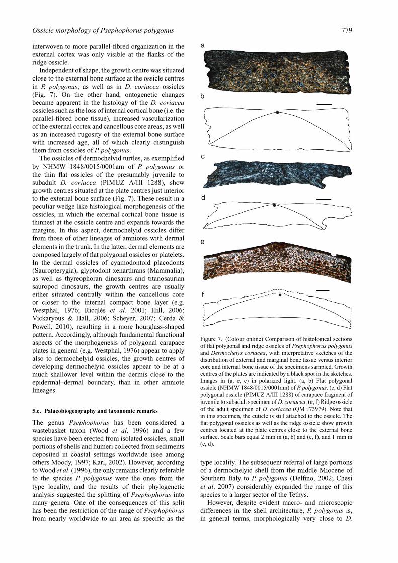
Independent of shape, the growth centre was situatedclose to the external bone surface at the ossicle centresin P. polygonus, as well as in D. coriacea ossicles(Fig. 7). On the other hand, ontogenetic changesbecame apparent in the histology of the D. coriaceaossicles such as the loss of internal cortical bone (i.e. theparallel-fibred bone tissue), increased vascularizationof the external cortex and cancellous core areas, as wellas an increased rugosity of the external bone surfacewith increased age, all of which clearly distinguishthem from ossicles of P. polygonus.
The ossicles of dermochelyid turtles, as exemplifiedby NHMW 1848/0015/0001am of P. polygonus orthe thin flat ossicles of the presumably juvenile tosubadult D. coriacea (PIMUZ A/III 1288), showgrowth centres situated at the plate centres just interiorto the external bone surface (Fig. 7). These result in apeculiar wedge-like histological morphogenesis of theossicles, in which the external cortical bone tissue isthinnest at the ossicle centre and expands towards themargins. In this aspect, dermochelyid ossicles differfrom those of other lineages of amniotes with dermalelements in the trunk. In the latter, dermal elements arecomposed largely of flat polygonal ossicles or platelets.In the dermal ossicles of cyamodontoid placodonts(Sauropterygia), glyptodont xenarthrans (Mammalia),as well as thyreophoran dinosaurs and titanosauriansauropod dinosaurs, the growth centres are usuallyeither situated centrally within the cancellous coreor closer to the internal compact bone layer (e.g.Westphal, 1976; Ricqlès et al. 2001; Hill, 2006;Vickaryous & Hall, 2006; Scheyer, 2007; Cerda &Powell, 2010), resulting in a more hourglass-shapedpattern. Accordingly, although fundamental functionalaspects of the morphogenesis of polygonal carapaceplates in general (e.g. Westphal, 1976) appear to applyalso to dermochelyid ossicles, the growth centres ofdeveloping dermochelyid ossicles appear to lie at amuch shallower level within the dermis close to theepidermal–dermal boundary, than in other amniotelineages.
5.c. Palaeobiogeography and taxonomic remarks
The genus Psephophorus has been considered awastebasket taxon (Wood et al. 1996) and a fewspecies have been erected from isolated ossicles, smallportions of shells and humeri collected from sedimentsdeposited in coastal settings worldwide (see amongothers Moody, 1997; Karl, 2002). However, accordingto Wood et al. (1996), the only remains clearly referableto the species P. polygonus were the ones from thetype locality, and the results of their phylogeneticanalysis suggested the splitting of Psephophorus intomany genera. One of the consequences of this splithas been the restriction of the range of Psephophorusfrom nearly worldwide to an area as specific as the
Figure 7. (Colour online) Comparison of histological sectionsof flat polygonal and ridge ossicles of Psephophorus polygonusand Dermochelys coriacea, with interpretative sketches of thedistribution of external and marginal bone tissue versus interiorcore and internal bone tissue of the specimens sampled. Growthcentres of the plates are indicated by a black spot in the sketches.Images in (a, c, e) in polarized light. (a, b) Flat polygonalossicle (NHMW 1848/0015/0001am) of P. polygonus. (c, d) Flatpolygonal ossicle (PIMUZ A/III 1288) of carapace fragment ofjuvenile to subadult specimen of D. coriacea. (e, f) Ridge ossicleof the adult specimen of D. coriacea (QM J73979). Note thatin this specimen, the cuticle is still attached to the ossicle. Theflat polygonal ossicles as well as the ridge ossicle show growthcentres located at the plate centres close to the external bonesurface. Scale bars equal 2 mm in (a, b) and (e, f), and 1 mm in(c, d).
type locality. The subsequent referral of large portionsof a dermochelyid shell from the middle Miocene ofSouthern Italy to P. polygonus (Delfino, 2002; Chesiet al. 2007) considerably expanded the range of thisspecies to a larger sector of the Tethys.
However, despite evident macro- and microscopicdifferences in the shell architecture, P. polygonus is,in general terms, morphologically very close to D.

780 M . D E L F I N O A N D OT H E R S
coriacea (but we note that they are both significantlydifferent from all the other marine turtles). It is thustempting to assume that, perhaps with the exceptionof a capacity for extremely deep dives, the ecology ofP. polygonus was similar to that of D. coriacea, whichis the most cosmopolitan extant turtle species, rangingthroughout the Pacific, Atlantic and Indian oceans (e.g.Pritchard, 1980), occurring in cool waters up to 69◦
north and down to 47◦ south (Frazier, Gramentz &Fritz, 2005). Although the shell of P. polygonus wasmore massive than that of D. coriacea (which hasthin ossicles without the internal cortex), the overallmorphology is so similar that it seems likely thatP. polygonus was similarly far ranging, such that itsremains may one day turn up outside of the Tethys.Even if all the Psephophorus fossils known so far comefrom sediments deposited in coastal settings (thereforewith a relative high energy that does not help theundisturbed preservation of large carcasses), this doesnot necessarily mean that Psephophorus inhabited onlycoastal waters. Despite the fact that Dermochelys isclearly a true pelagic turtle, its fossil record is virtuallynon-existent (Lapparent de Broin, 2001), being limitedto very few archaeological localities (Frazier, 2005; M.D. unpub. obs).
Moreover, the large size of Psephophorus, which wasat least comparable to that of extant Dermochelys (seeChesi et al. 2007), could demonstrate it was a gigan-totherm. Given that the capacity for gigantothermy isalso a key factor behind the worldwide distributionof Dermochelys (in the Dermochelyidae it can betraced back at least to the Middle Eocene; Albrightet al. 2003), it further supports our supposition that P.polygonus may have had a much broader geographicdistribution than its present fossils indicate.
Future revisions of Miocene Psephophorus-likefossils, as well the collection of new materials, willhelp in clarifying these issues. In particular, it should bepossible to test if ‘Psephophorus’ calvertensis Palmer,1909 from the middle Miocene Calvert Formation ofMaryland and ‘Psephophorus’ californiensis (Gilmore,1937) from the middle Miocene Temblor Formationof California, are not only conspecific, as recentlysuggested by Lynch & Parham (2003), but also if theyare junior synonyms of P. polygonus. If so, the rangeof P. polygonus would encompass both the Atlantic andthe Pacific oceans, mirroring the distribution of theextant D. coriacea.
According to Wood et al. (1996), ‘P.’ calvertensisdiffers from P. polygonus in only two characters: thecarapacial ridges are also expressed on the visceralsurface of the ossicles (the visceral surface of the ridgeossicles is concave; character 10) and the ridges aretectiform in cross-section (character 11). However, asshown in Figure 1n, the ossicles of P. polygonus canalso have at the same time a concave visceral surfaceand a tectiform shape in cross-section. As such, ‘P.’calvertensis and P. polygonus could be synonymous,something that should be verified in the future by directrevision of the material in question.
Acknowledgements. Thanks to H. Furrer (PIMUZ), M.Gasparik (HMNH), M. Harzhauser (NHMW) and U. Göhlich(NHMW) for assisting us while studying the fossil materials,for providing specimens for destructive sampling andrelevant information about the fossil remains and theirorigin. U. Göhlich also discussed the chronological allocationof the fossil material. L. Kordos (Geological Institute,Budapest) helped in solving the toponymic synonymy. B.Villier (Torino) provided technical assistance for somephotographs. A. Amey and P. Couper (Queensland Museum)are to be thanked for helping with the collection of QMJ73979, and permitted the sampling of QM J81592 and QMJ58751 for histological work. This work was supported bythe Swiss National Science Foundation (T.M.S., Grant no.31003A_127053/1), the Spanish Ministerio de Economia yCompetitividad (CGL2011-28681), the Italian MIUR PRIN2009MSSS9L_002 (to G. Pavia, Torino) and the SynthesysProgramme (M.D., AT-TAF-1281 and HU-TAF-1894). M.Delfino’s research was originally developed at Università diFirenze, thanks to the support of MIUR PRIN (to E. Abbate)and Fondi di Ateneo (to L. Rook).
References
ABEL, O. 1919. Die Stämme der Wirbeltiere. Berlin undLeipzig, 914 pp.
ALBRIGHT, L. B. III, WOODBURNE, M. O., CASE, J. A. &CHANEY, D. S. 2003. A leatherback sea turtle from theEocene of Antarctica: implication for the antiquity ofgigantothermy in Dermochelydae. Journal of VertebratePaleontology 23, 945–9.
ANDREWS, C. A. 1919. A description of a new species ofzeuglodont and of leathery turtle from the Eocene ofSouthern Nigeria. Proceedings of the Zoological Society,London 18, 309–19.
BARÁTH, I., NAGY, A. & KOVÁC, M. 1994. Sandberskévrstvy–vrchnobádenské marginálne sedimentyvýchodného okraja Viedenskej panvy. GeologickePráce 99, 59–99.
BATSCH, G. C. 1788. Versuch einer Anleitung, zur Kenntnisund Geschichte der Thiere und mineralien. Akademis-che Buchhandlung, 528 pp.
BEVER, G. S. & JOYCE, W. G. 2005. Dermochelyidae:Lederschildkröten. In Handbuch der Reptilien und Am-phibien Europas. Band 3/IIIB: Schildkröten (Testudines)II (Cheloniidae, Dermochelyidae, Fossile SchildkrötenEuropas) (ed. U. Fritz), pp. 235–48. Aula-Verlag.
BRONGERSMA, L. D. 1969. Miscellaneous notes on turtles,II A-B. Proceedings of the Koninklijke NederlandseAkademie van Wetenschappen, Serie C 72, 76–102.
CERDA, I. A. & POWELL, J. E. 2010. Dermal armorhistology of Saltasaurus loricatus, an Upper Cretaceoussauropod dinosaur from Northwest Argentina. ActaPalaeontologica Polonica 55, 389–98.
CHESI, F., DELFINO, M., VAROLA, A. & ROOK, L. 2007. Fossilsea turtles (Chelonii, Dermochelyidae and Cheloniidae)from the Miocene of Pietra Leccese (Late Burdigalian –Early Messinian) of Southern Italy. Geodiversitas 29,321–33.
DANILOV, I. G. 2005. Die fossilen Schildkröten Europas.In Handbuch der Reptilien und Amphibien Europas.Band 3/IIIB: Schildkröten (Testudines) II (Cheloniidae,Dermochelyidae, Fossile Schildkröten Europas) (ed. U.Fritz), pp. 329–448. Aula-Verlag.
DELFINO, M. 2002. Erpetofaune Italiane del Neogene e delQuaternario. Ph.D. thesis, Modena & Reggio EmiliaUniversity, Modena, Italy. Published thesis.

Ossicle morphology of Psephophorus polygonus 781
DERANIYAGALA, P. E. P. 1930. Testudinate evolution.Proceedings of the Zoological Society, London 68,1057–70.
DERANIYAGALA, P. E. P. 1939. The Tetrapod Vertebrates ofCeylon. Colombo Museum, 412 pp.
DOYLE, T. K., HOUGHTON, J. D. R., O’SÚILLEABHÁIN, P.F., HOBSON, V. J., MARNELL, F., DAVENPORT, J. &HAYS, G. C. 2008. Leatherback turtles satellite taggedin European waters. Endangered Species Research 4,23–31.
DUTTON, P. H., BOWEN, B. W., OWENS, D. W., BARRAGAN,A. & DAVIS, S. K. 1999. Global phylogeography of theleatherback turtle (Dermochelys coriacea). Journal ofZoology 248, 397–409.
FITZINGER, L. 1843. Systema Reptilium. Fasciculus primus.Amblyglossae. Braumüller & Seidel, 106 pp. + X pp.
FRAZIER, J. 2005. Marine turtles – the ultimate tool kit: areview of worked bones of marine turtles. MuinasajaTeadus 15, 359–82.
FRAZIER, J., GRAMENTZ, D. & FRITZ, U. 2005. DermochelysBlainville, 1816 – Lederschildkröten. In Handbuchder Reptilien und Amphibien Europas. Band 3/IIIB:Schildkröten (Testudines) II (Cheloniidae, Dermoche-lyidae, Fossile Schildkröten Europas) (ed. U. Fritz),pp. 249–328. Aula-Verlag.
GEMEL, R. & RAUSCHER, K. 2000. Fossile Schildkröten ausÖsterreich (Reptilia, Testudines). Stapfia 69, 63–86.
GERVAIS, M. P. 1872. Ostéologie du Sphargis Luth (Sphargiscoriacea). Nouvelles Archives du Muséum d’Histoirenaturelle de Paris 8(2), 199–228.
GILMORE, C. W. 1937. A new marine turtle from the Mioceneof California. Proceedings of the California Academy ofSciences, 4th series 23(10), 171–4.
HAUER, V. F. 1868. Fossilien von Metmach bei Ried (Ober-Oesterreich). Verhandlungen der Geologischen Reich-sanstalt (Kaiserlich königliche Geologische Reichsan-stalt), p. 387.
HAUER, V. F. 1870. Psephophorus polygonus aus demSandstein von Neudörfl. Verhandlungen der Geologis-chen Reichsanstalt (Kaiserlich königliche GeologischeReichsanstalt), p. 342.
HAY, O. P. 1898. On Protostega, the systematic positionof Dermochelys, and the morphogeny of the cheloniancarapace and plastron. The American Naturalist 32,929–48.
HILL, R. V. 2006. Comparative anatomy and histology ofxenarthran osteoderms. Journal of Morphology 267,1441–60.
HIRAYAMA, R. 1997. Distribution and diversity of Cretaceouschelonioids. In Ancient Marine Reptiles (eds J. M.Callaway & E. L. Nicholls), pp. 225–41. AcademicPress.
HIRAYAMA, R. & CHITOKU, T. 1996. Family Dermochelyidae(superfamily Chelonioidea) from the Upper Cretaceousof North Japan. Transactions and Proceedings of thePalaeontological Society of Japan, New Series 184, 597–622.
HOLEC, P. & EMRY, R. J. 2003. Another molar of the Miocenehominid Griphopithecus suessi from the type localityat Sandberg, Slovakia. In Vertebrate Fossils and theirContext: Contributions in Honor of Richard H. Tedford.Bulletin of the American Museum of Natural History279, 625–31.
HOUGHTON, J. D. R., DOYLE, T. K., DAVENPORT, J., WILSON,R. P. & HAYS, G. C. 2008. The role of infrequent andextraordinary deep dives in leatherback turtles (Der-mochelys coriacea). Journal of Experimental Biology211, 2566–75.
KARL, H.-V. 2002. Übersicht über die fossilen marinenSchildkrötenfamilien Zentraleuropas (Reptilia, Tes-tudines). Mauritania 18(2), 171–202.
KARL, H.-V. & LINDOW, B. E. K. 2010. Eocene leatherbackturtle material of the genus Egyptemys (Testudines:Dermochelyoidea) from Denmark. Studia GeologicaSalmanticensia 46, 55–63.
KOVÁC, M., BARÁTH, I., HARZHAUSER, M., HLAVATÝ, I. &HUDÁCKOVÁ, N. 2004. Miocene depositional systemsand sequence stratigraphy of the Vienna Basin. CourierForschungsinstitut Senckenberg 246, 187–212.
LAPPARENT DE BROIN, F. DE 2001. The European turtle faunafrom the Triassic to the Present. Dumerilia 4, 155–217.
LYDEKKER, R. 1889. Catalogue of the Fossil Reptilia andAmphibia in the British Museum (Natural History), Part3: Chelonia. London: Longmans & Co., 239 pp.
LYNCH, S. C. & PARHAM, J. F. 2003. The first report ofhard-shelled sea turtles (Cheloniidae sensu lato) fromthe Miocene of California, including a new species(Euclastes hutchisoni) with unusually plesiomorphiccharacters. PaleoBios 23, 21–35.
MEYER, H. VON 1846. [Without title, letter on severalfossil specimens]. Neues Jahrbuch für Mineralogie,Geognosie, Geologie und Petrefaktenkunde 1846, 462–76.
MEYER, H. VON 1847. Mittheilungen an Professor Bronngerichtet. Neues Jahrbuch für Mineralogie, Geognosie,Geologie und Petrefaktenkunde 1847, 572–81.
MŁYNARSKI, M. 1966. Die fossilen Schildkröten in denungarischen Sammlungen. Acta Zoologica Cracoviensia11(8), 223–88.
MOODY, R. T. J. 1993. Cretaceous-Tertiary marine turtlesof Northwest Europe. Revue de Paléobiologie 7, 151–60.
MOODY, R. T. J. 1997. The paleogeography of marine andcoastal turtles of the north Atlantic and Trans-Saharanregions. In Ancient Marine Reptiles (eds J. M. Callaway& E. L. Nicholls), pp. 259–78. Academic Press.
NIELSEN, E. 1959. Eocene Turtles from Denmark. Med-delelser fra Dansk Geologisk Forening 14(2), 96–114.
NIELSEN, E. 1963. On the post-cranial skeleton of Eosphargisbreineri Nielsen. Meddelelser fra Dansk GeologiskForening 15, 281–313.
PALMER, W. 1909. Description of a new species of leather-back turtle from the Miocene of Maryland. Proceedingof the United States National Museum 36, 369–73.
PRITCHARD, P. C. H. 1980. Dermochelys coriacea. Catalogueof American Amphibians and Reptiles 238, 1–4.
RICQLÈS, A. DE, PEREDA SUBERBIOLA, X., GASPARINI, Z.& OLIVERO, E. 2001. Histology of dermal ossificationsin an ankylosaurian dinosaur from the Late Cretaceousof Antarctica. Asociación Paleontológica Argentina(Publicación Especial) 7, 171–4.
RIEPPEL, O. 2001. Turtles as hopeful monsters. BioEssays23, 987–91.
SABOL, M. & HOLEC, P. 2002. Temporal and spatialdistribution of Miocene mammals in the WesternCarpathians (Slovakia). Geologica Carpathica 53, 269–79.
SCHEYER, T. M. 2007. Skeletal histology of the dermal armorof Placodontia: the occurrence of ‘postcranial fibro-cartilaginous bone’ and its developmental implications.Journal of Anatomy 211, 737–53.
SEELEY, H. G. 1880. Note on Psephophorus polygonus, v.Meyer, a new type of chelonian reptile allied to theLeathery Turtle. Quarterly Journal of the GeologicalSociety of London 36, 406–13.
782 Ossicle morphology of Psephophorus polygonus
SPOTILA, J. R. 2004. Sea Turtles: A Complete Guide to theirBiology, Behaviour and Conservation. James HopkinsUniversity Press, 227 pp.
ŠVAGROVSKÝ, J. 1974. Lithofazielle Entwicklung und Mol-luskenfauna des oberen Badeniens (Miozän M4d) indem Gebiet Bratislava-Devínska Nová Ves. ZápadnéKarpaty, Séria Paleontológia 7, 5–204.
ŠVAGROVSKÝ, J. 1978. Faciostratotypus Devínska Nová Ves –Sandberg. In Chronostratigraphie und neostratotypen,Miozän der Zentralen Paratethys, M4 Badenien (eds A.Papp, I. Cicha, J. Seneš & F. Steininger), pp. 188–93.Veda.
SZALAI, T. 1934. Die fossilen Schildkröten Ungarns. FoliaZoologica et Hydrobiologica 7, 97–142.
TONG, H., BUFFETAUT, E., THOMAS, H., ROGER, J.,HALAWANI, M., MEMESH, A. & LEBRET, P. 1999. Anew dermochelyid turtle from the Late Paleocene-EarlyEocene of Saudi Arabia. Comptes Rendus de l’Académiedes Sciences à Paris, Sciences de la terre e des planètes329, 913–19.
VANDELLI, D. 1761. Epistola de Holothurio, et TestudineCoriacea ad Celeberrimum Carolum Linnaeum EquitemNaturae Curiosum Dioscoridem. Conzatti, II + 12 pp.
VERSLUYS, J. 1913. Über die Phylogenie des Panzers derSchildkröten und über die Verwandtschaft der Leder-
schildkröte (Dermochelys coriacea). PaläontologischeZeitschrift 1, 321–47.
VICKARYOUS, M. K. & HALL, B. K. 2006. Osteodermmorphology and development in the nine-banded ar-madillo, Dasypus novemcinctus (Mammalia, Xenarthra,Cingulata). Journal of Morphology 267, 1273–83.
WESTPHAL, F. 1976. The dermal armour of some Triassicplacodont reptiles. In Morphology and Biology ofReptiles. Linnean Society Symposium Series no. 3 (edsA. d. A. Bellairs & C. B. Cox), pp. 31–41. AcademicPress.
WOOD, R. C., JOHNSON-GOVE, J., GAFFNEY, E. S. & MALEY,K. F. 1996. Evolution and phylogeny of leatherbackturtles (Dermochelyidae), with descriptions of newfossil taxa. Chelonian Conservation and Biology 2, 266–86.
ZANGERL, R. 1939. The homology of the shell elements inturtles. Journal of Morphology 65, 383–409.
ZANGERL, R. 1960. The vertebrate fauna of the SelmaFormation of Alabama. Part V: an advanced chel-oniid sea turtle. Fieldiana: Geology Memoirs 3, 279–312.
ZANGERL, R. 1969. The turtle shell. In Biology of the Reptilia.Vol. 1 Morphology A (eds C. Gans, A. d. A. Bellairs &T. S. Parsons), pp. 311–39. Academic Press.