Soleus H-reflex phase-dependent modulation is preserved during stepping within a robotic exoskeleton

Animal Reproduction Science 66 (2001) 209–223
Morphological and ultrastructural changes occurringduring degeneration of goat preantral follicles
preserved in vitro
J.R.V. Silva a,∗, S.N. Báo b, C.M. Lucci b, F.C.A. Carvalho a,E.R. Andrade a, M.A.L. Ferreira a, J.R. Figueiredo a
a Laboratory of Manipulation of Oocytes Enclosed in Preantral Follicles — LAMOFOPA, Faculty of Veterinary,University of Ceará, Fortaleza, CE, Brazil
b Department of Cell Biology, University of Brasilia, Brasilia, DF, Brazil
Received 29 July 2000; received in revised form 14 February 2001; accepted 19 February 2001
Abstract
The present work has investigated the morphological and ultrastructural changes occurring dur-ing degeneration of goat preantral follicles preserved in vitro and showed quantitative data aboutthe distribution of follicular degeneration types in the control and after preservation in coconutwater solution or Braun–Collins solution at different temperatures (4, 20 or 39◦C) and incubationtimes (4, 12 or 24 h). At the slaughterhouse, the pair of ovaries of each animal was divided into 19fragments. One ovarian fragment was immediately fixed (control: Time 0). The other 18 fragmentswere randomly distributed in tubes containing 2 ml of coconut water or Braun–Collins solution at4, 20 or 39◦C and stored for 4, 12 or 24 h. Normal preantral follicles exhibited a healthy oocyte sur-rounded by one or more well-organized layers of granulosa cells. The ooplasm contained numerousrounded or elongated mitochondria with continuous mitochondrial membranes. Golgi complexeswere rare. Both smooth and rough endoplasmic reticulum were observed, either as isolated aggre-gations or complex associations with mitochondria and vesicles. Degenerated preantral follicles inthe control tissue exhibited pycnotic nuclei of the oocyte, vacuolated ooplasm and normal granu-losa cells. This kind of degeneration also predominated significantly (P < 0.05) after preservationat 4◦C. In contrast, after preservation at 20 or 39◦C a significant predominance (P < 0.05) ofpreantral follicles showing a retracted oocyte and swollen granulosa cells was observed. These fol-licles showed large irregularity of the oocyte and nuclear outlines. The ooplasm exhibited moderateproliferation of the endoplasmic reticulum and mitochondria showed disappearance of most of thecristae and damage to the mitochondrial membrane. Some follicles had numerous vacuoles in theooplasm. Granulosa cells were spread and a low density of organelles was observed. The alterations
∗Corresponding author. Tel.: +55-85-299-2752; fax: +55-85-299-2740.E-mail address: roberto [email protected] (J.R.V. Silva).
0378-4320/01/$ – see front matter © 2001 Published by Elsevier Science B.V.PII: S0 3 7 8 -4 3 20 (01 )00102 -6

210 J.R.V. Silva et al. / Animal Reproduction Science 66 (2001) 209–223
in follicular structure progressed with an increase of temperature from 20 to 39◦C as well as withan increase of the incubation time from 4 to 12, or 24 h. In conclusion, the present study shows forthe first time that initial proliferation of the endoplasmic reticulum and damage to mitochondria arethe first signs of degeneration in goat preantral follicles during storage in vitro. © 2001 Publishedby Elsevier Science B.V.
Keywords: Goat; Preantral follicle; Preservation; Ultrastructural; Morphology
1. Introduction
At birth the mammalian ovary contains many thousands of preantral follicles but the vastmajority become atretic during their growth and maturation. As a result relatively few viableoocytes are produced during the reproductive lifespan of the female (Carroll et al., 1990). Thetechniques developed for the isolation (Figueiredo et al., 1993; Lucci et al., 1999), culture(Figueiredo et al., 1994) and cryopreservation (Carroll et al., 1990) of preantral folliclesoffers a means to utilize large numbers of preantral follicles in reproductive programs. Thesetechniques have been successfully applied in laboratory species. For example, mice offspringhave resulted from preantral follicles that were isolated, cryopreserved and cultured in vitro(Carroll et al., 1990).
In farm animals, the donor of the ovarian follicles is usually not housed near the labo-ratory where the research is done. Therefore, preservation of the follicles during transportfrom the field to the laboratory becomes a major concern. Recently, we described the ben-eficial effects of coconut water and Braun–Collins solutions on the preservation of caprinepreantral follicles (Silva et al., 1999, 2000). Coconut water and Collins solutions have beensuccessfully used for semen (Nunes, 1998) and organ (liver: Adam et al., 1996; lung: Fukuseet al., 1996; kidney: Savioz et al., 1996) preservation, respectively. The preservation of pre-antral follicles is of obvious importance for the maintenance of follicular morphology duringtransportation of ovaries to the laboratory, which is necessary to provide healthy oocytes forin vitro growth and maturation. Thus, the need for an understanding of the ultrastructuralevents occurring in preantral follicles during preservation in vitro is important to developsuitable preservation systems as well as to comprehend follicular atresia. Furthermore, theultrastructural features of cattle (Fair et al., 1997), sheep (Tassel and Kennedy, 1980), hu-man (Hertig and Adams, 1967; Oktay et al., 1997), guinea pig (Adams and Hertig, 1964)and goat (Sharma et al., 1994; Lucci et al., 2001) preantral follicles have been describedin several papers. In addition, Tassel and Kennedy (1980) and van den Hurk et al. (1998)showed the ultrastructural features of preantral follicles that underwent degeneration invivo. However, the sequence of events taking place during degeneration in goat preantralfollicles preserved in vitro is presently unknown.
The aims of the present study was to describe the morphological and ultrastructuralchanges occurring during degeneration of goat preantral follicles preserved in vitro and toshow quantitative data about the distribution of follicular degeneration types in the controland after preservation in coconut water or Braun–Collins solutions at different temperaturesand incubation times.
J.R.V. Silva et al. / Animal Reproduction Science 66 (2001) 209–223 211
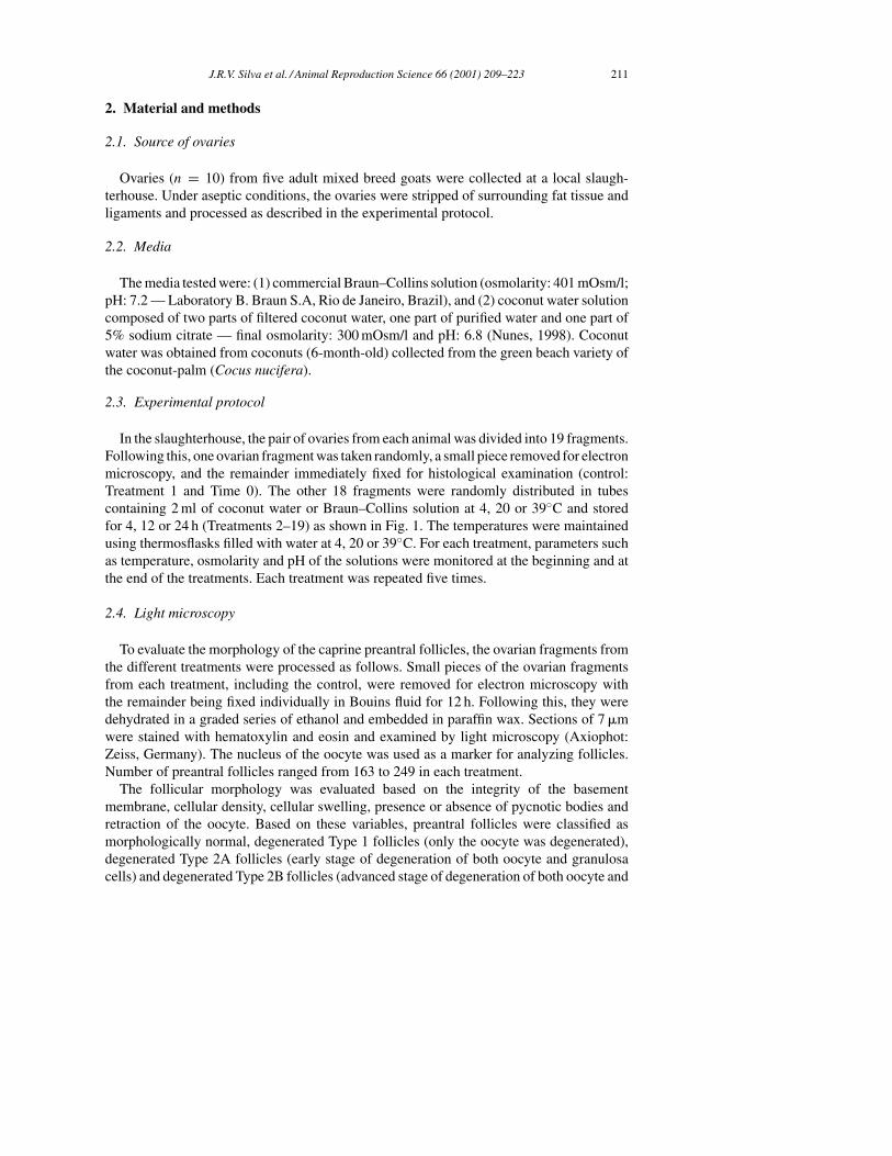
2. Material and methods
2.1. Source of ovaries
Ovaries (n = 10) from five adult mixed breed goats were collected at a local slaugh-terhouse. Under aseptic conditions, the ovaries were stripped of surrounding fat tissue andligaments and processed as described in the experimental protocol.
2.2. Media
The media tested were: (1) commercial Braun–Collins solution (osmolarity: 401 mOsm/l;pH: 7.2 — Laboratory B. Braun S.A, Rio de Janeiro, Brazil), and (2) coconut water solutioncomposed of two parts of filtered coconut water, one part of purified water and one part of5% sodium citrate — final osmolarity: 300 mOsm/l and pH: 6.8 (Nunes, 1998). Coconutwater was obtained from coconuts (6-month-old) collected from the green beach variety ofthe coconut-palm (Cocus nucifera).
2.3. Experimental protocol
In the slaughterhouse, the pair of ovaries from each animal was divided into 19 fragments.Following this, one ovarian fragment was taken randomly, a small piece removed for electronmicroscopy, and the remainder immediately fixed for histological examination (control:Treatment 1 and Time 0). The other 18 fragments were randomly distributed in tubescontaining 2 ml of coconut water or Braun–Collins solution at 4, 20 or 39◦C and storedfor 4, 12 or 24 h (Treatments 2–19) as shown in Fig. 1. The temperatures were maintainedusing thermosflasks filled with water at 4, 20 or 39◦C. For each treatment, parameters suchas temperature, osmolarity and pH of the solutions were monitored at the beginning and atthe end of the treatments. Each treatment was repeated five times.
2.4. Light microscopy
To evaluate the morphology of the caprine preantral follicles, the ovarian fragments fromthe different treatments were processed as follows. Small pieces of the ovarian fragmentsfrom each treatment, including the control, were removed for electron microscopy withthe remainder being fixed individually in Bouins fluid for 12 h. Following this, they weredehydrated in a graded series of ethanol and embedded in paraffin wax. Sections of 7 �mwere stained with hematoxylin and eosin and examined by light microscopy (Axiophot:Zeiss, Germany). The nucleus of the oocyte was used as a marker for analyzing follicles.Number of preantral follicles ranged from 163 to 249 in each treatment.
The follicular morphology was evaluated based on the integrity of the basementmembrane, cellular density, cellular swelling, presence or absence of pycnotic bodies andretraction of the oocyte. Based on these variables, preantral follicles were classified asmorphologically normal, degenerated Type 1 follicles (only the oocyte was degenerated),degenerated Type 2A follicles (early stage of degeneration of both oocyte and granulosacells) and degenerated Type 2B follicles (advanced stage of degeneration of both oocyte and

212 J.R.V. Silva et al. / Animal Reproduction Science 66 (2001) 209–223
Fig. 1. General experimental protocol for preservation of caprine preantral follicles.
granulosa cells). These four classifications were assigned on a basis of degeneration thatoccurred as result of in vitro storage as well as in normal or degenerated follicles observedin the control.
2.5. Transmission electron microscopy
Ultrastructural analysis was performed using preantral follicles considered morpholo-gically normal as well as in degenerated follicles after preservation. Briefly, small piecesof ovarian cortex were fixed in a solution containing 2% paraformaldehyde and 2.5% glu-taraldehyde in 0.1 M sodium cacodylate buffer pH 7.2. After fixation, the specimens wererinsed in buffer and post-fixed in 1% osmium tetroxide, 0.8% potassium ferricyanide and5 mM CaCl2 in 0.1 M sodium cacodylate buffer. Subsequently the samples were dehydratedin acetone and embedded in Spurr. Thin sections (65 nm) were prepared when an oocytenucleus appeared in the semi-thin sections (3 �m). Semi-thin sections were stained withtoluidine blue while thin sections were contrasted with uranyl acetate and lead citrate, andexamined using Jeol 100 C and Zeiss 912 transmission electron microscopes.
2.6. Statistical analysis of data
For each treatment, values of degenerated preantral follicles from five ovarian fragmentswere pooled. The percentages of degenerated follicles were compared using a Chi-squaredtest (Instat for Macintosh). Values were considered statistically significant when P < 0.05.

J.R.V. Silva et al. / Animal Reproduction Science 66 (2001) 209–223 213
3. Results
3.1. Light microscopy
Normal preserved preantral follicles exhibited a healthy spherical or oval oocyte witha large central or eccentrically located nucleus. Granulosa cells, well-organized in layers,without pycnotic nuclei were observed surrounding the oocyte (Fig. 2A). Degenerated Type1 follicles exhibited an oocyte with a pycnotic nucleus and well-organized granulosa cellswithout pycnotic nuclei (Fig. 2B). In degenerated Type 2A follicles a lightly retractedoocyte and swollen granulosa cells were observed (Fig. 2C). Moreover, degenerated Type2B follicles had a retracted oocyte, with or without a pycnotic nucleus, strongly eosinophiliccytoplasm and disorganized, low density swollen granulosa cells (Fig. 2D).
3.2. Ultrastructural aspects of preantral follicles after preservation
Normal preantral follicles exhibited sparse vesicles spread throughout the cytoplasmin all the oocytes. The cytoplasm also contained numerous rounded mitochondria withperipheral cristae and continuous mitochondrial membranes, although there were occasionalelongated forms with parallel cristae. Golgi complexes were rarely observed. Both smooth
Fig. 2. Histological sections of (A) normal, (B) degenerated Type 1, (C) degenerated Type 2A and (D) degeneratedType 2B preantral follicles. O: oocyte; ∗: nucleus; GC: granulosa cells. Bar = 10 �m.

214 J.R.V. Silva et al. / Animal Reproduction Science 66 (2001) 209–223
Fig. 3. Electron micrograph of a normal preantral follicle from the control group. O: oocyte; Nu: nucleus of oocyte;GC: granulosa cells; m: mitochondria; ser: smooth endoplasmic reticulum; v: vesicles. Bar = 5 �m.
and rough endoplasmic reticulum were present, either as isolated aggregations or as complexassociations with mitochondria and vesicles (Fig. 3). Granulosa cells had irregularly-shapednuclei, with a high nuclear-to-cytoplasm ratio. The cytoplasm contained a great numberof elongated mitochondria with lamellar cristae and well-developed rough endoplasmicreticulum (Fig. 3). These features were observed in preantral follicles preserved in coconutwater solution at 4◦C for up to 24 h and in Braun–Collins solution for up to 12 h, as well asin follicles from control.
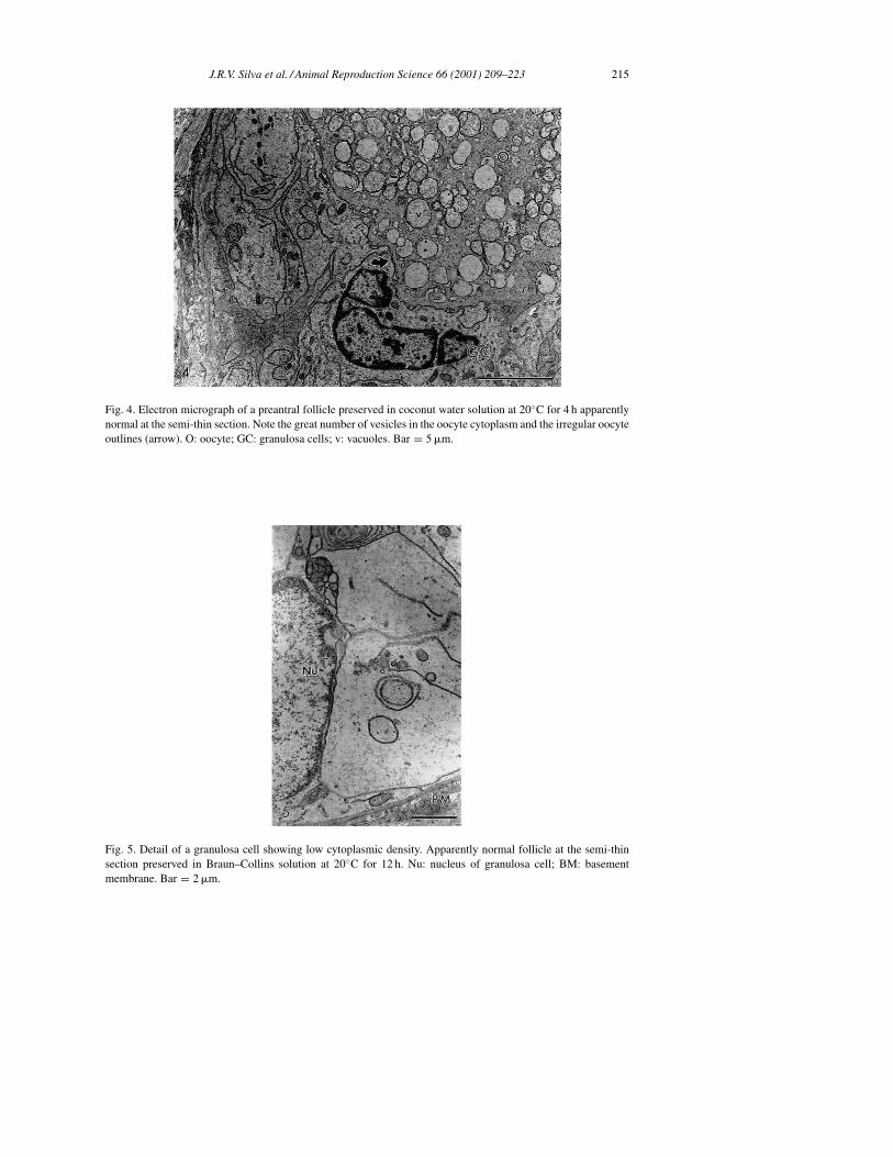
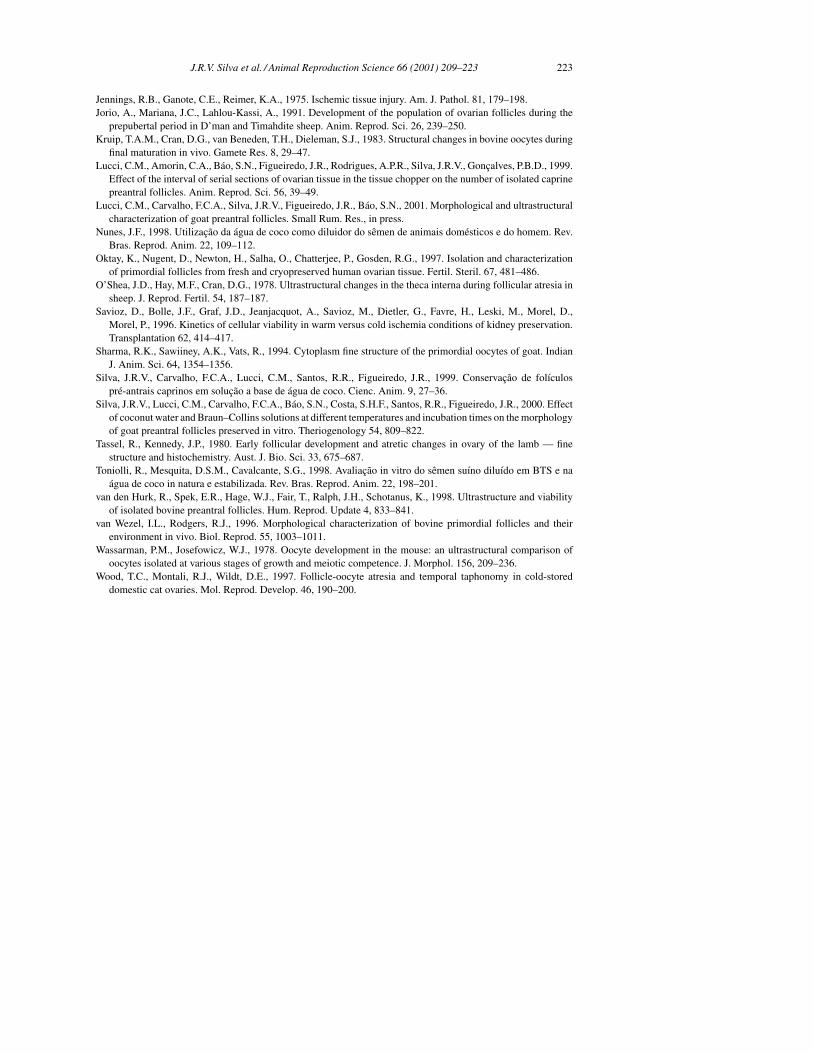
In some treatments follicles seemed to be well preserved according to histological anal-ysis, however, transmission electron microscopy revealed some discreet changes in theirultrastructure (apparently normal at semi-thin sections). These changes suggest an initialdegeneration that was observed in follicles preserved in Braun–Collins solution at 4◦C for24 h, in both solutions at 20◦C at all incubation times, and in coconut water solution at 39◦Cfor 4 h. These follicles showed a large number of vesicles spread throughout the cytoplasmin all oocytes (Fig. 4). In addition, initial signs of proliferation of the endoplasmic reticulumand damage to mitochondrial membranes and cristae were observed. Granulosa cells hada swollen aspect, with a low density of organelles present in their cytoplasm (Fig. 5). Theprogression of these alterations led to follicles with a larger number of vesicles in the oocytecytoplasm, and with damaged mitochondria, without cristae and with flaked content, closelyassociated with the smooth endoplasmic reticulum (Fig. 6). The oocyte nucleus appearedmisshapen and retracted with a wavy membrane.
Degenerated Type 1 follicles showed similar alterations to those observed in apparentlynormal preantral follicles (data not shown). Degenerated Type 2A follicles contained aretracted oocyte and large irregularity of the follicular, oocyte and nuclear outlines (Fig. 7),besides the above described changes. The nuclear membrane was broken in some follicles.Granulosa cells became disorganized and some disappeared leaving a vacated space. Manytimes, the connection between the oocyte and granulosa cells did not exist (data not shown).
Degenerated Type 2B follicles exhibited a progression of the changes described above.The oocyte cytoplasm was extremely vacuolated, with the vacuoles often fusing to producea greater vacated area (Fig. 8). Sometimes the cytoplasm appeared clotted, possibly as a
J.R.V. Silva et al. / Animal Reproduction Science 66 (2001) 209–223 215
Fig. 4. Electron micrograph of a preantral follicle preserved in coconut water solution at 20◦C for 4 h apparentlynormal at the semi-thin section. Note the great number of vesicles in the oocyte cytoplasm and the irregular oocyteoutlines (arrow). O: oocyte; GC: granulosa cells; v: vacuoles. Bar = 5 �m.
Fig. 5. Detail of a granulosa cell showing low cytoplasmic density. Apparently normal follicle at the semi-thinsection preserved in Braun–Collins solution at 20◦C for 12 h. Nu: nucleus of granulosa cell; BM: basementmembrane. Bar = 2 �m.

216 J.R.V. Silva et al. / Animal Reproduction Science 66 (2001) 209–223
Fig. 6. Detail of oocyte cytoplasm showing mitochondria closely associated with smooth endoplasmic reticulum.Note the mitochondrial flaked content (∗). Bar = 1 �m.
Fig. 7. Electron micrograph of a degenerated Type 2A follicle preserved in coconut water solution at 20◦C for 24 h.Note the retracted oocyte and large irregularity of the follicular, oocyte and nuclear outlines and the cytoplasmiclow density of granulosa cells. O: oocyte; Nu: nucleus of oocyte; GC: granulosa cell; ser: smooth endoplasmicreticulum. Bar = 5 �m.

J.R.V. Silva et al. / Animal Reproduction Science 66 (2001) 209–223 217
Fig. 8. Detail of the oocyte cytoplasm of a degenerated Type 2B follicle preserved in coconut water solution at39◦C for 4 h. Note the extreme vacuolization and the great holes present in the cytoplasm without any recognizableorganelle. O: oocyte; GC: granulosa cell. Bar = 2 �m.
result of nuclear content leak (data not shown). At this stage, organelles were generally nolonger recognizable. However, where it was possible to discern, mitochondria appeared tobe closely associated with large formations of swollen membranes of the endoplasmic retic-ulum in a parallel array. In these follicles, some granulosa cells were completely fragmented.In only one follicle was the basement membrane broken (Fig. 9).
3.3. Distribution of follicular degeneration types in the control and other treatments
A total of 3826 preantral follicles were examined by histological analysis. Number of fol-licles ranged from 163 to 249 in each treatment. Morphologically normal follicles were ob-served by histological analysis in all treatments, except after preservation in Braun–Collinssolution at 39◦C for 12 h and in both solutions at 39◦C for 24 h. Of the degenerated folli-cles, in the control as well as after storage in both solutions at 4◦C for 4 h only degeneratedType 1 follicles was observed (Tables 1 and 2). A significant predominance (P < 0.05) ofdegenerated Type 1 follicles when compared with degenerated Types 2A and 2B follicleswas observed after storage at 4◦C for 12 or 24 h, in both solutions. In contrast, a significantpredominance (P < 0.05) of degenerated Types 2A and 2B follicles was observed afterstorage in both solutions at 20 or 39◦C, when compared with degenerated Type 1 follicles(Tables 1 and 2). Compared with degenerated Type 2B follicles, a significant predominance(P < 0.05) of degenerated Type 2A follicles was observed after preservation in coconutwater solution at 20◦C for 12 h or at 39◦C for 4 h (Table 1) and in Braun–Collins solution at20 and 39◦C for 4 h (Table 2). A significantly greater (P < 0.05) percentage of degenerated

218 J.R.V. Silva et al. / Animal Reproduction Science 66 (2001) 209–223
Fig. 9. Electron micrograph of a degenerated Type 2B follicle preserved in Braun–Collins solution at 39◦C for4 h. O: oocyte; Nu: nucleus of oocyte; ser: smooth endoplasmic reticulum; m: mitochondria. Note a completelyfragmented granulosa cell (∗). The arrow marks the broken basement membrane. Bar = 2 �m.
Type 2B follicles was found after storage in coconut water solution at 20 or 39◦C for 24 h(Table 1) and in Braun–Collins solution at 20 or 39◦C for 12 and 24 h (Table 2).
There was no effect of incubation time on the percentage of degenerated Types 1, 2A and2B follicles using both solutions at 4◦C (Tables 1 and 2), except for Braun–Collins solution
Table 1Percentage distribution of degenerated Types 1, 2A and 2B follicles from control and after preservation in differenttreatments in coconut water solutiona,b,c
Treatments Degenerated Type 1follicles (%) (n)
Degenerated Type 2Afollicles (%) (n)
Degenerated Type 2Bfollicles (%) (n)
Control 13.2 a (28/212) 0 b (0/212) 0 b (0/212)4◦C–4 h 7.5 aAG (18/240) 0 bAI (0/240) 0 bH (0/240)4◦C–12 h 8.5 aAG (14/165) 1.8 bAI (3/165) 0 bI (0/165)4◦C–24 h 8.0 aAG (13/163) 0.6 bAH (01/163) 0 bI (0/163)20◦C–4 h 0 bH (0/163) 6.7 aCH (11/163) 6.1 aCG (10/163)20◦C–12 h 0 cH (0/234) 15.8 aBH (37/234) 6.0 bBH (14/234)20◦C–24 h 0 cH (0/249) 36.9 bAG (92/249) 51.4 aAH (128/249)39◦C–4 h 0 cH (0/194) 60.3 aAG (117/194) 3.1 bCG (6/194)39◦C–12 h 0 bH (0/239) 45.2 aBG (108/239) 50.6 aBG (121/239)39◦C–24 h 0 cH (0/172) 37.2 bCG (64/172) 62.8 aAG (108/172)
a Values with different letters (a, b, c) in the same row show significant differences (P < 0.05).b Values within columns with different letters (A, B, C) at the same preservation temperature show significant
differences among incubation times (P < 0.05).c Values within columns with different letters (G, H, I) at the same incubation time show significant differences
among preservation temperatures (P < 0.05).

J.R.V. Silva et al. / Animal Reproduction Science 66 (2001) 209–223 219
Table 2Percentage distribution of degenerated Types 1, 2A and 2B follicles from control and after preservation in differenttreatments in Braun–Collins solutiona,b,c
Treatments Degenerated Type 1follicles (%) (n)
Degenerated Type 2Afollicles (%) (n)
Degenerated Type 2Bfollicles (%) (n)
Control 13.2 a (28/212) 0 b (0/212) 0 b (0/212)4◦C–4 h 12.4 aAG (21/169) 0 bBI (0/169) 0 bBI (0/169)4◦C–12 h 11.4 aAG (24/211) 1.4 bABH (3/211) 1.4 bABI (3/211)4◦C–24 h 9.9 aAG (21/212) 4.7 bAH (10/212) 3.3 bAI (7/212)20◦C–4 h 0.6 cAH (1/177) 40.1 aAH (71/177) 8.5 bCH (15/177)20◦C–12 h 0.4 cAH (1/235) 33.6 bABG (79/235) 59.1 aBH (139/235)20◦C–24 h 0 cAH (0/225) 27.6 bBG (62/225) 72.4 aAH (163/225)39◦C–4 h 0.5 cAH (1/196) 50.0 aAG (98/196) 39.3 bCG (77/196)39◦C–12 h 0 cAH (0/182) 3.3 bBH (6/182) 96.7 aBG (176/182)39◦C–24 h 0 bAH (0/188) 0 bCI (0/188) 100 aAG (188/188)
a Values with different letters (a, b, c) at the same row show significant differences (P < 0.05).b Values within columns with different letters (A, B, C) at the same preservation temperature show significant
differences among incubation times (P < 0.05).c Values within columns with different letters (G, H, I) at the same incubation time show significant differences
among preservation temperatures (P < 0.05).
at 24 h, which increased significantly (P < 0.05) the percentage of degenerated Types 2Aand 2B follicles. In contrast for coconut water solution (Table 1), there was a significant(P < 0.05) increase (20◦C) and decrease (39◦C) of the percentages of degenerated Type2A follicles with the increase of incubation time, respectively. Similar results were obtainedfor Braun–Collins solution (Table 2), except for the storage temperature of 20◦C, at whichsimilar — 12 h (P > 0.05) and significantly lower — 24 h (P < 0.05) percentages ofdegenerated Type 2A follicles were observed when compared with values obtained at 20◦Cfor 4 h. In addition, there was a progressive increase of the percentage of degenerated Type2B follicles with the increase of the incubation times (P < 0.05) in the fragments stored at20 or 39◦C, in both solutions (Tables 1 and 2).
With regard to the effect of temperature, a significant reduction (P < 0.05) of degeneratedType 1 follicles was observed with the increase of temperature from 4 to 20 and 39◦C atall incubation times in both solutions. The percentages of degenerated Types 2A and 2Bfollicles increased significantly (P < 0.05) with the increase of temperature from 4 to 20 and39◦C for both solutions (Tables 1 and 2), except for the similar percentages (P > 0.05) ofdegenerated Type 2A follicles after storage in coconut water solution for 24 h at 20 and 39◦C(Table 1), and for a significant reduction (P < 0.05) of the percentages of degenerated Type2A follicles after preservation in Braun–Collins solution for 12 and 24 h with the increaseof temperature from 20 to 39◦C (Table 2).
4. Discussion
The present study shows for the first time the morphological and ultrastructural changesoccurring during degeneration of goat preantral follicles in ovarian tissue fragmentspreserved in vitro in coconut water or Braun–Collins solutions at different temperaturesand incubation times.

220 J.R.V. Silva et al. / Animal Reproduction Science 66 (2001) 209–223
Ultrastructural analysis of goat normal preantral follicles preserved in vitro showed thatthe ooplasm contains smooth (SER) and rough (SER) endoplasmic reticulum associatedwith round or elongated mitochondria and vesicles. Such a feature is commonly observedin fresh preantral follicles of goats (Sharma et al., 1994; Lucci et al., 2001) and other species(cattle: van Wezel and Rodgers, 1996; Fair et al., 1997; sheep: Cran et al., 1980; Tassel andKennedy, 1980; guinea pig: Adams and Hertig, 1964; human: Hertig and Adams, 1967;and mouse: Wassarman and Josefowicz, 1978). The association of endoplasmic reticulumwith mitochondria and vesicles suggests a mechanism for the transport of substrates into themitochondria. Furthermore, vesicles containing a variety of internal substances also appearto be evidence of transport to the paranuclear metabolic center from the oocyte surface orvice versa (Hertig and Adams, 1967).
The preantral follicles preserved in Braun–Collins solution at 4◦C for 24 h exhibited nu-merous vacuoles in the ooplasm as revealed by ultrastructural analysis. Tassel and Kennedy(1980) and van den Hurk et al. (1998) emphasized that oocytes showing signs of atre-sia in vivo contain numerous vacuoles. The cytoplasmic vacuoles are also a characteris-tic sign of degeneration in granulosa (Hay et al., 1976) and cumulus cells (Assey et al.,1994) during degeneration in vivo and may represent endoplasmic reticulum swelling.Conversely, these vacuoles may represent altered mitochondria, as observed in cryopre-served bovine oocytes by Fuku et al. (1995). The proliferation of endoplasmic reticulumand damage to the mitochondria were observed in degenerated follicles as well as in appar-ently normal follicles after preservation in both solutions at 20 and 39◦C. These changesincrease with incubation time, probably indicating that they were induced by the preservationcondition.
Degenerated Types 2A and 2B follicles contained aggregates of cytoplasmic organelles,proliferation of the endoplasmic reticulum and mitochondrial damage. In subordinateoocytes (Assey et al., 1994) and thecal cells (O’Shea et al., 1978) large aggregates ofendoplasmic reticulum were observed, providing evidence of atresia. In addition, bovineoocytes showed large aggregates of SER surrounded by mitochondria after 48 h of culture(Hyttel et al., 1986). Kruip et al. (1983) suggesting the existence of metabolic units inwhich vesicles and mitochondria are combined via the SER. Thus, the clustering of or-ganelles appears to indicate a decrease in oocyte metabolism during culture (Hyttel et al.,1986). Under our experimental conditions, the proliferation of endoplasmic reticulum mayindicate an attempted adaptation to adverse conditions throughout the preservation pe-riod. With regard to mitochondrial damage, according to Butler (1970) the changes at theconcentrations of intracellular K+, Ca2+ and P+ induce mitochondrial swelling and conse-quently disappearance of most of the cristae. In addition, Green and Reed (1998) showedthat the opening of non-selective channels in the inner mitochondrial membrane allowsfor an equilibration of ions with the matrix and intermembrane space of mitochondria,thus dissipating the H+ gradient across the inner membrane and uncoupling the respira-tory chain. Perhaps more importantly, channel opening results in a volume disregulation ofmitochondria due to the hyperosmolarity of the matrix, which causes the matrix space toexpand.
Concerning the distribution of degeneration types, the pycnotic nucleus of the oocytewas the predominant sign of degeneration observed (Type 1 degeneration) in the con-trol and after preservation at 4◦C. The greater rates of reduction of cellular metabolism

J.R.V. Silva et al. / Animal Reproduction Science 66 (2001) 209–223 221
may explain why there was not induction of other types of degeneration after preserva-tion at 4◦C. Similar results were also observed after analysis of fresh (caprine: Bezerraet al., 1998; Lucci et al., 1999; and ovine: Jorio et al., 1991) or cold-stored preantralfollicles at 4◦C (feline: Wood et al., 1997). Jorio et al. (1991) showed that degenera-tion of the oocyte is the mode of atresia most frequently observed in preantral follicles.In vitro studies have shown that in certain preantral follicles the oocyte degenerates orcompletely disappears while granulosa cells appear healthy and continue to proliferate,showing that the oocyte is much more sensitive to degenerative events than granulosacells (Braw-Tal and Yossefi, 1997; Figueiredo et al., 1994). In contrast, in the treatmentswhere the ovarian fragments were stored at 20 or 39◦C, a significantly higher percentageof degenerated oocyte and granulosa cells were observed. Under such conditions, oocyteand granulosa cells were equally affected by degenerative events. The changes observedin these follicles were probably induced by in vitro preservation conditions. These de-generated follicles exhibited a slightly or strongly retracted oocyte, with or without a py-cnotic nucleus, and disorganized, low density granulosa cells, probably since they wereenlarged in volume. The increase of preservation temperature from 4 to 20 and to 39◦Cand consequently increased oxygen consumption, associated with low oxygen tension invitro may have caused the progressive increase of degenerated Types 2A and 2B follicles.The increase of the incubation time from 4 to 12 and to 24 h also increased the follic-ular injury caused by low oxygen tension in vitro. Jennings et al. (1975) suggested thathypoxic-induced changes in the cellular membrane permeability caused changes of intra-cellular Na+, K+ and Cl−, which together with an altered distribution of Ca2+ and increaseof intracellular water, may lead to increased cellular volume and consequently cellulardegeneration.
The earliest appearance of degenerated Types 2A and 2B follicles was observed afterpreservation of preantral follicles at 20 or 39◦C in Braun–Collins solution in contrast tococonut water solution. Braun–Collins solution functions via hypothermic preservation inhyperosmotic solution, followed by a low cellular dehydration, thus avoiding consequentcell swelling. At greater temperatures, our results were similar to those obtained by Saviozet al. (1996) with kidney preservation at 14◦C. These authors demonstrated the lack of ef-fectiveness of Braun–Collins solution when its use was not associated with preservation at4◦C. According to Figueiredo et al. (1994) retraction of the oocyte occurs during degenera-tion of preantral follicles. Thus, the early cellular dehydration provoked by Braun–Collinssolution may have contributed to the initial oocyte retraction and consequently increasedpercentages of degenerated Types 2A and 2B follicles. In comparison, coconut water solu-tion is iso-osmotic and has been successfully used for semen preservation in sheep (Guerraand Nunes, 1999), pigs (Toniolli et al., 1998) and humans species (Nunes, 1998). Coconutwater solution also was as effective as TCM 199 during oocyte maturation (Blume et al.,1997a) and embryo culture (Blume et al., 1997b).
In conclusion, our study shows for the first time that initial proliferation of endoplasmicreticulum and damage to mitochondria are the first signs of degeneration in goat preantralfollicles during storage in vitro. These structural changes progress with the increase ofpreservation temperature and incubation time. Thus, the understanding of events occurringin preantral follicles during in vitro degeneration might be very useful to optimize thesystems of in vitro preservation as well as to understand follicular atresia.

222 J.R.V. Silva et al. / Animal Reproduction Science 66 (2001) 209–223
Acknowledgements
This study was supported by the Laboratório de Histopatologia of Federal University ofPará and the Laboratório de Microscopia Eletrônica of University of Brasilia, Brazil. Theauthors thank Dr. L.D.M. Silva and Dr. Y. P. Quinet for comments on the manuscript.
References
Adams, E.C., Hertig, A.T., 1964. Studies on guinea pig oocytes. I. Electron microscopic observations on thedevelopment of cytoplasmic organelles in oocytes of primordial and primary follicles. J. Cell Biol. 21, 397–427.
Adam, R., Astarcioglu, I., Raccuia, J.S., Ducot, B., Reynes, M., Bismuth, H., 1996. Beneficial effects of Eurocollinsas aortic flush for the procurement of human lives. Transplantation 61, 705–709.
Assey, R.J., Hyttel, P., Kanuya, N., 1994. Oocyte structure in dominant and subordinate follicles in zebu cattle(Bos indicus). Anat. Embryol. 190, 461–468.
Bezerra, M.B., Rondina, D., Lima, A.K.F., Oliveira, L.C., Cecchi, R., Lucci, C.M., Giorgetti, A., Figueiredo, J.R.,1998. Aspectos quantitativos e qualitativos da foliculogênese na fase pré-natal na espécie caprina. Cienc. Anim.8, 47–56.
Blume, H., Vale Filho, V.R., Marques Jr., A.P., Saturnino, H.M., 1997a. Avaliação da água de coco na maturaçãode oócitos bovinos. Rev. Bras. Reprod. Anim. 21, 72–75.
Blume, H., Vale Filho, V.R., Marques Jr., A.P., Saturnino, H.M., 1997b. Uso da água de coco no cultivo deembriões bovinos. Rev. Bras. Reprod. Anim. 21, 78–81.
Braw-Tal, R., Yossefi, S., 1997. Studies in vivo and in vitro on the initiation of follicle growth in the bovine ovary.J. Reprod. Fertil. 109, 165–171.
Butler, W.H., 1970. Ultrastructural studies on mitochondrial swelling. Biochem. J. 118, 883–886.Carroll, J., Whittingham, D.G., Wood, M.J., Telfer, E., Gosden, R.G., 1990. Extra-ovarian production of mature
viable mouse oocytes from frozen primary follicles. J. Reprod. Fertil. 90, 321–327.Cran, D.G., Moor, R.M., Hay, M.F., 1980. Fine structure of the sheep oocyte during antral follicles development.
J. Reprod. Fertil. 59, 125–132.Fair, T., Hulshof, S.C.J., Hyttel, P., Greve, T., Boland, M., 1997. Oocyte ultrastructure in bovine primordial to
early tertiary follicles. Anat. Embryol. 196, 327–336.Figueiredo, J.R., Hulshof, S.C.J., van den Hurk, R., Ectors, F.J., Fontes, R.S., Nusgens, B., Bevers, M.M., Beckers,
J.F., 1993. Development of a combined new mechanical and enzymatic method for isolation of intact preantralfollicles from fetal, calf and adult bovine ovaries. Theriogenology 40, 789–799.
Figueiredo, J.R., Hulshof, S.C.J., van den Hurk, R., Nusgens, B., Bevers, M.M., Ectors, F.J., Beckers, J.F.,1994. Preservation of oocyte and granulosa cell morphology in bovine preantral follicles cultured in vitro.Theriogenology 41, 1333–1346.
Fuku, E., Xia, L., Downey, B.R., 1995. Ultrastructural changes in bovine oocytes cryopreserved by vitrification.Cryobiology 32, 139–156.
Fukuse, T., Hirata, T., Ueda, M., Hitomi, S., Wada, H., 1996. Effects of Euro–Collins, University of Wisconsin,and new extracelular-type trehalase-containing Kyoto solutions in an ex vivo rat lung preservation model.Transplantation 62, 1212–1217.
Green, D.R., Reed, J.C., 1998. Mitochondria and apoptosis. Science 281, 1309–1312.Guerra, F.F.A., Nunes, J.F., 1999. Fertilidade in vivo e avaliação in vitro do sêmen ovino resfriado e conservado
em água de coco por 72 horas. Rev. Bras. Reprod. Anim. 23, 287–289.Hay, M.F., Cran, D.G., Moor, R.M., 1976. Structural changes occurring during atresia in sheep ovarian follicles.
Cell Tissue Res. 169, 515–529.Hertig, A.T., Adams, E.C., 1967. Studies on the human oocyte and its follicle. I. Ultrastructural and histochemical
observation on the primordial follicle stage. J. Cell Biol. 34, 647–675.Hyttel, P., Xu, K.P., Smith, S., Greve, T., 1986. Ultrastructure of in vitro oocyte maturation in cattle. J. Reprod.
Fertil. 78, 615–625.
J.R.V. Silva et al. / Animal Reproduction Science 66 (2001) 209–223 223
Jennings, R.B., Ganote, C.E., Reimer, K.A., 1975. Ischemic tissue injury. Am. J. Pathol. 81, 179–198.Jorio, A., Mariana, J.C., Lahlou-Kassi, A., 1991. Development of the population of ovarian follicles during the
prepubertal period in D’man and Timahdite sheep. Anim. Reprod. Sci. 26, 239–250.Kruip, T.A.M., Cran, D.G., van Beneden, T.H., Dieleman, S.J., 1983. Structural changes in bovine oocytes during
final maturation in vivo. Gamete Res. 8, 29–47.Lucci, C.M., Amorin, C.A., Báo, S.N., Figueiredo, J.R., Rodrigues, A.P.R., Silva, J.R.V., Gonçalves, P.B.D., 1999.
Effect of the interval of serial sections of ovarian tissue in the tissue chopper on the number of isolated caprinepreantral follicles. Anim. Reprod. Sci. 56, 39–49.
Lucci, C.M., Carvalho, F.C.A., Silva, J.R.V., Figueiredo, J.R., Báo, S.N., 2001. Morphological and ultrastructuralcharacterization of goat preantral follicles. Small Rum. Res., in press.
Nunes, J.F., 1998. Utilização da água de coco como diluidor do sêmen de animais domésticos e do homem. Rev.Bras. Reprod. Anim. 22, 109–112.
Oktay, K., Nugent, D., Newton, H., Salha, O., Chatterjee, P., Gosden, R.G., 1997. Isolation and characterizationof primordial follicles from fresh and cryopreserved human ovarian tissue. Fertil. Steril. 67, 481–486.
O’Shea, J.D., Hay, M.F., Cran, D.G., 1978. Ultrastructural changes in the theca interna during follicular atresia insheep. J. Reprod. Fertil. 54, 187–187.
Savioz, D., Bolle, J.F., Graf, J.D., Jeanjacquot, A., Savioz, M., Dietler, G., Favre, H., Leski, M., Morel, D.,Morel, P., 1996. Kinetics of cellular viability in warm versus cold ischemia conditions of kidney preservation.Transplantation 62, 414–417.
Sharma, R.K., Sawiiney, A.K., Vats, R., 1994. Cytoplasm fine structure of the primordial oocytes of goat. IndianJ. Anim. Sci. 64, 1354–1356.
Silva, J.R.V., Carvalho, F.C.A., Lucci, C.M., Santos, R.R., Figueiredo, J.R., 1999. Conservação de folı́culospré-antrais caprinos em solução a base de água de coco. Cienc. Anim. 9, 27–36.
Silva, J.R.V., Lucci, C.M., Carvalho, F.C.A., Báo, S.N., Costa, S.H.F., Santos, R.R., Figueiredo, J.R., 2000. Effectof coconut water and Braun–Collins solutions at different temperatures and incubation times on the morphologyof goat preantral follicles preserved in vitro. Theriogenology 54, 809–822.
Tassel, R., Kennedy, J.P., 1980. Early follicular development and atretic changes in ovary of the lamb — finestructure and histochemistry. Aust. J. Bio. Sci. 33, 675–687.
Toniolli, R., Mesquita, D.S.M., Cavalcante, S.G., 1998. Avaliação in vitro do sêmen suı́no diluı́do em BTS e naágua de coco in natura e estabilizada. Rev. Bras. Reprod. Anim. 22, 198–201.
van den Hurk, R., Spek, E.R., Hage, W.J., Fair, T., Ralph, J.H., Schotanus, K., 1998. Ultrastructure and viabilityof isolated bovine preantral follicles. Hum. Reprod. Update 4, 833–841.
van Wezel, I.L., Rodgers, R.J., 1996. Morphological characterization of bovine primordial follicles and theirenvironment in vivo. Biol. Reprod. 55, 1003–1011.
Wassarman, P.M., Josefowicz, W.J., 1978. Oocyte development in the mouse: an ultrastructural comparison ofoocytes isolated at various stages of growth and meiotic competence. J. Morphol. 156, 209–236.
Wood, T.C., Montali, R.J., Wildt, D.E., 1997. Follicle-oocyte atresia and temporal taphonomy in cold-storeddomestic cat ovaries. Mol. Reprod. Develop. 46, 190–200.
Copyright © 2022 FDOKUMEN