Estimate of the population of preantral follicles in the ovaries of Bos taurus indicus and Bos...
7
Estimate of the population of preantral follicles in the ovaries of Bos taurus indicus and Bos taurus taurus cattle K.C. Silva-Santos a , G.M.G. Santos a , L.S. Siloto a , M.F. Hertel a , E.R. Andrade a , M.I.B. Rubin b , L. Sturion c , F.A. Melo-Sterza a,d , M.M. Seneda a, * a Laboratório de Reprodução Animal, DCV-CCA-UEL, Londrina, PR, 86051-990 Brazil b Laboratório de Embriologia Animal, Departamento de Clínicas de Grandes Animais, Universidade de Santa Maria, Santa Maria, RS, 97105-900 Brazil c Universidade Norte do Paraná, Arapongas, PR, 86702-670 Brazil d Universidade Estadual de Mato Grosso do Sul, Aquidauana, MS, 79200-000 Brazil Received 25 May 2010; received in revised form 6 May 2011; accepted 6 May 2011 Abstract The number of oocytes recovered from Bos taurus indicus females subjected to ovum pick-up averaged two to four times greater compared to Bos taurus taurus females. The objective of the present study was to test the hypothesis that this difference in oocyte yield was due to more preantral follicles in the ovaries of Bos indicus females. Ovaries (n 64) from Nelore (Bos indicus) fetuses (n 10), heifers (n 12), and cows (n 10), and Aberdeen Angus (Bos taurus) fetuses (n 10), heifers (n 12), and cows (n 10) were cut longitudinally into halves, fixed, and processed for histological evaluation. The number of preantral follicles was estimated by counting them in each histological section, using the oocyte nucleus as a marker and employing a correction factor. The average number of preantral follicles in the ovaries of Bos indicus vs Bos taurus was (mean SD) 143,929 64,028 vs 285,155 325,195 for fetuses, 76,851 78,605 vs 109,673 86,078 for heifers, and 39,438 31,017 vs 89,577 86,315 for cows (P 0.05). The number of preantral follicles varied greatly among individual animals within the same category, as well as between breeds. In conclusion, we inferred that the higher oocyte yield from Bos indicus females was not due to a greater ovarian reserve of preantral follicles. Therefore, mechanisms controlling follicle development after the preantral stage likely accounted for differences between Bos indicus and Bos taurus females in number of oocytes retrieved at ovum pick-up. © 2011 Elsevier Inc. All rights reserved. Keywords: Preantral follicle; Fetus; Bos indicus; Bos taurus; Bovine 1. Introduction In recent years, Brazil has become the leading country in the world for the number of embryos produced in vitro [1]; this was attributed to the high number of follicles and oocytes recovered from Bos taurus indicus females [2]. This aspect has stimu- lated researchers to investigate reasons for differ- ences between Bos indicus and Bos taurus females. It was reported that B. indicus females have more fol- licular waves [3,4], more follicles per wave [5], and a greater population of antral follicles 5 mm in diameter than Bos taurus taurus females [6]. Further- more, dominant follicles and corporea lutea (CL) are smaller and estrus is shorter in B. indicus compared with B. taurus females [7,8]. * Corresponding author. Tel.: 55 43 3371-4064; fax: 55 43 3371-4063. E-mail address: [email protected] (M.M. Seneda). Available online at www.sciencedirect.com Theriogenology 76 (2011) 1051–1057 www.theriojournal.com 0093-691X/$ – see front matter © 2011 Elsevier Inc. All rights reserved. doi:10.1016/j.theriogenology.2011.05.008
Transcript of Estimate of the population of preantral follicles in the ovaries of Bos taurus indicus and Bos...

cw
cco7pimt©
Available online at www.sciencedirect.com
Theriogenology 76 (2011) 1051–1057
0d
Estimate of the population of preantral follicles in the ovaries ofBos taurus indicus and Bos taurus taurus cattle
K.C. Silva-Santosa, G.M.G. Santosa, L.S. Silotoa, M.F. Hertela, E.R. Andradea,M.I.B. Rubinb, L. Sturionc, F.A. Melo-Sterzaa,d, M.M. Senedaa,*
a Laboratório de Reprodução Animal, DCV-CCA-UEL, Londrina, PR, 86051-990 Brazilb Laboratório de Embriologia Animal, Departamento de Clínicas de Grandes Animais, Universidade de Santa Maria, Santa Maria, RS,
97105-900 Brazilc Universidade Norte do Paraná, Arapongas, PR, 86702-670 Brazil
d Universidade Estadual de Mato Grosso do Sul, Aquidauana, MS, 79200-000 Brazil
Received 25 May 2010; received in revised form 6 May 2011; accepted 6 May 2011
Abstract
The number of oocytes recovered from Bos taurus indicus females subjected to ovum pick-up averaged two to four times greaterompared to Bos taurus taurus females. The objective of the present study was to test the hypothesis that this difference in oocyte yieldas due to more preantral follicles in the ovaries of Bos indicus females. Ovaries (n � 64) from Nelore (Bos indicus) fetuses (n � 10),
heifers (n � 12), and cows (n � 10), and Aberdeen Angus (Bos taurus) fetuses (n � 10), heifers (n � 12), and cows (n � 10) wereut longitudinally into halves, fixed, and processed for histological evaluation. The number of preantral follicles was estimated byounting them in each histological section, using the oocyte nucleus as a marker and employing a correction factor. The average numberf preantral follicles in the ovaries of Bos indicus vs Bos taurus was (mean � SD) 143,929 � 64,028 vs 285,155 � 325,195 for fetuses,6,851 � 78,605 vs 109,673 � 86,078 for heifers, and 39,438 � 31,017 vs 89,577 � 86,315 for cows (P � 0.05). The number ofreantral follicles varied greatly among individual animals within the same category, as well as between breeds. In conclusion, wenferred that the higher oocyte yield from Bos indicus females was not due to a greater ovarian reserve of preantral follicles. Therefore,echanisms controlling follicle development after the preantral stage likely accounted for differences between Bos indicus and Bos
aurus females in number of oocytes retrieved at ovum pick-up.2011 Elsevier Inc. All rights reserved.
Keywords: Preantral follicle; Fetus; Bos indicus; Bos taurus; Bovine
www.theriojournal.com
1. Introduction
In recent years, Brazil has become the leadingcountry in the world for the number of embryosproduced in vitro [1]; this was attributed to the highnumber of follicles and oocytes recovered from Bos
* Corresponding author. Tel.: �55 43 3371-4064; fax: �55 433371-4063.
E-mail address: [email protected] (M.M. Seneda).
093-691X/$ – see front matter © 2011 Elsevier Inc. All rights reserved.oi:10.1016/j.theriogenology.2011.05.008
taurus indicus females [2]. This aspect has stimu-lated researchers to investigate reasons for differ-ences between Bos indicus and Bos taurus females. Itwas reported that B. indicus females have more fol-licular waves [3,4], more follicles per wave [5], anda greater population of antral follicles � 5 mm indiameter than Bos taurus taurus females [6]. Further-more, dominant follicles and corporea lutea (CL) aresmaller and estrus is shorter in B. indicus compared
with B. taurus females [7,8].
tsaots(owftWfwl
tdilbAist[pu
fnmmptTti
afA(a[althstwawpl0ifiO7
2
ww
ws
1052 K.C. Silva-Santos et al. / Theriogenology 76 (2011) 1051–1057
However, none of these comparisons is as relevantas the production of oocytes that can be obtained fromBos indicus (Zebu) females. Intriguingly, hundreds ofoocytes are routinely obtained in each ovum pick-up(OPU) from Bos indicus females. For instance, ouream harvested 251 oocytes from a Nelore cow in aingle OPU session (Seneda et al, unpublished results),nd there are reports of up to 564 oocytes from a femalef this same breed. Beside the number of oocytes (564),he quality of these oocytes collected is also impressive,ince the majority (77%) were classified as viableGrades I, II, and III) [9]. The average number ofocytes harvested from Nelore cows per OPU session,hich ranged from 18 to 25 recovered oocytes, was
requently reported to be three to four times higher thanhe average described for Bos taurus females [10–12].
e recently reported repeated harvests of 60 oocytesrom Zebu donors using subsequent OPU sessions,ithout hormone stimulation or synchronization of fol-
icular growth [2].Despite this intriguingly high number of oocytes,
here is still a lack of explanation for physiologicalifferences that account for the difference between Bosndicus and Bos taurus females in oocyte numbers. Thisack of understanding has stimulated researchers toetter understand folliculogenesis in Zebu females.mong the proposed hypotheses, a crucial aspect to be
nvestigated is the population of preantral follicles. De-pite suggestions that germline stem cells are present inhe ovary [13,14], the bone marrow, or peripheral blood15], preantral follicles constitute the non-replenishableool of healthy follicles (ovarian reserve) that will besed throughout reproductive life.
There are approximately 2 � 106 germ cells in theovaries of bovine fetuses at the end of the first trimesterof pregnancy, but this number is drastically diminishedduring the last period of the fetal stage until birth [16]when follicular activation begins [17]. Follicles that areeffectively recruited during the reproductive lifespanare those present at birth [18,19], but this ovarian re-serve is highly variable [20–22]. The fate of ovarianfollicles during postnatal life is ovulation or atresia;these two processes lead to a progressive reduction inthe number of oocytes, as the reserve of preantral fol-licles is gradually consumed [20–23]. Thus, the popu-lation of preantral follicles represents the ovarian re-serve, because it constitutes � 90% of all ovarianfollicles [24,25].
Despite valuable studies regarding follicular popu-lations [26,27], little information is available regarding
enumeration of preantral follicles in bovine females. A fclassic study stated that there were 2 � 105 primordialollicles per ovary [20]. However, studies on the totalumber of follicles in the ovaries of Bos indicus fe-ales are rare [28]. For instance, it is still to be deter-ined whether the number of preantral follicles is a
ossible explanation for the greater number of oocyteshat are obtained in vivo from Bos indicus females.herefore, the aim of the present work was to compare
he population of preantral follicles from Bos taurusndicus and Bos taurus taurus females by assessing the
number of preantral follicles in ovaries of fetuses andheifers.
2. Materials and methods
2.1. Ovary collection
Ovaries (n � 64) were collected at abbatoirs from180 to 240 d old Bos taurus indicus (Nelore, n � 10)nd Bos taurus taurus (Aberdeen Angus, n � 10)etuses, 20 to 24 mo old heifers (Nelore, n � 12, andberdeen Angus, n � 12) and 72 to 96 mo old cows
Nelore, n � 10, and Aberdeen Angus, n � 10). Fetalge was estimated following morphometric end points29]. Ovaries from fetuses were collected from cowsged 150 to 210 mo. Heifers whose ovaries were col-ected had never been submitted to follicular aspirationhroughout their reproductive lifespan. Both cows andeifers were kept on cultured pasture and fed mineralalt ad libitum. At slaughter, mean body condition ofhese animals was 4 � 0.5 (scale, 1–5) [30]. All cattleere carefully evaluated according to body condition
nd health parameters before the death. Only ovariesithout CL were used, to ensure good histologicalrocessing, and only one ovary per female was ana-yzed. Following collection, ovaries were washed in.9% saline, cut longitudinally into halves, and fixedn Bouin’s fixative for 24 h. After being immersed inxative, ovaries were transported to the laboratory.varies were then washed in tap water and placed in0% alcohol.
.2. Histological evaluation and follicle classification
Ovarian halves were dehydrated in alcohol, clearedith xylene, embedded in paraffin, and all the tissueas serially sectioned at 7 �m with a rotating micro-
tome (Leica®, Wetzlar, Germany). In all ovaries, one of120 histological sections [31] was mounted and stained
ith periodic acid Schiff (PAS) and hematoxylin. Allections were used to evaluate the number of healthy
ollicles. Preantral follicles were classified according to
Bbf(ti
(
(P � 0
1053K.C. Silva-Santos et al. / Theriogenology 76 (2011) 1051–1057
the developmental stage as primordial (one layer offlattened or flattened-cuboidal granulosa cells sur-rounding the oocyte), primary (a single layer of cuboi-dal granulosa cells around the oocyte), or secondary(oocyte surrounded by more than one complete layercuboidal granulosa cells) [32–33], and as normal ordegenerated according to their morphological appear-ance. Follicles were considered degenerated if they hadone or more of the following aspects: condensed oocytenucleus, shrunken oocyte, pycnotic bodies in the gran-ulosa cells, low cellular density, or basement membranebreakdown. Based on these parameters, only morpho-logically healthy follicles were evaluated [28]. Sectionswere examined and photographed using a light micro-scope (Nikon®, Tokyo, Japan). Using an ocular micro-meter, average diameters of oocytes were determinedby measuring two follicles of each category (primor-dial, primary, and secondary) per section in which thenucleolus of the oocyte was observed (equatorial sec-tion). Each follicle and its associated oocyte were mea-sured in two dimensions, and the arithmetic mean of thetwo measures was determined. The strategy for consid-ering oocyte nuclei was important to avoid counting thesame follicle in two sections. All procedures were per-formed by the same operator.
2.3. Follicular count
The number of preantral follicles was estimated bycounting all follicles in each histological section; count-ing was done by only one operator in a blinded trial. Toavoid counting the same follicle twice in the samesection, a pen mark was made at the border of thehistological section. Evaluation started from this pointand followed a clockwise direction until the corticalportion had all been evaluated. Only follicles in whichthe oocyte nuclei was visible in each histological sec-tion were counted. The nucleus of the oocyte was usedas a marker, according to the correction factor de-
Table 1Mean (� SD) number of preantral follicles per ovary of Bos indicu180–240 d), heifers (20–24 mo), and cows (72–96 mo).
Groups
Primordial
Bos indicus fetuses (n � 10) 89,051 � 40,050ab
Bos taurus fetuses (n � 10) 234,570 � 310,058a
Bos indicus heifers (n � 12) 47,436 � 61,888c
Bos taurus heifers (n � 12) 83,726 � 85,148b
Bos indicus cows (n � 10) 24,617 � 22,057c
Bos taurus cows (n � 10) 64,395 � 69,371bc
a-c Within a column, means without a common superscript differed
scribed [34] and the following formula:
Nt � �No � St � ts� ⁄ �So � do�Nt � Estimated total number of follicle of each
category; No � number of follicles observed in theovary; St � total number of cuts done in the ovary;ts � cutting thickness; So � total number of sectionsevaluated; and do � mean diameter of the folliclenucleus of each category.
2.4. Statistical analysis
Results are presented as means � SD. Bioestat 5.0software [35] was used to test the normality of thesamples, which was found not to be normal. Therefore,a Mann-Whitney test was used, and variables wereevaluated two by two. Significance was set at P � 0.05.
3. Results
A total of 619 histological sections were analyzedfrom the 64 ovaries examined; on average, 1,203 fol-licles per section were counted. The average number ofpreantral follicles per ovary was similar between Bosindicus and Bos taurus females. There were (mean �SD) 143,929 � 64,028 preantral follicles in the ovariesof Bos indicus fetuses, 285,155 � 325,195 in Bostaurus fetuses, 76,851 � 78,605 in Bos indicus heifers,109,673 � 86,078 in Bos taurus heifers, 39,438 �31,017 in Bos indicus cows, and 89,577 � 86,315 in
os taurus cows (Table 1). There were differencesetween the average number of primordial folliclesrom Bos indicus (47,436 � 61,888) and Bos taurus83,726 � 85,148) heifers (P � 0.037), and betweenhe average number of secondary follicles, from Bosndicus (1,423 � 1,648) and Bos taurus (4,172 �
3,437) fetuses (P � 0.041), and from Bos indicus(464 � 312) and Bos taurus (1,859 � 1,477) cows(P � 0.0006; Table 1).
The number of preantral follicles varied among in-
re) and Bos taurus (Aberdeen Angus) collected from fetuses
o. preantral follicles per ovary
rimary Secondary Total
� 36,072a 1,423 � 1,648b 143,929 � 64,028a
� 17,772a 4,172 � 3,437a 285,155 � 325,195a
� 19,926b 4,063 � 2,891a 76,851 � 78,605bc
� 6,666b 4,937 � 7,411a 109,673 � 86,079b
� 10,728b 464 � 312c 39,438 � 31,017c
� 18,073b 1,859 � 1,477ab 89,577 � 86,315bc
.05).
s (Nelo
N
P
53,45446,41425,35121,01014,35723,323
dividuals within the same category and between breeds.

ait
tpH
1054 K.C. Silva-Santos et al. / Theriogenology 76 (2011) 1051–1057
Variation within Nelore fetuses ranged from 41,957 to248,865 preantral follicles, and from 50,326 to1,090,140 in Angus fetuses. Variation in the ovaries ofheifers ranged from 9,623 to 260,371 preantral folliclesper ovary in Nelore and from 33,798 to 320,729 folli-cles in Angus, and from 8,010 to 94,301 in the ovariesof Nelore cows, and from 10,043 to 253,453 preantralfollicles in Angus cows.
Among the ovaries assessed, 26 of 64 (41%) hadpolyovular follicles (both primordial and primary).Polyovular follicles were observed in the ovaries ofNelore fetuses (n � 2), heifers (n � 3) and cows (n �3), and in those of Angus fetuses (n � 6), heifers (n �5), and cows (n � 6). The number of oocytes within apolyovular follicle ranged from 2 to 9 (Fig. 1). Thefollicular population in the ovaries with polyovularfollicles (n � 26) ranged from 25,166 (Nelore cow) to1,090,140 (Angus fetus), and half of them (n � 13) hada larger population of ovarian follicles than the cate-gory average. Cell cords were also observed in theovaries of two Nelore heifers, one Angus fetus and one
Fig. 1. Histological classification of polyovular follicles and cell cordcords in the ovary from Nelore heifer (C) and Aberdeen Angus fetuscell with a single layer of granulosa cells (GC). Sections were stainedmagnification X400.
Angus heifer. These structures were not observed in f
any ovary from Nelore and Angus cows. There was noapparent association between the presence of cell cordsand the number of follicles per ovary. The averagenumber of preantral follicles in the ovaries with cellcords (36,789 and 182,189 in Nelore heifers, 121,529 inAngus fetus and 91,673 in Angus heifer) was smallerthan the average follicular population.
4. Discussion
Herein, we reported the first comparative study ofthe population of preantral follicles in ovaries from Bosindicus (Nelore) and Bos taurus (Angus) fetuses heifersnd cows and contributed with new information regard-ng the number and morphology of preantral follicles inhese bovine breeds.
The follicular population (143,929 follicles) de-ected in the ovaries of Nelore fetuses was close to arevious report (163,216 follicles) in 180 d fetuses [36].owever, our findings (285,155 follicles) in Angus
ovular follicles in Nelore (A) and Aberdeen Angus (B) heifers. CellPresumptive nucleus of oocytes (Nu) enclosed within a follicle-likeeriodic acid Schiff (PAS) and hematoxylin. Bars � 50 �m. Original
s. Polyes (D).with p
etuses were higher than the approximately 102,000
Ts23
ftnwc
nspttGp
cfdgwi
1055K.C. Silva-Santos et al. / Theriogenology 76 (2011) 1051–1057
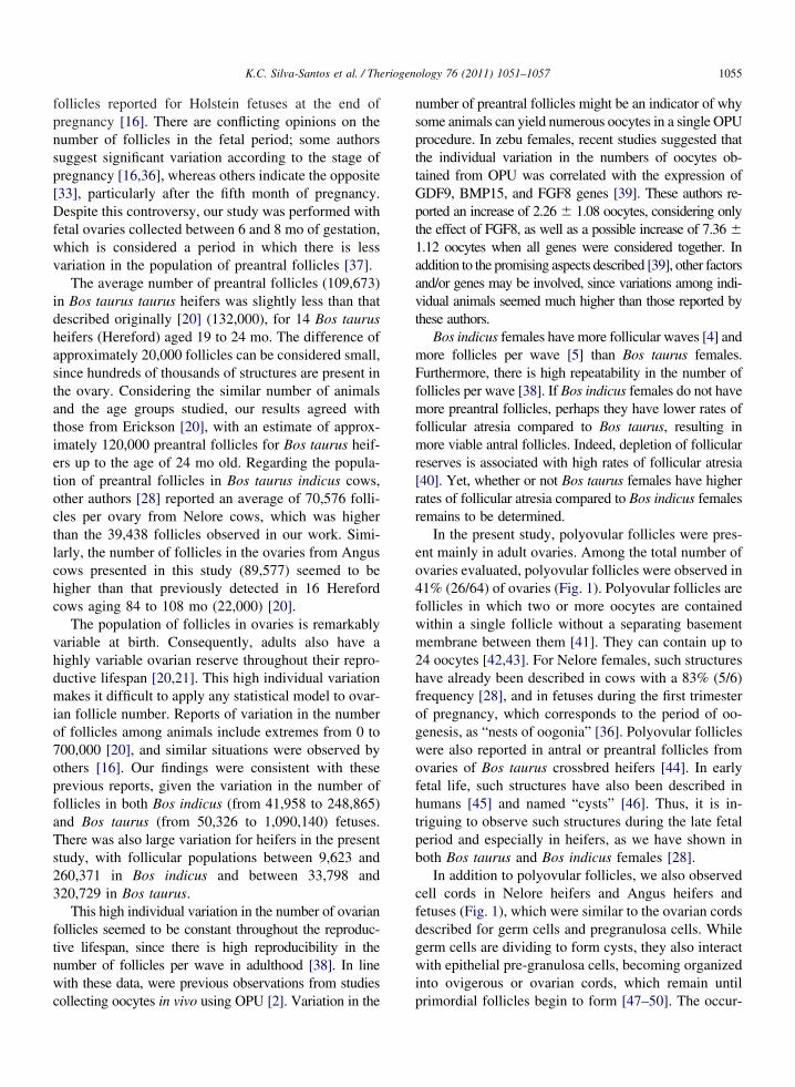
follicles reported for Holstein fetuses at the end ofpregnancy [16]. There are conflicting opinions on thenumber of follicles in the fetal period; some authorssuggest significant variation according to the stage ofpregnancy [16,36], whereas others indicate the opposite[33], particularly after the fifth month of pregnancy.Despite this controversy, our study was performed withfetal ovaries collected between 6 and 8 mo of gestation,which is considered a period in which there is lessvariation in the population of preantral follicles [37].
The average number of preantral follicles (109,673)in Bos taurus taurus heifers was slightly less than thatdescribed originally [20] (132,000), for 14 Bos taurusheifers (Hereford) aged 19 to 24 mo. The difference ofapproximately 20,000 follicles can be considered small,since hundreds of thousands of structures are present inthe ovary. Considering the similar number of animalsand the age groups studied, our results agreed withthose from Erickson [20], with an estimate of approx-imately 120,000 preantral follicles for Bos taurus heif-ers up to the age of 24 mo old. Regarding the popula-tion of preantral follicles in Bos taurus indicus cows,other authors [28] reported an average of 70,576 folli-cles per ovary from Nelore cows, which was higherthan the 39,438 follicles observed in our work. Simi-larly, the number of follicles in the ovaries from Anguscows presented in this study (89,577) seemed to behigher than that previously detected in 16 Herefordcows aging 84 to 108 mo (22,000) [20].
The population of follicles in ovaries is remarkablyvariable at birth. Consequently, adults also have ahighly variable ovarian reserve throughout their repro-ductive lifespan [20,21]. This high individual variationmakes it difficult to apply any statistical model to ovar-ian follicle number. Reports of variation in the numberof follicles among animals include extremes from 0 to700,000 [20], and similar situations were observed byothers [16]. Our findings were consistent with theseprevious reports, given the variation in the number offollicles in both Bos indicus (from 41,958 to 248,865)and Bos taurus (from 50,326 to 1,090,140) fetuses.
here was also large variation for heifers in the presenttudy, with follicular populations between 9,623 and60,371 in Bos indicus and between 33,798 and20,729 in Bos taurus.
This high individual variation in the number of ovarianollicles seemed to be constant throughout the reproduc-ive lifespan, since there is high reproducibility in theumber of follicles per wave in adulthood [38]. In lineith these data, were previous observations from studies
ollecting oocytes in vivo using OPU [2]. Variation in the p
umber of preantral follicles might be an indicator of whyome animals can yield numerous oocytes in a single OPUrocedure. In zebu females, recent studies suggested thathe individual variation in the numbers of oocytes ob-ained from OPU was correlated with the expression ofDF9, BMP15, and FGF8 genes [39]. These authors re-orted an increase of 2.26 � 1.08 oocytes, considering only
the effect of FGF8, as well as a possible increase of 7.36 �1.12 oocytes when all genes were considered together. Inaddition to the promising aspects described [39], other factorsand/or genes may be involved, since variations among indi-vidual animals seemed much higher than those reported bythese authors.
Bos indicus females have more follicular waves [4] andmore follicles per wave [5] than Bos taurus females.Furthermore, there is high repeatability in the number offollicles per wave [38]. If Bos indicus females do not havemore preantral follicles, perhaps they have lower rates offollicular atresia compared to Bos taurus, resulting inmore viable antral follicles. Indeed, depletion of follicularreserves is associated with high rates of follicular atresia[40]. Yet, whether or not Bos taurus females have higherrates of follicular atresia compared to Bos indicus femalesremains to be determined.
In the present study, polyovular follicles were pres-ent mainly in adult ovaries. Among the total number ofovaries evaluated, polyovular follicles were observed in41% (26/64) of ovaries (Fig. 1). Polyovular follicles arefollicles in which two or more oocytes are containedwithin a single follicle without a separating basementmembrane between them [41]. They can contain up to24 oocytes [42,43]. For Nelore females, such structureshave already been described in cows with a 83% (5/6)frequency [28], and in fetuses during the first trimesterof pregnancy, which corresponds to the period of oo-genesis, as “nests of oogonia” [36]. Polyovular follicleswere also reported in antral or preantral follicles fromovaries of Bos taurus crossbred heifers [44]. In earlyfetal life, such structures have also been described inhumans [45] and named “cysts” [46]. Thus, it is in-triguing to observe such structures during the late fetalperiod and especially in heifers, as we have shown inboth Bos taurus and Bos indicus females [28].
In addition to polyovular follicles, we also observedell cords in Nelore heifers and Angus heifers andetuses (Fig. 1), which were similar to the ovarian cordsescribed for germ cells and pregranulosa cells. Whileerm cells are dividing to form cysts, they also interactith epithelial pre-granulosa cells, becoming organized
nto ovigerous or ovarian cords, which remain until
rimordial follicles begin to form [47–50]. The occur-
ttcgi
A
otEPL
R
[
[
[
[
[
[
[
[
[
[
[
[
1056 K.C. Silva-Santos et al. / Theriogenology 76 (2011) 1051–1057
rence of polyovular follicles and ovarian cords wasreported during fetal life [36,51], but their biologicalsignificance remains to be determined. Germ cell clus-ters appear in greater quantity at the beginning of fetallife and gradually disappear as more primordial folli-cles are formed. At the end of fetal life, ovarian cordshave almost completely disappeared, and secondaryfollicles are present [51]. Both polyovular follicles andcell cords reported in the present study could representfollicular renewal activity, since these structures aretypically described in early folliculogenesis. However,more accurate methods are needed for further investi-gations. It was noteworthy that there was no associationamong these structures and the number of ovarian pre-antral follicles. However, recent studies reported thatthe high variation in the number of antral folliclesamong Bos taurus heifers was highly correlated withthe number of polyovular follicles [44].
In summary, the present results reported will pro-vide the basis for further studies to improve understand-ing of factors regulating folliculogenesis in cattle.There was substantial variation among individual ani-mals, but the overall population of preantral folliclesdid not differ significantly among Bos indicus and Bosaurus fetuses and heifers. Therefore, we inferred thathe greater numbers of oocytes produced by Bos indicusompared to Bos taurus females in ovum pick-up pro-rams were not due to the number of preantral folliclesn the fetal or postnatal female.
cknowledgments
The authors are grateful to Vilceu Bordignon for allf his important comments regarding this work. Wehank the Coordination for the Improvement of Higherducation Personnel (CAPES) and the Post-Graduationrogram in Animal Science at the State University ofondrina for their financial support.
eferences
[1] Thibier M. New records in the numbers of both in vivo-derivedand in vitro-produced bovine embryos around the world in2006. IETS Embryo Transfer News 2007;25:15–20.
[2] Pontes JHF, Nonato-Junior I, Sanches BV, Ereno-Junior JC,Uvo S, Barreiros TRR, Oliveira JA, Hasler JF, Seneda MM.Comparison of embryo yield and pregnancy rate between invivo and in vitro methods in the same Nelore (Bos indicus)donor cows. Theriogenology 2009;71:690–7.
[3] Figueiredo RA, Barros CM, Pinheiro OL, Sole JMP. Ovarianfollicular dynamics in Nelore breed (Bos indicus) cattle. Ther-
iogenology 1997;47:1489–505.[4] Viana JHM, Ferreira AM, Sá WF, Camargo LSA. Folliculardynamics in zebu cattle. Pesquisa Agropecuária Brasileira 2000;35:2501–9.
[5] Carvalho JBP, Carvalho NAT, Reis EL, Nichi M, Souza AH,Baruselli PS. Effect of early luteolysis in progesterone-basedtimed AI protocols in Bos indicus, Bos indicus x Bos taurus, andBos taurus heifers. Theriogenology 2008;69:167–75.
[6] Segerson EC, Hansen TR, Libby DW, Randel RD, Getz WR.Ovarian and uterine morphology and function in Angus andBrahman cows. J Anim Sci 1984;59:1026–46.
[7] Sartorelli ES, Carvalho LM, Bergfelt DR, Ginther OJ, BarrosCM. Morphological characterization of follicle deviation inNelore (Bos indicus) heifers and cows. Theriogenology 2005;63:2382–94.
[8] Rhodes FM, De’ath G, Entwistle KW. Animal and temporaleffects on ovarian follicular dynamics in Brahman heifers.Anim Reprod Sci 1995;38:265–77.
[9] Santos R, Soto MAB, Lourenço RX, Stranieri P, Bishop W,Accorsi MF, Watanabe MR, Dayan A, Watanabe YF. Follicularaspiration in Nelore. Case of high number of oocytes recovered.In: XVI Congresso Brasileiro de Reprodução Animal, 2005,Goiânia, GO, Brazil.
10] Machado SA, Reichenbach HD, Weppert M, Matos LF, Wolf E,Gonçalvez PBD. The variability of ovum pick-up response andin vitro embryo production from monozygotic twin cows. The-riogenology 2006;65:573–83.
11] Rubin KCP, Pontes JHF, Nonato I, Jr. Ereno JC, Jr. Pansard H,Seneda MM. Influence of Nelore blood on the in vivo productionof oocytes. Acta Scientiae Veterinariae 2005;33:183(Abstract).
12] Martins A, Jr Takada L, Abrahão RG, Freitas CP, Calegari RS.Follicular aspiration of calves oocytes by videoendoscopy: asuccessful approach to maximize in vitro bovine embryo pro-duction. Acta Scientiae Veterinariae 2007;35:1194(Abstract).
13] Johnson J, Canning J, Kaneko T, Pru JK, Tilly JL. Germlinestem cells and follicular renewal in the postnatal mammalianovary. Nature 2004;428:145–50.
14] Zou K, Yuan Z, Yang Z, Luo H, Sun K, Zhou L, Xiang J, ShiL, Yu Q, Zhang Y, Hou R, Wu J. Production of offspring froma germline stem cell line derived from neonatal ovaries. NatCell Biol 2009;11:631–6.
15] Johnson J, Bagley J, Skaznik-Wikiel M, Lee HJ, Adams GB,NiikuraY, Tschudy KS, Tilly JC, Cortes ML, Forkert R, SpitzerT, Iacomini J, Scadden DT, Tilly JL. Oocyte generation in adultmammalian ovaries by putative germ cells in bone marrow andperipheral blood. Cell 2005;122:303–15.
16] Tanaka Y, Nakada K, Moriyoshi M, Sawamukai Y. Appearance andnumber of follicles and change in the concentration of serum FSH infemale bovine fetuses. Reproduction 2001;121:777–82.
17] Nogueira GP, Marcassa GPSD, Beltran MP. Variation in theconcentration of gonadotropins and follicle population duringfetal development in Bos taurus indicus. In: XVI CongressoBrasileiro de Reprodução Animal, 2005, Goiânia, GO, Brazil.
18] Soto-Suazo M, Zorn TM. Primordial germ cells migration: morpho-logical and molecular aspects. Anim Reprod Sci 2005;3:147–60.
19] van Den Hurk R., Zhao J. Formation of mammalian oocytes andtheir growth, differentiation and maturation within ovarian fol-licles. Theriogenology 2005;63:1717–51.
20] Erickson BH. Development and senescence of the postnatalbovine ovary. J Anim Sci 1966;25:800–5.
21] Erickson BH. Development and radio-response of the prenatal
bovine ovary. J Reprod Fertil 1966;10:97–105.
[
[
[
[
[
[
[
[
[
[
[
[
[
[
[
[
[
[
[
[
[
[
[
1057K.C. Silva-Santos et al. / Theriogenology 76 (2011) 1051–1057
[22] Ireland JJ, Zielak-Steciwko AE, Jimenez-Krassel F, Folger J,Bettegowda A, Scheetz, D, Walsh S, Mossa F, Knight PG,Smith GW, Lonergan P, Evans ACO. Variation in the ovarianreserve is linked to alterations in intrafollicular estradiol pro-duction and ovarian biomarkers of follicular differentiation andoocyte quality in cattle. Biol Reprod 2009;80:954–64.
[23] Eggan K, Jurga S, Gosden R, Min IM, Wagers AJ. Ovulatedoocytes in adult mice derive from non-circulating germ cells.Nature 2006;441:1109–14.
[24] Saumande J. La Folliculogenèse Chez les Ruminants. Recueilde Médecine Vétérinaire 1991;167:205–18.
[25] Figueiredo JR, Celestino JJH, Rodrigues APR, Silva JRV. Im-portance of the biotechnique of MOEPF for the study of fol-liculogenesis and in vitro embryo production in large scale.Revista Brasileira de Reprodução Animal 2007;31:143–52.
[26] Picton HM. Activation of follicle development: the primordialfollicle. Theriogenology 2001;55:1193–210.
[27] Gosden RG, Telfer E. Numbers of follicles and oocytes inmammalian ovaries and their allometric relationships. J Zoo11987;211:169–175.
[28] Lucci CM, Rumpf R, Figueiredo JR, Báo SN. Zebu (Bos indi-cus) ovarian preantral follicles: morphological characterizationand development of an efficient isolation method. Theriogenol-ogy 2002;57:1467–83.
29] Evans HE, Sack WO. Prenatal development of domestic andlaboratory mammals: growth curves, external features and se-lected references. Anat Hist Embryol 1973;2:11–45.
30] Lowman BJ, Scott NA, Somerville SH. Condition scoring ofcattle, Revised edition. East Scotland College of Agriculture,p.1–31, 1976 (Bulletin 6).
31] Cahill LP, Mariana JC, Mauléon P. Total follicular populations inewes of high and low ovulation rates. J Reprod Fertil 1979;55:27–36.
32] Hulshof SCJ, Figueiredo JR, Beckers JF, Bevers MM, Van DenHurk R. Isolation and characterization of preantral folliclesfrom foetal bovine ovaries. Vet Quart 1994;16:78–80.
33] Carámbula SF, Gonçalves PBD, Costa LFS, Figueiredo JR,Wheeler MB, Neves JP, Mondadori RG. Effect of fetal age andmethod of recovery on isolation of preantral follicles frombovine ovaries. Theriogenology 1999;52:563–71.
34] Gougeon A, Chainy GBN. Morphometric studies of small fol-licles in ovaries of women at different ages. J Reprod Fertil1987;81:433–42.
35] Ayres, M., Ayres Jr., M., Ayres, D.L., Santos, A.S., 2007.BioEstat 5.0. Statistical applications in biological and medicalsciences. Sociedade Civil Mamirauá, Brasília, CNPq, 138 pp.
36] Diniz EG, Esper CR, Jacomini JO, Vieira RC. Morphologicaldevelopment of the ovaries in embryos and fetuses of Nelorebreed. Arquivo Brasileiro de Medicina Veterinária e Zootecnia
2005;57:70–6.37] Muranishi Y, Acosta TJ, Miyamoto A, Fukui Y. Changes in thenumber of preantral follicles and hormone concentrations in thebovine fetus. J Reprod Develop 2002;48:553–60.
38] Burns, D.S., Jimenez-Krassel, F., Ireland, J.L.H., Knight, P.G.,Ireland, J.J.. Numbers of antral follicles during follicular wavesin cattle: Evidence for high variation among animals, very highrepeatability in individuals, and an inverse association withserum follicle-stimulating hormone concentrations. Biol Reprod2005;73:53–62.
39] Biase FH, Fonseca MGK, Freitas SBWK, Martelli L, MeirellesFV. Global poly(A) mRNA expression profile measured inindividual bovine oocytes and cleavage embryos. Zygote 2008;16:29–38.
40] Krysko DV, Diez-Fraile A, Criel G, Svistunov AA, Vandena-beele P, D’herde K. Life and death of female gametes duringoogenesis and folliculogenesis. Apoptosis 2008;13:1065–87.
41] Tingen C, Kim A, Woodruff K. The primordial pool of folliclesand nest breakdown in mammalian ovaries. Mol Hum Reprod2009;15:795–803.
42] Lucci CM, Amorim CA, Rodrigues APR, Figueiredo JR, BáoSN, Silva JRV, Gonçalves PBD. Study of preantral folliclepopulation in situ and after mechanical isolation from caprineovaries at different reproductive stages. Anim Reprod Sci 1999;56:223–36.
43] Al-Mufti W, Bomsel-Helmreich O, Christides J Ph., Oocyte sizeand intrafolicullar position in polyovular follicles in rabbits. JReprod Fertil 1988;82:15–25.
44] Ireland JLH, Scheetz D, Jimenez-Krassel F, Themmen APN,Ward F, Lonergan P, Smith GW, Perez GI, Evans ACO, IrelandJJ. Antral follicle count reliably predicts number of morpholog-ically healthy oocytes and follicles in ovaries of young adultcattle. Biol Reprod 2008;79:1219–25.
45] Gondos B, Bhuraleus P, Habel CJ. Ultrastructure observationson germ cells in human fetal ovaries. Am J Obstet Gynecol1971;110:644–52.
46] Pepling ME. From primordial germ cell to primordial follicle: mam-malian female germ cell development. Genesis 2006;44:622–32.
47] Odor DL, Blandau RJ. Ultrastructural studies on fetal and earlypostnatal mouse ovaries. I. Histogenesis and organogenesis.Am J Anat 1969;124:163–86.
48] Byskov AG. Differentiation of mammalian embryonic gonad.Physiol Reviews 1986;66:71–117.
49] Hirshfield AN. Development of follicles in the mammalianovarian. Int Rev Cytol 1991;124:43–101.
50] Guigon CJ, Magre S. Contribution of germ cells to the differ-entiation and maturation of the ovary: Insights from models ofgerm cell depletion. Biol Reprod 2006;74:450–8.
51] Yang MY, Fortune JE. The capacity of primordial follicles infetal bovine ovaries to initiate growth in vitro develops duringmid-gestation and is associated with meiotic arrest of oocytes.
Biol Reprod 2008;78:1153–61.