Delivery of cloned offspring: experience in Zebu cattle ( Bos indicus
10
CSIRO PUBLISHING Reproduction, Fertility and Development, 2010, 22, 88–97 www.publish.csiro.au/journals/rfd Delivery of cloned offspring: experience in Zebu cattle (Bos indicus) Flávio V. Meirelles A,E , Eduardo H. Birgel Jr B , Felipe Perecin A , Marcelo Bertolini D , Anneliese S. Traldi B , José Rodrigo V. Pimentel B , Eliza R. Komninou B , Juliano R. Sangalli A , Paulo Fantinato Neto A , Mariana Tikuma Nunes B , Fábio Celidonio Pogliani B , Flávia D. P. Meirelles A , Flávia S. Kubrusly C , Camila I. Vannucchi B and Liege C. G. Silva B A Faculty of Animal Sciences and Food Engineering, University of São Paulo, Av. Duque de Caxias Norte, 225. Pirassununga, SP 13635-900, Brazil. B Faculty ofVeterinary Medicine and Animal Sciences, University of São Paulo, Av. Orlando Marques de Paiva, 87. Pirassununga, SP 05508-270, Brazil. C Instituto Butantan, Av.Vital Brasil, 1500 São Paulo, SP 05503-900, Brazil. D Center of Agronomy andVeterinary Sciences, Santa Catarina State University, Av. Luiz de Camões, 2090. Lages, SC 88520-000, Brazil. E Corresponding author. Email: [email protected] Abstract. The production of a healthy cloned calf is dependent on a multitude of successful steps, including repro- gramming mediated by the oocyte, the development of a functional placenta, adequate maternal–fetal interaction, the establishment of a physiological metabolic setting and the formation of a complete set of well-differentiated cells that will eventually result in well-characterised and fully competent tissues and organs. Although the efficiency of nuclear transfer has improved significantly since the first report of a somatic cell nuclear transfer-derived animal, there are many descriptions of anomalies concerning cloned calves leading to high perinatal morbidity and mortality. The present article discusses some our experience regarding perinatal and neonatal procedures for cloned Zebu cattle (B. indicus) that has led to improved survival rates in Nellore cloned calves following the application of such ‘labour-intensive technology’. Additional keywords: bovine, nuclear transfer, parturition. Steps towards moving nuclear transfer to the field There has been considerable discussion regarding advances in nuclear transfer (NT) in the past decade. Most of the discussion was triggered by the birth of Dolly, the first somatic cell nuclear transfer (SCNT)-derived mammal (Wilmut et al. 1997), which was quickly followed by many others (for a review, see Latham 2004). The possibility of producing identical animals was inter- esting enough to bring together researchers and technicians from most diverse fields (Wells et al. 1999; Wilmut et al. 2009). Since the early days, NT experiments have brought to light important issues in biology, leading to new discoveries and knowledge in many related areas, such as mechanisms controlling the cell cycle and nuclear reprogramming (Galli et al. 2003). In addition to its relevance to basic science, SCNT technology has received con- siderable interest for application in animal production (Bousquet and Blondin 2004; Heyman et al. 2004; Heyman 2005). This has enabled important improvements in the procedure, with the technology being gradually transferred to and applied in industry (Faber et al. 2003, 2004). Nowadays, many private companies are exploiting SCNT commercially in many species. Cloned individuals are common in agriculture; for exam- ple, people often drive on roads bordered by huge forests of clones (e.g. eucalyptus). Why not apply the same principle to ani- mal production? Cattle in particular could benefit from cloning because efforts to advance genetic merit are usually slow owing to large inter-generation intervals. Because of the large num- ber of offspring, the poultry and swine industries already profit from such non-additive genetic applications. The possibility of producing cloned cattle on a large scale could also accelerate genetic gain, increasing production and introducing a paradigm shift in the beef and dairy industry. Nonetheless, despite the potential benefits, pre- and postnatal abnormalities, including high rates of embryo and fetal losses, abortion, prolonged gestation, increased birthweight, placental anomalies and reduced neonatal survival are likely consequences of cloning by NT (Bertolini and Anderson 2002; Edwards et al. 2003; Miglino et al. 2007; Oback and Wells 2007; Oback 2008). The establishment and maintenance of SCNT-derived pregnan- cies have proven challenging, with the delivery and early care of these animals often being expensive, labour intensive and © IETS 2010 10.1071/RD09229 1031-3613/10/010088
Transcript of Delivery of cloned offspring: experience in Zebu cattle ( Bos indicus

CSIRO PUBLISHING
Reproduction, Fertility and Development, 2010, 22, 88–97 www.publish.csiro.au/journals/rfd
Delivery of cloned offspring: experience in Zebu cattle(Bos indicus)
Flávio V. MeirellesA,E, Eduardo H. Birgel JrB, Felipe PerecinA,Marcelo BertoliniD, Anneliese S. TraldiB, José Rodrigo V. PimentelB,Eliza R. KomninouB, Juliano R. SangalliA, Paulo Fantinato NetoA,Mariana Tikuma NunesB, Fábio Celidonio PoglianiB, Flávia D. P. MeirellesA,Flávia S. KubruslyC, Camila I. VannucchiB and Liege C. G. SilvaB
AFaculty of Animal Sciences and Food Engineering, University of São Paulo, Av. Duque de Caxias Norte,225. Pirassununga, SP 13635-900, Brazil.
BFaculty of Veterinary Medicine and Animal Sciences, University of São Paulo, Av. Orlando Marquesde Paiva, 87. Pirassununga, SP 05508-270, Brazil.
CInstituto Butantan, Av. Vital Brasil, 1500 São Paulo, SP 05503-900, Brazil.DCenter of Agronomy and Veterinary Sciences, Santa Catarina State University, Av. Luiz de Camões,
2090. Lages, SC 88520-000, Brazil.ECorresponding author. Email: [email protected]
Abstract. The production of a healthy cloned calf is dependent on a multitude of successful steps, including repro-gramming mediated by the oocyte, the development of a functional placenta, adequate maternal–fetal interaction, theestablishment of a physiological metabolic setting and the formation of a complete set of well-differentiated cells thatwill eventually result in well-characterised and fully competent tissues and organs. Although the efficiency of nucleartransfer has improved significantly since the first report of a somatic cell nuclear transfer-derived animal, there are manydescriptions of anomalies concerning cloned calves leading to high perinatal morbidity and mortality. The present articlediscusses some our experience regarding perinatal and neonatal procedures for cloned Zebu cattle (B. indicus) that hasled to improved survival rates in Nellore cloned calves following the application of such ‘labour-intensive technology’.
Additional keywords: bovine, nuclear transfer, parturition.
Steps towards moving nuclear transfer to the field
There has been considerable discussion regarding advances innuclear transfer (NT) in the past decade. Most of the discussionwas triggered by the birth of Dolly, the first somatic cell nucleartransfer (SCNT)-derived mammal (Wilmut et al. 1997), whichwas quickly followed by many others (for a review, see Latham2004). The possibility of producing identical animals was inter-esting enough to bring together researchers and technicians frommost diverse fields (Wells et al. 1999; Wilmut et al. 2009). Sincethe early days, NT experiments have brought to light importantissues in biology, leading to new discoveries and knowledge inmany related areas, such as mechanisms controlling the cell cycleand nuclear reprogramming (Galli et al. 2003). In addition to itsrelevance to basic science, SCNT technology has received con-siderable interest for application in animal production (Bousquetand Blondin 2004; Heyman et al. 2004; Heyman 2005). Thishas enabled important improvements in the procedure, with thetechnology being gradually transferred to and applied in industry(Faber et al. 2003, 2004). Nowadays, many private companiesare exploiting SCNT commercially in many species.
Cloned individuals are common in agriculture; for exam-ple, people often drive on roads bordered by huge forests ofclones (e.g. eucalyptus). Why not apply the same principle to ani-mal production? Cattle in particular could benefit from cloningbecause efforts to advance genetic merit are usually slow owingto large inter-generation intervals. Because of the large num-ber of offspring, the poultry and swine industries already profitfrom such non-additive genetic applications. The possibility ofproducing cloned cattle on a large scale could also accelerategenetic gain, increasing production and introducing a paradigmshift in the beef and dairy industry.
Nonetheless, despite the potential benefits, pre- and postnatalabnormalities, including high rates of embryo and fetal losses,abortion, prolonged gestation, increased birthweight, placentalanomalies and reduced neonatal survival are likely consequencesof cloning by NT (Bertolini and Anderson 2002; Edwards et al.2003; Miglino et al. 2007; Oback and Wells 2007; Oback 2008).The establishment and maintenance of SCNT-derived pregnan-cies have proven challenging, with the delivery and early careof these animals often being expensive, labour intensive and
© IETS 2010 10.1071/RD09229 1031-3613/10/010088

Delivering zebu cloned offspring Reproduction, Fertility and Development 89
frequently frustrating (Hill and Chavatte-Palmer 2002; Wilmut2006; Keefer 2008). Many factors contribute to the overall lowefficiency of NT, with the number of births per number ofembryos transferred still low, averaging 6% and usually vary-ing between 0% and 12% (Wells et al. 1999, 2004; Panaraceet al. 2007; Keefer 2008; Oback 2008).
The low success rate, combined with increased pregnancyand neonatal abnormalities and losses, seen after cloning hasnecessitated intensive care during the perinatal period, spawn-ing considerable advances in neonatology in cattle. In the presentreview, we describe and discuss some management and clini-cal procedures and issues related to the peri- and postnatal careof cloned Nellore calves that, in our experience, have helpedimprove survival rates. Special attention is given to the mostcommon risk-related clinical signs during the perinatal period.However, it is important to bear in mind that due to the consid-erable variations that exist between species, breeds, individuals,laboratories, procedures and personnel, cloning by SCNT mayresult in different phenotypes, thus requiring continuous adap-tation to each specific condition or outcome that challenges usas scientists, practitioners, managers and producers.
Perinatal management
The perinatal period in cattle is defined as the interval thatincludes the 2–4 weeks before delivery and the first week oflife. In our experience, as well as reported by others (Hill andChavatte-Palmer 2002; Wells et al. 2004; Buczinski et al. 2009),this is the most critical period in clone production due to thehigh rate of unpredictable anomalies that may arise, leading tofrustrating gestational outcomes. Indeed, approximately 50% ofclone pregnancies do not end normally, and this figure rangesfrom 0 to 100% depending on the cell lineage. Hence, thesepregnancies usually result in prolonged gestation of enlarged off-spring with poorer neonatal (Heyman et al. 2002). The increasedtime in utero may be indicative of a need for further maturation,but is usually associated with increased birthweight, dystocia andhigher morbidity and mortality (Hill and Chavatte-Palmer 2002).
In cattle and other domestic species, parturition is initiatedby the activation and maturation of the fetal adrenal cortex,leading to an increase in fetal cortisol secretion (Challis et al.2000). Previous studies have reported lower plasma cortisol con-centrations in newborn cloned calves delivered by Caesareansection compared with vaginally delivered controls (Matsuzakiand Shiga 2002), but no differences in plasma adrenocorti-cotrophin (ACTH) levels, suggesting that the prepartum increasein plasma cortisol in clones may be insufficient to trigger par-turition. Conversely, the responsiveness of the adrenocorticaltissue to ACTH in cloned calves is normal and the differencein plasma cortisol levels in cloned calves seems to be dueto the delivery method (Caesarean section v. natural delivery)rather than the cloning itself (Chavatte-Palmer et al. 2002;Hirayama et al. 2008). The prepartum increase in fetal corti-sol leads to increased production of oestrogens by the placenta,which is accompanied by a reduction in maternal progesterone(P4) plasma concentrations (Conley and Ford 1987; Conley andMason 1990; Challis et al. 2000). Under physiological con-ditions, these endocrine events stimulate myometrial oxytocin
receptor expression, prostaglandin synthesis, cervical efface-ment, sacro-iliac relaxation and vulva distensibility (Huszar andWalsh 1991; Jenkin and Young 2004). Such events appear to bemarkedly decreased in females carrying SCNT-derived concepti.Hill and Chavatte-Palmer (2002) attempted to correlate mater-nal P4 profiles in the last 2 weeks of gestation of SCNT-derivedpregnancies with the postnatal viability of cloned calves. Theauthors observed that atypical profiles (e.g. prepartum mainte-nance of elevated P4 levels) appeared to be associated with lowerneonatal viability, whereas usual P4 profiles were associatedwith higher postnatal viability. Conclusive studies to determinethe reason for the poor signs of parturition in cloned animals arestill lacking. Hirayama et al. (2008) suggested that oestrogen sul-foconjugation, a mechanism that regulates oestrogen activity byimpairing the binding of oestrogen to its nuclear receptors, maybe responsible for the reduced parturition signalling because alower prepartum oestrone : oestrone sulfate ratio was observedin cloned compared with control pregnancies.
When performed during the perinatal period preceding deliv-ery, the induction of parturition is believed to improve fetalviability by stimulating the production of surfactant phospho-lipids by fetal Type II alveolar cells, enhancing the expression ofsurfactant-associated proteins and accelerating the overall struc-tural maturation of the lungs (DeKruif and Benedictus 1993).Hence, the induction of parturition is desirable in recipientheifers and cows to reduce the interval to parturition, allow-ing a more assisted delivery; however, the incidence of retainedfetal membranes usually increases (Lewing et al. 1985; Garciaet al. 1992). Parturition may be induced via different proto-cols (Lewing et al. 1985; Nasser et al. 1994), but is generallyachieved by using a combination of corticoid treatment with aprostaglandin (PGF2α) analogue. The use of PGF2α is highlyconserved between different protocols, whereas the corticoidtreatment is highly variable: treatments may consist of a singledose of 20 mg dexamethasone in the last 36 h before delivery orthey may start 7 days before the expected date of delivery (EDD)using a slow-release, long-acting corticoid, such as opticortenol,betamethasone or triancinolone acetonide, followed by the stan-dard protocol (Nasser et al. 1994, 2008). The aim of the lattertreatment option is to accelerate final maturation of fetal lungsand organs, as well as the placenta.
Because the mean length of gestation in Zebu cattle is approx-imately 290 days, we generally induce parturition starting up to5 days before the EDD (between Days 285 and 290 of ges-tation), as suggested by others (Hill et al. 1999). To evaluatethe effect of the induction of parturition on perinatal survivalof Nellore cloned calves, we tested two different protocols:(1) administration of 20 mg, i.m., dexamethasone 36 h beforeEDD, followed by administration of d-cloprostenol 24 h beforeEDD; and (2) administration of 8 mg, i.m., triancinolone ace-tonide 9 days before EDD, followed by drug administrationas described for Protocol 1. Both protocols resulted in simi-lar survival rates (13/20 (65%) and 11/15 (73.3%), respectively;P = 0.72). Although there were no advantages in terms of sur-vival using the longer protocol, we routinely choose to use thissecond protocol because no negative effects on survival wereobserved and the protocol has potential benefits in that it mayallow preparation of both the fetus and the recipient for delivery.

90 Reproduction, Fertility and Development F. V. Meirelles et al.
In fact, we have observed, by visual inspection, an improve-ment in udder development and vulvar distensibility in femalerecipients subjected to Protocol 2 compared with females sub-jected to Protocol 1, a phenotypic event readily detectable fromthe third day of triancinolone acetonide administration. In addi-tion, the use of such protocol may be logistically useful; forexample, if evaluation of fetal well-being in the last 4 daysof pregnancy indicates signs of distress, an elective emergencyCaesarean section is more likely to be successful than with noprevious corticoid treatment. However, there is still a need forstudies on non-invasive and risk-free methodologies to evaluatefetal well-being before birth that provide accurate results. Somestudies using ultrasonography to assess fetal well-being haveindicated that fetal inactivity or hyperactivity, as well as hypere-choic particles in fetal annexes, are possible signs of fetal distress(Buczinski et al. 2007, 2009; Kohan-Ghadr et al. 2008). Otherstrategies, such as the quantification of pregnancy associatedglycoproteins in the first trimester, can also be used to pre-dict the outcome of gestation (Heyman et al. 2002; Breukelmanet al. 2005; Chavatte-Palmer et al. 2006). Nonetheless, we havebeen evaluating cardiotocography in animals in the last monthsof pregnancy. Thus far, the preliminary results suggest thathypoxia in the last trimester of gestation, especially from 90to 30 days prepartum, is associated with fetal hypoactivity, thelack of a cardiac response after inter-digital stimulus and deliv-ery of a meconium-stained calf. However, hypoactivity does notseem to invariably indicate hypoxic conditions near term, withsome hypoactive calves showing being healthy and viable afterdelivery.
Delivery of SCNT-derived calves
Birthweight of calves produced by SCNT tends to be increasedcompared with the birthweight of non-cloned calves. It is impor-tant to note that nuclear donor cells are frequently taken fromanimals of high genetic merit that are born often heavier than thepopulation average. However, deregulation of imprinted genesand/or mechanisms leading to large (or abnormal) offspring syn-drome (LOS) also contribute to the increased birthweight. Allthese factors tend to reduce chances for survival due to a greaterlevel of distress imposed upon the newborn. In our experience,aside from a group of oversized and heavier animals at birth(9/43; 20.9%), a large proportion of calves have been normal inboth size and weight (30/43; 69.8%) at birth, with a smaller pro-portion being significantly smaller (4/43; 9.3%) (F. V. Meirelles,unpubl. data). The mean birthweight for Nellore cloned calvesborn in our laboratory is approximately 38 kg (range 14–62 kg)for a breed with an average birthweight of 34 kg. Figure 1shows the distribution of birthweights for Nellore cloned calvesin a real population of 9047 calves, along with a theoreticalsimulation using the Monte Carlo methodology for a similarnumber of clones based on the distribution we found in our study(F. D. P. Meirelles, unpubl. data).
The systematic evaluation of the absolute and relative (com-pared with the recipient) size of a fetus and the signs ofparturition are important for planning the delivery procedure.Large animals are chosen to be delivered by Caesarean section,because these animals are less capable of enduring dystocia-induced hypoxia (Chavatte-Palmer et al. 2004). An experienced
Birth weight (kg)
Num
ber
of a
nim
als
0
500
1000
1500
2000
20 40 60 80
Nellore clone
Nellore AI
Fig. 1. Real and simulated data on calf weight at birth. Dark grey barsshow the weight at birth of a real population of 9047 Nellore (Zebu) calvesproduced by AI. The light grey bars represent the distribution of weight atbirth of a simulated population of 9200 Nellore calves produced by somaticcell nuclear transfer applying Monte Carlo methodology. The simulationapplied the weight distribution we observed in our service in 43 animalsdelivered over a period of 4 years (2005 to 2009).
veterinarian with sound training in obstetric procedures may esti-mate the size of the animal by measuring the limbs or head byrectal palpation. In addition to identifying large fetuses, rectalpalpation may help in the diagnosis of flexural deformities thatmay also complicate passage through the birth canal and requireobstetric assistance during delivery (Fecteau et al. 2005).
A Caesarean section should be chosen if there is uncertaintyabout the size or other indications of potential suffering. This isthe most appropriate method unless the neonate is known to beof normal size, thus having a high likelihood of passing quicklythrough the pelvic canal (Hill and Chavatte-Palmer 2002). Per-forming a Caesarean section provides an opportunity to betterdeal with the umbilical cord, which is frequently increased indiameter, allowing prompt draining of the umbilicus and clamp-ing. We routinely clamp and cut large-diameter umbilici to avoidinfections and haemorrhage, because an absence of blood vesselconstriction is more likely in enlarged umbilici after rupture orcutting (Hill and Chavatte-Palmer 2002).
There are other factors in addition to the size of the calf thatindicate the need for a Caesarean section. For example, visualisa-tion of hyperechoic structures in the allantoic or amniotic fluids,

Delivering zebu cloned offspring Reproduction, Fertility and Development 91
or within the placentomes, on ultrasound and/or the detectionof a hyperactive fetus, placental oedema or excessive fetal fluidvolume (hydramnio or hydroallantois) can be used as indicatorsfor the need for a Caesarean section.The decision whether to per-form a Caesarean section will depend on the time of gestationand on the time since starting the treatment to induce parturi-tion. Two attempts to induce parturition 2 weeks before EDD inpregnancies in which hydrops was identified resulted in neonataldeath due to atelectasis (F. V. Meirelles, unpubl. data).
Prior to surgery, we inject the recipient female with 50 mg,i.v., isoxsuprine chlorhydrate to relax the uterus and thus min-imise the chances of fetal hypoxia during the procedure. Thereare no negative effects associated with the recipient’s position(standing or in lateral recumbency) during surgery on survivaloutcome but, to avoid the risk of decreasing fetal viability, wediscourage the use of drugs that may depress the central nervoussystem (CNS).
Initial care
It is important to note that most perinatal deaths generally occurwithin the first hour after birth (Nagy 2009). Consequently, spe-cial attention must be given to the calf during and immediatelyafter delivery. After surgery, which usually takes 10–20 min, thecalf is exposed to the atmosphere. It is essential to keep in mindthat: (1) even apparently healthy animals require close moni-toring; and (2) their condition can deteriorate rapidly (Fecteauet al. 2005). After delivery, fluid drainage from the respiratorysystem is facilitated with the calf being transported immediatelyto a warm room, in a clean space and laid in sternal recumbency.At this point, the first clinical evaluations are made with estima-tion of the Apgar score. The Apgar scoring index can be usefulfor assessment of neonatal viability and as a predictor of earlysigns of peripartum asphyxia (House 2002). A chart reportingthe most important birth details is very important for keepingtrack of the parameters evaluated in newborns.
The most important parameters we take note are birth date andtime, method of delivery (vaginal or Caesarean section), timeto spontaneous respiration, the Apgar score at birth and after5 min (Herfen and Bostedt 1999), initial respiratory and heartrate, initial rectal temperature, time of first meconium elimi-nation, birthweight, haemorrhage in the umbilical cord, stateof alertness, suckling reflex, time to standing, time to nursing,initial blood measurements (i.e. glucose, lactate, packed cell vol-ume, total protein concentration, acid–base balance and arterialblood gas) and initial measurements of oxygen saturation in thehaemoglobin. Other critical elements we make note of duringthe physical examination for the detection of respiratory diseaseinclude mucous membrane colour (nasal and vulvar mucousmembranes), character and frequency of the respiratory effortand thoracic auscultation (Poulsen and McGuirk 2009). At theend of this initial period, the first clinical decisions are made and,depending on the neonate’s condition, some initial precautionsare prescribed and monitored over time.
Adaptation to extrauterine life
In utero, materno–fetal exchange within the placentomes pro-vides oxygen and nutrient to the conceptus’ blood. Oxygen and
nutrients are then distributed through the fetal circulation byshunting blood away from the pulmonary circulation via theductus arteriosus and foramen ovale, which is facilitated byhypoxia-induced pulmonary arterial constriction (Ardran et al.1952). Under physiological conditions, the fetus survives in mildhypoxic conditions (Pao2 38 mmHg) compared with adult ani-mals (Pao2 100 mmHg). The adaptation to this environment isdue to efficient O2 extraction from maternal placental blood byfetal haemoglobin (Poulsen and McGuirk 2009).
After delivery, the most important factor affecting the survivalof a neonate is the respiratory system. Calves should make activerespiratory movements within 30 s after being delivered. Initialhypercapnia is physiologically important, because it is detectedas a stimulus for respiration by the chemosensitive area in themedulla. Primary apnoea is defined as the absence of sponta-neous breathing for 1–5 min; if spontaneous breathing has notstarted in clones by 2 min, it is recommended that action betaken to help the calf. Rubbing calves with bedding or towelscan help stimulate the phrenic nerve (Brunson 1981). The useof acupuncture points on the muzzle is also useful in early care(Rogers 1977). Pouring cold water over the calf’s ear not onlystimulates thermoreceptors in the skin, but has also been rec-ommended for respiratory stimulation (Nagy 2009). In the caseof insufficient fluid drainage from the airways, the inductionof the sneezing reflex by inserting a clean straw into thenostrils or the use of suction devices may assist in expelling orremoving the remaining fluids. Hanging the calf from the backlegs for less than 90 s can also be used to clear the airways. It isalso important to keep the calves in sternal recumbency in orderto guarantee an adequate perfusion : ventilation rate. Both sternalrecumbency and suspension by the hind legs improve respiratoryadaptation to extrauterine life by newborn calves (Uystepruystet al. 2002a).
Monitoring blood parameters (arterial blood gases, acid–base balance, lactate and glucose concentrations) over time isessential. For any animal diagnosed with any grade of asphyxia,hypoxia, hypercapnia or respiratory acidosis due to respiratorydistress, we immediately place an intranasal catheter for O2 sup-plementation at a rate of 5–7 L min−1 depending on the size ofthe calf. If the Apgar score does not improve significantly at thesecond evaluation, 5–7 mg kg−1 aminophylline every 12 h maybe administered in the first days of life. Oxygen therapy shouldbe maintained until Pao2 is near physiological conditions (i.e.65 mmHg; Bleul 2009), when gradual withdrawal is initiated. Ifhaemogasometer equipment is not available, we suggest main-taining O2 supplementation for at least 24 h with gradual titrationfrom 5–7 to 1 L min−1 depending on the clinical progress of theanimal.
When hypoventilation or apnoea is present, drug interventionis indicated. Analeptic agents, such as doxapram hydrochloride,stimulate peripheral chemoreceptors and respiratory centres ofthe CNS. Doxapram hydrochloride has a wide margin of safetyand has been used successfully to stimulate respiration (Nagy2009). Many studies have demonstrated improved respiratoryparameters and survival in calves and lambs following theintravenous or sublingual administration of doxapram (Szenciet al. 1980; Nagy 2009). Doxapram hydrochloride stimulatesthe medullary respiratory centres via aortic and carotid body

92 Reproduction, Fertility and Development F. V. Meirelles et al.
chemoreceptors and can be given at a dose of 0.5 mg kg−1, i.v.,or as an injection of 5–10 mg kg−1 at the base of the tonguefor emergency resuscitation and anoxia (Sullivan et al. 2008).Several studies have demonstrated improvements only in mildlydepressed newborns (Brown 1987), whereas severely depressedcalves do not seem to respond to the agent (Nagy 2009).
Prior to birth, a fetus has a prompt supply of nutrients (e.g.oxygen, glucose), even if at relatively decreasing levels as par-turition approaches. Moving from an intrauterine to extrauterineenvironment results in marked shifts in temperature and in dis-continuation of the supply of nutrients, with all events beingmetabolically related to one another. Clinical changes are oftenseen in calves born following mild complications. Moderate tosevere metabolic disturbances can mirror the changes in tem-perature (Nagy 2009). For example, rapid decreases in bodytemperature (the ambient temperature may be 20◦C or morebelow that found in utero), failure in thermostatic regulatorymechanisms to restore normal rectal temperature and a delayin stimulation of the intermediate metabolism to maintain gly-caemia and oxidative metabolism for efficient ATP productionmay occur in neonates, regardless of exposure to hypoxia, creat-ing a vicious metabolic cycle that can result in the death of thecalf. Not surprisingly, such conditions are often seen in clonedcalves. In one study on the effects of infrared heaters on respira-tory and metabolic parameters in newborns, calves exposed to aninfrared heater for 24 h post partum had significantly improvedrectal temperature, arterial haemoglobin oxygen saturation, tidalvolume, dynamic lung compliance and respiratory rate com-pared with unheated calves (Uystepruyst et al. 2002b). Thisstudy highlights the need to ensure that adequate body tempera-ture is maintained in cloned neonates, along with optimised gasexchange, acid–base balance and blood sugar monitoring. In ourpractice, neonates receive a cold water stimulus for breathing.This stimulus is followed by very fast cleaning of the calf withdetergent and warm water to favour drying. Animals are thenquickly moved to a 30◦C room and dried immediately with tow-els and a heated blower. The animal is maintained under theseconditions until it is capable of adequately regulating its ownbody temperature. Even using this protocol, 23.3% (10/43) of ouranimals developed hypothermia (body temperature < 36.5◦C),which invariably resulted in death.
At this point (a few hours after birth), most of the initial clin-ical care has been applied. The calf is then closely monitoredand colostrum offered. The colostrum not only provides passiveimmunity, but also reduces morbidity and mortality in youngcalves (Cortese 2009). When calves are born, the epithelial cellsthat line the digestive tract allow absorption of colostral pro-teins via pinocytosis. As soon as the digestive tract is stimulatedby ingestion of any material, this population of cells begins tochange to those that no longer allow such absorption. By 6 hafter birth, only 50% of the absorptive capacity remains; by 8 honly 33% remains and by 24 h there is no absorption seen anylonger (Cortese 2009), which is usually referred as ‘gut closure’.This emphasises the importance of an early supply of good-quality colostrum as early as possible after birth. In our practice,we offer colostrum in small bottles until the animals receiveapproximately 10% of their bodyweight in the first day of life. Ifthe animals do not have the suckling reflex we introduce the
equivalent of 5% of the calf’s weight in colostrum via anoesophageal catheter within the first 5 h of life. Maintaining abank of frozen colostrum from healthy animals within a herdor environment is recommended. Only high-quality colostrumshould be frozen or provided to calves after testing t with acolostrum densimeter. Whenever possible, we allow the calvesto suckle the recipient, obviating the need for artificial feeding.
Glucose concentrations are evaluated every hour during thefirst 8 h of life or after 30 min of colostrum ingestion. Underour conditions, approximately half the calves are hypoglycaemicwithin the first 4–8 h of life. Some animals are capable of reg-ulating glucose levels soon after colostrum ingestion, whereasothers develop hypoglycaemia, especially after colostrum intake,requiring more intensive care for an additional 36–48 h. If an ani-mal’s plasma glucose level falls below 50 mg dL−1, 0.2 g kg−1,i.v., glucose supplementation is given starting with 2.5 g at25% concentration, followed by 5% glucose solution at slowflow, along with the administration of 0.1 mg kg−1 dexam-ethasone. After treatment, animals are monitored frequently toensure that glucose levels are maintained within normal limits(80–120 mg dL−1).
Finally, special attention should be given to oxygen satura-tion, cardiac frequency and acid–base balance.These parametersprovide important information regarding prognosis and shouldcontinue to be monitored closely to determine the extent ofadditional care the animal may need.
Additional care
Some of the issues discussed in the initial care section haveconsequences that must be addressed afterwards in more inten-sive care. The main problem, in our experience, is associatedwith cardiac and respiratory disorders likely due to insuffi-cient placentation in late pregnancy and peripartum asphyxia.If not well monitored, these problems may progress into anacid–base unbalance, further evolving into respiratory and/ormetabolic acidosis. Hypoxic neonates, likely affected by respi-ratory acidosis, have a weak or absent suckling reflex, difficultyin maintaining sternal recumbency and require more time tostand (Dufty 1977). A prolonged moderate hypoxic conditionmay promote the maintenance of the fetal circulation (patentforamen ovale and patent ductus arteriosus), intensifying thehypoxia. With time, a widespread chain of events may follow,such as depression and oedema of the CNS. Although respira-tory acidosis can be controlled to a certain degree by offeringO2 and NaHCO3, animals that are not capable of adequategas exchange at the alveolar–capillary interface may developa chronic, deteriorating condition of metabolic acidosis. Thiscan progress even further due to less efficient energy produc-tion. Oxidative metabolism is stalled by the hypoxic conditions,leading to higher glucose consumption and lactate production,which metabolically deepens the acidosis. Often, over time theprognosis becomes poor, resulting in death (Fig. 2).
There are some rather effective therapies that can be usedto improve an animal’s respiratory condition when needed. Tothat end, distinct approaches can be used to tackle at least twosides of the same broader problem involving an inadequateventilation : perfusion rate, depending on the diagnosis: (1) the

Delivering zebu cloned offspring Reproduction, Fertility and Development 93
(a)
(c) (d )
(b)
Fig. 2. Healthy Zebu male cloned calves (a) and a calf with a respiratory disterss (b–d). (b) Photograph of an animal with low Pao2 and clinical characteristicsof respiratory distress and acidosis. The arrow indicates protrusion of the tongue (b), a very common finding in cloned calves with a macroglossia of thetongue base, and a stenosis in the distal third of the trachea (c, d). On post mortem examination (d), the diameter of the trachea in the caudal region (lowertransversal cut) is notably smaller compared with the cranial region. Scale bars = 1 cm.
airway/alveolar compartment; and (2) the pulmonary circulatorysystem. Pathological conditions affecting one or both systemshave often been reported in cloned animals and are one of theleading causes of death in the immediate postnatal period (Hillet al. 1999; Chavatte-Palmer et al. 2002; Wells et al. 2004).
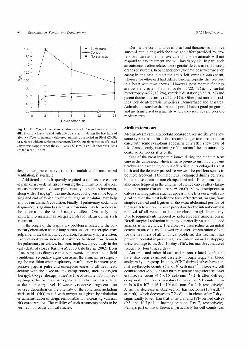
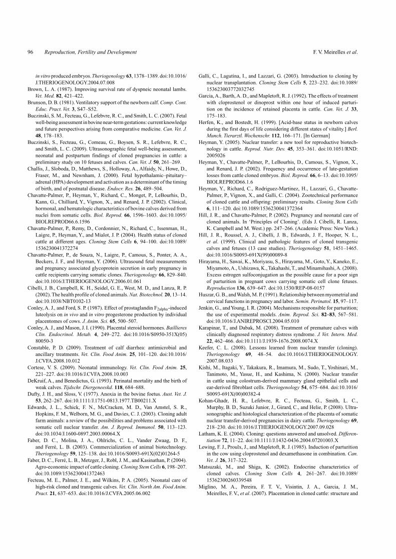
If the origin of the respiratory problem in an animal islung immaturity and there is little or no success after oxygentherapy and/or aminophylline administration, treatment with sur-factants is a good option. Our experience suggests that surfactanttreatment in animals suffering from meconium aspiration, forexample, may improve blood oxygenation. There are many sur-factants on the market and two main types may be available:(1) a crude surfactant extracted from bovine or swine lungs;and (2) a synthetic surfactant. A previous study showed that theuse of crude surfactants is more effective (Karapinar and Dabak2008). However, the cost of the treatment may hinder its use.In our practice, the surfactant is effective in rapidly increasingPao2 in cloned calves when after administration of 300 mg–1 g
in the first hour of life (Pao2 127.0 ± 11.1 mmHg after 6 h treat-ment v. 57.2 ± 5.8 mmHg in untreated animals; P < 0.05; Fig. 3).However, we did not observe any beneficial effect of treatinganimals 24 h after birth (88.7 ± 17.5 mmHg in animals treatedbefore 1 h of life v. 64.7 ± 5.2 mmHg in animals treated at 24 h v.62.1 ± 7.9 mmHg in untreated animals 48 h after birth). Otherstudies have also shown that administration of surfactant at anearly stage of lung disease results in superior results comparedwith delayed treatment (Karapinar and Dabak 2008). Surfac-tant is generally administered by intubation, because it must beintroduced intratracheally. In our facility, we do not performintubations and have instead chosen to treat animals by inject-ing lower doses (300 mg) through the trachea with a 26-gaugeneedle. After injection, the animal is turned in three differentpositions to allow diffusion of the surfactant to the caudal posi-tions and to each lung. Ideally, serial blood gas analyses mustbe done to monitor the calf’s response to whichever therapyis implemented. Ultimately, calves with persistent hypercapnia,
94 Reproduction, Fertility and Development F. V. Meirelles et al.
0 2 4 6
Hours after birth
20
40
60
80
100PaO
2
120
140
SurfactantControlNo surfactant160
180
8 22 24
Fig. 3. The Pao2 of cloned and control calves 1, 2, 6 and 24 h after birth.(�), Pao2 of clones treated with 0.3–1 g surfactant during the first hour oflife; (•), Pao2 of naturally delivered animals as reported in Bleul (2009);(�), clones without surfactant treatment. The O2 supplementation of clonedcalves was stopped when the Pao2 was >80 mmHg or 24 h after birth. Dataare the mean ± s.e.m.
despite therapeutic intervention, are candidates for mechanicalventilation, if available.
Additional care is frequently required to decrease the chanceof pulmonary oedema, also favouring the elimination of alveolarmucus/meconium. As examples, mucolytics such as bromexin,along with 0.1 mg kg−1 dexamethasone, both given at the begin-ning and end of topical treatment using an inhalator, may helpimprove an animal’s condition. Finally, if pulmonary oedema isdiagnosed, using diuretics such as furosemide may help decreasethe oedema and the related negative effects. Obviously, it isimportant to maintain an adequate hydration status during suchtreatment.
If the origin of the respiratory problem is related to the pul-monary circulation and/or lung perfusion, certain therapies mayhelp ameliorate the hypoxic condition. Pulmonary hypertension,likely caused by an increased resistance to blood flow throughthe pulmonary arterioles, has been implicated previously in theearly death of clones (Kishi et al. 2000; Cibelli et al. 2002). Evenif not simple to diagnose in a non-invasive manner under fieldconditions, secondary signs can assist the clinician in suspect-ing the condition when respiratory insufficiency is present (e.g.positive jugular pulse and unresponsiveness to all treatmentsdealing with the alveolar/lung compartment, such as oxygentherapy). Oxygen therapy is the first line of treatment for improv-ing lung perfusion, because oxygen can function as a vasodilatorat the pulmonary level. However, vasoactive drugs can alsobe used depending on the intensity of the condition, includingnitric oxide (NO) mixed with oxygen and given by inhalationor administration of drugs responsible for increasing vascularNO concentration. The validity of such treatments needs to beverified in broader clinical studies.
Despite the use of a range of drugs and therapies to improvesurvival rate, along with the time and effort provided by pro-fessional care at the intensive care unit, some animals will notrespond to any treatment and will invariably die. In part, suchan outcome is often related to congenital defects in vital tissues,organs or systems. In our experience, we have observed two suchcases; in one case, almost the entire left ventricle was absent,whereas the other calf had dilated cardiomyopathy that resultedin a heart with ‘two apexes’. However, post mortem findingsare generally patent foramen ovale (13/22; 59%), myocardialhypertrophy (4/22; 18.2%), ventricle dilatation (2/22; 9.1%) andpatent ductus arteriosus (2/22; 9.1%). Other post mortem find-ings include atelectasis, umbilicus haemorrhage and anasarca.Animals that survive the perinatal period have a good prognosisand are transferred to a facility where they receive care over themedium term.
Medium-term care
Medium-term care is important because calves are likely to showsome symptoms at birth that require longer-term treatment orcare, with some symptoms appearing only after a few days oflife. Consequently, monitoring of the animal’s health status maycontinue for weeks after birth.
One of the most important issues during the medium-termcare is the umbilicus, which is more prone to turn into a patenturachus and ascending omphaloflebitis due its enlarged size atbirth and the delivery procedure per se. The problem seems tobe more frequent if the umbilicus is clamped during delivery,but can also occur in non-clamped animals. Patent urachus isalso more frequent in the umbilici of cloned calves after clamp-ing and rupture (Batchelder et al. 2007). Many descriptions ofcalves showing patent urachus appear in the literature, with sur-gical ablation the most indicated form of treatment, ranging fromsimple removal and ligation of the extra-abdominal portion ofthe vessels to a more invasive procedure for the intra-abdominalremoval of all vessels and the urachus through laparotomy.Due to requirements imposed by Zebu breeders’ associations inBrazil, surgical reduction in many genetically valuable clonedanimals is not a choice. Therefore, we used iodine at an initialconcentration of 10% followed by a later concentration of 2%for the treatment of all umbilical problems; this treatment hasproven successful in preventing navel infections and in stoppingurine drainage by the 3rd–4th day of life, but must be conductedfrequently (four times a day).
Anaemia and other blood- and plasma-related conditionshave also been examined carefully through sequential bloodanalyses by our group. Initially, SCNT-derived calves have nor-mal erythrocyte counts (6.3 × 10⁶ cells mm−3). However, cellcounts decrease 6–12 h after birth, reaching a significantly lowererythrocyte count (4.5 × 10⁶ cells mm−3) 24 h after deliverycompared with counts in naturally mated or IVF control ani-mals (6.0 × 10⁶ and 6.1 × 10⁶ cells mm−3 at 24 h, respectively).A similar decrease is observed for haemoglobin (10.9 g dL−1
at birth), which decreases to 7.2 g dL−1 in clones after 7 days,significantly lower than that in natural and IVF-derived calves(9.5 and 10.7 g dL−1 haemoglobin on Day 7, respectively).Perhaps part of this difference, particularly for cell counts, can

Delivering zebu cloned offspring Reproduction, Fertility and Development 95
be explained by a higher haemodilution effect caused by theadministration of colostrum, because cloned calves will likelyreceive more colostrum at an earlier time (first few hours; viaoesophageal cannulation) than control animals. However, inaccordance with what has been reported previously (Chavatte-Palmer et al. 2004), both blood parameters start increasing after15 days, reaching normal values by 30 days after birth. Althoughthese parameters are different between cloned and natural orIVF-derived animals, one must evaluate such information withcaution, because our cloned calves have been exposed to oxy-gen therapy after birth, whereas IVF and natural calves are not.Regardless of the aetiology, if the animals are anaemic, we startadministering iron ion supplementation 3 days after birth. Inaddition, a series of autologous blood transfusions (200–300 mLwhole blood from a cell donor animal given daily in two or fourtransfusion sections or whenever deemed necessary) through thefirst few days of life is a good option to assist cloned calves inovercoming the unfavourable state caused by subclinical or evenclinical anaemia. In addition, such transfusions help boost thetransfer of passive immunity to the calf, along with cells fromthe white blood cell lineage (in fact, plasma can also be givento the calf for that same purpose). Nonetheless, the use of bloodtransfusions in tropical conditions requires further attention topotential infection with blood-borne diseases, such as babesiosisand/or anaplasmosis.
Many of our cloned animals develop diarrhoea days or weeksafter birth. Cloned calves with diarrhoea are generally treatedlike any other calf, with special attention to hydration status.However, if needed, many other treatments are offered, includingkaolin, activated charcoal, sulfonamides and antibiotics, depend-ing on the severity of the condition (Constable 2009). Becausemany calves exhibit voracious appetites early in life, specialcare with the environment where the calf is located is essentialbecause the ingestion of foreign bodies and bedding is rathercommon in such animals.
In general, hyperthermia should be viewed as a sign ofsepsis and the origin and/or cause should be determined andtreated. However, paradoxical hyperthermia in clones has beendescribed in many studies (Chavatte-Palmer et al. 2002; Fecteauet al. 2005). Some of our animals had peak rectal temper-atures of 43◦C with no other associated clinical signs. Thisparadoxical hyperthermia typically does not respond to treat-ment with non-steroidal anti-inflammatory drugs and the causeof this hyperthermia remains unclear. In our experience, we haveobserved this phenomenon in 14.3% (4/28) of animals duringthe first week of life (Days 1–7). Treatment includes maintain-ing animals in a fresh and well-ventilated area, providing a 1 : 4alcohol : water mix body bath and even i.v. or i.m. administra-tion of drugs to relieve the fever, such as dipirone sodium, ifthe temperature reaches 40.5◦C. Considering the disturbancesseen in basic physiological functions in clones, such as alteredappetite, satiety, temperature control and difficulties in regulat-ing cardiorespiratory function on occasions, these clinical eventsmay be linked to immaturity of the hypothalami regions. Studiesdesigned to explore these possibilities are still needed in clonedanimals.
Flexural and angular limb deformities are commonly seen incloned calves to various degrees, affecting primarily the carpi
and fetlocks (Fecteau et al. 2005). Our Zebu calves frequentlyshow tendon laxity, most often affecting the rear limbs. This lax-ity results in difficulty standing in the first hours, which graduallydisappears without any treatment. Although these animals havegood prognoses, it is important to protect the animals’ hoovesand to avoid abrasive surfaces.
Another interesting, but less relevant, and common symptomin cloned calves in our experience is alopecia. This phenomenonis intense in approximately one-third of animals, with a mildmanifestation generally occurring 30 days after birth or beforethe new hair coat is grown. Even though this is not relevantin terms of health and prognosis to adulthood, we observedthat treating calves with vitamins A, D and E decreases theoccurrence and severity of the alopecia.
Concluding remarks
As stated above, SCNT is a very promising biotechnology, espe-cially for cattle. However, in order for it to achieve broader-scaleapplication in the field, increased efficiency is required at manylevels. One of the major areas for further development is thenuclear reprogramming that occurs during the cloning process,which ideally should enable normal placental and/or fetal devel-opment, resulting in normal pregnancies that reach term and endin the delivery of healthy offspring.Although advances in knowl-edge are most likely to increase SCNT efficiency, the progressin this direction is quite slow. Thus, in the mean time, thereis a need for the development of obstetric and neonatal mea-sures that can increase the chances of obtaining viable offspring.This will certainly contribute to our knowledge in managinghigh-risk pregnancies in cattle, as well as contributing to theimplementation of SCNT after obtaining a greater knowledge ofdevelopmental reprogramming.
AcknowledgementsThe authors thank Dr Lawrence C. Smith for his valuable commentson the text and Dr Isaias Raw from the Butantan Foundation for providingthe surfactant used in our clone animals. Finally, the authors acknowledgethe collaboration with Clonest Ltd and their clients, which provided theopportunity to deal with cloned animals.
ReferencesArdran, G. M., Dawes, G. S., and Prichard, M. M. (1952). The effect of
ventilation of the fetal lungs upon the pulmonary circulation. J. Physiol.118, 12–22.
Batchelder, C. A., Bertolini, M., Mason, J. B., Moyer, A. L., Hoffert, K. A.,Petkov, S. G., Famula, T. R., Angelos, J., George, L. W., and Anderson,G. B. (2007). Perinatal physiology in cloned and normal calves:hematologic and biochemical profiles. Cloning Stem Cells 9, 83–96.
Bertolini, M., and Anderson, G. B. (2002). The placenta as a contributor toproduction of large calves. Theriogenology 57, 181–187. doi:10.1016/S0093-691X(01)00665-3
Bleul, U. (2009). Respiratory distress syndrome in calves. Vet. Clin. NorthAm. Food Anim. Pract. 25, 179–193. doi:10.1016/J.CVFA.2008.10.002
Bousquet, D., and Blondin, P. (2004). Potential uses of cloning in breed-ing schemes: dairy cattle. Cloning Stem Cells 6, 190–197. doi:10.1089/1536230041372373
Breukelman, S. P., Perényi, Z., Ruigh, L., van Wagtendonk-de Leeuw, A. M.,Jonker, F. H., et al. (2005). Plasma concentrations of bovine pregnancy-associated glycoprotein (bPAG) do not differ during the first 119days between ongoing pregnancies derived by transfer of in vivo and
96 Reproduction, Fertility and Development F. V. Meirelles et al.
in vitro produced embryos.Theriogenology 63, 1378–1389. doi:10.1016/J.THERIOGENOLOGY.2004.07.008
Brown, L. A. (1987). Improving survival rate of dyspneic neonatal lambs.Vet. Med. 82, 421–422.
Brunson, D. B. (1981). Ventilatory support of the newborn calf. Comp. Cont.Educ. Pract. Vet. 3, S47–S52.
Buczinski, S. M., Fecteau, G., Lefebvre, R. C., and Smith, L. C. (2007). Fetalwell-being assessment in bovine near-term gestations: current knowledgeand future perspectives arising from comparative medicine. Can. Vet. J.48, 178–183.
Buczinski, S., Fecteau, G., Comeau, G., Boysen, S. R., Lefebvre, R. C.,and Smith, L. C. (2009). Ultrasonographic fetal well-being assessment,neonatal and postpartum findings of cloned pregnancies in cattle: apreliminary study on 10 fetuses and calves. Can. Vet. J. 50, 261–269.
Challis, J., Sloboda, D., Matthews, S., Holloway, A., Alfaidy, N., Howe, D.,Fraser, M., and Newnham, J. (2000). Fetal hypothalamic–pituitary–adrenal (HPA) development and activation as a determinant of the timingof birth, and of postnatal disease. Endocr. Res. 26, 489–504.
Chavatte-Palmer, P., Heyman, Y., Richard, C., Monget, P., LeBourhis, D.,Kann, G., Chilliard, Y., Vignon, X., and Renard, J. P. (2002). Clinical,hormonal, and hematologic characteristics of bovine calves derived fromnuclei from somatic cells. Biol. Reprod. 66, 1596–1603. doi:10.1095/BIOLREPROD66.6.1596
Chavatte-Palmer, P., Remy, D., Cordonnier, N., Richard, C., Issenman, H.,Laigre, P., Heyman, Y., and Mialot, J. P. (2004). Health status of clonedcattle at different ages. Cloning Stem Cells 6, 94–100. doi:10.1089/1536230041372274
Chavatte-Palmer, P., de Souza, N., Laigre, P., Camous, S., Ponter, A. A.,Beckers, J. F., and Heyman, Y. (2006). Ultrasound fetal measurementsand pregnancy associated glycoprotein secretion in early pregnancy incattle recipients carrying somatic clones. Theriogenology 66, 829–840.doi:10.1016/J.THERIOGENOLOGY.2006.01.061
Cibelli, J. B., Campbell, K. H., Seidel, G. E., West, M. D., and Lanza, R. P.(2002). The health profile of cloned animals. Nat. Biotechnol. 20, 13–14.doi:10.1038/NBT0102-13
Conley, A. J., and Ford, S. P. (1987). Effect of prostaglandin F2alpha-inducedluteolysis on in vivo and in vitro progesterone production by individualplacentomes of cows. J. Anim. Sci. 65, 500–507.
Conley, A. J., and Mason, J. I. (1990). Placental steroid hormones. BaillieresClin. Endocrinol. Metab. 4, 249–272. doi:10.1016/S0950-351X(05)80050-3
Constable, P. D. (2009). Treatment of calf diarrhea: antimicrobial andancillary treatments. Vet. Clin. Food Anim. 25, 101–120. doi:10.1016/J.CVFA.2008.10.012
Cortese, V. S. (2009). Neonatal immunology. Vet. Clin. Food Anim. 25,221–227. doi:10.1016/J.CVFA.2008.10.003
DeKruif, A., and Benedictus, G. (1993). Perinatal mortality and the birth ofweak calves. Tijdschr. Diergeneeskd. 118, 684–688.
Dufty, J. H., and Sloss, V. (1977). Anoxia in the bovine foetus. Aust. Vet. J.53, 262–267. doi:10.1111/J.1751-0813.1977.TB00211.X
Edwards, J. L., Schick, F. N., McCracken, M. D., Van Amstel, S. R.,Hopkins, F. M., Welborn, M. G., and Davies, C. J. (2003). Cloning adultfarm animals: a review of the possibilities and problems associated withsomatic cell nuclear transfer. Am. J. Reprod. Immunol. 50, 113–123.doi:10.1034/J.1600-0897.2003.00064.X
Faber, D. C., Molina, J. A., Ohlrichs, C. L., Vander Zwaag, D. F.,and Ferré, L. B. (2003). Commercialization of animal biotechnology.Theriogenology 59, 125–138. doi:10.1016/S0093-691X(02)01264-5
Faber, D. C., Ferré, L. B., Metzger, J., Robl, J. M., and Kasinathan, P. (2004).Agro-economic impact of cattle cloning. Cloning Stem Cells 6, 198–207.doi:10.1089/1536230041372463
Fecteau, M. E., Palmer, J. E., and Wilkins, P. A. (2005). Neonatal care ofhigh-risk cloned and transgenic calves. Vet. Clin. North Am. Food Anim.Pract. 21, 637–653. doi:10.1016/J.CVFA.2005.06.002
Galli, C., Lagutina, I., and Lazzari, G. (2003). Introduction to cloning bynuclear transplantation. Cloning Stem Cells 5, 223–232. doi:10.1089/153623003772032745
Garcia, A., Barth, A. D., and Mapletoft, R. J. (1992). The effects of treatmentwith cloprostenol or dinoprost within one hour of induced parturi-tion on the incidence of retained placenta in cattle. Can. Vet. J. 33,175–183.
Herfen, K., and Bostedt, H. (1999). [Acid-base status in newborn calvesduring the first days of life considering different states of vitality.] Berl.Munch. Tierarztl. Wochenschr. 112, 166–171. [In German]
Heyman, Y. (2005). Nuclear transfer: a new tool for reproductive biotech-nology in cattle. Reprod. Nutr. Dev. 45, 353–361. doi:10.1051/RND:2005026
Heyman, Y., Chavatte-Palmer, P., LeBourhis, D., Camous, S., Vignon, X.,and Renard, J. P. (2002). Frequency and occurrence of late-gestationlosses from cattle cloned embryos. Biol. Reprod. 66, 6–13. doi:10.1095/BIOLREPROD66.1.6
Heyman, Y., Richard, C., Rodriguez-Martinez, H., Lazzari, G., Chavatte-Palmer, P., Vignon, X., and Galli, C. (2004). Zootechnical performanceof cloned cattle and offspring: preliminary results. Cloning Stem Cells6, 111–120. doi:10.1089/1536230041372364
Hill, J. R., and Chavatte-Palmer, P. (2002). Pregnancy and neonatal care ofcloned animals. In ‘Principles of Cloning’. (Eds J. Cibelli, R. Lanza,K. Campbell and M. West.) pp. 247–266. (Academic Press: New York.)
Hill, J. R., Roussel, A. J., Cibelli, J. B., Edwards, J. F., Hooper, N. L.,et al. (1999). Clinical and pathologic features of cloned transgeniccalves and fetuses (13 case studies). Theriogenology 51, 1451–1465.doi:10.1016/S0093-691X(99)00089-8
Hirayama, H., Sawai, K., Moriyasu, S., Hirayama, M., Goto, Y., Kaneko, E.,Miyamoto,A., Ushizawa, K.,Takahashi,T., and Minamihashi,A. (2008).Excess estrogen sulfoconjugation as the possible cause for a poor signof parturition in pregnant cows carrying somatic cell clone fetuses.Reproduction 136, 639–647. doi:10.1530/REP-08-0157
Huszar, G. B., andWalsh, M. P. (1991). Relationship between myometrial andcervical functions in pregnancy and labor. Semin. Perinatol. 15, 97–117.
Jenkin, G., andYoung, I. R. (2004). Mechanisms responsible for parturition;the use of experimental models. Anim. Reprod. Sci. 82–83, 567–581.doi:10.1016/J.ANIREPROSCI.2004.05.010
Karapinar, T., and Dabak, M. (2008). Treatment of premature calves withclinically diagnosed respiratory distress syndrome. J. Vet. Intern. Med.22, 462–466. doi:10.1111/J.1939-1676.2008.0074.X
Keefer, C. L. (2008). Lessons learned from nuclear transfer (cloning).Theriogenology 69, 48–54. doi:10.1016/J.THERIOGENOLOGY.2007.08.033
Kishi, M., Itagaki, Y., Takakura, R., Imamura, M., Sudo, T., Yoshinari, M.,Tanimoto, M., Yasue, H., and Kashima, N. (2000). Nuclear transferin cattle using colostrum-derived mammary gland epithelial cells andear-derived fibroblast cells. Theriogenology 54, 675–684. doi:10.1016/S0093-691X(00)00382-4
Kohan-Ghadr, H. R., Lefebvre, R. C., Fecteau, G., Smith, L. C.,Murphy, B. D., Suzuki Junior, J., Girard, C., and Helie, P. (2008). Ultra-sonographic and histological characterization of the placenta of somaticnuclear transfer-derived pregnancies in dairy cattle. Theriogenology 69,218–230. doi:10.1016/J.THERIOGENOLOGY.2007.09.028
Latham, K. E. (2004). Cloning: questions answered and unsolved. Differen-tiation 72, 11–22. doi:10.1111/J.1432-0436.2004.07201003.X
Lewing, F. J., Proulx, J., and Mapletoft, R. J. (1985). Induction of parturitionin the cow using cloprostenol and dexamethasone in combination. Can.Vet. J. 26, 317–322.
Matsuzaki, M., and Shiga, K. (2002). Endocrine characteristics ofcloned calves. Cloning Stem Cells 4, 261–267. doi:10.1089/15362300260339548
Miglino, M. A., Pereira, F. T. V., Visintin, J. A., Garcia, J. M.,Meirelles, F. V., et al. (2007). Placentation in cloned cattle: structure and

Delivering zebu cloned offspring Reproduction, Fertility and Development 97
microvascular architecture. Theriogenology 68, 604–617. doi:10.1016/J.THERIOGENOLOGY.2007.04.060
Nagy, D. W. (2009). Resuscitation and critical care of neonatal calves.Vet. Clin. North Am. Food Anim. Pract. 25, 1–11. doi:10.1016/J.CVFA.2008.10.008
Nasser, L. F., Bo, G.A., Barth,A. D., and Mapletoft, R. J. (1994). Induction ofparturition in cattle: effect of triamcinolone pretreatment on the incidenceof retained placenta. Can. Vet. J. 35, 491–496.
Nasser, L. F., Rezende, L. F., Bó, G. A., and Barth, A. (2008). Induc-tion of parturition in Zebu-cross recipients carrying in vitro-producedBos indicus embryos. Theriogenology 69, 116–123. doi:10.1016/J.THERIOGENOLOGY.2007.09.002
Oback, B. (2008). Climbing mount efficiency: small steps, not giant leapstowards higher cloning success in farm animals. Reprod. Domest. Anim.43, 407–416. doi:10.1111/J.1439-0531.2008.01192.X
Oback, B., and Wells, D. N. (2007). Cloning cattle: the methods in themadness. Adv. Exp. Med. Biol. 591, 30–57. doi:10.1007/978-0-387-37754-4_3
Panarace, M., Agüero, J. I., Garrote, M., Jauregui, G., Segovia, A.,et al. (2007). How healthy are clones and their progeny: 5 yearsof field experience. Theriogenology 67, 142–151. doi:10.1016/J.THERIOGENOLOGY.2006.09.036
Poulsen, K. P., and McGuirk, S. M. (2009). Respiratory disease of thebovine neonate. Vet. Clin. North Am. Food Anim. Pract. 25, 121–137.doi:10.1016/J.CVFA.2008.10.007
Rogers, P. A. M. (1977). Revival in collapse, shock, respiratory failure andnarcotic overdose. Vet. Rec. 101, 215–219.
Sullivan, H. E., Orsini, J. A., and Divers, T. J. (2008). Foal cardiopulmonaryresuscitation. In ‘Manual of Equine Emergencies’. (Eds T. J. Divers andJ. A. Orsini.) pp. 740–743. (Saunders: Philadelphia.)
http://www.publish.csiro.au/journals/rfd
Szenci, O., Fazekas, Z., and Tores, I. (1980). Treatment of asphyctic newborncalves with Dopram-V. Magyar Allatorvosok Lapja 35, 420–422.
Uystepruyst, C., Coghe, J., Dorts, T., Harmegnies, N., Delsemme, M.-H.,Art, T., and Lekeux, P. (2002a). Sternal recumbency or suspension by thehind legs immediately after delivery improves respiratory and metabolicadaptation to extra uterine life in newborn calves delivered by Caesareansection. Vet. Res. 33, 709–724. doi:10.1051/VETRES:2002051
Uystepruyst, C. H., Coghe, J., Dorts, T. H., Harmegnies, N.,Delsemme, M. H.,Art,T., and Lekeux, P. (2002b). Effect of three resusci-tation procedures on respiratory and metabolic adaptation to extra uterinelife in newborn calves. Vet. J. 163, 30–44. doi:10.1053/TVJL.2001.0633
House, J. K. (2002). Postpartum assessment and care of the newborn rumi-nants. In ‘Large Animal Internal Medicine’. (Ed. B. P. Smith.) p. 279.(Mosby: St Louis.)
Wells, D. N., Misica, P. M., and Tervit, H. R. (1999). Production of clonedcalves following nuclear transfer with cultured adult mural granulosacells. Biol. Reprod. 60, 996–1005. doi:10.1095/BIOLREPROD60.4.996
Wells, D. N., Forsyth, J. T., McMillan, V., and Oback, B. (2004). The healthof somatic cell cloned cattle and their offspring. Cloning Stem Cells 6,101–110. doi:10.1089/1536230041372300
Wilmut, I. (2006). Are there any normal clones? Methods Mol. Biol. 348,307–318. doi:10.1007/978-1-59745-154-3_21
Wilmut, I., Schnieke, A. E., McWhir, J., Kind, A. J., and Campbell, K. H.(1997). Viable offspring derived from fetal and adult mammalian cells.Nature 385, 810–813. doi:10.1038/385810A0
Wilmut, I., Sullivan, G., and Taylor, J. (2009). A decade of progress since thebirth of Dolly. Reprod. Fertil. Dev. 21, 95–100. doi:10.1071/RD08216