Iron Insufficiency Compromises Motor Neurons and Their Mitochondrial Function in Irp2-Null Mice
Upload
independentCategory
view
0download
0
ORIGINAL RESEARCH
Inhibition of phosphatase and tensinhomologue (PTEN) in human ovaryin vitro results in increased activationof primordial follicles but compromisesdevelopment of growing folliclesMarie McLaughlin1,*, Hazel L. Kinnell2, Richard A. Anderson2,and Evelyn E. Telfer1
1Institute of Cell Biology and Centre for Integrative Physiology, University of Edinburgh, Hugh Robson Building, George Square, Edinburgh EH89XD, UK 2Medical Research Council Centre for Reproductive Health, Queen’s Medical Research Institute, University of Edinburgh, 47 LittleFrance Crescent, Edinburgh EH16 4TJ, UK
*Correspondence address. Tel: +44-131-6511704; Fax: +44-131-6502872; E-mail:[email protected]
Submitted on January 14, 2014; resubmitted on May 1, 2014; accepted on May 9, 2014
abstract: In the mammalian ovarya small number of follicles are steadily recruited from the quiescent pool to undergo development. Follicleloss, maintenance and growth are strictly controlled by complex molecular interactions including the phosphoinositide 3-kinase (PI3K)-proteinkinase B (Akt) signalling pathway. Stimulation of PI3K promotes phosphorylation of Akt resulting in follicle survival and activation of growthwhereas this pathway is suppressed by the actions of the phosphatase and tensin homologue (PTEN). The aim of this study was to determinethe effect of dipotassium bisperoxo(5-hydroxypyridine-2-carboxyl)oxovanadate (bpV), a reversible inhibitor of PTEN, on the activation, survivaland development of human ovarian follicles in vitro. Biopsied ovarian tissue fragments were obtained from 17 women aged 23–46 years andexposed to 1 mM bpV(HOpic) (n ¼ 146) or control medium (n ¼ 128) for 24 h. Media were then replaced with control medium and alltissue incubated for a further 5 days. Ovarian tissue from each treatment group was fixed after the initial 24 h culture period and phosphorylatedAkt was quantified by western blotting. After 6 days incubation all tissue fragments were inspected under light microscopy and any secondaryfollicles ≥100 mm isolated. Isolated follicles were cultured individually in control medium supplemented with 100 ng/ml recombinant humanactivin A. Tissue fragments without follicles suitable for isolation were fixed and processed for histological and immunohistochemical analysis.During 6 days culture, follicle activation occurred in tissue samples from both treatment groups but with significantly more follicles progressingto the secondary stage of development in the presence of 1 mM bpV(HOpic) compared with control (31 versus 16%; P , 0.05). Increased ac-tivation was associated with increased Akt phosphorylation and increased nuclear export of FOXO3. However isolated and cultured follicles thathad been exposed to bpV(HOpic) showed limited growth and reduced survival compared with follicles from control fragments (P , 0.05). Thisstudy demonstrates that inhibition of PTEN with bpV(HOpic) affects human ovarian follicle development by promoting the initiation of folliclegrowth and development to the secondary stage, as in rodent species, but severely compromises the survival of isolated secondary follicles.
Key words: PTEN / in vitro / human / follicle / ovary
IntroductionHuman ovarian follicles largely exist as a quiescent population, of which asmall number daily initiate growth throughout reproductive life. Only asmall proportion of these follicles go on to complete growth andrelease a mature fertilizable oocyte. Remaining follicles degenerateeither from the dormant state (Tingen et al., 2009) or after growth hasbeen initiated (Kaipia and Hsueh, 1997). Development of growing
follicles is controlled by the coordinated actions of multiple complex,integrated signalling pathways regulated by local and systemic hormonalsignals (Pangas, 2007; Sobinoff et al., 2013) and reproductive senescenceoccurs when the quiescent follicle population is exhausted through acti-vation and degeneration. Prior to exhaustion of the follicle pool, the over-whelming majority of human follicles are dormant and can persist in thisstate for decades. The ability to recruit these dormant follicles into thegrowing pool and support their complete development in vitro would
& The Author 2014. Published by Oxford University Press on behalf of the European Society of Human Reproduction and Embryology.This is an Open Access article distributed under the terms of the Creative Commons Attribution License (http://creativecommons.org/licenses/by/3.0/), which permits unrestricted reuse,distribution, and reproduction in any medium, provided the original work is properly cited.
Molecular Human Reproduction, Vol.0, No.0 pp. 1–9, 2014
doi:10.1093/molehr/gau037 Mol. Hum. Reprod. Advance Access published June 9, 2014
at Edinburgh U
niversity on June 10, 2014http://m
olehr.oxfordjournals.org/D
ownloaded from

address the scarcity of oocytes available for assisted reproduction tech-niques (ART), fertility preservation and provide basic scientific informa-tion on human germ cell development.
Biochemical and genetic manipulation studies in the mouse have iden-tified the phosphoinositide 3-kinase - protein kinase B (PI3K-Akt) signal-ling pathway as a key mechanism involved in the maintenance of folliclegrowth and loss. Several growth factors, including follicle stimulatinghormone (FSH), stimulate PI3K to activate phosphoinositide-dependentprotein kinase-1 (PDK1) resulting in phosphorylation of Akt and down-stream transcription factors including the forkhead winged helix box O1(FOXO1) triggering follicle activation and development (Gonzalez-Robayna et al., 2000; Alum et al., 2004). These events are reversed bythe action of phosphatase and tensin homologue (PTEN) which causesdephosphorylation of phosphatidlyinositol 3, 4, 5-triphosphate (PIP3)thus regulating the initiation of follicle growth and preventing prematureexhaustion of the follicle pool (Cantley and Neel, 1999; John et al., 2008).The effect of PTEN can be reversibly inhibited by vanadate derivativesacting as protein tyrosine phosphatase inhibitors thereby promotingthe downstream phosphorylation of Akt (Schmid et al., 2004; Morohakuet al., 2013). Recent studies have demonstrated the importance of thePTEN and the PI3K pathway within the oocyte in the regulation ofmurine ovarian follicle activation. Deletion of PTEN in mouse oocytesresulted in pan-ovarian follicle activation and premature oocyte deple-tion whereas disruption of granulosa cell-specific PTEN did not affect ini-tiation of follicle growth but increased granulosa cell proliferation andenhanced ovulation (Fan et al., 2008; Reddy et al., 2008). Li et al.(2010) investigated the effect of exposing whole mouse ovaries to a com-bination of a vanadate derivative compound dipotassium bisperoxo(5-hydroxypyridine-2-carboxyl)oxovanadate (V) (bpV(HOpic)), and 740Y-P,a cell-permeable phospho-peptide PI3K pathway promoter. These experi-ments demonstrated increased activation of dormant follicles evidenced byincreased ovarian weight, the histological detection of large antral folliclesand immunohistological detection of nuclear exclusion of forkhead boxO3 protein (FOXO3) in oocytes. After bpV(HOpic) treatment, ovariesgrafted under the kidney capsule were able to complete developmentand generate mature eggs (Li et al., 2010). Moreover using a xeno-transplantation model human ovarian follicles treated with bpV(HOpic)developed into pre-ovulatory follicles with oocytes that appeared to becapable of undergoing in vitro maturation (Li et al., 2010). It has been sug-gested that PTEN inhibitors could be used to generate mature oocytes inwomen whose oocyte reserve is impaired due to illness or treatment andto provide oocytes for derivation of embryonic stem cells (Li et al., 2010;Adhikari et al., 2012). Investigation of the human follicle response tobpV(HOpic) has so far required a xeno-transplantation model to completefollicle development. Therefore, it is not yet known how promotion of thePI3Kpathwayaffects the developmentof in vitro grown (IVG)humanovarianfollicles or whether chemically enhanced initiation of follicle growth couldprovide a population of biopsy-derived growing follicles of sufficientquality for in vitro maturation.
Culture systems have been designed to support the key stages ofhuman follicle development. The initiation of follicle growth (Hovattaet al., 1997; Abir et al., 1999; Hovatta et al., 1999; Wright et al., 1999;Picton and Gosden, 2000; Zhang et al., 2004; Telfer et al., 2008),primary to secondary transition (Abir et al., 1997, 1999; Telfer et al.,2008), pre-antral follicle development (Roy and Treacy, 1993; Abiret al., 1997; Otala et al., 2004; Telfer et al., 2008; Xu et al., 2009) andoocyte maturation (Shea et al., 1975; Alak et al., 1998; Mikkelsen et al.,
1998) have all been achieved in vitro. Despite the prevalence of primordialfollicles in the ovary (Gougeon 1986; Gosden and Telfer, 1987) develop-ment of systemssupporting the continuum of initiation of growth throughto final oocyte maturation in vitro has been hampered by the limited avail-ability of human ovarian tissue and the recognized variation in follicledensity between and within cortical biopsies (Kohl et al., 2000; Quet al., 2000; Poirot et al., 2002; Schmidt et al., 2003). The purpose ofthis study was to investigate the effect of inhibition of PTEN on the initi-ation of human follicle growth and the subsequent survival and develop-ment of follicles in vitro, by incubating ovarian cortical fragments in thepresence of the phosphatase inhibitor bpV(HOpic). This approachuses a two-step culture system designed to promote the activation ofhuman follicle growth within fragments of ovarian cortex and thensupport the continued development of isolated secondary follicles.
Materials and Methods
Ovarian cortical tissueCortical tissue was obtained by ovarian biopsy from a total of 17 adultwomen; 13 undergoing elective Caesarean section and 4 who underwentlaparoscopy for non-malignant gynaecological conditions. Their mean(+SD) age was 36.5+1.3 years, ranging from 23 to 46 years. This studyreceived local ethical committee approval and all women gave informedconsent.
Tissue preparation and fragment cultureOvarian cortex was transferred to the laboratory in pre-warmed dissectionmedium [Leibovitz medium (Life Technologies Ltd, Paisley, UK) supplemen-ted with sodium pyruvate (2 mM), glutamine (2 mM) (both Life TechnologiesLtd), human serum albumin (HSA) (3 mg/ml), penicillin G (75 mg/ml) andstreptomycin (50 mg/ml) (Sigma-Aldrich Chemicals, Dorset, UK)]. The bi-opsied tissue pieces were transferred into fresh dissection medium underlaminar flow conditions and examined carefully using light microscopy to dis-tinguish cortical tissue from the underlying stroma. Damaged and/or haem-orrhagic tissue was excised allowing the pieces to flatten. With the cortexuppermost the tissue was gently stretched using the blunt edge of a scalpelblade and excess stromal tissue removed. Then using an angled incision thetissue was cut into fragments of �4 × 2 × 1 mm thick. Using a dissectingmicroscope, each fragment was examined for the presence of follicles. Amean diameter was recorded for each follicle observed. Any follicles meas-uring .40 mm were excised from the tissue fragments using either 25gauge needles attached to 1 ml syringe barrels or a no.10 blade and handleand fine forceps; this was to ensure a presumptive population of unilaminarfollicles. Three to four fragments were selected from each biopsy as 0 h con-trols and fixed in 10% buffered formalin (NBF) for histological evaluation. Theremaining pieces were cultured individually in culture medium [McCoy’s 5amedium with bicarbonate supplemented with HEPES (25 mM) (Life Tech-nologies Ltd), glutamine (3 mM) (Life Technologies Ltd), HSA (0.1%), peni-cillin G (0.1 mg/ml), streptomycin (0.1 mg/ml), transferrin (2.5 mg/ml),selenium (4 ng/ml) and ascorbic acid (50 mg/ml) (all obtained fromSigma-Aldrich Chemicals, Dorset, UK, unless otherwise stated)] or culturemedium supplemented with 1 mM bpV(HOpic) (Merck Chemicals Ltd,UK) at 378C in humidified air with 5% CO2 for 24 h. Half the tissue fragmentsfrom both groups were then snap-frozen and stored at 2808C for westernblot analysis. Medium was removed from the remaining fragments andreplaced with fresh culture medium without bpV(HOpic) and supplementedwith insulin (10 ng/ml) (Sigma-Aldrich Chemicals) and hFSH (1 ng/ml)(Sigma-Aldrich Chemicals). Tissue was incubated for a further 5 days withhalf the medium being removed and replaced every second day. A total of
2 McLaughlin et al.
at Edinburgh U
niversity on June 10, 2014http://m
olehr.oxfordjournals.org/D
ownloaded from

274 cortical fragments were cultured from 17 separate ovarian biopsies(control n ¼ 128; 1 mM bpV(HOpic) n ¼ 146).
Follicle isolation and cultureAfter 6 days of incubation tissue fragments were transferred to dissectionmedium and examined under light microscopy. Follicles were inspectedusing a dissecting microscope and a mean diameter for each was calculated.Secondary follicles ≥100 mm in diameter were dissected using 25 gaugeneedles. Tissue fragments with no detectable follicles or only unilaminar fol-licles present were fixed in NBF and processed as described below. A total of51 pre-antral follicles were isolated from tissue fragments incubated incontrol medium or medium supplemented with 1 mM bpV(HOpic). Isolatedfollicles were placed individually in 96-well V-bottomed culture plates in150 ml of culture medium supplemented with insulin (10 ng/ml), hFSH(1 ng/ml) and 100 ng/ml recombinant human activin A (rhAct A) (R & DSystems, Abingdon, UK). Isolated follicles were incubated for a further6 days at 378C in humidified air with 5% CO2. Every second day half theculture medium was removed and replaced and at the same time folliclediameter was measured using a dissecting microscope with a crossed mi-crometer.
Tissue processingOn completion of the incubation period isolated follicles and fragments ofcultured cortical tissue were fixed for 24 h in 10% neutral buffered formalinNBF and dehydrated in increasing concentrations of ethanol (70, 90 and100%). Absolute alcohol was removed and replaced with cedar wood oil(BDH Laboratory Supplies, Poole, UK) for 24 h. The tissue was removedto toluene (Fisher Scientific UK Ltd, Loughborough, UK) for 30 min toensure complete clearance of oil. Isolated follicles/cortical fragments wereindividually embedded in paraffin wax at 608C for 4 h with hourly changesof wax to ensure complete removal of toluene. Isolated follicles and corticalfragments were cut into 6 mm sections, mounted on gelatin-coated slides andleft to dryovernight prior to stainingwith haematoxylin and eosin. Unculturedtissue fragments collected from each biopsy were processed also for histo-logical analysis using the same methodology.
ImmunohistochemistrybpV(HOpic) and control treated tissue fragments were fixed in NBF, dehy-drated in alcohol, embedded in paraffin wax and cut into 6 mm sections asdescribed above to investigate the expression of FOXO3. Antigen retrievalwas performed using 0.01 M sodium citrate for 20 min and endogenous per-oxidase activity was quenched using 3% hydrogen peroxide in methanol.Tissue sections were incubated in FOXO3 #9467s monoclonal primary anti-body (Cell Signalling, Herts, UK) overnight at 48C. Negative controls wereestablished by replacing the primary antibody with goat serum. On comple-tion of incubation the sections were washed and probed with anti-rabbit sec-ondary antibody labelled with horseradish peroxidase for 30 min (ABC-EliteRabbit IgG, Vectastain Elite Kit, PK-6101, Vectastain ABC Kit, Vector, Peter-borough, UK). FOXO3 was detected using 3, 3′-diaminobenzidine (DAB)peroxidase substrate kit (Vector Laboratories Ltd, Peterborough, UK). Fol-licles were scored positive (activated) when brown staining was observed inthe ooplasm and negative (non-growing) when brown staining was observedin the germinal vesicle.
Western blottingProtein was lysed in RIPA buffer with Protease and Phosphatase Inhibitors(Roche Products Limited, Welwyn Garden City, UK). Samples (20 mg)were denatured and loaded onto 4–20% gels in Tris-Hepes running bufferwith 5 ml PageRuler Plus ladder (Thermo Fisher Scientific, Hemel Hemp-stead, UK) and run at 100 volts for �1 h. Immobilon-FL blotting paper
(Merck Millipore, Nottingham, UK) was soaked in methanol, rinsed in dis-tilled water (dH2O) and soaked in Semi-Dry Transfer Buffer (ThermoFisher Scientific). The gel was also equilibrated in dH20 and then Semi-DryTransfer Buffer before transfer for 9 min on a Pierce Fast Semi-Dry Blotter(Thermo Fisher Scientific).
Blots were rinsed in dH20 and blocked in Blocking Buffer (Rockland Immu-nochemicals, Inc. PA, USA) 1:1 in phosphate buffered saline plus 0.1% Tween(PBS-T) for 1 h at room temperature. Blots were then incubated with a rabbitpolyclonal antibody raised against either Akt (9272) or pAkt (9271) (Cell Sig-naling, both diluted 1:1000) and with a mouse monoclonal antibody raisedagainst beta-actin (Sigma-Aldrich; antibody A5441, diluted 1:5000) overnightat 48C. Blots were washed in 0.1% PBS-T and then incubated with a poly-clonal donkey antibody raised against rabbit IgG (heavy and light chain) con-jugated with Alexa Fluor680 (Invitrogen, Paisley, UK; diluted 1:10000) and adonkey polyclonal antibody raised against mouse IgG (H&L) conjugated withIRDye800 (Rockland Immunochemicals; diluted 1:10000) for 1 h at roomtemperature. They were then washed in PBS-Tween and then PBS beforescanning using a LI-COR Odyssey (LI-COR Biosciences UK Ltd, CowleyRoad, Cambridge, UK).
Evaluation of histologyA light microscope with a crossed micrometer was used to examine everysection of each fragment of cortical tissue. Using a slightly modified versionof the system employed by Telfer et al. (2008) follicles were classified accord-ing to their developmental stage dependent on the morphology and abun-dance of the granulosa cells observed as follows: (i) non-growing follicles:follicles constituting the resting pool, characterized by oocytes surroundedby a complete or incomplete single layer of cells, either all flat (primordial)or a mixed layer of flattened and cuboidal cells (transitory) (Smitz and Cortv-rindt, 2002), (ii) primary follicles: follicles comprising of an oocyte surroundedby a single layer of cuboidal granulosa cells, (iii) secondary follicles: multilami-nar follicles with more than a single layer of cuboidal granulosa cells and (iv)antral: antrum present within a multilaminar follicle. Evaluation of isolated fol-licles and follicles within cortical fragments was made on the section contain-ing the nucleolus except when determining the presence of an antrum inisolated follicles where all sections of the follicle were examined. Mean follicleand oocyte diameter was recorded for each isolated and fragment-enclosedfollicle evaluated.
Follicle morphological health was determined by assessment of oocyte ap-pearance and granulosa cell pyknosis on histological sections of tissuefragments. Follicles were assessed using the cross-section containing the nu-cleolus. Oocyte and granulosa cell morphology were assessed as describedby McLaughlin and Telfer (2010) and McLaughlin et al. (2010) with slightmodification. Briefly for follicles to be categorized as morphologically normal,the oocyte must be grossly circular, surrounded by a zona pellucida, have avisible germinal vesicle and defined nucleolus with ,10% of pyknotic granu-losa cells present; follicles meeting these criteria were considered to havesurvived the in vitro period.
Statistical analysesInter and intra-treatment differences in follicle and oocyte diameters werecompared by one-way ANOVA with subsequent t-tests. Proportional datawere compared using chi-squared analysis and Fishers Exact test.
Results
Assessment of cortical tissue piecesThe number and developmental stage of follicles in human ovarian cortexat 0 h was determined in 54 freshly fixed cortical tissue pieces of which 28contained follicles; a total of 152 follicles were identified and analysed.
Ovarian follicle growth and PTEN inhibition 3
at Edinburgh U
niversity on June 10, 2014http://m
olehr.oxfordjournals.org/D
ownloaded from

The number of follicles observed varied considerably within andbetween patients in both obstetric and gynaecological tissue piecesranging from 0 to 22 follicles. This highly irregular distribution of folliclesin human ovarian cortex has been reported previously (Block 1951;Hreinsson et al., 2002; Poirot et al., 2002; Schmidt et al., 2003; Kristensenet al., 2011). Of the 152 follicles analysed, 86.2% were non-growing withthe remaining follicles being at the primary (11.1%) or secondary stage(2.6%). No antral follicles were observed in any uncultured tissue frag-ments (Fig. 1A). No difference was observed in the maturity of folliclesbetween obstetric and gynaecological tissue pieces.
Follicle activation in cultured corticalfragmentsMicroscopic examination of cultured cortical fragments showed thatafter 6 days incubation significant initiation of follicle growth had occurredin both treatment groups compared with uncultured tissue. 45.5 and60% of follicles were observed to be growing in control and bpV(HOpic)
exposed tissue, respectively (86 of 189 follicles in control compared with187 of 312 follicles in bpV(HOpic)). Significantly more growing follicleswere observed in bpV(HOpic) treatment compared with control (P ¼0.0016). The most mature follicles present in either of the treatmentswere secondary follicles (Fig. 1B and C), with a significantly greater per-centage present in tissue exposed to bpV(HOpic) compared withcontrol (30.5 versus 16%) (P ¼ 0.012; Fig. 1D). No antral follicleswere observed in either treatment group after 6 days in vitro.
Activation of PI3K pathway in bpV(HOpic)exposed tissueTo investigate whether the PI3K pathway was influenced by suppressionof PTEN in human ovarian cortex, western blotting was performed oncontrol and bpV(HOpic) exposed tissue fragments (n ¼ 46 and 51, re-spectively) from 8 of the 17 patients included in this study; the numbervaried between patients owning to differing size of biopsies. Aminimum of four and a maximum of eight fragments per treatment
Figure 1 (A) Photomicrograph of 0 h human ovarian cortex; all follicles are at the earliest stages of development. (B and C) Photomicrographs of cul-tured ovarian cortex at Day 6 showing activation of follicle growth coincident with non-growing follicles in control (B), and in bpV(HOpic) treated corticaltissue (C) showing multiple secondary follicles. (D) Distribution of follicles in adult human ovarian cortical tissue bystage of development in uncultured tissue(0 h) and at Day 6. Follicle distribution is shown as a percentage of the total. Arrows indicate in vitro grown secondary follicles. Scale bar ¼ 50 microns.
4 McLaughlin et al.
at Edinburgh U
niversity on June 10, 2014http://m
olehr.oxfordjournals.org/D
ownloaded from
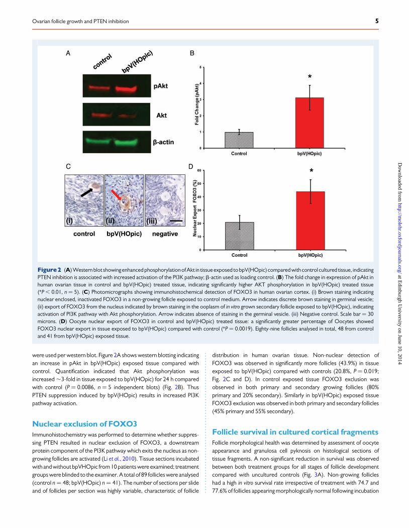
were used per western blot. Figure 2A shows western blotting indicatingan increase in pAkt in bpV(HOpic) exposed tissue compared withcontrol. Quantification indicated that Akt phosphorylation wasincreased �3-fold in tissue exposed to bpV(HOpic) for 24 h comparedwith control (P ¼ 0.0086, n ¼ 5 independent blots) (Fig. 2B). ThusPTEN suppression induced by bpV(HOpic) results in increased PI3Kpathway activation.
Nuclear exclusion of FOXO3Immunohistochemistry was performed to determine whether suppres-sing PTEN resulted in nuclear exclusion of FOXO3, a downstreamprotein component of the PI3K pathway which exits the nucleus as non-growing follicles are activated (Li et al., 2010). Tissue sections incubatedwith and without bpVHOpic from 10 patients were examined; treatmentgroups were blinded to the examiner. A total of 89 follicles were analysed(control n ¼ 48; bpV(HOpic) n ¼ 41). The number of sections per slideand of follicles per section was highly variable, characteristic of follicle
distribution in human ovarian tissue. Non-nuclear detection ofFOXO3 was observed in significantly more follicles (43.9%) in tissueexposed to bpV(HOpic) compared with controls (20.8%, P ¼ 0.019;Fig. 2C and D). In control exposed tissue FOXO3 exclusion wasobserved in both primary and secondary growing follicles (80%primary and 20% secondary). Similarly in bpV(HOpic) exposed tissueFOXO3 exclusion was observed in both primary and secondary follicles(45% primary and 55% secondary).
Follicle survival in cultured cortical fragmentsFollicle morphological health was determined by assessment of oocyteappearance and granulosa cell pyknosis on histological sections oftissue fragments. A non-significant reduction in survival was observedbetween both treatment groups for all stages of follicle developmentcompared with uncultured controls (Fig. 3A). Non-growing follicleshad a high in vitro survival rate irrespective of treatment with 74.7 and77.6% of follicles appearing morphologically normal following incubation
Figure2 (A) Western blot showing enhanced phosphorylation of Akt in tissue exposed to bpV(HOpic) compared with control cultured tissue, indicatingPTEN inhibition is associated with increased activation of the PI3K pathway; b-actin used as loading control. (B) The fold change in expression of pAkt inhuman ovarian tissue in control and bpV(HOpic) treated tissue, indicating significantly higher AKT phosphorylation in bpV(HOpic) treated tissue(*P , 0.01, n ¼ 5). (C) Photomicrographs showing immunohistochemical detection of FOXO3 in human ovarian cortex. (i) Brown staining indicatingnuclear enclosed, inactivated FOXO3 in a non-growing follicle exposed to control medium. Arrow indicates discrete brown staining in germinal vesicle;(ii) export of FOXO3 from the nucleus indicated by brown staining in the ooplasm of in vitro grown secondary follicle exposed to bpV(HOpic), indicatingactivation of PI3K pathway with Akt phosphorylation. Arrow indicates absence of staining in the germinal vesicle. (iii) Negative control. Scale bar ¼ 30microns. (D) Oocyte nuclear export of FOXO3 in control and bpV(HOpic) treated tissue: a significantly greater percentage of Oocytes showedFOXO3 nuclear export in tissue exposed to bpV(HOpic) compared with control (*P ¼ 0.0019). Eighty-nine follicles analysed in total, 48 from controland 41 from bpV(HOpic) exposed tissue.
Ovarian follicle growth and PTEN inhibition 5
at Edinburgh U
niversity on June 10, 2014http://m
olehr.oxfordjournals.org/D
ownloaded from

in control and bpV(HOpic), respectively, compared with 80.1% in uncul-tured tissue (non-growing versus control P ¼ 0.601; non-growing versusbpV(HOpic) P ¼ 0.655). No significant difference was observed inprimary or secondary follicle survival between control and 1 mMbpV(HOpic) with over 69% of follicles from both treatments appearingmorphologically normal after 6 days incubation (primary follicles,control versus bpV(HOpic) P ¼ 0.934; secondary follicles, controlversus bpV(HOpic) P ¼ 0.759) (Fig. 3A).
Growth of isolated folliclesCare was taken during mechanical isolation of follicles to ensure basallaminae were not exposed to prevent compromising follicle develop-ment. Due to the considerable variation in stromal tissue densitybetween and within biopsies the amount of tissue surrounding isolatedfollicles varied; however, sufficient stromal tissue was left enclosingeach follicle to provide a presumptive theca cell layer for further folliculardevelopment. On completion of the first step of the culture system there
was no significant difference in the diameter of follicles isolated frombpV(HOpic) exposed tissue compared with those isolated fromcontrol (Fig. 3B). Follicle diameters were recorded every second dayafter isolation until completion of the culture period. Measurementswere recorded using a dissecting microscope with acrossed micrometer.During the subsequent 6 day culture period follicles isolated from controltissue increased significantly in diameter (114+2 to 181+ 8 mm; P ,
0.001), whereas follicles isolated from tissue exposed to bpV(HOpic)exhibited limited, nonsignificant growth (115+ 1 to 120+ 3 mm;Fig. 3B). Thus at the end of this period follicles from control tissuewere significantly larger than those from bpV(HOpic) treated tissue(P ¼ 0.0001).
Survival of isolated folliclesSurvival of isolated follicles wasassessed as per the criteria used for evalu-ation of follicle survival within cultured tissue fragments described above.Three follicles degenerated during the first 4 days of individual culture; 1
Figure 3 (A) The percentage of morphologically normal follicles observed in tissue fragments in uncultured tissue (0 h) and after 6 days culture in controlor bpV(HOpic) medium. Percentages of healthy follicles are shown by developmental stage. (B) Mean diameter (mm) of follicles isolated from control(green) and bpV(HOpic) (red) exposed tissue over a further 6 days in vitro. At the end of the culture period follicles isolated from control tissue were sig-nificantly larger than bpV(HOpic) treated follicles (*P , 0.001). (C) On completion of the isolated follicle culture period a significantly greater percentage ofcontrol exposed follicles were morphologically normal compared with those exposed to bpV(HOpic) (*P , 0.01). (D) Photomicrographs of isolated fol-licles after a total of 12 days in culture. (i) Morphologically normal IVG follicle from control cultured tissue, and (ii) morphologically abnormal IVG follicleisolated from bpV(HOpic) exposed tissue. Scale bar ¼ 50 microns.
6 McLaughlin et al.
at Edinburgh U
niversity on June 10, 2014http://m
olehr.oxfordjournals.org/D
ownloaded from

from tissue exposed to control medium and 2 from tissue fragmentstreated with bpV(HOpic); data from these follicles were excludedfrom in-vitro growth analysis. Follicular morphology was assessed by in-spection of histological sections and was used to determine normalityand thus survival. Follicle survival was significantly higher in folliclesfrom tissue fragments exposed to control medium, with 13 of the 22(59%) follicles surviving the culture period being deemed morphological-ly normal compared with 7 of 26 (27%) surviving follicles from tissue frag-ments exposed to bpV(HOpic) (P ¼ 0.02; Fig. 3C). Morphologicalabnormalities observed included shrunken oocytes, widespread granu-losa cell pyknosis and loss of granulosa cell-oocyte proximity. Represen-tative images of both normal and morphologically abnormal follicles areshown in Fig. 3D.
DiscussionDisruption of the PI3K pathway using knockout models (Fan et al., 2008;Reddy et al., 2008) and pharmacological stimulators and/or inhibitors(Li et al., 2010; Adhikari et al., 2012) promotes activation of folliclegrowth in mice and xeno-transplanted human ovarian tissue andenhances murine ovulation. In this study we investigated the ability ofbpV(HOpic) a pharmacological inhibitor of PTEN, a major negative regu-lator of the PI3K pathway, to affect human ovarian follicle activation anddevelopment in both tissue fragments and isolated follicles in vitro. Wehypothesized that using an established two-step culture system (Telferet al., 2008) selective promotion of the PI3K pathway by pharmacologicalinhibition of PTEN would promote (i) initiation of growth in quiescent fol-licles within cortical tissue fragments and (ii) the development of in vitrogrown (IVG) secondary follicles to the large pre-antral and/or earlyantral stage. Our results show that exposure of human ovarian cortexto bpV(HOpic) increased the activity of the PI3K pathway and promotedfollicle activation and development to the secondary stage. However, fol-lowing isolation from cortical tissue fragments the key novel finding of thisstudy emerged, that secondary follicles previously exposed to bpV(HO-pic) grew very poorly and their survival was severely compromised.
In this study increased activity of the PI3K pathway was confirmed byan increase in Akt phosphorylation and nuclear export of FOXO3.Whilst within the germinal vesicle, FOXO3 is a recognized suppressorof primordial follicle growth; when relocated to the ooplasm this sup-pression is lifted and follicle activation occurs (Gonzalez-Robaynaet al., 2000). Li et al. (2010) also demonstrated nuclear export ofFOXO3 in the oocytes of human ovarian follicles; however, a recentstudy has challenged this paradigm and suggests that FOXO3 is not a re-quirement for human primordial follicle growth arrest (Tarnawa et al.,2013). Tarnawa and colleagues reported on data obtained from onehuman ovary whereas in this study we analysed immunohistochemicaldetection of FOXO3 exclusion from 10 patients. Whilst is unclearhow many ovaries Li et al. (2010) included in their study they also de-scribe results from multiple human biopsies; therefore we suggest the di-vergence of results may be due to the difference in the number biopsiesanalysed.
The controlled promotion of human ovarian follicle development toobtain oocytes capable of undergoing in vitro maturation and fertilizationwith subsequent embryo development is the ultimate goal of humanovarian follicle culture. Pharmacological promotion of follicle growth invitro followed by a xeno-transplantation model has resulted in embryodevelopment and live litters being achieved in mice (Li et al., 2010;
Adhikari et al., 2012). The initial results presented here are in agreementwith previous work demonstrating that pharmacological inhibition ofPTEN stimulates follicle activation in the human ovarian cortex (Liet al., 2010). In our study a concentration of 1 mM was chosenbecause in preliminary experiments using 10 and 100 mM bpV(HOpic),increased follicle growth wasassociated with grossmorphological abnor-malities in activated follicles prior to isolation from cortical fragments(not shown). We suggest that the reduced follicle survival observedwith higher concentrations of bpV(HOpic) treatment is associatedwith the specific manner in which tissue is prepared for incubation.Unlike tissue cubes, our method of tissue fragment preparation maxi-mizes the exposure of the ovarian cortex to the culture medium by loos-ening the cortical surface and removing excess dense connective stromaltissue which may prevent cortical exposure to media components andphysically impede follicle activation and development. The presentmethod of tissue preparation has been successfully employed topromote the activation of follicle growth in bovine and in fresh andcryopreserved-thawed pre-pubertal as well as pubertal and adulthuman ovarian tissue (Telfer et al., 2008; McLaughlin and Telfer, 2010;Anderson et al., 2014). We suggest that preparing the cortex in thismanner allows maximum cortical exposure to the culture medium,and in preliminary experiments determined that the optimal concentra-tion of bpV(HOpic) which promoted activation of follicle growth andmaintained normal morphology was1 mM. It is unclear whether themor-phological normality of human follicles activated by higher doses ofbpV(HOpic) was assessed prior to xeno-transplantation (Li et al.,2010). It is however noteworthy that Adhikari et al. (2012) exposedmouse ovaries to 1 mM bpV(HOpic) to produce fertilizable IVGoocytes (Adhikari et al., 2012). bpV(HOpic) activated follicles wereintact after 6 days incubation and not different in diameter from controls.bpV(HOpic) treatment did not therefore recruit a suboptimal folliclepopulation. Following a further 6 days in culture bpV(HOpic)-exposedfollicles grew poorly and deteriorated significantly. A previous studyreported normal follicular development in mice following a conditionaldeletion of the PTEN gene in oocytes with normal oocyte maturation, fer-tility and litter size observed (Jagarlamudi et al., 2009). However ourstudy is not the first report of bpV(HOpic) treatment affecting humanovarian follicle development deleteriously. Lerer-Serfaty et al. (2013) re-cently reported widespread follicle destruction in cryopreserved-thawed human tissue exposed to a high concentration of 100 mMbpV(HOpic); this supports the view that manipulation of the PI3Kpathway may also have negative impact on other signalling mechanisms(Blanco-Aparicio et al., 2007). Whilst PI3K activation and subsequentpAkt promotion by bpV(HOpic) can induce follicle activation in humanovarian tissue, it is possible that other Akt-independent effects ofPTEN inhibition cause the follicle degeneration seen in this study andby other investigators (Lerer-Serfaty et al., 2013).
Growth was limited and normal follicular morphology significantlycompromised in follicles isolated from tissue fragments exposed tobpV(HOpic). It is well established that maintenance of bi-directionaloocyte/somatic cell communication is vital for normal follicle develop-ment (Albertini et al., 2001; Eppig, 2001) and although an extensive mor-phological analysis was not possible due to degeneration, it is suggestedthat vital oocyte-somatic cell contact was damaged as a result ofbpV(HOpic) exposure. As activated follicles exposed to bpV(HOpic)were morphologically normal within tissue fragments at Day 6 of theculture period, it is possible that the surrounding stromal environment
Ovarian follicle growth and PTEN inhibition 7
at Edinburgh U
niversity on June 10, 2014http://m
olehr.oxfordjournals.org/D
ownloaded from

was able to modulate or circumvent the deleterious effects of PTEN in-hibition over a relatively short period of time. It is unlikely however thatallowing bpV(HOpic) activated follicles to remain within the presumptiveprotection of the stromal fragments would result in the development ofmorphologically mature follicles as it has been demonstrated that pro-longed culture results in widespread follicle loss by atresia (Hovattaet al., 1999).
A human live birth has recently been reported following treatment ofovarian tissue for 48 h in vitro with bpV(HOpic) and 740Y-P, an Aktstimulant, followed by replacement and IVF (Kawamura et al., 2013).This is an encouraging development but is difficult to compare directlyto the results reported here which utilize an entirely in vitro system.The human tissue Kawamura et al. (2013) incubate with Akt stimulantsis described as containing a range of both non-growing and growing fol-licles; the early oocytes retrieved from pre-ovulatory follicles werebelieved to be derived from rapidly growing secondary follicles containedin the auto-grafted tissue. Exposure to bpV(HOpic) and 740Y-P mayconfer direct or indirect protection from apoptosis on secondary folli-cles, although the absence of control experiments does not allow clearassessment of the effects of the Akt stimulants in that study. Thepresent study does not investigate this possibility as only follicles,40 mm in diameter, i.e. non-growing or primary are present at thetime of tissue exposure to bpV(HOpic).
In conclusion these data confirm that the PI3K pathway is involved inthe regulation of initiation of growth of human ovarian follicles. Treat-ment of human ovarian cortical tissue with bpV(HOpic) promotes acti-vation and development of follicles but does not confer any advantage onfollicle development beyond the secondary stage and does not prolongisolated follicle survival, indeed a significant deleterious effect was found.
It does not appear therefore that promotion of the PI3K pathway byinhibition of PTEN using bpV(HOpic) is a candidate treatment for therobust generation of numbers of good-quality mature IVG humanoocytes. Pharmacological manipulation of the PI3K pathway topromote follicle development has been successfully achieved in micevia promotion of other components of PI3K, e.g. by using 740Y-P (Liet al., 2010), and it remains to be seen whether this molecule can beused to promote follicle activation and maintain morphologicallynormal growth in human ovarian follicles.
AcknowledgementsThe authors thank Joan Creiger and Anne Saunderson for patient recruit-ment and John Binnie forexcellent technical assistance and all the patientswho donated tissue.
Authors’ rolesM.M.: conception and design of study, experimental work and acquisitionof data, analysis and interpretation of data, manuscript preparation, finalapproval of manuscript. H.L.K.: experimental work and acquisition ofdata, analysis and interpretation of data, manuscript preparation, final ap-proval of manuscript. R.A.A.: conception and design of study, analysisand interpretation of data, funding for study, manuscript preparation,final approval of manuscript. E.E.T.: conception and design of study, ana-lysis and interpretation of data, funding for study, manuscript prepar-ation, final approval of manuscript.
FundingFunded by MRC grants G0901839 and G1100357. Funding to pay theOpenAccess publication charges for this article was provided by theUniversity of Edinburgh.
Conflict of interestNone declared.
ReferencesAbir R, Franks S, Mobberley MA, Moore PA, Margara RA, Winston RM.
Mechanical isolation and in vitro growth of preantral and small antralhuman follicles. Fertil Steril 1997;68:682–688.
Abir R, Roizman P, Fisch B, Nitke S, Okon E, Orvieto R, Ben Rafael Z. study ofisolated early human follicles cultured in collagen gels for 24 hours. HumReprod 1999;14:1299–1301.
Adhikari D, Gorre N, Risal S, Zhao Z, Zhang H, Shen Y, Liu K. The safe use ofa PTEN inhibitor for the activation of dormant mouse primordial folliclesand generation of fertilizable eggs. PLoS one 2012;7:e39034.
Alak BM, Coskun S, Friedman CI, Kennard EA, Kim MH, Seifer DB. Activin Astimulates meiotic maturation of human oocytes and modulates granulosacell steroidogenesis in vitro. Fertil Steril 1998;70:1126–1130.
Albertini DF, Combelles CM, Benecchi E, Carabatsos MJ. Cellular basis forparacrine regulation of ovarian follicle development. Reproduction 2001;121:647–653.
Alum H, Maizels ET, Park Y, Ghaey S, Feiger Zj, Chandel NS, Hunzickler-Dunn M. Follicle-stimulating hormone activation of hypoxia-induciblefactor-1 by the Phosphatidylinositol 3-kinase/Akt/Ras homologenriched in brain (Rheb/mammalian target of rapamycin (mTOR)pathway is necessary for induction of select protein markers of folliculardifferentiation. J Biol Chem 2004;279:19431–19440.
Anderson RA, McLaughlin M, Wallace WHB, Albertini DA, Telfer EE. Theimmature human ovary shows loss of abnormal follicles and increasingfollicle developmental competence through childhood and adolescence.Hum Reprod 2014;29:97–106.
Blanco-Aparicio C, Renner O, Leal JF, Carnero A. PTEN, more than the AKTpathway. Carcinogenesis 2007;28:1379–1386.
Block E. Quantitative morphological investigations of the follicular system inwomen;methodsofquantitativedeterminations.ActaAnat1951;12:267–285.
Cantley LC, Neel BG. New insights into tumor suppression: PTENsuppresses tumor formation by restraining the phosphoinositide3-kinase/AKT pathway. Proc Natl Acad Sci USA 1999;96:4240–4245.
Eppig JJ. Oocyte control of ovarian follicular 454 development and function inmammals. Reproduction 2001;122:829–838.
Fan H-Y, Liu Z, Cahill N, Richards JS. Targeted disruption of Pten in ovariangranulosa cells enhances ovulation and extends the life span of luteal cells.Mol Endocrinol 2008;22:2128–2140.
Gonzalez-Robayna IJ, Falender AE, Ochsner S, Firestone GL, Richards JS.Follicle-Stimulating Hormone (FSH) stimulates phosphorylation andactivation of protein kinase B (PKB/Akt) and serum and glucocorticoid-induced kinases (Sgk): evidence for A kinase-independent signaling byFSH in granulosa cells. Mol Endocrinol 2000;14:1283–1300.
Gosden RG, Telfer E. Numbers of follicles and oocytes in mammalian ovariesand their allometric relationships. J Zool 1987;211:169–175.
Gougeon A. Dynamics of follicular growth in the human: a model frompreliminary results. Hum Reprod 1986;1:81–87.
Hovatta O, Silye R, Abir R, Krausz T, Winston RM. Extracellular matriximproves survival of both stored and fresh human primordial and primaryovarian follicles in long-term culture. Hum Reprod 1997;12:1032–1036.
8 McLaughlin et al.
at Edinburgh U
niversity on June 10, 2014http://m
olehr.oxfordjournals.org/D
ownloaded from
Hovatta O, Wright C, Krausz T, Hardy K, Winston RM. Human primordial,primary and secondary ovarian follicles in long-term culture: effect ofpartial isolation. Hum Reprod 1999;14:2519–2524.
Hreinsson JG, Scott JE, Rasmussen C, Swahn ML, Hsueh AJ, Hovatta O.Growth differentiation factor-9 promotes the growth, development, andsurvival of human ovarian follicles in organ culture. J Clin EndocrinolMetab 2002;87:316–321.
Jagarlamudi K, Liu L, Adhikari D, Reddy P, Idahl A, Ottander U, Lundin E,Liu K. Oocyte specific deletion of Pten in mice reveals a stage-specificfunction of PTEN/PI3K signalling in oocytes in controlling follicularactivation. PLoS One 2009;4:e6186.
John GB, Gallardo TD, Shirley LJ, Castrillon DH. Foxo3 is a PI3K-dependentmolecular switch controlling the initiation of oocyte growth. Dev Biol 2008;321:197–204.
Kaipia A, Hsueh AJ. Regulation of ovarian follicle atresia. Annu Rev Physiol1997;59:349–363.
Kawamura K, Cheng Y, Suzuki N, Deguchi M, Sato Y, Takae S, Ho CH,Kawamura N, Tamura M, Hashimoto S et al. Hippo signaling disruptionand Akt stimulation of ovarian follicles for infertility treatment. Proc NatlAcad Sci USA 2013;110:17474–17479.
Kohl J, Dittrich R, Siebzehnrubl E, Wildt L. Determination of follicle numbersin human ovarian biopsies—a method for estimation of outcome ofovarian cryopreservation. Fertil Steril 2000;74(Suppl 1):212.
Kristensen SG, Rasmussen A, Byskov AG, Andersen CY. Isolation ofpre-antral follicles from human ovarian medulla tissue. Hum Reprod2011;26:157–166.
Lerer-Serfaty G, Samara N, Fisch B, Shachar M, Kossover O, Seliktar D,Ben-Haroush A, Abir R. Attempted application of bioengineered/biosynthetic supporting matrices with phosphatidylinositol-trisphosphate-enhancing substances to organ culture of human primordial follicles.J Assist Reprod Genet 2013;30:1279–1288.
Li J, Kawamura K, Cheng Y, Liu S, Klein C, Liu S, Duan EK, Hsueh AJ.Activation of dormant ovarian follicles to generate mature eggs. ProcNatl Acad Sci USA 2010;107:10280–10284.
McLaughlin M, Telfer EE. Oocyte development in bovine primordial follicles ispromoted by activin and FSH within a two-step serum-free culture system.Reproduction 2010;139:971–978.
McLaughlin M, Bromfield JJ, Albertini DF, Telfer EE. Activin promotesfollicular integrity and oogenesis in cultured pre-antral bovine follicles.Mol Hum Reprod 2010;16:644–653.
Mikkelsen AL, Smith S, Lindenberg S. In-vitro maturation of immature humanoocytes. Hum Reprod 1998;13:23–24.
Morohaku K, Hoshino Y, Sasada H, Sato E. Incorporation of phosphataseinhibitor in culture promotes growth initiation of isolated non-growingoocytes. PLoS One 2013;8:e77533.
Otala M, Makinen S, Tuuri T, Sjoberg J, Pentikainen V, Matikainen T, Dunkel L.Effects of testosterone, dihydrotestosterone, and 17beta-estradiol onhuman ovarian tissue survival in culture. Fertil Steril 2004;82:1077–1085.
Pangas SA. Growth factors in ovarian development. Semin Reprod Med 2007;25:225–234.
Picton HM, Gosden RG. In vitro growth of human primordial follicles fromfrozen-banked ovarian tissue. Mol Cell Endocrinol 2000;166:27–35.
Poirot C, Vacher-Lavenu M-C, Helardot P, Guibert J, Brugieres L, Jouannet P.Human ovarian cryopreservation: indications and feasibility. Hum Reprod2002;17:1447–1452.
Qu J, Godin PA, Nisolle M, Donnez J. Distribution and epidermalgrowth factor receptor expression of primordial follicles in humanovarian tissue before and after cryopreservation. Hum Reprod 2000;15:302–310.
Reddy P, Liu L, Adhikari D, Jagarlamudi K, Rajareddy S, Shen Y, Du C, Tang W,Hamalainen T, Peng SL et al. Oocyte-specific deletion of Pten causespremature activation of the primordial follicle pool. Science 2008;319:611–613.
Roy SK, Treacy BJ. Isolation and long-term culture of human preantralfollicles. Fertil Steril 1993;59:783–790.
Schmid AC, Byrne RD, Vilar R, Woscholski R. Bisperoxovanadiumcompounds are potent PTEN inhibitors. FEBS Lett 2004;566:35–38.
Schmidt KLT, Byskov A, Nyboe Andersen A, Muller J, Yding Andersen C.Density and distribution of primordial follicles in single pieces of cortexfrom 21 patients and in individual pieces of cortex from three 527 entirehuman ovaries. Hum Reprod 2003;18:1158–1164.
Shea BF, Baker RD, Latour JP. Human follicular oocytes and their maturationin vitro. Fertil Steril 1975;26:1075–1082.
Smitz JE, Cortvrindt RG. The earliest stages of folliculogenesis in vitro.Reproduction 2002;123:185–202.
Sobinoff AP, Sutherland JM, McLaughlin EA. Intracellular signaling duringfemale gametogenesis. Mol Hum Reprod 2013;19:265–278.
Tarnawa ED, Baker MD, Aloisio GM, Carr BR, Castrillon DH. Gonadalexpression of Foxo1, but not Foxo3, is conserved in diverse Mammalianspecies. Biol Reprod 2013;88:1–11.
Telfer EE, McLaughlin M, Ding C, Thong KJ. A two-step serum-free culturesystem supports development of human oocytes from primordialfollicles in the presence of activin. Hum Reprod 2008;23:1151–1158.
Tingen CM, Bristol-Gould SK, Kiesewetter SE, Wellington JT, Shea L,Woodruff TK. Prepubertal primordial follicle loss in mice is not due toclassical apoptotic pathways. Biol Reprod 2009;81:16–25.
Wright CS, Hovatta O, Margara R, Trew G, Winston RM, Franks S, Hardy K.Effects of follicle-stimulating hormone and serum substitution on thein-vitro growth of human ovarian follicles. Hum Reprod 1999;14:1555–1562.
Xu M, Barrett SL, West-Farrell E, Kondapalli LA, Kiesewetter SE, Shea LD,Woodruff TK. In vitro grown human ovarian follicles from cancerpatients support oocyte growth. Hum Reprod 2009;24:2531–2540.
Zhang P, Louhio H, Tuuri T, Sjoberg J, Hreinsson J, Telfer EE, Hovatta O. Invitro effect of cyclic adenosine 3′, 5′-monophosphate (cAMP) on earlyhuman ovarian follicles. J Assist Reprod Genet 2004;21:301–306.
Ovarian follicle growth and PTEN inhibition 9
at Edinburgh U
niversity on June 10, 2014http://m
olehr.oxfordjournals.org/D
ownloaded from
Copyright © 2022 FDOKUMEN