Testosterone induces activation of porcine primordial follicles in vitro
10
ORIGINAL ARTICLE Testosterone induces activation of porcine primordial follicles in vitro Manjula P. S. Magamage • Mai Zengyo • Mohammad Moniruzzaman • Takashi Miyano Received: 20 July 2010 / Accepted: 1 September 2010 Ó Japan Society for Reproductive Medicine 2010 Abstract Purpose The mechanism underlying primordial follicle activation is poorly understood. In this study, in-vitro culture and subsequent xenotransplantation were conducted to determine whether testosterone promotes the activation of porcine primordial follicles. Methods Prepubertal porcine ovarian cortical strips containing primordial follicles were cultured in the pres- ence of testosterone for 7 days, and subsequently trans- planted to immunodeficient mice for 2 months. After culture and transplantation, development of follicles was examined histologically. The presence of androgen recep- tors in oocytes was assessed by use of western blot and immunohistochemical analyses. Results Testosterone at 10 -6 M induced the primordial follicle transition to the intermediate (19 ± 4%) and pri- mary (3 ± 1%) stages after 7-day culture, while 56 ± 5% of primordial follicles remained in the initial pool. Higher concentrations, above 10 -5 M, or lower concentrations, below 10 -6 M, did not induce follicle transition to the primary stage. After 7-day culture with 10 -6 M testoster- one, ovarian cortical strips were transplanted to immuno- deficient mice. Some of the follicles developed to the secondary (15 ± 3%) and antral (10 ± 3%) stages, whereas 44 ± 7% of primordial follicles remained in the initial pool. In the culture experiment, estradiol-17b (10 -7 – 10 -5 M) had no significant effect on follicle activation. The androgen receptor antagonist, cyproterone acetate, inhibited the stimulatory effect of testosterone on primor- dial follicle activation, suggesting an androgen receptor- mediated action of testosterone. Western blot and immunohistochemical analyses revealed that androgen receptors were present in the oocytes of primordial follicles. Conclusions These results suggest that testosterone at 10 -6 M promotes the activation of porcine primordial follicles in vitro through the androgen receptors in the oocytes. Keywords Androgen receptor Á Pig Á Primordial follicle activation Á Testosterone Á Xenografting Introduction In mammals, oocytes sequestered in primordial follicles remain quiescent in the ovary until recruited into the growing pool [1]. Folliculogenesis is the process by which the oocyte develops with the somatic cells and matures to a fertilizable egg [2]. The development of the follicle involves the integration of signals from both inside and outside the ovary [3]. The process of the gradual exit of primordial follicles from the non-growing follicle pool, and their transition to the intermediate or primary follicle stage, is regarded as primordial follicle activation [4]. However, the mechanism regulating primordial follicle activation is poorly understood. Elucidation of the mechanisms regu- lating primordial follicle activation, and preservation of them for a future stockpile, are important for successful assisted reproductive technology [5]. Primordial follicle activation seems to be directed pri- marily by signals in the ovary, whereas endocrine hormones from the pituitary and other organs are necessary for folli- culogenesis to proceed beyond the primary or secondary M. P. S. Magamage (&) Á M. Zengyo Á M. Moniruzzaman Á T. Miyano Laboratory of Reproductive Biology and Biotechnology, Graduate School of Agricultural Science, Kobe University, 1-1 Rokkodai-cho, Nada-ku, Kobe 657-8501, Japan e-mail: [email protected] 123 Reprod Med Biol DOI 10.1007/s12522-010-0068-z
Transcript of Testosterone induces activation of porcine primordial follicles in vitro
ORIGINAL ARTICLE
Testosterone induces activation of porcine primordialfollicles in vitro
Manjula P. S. Magamage • Mai Zengyo •
Mohammad Moniruzzaman • Takashi Miyano
Received: 20 July 2010 / Accepted: 1 September 2010
� Japan Society for Reproductive Medicine 2010
Abstract
Purpose The mechanism underlying primordial follicle
activation is poorly understood. In this study, in-vitro
culture and subsequent xenotransplantation were conducted
to determine whether testosterone promotes the activation
of porcine primordial follicles.
Methods Prepubertal porcine ovarian cortical strips
containing primordial follicles were cultured in the pres-
ence of testosterone for 7 days, and subsequently trans-
planted to immunodeficient mice for 2 months. After
culture and transplantation, development of follicles was
examined histologically. The presence of androgen recep-
tors in oocytes was assessed by use of western blot and
immunohistochemical analyses.
Results Testosterone at 10-6 M induced the primordial
follicle transition to the intermediate (19 ± 4%) and pri-
mary (3 ± 1%) stages after 7-day culture, while 56 ± 5%
of primordial follicles remained in the initial pool. Higher
concentrations, above 10-5 M, or lower concentrations,
below 10-6 M, did not induce follicle transition to the
primary stage. After 7-day culture with 10-6 M testoster-
one, ovarian cortical strips were transplanted to immuno-
deficient mice. Some of the follicles developed to the
secondary (15 ± 3%) and antral (10 ± 3%) stages,
whereas 44 ± 7% of primordial follicles remained in the
initial pool. In the culture experiment, estradiol-17b (10-7–
10-5 M) had no significant effect on follicle activation.
The androgen receptor antagonist, cyproterone acetate,
inhibited the stimulatory effect of testosterone on primor-
dial follicle activation, suggesting an androgen receptor-
mediated action of testosterone. Western blot and
immunohistochemical analyses revealed that androgen
receptors were present in the oocytes of primordial follicles.
Conclusions These results suggest that testosterone at
10-6 M promotes the activation of porcine primordial
follicles in vitro through the androgen receptors in the
oocytes.
Keywords Androgen receptor � Pig � Primordial
follicle activation � Testosterone � Xenografting
Introduction
In mammals, oocytes sequestered in primordial follicles
remain quiescent in the ovary until recruited into the
growing pool [1]. Folliculogenesis is the process by which
the oocyte develops with the somatic cells and matures to a
fertilizable egg [2]. The development of the follicle
involves the integration of signals from both inside and
outside the ovary [3]. The process of the gradual exit of
primordial follicles from the non-growing follicle pool, and
their transition to the intermediate or primary follicle stage,
is regarded as primordial follicle activation [4]. However,
the mechanism regulating primordial follicle activation is
poorly understood. Elucidation of the mechanisms regu-
lating primordial follicle activation, and preservation of
them for a future stockpile, are important for successful
assisted reproductive technology [5].
Primordial follicle activation seems to be directed pri-
marily by signals in the ovary, whereas endocrine hormones
from the pituitary and other organs are necessary for folli-
culogenesis to proceed beyond the primary or secondary
M. P. S. Magamage (&) � M. Zengyo � M. Moniruzzaman �T. Miyano
Laboratory of Reproductive Biology and Biotechnology,
Graduate School of Agricultural Science, Kobe University,
1-1 Rokkodai-cho, Nada-ku, Kobe 657-8501, Japan
e-mail: [email protected]
123
Reprod Med Biol
DOI 10.1007/s12522-010-0068-z
stage [6]. In the ovary, cross-talk between the oocytes and
somatic cells, for example granulosa cells and thecal cells,
occurs at every stage in follicle development [5]. Primordial
follicle activation is associated with oocyte growth, and
when activation occurs the surrounding pre-granulosa cells
differentiate to the cuboidal shape to form intermediate
follicles (having both cuboidal and pre-granulosa cells) [7]
or primary follicles (having a complete layer of cuboidal
granulosa cells) [6]. Subsequently, the follicle develops
to secondary, antral, and further advanced follicle stages
[6, 8, 9].
In vitro culture is an important technique for studying
the mechanism of primordial follicle activation, however,
success in the culture of primordial follicles as a method of
oocyte growth has been limited to mice. Eppig and O’Brien
[10] reported the first successful production of a baby
mouse derived from cultured primordial follicles. Several
studies with farm animals and primates have shown the
transition of primordial follicles to the primary stage
during culture of cortical strips from caprine [11], bovine
[12], baboon [13], and human ovaries [14]. In these studies,
however, primordial follicles hardly developed to the
secondary stage even when the tissues were cultured for
20 days [13, 15].
Xenografting of fresh and frozen–thawed ovarian tissues
is an alternative method for the growth of oocytes in the
primordial follicles of large animals [16, 17]. Cross-species
transplantation of ovaries from large mammals, including
humans [16, 18], marmosets [19] and sheep [20], to reci-
pient severe combined immunodeficient (SCID) mice or
nude mice results in the development of antral follicles.
The duration of ovarian xenografting depends on the time
course necessary for the follicular development of the
donors. Primordial follicles developed to the antral stage
45–75 days after xenografting of the ovarian tissues from
20-day-old piglets [21]. However, the development of
primordial follicles from cows [22] and prepubertal pigs
[23] was not initiated 6–8 weeks after xenografting. Pri-
mordial follicles from prepubertal pigs developed to the
antral stage after 6 months in xenografts [24].
Ovarian steroids, which include progesterone, androgen,
and estrogen, act via specific receptors and are essential for
folliculogenesis and ovulation [2, 25]. In females, estro-
gens are produced by locally expressed p450 aromatase
from follicular androgens in the ovary [25]. Therefore,
production of estrogen is dependent on the production of
androgens, for example androstenedione and testosterone
[25]. Androgens are produced during follicular develop-
ment and are present at high concentrations in follicular
fluid [26]. In the pig, it was reported that total androgen
concentration (5.2 9 10-8 M) in small follicles was higher
than the estrogen concentration (2.94 9 10-8 M) [27], and
that total testosterone concentrations in the follicular fluids
of small and large antral follicles were around 4.8 9 10-8
M and 1.35 9 10-7 M, respectively [28]. There are some
reports in primates [29, 30] and cows [31] showing that
exposure of the fetal ovary to androgens may cause an
increase in the number of primary or secondary follicles.
The objective of this study was to investigate the pos-
sible role of testosterone in primordial follicle activation in
the pig. We examined the activation of primordial follicles
in cultured ovarian cortical strips by treatment with tes-
tosterone and determined the development of activated
follicles by xenografting.
Materials and methods
Collection of ovarian cortical strips containing
primordial follicles
Ovaries were collected from 6-month-old crossbred gilts at a
slaughterhouse in Kobe, Japan. Because pigs reach puberty
at 6–7 months, ovaries without the corpus luteum were
collected from prepubertal gilts. The ovaries were washed
once in 0.2% (w/v) cetyltrimethylammonium bromide
(Wako Pure Chemical Industries, Osaka, Japan) and Dul-
becco’s phosphate-buffered saline (PBS) supplemented with
0.1% (w/v) polyvinyl alcohol (PVA; Sigma, MO, USA) 3
times. Cortical strips, from randomly selected ovaries, with a
thickness of 0.5 mm (approximately) were dissected with
surgical blades (No. 11; Feather Safety Razor, Osaka, Japan).
The strips were examined under dissection and inverted
microscopes, and strips containing primary and secondary
follicles were avoided. Primordial follicles were identified as
those having oocytes with diameters within the range
30–35 lm and by oocytes containing a large spherical
nucleus surrounded by small lipid droplets [24]. Ovarian
strips (approximately, 2 mm 9 1 mm 9 0.5 mm) that
contained primordial follicles were selected, and cut into two
pieces (each approximately 1 mm 9 1 mm 9 0.5 mm);
one part was fixed immediately for histological examination
to assess follicle number and morphology, and the other part
was washed 3 times and immersed in HEPES-buffered
TCM-199 (pH 7.4; Nissui Pharmaceutical, Tokyo, Japan)
before culture. The HEPES-buffered TCM-199 contained
25 mM HEPES (Sigma), 10 mM NaHCO3, 0.1% PVA, and
0.08 mg/ml kanamycin sulfate (Sigma) in TCM 199.
In vitro culture
In each experiment, a group of 4 cortical stripes (1 mm 9
1 mm 9 0.5 mm) was cultured on a cellulose acetate
floating membrane filter (pore size 0.45 lm and diameter
25 mm; Advantec, Toyo Roshi, Tokyo, Japan) in an organ
culture dish (# 3037; BD Falcon, NJ, USA) for 7 days
Reprod Med Biol
123
under a humidified atmosphere of 5% CO2 and 95% air at
38.5�C. The basic culture medium was alpha minimum
essential medium (a-MEM; Invitrogen, NY, USA) supple-
mented with 0.08 mg/ml kanamycin sulfate, 2.2 mg/ml
NaHCO3, 50 lM 2-mercaptoethanol (Nakalai Tesque,
Kyoto, Japan), 0.1 mg/ml sodium pyruvate (Sigma), 1%
(v/v) insulin–transferrin–selenium (ITS; Invitrogen), and
5% (v/v) bovine plasma (Nippon Biotest Laboratories,
Tokyo, Japan) after heat inactivation. During the culture
period, half of the total medium was changed every 2 days.
In the experimental groups the basic culture medium was
supplemented with 10-8, 10-7, 10-6, 10-5, or 10-4 M tes-
tosterone (Nakalai Tesque), or 10-7, 10-6 or 10-5 M estra-
diol-17b (Sigma). In the inhibitory experiments, 10-7 or
10-6 M cyproterone acetate (CPTA; Sigma) was used with
10-6 M testosterone. All the steroids and inhibitor were
dissolved in absolute ethanol and 1 ll of the solution was
added to each 1 ml of culture medium to obtain the final
concentrations just before use. As the vehicle, 1 ll ethanol
was added to the control groups.
Assessment of follicular development
The ovarian cortical strips before and after culture were
fixed in 3% (w/v) paraformaldehyde in PBS, then dehy-
drated, embedded in methacrylate resin (JB-4; Polyscience,
Niles, IL, USA), serially sectioned by 5 lm, and stained
with hematoxylin and eosin. The numbers of different
stages of follicles were recorded. The follicles were
counted in every section in which the oocyte nucleus was
seen. Double counting in adjacent sections was avoided.
The follicles were classified into 3 categories according to
the number and morphology of granulosa cell layers: pri-
mordial follicles with single layer of flattened granulosa
cells surrounding the oocyte, intermediate follicles with a
single layer containing a mixture of flat and cuboidal
granulosa cells, and primary follicles with a single layer of
cuboidal granulosa cells. Degenerated follicles were iden-
tified by the staining properties of the oocyte cytoplasm
and nucleus—pale cytoplasm and dark pyknotic nucleus.
Furthermore, follicles having a shrunken oocyte, extensive
cytoplasmic vacuolations, and disintegrated granulosa cell
layer were also regarded as degenerated follicles.
Xenografting
This study was approved by the Institutional Animal Care
and Use Committee and carried out according to the Kobe
University Animal Experimentation Regulations (permis-
sion numbers: 19-05-09 and 21-05-05). Six to 8-week-old
female SCID mice were purchased from Clea Japan (Tokyo,
Japan). Before xenografting, the mice were anaesthetized
and the left kidney exteriorized through a dorsal–horizontal
incision. A small hole was torn in the kidney capsule using
fine forceps. Four to six pieces of porcine ovarian strips
were inserted underneath the capsule. The surgery was
performed at room temperature and the mice were kept on a
warming plate (37�C) for 24 h. The mice were housed in
filter-topped cages in a positive pressure room, with free
access to clean water and balanced feed pellets. The light
cycle of the room was set at 12/12 h L/D. Ovarian strips
from prepubertal pigs either fresh for control or cultured for
7 days were xenografted into the SCID mice for 2 months.
Histological examination for xenografted tissues
Mouse kidney tissues containing ovarian grafts and ovarian
cortical strips before xenografting were fixed, stained, and
examined histologically as described above. The follicles
were classified into five categories according to the number
and morphology of granulosa cell layers: primordial,
intermediate, and primary follicles as described above and
secondary follicles with two or more layers of cuboidal
granulosa cells, and antral follicle having an antral cavity.
Degenerated follicles were identified by the same method
as described previously.
Immunohistochemistry for androgen receptor
Fresh cryostat sections of porcine ovarian cortical strips
5 lm thick were prepared on aminopropylsilane (APS)-
coated slides, and then air dried and fixed in 1% parafor-
maldehyde in PBS at room temperature for 15 min. To
prevent non-specific antibody binding, blocking was done
by use of 5% (w/v) bovine serum albumin (BSA; Wako
Pure Chemical Industries) for 1 h and immunostaining was
then performed with rabbit polyclonal anti-androgen
receptor antibody (1:100; Santa Cruz Biotechnology, Santa
Cruz, CA, USA; # sc-815) overnight at 4�C in a humidified
chamber. After washing with PBS, the sections were
reacted with Alexa Fluor 488-labeled goat anti-rabbit
immunoglobulin antibody (1:2000; Molecular Probes,
Eugene, OR, USA; # A11008) for 1 h, and then counter-
stained with propidium iodide (PI; 100 lg/ml; Sigma) for
15 min. After washing 3 times with PBS, the sections were
mounted with Vectashield Mounting Medium (Vector
Laboratories, Burlingame, CA, USA) and observed under a
fluorescence microscope (U-LH100HGPO; Olympus,
Tokyo, Japan). Some sections were incubated without the
primary antibody as negative controls. The pictures were
analyzed by ImageJ 1.42q software (Wayne Rasband,
National Institute of Health, USA), which visualized the
merged pictures, to confirm the exact localization of the
androgen receptor.
Reprod Med Biol
123
Western blot analysis for androgen receptor
Fresh ovarian cortical strips (1 mm 9 1 mm 9 1 mm)
containing primordial follicles (15–20 strips, weighing
20 mg), were collected and washed 3 times in PBS and
homogenized by means of a glass homogenizer kept on ice
in a radioimmune precipitation assay (RIPA) lysis buffer
containing 50 mM Tris–HCl (pH 7.4), 150 mM NaCl,
1 mM EDTA, 0.1% (w/v) sodium dodecyl sulfate (SDS),
0.5 mM p-aminophenyl methane sulfonyl fluoride hydro-
chloride (p-APMSF; Wako Pure Chemical Industries), and
1% (v/v) Triton X-100. The lysates were transferred to an
Eppendorf tube to be sonicated by Vibra cellTM (Sonics and
Materials, Danbury, CT, USA) with three short bursts of
10 s at 20% amplitude followed by intervals of 1 min on
ice. After centrifugation at 11000g for 10 min at 4�C, the
supernatants were collected and protein concentrations
were measured using a Bradford assay containing bicinch-
oninic acid (Bio-Rad Laboratories, Hercules, CA, USA).
The protein samples were then transferred to Eppendorf
tubes and an equal volume of 2-times-concentrated SDS
sample buffer [32] was added. The samples were boiled for
5 min and kept at -20�C until use. The specificity of the
antibody was tested with two porcine tissues (testis and
ovary) that have been reported to be positive, and one tissue
(spleen) that has been reported to be negative for androgen
receptors in pigs, mice, and humans [33, 34]. The porcine
testis and spleen samples were prepared for western blotting
as described above.
Oocytes were collected from primordial follicles for
western blot analysis. Ovarian cortical slices were minced
into 1 mm square pieces. After 3 washes in 25 mM HEPES-
buffered TCM-199, the tissues were incubated for 2.5 h
with gentle agitation in TCM-199 containing 2.5 mg/ml
pronase (actinase E; Kaken Pharmaceutical, Tokyo, Japan)
at 38.5�C under an atmosphere of 5% CO2 in humidified air.
After washing 3 times in HEPES-buffered TCM-199, the
tissues were gently pipetted for 15–20 min in HEPES-buf-
fered TCM-199 containing 10% (v/v) fetal calf serum (FCS;
Dainippon Pharmaceutical, Osaka, Japan). Oocytes with
diameters ranging from 30 to 35 lm were picked up using a
fine glass pipette. After washing three times in PBS–PVA,
each group of 800 oocytes was transferred into an Eppen-
dorf tube with 3 ll PBS–PVA. The samples were prepared
and stored at -20�C for western blotting.
The samples were run on 10% SDS-polyacrylamide gel
electrophoresis and the proteins were transferred to hydro-
phobic polyvinylidene difluoride membranes (Immobilon;
Millipore, Bedford, MA, USA). The membranes were cut
into two pieces: one part contained [50 kDa and the
other 50–25 kDa. The membranes were blocked with
10% (v/v) FCS in PBS containing 0.1% (v/v) Tween 20
(PBS–Tween) for 1 h and incubated overnight at 4�C with
rabbit polyclonal anti-androgen receptor antibody (1:1000;
Santa Cruz Biotechnology; # sc-815) in PBS–Tween con-
taining 10% FCS. After washing 3 times in PBS–Tween, the
membranes were treated with horseradish peroxidase-con-
jugated (HRP) goat anti-rabbit immunoglobulin (1:2000;
GE Healthcare UK, Little Chalfont, Buckinghamshire, UK)
for 1 h at room temperature. For the control, the membranes
containing 50–25 kDa were probed with mouse monoclonal
anti-b-actin antibody (1:10000; Sigma; # A2228) and sub-
sequently with HRP-conjugated goat anti-mouse immuno-
globulin antibody (1:20000; DakoCytomation Denmark,
Glostrup, Denmark; # P0447). After washing 3 times in
PBS–Tween, the peroxidase activity was visualized using
western blotting luminol reagent (Chemi-Lumi One; Nak-
alai Tesque).
Data and statistical analysis
All the culture experiments were repeated at least three
times, and data were pooled and analyzed. For the immu-
nofluorescence data, one representative picture of the results
from three independent experiments was presented. The
quantitative results were presented as the mean ± SEM.
The data were analyzed using nonparametric ANOVA fol-
lowed by Dunn’s multiple comparisons among multiple
groups as appropriate. For statistical comparison between
two groups, a t test was used. All percentile data were
transformed to Arc sin values before analysis. Differences at
P \ 0.05 were regarded as statistically significant.
Results
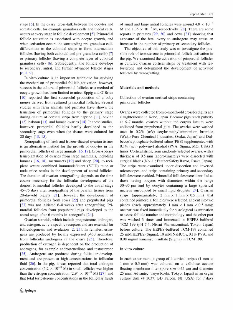
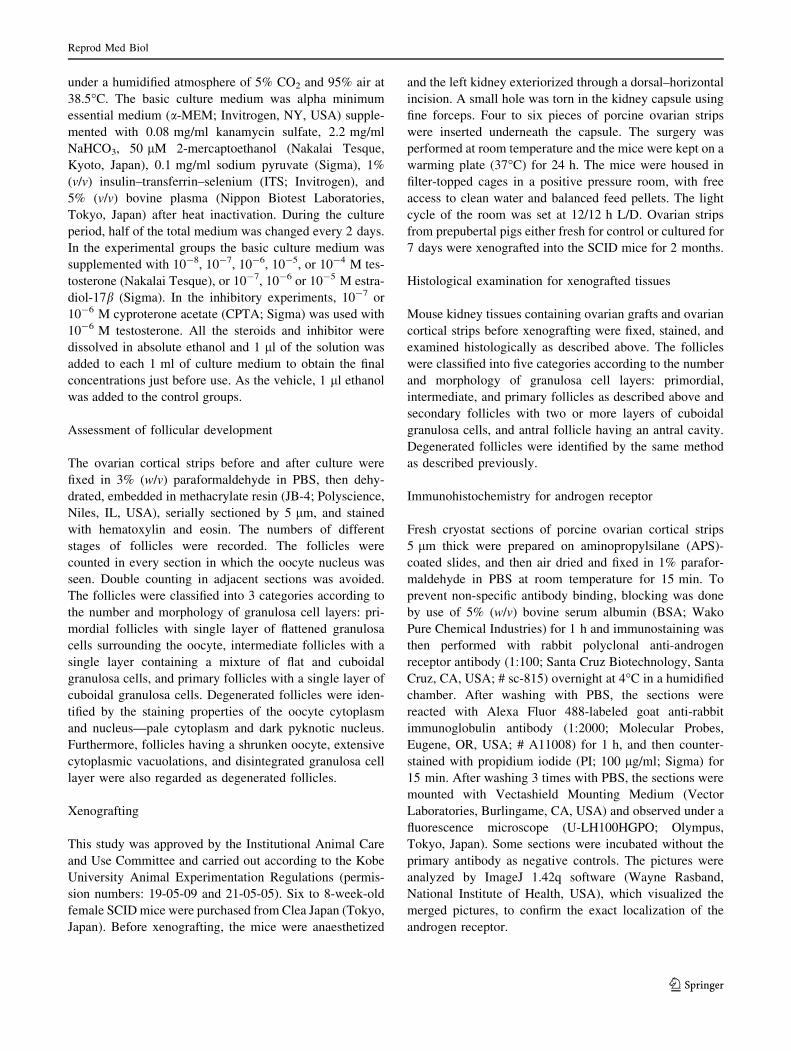
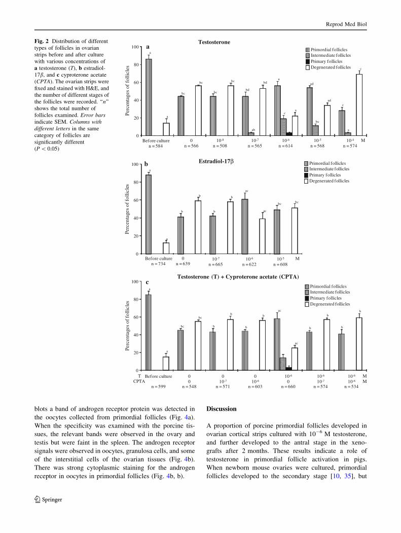
Effects of testosterone on primordial follicle activation
Ovarian cortical strips before culture contained almost all
primordial follicles (Fig. 1a), whereas after 7-day culture
with 10-6 M testosterone, primordial follicles had devel-
oped to intermediate (19 ± 4%) or primary follicles
(3 ± 1%, Fig. 1c, d), and 56 ± 5% remained in the pri-
mordial stage (Fig. 2a). Higher concentrations above
10-5 M, or lower concentrations below 10-6 M did not
induce follicle transition to the primary stage (Fig. 2a). At
10-6 M, testosterone reduced the percentage of degener-
ated follicles (22 ± 4%) compared with the control
0 M group (56 ± 1%). However, lower concentrations,
below 10-6 M, or higher concentrations, above 10-5 M,
increased the number of degenerated follicles. 10-6 and
10-5 M testosterone increased the percentages of healthy
primordial follicles compared with the control (P \ 0.05).
Because testosterone can be aromatized to estradiol
[25], the effect of estradiol was examined. In cortical strips
treated with 10-7, 10-6, or 10-5 M estradiol-17b, there
Reprod Med Biol
123
were only primordial and degenerated follicles, without
any intermediate or primary follicles (Fig. 1e). There was
an increase in the percentage of primordial follicles and a
decrease in degenerated follicles in cortical strips cultured
with 10-6 M estradiol-17b (Fig. 2b). However, there was
no dose-dependent relationship with the percentages of
primordial follicles in the estradiol-17b-treated groups.
An androgen receptor antagonist, cyproterone acetate,
was used to determine whether the stimulatory effect of
testosterone on primordial follicle activation was mediated
by androgen receptor. Cyproterone acetate inhibited the
activation of primordial follicles and increased the per-
centages of degenerated follicles (Figs. 1f, 2c).
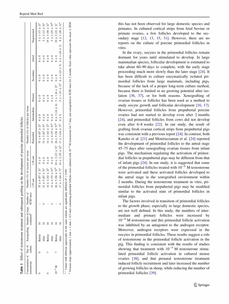
Development of primordial follicles in xenografts
after testosterone treatment
Histological examination confirmed that the strips dissected
from ovaries contained only primordial follicles and a few
degenerated follicles before culture (Table 1). The oocyte
diameters were 35 lm or less and each oocyte was
surrounded by a single layer of 5–7 flattened cells. After
7 days of culture, ovarian cortical strips were xenografted to
SCID mice. Two months after grafting, the fresh ovarian
cortical strips contained only primordial and degenerated
follicles, and none of them developed to the advan-
ced stages (Fig. 3a; Table 1). In the grafts cultured without
testosterone, none of the primordial follicles were acti-
vated (Table 1). In the grafts cultured with testosterone at
10-6 M, some follicles developed to the primary
(11 ± 3%), secondary (15 ± 3%) and antral (10 ± 3%)
stages (Fig. 3c–e). However, 44 ± 7% of primordial folli-
cles in the grafts remained in the primordial stage (Fig. 3d).
Abnormally enlarged antral cavities of the developing fol-
licles in the grafts were filled with blood (Fig. 3e).
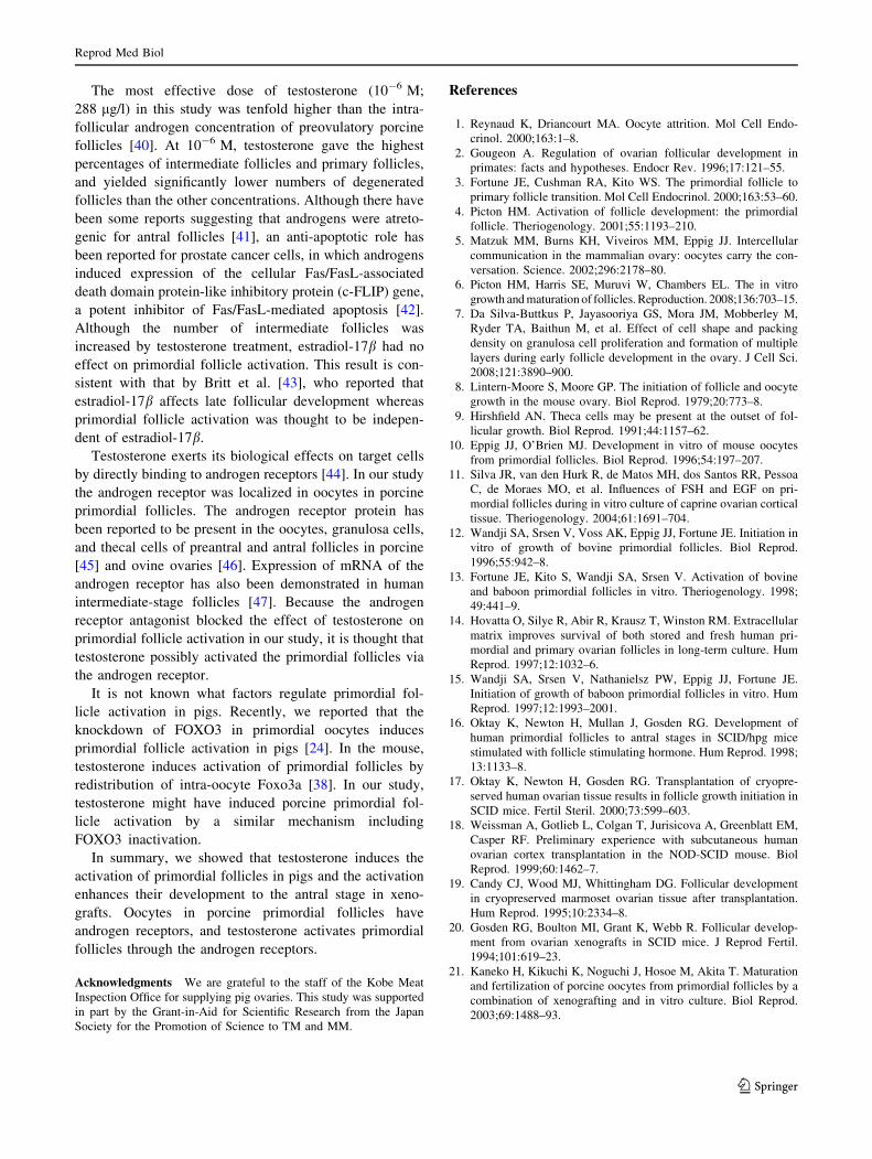
Expression of androgen receptors in oocytes
Because the results suggest a receptor-mediated effect of
testosterone on the transition of primordial follicles to the
intermediate or primary stage, we examined the expression
of the androgen receptor in the oocytes. In the western
Fig. 1 Testosterone-induced
primordial follicle activation in
porcine ovarian strips. The
ovarian strips were fixed in
paraformaldehyde and stained
with hematoxylin and eosin.
The ovarian strips before culture
(a) contained mainly healthy
primordial follicles. 10-6
M testosterone treatment for
7 days yielded intermediate
follicles (arrow in c) and
primary follicles (double arrowin d). Ovarian strips treated with
estradiol-17b (e), cyproterone
acetate ? testosterone (f), and
control treated with vehicle (b),
in which the primordial follicles
were intact, had not started to
develop after 7 days. Scale barrepresents 40 lm
Reprod Med Biol
123
blots a band of androgen receptor protein was detected in
the oocytes collected from primordial follicles (Fig. 4a).
When the specificity was examined with the porcine tis-
sues, the relevant bands were observed in the ovary and
testis but were faint in the spleen. The androgen receptor
signals were observed in oocytes, granulosa cells, and some
of the interstitial cells of the ovarian tissues (Fig. 4b).
There was strong cytoplasmic staining for the androgen
receptor in oocytes in primordial follicles (Fig. 4b, b).
Discussion
A proportion of porcine primordial follicles developed in
ovarian cortical strips cultured with 10-6 M testosterone,
and further developed to the antral stage in the xeno-
grafts after 2 months. These results indicate a role of
testosterone in primordial follicle activation in pigs.
When newborn mouse ovaries were cultured, primordial
follicles developed to the secondary stage [10, 35], but
Before culturen = 584
10-8
n = 50810-7
n = 56510-6
n = 61410-4
n = 57410-5
n = 568
0
20
40
60
80
100
M
Primordial folliclesIntermediate follicles
Degenerated folliclesPrimary follicles
a
a
bc
bc
bc
bc
bd
ab
bda
ca
bc
adc
c
a
ad
aTestosterone
0
20
40
60
80
100
Before culturen = 734
0n = 639
10-7
n = 66510-5
n = 60810-6
n = 622M
Primordial folliclesIntermediate follicles
Degenerated folliclesPrimary follicles
a
b
b
b
b
ac
ac
bc bc
b Estradiol-17βa
0n = 566
0
20
40
60
80
100
Before culture
n = 599
00
n = 548
010-7
n = 571
010-6
n = 603
10-6
0n = 660
10-6
10-7
n = 574
10-6
10-6
n = 534
MM
a
a
bc
bc
b
b
b
b
ac
ac
b
b
b
b
TCPTA
cTestosterone (T) + Cyproterone acetate (CPTA)
Primordial folliclesIntermediate follicles
Degenerated folliclesPrimary follicles
Perc
enta
ges
of f
ollic
les
Perc
enta
ges
of fo
llicl
esPe
rcen
tage
s of
folli
cles
Fig. 2 Distribution of different
types of follicles in ovarian
strips before and after culture
with various concentrations of
a testosterone (T), b estradiol-
17b, and c cyproterone acetate
(CPTA). The ovarian strips were
fixed and stained with H&E, and
the number of different stages of
the follicles were recorded. ‘‘n’’
shows the total number of
follicles examined. Error barsindicate SEM. Columns withdifferent letters in the same
category of follicles are
significantly different
(P \ 0.05)
Reprod Med Biol
123
this has not been observed for large domestic species and
primates. In cultured cortical strips from fetal bovine or
primate ovaries, a few follicles developed to the sec-
ondary stage [12, 13, 15, 31]. However, there are no
reports on the culture of porcine primordial follicles in
vitro.
In the ovary, oocytes in the primordial follicles remain
dormant for years until stimulated to develop. In large
mammalian species, follicular development is estimated to
take about 60–90 days to complete, with the early stage
proceeding much more slowly than the later stage [24]. It
has been difficult to culture enzymatically isolated pri-
mordial follicles from large mammals, including pigs,
because of the lack of a proper long-term culture method,
because there is limited or no growing potential after iso-
lation [36, 37], or for both reasons. Xenografting of
ovarian tissues or follicles has been used as a method to
study oocyte growth and follicular development [16, 17].
However, primordial follicles from prepubertal porcine
ovaries had not started to develop even after 2 months
[24], and primordial follicles from cows did not develop
even after 6–8 weeks [22]. In our study, the result of
grafting fresh ovarian cortical strips from prepubertal pigs
was consistent with a previous report [24]. In contrast, both
Kaneko et al. [21] and Moniruzzaman et al. [24] reported
the development of primordial follicles to the antral stage
45–75 days after xenografting ovarian tissues from infant
pigs. The mechanism regulating the activation of primor-
dial follicles in prepubertal pigs may be different from that
of infant pigs [24]. In our study, it is suggested that some
of the primordial follicles treated with 10-6 M testosterone
were activated and these activated follicles developed to
the antral stage in the xenografted environment within
2 months. During the testosterone treatment in vitro, pri-
mordial follicles from prepubertal pigs may be modified
similar to the activated state of primordial follicles in
infant pigs.
The factors involved in transition of primordial follicles
to the growth phase, especially in large domestic species,
are not well defined. In this study, the numbers of inter-
mediate and primary follicles were increased by
10-6 M testosterone and this primordial follicle activation
was inhibited by an antagonist to the androgen receptor.
Moreover, androgen receptors were expressed in the
oocytes in primordial follicles. These results suggest a role
of testosterone in the primordial follicle activation in the
pig. This finding is consistent with the results of studies
showing that treatment with 10-5 M testosterone stimu-
lated primordial follicle activation in cultured mouse
ovaries [38], and that prenatal testosterone treatment
induced follicle recruitment and later increased the number
of growing follicles in sheep, while reducing the number of
primordial follicles [39].Ta
ble
1E
ffec
to
fte
sto
ster
on
etr
eatm
ent
and
sub
seq
uen
tg
raft
ing
on
the
dev
elo
pm
ent
of
po
rcin
ep
rim
ord
ial
foll
icle
s
Conce
ntr
atio
nof
test
ost
erone
Day
of
cult
ure
Xen
ogra
ftin
gN
um
ber
of
stri
ps
exam
ined
dN
um
ber
of
SC
IDm
ice
Num
ber
(%)
of
oocy
tes
per
tiss
ue
Num
ber
(%)
of
foll
icle
sper
tiss
ue
B35
lm
C36
lm
Pri
mord
ial
Inte
rmed
iate
Pri
mar
yS
econdar
yA
ntr
alD
egen
erat
ed
00
Bef
ore
16
–35
±6
(87
±2)a
0±
035
±6
(87
±2)a
0±
00
±0
0±
00
±0
5±
1(1
3±
2)a
Aft
er16
433
±5
(87
±5)a
0±
033
±5
(87
±5)a
0±
00
±0
0±
00
±0
5±
2(1
3±
5)a
07
Bef
ore
16
–14
±2
(42
±2)b
0±
014
±2
(42
±2)b
0±
00
±0
0±
00
±0
19
±3
(58
±2)b
Aft
er16
415
±4
(75
±5)a
c0
±0
15
±4
(75
±5)a
c0
±0
0±
00
±0
0±
05
±2
(25
±5)b
c
10
-6
M7
Bef
ore
16
–26
±5
(56
±5)b
c10
±2
(23
±5)a
26
±5
(56
±5)b
c8
±2
(16
±4)
3±
1(6
±2)a
0±
00
±0
10
±2
(22
±2)c
Aft
er16
417
±3
(44
±7)b
14
±2
(35
±4)b
17
±3
(44
±7)b
0±
05
±1
(11
±3)b
6±
1(1
5±
3)
4±
1(1
0±
3)
8±
1(2
0±
3)a
c
a–
cV
alues
wit
hdif
fere
nt
super
scri
pts
inth
esa
me
colu
mn
are
signifi
cantl
ydif
fere
nt
(P\
0.0
5)
dO
var
ian
stri
ps
(appro
xim
atel
y1
mm
91
mm
90.5
mm
)co
nta
inin
gpri
mord
ial
foll
icle
sw
ere
cult
ure
dw
ith
10
-6
Mte
stost
erone
for
7day
san
dxen
ogra
fted
toS
CID
mic
efo
r2
month
s.E
ach
val
ue
repre
sents
the
mea
n±
SE
M
Reprod Med Biol
123
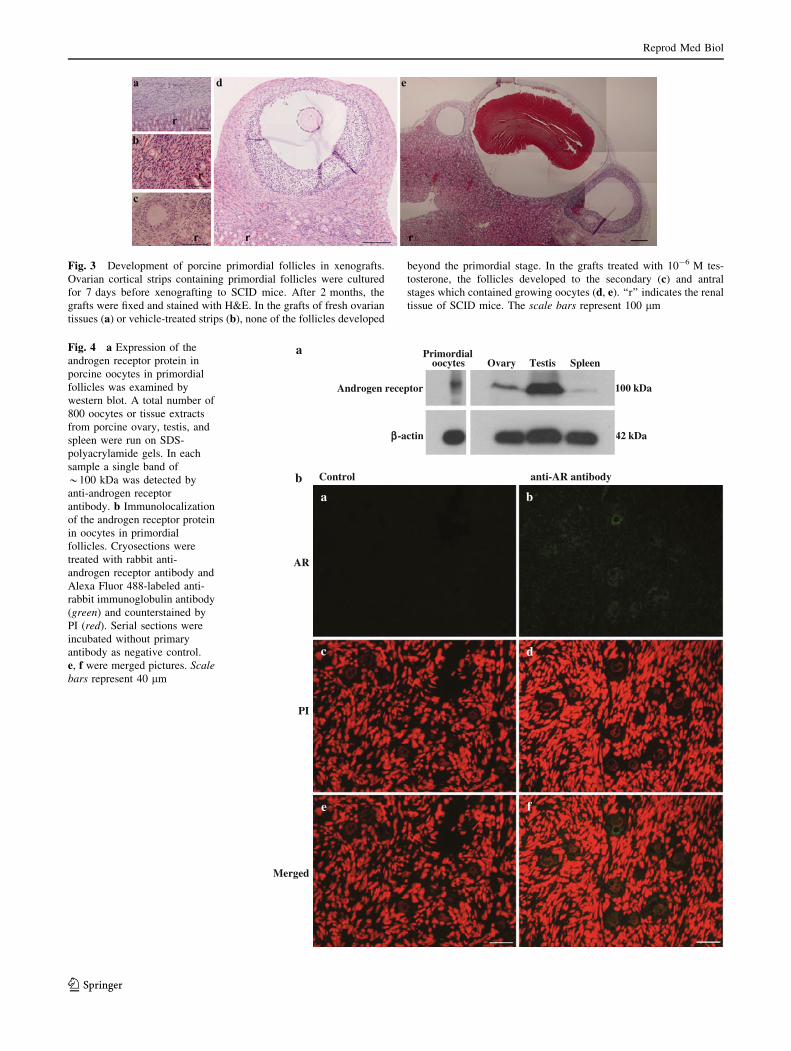
Fig. 3 Development of porcine primordial follicles in xenografts.
Ovarian cortical strips containing primordial follicles were cultured
for 7 days before xenografting to SCID mice. After 2 months, the
grafts were fixed and stained with H&E. In the grafts of fresh ovarian
tissues (a) or vehicle-treated strips (b), none of the follicles developed
beyond the primordial stage. In the grafts treated with 10-6 M tes-
tosterone, the follicles developed to the secondary (c) and antral
stages which contained growing oocytes (d, e). ‘‘r’’ indicates the renal
tissue of SCID mice. The scale bars represent 100 lm
a
b Control anti-AR antibody
PI
AR
Merged
a
f
dc
e
b
Primordialoocytes Ovary Testis Spleen
Androgen receptor
ββ-actin
100 kDa
42 kDa
Fig. 4 a Expression of the
androgen receptor protein in
porcine oocytes in primordial
follicles was examined by
western blot. A total number of
800 oocytes or tissue extracts
from porcine ovary, testis, and
spleen were run on SDS-
polyacrylamide gels. In each
sample a single band of
*100 kDa was detected by
anti-androgen receptor
antibody. b Immunolocalization
of the androgen receptor protein
in oocytes in primordial
follicles. Cryosections were
treated with rabbit anti-
androgen receptor antibody and
Alexa Fluor 488-labeled anti-
rabbit immunoglobulin antibody
(green) and counterstained by
PI (red). Serial sections were
incubated without primary
antibody as negative control.
e, f were merged pictures. Scalebars represent 40 lm
Reprod Med Biol
123
The most effective dose of testosterone (10-6 M;
288 lg/l) in this study was tenfold higher than the intra-
follicular androgen concentration of preovulatory porcine
follicles [40]. At 10-6 M, testosterone gave the highest
percentages of intermediate follicles and primary follicles,
and yielded significantly lower numbers of degenerated
follicles than the other concentrations. Although there have
been some reports suggesting that androgens were atreto-
genic for antral follicles [41], an anti-apoptotic role has
been reported for prostate cancer cells, in which androgens
induced expression of the cellular Fas/FasL-associated
death domain protein-like inhibitory protein (c-FLIP) gene,
a potent inhibitor of Fas/FasL-mediated apoptosis [42].
Although the number of intermediate follicles was
increased by testosterone treatment, estradiol-17b had no
effect on primordial follicle activation. This result is con-
sistent with that by Britt et al. [43], who reported that
estradiol-17b affects late follicular development whereas
primordial follicle activation was thought to be indepen-
dent of estradiol-17b.
Testosterone exerts its biological effects on target cells
by directly binding to androgen receptors [44]. In our study
the androgen receptor was localized in oocytes in porcine
primordial follicles. The androgen receptor protein has
been reported to be present in the oocytes, granulosa cells,
and thecal cells of preantral and antral follicles in porcine
[45] and ovine ovaries [46]. Expression of mRNA of the
androgen receptor has also been demonstrated in human
intermediate-stage follicles [47]. Because the androgen
receptor antagonist blocked the effect of testosterone on
primordial follicle activation in our study, it is thought that
testosterone possibly activated the primordial follicles via
the androgen receptor.
It is not known what factors regulate primordial fol-
licle activation in pigs. Recently, we reported that the
knockdown of FOXO3 in primordial oocytes induces
primordial follicle activation in pigs [24]. In the mouse,
testosterone induces activation of primordial follicles by
redistribution of intra-oocyte Foxo3a [38]. In our study,
testosterone might have induced porcine primordial fol-
licle activation by a similar mechanism including
FOXO3 inactivation.
In summary, we showed that testosterone induces the
activation of primordial follicles in pigs and the activation
enhances their development to the antral stage in xeno-
grafts. Oocytes in porcine primordial follicles have
androgen receptors, and testosterone activates primordial
follicles through the androgen receptors.
Acknowledgments We are grateful to the staff of the Kobe Meat
Inspection Office for supplying pig ovaries. This study was supported
in part by the Grant-in-Aid for Scientific Research from the Japan
Society for the Promotion of Science to TM and MM.
References
1. Reynaud K, Driancourt MA. Oocyte attrition. Mol Cell Endo-
crinol. 2000;163:1–8.
2. Gougeon A. Regulation of ovarian follicular development in
primates: facts and hypotheses. Endocr Rev. 1996;17:121–55.
3. Fortune JE, Cushman RA, Kito WS. The primordial follicle to
primary follicle transition. Mol Cell Endocrinol. 2000;163:53–60.
4. Picton HM. Activation of follicle development: the primordial
follicle. Theriogenology. 2001;55:1193–210.
5. Matzuk MM, Burns KH, Viveiros MM, Eppig JJ. Intercellular
communication in the mammalian ovary: oocytes carry the con-
versation. Science. 2002;296:2178–80.
6. Picton HM, Harris SE, Muruvi W, Chambers EL. The in vitro
growth and maturation of follicles. Reproduction. 2008;136:703–15.
7. Da Silva-Buttkus P, Jayasooriya GS, Mora JM, Mobberley M,
Ryder TA, Baithun M, et al. Effect of cell shape and packing
density on granulosa cell proliferation and formation of multiple
layers during early follicle development in the ovary. J Cell Sci.
2008;121:3890–900.
8. Lintern-Moore S, Moore GP. The initiation of follicle and oocyte
growth in the mouse ovary. Biol Reprod. 1979;20:773–8.
9. Hirshfield AN. Theca cells may be present at the outset of fol-
licular growth. Biol Reprod. 1991;44:1157–62.
10. Eppig JJ, O’Brien MJ. Development in vitro of mouse oocytes
from primordial follicles. Biol Reprod. 1996;54:197–207.
11. Silva JR, van den Hurk R, de Matos MH, dos Santos RR, Pessoa
C, de Moraes MO, et al. Influences of FSH and EGF on pri-
mordial follicles during in vitro culture of caprine ovarian cortical
tissue. Theriogenology. 2004;61:1691–704.
12. Wandji SA, Srsen V, Voss AK, Eppig JJ, Fortune JE. Initiation in
vitro of growth of bovine primordial follicles. Biol Reprod.
1996;55:942–8.
13. Fortune JE, Kito S, Wandji SA, Srsen V. Activation of bovine
and baboon primordial follicles in vitro. Theriogenology. 1998;
49:441–9.
14. Hovatta O, Silye R, Abir R, Krausz T, Winston RM. Extracellular
matrix improves survival of both stored and fresh human pri-
mordial and primary ovarian follicles in long-term culture. Hum
Reprod. 1997;12:1032–6.
15. Wandji SA, Srsen V, Nathanielsz PW, Eppig JJ, Fortune JE.
Initiation of growth of baboon primordial follicles in vitro. Hum
Reprod. 1997;12:1993–2001.
16. Oktay K, Newton H, Mullan J, Gosden RG. Development of
human primordial follicles to antral stages in SCID/hpg mice
stimulated with follicle stimulating hormone. Hum Reprod. 1998;
13:1133–8.
17. Oktay K, Newton H, Gosden RG. Transplantation of cryopre-
served human ovarian tissue results in follicle growth initiation in
SCID mice. Fertil Steril. 2000;73:599–603.
18. Weissman A, Gotlieb L, Colgan T, Jurisicova A, Greenblatt EM,
Casper RF. Preliminary experience with subcutaneous human
ovarian cortex transplantation in the NOD-SCID mouse. Biol
Reprod. 1999;60:1462–7.
19. Candy CJ, Wood MJ, Whittingham DG. Follicular development
in cryopreserved marmoset ovarian tissue after transplantation.
Hum Reprod. 1995;10:2334–8.
20. Gosden RG, Boulton MI, Grant K, Webb R. Follicular develop-
ment from ovarian xenografts in SCID mice. J Reprod Fertil.
1994;101:619–23.
21. Kaneko H, Kikuchi K, Noguchi J, Hosoe M, Akita T. Maturation
and fertilization of porcine oocytes from primordial follicles by a
combination of xenografting and in vitro culture. Biol Reprod.
2003;69:1488–93.
Reprod Med Biol
123
22. Senbon S, Ota A, Tachibana M, Miyano T. Bovine oocytes in
secondary follicles grow and acquire meiotic competence in severe
combined immunodeficient mice. Zygote. 2003;11:139–49.
23. Moniruzzaman M, Miyano T. KIT-KIT ligand in growth of
porcine primordial follicles. J Reprod Dev. 2007;53:1273–81.
24. Moniruzzaman M, Lee J, Zengyo M, Miyano T. Knockdown of
FOXO3 induces primordial oocyte activation in pigs. Repro-
duction. 2010;139:349–57.
25. Drummond AE. The role of steroids in follicular growth. Reprod
Biol Endocrinol. 2006;4:16.
26. McNatty KP. Hormonal correlates of follicular development in
the human ovary. Aust J Biol Sci. 1981;34:249–68.
27. Eiler H, Nalbandov AV. Sex steroids in follicular fluid and blood
plasma during the estrous cycle of pigs. Endocrinology. 1977;
100:331–8.
28. Chang SC, Jones JD, Ellefson RD, Ryan RJ. The porcine ovarian
follicle: 1. Selected chemical analysis of follicular fluid at dif-
ferent developmental stages. Biol Reprod. 1976;15:321–8.
29. Vendola KA, Zhou J, Adesanya OO, Weil SJ, Bondy CA.
Androgens stimulate early stages of follicular growth in the pri-
mate ovary. J Clin Invest. 1998;101:2622–9.
30. Abbott DH, Barnett DK, Bruns CM, Dumesic DA. Androgen
excess fetal programming of female reproduction: a develop-
mental aetiology for polycystic ovary syndrome? Hum Reprod
Update. 2005;11:357–74.
31. Yang MY, Fortune JE. Testosterone stimulates the primary to
secondary follicle transition in bovine follicles in vitro. Biol
Reprod. 2006;75:924–32.
32. Laemmli UK. Cleavage of structural proteins during the assembly
of the head of bacteriophage T4. Nature. 1970;227:680–5.
33. Burek M, Duda M, Knapczyk K, Koziorowski M, Słomczynska
M. Tissue-specific distribution of the androgen receptor (AR) in
the porcine fetus. Acta Histochem. 2007;109:358–65.
34. Takeda H, Chodak G, Mutchnik S, Nakamoto T, Chang C.
Immunohistochemical localization of androgen receptors with
mono- and polyclonal antibodies to androgen receptor. J Endo-
crinol. 1990;126:17–25.
35. O’Brien MJ, Pendola JK, Eppig JJ. A revised protocol for in vitro
development of mouse oocytes from primordial follicles dra-
matically improves their developmental competence. Biol Re-
prod. 2003;68:1682–6.
36. Hirao Y, Nagai T, Kubo M, Miyano T, Miyake M, Kato S. In
vitro growth and maturation of pig oocytes. J Reprod Fertil.
1994;100:333–9.
37. Miyano T, Hirao Y. In vitro growth of oocytes from domestic
species. J Mam Ova Res. 2003;20:78–85.
38. Yang JL, Zhang CP, Li L, Huang L, Ji SY, Lu CL, et al. Tes-
tosterone induces redistribution of forkhead box-3a and down-
regulation of growth and differentiation factor 9 messenger
ribonucleic acid expression at early stage of mouse folliculo-
genesis. Endocrinology. 2010;151:774–82.
39. Smith P, Steckler TL, Veiga-Lopez A, Padmanabhan V. Devel-
opmental programming: differential effects of prenatal testoster-
one and dihydrotestosterone on follicular recruitment, depletion
of follicular reserve, and ovarian morphology in sheep. Biol
Reprod. 2009;80:726–36.
40. Ainsworth L, Tsang BK, Downey BR, Marcus GJ, Armstrong
DT. Interrelationships between follicular fluid steroid levels,
gonadotropic stimuli, and oocyte maturation during preovulatory
development of porcine follicles. Biol Reprod. 1980;23:621–7.
41. Cheng G, Weihua Z, Makinen S, Makela S, Saji S, Warner M,
Gustafsson JA, Hovatta O. A role for the androgen receptor in
follicular atresia of estrogen receptor beta knockout mouse ovary.
Biol Reprod. 2002;66:77–84.
42. Gao S, Lee P, Wang H, Gerald W, Adler M, Zhang L, Wang YF,
Wang Z. The androgen receptor directly targets the cellular Fas/
FasL-associated death domain protein-like inhibitory protein
gene to promote the androgen-independent growth of prostate
cancer cells. Mol Endocrinol. 2005;19:1792–802.
43. Britt KL, Saunders PK, McPherson SJ, Misso ML, Simpson ER,
Findlay JK. Estrogen actions on follicle formation and early
follicle development. Biol Reprod. 2004;71:1712–23.
44. Quigley CA, De Bellis A, Marschke KB, el-Awady MK, Wilson
EM, French FS. Androgen receptor defects: historical, clinical,
and molecular perspectives. Endocr Rev. 1995;16:271–321.
45. Słomczynska M, Tabarowski Z. Localization of androgen receptor
and cytochrome P450 aromatase in the follicle and corpus luteum
of the porcine ovary. Anim Reprod Sci. 2001;65:127–34.
46. Juengel JL, Heath DA, Quirke LD, McNatty KP. Oestrogen
receptor alpha and beta, androgen receptor and progesterone
receptor mRNA and protein localisation within the developing
ovary and in small growing follicles of sheep. Reproduction.
2006;131:81–92.
47. Rice S, Ojha K, Whitehead S, Mason H. Stage-specific expres-
sion of androgen receptor, follicle-stimulating hormone receptor,
and anti-Mullerian hormone type II receptor in single, isolated,
human preantral follicles: relevance to polycystic ovaries. J Clin
Endocrinol Metab. 2007;92:1034–40.
Reprod Med Biol
123