Telangiectases in Venous Insufficiency: Point of Reflux and Treatment Strategy

Iron Insufficiency Compromises Motor Neurons and TheirMitochondrial Function in Irp2-Null MiceSuh Young Jeong1, Daniel R. Crooks1, Hayden Wilson-Ollivierre1, Manik C. Ghosh1, Rachid Sougrat2,
Jaekwon Lee3, Sharon Cooperman1, James B. Mitchell4, Carole Beaumont5, Tracey A. Rouault1*
1 Eunice Kennedy Shriver National Institute of Child Health and Development, National Institutes of Health, Bethesda, Maryland, United States of America, 2 Nanobiology
Lab, King Abdullah University of Science and Technology (KAUST), Thuwal, Saudi Arabia, 3 Department of Biochemistry, University of Nebraska, Lincoln, Nebraska, United
States of America, 4 National Cancer Institute, National Institutes of Health, Bethesda, Maryland, United States of America, 5 INSERM U773, Centre de Recherche
Biomedicale Bichat-Beaujon, Universite Paris Diderot, Paris, France
Abstract
Genetic ablation of Iron Regulatory Protein 2 (Irp2, Ireb2), which post-transcriptionally regulates iron metabolism genes,causes a gait disorder in mice that progresses to hind-limb paralysis. Here we have demonstrated that misregulation of ironmetabolism from loss of Irp2 causes lower motor neuronal degeneration with significant spinal cord axonopathy.Mitochondria in the lumbar spinal cord showed significantly decreased Complex I and II activities, and abnormalmorphology. Lower motor neurons appeared to be the most adversely affected neurons, and we show that functional ironstarvation due to misregulation of iron import and storage proteins, including transferrin receptor 1 and ferritin, may have acausal role in disease. We demonstrated that two therapeutic approaches were beneficial for motor neuron survival. First,we activated a homologous protein, IRP1, by oral Tempol treatment and found that axons were partially spared fromdegeneration. Secondly, we genetically decreased expression of the iron storage protein, ferritin, to diminish functional ironstarvation. These data suggest that functional iron deficiency may constitute a previously unrecognized molecular basis fordegeneration of motor neurons in mice.
Citation: Jeong SY, Crooks DR, Wilson-Ollivierre H, Ghosh MC, Sougrat R, et al. (2011) Iron Insufficiency Compromises Motor Neurons and Their MitochondrialFunction in Irp2-Null Mice. PLoS ONE 6(10): e25404. doi:10.1371/journal.pone.0025404
Editor: Mark L. Baccei, University of Cincinnatti, United States of America
Received February 14, 2011; Accepted September 2, 2011; Published October 7, 2011
This is an open-access article, free of all copyright, and may be freely reproduced, distributed, transmitted, modified, built upon, or otherwise used by anyone forany lawful purpose. The work is made available under the Creative Commons CC0 public domain dedication.
Funding: This work was supported by the intramural program of Eunice Kennedy Shriver National Institute of Child Health and Human Development, NationalInstitutes of Health. The funders had no role in study design, data collection and analysis, decision to publish, or preparation of the manuscript.
Competing Interests: The authors have declared that no competing interests exist.
* E-mail: [email protected]
Introduction
The central nervous system (CNS) is one of the highest-energy
requiring areas of the body, consuming energy at a rate nearly
seven times faster per tissue weight compared to non-CNS areas
[1], and neurons are highly dependent on efficient mitochondrial
ATP production [2]. The respiratory chain complexes of
mitochondria depend on iron cofactors, as they include twelve
iron-sulfur [Fe-S] cluster prosthetic groups, and several hemes [3].
Therefore, large amounts of iron are needed to fully constitute
respiratory chain complexes in the CNS. However, excess iron can
cause devastating toxic effects to cells through the Fenton reaction
[4], and cellular iron homeostasis is accordingly highly regulated.
In mammalian cells, two cytosolic proteins known as Iron
Regulatory Proteins (IRPs) regulate intracellular iron homeostasis
by binding to mRNA stem-loop sequences known as Iron
Regulatory Elements (IREs) in target transcripts. These two IRPs,
IRP1 and IRP2, bind to IREs and regulate expression of proteins
involved in iron homeostasis including the iron importer,
transferrin receptor 1 (TfR1), the iron storage protein ferritin,
which is a heteropolymer composed of FtH and FtL monomers, an
iron exporter, ferroportin, and other transcripts [5,6].
Previously, we have reported that mice lacking Irp2 (Irp1+/+;
Irp2-/-) show neurodegenerative symptoms including hind-limb
weakness, tremor, subtle kyphosis, and abnormal gait [7,8].
Moreover, we also generated mice lacking one copy of Irp1 and
both copies of Irp2 (Irp1+/-;Irp2-/-) and reported that there is an Irp
gene dosage effect on severity of neurodegeneration, wherein onset
of disease and severity of symptoms occurs earlier in Irp1+/-;Irp2-/-
mice [9]. Both Irp1+/+;Irp2-/- and Irp1+/-;Irp2-/- mice show
misregulation in proteins involved in iron metabolism, with
neuronal loss in different brain areas including cerebellum and
substantia nigra [7,9,10]. Interestingly, these mice also show
microcytic anemia, which was attributed to low expression of
TfR1 and concomitant low iron uptake in the erythroid precursor
cells [11,12].
Despite the multiple clinical and pathological problems
previously observed in Irp2-null mice, we had not previously
analyzed the integrity of upper and lower motor neurons and
assessed their potential contributions to the loss of locomotion
observed in the Irp2-null mice. In this study, we analyzed brain
and spinal cord cells that are involved in the motor function in
both Irp1+/+;Irp2-/- and Irp1+/-;Irp2-/- mice, and we found that
lower motor neurons appear to be the cells that are most adversely
affected in Irp2-null mice. Here, we performed multiple studies
including immunostaining and spinal cord lysate analyses which
suggested that loss of Irps may cause functional iron starvation in
these cells and may thereby impair mitochondrial activity. Finally,
we attempted to mitigate disease using two approaches, including
chemical recruitment of IRP1 activity, or genetic reduction of
ferritin H synthesis, and we observed that both interventions
significantly decreased neuronal degeneration in Irp2-null mice.
PLoS ONE | www.plosone.org 1 October 2011 | Volume 6 | Issue 10 | e25404

Results
Lack of Iron regulatory proteins caused axonaldegeneration in spinal cord
As reported previously, Irp2-null mice showed abnormal
neurological symptoms as they aged [7]. These symptoms include
pronounced hind-limb weakness, poor weight bearing, tremor,
and high muscle tone. To assess the integrity of neurons in the
spinal cord from Irp2-null mice, we analyzed semi-thin plastic
sections of lumbar spinal cord cross-sections (L4 level) from older
adult (between 11 and 13 months of age) mice. We found massive
accumulations of myelin dense bodies (MDB, also called myelin
ovoids) in the ventral and lateral white matter of the Irp1+/-;Irp2-/-
mice as well as in the Irp1+/+;Irp2-/- mice (Figure 1A, red circled
areas, Figure 1Bb–c, arrows). MDBs are one of the hallmarks for
neurodegeneration because they accumulate when axonal degen-
eration occurs and the myelin sheath collapses into the area
formally occupied by the axon [13]. Figure 1B shows a higher
magnification of the ventral white matter (asterisk area in
Figure 1A). The number of MDBs significantly increased in the
Irp1+/-;Irp2-/- mice compared not only to the wildtype, but also in
comparison to the Irp1+/+;Irp2-/- mice (Figure 1C), which
demonstrated a dose-dependent effect of Irp-null mutations.
Moreover, the number of MDBs increased as mice aged. Mice
of each genotype were analyzed at ages of 4, 7.5 and 12 months,
and progressive degeneration of axons was quantified (Figure 1C-
D, inter-age comparison is in Table S1). These data indicate that
lack of Irp2 causes axonal degeneration in mice, and severity of
axonal degeneration increases in Irp1+/-;Irp2-/- mice.
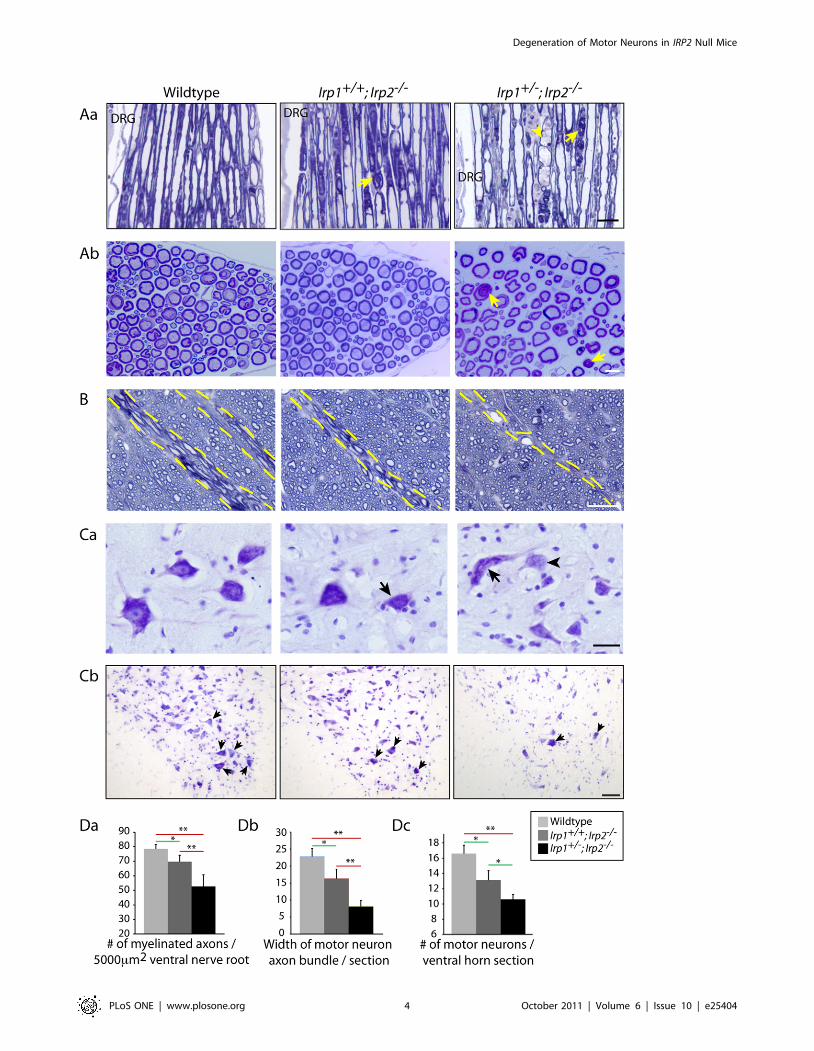
Degeneration of lower motor neuronsThe axons of lower motor neurons, which are involved in control
of movements, cross through the ventral white matter to exit the
spinal cord. Localization of the MDBs in the ventral and lateral
spinal cord led us to hypothesize that some of the degenerating
axons represented motor neuronal axons. To evaluate whether
motor neuronal axons were affected, we analyzed ventral nerve
roots located next to the dorsal root ganglion (DRG, Figure 2Aa),
where these motor neuronal axons are known to exit the spinal cord.
We found that there were increased numbers of swollen axons
(Figure 2Aa, arrowhead) and also accumulations of MDBs
(Figure 2A, arrows) in Irp2-null mice compared to wildtype.
Moreover, the numbers of myelinated fibers in the ventral nerve
roots were significantly decreased in Irp2-null mice (Figure 2Ab, Da,
7863.1 myelinated fibers per 5000 mm2 for wildtype, 7064.4 and
5367.9 for Irp2-null mice). Then we analyzed the width of motor
neuronal axon bundles in the ventral white matter. The diameter of
the motor neuronal axon bundles was significantly decreased in
Irp2-null mice (Figure 2B, Db, between dotted lines). Furthermore,
when we analyzed the morphology of the motor neuronal cell
bodies in the ventral horn by cresyl violet staining, Irp2-null mice
showed several hallmarks of retrograde cell body degeneration,
including distorted shape, rounding of cell bodies and loss of
multipolarity, loss of Nissl body staining (chromatolysis, arrowhead),
and eccentrically positioned nuclei (Figure 2Ca, arrows, [14]).
There was also marked reduction in the number of large diameter
cells observed when the ventral horn was stained with cresyl violet
(Figure 2Cb). Finally, we quantified the number of motor neuronal
cell bodies that stained with cresyl violet, and there was a significant
decrease in the number of motor neuronal cell bodies in the Irp2-
null mice compared to the wildtype (Figure 2Dc, 16.661.08 in
wildtype, 13.161.23 in Irp1+/+;Irp2-/- and 10.660.64 in Ir-
p1+/-;Irp2-/- per ventral horn section). We also analyzed the
morphology of the dorsal nerve root where sensory axons are
present, but there was no apparent pathology in the sensory axons
(Figure S2A). These data demonstrate that motor neurons in the
Irp2-null mice show significant degeneration, particularly if
complete loss of Irp2 is combined with heterozygous loss of Irp1,
which corresponds well to the phenotype of these mice.
Upper motor neuronal atrophyUpper motor neurons connect between the motor cortex and
spinal cord, and damage of upper motor neurons might contribute
to the high muscle tone that we observed in Irp2-null mice.
Accordingly, we analyzed morphology of cells in the primary
motor cortex area by H&E staining and found fewer large
diameter neurons (arrows) in Irp2-null mice (Figure 3A, B). In
addition, H&E staining showed that there were more cells with
chromatolysis (Figure 3A, inset), which is considered to be a
marker for cell stress. Therefore, these data suggest that Irp2-null
mice have not only lower motor neuronal degeneration, but also
upper motor neuronal abnormalities.
Stress markers are increased in the Irp2-null miceAs shown in Figures 1 and 2, evidence for degeneration of lower
motor neurons was observed in both axons and neuronal cell bodies
in Irp2-null mice. We further evaluated motor neurons by
examining expression of stress markers in both areas. First we
stained sections using SMI32 (anti-non-phosphorylated neurofila-
ment), which normally stains neuronal cell bodies but not healthy
axons. As predicted, we could not detect immunoreactivity in axons
of the wildtype, whereas a strong signal was detected in the lumbar
ventral white matter of Irp2-null mice (Figure 4A). Also, there was a
significant increase of anti-ubiquitin immunoreactivity in the motor
neuronal cell bodies (Figure S3) of Irp2-null mice, suggesting that
these neurons are under stress. Additionally, we found clusters of
macrophages and/or microglia (positive for F4/80) by confocal
microscopy in the Irp1+/-;Irp2-/- mice white matter, suggesting that
immune cells were attracted into this area (Figure 4B, arrows). Thus,
these data indicate that there is stress in the axons and motor
neuronal cell bodies of the lumbar spinal cord of Irp2-null mice.
Dysregulation of iron homeostasis proteins causesfunctional iron starvation
IRPs are key regulators of intracellular iron homeostasis. Under
low iron conditions, these proteins bind to IREs to regulate
expression of several transcripts that encode iron homeostasis
proteins. When IRE-binding activity is diminished by genetic loss
of Irps, expression of an iron importer, transferrin receptor 1
(TfR1), decreases and expression of the iron storage protein,
ferritin, increases, and these changes can cause functional iron
deficiency. Although the IRE/IRP regulatory system is ubiquitous
in all cells, we found that the most significant misregulations of
TfR1 and ferritin in the spinal cord were in the motor neurons.
Anti-TfR1 staining showed significantly decreased TfR1 immu-
noreactivity (Figure 5Ab) in the motor neuronal cell bodies
(Figure 5Aa, arrows) and endothelial cells of the blood-brain-
barrier (BBB, Figure 5Aa, arrowheads, [1]) although all other cell
types were also affected. Increased expression of ferritin was
prominent in both white matter (Figure 5Ba), and in the motor
neuronal cell bodies in the grey matter of spinal cord (Figure 5Bb,
arrows). Some glial cells that likely represent inflammatory cells
also showed increased expression of ferritin (Figure 5Bb, arrow-
heads). Changes in TfR1 and ferritin expression were also
confirmed by Western blot analyses (Figure 5C). Finally we
measured total tissue iron concentrations using ICP-MS and found
that all three segments of spinal cord in Irp2-null mice had
Degeneration of Motor Neurons in IRP2 Null Mice
PLoS ONE | www.plosone.org 2 October 2011 | Volume 6 | Issue 10 | e25404

Figure 1. Axonal degeneration in the spinal cord of Irp2-null mice. A; Toluidine blue staining of Epon-embedded spinal cord cross-sections at12 months. Red-circled areas indicate where myelin dense body (MDBs) were found. The yellow star represents the area enlarged in Figure 1B. Scalebar = 200 mm. B; Ventral white matter (L4) from wildtype (a), Irp1+/+;Irp2-/- (b), Irp1+/-;Irp2-/- (c) mice at 12 months. Arrows indicate MDBs. Scalebars = 50 mm, 20 mm (inset). C; Quantification of MDBs per spinal cord sections at 4, 7.5, 12 months. The bar chart show average6SEM, n = 4-5,**; p,0.001, analyzed by two-way ANOVA. A summary of the pairwise comparisons is presented in Table S1. D; Progressive accumulation of MDBs inventral white matter of spinal cords at 4, 7.5 months. Scale bars = 10 mm.doi:10.1371/journal.pone.0025404.g001
Degeneration of Motor Neurons in IRP2 Null Mice
PLoS ONE | www.plosone.org 3 October 2011 | Volume 6 | Issue 10 | e25404
Degeneration of Motor Neurons in IRP2 Null Mice
PLoS ONE | www.plosone.org 4 October 2011 | Volume 6 | Issue 10 | e25404

significantly lower amounts of iron than controls (Figure 5D)
whereas there was no change in Zn levels. Therefore, we suggest
that loss of Irps led to decreased iron uptake and increased iron
sequestration in cells, which led to functional iron starvation in the
spinal cords of Irp2-null mice. Moreover, the magnitude of
misregulation appeared to be most significant in the motor
neurons, which likely damaged these vulnerable cells.
Mitochondrial dysfunction and atrophy caused bydisrupted iron homeostasis
Iron is crucial for energy generation in mitochondria because
respiratory chain complexes require [Fe-S] clusters and heme
cofactors for function. Thus, functional iron starvation in Irp2-null
mice might be expected to cause problems in mitochondrial
respiratory chain activities. First, we assessed activity of respiratory
chain Complex I, which contains eight [Fe-S] clusters. The activity
of Complex I was significantly decreased in Irp2-null mice
(Figure 6A, 74.464.19 and 54.7618.03% compared to wildtype),
whereas there were no changes in the amount of a key Complex I
subunit protein, GRIM-19 (Figure 6A, [15]) as measured by a
Complex I quantification kit. Moreover, activity of respiratory
complex II, succinate dehydrogenase, which contains several [Fe-
S] clusters in subunit B, was markedly decreased (SDH,
Figure 6Ba), whereas there was no change in the activity of
Complex IV, which does not contain [Fe-S] (Figure 6Bb). This
effect of cellular iron starvation on [Fe-S] containing proteins was
also confirmed by Western blot analyses, where we found marked
reductions of the [Fe-S]-containing proteins SDH-B and ferroche-
latase (FECH), and also mild reductions in SDH-A, which forms a
complex with SDH-B, in the Irp2-null mice (Figure 6C). Decreased
ferrochelatase has been observed previously in erythropoietic cells
of Irp2-null mice [16], where decreased iron levels caused
decreased stability of the protein. Consistent with these observa-
tions, total Irp-null mutations were recently reported to diminish
respiratory chain complex activities in mouse livers [17].
In addition to abnormal mitochondrial respiratory chain
activities, significant mitochondrial pathology was also observed
in EM studies of Irp2-null mice. Abnormal mitochondria from
Irp2-null mice were swollen, and had disrupted and vacuolized
cristae (Figure 6D, arrows). Also, some axons showed evidence of
mild demyelination (Figure 6Dc) and clustering of neurofilaments
Figure 3. Status of primary motor cortex neurons in Irp2-null mice. A; H&E staining of the primary motor cortex (PMC). Arrows indicate largeneurons in this area. Scale bar = 10 mm (both) B; Quantification of cell bodies that are larger than 10 mm in diameter / 20,000 mm2. n = 3. *; p,0.05,**; p,0.001, analyzed by one-way ANOVA.doi:10.1371/journal.pone.0025404.g003
Figure 2. Atrophy and loss of lower motor neurons in the Irp2-null mice. Aa; Toluidine blue staining of Epon-embedded sections of ventralnerve root. Arrows show MDBs and arrowhead shows occasional swelling of axon. DRG; dorsal root ganglia. Scale bar = 25 mm. Ab; Cross sectionpictures of ventral nerve root. Arrows show MDBs. Scale bar = 10 mm. B; Motor neuronal axon exit area of the ventral white matter (dotted line). Scalebar = 30 mm. Ca; Cresyl violet staining showed distorted motor neuronal cell bodies (arrows) and motor neurons with chromatolysis (arrowhead) inthe Irp2-null mice ventral horn. Scale bar = 25 mm. Cb; Lower magnification of ventral horn stained with cresyl violet. Arrows indicate motor neurons.Scale bar = 50 mm. Da; Quantification of number of myelinated fibers in the venral nerve root per 5000 mm2. n = 3,. Db; Quantification of motorneuronal axon bundles diameters per spinal cord. n = 5,. Dc; Quantification of motor neurons in the ventral horn stained with cresyl violet. n = 4, Alldata in D show average value6SEM per genotype, *; p,0.05, **; p,0.001, analyzed by one-way ANOVA.doi:10.1371/journal.pone.0025404.g002
Degeneration of Motor Neurons in IRP2 Null Mice
PLoS ONE | www.plosone.org 5 October 2011 | Volume 6 | Issue 10 | e25404

(Figure 6Dc, arrowhead), which are markers for neurodegenera-
tion [18,19]. Therefore, we concluded that functional iron
starvation in Irp2-null mice neurons might be one of the causes
of decreased mitochondrial function and atrophy.
Therapeutic approaches; Tempol treatmentAs we reported previously, oral Tempol treatment attenuated
neuromuscular compromise of the Irp1+/+;Irp2-/- mice [8]. Tempol
is a stable nitroxide that readily crosses the blood brain barrier and it
can act as an antioxidant, or as an iron-sulfur cluster destabilizing
reagent [8,20]. Here we further analyzed whether Tempol could
prevent axonal degeneration in the spinal cord. We observed that
the number of MDBs was significantly decreased in the Tempol-
treated Irp1+/+;Irp2-/- mice (Figure 7A, B). This treatment also
protected these mice from loss of neuromuscular skills as assessed by
hang-tests ([8], Movie S1 or Figure S4). Interestingly, there was no
significant beneficial effect of Tempol on Irp1+/-;Irp2-/- mice
(Figure 7B), consistent with previous findings that Tempol could
not prevent motor function decline in mice that had only one copy
of Irp1 [8]. In a previous paper, we have shown that Tempol
destabilizes a synthetic [4Fe-4S] cluster in vitro, suggesting a
mechanism for how Tempol activates IRP1 into the IRE-binding
form. To confirm the effect of Tempol on activating IRP1, a gel-
shift assay was performed using wildtype mouse fibroblasts. In this
experiment, it was clear that Tempol converts IRP1 into the IRE-
binding form without changing the total amount of IRP1
(Figure 7C). Likely as a consequence of this activation of IRE-
binding activity, Tempol treatment increased expression of TfR1 in
the motor neurons of Irp1+/+;Irp2-/- mice (Figure 7D), a change that
might partially relieve these cells from functional iron starvation.
Moreover, decreased mitochondrial Complex I activity in the Irp1+/+;
Irp2-/- mice in the Tempol treated mice (Figure 7E) returned to near
the levels of wildtypes. Therefore, these data suggest that motor
neuronal degeneration might be caused by functional iron starvation,
and that Tempol exerted its therapeutic effect in part by increasing
expression of TfR1.
Genetic modification to prevent motor neurondegeneration
As shown in Figure 5, loss of IRP activity not only decreased
expression of TfR1 and limited iron uptake of cells, but also
permitted increased expression of ferritin, which may have further
contributed to functional iron starvation by sequestering iron
within ferritin. Based on the abnormally high ferritin expression,
we hypothesized that decreased expression of ferritin might be
beneficial to these mice. To assess the role of ferritin, Irp1+/-;Irp2-/-
mice were crossed with Fth+/- mice (Figure S1, [21]) to decrease
expression of ferritin (Figure 8A), and MDBs in the ventral white
matter were quantified. Perhaps due to the different genetic
background of the Fth+/+ mice, Irp1+/-;Irp2-/-;Fth+/+ did not have
as many MDBs as our Irp1+/-;Irp2-/- mice (Figure 8B,C, compared
to Figure 1C); however Irp1+/-;Irp2-/-;Fth+/- mice showed approx-
imately 50% sparing of axonal degeneration (57616.9 vs.
27.765.24, Figure 8C) compared to their background-matched
controls, demonstrating that decreased ferritin expression was
beneficial in these mice. This sparing of axonal degeneration was
prominent not only in the spinal cord but also in the ventral nerve
root fibers, where MDBs were significantly decreased in the
Irp1+/-;Irp2-/-; Fth+/- mice (Figure 8D, E). As mentioned above, we
could not detect any significant axonopathies in the dorsal nerve
roots (Figure 8E). Taken together, these data demonstrate that
functional iron starvation due to abnormal iron homeostasis may
be a major cause of motor neuronal degeneration in Irp2-null
mice.
Discussion
In this study, we report that disruption of iron homeostasis
caused by Irp2-null mutations causes degeneration of motor
neurons. One of the possible causes of this degeneration might be
that functional iron starvation impairs the activity of mitochon-
drial respiratory chain complexes and disrupts mitochondrial
integrity in these neurons. Moreover, two therapeutic approaches,
including either oral Tempol treatment or genetic reduction of
ferritin expression, delayed neurodegeneration, suggesting possible
approaches to treatment if a human neurodegenerative disease
attributable to loss of IRP2 is identified in the future.
Loss of Irps caused motor neuronal degeneration withmitochondrial atrophy
As we reported previously, Irp2-null mice showed neurodegen-
erative symptoms including tremor, hind-limb weakness, problems
in weight bearing, and kyphosis [7,9]. In these papers, we also
Figure 4. Increased expression of stress markers in the Irp2-null mice. A; Immunoreactivity for SMI32 (non-phosphoneurofilament antibody)was increased in Irp2-null mice. Scale bar = 30 mm. B; Infiltration of macrophages and/or microglia (F4/80 positive, arrows) was detected by confocalmicroscopy. Scale bar = 10 mm.doi:10.1371/journal.pone.0025404.g004
Degeneration of Motor Neurons in IRP2 Null Mice
PLoS ONE | www.plosone.org 6 October 2011 | Volume 6 | Issue 10 | e25404

reported abnormalities in the brain of Irp2-null mice including
axonopathy and neuronal loss, but we did not analyze pathology
in the spinal cord. Here we report that there is significant lower
motor neuronal degeneration in the lumbar spinal cord of Irp2-
null mice consistent with the marked gait abnormalities and hind-
limb weakness (Figures 1, 2). This degeneration was more severe in
the Irp1+/-; Irp2-/- mice than in the Irp1+/+; Irp2-/- mice,
demonstrating a dose-dependent effect of Irp-null mutations.
Moreover, analysis of motor cortex also suggested that there
might be atrophy of upper motor neurons (Figure 3) that might
contribute to the high-muscle tone that we observed in these mice,
although we did not detect abnormalities in the dorsal corticospi-
nal tract of the spinal cord (data not shown). However, we found
accumulations of MDBs in the ventral funiculus where a minor
portion of the corticospinal tract is proposed to run through the
ventral white matter [22]. Moreover, the lateral localization of
MDBs also suggested that neurons in the reticulospinal tract were
adversely affected in the Irp2-null mice [22]. There was also minor
demyelination of some axons and there were distorted mitochon-
dria (Figure 6D), but we did not observe pathology in astrocytes or
in oligodendrocytes (Figure S2B, C). Thus, it appears that motor
neurons are the cells in the central nervous system that are most
adversely affected by the disruption of iron homeostasis caused by
loss of Irp2. Currently, it is not clear why motor neurons are more
vulnerable than other neurons and glial cells to loss of Irp2.
However, based on compromised respiratory complex activities in
Irp2-null mice (Figure 6), it is possible that motor neurons depend
more on IRP2 to maintain normal iron homeostasis and support
mitochondrial function than other cells. Moreover, because motor
neurons are the longest cells in the body, they are very dependent
on mitochondrial activity to provide energy for ion pumps located
along axons and concentrated at the nodes of Ranvier [23].
Potential candidate gene for motor neuron diseasesTo our knowledge, IRP2 mutations have not yet been found in
human patients with motor problems. Among motor neuron
diseases, one example of disease that affects both upper and lower
motor neurons is Amyotrophic Lateral Sclerosis (ALS). Recently,
relationships between other iron homeostasis proteins and ALS
have been reported in both mouse and human diseases
Figure 5. Abnormal expression of TfR1 and ferritin in motor neurons and glia of Irp2-null mice and diminished iron concentrationsin spinal cord. Aa; Anti-TfR1 staining was decreased in Irp2-null mice motor neurons (arrows) and the endothelial cells of blood-brain barrier(arrowheads). Scale bar = 20 mm. Ab; Lower magnification picture of spinal cord ventral horn stained with anti-TfR1 antibody. Scale bar = 100 mm. B;Ferritin expression was prominent in both white (a) and grey matter (b) of the spinal cord. Motor neurons exhibited high ferritin expression (arrows)as did in glial cells (arrowheads). Scale bars = 100, 30 mm (respectively). C; Western blot analysis of TfR1 and Ferritin light chain from total spinal cordlysate. Beta-actin was used as a loading control. n = 5. D; Total tissue metal concentrations from different areas of spinal cord by ICP-MS revealdiminished Fe concentrations in the Irp2-null mice *; p,0.05, compared to wildtype, as analyzed by one-way ANOVA.doi:10.1371/journal.pone.0025404.g005
Degeneration of Motor Neurons in IRP2 Null Mice
PLoS ONE | www.plosone.org 7 October 2011 | Volume 6 | Issue 10 | e25404

[24,25,26,27]. Therefore, it is interesting for us that null mutation
in a key regulatory factor for iron homeostasis causes a phenotype
comparable to human ALS and to mouse SOD1 transgenic
models. Our results show not only motor neuronal degeneration in
the spinal cord, but also degeneration in the ventral root nerves
(Figure 2), where motor neuronal axons run, as was reported in the
SOD1 mice [28]. Moreover, EM analysis showed significant
vacuolization of mitochondria in axons of the Irp2 null mice,
similar to the SOD1 mice [29]. It is also interesting that the large
increase in serum ferritin levels observed in Irp2-null mice [30] was
also observed in sporadic ALS patients [31]. However, even
though SOD1 mice and Irp2-null mice show somewhat similar
phenotypes, iron metabolism may be misregulated in different
ways, depending on the animal model. In some instances,
Figure 6. Mitochondrial dysfunction and atrophy in the Irp2-null mice. A; Complex I activity was decreased in Irp2-null mice whereas therewere no changes in the amount of GRIM-19, a subunit of Complex I that was quantified. n = 6. Ba; SDH activity (Complex II) was markedly decreased inIrp2-null mice. Bb; There was no change in Complex IV activity which contains heme as a cofactor. *; p,0.05, **; p,0.001, analyzed by one-wayANOVA. C; The mitochondrial proteins SDH-A, SDH-B and ferrochelatase were significantly decreased in Irp2-null mice, whereas citrate synthase,which does not contain a [Fe-S] cluster, did not change and was used as a mitochondrial loading control. n = 5 **; p,0.001 compared to wildtype,analyzed by one-way ANOVA, D; TEM showed mitochondrial vacuolization (arrows) in axons in the ventral white matter of Irp2-null mice andabnormal bundling of neurofilaments within axons was also noted (arrowhead). Scale bar = 0.5 mm.doi:10.1371/journal.pone.0025404.g006
Degeneration of Motor Neurons in IRP2 Null Mice
PLoS ONE | www.plosone.org 8 October 2011 | Volume 6 | Issue 10 | e25404

Degeneration of Motor Neurons in IRP2 Null Mice
PLoS ONE | www.plosone.org 9 October 2011 | Volume 6 | Issue 10 | e25404

misregulation can lead to cellular iron overload, as was reported
for the spinal cords of SOD1 mice [32] and treatment of the mice
with an iron chelator was beneficial in these mice [32,33].
However, our work here shows that Irp2-null mice seem to suffer
from functional iron starvation, as evidenced by ICP-MS
measurements of spinal cord lysates, caused by low TfR1
expression and high ferritin expression in motor neuron cell
bodies. In conclusion, we suggest that IRP2 might be a potential
candidate gene for human diseases in which motor neuron
problems predominate, based on our data that Irp2-null mice
showed many pathologic traits that were reported in human
patients, including axonal degeneration, accumulation of myelin
dense bodies, lower and upper motor neuronal degeneration, and
accumulation of ubiquitin positive aggregates in motor neurons.
IRPs and potential relationship to human diseasesALS is a complex disease with multiple possible genetic causes
[34]. When Sreedharan et al. reported TDP43 mutations in ALS
patients, they also found an additional locus with LOD score
(logarithm (base 10) of odds) that was higher than 1.0 at 15q23-
q26 [32]. Interestingly, this locus is very close to IRP2 (15q25),
although the authors could not find contiguous markers nor could
they identify a haplotype in this locus. Moreover, van Es et al.
recently reported that chromosome 9p21.2 is a possible linkage
locus for the sporadic form of ALS [35], and human IRP1 is
located at chromosome 9p21.1. Lastly, target gene searches for
another movement disorder called Hereditary spastic paraplegia
(HSP) resulted in identification of a disease gene at the 4p16-p15
locus (SPG 38, [36]), which is near a new E3 ubiquitin ligase called
F-box and leucine-rich repeat protein 5 (FBXL5, 4p15.32).
FBXL5 is a novel E3 ubiquitin ligase responsible for degradation
of both IRPs [30,37]. Therefore, if mutations in SPG38 changed
stability of FBXL5, it might misregulate expression of both IRPs in
cells. Taken together, these genetic data suggest that both IRPs
and FBXL5 are located near loci thought to contain human motor
neuron disease genes.
Rescue approachesPreviously, Tempol treatment has been shown to have
beneficial effects on different mouse models [16,38]. We also
have shown that Irp1+/+; Irp2-/- mice treated with Tempol showed
improvement in neuromuscular function and reduced iron burden
in mouse cerebellum [8]. Here, we showed in further analysis that
ventral white matter axons were partially spared from degener-
ation in the spinal cord of Tempol-treated Irp2-null animals
(Figure 7A, B). As reported previously, Tempol treatment was
ineffective for the Irp1+/-; Irp2-/- mice, suggesting that the salutary
effect of Tempol might be due to activation of IRP1 ([8],
Figure 7C), which led to increased expression of TfR1 (Figure 7D)
and decreased expression of ferritin. Taken together, these data
indicate that Tempol may be a good therapeutic reagent for
animals with Irp2-null mutations.
As reported previously [7], Irp2-null mice show significant
increase in iron storage protein ferritin expression. This upregula-
tion was also noticed in the lumbar spinal cord, particularly in
motor neurons (Figure 5). We also reported that oral treatments of
Tempol decreased ferritin expression [8]. It has been reported that
maintenance of proper levels of ferritin is important for reducing
oxidative stress and enhancing neuronal survival [39,40]. There-
fore, we hypothesized that reduction of ferritin expression by
eliminating one ferritin H allele might be beneficial for
degenerating neurons. We demonstrated that Irp1+/-; Irp2-/- mice
crossed with Fth+/- showed less neurodegeneration (Figure 8).
Although there is no significant difference in total tissue iron
between Irp1+/+; Irp2-/- and Irp1+/-; Irp2-/- mice (Figure 5D), we
demonstrated that pathologic changes worsen in Irp1+/-; Irp2-/-
mice. These data suggest that one of the reasons that motor
neurons die in the Irp2-null mice is that the size of their ‘functional’
iron pool (compared to ‘total’) is reduced due to diminished TfR1
expression and increased ferritin expression. Reduced expression
of TfR1 decreases iron uptake into cells, and increased expression
of ferritin leads to increased sequestration of iron within ferritin
heteropolymers. This functional iron deprivation likely impairs
mitochondrial viability and function, and causes motor neuronal
dysfunction and loss in these mice.
Materials and Methods
Animals and genotypingMice lacking Irp(s) were generated by targeting AcoI (Irp1) and
Ireb2 (Irp2) genes [7,9,41]. Genotypes of mice were determined by
Southern blotting using gene-specific probes (Figure S1B). These
mice have mixed genetic backgrounds consisting of C57BL/6 and
B129S4/SVJ. Mice were anesthetized between 11-13 months of
age and all the experimental protocols used in this study followed
NICHD ACUC (Eunice Kennedy Shriver National Institute of Child
Health and Human Development Animal Care and Use
Committee) guideline and approved by the same committee
(protocol number 09-038). For the Tempol experiments, mice
were fed with either control or Tempol containing diet (10 mg/g,
[8]) from the time of weaning until the time of sacrifice. Mice
lacking one copy of ferritin H chain (Fth+/-, [21]) were provided by
Dr. Carole Beaumont (INSERM U773, France). For the
genotyping of Fth+/- mice, tissue was collected by ear clipping
and incubated in an X-gal containing buffer for 2 hours at 37uC.
When the beta-galactosidase gene was expressed under control of
the Fth promoter, blue color was generated (Figure S1C) in Fth+/-
mice. Fth+/- mice were crossed to Irp-null mice to generate
Irp1+/-;Irp2-/-;Fth+/- mice (Detailed mating strategy is described in
Figure S1A). Fth mice have C57BL/6 and 129SV mixed
background. Background and age-matched control mice were
used for comparison (2–11 days difference in age between
genotypes). For each experiment, ‘n’ indicates number of animals
used per genotype.
HistologyMice were deeply anesthetized and perfused with 2.5%
glutaraldehyde (EM grade, Electron Microscopic Sciences) in
0.1 M sodium cacodylate buffer, pH 7.4. Dissected tissue samples
were post-fixed and treated with 1.3% osmium tetroxide solution
Figure 7. Dietary Tempol supplementation partially prevented axonal degeneration. A; Toluidine blue staining of Epon-embeddedsections showed decreased MDBs (arrows) in the ventral white matter of Tempol treated mice. Scale bar = 50 mm. B; Quantification of MDBs per spinalcord section. n = 4, **; p,0.001, analyzed by two-way ANOVA. C; Gel-shift assay (top panel) show activation of IRP1 by Tempol treatment (100 mM) inwildtype mouse embryonic fibroblasts. The iron chelator, DFO, was used to show both IRP1 and IRP2 bands. Tempol treatment did not change totalIRP1 (middle panel) by Western blot. Anti-tubulin antibody was used as a loading control. Da; Immunoreactivity for TfR1 was increased in the Tempoltreated in the motor neurons of Irp1+/+;Irp2-/- mice (arrows). Db; Western blotting for TfR1 also showed increased expression of TfR1. E; Dietary Tempoltreatment increased mitochondrial Complex I activity in Irp1+/+; Irp2-/- mice. n = 5, *; p,0.05, analyzed by two-way ANOVA.doi:10.1371/journal.pone.0025404.g007
Degeneration of Motor Neurons in IRP2 Null Mice
PLoS ONE | www.plosone.org 10 October 2011 | Volume 6 | Issue 10 | e25404

with 0.1 N potassium ferrocyanide. Samples were embedded in
Epoxy Resin (EMS). Semithin (0.7 mm) sections were obtained
using an ultramicrotome (Leica Microsystems) and stained with
Epoxy Tissue Stain solution (EMS) for structural analysis. Some of
the Irp-null mice spinal cord and longitudinal-cut root nerve
sections were processed by the Laboratory for Neurotoxicity
Studies at Virginia Tech (Blacksburg, VA, [7]). Spinal cord and
root nerve sections were examined using bright field microscopy
(Nikon Instruments). The number of myelin dense bodies was
counted in the ventral and lateral white matter (n = 425 per
genotype).For the motor cortex analysis, 35 mm sections were
stained with Hematoxylin and Eosin by Dr. Robert Switzer at
Neuroscience, Ltd. (Knoxville, TN, [9]). Neuronal cell bodies that
were larger than 10 mm in diameter were quantified in the
primary motor cortex. For the cresyl violet staining, 14 mm frozen
sections from mouse spinal cord and brain were incubated with
Figure 8. Decreased expression of ferritin H chain is beneficial for axonal survival. A; Anti-ferritin staining showed decreased expression oftotal ferritin in the spinal cord of Irp1+/-;Irp2-/-;Fth+/- mice. Scale bar = 50 mm. B; Toluidine blue staining of the ventral white matter showed decreasednumber of MDBs in the Irp1+/-;Irp2-/-;Fth+/- mice. Scale bar = 30 mm. C; Quantification of MDBs per spinal cord section. The bar chart showaverage6SEM, n = 3, *; p,0.05, student’s t-test. D; Toluidine blue staining of the cross section of ventral nerve root. Irp1+/-;Irp2-/-;Fth+/- mice showeddecreased number of MDBs (arrows). Scale bar = 20 mm. E; Quantification of MDBs per nerve root section. The bar chart show average6SEM, n = 3,**; p,0.001, student’s t-test.doi:10.1371/journal.pone.0025404.g008
Degeneration of Motor Neurons in IRP2 Null Mice
PLoS ONE | www.plosone.org 11 October 2011 | Volume 6 | Issue 10 | e25404

0.1% cresyl violet solution for 10 min. For demyelination analysis,
spinal cord sections were first dehydrated and immersed in 0.1%
Luxol Fast Blue solution overnight at 37uC followed by chilling at
4uC for 30 min on the next day. After washing, slides were
incubated in 0.05% lithium carbonate solution for 5 min,
dehydrated and then mounted.
ImmunohistochemistryIrp2-null and control mice were anesthetized as above and
perfused with PBS and 4% paraformaldehyde, and 14 mm cryostat
sections were then obtained. Immunolabeling of tissue sections was
performed as described previously [32]. Briefly, tissue sections
were incubated with PBS containing 2% normal goat serum and
1% ovalbumin to block nonspecific binding of antibodies. This
was followed by an overnight incubation with mouse anti-TfR1
(1:200, Invitrogen) or rabbit anti-ferritin antibody (1:1000; gift
from Dr. Esther Meyron-Holtz, Israel Institute of Technology,
Haifa, Israel) or a rabbit anti-ubiquitin (1:200; Dako). After
washing, primary antibodies were recognized using biotin-
conjugated secondary antibodies and signal was amplified using
Vectastain ABC kit (Vector Lab). Immunoreactivity was visualized
using 0.5 mg/ml 3,39- diaminobenzidine as a chromagen (Sigma).
Methyl green counterstain (20 mg/ml, Acro Chemicals) was used
to visualize nuclei.
ImmunofluorescenceSpinal cord sections of mice were prepared as above and
incubated with rat F4/80 (1:100, Serotec), SMI32 (anti-non-
phosphorylated neurofilament, 1:500, Covance), anti-ferritin, or
anti-GFAP (1:100, Dako) overnight. After washing, sections were
incubated with Alexa Fluor-conjugated secondary antibodies
(1:400, Invitrogen). 49-6-diamidino-2-phenylindole (DAPI,
100 ng/ml, Vector Lab) was used to counterstain nuclei.
Western blottingWestern blots were performed using total spinal cord lysates. Anti-
TfR1 (1:1000, Zymed), rabbit anti-FtL, anti-SDH-A, anti-SDH-B
(1:2000, Mitosciences), anti-ferrochelatase (1:5000, [16]), anti-IRP1
(1:5000, [8]) were used to detect antigen. Anti-actin (1:400, Sigma),
anti-tubulin (1:5000, Sigma) and anti-citrate synthase (1:10,000,
Sigma) antibodies were used to confirm equal loading.
Measuring tissue metalsAnimals were deeply anesthetized and blood was removed by
extensively perfusing PBS through the heart. Spinal cord samples
were dissected in three segments (cervical, thoracic, lumbar) and
snap frozen in liquid nitrogen. Total iron and zinc concentrations
were measured by inductively coupled plasma mass spectroscopy
(ICP-MS) as described previously [42] and normalized by wet
tissue weight (n = 3).
Mitochondrial respiratory complex assaysActivities and/or quantities of Complexes I and IV from the
mitochondrial respiratory chain were assessed using specific
Dipstick assay kits (MitoSciences) following the manufacturer’s
protocol. Briefly, snap-frozen tissue samples were homogenized and
protein was extracted. After protein assay, 2 ug of total protein was
used for the activity assay of Complex I (12 ug for the Complex IV).
20 ug of total protein was used to detect the level of GRIM-19 in
Complex I (Complex I quantity assay kit, Mitosciences). Average
readings of band densities were obtained using ImageJ (http://rsb.
info.nih.gov/ij/) and data was plotted against wildtype value as
100%. n = 6 per genotype, two separate measurements per animal.
Succinate dehydrogenase (SDH) activity was measured according to
a previously published method [19] with modifications. Briefly,
50 ug of total spinal cord protein was mixed with a reaction buffer
containing 50 mM Tris (pH 8.0), 0.5 mM ethylenediaminetetra-
acetic acid, 12 g/L Cremaphor EL (Sigma), 2 mM iodonitrote-
trazolium chloride, 20 mM sodium succinate, 2 mM potassium
cyanide, and 1 mM sodium azide. The reaction was followed at
492 nm for 10 min using a spectrophotometer (Thermo) at room
temperature. Blank reactions without succinate were included to
assess background activity. Assay specificity was verified by pre-
incubation of some samples with the SDH inhibitor 3-nitropro-
pionic acid (10 mM) for 30 minutes, which resulted in negligible
residual enzymatic activity (data not shown). Data are presented as
first-order kinetic rates normalized to the average of the wildtype
sample values, as percent of control. Statistical analysis was
performed on the raw, non-normalized data. n = 6 per genotype.
Electron microscopyUltrastructural analysis of mitochondria in axons was per-
formed in the ventral white matter following previously reported
protocols [34]. Briefly, Epon-embedded spinal cord blocks were
generated as described above and plastic sections were generated
using an ultramicrotome (Leica). Sections were stained using lead
citrate and analyzed by a Transmission Electron Microscope
(Tecnai T20, FEI Company).
Gel-shift assayTo test the effect of Tempol on IRP activation, wildtype mouse
embryonic fibroblasts were treated with control media, control
media plus Tempol (100 mM), and iron chelator DFO (100 mM)
for 15 hours. Cells were collected and total protein was extracted
using an NP-40 (0.2%) containing buffer. Gel-shift assays were
performed using a 32P-labeled IRE probe following a protocol
published previously [3].
Mouse hang-testThe hang-test for assessment of motor function was performed in
a blinded manner as published previously [8]. Briefly, each mouse
was put on a wire mesh, which was then gently inverted. A video
camera was used to record how long the mouse was able to hang on
to the wire by clutching with the upper and lower extremities.
Statistical analysisStatistical significance was determined using a one-way or two-
way ANOVA, as appropriate, and Tukey’s post-hoc test was
applied (SigmaPlot 12, Systat Software Inc). Two-tailed student’s
t-tests were applied for the pairwise comparisons in Figures 8C
and 8E. (*; p,0.05, **; p,0.001).
Supporting Information
Figure S1 Generation of Irp1+/-;Irp2-/-;Fth+/- mice. A; A
schematic diagram showing four generations of mating strategy to
generate Irp1+/-;Irp2-/-;Fth+/- and control mice (red box). Asterisks
indicate embryonically lethal genotypes. P; parents, F1-3; progeny
generation 1–3. B; Genotyping analysis by Southern blot showed
specific bands for Irp1 and Irp2 (10.1 and 15.1 kb, respectively).
Upon targeted deletion, each probe detected a shorter band (4.1
and 3.5 kb, respectively). C; Beta-galactosidase reporter assay
distinguished Fth+/- (blue color, bottom) from wildtype (top).
(PDF)
Figure S2 No significant pathological changes wereobserved in dorsal root nerve fibers and glia of Irp2-
Degeneration of Motor Neurons in IRP2 Null Mice
PLoS ONE | www.plosone.org 12 October 2011 | Volume 6 | Issue 10 | e25404

null mice. A; Toluidine blue staining of Epon-embedded sections
from mouse dorsal root nerve do not show significant degeneration
in this area. B; Luxol Fast Blue staining of mouse ventral white
matter does not show significant demyelination. C; anti-GFAP
staining was performed to examine reactive astrocytes in ventral
white matter. Immunoreactivity was not significantly increased in
Irp2-null mice. Scale bars = 5 mm, 50 mm, 100 mm.
(PDF)
Figure S3 Motor neurons (arrows) in ventral horn ofIrp2-null mice showed increased ubiquitin expression.(PDF)
Figure S4 Screen capture from Movie S1 showingbeneficial effect of Tempol on mice neuromuscularbehavior.(PDF)
Movie S1 Beneficial effect of Tempol in Irp1+/+;Irp2-/-
mice. Mice treated with Tempol diet (right) since weaning showed
spared motor skill compared to the control diet mice (left). *If the
supplemental movie does not play, please see the screen captures in
Figure S4 or go to http://science.nichd.nih.gov/confluence/down-
load/attachments/44106370/13+control+and+Tempol+combined+small.mov?version = 1&modificationDate = 1285279775000.
(MOV)
Table S1 Summary of two-way ANOVA pairwise multi-ple comparisons (Tukey’s Test), computed to evaluatethe effect of age and genotype on the number of MyelinDense Bodies (MDBs) in ventral spinal cord sections(average values are summarized in Figure 1C). Two
factors (age, genotype) were used for comparison and p,0.05 was
used for statistical significance.
(PDF)
Acknowledgments
SYJ would like to thank Dr. Wing-Hang Tong for scientific discussions
throughout this project. We thank Dr. Javier Seravalli at the ICP-MS core
of the Redox Biology Center in University of Nebraska-Lincoln for
performing metal measurements. SYJ also thanks Dr. Arnold Y. Seo for his
advice on statistical analyses.
Author Contributions
Conceived and designed the experiments: SYJ DRC JL TAR. Analyzed
the data: SYJ DRC RS TAR. Contributed reagents/materials/analysis
tools: JBM CB. Wrote the paper: SYJ DRC TAR. Performed most of the
experiments: SYJ. Performed contributing experiments: HWO MCG
DRC RS JL SC.
References
1. Jefferies WA, Brandon MR, Hunt SV, Williams AF, Gatter KC, et al. (1984)
Transferrin receptor on endothelium of brain capillaries. Nature 312: 162–163.
2. Rouault TA (2006) The role of iron regulatory proteins in mammalian iron
homeostasis and disease. Nat Chem Biol 2: 406–414.
3. Meyron-Holtz EG, Ghosh MC, Iwai K, LaVaute T, Brazzolotto X, et al. (2004)
Genetic ablations of iron regulatory proteins 1 and 2 reveal why iron regulatory
protein 2 dominates iron homeostasis. EMBO J 23: 386–395.
4. Mori F, Tanji K, Zhang HX, Nishihira Y, Tan CF, et al. (2008) Maturation
process of TDP-43-positive neuronal cytoplasmic inclusions in amyotrophic
lateral sclerosis with and without dementia. Acta Neuropathol 116: 193–203.
5. Muckenthaler MU, Galy B, Hentze MW (2008) Systemic iron homeostasis and
the iron-responsive element/iron-regulatory protein (IRE/IRP) regulatory
network. Annu Rev Nutr 28: 197–213.
6. Koppenol WH (1993) The centennial of the Fenton reaction. Free Radic Biol
Med 15: 645–651.
7. LaVaute T, Smith S, Cooperman S, Iwai K, Land W, et al. (2001) Targeted
deletion of the gene encoding iron regulatory protein-2 causes misregulation of
iron metabolism and neurodegenerative disease in mice. Nat Genet 27:
209–214.
8. Ghosh MC, Tong WH, Zhang D, Ollivierre-Wilson H, Singh A, et al. (2008)
Tempol-mediated activation of latent iron regulatory protein activity prevents
symptoms of neurodegenerative disease in IRP2 knockout mice. Proc Natl Acad
Sci U S A 105: 12028–12033.
9. Smith SR, Cooperman S, Lavaute T, Tresser N, Ghosh M, et al. (2004) Severity
of neurodegeneration correlates with compromise of iron metabolism in mice
with iron regulatory protein deficiencies. Ann N Y Acad Sci 1012: 65–83.
10. Lagier-Tourenne C, Cleveland DW (2009) Rethinking ALS: the FUS about
TDP-43. Cell 136: 1001–1004.
11. Grabill C, Silva AC, Smith SS, Koretsky AP, Rouault TA (2003) MRI detection
of ferritin iron overload and associated neuronal pathology in iron regulatory
protein-2 knockout mice. Brain Res 971: 95–106.
12. Huang G, Lu H, Hao A, Ng DC, Ponniah S, et al. (2004) GRIM-19, a cell death
regulatory protein, is essential for assembly and function of mitochondrial
complex I. Mol Cell Biol 24: 8447–8456.
13. Dyck PJ, Stevens JC, Mulder DW, Espinosa RE (1975) Frequency of nerve fiber
degeneration of peripheral motor and sensory neurons in amyotrophic lateral
sclerosis. Morphometry of deep and superficial peroneal nerves. Neurology 25:
781–785.
14. Kaur D, Rajagopalan S, Andersen JK (2009) Chronic expression of H-ferritin in
dopaminergic midbrain neurons results in an age-related expansion of the labile
iron pool and subsequent neurodegeneration: implications for Parkinson’s
disease. Brain Res 1297: 17–22.
15. Valdmanis PN, Daoud H, Dion PA, Rouleau GA (2009) Recent advances in the
genetics of amyotrophic lateral sclerosis. Curr Neurol Neurosci Rep 9: 198–205.
16. Crooks DR, Ghosh MC, Haller RG, Tong WH, Rouault TA (2010)
Posttranslational stability of the heme biosynthetic enzyme ferrochelatase is
dependent on iron availability and intact iron-sulfur cluster assembly machinery.
Blood 115: 860–869.
17. Thompson K, Menzies S, Muckenthaler M, Torti FM, Wood T, et al. (2003)
Mouse brains deficient in H-ferritin have normal iron concentration but a
protein profile of iron deficiency and increased evidence of oxidative stress.
J Neurosci Res 71: 46–63.
18. Dhib-Jalbut S, Arnold DL, Cleveland DW, Fisher M, Friedlander RM, et al.
(2006) Neurodegeneration and neuroprotection in multiple sclerosis and other
neurodegenerative diseases. J Neuroimmunol 176: 198–215.
19. Tradewell ML, Durham HD, Mushynski WE, Gentil BJ (2009) Mitochondrial
and axonal abnormalities precede disruption of the neurofilament network in a
model of charcot-marie-tooth disease type 2E and are prevented by heat shock
proteins in a mutant-specific fashion. J Neuropathol Exp Neurol 68: 642–652.
20. Zhelev Z, Bakalova R, Aoki I, Matsumoto KI, Gadjeva V, et al. (2009) Nitroxyl
Radicals for Labeling of Conventional Therapeutics and Noninvasive Magnetic
Resonance Imaging of Their Permeability for Blood-Brain Barrier: Relationship
between Structure, Blood Clearance, and MRI Signal Dynamic in the Brain.
Mol Pharm.
21. Munujos P, Coll-Canti J, Gonzalez-Sastre F, Gella FJ (1993) Assay of succinate
dehydrogenase activity by a colorimetric-continuous method using iodonitrote-
trazolium chloride as electron acceptor. Anal Biochem 212: 506–509.
22. Rolfe DF, Brown GC (1997) Cellular energy utilization and molecular origin of
standard metabolic rate in mammals. Physiol Rev 77: 731–758.
23. Rouault TA, Tong WH (2008) Iron-sulfur cluster biogenesis and human disease.
Trends Genet 24: 398–407.
24. Goodall EF, Haque MS, Morrison KE (2008) Increased serum ferritin levels in
amyotrophic lateral sclerosis (ALS) patients. J Neurol 255: 1652–1656.
25. Blasco H, Vourc’h P, Nadjar Y, Ribourtout B, Gordon PH, et al. (2011)
Association between divalent metal transport 1 encoding gene (SLC11A2) and
disease duration in amyotrophic lateral sclerosis. J Neurol Sci 303: 124–127.
26. Mitchell RM, Simmons Z, Beard JL, Stephens HE, Connor JR (2010) Plasma
biomarkers associated with ALS and their relationship to iron homeostasis.
Muscle Nerve 42: 95–103.
27. Ryberg H, An J, Darko S, Lustgarten JL, Jaffa M, et al. (2010) Discovery and
verification of amyotrophic lateral sclerosis biomarkers by proteomics. Muscle
Nerve 42: 104–111.
28. Gurney ME, Pu H, Chiu AY, Dal Canto MC, Polchow CY, et al. (1994) Motor
neuron degeneration in mice that express a human Cu,Zn superoxide dismutase
mutation. Science 264: 1772–1775.
29. Dal Canto MC, Gurney ME (1995) Neuropathological changes in two lines of
mice carrying a transgene for mutant human Cu,Zn SOD, and in mice
overexpressing wild type human SOD: a model of familial amyotrophic lateral
sclerosis (FALS). Brain Res 676: 25–40.
30. Cooperman SS, Meyron-Holtz EG, Olivierre-Wilson H, Ghosh MC,
McConnell JP, et al. (2005) Microcytic anemia, erythropoietic protoporphyria,
and neurodegeneration in mice with targeted deletion of iron-regulatory protein
2. Blood 106: 1084–1091.
31. Mackenzie IR, Rademakers R, Neumann M (2010) TDP-43 and FUS in
amyotrophic lateral sclerosis and frontotemporal dementia. Lancet Neurol 9:
995–1007.
Degeneration of Motor Neurons in IRP2 Null Mice
PLoS ONE | www.plosone.org 13 October 2011 | Volume 6 | Issue 10 | e25404
32. Jeong SY, Rathore KI, Schulz K, Ponka P, Arosio P, et al. (2009) Dysregulation
of iron homeostasis in the CNS contributes to disease progression in a mousemodel of amyotrophic lateral sclerosis. J Neurosci 29: 610–619.
33. Danzeisen R, Achsel T, Bederke U, Cozzolino M, Crosio C, et al. (2006)
Superoxide dismutase 1 modulates expression of transferrin receptor. J BiolInorg Chem 11: 489–498.
34. Altan-Bonnet N, Sougrat R, Liu W, Snapp EL, Ward T, et al. (2006) Golgiinheritance in mammalian cells is mediated through endoplasmic reticulum
export activities. Mol Biol Cell 17: 990–1005.
35. Galy B, Ferring D, Minana B, Bell O, Janser HG, et al. (2005) Altered body irondistribution and microcytosis in mice deficient in iron regulatory protein 2
(IRP2). Blood 106: 2580–2589.36. Kim H, Son HY, Bailey SM, Lee J (2009) Deletion of hepatic Ctr1 reveals its
function in copper acquisition and compensatory mechanisms for copperhomeostasis. Am J Physiol Gastrointest Liver Physiol 296: G356–364.
37. Vashisht AA, Zumbrennen KB, Huang X, Powers DN, Durazo A, et al. (2009)
Control of iron homeostasis by an iron-regulated ubiquitin ligase. Science 326:718–721.
38. Galy B, Ferring-Appel D, Sauer SW, Kaden S, Lyoumi S, et al. (2010) Iron
regulatory proteins secure mitochondrial iron sufficiency and function. Cell
Metab 12: 194–201.
39. Schubert R, Erker L, Barlow C, Yakushiji H, Larson D, et al. (2004) Cancer
chemoprevention by the antioxidant tempol in Atm-deficient mice. Hum Mol
Genet 13: 1793–1802.
40. Salahudeen AA, Thompson JW, Ruiz JC, Ma HW, Kinch LN, et al. (2009) An
E3 ligase possessing an iron-responsive hemerythrin domain is a regulator of iron
homeostasis. Science 326: 722–726.
41. Ferreira C, Bucchini D, Martin ME, Levi S, Arosio P, et al. (2000) Early
embryonic lethality of H ferritin gene deletion in mice. J Biol Chem 275:
3021–3024.
42. Sreedharan J, Blair IP, Tripathi VB, Hu X, Vance C, et al. (2008) TDP-43
mutations in familial and sporadic amyotrophic lateral sclerosis. Science 319:
1668–1672.
Degeneration of Motor Neurons in IRP2 Null Mice
PLoS ONE | www.plosone.org 14 October 2011 | Volume 6 | Issue 10 | e25404
Copyright © 2022 FDOKUMEN