The management of the patient with respiratory insufficiency
44
THE MANAGEMENT OF THE PATIENT WITH RESPIRATORY iNSUFFICIENCY l-[. B.kRR[E EAIRLEY, M.B., B.S., F.F.A.R.C.S., and R. A CHAMBERS t, M.D., M.R.C.P. 2 A~td the Lord God formed map~ of the dust of lhe ground and breathed into his nostrils the breath of life .... GENESIS 2 : 7 A[~TFIO(GH Intermittent I'ositive Pressure ([.P.P.) respiration has been in use since Biblical times (1), methods of maintaining assisted respiration for pr,o -'- longed periods were developed more recently. The prolonged use of I.P.P. wa> considered harmful to the lungs (2) and other methods were preferred (3-6). Of these, the cabinet respirator, in spite of certain disadvantages, achieved the greatest success. This was particularly so in the management of diseases producing muscular paralysis. However, in other conditions in which under-ventilation is a common cause of death, the use of the cabinet respirator was less successful. In 1950, during a major epidemic of poliomyelitis, {he prolonged use of I.P.P. provided satisfactory (7;). This success led to the more, frequent use of such techniques, which have been foufid applicable in a wide range of conditions. This paper presents a review of the mauagemeat of such co~lditions, aided by.experience with one hundred consecutive admissions to the Toronto General Hospital Respiratory Unit (8). For the purposes of this discussion, Respiratory Insufftciency is present when underventilation endangers a patient's life. PHYSIOLOGICAL CONSIDERATIONS The main function of ventilation is to achieve oxygenation of the arterial blo,od and to eliminate carbon clio-ride. Any depression of ventilation will lower the tension of oxygen in arterial blood (pO..,) and raise that of carbon dioxide (pCO2). However, owing to the form of the oxygen dissociation curve in blood, ventilation must be quite markedly depressed before a fall in oxygen saturation occurs. Thus, a fall in saturation detectable clinically (or a significant fall in content ineasured manometrically) will not occur until the tension is reduced by nearly 50 per cent (9). Oxygen saturation or conteat is therefore a poor index of ventilatory efficiency. I~ contrast, arterial carbo~ dioxide tension and content show a very quick response to any ventilatory change, tn patients with normal lungs and r~o vascular, shunts, oxygenation will always be satisfactory when the pCO2 is normal. The latter is therefore an excellent index of ventilation. 1Present address: Associate ProfeCsor of Neurology, Seton Hall College of Medicine and Dentistr.v, Jersey City 4, New Jersey, U.S.A. =From the Departments of Anaesthesia and Medicine, University of Toronto, and the Toronto General Hospital Respiratory Unit 447 Can. Anaes. Soc. J., vol. 7, no. 4, October, t%0
-
Upload
khangminh22 -
Category
Documents
-
view
2 -
download
0
Transcript of The management of the patient with respiratory insufficiency
T H E M A N A G E M E N T OF T H E P A T I E N T W I T H
R E S P I R A T O R Y i N S U F F I C I E N C Y
l-[. B.kRR[E EAIRLEY, M.B., B.S., F.F.A.R.C.S., and R. A CHAMBERS t, M.D., M.R.C.P. 2
A~td the Lord God formed map~ of the dust of lhe ground and breathed into his nostrils the breath of life . . . .
GENESIS 2:7
A[~TFIO(GH In te rmi t t en t I 'ositive Pressure ( [ .P .P . ) respirat ion has been i n use since Biblical t imes (1), methods of mainta ining assisted respirat ion for pr,o -'- longed periods were developed more recently. The prolonged use of I .P .P . wa> considered harmful to the lungs (2) and other methods were preferred (3-6). Of these, the cabinet respirator, in spite of certain disadvantages, achieved the greatest success. This was part icularly so in the managemen t of diseases producing muscular paralysis. However, in other conditions in which under-vent i la t ion is a common cause of death, the use of the cabinet respirator was less successful.
In 1950, during a major epidemic of poliomyelitis, {he prolonged use of I .P.P. provided satisfactory (7;). This success led to the more, f requent use of such techniques, which have been foufid applicable in a wide range of conditions. This paper presents a review of the mauagemea t of such co~lditions, aided by.experience with one hundred consecutive admissions to the Toronto General Hospital Respiratory Unit (8).
For the purposes of this discussion, Respiratory Insufftciency is present when underventi la t ion endangers a pat ient ' s life.
PHYSIOLOGICAL CONSIDERATIONS
The main function of venti lat ion is to achieve oxygenation of the arterial blo, od and to eliminate carbon clio-ride. Any depression of venti lat ion will lower the tension of oxygen in arterial blood (pO..,) and raise t ha t of carbon dioxide (pCO2). However, owing to the form of the oxygen dissociation curve in blood, vent i la t ion must be quite markedly depressed before a fall in oxygen sa tura t ion occurs. Thus , a fall in sa turat ion detectable clinically (or a significant fall in content ineasured manometrical ly) will not occur until the tension is reduced by nearly 50 per cent (9). Oxygen saturat ion or contea t is therefore a poor index of vent i la tory efficiency. I~ contrast , arterial carbo~ dioxide tension and content show a very quick response to any vent i la tory change, tn pat ients with normal lungs and r~o vascular, shunts, oxygenation will always be satisfactory when the pCO2 is normal. The latter is therefore an excellent index of ventilation.
1Present address: Associate ProfeCsor of Neurology, Seton Hall College of Medicine and Dentistr.v, Jersey City 4, New Jersey, U.S.A.
=From the Departments of Anaesthesia and Medicine, University of Toronto, and the Toronto General Hospital Respiratory Unit
447
Can. Anaes. Soc. J., vol. 7, no. 4, October, t%0
448 CANADIAN ANAESTHETISTS' SOCIETY JOURNAL
Whereas a rise in inspired oxygen levels will improve oxygenation in most iflstances, even in the presence of respiratory insufficiency, carbon dioxide elimination is entirely dependent upon gaseous volume exchange. Thus, all patients with ventilation below normal will retain carbon dioxide, the effects of which are potentially lethal. To avoid the effects of carbon dioxide retention, the lungs of pat ients with respiratory insufficiency may be inflated with ~he necessary additional volume, by I.P.P.
The physiology of in termi t tent positive pressure venti lat ion must therefore be examined in an a t t empt to determine whether assisted venti lat ion, performed in this way, will produce adequate gaseous exchange and whether it will do so without embarrassing the circulation.
Gaseous Exchange
By creating a pressure gradient across, he walls of the alveoli, positive pressure applied in termit tent ly to the airway results in a flow of air and causes the lungs to expand. The volume of air entering the lung oi1 each inflation will depend upon the pressure exerted (10). Pressures varying from 10-60 cm. H20 above atmospheric, in the upper airway, may be necessary to deliver a normal tidal volume (Vw), depending on the extent of changes in total resistance due to pulmonary lesions. The higher figure is only necessary in rare insl ances such as se~yere bronchospasm or diffuse bilateral consolidation.
The adequacy of any given tidal volume wdl depend, in part, uRon the size of the dead space.
Dead Space. The anatomical dead space is the volume of the a&wav from the point at which no rebreathing occurs down to the terminal bronchioles. While the former point is usually the mouth or nose, once the pat ient is connected to appara tus the dead space of the lat ter must be included. This will vary with each machine.
The significant or physiological dead space consists of the anatomical dead space, plus the volume of air entering any alveoli not being perfused by pulmonary capillary blood, plus any air entering perfusect alveoli in excess of tha t necessary to achieve gaseous exchange. Adequate alveolar venti lat ion (V~,) begins once this dead space (VD) is filled as shown in the equation VA = f (VT -- VD) (f = resps, per min.). (At tidal volumes less than tha t of the dead space, the above equation is incorrect [11].)
A given minute volume is, within limits, better achieved by delivering large tidal volumes infrequently rather than the reverse, because of the need to over- come dead space on each inspiration (12, la).
Nomograms are available for the determinat ion of normal minute volumes in individuals in whom the physiological is equal to the anatomical dead space (14, 15). In patients with pulnqonary lesions causing an enlarged physiological dead space, these figures will provide under-venti lat ion. This enlargement of physiological dead space is usually due to uneven distr ibution of inspired air throughout the lungs, causing uneven gaseous mixing.
Distribution. At one time it was believed tha t a gross distr ibution clefect rendered I.P.P. for prolonged periods, unacceptable (2). Although it has recently been con-

FAIRLEY & CHAMBERS: RESPIRATORY INSUFFICIENCY 449
firmed that there is an increase in physiological dead Space during I.P.P. (16) and that some mixing defect is inherent in this type of ventilati~)n, it is now realized that this is of minor significance quantitatively.
Diffusion. It has been suggested the I.P.P. may Iproduce a diffusion defect, either by trauma to the alveolo-capillary membrane o~ ~ by reduction in pulmonary capillary flow. Diffusion defects have been demonstrqted, however, only in post- thoracotomy patients where other aetiologies obtain (17). There is, in fact, cir- cumstantial evidence to suggest that pulmonary capillJary flow may be aligmented at certain phases of I.P.P.
Defective diffusion of carbon dioxide is extremely rare, the nlore slowly diffus- ing oxygen being affected first. Thus a diffusion defe~:t rarely demands increased tidal volumes, but always requires a raised oxygen (ontent in the inflating gas.
Mechanics of Respiration In normal spontaneous respiration, the work of breathing (18, 19) is performed
by a muscular effort which creates a negative intrapleural pressure, with a resulting pressure difference across the alveolar wall. Vv'hen this difference is sufficient to overcome the various resistances present, the lungs expand and air is drawn into the alveoli. The volume and distribution of this air depend upon the amount of muscular effort, the efficiency with which it is applied and the resistance offered.
Respiratory resistance consists of elastic and non-elastic elements (10). The former is produced by the elastic tissue of the lung and the chest wall (lung and thorax compliance), while the latter is produced by the inelastic tissues of the lungs and chest wall, the resistance to diaphragmatic movements, and the resistance to air flow. Through a normal respiratory cycle, air flow will vary from 0-40 L./min. at rest. Expiration is passive, the elastic forces overcoming tti'e resistance to air flow.
Any change in resistance will require a similar change in the work of breathing if ventilation is to remain constant. Many patients are unable to increase their work of breathing and consequently underventi!ate as resistance rises. The most obvious change in mechanics during I.P.P. is that the work of breathing is taken over, on inspiration, either partially or completely by the ventilator. Passive expiration is usually left to the patient, unless a negative .phase is introduced into the respirator cycle. Machines designed for I.P.P. respiration should be capable of performing work equal to any resistmme likely to be offered in clinical practice.
Elastic resistance (compliance) has been shown to vary under certain circum- stances of interest in connection with respiratory insufficiency:
A direct relationship has been shown between heig]ht, lung volumes, and compliance, except in neonates in whom the compliance is about half that see~t at all other ages (20), and in whom it may be still lower in the presence of hyaline membrane disease (21).
The lung-thorax compliance is low in anaesthetized patients (22) and in anaesthetized paralyzed patients (23, 24). In the latter, it has been shown that both total and component compliances are reduced and that this is not related

450 CANADIAN ANAESTHETISTS -JSOCIETY JOURNAL
to a rise ia functional residual capacity. The reduction is related to a difference irL distribution of the trans-alveolar pressure gradient by I .P.P. compared with normal spontaneous respiration (24).
A reduction in compliance has beeta shown in artificially venti lated polio- myelitics (23). In one series (25), it was observed tha t when spontaneous respira- tion was replaced by I.P.P. compliance and non-elastic r e s i s t ance fe l l to approximately half the original value. Additionally, following I .P.P. during anaesthesia, using a respirator producing square wave respiration, a decrease in elastic recoil was noted (2(i).
Assuming the fall in compliance to be part ly due to disuse, it has been suggested tha t it might be prevented in long-term respiratory insufficiency by daily inflation of the lungs to full volume. However, other reasons for the fall in. compliance have been suggested (22, 9" ~o, 24).
Many other causes of raised resista~lce arise, bronchospasm, atelectasis, accumulat ion of secretions, consolidation or pulmonary oedema, and pleurM effusions being well recognized. Any small: increase in dead space or reduction in compliance produced by I .P.P. itself is of small importance, given a machine capable of delivering adequate volumes of gas.
Physiology of Breathlessness Breathlessness is an important , if sometimes a misleading, symptom whose
applied physiology is obscure, in the context of respiratory paralysis, uncompli- cated by pulmonary disorder, circumstances can be created in which breathless- ness is not an indication of underventi lat ion, defined in terms of blood gases (27). For example, if such a pat ient is chronically overventi lated, the addit ion of carbon dioxide to the inspired air, wi thout al terat ion o f r a t e or tidal volume, will give rise to the complaint of breathlessness wlhile t h e p C O 2 is in the alkalotic range. Hence change, rather than absolute level[ of arterial pCO2, is the st imulus causing change in the act ivi ty of the respiratory centre (28). Conversely, if the patient 's tidal volume is reduced, he will complain of breathlessness before there is significant al teration of the blood gases. Tha,c is, mechanical stimuli alone will produce the symptom.
It is possible tha t breathlessness reflects a change in proprioceptive act ivi ty of muscles and tha t such a change may be induced by various means., mechanical and central. Central al teration of muscular act ivi ty via the muscle spindles is produced by change in the act ivi ty of the reticular formation of which the respiratory centre forms a part. Some such explanation is needed to account for the occasional anomaly in the occurrence and absence of breathlessness.
Cardiovascular Efl'ec/s An intermit tent rise in intra-alveolar pressure, ra ther than a fall in intrapleural
pressure, might be expected to reduce venous return and, consequently, cardiac output. However, it has been shown tha t provided venti lat ion is achieved by means of a respiratory pressure wave producing a low mean intrathoracic pressure, cardiac output does not fall and may even rise (29). The cause of this rise is not
.x=
>
- j j ~
C - -
~Il
IPIN
E
/ ii//r
/!
/~/,
,/
~, ,'~
//i /t
,~o-
-~,
, I
l,i
I ~
I,
, ;l
i s
/~
I [[
[ /
I!
I I
,!Il
l I[
~ i
, ~
I /
L~t:!
,
~k
4_
~f
f~
~
!. ,',
4..~
T~
I t
~ t / ~
}4r4
_L
t IC
tl41
2
\ f
~I
~ 1
I r
] rt
cr',
t
~ cx
lcr
l j
J~,
] e
~t
pro
s rc
[I
t k
] t
~ th
[
lule
~ pr
odu
mE
[dlr
t
~ir
~
J ir
t~,-
rc
r~
es
\I
lllz
]
~ ]{
" ~
'~
xt
] tr
]i
ra]
[r~
s
r+~
FAIRLEY & CI-IAMBERS: RESPIRATORY INSUFFICIENCY 453
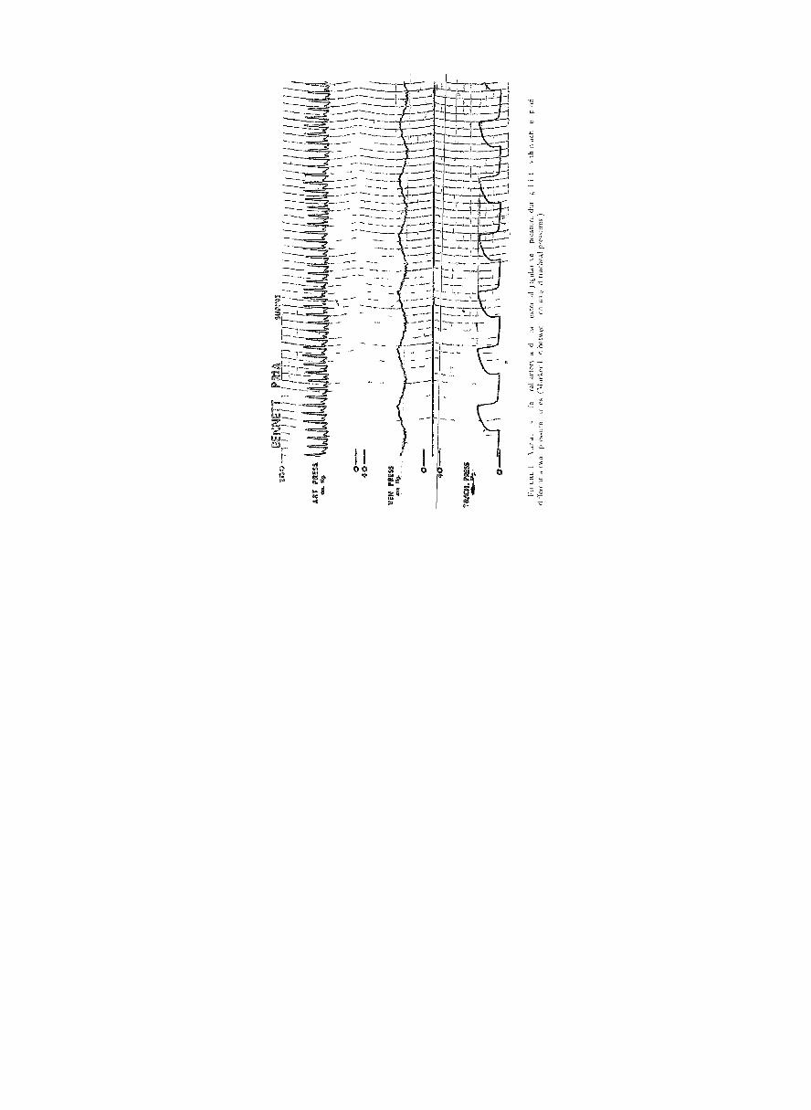
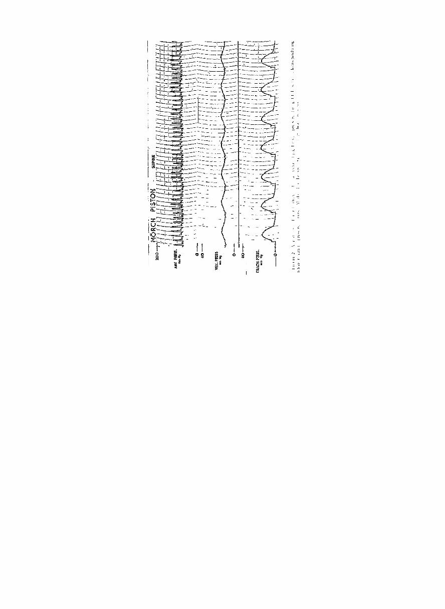
known, but it is of interest that modern respirators usually produce variations of systematic blood pressure within each respiratory cycle, the peak occurring at the point of maximum inflation pressure (Figs. i and 2). This may be due to improved left ventricular filling during a relatively shorjt inflation phase. I t has been suggested that this may be related to an increase inlexpanded lung, opening an unusual number of pulmonary capillaries, or to an increasing ejection of blood from the lungs through the course of each inflation. ThtJls, I.P.P. would convert the lungs into an auxiliary pump, filling during expiration) and pushing blood into the left heart on inflation. Provided that the point of ma~,!imum efficiency was not exceeded, then, the heart action would improve and the stroke volume increase (Starling s Law). In the event of hypovolaemia, I .P.P lowers cardiac output, and this reduction may be reversed by introducing a negative phase to the respiratory cycle (30).
There is thus the concept that each inflation reduces venous return but that this is offset by the ejection of blood from the lungs. If Mood volume is low, the effect on venous return is greater and the intra-pulmonary blood volume less. Consequently, the reduction in venous return then has the predominant effect on cardiac output.
While the effects of I.P.P. on cardiac output predominate in variations in systemic blood pressure, the calibre of systemic vessels also varies with airway pressure (31-34) and with blood and tissue gas levels. The diameter of pulmonary vessels varies with the oxygen tension of blood perfusing chemoreceptors in the carotid and aortic areas and, possibly, with carbon dioxide tension (35, 36).
.4 cid-Base Balance
The effects of I.P.P. on acid-base balance will depend primarily upon alveolar ventilation and secondarily upon renal function. In conditions of ventilatory instability 50 per cent of the change in body COs levels will occur within four minutes of changing ventilatory volumes (37).
In the t reatment of patients previously in chronic respiratory acida~s it may take several days for the metabolic compensation to adjust. In such instances, reduction of the arterial pCO_~ to normal will produce marked alkalosis. This has been noted in the presence of satisfactory renal function and in part may reflect the lag between the more rapid changes in labile forms of plasma carbon dioxide and the slower changes in tissue carbon dioxide stores.
Certain of the physiological problems encountered in the commoner causes of respiratory insufficiency will be discussed below, in association with their clinical management.
]INDICATIONS FOR ASSISTING VENTILATION
The main indication for assisting ventilation is the presence of respiratory insufficiency due to a reversible condition. There are many such conditions other than those producing muscular paralysis and I.P.P. has been used in all. Other

45~ CANADIAN ANAESTHETISTS' SOCIETY JOURNAL
circumstances are, for example, to rest the r~spiratory muscles in a patient with advancing poliomyelitis, to prevent or relieve exhaustion from the work of breathing, and to prevent paradoxical mo~zement of the chest wall. Finally, under some circumstances it is indicated as 'a means of delivering 100 per cent 02 for short periods.
The conditions in which undervenfilation may arise are exemplified in the following list:
(a) Conditions obstm~cting the airway, for elxample haematomata, inflammation of the upper airway, oedema, bulbar palsyj excessive secretions. This group is treated by relieving the obstruction but in the event of hypoxia, hypercarbia, and exhaustion, a period of respiratory assistance is often valuable.
(b) NeurornuscMar disorders, for example, head injuries, overdosage of narcotics or hypnotics, brain stem lesions, cervical cord lesJ~ons, poliomyelitis, polyneuritis, myasthenia gravis, disorders of potassium metabolism, and so forth.
(c) Following the llse of relaxant drugs, usually in the t reatment of status epilepticus or tetanus.
(d) Pulmonary dtseases, for example, emphysema, bronchiectasis, pneumonia, atelectasis, fibrosis, and left heart failure.
(e) Disorders of the thoracic walls, stove-in-chest, post-thoractom.v, post- laparotomy, paralytic ileus, ankylosing spondylitis or kypho-scoliosis, extreme weakness due to debility.
Many of these conditions occur together'. For example, a patient with anky- losing spondylitis may be precipitated into respirator?' failure by pneumonia or by limitation of diaphragmatic movement and of coughing after laparotomy.
The clinical picture of underventilation is not uniform and varies with the condition which gives rise to it. Further, a perusal of the list will show how many other possible causes there may be of symptoms and signs such as disorder of consciousness, breathlessness, changes in pulse and blood pressure, often held to be characteristic of underventilation. This fallibility of clinical diagnosis is such that awareness of the possibility is the most important clinical factor and any suspicion should be promptly confirmed or rejected by measurement of the ventilation. Nonetheless, the clinical picture of acute underventilation demanding immediate treatment is characteristic, although uncommon. Extreme distress, sweating, cyanosis, violent respiratory efforts, tachycardia, hypertension proceed rapidly to coma, hypotension, gasping respiration, and death. The early stages have been called the Alarm Syndrome.
Similarly, chronic respiratory acidosis has characteristic clinical forms. The best known of these is the familiar aspect of the patient with advanced emphy- sema. Less well known is the presentation as a neurological syndrome (38) made up of headache, disorder of consciousness, papilloedema, involuntary movements, and a variable rigidity. These signs vary in prominence from case to case. A syndrome has also been described in which the patient appears partially curarized (39). This is probably non-specific and a similar picture has been described in association with marked metabolic acidosis (40").
It must be reemphasized that the pitfalls of diagnosis and of differential
FAIRLEY & CHAMBERS: RESPIRATORY INSUFFICIENCY 4 5 5
diagnosis are such that, in most cases, meast~Fement of ~ventilatory volumes, with or without measurement of the blood gases, is essenotial.
Ve nt ilatory _/1,leasur ements
The clinical suspicion that underventilation is present may be confirmed by measuring ventilation itself.
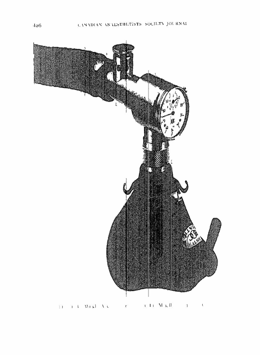
Tidal volume. ~This may be measured in one of the following ways. (i) Spiro- merry- - th i s is most applicable in the conscious unasslsted, patient. However, it is cumbersome and requires special measures to oveiIcome problems of carbon dioxide retention and expiratory resistance. (ii) Collection of expired gases, via a non-return valve (41) in a Douglas or plastic bag (42, 43). This also is somewhat cumbersome and still requires that the collected gas be measured by passing it through a spirometer or gas meter. (iii) Dry Gas Meter - - th i s is a relatively accurate method of measuring expired gas volumes. The apparatus looks and functions in the same way as a domestic gas meter. By means of a non-return valve, expired gas is fed through the meter for one or more minutes, respirations counted, and a mean tidal volume calculated. (iv) Ventilation Mete r s~ these are perhaps the most convenient means of measuring expired gas volumes. The gas passes directly through them, turning a system of vanes. Reasonable accuracy is attained at all except the lowest gas flows. The most rece~lt of these, the Wright Anemometer (44) (Fig. 3), can be placed in continuity with the airway, measuring gases passing in one direction only, and offering minimal resistance. It is extremely small and portable, thee face being the size of a large wrist watch. The main objection to this type of 'equipment has been the fragility of the last named and the possibility of moisture from humidifiers Mtering the response of the vane and therefore the accuracy. More rugged but less easi|y manipulated are the Draeger and Monaghan meters (41)o
By means of equipment of this type, the patient 's tidal volume may b4 followed. Expired volumes are chosen as these are the more reliable when using I.P.P. Under such circumstances, some of the inflation volu~me ma3 ~ leak out between being measured and passing into the bronchi, whereas all the measured expired volume must come from the tracheo-bronchial tree. Comparison with a ventilation nomogram and consideration of the possibility of an enlarged physiological dead space will determine the likely adequacy of ventitatory exchange.
Having confirmed one's clinical impression by measuring venti latory volume, any remaining doubts may be cleared by measuring arterial pH and CO2 levels, which provide the only absolute guide to respiratory status (45). It should be stressed that CO.. levels alo~e are not always sufficient owing to the not infrequent coincident metabolic changes in seriously ill patients.
Blood gas measurement. Arterial pCO.~ may be measured as follows: (1) Directly (46, 47). This is technically difficult and not available in the
average hospital laboratory. (2) Derived from:
(i) ph and COo content of arterial blood (48); (ii) pH of arterial blood and pH of the same sample after equilibratiofi
with a gas of known pCOe (4,9) ;
-Io6 r \NkL;bTHI~TI~Tb bO(.ll.T~t jOLRNAI
t I i ~ ~ \ ~ - I . t \ c �9 ~ 1 I ~,I k L1 1 t

F A I R L E Y & C H A M B E R S : R E S P I R A T O R Y I N S U F F I C I E N C Y 457
(iii) Alveolar gas sample, by single prolonged expir~ tion or by modifications of the Plesch method (50-53), the gas being analysed b~.~ Haldane (47) or Scho- lander (54) appara tus , infra-red analyser (55-58) or on of several o ther more simple but, in most instances, less accurate methods (5 ' -62 ) . One of the latest of these methods has the meri t of s implici ty and relati,~e accuracy (52) bu t all require an addi t ional pH value for a complete picture.
(3) A_~sumed from: (l) CO._, combining power. This is essentially a measure of slowly changing
metabolic factors. I t i~ relat ively wtlueless in acute respi ra tory problems and in the presence of combined metabolic and respiratory disorder (45). I t may indicate the trend in chronic respiratory states. To derive maximum benefit from this est imation, within its limits, it should be considered in association" with a pH value;
Lii) Arterialized venous samples (63, 64). When the forearm and hand are warmed and venous blood is taken from the dorsum of the hand, wi thout tour- niquet, good correlation with arterial values is obtained in normothermic normo- volaemic pat ients;
~,iii) End- t idal samples (65-67). Continuous readiltgs may be m~de of the carbon dioxide levels in end-t idal air, using infra-red or photochemical l~*inciples. This value will follow tha t of the alveolar air if the tidal volume remains constant , but the absolute level will be of no special significance when measured alone.
Thus, arterial carbon dioxide levels may be determined by one of a number of procedures, of which the Ast rup method has been found very sa t i s fac tory (method 2 (ii) above). A spegial~zed vir tue of this technique lies in the possibili ty of its use in pat ients recent ly given a volatile anaesthet ic "agent and the s imul taneous avai labi l i ty of a pH value.
Oxygen administration. One must consider under what c i rcumstances oxygen should be added to the inspired air. if a pat ient has normal lungs, this will not be necessary. However, any suspicion of a dis t r ibut ion or diffusion defect will indicate the addit ion of oxygen. I .P.P. creates one of the few circumstances in which 100 per cent oxygen may be delivered to the lungs for a prolonged period and the harmful effects of this should be remembered (68).
When adding oxygen to an appara tus delivering air, it m a y be difficult to assess the effectiveness of tt~is addit ion. Improvement in oxygenat ion m a y be shown b\" a slowing of the hear t rate but more reliable informat ion m a y be ob- tained by (i) analysis of inspired or expired air by oxygen analyzer (69), (ii) oximetry (70-72), or (iii) in te rmi t t en t arterial sampling and manomet r ic analysis (73). In cases of marked desatura t ion , one may increase the flow of oxygen until no further improvement occurs in (ii) or (iii). In this way, maximum efficacy" is achieved wi thout the possibility of oxygen toxicity.
Clinical Application of Measurements
The way in which the measurements of respi ra tory volumes and blood gases are used ill clinical 9ractice is as follows:
(a) The initial decision to venttlate. In all cases not in extremis, vent i la t ion is measured. The conscious pat ient is asked to breathe through a n{.outhpiece into

458 CANADIA~N ANAESTHETISTS' SOCIETY JOURNAL
one of the measuring devices described above, after the application of a nose-clip. In the uuconscious subject; nose-clip and lar,~e-flanged mouthpiece, facepiece, or cuffed endotracheal tube may be used, Val ventilation nomogram, and the first of a set decision to ventilate may" be made on the b~
ues obtained are compared with a ies of values is thus available. The sis of unequivocal underventi lat ion
shown on the first reading, or as a result of g~-adual deterioration as indicated by serial reading .
Unless ventilatior~ is very obviously w~tequate, the arterial pCO2 is then determined. Underventilation, as shown byl t idal volume and arterial pCO~, is taken as an absolute indication for assisted respiration except in two circum- stances: (i) chronic respiratory acidosis with~ no evidence of recent deterioration and without trial of less drastic measures; (iiJ) minor degrees of acute respiratory acidosis in a rapidly improving situation, for example, recovery from deep anaesthesia.
(b) Monitoring of efficiency of assisted respiration. During I .P.P. the following measurements are made, in addition to routine clinical observations: (i) tidal volume, (ii) inflation pressure, (iii) arterial pCO2. The volume necessary to achieve a normal arterial pCO., is noted at the outset and the pressure necessary to achieve this volume is observed. Frequent measurements of ventilation will be needed in the acute stages.
Further arterial pCO2 values will be obtained only when doubt exists as to the respiratory status, for example, in managing patients with gross puhnonary disease or as a very occasional check in patients needing prolonged t reatment .
.~/J[ANAGEMENT OF AIRWAY AND VENTILATION
The equip,nent necessary for the management of respiratory hlsufficienc3" by I.P.P. may be grouped as follows: airway and suction equipment; non-return valve; humidifier; ventilator.
AIRWAY. A small group of patients can be ma~laged satisfactorily by means of a face-mask or nose-clip and mouthpiece, using no artificial airway of any sort. These "are conscious patients with chronic respiratory disease and considerable insight. With a little training, they can use /[.P.P. in this way to deliver broncho- dilators, vasoconstrictors, and wetting agents 1174, 75). Improved bronchiolar calibre and eliminatio~ of secretions may then improve ventilation. Such therapy has usually proved impracticable in the t rea tment of severe respirator3" insuffi- ciency and lends itself more to the less severely affected group of patients with chronic respiratory disease.
It is obligatory to create an airway and start ventilation as soon as under- ventilation is recognized. Even brief delay may result in cardiac arrest. The usual practice is to pass a cuffed orotracheal tube, a t racheostomy being performed subsequently if required. Tracheostomy is considered to be indicated when it is evident tha t the problem cannot be corrected within an arbi t rary 24-48 hours. When early recovery is probable (e.g., barbi turate poisoning), t racheostomy is postponed. In all other cases, it is best delayed m~t;il resuscitation has been carried out, when the patient will be adequately ventilated, hydrated, and, if necessary, transfused and digitalized.
FAIRLEY & CHAMBERS: RESPIRATORY INSUFFICIENCY 459
In all cases not in extremis, intflbation is carried ot~t uncter a minimal dose of sodium thiopentone and succin}lcholine, using careful topical anaesthesia, if thiopentone is contra-indicated, the technique depen~ts on th'e patient~'s level of consciousness. Thus, if amnesia is probable, muscle relaxant'i* and topi]cal anaes- thesia are used. If the patient is more conscious, topical anaesthesia is flsed alone, in which case transtracheal instillation is preferred.
The need for great gentlelmss and maximal relaxaltion is emphasized by the occurrence of post-intubation granulomata of the vocal ,cords in two cases in which technical difficulty arose. Relaxants were not ~,used and in one case the tube was only in place eight hours.
'Once the airway is secure, the patient is sedated ufltil he becomes accustomed to his new circumstances, in many instances he will become very much less agitated once adequate ve, t i la t ion is established. However, in rare instances where sedation does not settle the patient and adeq~uate ventilation cannot be achieved, muscle relaxants may be required as a temporary measure.
Tracheostomy. !The virtues of tracheostomy in thie t rea tment of respiratory ii~sul~ciei~cy a r e a s follows:
(i) Access foriI.P.P. (ii) Access foe suction. (iii) Protection from aspiration of pharyngeal secretions by using a cuffed
t racheostomy tube. (ix') Freedom from equipment about the mouth and nose, permitt ing the use
of duodenal tubes, normal eating and drinking, and so on. (v) Reduction of de~d space. The latter factor is offset, when using l .P.P., by
the added dead space of the apparatus and by the ~n,grease in physiological dead space produced by I.P.P. itself (l(i).
i. P. P. is not possible in conjunctiou with the usual types of tracheostomy tube, without an adaptor which fits into their lumen. This reduces the diaiheter of an already" narrow airway.
Special equipment is usually preferred and a variety of tubes has been des- cribed. They should (a) be of adequate diameter (from 9-12 ram. internal dia- meter for adults), to minimize resistance to gas flow and to decrease the likelihood of obstruction by secretions, and (b) have adequate connections for a t tachmeut to I.P.P. equipment. The latter should not narrow the lumen and should be moveable through 360 ~ (e.g., metal to metal slip-joint), in order that the I.P.P. equipment can remain stationary when the patient is moved.
Two main types of tracheostomy tube are in use: those with and without inflatable cuffs. The latter (76, 77) are less versatile, do not permit measurement of ventilation, and do not prevent aspiration of secretions from the pharynx. However, they have been advocated in the t r ea tmen t of stove-in-chest by induced respiratory alkalosis (78). They are also of value in the management of tracheal ulceration secondary to over-inflation of cuffed tubes.
Cuffed trache0}tomy tubes may be improvised by stretching a latex Cuff %ver a large tracheostomy o~- laryngectomy tube but are also commercially available with an incorporated cuff. The former are usually made of metal or plastic and have the advantage of an i~lner cannula. However, the metal tubes usually leak

4 ~ 0 C-k',.-'iDlk",. kN~.] .>rI'IILT1ST'. "~O(.li T'~ JOL RN4.L
bet , , ; eeT1 l h c i n n e r ~nd c~tll-(.r c ,~ ,~uI a I }~t ', i r t un( 1~cr t o b e sIlf3rt tu]c-] ~, t t}ltt k
necked p l t l e l l t OF i l l oi led \ \ 1 " [ h o(._(]ot'll t I't it ~'~l ].[('~Ill t OI SI.IFLI(. 11 L t ' t ' t p | l " , . r 1 I}IC
() ' t i f f [II L TM, l i e 11] t}lI._ C_~.~.F { {-1 tt ]tL l.I 1 i~, (_. r-~ (J[ llJ{, (.Z [ttl(-(_ ~0[" t]l~CrO['~lr ~, ])LI[-i~,(] llt_} ]tk
,1.:~: tev i h e t u b e s n~ l d c l o t I ] i ~]-t t l~ t l ~llx i rtit?bc~ tlar ~, iF ~, t l ~lc~,li. ] t7 (} %(1)
{)tit experlel~ce h i s betel l]zit i k, y t t ] l l l i I ~ , ~ [ ) i t - l i l t ' - , t i a \ l) i",% IL, L ~ I t '-,11{ l l tgl ]
c x t h e t c r t h t t "t s c i [ l l l c q s ctil-t t)c[111 l,, t i l t { t i l e t IlL e l i x i r t l l \ n~ l h t I f ' i / h i . t
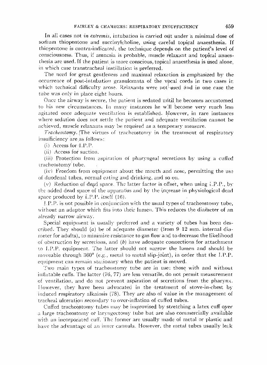
{t~,g 4) re t luc~,~g tili. c l ~ i n ~ . ~ ) t [ , l t ] t ( .~ , l [~[ , , l t~oll i n t t t h t t i t lc ,pt l l~_[l{] x; t t}aotiI
bt l~l t ] n t o n n e c t o r o z t i ] n L c t3ci1~1~I~ I ] ] t l~ , t l t J l l I t>l ] ] t l s ]{ l ]~ldu tl ' ~ ] t t l t]]lt ki lo '- ' .
i~c.} ~xo l r ~.h(_. c ( ) } ]~ ]e(_ tor pi]llin~, } i l l { 1 I1 t [1 )1t L [ 1. t1'. N~()~, \~.o[ I . t I \ (~ l t l l ~ !q l
O l l t O -~%hlch I ~ t l t t , e I1 l s b e c l l } till[ ]1 1. i , _L ] {t_'-l,.,ll~_tt ( [ I t S ) t[1~.{ *:) J [11%
,,~ ib~l izes t h e ~ h o l e tu loc
] l e t I F 4 C I l f c J tr h t t I x I 14.
l l t x ~ t h ' , , 1 1t i t ~,r l t r I1 I fL f l ~ t 4 I ] L ~ i t LI, LFI
! ~.IR] II ] ~ ( I ] a ~ \ l ] ] I R b R L ~ P I R \ T C I R ~ 1%~L b ~ - l ( l l ' , , L ' ~ 4bl
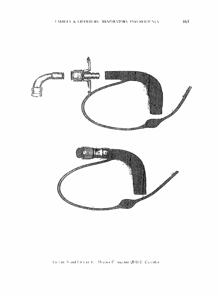
1 lc [ Rt "~ ~It , l I 1~ ~ I~1 I* ] t c , m , - C 1 , / t ~ l o r ~ [ I , ( ) ( " C t l / , [ , i
462 CANADIAN ANAESTHETIST~' SOCIETY JOURNAL
To permit the cuff to lie within the trachea, the intratracheal portion is rela- tively long. In consequence, the tracheostomy must be high and the second ring or space should be divided. The cricoid should be protected by an intact first tracheal ring. I t is important tha t the end of tlne t racheostomy or endotracheal tube should be high enough to allow suction catheters to pass easily into the left main bronchus. Adequate air entry bilaterally does not indicate tha t the tube is high enough, only tha t it is not in the right main bronchus. A fm'ther reason for high tracheotomy is that the lower the opening, the nearer the tube is to the great vessels of the neck and the greater is the danger of massive secondary haemorrhage. This is more probable if Lhe origin of the vessels is anomalous (81).
A circular window is cut from the trachea, following division of the thyroid isthmus. Both these manoeuvres are performed routinely and have been found valuable not only at the initial insertion but in case early changing of the tube should be necessary~ I n the lat ter event, there is less likelihood of the t racheostomy being closed, as though by a series of shutters, when the tube is withdrawn.
As stated above, tracheostomy is performed semi-electively, assisted[ venti lat ion being carried out throughout the operation. The endotracheal tube is wi thdrawn completely from the glottis only when the tracheostomy tube is securely in place. Atropine is[given as premedication in all iastances and this is accompanied by a narcotic uniess col~traindicated. Respiratory depression from the narcotic is not usually a serious consideration in patients receiving artificial ventilation.
In this series, general anaesthesia was used for almost all tracheostomies. This varied from 50 per cent nitrous oxide with oxygen and muscle relaxarJLt to sodium thiopentone: nitrous oxide: halothane with or without relaxant, according to the patient 's general condition.
Changing Tracheostomy Tubes. It has been found advisable to change rubber tubes more frequently than metal, if trach,eostomy is followed by marked oozing of blood, a change after 24-48 hrs. may be required. More usually, however, a weekly change suffices. Until the traeheostomy is firmly established, difficult3" may arise during this procedure and two people should be present. One intubates the trachea through the glottis, before the t racheostomy tube is removed. [n this way, ventilation is under control throughout.
Complications of "Fracheostomy. Those most commonly seen are infection and mucosal ulceration. The former is t reated in the normal way and did not prove worrying in this series. The latter may give rise to considerable anxiety and is most likely in patients exposed to the effects of hypotension. Bleeding from tracheal ulceration must be distinguished from tha t due to other causes, for example, tracheitis sicca. Trea tment seems best carried out by use of an uncuffed tube and the M6rch technique. Suction may aggravate the si tuation and, if tracheitis sicca is suspected, the use of a machine producing sudden powerful expiratory movements has been recommended (82).
Cardiovascular collapse following tracheostomy has been described and various causes suggested (83-85). Among these, sudden reversal of hypercarbia with possible accompa~ying electrolyte disturbances is commonly accused. Sudden collapse during tracheostomy may relate to wtgal overact ivi ty from hypoxia and mechanical stimulus especially when atropine has been 'omit ted.

FAIRLEY & CHAMBERS: RESPIRATORY I N S U F F I C I E N C Y 463
In the present series, no post- tracheostonly deaths were observed, a l though hypotension requiring vasopressors for some hours has been seen. This usually followed t racheostomy in pat ients who had suffered severe hypoxia and it is thought tha t a resulting reduction in myocardial efficiency was responsible. Electrocardiographic evidence of myocardial ischaeniia was observed in several instances.
Bronchoscopy. This has been found to be of very occasional value and may be dangerous. Bronchoscopic equipment should be constant ly available and has been found life-saving in two situations: (i) Haemorrhage within the bronchial tree, the combination of blood and secretion forming crusts not removable by suction catheter. (ii) Marked crusting in association with staphylococcal pneu- monia. Again, two persons should be present and only very limited periods wi thout vent i la tory assistance should be permitted. If the larynx is not of immedia te interest, the bronchoscope may be passed through the t racheostomy.
N O N - R E T U R N VALVES (A.1). SOl l l e form of non-rebreathing system is preferable to one providing carbon dioxide absorption, for a nuinber of reasons:
(i) The circuit becomes simpler. (ii) Soda-lime does not have to be changed. (iii) Expired gas can be collected and measured. (iv) Contaminat ion of the machine is less likely. (v) Expiratory resistance may be lowered. In practice, non-return valves have certain l imitations: (i) They tend to leak,' so that a portion of the expired gas is rebreathed.
This is of little consequence, except in volume measurements . Using such a valve, for example, F lu t ter - type valve, Reuben Valve, MSrch Valve (78), a meter must be placed between the valve and the patient. In this regard, the Wright Anemo- meter is part icularly useful.
(ii) They render insertion of a negative phase more difficult, a l though such a phase will increase the efficiency of flutter valves by causing early closure.
(iii) Some of those which close efficiently on expiration, offer a small but signi- ficant expiratory resistance at normal flow rates, for e:~ampl% the Etsen modifica- tion of the Fink valve permits collection of expired gases and closes prompt ly bu t has an expiratory resistance of 3-5 cmo HoO at normal flows. However, such a valve may be inserted in a circuit temporari ly for purposes of measurement .
Many machines are equipped with non-return systems and most ly belong ko the group of in termi t tent ly ope~ing reducing valves (see below). Some have a housing around the expiratory valve from which expired gases may be collected.
Whe~l the machine is not equipped with a valve, the 5ltSrch non-return val~ve has been found to be efficient, as has the Beaver flutter valve. These are placed as near to the pat ient as possible, to minimize dead space. They must be changed at intervals and therefore more than one must be awailable for each patient. Tl~e forlner is changed and cleaned daily, the lat ter every two hours. This is necessary because moisture collects on the rubber d iaphragm and hampers its free movement .
ftUMIDIFIERS. Pat ients must be adequately hydra ted and the respiratory gases must be humidified. Lack of a t tent ion to this causes a high incidence of pulmonary

404 CANADIAN ANAESTHETISTS' SOCIETY JOURNAL
complications. Certain machines are equ~ipped with humidifiers, al though the efficiency of these varies. While a high relative humidi ty is desirable in all patients, actual droplet administrat ion is[ frequently of value. This is the case in all patients with copious secretions, or it1 those requiring intrabronchial medication. The droplet size should be 1O.5-10 g (86), which is provided by relatively few pieces of equipment. Large~- droplets are inefficient in liquefying
- -
sputum and may cause respiratory embarras.,ment, while smaller droplets are absorbed too quickly. The optimum size i.,I; probably 3-5 #.
Where a machine is to be used which does not have a humidifier, one must be placed in the circuit. One may either deliger the inflation volume over hot water (87) or put a T-piece into the circuit neai~ the pat ient and deliver droplets from a nebulizer by this means. Alternatively water (not saline) may be delivered from an infusion set through a needle inserted in the inflation tubing. The possibility of tracheostomy infectior~ from humidifiers should be remembered. Meticulous cleanliness is essential.
VENTILATORS. These have been discussed at lel~gth in recent publications (41, 88) and vary greatly in design and efficiency. They may be discussed under a variety of headings.
(a) Motive force. This is either an electrical motor or compressed gas, each having its advantages and disadvantages. The former is relatively ~ reliable, expensive initially and requires to be of a special approved type. The lat ter is more common, tess expensive, and very variable in reliability. Both forms of motive force are subject to failure and each pat ient should have an a l ternat ive method of hand ventilation available at all tinles. In this regard, the Ambu type resuscitator (89) has proved most useful.
(b) Circuits. Essentially, these fall into three groups. (i) Pistons or bellows which draw in room air on opening, then, by a series of valves, deliver this to the patient. Both electrical- and gas-operated machines fall into this group. (ii) Motor blowers: essentially, these are in termit tent ly functioning revel-seal vacuum cleaners. (iii) Intermittentl)" opening reducing valves. These are all compressed- gas-operated machines, functioning by exposing the pat ient 's airway inter- mit tent ly to a source of compressed air or oxygen. The pressure at" which this exposure is made and the rate at which the gas flows is controlled by the machine. These are usuatly patient triggered but may also be equipped with an automat ic sett ng.
Also ira this group is the pneophore valve which, by means of a diaphragm mechanism, controls the flow of gases. This lat ter type of equipment is of limited value for the prolonged t rea tment of respiratory insufficiency.
(c) Principles of operation. Machines must have one of two limiting factors to inflation. This is either a preset pressure or volume. Consequently, machines are all either pressure constant-volume variable or volume constant-pressure variable (90). Ira the first instance, tidal volume 'will fluctt~ate inversely with changes in total resistance. In the second, the tidal volume will remain constant while the pressure at which it is delivered varies directly with the resistance. In each instaitce, this variable factor is limited by the machine's capabilities. For most
FAIRLEY & CHAMBERS: RESPIRATORY INSUFFICIENCY 465
purposes, a versatile machine should be capable of del ivering over 1000 ml. per stroke and pressures of 50-60 cm. H20.
Certain features are essential on all volume-con~gtant, machines. (i) They should have a manometer in the circuit, for use as a re{istance meter. An increase in pressure suggests the possibility of the need for bronchial/aspiration, correctioll of bronchospasm or left heart ifailure, or the need fo~-~ more muscle re laxant in conditions such as tetanus. ( i i ) 'They should also poss(!.'ss a safety valve so tha t a lung will not be ruptured, if the resistance rises Drecipitously, the machine inexorably delivering its preset volume. The ~[6rch respirator (78) has been found to be a very reliable, if not versatile, machine. Being intended for use with an uncuffed tube, it possesses neither safety valve nor manometer . The humidifier has been found inefficient and, in consequence, the following modification has been introduced and found to be a great improvement . The humidifier cyl inder was removed and, in its place, a one-inch d iameter threaded tube was inserted. To this was a t tached the delivery tube to a separaLe humidifier. An anaeroid manometer and a blow-off valve (loaded at 50 cm. H~O) were tapped into the top of the one inch tubing.
(d) Respiratory wave form. ()pinions differ as to the op t imum wave form. Certain macllines apply I .P.P. to the upper airway in a preset wave form while, in others, the durat ion of each phase and the shape of the pressure curve produced may be adjusted. The sinusoidal curve has been strongly advocated a s . t h a t likely to effect cardiac ou tput least. However, a square wave is held by" many to give the greatest inflation in the shortest time. The square wa've is produced by" machines delivering high gas ttows from the s tar t (e.g. 100-200 L . , m i n . ) . . X l a x i m u m pressure is reached rapidly in the upper a i r w a \ (giving a vertical up-stroke to the i~flafion curve), Ventilation is then largely dependent upon the durat ion of appl~catio~ of this pressure (giving a horizontal component to the curve). The pote~tial d isadvantage of such wave form might be tha t in pat ients with low compliance ventilatio~ would only be achieved at the expense of venous return.
If a severe distributiola defect necessitates slow inflation or spree expiratory retard is utilized to prevent t rapping, the vertical components of the wave form will be impracticable and a siuusoidal curve more desirable. Again, venous return will be impeded.
[,/Tatio,. During this phase, tidal volume will be determined. Air should be delivered at a flow of at least 40-60 L. per rain., and it. should be possible to achieve this fairly rapidly. The iu te rmi t ten t lv opening reducing valve type of machine usually delivers gas at over 100 L. per rain. and at least one is capable of flows up to 200 L. per rain.
This phase is adjustable on re,my mJchines and should be as fast as is corn- patible with adequate venti lat ion, distr ibution and comfort. Pat ients with pain in the chest object to machines reaching max imum inflati~on pressures extremelY
rat)lilly. E:cDiration a,~d Pause. These two ph,lses together should be at least as long
as ilLspiration aIld preferably lozlger to permit adequate venous re turn (29). Expiratio~ itself should be ,~s ratfid as possible except i~n pat ients with air t rapping.
466 CANADIAN ANAESTHETISTS: SOCIETY JOURNAL
In .the latter, a small resistance in the first part of expiration may diminish trapping. Again, phase adjustabi l i ty and an adjustable expiratory retard may be of value.
Negative Phase. This has been shown to b,l~ of value in hypovolaemia (30) and in pat ients with low lung volumes (91). The danger of accentuat ing t rapping in emphysema has been suggested (92) and no/ilnprovenlent in venti lat ion has been demonstra ted using a negative phase in a ,miall series of such pat ients (25). I t may be most valuable when inserted at thel end of the expiratory phase (93).
f
(e) Patient-triggering mechanism. Certain machines possess a mechanism whereby each respiratory cycle is in i t i a t ed by the pat ient creating a small negative pressure in the circuit. This may be fixed to work at, say, --2 era. H_~O or may be variable. Most machines with such a device can also be set to work automatical ly. Frequently, the automat ic control may be set at a slower rate than tha t of the pat ient so that , should he fail to trip the inachine, it will then cut in on its own. While this mechanism is not essential, it has been used very fre- quently in the present series and has been most helpful. The pat ient tr iggering mechanism is contraindicated in the t r ea tment of stove-in-chest,, until the weaning phase is re hched. Clearly, it will b e inappropriate in the presence of apnoea.
( f ) ]lachines of z~se in both operating room and respiratory insz~fl~ciency. Certain machines are designed in such a way tha t they may be used either in the operat ing room or in the t rea tment of respiratory insufficiency. Although such versat i l i ty usually leads to compromise, the concept is most appealing to the smaller hospital. The essential differences between the two types of vent i la tor are these:
(1) A machine for use in anaesthesia must have a circuit, separate from the motive force, into which one may i~troduce anaesthet ic gases. Several machines, designed for anaesthesia and possessing such a circuit, can be modified for I .P.P. therapy by opening an air intake valve. Thus, as the bellows expands, ii~stead of anaesthetic agents filling it, air is drawn in. As the bellows empties, the air valve closes and the air is delivered to the patient. Under such circumstances, the machine must obviously be used with a humidifier and non-return valve.
o
When used for anaesthesia, these are removed and the machine subst i tu ted for the rebreathing bag of an ordinary anaesthetic circuit, with its own circle s vslcnl, to and fro absorptions, or non-return valve.
Clearly, such interchangeabil i ty is not possible with the in te rmi t ten t ly opening reducing valve type of respirator. However, they may be adapted< for use in anaesthesia (as may the bellows or piston type not designed for this purpose) by placing the rebreathing bag of the anaesthe.tic circuit in an air t ight container and applying tF.P.P, to the space between bag and container (94). Such a machine has now been made available commerciall::.
~(2) Machines for use in operating rooms must, be explosion-proof, rendering those which are gas-operated most frequet~Ltly acceptable.
(3) M~kchines used in operat ing rooms may be relatively complex, but satis- factory under the continua1 observation of a physician. Such a machine may be difficult for the inexperienced nurse to understand.

FAIRLEY & CHkMBERS: RESPIR.~TORX" INSUFFICIENCY" 467
(4) Machines for respiratory insufficiency require a humidifier and non-return valve. (See above.)
(5) Machines used in t reat ing respiratory insufficiL~ncy may be in use for weeks on end; after anyth ing more than the few hours cus tomary for anaesthesia, minor imperfections of pressure wave form, expiratory re.'sistance, and long-term reliability become of increasing importance.
(g) .4lternalive methods of ventilation. Emergency" equipment , with which to venti late any patient whose venti lator ceases to function satisfactorily for an?" reason, must always be available. At tendan ts should all know how to use this and, in cases of emergency, must do so until help is obtained.
(h) [ .P .P . in children. As mentioned above, a given volume of air will be delivered at a specific pressure regardless of the alge of the patient-, the only exception being neonates. Thus I .P.P. equipment for children will require only slightly lower pressure ranges than those of adults. However, considerations of volume and rate will be very much more critical. Bearing in mind tha t a 7-lb. infant may have a tidal volume of 20 cc., it is obvious tha t dead space must be minimal. Similarly, an infant 's respiratory rate may require a vent i la tor to function as fast as 40 per rain.
CLINICAL ~ANAGEMENT
The sections tha t follow will be concerned with the clinical application of the above principles, without any discussion of first aid:
Pat ients requiring assisted respiration present in two ways: the first is the pat ient who needs respiratory assistance at once; the second is the pat ient whose venti lat ion fails gradually while he is under observ~tion.
The first group needs emergency t rea tment . An airway is established and venti lat ion started as previously described. In an acute emergency of this type, measures to secure these ends take precedence over all other considerations. When the?" are secured, a complete history can be o b t a i n ~ , physical examinat ion carried out, and other emergencies dealt with.
History taking and examinat ion will have been done, of course, in the case of the pat ient with respiratory failure of gradual onset, but the following investiga- tions are i m p o r t a n t chest X-ray, Hgb., W B ~ and differential, complete urine examination, serum electrolytes and NPN, E C G examination, culture of the bronchial secretions, and measurement of venti lat ion. If facilities exist, the arterial pH, pCO~, and CO2 content should be determined before the inst i tut ion of artificial respiration and an hour or so after it has begun.
~n general, the use of I .P.P. does not alter the management of most conditions in which it may be needed but a few points merit special emphasis. Successful t r ea tment depends upon a t tent ion to detail and this must be impressed upon ~tll who deal with these patients. This is the only way in which minor devia.~ions from normal can be prev~")ted from turning into major ca tas t rophes ardt this /. responsibility falls in the first place upon the nurses.
Nursing care. The successful use of assisted venti lat ion depends upon the skill
4 6 8 CANADIAN A N A E S T H E T I S T S 'P SOCIETY J O U R N A L
h . . . y of t e nurses, who should therefore be specl:lahsts. In practme, several months experience are needed to acquire the necessary skill and confidence. TIhe nursing problems are those associated with the nursling of very sick patients, the use and the understanding of the specialized equipment, and the recognition and manage- merit of a developing emergency.
Apart from general nursing care, the follgwing are the routines in use in the Respiratory Unit at the Toronto General Hospital in acute cases: half-hourly observation of heart rate, blood pressure, respiratory rate, and airway pressure; half-hourly tracheal to i le t - -or more frequer~tly if necessary- -and note taken of quanti ty, consistency, and colour of the aspiirate. The humidifier must always be kept full. In some patients, half 'hourly or hourly record of the venti lat ion is made. Every two hours, the patient is turned and the cuff on the t racheostomy tube is deflated for two minutes and then reinflated; .there is no good evidence tha t this is necessary to prevent ischaemic ulceration of the trachea but it is done in the hope tha t it will. The patient 's temperature is recorded every four hours and the fluid balance every eight.
The large number and variety of nursing problems tha t arise in these patients will become apparent in the sections tha t follow. The outline given above refers to the acute case. As the patient improves, the various observations may be made less often and the frequency of suctioning is adjusted to the amount of secretions present.
The care of the chest. The problems in the chest are the prevention of atelectasis and of infection. These arise because the natural barriers to and humidification of the respirator>" tract are bypassed and because effective coughing is abolished. The presez~ce,of a foreign body, the t racheostomy tube, in the trachea promotes the formation of secretions and, particularly in asthmatics, tends initially to aggravate spasm of the brohchi, in a few patients, the bronchial and tracheal mucosa is destroyed by infection.
Bronchial secretions are removed by catheter suction. They are brought within reach of the catheter by, physiotherapy to the chest. The value o fches t physio- therapy, t ha t is of percussion and squeezing of the chest wall, has been empha- sized by many writers (95, 96). All patients in the Toronto General Hospital Respiratory Unit, except those with stove-in-chest or in shock, are given physiotherapy twice daily, by members of t]~e depar tment of Physical Medicine. It is useful for the nurses to have some knowledge of chest physiotherapy.
Suctioning is done with sterile coud6 rubber catheters, size 16 or 18, which are used once and then resteri~ized. These catheters are of soft rubber, hollow to the end. The hole is about 1 cm. from the encl. Straight catheters enter the r ight bronchial tree on the vast majority of occasions (97).
The catheter is connected to the suction, machine via a y-piece, one limb of which is open to room air. The machine zs turned on and the catheter inserted with the tip pointing to right or left, del~ending upon which main bronchus is to be aspirated. It is gently insinuated as far as it g, ill go. A finger is then placed over the limb of the "y" and the catheter gently withdrawn with a slight twisting movement. The procedure is repeated until a][l reachable secretion has been aspirated from both sides. V~:hile this is d'~one, the pat ient must be disconnected

FAIRLEY & CHAMBERS" RESPIRATORY INSIUFFICIENC~ ~ 4 6 9
from the ventilator. To prevent hypoxia, it-is a safe rule for the inexperienced nurse to hold her breath from the moment tha t she dis(~onnects the venti lator and then reconnect it when she has to take a breath herself. ~
To maintain secretions fluid enough tO be aspirated, Ithe inspired gases must be properly humidified, but the most perfect humidification Of the inspired air wil~ be useless if the patient is dehydrated. The avoidaI!,ce of dehydrat ion is the essential factor in maintaining fluid bronchial secretions. Once hydrat ion is achieved, humidification is relatively simple.
The use of these technique s will prevent the occurrelme of serious intrathoracic complications in ahnost all patients who present wit:hour intrathoracic abnor- mality and will control these complications in those! who present with a "wet chest." They will also render bronchoscopy a rare event.
The management of infection and other intrathor~[cic complications in these patients is the same as in any other circumstances. Occas!o~}ally, unusual organisms will be found in the bronchial aspirate, for examp el~.B. Pyocyaneus. We have not seen such organisms cause any serious pulmonary or .bronchial infection nor to persist in the bronchial secretions once the t racheotomy tube is removed, although rarely they are capable of causing serious pulmonary infection.
7"he Cardiovascular System. Congestive'cardiac failure is recognized and treated in the usual way. I t is not a contraindication to the use of I.P.P. which may, by providing adequate ventilation, relieve it.
Shock and hypovolemic states are particularly dangerous because many of these patients have cardiac disorders of various types- -myocard i t i~ t rauma, arteriosclerotic heart disease, previous ischaemia of'rnTocardium due to anoxia rendering them liable to cardiac arrest from hypoxia. Further , I .P.P. aggravates the circulatory difficulties present in hypovolaemia, 'endangering renal function and exposing the pat ient to~ the risk of anoxic encephalopathy. Such states are deatt with in the usual ways, by vasopressors, blood transfusion, and arrest of haemorrhage. The use of a negative phase has been discussed above.
Puhnonary oedema may be the cause 9f a gradual or acute change in the other- wise satisfactory progress of many patients. It may be detected clinically and by a fall in tidal volume, with pressure constant machines, or by a rise in pressure with volume constant machines. It~%ay occur in any elderly pat ient secondary to a change in cardiac rhy thm or in any post- thoracotomy or stove-in-chest pat ient from either atrial fibrillation or actual myocardial damage. Methods of management are not altered by I.P.P. Which may' in :fact be advantageous. "Pulmonary oedema" has also been seen in the course of 1Lreating severe emphy- sema and this is discussed separa te ly under tha t heading.
The Nervous Syslem. The state of sensory deprivation (98, 99) occurs irr these patients, par t icular ly in those in whom 'there is interruption of the sensory pathway; it occurs less often in patients treated with I.P.P. than in those treated in tank respirators. Col.~fusional states and deliria are nol: uncommon and wl~en they occur some sort of restraix~t is usua[ly necessary to prevent the pat ient interfering with the ventilator.
Anxiety and fear are inevitable in any conscious pat ient who ;needs artificial resp{ration and should be treated with fl]ll doses of appropriate drugs. The main
470 CANADIAN ANAESTHETISTS' SOCIETY JOURNAL
factor limiting choice, dose, and routes of adminis t ra t ion of drugs is their liability to produce hypotension. Meperidine is a frequent offender. Other factors are their liability to constipate and to cause addiction.
The care of paralysed limbs and the pr impor tan t parts of the nursing routine. In tt mat t ress" is helpful but does not obviate the n points and frequent turning of the patient. Th~ in paralysed patients and in those who are ul
~vention of " t rophic" lesions are Le prevention of bedsores a "r ipple eed for careful a t ten t ion to pressure
care. of the eyes may be overlooked 1conscious for a long time, All such
patients should be given antiseptic eye o in tment prophylactically and if there is any facial weakness or any injection of the conjunctiva, an ophthalmic consulta- tion should be sought at once. A tarsorrhaph:,; is quick and easy to do and undo, and will prevent disastrous ocular lesions.
Diet. Many patients are unable to swallow as a result of bulbar palsy, pseudo- bulbar palsy, or disturbance of consciousness, and many others need gastric or duodenal suction. Hence a duodenal tube is often needed for shorter or longer periods. Gast ros tomy may be needed for oesophagitis or because of persistent bulbar pals?'.
In the absence of dysphagia, patients should have a normal diet or a diet appropriate to their condition~ If they need ~Lube feeding, the diet must contain sufficient flflid, calories, vitamins, and minerals. An effective method is to homoge- nise a normal day 's diet in a mechanical blender and to adjust its volume to provide 1 calorie/ml. This mixture is a fluid of unappetis ing appearance, high viscosity, and low salt content. Diluted appropriately, this has proved entirely satisfactory. The low salt content is an advantage tat 'her than otherwise, as no special mixture is needed for patients with cardiac failure. Salt may be added as required for other patients.
Fluid Balance. In the adul t a daily (24 hrs.) intake of 3 L. is satisfactory but this volume may need to be altered according to the circumstances of an?" given case. Unless the volume and type of fluid ingested are charted, it is easy for these patients to become deficient, leading to drying of bronchial secretions and to electrolyte disturbances. The sett ing of vent i la tory volumes is not exact and some overventi lat ion is permissible in vir tue of renal compensation. This margin is reduced by electrolyte disturbances. This is part icularly impor tan t if vent i la tors wi thout a patient-tr iggering device are used. Other frequently occurring complica- t ions tha t may be mentioned under this heading are paralytic ileus and constipation. They are managed in the usual ways.
Urinary Tract. Retent ion of urine is a common event and may need t r ea tmen t by indwelling catheter. It is part icularly impor tan t to avoid infection or to control it promptly. Two among many reasons may be emphasized: first, because such infections are commonly due to gram-negat ive organisms and gram-negat ive septicaemia is a potent cause of hypotension and, second, because of the danger of calculus formation in the renal calyces in those who are recumbent for long periods. Large volumes of urine are an aditionM protection against both infection and calculus formation. A further protection, in paralysed patients, is to nurse them on a rocking bed as soon as this is possible.
The Use of Drugs. t n general, the use of drugs is not in any way affected by artificial ventilation. A few points are wort h special mention.
F. \ IRLE\" & ( 'HAMBEKS: RESPIRATt)R\" INSII ' [ ,TI( IENCY
Sedatives can be used in full dosage withont fear of i by depression of the respiratory centre. In.most of the., used i~ sufficient dosage to induce amnesia for the acul this should be explained to the patient 's relatives. The
471
terfering with ver~tilation patients, they should be
e stages of the illness and main contra-indication to
this use of drugs is the presence or danger of hypotensl,, m. Drugs may be used in these t~atients i,, unusual w'~ys and cause unexpected
results'. The best example of this is the u s e o f nitrous oxide/continuously- - f o r days on end in the treatment of tetanus and the productiolt of agranulocytosis (100).
Muscle relaxants may be of great value and, provided that skilful nursing is available, there is no great damager i~1 their use. There 'are two main indicationS- for the use of relaxants: first, to abolish the convulsions of tetanus or status epilepticus, and second, to remove other causes of interference with artificial respiration. Tetanus and status epilepticus will be considered below and only muscular interfere,me of other origin will be considered here.
Such interference occurs to some extent in all patients for a short while when assisted ventilation is begun. Most patients adapt themselves easily arid quickly to the rhythm of the respirator provided that attention has been paid to mini- mizing the irritation of the tracheostomy tube and that sedation is adequate. A few do not and their resistance to the ventilator may be such that it is impossible to overventilate them and reduce their stimulus to breathe. If this situatioll canimt be quickly controlled by simpler re&hods, thea relaxants will permit the establishment of adequate ventilation and allow time for the patient to adapt himself to the situatio~.
A suggested indication is to remove part of the resistance to ventilation, not on account of the inability of the patient to co-operate but to reduce the pressure reqtfired to secure adequate ve~ltilation. This situation arises in patients with some intrathoracic abnormality, of which bv far the commonest is emphysema with or without bronchospasm, pneumo,~ia, and excessive amounts of bronchial secre'Lion. I~1 these patients, ~he increased physiologic'a[ dead space may 'require large tidal volumes. To secure these, high pressures and slow inspiratQry flow are often ~eedecl, the mea~l intrathoracic pressure is therefore raised and the cardiac output may be reduced. I t has been suggested that the elimination of the muscular component ot the thoracic wall resistance to ventilation may enable the pressure to be reduced to ~evels that do not embarrass the circulation. There is evidence to suggest that relaxants will lower total resistance to inflation, possiblv t)3" abdominal relaxation. This indication must be regarded as still sub judice.
A third small group of patients in whom relaxants are-useful are those with ~ross tachypnoea and conseque~t low tidal volumes. This is usually associated with p~eu,nonia, occasionaltv with trauma to the chest and lungs. These cor~d].- tions in themselves interfere with ventilation but the larger factor demanding respiratory assista~lce is exhaustio,~ by the labour of inefficient respiration.~ this group of patie,~ts, if narcotics prove ineffective or are contraindicated, the use of relaxants may have to be prolonged until t h e lesions stimulating the tachvpnoea have resolved sL~cie~tly to permit adequate ventilation without p~tralysis.
There is ,t fill,I1 R"~)Ul)<~f l~,~li~:'/ts ill xvh<)lll r e l , t x a n t s m , t v be useful. They are l~,Lticnt> i,l \vholll ,talc(in,tic x'cltlil,ttiol~ ('~t~ ()~l'v 1)el ach.ieved by' assisted respira-
472 CANADIAN ANAESTHETIS'I4,,'S' SOCIETY JOURNAL
tion but in whom adequate ventilation also permits them to be restless. This may occur in any condition of which a confusiojnal state is a complication'. Most such
�9 . ]
cases can be controlled by skilled nursing and sedation but, in some, sedation I
may be contraindicated and relaxants may be necessary to secure ventilation. The choice of relaxant depends upon the probable time for whiffh it will be
needed and the undesirability of combining depolarising agents with!competitive inhibitors. Both types of~relaxant have been used for long and short-term cases. In general it is our practice to use short-acting compounds to establish artificial
!
ventilati, on and longereacting compounds (laudexium) for other purposes. If succinylcholine is used for long periods, the concentration must be adjusted to avoid over-hydration; it is most conveniently ~ given in a 1" 500 solution.~The rate at which this is infused may be altered to provide a required degree of paralysis. When prescribing relaxants for administration by nurses, no a t tempt has been made, with the exception of tetanus, to adjust the dose to the patient 's response. A regular intramuscular dose has been ordered and any excess will be of little consequence during artificial respiration. Accumulation may occur; paralysis persisted for 24 hrs. beyond the last dose of laudexium in one instance. At that time, prostigmine was effective.' The dosage of laudexium is 30-60 rag. intra- venously to induce paralysis and 12 mg. intramuscularly repeated as often as necessary (e.g., every 2 hrs.).
The decision to use relaxants is not to be taken lightly. It introduces the com- plication of total paralysis into an already hazardous situation and interferes with physical examination. It should be remembered that auditory acuity is unaltered or enhanced during such states (101).
A last point in connection with the use of relaxants is one of diagnosis. As the effect of relaxants wears off and the patient starts to move, the first feeble lflove- lnents are poorly co-ordinated and we have seen these diagnosed as almost any sort of involuntary movement from epilepsy to clonus.
The Restoration of Spontaneous Ventilation. Patients who have required / ,o . �9 ]
I. P.P. for a few hours after taklflg a large dose of short-acting barbjttKate present ligtle difficulty. Their ventilation is easily measured and when it is ~dequate the respirator is disconnected in exactly the same way as the anaesthetist ceases to ventilate a patient after an operation.
In those who have needed I.P.P. for longer periods, two problergs arise. The first is the patient 's ventilatory ability, the second is psychologicM dependence on the respirator. The latter is dealt with by explanation, reassurance, and by permitting spontaneous ventilation initially for brief periods onl~z. The most
, important measures are to wean tiae patient from the respirator, only when his ventilatory ability is adequate and to ensure that the patient knows that the respirator can be reconnected at any moment.
The velltilatory measurements of most value,,are the tidal volume, respiratory rate, and vital capacity�9 They are more informative if recorded onla spirometer than as number of ml., and have two purposes. [['he first is to determine whether the minute volume and tidal,~volume are adequate at the moment a~d the second is to determine the ventilatd~, reserve. This term is used to mean tJhe difference between the tidal volume an(t~the vital capacity. The latter is, Of course, of
,FAIRLEY & CHAMBERS: RESPIRATORY INSUFFICIENCY 473
limited significance, but it is easy to do and ~mposes little effort on the patient i. Serial observations are essential.
In the patient without muscular weakness, the factors precipitating ~-espirator# failure, for example, excessive bronchial secretions, have been controIle6, atelec~ tasis, ileus, and pneumonic consolidation have been corfected, and the appI.icatiofi of the principles set out above is then quite straight-forward. The' problem is fundamentally to prevent a relapse, whether cardiac or respiratory.
In the patient recovering from paralysis, additional problems arise. Every physician who has looked after cases of poliomyelitis: with respiratory paralysis is familiar with the danger of sudden death in these patients. This is likely to occur when they have been free of the respirator and apparefitly stable for some days or longer. The cause of death is unexplained but is probably sudden failure of the respiratory muscles. Because such fatigue is rapid (102), measurement of
.
ventilatory volumes offers poor protection. Consequently, respiratory as~mtance must not be'withdrawn too early. One very satisfactory method is gradually to transfer the patient from a respirator to a rocking bed ('103) as the first step in his convalescence. As the vital capacity improves, the rocking bed may be turned off for gradually increasing periods. During these periods, the position of the patient should not hamper his ventilation. Thus a patient with diaphragmatic weakness should never be placed head-down unless he is being ventilated, whereas a patient with good diaphragmatic movement and weak intercostal and abdominal muscles is often better able to ventilate if he is placed slightly head-down, rather than strictly horizonta~l (104).
Rough guides, in adult patients, are that a vital capacity of 300-400 mL will allow a patient to use a rocking bed but will not enable him to dispense with a respirator for long. A vital capacity of 400-600 ml. allows useful spontaneous ventilation for short periods and patients ~ i t h vital capacities of 800 mlJ and over usually do not need assistance unless some intrathoracic disorder occurs.
In the sections that follow, some of the main points in the management of the various groups, classifiecl by diagnosis, (Tal31e ":I) will be discussed.
~r of Neurological Disorders
4 " " ~ I With the exception of tetanus, patients with diseases m this group preseot little difficulty in the management of assisted ventilation. The general principles outlined previously apply.
Drug Intoxication. Of the patients with respiratory insufficiency resultihg from affection of the central nervous system, the largest group were those' with b~r- t~iturate intoxication (Table I). These are the simplest group to manage and.tile principles and details are well recognized. The general opinion is, that, provid~.~d a patient is not moribund when first seen, it is almost always possible to save [is life. The essential points are the airway, adequate ventilation, the correctionaof hypotension, the regulation of fluid and electrolytes, and other features of tile care of the unconscious patient (105, 106). The main variables are the age apd clinical state of the patient before ingestion of drugs and the nature and dose ~)f the drug ingested. Once the airway is secure and adequate ventilation is estab- lished, the most urgent problem is to correct hypotension. Gastric lavage shot~ld
474 CANADIAN A N A E S T H E T I S T S ' SOCIETY J O U R N A L
not be performed and, if practicable, stimulants should not be given luntil the �9 4, j �9 �9 .
airway is protected by a cuffed endotracheal tube, lest aspiration ~f gastric contents should occur. Blood transfusion and infusion of plasma are s~metimes recommended to restore satisfactory blood pressure. It must be. very Irate that simpler measures such as pressor agents fail.
Lower Motor Neurone Lesions. This group includes poliomyelitis, pol}zneuritis, myasthenia gravis, certain metabolic disorders such as hypo- and hyper-potas- saemia, and certain muscular disorders, particularly polymyositis~
Underventilation may be caused by airway obstruction due to bulbar palsy, by weakriess of the respiratory muscles, or, rarely, in poliomyelitis by damage to the medullary centres. Atelectasis, pneumonia, and pulmonary I oed~ma ~ may aggravate any such respiratory embarrassment and are particuiarlyJlikely to occur if there has been any delay in the recognition and institution !of proper treatment of bulbar palsy.
The clinical diagnosis of both bulbar palsy and respiratory impairment (107) have been widely fliscussed in the recent literature and will not be " con~ldered in detail here. Respiratory failure is not of abrupt onset in these conditions. The clinical problem is to,assess the extent to which ventilatory function is affected, to follow its changes by serial observations, and to institute appropriate t reatment in good time. Respiratory insufficiency, as judged by alterations in pCO.~, is not present until 80 or 90 per cent of ~he ventilatory function isodestroyed. Hence, arterial pCO2 levels will be of no value and if anxiety has led to overbreathing, the pCO2 may be low when the ventilatory reserw~ is a~eady greatly dilminished. The most convenient method in practice is to measure the vital capacity twice daily or more often if necessary. If appropriate apparatus is not available, a useful rough guide to the vital capacity~is to ask the patient to count alojud, at the
I rate of two digits a se~:ond, as high as he can in one breath. Ten digits is approxi-
I
mately equivalent to a vital capacity of 1 L. More elaborate methods Of investi- gating respiratory function are often either impossible or contraindicated.
In y e w general terms, if the vital capacity is less than 50 per c~nt of the predicted normal, a respirator should be available and if less than 3Qper cent of the predicted normal it is likely to be needed. In cases of poliomyelitiS, to avoid fatigue of the respiratory muscles, it should be used when the vitall capacity is less than 30 per cent of the predicted normal.
Bulbar palsy and the consequent accumulation of pharyngeal secrettions may be dealt with, if isolated, by postural drainage. This is easier to manage in children than in adults. It should not be attempted if there is any diapl!ragmatic weakness lest the additional work required of the respiratory muscles lead to sudden respiratory failure. Paralytic ileus and severe pain in poliom~'elitis and polyneuritis often make it impossible to impose o~n the patient the additional discomforts of. lying prone or head-down and a c flffed tracheostomyl tube will frequently be preferred.
The problems of respiratory insufficiency due to p~ralysis are more colmplicated in myasthenia gravis. The paralysis is variable and may change rapidly. Sudden fatigue of the respiratory muscles is the probable cause of the sudden death that is notorious in this condition (108). Secretions are apt to be more profuse as a
F A I R L E Y & C H A M B E R S : R E S P I R A T O R Y I N S U F F I C I E N C Y 47&
result of anficholinesterase therapy, which ma~y also c~ause respiratory paralysis~ These factors are present in any case of moderately seQere_ myasthenia gravis a~nd are aggravated by the hazards of sternotomy after t~ymectomy. These can be reduced if a tracheotomy is done, through a separMe,incision, at the time Of thymectomy; and if anticholinesterases are withheld .in the immediate post, operative period�9 Respiratory insufficiency can then :be easily handled by the
n ~ ~ �9 �9 I . ~ �9 I respirator a d the correct dose of antlchohnesterases re-estabhshed in ~he usual t �9 ' . - �9 3 �9 way. This method of managing the severe case of myd~sthema gravls undergoing
thymectomy is ~satisfactory, but it demands experience of the illness, of post~ operative management, and of I.P.P.
Spinal Cord Lesions. The consequences to respiratory fudction of spinal e6rd I lesions depend, on the situation of the lesion. If the s~gments giving riselto the
phrenic nlerve, usually C3, 4, 5, are damaged, then inspiration, expiration, and coughing will be impaired. If the lesion spares these segments or-roots, then only active expiration and coughing will be affected. In thel latter case, .the expiratory,
�9
reserve volume is diminished; in the former, respiratory reserve volume is diminished also.
Clinically, therefore, the consequences are a liability to re-'tention of bronchial secretions, atelectasis, and pneumonitis in all cases, a s well as underventilation in some. The possibility of underventilation is illcrea~ed by the frequent occur ~- fence of paralytic ileus. In the later stages, the situation is sometimes complicate d by spasticity of the trunk muscles.
Most of the patients w~om we have ~een with respiratory insufficiency due to cervical cord lesions are cases of injury often complicated ~y chest. (vide infra) and head injuries, both of which may hamper respiration istiIl further. The situation under these circumstances needs 'very careful neurologicJal, surgical, andl respiratory supervision.
A final diagnostic point is the occurrence of min~r cervical cord injur~ies in patients whose major injury is to some otl~er part, ugually the head, sometimes
�9 �9 . [ ~ �9 �9 .
the chest (109)�9 In many cases these injuries to the cord occur m patients with cervical spondylosis (110) and a characteristic feature, not always present, is the presence of spasticity in the acute phase. Tl~i~ injury may account for respiratory embarrassment which is otherwise difficult to explain.
The injured neck is most conveniently immobilized by ice-tong traction or, i n cases of minor injury, by placing the head between sand bags~
Tetanus. In Western Europe and NortI~ America, tetanus is a relatively rare r
disease and large series come mostly from lands with less lavish medical facilities in which the use of modern methods of assisted respiration are not widely avail; able�9 The use of relaxants and artificial respiration in tetanus is an innovation ii~ treatment and has been of widespread interest (111-114). Their place is by n 0 means established.
The theory is that tetanus is a self-limited disease and, if it is not fatal, the patient will make a complete recovery. This view is not altogether certain in very severe cases and there is evidence to suggest that sometimes tetanus is "inevit- ably" fatal. Secondly, in a very large series, over man!? years, there has been a steady fall in the mortality not related to any particular treatmenL Thirdly, other
476 CANADIAN ANAESTHETISTS' !SOCIETY JOURNAL
methods of treatment are very effective (I15). In seven years at th~ Toronto General Hospital, there have been ten cases ]with one death. This waslin a very unpromising therapeutic prospect, who died i of staphylococcal septicaemia, but was the only case treated by relaxants and I.P.P.
i
The place of relaxants and I.P.P. is therefore with only the most severe cases, b~ut in some of them it may be lifesaving. The indiications for its use are not clear (i18). One group took as an indication the occurrence of a spasm that stopped ventilation.
The situation is complicated by a number of factors, the first of which is that little is known of ventilatory function in tetarms. Partial laryngeal obstruction due to spasm, aggravated by efforts to swallow or speak, is com.rr~n, land it is the usual practice to do a tracheostomy in any case of generalized tbtanus. Another important factor is that inspiration produces reflex spasm of the expiratory muscles and, hence, shallow respirations. Another is that the spasm of muscles increased the need for oxygen. The dangers of underventilation are great, partic- ularly as a cardiopathy similar to that of diphtheria has been reported in severe cases of tetanus.
The most satisfactory regimen in a case of generalized tetanus may prove to be early tracheostomy, a constantly maintained ]eve] of sedation and a fine adjustment, so .to speak, with shortacting intravenous barbiturates, pheno- thiazines, or meprobamate (i17-120). This requires the almost constant presence of a physician. If it proves impossible to control the spasms and allov~r adequate spontaneous respiration by these means, then relaxants and I.P.P. should be used.
Relaxants or sedation may be needed continuously for three weeks. The appropriate daily dosages vary but are remarkably constant in any one patient. There is, in any patient receiving I.P.P., some variability in the resistance to ventilation, depending on the accumulation and removal of bronchial secretions, on changes of posture, and on residual muscular power. These do not i11 the usual case cause very much difficulty. In tetanus, as the effect of relaxants ~anes,.l there may b~.considerable alterations in resistance and extra vigilance i~ needed to ensure that correct tidal and minute volumes are' achieved. This is, t~erefore, an excellent indication for a volume-constant ventilator; changes in pressure are then taken to indicate the need for suction or for change in relaxan~ dosage.
In emphasizing the uncertainties and difficulties in the use of this~method of treating tetanus, it should not be overlooked that there are a number of advan- tages. Spasms are abolished, sedation can be lighter, and there is les~ danger of extreme hypotension and hypoxia. The nursing care of the patient is ea~i'er because the same urgency does not attach to reducing the number of stimuli received by the patient. With well-trained nurses, the constant attendance~ of a physician is not needed.
Management of Emphysema Underventilation associated with emphysema is, in our experi~_~nce, most
difficult to manage. The problems are those of complex and incompleltely under- stood alterations of pulmonary 15hysiology associated with extehsive ~lestruction of lung.
FAIRLEY & CHAMBERS: RESPIRATORY INSUFFICIENCY 477
These patients frequently present with severe respiratory insufficiency as a result of a complicating factor, usually infection,, but occastonally the incau~i~usj use of opiates or oxygen, or myocardial ischaemia.
A proportion of these patients may be returned t o a contented life by caref~I treatment--rest in bed, nursing, antibiotics, steroids, bronchodilators, digitals, diuretics .... and assisted ventilation provide~ the opportunity So-attempt thi~ In any particular case, prognosis is most difficult. It wilt depend, to a large exgent, upon the relative importance of the basic pulmonary pathology and (/[ the complicating factors. Thus, a knowledge of the detailed history will be most valuable.
The table shows our experience with 23 cases, 8 of whom died. The average length of stay was 24 days and assisted respiration was required for an average time of two weeks. None was admitted until less drastic therapy had been unsuccessful and, in this regard, the use of Nikethamide continuously rather than I.P.P. is of great interest.
The complex problems involved in these cases may be summarized thus: (a) Carbon dioxide retention. These patients are frequently accustomed to high
arterial CO2 tensions in the range of 45-60 ram. Hg. Additional acute CO2 retention may raise the level to 110 ram. Hg. or more. Neither disorder of con- sciousness nor any other clinical sign has been found to correlate well with levels of arterial CO2 but disorientation is usual at levels over 90 ram. Hg.
(b) Hypoxia. ~Although:~much attention has been given to the effects of C02 retention in emphysema, the commonly associated }iyiboxia deserves equal or greater respect. The relative degree of hypoxia or hypercarbia varies from case to case and oxygen administration must be carefully controlled, to avoid further CO2 retention, if respiration is not assisted.
When underventilation occurs, hypoxia may cause cerebral dysfunction, myocardial ischaemia with hypotension and subsequent ECG changes, renal ischaemia with oliguria~ and, possibly, gastro-intestinal ischaemia as shown by paralytic ileus and gastric dilatation. In the acute phases, it has been considered advisable to assist respiration with 100 per cent O~. Once ventilation has been established satisfactorily, 40 per cent oxygen is used, this being the minimum delivered by the intermittently opening reducing valve type of machine commonly used in the management of these cases. I.P.P. by tracheotomy is one of the few ways in which 100 per cent 02 may be delivered to th6 alveoli and the dangers of oxygen intoxication must be remembered.
(c) Cardiac failure. Emphysema progressing to the stage requiring ventilatory assistance is very frequently associated with right heart fmilure and fluid retention (121). This appears to be secondary to the inability of the myocardium to overcome a raised pulmonary vascular resistance. It has been noticeable that failure resistalnt to routine therapy may reverse within a very few days, once respiratory assistance is initiated. It is assumed that this is related predominantly to an improved arterial oxygen tension, producing a reduced puhnonary vascular resistance and increased myocardial efficiency.
In the present series, left heart failure has been seen following myocardial infarction, with pulmonary congestion sufficient to produce respiratory ins~ff-
478 CANADIAN ANAESTHETIST S ' SOCIETY JOURNAL
ficiency in an emphysematous patient. A small[ group of cases h~/ve simulated left heart failure terminally. The pattern i fl this last group has been as follows" a patient with severe emphysema has developed acute respiratory insufficiency usually associated with hypotension, hypoxia, and depression of leyel of con- sciousness. With vigorous ventilatory treatment, the arterial pC0? has been reduced, and the level of consciousness, co!our, and general condition markedly improved. After two or three days of such progress, hypotension and fine crepita- tions have appeared. Death has resulted. At autopsy, the picture has been obscure but most closely resembles the effects of experimental oxygen toxicity. The lungs have been grossly congested and microscopically the alveoli have contained red blood cells�9 Pulmonary oedema was not present. The alveoli have undergone "foetalization," that is, have become lined with cuboidal epithelium. It is felt that the last finding relates to the 100 per cent oxygen given to these patients for the preceding few days. While the congestion may also be the result of oxygen toxicity, other possibilities are left heart failure, possibly due to a previously anoxic left ventricular myocardium ,suddenly being presented with a much elevated input, resulting from the improved ventilation and oxygenation, or to trauma from I.P.P. on abnormal lungs.
(d) Neurological accompaniments. The neurological symptoms which may be associated with chronic respiratory disease are disorders of consciousness, papilloedema, he.adache, involuntary movelnents, and rigidity. No characteristic neuropathological findings have been established. The genesis of these symptoms is not certain, but as well as C02 narcosis and hypoxia, ammonia intoxication has been suggested. The most common symptoms in our experience have been disorders of consciousness and of behaviour, which do not differ from similar disorders seen in other encephalopathies. Variable rigidity is a usual accompani- ment of these symptoms. Involuntary movements, most frequently twitching of muscles on maintaining a posture, have been common. In some patients, the
�9 I
typical movements of asterixis have been present and as the patmnt improves the twitching is all that remains of this. Occas~ionally, we have ob~;erved gross clonic movements similar to the myoclonic jerks of epileptics and haye ~ taken this to be an indication to use anticonvulsants. Papilloedema has been ~he rarest of the neurological signs.
With correction of the ventilatory defect, the neurological sig~s improve, papilloedema and the disorder of consciousness disappear although It may take some months for the latter to do so completely. The usual complaint, in those in whom it does not clear up and in whom spontaneous ventilation~s adequate, is that they are difficult and touchy. We have not seen any gross social incapacity from persisting confusion, defects of memory, or dementia. Similarly, the in- voluntary movements have improved. Although minor degrees of twitching may still be present on formal examination, they are not ~ then a symptom of which complaint is made.
The ventilatory requirements are a high tidal voh~me,~ to overcome the dead space, delivered slowly enough not to exaggerate the distribution defect, and a long expiratory phase to permit maximum emptying and to produce]a low mean intrathoracic pressure. Clearly, the higher the tidal volume the longer the expira-
FAIRLEY & CHAMBERS: RESPIRATORY I N S U F F I C I E N C Y 479
tory phase that will be required. In one small-series a negative phase did not prove of value (25) and the danger of this phase aggravating trapping has been pointed out (92). In practice, one is frequently hard put to produce adequate ventilation. One requires a inachine with wide phase adjustability and, measuring ventilation continuously, phases and volumes are adjusted to produce maximum ventilation with minimal trapping. This can be ddtected simply: (i) expiration is observed on the gas meter and if inflation startszwhile expired gas is still flowing, trapping is occurring and must be corrected. (ii) This may be confirmed by ausculation of the chest. A needle may be placed in the external jugular vein and connected to tubing filled with heparin and saline. Venous pressure may then be observed continubusly and the ventilation adjusted to produce the lowest level possible.
The resistance to inflation may be high especially when significant broncho- spasm, pneumonia, left heart failure, or pulmonary fibrosis are present. Thoracic wall compliance may be low and, when marked distress exists, tachypnoea may further hamper ventilation. Patient-triggered respiration, set at pressures which produce adequate tidal volumes, may slow the rate. However, if the slowing is insufficient to prevent trapping, small doses'of intravenous narcotic may be beneficial. If this is ineffective, or when it produces hypotension, one may paralyse the patient with muscle relaxant. This is a serious and somewhat retro- grade step and, although used in four instances in the present series, its necessity is of grave prognostic import. In all cases, ventilation will be improved by the efficient aspiration of secgetions, whicb may b~ assisted by the use of broncho- dilators and vasoconstrictors delivered by nebulizer.
Essentially, the management of emphysema involves vigorous and meticulous routine measures, with ventilatory assistance where death could result without it. This description does not refer to cases of emphysema in which I.P.P. is used solely as a means of delivering nebulized medications to the bronchial tree.
Three different methods of giving assisted respiration in emphysema have been assessed recently, I.P.P. being preferred to tank and cuirass respirators (122).
.~fanagement of Post-Operative Respiratory Insufficiency Patients unable to breathe following surgery fall into two broad groups:
(i) those with temporary, easily reversible problems such as persistent curariza~ tion, excessive cental depression, either by general anaesthesia or by narcotics, pneumo-thorax, haemothorax, atelectasis, and so on, and (ii) those with states considerably more resistant to correction. It is with the second group that we are primarily concerned. These patients almost always have some form of pre~ existing respiratory defect to which the effects of surgery are added. Another group are those who have undergone neurosurgery or sustained head injuries witlt resulting damage to respiratory pathways and who may be treated in the. same way as any other central nervous system lesion (123).
The limitations imposed by surgery are those of pain in the abdomen and thorax, tight dressings, abdominal distension, and various intrathoracic disorders,. Among the latter, disorder of thoracic wall movement following thoracotomy; broncho-pneumonia, and atelectasis are well recognized. Changes in pulmonary

480 CANADIAN ANAESTHETISTS' SOCIETY JOURNAL
function following thoracotomy and I.P.P. ~ave been described and th~ deteriora- tion in diffusion emphasized (17). Distribution is also said to be impaired and, following muscle relaxants, the decreased th:bracic and, hence, lung vo!ume is said
I
to lead to poor expansion and perfusion fin peripherally placed lobules. This obviously depends upon many. variables.
One group which has not received attmition in the literature includes those with poor thoracic movements because of age, debility, toxicity, arid so on. It has been noted that these patients behave ' " very like those with muscular weakness produced by poliomyelitis or myasthenia, for example. Thus, after relatively good respiratory efforts for a period, they may quickly become exhausted and even apnoeic.
Essentially, the treatment consists of tiding the patient over until the limita- tions added by surgery resolve. In the abselme of an obviously reversible compli- cation, the patient with a chronic respiratory disorder, breathing inadequately after surgery to chest or abdomen, will usually require several days respiratory assist- ance. Early recognition of this situation is therefore important. Repeated failed trials of spontaneous respiration frequentl3f change a.mild to a serioras situation as a result of recurrent bouts of hypoXia and hypercarbia.
Features peculiar to cardiac surgery. Pulmonary lesions occurring after cardiac surgery frequently produce hypoxia and the need for adequate oxygenation is paramount. The myocardium and its conduction mechanisni are extremely sensitive to hypoxia at this stage.
Many patients with the respiratory embarrassm.ent of chronic left heart failure pass through the first few post-operative days on the brink of respiratory insufficiency. Inability to cough~ up secretions frequently necessitates trache- ostomy with or without I.P.P.
Following surgery for conditions associated with pulmonary hypertension- mitral valve disease, atrN1 and ventricular septal defects~and especially when extracorporeal circulation is used, respiratory insufficiency is among the most common causes of death. This appears to occur in two forms" (i) diffusion defects withoutf copious sputum and without marked clinical or radiological evidence of changes in the tung fields. Such patiegts benefit from 100 per qent oxygen, administered via tracheostomy by I.P.P., and (ii) the respiratory insufficiency, syndrome, characterized by patchy puhnonary congestion, copious sputum, severe respiratory distress, and rapid progres,;ion to death. While this can be avoided to some extent by selection of patients and by extracorporeal technique, once the situation has arisen its course carl often be reversed by I.P;P.
Whatever the cause, any increased work of breathing may be dangerous in a patient with a damaged myocardium and, in this group, ventilatory assistance should be considered early. Tachypnoea and high total resistance may indicate the use of relaxants and a volume-constant machine. An occasional indication
I
for assisted ventilation occurs when de~cient resp!ration prevedts adequate compensation for metabolic acidosis. This has been seen in pneumonia accom- panying gross oliguria and is not uncommon followifig cardiac surgery, particu- larly when hypothermia or extracorporeal circulation have been used.

FAIRLEY & CHAMBERS: RESPIRATORY INSUFFICIENCY 481
Management of Pneumonia In our experience, pneumonia has led to vel~til~kory insufficiency in the
following circumstances: (i) as a complication of l~re-exlisting respiratory disease, for example, emphysema; (ii) post-operatively; (iii) in s,taphylococcal pneumonia; and (iv) when coexisting with metabolic acidosis.
Carbon dioxide retention is relatively rare in pneumonia but each of Ithe first i two groups has added reason for hypercarbia. Those patients with staphy~lococcal
pneumonia have developed serious trouble due t~ cr~lsting of secretions within the bronchial tree. Certain points are significant:
(a) Oxygenation. This group causes gr.eat anxiety in this regard. Venou~ admixture, poor diffusion, and, in many instances, disturbances in distribution al~ lead to low arterial O~ tension without C02 retenl~ion. ~n fact, the tachypnoea of pneumonia may produce hypocarbia. Oxygen must be added to the inflatiol 1 gases in quantities adequate to produce an oxygen saturation as near normal as possible.
(b) Tachypnoea. The extreme tachypnoea sometimes ,~een may limit tida 1 volume to an extent where alveolar ventilation falls. ~ntravenous narcotics may' prove helpful, but in extreme cases muscle relaxants may be required. The: tachypnoea may produce exhaustion and death, and is an indication for early assisted ventilation.
(c) Resistance to ventilation. This is raised and ventilators with high maximunt pressures may be required to ventilate patients with extensive disease. Experience has shown that even in gross multilobar consolidation adequate ventilation caI! be achieved by such equipment.
(d) Bronchoscopic equipment. Emergency bronchoscopy has become a very rare procedure in the Toronto General Hospital unit, with the exception oif staphylococcal pneumonia, where plugs too large for suction equipment may have to be removed with grasping forceps. This is particularly liable to occur when th~ tracheo-bronchial mucosa is destroyed by infection, as in post-influenz~l pneumonia.
Management of Stove-In-Chest The use of I.P.P. in stove-in-chest has revolutionized its management (78).
Death from asphyxia is prevented, pain is controlled, and mechanical devices tl.~ support the thoracic cage are eliminated, as the chest wall is uniformly supporreel l@ the intermittent intrathoracic positive pressure.
While conservative management is satisfactory in certain of these cases, thils will usually be the exception when paradoxical movement of the chest wail occurs. Shortly after injury only relatively minor degrees of paradox may b~e present but, uncorrected, this is sometimes progressive and leads to gradual deterioration. We have twice seen neglect of such mi~or degrees of paradox: l ead to death from cardiac arrest.
The immediate requirements are to secure an airway and to control ventilatiol~. The patient is intubated, usually under general anaesthesia, and ventilation is begun. Control is achieved by overventilation until respiratory alkalosis is
482 CANADIAN ANAESTHETISTS' SOCIETY JOURNAL
sufficient to remove the CO., stimulus to spontaneous respiration. "] expedited by one dose of muscle relaxant at the outset. The deg ventildtion is adjusted to the point wher~ any reduction in minut followed by spontaneous respiratory efforts. Oxygen in percentage that in room air may be required. No adva~tage has been found in us:
'his may be 'ee of over- e volume is higher than ng uncuffed
tubes and any machine capable of deliveriing stroke volumes up to 1 L. may be used satisfactorily with cuffed tubes.
~When satisfactory ventilation is secured, other conditions are searched for and I dealt with (124). Air or blood in the pleura l or pericardial cavities, gastric dilata-
tion and paralytic ileus, abdominal injuries, head injuries, whip-lash injuries, and fractures and dislocations around the shoulder girdle are common. An ECG will often reveal evidence of myocardial injury, sometimes with an ass?Iciated atrial
/
arrhythmia. A chest X-ray should be taken as early as possible and usually shows extensive pulmonary mottling due to intrapulmonary haemorrhage.
�9 l
When ventilation has been established and the patient examined, and when other conditions have been dealt with and the patient's general condition im- proved by transfusion and whatever other measures are needed, tracheostomy is done.
These patients are nursed on a ripple-mattress in a semi-sitting position and are the exceptions to the rule that those needing artificial respiration are turned frequently. Turning is very painful and delays union.
The presence 6f intra-alveolar blood is taken as an indication for prophylactic antibiotic therapy. Such patients are pyrexial and the onset of infection is difficult to detect. Other conditions arising from this type of injury are dealt with on their merits, irrespective of the need for assisted respiration'.
The chest becomes stable in from 10 to 30 days, depending lat:gely on the severity of the injury and the age of the patient. I.P.P. should be continued until paradox is absent or very minimal, or some deformity of the chest wall will result.
TRANSPiDRTATION OF PATIENTS WITH RESPIRATORY INSUFFICIENCY
Patients with respiratory inadequacy ~ have travelled all over the world, assisted by cabinet respirators or I.P.P. The latter lends itself particularly well to this purpose, as the equipment does not require much space and is easily portable.
For transportation, one requires a clelar airway, suction equipment, and a method of applying I.P.P.
(a) Airway. Endotracheal intubation remains the method of choice, requiring only simple equipment readily available in most hospitals. Adequate topical anaesthesia at the outset and subsequent administration of sedation and cough depressants render the tubes tolerable.
It has been suggested that intubation 1nay conve~t a "d ry" to a "wet" chest (125). This has not been our experience, although marked salivation dmd bronchial secretion occur in association with distress and obje&ion to the tube. Adequate
FAIRLEY & CHAMBERS" RESPIRATORY INSUFFICIENCY 48a
sedation, topical anaesthesia, and efficient suction have proved satisfactory ig avoiding this.
(b) Suction equipment. Portable equipment may bie obtained which runs ot~ compressed air, D.C. bat tery, or foot pump. Catheters for use with endotrache~l tubes should be longer than ordinary urethral types and special endobronchi~l suctmn catheters are available. In their absence a shortened duodenal tube may be better than a urethral catheter.
(c) I .P.P. equipment. For short periods, a bag and non-return va l i e may ble used with a source of oxygen. Alternatively, a self-inflating bag may beused with air. For longer journeys, mechanical respirators are available, working from D.C. batteries.
The practice of t ransport ing a patient with inadequate respiration with a!n unsecured airway and no means of ventilation or suction is very dangerous.
RESULTS OF TREATMENT
This article is concerned with a discussion of method. For this reason ~nd because, with some notable exceptions, the numbers of cases of any one condition yet reported arersmall, a detailed consideration of prognosis would be out \0f place�9 This section will, therefore, be confined to a discussion of the results in 1~0 consecutive admissions to the Respiratory Unit of the Toronto General Hospital�9 (See Table :I.)
The first-consideration in assessingTesults is the selection of cases. This has depended m the first place on the referring physician. In general, the cases th{it have been referred have been ones of underventi lat ion as a result of disorde~-s tha t carry a fairly good prognosis and ones of undiagnosed disease, not those resulting from progressive and mortal illness or from illness tha t directly destroys the mind and personality. Hence, if the patient can be tided over the period pf underventilat ion, the prognosis is tha t of the pr imary condition and is not ~ltered by the fact tha t artificial respiration has been necessary. The possibility tha t a pat ient will never become free of the need for respiratory assistance is often present, of course, and of the 75 survivors, one, a myasthenic, needs help frolm the rocking bed for some part of each day.
Emphysema is to some extent a special case in tha t the fundamental lesion/is progressive. In common with all who have treated cases of emphysema in ttlis way, we have been impressed by the difficulty of predicting the outcome onlce the need for respiratory assistance is over. In some, We have been surprised by the improved and relatively excellent health of the patient; in others there hlas been no significant improvement . We have not found it possible to predict h, to which category any given patient would fall.
With the exception of patients with neurological disease, in whom the pdssible o need for artificial respiration has long been stressed, the use of these techniqt~es is relatively unfamiliar. Hence,. patients are often, not. referred. . until they 9re thought to be dying. While it. is true tha t artifimal respwatlon is hazardous a/nd not to be entered into lightly, and tha t skilful man agement of those on th, e brink
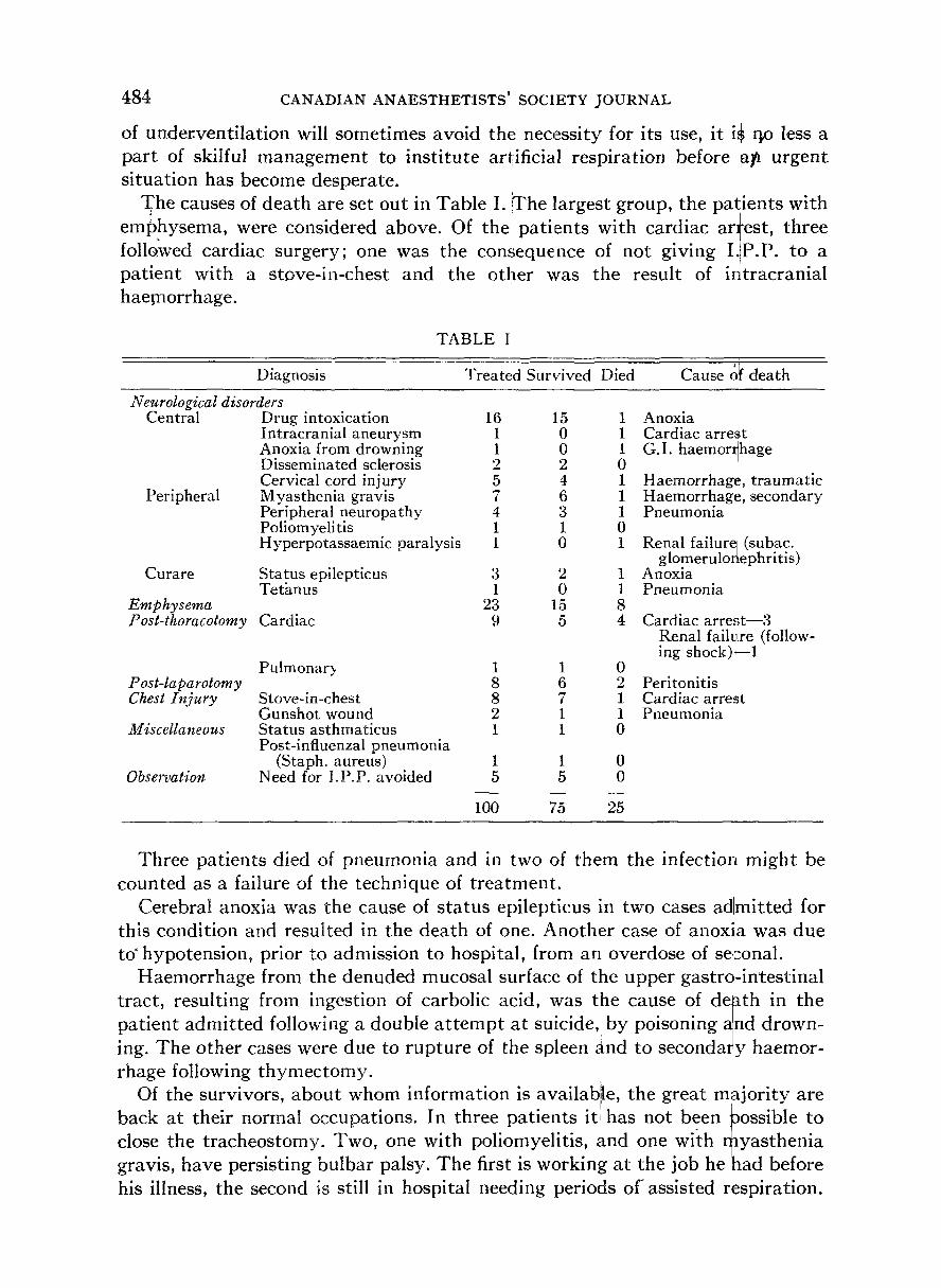
484 CANADIAN ANAESTHETISTS' SOCIETY JOURNAL
of underventilation will sometimes avoid the necessity for its use, it i* r~o less a part of skilful management to institute artificial respiration before ~1~ urgent situation has become desperate.
~he causes of death are set out in Table I. :The largest group, the palients with emlShysema, were considered above. Of the patients with cardiac arrest, three followed cardiac surgery; one was the consequence of not giving I.IP.P. to a patient with a stove-in-chest and the other was the result of intracranial hae~norrhage.
Diagnosis
TABLE I
Treated Survived Died Cause ~ death
Neurological disorders Central Drug intoxication 16
Intracranial aneurysm 1 Anoxia from drowning 1 Disseminated sclerosis 2 Cervical cord injury 5
Peripheral Myasthenia gravis 7 Peripheral neuropathy 4 Poliomyelitis 1 Hyperpotassaemic paralysis I
Curare Status epilepticus 3 Teti~nus 1
Emphysema 23 Post-thoracotomy Cardiac 9
Pulmonar? 1 P ost-laparotomy 8 Chest Injury Stove-in-chest 8
Gunshot wound 2 Miscellaneous Status asthmaticus 1
Post-influenzal pneumonia (Staph. aureus)
Need for I.P.P. avoided Observation
15 0 0 2 4 6 3 1 0
2 0
15 5
1 Anoxia 1 Cardiac arrest 1 G.I. haemor~hage 0 ! Haemorrhage, traumatic 1 Haemorrhage, secondary 1 Pneumonia 0 1 Renal failure (subac.
glomeruloriephritis) Anoxia Pneumonia
Cardiac arrest--3 Renal failure (follow- ing shock)---1
1 0 6 2 Peritonitis 7 1 Cardiac arrest 1 1 Pneumonia 1 0
1 1 0 5 5 0
100 75 25
Three patients died of pneumonia and in two of them the infection might be counted as a failure of the technique of treatment.
Cerebral anoxia was the cause of status epilepticus in two cases adlmitted for this condition and resulted in the death of one. Another case of anoxla was due to ~ hypotension, prior to admission to hospital, from an overdose of seconal.
Haemorrhage from the denuded mucosal surface of the upper gastrq,-intestinal tract, resulting from ingestion of carbolic acid, was the cause of degth in the patient admitted following a double a t tempt at suicide, by poisoning ~nd drown- ing. The other cases were due to rupture of the spleen and to secondary haemor- rhage following thymectomy.
Of the survivors, about whom information is available, the great mlajority are back at their normal occupations. In three patients it, has not been possible to close the tracheostomy. Two, one with poliomyelitis, and one wi:ch ~lyasthenia gravis, have persisting bulbar palsy. The first is working at the job he had before his illness, the second is still in hospital needing periods of assisted rlespiration.
FAIRLEY & CHAMBERS: RESPIRATORY INSUFFICIENCY 485
The third, with emphysema, leads a contented life of iretirement. Those who have not returned to their usual occupations are the cases of disseminated sclerosis al~d of cervical cord injuries. ~Fhese patients have followed the course usual in th6se conditions and have done perhaps rather better than most. Other than the t~VO patients with granulomata of the vocal cords referred to above, no sequelae attributable to I.P.P. have been observed.
SUMMARY
The management.~f patients with respiratory insufficiency, using intermittent positive pressure, has been discussed in detail. Emphasis has beeh laid on ~he underlying physiological problems and on the need. for detailed specialized knowledge.
The available equipment and methods of use are discussed, together with methods of monitoring assisted respiration. Problems of diaghosis of under- ventilation, general principles of management and the problems associated with the more common causes of respiratory insufficiency are discussed.
ACKNOWLEDGMENTS
The authors wish to acknowledge the contribution of their colleagues on ~the attending staff of the Toronto General Hospital Respiratory Unit, Drs. H.i/O. Barber and C. R. Woolf. They also wish to thank Dr, C R. Woolf for assistapce and advice in preparing the manuscript and Dr. H. J. Barrie for his opinion[ on the morbid anatomy 9f einphysematous lungs, following intermittent positive pressure respiration.
REFERENCES
1. II KINGS 4: 32-34. 2. DRINKER, C.K. Pulmonary Oedema and Inflammation. Cambridge, Mass.: Harvard Univ.
Press (1945). 3. DRINKER, P., • SHAW, L.A. An Apparatus for the Prolonged Administration of Artificial
Respiration. 1. A Design for Adults and Children. J. Clin. Invest. 7:229 (1929). 4. BRAGG, W.H. Bragg-Paul Pulsator. Brit. Med. J. ii: 254 (1938). 5. KELLEHER, W. H.; WILSON, A. B. K.; RUSSEL, W. R.; & STOTT, F . D . Notes on Cuirass
Respirators. Brit. Med. J. 2:413 (1952). 6. EVE, F.C. Actuation on the inert Diaphragm by a Gravity Method. Lancet 2:995 (1932). 7. LASSEN, H. C.A. A Preliminary Report on the 1952 Epidemic of Poliomyelitis in Cqpen-
hagen with Special Reference to the Treatment of Acute Respiratory Insufficiency. Lancet 1:37 (1953).
8. BARBER, H. O.; CHAMHERS, R. A.; FAIRLEY, H. B.; & WOOLF, C.R. A Respiratory Unit. Canad. M.A.J. 81:97 (1959).
9. COM~t~)E, J. H., JR.; FORSTER, R. E., II; DUBOIS, A. B.; BRISCOE; W. A.; & CARLS~r E. Th~@ung. Chicago: Year Book Publishers (1955).
10. NUNN, J .F . Physiological Aspects of Artificial Ventilation. Brit. J. Anaesth. 29:540 (1957). 11. BRISCOE, W. A., FORSTER, R. E., & COMROE, J. H., JR. Alveolar Ventilation at VerylLow
Tidal Volumes. J. Appl. Physiol. 7:27 (1954). 12. WOOLMER, R. The Management of Respiratory Insafficiency. Anaesthesia 11:281 (1956).
486 CANADIAN ANAESTHETISTS' SOCIETY JOURNAL
13. DOBKIN, A.B. Regulation of Controlled Respiration: Recent Concepts of Importance to the Anaesthetist. Brit. J. Anaesth. 3 0 : 2 8 2 (1958).
14. RADFORD, E. P., JR., FERRIS, B. G., • KRIETE, B. C. Clinical Use of a i Nomogram to Estimate Proper Ventilation During Artificial Respiration. New Eng. J. !Med. 251:877 (1954).
15. HERTZOG, P., & ENGSTROM, C.G. In MUSmN W. W. et al., Automatic Vehtilation of the \ Lungs. Oxford: Blackwell (1959).
16. CAMPBELL, E. J. M., NUNN, J. F., & PECKETT, B. W. A comparison of Artificial and Spontaneous Respiration with Particular Reference to Ventilation-Blood ~'low Relation- ships. Brit. J. Anaesth. 3 0 : 1 6 6 (1958).
17. SCHRAMEL, R. J.; CAMERON, R.; ZISKIND, M. M.; ADAM, M.; & CREECH, 0 " JR. Studies of Pulmonary Diffusion after Open Heart Surgery. J. Thorac. Surg. 3 8 : 2 8 1 (1959).
18. OTIS, A.B. The Work of Breathing. Physiol. Rev. 34:449 (1954). 19. MCILROY, M. B., MARSHALL, R., & CHRISTIE, R.V. The Work of Breathing in Normal
Subjects. Clin. Sci. 13:127 (1954). 20. COOK, C. D., HELLIESEN, P. J., & AGATHON, S. Relation between Mechanic~ of Respiration,
Lung Size and Body Size From Birth to Young Adulthood. J. Appl. Physiol. 1 3 : 3 4 9 (1958).
21. SWYER, P .R. The Problem of Prematurity. Canad. M.A.J. '82:347 (1960) I 22. NIMS, R. C., CONNOR, E. H., & COMROE, .[. H., JR. The Compliance of the Human Thorax
in Anaesthetized Patients. J. Clin. Invest. 3~/: 744 (1955). 23. BUTLER, J., ~: SMITH, B.H. Pressure-Volume Rclafionships of the Chest in ~the Completely
Relaxed Anaesthetised Patient. Clin. Sci. 16:125 (1952). 24. HOWELL, J. B. L~ (~: PECKETT, B . V . Studies of the Elastic Properties of the Thorax of
Supine Anaesthetized Paralyzed Human Subjects. J. Physiol. (Lond.) 1 3 6 : 1 (195.7). 25. OPIE, L. H., SPALDING, J. M. K., & STOTT, F. D. Mechanical Properties of the Chest
During Intermittent Positive Pressure Respiration. Lancet 2:545 (1959)�9 26. GLIEDMAN, M. L.; SIEBENS, A. A.; VESTAL, B. L. ; TIMMES, J. J.; GRANT, R- N. ; MURPHY,
J. L.; & KARLSON, K.E. Effect of Manual Versus Automatic Ventilatio n on the Elastic Recoil of the Lung. Ann. Surg. 148:899 (1958).
27. OPIE, L. a . , SMITH, A. C., • SPALDING, J. M . C . Concious Appreciation of the Effects Produced by Independent Changes of Ventilation Volume and of End-tidal pCO2 in Paralysed Patients. J. Physiol. (Lond.) 14[): 494 (1959).
28. CRANE, M. G.; AFFELDT, J. E.; AUSTIN, E.; & BOWER, A. G. Alveolar Carbon Dioxide Levels in Acute Poliomyelitis. J. Appl�9 Physiol. 9:11 (1956). !
29. COUR~AND, A. ; MOTLEY, H. L.; WERKO, L. ; • RICI-IARDS, D. W., JR. Physiological Studies on the Effects of Intermittent Positive Pressure Breathing on'Cardiac Output in Man. Am. J�9 Physiol. 152:162 (1948).
30. MALONEY, J. V. ; ELAM, J. O.; HANDFORD, S. W. ; BALLA, G. A. ; EASTWOOD, ID. W. ; BROWN, E. S.; & TEN PAS, R.H. Importance of a Negative Pressure Phase in Mechanical Respi- rators. J. Amer. M.A. 152:212 (1953).
31. BLAIR, D. A., GLOVER, W. E., & KIDD, B. S. L. The Effect of Continuous Positive and Negative Pressure Breathing upon the Resistance and Capacity Blood Vessels of the Human Forearm and Hand. Clin. Sci. 1 8 : 9 (1959i.
32. SIEKER, H. O., GAUER, O. H., & HENRY, J .P . The Effect of Continuous N~gative Pressure Breathing on Water and Electrolyte Excretion by the Human Kidney. J. Clin. Invest. 33 : 572 (1954).
�9 * e 33. RODDIE, I. C., & SHEPHERD, J. T. Receptors in the High ressure anal Low Pressure the Human Clrcul~tlon. Lancet 1: Vascular Systems: Their Role in the Reflex Control of i �9 .
493 (1958). 34. ERNSTING, J. The Effect of Raised Intrapulmonary Presgure upon the Distensibility of
�9 �9 , I the Capacity Vessels of the Upper Limb. j. Physiol. (Lond.) 137: 52P (1~957). 35. DALY, I. DE B. Intrinsic Mechanisms of the Lung. Quart. J�9 Exper. Physlol. 43 :2 (1958). 36. AVlADO, D. M., JR., LING, J. S., & SCHMIDT, C.F. Effects of Anoxia on Pul~nonary Circula-
tion: Reflex Pulmonary Vasoconstriction!. Am. J. Physiol. 189:253 (1957)~
FAIRLEY & CHAMBERS: RESPIRATOkY INSUFFICIENCY -r 37. FAHRI, L. E., & RAHN, H. Gas Stores of the Body and thelUnsteady State. J. Appl. Physi~)I.
7:472 (1955). 38. AUSTEN, F. K., CARMICrIAEL, M. W., & ADA~fS, R. D. Neurologic Manifestationsl }of
Chronic Pulmonary Insufficiency. New Eng. J. Med. 257,: 579' (1957). 39. SCURR, C. F. Carbon Dioxide Retention Simulating CUrarization. Brit. Med. J. 1:5~5
(1954). 40. FAIRLEY, H. B., WADDELL, W, G., & BIGELOW, W, G. Hypothermia for Cardiovascular
Surgery: Acidosis in the Rewarming Period. Brit. J,. Anaresth. 29:310 (1957). 41. MvsmN, W. W., RENDELL-BAKER, L., ~ THOMPSOrr P.W. Automatic Ventilation of the
Lungs. Oxford: Blackwell (1959). 42. COOPER, E. A., & PASK, E.A. A Bag for Measuring ReSpiratory Volumes. Lancet 1:3B9
(1960). 43. SALEM, H., LUCAS, G. H. W., & LucAs, D.M. Saran Plasltic Bags as Containers for Brea~h
Samples. Canad. M.A.J. 82:~682 (1960). 44. WRIGHT, B.M. A Respiratory Anemometer. J. Ph3~siol. iLond.) 127: 25P (1955). 45. DAVENeORT, H . W . The ABC of Acid-Base Chemistry, 4th edition. Chicago: Univ. of
Chicago Press (1958). 46. RILEY, R. L.,'Pt~o~EL, D. D., & FRANKE, R.L. A Direct Method of beterminatior~ iof
Oxygen and Carbon Dioxide Tension in Blood. J. Biol. Chem. 161:621 (1945)., �9 i 47. DORNETTE, W. H. L., & BRECnNER, V.L. Instrumentatton in Anesthesiollogy. Philadelpeh!a:
Lea & Ferbiger (1959). 48. PETERS, J. P., & VAN SLYEE, D. D. Quantitative Clinical Chemistry: II, Methods. Balti-
more: Williams & Wilkins (1932). 49. ASTRUP, R. A Simple Electrometric Technique for the Determination of Carbon Dioxide
Tension in Blood and Plasma, Total Content of Carbon Dioxide in Plasma and Bicarbonate Content'in "Separated Plasma at a Fixed Carbon Dioxide Tension. Scand. J. Clin. L~b. Invest. 8:33 (1956).
50. PLESCH, J. H~imodynamische Studien. Z. exp. Path. Ther. "6 :*380. (1909). 51. HACKNEY, J. D., SEARS, C. H., & COLLIER, C.R. Estiroation of Arterial CO2 Tension/by
Rebreathing Techniq]ue. J. Appl. Physiol. 12:425 (1958,). 52. CAMPBELL, E. J. M., & HOWELL, J. B.L. Simple Rap~d Methods of Estimating Arterial
and Mixed Venous pCO~. Brit. Med. J. 1:458 (19~50}. 53. CAMPBELL, E. J. M., & HOWELL, J. B.L. The Rebreathlng Method of Estimating Arterial
and Mixed Venous COe Tension. Brit. J. Dis Chest 54:~ 137 (1960). 54. SCHOLANOER, P. F. Analyzer for Accurate Estimation of Respiratory Gases in One I-{alf
Cubic Centimeter Samples. J. Biol. Chem. 167:235 (19~5). 55. FOWLER, R.C. Rapid Infrared Gas Analyzer. Rev. Scie~nt. Instruments $0:175 (1949). 56. DUBOm, A. B. ; FOWLER, R. C. ; SAFFER, A.; & F~N~, IE. O. Alveolar Carbon Dioxide
Measurements by Expiration into the Rapid Infrared Gas Analyzer. J. Appl. Physiol~4:. 526 (1951)�9
57. COLLIER, C. R., A~'FELDT, J. E., & FARR, A.F. Continuous Rapid Infrared Carbon Dioxide Analysis. J. Lal~l~ & Clin. Med. 45:526 (1955).
58. ELAM, J. O., BROWN, E. S., & TEN PAS, R.H. Carbon Dlioxide Homeostasis During Anaes- thesia: Instrumentation. Anesthesiology 16:876 '(~955).
59. ENGEL, H. C., & IBSEN, B. Continuous Carbon Dioxide Measurements in Respiratory ~ir During Anaesthesia in Thoracic Operations. Acta Clin. Scand. 10~: 313 (1952).
60. NVNN, J . F . The Dr~iger Carbon Dioxide Analyzer�9 Bri~. J. Anaesth. 30:264 (1958). 61. RAVIN, H. A., & STEIN, M. Bedside Measurement of Alveolar Carbon Dioxide Tension.
New Eng. J. Med. ~59:811 (1958). / 62. CAPEL, L." H., & ~-~ODGSON, D. C. tSimple Autohaaticl Sampler of End-expired Air for
Pressure Breathing Lancet 2:609 (1956). 63.-WEINER, R. S., & COOeER, P. The Use of "Arterialize~" Blood for the Determinatiorl of
Arterial O2 and CO2 Tensions. J. Thorac. Surg. 30: 683[ (1955). 64. BROOKS, D., & WYNN, V. Use of Venous Blood for pH~ and Carbon Dioxide Studies. Lar~cet
1:227 (1959).

488 CANADIAN ANAESTHETISTS' ',SOCIETY JOURNAL
65. DUBOlS, A. B., BRITT, A. G., & FENN, W.O. Alveolar CO2 during the R~spiratory Cycle. J. Appl. Physiol. 4:535 (1952).
66. b~UNN, J. F., & PINCOCK, A.C. A Time-pleased End Tidal Sampler Suitable for Use during Anaesthesia. Brit. J. Anaesth. 29:98 (1957).
67. SMITH, A. C., SCHUSTER, E., & SPALDING, J. M . K . An End-tidal Air Sampler. Lancet 1: 277 (1959).
68. COMROE, J. H., JR., & DRIPVS, R.D. The Physiological Basis for Oxygen Therapy. Spring- \ field: Thomas (1953). 69. PAULING, L., WooD, R. E., & STURDIVANT, J.H. An Instrument for Determining Partial
Pressure of Oxygen in a Gas. Science 103:338 (1946). 70~ WOOD, E. H. A Single Scale Absolute Reading Ear Oximeter. Proc. Sfaff Meet., Mayo
.~ Clinic 25:384 (1950). 71. ZJILSTRA, W.G. Fundamentals and Applications of Clinical Oximetry. Asse n, Netherlands:
Van Gorcum (1953). 72. CATTON, W.T. Physical Methods in Physiology. London: Pitman (19571. 73. VAN SLYKE, D. D., & NEILL, J. M~ The Determination of Gases in Blood and Other
Solutions by Vacuum Extraction and Manometric Measurement. J. BiTl. Chem. 61:523 (1924).
74. SMART, R. H., DAVENVORT, C. 'K., & PEARSON, G. W. Intermittent Positive Pressure Breathing in Emphysema and Chronic Lung Diseases. J.A.M.A. 150:1385 (1952).
I 75. SEGAL, M. S. ; SALAMONA, A. ; DULFANO, M. J. ; & HERSr J .A. Intermittent Positive
Pressure Breathing. New Eng. J. Med. 250:225 (1954). 76. M6RCH, E. T., SAXTON, G. A., & GIsH, G. Artificial Respiration via theJ Uncuffed Trach-
eostomy Tube. J.A.M.A. 160:864 (1956) 77. M6RCH, E. T), & SAXTON, G. A. Tracheostomy Tube Connectors. Anesthesiology. 17:
366 (1956). 78. AVERY, E. E., MORCH, E. T., & BENSON, D. ~Y. Critically Crushed Chests. J. Th0rac.
Surg. 3g: 291 (1956). 79. SVALDING, J. M. K., & SMITH, A. C. A New Tracheotomy Tube. Lancet 2:1247 (1956). 80. BULLOtlGtt, J. Tracheostomy Tube and Connections for Patients Having Intermittent
Positive Pressure Respiration. Lancet 2:372 (1957). 81. DREIFUSS, F. E., HURWITZ, L. J., & JOHN, C. Polyneuritis Requiring Artificial Respiration.
Lancet 2:59 (1957). 82. BARACH, A. L., BECK, G. J., & SMITH, W.H. Mechanical Production of Expiratory Flow
Rates Surpassing the Capacity of HumankCoughing. Am. J. Med. Sci. 226:,~241 (1953). 83. GREENE, N.' M. Fatal Cardiovascular and Respiratory Failure Associated with Trache-
otomy. New Eng. J. Med. 261:846 (1959). 84. DAVIS, a . S., KRETCHMER, H. E., & BRYCE-SMITH, R. Advantages and, Complications of
Tracheotomy. J.A.M.A. 153:1156 (1953). 85. NELSON, T. G. Tracheotomy: Clinical and Experimental Study. Pt. II. Am. Surg. 23:
750 (1957). 86. TALBOT, T. R., JR., QUIMBY, E. H,, & BARACH, A. L. Method of D~termining Site of
Retention of Aerosols Within Respiratory Tract of Man by Use of Rffdioactive Sodium. Preliminary Report. Am. J. M. Sc. 214:585 (1947).
87. MARSHALL, J., & SPALDING, J. M . K . Humidification in Positive PressUre Respiration for Bulbospinal Paralysis. Lancet 2:1'022 (1953).
88. FAIRLEY, H.B. The Selection of a Mechanical Ventilator. Canad. Anae!sth. Soc. J. 6:219 (1959).
89. RUBEN, H., & RUBEN, A.. Apparatus for Resuscitation a~d Suction. Lar~cet 2:373 (1957). 90. ELAM, J. O., KERR, J. H., & JANN:EY, C.D. Performance~f Ventilators: ]Effects of Changes
in Lung-Thorax Compliance. Anesthesiology 1.9:56 (1958). 91:. BJORK, V. O., & ENGSYROM, C. G. The Treatment of Ventilatory Insufficiency After
Pulmonary Resection with Tracheostomy and Prolonged Artificial Ventilation. J. Thorac. Surg. 30:356 (1955).
FAIRLEY & CHAMBERS: RESPIRATORY IN[SUFFICIENCY; 489
92.
93.
94.
95. 96.
97.
98.
99.
100.
101.
102.
103.
104.
105.
106. 107. 108.
109.
110.
111.
112.
113.
114.
115.
116.
117.
118.
NUN•, J. F. The Anaesthetist and the Embh:~sema~qus Patient. Brit. J. Anaesth. 80:134 (1958).
VAN BERGEN, F. H.; BUCKLEY, J. J.; WEATHERIAEADi, , D " S[P. ; SCHULTZ, E. A. ; & GORDOn, J .R . A New Respirator. Anesthesiology. 17: 708(1956)[.
DINNICK, O.T. Modified Beaver Respirator: The - tApplicatlon of Negative Phase. Brit.I ~. Anaesth. 81:554 (1959).
SYMONS, A. Physiotherapy and the Radcliffe Pump~ Phydiotherapy (July, 1958~. OPIE, L. H., & SPALDING, J. M. K. Chest Phzs~otherhpy During I.P.P. gespirati6~l.
Lancet 2:671 (1958). OPIE, L. H., & SmTH, A.C. Tracheobronchial Toil6t Through a Tracheostome. Lancet[ [1 :
600 (1959). LILLY, J. Mental Effects of Reduction of Ordinar3~ Levels of Physical Stimuli.on Intalct
Healthy Persons. Psychiat. Res. Reports 5: 1;(1956). MENDELSON, J. H., & FOLEY, .J.M. An Abnorm~/lit)~ of Mietital Function Affecting Patier~ts
with~[~oliomyelitis in a Tank-type Respirator. Tr. Am. l~eurdl. Assoc. 134--136 (1956). LASSEN, H. C. A.; HENRIKSEN, E.; NEUKIRCH, F. t & K/RISTENSEN , H. S. Treatmenf~of
Tetanus: Severe Bone Marrow Depression After Proloilged ]Nitrous Oxide Anaesthesia. Lancet 1:527 (1956).
SMITH, S. M. ; BROWN, H. O. ; TOMAN, J. E. P.; & .GOODM[AN, ~'~L. S. The Lack of CerebiTal Effects of d-Tubocurarine. Anesthesiology. 8:.1 (1947).
DENNY-BROWN, D.E. Effect of Poliomyelitis on th6 Fun~tiofi of the Motor Neurone. Ardh. Neurol. and ~ Psychiat. 66:1 (1950).
PLUM, F., & WHEVON, G.D. Rapid Rocking Bed: its Effect on Ventilation of Poliomyeliltis Patients with Respiratory Paralysis. New Eng. J. ~ed. ~45:235 (1951).
CAMERON, G. S. ; SCOTT, J. W. ; JoussE, A. T. ; & BO~'TERELL, E. H. Diaphragmatic Respira- tion in Quadraplegic Patient and Effect 6f Posi'tion on h~s Vi.tal ~apacity. Ann. Surg. 1A1: 451 (1955).
NILSSON, E. On Treatment of Barbiturate Poisoning: a 1V~odifted Clinical Aspect. Acta m6d. Scand., Suppl. 253 (1951).
WISE, R .P . Treatment of Barbiturate Poisoning. Brit. j. Anaesth. 30:533 (1958). RUSSELL, W.R. Poliomyelitis. 1st edition. London: Arnbld.(12952). DENNY-BROWN, D. E. Clinical Problems in Neqromu~cular" Physiology. -Am. J. Mbd.
15:368 (1953). WALSHE, F. M. R., & Ross, J. The Clinical Picture of 1]/linor.Cord Lesions in Association
with Injuries of the Cervical Spine': with Special R~feren(~e to the Diagnostic and Localizing Value of the Tendon Reflexes of the Arm (Inversion of~he Radial Reflex). Brain. 59:277 (1936).
SYMONDS, C. The Interrelation of Trauma and Cerlvical Spolldytosis in Compression of the Cervical Cord. Lancet 1:451 (1953).
VAN BERGEN, F. H., & BUClLLEY, J. J. The Managen~ent of Severe Systemic Tetaims: Anesthesiology 13:599 (1952).
SMITH, A. C., HILL, E. E., & HoPSON, J.H. Treatment of Severe Tetanus with d-Tu~o- curarlne Chloride and Intermittent Positive Pressure I~espiration. Lancet 2 :550 (19~6).
WOOLMER, R., & CATES, J. E. Succinylcholine in the 7Freat,ment of Tetanus. Lancet ~: 808 (1952).
SHACKLETON, P.~ The Treatment of Tetanus: The Ro~e of the Anaesthetist. Lancet 2: 155 (1954).
CREECrI, O., GLOVER, A., & OCHSNER, A. Tetanqs. E,~Jaluation of Treatment at Cha~'ity Hospital, New Orleans, Louisiana. Amer. Surg. 146: 36~ (1957).
ALHADY, S. M. A. ; BOWLER, D. P. ; REn), H. A. ; & SCO~T, L .T . Total Paralysis Reglime in Severe Tetanus. Brit. Med. J. 1:540 (1960).
ABLETT, J. J .L. Tetanus and the Anaesthetist:/A' Review of the Symptomatology andfthe Recent Advances in Treatment. Brit. J~ Anaesth.[28:2~i8 (1956).
IS.ELl'C, R E , & I,AWRI,:NCE, l) R. Effect of Ch/lorpromazine bn Convulsmns of Experi- mental and Clinical Tetanus. l.ancet 1:118 (195fi).
490 CANADIAN ANAESTHETISTS ~ SOCIETY JOURNAL
119. COLG A. C. E., & ROBERTSON, D. H.H. Chlorpromazine in the Management of Tetanus. Lancet 2:1063 (1955).
120. PERLSTEtN, J.A. Control of tetanu~ spaslns by injection of meprobamMe. J.A.M.A. 170: 1093 (1959).
121. KELLAWAY, G. Acute Anoxic Cardiac Failurc in Pulmonary Heart Disea~se. Lancet 2:768 (1959).
122. JAMESON, A. C., FERRIER, M. I., & HARVEY, R.M. Some Effects of Mechanical Respirators upon Respiratory Gas Exchange and Vcntilation in Chronic Pulmonary Emphysema. Am. Rev. Resp. Dis. 80:510 (1959)].
123. MACIVER, I. N., FREW, I. J., & Ma'rHSSON, J.G. The Role of Respiratory Insufficiency in the Mortality of Severe Head Injuries. Lancet i: 390 (1958).
124. BARRETT, N . R . Early Treatment of Stove-in-chest. Lancet 1:293 (1960). 125. SANDIFORD, l--~. B. C. Emergency Treatment of Respiratory Failure in Poliomyelitis.
Lancet 2:823 (1957).
NoTIcE
T H E
Reprints of the Review Article on
M A N A G E M E N T O F T H E P A T I E N T W I T H
RESPIlqATORY I N S U F F I C I E N C Y
by H. Barrie Fairley, M.B., B.S., F.~.A.n.C.S., and
R. A. Chambers, M,D., ~r.KC.P.
are available on application to
The Canadian Anaesthetists ' Society Journal,
178 St. George Street, Toronto 5, Canada.
Price: ,50 cents per copy
_ _ L . . . . . . ' ' i