Development of fresh and cryopreserved early-stage ovarian follicles, with special attention to...
24
Anim. Reprod., v.6, n.1, p.72-95, Jan./Mar. 2009 _________________________________________ 3 Corresponding author. [email protected] Development of fresh and cryopreserved early-stage ovarian follicles, with special attention to ruminants R. van den Hurk 1,3 , R. Santos 2 1 Department of Pathobiology, Utrecht University, PO Box 80151, 3508 TD Utrecht, The Netherlands. 2 Department of Farm Animal Health, Utrecht University, PO Box 80157, 3508 TD Utrecht, The Netherlands. Abstract This paper gives a survey of our research on the development, cryopreservation, in vitro culture and transplantation of early-stage follicles in the past two decades, relative to those obtained by other researchers. Hereby, attention is specially focussed on findings with ruminants. Apart from our histological, immunocytochemical, viability, biochemical, endocrine, cryostore, in vitro and in vivo data on cow, sheep and goat follicles, some general information on the various in vitro culture, cryopreservation and transplantation techniques is given. We conclude that (i) we are still far away from obtaining fertilizable oocytes from in vitro cultured ruminant early-stage follicles, that can be used for in- vitro production of healthy embryos or offspring, and (ii) our cryopreservation techniques seem good enough to store ruminant early-stage follicles for an undetermined time without viability impairment, followed by further development after thawing in short- or long-term in vitro cultures or in transplantation experiments. Keywords: cryopreservation, follicle, in vitro culture, ovary, transplantation. Introduction Fully mature oocytes are the survivors of the long selection process of folliculogenesis. They are competent to undergo fertilization and subsequent embryonic development, eventually leading to offspring. However, they are also the rarest cells in the body, since the great majority of the oocytes encounters atresia. To increase ovulation rates for embryo production, techniques such as superovulation are used in farm animals. The efficiency of these techniques in these animals is, however, often influenced by the large variability between oocyte donors. In assisted human reproduction, follicular development is often stimulated by administration of gonadotropins, followed by an artificial LH-peak to induce oocyte maturation. The in vivo matured oocytes are then retrieved by ovum-pickup for in vitro fertilization (IVF), but the gonadotropic stimulation used in the treatments may lead to ovarian hyper stimulation syndrome. Another used technique is in vitro maturation (IVM) of immature oocytes collected from small antral follicles. After IVM, oocytes are fertilized in vitro and the presumptive zygotes are cultured to a certain developmental stage and, depending on the species, to produce embryos. The very early follicles, however, form a huge oocyte reservoir, as compared to the small antral follicles. It is therefore attractive to exploit and freeze the reservoir of early follicles to obtain more fertilizable oocytes for in vitro and in vivo embryo production. In addition to commercial purposes with farm animals or to treatments of human reproductive problems, the use of early follicles may be of great importance for saving endangered animal species. However, most of the knowledge on the developmental conditions of follicles and their oocytes is hitherto restricted to the late-stage follicles, while the events occurring during early folliculogenesis still need to be more extensively explored. In the past two decades, our studies were focussed on in vivo and in vitro folliculogenesis in mammalian ovaries, i.e., in those of rats (e.g., Dijkstra et al., 1996; Zhao, 2000), Djungarian hamsters (Van den Hurk et al., 2002), cows (e.g., Van den Hurk et al., 1992, 1998; Figueiredo, 1995; Hulshof, 1995; Schotanus et al., 1997), pigs (Van den Hurk and Van de Pavert, 2001; Da Silva-Buttkus et al., 2003), humans (Abir et al., 2004, 2005; Biron-Shental et al., 2004), goats (e.g., Silva, 2005; Santos, 2007) and sheep (Santos et al., 2006a, 2007b). During this period, radio-immunoassays were carried out to detect various hormone concentrations, while histological, histometrical, (enzyme)histochemical, immunohistochemical, fluorescent microscopical, electron microscopical, and molecular biological studies with isolated or ovarian tissue enclosed follicles were performed to get information on the local control of folliculogenesis and/or the viability of early-stage follicles after their freezing/thawing, in vitro culture (IVC) or transplantation. This paper summarizes our findings on mammalian ovarian follicles, i.e., those of ruminants especially, and integrates them with those obtained by other scientists. Additionally, technical background information is given on cryopreservation, IVC and transplantation of early-stage follicles. Structure, nomenclature and development of ovarian follicles The paired ovaries generally are ovoid-shaped structures, which produce sex hormones and the female’s gametes (ova). The ovaries are lined by surface
Transcript of Development of fresh and cryopreserved early-stage ovarian follicles, with special attention to...

Anim. Reprod., v.6, n.1, p.72-95, Jan./Mar. 2009
_________________________________________ 3Corresponding author. [email protected]
Development of fresh and cryopreserved early-stage ovarian follicles, with special attention to ruminants
R. van den Hurk1,3, R. Santos2
1Department of Pathobiology, Utrecht University, PO Box 80151, 3508 TD Utrecht, The Netherlands.
2Department of Farm Animal Health, Utrecht University, PO Box 80157, 3508 TD Utrecht, The Netherlands.
Abstract
This paper gives a survey of our research on the development, cryopreservation, in vitro culture and transplantation of early-stage follicles in the past two decades, relative to those obtained by other researchers. Hereby, attention is specially focussed on findings with ruminants. Apart from our histological, immunocytochemical, viability, biochemical, endocrine, cryostore, in vitro and in vivo data on cow, sheep and goat follicles, some general information on the various in vitro culture, cryopreservation and transplantation techniques is given. We conclude that (i) we are still far away from obtaining fertilizable oocytes from in vitro cultured ruminant early-stage follicles, that can be used for in-vitro production of healthy embryos or offspring, and (ii) our cryopreservation techniques seem good enough to store ruminant early-stage follicles for an undetermined time without viability impairment, followed by further development after thawing in short- or long-term in vitro cultures or in transplantation experiments. Keywords: cryopreservation, follicle, in vitro culture, ovary, transplantation.
Introduction
Fully mature oocytes are the survivors of the long selection process of folliculogenesis. They are competent to undergo fertilization and subsequent embryonic development, eventually leading to offspring. However, they are also the rarest cells in the body, since the great majority of the oocytes encounters atresia. To increase ovulation rates for embryo production, techniques such as superovulation are used in farm animals. The efficiency of these techniques in these animals is, however, often influenced by the large variability between oocyte donors.
In assisted human reproduction, follicular development is often stimulated by administration of gonadotropins, followed by an artificial LH-peak to induce oocyte maturation. The in vivo matured oocytes are then retrieved by ovum-pickup for in vitro fertilization (IVF), but the gonadotropic stimulation used in the treatments may lead to ovarian hyper stimulation syndrome. Another used technique is in vitro maturation (IVM) of immature oocytes collected from small antral follicles. After IVM, oocytes are
fertilized in vitro and the presumptive zygotes are cultured to a certain developmental stage and, depending on the species, to produce embryos. The very early follicles, however, form a huge oocyte reservoir, as compared to the small antral follicles. It is therefore attractive to exploit and freeze the reservoir of early follicles to obtain more fertilizable oocytes for in vitro and in vivo embryo production. In addition to commercial purposes with farm animals or to treatments of human reproductive problems, the use of early follicles may be of great importance for saving endangered animal species. However, most of the knowledge on the developmental conditions of follicles and their oocytes is hitherto restricted to the late-stage follicles, while the events occurring during early folliculogenesis still need to be more extensively explored.
In the past two decades, our studies were focussed on in vivo and in vitro folliculogenesis in mammalian ovaries, i.e., in those of rats (e.g., Dijkstra et al., 1996; Zhao, 2000), Djungarian hamsters (Van den Hurk et al., 2002), cows (e.g., Van den Hurk et al., 1992, 1998; Figueiredo, 1995; Hulshof, 1995; Schotanus et al., 1997), pigs (Van den Hurk and Van de Pavert, 2001; Da Silva-Buttkus et al., 2003), humans (Abir et al., 2004, 2005; Biron-Shental et al., 2004), goats (e.g., Silva, 2005; Santos, 2007) and sheep (Santos et al., 2006a, 2007b). During this period, radio-immunoassays were carried out to detect various hormone concentrations, while histological, histometrical, (enzyme)histochemical, immunohistochemical, fluorescent microscopical, electron microscopical, and molecular biological studies with isolated or ovarian tissue enclosed follicles were performed to get information on the local control of folliculogenesis and/or the viability of early-stage follicles after their freezing/thawing, in vitro culture (IVC) or transplantation.
This paper summarizes our findings on mammalian ovarian follicles, i.e., those of ruminants especially, and integrates them with those obtained by other scientists. Additionally, technical background information is given on cryopreservation, IVC and transplantation of early-stage follicles.
Structure, nomenclature and development of ovarian follicles
The paired ovaries generally are ovoid-shaped
structures, which produce sex hormones and the female’s gametes (ova). The ovaries are lined by surface

van den Hurk and Santos. Development of fresh and frozen ovarian follicles.
Anim. Reprod., v.6, n.1, p.72-95, Jan./Mar. 2009 73
(= germinal) epithelium, and further consist of a cortex and a medulla. The cortex contains the preliminary stages of the gametes (oocytes), which are surrounded by somatic cells to form a follicle. These follicles are embedded in a stroma, which is composed of well-vascularized connective tissue. In livestock animals and humans, a tough connective tissue forms the main component of the ovarian stroma. In rodents, however, this stroma is spongy and rich in steroid-secreting cells (Van den Hurk et al., 2002). In many species, the ovarian medulla consists of connective tissue and blood vessels, which ramify into the cortical zone.
The follicles in the ovarian cortex are at various stages of development or degeneration. They are formed during a process, termed folliculogenesis, in which a small primordial follicle, consisting of a germ cell and a layer of few flattened granulosa cells, grows and differentiates through various developmental stages (see next two paragraphs) into a large structure, termed the preovulatory follicle. This final follicle stage contains a fully-grown and maturation-ready oocyte and, dependent on the species, thousands to millions of highly differentiated granulosa cells, which enclose a fluid-filled antrum and are surrounded by a well-differentiated inner and outer theca-layer. The inner theca-layer (theca interna) has abundant well-vascularized steroidogenic cells, distributed in a loose connective tissue. The outer theca layer (theca externa) mainly consists of tough connective tissue.
In previous reviews, we described and illustrated the structure of the mammalian ovaries and their follicles, as well as the dynamics of folliculogenesis (Van den Hurk et al., 1997, 1999, 2000; Van den Hurk and Zhao, 2005). In short, the process of folliculogenesis can be divided into three developmental phases: (1) the initiation of primordial follicle growth and formation of pre-antral (i.e., primary and secondary) follicles (2) the development of secondary to early antral follicles (basal follicular growth) and (3) the development of early antral to preovulatory follicles (terminal follicular growth). There are some 75,000 germ cells in rat fetal ovaries around day 18 of gestation, while the number has been reduced to 27,000 on day 2 after birth. At that latter time, all germ cells are part of an early-stage follicle, most follicles being at the primordial stage. In 6 months and 3 years old goats, 24,000 and 12,000 follicle-enclosed germs cells have been counted, respectively, whereas neonatal pigs, cows and sheep have, respectively, a maximal number of about 500,000, 700,000 or 1 million primordial follicles at their disposal, which generally measure about 30 µm in diameter.
As soon as the stock of primordial follicles is established, some follicles begin to grow, while the others remain quiescent. In cows, this process is marked by innervation of the primordial follicles through vasoactive intestinal peptide (VIP) and neuropeptide-Y containing nerve fibres, outgrowth of vimentin containing blood vessels towards the activated follicles, and the appearance in the single-layered granulosa of cells with a round nucleus and cytoplasmic vimentin-containing intermediate filaments, which are associated
with changes in granulosa cell shape (flat to cubical) and mitotic activity of granulosa cells (Van den Hurk et al., 1995, 1997) as well as secretory activity of cells (Quintanar, 2000; Johnson et al., 2001). The initiation of follicular growth not only marks the start of somatic cell proliferation and differentiation, but also the growth and differentiation of the oocyte. The growing follicles may then be termed primary (i.e., an immature oocyte surrounded with a single layer of cubical granulosa cells) or secondary (i.e., an immature oocyte surrounded by at least two layers of granulosa cells and, peripherally, a from local stroma cells originating developing theca-layer) follicles. Because of their multilayered granulosa, secondary follicles are also called multilaminar follicles. In contrast, primordial and primary follicles have only one layer of granulosa cells and thus are unilaminar follicles. Multilaminar follicles from large mammalian species start to form a protective membrane, the zona pellucida, between the oocyte and granulosa. In rodents, the formation of this zone starts already in the primary follicle stage. During early antral folliculogenesis, granulosa cells undergo further proliferation and differentiation, whereby numerous cells acquire vimentin-containing intermediate filaments (Van den Hurk et al., 1995, 1997). The oocyte increases in size and protein content (Schultz and Wassarman, 1977) and transcripts essential for early embryo development are accumulated in dormant form, which eventually are activated and translated during meiotic maturation and cleavage divisions of a possible future embryo (Eppig, 1994). In most mammalian species, oocytes obtain their maximal size during the early-antral stage, which is 120-150 μm in ruminants, 130 μm in pigs and 125 μm in humans (Van den Hurk et al., 1997; Van den Hurk and Zhao, 2005). In rodents, maximum oocyte diameter (about 70 µm) is already reached at the end of the secondary follicle stage.
Although pre-antral follicle formation is accompanied by outgrowth of blood vessels into the direction of these follicles, follicular vascularization still is relatively poorly developed at this stage. This could indicate a paracrine rather than an endocrine control of early follicular development, Based on IVC-studies, in particular, cytokines and growth factors, leukaemia-like inhibiting factor, growth differentiation factor-9 (GDF-9), bone morphogenetic proteins (BMPs), anti-Müllerian hormone (AMH, also referred to as Müllerian inhibiting factor or Müllerian inhibiting hormone), activin-A, transforming growth factor (TGF)-α, TGF-β, epidermal growth factor (EGF), insulin-like growth factor-1 (IGF-1), connective tissue growth factor, fibroblast growth factors (FGFs), and Kit-ligand (also referred to as Steel factor or Stem cell factor), as well as steroids are believed to be important intrafollicular compounds that locally regulate early-stage follicle development (reviewed by Gougeon, 1996; Roy and Greenwald, 1996; Van den Hurk et al., 1997, 2000; Fortune, 2003; Van den Hurk and Zhao, 2005; McNatty et al., 2007; Webb et al., 2007). This, however, does not exclude an important role of compounds coming from peripheral nerves (like the neuropeptides VIP and neuropeptide-Y

van den Hurk and Santos. Development of fresh and frozen ovarian follicles.
Anim. Reprod., v.6, n.1, p.72-95, Jan./Mar. 2009 74
and the circulatory system (like all kinds of growth factors, cytokines, steroids, prostaglandins, metabolic products, feeding components and oxygen) in the regulation of pre-antral follicle development (reviewed and illustrated by Van den Hurk et al., 1997). Lack of oxygen in follicles may arise as a result of intensive division and differentiation of cells and is a trigger for local formation of angiogenic growth factors, that stimulate the outgrowth of blood vessels toward follicles, or is a main cause of follicular atresia, when follicular angiogenesis is inadequate (Van den Hurk and Zhao, 2005; Bedaiwy et al., 2006).
During terminal follicular growth, antral follicles are recruited from earlier-stage follicles and then follow a selection process in which they become subordinate, dominant or preovulatory, or degenerate through apoptosis (reviewed by Van den Hurk et al., 1999, 2000; Webb et al., 1999, 2007; Driancourt, 2001; Fortune et al., 2001; Findlay et al., 2002; Van den Hurk and Zhao, 2005). Intact antral follicles are characterized by the presence of an internal and an external theca-layer, a several cell layers thick (mural) granulosa, and an oocyte surrounded by (cumulus) granulosa cells which connect the oocyte to the mural part of the granulosa. Within the granulosa, a distinct enlargement of an antrum is manifest, in which the oocyte and its surrounding cells form a peninsula. Subordinate and dominant antral follicles have an immature oocyte, the oocyte being enveloped by a compact cumulus investment, while numerous vimentin-positive cumulus projections penetrate the zona pellucida and end on the oolemma with gap junctions (Van den Hurk et al., 1995, 1997). In contrast, oocytes from a preovulatory follicle will encounter the cyclic LH surge, and then mature and ovulate. The maturation process includes disruption of the cumulus-oolemma junctions, redistribution of cell organelles, migration of mitochondria to a perinuclear position and accumulation of cortical granules along the oolemma (ooplasmic maturation), and two consecutive nuclear divisions in the absence of deoxyribonucleic acid (DNA) replication (nuclear maturation; Van den Hurk and Zhao, 2005). Preovulatory follicles may reach a size of 1 mm in rats (Tempel-Brami and Neeman, 2002), 6-7 mm in sows, 8-10 mm in ewes, 15 to >20 mm in cows (Van den Hurk and Zhao, 2005), and about 20 mm in humans (Martinuk et al., 1992). Complete folliculogenesis, i.e. the development of a primordial into a preovulatory follicle, takes about 18-24 days in mice (Eppig, 1977), 12 weeks in humans (Gougeon, 1986; Lunenfeld and Insler, 1993), 15 weeks in cows (Ireland, 1987; Britt, 1991) and around 17 weeks in sheep (Bordes et al., 2005).
Terminal follicular growth takes place when follicles become strictly gonadotropin-dependent and eventually undergo ovulation or atresia (Driancourt, 2001; Van den Hurk and Zhao, 2005). Although the mechanisms of follicle selection are not fully elucidated, it is generally accepted that no two follicles are identical and that individual differences in their vascular development, responsiveness to follicle stimulating
hormone (FSH) and luteinizing hormone (LH), aromatase activity and cellular cyclic adenosine monophosphate production may eventually lead to a different follicular fate. The entry into the phase of terminal follicular growth is related with the size of an antral follicle, which is, for example, approximately 0.2 mm in rats, 1 mm in sows, 2 mm in ewes and humans, and 3 mm in goats, and 4 mm in cows. It is now established that FSH plays a determinant role in promoting the proliferation and survival of granulosa cells (Monniaux et al., 1997). It induces terminal follicular growth by promoting in granulosa cells the expression of FSH- and LH-receptors, steroidogenic enzymes and different peptides like inhibin, IGF-1, interleukin-6 or plasminogen activator (reviewed by Richards, 1994; Van den Hurk and Zhao, 2005). Expression of LH receptors in granulosa cells only occurs in preovulatory follicles, which are in rat, goat, sow, ewe and cow follicles larger than 0.5 (Monniaux et al., 1997), 5 mm (Rubianes, 2000), 6, 8, and 15 mm (Van den Hurk and Zhao, 2005), respectively. LH sustains terminal maturation of granulosa cells in preovulatory follicles, which results in enhanced estradiol synthesis in granulosa cells. In addition, various intrafollicular regulatory compounds, including steroids (like estradiol and progesterone) and peptides (like IGFs, activin-A and TGF-β) act locally to amplify the gonadotropic stimuli (Adashi and Rohan, 1992; Monget and Monniaux, 1995; Van den Hurk and Zhao, 2005).
Despite the enormous follicular population present in the ovary, only less or far less than 1% of the available follicles at birth contains an oocyte that may reach ovulation during the reproductive lifespan, while the rest of the follicles become atretic during follicular growth and oocyte maturation (Saumande, 1991). In this regard, we studied the incidence of follicular atresia in vivo by measuring the percentages of degenerating follicles (i) of heifers, which were treated with, respectively, pregnant mares’ serum gonadotropin (Van den Hurk et al., 1992) and prostaglandin F2α (Van den Hurk et al., 1994) that, when given in combination, evoke a superovulation response in farm animals, (ii) of rats, which were made hypothyroidic (Dijkstra et al., 1996), (iii) of Djungarian hamsters, which were kept under short photoperiod (Van den Hurk et al., 2002), and (iv) in vitro, after culturing early-stage follicles from cows (Figueiredo et al. 1993, 1994a, b, 1995; Hulshof et al., 1994b, 1995a, b, 1997; Schotanus et al., 1995, 1997), sheep (Santos et al., 2007a), goats (Silva et al., 2004b, 2006a; Santos et al., 2006b, 2007b, 2008b; Matos et al., 2007), rats (Zhao et al., 2000a, b, 2001a, b) and humans (Biron-Shental et al., 2004). Most remarkable findings were (i) the appearance of elongated, intermediate filaments-containing granulosa cells with elongated nuclei in most in vivo-growing, antral follicles (≥0.3 mm; thus during both their basal and terminal growth) in cows (Van den Hurk and Dijkstra, 1992; Van den Hurk et al., 1992, 1994, 1995),

van den Hurk and Santos. Development of fresh and frozen ovarian follicles.
Anim. Reprod., v.6, n.1, p.72-95, Jan./Mar. 2009 75
sows and goats, but not in rodents (Van den Hurk, unpublished results), the phenomenon being considered as an initial sign of granulosa degeneration, (ii) the appearance, in Djungarian hamsters kept from their birth under short daylight, of luteinized atretic follicles, which arise after the degeneration of the oocyte of follicles during their basal growth (Van den Hurk et al., 2002), (iii) the inadequacy of only histological criteria and necessity of ultrastructural or fluorescent markers of cells and their nuclei to judge the viability of a follicle, which is put in culture or has been in vitro cultured (Schotanus et al., 1995, 1997; Van den Hurk et al., 1998), (iv) the compared to control follicles negligible degeneration percentage of developing follicles and subsequent in vitro formation of antral follicles with fertilizable oocytes from rat pre-antral follicles, when follicles were cultured in a serum-free medium supplemented with activin-A, in groups of 4-6 specimens, and at a distance of 3-6 follicle diameters from each other (Zhao, 2000), and (v) the formation of an ovary and full recovery of follicle development after freezing-thawing and subsequent auto-transplantation of caprine ovarian cortical tissue fragments (Santos, 2007; Santos et al., 2008a).
In vitro development of early-stage follicles Background
In clinics, in agriculture and in research labs,
methods for in vitro culture of oocytes from antral follicles are used to obtain fertilizable oocytes which further develop into embryos with variable success. This variation in success of embryo production may for example be due to the poor quality of oocytes that are being selected for maturation, since they are taken from antral follicles of which many are atretic. To eliminate this variation and to increase the numbers of embryos produced, it could be better to use the large source of oocytes from early-stage follicles by developing systems for their in vitro growth, since this population of early-stage follicles is considered to have a relatively low incidence of atresia and thus to be more homogeneous than antral follicles. Apart from the potential to deliver a higher yield of embryos, IVC of early follicles is an essential tool in understanding underlying mechanisms of oocyte and follicle growth and differentiation.
The first problem one has to encounter when dealing with in vitro studies is whether one isolates individual follicles and culture them alone or in groups or whether one cultures a piece of ovarian tissue, like a cortical slice with many early follicles. The choice is dependent on the aim of a researcher and the animal species that serves as a follicle donor. If one’s aim is to obtain as many as fertilizable oocytes from primordial follicles, one better can choose for first culturing slices of ovarian cortex for a certain period, then isolate the developed multilayered follicles, grow them individually in
an culture medium and finally mature the grown follicles in a specific maturation culture medium. If one’s aim is to study the culture conditions for the various stages of follicular development including the activation of primordial follicles, it is better to try to isolate these early follicles and then culture them individually or in groups to let them further grow for a certain period. The choice of an isolation method may also depend on the species under study, since larger species (like ruminants and humans) generally have less densely-packed early follicles distributed in a more compact fibrous stroma tissue than rodents. Therefore, techniques that work in rodents cannot be readily applied for these larger species, and is culture of pre-antral follicles far more difficult in farm animals (Fortune, 1994; Roy and Greenwald, 1996; Telfer, 1996, 1998; Van den Hurk et al., 1997) and humans (Salha et al., 1998; Smitz and Cortvrindt, 1999).
Isolation of early-stage follicles
Early follicles can be isolated either using enzyme digestion or by manual dissection procedures or by a combination of these methods. Enzymes like collagenase, DNA-se and trypsin are often used to digest ovarian tissue. Enzymatic methods generally allow large numbers of early follicles to be collected. It is very effective for species as rodents that have soft ovaries. Enzymatic techniques may result in alteration of surface protein molecules and the degradation of the basement membrane, which may cause detachment of granulosa cells and separation of the oocyte. This disruption in oocyte-somatic cell contact comprimises development of the follicle, because communication between oocyte and its companion granulosa cells through gap junctions is essential for further oocyte growth and development. So, it is important during IVC to maintain these junctions. Because of the tough fibrous ovarian stroma and the less compact follicular distribution, it is more difficult to isolate early-stage follicles from domestic species or humans than from rodents. Follicles, especially the larger secondary ones, can also be isolated under the stereomicroscope by manual dissection. This is a relative straightforward procedure, but is slow and labour intensive, and results in lower yields of follicles because of time constraints; one has to isolate follicles from an ovary within 2-3 hours, otherwise they will become atretic.
Mechanical micro-dissection of early-stage follicles has been performed in mouse, rat, cat, sheep, bovine and human, and enzymatic isolation in mouse, hamster, pig, bovine, goat and human (reviewed by Van den Hurk et al., 2000; Smitz and Cortvrindt, 2002; Fortune, 2003). In our studies with the bovine (Figueiredo et al. 1993, 1994a, b, 1995; Hulshof et al., 1994b, 1995a, b, 1997; Schotanus et al., 1995, 1997), we used a micro-dissection method to obtain isolated early follicles. We chosed for this method, because of

van den Hurk and Santos. Development of fresh and frozen ovarian follicles.
Anim. Reprod., v.6, n.1, p.72-95, Jan./Mar. 2009 76
the far higher follicle survival rates obtained after culture when compared with those of follicles that were isolated by collagenase treatment. Within 45 min, about 300 to more than 2000 early-stage (predominantly primary and early secondary) follicles were obtained by micro-dissection, the number being dependent of the age of the bovine: high numbers for fetal tissue, lower numbers for calves and adult cows. Actually, complexes consisting of an oocyte and a granulosa with an intact basement membrane were obtained. By applying only simple micro-dissection under the microscope, Ralph et al. (1996) could isolate bovine large secondary follicles (150-250 μm), having some theca tissue around the granulosa. Their yield is around 50-60 follicles after 2-3 h isolation. Zhao et al. (2000a) used the same simple method to isolate rat secondary follicles and was able to collect, per rat, 30-50 specimens of approximately 150 μm, which also yield thecal tissue. In correspondence with our findings with the bovine, Demeestere et al. (2002) showed that the survival rate of cultured mouse large secondary follicles, that had been isolated with use of enzyme treatment, also were lower than that of mechanically isolated follicles. They observed, however, a far lower percentage of antral-like cavity formation in mechanically isolated follicles, while no differences on oocyte maturation and embryo development rates were found using both isolation methods. Their findings show that, compared to mechanical isolation, enzymatic isolation not necessarily more attenuates the development of follicles, and should not discourage us, researchers, working with ovaries from domestic species or primates to use enzymatic isolation methods. Culture methods for early-stage follicles
In 1997, Hartshorne published a nicely
illustrated overview of approaches used to culture follicles. Briefly, it is possible to culture whole (relative small) ovaries or ovarian slices usually for a relatively short period of about 1 week, because of occurring necrosis. Nowadays this is often done as a first step in obtaining fertilizable oocytes of rodents or in obtaining multiple primary or secondary follicles from larger species for studies on their growth. Stepwise in vitro development of early follicles is done either in plastic wells, coated wells, on porous membranes or in or on gels, like agar, Matrigel (composed of basement membrane components) or collagen gels. The latter techniques have consequences for the developing follicle’s structure. This structure remains 3-dimensional, when cultured within gels, but transforms in a flattened pancake-like (2-dimensional) structure, when cultured on a membrane as Eppig and O’Brien (1996) did, or in poly-L-lysine coated or uncoated dishes or wells, the so-called substrate-adherents model, as were used by Cain et al (1995) and Cortvrindt et al. (1996), respectively, for rodent follicles. In this latter
method theca cells attach to the bottom of the dish and start to proliferate. Proliferating granulosa cells grow on top of the theca cell monolayer, where after granulosa cells differentiate in two sub-populations: steroid producing mural granulosa cells separated by an antrum-like cavity from the cumulus (granulosa) cells, which tightly enclose the oocyte. As a result, the cumulus-oocyte-complex arises above the culture surface, whereby the oocyte grows but remains blocked in the germinal vesicle stage.
In 2000, Fortune and co-workers described an interesting culture technique, whereby cortical tissue pieces of bovine and baboon ovaries were grafted between the chorio-allantoic membrane and the yolk sac membrane of chicken eggs. This in ovo-grafting technique prohibited the massive spontaneous activation of primordial follicles, which is observed when follicles are cultured in vitro. Such a model could provide clues to the mechanisms that regulate initiation of follicle growth.
Follicles are cultured in the presence of defined media like α-minimal essential medium (α-MEM), McCoy- or Waymuth-medium. To prevent follicular atresia, these media mostly include a protein source (like albumin, fetal calf serum or homogenous serum), antibiotics, antimycotics and often transferrin (binds metal ions) and selenium (binds free radicals), glutamine, pyruvate (energy sources) and, occasionally, growth factors (like IGF-1, EGF and activin-A) and endocrine factors (like insulin, FSH, LH or growth hormone [GH]) or steroids (like androstenedione, testosterone or estradiol), sometimes added in a timely manner (reviewed by Hartshorne, 1997; Telfer, 1998; Fortune et al., 1999; Van den Hurk et al., 2000; Picton et al., 2003). FSH has been found to stimulate glucose utilization, estradiol production, granulosa cell proliferation and antrum formation in vitro. Oxidative stress may be increased by local tissue damage during isolation of follicles and also by their culture under super-physiological oxygen tension, which is frequently 5% carbon dioxide in air containing 20% oxygen rather than 5% oxygen as in vivo (Hartshorne, 1997; Gigli et al., 2006). A supra-physiological oxygen tension in the culture medium may lead to production of free radicals which interfere with key developmental processes and lead to arrest in development. Apart from oxygen tension, maintenance of physical parameters (like pH and temperature changes) under strict control is essential to guarantee good culture results. Culture of rodent early-stage follicles
In regard in vitro studies on mice early-stage follicles, especially the results of Eppig and co-workers (Eppig and O’Brien, 1996, 1998; O’Brien et al., 2003) are noteworthy. They devised a 2-step culture protocol to produce oocytes and succeeded in obtaining embryos and viable offspring from in vitro cultured early-stage follicles from newborn mice. As a first step, whole

van den Hurk and Santos. Development of fresh and frozen ovarian follicles.
Anim. Reprod., v.6, n.1, p.72-95, Jan./Mar. 2009 77
ovaries were cultured for 8 days, while in a second step collagen digestion was applied to isolate granulosa cell-oocyte complexes (GOCs) from secondary follicles measuring 100-150 μm in diameter. The complexes were then cultured for 14 days on collagen impregnated membrane inserts, in Waymouth-medium (with bovine serum albumin, EGF and insulin), supplemented with purified ovine FSH. Subsequently, GOCs were brought in a maturation medium for 17 h, where after the granulosa cells were removed, oocytes were inseminated, and pre-implantation embryo development occurred. Initially, the success rates were low and only one live pup was produced (Eppig and O’Brien, 1996). The newborn mouse, however, had health problems and died within a year (Eppig and O’Brien, 1998). By omitting FSH and the replacement of Waymouth-medium by α-MEM (thereby lowering the glucose concentration and increasing the ascorbic acid concentration) during the last 6 from 14 days of GOC-culture, the developmental competence of oocytes from primordial follicles could be improved, resulting in 59 descendants that survived adulthood (O’Brien et al., 2003).
A Belgian group of researchers was also successful in culturing mouse early-stage follicles and obtained fertilizable mature oocytes that developed into blastocysts (Cortvrindt et al., 1996; Smitz and Cortvrindt, 2002; Lenie et al., 2004). In contrast to Eppig and co-workers, they isolated complete early-stage follicles with a diameter of 80-130 μm from 8-14 days old mice only by micro-dissection, and cultured them according to the 2-dimensional substrate-adherents method, whereby normal tissue organization of follicles is lost.
Using a 2-dimensional culture system, Cain et al. (1995) cultured enzymatically isolated 14-days old rat primordial and primary follicles for 7 days and reported the formation of functional antral follicles with fully-grown oocytes and capable of producing progesterone and estradiol, when FSH was added to McCoy medium, which further contains insulin and hydrocortisone. Ovulation was brought about, after introduction of LH into the medium at the end of the culture period. The meiotic competence of the ovulated oocytes was, however, not investigated in this study. In contrast to this study and the referred studies with mice follicles, our in vitro studies with rat early-stage follicles (Zhao, 2000; Zhao et al., 2000a, b, 2001a, b) were aimed to maintain the 3-dimensional spherical structure and organization of the follicles, and to learn the in vitro conditions to culture these follicles up to a fertilizable stage. To that end, we used approximately 150 μm large secondary follicles, which were cultured for 6 days within collagen gel in α-MEM. We could demonstrate that the presence of FSH, activin, IGF-1 and GH contributes to successful 3-dimensional follicle growth (50-60 μm) and that GH, and particularly activin and (10-100 mIU/ml) LH, stimulate antrum formation. Furthermore, in follicular compartments, we could demonstrate the ribonucleic acid messengers (mRNAs)
and proteins of IGF-1, activin and their receptors, as well as those of the GH receptor (Zhao et al., 2001b, 2002). In concentrations of 1 and 10 ng/ml, IGF-1 has been found to stimulate the in vitro development of secondary follicles in terms of follicular cell proliferation and ultrastructural differentiation (Zhao et al., 2001b). These data, together with the demonstrated local synthesis of IGF-1 and IGF-1 receptor (IGF-1R) in both oocytes and granulosa cells of early-stage follicles (Zhao et al., 2002), indicate IGF-1 as a local modulator of early follicle development. This conclusion was confirmed by later experiments with IGF-1 in ruminants (cows: Armstrong et al., 2002; Thomas et al., 2007; goats: Zhou and Zhang, 2005). To have an impression of the oocyte quality of cultured rat tissues, we placed 6 and 12 days in vitro grown rat oocytes in a maturation medium containing a high concentration (1.5 IU/ml) human chorionic gonadotropin (hCG; Zhao, 2000). After addition of this peak-concentration of hCG to the culture medium for an additional 20 h, all oocytes from rat follicles that had in vitro grown for 12 days, in contrast to those grown for 6-days, were able to mature to the metaphase-II stage. We did not investigate whether the in vitro grown oocytes were fertilizable and competent to deliver a healthy embryo.
Culture of farm animal early-stage follicles
It is almost impossible to completely standardize the handling of follicles obtained from farm animals, because the ovaries are usually collected from a slaughterhouse and are likely to have been subjected to widely differing conditions between slaughter of the animal and arrival of the ovary in the laboratory. This may lead to heterogeneity in the quality of collected early-stage follicles. Indeed, morphological studies have proved large differences in quality of collected bovine early-stage follicles (Van den Hurk et al., 1998). Furthermore, the isolation of early-stage follicles from the tough ovarian stroma tissue is a time-consuming procedure and thus may be another cause of differences in follicle quality (Figueiredo et al., 1993; Abir et al., 1999). As remarked above, compared to rodents, it is far more difficult to let secondary follicles from domestic animals and humans grow to a size at which the oocyte can be matured, since oocytes still have to grow more than double the size it reaches at the end of the multilaminar stage. Bovine primordial follicles, for example, have to grow 500 fold (30 μm - 15 mm) to reach the preovulatory stage (Van den Hurk et al., 1999). In vivo, this may take a few months (Lussier et al., 1987; Fortune, 2003), which probably also will be needed for appropriate in vitro growth of the oocyte and their surrounding follicular cells.
In pigs, Hirao et al. (1994) and Telfer et al. (2000) have been successful at producing metaphase-II oocytes from enzymatically and mechanically isolated large secondary follicles (>200 μm), respectively. Such oocytes are fertilizable, which then may be followed by

van den Hurk and Santos. Development of fresh and frozen ovarian follicles.
Anim. Reprod., v.6, n.1, p.72-95, Jan./Mar. 2009 78
embryo development (Wu and Tian, 2007). From studies of Donnelly and Telfer (1994), it appeared that once follicles reach a critical size of 150-200 µm it is better to transfer them from a position within a collagen matrix to the surface of this matrix to improve oocyte growth. Consequently, a 3-dimensional gel support is required at an early stage of follicle development, while this environment is inhibitory at later stages. Possibly, larger follicles cannot optimally expand within the mazes of the collagen matrix, which may attenuate oxygenation and nutrition and thus normal cell function.
In contrast to studies on the long-term in vitro development of early-stage follicles to fertilizable stages, we focussed on intrafollicular factors that control early-follicular development in ruminants. In collaboration with the University of Liège, we started, in the nineties, research on the IVC of bovine primary and early secondary follicles, and developed a follicular isolation method and IVC-conditions for these studies (Figueiredo et al., 1993, 1994a, b; Hulshof et al., 1994b). We then investigated the effects of some endocrine and expressed local factors on the development of these early-stage follicles (Hulshof, 1995; Hulshof et al., 1995b, 1997). Briefly, ovarian portions were cut into small fragments using a tissue chopper. The fragments were filtered through 500 and 100 μm mesh nylon filters, successively, where after isolated follicles of 30-70 μm in diameter were selected under an inverted microscope. In control media, these follicles were then cultured in α-MEM, supplemented with antibiotics, bovine serum albumin, insulin and bromo-deoxy-uridin (BrDU, to measure granulosa cell proliferation). Groups of five follicles were then put into a 50 μl droplet of collagen solution. After setting of the collagen gel, 350 µl medium was added per well. In our test media, either recombinant FSH, estradiol, intrafollicular demonstrated NPY, VIP and activin-A, or a combination of these substances was additionally added at various concentrations (Hulshof et al., 1994a, 1995b, 1997; Hulshof, 1995). The follicles were cultured for 7 days at 39oC. Like the in vivo situation, in vitro growth of small follicles was very slow: only 5 μm in control medium and 12 μm (which is 24% of the total size) in medium with both FSH and activin. This was due to granulosa cell proliferation as measured with BrdU and, in case of estradiol, of granulosa cell enlargement as measured by the number of cells per area unit. The oocyte did not grow. Culture of small secondary follicles in three different and characterized conditioned media from immortalized mouse granulosa cells also stimulated bovine follicle growth up to 9-11 μm, dependent on the growth factors, cytokines and steroids present in these media (Schotanus et al., 1997).
In studies from Ralph and co-workers (1996) with bovine large secondary and small antral follicles, measuring 150-200 μm in diameter (oocytes: 60 μm), that were isolated by micro-dissection and then cultured in Medium 199 supplemented with for instance insulin
and FSH, follicles increased 55-70 μm within 5 days, due to granulosa cell proliferation. This is an increase of about 35%, which is 11% more than in our previous studies with smaller follicles. Also in their studies the oocytes did not grow. In studies of Wandji et al. (1996) with cortical pieces from fetal bovine ovaries cultured for 7 days according to the method of Eppig and O’Brien (1996), primary follicles and their oocyte increased 40 and 20% in diameter, respectively, but 50% of the oocytes appeared atretic.
The number or percentage of oocytes that gives rise to live-young born of course is the best parameter to measure viability of cultured pre-antral follicles. However, our small bovine follicles and the larger ones used for instance in the studies of Ralph et al. (1996) are far from the preovulatory stage. Therefore, other viability parameters than the percentage of blastocysts or live-young born are needed. Only histological evaluation of follicles is mostly used to judge their quality. Indeed, histology can be used to rapidly evaluate follicular quality after a freezing-thawing process. However, with such a simple approach only obvious signs of light-microscopically visible atresia (like nuclear pycnosis, cytoplasmic damage, basement membrane irregularity, and granulosa cell detachment from the oocyte) are demonstrated (Jorio et al., 1991; Hulshof et al., 1995b; Demirci et al., 2002), which are too rough to assess follicular viability. We were the firsts, who used confocal laser-scan microscopy to detect fluorescent markers for viability (calcein and rhodamin) or death (ethidium homodimer) of the oocyte and granulosa cells from isolated follicles (Schotanus et al., 1995; Van den Hurk et al., 1998). Later on, this method was also used in other studies (e.g., Cortvrindt and Smitz, 2001; Martinez-Madrid et al., 2004; Santos et al., 2006b, 2007b, 2008b). With this technique we were surprised to learn that our isolated early stage follicles were far lesser viable than their histological image did presume, especially after their culturing for 7 days, when 65% and 43% of the follicles appeared to have a few non-viable granulosa cells, and non-viable oocytes, respectively. The results of our ultra-structural studies were even more disappointing. We detected a poor ultra-structure in 94% of isolated follicles coming from adult cows, while after 8-days of culture all follicles had a poor ultra-structure (Van den Hurk et al., 1998). Apart from the inferior ultra-structural quality of the small follicles, we also detected a poor ultra-structure of the large isolated multilaminar follicles used in the studies of Ralph et al. (1996). After culture, however, most of these follicles showed normal ultra-structure, which may be indicative for the recovery of the ultra-structure of large multilaminar follicles during culture, and which makes them more suitable for in vitro studies than the smaller ones. As mentioned before, the population of early follicles is considered to be relatively homogeneous with little degeneration. This has only partly been confirmed by our ultra-structural findings, showing
van den Hurk and Santos. Development of fresh and frozen ovarian follicles.
Anim. Reprod., v.6, n.1, p.72-95, Jan./Mar. 2009 79
that 11% of the late secondary and early antral follicles in adult cows have a good ultra-structure, while 42% of the smaller follicles are really good. Younger animals have higher percentages of good quality follicles, but 20% of the follicles still show a poor ultra-structure.
As in our studies with cows, the aim of our studies with goats was to discover compounds in follicles that are involved in early folliculogenesis. By using immunohistochemistry and reverse transcriptase polymerase chain reaction, we detected the proteins and mRNAs, respectively, from Kit-ligand, EGF, GDF-9, BMP-15 (also known as GDF-9b), and activin-A as well as their receptors and the activin binding protein follistatin in various follicular components and at different stages of follicular development (Silva et al., 2004b, 2005, 2006a, b). Table 1 shows our findings with the goat and those obtained by other researchers with sheep (Braw-Tal, 1994; Clark et al., 1996; Tisdall et al., 1997; Bodensteiner et al., 1999; McNatty et al., 1999; Galloway et al., 2000; Juengel et al., 2000; Wilson et al., 2001; Souza et al., 2002) and cows (Wandji et al., 1992; Hulshof et al., 1997; Izadyar et al., 1998; Bodensteiner et al., 1999; Silva et al., 2003; Glister et al., 2004; Fatehi et al., 2005), most data being elaborately exemplified by Silva (2005) and those with EGF by Silva et al. (2006b). The table shows that the expression patterns of GDF-9 and BMP-15, especially, differ between goat, sheep and cows. The proteins, listed in the table, are involved in various follicular developmental processes (reviewed by Fortune, 2003; Van den Hurk and Zhao, 2005). From these proteins we investigated the role of EGF (Silva et al., 2004a), activin-A and follistatin (Silva et al., 2006a) in early folliculogenesis. To that end, ovarian cortical slices or isolated early-stage follicles from adult goats were in vitro cultured up to 6 days in absence or presence of activin-A and/or follistatin or in absence or presence of FSH and/or EGF to investigate their role in primordial follicle activation and primary follicle development. The survival percentages of follicles, the growth in diameter of follicles and oocytes and the, mitotic activity of granulosa cells, based on the immunoreactivity for proliferating-cell nuclear antigen (PCNA), were used as parameters for in vitro follicle development. Besides, fluorochrome-labeling of 3'-OH termini of DNA strand breaks in situ with the use of exogenous terminal deoxynucleotidyl transferase, commonly defined as the TUNEL assay, was performed to study DNA fragmentation in follicles with use of a confocal laser-scanning microscope. Additionally, mRNA and protein expressions of activin-A, follistatin, BMP-15, GDF-9, KL and their receptors were followed in cortical tissue enclosed follicles, that were either or not in vitro cultured in presence of activin and/or follistatin. In brief, it can be concluded that IVC spontaneously activated goat early-stage follicles and that they grow and maintain their ability to express activin-A, follistatin, Kit-ligand, GDF-9 and BMP-15.
Hereby, activin-A was demonstrated to be involved in the growth and survival of activated, cortical tissue enclosed primordial follicles and in the growth of isolated primary follicles (Silva et al., 2006a). Furthermore, both FSH and EGF evoke an increase in follicle size by promoting oocyte growth (Silva et al., 2004a). Unfortunately, after 5 days of culture, mean oocyte and follicular growth of isolated and ovarian tissue enclosed early-stage follicles was only 8 and 3 μm (after activin treatment) and 8 and 6 μm (after EGF treatment), respectively; the goat follicles did not grow beyond a very early secondary stage. Under comparable culture circumstances but in absence of growth factors, sheep isolated and ovarian tissue enclosed early-stage follicles grow up to 15 µm (Santos et al., 2007b). Long-term follicle cultures
Thus far, culture systems for ruminant and human follicles have not been successful in terms of producing meiotically competent oocytes. Most systems are still at the stage of defining conditions for the early stages of follicle growth. It is clear, if oocyte development is to occur, it will require a long term system to support oocyte development and appropriate differentiation of granulosa cells. In regard to cows, a long-term IVC system has been developed for secondary follicles larger than 165-170 µm, i.e., follicles were cultured in McCoy medium containing FSH, EGF and IGF-1 for 28 days in absence of serum (Gutierrez et al., 2000). Follicles significantly increased in size and, between days 10-28 of culture, antrum formation occurred, whereas oocytes did not grow. In sheep, Cecconi et al. (1999) and Newton et al. (1999) reported antrum-formation in grown early-stage follicles, which had been cultured for 10 and 30 days, respectively. In humans, antral follicles can be obtained from enzymatically (Roy and Treacy, 1993) and mechanically (Abir et al., 1997) isolated large secondary follicles. Such human late secondary follicles can be kept viable in a culture dish for 4 weeks, in α-MEM supplemented with human serum, FSH and low LH concentrations, during which they grow to antral stages. However, most oocytes have degenerated (Abir et al., 1997; Picton et al., 1999). In collaboration with Abir and co-workers, we reported a successful 4 weeks culture of ovarian slices from aborted human fetuses, which mainly contained unilaminar follicles (Biron-Shental et al., 2004). Although the granulosa cells strongly proliferated, and the oocyte did not degenerate, the latter structure did not grow. Large secondary follicles of women, aspirated during egg retrieval for IVF, could be cultured up to the metaphase-II stage (Wu et al., 1998; Picton and Gosden, 2000), which may provide an additional source for harvesting oocytes. Due to the limited number and possible comprimized quality of human large secondary follicles, the use of primordial follicles is considered to be preferable, despite they are difficult to isolate and require a long-term culture (Hovatta et al., 1997).

van den Hurk and Santos. Development of fresh and frozen ovarian follicles.
Anim. Reprod., v.6, n.1, p.72-95, Jan./Mar. 2009 80
Table 1. Protein- and mRNA-expression of activin-A, GDF-9, BMP-15, EGF and KL, their receptors and the binding protein follistatin in goat, sheep and cow ovarian follicles. Ligands/ Goat Sheep Cow Receptors Follicle stage WF OO GC TC OO GC TC OO GC TC
Primordial ■ ● ● x □○ □○ x ● ● x Activin-A Primary ■ ● ● x □● ■● x ● ● x Secondary ■ ● ● ○ □● ■● □○ ● ● ○
Primordial ■ ● ○ x □○ □○ x x Follistatin Primary ■ ● ● x □● □○ x x Secondary ■ ● ● ○ □● ■● □○
Primordial ■ ● ● x x x ActR-IA Primary ■ ● ● x x x Secondary ■ ● ● ○
Primordial ■ x x x ActR-IB Primary ■ x x x Secondary ■
Primordial ■ ● ● x x ● ● x ActR-IIA Primary ■ ● ● x x ● ● x Secondary ■ ● ● ○ ● ● ○
Primordial □ ● ● x x ● ● x ActR-IIB Primary □ ● ● x x ● ● x Secondary □ ● ● ○ ● ● ○
Primordial ■ ● ○ x ● ○ x ● ○ x GDF-9 Primary ■ ● ● x ● ○ x ● ○ x Secondary ■ ● ● ○ ● ○ ○ ● ○ ○
Primordial ■ ● ○ x ● ○ x ● ○ x BMP-15 Primary ■ ● ● x ● ○ x ● ○ x Secondary ■ ● ● ○ ● ○ ○ ● ○ ○
Primordial ■ x ● ● x x BMPR-IA Primary ■ x ● ● x x Secondary ■ ● ● ○
Primordial ■ x ● ● x x BMPR-IB Primary ■ x ● ● x x Secondary ■ ● ● ○
Primordial ■ x ● ● x ● ● x BMPR-II Primary ■ x ● ● x ● ● x Secondary ■ ● ● ○ ● ●
Primordial ■ ● ● x ● ■● x x Kit-Ligand Primary ■ ○ ● x ● ■● x x Secondary ■ ○ ● ● ● ■● □○
Primordial ■ ● ○ x ■● □○ x x c-Kit Primary ■ ● ● x ■● □○ x x Secondary ■ ● ● ● ■● ■● □○
Primordial ■ ● ● x EGF Primary ■ ● ● x Secondary ■ ● ● ●
Primordial ■ ● ● x ∇ ∇ x EGF-R Primary ■ ● ● x ∇ ▼ x Secondary ■ ● ● ○ ∇ ▼ ▼
WF, whole follicles; OO, oocytes; GC, granulosa cells; TC, theca cells; x, not developed; , protein presence; ○, protein absence; ■, mRNA presence; □, mRNA absence;▼, binding, based on autoradiography; ∇, no binding, based on autoradiography; empty spots mean that no data are available (data derived from Wandji et al., 1992; Silva, 2005 and Silva et al., 2006b.

van den Hurk and Santos. Development of fresh and frozen ovarian follicles.
Anim. Reprod., v.6, n.1, p.72-95, Jan./Mar. 2009 81
Cryopreservation of early-stage follicles
Female reproductive capacity can be lost for instance by senescence, disease, or cancer treatment (radio- or chemotherapy). This may endanger the existence of species or, in case of humans, prevent having a baby. Cryopreservation of ovarian tissue for an undefined period enables the use of gametes in future efficient IVC-systems or transplantation experiments and thus is a promising technique to advance our knowledge on early folliculogenesis. Furthermore, it contributes in rescuing animal species and breeds from extinction and in restoring fertility of women after cancer treatment by preserving a major source of genetic material, i.e., the numerous immature oocytes enclosed in early-stage follicles. The preservation of ovarian tissue implies that, apart from oocytes, also somatic cells are preserved, offering a proper physiological environment for oocyte growth and maturation through their production of local growth factors and steroids.
Cryopreservation can be performed by a conventional method or by vitrification. Conventional freezing, also called slow cooling, is characterized by the exposure of cells or tissues to low concentrations of cryoprotective agents (1.5 mol/l; Paynter, 2000) for a period ranging from 20 (Rodrigues et al., 2004a, b) to 60 min (Candy et al., 1997), followed by a slow and gradual temperature reduction. Briefly, the sample is slowly cooled at a speed of 2ºC/min until a temperature of -4 to -9ºC (generally -7ºC) is reached, and kept at this temperature for a short period (10-15 min) to allow thermal stabilization and to perform seeding (ice-nucleation induction), the latter process preventing super-cooling by an osmotic cell dehydration induction. Subsequently, the freezing process is re-started slowly at a speed of 0.3ºC/min, until samples are sufficiently dehydrated (-30ºC), thus avoiding lethal intracellular ice formation. Finally, frozen material is stored in containers filled with liquid nitrogen (-196°C).
Advantages of conventional freezing are the feasibility to control every step, including temperature changes, and the use of low cryoprotectant concentrations, which limits the occurrence of toxic effects. Disadvantages are the use of expensive equipment and the long duration to perform the procedure (about 110 min). In contrast to slow cooling, vitrification samples are exposed to relative high concentrations of cryoprotectants (~ 4 to 6 mol/l) for a shorter period (25 sec to 5 min) until a glass-like amorphous status is reached, after which samples are immediately stored in liquid nitrogen. When applying the vitrification technique, there is a risk of cell damage by the high concentration of cryoprotectants, while it is not possible to control the temperature. However, compared to slow cooling, intracellular ice-formation is avoided and exposure to cryoprotectant shortened. Similar to the slow cooling procedure, vitrified samples can be warmed up and cryoprotectants removed. Other
advantages of the vitrification method are the low costs, its practicality (acquisition of a programmable freezer is not necessary), and its speed (the complete process can be performed in several minutes).
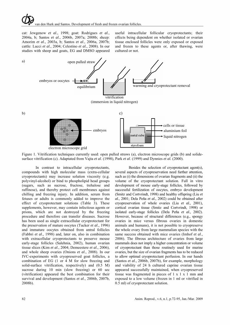
Currently, several vitrification methods are applied, whereby those using glass capillaries (Hochi et al., 1994), open pulled straws (OPS; Vajta et al., 1998), electron microscope (EM) grids (Park et al., 1999), and a solid-surface (Dinnyès et al., 2000) in or on which material is frozen. The OPS method includes the narrowing of the inner diameter of 250 μl glass straws by heat-softening and manually pulling the straws (Fig. 1a). This method allows the loading of embryos and oocytes by a capillary effect and subsequent extrusion by sedimentation. Generally, vitrification in EM-grids is performed by using 400 μm mesh copper EM grids (Fig. 1b), which are used as a physical support to maximize the cooling rates during the vitrification process. In the solid-surface method, a metal cube covered with aluminium foil is placed in a styrofoam box and partially submerged with liquid nitrogen. Cryoprotected biological material is put onto the cold upper surface and instantaneously vitrified (Fig. 1c). Thus far, vitrification with the help of straws or EM-grids, have not led to successful storage of ovarian tissue. Although the solid-surface technique was developed for embryos and oocytes, it appeared also be applicable in our studies with goat ovarian tissue (Santos et al., 2007b).
For the application of cryopreservation of ovarian tissue, it is necessary to determine the best cryoprotectant, its concentration, and exposure time. Cryoprotective agents protect cells against dehydration, cooling and freezing damage during cryopreservation. There are intracellular and extracellular cryoprotectants. Intracellular cryoprotectants act through diminishing the extent of osmotic shrinkage by permeating the cells and replacing water molecules herein and by maintaining the salt balance during freezing. Extracellular cryoprotectants reduce the freezing point by increasing the viscosity of the cryopreservation medium, or protect the cell membranes against chilling and freezing injury by binding to phospholipid head groups. However, cryoprotective agents can be toxic or may facilitate the entrance of toxic agents into cells. Glycerol is an intracellular cryoprotectant (low molecular mass compound), which has antifreeze properties and is able to counteract dehydration. Its metabolic product, however, is formaldehyde, which may induce metabolic acidosis. A similar effect can be observed after conversion of ethylene glycol (EG) into oxalate, or propylene glycol into lactic acid. Despite its antioxidant effect, dimethyl sulfoxide (DMSO) increases the permeability of cell membranes, which may permit entrance of toxic agents into the cell (Table 2). We and various other researchers tested and compared the effects of different intracellular cryoprotectants on mammalian ovarian tissue, in concentrations ranging from 0.5 to 3.0 M (e.g., human: Newton et al., 1996;
van den Hurk and Santos. Development of fresh and frozen ovarian follicles.
Anim. Reprod., v.6, n.1, p.72-95, Jan./Mar. 2009 82
cat: Jewgenow et al., 1998; goat: Rodrigues et al., 2004a, b; Santos et al., 2006b, 2007a; 2008b; sheep: Amorim et al., 2003a, b; Santos et al., 2006a, 2007b; cattle: Lucci et al., 2004; Celestino et al., 2008). In our studies with sheep and goats, EG and DMSO appeared
useful intracellular follicular cryoprotectants; their effects being dependent on whether isolated or ovarian tissue enclosed follicles were only exposed or exposed and frozen to these agents or, after thawing, were cultured or not.
a)
b) c) Figure 1. Vitrification techniques currently used: open pulled straws (a), electron microscope grids (b) and solide-surface vitrification (c). Adaptated from Vajta et al. (1998), Park et al. (1999) and Dynnies et al. (2000).
In contrast to intracellular cryoprotectants, compounds with high molecular mass (extra-cellular cryoprotectants) may increase solution viscosity (e.g. polyvinyl-alcohol) or bind to phospholipid head groups (sugars, such as sucrose, fructose, trehalose and raffinose), and thereby protect cell membranes against chilling and freezing injury. In addition, serum from fetuses or adults is commonly added to improve the effect of cryoprotectant solutions (Table 3). These supplements, however, may contain infectious agents or prions, which are not destroyed by the freezing procedure and therefore can transfer diseases. Sucrose has been used as single extracellular cryoprotectant for the preservation of mature oocytes (Tucker et al., 1996) and immature oocytes obtained from antral follicles (Fabbri et al., 1998) and, later on, also in combination with extracellular cryoprotectants to preserve mouse early-stage follicles (Salehnia, 2002), human ovarian tissue slices (Kim et al., 2004; Demeestere et al., 2006), and whole sheep ovaries (Onions et al., 2008). In our IVC-experiments with cryopreserved goat follicles, a combination of EG (1 or 4 M for slow freezing and solid-surface vitrification, respectively) and (0.5 M) sucrose during 10 min (slow freezing) or 60 sec (vitrification) appeared the best combination for their survival and development (Santos et al., 2006b, 2007b, 2008b).
Besides the selection of cryoprotectant agent(s), several aspects of cryopreservation need further attention, such as (i) the dimensions of ovarian fragments and (ii) the volume of the cryoprotectant solution. Full in vitro development of mouse early-stage follicles, followed by successful fertilization of oocytes, embryo development (Smitz and Cortvrindt, 1998) and healthy offspring (Liu et al., 2001; Dela Peña et al., 2002) could be obtained after cryopreservation of whole ovaries (Liu et al., 2001), cortical ovarian tissue (Smitz and Cortvrindt, 1998) or isolated early-stage follicles (Dela Peña et al., 2002). However, because of structural differences (e.g., spongy ovaries in mice versus fibrous ovaries in domestic animals and humans), it is not possible to cryopreserve the whole ovary from large mammalian species with the same success obtained with mice ovaries (Imhof et al., 2006). The fibrous architecture of ovaries from large mammals does not imply a higher concentration or volume of cryoprotectant than those routinely used for murine ovaries, but the size of ovarian fragments has to be reduced to allow optimal cryoprotectant perfusion. In our hands (Santos et al., 2006b, 2007b), for example, morphology and viability of 24 h cultured caprine ovarian tissue appeared successfully maintained, when cryopreserved tissue was fragmented in pieces of 1 x 1 x 1 mm and exposed to a low volume (frozen in 1 ml or vitrified in 0.5 ml) of cryoprotectant solution.
embryos
electron microscope grid
aluminium foil liquid nitrogen
cells or tissue
equilibrium
vitrification (immersion in liquid nitrogen)
warming and cryoprotectant removal embryos or oocytes
open pulled straw

van den Hurk and Santos. Development of fresh and frozen ovarian follicles.
Anim. Reprod., v.6, n.1, p.72-95, Jan./Mar. 2009 83
Table 2. Intra-cellular cryoprotective agents commonly used for oocyte and ovarian cryopreservation. Compound Source Formula Molar mass Use Metabolic product Toxicity alcohols and derivatives
glycerol (1,2,3-trihydroxypropane; glycerin; propane-1,2,3 -triol; glycyl alcohol)
ethylene glycol (monoethylene glycol; 1,2-ethanediol)
propylene glycol (1,2-propanediol; 1,2- methylethylene glycol; dihydroxypropane; trimethyl glycol; methyl glycol)
animal and vegetable fat and oil via reaction of ethylene oxide with water via hydration of propylene oxide
C3H8O3 C2H6O2 C3H8O2
92.09 g/mol 62.07 g/mol 76.09 g/mol
antifreeze surfactant basis counteract dehydration prevent cell lyses coolant antifreeze solvent emulsifier humectant
formaldehyde glucose (only in hepatocytes)
oxalate glyoxylic acid glutamate lactic acid
metabolic acidosis toxic if converted to oxalate cell acidosis
sulfoxides
dimethyl sulfoxide (methylsulfinylmethane; DMSO)
wood pulp
C2H6OS
78.13 g/mol
solvent antioxidant permeability of cell membrane respiratory chain activity ethanol oxidation
cytokine/chemokine production
dimethyl sulfide
allows toxic agents to enter into the cell due to increase in cell membrane permeability
OH
HO
OH
HO
OH
OH HO
O
S

van den Hurk and Santos. Development of fresh and frozen ovarian follicles.
Anim. Reprod., v.6, n.1, p.72-95, Jan./Mar. 2009 84
Table 3. High molar mass compounds commonly used as cryoprotective agents for oocyte and ovarian cryopreservation. Compound Source Formula Molar mass Use Metabolic product Toxicity alcohols and derivatives
polyvinyl alcohol (polyvinol; ethenol homopolymer; PVA)
synthetic resin obtained from hydrolysis of polyvinyl acetate
[ -C2H4O- ]n
( )
not applicable
preparation of other resins resistant to solvents or oxygen anticoagulant viscosity increasing agent
not applicable
non toxic; no success when used alone as cryoprotectant
proteins blood serum; albumin
bovine fetuses
not applicable
not applicable
nutrient source; there is no evidence to proof its role during freezing and thawing
not applicable
virus present in serum is not inactivated by the cryopreservation procedure
saccharides
sucrose (α-D-glucopyranosyl-1- 2-β-D-fructofuranose; saccharose)
trehalose (α-D-glucopyranosyl-1- 1-α-D-glucopyranoside)
produced by plants fungi, plants and invertebrate animals
C12H22O11
C12H22O11.2H2O
342.30 378.33
nutrient protects cell membranes against chilling and freezing binding to the phospholipid head groups and forming a gel phase as cells dehydrate similar to sucrose but with higher stability and less solubility
glucose + fructose glucose
non toxic; no success when used alone as cryoprotectant non toxic; no success when used alone as cryoprotectant
OH n
HO
H H OH
H
H
OH
H CH2OH
O
O O H
H
H
OH
HO
CH2OH
CH2C
O
O
O
OH OH
OH OH HO
HO
HO HO

van den Hurk and Santos. Development of fresh and frozen ovarian follicles.
Anim. Reprod., v.6, n.1, p.72-95, Jan./Mar. 2009 85
A good cryoprotectant must not only efficiently protect cells during cryopreservation, but its removal from cells should also not impair follicular viability and developmental capacity. It appeared that samples are better quickly than slowly thawed (El-Naggar et al., 2006) and cryoprotectant can be removed in one (Leibo, 1984), three (Rodrigues et al., 2004a, b) or even six steps (Shelton, 1992). Although one wash-out step seems to be practical for thawed embryos, more successful results with ovarian tissue were obtained after three washing steps (Lima et al., 2006; Sadeu et al., 2006; Santos et al., 2006b). Sucrose had to be added in the thawing medium to maximize the survival chances of slowly frozen goat ovarian tissue enclosed follicles (Santos et al., 2006b), whereas this sugar had to be omitted in this thawing fluid for vitrified ovarian tissue (Santos et al., 2007a). These rapid and slow cooling protocols appeared also to successfully maintain the morphology and viability of isolated and 24 h in vitro cultured early-stage follicles (Santos et al., 2008b).
Finally, apart from defining an efficient freezing/thawing protocol, the choice of efficient methods that assess follicular quality deserves notice, the more since follicular damage cannot always immediately be observed after thawing or warming of the cryopreserved tissue. Several follicular impairments caused by cryopreservation, such as granulosa cell-oocyte junction damage (Cecconi et al., 2004), basement membrane disruption (Santos et al., 2007b) and reduction or delay in granulosa cell proliferation (Cortvrindt et al., 1996), are better detected after 24 h IVC of follicles than immediately after their thawing (Paynter et al., 1999; Santos et al., 2006b, 2007a; 2008b). Indeed, this can be investigated with use of simple histological staining of follicles, among which for example trypan-blue staining (for determination of cellular plasma membrane integrity; Rodrigues et al., 2005; Santos et al., 2006b, 2008b; Fauque et al., 2007) and BrDU- (Jewgenow et al., 1998; Onions et al., 2008) or PCNA-staining (for multiplication of cells; Oktay et al., 2000; Abir et al., 2003; Choi et al., 2008) are used. As for the evaluation of in vitro cultured follicles, it is also advisable for cryopreserved follicles to additionally use specific ultra-structural (like the integrity of mitochondria and endoplasmic reticulum, and cell vacuolization; Van den Hurk et al., 1998; Santos et al., 2006a), fluorescent (like ethidium homodimer to detect nuclear membrane integrity, and rhodamine or calcein to demonstrate cytoplasmic activity; Schotanus et al., 1997; Van den Hurk et al., 1998), or immunohistochemical (like the apoptotic protein p53 and the anti-apoptotic protein Bcl-2; Bedaiwy et al., 2006) to validate their viability (Fig. 2). Besides, demonstration of functional markers, like the mRNA expression of growth factors and their receptors, could be useful for this goal (Silva et al., 2006a).
Evidently, long-term IVC of frozen-thawed
ovarian tissue, during which primordial follicles can grow and differentiate to the preovulatory stage, would be the optimal method to control follicular viability but, as remarked before, full morphological and physiological in vitro development of large mammalian follicles, are yet far from practice. Transplantation of cryopreserved ovarian tissue currently seems to be the most attractive method to evaluate folliculogenesis after cryopreservation (Fig. 2).
Transplantation of cryopreserved early-stage follicles
The first ovarian transplantation was performed
more than 100 years ago in humans by Morris (1895), who transplanted ovarian tissue into the uteri of women with infertility signals. His studies were repeated one year later in rabbits by Knauer (1896), who showed that the presence of ovarian tissue in the body prevents uterine atrophy, which was the first indication for an ovarian endocrine function. Thereafter, many of such studies were carried out with mammalian ovarian tissue, but with the development of cryopreservation methods and advances in cancer treatments during the last decades, attention for transplantation of tissues has increased (reviewed by Huser et al., 2007; Lee, 2007; Paris and Schlatt, 2007; Onions et al., 2008). Transplantation has been performed (i) by re-implanting ovarian tissue strips into the donor animal itself (auto-transplantation), (ii) into a recipient from the same species as the donor (allo-transplantation), or (iii) into a recipient from other species than the donor (xeno-transplantation), for which severely compromized immunodeficient (SCID) mice or athymic rat strains are commonly used. For allo-transplantation, immunological suppression is required, demanding extra care for the animal subjected to the grafting. Successful xeno-transplantation may also be hampered by a specific graft rejection mechanism or local physiological and infectious immune defenses (Fassbender et al., 2007), which reduce the amount of recovered ovarian tissue. When compared to xeno- and allo-transplantation, auto-transplantation currently seems the best tool for the evaluation of cryopreservation procedures for ovarian follicles of large mammals. Thereby, grafted tissues or organs can be placed back to their original site (orthotopic transplantation), like the cortex of a sterile ovary (Radford et al., 2001; Meirow et al., 2005) or the peritoneum beneath the ovarian hilus (Salle et al., 2002; Donnez et al., 2004; Tryde-Schmidt et al., 2004; Bordes et al., 2005), or to another site (heterotopic transplantation), like the kidney (Liu et al., 2001), the uterus (Aubard et al., 1999), the rectus abdominal muscle (Callejo et al., 2001), the retroperitoneum (Oktay and Karlikaya, 2000), and the forearm skin (Oktay et al., 2001).

van den Hurk and Santos. Development of fresh and frozen ovarian follicles.
Anim. Reprod., v.6, n.1, p.72-95, Jan./Mar. 2009 86
Figure 2. Schematic presentation, showing the most common methods to evaluate the quality of follicles after cryopreservation.
allo- or auto-transplantation: most appropriate to determine the efficiency of cryopreservation, and comparing folliculogenesis in transplanted ovarian tissue with that in control animals
classical histology morphology oocyte and granulosa cells
transmission electron microscopy ultrastructure of cellular organelles
viability markers
trypan blue membrane integrity
viable
non-viable
ethitium homodimer: enter permeable (dead) cells and binds to DNA with high afinity calcein-AM: stain live cells with normal enzymatic activity
in vitro culture
short-term (hours/days): longer time is necessary for acquisition of fertilizable oocytes from large mammals
long-term (weeks/months): not yet suitable for acquisition of fertilizable oocytes from large mammals
in vivo culture
xeno-transplantation: SCID mice or athymic rats
apoptotic protein p53
anti-apoptic protein Bcl-2

van den Hurk and Santos. Development of fresh and frozen ovarian follicles.
Anim. Reprod., v.6, n.1, p.72-95, Jan./Mar. 2009 87
We recently demonstrated, full follicular development of caprine early-stage follicles after freezing-thawing of ovarian tissue fragments and their heterotopic auto-transplantation onto the uterus of ovariectomized donor goats (Santos, 2007; Santos et al., 2008a). Hereby, ovarian cortical fragments reorganized into an ovarian-like organ, from which blood supply and endocrine function (estradiol and progesterone production) were restored. The average time required to complete folliculogenesis was about 71 days both for fresh and frozen-thawed grafts, which was accompanied by estrous behaviour. Increased plasma estrogen concentrations were followed by a rise in those of progesterone, indicative of ovulation and luteal activity. Previously, orthotopic allo-transplantation of slowly frozen-thawed ovarian material was successfully carried out in mice, either by using cortical tissue fragments (Parrott, 1960) or isolated primordial mouse follicles from juvenile specimens (Carroll and Gosden, 1993). The grafts reorganized into ovarian structures and, after mating, females produced normal offspring. Similar success was also obtained after allo-transplantating both slowly frozen-thawed fetal mouse ovarian tissue (orthotopic and heterotopic: Cox et al., 1996; orthotopic: Gunasena et al., 1997; heterotopic: Liu et al., 2001), and vitrified-warmed 10 day-old ovaries or adult ovarian fourths (Hani et al., 2006) to recipients. However, in contrast with the recipients transplanted with whole immature mice ovaries, pup birth rate had dramatically decreased at and beyond the second litter of those transplanted with ovarian tissue from adult mice (Hani et al., 2006). Orthotopic auto-transplantation of slowly frozen-thawed ovarian tissue in sheep resulted in recovery of cyclical ovarian function, but with formation of follicular cysts and a drastic reduction (up to 70%) in the total number of primordial follicles (Baird et al., 1999). More recently, however, offspring has been obtained in ewes after auto-transplantation of half of the vitrified-warmed ovarian cortex to the ovarian hilus (Salle et al., 2002, 2003; Bordes et al., 2005). Although the procedure used in these latter experiments resulted in long-term (≥2 years) recovery of fertility, various lambs showed abnormalities, while a drastic reduction in the total number of follicles was observed in the transplants. In humans, viability of primordial follicles and their initial growth were observed when ovarian cortical strips were frozen/thawed and xeno-grafted to adult mice (Oktay et al., 2000). A few years later, antral follicles were obtained from human fetal ovarian slices that were collected after 4 months of xeno-transplantation into the kidney capsules of SCID-mice (Abir et al., 2003). More recently, ovulation and subsequent embryo development from heterotopic auto-transplanted human ovarian tissue (Oktay et al., 2004) and live-births from orthotopic ovarian auto-transplants following spontaneous (Donnez et al., 2004) and IVF (Meirow et al., 2005) cycles were reported. Hereby, modified cryopreservation and
surgical protocols were followed those that have been developed for sheep by Gosden and coworkers (Gosden et al., 1994; Baird et al., 1999). Unfortunately, the results obtained by Donnez and associates (2004) have not given final proof that transplantation of cryopreserved ovarian tissue can restore the reproductive function. It is unknown whether the ovum that was fertilized after transplantation of human ovarian tissue came from the re-implanted tissue or from the remaining native ovary (Oktay and Tilly, 2004). Pregnancy was also obtained from cryopreserved ovarian tissue that was transplanted at the remaining ovarian site (Meirow et al., 2005). Demeestere and coworkers (2006) reported six spontaneous cycles and a pregnancy after combined orthotopic and heterotopic transplantation of cryopreserved ovarian tissue in a woman previously treated by bone marrow transplantation. Although this finding provides further evidence of the putative efficiency of cryopreserved ovarian tissue transplantation for restoring natural fertility, it is of importance to mention that the patient miscarried at 7 weeks of gestation because of aneuploidy, which could be related to the procedure.
Ischemic damage to the tissue, revascularization injury and the theoretical possibility of reintroducing malignant tumour cells appear main problems for a transplantation procedure whereby a small mass of ovarian tissue is used (Bedaiwy and Falcone, 2004). Ischemic damage to ovarian tissues is a cause of high rate of follicular loss and thus limits the function of implanted tissue (Nugent et al., 1998; Baird et al., 1999; Demirci et al., 2002). Ovarian cortex tolerates ischaemia at least for 3 h and stromal cells are more vulnerable to ischemia than primordial follicles (Kim et al., 2004). Elimination of ovarian ischemic damage could result in the maintenance of both the viability and functioning of transplants. This could be reached by transplantation of a whole ovary together with its vascular pedicle with achievement of vascular anastomosis. Such an auto-transplantation has heterotopically been done with cryopreserved-thawed ovaries both in rats (Wang et al., 2002; Yin et al., 2003) and in sheep (Bedaiwy et al., 2003), and resulted in partial restoration of hormonal functions in sheep (Bedaiwy et al., 2003) and pregnancy in rats (Wang et al., 2002). In rats, however, the ovarian follicle reserve still was compromised by fewer follicles ≥60 days after surgery. In sheep, follicle numbers had also decreased, while follicular viability in whole ovarian transplants did not significantly differ from animals that received cortical strips. More recently, orthotopic microvascular re-anastomosis of whole cryopreserved ovine ovaries resulted in normal ovulation, embryo development and long-term (≥36 months post-transplantation) hormonal restoration (Arav et al., 2005), and in a pregnancy and a healthy lamb 545 days after transplantation (Imhof et al., 2006). High post-thaw survival rates of follicles, small vessels and stromal cells as well as normal

van den Hurk and Santos. Development of fresh and frozen ovarian follicles.
Anim. Reprod., v.6, n.1, p.72-95, Jan./Mar. 2009 88
histological structure in all the ovarian components were reported after cryopreservation of a whole human ovary together with its vascular pedicle (Martinez-Madrid et al., 2004). Based on alterations in apoptotic and survival proteins, the viability of human primordial follicles appeared well-maintained immediately after freezing/thawing of whole ovaries together with their vascular pedicle (Bedaiwy et al., 2006). The data show, that (i) cryopreservation of an intact ovary is a useful method for the preservation of female germ cells, which still can be further improved, and (ii) transplantation of a cryopreserved whole ovary together with its vascular pedicle is technically feasible and a promising application of mammalian ovarian tissue banking.
Concluding remarks and perspectives
This paper gives a survey of our research on the development, cryopreservation, in vitro culture and transplantation of early-stage follicles in the past two decades, relative to those obtained by other researchers. Hereby, attention has specially been focussed on findings with ruminants. Apart from our histological, immunocytochemical, viability, biochemical, endocrine, in vitro and in vivo studies on cow, sheep and goat follicles, some general information on the various in vitro culture, cryopreservation and transplantation techniques, that can be used, has been given. It is concluded, that (i) we are only beginning to understand the complexities of in vitro and in vivo formation of follicles up to a fertilizable state, (ii) practical application of these follicles for in vitro production of healthy fertilizable oocytes, embryos or offspring from ruminants and humans is still far away, because of insufficient knowledge about the regulatory mechanisms underlying early-stage follicle development and their culture conditions, and (iii) current cryopreservation techniques satisfactorily maintain the huge stock of very early-stage follicles for an undetermined time without viability impairment after thawing, followed by further development in short- or long-term in vitro cultures or in transplantation experiments.
Above and beyond superovulation and IVM of immature oocytes recovered from antral follicles, for in vitro embryo production, the exploitation of the oocyte reservoir present in early-stage follicles gathers increasing attention, because these follicles constitute an enormous source of germ cells. However, we do not yet know if all or most of these ovarian follicles really are of sufficient quality to be used as embryo donors, since to date we lack non-invasive parameters for good quality oocytes. Only a light-microscopical impression of a follicle is not appropriate to select for good follicular quality. After ultra-structural studies, viability studies with fluorescent, immunohistochemical or functional markers, or gene-labelling studies of oocytes, one cannot use them anymore for further culture. Determination of markers
like steroids, methalloproteinases or their inhibitors may reflect the status of the follicle, especially of their somatic cells, but does not provide sufficient information on their oocyte quality. Nevertheless, we have to continue our efforts to learn the factors that contribute to the growth and differentiation of oocytes and follicles and thus continue with our attempts to in vitro culture early-stage follicles. Many factors, endocrine and locally derived, have been demonstrated to be involved in follicular development. Such a role has been in vitro demonstrated for only a few compounds and done for a restricted number of mammalian species, rodents and ruminants especially. To this end, it is of importance to implicate more animal species for these in vitro studies and to test the effects of far more compounds. Particularly, the study of the role of various TGF-β family members, like TGF-β itself, TGF-α, Nodal, all kinds of BMPs, activin and their regulating protein follistatin will be of interest in this respect. Also more detailed studies on the follicular IGF-system are needed, specifically on the role of IGF-binding proteins in early folliculogenesis. The role of steroids, androgens in particular, in early follicle development is fairly neglected. Besides, there are many other compounds that may have a role on follicle development, such as thyroid hormones, prolactin, oxytocin, prostaglandins, angiotensins, and neurotropins.
Numerous early-stage follicles are needed to study (i) the regulation of their in vitro development to mature structures with fertilizable oocytes and, thus, to learn the required composition of media for their inevitable long-term IVC, and (ii) the in vivo conditions needed for transplanted follicles or ovarian tissue to survive and develop until a mature stage at which oocytes can be fertilized and healthy embryos or offspring can be obtained. To achieve large scale use of them in such experiments, it is pleasant to know that we currently have adequate cryopreservation methods at our disposal to maintain a collected stock of early-stage follicles viable for a long period and to protect them from immediate degeneration after thawing. Hopefully, in the coming decades, these cryostore methods will enable the use of numerous early-stage follicles from large mammalian species in future long-term in vitro systems or in transplantation experiments to produce healthy offspring from fertilizable matured oocytes.
References Adashi EY, Rohan RM. 1992. Intraovarian regulation: Peptidergic signaling systems. Trends Endocrinol Metab, 3:243-248. Abir R, Franks S, Mobberley MA, Moore PA, Margara RA, Winston RM. 1997. Mechanical isolation and in vitro growth of pre-antral and small antral human follicles. Fertil Steril, 68:682-688. Abir, R, Roizman P, Fisch B, Nitke S, Okon E, Orvieto R, Ben Rafael Z. 1999. Pilot study of isolated

van den Hurk and Santos. Development of fresh and frozen ovarian follicles.
Anim. Reprod., v.6, n.1, p.72-95, Jan./Mar. 2009 89
early human follicles cultured in collagen gels for 24 hours. Hum Reprod, 14:1299-1301. Abir R, Orvieto R, Raanani H, Feldberg D, Nitke S, Fisch B. 2003. Parameters affecting successful transplantation of frozen-thawed human fetal ovaries into immunodeficient mice. Fertil Steril, 80:421-428. Abir R, Fisch B, Jin S, Barnnet M, Freimann S, van den Hurk R, Feldberg D, Nitke S, Krissi H, Ao A. 2004. Immunocytochemical detection and RT-PCR expression of leukaemia inhibitory factor and its receptor in human fetal and adult ovaries. Mol Hum Reprod, 10:313-319. Abir R, Ao A, Jin S, Barnett M, van den Hurk R, Freimann S, Fisch B. 2005. Immunocytochemical detection and reverse transcription polymerase chain reaction expression of oncostatin M (OSM) and its receptor (OSM-Rβ) in human fetal and adult ovaries. Fertil Steril Suppl, 1:1188-1196. Amorim CA, Rodrigues APR, Rondina D, Gonçalves PBD, Figueiredo JR, Giorgetti A. 2003a. Cryopreservation of ovine primordial follicles using dimethyl sulfoxide. Fertil Steril, 79:682-686. Amorim CA, Rondina D, Rodrigues APR, Costa SHF, Goncalves PBD, Figueiredo JR, Giorgetti A. 2003b. Isolated ovine primordial follicles cryopreserved in different concentrations of ethylene glycol. Theriogenology, 60:735-742. Arav A, Revel A, Nathan Y, Bor A, Gacitua H, Yavin S, Gavish Z, Uri M, Elami A. 2005. Oocyte recovery, embryo development and ovarian function after cryopreservation and transplantation of whole sheep ovary. Hum Reprod, 20:3554-3559. Armstrong DG, Baxter G, Hogg CO, Woad KJ. 2002. Insulin-like growth factor (IGF) system in the oocyte and somatic cells of bovine preantral follicles. Reproduction, 123:789-797. Aubard Y, Piver P, Cogni Y, Fermeaux V, Poulin N, Driancourt MA. 1999. Orthotopic and heterotopic autografts of frozen-thawed ovarian cortex in sheep. Hum Reprod, 14:2149-2154. Baird DT, Webb R, Campbell BK, Harkness LM, Gosden RG. 1999. Long-term ovarian function in sheep after ovariectomy and transplantation of autografts stored at -196oC. Endocrinology, 140:462-471. Bedaiwy MA, Jeremias E, Gurunluoglu R, Hussein MR, Siemianow M, Biscotti C, Falcone T. 2003. Restoration of ovarian function after autotransplantation of intact frozen-thawed sheep ovaries with microvascular anastomosis. Fertil Steril, 79:594-602. Bedaiwy MA, Falcone T. 2004. Ovarian tissue banking for cancer patients: reduction of post-transplantation ischaemic injury: intact ovary freezing and transplantation. Hum Reprod, 9:1242-1244. Bedaiwy MA, Hussein MR, Biscotti C, Falcone T. 2006. Cryopreservation of intact human ovary with its vascular pedicle. Hum Reprod, 21:3258-3269. Biron-Shental T, Fisch B, van den Hurk R, Felz C, Feldberg D, Abir R. 2004. Survival of frozen-thawed
human ovarian fetal follicles in long-term organ culture. Fertil Steril, 81:716-719. Bodensteiner KJ, Clay CM, Moeller CL, Sawyer HR. 1999. Molecular cloning of the ovine Growth/Differentiation factor-9 gene and expression of growth differentiation factor-9 in ovine and bovine ovaries. Biol Reprod, 60:381-386. Bordes A, Lornage J, Demirci B, Franck M, Courbiere B, Guerin JF, Salle B. 2005. Normal gestations and live births after orthotopic autograft of vitrified-warmed hemi-ovaries into ewes. Hum Reprod, 20:2745-2748. Braw-Tal R. 1994. Expression of mRNA for follistatin and inhibin/activin subunits during follicular growth and atresia. J Mol Endocrinol, 13:253-264. Britt JH. 1991. Impacts of early postpartum metabolism on follicular development and fertility. Bov Pract, 24:39-43. Cain L, Chatterjee S, Collins TJ. 1995. In vitro folliculogenesis of rat preantral follicles. Endocrinology, 136:3369-3377. Callejo J, Salvador C, Miralles A, Vilaseca S, Lailla JM, Balasch J. 2001. Long-term ovarian function evaluation after autografting by implantation with fresh and frozen-thawed human ovarian tissue. J Clin Endocrinol Metab, 86:4489-4494. Candy CJ, Wood MJ, Whittingham DG. 1997. Effect of cryoprotectants on the survival of follicles in frozen mouse ovaries. J Reprod Fertil, 110:11-19. Carroll J, Gosden RG. 1993. Transplantation of frozen-thawed mouse primordial follicles. Hum Reprod, 8:1163-1167. Cecconi S, Barboni B, Coccia M, Mattioli M. 1999. In vitro development of sheep preantral follicles. Biol. Reprod, 60:594-601. Cecconi S, Capacchietti G, Russo V, Berardinelli P, Mattioli M, Barboni B. 2004. In vitro growth of preantral follicles isolated from cryopreserved ovine ovarian tissue. Biol Reprod, 70:12-17. Celestino JJ, Santos RR, Lopes CA, Martins FS, Matos MH, Melo MA, Báo SN, Rodrigues AP, Silva JR, Figueiredo JR. 2008. Preservation of bovine preantral follicle viability and ultra-structure after cooling and freezing of ovarian tissue. Anim Reprod Sci, 108:309-318 Choi J, Lee B, Lee E, Yoon BK, Bae D, Choi D. 2008. Cryopreservation of ovarian tissues temporarily suppresses the proliferation of granulosa cells in mouse preantral follicles. Cryobiology, 56:36-42. Clark DE, Tisdall DJ, Fidler AE, McNatty KP. 1996 Localization of mRNA encoding c-kit during the initiation of folliculogenesis in ovine fetal ovaries. J Reprod Fertil, 106:329-335. Cortvrindt RG, Smitz J, Van Steirteghem AC. 1996. A morphological and functional study of the effect of slow freezing followed by complete in-vitro maturation of primary mouse ovarian follicles. Hum Reprod, 11:2648-2655.

van den Hurk and Santos. Development of fresh and frozen ovarian follicles.
Anim. Reprod., v.6, n.1, p.72-95, Jan./Mar. 2009 90
Cortvrindt RG, Smitz J. 2001. Fluorescent probes allow rapid and precise recording of follicle density and staging in human ovarian cortical biopsy samples. Fertil Steril, 75:588-593. Cox SL, Shaw J, Jenkin G. 1996. Transplantation of cryopreserved fetal ovarian tissue to adult recipients in mice. J Reprod Fertil, 107:315-322. Da Silva-Buttkus P, van den Hurk R, te Velde ER, Taverne MAM. 2003. Ovarian development in intrauterine growth retarded and normally developed piglets originating from the same litter. Reproduction, 126:249-258. Dela Peña EC, Takahashi Y, Katagiri S, Atabay EC, Nagano M. 2002. Birth of pups after transfer of mouse embryos derived from vitrified preantral follicles. Reproduction, 123:593-600. Demeestere I, Delbaere A, Gervy C, van den Bergh M, Devreker F, Englert Y. 2002. Effect of preantral follicle isolation technique on in-vitro follicular growth, oocyte maturation and embryo development in mice. Hum Reprod, 17:2152-2159. Demeestere I, Simon P, Buxant F, Robin V, Fernandez SA, Centner J, Delbaere A, Englert Y. 2006. Ovarian function and spontaneous pregnancy after combined heterotopic and orthotopic cryopreserved ovarian tissue transplantation in a patient previously treated with bone marrow transplantation: case report. Hum Reprod, 21:2010-2014. Demirci B, Salle B, Frappart L, Franck M, Guerin JF, Lornage J. 2002, Morphological alterations and DNA fragmentation in oocytes from primordial and primary follicles after freezing-thawing of ovarian cortex in sheep. Fertil Steril, 77:595-600. Dinnyès A, Dai Y, Jiang S, Yang X. 2000. High developmental rates of vitrified bovine oocytes following parthenogenetic activation, and somatic cell nuclear transfer. Biol Reprod, 63:513-518. Dijkstra G, de Rooij DG, de Jong FH, van den Hurk R. 1996. Effect of hypothyroidism on ovarian follicular development, granulosa cell proliferation and peripheral hormone levels in the prepubertal rat. Eur J Endocrinol, 134:649-654. Donnelly K, Telfer EE. 1994. Growth of porcine preantral ovarian follicles in different culture systems. J Reprod Fertil Abstr Ser, 13:76. Donnez J, Dolmans MM, Demylle D, Jadoul P, Pirard C, Squifflet J, Martinez-Madrid B, van Langendonckt A. 2004. Livebirth after orthotopic transplantation of cryopreserved ovarian tissue. Lancet, 364:1405-1410. Driancourt MA. 2001. Regulation of ovarian follicular dynamics in farm animals: implications for manipulation of reproduction. Theriogenology, 55:1211-1239. El-Naggar MM, Al-Mashat FM, Elayat AA, Sibiany AR, Ardawi MS, Badawoud MH. 2006. Effect of thawing rate and post-thaw culture on the cryopreserved fetal rat islets: functional and morphological correlation.
Life Sci, 78:1925-1932. Eppig JJ. 1977. Mouse oocyte development in vitro with various culture systems. Dev Biol, 15:371-388. Eppig JJ. 1994. Further reflections on culture systems for the growth of oocytes in vitro. Hum Reprod, 9:969-976. Eppig JJ, O’Brien MJ. 1996. Development in vitro of mouse oocytes from primordial follicles. Biol Reprod, 54:197-207. Eppig JJ, O’Brien MJ. 1998. Comparison of preimplantation developmental competence after mouse oocyte growth and development in vitro and in vivo. Theriogenology, 49:415-422. Fabbri R, Porcu E, Marsella T, Primavera MR, Seracchioli R, Ciotti PM, Magrini O, Venturoli S, Flamigni C. 1998. Oocyte cryopreservation. Hum Reprod, 13:98-108. Fassbender M, Hildebrandt TB, Paris MC, Colenbrander B, Jewgenow K. 2007. High-resolution ultrasonography of xenografted domestic cat ovarian cortex. J Reprod Dev, 53:1023-1034. Fatehi AN, van den Hurk R, Colenbrander C, Daemen AJ, van Tol HTA, Monteiro JM, Roelen BAJ, Bevers MM. 2005. Expression of bone morphogenetic protein 2 (BMP2), BMP4 and BMP receptors in the bovine ovary but absence of effects of BMP2 and BMP4 during IVM on bovine oocyte nuclear maturation and subsequent embryo development. Theriogenology, 63:872-889. Fauque P, Ban Amor A, Joanne C, Agnani G, Bresson JL, Roux C. 2007. Use of trypan blue staining to assess the quality of ovarian cryopreservation. Fertil Steril, 87:1200-1207. Figueiredo JR, Hulshof SCJ, van den Hurk R, Bevers MM, Nusgens B, Beckers JF. 1993. Development of a combined new mechanical and enzymatic method for the isolation of intact preantral follicles from fetal, calf and adult bovine ovaries. Theriogenology, 40:789-799. Figueiredo JR, Hulshof SCJ, van den Hurk R, Bevers MM, Thiry M, Nusgens B, Beckers JF. 1994a. The physiological status of the ovarian donor affects the in vitro development of isolated bovine preantral follicles. Theriogenology, 42:1303-1310. Figueiredo JR, Hulshof SCJ, van den Hurk R, Nusgens B, Bevers MM, Ectors FJ, Beckers JF. 1994b. Preservation of oocyte and granulosa cell morphology in bovine preantral follicles cultured in vitro. Theriogenology, 41:1333-1346. Figueiredo JR. 1995. Isolement, Caractérisation et Culture de Follicules Préantaux chez les Bovins. Liège, Belgium: Université de Liège. Thesis. Figueiredo JR, Hulshof SC, Thiry M, van den Hurk R, Bevers MM, Nusgens B, Beckers JF. 1995. Extracellular matrix proteins and basement membrane: their identification in bovine ovaries and significance for the attachment of cultured preantral follicles. Theriogenology, 43:845-858.

van den Hurk and Santos. Development of fresh and frozen ovarian follicles.
Anim. Reprod., v.6, n.1, p.72-95, Jan./Mar. 2009 91
Findlay JK, Drummond AE, Dyson ML, Baillie AJ, Robertson DM, Ethier JF. 2002. Recruitment and development of the follicle; the roles of the transforming growth factor-β superfamily. Mol Cell Endocrinol, 191:35-43. Fortune JE. 1994. Ovarian follicular growth and development in mammals. Biol Reprod, 50: 225-232. Fortune JE, Kito S, Byrd DD. 1999. Activation of primordial follicles in vitro. J Reprod Fertil Suppl, 54:439-448. Fortune JE, Cushman RA, Wahl CM, Kito S. 2000. The primordial to primary follicle transition. Mol Cell Endocrinol, 163:53-60. Fortune JE, Rivera GM, Evans AC, Turzillo AM. 2001. Differentiation of dominant versus subordinate follicles in cattle. Biol Reprod, 65:648-654. Fortune JE. 2003. The early stages of follicular development: activation of primordial follicles and growth of preantral follicles. Anim Reprod Sci, 78:135-163. Galloway SM, McNatty KP, Cambridge LM, Laitinen MP, Juengel JL, Jokiranta TS, McLaren RJ, Luiro K, Dodds KG, Montgomery GW, Beattie AE, Davis GH, Ritvos O. 2000. Mutations in an oocyte-derived growth factor gene (BMP15) cause increased ovulation rate and infertility in a dosage-sensitive manner. Nat Genet, 25:279-283. Gigli DA, Byrd DD, Fortune JE. 2006. Effects of oxygen tension and supplements to the culture medium on activation and development of bovine follicles in vitro. Theriogenology, 66:344-353. Glister C, Kemp CF, Knight PG. 2004. Bone morphogenetic protein (BMP) ligands and receptors in bovine ovarian follicle cells: actions of BMP-4, -6 and -7 on granulosa cells and differential modulation of Smad-1 phosphorylation by follistatin. Reproduction, 127:239-254. Gosden RG, Baird DT, Wade JC, Webb R. 1994. Restoration of fertility to oophorectomized sheep by ovarian autografts stored at -196 degrees C. Hum Reprod, 9:597-603. Gougeon A. 1986. Dynamics of follicular growth in the human: a model from preliminary results. Hum Reprod, 1:81-87. Gougeon A. 1996. Regulation of ovarian follicular development in primates: facts and hypotheses. Endocr Rev, 17:121-155. Gunasena KT, Villines PM, Critser ES, Critser JK. 1997. Live births after autologous transplant of cryopreserved mouse ovaries. Hum Reprod, 12:101-106. Gutierrez CG, Ralph JH, Telfer EE, Wilmut I, Webb R. 2000. Growth and antrum formation of bovine preantral follicles in long-term culture in vitro. Biol Reprod, 62:1322-1328. Hani T, Tachibe T, Shingai S, Kamada N, Ueda O, Jishage K. 2006. Fertility of mice receiving vitrified adult mouse ovaries. Reproduction, 31:681-687. Hartshorne GM. 1997. In vitro culture of ovarian
follicles. Rev Reprod, 2:94-104. Hirao Y, Nagai T, Kubo M, Miyano T, Miyake M, Kato S. 1994. In vitro growth and maturation of pig oocytes. J Reprod Fertil, 100:333-339. Hochi S, Fujimoto T, Braun J, Oguri N. 1994. Pregnancies following transfer of equine embryos cryopreserved by vitrification. Theriogenology, 42:483-488. Hovatta O, Silye R, Abir R, Krausz T, Winston RML. 1997. Extracellular matrix improves survival of both stored and fresh human primordial and primary ovarian follicles in long-term culture. Hum Reprod, 12:1032-1036. Hulshof SCJ, Dijkstra G, van der Beek EM, Bevers MM, Figueiredo JR, Beckers JF, van den Hurk R. 1994a. Immunocytochemical localization of vasoactive intestinal peptide and neuro-peptide Y in the bovine ovary. Biol Reprod, 50: 553-560. Hulshof SCJ, Figueiredo JR, Beckers JF, Bevers MM, van den Hurk R. 1994b. Isolation and characterization of pre-antral follicles from fetal bovine ovaries. Vet Q, 16:78-80. Hulshof SCJ. 1995. Bovine Pre-antral Follicles and their Development in Vitro. Utrecht, Netherlands: Utrecht University. Thesis. Hulshof SCJ, Dijkstra G, van der Beek EM, Bevers MM, van den Hurk R. 1995a. Immunohistochemical localization of vasoactive intestinal peptide (VIP) and neuropeptide Y (NPY) in the bovine ovary. Eur J Morphol, 33:192-193. Hulshof SCJ, Figueiredo JR, Beckers JF, Bevers MM, van den Donk HA, van den Hurk R. 1995b. Effects of fetal bovine serum, FSH and 17β-estradiol on the culture of bovine pre-antral follicles. Theriogenology , 44:217-226. Hulshof SCJ, Figueiredo JR, Beckers JF, Bevers MM, Vanderstichele H, van den Hurk R. 1997. Bovine preantral follicles and activin: immunohistochemistry for activin and activin receptor and the effect of bovine activin A in vitro. Theriogenology, 48:133-142. Huser M, Crha I, Hudecek R, Ventruba P, Zakova J, Smardova L, Kral Z. 2007. Ovarian tissue cryopreservation--new opportunity to preserve fertility in female cancer patients. Eur J Gynaecol Oncol, 28:249-255. Imhof M, Bergmeister H, Lipovac M, Rudas M, Hofstetter G, Huber J. 2006. Orthotopic microvascular reanastomosis of whole cryopreserved ovine ovaries resulting in pregnancy and live birth. Fertil Steril, 85:1208-1215. Ireland JJ. 1987. Control of follicular growth and development. J Reprod Fertil, 34:39-54. Izadyar F, Dijkstra G, van Tol HYA, van den Hurk R, Colenbrander B, Bevers MM. 1998. Immunohistochemical localization and mRNA analysis of activin, inhibin, follistatin and activin receptor in bovine cumulus oocyte complexes during in vitro

van den Hurk and Santos. Development of fresh and frozen ovarian follicles.
Anim. Reprod., v.6, n.1, p.72-95, Jan./Mar. 2009 92
maturation. Mol Reprod Dev, 49:186-195. Jewgenow K, Penfold LM, Meyer HH, Wildt DE. 1998. Viability of small preantral ovarian follicles from domestic cats after cryoprotectant exposure and cryopreservation. J Reprod Fertil, 112:39-47. Johnson JL, van Eys GJJM, Angelini GD, George SJ. 2001. Injury induces dedifferentiation of smooth muscle cells and increased matrix-degrading metalloproteinase activity in human saphenous vein. Arterioscler Thromb Vasc Biol, 21:1146-1151. Jorio A, Mariana JC, Lahlou-Kassi A. 1991. Development of the population of ovarian follicles during the prepubertal period in D’man and Timahdite sheep. Anim Reprod Sci, 26:239-250. Juengel JL, Quirke LD, Tisdall DJ, Smith P, Hudson NL, McNatty KP. 2000. Gene expression in abnormal ovarian structures of ewes homozygous for the inverdale prolificacy gene. Biol Reprod, 62:1467-1478. Kim SS, Yang HW, Kang HG, Lee HH, Lee HC, Ko DS, Gosden RG. 2004. Quantitative assessment of ischemic tissue damage in ovarian cortical tissue with or without antioxidant (ascorbic acid) treatment. Fertil Steril, 82:679-685. Knauer E. 1896. Einige Versuche über Ovarientransplantation bei Kaninchen. Zentralbl Gynaekol, 20:524-528. Lee D. 2007. Ovarian tissue cryopreservation and transplantation: banking reproductive potential for the future. Cancer Treat Res, 138:110-129. Leibo SP. 1984. One-step method for direct non-surgical transfer of frozen-thawed bovine embryos. Theriogenology, 21:767-790. Lenie S, Cortvrindt R, Adriaenssens T, Smitz J. 2004. A reproducible two-step culture system for isolated primary mouse ovarian follicles as single functional units. Biol Reprod, 71:1730-1738. Lima AKF, Silva AR, Santos RR, Sales DM, Evangelista AF, Figueiredo JR, Silva LDM. 2006. Cryopreservation of preantral ovarian follicles in situ from domestic cats (Felis catus) using different cryoprotective agents. Theriogenology, 66:1664-1666. Liu J, van der Elst J, van den Broecke R, Dhont M. 2001. Live offspring by in vitro fertilization of oocytes from cryopreserved primordial mouse follicles after sequential in vivo transplantation and in vitro maturation. Biol Reprod, 64:171-178. Lucci CM, Kacinskis MA, Lopes LHR, Rumpf R, Báo SN. 2004. Effect of different cryoprotectants on the structural preservation of follicles in frozen zebu bovine (Bos indicus) ovarian tissue. Theriogenology, 61:1101-1114. Lunenfeld B, Insler V. 1993. Follicular development and its control. Gynecol Endocrinol, 7:285-291. Lussier JP, Matton P, Dufour JJ. 1987. Growth rates of follicles in the ovary of the cow. J Reprod Fertil, 81:301-307. Martinez-Madrid B, Dolmans MM, van Langendonckt A, Defrere S, Donnez J. 2004. Freeze-
thawing intact human ovary with its vascular pedicle with a passive cooling device. Fertil Steril, 82:1390-1394. Martinuk SD, Chizen DR, Pierson RA. 1992. Ultrasonographic morphology of the human preovulatory follicle wall prior to ovulation. Clin Anat, 5:339-352. Matos MHT, Lima Verde I, Luque MCA, Santos KDA, Martins FS, Bao SN, Lucci CM, van den Hurk, R, Figueiredo JR. 2007. Effects of fibroblast growth factor-2 on the in vitro culture of caprine preantral follicles. Cells Tissues Organs, 186:112-120. McNatty KP, Heath DA, Lundy T, Fidler AE, Quirke L, O'Connell A, Smith P, Groome N, Tisdall DJ. 1999. Control of early ovarian follicular development. J Reprod Fertil Suppl, 54:3-16. McNatty KP, Reader K, Smith P, Heath DA, Juengel JL. 2007. Control of ovarian follicular development to the gonadotrophin-dependent phase: a 2006 perspective. Soc Reprod Fertil Suppl, 64:55-68. Meirow D, Levron J, Eldar-Geva T, Hardan I, Fridman E, Zalel Y, Schiff E, Dor J. 2005. Pregnancy after transplantation of cryopreserved ovarian tissue in a patient with ovarian failure after chemotherapy. N Engl J Med, 353:318-321. Monget P, Monniaux D. 1995. Growth factors and the control of folliculogenesis. J Reprod Fertil Suppl, 49:321-333. Monniaux D, Huet C, Besnard N, Clement F, Bosc M, Pisselet C, Monget, P, Mariana JC. 1997. Follicular growth and ovarian dynamics in mammals. J Reprod Fertil Suppl, 51:3-23. Morris RT. 1895. The ovarian graft. NY Med J, 62:436. Newton H, Aubard Y, Rutherford A, Sharma V, Gosden R. 1996. Low temperature storage and grafting of human ovarian tissue. Hum Reprod, 11:1487-1491. Newton H, Picton H, Gosden RG. 1999. In vitro growth of oocyte-granulosa cell complexes isolated from cryopreserved ovine tissue. J Reprod Fertil, 115:141-150. Nugent D, Newton H, Gallivan L, Gosden RG. 1998. Protective effect of vitamin E on ischaemia-reperfusion injury in ovarian grafts. J Reprod Fertil, 114:341-346. O’Brien MJ, Pendola JK, Eppig JJ. 2003. A revised protocol for in vitro development of mouse oocytes from primordial follicles dramatically improves their developmental competence. Biol Reprod, 68:1682-1686. Oktay K, Karlikaya G. 2000. Ovarian function after transplantation of frozen, banked autologous ovarian tissue. N Engl J Med, 342:1919. Oktay K, Newton H, Gosden RG. 2000. Transplantation of cryopreserved human ovarian tissue results in follicle growth initiation in SCID mice. Fertil Steril, 73:599-603. Oktay K, Economos K, Kan M, Rucinski J, Veeck L, Rosenwaks Z. 2001. Endocrine function and oocyte retrieval after autologous transplantation of ovarian cortical strips to the forearm. J Am Med Assoc,

van den Hurk and Santos. Development of fresh and frozen ovarian follicles.
Anim. Reprod., v.6, n.1, p.72-95, Jan./Mar. 2009 93
286:1490-1493. Oktay K, Buyuk E, Veeck L, Zaninovic N, Xu K, Takeuchi T, Opsahl M, Rosenwaks Z. 2004. Embryo development after heterotopic transplantation of cryopreserved ovarian tissue. Lancet, 363:837-840. Oktay K, Tilly J. 2004. Livebirth after cryopreserved ovarian tissue autotransplantation. Lancet, 364:2091-2092. Onions VJ, Mitchell MR, Campbell BK, Webb R. 2008. Ovarian tissue viability following whole ovine ovary cryopreservation: assessing the effects of sphingosine-1-phosphate inclusion. Hum Reprod, 23:606-618. Paris MCJ, Schlatt S. 2007. Ovarian and testicular tissue xenografting: its potential for germline preservation of companion animals, non-domestic and endangered species. Reprod Fertil Dev, 19:771-782. Park SP, Kim EY, Kim DI, Park NH, Won YS, Yoon SH, Chung KS, Lim JH. 1999. Simple, efficient and successful vitrification of bovine blastocysts using electron microscope grids. Hum Reprod, 14:2838-2843. Parrott DMV. 1960. The fertility of mice with orthotopic ovarian grafts derived from frozen tissue. J Reprod Fertil, 1:230-241. Paynter SJ, Cooper A, Fuller BJ, Shaw RW. 1999. Cryopreservation of bovine ovarian tissue: structural normality of follicles after thawing and culture in vitro. Cryobiology, 38:301-309. Paynter SJ. 2000. Current status of the cryopreservation of human unfertilized oocytes. Hum Reprod Update, 6:449-456. Picton HM, Campbell BK, Hunter MG. 1999. Maintenance of oestradiol production and expression of cytochrome P450 aromatase enzyme mRNA in long-term serum-free cultures of pig granulosa cells. J Reprod Fertil, 115:67-77. Picton HM, Gosden RG. 2000. In vitro growth of human primordial follicles from frozen-banked ovarian tissue. Mol Cell Endocrinol, 166:27-35. Picton HM, Danfour MA, Harris SE, Chambers EL, Huntriss J. 2003. Growth and maturation of oocytes in vitro. Reproduction Suppl, 61:445-462. Quintanar Jl. 2000. Vimentin in cultured chromaffin cells: an immunofluorescent, biochemical and functional study. Cell Physiol Biochem, 10:91-98. Ralph JH, Wilmut I, Telfer EE. 1996. The effect of FSH on bovine pre-antral to early antral ovarian follicle growth in vitro. Biol Reprod Suppl, 1:58. Radford JA, Lieberman BA, Brison DR, Smith AR, Critchlow JD, Russell SA, Watson AJ, Clayton JA, Harris M, Gosden RG, Shalet SM. 2001. Orthotopic reimplantation of cryopreserved ovarian cortical strips after high-dose chemotherapy for Hodgkin's lymphoma. Lancet, 357:1172-1175. Richards JS. 1994. Hormonal control of gene expression in the ovary. Endocr Rev, 15:725-751. Rodrigues APR, Amorim CA, Costa SH, Matos MHT, Santos RR, Lucci CM, Bao SN, Ohashi OM,
Figueiredo JR. 2004a. Cryopreservation of caprine ovarian tissue using dimethylsulphoxide and propanediol. Anim Reprod Sci, 84:211–227. Rodrigues APR, Amorim CA, Costa SH, Matos MHT, Santos RR, Lucci CM, Bao SN, Ohashi OM, Figueiredo JR. 2004b. Cryopreservation of caprine ovarian tissue using glycerol and ethylene glycol. Theriogenology, 61:1009-1024. Rodrigues APR, Amorim CA, Costa SHF, Santos RR, Lucci CM, JF Nunes, Figueiredo JR. 2005. Cryopreservation and short-term culture of isolated caprine primordial follicles. J Small Rum Res, 56:103-111. Roy SK, Treacy BJ. 1993. Isolation and long-term culture of human preantral follicles. Fertil Steril, 59:783-790. Roy SK, Greenwald GS. 1996. Methods of separation and in-vitro culture of pre-antral follicles from mammalian ovaries. Hum Reprod Update, 2:236-245. Rubianes E. 2000. Avances en el conocimiento de la fisiología ovárica de los pequeños rumiantes y su aplicación para el manejo reproductivo. Actas Fisiol, 6:93-103. Sadeu JC, Cortvrindt R, Ron-El R, Kasterstein E, Smitz J. 2006. Morphological and ultrastructural evaluation of cultured frozen–thawed human fetal ovarian tissue. Fertil Steril, 85:1130-1141. Salehnia M. 2002. Autograft of vitrified mouse ovaries using ethylene glycol as cryoprotectant. Exp Anim, 51:509-512. Salha O, Abusheika N, Sharma V. 1998. Dynamics of human follicular growth and in-vitro oocyte maturation. Hum Reprod Update, 4:816-832. Salle B, Demirci B, Franck M, Rudigoz RC, Guerin JF, Lornage J. 2002. Normal pregnancies and live births after autograft of frozen-thawed hemi-ovaries into ewes. Fertil Steril, 77:403-408. Salle B, Demirci B, Franck M, Berthollet C, Lornage J. 2003. Long-term follow-up of cryopreserved hemi-ovary autografts in ewes: pregnancies, births, and histologic assessment. Fertil Steril, 80:172-177. Santos RR, Rodrigues APR, Costa SHF, Silva JRV, Matos MHT, Lucci CM, Bao SN, van den Hurk R, Figueiredo JR. 2006a. Histological and ultrastructural analysis of cryopreserved sheep pre-antral follicles. Anim Reprod Sci, 91:249-263. Santos RR, Tharasanit T, Figueiredo JR, van Haeften T, van den Hurk R. 2006b. Preservation of caprine pre-antral follicles viability after cryopreservation in sucrose and ethylene glycol. Cell Tissue Res, 325:523-531. Santos RR. 2007. Cryopreservation of Caprine Ovarian Tissue: Recovery of Gonadal Function after Auto-transplantation. Utrecht, Netherlands: Utrecht University. Thesis. Santos RR, Tharasanit T, van Haeften T, Figueiredo JR, Silva JRV, van den Hurk R. 2007a. Vitrification of goat pre-antral follicles enclosed in ovarian tissue by

van den Hurk and Santos. Development of fresh and frozen ovarian follicles.
Anim. Reprod., v.6, n.1, p.72-95, Jan./Mar. 2009 94
using conventional and solid-surface vitrification methods. Cell Tissue Res, 327:167-176. Santos RR, van den Hurk R, Rodrigues APR, Costa SHF, Martins FS, Matos MHT, Celestino JJH, Figueiredo JR. 2007b. Effect of cryopreservation on viability, activation and growth of in situ and isolated ovine early-stage follicles. Anim Reprod Sci, 99:53-64. Santos RR, Knijn HM, Vos PLAM, Oei CHY, van Loon T, Colenbrander B, Gadella BM, van den Hurk R, Roelen, BAJ. 2008a. Complete follicular development and recovery of ovarian function of frozen-thawed, auto-transplanted caprine ovarian cortex. Fertil Steril. Aug 21. Epub ahead of print. Santos RR, van Haeften T, Roelen BA, Knijn HM, Colenbrander B, Gadella BM, van den Hurk R. 2008b. Osmotic tolerance and freezability of isolated caprine early-stage follicles. Cell Tissue Res, 333:323-331. Saumande J. 1991. Folliculogenesis in the ruminants. Rec Méd Vét, 167:205-218. Schotanus K, Hage WJ, van den Hurk R. 1995. Novel methods to determine viability and growth aspects of cultured bovine pre-antral follicles. J Reprod Fertil Abstr Ser, 15:49-50. Schotanus K, Hage WJ, Vanderstichele H, van den Hurk R. 1997. Effects of conditioned media from murine granulosa cell lines on the growth of isolated bovine pre-antral follicles. Theriogenology, 48:471-483. Schultz RM, Wassarman PM. 1977. Biochemical studies of mammalian oogenesis: protein synthesis during oocyte growth and meiotic maturation in the mouse. J Cell Sci, 24:167-194. Shelton JN. 1992. Factors affecting viability of fresh and frozen-thawed sheep demi-embryos. Theriogenology, 37:713-721. Silva CC, Groome NP, Knight PG. 2003. Immunohistochemical localization of inhibin/activin alpha, betaA and betaB subunits and follistatin in bovine oocytes during in vitro maturation and fertilization. Reproduction, 125:33-42. Silva JRV, van den Hurk R, Matos MHT, Santos RR, Pessoa C, Moraes MO, Figueiredo JR. 2004a. Influences of FSH and EGF on primordial follicles during in vitro culture of caprine ovarian cortical tissue. Theriogenology, 61:1691-1704. Silva JRV, van den Hurk R, van Tol HT, Roelen BA, Figueiredo JR. 2004b. Gene expression and protein localization of activin-A, follistatin and activin receptors in goat ovaries. J Endocrinol, 183:405-415. Silva JRV. 2005. Growth Factors in Goat Ovaries and the Role of Activin-A in the Development of Early-stage Follicles. Utrecht, Netherlands: Utrecht University. Thesis. Silva JRV, van den Hurk R, van Tol HT, Roelen BA, Figueiredo JR. 2005. Expression of growth differentiation factor-9 (GDF-9), bone morphogenetic protein-15 (BMP-15), and BMP receptors in the ovaries of goats. Mol Reprod Dev, 70:11-19. Silva JRV, Tharasanit T, Taverne MA, van der Weijden GC, Santos RR, Figueiredo JR, van den
Hurk R. 2006a. The activin-follistatin system and in vitro early follicle development in goats. J Endocrinol, 189:113-125. Silva JRV, van den Hurk, R, Figueiredo JR. 2006b. Expression of mRNA and protein localization of epidermal growth factor and its receptor in goat ovaries. Zygote, 14:107-117. Smitz J, Cortvrindt R. 1998. Follicle culture after ovarian cryostorage. Maturitas, 12:171-179. Smitz J, Cortvrindt R. 1999. Oocyte in-vitro maturation and follicle culture: current clinical achievements and future directions. Hum Reprod, 14(suppl 1):145-161. Smitz J, Cortvrindt R. 2002. The earliest stages of folliculogenesis in vitro. Reproduction, 123:185-202. Souza CJ, Campbell BK, McNeilly AS, Baird DT. 2002 Effect of bone morphogenetic protein 2 (BMP2) on oestradiol and inhibin A production by sheep granulosa cells, and localization of BMP receptors in the ovary by immunohistochemistry. Reproduction, 123:363-369. Telfer EE. 1996. The development of method for isolation and culture of preantral follicles from bovine and porcine ovaries. Theriogenology, 45:101-110. Telfer EE. 1998. In vitro models for oocyte development. Theriogenology, 49:451-460. Telfer EE, McCaffery F, Campbell BK. 2000. In vitro development of oocytes from porcine and bovine primary follicles. Mol Cell Endocrinol, 163:117-123. Tempel-Brami C, Neeman M. 2002. Non-invasive analysis of rat ovarian angiogenesis by MRI. Mol Cell Endocrinol, 187:19-22. Thomas FH, Campbell BK, Armstrong DG, Telfer EE. 2007. Effects of IGF-I bioavailability on bovine preantral follicular development in vitro. Reproduction, 133:1121-1128. Tisdall DJ, Quirke LD, Smith P, McNatty KP. 1997. Expression of the ovine stem cell factor gene during folliculogenesis in late fetal and adult ovaries. J Mol Endocrinol, 18:127-135. Tryde-Schmidt KL, Yding-Andersen C, Starup J, Loft A, Byskov AG, Nyboe-Andersen A. 2004. Orthotopic autotransplantation of cryopreserved ovarian tissue to a woman cured of cancer - follicular growth, steroid production and oocyte retrieval. Reprod Biomed Online, 8:448-453. Tucker M, Wright G, Morton P, Shanguo L, Massey J, Kort H. 1996. Preliminary experience with human oocyte cryopreservation using 1,2-propanediol and sucrose. Hum Reprod, 11:1513-1515. Vajta G, Holm P, Kuwayama M, Booth PJ, Jacobsen H, Greve T, Callesen H. 1998. Open pulled straw (OPS) vitrification: a new way to reduce cryoinjuries of bovine ova and embryos. Mol Reprod Dev, 51:53-58. Van den Hurk R, Dijkstra G. 1992. An immunohistochemical study of bovine antral follicles, with special attention to non-atretic follicles with and without atypical granulosa cells. Vet Q, 14:148-151.

van den Hurk and Santos. Development of fresh and frozen ovarian follicles.
Anim. Reprod., v.6, n.1, p.72-95, Jan./Mar. 2009 95
Van den Hurk R, Spek ER, Dijkstra G, van Vorstenbosch CJ, Hulshof SC, Dieleman SJ. 1992. Effect of pregnant mares' serum gonadotropin on the micromorphology of antral follicles in heifers, with special reference to atypical granulosa cells. J Reprod Fertil, 95:903-913. Van den Hurk R, Dijkstra G, Hulshof SC, Vos PL. 1994. Micromorphology of antral follicles in cattle after prostaglandin-induced luteolysis, with particular reference to atypical granulosa cells. J Reprod Fertil, 100:137-142. Van den Hurk R, Dijkstra G, Van Mil FN, Hulshof SC, van den Ingh TS. 1995. Distribution of the intermediate filament proteins vimentin, keratin, and desmin in the bovine ovary. Mol Reprod Dev, 41:459-467. Van den Hurk R, Bevers MM, Beckers JF. 1997. In vivo and in-vitro development of pre-antral follicles. Theriogenology, 47:73-82. Van den Hurk R, Spek ER, Hage WJ, Fair T, Ralph JH, Schotanus K. 1998. Ultrastructure and viability of isolated bovine pre-antral follicles. Hum Reprod Update, 4:833-841. Van den Hurk R, Bevers MM, Dieleman SJ. 1999. Folliculogenesis and oocyte development in mammals (livestock animals). In: Joy KP, Krishna A, Haldar C (Ed.). Comparative Endocrinology and Reproduction. New Delhi, India: Narosa Publishing House. pp. 296-312. Van den Hurk R, Abir R, Telfer EE, Bevers MM. 2000. Primate and bovine immature oocytes as sources of fertilizable oocytes. Hum Reprod Update, 6:457-474. Van den Hurk R, van de Pavert SA. 2001. Localization of an activin/activin receptor system in the porcine ovary. Mol Reprod Dev, 60:463-471. Van den Hurk R, Dijkstra G, de Jong FH. 2002. Enhanced serum oestrogen levels and highly steroidogenic, luteinized atretic follicles in the ovaries of the Djungarian hamster (Phodopus sungorus) kept under a short photoperiod from birth. Eur J Endocrinol, 147:701-710. Van den Hurk R, Zhao J. 2005. Formation of mammalian oocytes and their growth differentiation and maturation within ovarian follicles. Theriogenology, 63:1717-1751. Wandji SA, Pelletier G, Sirard MA. 1992. Ontogeny and cellular localization of 125I-labeled basic fibroblast growth factor and 125I-labeled epidermal growth factor binding sites in ovaries from bovine fetuses and neonatal calves. Biol Reprod, 47:807-813. Wandji SA, Srsen V, Voss AK, Eppig JJ, Fortune JE. 1996. Initiation in vitro of growth of bovine primordial follicles. Biol Reprod, 55:942-948. Wang X, Chen H, Yin H, Kim SS, Lin Tan S, Gosden RG. 2002. Fertility after intact ovary transplantation. Nature, 415:385. Webb R, Campbell BK, Garverick HA, Gong JG,
Gutierrez CG, Armstrong DG. 1999. Molecular mechanisms regulating follicular recruitment and selection. J Reprod Fertil Suppl, 54:33-48. Webb R, Garnsworthy PC, Campbell BK, Hunter MG. 2007. Intra-ovarian regulation of follicular development and oocyte competence in farm animals. Theriogenology, 68(suppl 1):S22-29. Wilson T, Wu XY, Juengel JL, Ross IK, Lumsden JM, Lord EA, Dodds KG, Walling GA, McEwan JC, O'Connell AR, McNatty KP, Montgomery GW. 2001. Highly prolific Booroola sheep have a mutation in the intracellular kinase domain of bone morphogenetic protein IB receptor (ALK-6) that is expressed in both oocytes and granulosa cells. Biol Reprod, 64:1225-1235. Wu J, Zhang L, Liu P. 1998. A new source of human oocytes: preliminary report on the identification and maturation of human preantral follicles from follicular aspirates. Hum Reprod, 13:2561-2563. Wu J, Tian Q. 2007. Role of follicle stimulating hormone and epidermal growth factor in the development of porcine preantral follicle in vitro. Zygote, 15:233-240. Yin H, Wang X, Kim SS, Chen H, Tan SL, Gosden RG. 2003. Transplantation of intact rat gonads using vascular anastomosis: effects of cryopreservation, ischaemia and genotype. Hum Reprod, 18:1165-1172. Zhao J. 2000. Development of Rat Pre-antral Follicles in Vitro. Utrecht, Netherlands: Utrecht University. Thesis. Zhao J, Dorland M, Taverne MA, van der Weijden GC, Bevers MM, van den Hurk R. 2000a. In vitro culture of rat pre-antral follicles with emphasis on follicular interactions. Mol Reprod Dev, 55:65-74. Zhao J, van Tol HTA, Taverne MA, van der Weijden GC, Bevers MM, van den Hurk R. 2000b. The effect of growth hormone on rat pre-antral follicles in vitro. Zygote, 8:275-283. Zhao J, Taverne MA, van der Weijden GC, Bevers MM, van den Hurk R. 2001a. Effect of activin A on in vitro development of rat pre-antral follicles and localization of activin A and activin receptor II. Biol Reprod, 65:967-977. Zhao J, Taverne MA, van der Weijden GC, Bevers MM, van den Hurk R. 2001b. Insulin-like growth factor-I (IGF-I) stimulates the development of cultured rat pre-antral follicle. Mol Reprod Dev, 58:287-296. Zhao J, Taverne MA, van der Weijden GC, Bevers MM, van den Hurk R. 2002. Immunohistochemical localisation of growth hormone (GH), GH receptor (GHR), insulin-like growth factor-1 (IGF-1) and type I IGF-1 receptor and gene expression of GH and GHR in rat pre-antral follicles. Zygote, 10:85-94. Zhou H, Zhang Y. 2005. Effect of growth factors on in vitro development of caprine preantral follicle oocytes. Anim Reprod Sci, 90:265-272.